Ankle Bones and Ligaments. Lateral Ankle ligaments.
43
Ankle Bones and Ligaments
-
Upload
nicholas-daniels -
Category
Documents
-
view
245 -
download
4
Transcript of Ankle Bones and Ligaments. Lateral Ankle ligaments.
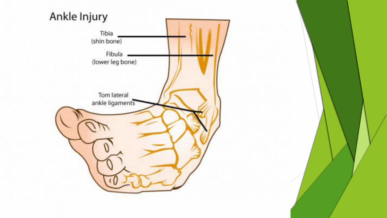
Ankle Bones and Ligaments
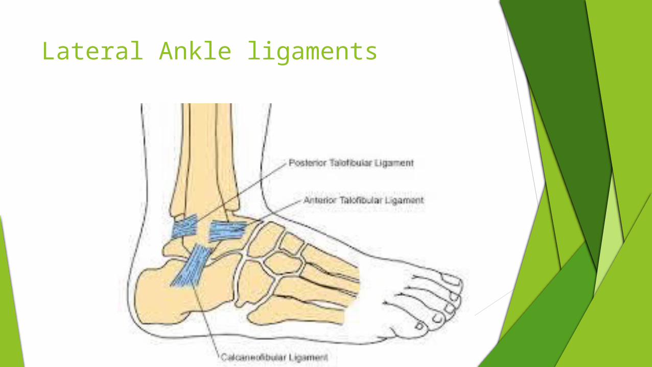
Lateral Ankle ligaments
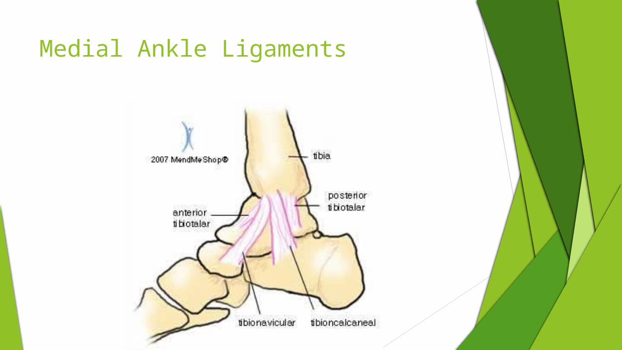
Medial Ankle Ligaments
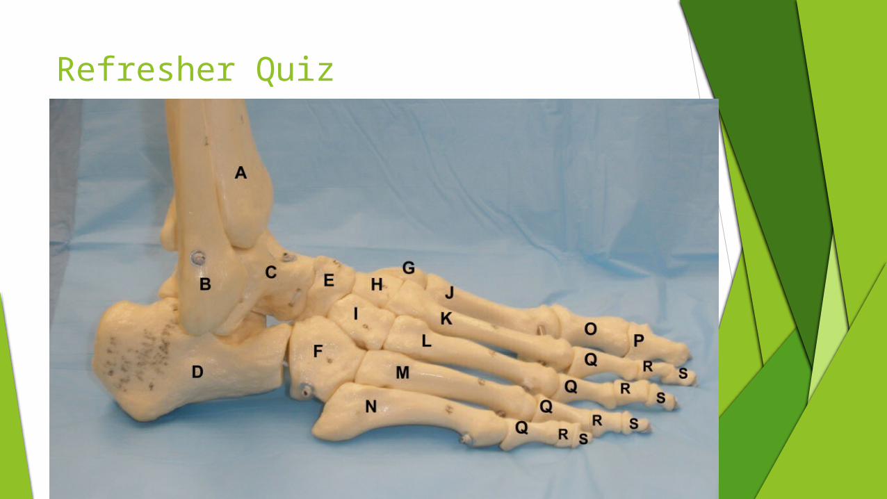
Refresher Quiz

Medial view
Injuries of the Foot, Ankle, and Lower Leg
Acute Soft Tissue Injuries
Commonly occur as a result of direct contact and intrinsic and extrinsic forces acting on the foot, ankle, and leg.
Can include:
Contusions
Sprains
Strains

Contusions
Mechanism:
Contact with ground or opposing player
Kicking unyielding object
Be hit or stepped on
2 types: Bone and Soft Tissue
Bone:
Direct contact to the medial tibia can result in localized swelling (periostitis) and hematoma formation under the periosteum.
This swelling can take considerable time for the body to absorb.
Contusions
Soft Tissue:
Disability and loss of function are usually much worse with muscle contusions due to tenderness, swelling, and spasm within the muscle tissue.
Decrease ROM and strength also occur depending on the severity of the contusion.
Rarely result in serious injury.
Complications can arise from severe contusions and excessive bleeding within the enclosed anterior compartment.
Closely monitor any contusions that result in severe swelling of the compartment that result in neurovascular compromise.
Stone bruises can be particularly problematic because of the nature of the injury.
Can cause pain and point tenderness, making it difficult to bear weight.
Discoloration and swelling may vary depending on severity.
Sprains Signs of inflammation
Pain
Heat
Swelling
Redness
Loss of function
1st degree
Ligament is stretched, causing minor pain and inflammation
2nd degree
Ligament is partially torn, causing considerable pain, and inflammation
3rd degree
Ligament is completely ruptured, may not be as painful, but inflammation is obvious and function may be completely lost.
Lateral Ankle Sprains
Mechanism:
Inversion with or without plantar flexion.
Typical scenario involves a player landing on opponent’s foot or landing awkwardly on the outside of the foot.
Signs and Symptoms:
Immediate pain
May hear or feel a “pop”
Most sprains will follow general characteristics of 1st-3rd degrees.
Swelling may be a poor indicator of severity of injury because even minor sprains can result in considerable swelling.
2nd and 3rd degree sprains will commonly involve other structures due to compressive forces.
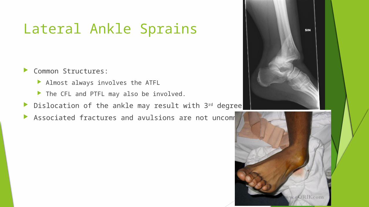
Lateral Ankle Sprains
Common Structures:
Almost always involves the ATFL
The CFL and PTFL may also be involved.
Dislocation of the ankle may result with 3rd degree sprains
Associated fractures and avulsions are not uncommon
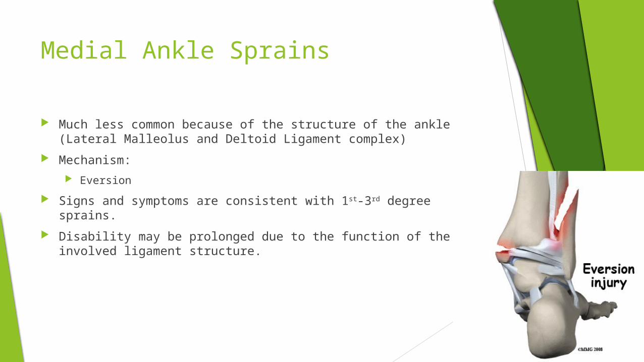
Medial Ankle Sprains
Much less common because of the structure of the ankle (Lateral Malleolus and Deltoid Ligament complex)
Mechanism:
Eversion
Signs and symptoms are consistent with 1st-3rd degree sprains.
Disability may be prolonged due to the function of the involved ligament structure.
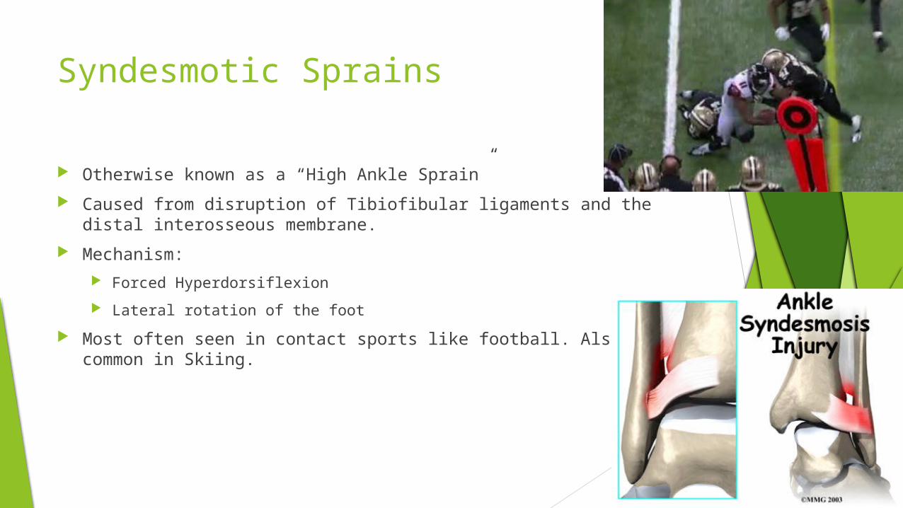
Syndesmotic Sprains
Otherwise known as a “High Ankle Sprain”
Caused from disruption of Tibiofibular ligaments and the distal interosseous membrane.
Mechanism:
Forced Hyperdorsiflexion
Lateral rotation of the foot
Most often seen in contact sports like football. Also common in Skiing.
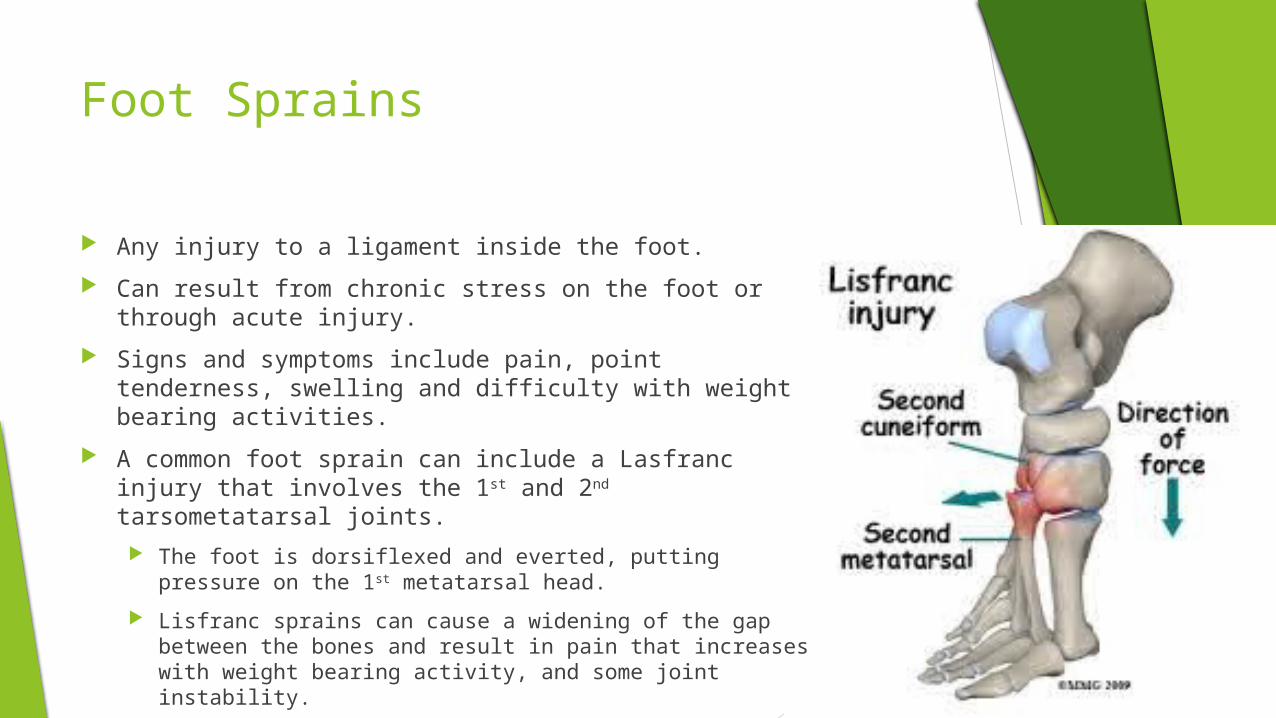
Foot Sprains
Any injury to a ligament inside the foot.
Can result from chronic stress on the foot or through acute injury.
Signs and symptoms include pain, point tenderness, swelling and difficulty with weight bearing activities.
A common foot sprain can include a Lasfranc injury that involves the 1st and 2nd tarsometatarsal joints.
The foot is dorsiflexed and everted, putting pressure on the 1st metatarsal head.
Lisfranc sprains can cause a widening of the gap between the bones and result in pain that increases with weight bearing activity, and some joint instability.
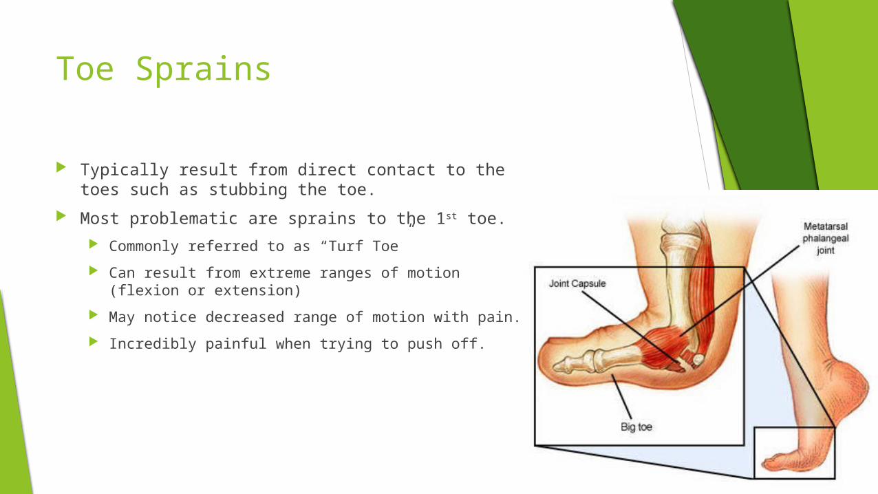
Toe Sprains
Typically result from direct contact to the toes such as stubbing the toe.
Most problematic are sprains to the 1st toe.
Commonly referred to as “Turf Toe”
Can result from extreme ranges of motion (flexion or extension)
May notice decreased range of motion with pain.
Incredibly painful when trying to push off.
Strains
Injury to a muscle or tendon
Typically result from either overstretching or muscular overload.
Most strains are of the acute variety, but there are some that are associated with chronic repetitive stress.
Signs and symptoms include the 5 signs of inflammation along with muscle spasm
2nd and 3rd degree strains may present a palpable defect in the muscle belly or tendon.
Cramping or prolonged spasm can lead to symptoms of a 1st degree strain on the following day.
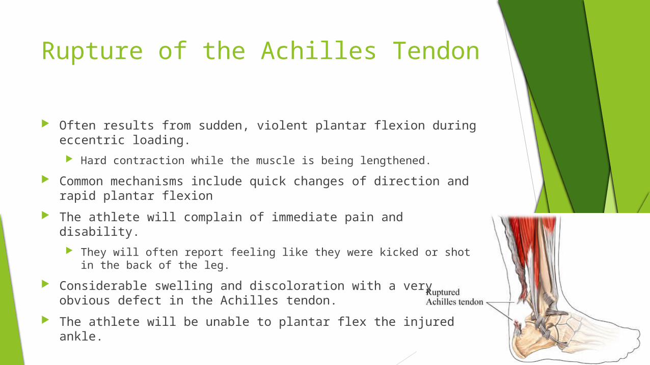
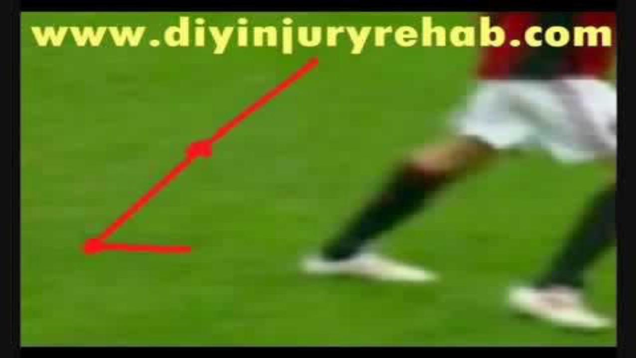
Rupture of the Achilles Tendon
Often results from sudden, violent plantar flexion during eccentric loading.
Hard contraction while the muscle is being lengthened.
Common mechanisms include quick changes of direction and rapid plantar flexion
The athlete will complain of immediate pain and disability.
They will often report feeling like they were kicked or shot in the back of the leg.
Considerable swelling and discoloration with a very obvious defect in the Achilles tendon.
The athlete will be unable to plantar flex the injured ankle.
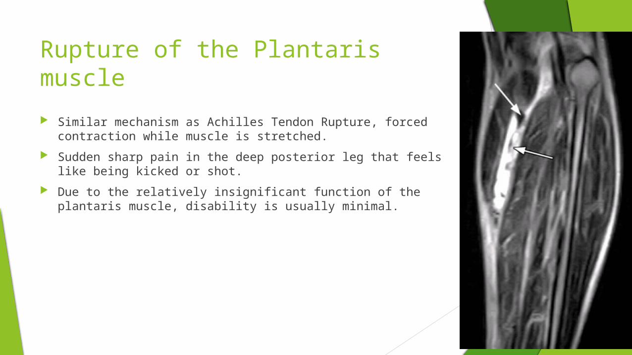
Rupture of the Plantaris muscle
Similar mechanism as Achilles Tendon Rupture, forced contraction while muscle is stretched.
Sudden sharp pain in the deep posterior leg that feels like being kicked or shot.
Due to the relatively insignificant function of the plantaris muscle, disability is usually minimal.
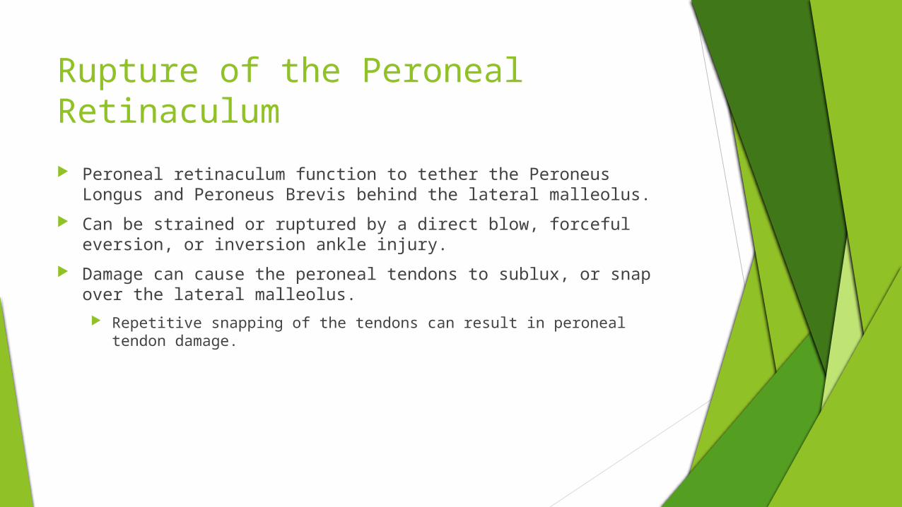
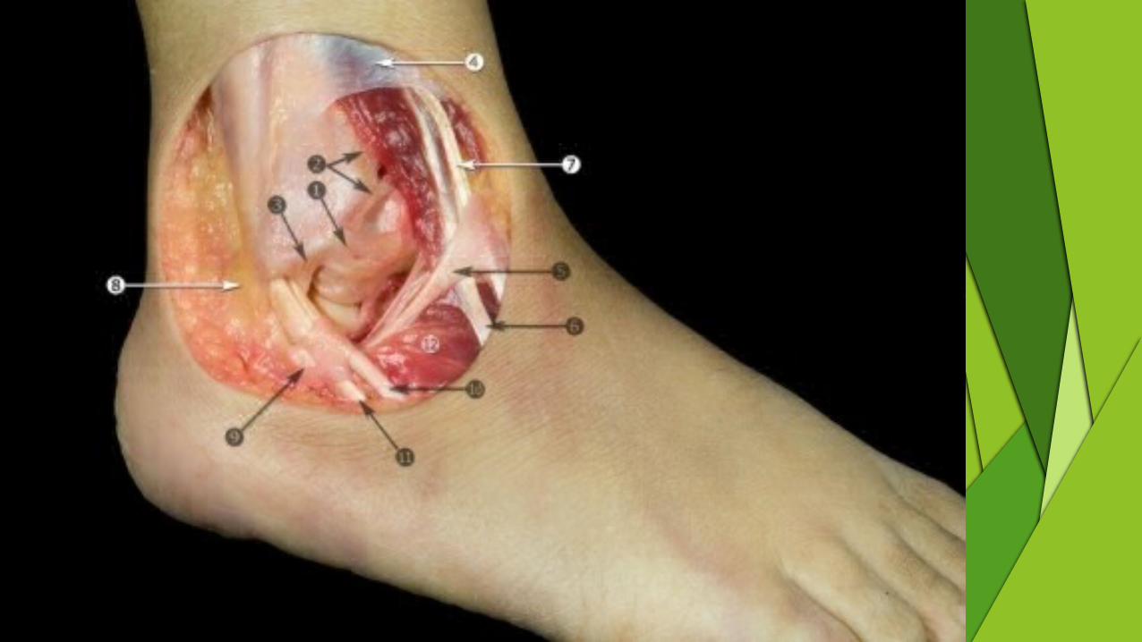
Rupture of the Peroneal Retinaculum
Peroneal retinaculum function to tether the Peroneus Longus and Peroneus Brevis behind the lateral malleolus.
Can be strained or ruptured by a direct blow, forceful eversion, or inversion ankle injury.
Damage can cause the peroneal tendons to sublux, or snap over the lateral malleolus.
Repetitive snapping of the tendons can result in peroneal tendon damage.

Chronic or Overuse Soft Tissue Injuries
Chronic Soft Tissue Injuries
Frequent complain in active individuals as a consequence of repeated microtrauma
The body is not allowed enough time to heal from normal wear and tear between workouts
This includes abnormal friction, traction, structural mechanics, or some combination of these.
Most problems stem from over pronation of the foot.
This can send excessive or abnormal stresses further up the body chain to the shins, knees, hips, and back.
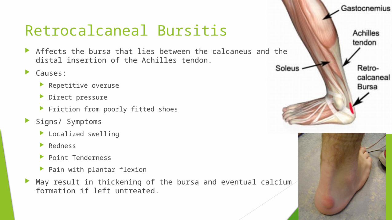
Retrocalcaneal Bursitis Affects the bursa that lies between the calcaneus and the distal
insertion of the Achilles tendon.
Causes:
Repetitive overuse
Direct pressure
Friction from poorly fitted shoes
Signs/ Symptoms
Localized swelling
Redness
Point Tenderness
Pain with plantar flexion
May result in thickening of the bursa and eventual calcium formation if left untreated.
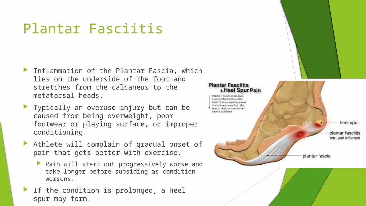
Plantar Fasciitis
Inflammation of the Plantar Fascia, which lies on the underside of the foot and stretches from the calcaneus to the metatarsal heads.
Typically an overuse injury but can be caused from being overweight, poor footwear or playing surface, or improper conditioning.
Athlete will complain of gradual onset of pain that gets better with exercise.
Pain will start out progressively worse and take longer before subsiding as condition worsens.
If the condition is prolonged, a heel spur may form.
Tendinitis
Inflammation of a tendon
Relatively common in the athletic population and typically results form overuse, friction, or tendon traction.
Signs and symptoms include pain, point tenderness, and crepitus over the inflamed tendon.
Crepitus is a grinding or grating sensation felt when moving the joint or muscle.
Pain may be evident with active or passive stretching.
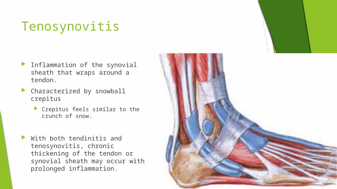
Tenosynovitis
Inflammation of the synovial sheath that wraps around a tendon.
Characterized by snowball crepitus
Crepitus feels similar to the crunch of snow.
With both tendinitis and tenosynovitis, chronic thickening of the tendon or synovial sheath may occur with prolonged inflammation.
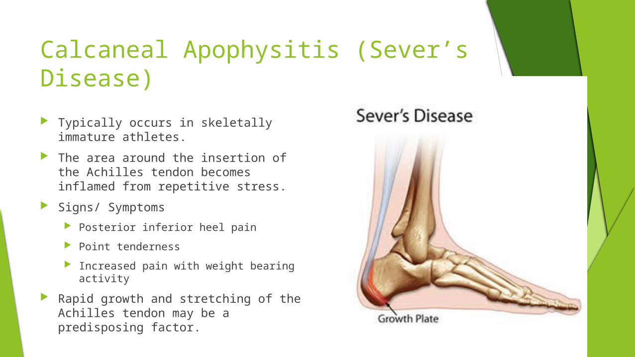
Calcaneal Apophysitis (Sever’s Disease)
Typically occurs in skeletally immature athletes.
The area around the insertion of the Achilles tendon becomes inflamed from repetitive stress.
Signs/ Symptoms
Posterior inferior heel pain
Point tenderness
Increased pain with weight bearing activity
Rapid growth and stretching of the Achilles tendon may be a predisposing factor.
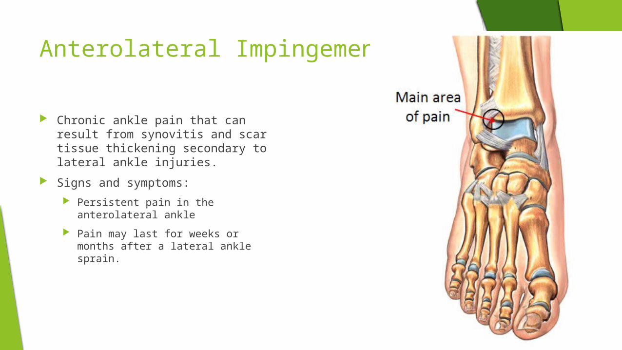
Anterolateral Impingement
Chronic ankle pain that can result from synovitis and scar tissue thickening secondary to lateral ankle injuries.
Signs and symptoms:
Persistent pain in the anterolateral ankle
Pain may last for weeks or months after a lateral ankle sprain.
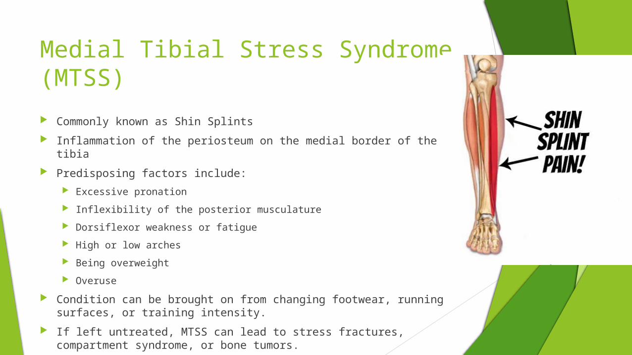
Medial Tibial Stress Syndrome (MTSS)
Commonly known as Shin Splints
Inflammation of the periosteum on the medial border of the tibia
Predisposing factors include:
Excessive pronation
Inflexibility of the posterior musculature
Dorsiflexor weakness or fatigue
High or low arches
Being overweight
Overuse
Condition can be brought on from changing footwear, running surfaces, or training intensity.
If left untreated, MTSS can lead to stress fractures, compartment syndrome, or bone tumors.
Traumatic Fractures
Tibial and Fibular Fractures
Can result from direct blow or indirect torsional stress
Fibula
Can result from direct blow or from severe eversion stress
Avulsion fractures are not uncommon in association with lateral ankle sprains.
Tibia
Requires much more force.
Can result from direct impact in sports like football.
Push off and avulsion fractures are relatively common as a result of severe inversion or eversion forces on the ankle.


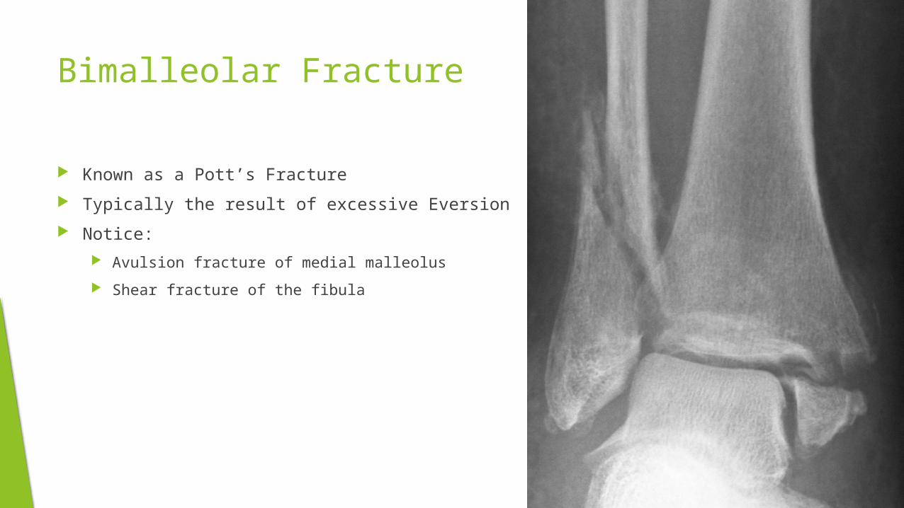
Bimalleolar Fracture
Known as a Pott’s Fracture
Typically the result of excessive Eversion
Notice:
Avulsion fracture of medial malleolus
Shear fracture of the fibula
Foot Fractures
Typically the result of the same forces associated with foot sprains.
May also result from being stepped on, having heavy objects dropped on the foot, or from falling from a great hight.
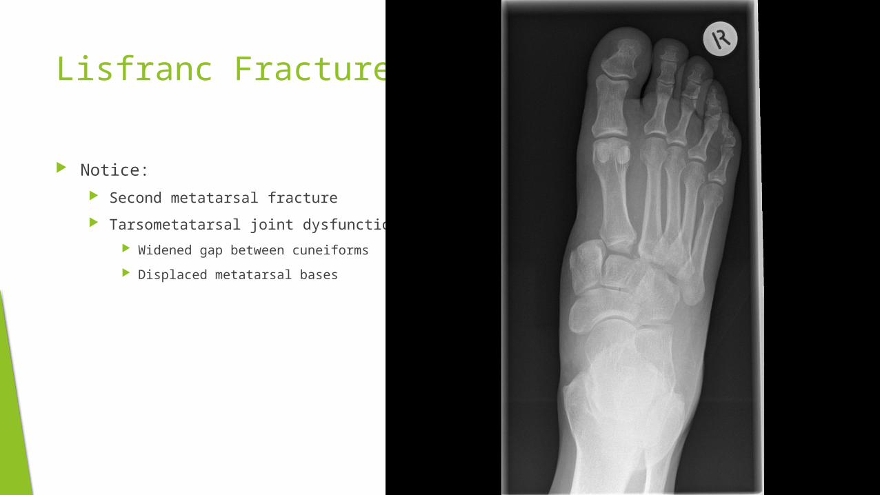
Lisfranc Fractures
Notice:
Second metatarsal fracture
Tarsometatarsal joint dysfunction
Widened gap between cuneiforms
Displaced metatarsal bases
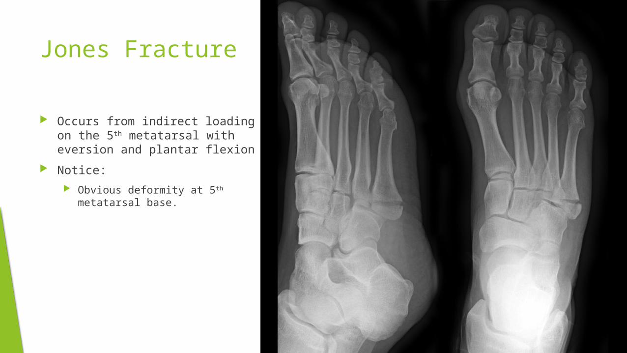
Jones Fracture
Occurs from indirect loading on the 5th metatarsal with eversion and plantar flexion
Notice:
Obvious deformity at 5th metatarsal base.

Calcaneal Fractures
Much less common
The most common cause happens when falling and landing on the heel from a great height.