The Structure, Function, and Evolution of Biological Systems
Upload
laurence-blairCategory
view
220download
1
Animal structure and function (chap 40)

Introduction
Anatomy:Biological formPhysiology:Biological functionInterstitial fluid:Fluid surrounding the cells

Body plan
Exchange
Mouth
Gastrovascularcavity
Exchange
Exchange
A hydra, an animal with twolayers of cells
(b)(a) An amoeba, a single-celledorganism
0.1 mm 1 mm
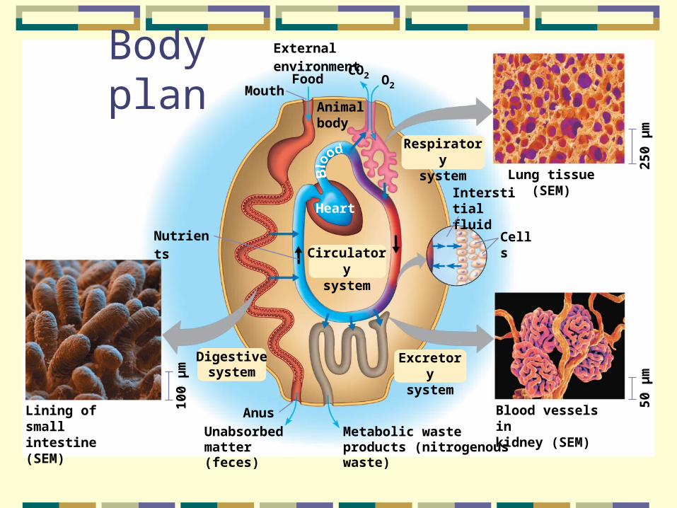
Body plan
External environment
FoodMouth
Animalbody
O2
CO2
Respiratorysystem
Lung tissue (SEM)Interstitialfluid
Cells
Excretorysystem
Blood vessels inkidney (SEM)
50 µ
m
Heart
Circulatorysystem
Nutrients
Digestivesystem
Anus
Metabolic wasteproducts (nitrogenous waste)
Unabsorbedmatter (feces)
250
µm
100
µm
Lining of smallintestine (SEM)

Tissues
EpithelialConnectiveMuscleNervous

Epithelial tissue
Epithelial tissue (epithelium):Covers all surfaces of the bodyEpidermis (ectoderm): Outer portion of skinEndoderm:Lining of inner surfaces of digestive tract Mesoderm:Inner surface of body cavities

Epithelial tissue
Closely packedTight junctionsOne/or few cell layers thickSelective absorption in the intestinesRapid gas exchange in lungsProtection from microbes, water loss
Epithelial tissue
Regenerative capabilitiesLiver (gland from epithelial tissues)Epidermis every 2 weeksStomach lining every 2-3 days

Epithelial tissue
TypesBased on cell thicknessShape on exposed surfaceSimpleOne layer thickStratifiedMultiple layers of cells
Epithelial tissue
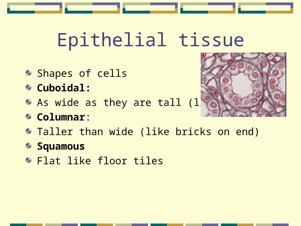
Shapes of cells Cuboidal:As wide as they are tall (like dice) Columnar:Taller than wide (like bricks on end) Squamous Flat like floor tiles

Epithelial tissues

Epithelial tissue
Simple squamous Lining of lungs, capillary walls and blood vesselsSimple cuboidal Lining of some glandsSimple columnar Lining of stomach, intestines and parts of respiratory tract
Epithelial tissue
Stratified squamous Outer layer of skin and mouthKeratin Water resistant protein

Epithelial tissue
Exocrine glands (duct system)Duct:Connection from gland to tissueSecrete/absorb chemical solutionsSweat & sebaceous glandsLining of intestines & lungs that secrete mucous

Epithelial tissue
Endocrine glands (ductless glands)HormonesSecreted into blood stream

Glands
Connective tissue
Holds tissues & organs togetherSupports, insulates and strengthens Derived from mesodermLoosely packed cellsScattered in an extracellular matrix
Connective tissue
Matrix: Composed of a web of fibers In a foundation of liquid, jellylike or solidFibers (proteins) are collagen, elastic, or reticular
Connective tissue
Collagen:Non-elastic-doesn’t tear easilyElastic:Makes tissue elasticElastin (protein)Reticular:Thin, branched, joins connective tissue to adjacent tissues

Connective tissue
Cells in matrixFibroblasts: Produce & secrete extracellular matrixMacrophages:Engulf foreign bodies & debrisMast cells & heparin

Connective tissue types
1. Loose connective tissueBeneath skin & between organsSupport, insulation, food storageAdipose tissue (fat)Cells become larger when gain weightShrink with weight loss

Connective tissue
2. Dense connective tissueTendons, ligaments, sheath around organs (periosteum), dermis of skinSupport, strong connections3. Special connective tissueCartilage, bone, blood,

Connective tissue

Special connective tissue
Cartilage Consists of chondroitin (glycoprotein) & collagenStrong, flexible tissue, absorb stressJoints, ear pinna, nose, intervertebral discs, larynxChondrocytes: Cartilage cells

Cartilage

Bone
Embryos---more cartilageCartilage is replaced with bone cells or osteocytesMatrix hardens with crystals of calcium phosphate mixed with collagen

Bone
Osteoblasts: Lay down new boneOsteoclasts: Dissolve boneOsteons: Unit of bone structureContains calcified matrix, osteocytes, nerve fibers, blood vessels

Bone
Flat bonesLong bonesSpongy bone: Contains marrowBlood cells formedCompact bone:More dense, gives strength

Bone

Bone

Blood
Plasma (matrix)CellsRBC (erythrocytes)Contain hemoglobin (carries oxygen)WBC (leukocytes)Neutrophils, eosinophils, basophils, lymphocytes, monocytesPlatelets (thrombocytes)

Blood

Blood
Plasma containsWastes, nourishmentHormonesNa+, Ca2+, other ionsFibrinogen, albumin, antibodies

Connective Tissue
Loose connective tissue Blood
Red blood cells
Chondrocytes
Chondroitin sulfate
Fat dropletsCentralcanal
Osteon
Nuclei
Elastic fiber
Collagenous fiberWhitebloodcells
Plasma
Cartilage
Adipose tissueBone
Fibrous connective tissue
120
µm
30 µ
m
150
µm
100
µm
55 µ
m
700
µm

Muscle tissue
MovementOrganization of actin & myosin filamentsSmooth, skeletal & cardiac musclesStriated muscles: skeletal & cardiacSkeletal muscles: voluntary controlSmooth & cardiac muscles: involuntary control

Muscle tissue
Smooth muscle Walls of blood vessels, stomach, intestinesViscera: Internal organsMade of sheets of cells each with a single nucleus

Muscle tissue
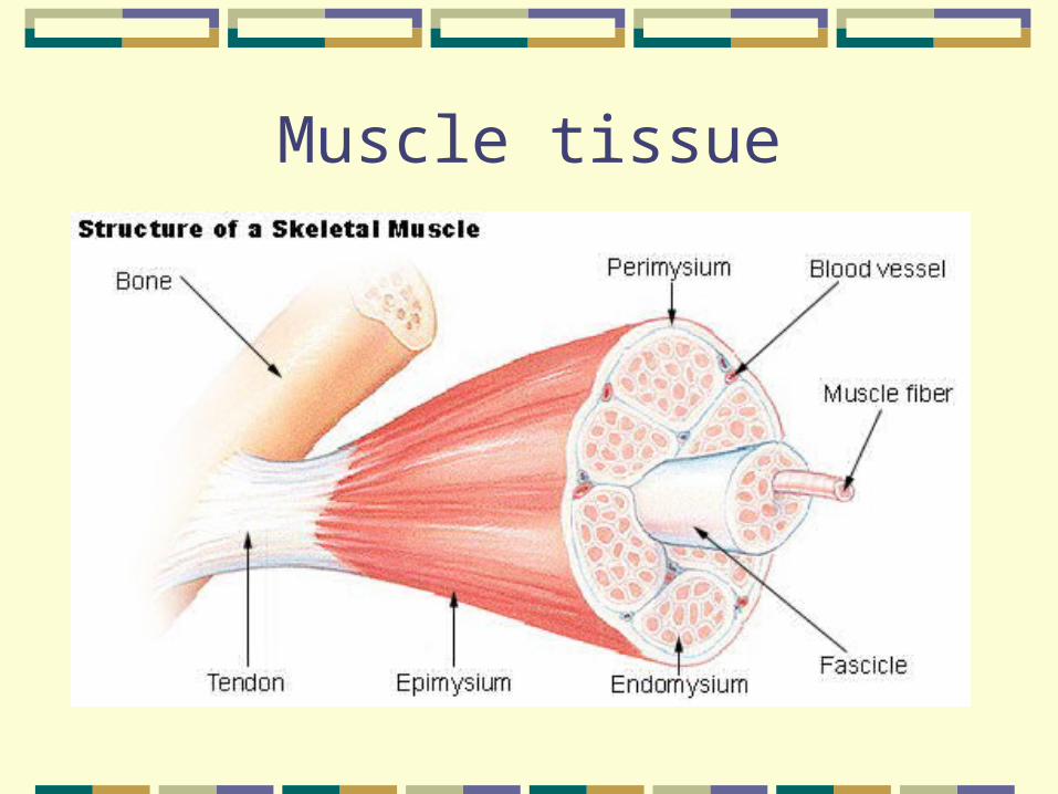
Skeletal muscleAttached by tendons to bonesContract move bones
Muscle tissue

Muscle tissue
Cardiac muscleSmall interconnected cellsLinked by gap junctionsOpenings allow small substances & electrical charges to pass between cellsMyocardium Single functioning units


Nerve tissue
NeuronsCell body, dendrites, axon Neuroglia Supporting cellsInsulate neuronsEliminate foreign bodies

Nerve tissue
DentritesThin, branched extensions Receive impulsesAxonsSingle extension of cell bodyCarries impulse awayMyelin sheaths, insulating cover

Neurons

Neurons
Sensory neuronsEye,ears, surface of skinMotor neuronsBrain & spinal cordInterneuronsBrain & spinal cordNeurons within the CNS

Neurons
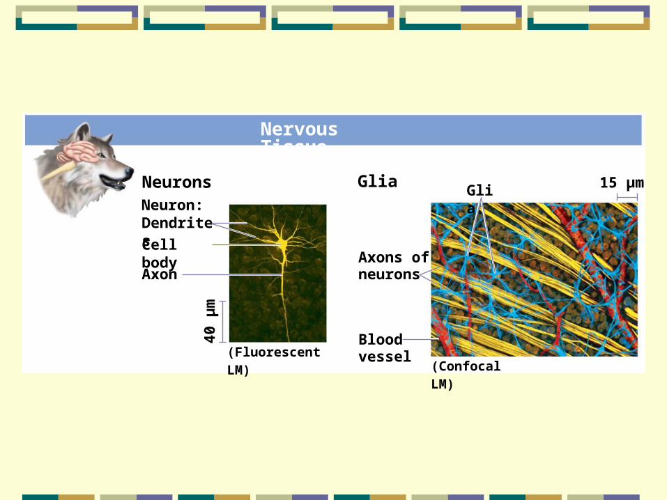
Nervous Tissue
NeuronsNeuron:Dendrites
Cell body
Axon
(Fluorescent LM)(Confocal LM)
40 µ
m
Glia
Axons ofneurons
Bloodvessel
Glia 15 µm

Summary
Epithelial tissuesSimple or stratifiedCuboidal, columnar, squamousConnective tissuesLoosely packed, tightly packedSpecial (bone, cartilage, blood)Matrix

Summary
Muscle tissuesSmooth, cardiac, skeletalNerve tissues:Neurons (cell body, dentrites, axons)Sensory, motor and interneurons
Coordination
HormonesNervous systemHomeostasis

(a) Signaling by hormones
STIMULUS
Endocrinecell
Hormone
Signal travelseverywhere.
Bloodvessel
Nerveimpulse
Axons
Nerveimpulse
Signal travelsto a specificlocation.
Axon
Cell bodyof neuron
(b) Signaling by neurons
STIMULUS
ResponseResponse

Homeostasis
Dynamic constancy of internal environmentDynamic because conditions fluctuate Narrow rangepHTempGlucoseOxygen

Regulation
1. Negative feedback loops2. Positive feedback loops
Negative Feedback

Negative feedback loops
SensorsMeasure internal environmentIntegrating centerReceives information from sensorsCompares to normal rangeResponds

Negative feedback loops
Effectors:Muscles or glandsReceive information from centerResponse
Negative feedback loops
Temperature increaseHypothalamus senses deviationSends signals to relieve heatSweating & vasodilationReach baseline Negative feedback stops response
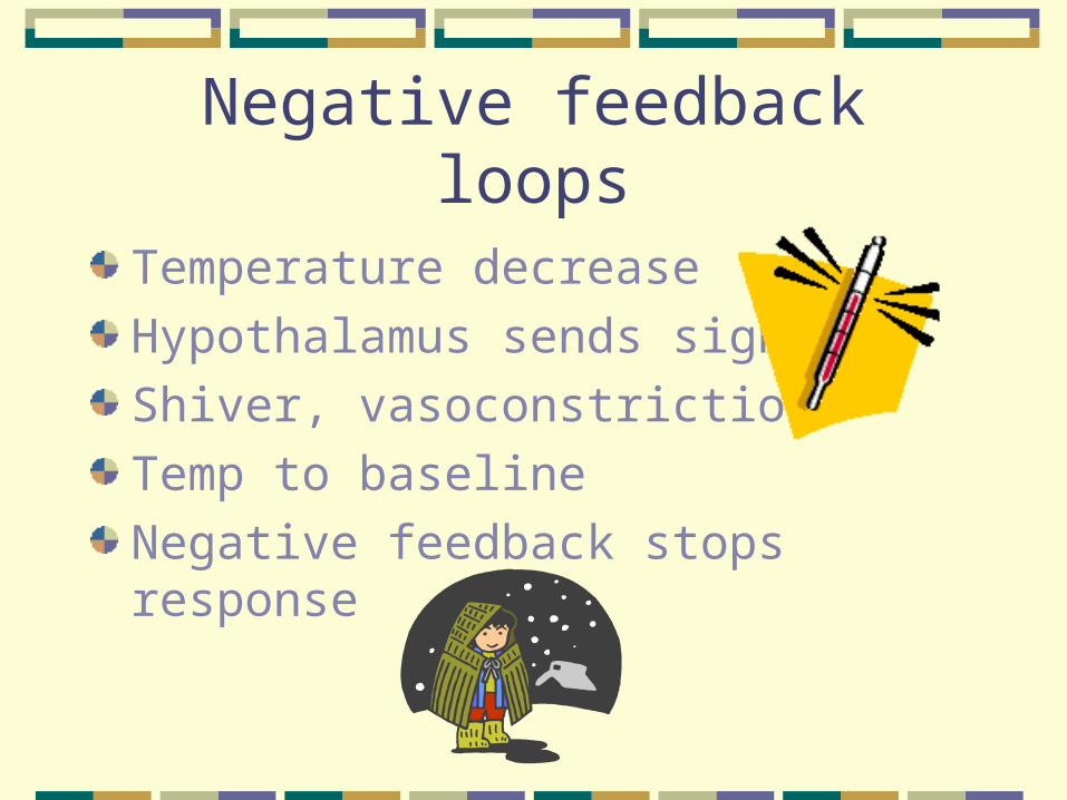
Negative feedback loops
Temperature decreaseHypothalamus sends signalsShiver, vasoconstrictionTemp to baseline Negative feedback stops response

Thermoregulation

Fig. 40-9
(a) A walrus, an endotherm
(b) A lizard, an ectotherm

Negative feedback loops
Glucose (eat a meal)Elevated blood levelIslets of Langerhans (sensor, center)InsulinLowers blood sugar (uptake in muscle, fat & liver cells)Negative feedback stops insulin release

Regulating Blood Sugar
Positive Feedback

Positive feedback loops
Uterine contractionsPressure from baby on uterusCauses contractionsCauses more stretchingMore contractionsContinues until birth

Positive feedback loop
Blood clottingClotting factors stimulate the formation of more factorsClot formsMaintain blood volume

Bioenergetics
Overall flow & transformation of energy in an animalDetermines nutritional needsAnimal size, activity and environment
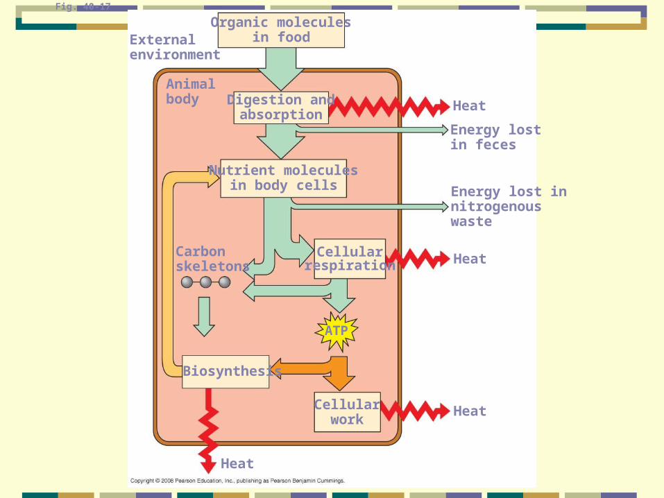
Fig. 40-17
Organic moleculesin foodExternal
environment
Animalbody Digestion and
absorption
Nutrient moleculesin body cells
Carbonskeletons
Cellularrespiration
ATP
Heat
Energy lostin feces
Energy lost innitrogenouswaste
Heat
Biosynthesis
Heat
Heat
Cellularwork

Metabolic rate
Amount of energy an animal uses in a unit of timeTorpor:Physiological state of low activity with low metabolismHibernation:Long term torpor