Animal Models of Parkinson’s Diseasedownloads.hindawi.com/journals/specialissues/203981.pdf ·...
174
Parkinson’s Disease Guest Editors: Yuzuru Imai, Katerina Venderova, David S. Park, Huaibin Cai, and Enrico Schmidt Animal Models of Parkinson’s Disease
Transcript of Animal Models of Parkinson’s Diseasedownloads.hindawi.com/journals/specialissues/203981.pdf ·...

Parkinson’s Disease
Guest Editors: Yuzuru Imai, Katerina Venderova, David S. Park, Huaibin Cai, and Enrico Schmidt
Animal Models of Parkinson’s Disease

Animal Models of Parkinson’s Disease

Parkinson’s Disease
Animal Models of Parkinson’s Disease
Guest Editors: Yuzuru Imai, Katerina Venderova,David S. Park, Huaibin Cai, and Enrico Schmidt

Copyright © 2011 SAGE-Hindawi Access to Research. All rights reserved.
This is a special issue published in volume 2011 of “Parkinson’s Disease.” All articles are open access articles distributed under theCreative Commons Attribution License, which permits unrestricted use, distribution, and reproduction in any medium, provided theoriginal work is properly cited.

Editorial Board
Jan O. Aasly, NorwayCristine Alves da Costa, FranceIvan Bodis-Wollner, USAD. J. Brooks, UKCarlo Colosimo, ItalyMark R. Cookson, USAAlan R. Crossman, UKT. M. Dawson, USAH. J. Federoff, USA
Francisco Grandas, SpainPeter Hagell, SwedenN. Hattori, JapanMarjan Jahanshahi, UKE. D. Louis, USAP. Martinez Martin, SpainF. Mastaglia, AustraliaHuw R. Morris, UKM. Maral Mouradian, USA
Antonio Pisani, ItalyJose Rabey, IsraelHeinz Reichmann, GermanyFabrizio Stocchi, ItalyEng King Tan, SingaporeHelio Teive, BrazilDaniel Truong, USAYoshikazu Ugawa, Japan

Contents
Animal Models of Parkinson’s Disease, Yuzuru Imai, Katerina Venderova, David S. Park, Huaibin Cai,and Enrico SchmidtVolume 2011, Article ID 364328, 2 pages
Toxin-Induced and Genetic Animal Models of Parkinson’s Disease, Shin Hisahara and Shun ShimohamaVolume 2011, Article ID 951709, 14 pages
VMAT2-Deficient Mice Display Nigral and Extranigral Pathology and Motor and Nonmotor Symptomsof Parkinson’s Disease, Tonya N. Taylor, W. Michael Caudle, and Gary W. MillerVolume 2011, Article ID 124165, 9 pages
The Endotoxin-Induced Neuroinflammation Model of Parkinson’s Disease, Kemal Ugur Tufekci,Sermin Genc, and Kursad GencVolume 2011, Article ID 487450, 25 pages
Models for LRRK2-Linked Parkinsonism, Tianxia Li, DeJun Yang, Sarah Sushchky, Zhaohui Liu,and Wanli W. SmithVolume 2011, Article ID 942412, 16 pages
α-Synuclein Transgenic Drosophila As a Model of Parkinson’s Disease and Related Synucleinopathies,Hideya Mizuno, Nobuhiro Fujikake, Keiji Wada, and Yoshitaka NagaiVolume 2011, Article ID 212706, 7 pages
Drosophila Models of Parkinson’s Disease: Discovering Relevant Pathways and Novel TherapeuticStrategies, Veronica Munoz-Soriano and Nuria ParicioVolume 2011, Article ID 520640, 14 pages
Optimizing a Rodent Model of Parkinson’s Disease for Exploring the Effects and Mechanisms of DeepBrain Stimulation, Karl Nowak, Eilhard Mix, Jan Gimsa, Ulf Strauss, Kiran Kumar Sriperumbudur,Reiner Benecke, and Ulrike GimsaVolume 2011, Article ID 414682, 19 pages
Therapeutic Effects of Hydrogen in Animal Models of Parkinson’s Disease, Kyota Fujita,Yusaku Nakabeppu, and Mami NodaVolume 2011, Article ID 307875, 9 pages
Limitations of Animal Models of Parkinson’s Disease, J. A. Potashkin, S. R. Blume, and N. K. RunkleVolume 2011, Article ID 658083, 7 pages
Manganese Inhalation as a Parkinson Disease Model, Jose Luis Ordonez-Librado,Veronica Anaya-Martınez, Ana Luisa Gutierrez-Valdez, Laura Colın-Barenque, Enrique Montiel-Flores,and Maria Rosa Avila-CostaVolume 2011, Article ID 612989, 14 pages
MPTP Neurotoxicity and Testosterone Induce Dendritic Remodeling of Striatal Medium Spiny Neuronsin the C57Bl/6 Mouse, Eleni Antzoulatos, Michael W. Jakowec, Giselle M. Petzinger, and Ruth I. WoodVolume 2011, Article ID 138471, 10 pages

Protective Role of rAAV-NDI1, Serotype 5, in an Acute MPTP Mouse Parkinson’s Model,Jennifer Barber-Singh, Byoung Boo Seo, Akemi Matsuno-Yagi, and Takao YagiVolume 2011, Article ID 438370, 10 pages
Effects of Human Alpha-Synuclein A53T-A30P Mutations on SVZ and Local Olfactory Bulb CellProliferation in a Transgenic Rat Model of Parkinson Disease, Faustine Lelan, Cecile Boyer,Reynald Thinard, Severine Remy, Claire Usal, Laurent Tesson, Ignacio Anegon, Isabelle Neveu,Philippe Damier, Philippe Naveilhan, and Laurent LescaudronVolume 2011, Article ID 987084, 11 pages

SAGE-Hindawi Access to ResearchParkinson’s DiseaseVolume 2011, Article ID 364328, 2 pagesdoi:10.4061/2011/364328
Editorial
Animal Models of Parkinson’s Disease
Yuzuru Imai,1 Katerina Venderova,2 David S. Park,3 Huaibin Cai,4 and Enrico Schmidt5
1 Department of Neuroscience for Neurodegenerative Disorders, Juntendo University Graduate School of Medicine,Tokyo 113-8421, Japan
2 Department of Physiology and Pharmacology, Thomas J. Long School of Pharmacy and Health Sciences, University of the Pacific,Stockton, CA 95211, USA
3 Ottawa Health Research Institute, Neuroscience Research Institute, 451 Smyth Road, Ottawa, ON, Canada K1H 8M54 Laboratory of Neurogenetics, National Institute on Aging, National Institutes of Health, 35 Convent Drive, Bethesda,MD 20892-3707, USA
5 Department of Bioinformatics and Molecular Genetics and Center for Biological Systems Analysis, University of Freiburg,79104 Freiburg, Germany
Correspondence should be addressed to Yuzuru Imai, [email protected]
Received 14 November 2011; Accepted 14 November 2011
Copyright © 2011 Yuzuru Imai et al. This is an open access article distributed under the Creative Commons Attribution License,which permits unrestricted use, distribution, and reproduction in any medium, provided the original work is properly cited.
Parkinson’s disease (PD) is considered a multifactorial dis-order, which is neuropathologically characterized by age-dependent neurodegeneration of dopaminergic neurons inthe midbrain. Different neurotoxins including syntheticcompounds, heavy metals, and dopamine itself have beenproposed to be environmental risk factors of PD. Recentgenome-wide genetic and mutational studies provide infor-mation on various genetic risk factors while microglial acti-vation in the affected regions has emerged to be involvedin the disease development as a local microenvironmentalfactor. A wide variety of animal models of PD substantiallycontribute to the understanding of these issues and thedevelopment of therapeutic approaches as an alternative tohumans although none of them fully recaptures the symp-toms and pathology of PD. This special issue is composed of9 excellent reviews and 3 distinguished original articles thatsummarize the most recent progresses and ideas obtainedfrom animal models in the pertinent field, while reportingthe putative molecular mechanisms of neurodegeneration,therapeutic challenges and limitations using PD models, andgeneration of new versions of PD models.
The first review paper briefly outlines animal models ofPD, covering toxin-induced and genetic models of vertebrateand invertebrate animals, in which characteristic features ofeach model are discussed.
Mishandling of monoamines including dopamine hasbeen hypothesized to damage neurons. The second reviewpaper describes mice with impaired functions of the vesicular
monoamine transporter VMAT2, in which progressive loss ofcatecholamine-secreting neurons is observed. Such modelsmay be potentially useful for the development of newtherapeutic strategies, which would complement currentdopamine replacement.
Neuropathological analysis of the postmortem PD braintissues suggests that an adverse interaction with surroundingglia and other nonneuronal cells may be one of criticalsteps in neurodegeneration. The third review highlightsendotoxin-induced inflammation models, in which activa-tion of microglia and lymphocyte by a bacterial lipopolysac-charide deteriorates a healthy relationship with neurons.
Mutations in the leucine-rich repeat kinase 2 (LRRK2)gene have been identified to cause autosomal-dominantlate-onset PD and are also implicated in sporadic PD. Theneuropathological features of PD brain tissues with theLRRK2 mutations are characterized by typical Lewy bodypathology in the brainstem. The forth paper reviews a varietyof LRRK2-related models.
Mutations and increased expression in the α-synucleingene cause the development of early-onset familial PD. Theformation of α-synuclein fibrils and aggregates, a main com-ponent of Lewy bodies and Lewy neurites, is considered a keyprocess in the pathogenesis of PD and other synucleino-phathies. Other genetic determinants include the genes forMendelian forms of PD and susceptible genes. The followingtwo papers focus on the potential of Drosophila geneticmodels to examine α-synuclein and other responsible genes.

2 Parkinson’s Disease
Deep brain stimulation (DBS) by electrical pulses couldbe one of useful therapeutic avenues for PD. However, DBS’stechnique requires advancement and poor understanding ofthe mechanisms involved hinder application in clinical prac-tice. The seventh review paper discusses the optimization ofa rat PD model for DBS.
Hydrogen has turned out to reduce oxidative damage.The eighth paper introduces the neuroprotective effects ofhydrogen on experimental animal models for PD and pos-sible application in treatment and prevention of PD.
The last review explains the limitations of animal models,showing differences between humans and animals, and dif-ficulties in interpretation of obtained results with animalmodels.
The first research paper investigates selective degenera-tion of dopaminergic neurons in the substantia nigra andassociated motor dysfunction induced by inhalation ofmixed manganese compounds on mice. This model couldbe instrumental for evaluating some aspects of a progressiveloss of dopaminergic neurons. The second research paperexamines the possible effects of testosterone on PD using amouse model induced by 1-methy-4-phenyl-1,2,3,6-tetrahy-dropyridine (MPTP) administration. The study suggests thatloss of testosterone induces remodeling in the morphologyof medium spiny neurons where dopaminergic neurons ofthe substantia nigra project although no interaction betweentestosterone and loss of dopaminergic neurons by MPTPadministration is observed. The third research paper ofthis special issue addresses improvement of potential genetherapy to compensate for impaired complex I activity ofthe mitochondria using the yeast single-subunit NADH-ubiquinone oxidoreductase, NDI1. NDI1 is functionally ableto replace complex I, activity of which is thought to becompromised in most of PD cases.
A decreased sense of smell is one of early signs of PD.Although degeneration of tyrosine hydroxylase-positive neu-rons in the olfactory bulbs is observed, the pathogenicmechanism underlying olfactory deficits is not well under-stood. The forth research paper addresses this issue using arat model bearing the pathogenic α-synuclein.
Yuzuru ImaiKaterina Venderova
David S. ParkHuaibin Cai
Enrico Schmidt

SAGE-Hindawi Access to ResearchParkinson’s DiseaseVolume 2011, Article ID 951709, 14 pagesdoi:10.4061/2011/951709
Review Article
Toxin-Induced and Genetic Animal Models of Parkinson’s Disease
Shin Hisahara and Shun Shimohama
Department of Neurology, Sapporo Medical University, South1, West17, chuo-ku, Sapporo 060-8556, Japan
Correspondence should be addressed to Shun Shimohama, [email protected]
Received 13 October 2010; Accepted 31 October 2010
Academic Editor: Yuzuru Imai
Copyright © 2011 S. Hisahara and S. Shimohama. This is an open access article distributed under the Creative CommonsAttribution License, which permits unrestricted use, distribution, and reproduction in any medium, provided the original work isproperly cited.
Parkinson’s disease (PD) is a common progressive neurodegenerative disorder. The major pathological hallmarks of PD are theselective loss of nigrostriatal dopaminergic neurons and the presence of intraneuronal aggregates termed Lewy bodies (LBs), butthe pathophysiological mechanisms are not fully understood. Epidemiologically, environmental neurotoxins such as pesticidesare promising candidates for causative factors of PD. Oxidative stress and mitochondrial dysfunction induced by these toxinscould contribute to the progression of PD. While most cases of PD are sporadic, specific mutations in genes that cause familialforms of PD have led to provide new insights into its pathogenesis. This paper focuses on animal models of both toxin-inducedand genetically determined PD that have provided significant insight for understanding this disease. We also discuss the validity,benefits, and limitations of representative models.
1. Introduction
Parkinson’s disease (PD) is one of the most commonchronic neurodegenerative disorders. It is characterized by avariety of motor (bradykinesia, rigidity, tremor, and posturalinstability) and nonmotor (autonomic disturbances and psy-chosis) symptoms. Although it can be diagnosed accurately,no therapeutic strategies can cure or completely block theprogression of PD. Pathologically, PD is characterized bythe severe loss of dopaminergic (DAergic) neurons in thepars-compacta nigra and the presence of proteinaceous α-synuclein inclusions, called Lewy bodies (LBs), which arepresent in neurons of the central nervous system (specificcortical regions, brain stem, and spinal cord), peripheralautonomic nervous system, enteric nervous system (ENS),and cutaneous nerves [1–3]. Similar to other neurodegenera-tive diseases, such as Alzheimer’s disease, age is the major riskfactor for PD although 10% of the people with the disease areyounger than 45.
Although PD is regarded as a sporadic disorder, remark-ably few environmental causes or triggers have been iden-tified [4–6]. Pesticides and herbicides are the most likelycandidates for environmental agents associated with thepathogenesis of PD. On the other hand, PD characteristicsare seen in a number of familial motor disorders caused
by different genetic factors. Animal models of neurode-generative diseases, including PD, have in general beenquite instructive in understanding their pathogenesis. Ideally,animal models of PD, whether induced by environmentalrisk factors (neurotoxins) or genetic manipulations, shouldfaithfully reproduce the clinical manifestations (behavioralabnormalities), pathological features, and molecular dys-functions characterizing the disease. Unfortunately, animalmodels rarely mimic the etiology, progression, and pathologyof PD completely, and in most cases, only partial insightcan be gained from these studies. Despite these difficulties,animal models are considered to be very helpful in thedevelopment of therapies to treat PD. In this paper, wediscuss recently developed neurotoxin-induced and geneticmodel animals of PD.
2. Animal Models of PD Induced by Neurotoxins
PD is currently viewed as a multifactorial disease. Environ-mental exposures, particularly to pesticides, are thought tobe involved in the pathogenesis of sporadic PD. Specifically,the herbicide Paraquat (PQ) and the fungicide Maneb(MB; manganese ethylene-bis-dithiocarbamate) have beenassociated with the incidence of PD [7, 8]. However, a causal

2 Parkinson’s Disease
role for pesticides in the etiology of PD has yet to bedefinitively established. In animal models, PD-like disordersinduced by neurotoxins or other chemical compounds haveled to a better understanding of the pathophysiology of PD(Table 1).
3. 1-Methyl-4-Phenyl-1,2,3,6-Tetrahydropyridine (MPTP)
In 1979 and 1983, MPTP was initially identified as astrong neurotoxin when heroin addicts accidentally self-administered MPTP and developed an acute form ofparkinsonism that was indistinguishable from idiopathicPD [9, 10]. A detailed neuropathological study of MPTP-induced parkinsonism in humans showed severe neuronaldegeneration in the substantia nigra and the absence of LBs[11]. The lack of LBs may have reflected the age of thepatient and the duration of exposure to MPTP. The tragicresults of MPTP poisoning in the heroin addicts led tothe development of MPTP-induced rodent and nonhumanprimate animal models of PD, which have proved extremelyvaluable [12–16]. The MPTP-exposed primates show goodresponse to therapy with L-3,4-dihydroxy-L-phenylalanine(L-DOPA) and dopamine (DA) receptor agonists [15, 16].However, rats are relatively insensitive to MPTP neurotox-icity compared with primates. Rats given MPTP at dosescomparable to those used in mice do not show remarkableneurodegeneration [17, 18]. Only high doses of MPTP causeDAergic neurodegeneration in rats, indicating that completeblockade of the DA receptors is required for them to displaysigns of parkinsonism. Mice, like rats, are also less sensitiveto MPTP than primates [19, 20].
This model also shows pathological changes in the ENS,as observed in PD. In PD, gastrointestinal (GI) dysfunctionwas hypothesized to depend on neuronal degeneration in theENS that is similar to that seen in the CNS. Recent studiesshow that the administration of MPTP results in decreasedtyrosine hydroxylase- (TH-) positive enteric neurons in mice,indicating that the MPTP model mice should be suitablefor understanding the extranigral pathophysiology of PD[21, 22].
4. 6-Hydroxy-Dopamine (6-OHDA)
Like MPTP, 6-OHDA is a neurotoxin that has been suc-cessfully used in induction animal models of PD. 6-OHDA’sstrong neurotoxic effects were described by Ungerstedt in1971, in a study presenting the first example of using achemical agent to produce an animal model of PD [23].Since 6-OHDA cannot cross the blood-brain barrier (BBB),systemic administration fails to induce parkinsonism. Thisinduction model requires 6-OHDA to be injected into thesubstantia nigra, medial forebrain bundle, and striatum [24,25]. The effects resemble those in the acute MPTP model,causing neuronal death over a brief time course (12 hours to2-3 days).
Interestingly, the intrastriatal injection of 6-OHDAcauses progressive retrograde neuronal degeneration in the
substantia nigra and ventral tegmental complex (ST-VTA)[25–27]. As in PD, DAergic neurons are killed, and thenon-DAergic neurons are preserved. However LBs do notform. Typically, 6-OHDA is used as a hemiparkinson model,in which its unilateral injection into the substantia nigracauses asymmetric motor behavior (turning, rotation) whenapomorphine, a DAergic receptor agonist, or amphetamine,a dopamine releasing agent, is given systemically. In thismodel, the quantifiable motor behavior is a major advantagefor screening pharmacological screening agents for theireffects on the DAergic system and for testing cell replacementtherapies [28–30].
5. Rotenone
Rotenone is a naturally occurring complex ketone pesticidederived from the roots of Lonchocarpus species. It can rapidlycross cellular membranes without the aid of transporters,including the BBB. Rotenone is a strong inhibitor of complexI, which is located at the inner mitochondrial membrane andprotrudes into the matrix.
In 2000, Betarbet et al. demonstrated in rats thatchronic systemic exposure to rotenone causes many featuresof PD, including nigrostriatal DAergic degeneration [31].Importantly, pathological features match those seen intypical PD. For example, many of the degenerating neuronshave intracellular inclusions that are morphologically similarto LBs. These inclusions also show immunoreactivity forα-synuclein and ubiquitin, like true LBs [31, 32]. Therotenone-administered model animals also reproduce allthe behavioral and pathological features seen in the typicalform of human PD. However, rotenone-injected rats withoutnigrostriatal DAergic neuronal loss demonstrate the sameabnormal motor behaviors as those with such pathologicalfeatures [32, 33]. This finding suggested that the abnormalbehaviors of PD could depend, at least partly, on thedamage to non-DAergic neurons in the nigrostriatal area.Furthermore, rotenone exposure also causes the loss ofmyenteric neurons in the rat [34].
6. Paraquat and Maneb
Because of its close structural similarity to 1-methyl-4-phenylpyridinium (MPP+, the active metabolite formof MPTP), an herbicide, 1,1′-dimethyl-4,4′-bipyridinium,named paraquat has been suggested as a risk factor for PD[35]. The systemic administration of paraquat to adult miceresults in a significant decrease in substantia nigra DAergicneurons, a decline in striatal dopamine nerve terminaldensity, and a neurobehavioral syndrome characterized byreduced ambulatory activity [36]. These data support theidea that paraquat crosses the BBB to cause destruction of thedopamine neurons in the substantia nigra, like MPP+ [36].The prolonged exposure to paraquat leads to a remarkableaccumulation of α-synuclein-like aggregates in neurons ofthe substantia nigra pars compacta in mice [37]. Chronicexposure to paraquat also reduces the expression of thenicotinic acetylcholine receptor (nAChR) subunit α3/α6β2∗

Parkinson’s Disease 3
Table 1: Representative neurotoxin-induced mammalian models of Parkinson’s disease.
Neurotoxin Behavioral and pathological features Molecular mechanisms
MPTP
(1) Parkinsonism (akinesia, rigidity, and tremor) withacute onset(2) Relatively less potent in rodents(3) Good response to L-DOPA and DA-agonists(4) Loss of TH-neurons (-fibers) and DA-content innigrostriatal region(5) Loss of TH-neurons (-fibers) in ENS(6) α-Synuclein-positive inclusions(7) No typical LBs
(1) Easily crosses the BBB(2) Converted to MPP+ in glial cells(3) Transferred into mitochondria by transporters(4) Inhibits electron transport chain complex I(5) Upregulation of iNOS, NADPH-oxidase, and ROS(6) Microglial activation
6-OHDA
(1) Intracerebral administration(2) Quantifiable locomotor abnormalities (rotation,akinsesia)(3) Good response to L-DOPA and DA-agonists(4) Loss of TH-neurons (-fibers) and DA-content innigrostriatal region(5) No typical LBs
(1) Transferred into mitochondria by transporters(2) Inhibits electron transport chain complex I(3) Microglial activation
Rotenone
(1) Parkinsonism (bradykinesia, fixed posture, andrigidity)(2) Good response to L-DOPA and DA-agonists(3) Loss of TH-neurons (-fibers) and DA-content innigrostriatal region(4) α-Synuclein-positive inclusions, resemblance totrue LBs(5) Loss of myenteric neurons
(1) Easily crosses the BBB(2) Inhibits electron transport chain complex I(3) Upregulation of NADPH-oxidase(4) Microglial activation
Paraquat (+ Maneb)(1) Parkinsonism similar to that of induced by MPTP(2) Loss of DA-content in nigrostriatal region(3) α-Synuclein-positive inclusions with long exposure
(1) Crosses the BBB by neutral amino acid transporter(2) Inhibits electron transport chain complex I(3) Reduction of nAchR-mediated DA release(4) Inhibits complex III (Maneb)
MPTP: 1-Methyl-4-phenyl-1,2,3,6-tetrahydropyridine; 6-OHDA: 6-hydroxy-dopamine; L-DOPA: L-3,4-dihydroxy-L-phenylalanine; TH: tyrosine hydroxy-lase; DA: dopamine; ENS: enteric nervous system; LB: Lewy body; BBB: blood-brain barrier; MPP+: 1-methyl-4-phenylpyridinium; iNOS: inducible nitricoxide synthase; ROS: reactive oxygen species; nAchR: nicotinic acetylcholine receptor.
(the asterisk indicates the possible presence of additionalsubunits). Normally, the activation of both nAChR subtypesstimulates DA release in the striatum [38–40]. The injectionof paraquat selectively reduces the α3/α6β2∗-mediated DArelease from the striatum in primates [41].
Manganese ethylenebis-dithiocarbamate (Maneb) is anorganomanganese fungicide that is broadly used in agricul-ture and is a putative causative agent for PD. Surprisingly,Thiruchelvam et al. found that the neurotoxic effects ofmaneb or paraquat on the nigrostriatal DA system in miceare synergistically potentiated in combination [42]. Theirreport argued that this finding has important implicationsfor the human risk of PD, because the marked geographicaloverlap in the estimated annual agricultural applications ofparaquat and maneb means that people living in these areasmay be exposed to the synergistic neurotoxicity of these twoagents [42, 43].
7. Pathophysiological Mechanisms ofDAergic Neurotoxins
All the representative neurotoxin-induced PD modelsdescribed above show defective mitochondrial function,manifested by the inhibition of mitochondrial complex I
or III. MPTP is a highly lipophilic agent. After its systemicadministration, MPTP rapidly crosses the BBB. Once inthe brain, MPTP is converted to 1-methyl-4-phenyl-2,3-dihydropyridium (MPDP+) in glial cells (astrocytes) andserotonin neurons by monoamine oxidase B (MAO-B) andthen spontaneously oxidizes to MPP+ [44, 45]. Thereafter,MPP+ is released into the extracellular space. Unlike MPTP,MPP+ is a polar molecule that cannot freely enter DAergicneurons. Thus, a plasma membrane transport system isrequired. MPP+ has a high affinity for dopamine trans-porter (DAT) as well as for norepinephrine and serotonintransporters [46, 47]. Once inside DAergic neurons, MPP+
can accumulate in mitochondria and impair mitochondrialrespiration by inhibiting complex I in the electron transportchain [44, 48], which induces the generation of reactiveoxygen species (ROS). MPP+ can also bind to vesicu-lar monoamine transporters (VMATs), which help moveselected materials into synaptic vesicles containing DA [49].MPP+ can also remain in the cytoplasm and interact withcytosolic enzymes [50].
Inducible nitric oxide synthase (iNOS) is also involvedin the pathogenesis of MPP+-induced parkinsonism inanimal models. Increased iNOS has also been found inthe substantia nigra of autopsied PD patients, indicatingthat NO overproduction is a feature of the human disease

4 Parkinson’s Disease
[51, 52]. Excess NO could contribute to the formationof free radicals, which could damage DAergic neurons,leading to the development of PD symptoms. Mice nullfor iNOS show a resistance to neuronal damage by MPTP,and iNOS inhibitors protect against the degeneration ofDAergic neurons in MPTP-treated mice [53, 54]. Further-more, microglial cells can be activated by the formationof free radicals and iNOS-mediated damage, and therebyexacerbate the toxicity of MPTP [55–57]. Finally, MPTPcan also upregulate NADPH-oxidase in the substantia nigraof mice [56], which is significant because NADPH-oxidaseappears to be ubiquitously expressed in all brain regionsand metabolizes molecular oxygen, generating superoxideas a product. In fact, MPTP toxicity is diminished in micelacking functional NADPH-oxidase, indicating a pivotal rolefor superoxide ions in the neurotoxicity induced by MPTP[56].
The toxicity of 6-OHDA also involves mechanisms ofoxidative stress. 6-OHDA can be taken up by DAergicneurons through DAT [58, 59]. Once transported into neu-rons, 6-OHDA is oxidized like DA. The oxidized moleculegenerates free radicals inhibits mitochondrial complex I andproduces superoxide and hydroxyl radicals [58, 59]. It isnot only toxic to the DAergic neurons but can also inducemicroglial activation [59].
Like MPTP, the pesticide rotenone is very lipophilic,crosses the BBB, and is distributed evenly throughout thebrain [59, 60]. It can enter mitochondria, where it inhibitscomplex I of the electron transport chain with high affinity[59]. Interestingly, the inhibition of microglial activation byan antibiotic, minocycline, can attenuate the neurotoxicity ofrotenone [61]. Gao et al. also showed that the neurotoxicityof rotenone is reduced in neuron-glia cocultures fromNADPH oxidase-null mice [62]. The DA uptake of theneuron-enriched cultures was not affected by the addition ofmicroglia from NADPH oxidase-null mice, the addition ofmicroglia from wild-type (WT) mice significantly increasedthe sensitivity of DAergic neurons either from WT orknockout (KO) mice to rotenone neurotoxicity. These dataindicate that microglial NADPH oxidase, but not neuronalNADPH oxidase, is responsible for the NADPH oxidase-mediated neurotoxicity of rotenone [62]. Paraquat mainlycrosses the BBB through the neutral amino acid transporter[63–65]. Once in the brain, it is selectively taken up bythe terminals of DA-containing neurons in the substantianigra by the DAT, and it inhibits mitochondrial complex I[63]. Maneb contains a major active fungicidal component,manganese ethylene-bis-dithiocarbamate (Mn–EBDC). Ina rat model in which Mn–EBDC is directly delivered tothe lateral ventricles, Mn–EBDC causes selective DAergicneurodegeneration [66]. Mn–EBDC preferentially inhibitsmitochondrial complex III [66].
8. Genetic Animal Models of PD
Although the etiopathogenesis (including environmentalfactors) of PD is not fully understood, the extensive exam-ination of human postmortem material, the genetic analysisof patients, and the study of experimental animal models
have shed significant light on the molecular mechanismsinvolved in its progression. However, since the number ofpatients with familial PD is extremely low compared to thenumber with sporadic PD, genetic studies in affected humanfamilies are very difficult. Therefore, the development ofanimal genetic models for PD is especially important, andsuch models provide an opportunity not only to investigatethe genetic etiology of PD but also to identify new factorsthat could be invaluable in terms of diagnosis, drug design,and/or therapy [67, 68]. Even invertebrate animals, for exam-ple, Drosophila melanogaster, are useful models for surveysof human PD. While their numbers of neurons and gliaare obviously much smaller than in rodents and primates,Drosophila have the same types of neuron-glia systems, and agreat number of genes and molecular transduction pathwaysare conserved between Drosophila and humans.
In recent years, several genetic animal models of PD havebeen reported, including models for autosomal-dominant(AD) inheritance patterns. The genes manipulated in thesemodels include α-synuclein, leucine rich repeat kinase 2(LRRK2), ubiquitin carboxyl-terminal esterase L1 (UCHL1),and high temperature requirement A2 (HTRA2/Omi)(Table 2). There are also models of autosomal-recessive (AR)inherited PD, which involve KO or knockdown genes forparkin, DJ-1, and phosphatase and tensin homolog- (PTEN-)induced novel kinase 1 (PINK1) (Table 3). In addition, wewill review a PD mouse model deficient in nuclear receptor-related 1 (Nurr1), also named nuclear receptor subfamily 4,group A, member 2 (NR4A2), which is a susceptibility genefor familial PD (Table 2).
8.1. α-Synuclein. α-synuclein was the first gene linked toan AD-type familial PD, called Park1. The identificationof an α-synuclein mutation in this family revolutionizedPD research, since α-synuclein is the main component ofLBs, which are observed in the sporadic PD brain. Thisstriking result strongly indicates that genetic and sporadicPD may share similar etiologies and that investigatingα-synuclein-mediated pathogenesis in familial PD coulduncover important information about sporadic PD. Threemissense mutations of α-synuclein, encoding the substitu-tions A30P, A53T, and E46K, have been identified in familialPD [67–70]. Furthermore, the duplication or triplicationof α-synuclein is sufficient to cause PD, suggesting that thelevel of α-synuclein expression is a critical determinant ofPD progression [71, 72]. Even though no direct relationshipbetween sporadic PD and α-synuclein expression has yetbeen shown, the existence of several polymorphisms in thepromoter or 3′-UTR of the α-synuclein gene suggests that itsexpression level might be a risk factor [73–75].
Human α-synuclein is an abundant 140-amino acidpresynaptic phosphoprotein involved in vesicle handlingand neurotransmitter release. Mutations in α-synuclein thatincrease the propensity for misfolding are probably deleteri-ous, because the misfolded forms are toxic, and they inducecell death in vitro [76, 77]. Among the variety of abnormalforms that mutant α-synuclein can adopt, protofibrils andfibrils seem to be the most toxic [77]. These demonstrations

Parkinson’s Disease 5
Table 2: Autosomal-dominant PD models.
Gene Animal Manipulation DA neuron loss LB-like inclusions1 DA-responsive motor deficits2 References
a-synuclein(PARK1)
Nematode Transgenic Yes§ No Yes [79, 80]
Fly Transgenic Yes Yes Yes [78]
Mouse Transgenic No Yes§ (PrP promoter) Yes§ (PDGFβ promoter) [81–91]
Rat Transgenic Yes No Yes [92–95]
Monkey Transgenic Yes No ND [96]
UCHL1(PARK5)
Mouse Transgenic Yes No Yes [105, 106]
LRRK2(PARK8)
Nematode Transgenic Yes ND ND [116]
Fly Transgenic Yes No Yes [113–115]
Mouse Transgenic No No Yes [117–119]
DA, dopamine; LB, Lewy body; ND, not determined; PrP, prion; PDGFβ platelet-derived growth factor β.1LB-like inclusions by definition contain filamentous α-synuclein.2ND could include some degree of behavioral impairment in spontaneous and locomotor activity and in response to sensory stimulation.§Controversial. The opposite result has also been shown.
Table 3: Autosomal-recessive PD models and other causative genes of PD.
Gene Animal Manipulation DA neuron loss LB-like inclusion1 DA-responsive motor deficits2 References
Parkin (PARK2)
Nematode Knockout No No No [124]
FlyKnockout Yes No Yes [125, 126]
Transgenic Yes No Yes [131, 132]
MouseKnockout No No ND [127–130]
Transgenic Yes Yes ND [133]
PINK1 (PARK6)Fly Knockout Yes No Yes [135, 136]
Mouse Knockout No No ND [137–139]
DJ-1 (PARK7)Fly Knockout Yes No Yes [144–148]
Mouse Knockout No No ND [149–151]
HtrA2/Omi (PARK13)Fly Knockout No No No [153]
Mouse Knockout No No ND [154, 155]
Nurr1 (NR4A2) Mouse Knockout Yes No ND [158–160]
DA, dopamine; LB, Lewy body; ND, not determined.1LB-like inclusions by definition contain filamentous α-synuclein.2ND could include some degree of behavioral impairment in spontaneous and locomotor activity and in response to sensory stimulation.
of α-synuclein toxicity in vitro led to the creation andextensive analysis of many α-synuclein-based animal modelsof PD.
Although flies (Drosophila) and nematodes (C. ele-gans) do not have complex nervous systems compared tovertebrates and do not express endogenous α-synuclein,they are useful for identifying genetic and pharmacologicalmodifiers of α-synuclein and its product. In Drosophila, theoverexpression of WT and mutated (A30P, A53T) humanα-synuclein causes the age-dependent loss of dorsomedialDAergic neurons, an accumulation of LB-like filamentousinclusions with α-synuclein immunoreactivity, and com-promised locomotor activity (climbing ability) [78]. InC. elegans, α-synuclein overexpression leads to acceleratedDAergic neuronal loss and motor impairment [79, 80].However, the neurons of these nematodes do not containnotable synuclein-containing inclusions.
Many different mouse lines that overexpress α-synucleinunder various promoters have been generated in the last ten
years, and most have been described in recent reviews [81–83]. Mice expressing α-synuclein containing two mutations(A30P + A53T) under the TH promoter show progressivedeclines in locomotor activity and the loss of substantia nigraneurons and striatal DA content [84, 85]. Similarly, miceoverexpressing WT human α-synuclein under the neuron-specific platelet-derived growth factor β (PDGFβ) promotershow reduced TH immunoreactivity and DA content in thestriatum and impaired motor performance [86]. Mice over-expressing WT human α-synuclein under another neuron-specific promotor, Thy1, show strong widespread expressionin cortical and subcortical neurons, including the substantianigra pars compacta, but no glial, spinal, or neuromus-cular pathology [87–89]. These mice have an increasedsensitivity to mitochondrial damage from low doses ofMPTP [89]. Mice in which the mouse prion promoter(mPrP) is used to drive the expression of α-synuclein A53Tshow α-synuclein aggregation, fibrils and truncation, α-synuclein phosphorylation, ubiquitination, and progressive

6 Parkinson’s Disease
age-dependent neurodegeneration, just as in humans [90,91].
Several viral vectors, primarily lentiviruses and adeno-associated viruses (AAVs), have been used to drive exogenousα-synuclein. Because viral vector delivery requires stereo-tactic injections within or near the site of the neuronalcell bodies in the substantia nigra pars compacta, rats aregenerally used for these studies although the model has beenreproduced in other rodents [92–95]. The overexpression ofhuman WT or A53T mutant α-synuclein by AAVs in the SNcneurons of rats causes the progressive age-dependent loss ofDA neurons, motor impairment, and α-synuclein-positivecytoplasmic inclusions [92]. Kirik et al. also overexpressedWT or A53T mutant α-synuclein in marmosets [96], inwhich the α-synuclein protein was expressed in 90%–95%of all substantia nigra DA neurons. The transduced neuronsshowed evidence of severe pathology, including α-synuclein-positive cytoplasmic inclusions, granular deposits, and lossof the TH-positivity.
It is particularly notable that the phenotypic outcome ofα-synuclein overexpression in mice heavily depends on thepromoter used to drive transgene expression. Unfortunately,most of these models fail to accurately mimic PD in that thereis no progressive loss of DA neurons. The loss of TH-positivecell bodies in the substantia nigra does not necessarilyindicate cell death. Despite the lack of overt degenerativepathology in the DA-positive neurons, obvious locomoterabnormalities due to degeneration of the nigrostriatal systemand a lack of DA responsiveness are observed in the variousmouse α-synuclein models. Thus, most of these lines areexcellent models of α-synuclein-induced neurodegenerativedisorders, such as PD.
Although mutated α-synuclein causes human familialPD, α-synuclein’s physiological roles in PD are not fullyunderstood. In KO mice of α-synuclein, neuronal develop-ment and the formation of presynaptic terminals are normal[97]. Moreover, double KO mice that lack α- and β-synucleinexhibit normal basic brain functions and survive to adult-hood [98]. Thus, the loss of α-synuclein function is unlikelyto play a role in the pathogenesis of α-synuclein-inducedneurodegeneration. Meanwhile, α-synuclein KO mice showreduced rearing activity in the open field, decreased DAcontent in the striatum, and a decrease in the reservepool of vesicles in the hippocampus [97, 99]. These resultsindicate that α-synuclein may play a regulatory role in vivo,possibly in the fine tuning of synaptic plasticity and/or vesiclemaintenance. Interestingly, several lines of α-synuclein-nullmice have a complete or partial resistance to the MPTP [100,101]. Dauer et al. showed that this resistance is not due toabnormalities of the DA transporter, which appears to func-tion normally in α-synuclein null mice [100]. These reportsindicate that α-synuclein is not obligatorily coupled to MPTPsensitivity, but can influence MPTP toxicity on some geneticbackground.
8.2. UCHL1. A rare AD-inherited form of PD, PARK5, iscaused by a missense mutation in the UCHL1 gene. UCHL1constitutes 1%-2% of the brain proteins and functions
in the ubiquitin-proteasome system. The ubiquitin hydro-lase activity of UCHL1 is important for freeing reusableubiquitin monomers. The missense mutation in PARK5causes an Ile93Met substitution in the UCHL1 protein(UCHL1Ile93Met), and this mutant was initially shown tohave decreased ubiquitin hydrolase activity [102]. Interest-ingly, UCHL1 is detected in LBs in sporadic PD cases [103].These findings initiated a debate on whether the Ile93Metmutation causes a gain of function (toxicity) or loss offunction (deficiency).
The gracile axonal dystrophy (gad) mouse is an AR-mutant that shows sensory ataxia at an early stage, followedby motor ataxia. Saigoh et al. showed that these mice exhibitspontaneous intragenic deletion of the UCHL1 gene and donot express the UCHL1 protein [104]. These mice do notshow obvious pathological changes in the nigrostriatal DApathway; in particular, there is no loss of DA cell bodies in thesubstantia nigra. Setsuie et al. generated UCHL1Ile93Met-overexpressing mice and reported a reduction in the DAergicneurons of the substantia nigra and of the DA contentin the striatum [105]. These mice show behavioral andpathological phenotypes of parkinsonism at 20 weeks ofage. Moreover, recently, Yasuda et al. performed a viralvector-mediated α-synuclein injection into the substantianigra of the UCHL1Ile93Met transgenic mice [106]. Thesemice show a significantly enhanced loss of DA-positivecell bodies in the substantia nigra and of DA content inthe striatum. The neurotoxicity is enhanced by PARK5-associated UCHL1Ile93Met mutant, but not influenced bythe loss of UCH-L1 WT protein in vivo, indicating that theUCHL1Ile93Met toxicity results from a gain of function.
8.3. LRRK2. The LRRK2 mutation is another type of AD-PD, called PARK8. LRRK2 is a large protein containinga serine/threonine kinase and a GTPase domain that islocalized to membranous structures [107]. The frequencyof the common LRRK2 Gly2019Ser mutation was 1% inpatients with sporadic PD and, interestingly, 4% of patientswith hereditary PD [108]. The risk of PD when the LRRK2Gly2019Ser mutation was present was 28% at age 59 years,51% at 69 years, and 74% at 79 years. The motor symptomsand non-motor symptoms of LRRK2-associated PD aremore benign than those of idiopathic PD. In autopsied tissue,the LB pathology was present in a representative LRRK2G2019S case, indicating that LRRK2 and α-synuclein sharesome pathogenic mechanisms [109]. Yet, LRRK2 may playa role in neuronal outgrowth and guidance, and its precisephysiological function remains to be clarified [110].
dLRRK is a Drosophila orthologue of LRRK2, and itshows elevated expression in DA neurons of the head [111,112]. Liu et al. overexpressed constructs with mutationssimilar to those found in patients (G2019S), in Drosophila[113]. The neuronal expression of LRRK2 or LRRK2-G2019Sproduces an adult-onset selective loss of DAergic neurons,locomotor dysfunction, and early mortality. However, thephenotype caused by the G2019S-LRRK2 mutant is moresevere than that cause by the expression of equivalentlevels of WT LRRK2. Treatment with L-DOPA improves

Parkinson’s Disease 7
the mutant LRRK2-induced locomotor impairment but doesnot prevent the loss of TH-positive neurons. Some fly modelsthat overexpress other LRRK2 mutations, such as I1122V,Y1699C, and I2020T, show similar results, in terms of an age-dependent impairment of locomotor activity that improveswith DA stimulation, and the loss of DA neurons [113–115].Moreover, in transgenic C. elegans, DA marker loss is greaterin those expressing G2019S LRRK2 than WT LRRK2 [116].
Transgenic mice made using bacterial artificial chromo-some (BAC) technology and expressing WT LRRK2, or theR1441G or G2091S mutation exhibit mild axonal pathologyin the nigrostriatal DA projection [117, 118]. However,the conditional overexpression of neither WT LRRK2 norits G2019S mutation causes degeneration of the DA-containing neurons [119]. Interestingly, although the LRRK2conditional transgenic mice show minimal nigrostriatalpathologies, they exhibit a progressive age-dependent motorimpairment that is improved by DA stimulation. LRRK2involvement in the pathogenesis of PD may be limited,and other genetic and/or environmental factors are probablyrequired to trigger DA neuronal degeneration.
LRRK2 KO mice are viable, have no major abnormalities,and live to adulthood, and there is no significant difference inthe susceptibility of LRRK2-deficient and WT mice to MPTP[120]. In LRRK2-KO Drosophila models, differing resultson the pathology of the DA neurons have been obtained[111, 121]. Lee et al. showed that LRRK loss-of-functionmutants exhibited severely impaired locomotive activity[111]. Moreover, DAergic neurons in LRRK mutants showeda severe reduction in tyrosine hydroxylase immunostainingand shrunken morphology. Conversely, Wang et al. demon-strated that mutants lacking dLRRK kinase activity are viablewith normal development and life span as well as unchangednumber and pattern of DAergic neurons [121]. Nematodedeletion mutants indicate that LRRK2 is dispensable for thedevelopment and maintenance of DA neurons [122].
8.4. Parkin. Parkin covers approximately 1.3 Mb of genomicDNA and is the causative gene for representative AR juvenilePD (PARK2). Mutations in parkin are not only a cause offamilial PD but are also seen in 20% of young-onset sporadicPD cases [123]. Parkin is an E3 ubiquitin ligase that functionsin the ubiquitin-proteasome system. The loss of parkinfunction is believed to result in abnormal accumulations ofparkin’s substrates. Springer et al. demonstrated that pdr-1 (the nematode parkin homolog) mutants are viable anddisplay no obvious morphological defects or alterations inmotility, egg-laying behavior, brood size, or life span understandard growth conditions [124]. Moreover, the authors didnot detect any effect of the mutations on the survival ofthe DA neurons in the worms. However, overexpression ofthe α-synuclein A53T mutation in pdr-1 mutants leads todevelopmental arrest and lethality, indicating this C. elegansmodel recapitulates parkin insolubility and aggregationsimilar to several AR juvenile PD-linked parkin mutations[124].
Drosophila parkin-null mutants exhibit a reduced lifes-pan, locomotor defects (flight and climbing abilities), and
male sterility [125, 126]. The locomotor defects derive fromthe apoptotic cell death of muscle subsets whereas the malesterile phenotype derives from a spermatid individualizationdefect at a late stage of spermatogenesis. Mitochondrialpathology is the earliest manifestation of muscle degen-eration and a prominent characteristic of individualizingspermatids in parkin mutants. These mutants also displaya decrement in the TH level and degeneration of a subsetof DA neurons in the brain [126]. Several parkin-nullmice have been generated and display motor and cognitivedeficits including reduced locomotor activity and decreasedspontaneous alteration in the T-maze; however, they showno substantial DAergic behavioral abnormalities [127–130].Pathologically, KO mice exhibit slightly abnormal DA nigros-triatal and locus coeruleus noradrenergic regions [128, 129].
The overexpression of human mutant parkin in Dro-sophila causes an age-dependent, selective degeneration ofDA neurons accompanied by progressive motor impairment[131, 132]. Parkin-Q311X mice also exhibit multiple late-onset and progressive hypokinetic motor deficits [133].Stereological analyses revealed that the mutant mice developage-dependent DA neuron degeneration in the substantianigra and a significant reduction of the striatal DA level,accompanied by a significant loss of DA neuron terminalsin the striatum. These results indicate that parkin mutantsmay play a pivotal role in the dominant-negative etiologicalmechanisms of PD.
8.5. PINK1. PINK1 is another causative gene for the ARinherited PD called PARK6. PARK6 is the second mostfrequent early-onset AR PD. PINK1 is located in mito-chondria and is a putative mitochondrial kinase, becauseit contains a conserved serine/threonine kinase domainwith an N-terminal mitochondrial-targeting motif [134].Thus, the PD-causative mutations of PINK1 may cause lossof function. Park et al. and Clark et al. generated andcharacterized loss-of-function Drosophila PINK1 mutants[135, 136]. These flies exhibit male sterility, apoptotic muscledegeneration, defects in mitochondrial morphology, andincreased sensitivity to multiple stresses, including oxidativestress.
Park et al. showed an age-dependent decrease in DA levelsand a mild loss of DA neurons in these Drosophila mutants[135]. Notably, the PINK1 mutants share marked phenotypicsimilarities with parkin mutants. Parkin overexpression isable to rescue the mitochondrial defects found in PINK1,although the double mutants do not show an enhanced phe-notype. PINK1 overexpression does not rescue parkin phe-notypes. Together, the data indicate that parkin and PINK1function, at least partly, in a common pathway, and PINK1acts upstream of parkin. Whereas PINK1-deficient miceshow age-dependent mitochondrial dysfunction, increasedsensitivity to oxidative stress, decreased evoked DA release,and DA receptor agonist-responsive impairment of striatalplasticity, the number of DA neurons, the level of striatal DA,and the level of DA receptors are the same as in WT animals[137–139]. These phenotypes are similar to those of parkin-KO mice.

8 Parkinson’s Disease
8.6. DJ-1. Deletion or point mutations in DJ-1 have beenidentified in early onset AR PD (PARK7). DJ-1 plays arole as an antioxidant and chaperone, and it is expressedubiquitously in the cytosol, mitochondrial matrix, andintermembranous space [140]. In vitro, downregulation orKO of the endogenous DJ-1 increases cells’ vulnerabilityto oxidative stress and proteasome inhibition, implicatingit in the cellular response to oxidative stress [141–143].Drosophila possesses two different orthologs of the humanDJ-1 gene, named DJ-1α and DJ-1β. While loss-of-functionDJ-1β mutants have normal numbers of DA neurons,classical genetic analyses and RNAi experiments have yieldedcontradictory results regarding the function of DJ-1α in DAneuron maintenance [144–148]. However, DA neuron losscannot be detected in DJ-1α/DJ-1β double-deletion mutants,which are also viable, fertile, and have a normal life span.Some studies have reported a loss of DA neurons upon acuteRNA silencing of DJ-1α [147, 148].
Similar to α-synuclein and parkin KO mice, DJ-1 KOmice do not show major DA-agonist-responsive behavioralabnormalities or the loss of nigrostriatal DA neurons [149–151]. In particular, although the levels of striatal DA andDA receptors are unchanged, the evoked dopamine releasefrom striatal slices is clearly reduced, most likely as aconsequence of increased reuptake. DJ-1 mutant mice alsoshow an increased sensitivity to MPTP [150]. This is rescuedby restoring the DJ-1 expression in mutant mice, furtherindicating a role for DJ-1 in the oxidative stress response.
8.7. HtrA2/Omi. HtrA2/Omi has been identified as thecausative gene for a rare inherited PD, PARK13. HtrA2/Omihas a PDZ domain in addition to a serine proteasedomain and is localized to the mitochondrial intermembranespace by its mitochondria-targeting sequence. Whitworthet al. have demonstrated a genetic interaction betweenHtrA2/Omi and PINK1, described below, by investigatingthe eye phenotype of double mutant flies [152]. Their studyrevealed that HtrA2/Omi acts downstream of PINK1 and isindependent of the parkin gene. Yet, Yun et al. indicatedthat HtrA2/Omi null fly mutants show neither mitochondrialmorphological defects nor DAergic neuronal loss [153]. Theyalso generated a Drosophila HtrA2/Omi mutant analogueto the human mutation G399S, which was identified inPARK13 patients. HtrA2/Omi G399S retains a significant,if not complete, function of HtrA2/Omi, compared withprotease-compromised versions of the protein, indicatingthat HtrA2/Omi is unlikely to play a pivotal role inPD pathogenesis or as an etiological factor. The targeteddeletion of HtrA2/Omi in mice increases their sensitivityto stress-induced cell death [154, 155]. Animals lackingHtrA2/Omi display a progressive movement disorder similarto progressive akinesia, a rigidity syndrome, showing lack ofcoordination, decreased mobility, bent posture, tremor, anda decreased number of TH-positive striatal neurons [155].
8.8. Nurr1 (NR4A2). Nurr1 is a member of the nuclearreceptor superfamily and is involved in the differentiationand development of nigrostriatal DA neurons. Le et al.
identified two mutations in Nurr1 associated with Parkinsondisease (–291Tdel and –245T→G), which map to the firstexon of NR4A2 and affected one allele in 10 of 107 indi-viduals with familial Parkinson disease [156]. Mutations inNurr1 alter the transcription of TH and the DA transporter,suggesting that alterations in Nurr1 may cause chronicDA alterations that could increase susceptibility to PD[157]. Nurr1 is essential for the development of the ventralmesencephalic DA neurons, because homozygous Nurr1-KOmice do not develop DA neurons in the substantia nigraand die soon after birth [158]. Heterozygous Nurr1-KO miceexhibit a significant decrease in rotarod performance andlocomotor activities [159]. These phenotypes are associatedwith decreased DA levels in the striatum, decreased numbersof DAergic neurons, and a reduced expression of Nurr1 andDAT in the substantia nigra. Moreover, Le et al. reported thatheterozygous Nurr1-KO mice show a significant decreasein the total number of TH-positive neurons in the sub-stantia nigra and reduced DA in the striatum after MPTPadministration [160]. Thus, these mice show a progressiveDA phenotype that bears some resemblance to that foundin α-synuclein-overexpressing and mutant mice. Therefore,Nurr1-knockdown mice may provide a good model forinvestigating the later stages of PD characterized by severeDA neuron loss.
9. Concluding Remarks
The symptoms of PD become apparent after more than 80%of the DA neurons have died. The rate of substantia nigral cellloss is assumed to be about 2,500 per year in normal people.The loss of DA function can be accelerated by exposureto neurotoxins and by molecular (genetic) abnormalities,leading to a fast and significant decrease in the number ofDA neurons. Consequently, these pharmacological and/orgenetic insults can cause early onset of PD. This scenarioindicates that critical pathological changes could be initiatedone or two decades prior to the onset of PD.
As described above, whether the causative factor is a toxiccompound or a mutated gene, we have no perfect animalmodels of PD. So far, the neurotoxin-induced vertebratemodels of PD are suitable for investigating disease-modifyingtherapies, since they have already proved predictive. Severalgenetic animal models of PD are useful for understandingthe early processes of degeneration in the nigrostriatal DAsystem. In particular, transgenic α-synuclein animals arevaluable for researching general toxicity effects and themechanisms of α-synuclein pathology, as well as for confirm-ing potential therapeutic strategies. Recently, causative muta-tions and risk factors for PD have been identified in moregenes. The homozygous loss of function of glucocerebrosidase(GBA) causes Gaucher’s disease whereas its heterozygousloss of function increases the risk of developing sporadicPD [161]. ATP13A2 is causative for a juvenile onset ARhereditary PD with dementia (PARK9) [162]. Animal modelsof these mutations have not been described, but once theyare available, they will undoubtedly shed new light on themechanisms of PD.

Parkinson’s Disease 9
Conflict of Interests
The authors declare no conflict of interest.
References
[1] H. Braak, R. A. I. de Vos, J. Bohl, and K. Del Tredici, “Gastricα-synuclein immunoreactive inclusions in Meissner’s andAuerbach’s plexuses in cases staged for Parkinson’s disease-related brain pathology,” Neuroscience Letters, vol. 396, no. 1,pp. 67–72, 2006.
[2] M. Ikemura, Y. Saito, R. Sengoku et al., “Lewy body pathol-ogy involves cutaneous nerves,” Journal of Neuropathologyand Experimental Neurology, vol. 67, no. 10, pp. 945–953,2008.
[3] T. Lebouvier, T. Chaumette, S. Paillusson et al., “Thesecond brain and Parkinson’s disease,” European Journal ofNeuroscience, vol. 30, no. 5, pp. 735–741, 2009.
[4] C. M. Tanner, “Is the cause of Parkinson’s disease environ-mental or hereditary? Evidence from twin studies,” Advancesin neurology, vol. 91, pp. 133–142, 2003.
[5] K. S. M. Taylor, C. E. Counsell, J. C. Gordon, and C. E.Harris, “Screening for undiagnosed parkinsonism amongolder people in general practice,” Age and Ageing, vol. 34, no.5, pp. 501–504, 2005.
[6] F. D. Dick, G. De Palma, A. Ahmadi et al., “Environmen-tal risk factors for Parkinson’s disease and parkinsonism:the Geoparkinson study,” Occupational and EnvironmentalMedicine, vol. 64, no. 10, pp. 666–672, 2007.
[7] A. Ascherio, H. Chen, M. G. Weisskopf et al., “Pesticide expo-sure and risk for Parkinson’s disease,” Annals of Neurology,vol. 60, no. 2, pp. 197–203, 2006.
[8] H. B. Ferraz, P. H. F. Bertolucci, J. S. Pereira, J. G. C. Lima,and L. A. F. Andrade, “Chronic exposure to the fungicidemaneb may produce symptoms and signs of CNS manganeseintoxication,” Neurology, vol. 38, no. 4, pp. 550–553, 1988.
[9] G. C. Davis, A. C. Williams, and S. P. Markey, “Chronicparkinsonism secondary to intravenous injection of meperi-dine analogues,” Psychiatry Research, vol. 1, no. 3, pp. 249–254, 1979.
[10] J. W. Langston, P. Ballard, J. W. Tetrud, and I. Irwin, “Chronicparkinsonism in humans due to a product of meperidine-analog synthesis,” Science, vol. 219, no. 4587, pp. 979–980,1983.
[11] J. W. Langston, L. S. Forno, J. Tetrud, A. G. Reeves, J.A. Kaplan, and D. Karluk, “Evidence of active nerve celldegeneration in the substantia nigra of humans years after 1-methyl-4-phenyl-1,2,3,6-tetrahydropyridine exposure,” An-nals of Neurology, vol. 46, no. 4, pp. 598–605, 1999.
[12] C. C. Chiueh, S. P. Markey, and R. S. Burns, “Neuro-chemical and behavioral effects of 1-methyl-4-phenyl-1,2,3-tetrahydropyridine (MPTP) in rat, guinea pig, and monkey,”Psychopharmacology Bulletin, vol. 20, no. 3, pp. 548–553,1984.
[13] J. W. Langston, L. S. Forno, C. S. Rebert, and I. Irwin,“Selective nigral toxicity after systemic administration of 1-methyl-4-phenyl-1,2,5,6-tetrahydropyridine (MPTP) in thesquirrel monkey,” Brain Research, vol. 292, no. 2, pp. 390–394, 1984.
[14] S. P. Markey, J. N. Johannessen, C. C. Chiueh, R. S. Burns,and M. A. Herkenham, “Intraneuronal generation of a pyri-dinium metabolite may cause drug-induced parkinsonism,”Nature, vol. 311, no. 5985, pp. 464–466, 1984.
[15] I. J. Kopin and S. P. Markey, “MPTP toxicity: implicationsfor research in Parkinson’s disease,” Annual Review of Neuro-science, vol. 11, pp. 81–96, 1988.
[16] J. W. Langston and I. Irwin, “MPTP: current concepts andcontroversies,” Clinical Neuropharmacology, vol. 9, no. 6, pp.485–507, 1986.
[17] A. Giovanni, B. A. Sieber, R. E. Heikkila, and P. K. Sonsalla,“Studies on species sensitivity to the dopaminergic neu-rotoxin 1-methyl-4- phenyl-1,2,3,6-tetrahydropyridine. Part1: systemic administration,” Journal of Pharmacology andExperimental Therapeutics, vol. 270, no. 3, pp. 1000–1007,1994.
[18] A. Giovanni, P. K. Sonsalla, and R. E. Heikkila, “Studieson species sensitivity to the dopaminergic neurotoxin 1-methyl-4- phenyl-1,2,3,6-tetrahydropyridine. Part 2: centraladministration of 1- methyl-4-phenylpyridinium,” Journal ofPharmacology and Experimental Therapeutics, vol. 270, no. 3,pp. 1008–1014, 1994.
[19] S. Przedborski, V. Jackson-Lewis, A. B. Naini et al.,“The parkinsonian toxin 1-methyl-4-phenyl-1,2,3,6-tetrahydropyridine (MPTP): a technical review of its utilityand safety,” Journal of Neurochemistry, vol. 76, no. 5, pp.1265–1274, 2001.
[20] N. Schmidt and B. Ferger, “Neurochemical findings in theMPTP model of Parkinson’s disease,” Journal of NeuralTransmission, vol. 108, no. 11, pp. 1263–1282, 2001.
[21] G. Anderson, A. R. Noorian, G. Taylor et al., “Loss ofenteric dopaminergic neurons and associated changes incolon motility in an MPTP mouse model of Parkinson’sdisease,” Experimental Neurology, vol. 207, no. 1, pp. 4–12,2007.
[22] G. Natale, O. Kastsiushenka, F. Fulceri, S. Ruggieri, A. Papar-elli, and F. Fornai, “MPTP-induced parkinsonism extends toa subclass of TH-positive neurons in the gut,” Brain Research,vol. 1355, pp. 195–206, 2010.
[23] U. Ungerstedt, “Postsynaptic supersensitivity after 6-hydroxy-dopamine induced degeneration of the nigro-striatal dopamine system,” Acta Physiologica Scandinavica,Supplement, vol. 367, pp. 69–93, 1971.
[24] D. A. Perese, J. Ulman, J. Viola, S. E. Ewing, and K. S.Bankiewicz, “A 6-hydroxydopamine-induced selective par-kinsonian rat model,” Brain Research, vol. 494, no. 2, pp. 285–293, 1989.
[25] S. Przedborski, M. Levivier, H. Jiang et al., “Dose-dependentlesions of the dopaminergic nigrostriatal pathway induced byintrastriatal injection of 6-hydroxydopamine,” Neuroscience,vol. 67, no. 3, pp. 631–647, 1995.
[26] K. Berger, S. Przedborski, and J. L. Cadet, “Retrograde degen-eration of nigrostriatal neurons induced by intrastriatal 6-hydroxydopamine injection in rats,” Brain Research Bulletin,vol. 26, no. 2, pp. 301–307, 1991.
[27] H. Sauer and W. H. Oertel, “Progressive degenerationof nigrostriatal dopamine neurons following intrastriatalterminal lesions with 6-hydroxydopamine: a combined ret-rograde tracing and immunocytochemical study in the rat,”Neuroscience, vol. 59, no. 2, pp. 401–415, 1994.
[28] M. F. Beal, “Experimental models of Parkinson’s disease,”Nature Reviews Neuroscience, vol. 2, no. 5, pp. 325–334, 2001.
[29] R. Deumens, A. Blokland, and J. Prickaerts, “ModelingParkinson’s disease in rats: an evaluation of 6-OHDA lesionsof the nigrostriatal pathway,” Experimental Neurology, vol.175, no. 2, pp. 303–317, 2002.
[30] E. C. Hirsch, G. Hoglinger, E. Rousselet et al., “Animalmodels of Parkinson’s disease in rodents induced by toxins:

10 Parkinson’s Disease
an update,” Journal of Neural Transmission, Supplement, no.65, pp. 89–100, 2003.
[31] R. Betarbet, T. B. Sherer, G. MacKenzie, M. Garcia-Osuna, A.V. Panov, and J. T. Greenamyre, “Chronic systemic pesticideexposure reproduces features of Parkinson’s disease,” NatureNeuroscience, vol. 3, no. 12, pp. 1301–1306, 2000.
[32] T. B. Sherer, J. H. Kim, R. Betarbet, and J. T. Greenamyre,“Subcutaneous rotenone exposure causes highly selectivedopaminergic degeneration and α-synuclein aggregation,”Experimental Neurology, vol. 179, no. 1, pp. 9–16, 2003.
[33] N. Lapointe, M. St-Hilaire, M. G. Martinoli et al., “Rotenoneinduces non-specific central nervous system and systemictoxicity,” The FASEB Journal, vol. 18, no. 6, pp. 717–719,2004.
[34] R. E. Drolet, J. R. Cannon, L. Montero, and J. T. Greenamyre,“Chronic rotenone exposure reproduces Parkinson’s diseasegastrointestinal neuropathology,” Neurobiology of Disease,vol. 36, no. 1, pp. 96–102, 2009.
[35] D. Di Monte, M. S. Sandy, G. Ekstrom, and M. T. Smith,“Comparative studies on the mechanisms of paraquat and 1-methyl-4-phenylpyridine (MPP+) cytotoxicity,” Biochemicaland Biophysical Research Communications, vol. 137, no. 1, pp.303–309, 1986.
[36] A. I. Brooks, C. A. Chadwick, H. A. Gelbard, D. A. Cory-Slechta, and H. J. Federoff, “Paraquat elicited neurobehav-ioral syndrome caused by dopaminergic neuron loss,” BrainResearch, vol. 823, no. 1-2, pp. 1–10, 1999.
[37] A. B. Manning-Bog, A. L. McCormack, J. Li, V. N. Uversky,A. L. Fink, and D. A. Di Monte, “The herbicide paraquatcauses up-regulation and aggregation of α-synuclein in mice:paraquat and α-synuclein,” Journal of Biological Chemistry,vol. 277, no. 3, pp. 1641–1644, 2002.
[38] M. Khwaja, A. McCormack, J. M. McIntosh, D. A. DiMonte, and M. Quik, “Nicotine partially protects againstparaquat-induced nigrostriatal damage in mice; link toα6β2∗ nAChRs,” Journal of Neurochemistry, vol. 100, no. 1,pp. 180–190, 2007.
[39] S. Wonnacott, S. Kaiser, A. Mogg, L. Soliakov, and I.W. Jones, “Presynaptic nicotinic receptors modulatingdopamine release in the rat striatum,” European Journal ofPharmacology, vol. 393, no. 1–3, pp. 51–58, 2000.
[40] S. E. McCallum, N. Parameswaran, T. Bordia, J. M. McIntosh,S. R. Grady, and M. Quik, “Decrease in α3∗/α6∗ nicotinicreceptors but not nicotine-evoked dopamine release inmonkey brain after nigrostriatal damage,” Molecular Phar-macology, vol. 68, no. 3, pp. 737–746, 2005.
[41] K. T. O’Leary, N. Parameswaran, L. C. Johnston, J. M.McIntosh, D. A. Di Monte, and M. Quik, “Paraquat expo-sure reduces nicotinic receptor-evoked dopamine release inmonkey striatum,” Journal of Pharmacology and ExperimentalTherapeutics, vol. 327, no. 1, pp. 124–129, 2008.
[42] M. Thiruchelvam, B. J. Brockel, E. K. Richfield, R. B. Baggs,and D. A. Cory-Slechta, “Potentiated and preferential effectsof combined paraquat and maneb on nigrostriatal dopaminesystems: environmental risk factors for Parkinson’s disease?”Brain Research, vol. 873, no. 2, pp. 225–234, 2000.
[43] M. Thiruchelvam, E. K. Richfield, R. B. Baggs, A. W. Tank,and D. A. Cory-Slechta, “The nigrostriatal dopaminergicsystem as a preferential target of repeated exposures tocombined paraquat and maneb: implications for Parkinson’sdisease,” Journal of Neuroscience, vol. 20, no. 24, pp. 9207–9214, 2000.
[44] W. J. Nicklas, I. Vyas, and R. E. Heikkila, “Inhibition ofNADH-linked oxidation in brain mitochondria by 1-methyl-4-phenyl-pyridine, a metabolite of the neurotoxin, 1-methyl-4-phenyl-1,2,5,6-tetrahydropyridine,” Life Sciences, vol. 36,no. 26, pp. 2503–2508, 1985.
[45] S. Przedborski and M. Vila, “The 1-methyl-4-phenyl-1,2,3,6-tetrahydropyridine mouse model: a tool to explore thepathogenesis of Parkinson’s disease,” Annals of the New YorkAcademy of Sciences, vol. 991, pp. 189–198, 2003.
[46] R. A. Mayer, M. V. Kindt, and R. E. Heikkila, “Prevention ofthe nigrostriatal toxicity of 1-methyl-4-phenyl-1,2,3,6-tetra-hydropyridine by inhibitors of 3,4-dihydroxyphenylethylam-ine transport,” Journal of Neurochemistry, vol. 47, no. 4, pp.1073–1079, 1986.
[47] E. Bezard, C. E. Gross, M. C. Fournier, S. Dovero, B. Bloch,and M. Jaber, “Absence of MPTP-induced neuronal deathin mice lacking the dopamine transporter,” ExperimentalNeurology, vol. 155, no. 2, pp. 268–273, 1999.
[48] R. R. Ramsay and T. P. Singer, “Energy-dependent uptakeof N-methyl-4-phenylpyridinium, the neurotoxic metaboliteof 1-methyl-4-phenyl-1,2,3,6-tetrahydropyridine, by mito-chondria,” Journal of Biological Chemistry, vol. 261, no. 17,pp. 7585–7587, 1986.
[49] M. Del Zompo, M. P. Piccardi, S. Ruiu, M. Quartu, G. L.Gessa, and A. Vaccari, “Selective MPP+ uptake into synapticdopamine vesicles: possible involvement in MPTP neurotoxi-city,” British Journal of Pharmacology, vol. 109, no. 2, pp. 411–414, 1993.
[50] L. K. Klaidman, J. D. Adams Jr., A. C. Leung, S. S. Kim,and E. Cadenas, “Redox cycling of MPP: evidence for a newmechanism involving hydride transfer with xanthine oxidase,aldehyde dehydrogenase, and lipoamide dehydrogenase,”Free Radical Biology and Medicine, vol. 15, no. 2, pp. 169–179,1993.
[51] S. Hunot, F. Boissiere, B. Faucheux et al., “Nitric oxidesynthase and neuronal vulnerability in Parkinson’s disease,”Neuroscience, vol. 72, no. 2, pp. 355–363, 1996.
[52] C. Huerta, E. Sanchez-Ferrero, E. Coto et al., “No associationbetween Parkinson’s disease and three polymorphisms in theeNOS, nNOS, and iNOS genes,” Neuroscience Letters, vol.413, no. 3, pp. 202–205, 2007.
[53] G. T. Liberatore, V. Jackson-Lewis, S. Vukosavic et al.,“Inducible nitric oxide synthase stimulates dopaminergicneurodegeneration in the MPTP model of Parkinson dis-ease,” Nature Medicine, vol. 5, no. 12, pp. 1403–1409, 1999.
[54] T. Dehmer, J. Lindenau, S. Haid, J. Dichgans, and J. B. Schulz,“Deficiency of inducible nitric oxide synthase protectsagainst MPTP toxicity in vivo,” Journal of Neurochemistry,vol. 74, no. 5, pp. 2213–2216, 2000.
[55] T. Breidert, J. Callebert, M. T. Heneka, G. Landreth, J.M. Launay, and E. C. Hirsch, “Protective action of theperoxisome proliferator-activated receptor-γ agonist piogli-tazone in a mouse model of Parkinson’s disease,” Journal ofNeurochemistry, vol. 82, no. 3, pp. 615–624, 2002.
[56] DU. C. Wu, V. Jackson-Lewis, M. Vila et al., “Blockade ofmicroglial activation is neuroprotective in the 1-methyl-4-phenyl-1,2,3,6-tetrahydropyridine mouse model of Parkin-son disease,” Journal of Neuroscience, vol. 22, no. 5, pp. 1763–1771, 2002.
[57] C. Barcia, A. Sanchez Bahillo, E. Fernandez-Villalba et al.,“Evidence of active microglia in substantia nigra pars com-pacta of parkinsonian monkeys 1 year after MPTP exposure,”Glia, vol. 46, no. 4, pp. 402–409, 2004.

Parkinson’s Disease 11
[58] A. Schober, “Classic toxin-induced animal models of Parkin-son’s disease: 6-OHDA and MPTP,” Cell and Tissue Research,vol. 318, no. 1, pp. 215–224, 2004.
[59] J. Bove, D. Prou, C. Perier, and S. Przedborski, “Toxin-induced models of Parkinson’s disease,” NeuroRx, vol. 2, no.3, pp. 484–494, 2005.
[60] V. N. Uversky, “Neurotoxicant-induced animal models ofParkinson’s disease: understanding the role of rotenone,maneb and paraquat in neurodegeneration,” Cell and TissueResearch, vol. 318, no. 1, pp. 225–241, 2004.
[61] M. J. Casarejos, J. Menendez, R. M. Solano, J. A. Rodrıguez-Navarro, J. Garcıa De Yebenes, and M. A. Mena, “Susceptibil-ity to rotenone is increased in neurons from parkin null miceand is reduced by minocycline,” Journal of Neurochemistry,vol. 97, no. 4, pp. 934–946, 2006.
[62] H. M. Gao, B. Liu, and J. S. Hong, “Critical role formicroglial NADPH oxidase in rotenone-induced degenera-tion of dopaminergic neurons,” Journal of Neuroscience, vol.23, no. 15, pp. 6181–6187, 2003.
[63] K. Shimizu, K. Matsubara, K. Ohtaki, S. Fujimaru, O. Saito,and H. Shiono, “Paraquat induces long-lasting dopamineoverflow through the excitotoxic pathway in the striatum offreely moving rats,” Brain Research, vol. 976, no. 2, pp. 243–252, 2003.
[64] W. L. Yang and A. Y. Sun, “Paraquat-induced free radi-cal reaction in mouse brain microsomes,” NeurochemicalResearch, vol. 23, no. 1, pp. 47–53, 1998.
[65] A. L. McCormack and D. A. Di Monte, “Effects of L-dopa andother amino acids against paraquat-induced nigrostriataldegeneration,” Journal of Neurochemistry, vol. 85, no. 1, pp.82–86, 2003.
[66] J. Zhang, V. A. Fitsanakis, G. Gu et al., “Manganese ethylene-bis-dithiocarbamate and selective dopaminergic neurode-generation in rat: a link through mitochondrial dysfunction,”Journal of Neurochemistry, vol. 84, no. 2, pp. 336–346, 2003.
[67] T. Gasser, “Molecular pathogenesis of Parkinson disease:insights from genetic studies,” Expert Reviews in MolecularMedicine, vol. 11, p. e22, 2009.
[68] A. J. Lees, J. Hardy, and T. Revesz, “Parkinson’s disease,” TheLancet, vol. 373, no. 9680, pp. 2055–2066, 2009.
[69] R. Kruger, W. Kuhn, T. Muller et al., “Ala30Pro mutation inthe gene encoding α-synuclein in Parkinson’s disease,” NatureGenetics, vol. 18, no. 2, pp. 106–108, 1998.
[70] M. H. Polymeropoulos, C. Lavedan, E. Leroy et al., “Mutationin the α-synuclein gene identified in families with Parkinson’sdisease,” Science, vol. 276, no. 5321, pp. 2045–2047, 1997.
[71] A. B. Singleton, M. Farrer, J. Johnson et al., “α-Synucleinlocus triplication causes Parkinson’s disease,” Science, vol.302, no. 5646, p. 841, 2003.
[72] A. B. Singleton, “Altered α-synuclein homeostasis causingParkinson’s disease: the potential roles of dardarin,” Trendsin Neurosciences, vol. 28, no. 8, pp. 416–421, 2005.
[73] C. Holzmann, R. Kruger, A. M. M. Vieira Saecker et al.,“Polymorphisms of the α-synuclein promoter: expressionanalyses and association studies in Parkinson’s disease,”Journal of Neural Transmission, vol. 110, no. 1, pp. 67–76,2003.
[74] P. Pals, S. Lincoln, J. Manning et al., “α-Synuclein promoterconfers susceptibility to Parkinson’s disease,” Annals ofNeurology, vol. 56, no. 4, pp. 591–595, 2004.
[75] S. Winkler, J. Hagenah, S. Lincoln et al., “α-synuclein andParkinson disease susceptibility,” Neurology, vol. 69, no. 18,pp. 1745–1750, 2007.
[76] M. R. Cookson, “The biochemistry of Parkinson’s disease,”Annual Review of Biochemistry, vol. 74, pp. 29–52, 2005.
[77] V. M. Y. Lee and J. Q. Trojanowski, “Mechanisms ofParkinson’s disease linked to pathological α-Synuclein: newtargets for drug discovery,” Neuron, vol. 52, no. 1, pp. 33–38,2006.
[78] M. B. Feany and W. W. Bender, “A Drosophila model ofParkinson’s disease,” Nature, vol. 404, no. 6776, pp. 394–398,2000.
[79] M. Lakso, S. Vartiainen, A. M. Moilanen et al., “Dopamin-ergic neuronal loss and motor deficits in Caenorhabditiselegans overexpressing human α-synuclein,” Journal of Neu-rochemistry, vol. 86, no. 1, pp. 165–172, 2003.
[80] T. Kuwahara, A. Koyama, K. Gengyo-Ando et al., “FamilialParkinson mutant α-synuclein causes dopamine neurondysfunction in transgenic Caenorhabditis elegans,” Journal ofBiological Chemistry, vol. 281, no. 1, pp. 334–340, 2006.
[81] P. O. Fernagut and M. F. Chesselet, “Alpha-synuclein andtransgenic mouse models,” Neurobiology of Disease, vol. 17,no. 2, pp. 123–130, 2004.
[82] S. M. Fleming and M. F. Chesselet, “Behavioral phenotypesand pharmacology in genetic mouse models of Parkinson-ism,” Behavioural Pharmacology, vol. 17, no. 5-6, pp. 383–391, 2006.
[83] M. F. Chesselet, “In vivo alpha-synuclein overexpression inrodents: a useful model of Parkinson’s disease?” ExperimentalNeurology, vol. 209, no. 1, pp. 22–27, 2008.
[84] E. K. Richfield, M. J. Thiruchelvam, D. A. Cory-Slechta etal., “Behavioral and neurochemical effects of wild-type andmutated human α-synuclein in transgenic mice,” Experimen-tal Neurology, vol. 175, no. 1, pp. 35–48, 2002.
[85] M. J. Thiruchelvam, J. M. Powers, D. A. Cory-Slechta, andE. K. Richfield, “Risk factors for dopaminergic neuron lossin human α-synuclein transgenic mice,” European Journal ofNeuroscience, vol. 19, no. 4, pp. 845–854, 2004.
[86] E. Masliah, E. Rockenstein, I. Veinbergs et al., “Dopaminergicloss and inclusion body formation in α-synuclein mice:implications for neurodegenerative disorders,” Science, vol.287, no. 5456, pp. 1265–1269, 2000.
[87] P. J. Kahle, M. Neumann, L. Ozmen et al., “Selectiveinsolubility of α-synuclein in human lewy body diseasesis recapitulated in a transgenic mouse model,” AmericanJournal of Pathology, vol. 159, no. 6, pp. 2215–2225, 2001.
[88] E. Rockenstein, M. Mallory, M. Hashimoto et al., “Differen-tial neuropathological alterations in transgenic mice express-ing α-synuclein from the platelet-derived growth factor andThy-1 promoters,” Journal of Neuroscience Research, vol. 68,no. 5, pp. 568–578, 2002.
[89] D. D. Song, C. W. Shults, A. Sisk, E. Rockenstein, and E.Masliah, “Enhanced substantia nigra mitochondrial pathol-ogy in human α-synuclein transgenic mice after treatmentwith MPTP,” Experimental Neurology, vol. 186, no. 2, pp.158–172, 2004.
[90] B. I. Giasson, J. E. Duda, S. M. Quinn, B. Zhang, J. Q.Trojanowski, and V. M. Y. Lee, “Neuronal α-synucleinopathywith severe movement disorder in mice expressing A53Thuman α-synuclein,” Neuron, vol. 34, no. 4, pp. 521–533,2002.
[91] M. K. Lee, W. Stirling, Y. Xu et al., “Human α-synuclein-harboring familial Parkinson’s disease-linked Ala-53 → Thrmutation causes neurodegenerative disease with α-synucleinaggregation in transgenic mice,” Proceedings of the NationalAcademy of Sciences of the United States of America, vol. 99,no. 13, pp. 8968–8973, 2002.
12 Parkinson’s Disease
[92] D. Kirik, C. Rosenblad, C. Burger et al., “Parkinson-likeneurodegeneration induced by targeted overexpression of α-synuclein in the nigrostriatal system,” Journal of Neuroscience,vol. 22, no. 7, pp. 2780–2791, 2002.
[93] C. Lo Bianco, J. L. Ridet, B. L. Schneider, N. Deglon, andP. Aebischer, “α-Synucleinopathy and selective dopaminergicneuron loss in a rat lentiviral-based model of Parkinson’sdisease,” Proceedings of the National Academy of Sciences ofthe United States of America, vol. 99, no. 16, pp. 10813–10818,2002.
[94] R. L. Klein, M. A. King, M. E. Hamby, and E. M. Meyer,“Dopaminergic cell loss induced by human A30P α-synucleingene transfer to the rat substantia nigra,” Human GeneTherapy, vol. 13, no. 5, pp. 605–612, 2002.
[95] E. Lauwers, Z. Debyser, J. Van Dorpe, B. De Strooper, B.Nuttin, and V. Baekelandt, “Neuropathology and neurode-generation in rodent brain induced by lentiviral vector-mediated overexpression of α-synuclein,” Brain Pathology,vol. 13, no. 3, pp. 364–372, 2003.
[96] D. Kirik, L. E. Annett, C. Burger, N. Muzyczka, R. J.Mandel, and A. Bjorklund, “Nigrostriatal α-synucleinopathyinduced by viral vector-mediated overexpression of humanα-synuclein: a new primate model of Parkinson’s disease,”Proceedings of the National Academy of Sciences of the UnitedStates of America, vol. 100, no. 5, pp. 2884–2889, 2003.
[97] A. Abeliovich, Y. Schmitz, I. Farinas et al., “Mice lackingα-synuclein display functional deficits in the nigrostriataldopamine system,” Neuron, vol. 25, no. 1, pp. 239–252, 2000.
[98] S. Chandra, F. Fornai, H. -B. Kwon et al., “Double-knockoutmice for α- and β-synucleins: effect on synaptic functions,”Proceedings of the National Academy of Sciences of the UnitedStates of America, vol. 101, no. 41, pp. 14966–14971, 2004.
[99] D. E. Cabin, K. Shimazu, D. Murphy et al., “Synaptic vesi-cle depletion correlates with attenuated synaptic responsesto prolonged repetitive stimulation in mice lacking α-synuclein,” Journal of Neuroscience, vol. 22, no. 20, pp. 8797–8807, 2002.
[100] W. Dauer, N. Kholodilov, M. Vila et al., “Resistance of α-synuclein null mice to the parkinsonian neurotoxin MPTP,”Proceedings of the National Academy of Sciences of the UnitedStates of America, vol. 99, no. 22, pp. 14524–14529, 2002.
[101] O. M. Schluter, F. Fornai, M. G. Alessandrı et al., “Role of α-synuclein in 1-methyl-4-phenyl-1,2,3,6-tetrahydropyridine-induced Parkinsonism in mice,” Neuroscience, vol. 118, no.4, pp. 985–1002, 2003.
[102] E. Leroy, R. Boyer, G. Auburger et al., “The ubiquitin pathwayin Parkinson’s disease,” Nature, vol. 395, no. 6701, pp. 451–452, 1998.
[103] J. Lowe, H. McDermott, M. Landon, R. J. Mayer, and K.D. Wilkinson, “Ubiquitin carboxyl-terminal hydrolase (PGP9.5) is selectively present in ubiquitinated inclusion bodiescharacteristic of human neurodegenerative diseases,” Journalof Pathology, vol. 161, no. 2, pp. 153–160, 1990.
[104] K. Saigoh, YU. L. Wang, J. G. Suh et al., “Intragenic deletionin the gene encoding ubiquitin carboxy-terminal hydrolasein gad mice,” Nature Genetics, vol. 23, no. 1, pp. 47–51, 1999.
[105] R. Setsuie, YU. L. Wang, H. Mochizuki et al., “Dopaminergicneuronal loss in transgenic mice expressing the Parkinson’sdisease-associated UCH-L1 I93M mutant,” NeurochemistryInternational, vol. 50, no. 1, pp. 119–129, 2007.
[106] T. Yasuda, T. Nihira, Y.-R. Ren et al., “Effects of UCH-L1on α-synuclein over-expression mouse model of Parkinson’sdisease,” Journal of Neurochemistry, vol. 108, no. 4, pp. 932–944, 2009.
[107] S. Biskup, D. J. Moore, F. Celsi et al., “Localization of LRRK2to membranous and vesicular structures in mammalianbrain,” Annals of Neurology, vol. 60, no. 5, pp. 557–569, 2006.
[108] D. G. Healy, M. Falchi, S. S. O’Sullivan et al., “Phenotype,genotype, and worldwide genetic penetrance of LRRK2-associated Parkinson’s disease: a case-control study,” TheLancet Neurology, vol. 7, no. 7, pp. 583–590, 2008.
[109] O. A. Ross, M. Toft, A. J. Whittle et al., “Lrrk2 and Lewybody disease,” Annals of Neurology, vol. 59, no. 2, pp. 388–393, 2006.
[110] D. MacLeod, J. Dowman, R. Hammond, T. Leete, K. Inoue,and A. Abeliovich, “The familial Parkinsonism gene LRRK2regulates neurite process morphology,” Neuron, vol. 52, no.4, pp. 587–593, 2006.
[111] S. B. Lee, W. Kim, S. Lee, and J. Chung, “Loss of LRRK2/PARK8 induces degeneration of dopaminergic neurons inDrosophila,” Biochemical and Biophysical Research Commu-nications, vol. 358, no. 2, pp. 534–539, 2007.
[112] Y. Imai, S. Gehrke, H. Q. Wang et al., “Phosphorylation of4E-BP by LRRK2 affects the maintenance of dopaminergicneurons in Drosophila,” EMBO Journal, vol. 27, no. 18, pp.2432–2443, 2008.
[113] Z. Liu, X. Wang, YI. Yu et al., “A Drosophila model forLRRK2-linked parkinsonism,” Proceedings of the NationalAcademy of Sciences of the United States of America, vol. 105,no. 7, pp. 2693–2698, 2008.
[114] C. H. Ng, S. Z. S. Mok, C. Koh et al., “Parkin protects againstLRRK2 G2019S mutant-induced dopaminergic neurodegen-eration in Drosophila,” Journal of Neuroscience, vol. 29, no.36, pp. 11257–11262, 2009.
[115] K. Venderova, G. Kabbach, E. Abdel-Messih et al., “Leucine-rich repeat kinase 2 interacts with Parkin, DJ-1 and PINK-1in a Drosophila melanogaster model of Parkinson’s disease,”Human Molecular Genetics, vol. 18, no. 22, pp. 4390–4404,2009.
[116] S. Saha, M. D. Guillily, A. Ferree et al., “LRRK2 modulatesvulnerability to mitochondrial dysfunction in Caenorhabdi-tis elegans,” Journal of Neuroscience, vol. 29, no. 29, pp. 9210–9218, 2009.
[117] Y. Li, W. Liu, T. F. Oo et al., “Mutant LRRK2 BAC transgenicmice recapitulate cardinal features of Parkinson’s disease,”Nature Neuroscience, vol. 12, no. 7, pp. 826–828, 2009.
[118] X. Li, J. C. Patel, J. Wang et al., “Enhanced striatal dopaminetransmission and motor performance with LRRK2 overex-pression in mice is eliminated by familial Parkinson’s diseasemutation G2019S,” Journal of Neuroscience, vol. 30, no. 5, pp.1788–1797, 2010.
[119] X. Lin, L. Parisiadou, X. L. Gu et al., “Leucine-rich repeatkinase 2 regulates the progression of neuropathology inducedby Parkinson’s-disease-related mutant α-synuclein,” Neuron,vol. 64, no. 6, pp. 807–827, 2009.
[120] E. Andres-Mateos, R. Mejias, M. Sasaki et al., “Unexpectedlack of hypersensitivity in LRRK2 knock-out mice to MPTP(1-methyl-4-phenyl-1,2,3,6-tetrahydropyridine),” Journal ofNeuroscience, vol. 29, no. 50, pp. 15846–15850, 2009.
[121] D. Wang, B. Tang, G. Zhao et al., “Dispensable role ofDrosophila ortholog of LRRK2 kinase activity in survival ofdopaminergic neurons,” Molecular Neurodegeneration, vol. 3,no. 1, article no. 3, 2008.
[122] A. Sakaguchi-Nakashima, J. Y. Meir, Y. Jin, K. Matsumoto,and N. Hisamoto, “LRK-1, a C. elegans PARK8-relatedkinase, regulates axonal-dendritic polarity of SV proteins,”Current Biology, vol. 17, no. 7, pp. 592–598, 2007.

Parkinson’s Disease 13
[123] C. B. Lucking, A. Durr, V. Bonifati et al., “Associationbetween early-onset Parkinson’s disease and mutations in theparkin gene,” New England Journal of Medicine, vol. 342, no.21, pp. 1560–1567, 2000.
[124] W. Springer, T. Hoppe, E. Schmidt, and R. Baumeister, “ACaenorhabditis elegans Parkin mutant with altered solubil-ity couples α-synuclein aggregation to proteotoxic stress,”Human Molecular Genetics, vol. 14, no. 22, pp. 3407–3423,2005.
[125] J. C. Greene, A. J. Whitworth, I. Kuo, L. A. Andrews, M.B. Feany, and L. J. Pallanck, “Mitochondrial pathologyand apoptotic muscle degeneration in Drosophila parkinmutants,” Proceedings of the National Academy of Sciences ofthe United States of America, vol. 100, no. 7, pp. 4078–4083,2003.
[126] A. J. Whitworth, D. A. Theodore, J. C. Greene, H. Benes, P. D.Wes, and L. J. Pallanck, “Increased glutathione S-transferaseactivity rescues dopaminergic neuron loss in a Drosophilamodel of Parkinson’s disease,” Proceedings of the NationalAcademy of Sciences of the United States of America, vol. 102,no. 22, pp. 8024–8029, 2005.
[127] J. M. Itier, P. Ibanez, M. A. Mena et al., “Parkin gene inacti-vation alters behaviour and dopamine neurotransmission inthe mouse,” Human Molecular Genetics, vol. 12, no. 18, pp.2277–2291, 2003.
[128] M. S. Goldberg, S. M. Fleming, J. J. Palacino et al., “Parkin-deficient mice exhibit nigrostriatal deficits but not loss ofdopaminergic neurons,” Journal of Biological Chemistry, vol.278, no. 44, pp. 43628–43635, 2003.
[129] R. Von Coelln, B. Thomas, J. M. Savitt et al., “Loss of locuscoeruleus neurons and reduced startle in parkin null mice,”Proceedings of the National Academy of Sciences of the UnitedStates of America, vol. 101, no. 29, pp. 10744–10749, 2004.
[130] F. A. Perez and R. D. Palmiter, “Parkin-deficient mice are nota robust model of parkinsonism,” Proceedings of the NationalAcademy of Sciences of the United States of America, vol. 102,no. 6, pp. 2174–2179, 2005.
[131] C. Wang, R. Lu, X. Ouyang et al., “Drosophila overexpress-ing parkin R275W mutant exhibits dopaminergic neurondegeneration and mitochondrial abnormalities,” Journal ofNeuroscience, vol. 27, no. 32, pp. 8563–8570, 2007.
[132] T. K. Sang, H. Y. Chang, G. M. Lawless et al., “A Drosophilamodel of mutant human parkin-induced toxicity demon-strates selective loss of dopaminergic neurons and depen-dence on cellular dopamine,” Journal of Neuroscience, vol. 27,no. 5, pp. 981–992, 2007.
[133] X. H. Lu, S. M. Fleming, B. Meurers et al., “Bacterial artificialchromosome transgenic mice expressing a truncated mutantparkin exhibit age-dependent hypokinetic motor deficits,dopaminergic neuron degeneration, and accumulation ofproteinase k-resistant α-synuclein,” Journal of Neuroscience,vol. 29, no. 7, pp. 1962–1976, 2009.
[134] L. Silvestri, V. Caputo, E. Bellacchio et al., “Mitochondrialimport and enzymatic activity of PINK1 mutants associatedto recessive parkinsonism,” Human Molecular Genetics, vol.14, no. 22, pp. 3477–3492, 2005.
[135] J. Park, S. B. Lee, S. Lee et al., “Mitochondrial dysfunctionin Drosophila PINK1 mutants is complemented by parkin,”Nature, vol. 441, no. 7097, pp. 1157–1161, 2006.
[136] I. E. Clark, M. W. Dodson, C. Jiang et al., “Drosophilapink1 is required for mitochondrial function and interactsgenetically with parkin,” Nature, vol. 441, no. 7097, pp. 1162–1166, 2006.
[137] T. Kitada, A. Pisani, D. R. Porter et al., “Impaired dopaminerelease and synaptic plasticity in the striatum of PINK1-deficient mice,” Proceedings of the National Academy ofSciences of the United States of America, vol. 104, no. 27, pp.11441–11446, 2007.
[138] C. A. Gautier, T. Kitada, and J. Shen, “Loss of PINK1 causesmitochondrial functional defects and increased sensitivityto oxidative stress,” Proceedings of the National Academy ofSciences of the United States of America, vol. 105, no. 32, pp.11364–11369, 2008.
[139] S. Gispert, F. Ricciardi, A. Kurz et al., “Parkinson phenotypein aged PINK1-deficient mice is accompanied by progressivemitochondrial dysfunction in absence of neurodegenera-tion,” PLoS One, vol. 4, no. 6, Article ID e5777, 2009.
[140] LI. Zhang, M. Shimoji, B. Thomas et al., “Mitochondriallocalization of the Parkinson’s disease related protein DJ-1:implications for pathogenesis,” Human Molecular Genetics,vol. 14, no. 14, pp. 2063–2073, 2005.
[141] A. Mitsumoto, Y. Nakagawa, A. Takeuchi, K. Okawa, A. Iwa-matsu, and Y. Takanezawa, “Oxidized forms of peroxiredox-ins and DJ-1 on two-dimensional gels increased in responseto sublethal levels of paraquat,” Free Radical Research, vol. 35,no. 3, pp. 301–310, 2001.
[142] T. Yokota, K. Sugawara, K. Ito, R. Takahashi, H. Ariga, andH. Mizusawa, “Down regulation of DJ-1 enhances cell deathby oxidative stress, ER stress, and proteasome inhibition,”Biochemical and Biophysical Research Communications, vol.312, no. 4, pp. 1342–1348, 2003.
[143] C. Martinat, S. Shendelman, A. Jonason et al., “Sensitivity tooxidative stress in DJ-1-deficient dopamine neurons: An ES-derived cell model of primary Parkinsonism,” PLoS Biology,vol. 2, no. 11, article e327, 2004.
[144] M. Meulener, A. J. Whitworth, C. E. Armstrong-Gold etal., “Drosophila DJ-1 mutants are selectively sensitive toenvironmental toxins associated with Parkinson’s disease,”Current Biology, vol. 15, no. 17, pp. 1572–1577, 2005.
[145] F. M. Menzies, S. C. Yenisetti, and K. T. Min, “Roles ofDrosophila DJ-1 in survival of dopaminergic neurons andoxidative stress,” Current Biology, vol. 15, no. 17, pp. 1578–1582, 2005.
[146] J. Park, S. Y. Kim, G. H. Cha, S. B. Lee, S. Kim, and J. Chung,“Drosophila DJ-1 mutants show oxidative stress-sensitivelocomotive dysfunction,” Gene, vol. 361, no. 1-2, pp. 133–139, 2005.
[147] Y. Yang, S. Gehrke, M. E. Haque et al., “Inactivation ofDrosophila DJ-1 leads to impairments of oxidative stressresponse and phosphatidylinositol 3-kinase/Akt signaling,”Proceedings of the National Academy of Sciences of the UnitedStates of America, vol. 102, no. 38, pp. 13670–13675, 2005.
[148] E. Lavara-Culebras and N. Paricio, “Drosophila DJ-1 mu-tants are sensitive to oxidative stress and show reducedlifespan and motor deficits,” Gene, vol. 400, no. 1-2, pp. 158–165, 2007.
[149] M. S. Goldberg, A. Pisani, M. Haburcak et al., “Nigrostriataldopaminergic deficits and hypokinesia caused by inactiva-tion of the familial parkinsonism-linked gene DJ-1,” Neuron,vol. 45, no. 4, pp. 489–496, 2005.
[150] R. H. Kim, P. D. Smith, H. Aleyasin et al., “Hypersensitivity ofDJ-1-deficient mice to 1-methyl-4-phenyl-1,2,3,6- tetrahy-dropyrindine (MPTP) and oxidative stress,” Proceedings of theNational Academy of Sciences of the United States of America,vol. 102, no. 14, pp. 5215–5220, 2005.

14 Parkinson’s Disease
[151] E. Andres-Mateos, C. Perier, LI. Zhang et al., “DJ-1 genedeletion reveals that DJ-1 is an atypical peroxiredoxin-likeperoxidase,” Proceedings of the National Academy of Sciencesof the United States of America, vol. 104, no. 37, pp. 14807–14812, 2007.
[152] A. J. Whitworth, J. R. Lee, V. M.-W. Ho, R. Flick, R.Chowdhury, and G. A. McQuibban, “Rhomboid-7 andHtrA2/Omi act in a common pathway with the Parkinson’sdisease factors Pink1 and Parkin,” DMM Disease Models andMechanisms, vol. 1, no. 2-3, pp. 168–174, 2008.
[153] J. Yun, J. H. Cao, M. W. Dodson et al., “Loss-of-functionanalysis suggests that Omi/HtrA2 is not an essential com-ponent of the pink1/parkin pathway in vivo,” Journal ofNeuroscience, vol. 28, no. 53, pp. 14500–14510, 2008.
[154] J. M. Jones, P. Datta, S. M. Srinivasula et al., “Loss of Omimitochondrial protease activity causes the neuromusculardisorder of mnd2 mutant mice,” Nature, vol. 425, no. 6959,pp. 721–727, 2003.
[155] L. M. Martins, A. Morrison, K. Klupsch et al., “Neuropro-tective role of the reaper-related serine protease HtrA2/Omirevealed by targeted deletion in mice,” Molecular and CellularBiology, vol. 24, no. 22, pp. 9848–9862, 2004.
[156] W. D. Le, P. Xu, J. Jankovic et al., “Mutations in NR4A2associated with familial Parkinson disease,” Nature Genetics,vol. 33, no. 1, pp. 85–89, 2003.
[157] P. Sacchetti, T. R. Mitchell, J. G. Granneman, and M.J. Bannon, “Nurr1 enhances transcription of the humandopamine transporter gene through a novel mechanism,”Journal of Neurochemistry, vol. 76, no. 5, pp. 1565–1572,2001.
[158] R. H. Zetterstrom, L. Solomin, L. Jansson, B. J. Hoffer, L.Olson, and T. Perlmann, “Dopamine neuron agenesis inNurr1-deficient mice,” Science, vol. 276, no. 5310, pp. 248–250, 1997.
[159] C. Jiang, X. Wan, YI. He, T. Pan, J. Jankovic, and W. Le, “Age-dependent dopaminergic dysfunction in Nurr1 knockoutmice,” Experimental Neurology, vol. 191, no. 1, pp. 154–162,2005.
[160] W. D. Le, O. M. Conneely, Y. He, J. Jankovic, and S. H. Appel,“Reduced Nurr1 expression increases the vulnerability ofmesencephalic dopamine neurons to MPTP-induced injury,”Journal of Neurochemistry, vol. 73, no. 5, pp. 2218–2221,1999.
[161] O. Goker-Alpan, R. Schiffmann, M. E. LaMarca, R. L. Nuss-baum, A. McInerney-Leo, and E. Sidransky, “Parkinsonismamong Gaucher disease carriers,” Journal of Medical Genetics,vol. 41, no. 12, pp. 937–940, 2004.
[162] A. Ramirez, A. Heimbach, J. Grundemann et al., “Hereditaryparkinsonism with dementia is caused by mutations inATP13A2, encoding a lysosomal type 5 P-type ATPase,”Nature Genetics, vol. 38, no. 10, pp. 1184–1191, 2006.

SAGE-Hindawi Access to ResearchParkinson’s DiseaseVolume 2011, Article ID 124165, 9 pagesdoi:10.4061/2011/124165
Review Article
VMAT2-Deficient Mice Display Nigral and Extranigral Pathologyand Motor and Nonmotor Symptoms of Parkinson’s Disease
Tonya N. Taylor,1, 2, 3 W. Michael Caudle,1, 2 and Gary W. Miller1, 2, 4, 5
1 Department of Environmental Health, Rollins School of Public Health, Emory University,1518 Clifton Road, Atlanta, GA 30322, USA
2 Center for Neurodegenerative Disease, Rollins School of Public Health, Emory University,1518 Clifton Road, Atlanta, GA 30322, USA
3 Department of Physiology, Anatomy and Genetics, University of Oxford, Oxford OX1 3QX , UK4 Department of Neurology, Rollins School of Public Health, Emory University, 1518 Clifton Road, Atlanta, GA 30322, USA5 Department of Pharmacology, Rollins School of Public Health, Emory University, 1518 Clifton Road, Atlanta, GA 30322, USA
Correspondence should be addressed to Gary W. Miller, [email protected]
Received 27 October 2010; Accepted 3 January 2011
Academic Editor: Huaibin Cai
Copyright © 2011 Tonya N. Taylor et al. This is an open access article distributed under the Creative Commons AttributionLicense, which permits unrestricted use, distribution, and reproduction in any medium, provided the original work is properlycited.
Dopamine is transported into synaptic vesicles by the vesicular monoamine transporter (VMAT2; SLC18A2). Disruption ofdopamine storage has been hypothesized to damage the dopamine neurons that are lost in Parkinson’s disease. By disruptingvesicular storage of dopamine and other monoamines, we have created a progressive mouse model of PD that exhibitscatecholamine neuron loss in the substantia nigra pars compacta and locus coeruleus and motor and nonmotor symptoms. Witha 95% reduction in VMAT2 expression, VMAT2-deficient animals have decreased motor function, progressive deficits in olfactorydiscrimination, shorter latency to behavioral signs of sleep, delayed gastric emptying, anxiety-like behaviors at younger ages, anda progressive depressive-like phenotype. Pathologically, the VMAT2-deficient mice display progressive neurodegeneration in thesubstantia nigra (SNpc), locus coeruleus (LC), and dorsal raphe (DR) coupled with α-synuclein accumulation. Taken together,these studies demonstrate that reduced vesicular storage of monoamines and the resulting disruption of the cytosolic environmentmay play a role in the pathogenesis of parkinsonian symptoms and neurodegeneration. The multisystem nature of the VMAT2-deficient mice may be useful in developing therapeutic strategies that go beyond the dopamine system.
1. Introduction
Parkinson’s disease (PD) is a devastating neurodegenera-tive disease and is characterized by a preferential loss ofdopamine neurons. PD is distinguished by the cardinalsymptoms of resting tremor, rigidity, bradykinesia, andpostural instability [1–3]. The incidence of PD is positivelycorrelated with age; there is a greater than 40-fold increase inprevalence between the ages of 55 and 85 [3]. Approximately5–10% of PD patients have a familial form of Parkinsonismwith either an autosomal dominant or autosomal recessivepattern of inheritance. These familial forms are characterizedby an age of onset before 40 years and a slowly progressivecourse [4]. Pathogenic changes in PD are extensive and,in addition to the loss of dopaminergic neurons in the
substantia nigra pars compacta (SNpc) and loss of striatalinnervation, include degeneration of the norepinephrine(NE) neurons of the locus coeruleus (LC), serotonin (5-HT)neurons of the raphe nuclei, the dorsal motor nucleus ofthe vagus, and the peripheral autonomic nervous system,among others [3, 5, 6]. Furthermore, Lewy body pathologycan also be found in the LC, nucleus basalis of Meynert,hypothalamus, cerebral cortex, and in components of theperipheral nervous system [2, 3, 7]. As the acknowledgementof pathology associated with PD expands, symptoms beyondthe cardinal motor phenotype are also more commonly rec-ognized, including hyposmia, sleep disturbances, gastroin-testinal dysfunction, anxiety, depression, and autonomicdisturbances [8, 9]. The onset of these nonmotor symptomstypically comprise a prodromal phase of the disease, which

2 Parkinson’s Disease
can last anywhere from a few years to decades. Thesesymptoms often play a large role in the quality of life anddisease etiology, and highlight the need to be more vigilantas we look beyond a dopamine centric view and broaden ourunderstanding of PD pathogenesis. In doing so, targets fortherapeutic intervention may be revealed and provide a morecomprehensive view of the disorder.
Abnormalities with monoaminergic handling and neu-rotransmission are associated with a number of neurolog-ical disorders, in addition to PD, such as schizophrenia,depression, and drug addiction. Although the etiopatho-genesis of PD remains unclear, it has been hypothesizedthat the mishandling of DA as well as other monoaminescould underlie disease development. In this regard, manyresearchers have proposed that the accumulation of cytosolicDA has the ability to induce cytotoxicity with age; however,the long-term toxicity of DA in vivo has only recentlybeen firmly established [10]. Many chemical models of PD,such as 6-OHDA, manipulate the oxidative environment ofdopaminergic neurons to induce cell death. The endogenousgeneration of reactive oxygen species (ROS), resulting fromboth metabolism of monoamines in the cytosol and auto-oxidation of monoamines, has been implicated as a mediatorin the pathophysiology of PD [10, 11]. However, physiolog-ically, neurons have many safeguards to maintain neuronalhealth and protect against degeneration.
The vesicular monoamine transporter 2 (VMAT2) isone such custodian that functions to regulate the cytosolicenvironment of the neuron, protecting it from endogenousand exogenous toxins. Localized on vesicular membranes inneurons, VMAT2 acts to accumulate cytosolic monoaminesinto synaptic vesicles after they have been synthesized fromtheir precursors for regulated exocytotic release [12]. Thesequestration of monoamines is important for maintenanceof normal neurotransmission and also acts to keep intracel-lular levels of the monoamines below potentially toxic levels[13, 14]. VMAT2 is a 12-transmembrane domain H+-ATPaseantiporter, which uses an electrochemical gradient to drivetransport; two protons are exchanged for one monoaminemolecule [13, 15, 16]. VMAT2 has a similar selectivity for allmonoamines and is present throughout the central nervoussystem and in the periphery in mast cells and platelets.Phylogenetically, VMAT2 is a member of the solute carrierprotein family and the toxin-extruding antiporter (TEXAN)gene family, which includes bacterial resistance genes [17,18]. Moreover, VMAT2 contains sequence homology andfunctional similarities to the major facilitator superfamilyof drug resistance transporters; many researchers havehypothesized that VMAT2 has evolved to serve an analogousrole in eukaryotic systems by providing a mechanism tosequester and clear toxins from the cell [19, 20]. Thus,vesicular sequestration serves a dual purpose: preventing theinteraction of toxins with molecular machinery and limitingexposure of neighboring cells to the toxin. In fact, VMAT2was partly identified via its ability to confer resistanceto the dopaminergic toxin 1-methyl-4-phenylpyridinium(MPP+), which is commonly used to induce a Parkinsonianphenotype in mice [14]. The level of VMAT2 expression isessential to proper monoaminergic handling, as it regulates
both the size of the vesicular monoamine pool and influencesthe availability of monoamines in the cytosol, influencingcellular susceptibility to oxidation [14]. The monoamines,particularly DA and norepinephrine (NE) have the ability tospontaneously oxidize in the cytosol, potentially damagingcellular machinery [21].
2. VMAT2 and PD
Evidence for the monoamine theory of PD surfaced asearly as the 1950s but has not begun to be fully appre-ciated until recently. Reserpine, an inhibitor of vesicularmonoamine transport, was first introduced as a potentantihypertensive drug [22]. Reserpine acts by depleting cellsof their monoamine stores; however, it is not selective for theperiphery and affects the central nervous system as well [22,23]. Patients who took reserpine chronically began to displaylethargy similar to that seen in depression, contributingto the monoamine hypothesis of affective disorders [22].Furthermore, treatment with reserpine also reproducedmany of the deficits observed in PD, including a decreasein locomotor activity, akinesia, L-DOPA responsive stridelength, a depressive-like phenotype, and cognitive decline[24–27]. Acute depletion of monoamine stores was found toreproduce a similar symptom profile as mice dosed with thecommonly used MPTP model of PD.
Theoretically, the loss of VMAT2 function within theneuron would be associated with a reduction in vesicularsequestration of monoamines, a concomitant accumulationof cytosolic monoamines, depletion of striatal monoamines,and the development of a Parkinsonian phenotype. It isthought that together with the dopamine transporter (DAT),VMAT2 may be able to modulate susceptibility to neu-rodegeneration [20, 28]. There has been much speculationabout the role of VMAT2 in mediating efficient clearanceof DA in those populations vulnerable to neurodegener-ation [28, 29]. To this end, a positive correlation existsbetween VMAT2 expression levels and regions of the brainspared from Parkinsonian degeneration. For example, thecaudate and putamen have higher DAT expression relativeto VMAT2, consequently incurring more damage than othermonoaminergic areas of the brain like the hypothalamus,which has high levels of VMAT2 relative to DAT [20]. In vivoimaging and postmortem-binding studies displayed markedreductions in VMAT2 immunoreactivity in the caudate,putamen, and nucleus accumbens of PD brains [30, 31].Interestingly, a gain of function haplotype of VMAT2 wasfound to be protective against the development of PDin humans [32]. Despite these data, it is still unclear ifreductions in VMAT2 are a contributor to or a consequenceof PD.
VMAT2 has been directly implicated with a pathologicalhallmark of PD: α-synuclein. This key component of Lewybodies has been found to bind and permeabilize vesicles,potentially causing leakage of monoamines into the cytosol[33]. This has been hypothesized to be mediated via a directinteraction between VMAT2 and α-synuclein, disruptingsynaptic vesicle dynamics [34]. Moreover, overexpression of

Parkinson’s Disease 3
0
1
2
3
4
5
612 months
VMAT2 WT VMAT2 LO
0
1
2
3
4
5
618 months
VMAT2 WT VMAT2 LO
0
1
2
3
4
5
628 months
VMAT2 WT VMAT2 LO
∗∗
Stri
dele
ngt
h(c
m)
Stri
dele
ngt
h(c
m)
Stri
dele
ngt
h(c
m)
(a)
VMAT2 LOVMAT2 LOVMAT2 WT VMAT2 WT
28 months12 months
(b)
Figure 1: VMAT2-deficient animals display impaired stride length at older ages. (a) No deficits in forepaw stride length were apparent at12 or 18 months of age in VMAT2-deficient mice. At 28 months of age, VMAT2-deficient mice display motor deficits as measured by inkedpaw stride length. Results represent average stride length (cm) ± SEM for 4–6 animals per genotype, ∗∗P < .01. (b) Representative forepawstride lengths for VMAT2 WT and deficient mice at 12 and 28 months of age.
α-synuclein causes the downregulation of VMAT2 protein invitro, triggering increases in cytosolic DA and ROS [33, 35].Taken together with evidence from oxidative stress studies,these data demonstrate that the perturbation of VMAT2 cancreate an environment conducive to PD-related cell damageand pathology.
3. Genetic Manipulation of VMAT2
3.1. VMAT2 Knockout Mice. In order to investigate the exactrole of VMAT2 in monoaminergic signaling several lines oftransgenic VMAT2 mice have been generated. Unfortunately,complete deletion of the VMAT2 gene resulted in an animal,

4 Parkinson’s Disease
which moved little, fed poorly, and died within a few daysafter birth [36, 37], most likely a consequence of significantlyreduced monoamine concentrations required for propermonoaminergic signaling. In light of this lethality, importantinformation concerning the role of VMAT2 in monoamineneurotransmission was gleaned in vitro. For example, it wasdetermined that VMAT2 is a key determinant of quantalrelease from monoaminergic cells, as levels were severelyreduced or absent from transgenic cultures. Moreover, thesedata provided further evidence for the importance of presy-naptic storage and release of monoamines for postsynapticreceptor responsiveness [36, 37].
Although the VMAT2 KO mice do not survive intoadulthood, their creation also yielded mice that are het-erozygous for VMAT2 (VMAT2 HT). Unlike the VMAT2KO mice, the VMAT2 HTs are fully viable into adulthood,display a 50% reduction in VMAT2 expression, and werephysiologically similar to their wildtype littermates [38].Although reports have varied, overall the VMAT2 HTs appearto have a significant reduction in monoamines, perceived tobe a consequence of reduced vesicular storage capacity [37–40]. Behaviorally, the VMAT2 HT mice perform normally inpassive avoidance and locomotor activity tests, but displaya depressive-like phenotype including anhedonia, locomotorretardation, and sensitivity to stress [38, 41]. This phenotypeis ameliorated with the administration of antidepressantssuch as imipramine, fluoxetine, and bupropion, suggesting acombined involvement of all three monoamine neurotrans-mitters [41].
When challenged with various exogenous toxicants, theVMAT2 HTs begin to manifest deficits due to reducedvesicular storage capacity. Methamphetamine causes greaterneurotoxicity in the VMAT2 HT mice compared to wild-typeanimals, with significant reductions in DA, DA metabolites,and DAT [42]. These findings were coupled with a lesspronounced increase in extracellular DA, suggesting thatcytosolic DA is the prevailing factor in the potentiationof methamphetamine toxicity observed in the mice [42].Behaviorally, amphetamine produced enhanced locomotoractivity but reduced reward as measured by conditionedplace preference [38]. In addition to the amphetamines,VMAT2 HT mice were also found to be acutely moresensitive to the effects of the Parkinsonian drug MPTP.Presumably, due to their reduced capacity to sequesterMPP+, VMAT2 HT mice undergo twice the dopaminergiccell loss observed in wild-type animals, accompanied bymarkers of striatal damage such as reductions in DA, DATand increased glial fibrillary acidic protein (GFAP) mRNA[38, 39]. Although the VMAT2 HT mice did not displayany overt signs of Parkinsonism or PD-like neuropathology,they do exhibit an increased susceptibility to MPTP toxicityand thus, researchers postulated that the mice may beuseful in teasing out the mechanisms of L-DOPA toxicity.It was found that primary DA neurons harvested fromVMAT2 HTs were more vulnerable to L-DOPA than wild-type neurons; decreased VMAT2 activity might attenuateL-DOPA efficacy by augmenting endogenous dopaminergictoxicity [43]. However, these results were not observed in vivo[40]. Despite the absence of a clear link between vesicular
storage and L-DOPA-induced dopaminergic dysfunction,manipulating VMAT2 still produces an increased sensitivityto parkinsonian toxins and signs of depression, one of themost prevalent nonmotor symptoms associated with PD.
3.2. The VMAT2 Hypomorph Mouse. As investigators con-tinued to ponder the role of VMAT2 in the pathogenesisof PD, further perturbation of the gene was necessaryto produce a more profound disruption of monoaminestorage than previously achieved with the VMAT2 HT mice.This perturbation was manifested in a line of mice thatexpressed only 5% of the VMAT2 protein. It is importantto note that unlike the previous VMAT2 KO and HT mice,the KA1 line was created through gene targeting using acompletely differently strain of mouse, which was found tobe α-synuclein null [44]. Both the hypomorphic VMAT2allele and the α-synuclein-null allele were both unintendedconsequences of an attempt to make VMAT2 knockout mice,but notably serendipitous to the PD field (see below). Unlikethe VMAT2 KO mice, the KA1 mice are fully viable intoadulthood with the absence of gross physical defects [45].The survival of these KA1 mice allowed the examinationof the effects of reduced vesicular storage over a lifetime,in addition to the study of the nuances of vesicular uptakemechanisms; whereas, both VMAT2 KO and chronicallyreserpinized animals are not amenable to studying the effectsof aging on monoamine packaging defects.
Although no VMAT2 expression was detected in thesemice through immunohistochemistry or in situ hybridiza-tion, residual VMAT2 was observed using western blottingapproximating a 95% reduction [10, 45]. Consequently,there were general reductions in tissue levels of the majormonoamines, DA, NE, and 5-HT reduced by 92%, 87%, and82%, respectively, which became progressively worse withage, accompanied by increased monoamine turnover andreduced DA availability in terminal and cell body regionsof the SNpc and ventral tegmental area (VTA) [45, 46]. Inaddition to the reduction of monoamines, the KA1 micewere also found to have altered striatal neurotransmissionand signaling. Although levels of DAT mRNA, protein, andactivity and D1/D2 receptor expression remained unchanged,electrically stimulated DA release was dramatically reducedby approximately 70% compared to age-matched wild-typeanimals [47, 48]. As demonstrated in the VMAT2 KO mice,a decrease in striatal DA release this dramatic is indicativeof smaller vesicular DA stores due to a reduction in VMAT2expression [47]. Considering that electrically stimulated DArelease is absent in VMAT2 KO neurons, these data suggestconsiderable intraneuronal compensation for the 95% deficitin VMAT2 [37]. Moreover, due to the disproportionatedecrease in DA release compared to the reduction in VMAT2expression, it is possible to conceive that in wild-typeneurons, not all VMAT2 protein is required to fill vesiclesfor exocytotic release; many transporters may, in fact, act as areserve [47]. Additionally, even though no compensation wasseen through changes in DA receptor expression, ablatingVMAT2 by 95% did induce a supersensitization of the D2/D3
autoreceptors and downregulated phosphorylation of tyro-sine hydroxylase (TH) at serine residues (Ser19, Ser31, and

Parkinson’s Disease 5
Ser40), which are critical for catechol feedback inhibition[46]. Finally, the KA1 mice were found to downregulatesubstance P while upregulating enkephalin, allowing for thepossibility of abnormalities in organization of DA-mediatedsignaling via both the direct and indirect pathways [45, 48].Taken together, these data provide further evidence for therole of VMAT2 expression in regulating the size of bothvesicular and cytosolic DA pools within the CNS, thusinfluencing extracellular neurotransmission [46, 47].
With the abundance of changes in striatal neurotrans-mission, the KA1 mice were tested for the presence of abehavioral phenotype that correlated with PD. As in reser-pinized animals, reductions in VMAT2 in the KA1 mice causea general decrease in locomotor activity [45]. At an earlyage, the KA1 mice demonstrated a significant impairmentin motor coordination, independent of motivational factors,as measured by the challenging beam traversal and rotarod,which becomes progressively more severe with age [45, 46].However, they exhibit normal reactivity in novelty placepreference task [45]. As expected, the KA1 are exquisitelysensitive to acute doses of MPTP and amphetamine. Whenexposed to amphetamine, the KA1 mice display an increasein stereotypic behaviors and abnormalities in DA release [45,47]. Similarly, the KA1 have a lower threshold to MPTP tox-icity, demonstrating dopaminergic damage and locomotordeficits [45]. Conversely, when L-DOPA is administered, theKA1 mice exhibit locomotor hyperactivity and ameliorationof deficits in motor coordination and balance [45, 46].Interestingly, despite the presence of both striatal dopaminedeficiency and a motor phenotype, when assessed for signs ofParkinsonian degeneration, no evidence of DA cell loss wasfound at any age [46]. However, as mentioned above, thesemice contain a spontaneous chromosomal deletion spanningthe α-synuclein gene locus [44–47]. The lack of this note-worthy gene may account for the absence of degenerationas cytosolic dopamine and other monoamines have beenproposed to inhibit α-synuclein fibrillization by oxidativelyligating to α-synuclein [49, 50], thus retaining α-synucleinin its neurotoxic protofibril conformation. Assuming thatprotofibrils are the pathogenic species, a 95% decrease ofVMAT2 in neurons should have lethal implications, causingthe cytosolic auto-oxidation of catecholamines to increase,amplifying protofibril concentration. To answer the questionmore fully, it was necessary to introduce the α-synuclein geneinto the mice with low VMAT2 expression.
3.3. Perfected Perturbation: VMAT2-Deficient Mice. Al-though the availability of the VMAT2 KA1 mice provided anextremely useful model with which to further examine theimportance of DA handling, the complete ablation of sucha ubiquitous protein such as α-synuclein severely limitedthe utility of these mice from the perspective of dopaminehandling and PD pathogenesis. Fortunately, the Emorycolony of KA1 mice contained animals that were heterozy-gous for both the VMAT2 and α-synuclein alleles. Throughdiligent breeding, all traces of the α-synuclein mutation wereeliminated from the KA1 line of mice yielding the VMAT2-deficient mice. Consistent with previous reports of geneticand pharmacological reductions of VMAT2, striatal DA
levels were reduced by 85% in VMAT2-deficient mice with aconcomitant reduction in the metabolites, DOPAC and HVA;VMAT2-deficient mice also exhibited an age-dependentdecline in DA [10]. Several intraneuronal compensatorymechanisms were also observed in the VMAT2-deficientmice including an increase in TH activity, increased DAturnover, and an age-dependent decline in DAT expressionand activity [10]. Additionally, several markers of oxidativestress and damage were observed in the VMAT2-deficientmice. Although cysteinyl-DA was undetectable due to thereduced basal levels of DA and increased DA turnover,free cysteinyl-DOPA and DOPAC adducts were significantlyincreased at both 2 and 12 months of age; protein carbonylsand 3-nitrotyrosine did not manifest until 12 months ofage, demonstrating that neuronal oxidative stress becameprogressively worse with age [10]. The chronic dysregulationof DA within VMAT2-deficient neurons began to contributeto neuronal degeneration in older animals, as evidenceof cell death was seen through silver deposition and aprogressive loss of TH-positive neurons within the SNpc[10].
Behaviorally, VMAT2-deficient mice exhibit many of theParkinsonian motor phenotypes. Beginning at 2 months ofage, VMAT2-deficient mice have general deficits in novelty-induced locomotor activity, which is L-DOPA responsive(Table 1) [10]. Interestingly, in the VMAT2-deficient miceit has been observed that the major motor deficits donot appear until 28 months of age, coinciding with themost severe nigral cell loss (Taylor and Miller, unpub-lished observations). Compared to age-matched wild-typelittermates, VMAT2-deficient mice do not demonstrate adeficit in forepaw stride length until 28 months of age; thisbehavior is thought to mimic the shuffling gait observed inPD patients [51] (Figure 1). This behavior is also L-DOPAresponsive, establishing that the motor phenotype is due todopamine insufficiency. Combined with the dopaminergiccharacterization of these mice, these data reveal that reducedvesicular storage of DA is enough to induce Parkinsonianneurodegeneration.
Mounting evidence for degeneration of the locuscoeruleus (LC) in human PD highlights the importance ofexpanding the focus of research from the nigrostriatal systemin order to expose the deficits in other neurotransmittersystems [3, 7, 52–55]. Beginning at 18 months of age, theVMAT2-deficient mice displayed a mild reduction in THstaining in the SNpc and striatum, which increased mod-erately with age [10, 56]. More dramatic reductions in THstaining were observed in the locus coeruleus (LC) at 18, 24,and 30 months of age [56]. This pattern of neuronal loss wasverified using unbiased stereological counts, demonstratingthat neuronal loss in the LC precedes nigral loss in theVMAT2-deficient mice [56]. The LC of VMAT2-deficientmice undergoes a much more rapid decline from 12 to 18months of age, with an overall 72% neuronal loss from 6-30 months of age [56]. The SNpc of the VMAT2-deficientmice does not start to degenerate until 18 months of age,with an overall 59% cell loss, similar to the loss observed inhumans [56]. Taken together, these data suggest that, unlikeother chemical and genetic models of PD, the LC undergoes

6 Parkinson’s Disease
a much more severe degeneration than the SNpc in theVMAT2-deficient mice.
In classical PD, motor disturbances do not presentclinically until approximately 70–80% of striatal dopamineand 40–50% of nigral cell bodies have already been lost;however, other nonmotor symptoms are evident before theonset of motor disturbances. These include, but are notlimited to, hyposmia/anosmia, gastrointestinal disturbances,sleep abnormalities, autonomic dysfunction, anxiety, anddepression [52, 57]. It is probable that other neurotrans-mitters such as NE and 5-HT significantly contribute tothese symptoms, as both the LC and raphe nucleus havealso been shown to degenerate in PD, in addition to theSNpc [3, 54, 55]. With the pathology observed in the majormonoaminergic systems of the VMAT2-deficient mice, thepresence of nonmotor phenotypes would not be unlikely.
Olfactory disturbances are one of the first nonmotorsymptoms observed in PD; patients have demonstratedimpairments in odor detection, differentiation, and iden-tification [58–60]. Moreover, this nonmotor symptom isnot responsive to traditional dopaminergic therapies [61].When subjected to a battery of olfactory discrimination testsat various ages, VMAT2-deficient animals were unable todiscriminate between two blocks (one scented with beddingfrom their home cage and one scented from the cage of aforeign animal of the same sex), and consequently displayedno preferential exploration of either block [62]. VMAT2wild-type animals displayed preferential exploration of theforeign-scented block at all ages tested [62]. When challengedin a similar test of olfactory acuity using scents commonlyused on the University of Pennsylvania Smell IdentificationTest (UPSIT), VMAT2-deficient mice again showed nopreferential exploration of the novel scent as compared withwater, whereas VMAT2 wild-type animals spent more timeinvestigating the novel scent [62]. The olfactory deficit is notcorrected by L-DOPA treatment in human PD patients, noris it effective in our mice (Table 1). To ensure there was nota problem in general sensory perception, mice were testedfor nonolfactory sensory deficits. VMAT2-deficient animalsshowed no deficits in response to tactile stimulation, quininetaste aversion, trigeminal nerve function, muscle strength, orvisual acuity [62].
In order to investigate behavioral sleep disturbancesin the VMAT2-deficient mice, sleep latency tests wereconducted in VMAT2 wild-type and deficient mice duringtheir circadian nadir. Beginning at 2 months of age, VMAT2-deficient mice show a shorter latency to behavioral signs ofsleep compared to age-matched wild-type controls, which isresponsive to an acute dose of L-DOPA (Table 1) [62]. Thecircadian activity of VMAT2-deficient animals is also signif-icantly lower than that of age-matched wild-type controlsat younger ages, but follows normal patterns compared towild-type animals [62]. VMAT2-deficient animals were nextbehaviorally examined for gastric emptying at 2, 6, 12, and 18months of age, as gastrointestinal dysfunction in PD occursin over 70% of PD patients [9, 57]. Solid gastric emptyingwas significantly delayed overall in VMAT2-deficient mice,with an increased stool frequency, indicating a fair amount ofgastrointestinal dysfunction in the VMAT2-deficient animals
Table 1: Summary of L-DOPA responsive Parkinsonian symptoms.
BehaviorL-DOPA
responsive inVMAT2-Deficient?
L-DOPAresponsive in
humans?
Olfactory Discrimination No No
Sleep Latency Yes No
Anxiety Suggested Variable
Depression Yes Variable
GastrointestinalDysfunction
No No
Locomotor Activity Yes Yes∗
Forepaw Stride Length Yes Yes∗
∗Falling, freezing of gait, and postural instability are all L-DOPA unrespon-sive.
[62]. As in humans, an acute dose of L-DOPA did notameliorate the gastrointestinal dysfunction observed in theseanimals (Table 1).
Disruptions in DA, NE, and 5-HT neurotransmission,including degeneration of the LC and DR, have been found inPD patients with anxiety and/or depression; similar pathol-ogy has been observed in the VMAT2-deficient mice indi-cating the possibility for both anxiety-like and depressive-like phenotypes [9, 53, 62]. Moreover, the VMAT2 HTmice have been previously found to display a depressive-likephenotype [41]. Severe reduction of VMAT2 expression inthe VMAT2-deficient mice was found to trigger both anxietyand progressive depressive behavior. VMAT2-deficient miceshowed a significant increase in percentage of open arm timein the elevated plus maze at 4–6 months of age, while theincreased immobility time in the forced swim and tail sus-pension tests did not occur until 12 months of age; suggestingthat anxiety precedes depressive symptoms in VMAT2-deficient animals and that the depressive-like phenotype isprogressive [62]. Additionally, a low dose of desipramine thathad no effect in wild-type animals was able to normalizeimmobility times in VMAT2-deficient mice; similarly, anacute dose of L-DOPA was also able to ameliorate depressive-like symptoms in the VMAT2-deficient mice (Table 1) [62].Despite the presence of many of the nonmotor symptomsassociated with PD in the VMAT2-deficient animals, theanimals have not yet been tested for cognitive deficits orpresence of autonomic dysfunction. The involvement ofmultiple neurotransmitter systems and evidence from othermouse models with noradrenergic degeneration suggeststhat cognitive and cardiovascular deficit may also be presentin the VMAT2-deficient animals [63, 64].
4. Conclusions
As the VMAT2-deficient mice have reduced levels of DA, NE,and 5-HT, L-DOPA responsive motor deficits, and almostthe full constellation of nonmotor symptoms, mice withaltered VMAT2 expression may represent a new model ofPD that encompasses many of the motor and nonmotorsymptoms, as well as the neurochemical pathophysiology(Figure 2) [10, 41, 45, 62]. Moreover, most current models

Parkinson’s Disease 7
Beh
avio
ralp
hen
otyp
esN
auro
chem
ical
path
olog
y
Shortened sleep latency
Anxiety-like behavior
Cysteinyl adducts Protein carbonyls, 3-NT
Reduced locomotor activity
Delayed gastric emptying
Loss of striatal DAT
-synuclein accumulation
30 months
LC degeneration
SNpc degeneration
Loss of striatal TH
Shortened strideleng ht
Anosmia
2 months
Depressive behaviors
Figure 2: Timeline of Parkinsonian features observed in the VMAT2-deficient mice from 2–30 months of age. Symptoms or pathologyindicated by a solid colored box did not increase in severity as the mice aged. All boxes end at the last age the symptom or pathology wasmeasured. Behavioral phenotypes: reductions in locomotor activity and latency to behavioral signs of sleep were first observed at 2 months ofage and were found to persist until their last measurements at 6 months of age and 18 months of age, respectively. Gastric emptying was firstmeasured at 2 months of age, and increased in severity until the last time point at 18 months of age. Hyposmia began at 4 months, with fullanosmia at 6 months of age and persisting until the last evaluation at 18 months. Anxiety-like behavior was first assessed at 4 months of age,persisting until 6 months of age. Even though depressive-like behaviors were measured at 4–6 months of age, presence of a depressive-likephenotype was not detected until 12 months of age, lasting until 15 months of age. Finally, reductions in forepaw stride length were not seenuntil 27 months, reaching full severity at 30 months of age. Neurochemical pathology: evidence of oxidative damage was first observedthrough the formation of cysteinyl adducts at 2 months, which were still present at 12 months. Protein carbonyls and 3-nitrotyrosineformation did not occur until 12 months of age. Accumulation of α-synuclein began at 18 months with evidence remaining until 24 monthsof age. Loss of striatal DAT expression measured immunohistochemically began at 6 months of age progressing in severity until 22 monthsof age. Reductions in striatal TH expression begin at 18 months of age, reaching maximal severity at 30 months. Degeneration of the LCstarts at 12 months of age in the VMAT2-deficient animals, preceding nigral loss, which does not begin until 18 months of age.
of PD, genetic and chemical, represent a relatively shortdisease progression. The average lifespan of a mouse istwo years; disease progression must reflect this becausesporadic PD, like Alzheimer’s disease, is a disease of aging.The VMAT2-deficient mice exhibit a high age dependencycoupled with a progressive behavioral decline (Figure 2).The nigral and extranigral pathology combined with themotor and nonmotor symptoms in the VMAT2-deficientmice strongly argue that the underlying pathogenesis ofhuman PD likely has some common features. For example,many of the PARK genes have been shown to disruptproper recycling, trafficking, and release of vesicles. While themode of vesicular disturbance may differ in individual PDcases, disrupted vesicular function, whether it is via storageor trafficking, of monoamines may represent a commonpathogenic mechanism. These mice demonstrate that it ispossible that PD pathogenesis represents more than alteredDA homeostasis; a global disruption of monoamine storage
and handling may be necessary to fully invoke the pathologyassociated with the disease. Utilizing the VMAT2-deficientmice as a new model of PD, could potentially lead to newadjunct therapeutic strategies, which complements currentdopamine replacement therapy, improving the quality of lifefor many patients.
References
[1] J. Parkinson, An Essay on the Shaking Palsy, Sherwood, Neely,and Jones, London, UK, 1817.
[2] C. W. Olanow and W. G. Tatton, “Etiology and pathogenesisof Parkinson’s disease,” Annual Review of Neuroscience, vol. 22,pp. 123–144, 1999.
[3] S. Fahn and D. Sulzer, “Neurodegeneration and neuroprotec-tion in Parkinson disease,” NeuroRx, vol. 1, no. 1, pp. 139–154,2004.

8 Parkinson’s Disease
[4] M. Saito, M. Maruyama, K. Ikeuchi et al., “Autosomal recessivejuvenile parkinsonism,” Brain and Development, vol. 22,supplement 1, pp. S115–S117, 2000.
[5] K. Jellinger, “New developments in the pathology of Parkin-son’s disease,” Advances in Neurology, vol. 53, pp. 1–16, 1990.
[6] P. Jenner and C. W. Olanow, “The pathogenesis of cell deathin Parkinson’s disease,” Neurology, vol. 66, no. 10, supplement4, pp. S24–S30, 2006.
[7] H. Braak, K. Del Tredici, H. Bratzke, J. Hamm-Clement,D. Sandmann-Keil, and U. Rub, “Staging of the intracere-bral inclusion body pathology associated with idiopathicParkinson’s disease (preclinical and clinical stages),” Journal ofNeurology, Supplement, vol. 249, supplement 3, pp. III1–III5,2002.
[8] ED. G. Gonera, M. Van’t Hof, H. J. C. Berger, C. Van Weel,and M. W. I. M. Horstink, “Symptoms and duration of theprodromal phase in Parkinson’s disease,” Movement Disorders,vol. 12, no. 6, pp. 871–876, 1997.
[9] T. Ziemssen and H. Reichmann, “Non-motor dysfunction inParkinson’s disease,” Parkinsonism and Related Disorders, vol.13, no. 6, pp. 323–332, 2007.
[10] W. M. Caudle, J. R. Richardson, M. Z. Wang et al., “Reducedvesicular storage of dopamine causes progressive nigrostriatalneurodegeneration,” Journal of Neuroscience, vol. 27, no. 30,pp. 8138–8148, 2007.
[11] P. Jenner, . Hunot, . Olanow et al., “Oxidative stress inParkinson’s disease,” Annals of Neurology, vol. 53, no. 3, pp.S26–S38, 2003.
[12] C. K. Surratt, A. M. Persico, D. X. Yang et al., “A humansynaptic vesicle monoamine transporter cDNA predicts post-translational modifications, reveals chromosome 10 genelocalization and identifies TaqI RFLPs,” FEBS Letters, vol. 318,no. 3, pp. 325–330, 1993.
[13] J. D. Erickson, L. E. Eiden, and B. J. Hoffman, “Expressioncloning of a reserpine-sensitive vesicular monoamine trans-porter,” Proceedings of the National Academy of Sciences of theUnited States of America, vol. 89, no. 22, pp. 10993–10997,1992.
[14] Y. Liu, D. Peter, A. Roghani et al., “A cDNA that suppressesMPP toxicity encodes a vesicular amine transporter,” Cell, vol.70, no. 4, pp. 539–551, 1992.
[15] M. Forgac, “Structure and function of vacuolar class of ATP-driven proton pumps,” Physiological Reviews, vol. 69, no. 3, pp.765–796, 1989.
[16] G. Rudnick, “ATP-driven H pumping into intracellularorganelles,” Annual Review of Physiology, vol. 48, pp. 403–413,1986.
[17] L. E. Eiden, M. K. H. Schafer, E. Weihe, and B. Schutz, “Thevesicular amine transporter family (SLC18): amine/protonantiporters required for vesicular accumulation and regu-lated exocytotic secretion of monoamines and acetylcholine,”Pflugers Archiv, vol. 447, no. 5, pp. 636–640, 2004.
[18] S. Schuldiner, A. Shirvan, and M. Linial, “Vesicular neuro-transmitter transporters: from bacteria to humans,” Physiolog-ical Reviews, vol. 75, no. 2, pp. 369–392, 1995.
[19] E. Vardy, I. T. Arkin, K. E. Gottschalk, H. R. Kaback, and S.Schuldiner, “Structural conservation in the major facilitatorsuperfamily as revealed by comparative modeling,” ProteinScience, vol. 13, no. 7, pp. 1832–1840, 2004.
[20] G. W. Miller, R. R. Gainetdinov, A. I. Levey, and M. G.Caron, “Dopamine transporters and neuronal injury,” Trendsin Pharmacological Sciences, vol. 20, no. 10, pp. 424–429, 1999.
[21] D. G. Graham, “Oxidative pathways for catecholamines in thegenesis of neuromelanin and cytotoxic quinones,” MolecularPharmacology, vol. 14, no. 4, pp. 633–643, 1978.
[22] E. D. Freis, “Mental depression in hypertensive patients treatedfor long periods with large doses of reserpine,” The NewEngland Journal of Medicine, vol. 251, no. 25, pp. 1006–1008,1954.
[23] D. Peter, J. Jimenez, Y. Liu, J. Kim, and R. H. Edwards, “Thechromaffin granule and synaptic vesicle amine transportersdiffer in substrate recognition and sensitivity to inhibitors,”Journal of Biological Chemistry, vol. 269, no. 10, pp. 7231–7237, 1994.
[24] J. A. Schneider, “Reserpine antagonism of morphine analgesiain mice,” Proceedings of the Society for Experimental Biologyand Medicine, vol. 87, no. 3, pp. 614–615, 1954.
[25] P. O. Fernagut, E. Diguet, B. Labattu, and F. Tison, “A simplemethod to measure stride length as an index of nigrostriataldysfunction in mice,” Journal of Neuroscience Methods, vol.113, no. 2, pp. 123–130, 2002.
[26] L. L. Skalisz, V. Beijamini, S. L. Joca, M. A. B. F. Vital,C. Da Cunha, and R. Andreatini, “Evaluation of the facevalidity of reserpine administration as an animal model ofdepression-Parkinson’s disease association,” Progress in Neuro-Psychopharmacology and Biological Psychiatry, vol. 26, no. 5,pp. 879–883, 2002.
[27] R. H. Silva, V. C. Abılio, D. Torres-Leite et al., “Concomitantdevelopment of oral dyskinesia and memory deficits inreserpine-treated male and female mice,” Behavioural BrainResearch, vol. 132, no. 2, pp. 171–177, 2002.
[28] G. R. Uhl, “Hypothesis: the role of dopaminergic transportersin selective vulnerability of cells in Parkinson’s disease,” Annalsof Neurology, vol. 43, no. 5, pp. 555–560, 1998.
[29] Y. Liu and R. H. Edwards, “The role of vesicular transportproteins in synaptic transmission and neural degeneration,”Annual Review of Neuroscience, vol. 20, pp. 125–156, 1997.
[30] M. R. Kilbourn, J. N. DaSilva, K. A. Frey, R. A. Koeppe,and D. E. Kuhl, “In vivo imaging of vesicular monoaminetransporters in human brain using [C]tetrabenazine andpositron emission tomography,” Journal of Neurochemistry,vol. 60, no. 6, pp. 2315–2318, 1993.
[31] G. W. Miller, J. D. Erickson, J. T. Perez et al., “Immunochem-ical analysis of vesicular monoamine transporter (VMAT2)protein in Parkinson’s disease,” Experimental Neurology, vol.156, no. 1, pp. 138–148, 1999.
[32] C. E. Glatt, A. D. Wahner, D. J. White, A. Ruiz-Linares,and B. Ritz, “Gain-of-function haplotypes in the vesicularmonoamine transporter promoter are protective for Parkin-son disease in women,” Human Molecular Genetics, vol. 15, no.2, pp. 299–305, 2006.
[33] J. Lotharius and P. Brundin, “Pathogenesis of Parkinson’s dis-ease: dopamine, vesicles and alpha-synuclein,” Nature reviews.Neuroscience, vol. 3, no. 12, pp. 932–942, 2002.
[34] J. T. Guo, AN. Q. Chen, QI. Kong, H. Zhu, C. M. Ma,and C. Qin, “Inhibition of vesicular monoamine transporter-2 activity in α-synuclein stably transfected SH-SY5Y cells,”Cellular and Molecular Neurobiology, vol. 28, no. 1, pp. 35–47,2008.
[35] E. V. Mosharov, R. G. W. Staal, J. Bove et al., “α-Synuclein over-expression increases cytosolic catecholamine concentration,”Journal of Neuroscience, vol. 26, no. 36, pp. 9304–9311, 2006.
[36] E. A. Fon, E. N. Pothos, B. C. Sun, N. Killeen, D. Sulzer,and R. H. Edwards, “Vesicular transport regulates monoaminestorage and release but is not essential for amphetamineaction,” Neuron, vol. 19, no. 6, pp. 1271–1283, 1997.

Parkinson’s Disease 9
[37] Y. M. Wang, R. R. Gainetdinov, F. Fumagalli et al., “Knock-out of the vesicular monoamine transporter 2 gene resultsin neonatal death and supersensitivity to cocaine andamphetamine,” Neuron, vol. 19, no. 6, pp. 1285–1296, 1997.
[38] N. Takahashi, L. L. Miner, I. Sora et al., “VMAT2 knock-out mice: heterozygotes display reduced amphetamine-conditioned reward, enhanced amphetamine locomotion, andenhanced MPTP toxicity,” Proceedings of the National Academyof Sciences of the United States of America, vol. 94, no. 18, pp.9938–9943, 1997.
[39] R. R. Gainetdinov, F. Fumagalli, Y. M. Wang et al., “IncreasedMPTP neurotoxicity in vesicular monoamine transporter 2heterozygote knockout mice,” Journal of Neurochemistry, vol.70, no. 5, pp. 1973–1978, 1998.
[40] M. E. Reveron, K. V. Savelieval, J. L. Tillerson, A. L. McCor-mack, D. A. Di Monte, and G. W. Miller, “L-DOPA does notcause neurotoxicity in VMAT2 heterozygote knockout mice,”NeuroToxicology, vol. 23, no. 4-5, pp. 611–619, 2002.
[41] M. Fukui, R. M. Rodriguiz, J. Zhou et al., “Vmat2 het-erozygous mutant mice display a depressive-like phenotype,”Journal of Neuroscience, vol. 27, no. 39, pp. 10520–10529, 2007.
[42] F. Fumagalli, R. R. Gainetdinov, Y. M. Wang, K. J. Valenzano,G. W. Miller, and M. G. Caron, “Increased methamphetamineneurotoxicity in heterozygous vesicular monoamine trans-porter 2 knock-out mice,” Journal of Neuroscience, vol. 19, no.7, pp. 2424–2431, 1999.
[43] S. Kariya, N. Takahashi, M. Hirano, and S. Ueno, “Increasedvulnerability to L-DOPA toxicity in dopaminergic neuronsfrom VMAT2 heterozygote knockout mice,” Journal of Molec-ular Neuroscience, vol. 27, no. 3, pp. 277–279, 2005.
[44] C. G. Specht and R. Schoepfer, “Deletion of the alpha-synuclein locus in a subpopulation of C57BL/6J inbred mice,”BMC Neuroscience, vol. 2, article 11, 2001.
[45] K. A. Mooslehner, . Pok Man Chan, W. Xu et al., “Mice withvery low expression of the vesicular monoamine transporter2 gene survive into adulthood: potential mouse model forparkinsonism,” Molecular and Cellular Biology, vol. 21, no. 16,pp. 5321–5331, 2001.
[46] R. E. Colebrooke, T. Humby, P. J. Lynch, D. P. McGowan, J.Xia, and P. C. Emson, “Age-related decline in striatal dopaminecontent and motor performance occurs in the absence ofnigral cell loss in a genetic mouse model of Parkinson’sdisease,” European Journal of Neuroscience, vol. 24, no. 9, pp.2622–2630, 2006.
[47] J. Patel, K. A. Mooslehner, P. M. Chan, P. C. Emson, andJ. A. Stamford, “Presynaptic control of striatal dopamineneurotransmission in adult vesicular monoamine transporter2 (VMAT2) mutant mice,” Journal of Neurochemistry, vol. 85,no. 4, pp. 898–910, 2003.
[48] R. E. Colebrooke, P. M. Chan, P. J. Lynch, K. Mooslehner, andP. C. Emson, “Differential gene expression in the striatum ofmice with very low expression of the vesicular monoaminetransporter type 2 gene,” Brain Research, vol. 1152, no. 1, pp.10–16, 2007.
[49] E. H. Norris, B. I. Giasson, R. Hodara et al., “Reversibleinhibition of α-synuclein fibrillization by dopaminochrome-mediated conformational alterations,” Journal of BiologicalChemistry, vol. 280, no. 22, pp. 21212–21219, 2005.
[50] K. A. Conway, J. C. Rochet, R. M. Bieganski, and P. T.Lansbury, “Kinetic stabilization of the α-synuclein protofibrilby a dopamine-α-synuclein adduct,” Science, vol. 294, no.5545, pp. 1346–1349, 2001.
[51] J. L. Tillerson, W. M. Caudle, M. E. Reveron, and G. W.Miller, “Detection of behavioral impairments correlated to
neurochemical deficits in mice treated with moderate doses of1-methyl-4-phenyl-1,2,3,6-tetrahydropyridine,” ExperimentalNeurology, vol. 178, no. 1, pp. 80–90, 2002.
[52] H. Braak, K. Del Tredici, U. Rub, R. A. I. De Vos, E. N. H.Jansen Steur, and E. Braak, “Staging of brain pathology relatedto sporadic Parkinson’s disease,” Neurobiology of Aging, vol. 24,no. 2, pp. 197–211, 2003.
[53] M. R. Lemke, G. Fuchs, I. Gemende et al., “Depression andParkinson’s disease,” Journal of Neurology, Supplement, vol.251, supplement 6, pp. VI/24–VI/27, 2004.
[54] W. Dauer and S. Przedborski, “Parkinson’s disease: mecha-nisms and models,” Neuron, vol. 39, no. 6, pp. 889–909, 2003.
[55] K. S. Rommelfanger and D. Weinshenker, “Norepinephrine:the redheaded stepchild of Parkinson’s disease,” BiochemicalPharmacology, vol. 74, no. 2, pp. 177–190, 2007.
[56] T. N. Taylor et al., “Progressive noradrenergic degenerationprecedes nigral cell loss in a mouse model of Parkinson’sdisease,” in Proceedings of the Annual Meeting of the Society forNeuroscience, San Diego, Calif, USA, 2010.
[57] J. W. Langston, “The Parkinson’s complex: parkinsonism isjust the tip of the Iceberg,” Annals of Neurology, vol. 59, no.4, pp. 591–596, 2006.
[58] R. L. Doty, M. B. Stern, C. Pfeiffer, S. M. Gollomp, andH. I. Hurtig, “Bilateral olfactory dysfunction in early stagetreated and untreated idiopathic Parkinson’s disease,” Journalof Neurology Neurosurgery and Psychiatry, vol. 55, no. 2, pp.138–142, 1992.
[59] G. Tissingh, J. Booij, P. Bergmans et al., “Iodine-123-N-ω-fluoropropyl-2/β-carbomethoxy3β-(4-iodophenyl)tropaneSPECT in healthy controls and early-stage, drug-naiveParkinson’s disease,” Journal of Nuclear Medicine, vol. 39, no.7, pp. 1143–1148, 1998.
[60] C. D. Ward, W. A. Hess, and D. B. Calne, “Olfactoryimpairment in Parkinson’s disease,” Neurology, vol. 33, no. 7,pp. 943–946, 1983.
[61] S. M. Kranick and J. E. Duda, “Olfactory dysfunction inParkinson’s disease,” NeuroSignals, vol. 16, no. 1, pp. 35–40,2007.
[62] T. N. Taylor, W. M. Caudle, K. R. Shepherd et al., “Nonmotorsymptoms of Parkinson’s disease revealed in an animalmodel with reduced monoamine storage capacity,” Journal ofNeuroscience, vol. 29, no. 25, pp. 8103–8113, 2009.
[63] S. J. Swoap, D. Weinshenker, R. D. Palmiter, and G. Gar-ber, “Dbh(-/-) mice are hypotensive, have altered circadianrhythms, and have abnormal responses to dieting and stress,”American Journal of Physiology, vol. 286, no. 1, pp. R108–R113,2004.
[64] M. T. Heneka, M. Ramanathan, A. H. Jacobs et al., “Locusceruleus degeneration promotes Alzheimer pathogenesis inamyloid precursor protein 23 transgenic mice,” Journal ofNeuroscience, vol. 26, no. 5, pp. 1343–1354, 2006.

SAGE-Hindawi Access to ResearchParkinson’s DiseaseVolume 2011, Article ID 487450, 25 pagesdoi:10.4061/2011/487450
Review Article
The Endotoxin-Induced Neuroinflammation Model ofParkinson’s Disease
Kemal Ugur Tufekci, Sermin Genc, and Kursad Genc
Department of Neuroscience, Health Science Institute, Dokuz Eylul University, Inciralti, 35340 Izmir, Turkey
Correspondence should be addressed to Kursad Genc, [email protected]
Received 14 September 2010; Revised 18 November 2010; Accepted 16 December 2010
Academic Editor: Enrico Schmidt
Copyright © 2011 Kemal Ugur Tufekci et al. This is an open access article distributed under the Creative Commons AttributionLicense, which permits unrestricted use, distribution, and reproduction in any medium, provided the original work is properlycited.
Parkinson’s disease (PD) is a common neurodegenerative disorder characterized by the progressive loss of dopaminergic (DA)neurons in the substantia nigra. Although the exact cause of the dopaminergic neurodegeneration remains elusive, recentpostmortem and experimental studies have revealed an essential role for neuroinflammation that is initiated and driven byactivated microglial and infiltrated peripheral immune cells and their neurotoxic products (such as proinflammatory cytokines,reactive oxygen species, and nitric oxide) in the pathogenesis of PD. A bacterial endotoxin-based experimental model of PDhas been established, representing a purely inflammation-driven animal model for the induction of nigrostriatal dopaminergicneurodegeneration. This model, by itself or together with genetic and toxin-based animal models, provides an important tool todelineate the precise mechanisms of neuroinflammation-mediated dopaminergic neuron loss. Here, we review the characteristicsof this model and the contribution of neuroinflammatory processes, induced by the in vivo administration of bacterial endotoxin,to neurodegeneration. Furthermore, we summarize the recent experimental therapeutic strategies targeting endotoxin-inducedneuroinflammation to elicit neuroprotection in the nigrostriatal dopaminergic system. The potential of the endotoxin-based PDmodel in the development of an early-stage specific diagnostic biomarker is also emphasized.
1. Introduction
Parkinson’s disease (PD) is a progressive neurodegenerativedisorder characterized by tremor, rigidity, bradykinesia, andpostural instability, which result from the progressive loss ofdopaminergic (DA) neurons in the substantia nigra [1]. Theprimary cause of PD is still unknown although aging seemsto be a major risk factor.
Parkinson’s disease displays racial differences as can beseen from recent studies which have shown that incidenceof PD in African-Americans is lower than in Caucasianwhites and Asians [2, 3]. Both environmental and geneticfactor contribute to PD pathogenesis. Pesticides exposure(paraquat, organophosphates, and rotenone), rural living,farming, well water drinking, metals (manganese, copper,mercury, lead, iron, zinc, and aluminum), diet, head trauma,and infections have been proposed as potential risk factors[4–6]. Caffeine intake and smoking reduces the risk of PD
[4, 5]. 10%–15% of all PD cases have a genetic component[7]. Fifteen chromosomal loci have been linked to PD[8]. Genes associated with PD are α-synuclein, parkin,ubiquitin carboxy-terminal hydrolase L1 (UCH-L1), PTEN-induced putative kinase 1 (PINK1), DJ-1, and leucine-richrepeat kinase 2 (LRRK2 or dardarin) [6]. Recent data hasshown the involvement of mitochondrial dysfunction inmolecular cell death pathways in PD [9]. Moreover, somestudies revealed that several PD-associated genes impacton mitochondrial integrity directly or indirectly, whichprovides a specific link between mitochondrial dysfunctionobserved in sporadic PD [10, 11]. α-syn, Parkin, PTEN-induced kinase 1 (PINK1), DJ-1, leucine rich repeat kinase 2(LRRK2), and HTR2A were found to be localized in themitochondria under certain conditions where they maintainmitochondrial integrity and morphology [11, 12]. Althoughmitochondria produce energy for cellular events, duringcatabolism, this organelle also produces reactive oxygen

2 Parkinson’s Disease
species (ROS) that can cause oxidative damage, directly onmitochondrial enzymes, mitochondrial genome, and mito-chondrial membrane permeability resulting in apoptosis.For neurodegenerative diseases, mitochondrial dysfunctionis one of the hallmarks of pathogenesis caused by ROSinducing cell death [13]. Mitochondrial dysfunction andneuroinflammation may simultaneously induce neuronalcell death. Because mitochondria is the major source of ROS,and mitochondria can be easily affected by ROS [14, 15].The α-synuclein mutation is autosomal dominant whereasthe parkin, DJ-1, and PINK1 gene mutations are autosomalrecessive during inheritance. LRRK2 is frequently mutated inlate onset PD [16]. PD diagnosis is based on clinical findings,but there is no conclusive test for diagnosis yet [17]. Thepathological hallmark of PD is selective loss of dopaminergic,neuromelanin-containing neurons in the pars compacta ofthe substantia nigra and presence of intraneuronal inclusionscalled the Lewy body [6]. Mechanisms involved in neurode-generation in PD are protein misfolding, mitochondrial andubiquitin-proteasome dysfunction, oxidative stress, inflam-mation, and apoptosis [18]. There is no current treatmentin PD, but replacement of L-DOPA- is a viable therapeuticapproach for arresting PD [19].
The current knowledge about pathogenesis of PD is stilllimited; therefore, the development of animal models isessential for better understanding of PD pathogenesis andthe testing of new drugs [20]. An ideal animal model shouldmimic clinical and pathological features of the disease.Available animal models of PD can be divided into two cate-gories: toxin-based and genetic [21]. 6-hydroxydopamine (6-OHDA) and 1-methyl-4-phenyl-1,2,3,6-tetrahydropyridine(MPTP) selectively destroy catecholaminergic neurons.Recent studies have shown that environmental toxins suchas rotenone and paraquat induce progressive loss of DAneurons through inhibition of mitochondrial respiratorychain complex I [21]. Toxin-based animal models for PD arelimited in that they do not model the slow and progressiveloss of dopaminergic neurons and the decrease in generationof Lewy bodies [8].
Like toxin-induced models, genetic animal models ofPD have contributed to the understanding of the disease.Knockout mice with deletion of parkin, DJ-1 or PINK1genes have been generated [22–24]. Several transgenic mousemodels of α-synuclein gene have been developed, includingmice overexpressing α-synuclein [25], carrying the pointmutations of α-synuclein [26] or knockout mice for α-synuclein [27]. Recently, conditional knockout models of PDhave been generated. In MitoPark mice, the mitochondrialtranscription factor A (TFAM) has been selectively deletedin dopaminergic neurons [21]. Loss of TFAM activity inMitoPark mice leads to impaired oxidative phosphorylationspecifically in dopaminergic neurons [21].
There is some evidence that inflammation plays a majorrole in the pathogenesis of PD. Activated microglia werefound in the striatum and the substantia nigra in PD [28, 29]and proinflammatory cytokine such as tumor necrosis factor(TNF), interleukin-1beta (IL-1β), interleukin-6 (IL-6), andinducible nitric oxide synthase (iNOS) are increased in cere-brospinal fluid of patients with PD [30, 31]. Epidemiological
studies also support the role of inflammation in PD disease.A study found that the risk of PD was lower in personswho regularly took nonsteroidal anti-inflammatory drugs(NSAIDs) than in persons who did not take these drugs[32]. In addition, inflammation has a major impact onpathogenesis toxin-induced and even genetic models for PD[33, 34]. Due to the role of inflammation in PD, the needfor purely inflammation-driven animal models has emerged.Firstly, an in vitro model developed by (lipopolysaccharide)LPS-induced neurotoxicity in mixed cortical neuron/gliacultures [35]. Later, an in vivo LPS-induced PD modelwas devoloped by Castano et al. [36]. Since then, LPS-induced PD model has been widely accepted and used forunderstanding pathogenesis of PD and testing new drugsin the treatment of PD. In this paper, we will summarizethe various in vivo LPS-induced PD models. Furthermore,we will highlight the combined models of LPS with toxin-induced or genetic models and pathogenesis of LPS-inducedPD models. We have mentioned the contribution of LPS-induced PD models to studies of PD pathogenesis and to newdrug development for the treatment of PD.
2. Neuroinflammation in Parkinson’s Disease(Epidemiological Data, Toxin-Based AnimalModels, Genetic Models, PET Imaging, andPeripheral Immune System in PD)
The process of neuroinflammation has been shown to beinvolved in PD by McGeer et al. in 1988. They have foundthat activated microglia and T-lymphocytes are presentaround the Substantia Nigra pars compacta (SNpc) ofpostmortem PD patients [28, 37, 38]. Followup studies haveconfirmed the presence of inflammation related enzymesiNOS and cyclooxygenase-2 (COX2) in SNpc.; Mogi et al.reported the increased levels of TNFα, β2-microglobulin,epidermal growth factor (EGF), transforming growth factorα (TGFα), TGFβ1, and interleukins 1β, 6, and 2 in striatumof PD brain at the molecular level [39–42]. When thecerebrospinal fluid and serum of PD patients were analyzed,IL-2, TNFα, IL-6, and RANTES levels were found to beincreased [42–45]. Immunological studies have also shownthe presence of activated (CD4+ CD45RO+) T-cells in serumof PD patients [46, 47]. In order to monitor activatedmicroglia in the PD brain, [11C](R)-PK11195, which is amarker of peripheral benzodiazepine binding sites that isselectively expressed by activated microglia, is used in PETstudies [48, 49]. It has been found that the density ofactivated microglia is highest in clinically affected regions ofthe brain, supporting the fact that inflammatory responsesby intrinsic microglia contribute to the progression of PD.All these studies show that activated microglia take part inPD pathogenesis; however, in most of the studies, late stagesof PD brains were examined and involvement of microgliato the inflammation at early or late stages was mere spec-ulations. Recent data from tissue culture studies, however,supports the notion that microglia contribution occurs inearly stage PD [50, 51]. In addition to etiologic studies, thedetermination of risk factors for developing PD has been

Parkinson’s Disease 3
tried. For genetic analyses, polymorphisms of TNFα, IL-1β,IL-1α, IL-6, and CD14 genes were analyzed, and associationstudies demonstrated that the polymorphisms are commonamong patients [52–57].
In 1-Methyl-4-Phenyl-1,2,3,6-tetrahydropyridine(MPTP)models of PD, mitochondria complex I is inhibited and ATPlevels decrease resulting in cell death. In this model, activatedmicroglia and infiltration of activated T-lymphocytes weredetected in brains of MPTP-treated animals [58–61]. Inanother model for PD, 6-hydroxydopamine (6-OHDA), cellsare selectively killed by generation of free radicals andoxidative stress. Crotty et al. have recently shown a significantincrease in number of activated microglia in 6-OHDAlesioned rats [62]. A study by Depino et al. concerning 6-OHDA lesioned rats did not find an increase in TNFα bothon the mRNA and protein levels. An increase in IL-1β proteinlevels has not been detected whereas significant increase inmRNA levels of IL-1β has been detected [63].
3. Experimental Considerations
LPS is now well established as an effective initiator of DAneurodegeneration. The neurotoxic effect of LPS has beenfirst demonstrated in cell culture-based in vitro models. Thein vitro cell culture model of LPS-mediated neuroinflam-mation and neurotoxicity is based on the mesencephalicmixed neuron-glia culture system [64]. In vitro studies on ratmesencephalic cultures suggest that dopaminergic neuronsare twice as sensitive to LPS as nondopaminergic neuronsand that the toxicity of LPS occurs via microglial activation[65, 66]. As an economical and efficient system, in vitro stud-ies are still valuable to explore the molecular mechanismsof LPS-mediated neurotoxicity and for screening candidatetherapeutic compounds.
3.1. Characteristics and Versions of the Model. To extendthe observations made in the in vitro LPS-mediated neu-roinflammation model to a physiologically more relevantsetting, the single intranigral LPS injection model has beendeveloped in 1998 [36]. Compared with the in vitro LPSmodel, a single injection of low microgram quantities ofLPS to the SN enables the comparison of the relative vul-nerability to inflammatory damage of dopaminergic neuronsin the SN versus those in the VTA, dopaminergic versusnondopaminergic neurons in the SN, and dopaminergicversus nondopaminergic neuronal projections in the corpusstriatum [36, 51, 64]. Consistent with previous in vitrofindings, an in vivo endotoxin model has shown thatLPS-induced neurodegeneration is primarily observed indopaminergic neurons and nondopaminergic neurons suchas GABAergic neurons. SN are mostly spared by this process;microglial activation precedes dopaminergic neurodegen-eration indicating a temporal relationship between glialactivation and neurodegeneration, and finally LPS-inducedmicroglial activation plays a more prominent role thanastroglial activation in the release of various neurotoxicmediators that lead to dopaminergic neurodegeneration[64]. Acute intranigral or supranigral LPS injections (2 μg)produce a rapid activation of microglia (within 24 h) and
loss of striatal dopamine (by day 4) accompanied by loss ofSN DA neurons (within 21 days) [67, 68]. Injection of LPSto the SN results in an irreversible, but not progressive lossof the dopaminergic neurons in SNpc. While striatal DA israpidly reduced, no further decline is seen during 1 year,indicating a permanent lesion but a lack of progression [69].This model does not induce DA neuron death directly byactivating microglia/monocytes. Although acute intranigralLPS administration produces rapid and intense microglialactivation, microglia morphology reverts to normal formwithin 30 days, indicating a short-lived response and not aprolonged or progressive state of activation [70]. The suc-cessful demonstration of single intracerebral LPS injectioninduced dopaminergic neurodegeneration prompted furtherexamination on whether a less intense and chronic period ofinflammation in the SN would lead to a delayed and progres-sive nigrostriatal dopaminergic neurodegeneration. Indeed,chronic infusion of nanogram quantities of LPS to the SNvia an osmotic minipump for two weeks induces significantglial activation accompanied by delayed, progressive, andpreferential degeneration of SNpc dopaminergic neurons[71]. Although the SN is far more sensitive than the striatumto the inflammatory stimulus [69], intrastriatal or intrapal-lidal injection of LPS also induces neuroinflammation anddopaminergic neurodegeneration in rodents [72–76]. TheGlobus pallidus is an integral component of the basal gangliathat is important in regulation of movement. The intranigralLPS model has recently been established in mice [77]. Futureresearch can be performed using knockout mice to studyother potential mechanisms of neuroinflammation-inducedneurodegeneration [77, 78]. Systemic inflammation has beensuspected to influence the activities of the immune cellsin the brain and consequently contributes to the chronicneurodegenerative process for diseases such as PD [79].Systemic administration of LPS has been found to induceprogressive degeneration of nigral dopaminergic neurons inmale C57BL/6 mice [80]. Systemic LPS injection also inducesapoptotic cell death in SN [81]. Interestingly, progressivedopaminergic cell loss occurs in mice given a single systemicexposure to LPS, which contrasts with the lack of progressivedopaminergic neuron loss in rats provided with a single,acute, intranigral LPS infusion [67, 69, 70].
Several experimental considerations including LPSstrain, administration route and dosing of LPS, strain,gender, and age differences of experimental animals shouldbe taken into account for the design of experimental settingin LPS-based PD model. As discussed above, administrationroute and location of LPS determine the characteristicsof the LPS-based PD model. While a single intranigral orsupranigral injection of LPS does not cause progressivedopaminergic cell loss, chronic infusion of endotoxin to SNor systemic LPS administration leads to a time-dependentprogression in dopaminergic neurodegeneration. The degreeof dopaminergic neurodegeneration is also concentration-dependent [51]. 14 days after injection of 0.1 μg to 10 μgLPS into the rat SN, TH-positive (TH+) neurons in the SNwere decreased by 5%, 15%, 20%, 45%, 96%, and 99%,respectively [82]. The possible effect of the differencesbetween LPS strains has not been evaluated to date.

4 Parkinson’s Disease
3.2. Strain, Gender, and Age Differences. Although differentmouse strains present striking differences in the extentof dopaminergic neurodegeneration induced by neurotoxinMPTP, injection of LPS to the SN region of Wistar, Fisher, orSprague-Dawley rats have a similar loss of SNpc DA neurons[36, 51, 83]. Differences between rat strains have not beenreported for the acute intracerebral LPS model.
Gender differences seem to be an important factor in thesensitivity to the LPS-induced dopaminergic neurodegenera-tion. C57BL/6 female mice are more resistant to systemic LPSthan male mice [80, 81]. Repeated monthly LPS injectionsare required to cause both motor behavioral deficits anddopaminergic neuronal loss in female mice.
Several studies compared the detrimental effects of LPSon the nigrostriatal pathway and its behavioral consequencesbetween young and aged animals. Four weeks after bilateralintrapallidal injection of LPS (10μg), a greater loss of SNpcDA neurons in the older (16 months old) than the youngerFisher F344 rats (3 months old) with alpha-synuclein-positive intracellular inclusions in the SN dopaminergicneurons of the LPS-injected middle-aged rats could beobserved [76]. While young rats recovered from LPS-induced locomotor deficits four weeks after intrapallidal LPSinjection, aged rats failed to improve on measures of speedand total distance moved, which may be caused by microglialactivation and proinflammatory cytokine expression [74]. Inaddition, greater nitration of proteins like alpha-synucleinoccurred in the SN of elderly rats versus young rats,accompanied by higher expression level of iNOS. The Lewybody, a pathological hallmark of PD, contains nitrated alpha-synuclein, which is prone to oligomerization. These resultsimply that an exaggerated neuroinflammatory responsethat occurs with aging might be involved in the increasein prevalence of neurodegenerative diseases like PD [74].One month after intrastriatal injection of LPS microglialactivation, lipid peroxidation, ferritin expression, and totalnigral iron content in aged rats significantly increased. Inaddition, LPS significantly altered the turnover ratio of HVAto DA [74]. Injection of LPS into the globus pallidus of youngand middle-aged rats substantially decreased TH as can beevidenced by immunostaining in SNpc one month afterinjection [76]. Loss of TH expression was accompanied byincrease iron and iron-storage protein ferritin levels in glialcells of the SN pars reticulata. Despite great increase in nigraliron levels, ferritin induction was less pronounced in olderrats, suggesting the regulation of ferritin is compromisedwith age. Intrapallidal LPS injection also increased expres-sion of alpha-synuclein and ubiquitin in TH(+) neurons ofthe SNpc. These findings suggest that pallidal inflammationsignificantly increases stress on dopaminergic neurons inthe SNpc. Alterations in nigral iron levels may increase thevulnerability of nigral neurons to degenerative processes.Thus, an age-related increase in iron as well as susceptibilityto inflammation may play an important role in PD-relatedneurodegeneration, as free radicals produced from theinflammatory response can become more toxic throughincreased ferrous iron catalyzed Fenton chemistry. This mayenhance oxidative stress, exacerbate microglia activation, anddrive the progression of PD [76].
3.3. Assessment of the Neuroinflammation, Neurodegeneration,and Their Effect. Several immunohistochemical, histological,biochemical, and behavioral parameters are used to evaluatethe neuroinflammation and neurodegeneration in LPS-based PD models. Reduction of TH immunoreactivity is usedas an index for dopaminergic cell death. The preferentialdegeneration of SNpc DA neurons was further corroboratedby studies that employ fluorogold retrograde labeling of thestriatonigral DA pathway prior to LPS injection.
Similar reduction of TH immunoreactivity and fluoro-gold-labelled neurons in the SN following LPS administra-tion suggests dopaminergic cell death rather than downregu-lation of TH [68]. The number of TH(+) cells is determinedusing stereological analysis.
TH enzyme activity from striatal tissue can be measuredas an indirect index of dopaminergic neurodegeneration.A single intranigral injection of LPS causes reduction inTH enzyme activity [36]. In vivo microdialysis can beused to measure changes in extracellular concentrationsof dopamine and its metabolites in freely moving rats inresponse to administration of an endotoxin. In a recentstudy, dopamine metabolites in the dialysate obtained fromthe rat brain were measured by high-performance liquidchromatography (HPLC) using electrochemical detection[84]. Results showed that intrastriatal perfusion of differentconcentrations of LPS produced a dose-dependent decreasein the extracellular DOPAC output.
Intracerebral injections of LPS (5 or 10 μg) into thecortex, hippocampus, striatum, or SN of rats enhances thedeath of only SN DA neurons, possibly because microglialcell density in the SN is 4-5 times higher than in other regions[69, 71, 85]. LPS administration induces a rapid activation ofmicroglia within hours as demonstrated by morphologicaltransformation of OX-42-positive microglia. SN microgliabecame fully activated exhibiting the characteristic amoeboidmorphology [71]. This is accompanied by intense expressionof glial fibrillary acidic protein- (GFAP-) immunoreactiveastrocytosis in the SN [68]. Double immunostaining of thetissue slices shows that iNOS and 3-nitrotyrosine (3-NT)-immunoreactive cells are predominantly microglia [70].Activated microglia can even be found in the basal gangliaand brainstem of PD cases or in rodents using positronemission tomography (PET) with [11C](R)-PK11195 [86–89]. To the best of our knowledge, in vivo PET imaging forthe evaluation of microglial activation has only been usedin intraperitoneally LPS-treated rats in a recent study by Itoet al. [90]. For this model, the authors have concluded thatthe intensity of peripheral benzodiazepine receptor signals in[(11)C]PK11195 PET may be related to the level of microglialactivation rather than the number of activated microglia.
Neuroinflammation-mediated dopaminergic neuronalloss induced by LPS may also have functional signifi-cance as demonstrated by behavioral tests. Thirty daysfollowing supranigral LPS injection, rats show unilateralbehavioral deficits as evidenced by ipsilateral circling fol-lowing amphetamine administration [70]. Intrapallidal LPSinjection causes permanently slowed locomotor activity inaged rats [76]. Automated movement tracking analyses hasshown that young rats (3 months old) recovered from

Parkinson’s Disease 5
LPS-induced locomotor deficits four weeks after intrapallidalLPS injection, yet older rats (16 months old) failed toimprove on measures of speed and total distance moved.In contrast to MPTP and 6-OHDA, intranigral LPS admin-istration does not produce behavioral dysfunction in earlyperiods (1, 3, and 7 days after the lesions); however, LPSdrastically increases HVA at the first time point, simulatingfeatures of the premotor phase of PD [91]. The combinationof both systemic LPS and MPTP causes striatal DA andgait instability as revealed by reduced stride length in maleC57Bl/6J mice at 4 months after injection [92].
3.4. Combined Models. In most environmental models forPD, a single neurodegenerative agent is introduced to causenigrostriatal dopamine depletion. However, cell loss inhuman PD may often be caused synergistically by multipletoxins or vulnerabilities. Recent studies have also focusedon the effects of LPS challenge in toxin-based and geneticmodels of PD. As discussed in Section 2, the findings of neu-roinflammation are also observed in toxin-based and geneticmodels of PD. Increased mRNA and protein expression ofboth CD14 and TLR4 in the SN, but not in the caudate-putamen nuclei of mice treated with MPTP, in comparisonto untreated animals, suggests that the endotoxin receptorsare overexpressed in specific areas of the CNS duringexperimental PD [93]. Thus, the neurotoxin challenge mayalso cause a predisposition for the exacerbation of chronicneuroinflammation.
A recent study by Koprich et al. has shown that injectionof a nontoxic dose of LPS into adult rat SNpc leads tomicroglial activation and increased levels of IL-1β, withoutcausing death of dopaminergic neurons in vivo, but causingincreased vulnerability for DA neurons to a subsequentlow dose of 6-OHDA [94]. This exacerbation of 6-OHDA-induced neuronal loss by LPS appears to be partly mediatedby IL-1β, since treatment with both LPS and IL-1 receptorantagonist rescued some of the dopaminergic neurons from6-OHDA-induced death following LPS-induced sensitiza-tion to dopaminergic degeneration. Another recent study hasshown that 6-OHDA injection into the adult rat striatumand subsequent nontoxic LPS injection into the SNpc causean increased level of dopaminergic neuronal death andmotor deficits compared with the administration of eithertoxin alone [95]. Thus, the initial insult causes priming ofmicroglia, while the second insult shifts microglial activationtowards a proinflammatory phenotype with increased IL-1βsecretion. Specific IL-1β inhibition reversed these effectsand nitric oxide (NO), a downstream molecule of IL-1βaction, is partially responsible for the exacerbation ofthe neurodegeneration that has been observed [95]. Thecombination of systemic LPS and MPTP, but not eitheralone, causes striatal DA and gait instability in male C57Bl/6Jmice about 4 months after injection [92]. MPTP aloneacutely reduced striatal DA levels, but this effect was transientas striatal DA recovered to normal levels after 4 months.The nigrostriatal dopaminergic neurons can succumb tomultiple toxic agents that independently may have onlya transient adverse effect. The effect of methamphetamine(MA) dopaminergic toxicity, like MPTP toxicity, frequently
cited as a model of PD, is potentiated by intrastriatal LPSadministration [96]. This combined model leads to behav-ioral impairment and striatal dopaminergic deficits, butnot to alteration in other monoaminergic systems includingserotonin, norepinephrine, and histamine. The combinationof striatal LPS and MA results in microglial activationlimited to the nigrostriatal region. Furthermore, neuroin-flammation, oxidative stress, and proapoptotic changes inthe striatum are more accentuated with combined treatmentof LPS and MA compared to individual treatments. Inaddition, cytoplasmic accumulation of alpha synuclein hasbeen observed in the SN of mice treated with LPS and MA.L-Dopa treatment, also, significantly attenuates behavioralchanges, and dopaminergic deficits can be induced by LPSand MA [96].
Inflammatory priming of the SN by LPS influences theimpact of later neurotoxin exposure, and this process wascalled as neuroimmune sensitization of neurodegeneration[97].
Repeated injection with the herbicide paraquat causesoxidative stress and a selective loss of dopaminergic neuronsin mice. In this model, the first paraquat exposure, thoughnot sufficient to induce any neurodegeneration, predisposesneurons to damage by subsequent insults. Multiple toxinexposure may synergistically influence microglial-dependentDA neuronal loss and, in fact, pretreatment with one toxinmay sensitize DA neurons to the impact of subsequentinsults. Priming the SNpc neurons with LPS influences theimpact of later exposure to paraquat [97]. LPS infusioninto the SN-sensitized DA neurons to the neurodegenerativeeffects of a series of paraquat injections commencing 2 dayslater. In contrast, LPS pretreatment protects against someof neurodegenerative effects of paraquat when the pesticideis administered 7 days after the endotoxin, suggesting theimportance of the time of exposure. These results suggestthat inflammatory priming may influence DA neuronal sen-sitivity to subsequent environmental toxins by modulatingthe state of glial and immune factors, and these findings maybe important for neurodegenerative conditions, such as PD[97]. Microglial activation acts as a priming event leading toparaquat-induced dopaminergic cell degeneration. A studyby Purisai et al. elucidated the mechanism underlying thispriming event. They found that a single paraquat exposure isfollowed by an increase in the number of cells with immuno-histochemical, morphological, and biochemical characteris-tics of activated microglia, including induction of NADPHoxidase [98]. When initial microglial response was inhibitedby the anti-inflammatory drug minocycline, subsequentexposures to the paraquat fail to cause oxidative stress andneurodegeneration. If microglial activation was induced bypretreatment with LPS, a single paraquat exposure sufficesto trigger a loss of dopaminergic neurons. Moreover, mutantmice lacking functional NADPH oxidase are spared fromneurodegeneration caused by repeated paraquat exposure[98].
The LPS-based model has also been combined witha genetic model of PD [77]. In mutant alpha synuclein(αSYN) transgenic mice, but not synuclein knockout mice,intranigral LPS administration led to neuroinflammation

6 Parkinson’s Disease
associated with dopaminergic neuronal death and the ac-cumulation of insoluble SYN aggregates as cytoplasmicinclusions in nigral neurons. Nitrated/oxidized SYN has alsobeen detected in these inclusions. These results suggest thatNO and superoxide release by activated microglia may bethe mediator that links inflammation and abnormal αSYNin PD neurodegeneration [77]. Although loss-of-functionmutations in the parkin gene cause early-onset familial PD,Parkin-deficient (parkin−/−) mice do not display the nigros-triatal degeneration pathway, suggesting that a genetic factoris not sufficient, and an environmental trigger may be neededto cause dopaminergic neuron loss. Upon administration oflow-dose systemic LPS for prolonged periods, parkin−/−mice display subtle fine-motor deficits and selective loss ofdopaminergic neurons in the SN, suggesting that the loss ofthe Parkin function increases the vulnerability of the nigralDA neurons to inflammation-related degeneration [99].
4. Neuroinflammation Model ofParkinson’s Disease Induced byPrenatal Exposure to Lipopolysaccharide
Parkinson’s disease symptoms’ typically manifest in lateadulthood, after loss of dopaminergic neurons in the nigros-triatal system. Lack of heritability for idiopathic PD hasimplicated adulthood environmental factors in the etiologyof the disease. However, compelling evidence from recentexperimental studies has shown that exposure to a widevariety of environmental factors during the perinatal period(environmental toxins such as pesticides) and during theprenatal period (bacterial endotoxin LPS) can either directlycause a reduction in the number of dopamine neuronsor cause an increased susceptibility to degeneration ofthese neurons with subsequent environmental insults orwith aging alone [100] (Figure 1). A fraction of pregnantwomen suffer from vaginal or cervical bacterial infections,and there may be a risk for bacterial toxins includingLPS to impact the fetal development. One of the potentialtargets for an endotoxin assault may be the developingnigrostriatal DA pathway [64]. The endotoxin model impliesa role of proinflammatory cytokines, which may relate toepidemiological studies of early-life infectious agents andintrauterine infections.
The proinflammatory cytokine TNFα kills DA neu-rons and is elevated in the brains of patients with PD(Figure 1). LPS is a potent inducer of TNFα and both areincreased in the chorioamniotic environment of womenwho have bacterial vaginosis during pregnancy. This suggeststhat prenatal maternal infection might interfere with thenormal development of fetal DA neurons [101]. In uteroexposure to LPS following a single injection of the endo-toxin intraperitoneally (10 000 endotoxin units) into gravidSprague-Dawley rats at embryonic day 10.5, a critical timepoint during embryonic dopaminergic neuron development,causes a significant reduction in the striatal DA and nigraldopaminergic cell number, accompanied by elevated levelsof striatal and nigral TNFα in offspring sacrificed at 21 days,indicating that prenatal exposure to LPS not only creates aneuroinflammatory response but also disrupts the normal
development of dopaminergic neurons [101]. Dopaminergicneuron loss is apparently permanent as it is still presentin 16 months old animals [102]. In utero LPS exposuredoes not appear to affect dopaminergic neurons in theventral tegmental area (VTA) or nondopaminergic neuronsin the substantia nigra [101]. In contrast to TNFα, levels ofIL-1β are not affected by prenatal LPS treatment [101]. LPSadministration results in significant microglial activationand sustained elevation of TNFα in both the SN and thecorpus striatum, even several weeks after the sole initialexposure, suggesting a persisting effect [103].
However, endotoxin-induced dopaminergic cell loss doesnot seem to progress as prenatal LPS reduces the baselinenumber of dopaminergic neurons in offspring, but thebaseline remains stable once it has been established evenbeyond 16 months of age (similarly 20%–30% reduction inthe number of SNpc dopaminergic neurons across studiesand across ages) [100, 101, 103, 104].
In utero LPS exposure may predispose the nigrostriataldopaminergic system of the pups to enhanced susceptibilityto neurotoxins such as rotenone and 6-OHDA [103, 104].Using male offspring at 3 months of age, Ling et al.has not been able to find any synergistic toxic effects ofprenatal LPS and postnatal 6-hydroxydopamine (6OHDA)exposures [104]. In contrast, a subtoxic dose of neurotoxinrotenone (1.25 mg/kg/day, 14 days, intrajugular) injectedat 18 months of age to female rats exposed prenatally toLPS, exerted a synergistic effect on dopaminergic cell loss,suggesting that a preexisting proinflammatory state can bea risk factor for environmental toxins [103]. One subtoxicrotenone dose did not directly lead to cell loss in these agedfemale rats. However, against the background of prenatal LPSexposure, cell loss was significant in the SNpc, displaying aninteraction of prenatal exposure and adulthood challenges,which suggests that age and multiple environmental hitsplay a role. Dopaminergic cell loss was associated withdecreased striatal DA and increased striatal dopaminergicactivity ([HVA]/[DA]). Animals prenatally exposed to LPSexhibited a marked increase in the number of reactivemicroglia that was further increased by rotenone exposure.Prenatal LPS exposure also led to increased levels of oxidizedproteins and the formation of α-Syn and eosin positiveinclusions resembling Lewy bodies. These results suggestthat exposure to low doses of an environmental neurotoxinlike rotenone can produce synergistic dopaminergic neuronlosses in animals with a preexisting proinflammatory state[103]. This supports the notion that PD may be causedby multiple factors and the result of multiple hits fromenvironmental toxins. Yet, despite neuroinflammation, theprogressive loss of dopaminergic neurons that characterizesPD is rarely seen in animals. In a recent study, 7-month-old male rats prenatally exposed to LPS were subjected tosupranigral infusion of LPS and sacrificed after 2 or 12weeks [105]. LPS infusion into animals prenatally exposedto LPS produced a neuroinflammatory response duringthe 14 days of LPS infusion that subsequently revertedto normal state over the next 70 days. In animals withpreexisting inflammation (i.e., prenatal LPS); however, theacute changes seen were attenuated but the return to normal
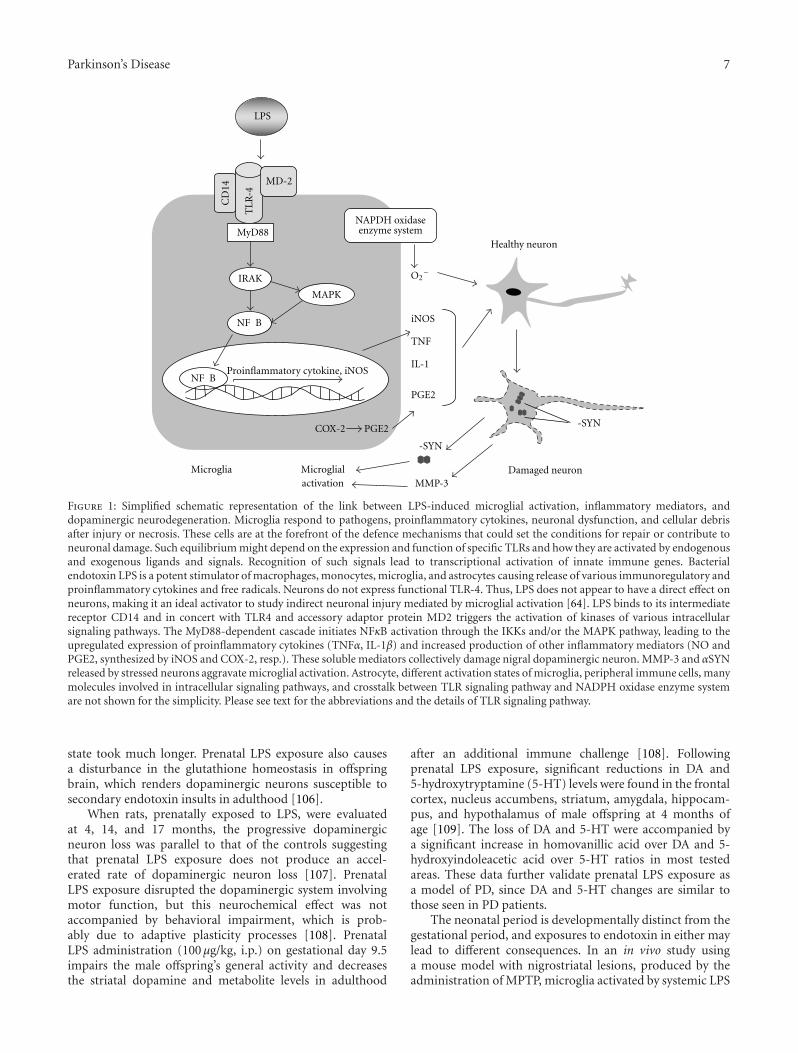
Parkinson’s Disease 7
Healthy neuron
Damaged neuron
IRAK
MAPK
Microglia
LPS
TL
R-4
MD-2
NAPDH oxidaseenzyme systemMyD88
COX-2
CD
14
Microglialactivation
PGE2
NF B
NF B
-SYN
MMP-3
-SYN
O2−
PGE2
iNOS
IL-1
TNF
Proinflammatory cytokine, iNOS
Figure 1: Simplified schematic representation of the link between LPS-induced microglial activation, inflammatory mediators, anddopaminergic neurodegeneration. Microglia respond to pathogens, proinflammatory cytokines, neuronal dysfunction, and cellular debrisafter injury or necrosis. These cells are at the forefront of the defence mechanisms that could set the conditions for repair or contribute toneuronal damage. Such equilibrium might depend on the expression and function of specific TLRs and how they are activated by endogenousand exogenous ligands and signals. Recognition of such signals lead to transcriptional activation of innate immune genes. Bacterialendotoxin LPS is a potent stimulator of macrophages, monocytes, microglia, and astrocytes causing release of various immunoregulatory andproinflammatory cytokines and free radicals. Neurons do not express functional TLR-4. Thus, LPS does not appear to have a direct effect onneurons, making it an ideal activator to study indirect neuronal injury mediated by microglial activation [64]. LPS binds to its intermediatereceptor CD14 and in concert with TLR4 and accessory adaptor protein MD2 triggers the activation of kinases of various intracellularsignaling pathways. The MyD88-dependent cascade initiates NFκB activation through the IKKs and/or the MAPK pathway, leading to theupregulated expression of proinflammatory cytokines (TNFα, IL-1β) and increased production of other inflammatory mediators (NO andPGE2, synthesized by iNOS and COX-2, resp.). These soluble mediators collectively damage nigral dopaminergic neuron. MMP-3 and αSYNreleased by stressed neurons aggravate microglial activation. Astrocyte, different activation states of microglia, peripheral immune cells, manymolecules involved in intracellular signaling pathways, and crosstalk between TLR signaling pathway and NADPH oxidase enzyme systemare not shown for the simplicity. Please see text for the abbreviations and the details of TLR signaling pathway.
state took much longer. Prenatal LPS exposure also causesa disturbance in the glutathione homeostasis in offspringbrain, which renders dopaminergic neurons susceptible tosecondary endotoxin insults in adulthood [106].
When rats, prenatally exposed to LPS, were evaluatedat 4, 14, and 17 months, the progressive dopaminergicneuron loss was parallel to that of the controls suggestingthat prenatal LPS exposure does not produce an accel-erated rate of dopaminergic neuron loss [107]. PrenatalLPS exposure disrupted the dopaminergic system involvingmotor function, but this neurochemical effect was notaccompanied by behavioral impairment, which is prob-ably due to adaptive plasticity processes [108]. PrenatalLPS administration (100 μg/kg, i.p.) on gestational day 9.5impairs the male offspring’s general activity and decreasesthe striatal dopamine and metabolite levels in adulthood
after an additional immune challenge [108]. Followingprenatal LPS exposure, significant reductions in DA and5-hydroxytryptamine (5-HT) levels were found in the frontalcortex, nucleus accumbens, striatum, amygdala, hippocam-pus, and hypothalamus of male offspring at 4 months ofage [109]. The loss of DA and 5-HT were accompanied bya significant increase in homovanillic acid over DA and 5-hydroxyindoleacetic acid over 5-HT ratios in most testedareas. These data further validate prenatal LPS exposure asa model of PD, since DA and 5-HT changes are similar tothose seen in PD patients.
The neonatal period is developmentally distinct from thegestational period, and exposures to endotoxin in either maylead to different consequences. In an in vivo study usinga mouse model with nigrostriatal lesions, produced by theadministration of MPTP, microglia activated by systemic LPS

8 Parkinson’s Disease
were neurotoxic toward dopamine neurons in aged mice butunexpectedly neuroprotective in neonatal mice [110]. Theinflammatory process in the brain, which is accompaniedby changes in the levels of proinflammatory cytokinesand neurotrophins, along with the presence of activatedmicroglia, has recently gained much attention in the areaof neurodegenerative diseases. Activated microglia produceeither neuroprotective or neurotoxic factors. Many reportsindicate that activated microglia promote degeneration ofdopaminergic neurons in PD. On the other hand, thereare several lines of evidence that microglia also have aneuroprotective function [111]. Microglia activated with LPSin the nigrostriatum of neonatal mice protect dopaminergicneurons against the neurotoxin MPTP whereas activatedmicroglia in aged mice promote death of dopaminergiccells by MPTP. Recent findings suggest that the function ofactivated microglia may change in vivo from neuroprotectiveto neurotoxic during aging as neurodegeneration progressesin the PD brain [111]. These results suggest that the activatedmicroglia in neonatal mice are different from those in agedmice, with the former having neurotrophic potential towardthe dopamine neurons in the SN in contrast to the neurotoxiceffect of the latter [112].
As discussed above, recent studies have begun to identifyspecific factors occurring as part of the in utero or perinatalenvironment that may predispose or even cause damageto the nigrostriatal system, suggesting that environmentalfactors early in life of an individual cause a predispositionto develop symptoms of PD. Interactions of prenatal envi-ronment, adulthood environment, gender, age, and geneticbackground may also modify this risk [100]. Recently, animalstudies have been described that specifically consider therole of gestational exposures in disrupting the nigrostriatalsystem and each has implications for elaborating on ourcurrent understanding of the etiology of PD.
5. Cellular and Molecular Mediators ofEndotoxin-Mediated DopaminergicNeurodegeneration
Unlike the direct death of dopaminergic neurons causedby neurotoxins such as MPP+ or 6-OHDA, endotoxin-mediated dopaminergic neurodegeneration seems to resultfrom indirect neuronal death due to inflammatory reac-tions. Bacterial endotoxin LPS is capable of activating glialcells, predominantly microglia, to release a wide variety ofproinflammatory and neurotoxic factors that include reactiveoxygen and nitrogen species, proinflammatory cytokines,and lipid mediators [113]. A number of mechanismsby which inflammatory-activated microglia and astrocyteskill neurons have been identified in cell-culture studies[114]. Results from studies employing enzyme inhibitors,neutralizing antibodies, specific inhibitors of inflammatorysignaling pathways, and knockout animals have identifiedthese soluble factors and signaling molecules involved inmicroglial activation as major contributors to the endotoxin-mediated dopaminergic neurodegeneration [64].
The toll encoding gene has first been identified inDrosophila embryos, where it has a role in dorsoventral axis
determination [115, 116]. Many organisms have multiplehomologues of the Drosophila toll gene, which is very con-served among species [117]. In vertebrates, TLR (Toll-likereceptors) recognize pathogen associated molecular patternsof bacteria, fungi, and viruses and play roles in host defensemechanism. TLR4 takes part in recognition of stronglyconserved patterns of gram-negative cell wall components,LPS and discriminates indigenous from foreign molecules[118]. In TLR4 signaling, TLR4 must first associate withits extracellular binding partner, myeloid differentiationfactor 2 (MD-2), before ligands can bind to the TLR4-MD-2complex [119, 120]. The TLR4-MD-2-Ligand complex formsa heterodimer with another TLR4-MD-2 ligand complexand the signal is transferred to the TLR4’s Toll/interleukin-1receptor (TIR) domain. The signal is than further transducedvia an unknown mechanism [118, 121]. The signal is thentransmitted to two separate pathways which are the MyD88path activating Nf-κB and Toll/IL-1 receptor also containingadaptor inducing IFN-β (TRIF) path. In the MyD88 path,MyD88 adaptor-like protein (Mal or TIRAP) mediates theTIR-TIR association between TLR4 and MyD88 [122].Next, an interaction occurs between IL-1 receptor-associatedkinase (IRAK) and MyD88. That interaction results in theactivation of a cascade leading to the phosphorylation ofNf-κB transcription factors. This path results in activationof Activator Protein-1, RelA and p50 heterodimers andregulates expression of proinflammatory cytokines [123,124]. In the other pathway, TRIF and TLR4 require anadaptor molecule called TRAM for signal transduction,which mediates endocytosis of the TLR4 receptor complex[125, 126]. TRIF forwards the signal after incorporation ofTRAF3- or TRAF6-mediated adaptor molecules to eitherTRIF-binding kinase- (TBK-) IKK or RIP, respectively[127]. TBK-IKK terminates Interferon regulatory factor-3dimerization and translocation into nucleus to induce IFN-β synthesis; in this way, TBK-IKK regulates cellular responseto inflammation [128]. On the other hand, TRAF6 interactswith RIP and activates Nf-κB through TAK1, which operatesthe same as in the MyD88 pathway, causing late phase Nf-κBactivation [127].
5.1. Nitric Oxide. Nitric oxide (NO) is an important messen-ger molecule in a variety of physiological systems. NO, a gas,is produced from L-arginine by different isoforms of NOSand takes part in many normal physiological functions, suchas promoting vasodilation of blood vessels and mediatingcommunication between cells of the nervous system. Inaddition to its physiological actions, free radical activityof NO can cause cellular damage through a phenomenonknown as nitrosative stress [129]. Although many in vitroand in vivo studies support an involvement of NO inmicroglial-mediated dopaminergic neuronal death due toLPS-treatment, some studies suggested that NO is notinvolved [113]. For instance, the first in vivo study of theendotoxin-based PD model reported that the neurotoxiceffect of LPS was not mediated by NO [36]. However,increasing evidence from recent studies supports for thenotion that excessive production and accumulation of NOin the LPS-induced DA lead to neurodegeneration [64].

Parkinson’s Disease 9
Intracerebral administration of LPS causes increase in theiNOS enzyme activity and NO production [130, 131].Immunofluorescence and immunohistochemical analyseshave revealed that iNOS is located in fully activated microgliahaving a characteristic amoeboid morphology [70, 132].After intranigral LPS injection, iNOS mRNA levels and pro-tein expression increase [132]. In Western blot analysis, iNOShas been shown to be induced in the SN after injection of LPSin a time- and dose-dependent manner [133]. The increase iniNOS expression inversely correlates with the TH immuno-labeling and animals pretreated with a selective inhibitorof iNOS, N(G)-nitro-L-arginine methyl ester (L-NAME),exhibited complete protection against behavioral deficitsinduced by intrastriatal LPS injection [130]. Furthermore,LPS-induced loss of dopaminergic neurons is significantlyinhibited by the administration of L-NAME [133]. Decreasein DA level and increase in cytochrome-c release and caspase-3 activation were significantly reversed with treatment ofL-NAME [131]. Thus, increased NO availability subsequentto iNOS induction seems to play an important role in the ini-tial phase of neurodegeneration. Hunter et al. have suggestedthat permanent expression of the iNOS plays a role in theprogressive loss of dopaminergic neurons but not the initialloss induced by LPS [75]. Although the mechanism of NO-mediated neurodegeneration remains uncertain, it has beensuggested that NO contributes to LPS-induced dopaminergicneurodegeneration through several mechanisms. NO hasbeen shown to modify protein function by nitrosylationand nitrotyrosination, contribute to glutamate excitotoxicity,inhibit mitochondrial respiratory complexes, participate inorganelle fragmentation, and mobilize zinc from internalstores [129, 134]. NO can react with superoxide radicalsto form peroxynitrite radicals that are short-lived oxidantsand highly damaging to neurons [64, 135]. Mitochondrialinjury is prevented by treatment with L-N(6)-(l-iminoethyl)-lysine, an iNOS inhibitor, suggesting that iNOS-derivedNO is also associated with the mitochondrial impairment[72]. NO inhibits cytochrome oxidase in competition withoxygen, resulting in glutamate release and excitotoxicity[114].
The main cellular source of NO in the CNS are microgliawhereas astroglia constitute the main defense system againstoxidative stress. However, under pathological or chronicinflammatory conditions, astroglial cells may also releaseneurotoxic mediators. Although the PD-associated geneDJ-1 mediates direct neuroprotection, the upregulation ofDJ-1 in reactive astrocytes also suggests a role in glia[136]. The intracerebral LPS-based PD model is associatedwith a moderate reactive astrogliosis [70]. DJ-1 acts asa regulator of proinflammatory responses, and its losscontributes to PD pathogenesis by deregulation of astrocyticneuroinflammatory damage [137]. When treated with LPS,DJ-1-knockout astrocytes generate significantly more NOthan littermate controls. The enhanced NO productionin DJ-1(−/−) astrocytes is mediated by a signaling path-way involving reactive oxygen species (ROS) leading tospecific hyperinduction of iNOS. These effects coincidewith significantly increased phosphorylation of the p38mitogen-activated protein kinase (MAPK), p38 inhibition,
suppressed NO production, and iNOS mRNA as well asprotein induction. DJ-1(−/−) astrocytes also induce theproinflammatory mediators COX-2 and IL-6 in high levels.Primary neuron cultures grown on DJ-1(−/−) astrocytesbecame apoptotic in response to LPS in an iNOS-dependentmanner suggesting the neurotoxic potential of astrocyticDJ-1 deficiency [137]. These findings warrant in vivo con-firmation.
5.2. Reactive Oxygen Species. A large body of evidence sup-ports the involvement of oxidative stress in the pathogenesisof PD [134]. Besides NO, ROS generated by activatedglia, especially microglia are major mediators of the DAneurodegeneration cause by inflammation [64]. ROS cancause lipid peroxidation, protein oxidation, DNA damage,and mitochondrial dysfunction. LPS-induced ROS produc-tion in microglia is mediated by nicotinamide adeninedinucleotide phosphate (NADPH) oxidase, a multisub-unit enzyme [114]. This complex is responsible for theproduction of both extracellular and intracellular ROSby microglia. Importantly, NADPH oxidase expression isupregulated in PD and is an essential component ofmicroglia-mediated dopaminergic neurotoxicity. Activationof microglial NADPH oxidase causes neurotoxicity throughtwo mechanisms. Firstly, extracellular ROS released fromactivated microglia are directly toxic to neurons. Secondly,intracellular ROS amplifies the production of several proin-flammatory and neurotoxic cytokines and compounds suchas TNFα, prostaglandin E2 (PGE2), COX-2, and IL-1β [138].The activation of the phagocyte NADPH oxidase (PHOX)by cytokines, LPS, or arachidonic acid metabolites causesmicroglial proliferation and inflammatory activation; thus,PHOX is a key regulator of inflammation. Pharmacologicinhibition of NADPH oxidase provides protection againstLPS-induced neurotoxicity and PHOX knockout mice areresistant to LPS-induced loss of SNpc dopaminergic neurons[139, 140]. Gene expression and release of tumor necrosisfactor alpha was much lower in PHOX−/− mice than incontrol PHOX+/+ mice [140]. By injecting LPS into thestriatum of wild type and Nox1 knockout mice, it hasbeen shown that Nox1, a subunit of NADPH oxidase,also enhances microglial production of cytotoxic nitritespecies and promotes loss of presynaptic proteins in striatalneurons [141]. Activation of PHOX alone causes no celldeath, but when combined with expressed iNOS, it resultsin extensive neuronal cell death via the production ofperoxynitrite [114]. The relationship between the signalingpathway downstream of TLR4, after LPS stimulation, and theactivation of the oxidase remains elusive. Using mice lackinga functional TLR4, it has been demonstrated that TLR4and ROS work in concert to mediate microglia activation[142]. Both TLR4(−/−) and TLR4(+/+) microglia displaya similar increase in extracellular superoxide productionwhen exposed to LPS. These data indicate that LPS-inducedsuperoxide production in microglia is independent of TLR4and that ROS derived from the production of extracellularsuperoxide in microglia mediates the LPS-induced TNF-αresponse of both the TLR4-dependent and independentpathway [142].

10 Parkinson’s Disease
The integrin CD11b/CD18 (MAC1, macrophage antigencomplex-1) pattern recognition receptor mediates LPS-induced production of superoxide by microglia [143].MAC1 is a TLR4-independent receptor for the endotoxinLPS. MAC1 is essential for LPS-induction of superoxide inmicroglia, implicating that MAC1 acts as a critical triggerin microglial-derived oxidative stress during inflammation-mediated neurodegeneration. Interestingly, MAC1 mediatesreactive microgliosis and progressive dopaminergic neu-rodegeneration in the MPTP model of PD, suggesting arole for this receptor in neurodegeneration [144]. Activatedmatrix metalloproteinase-3 (MMP-3) released from stresseddopaminergic neurons is also responsible for microglialactivation and generation of NADPH oxidase-derived super-oxide and eventually enhances nigrostriatal DA neuronaldegeneration [145].
5.3. Proinflammatory Cytokines. Of the variety of cytokinesthat are released by LPS-activated glia, the proinflammatoryIL-1β and TNFα may be the major cytokines involved inthe LPS-induced dopaminergic neurodegeneration [64]. Thecontribution of these cytokines to neurodegeneration issupported by studies showing that neutralizing antibodiesagainst TNFα or IL-1 markedly reduce the LPS-induced lossof nigral dopaminergic neurons [64]. Activated microglialcells in the SN are found in all animal models of PDand patients with the illness. Compared with astroglia ormicroglia, they appear to possess a larger repertoire ofcytokine production [64, 113]. Elevated levels of TNFα inthe cerebrospinal fluid (CSF) and the postmortem brainsof PD patients as well as in animal models of PD implicatethat proinflammatory cytokines significantly influence thepathophysiology of the disease [146]. TNFα has a pivotalrole in mediating the loss of DA neurons in PD, whichhas been demonstrated using the endotoxin-based model.A sustained elevation of TNFα has been observed in thestriatum and the mesencephalon of rats prenatally exposed toLPS [104]. Furthermore, in the chronic LPS nigral infusionmodel of PD, the loss of SNpc dopaminergic neurons,and the activation of microglia are significantly reduced byblockade of the soluble form of the TNFα receptor [146].Systemic LPS administration results in rapid increase ofTNFα in the brain, which remains elevated for 10 months[80]. Furthermore, LPS leads to microglial activation, to anincrease in the expression of proinflammatory factors such asIL-1β, and NFκB p65, and to a progressive loss of nigral TH-immunoreactive neurons in wild-type mice, but not in micelacking TNFα receptors [80]. Nontoxic doses of LPS alsoinduce secretion of cytokines and predispose dopaminergicneurons to be more vulnerable to a subsequent low doseof neurotoxins such as 6-OHDA. Alterations in cytokines,prominently an increase in IL-1β, have been identified asbeing potential mediators of this effect that is associatedwith the activation of microglia [94, 95]. Administrationof an IL-1 receptor antagonist results in significant reduc-tions in TNFα and interferon gamma and attenuates theaugmented loss of dopaminergic neurons caused by theLPS-induced sensitization to dopaminergic degeneration.Nigral injection of LPS in a degenerating SN exacerbates
neurodegeneration and accelerates and increases motor signsand shifts microglial activation towards a proinflammatoryphenotype with increased IL-1β secretion [95]. Importantly,chronic systemic expression of IL-1 also exacerbates neu-rodegeneration and causes microglial activation in the SN.It has been found by in vivo studies that NO is a downstreammolecule of IL-1 action and partially responsible for theexacerbation of dopaminergic neurodegeneration, suggest-ing that IL-1 exerts its exacerbating effect on degeneratingdopaminergic neurons by direct and indirect mechanisms[95].
Part of the challenge to sort out the contributions ofindividual cytokines to neurodegeneration may be a resultof the complex interplay by various positive or negativefeedback and feedforward loops among various cytokines,pro- and anti-inflammatory cytokines [64]. Microglial TNFαnot only upregulates its own production in an autocrinefashion but also can further increase the surface expressionof the neuronal TNFα cell death receptor (TNF p55 receptor)in a paracrine manner, thus exacerbating the LPS-inducedneurotoxicity [64]. On the other hand, anti-inflammatorycytokines have been shown to reduce LPS-induced microglialactivation and loss of SNpc dopaminergic neurons [147,148]. The transforming growth factor beta 1 (TGF-β1),one of the most potent endogenous immune modulatorsof inflammation, exerts significant neuroprotection againstLPS induction via its anti-inflammatory properties [147].TGF-β1 inhibits the translocation of the cytosolic subunitp47phox of the LPS-induced PHOX from the cytosol tothe membrane in cultured microglia. The molecular mech-anisms of TGF-β1-mediated anti-inflammatory propertiesworks via the inhibition of PHOX activity by preventingthe ERK-dependent phosphorylation of Ser345 on PHOX’scytosolic subunit p47phox in microglia, thus reducingoxidase activities induced by LPS [147]. Using the terminaldeoxynucleotidyl transferase biotin-dUTP nick end labeling(TUNEL) assay and electron microscopy, Arimoto et al.have shown that intranigral injection of LPS causes markedmicroglial activation and a dose-dependent selective lossof dopaminergic neurons, which is mediated by apoptosis[148]. LPS injection leads to an increase in the mRNAexpression of the proapoptotic proteins Bax, Fas, and theproinflammatory cytokines IL-1β, IL-6, and TNFα, whileexpression of the antiapoptotic gene Bcl-2 is decreased. Infu-sion of interleukin-10 (IL-10) by osmotic minipump protectsagainst LPS-induced cell death of dopaminergic neurons. Acorresponding decrease in the number of activated microgliasuggests that the reduction in microglia-mediated releaseof anti-inflammatory mediators may contribute to the anti-inflammatory effect of IL-10 [148].
NFκB plays a key role in regulating neuroinflammation.Activation of NFκB depends on the phosphorylation of itsinhibitor, IkappaB, by the specific IkappaB kinase (IKK) sub-unit IKK-beta. Compound A, a potent and selective inhibitorof IKK-beta, inhibits the activation of microglia, inducedby nigral injection of LPS, and significantly attenuates LPS-induced loss of dopaminergic neurons in the SN [149]. Selec-tive inhibition of NFκB activation affords neuroprotectionby suppressing the activity of microglial NADPH oxidase

Parkinson’s Disease 11
and by decreasing the production of ROS, and by inhibitinggene transcription of various proinflammatory mediatorsin microglia via IKK-beta suppression. Microglial activationmay involve kinase pathways controlled by mixed lineagekinases (MLKs), a distinct family of mitogen-activatedprotein kinases, which might contribute to the pathology ofPD. A potent MLK inhibitor, CEP-1347, inhibits brain TNFαproduction induced by intracerebroventricular injection ofLPS in mice [150]. Coinjections of LPS with a p38 MAPkinase inhibitor to SN reduces iNOS and caspase-11 mRNAexpression and rescues dopaminergic neurons in the SN[132]. Thus, LPS-induced dopaminergic cellular death in SNcould be mediated, at least in part, by the p38 signal pathwayleading to activation of inducible nitric oxide synthase andcaspase-11.
5.4. Cyclo-Oxygenase-2 and Prostaglandin E2. Prostaglandinsare potent autocrine and paracrine oxygenated lipidmolecules that contribute appreciably to physiologic andpathophysiologic responses in brain and other organs [151].Emerging data indicate that PGE2 plays a central role inneurodegenerative diseases. PGE2 signaling is mediated byinteractions with four distinct G protein-coupled receptors,EP1-4, which are differentially expressed on neuronal andglial cells throughout the CNS, (here something is missingto make a sentence) [151]. EP2 activation has been shown tomediate microglial-induced paracrine neurotoxicity as wellas to suppress the internalization of aggregated neurotoxicpeptides in microglia [152]. PGE2 is produced at highlevels in the injured CNS, where it is generally considereda cytotoxic mediator of inflammation. LPS upregulates theexpression of COX-2 and increase the release of PGE2 incultured microglia [64]. Intracerebral injections of LPS resultin a significant upregulation of the striatal and nigral proteinexpression of COX-2 as well as the activation of microglia[153, 154]. Double labeling using immunohistochemistryidentified that activated microglia rather than intact restingmicroglia are the main intracellular locations of COX-2expression [64, 155]. In vivo pharmacological inhibition ofCOX-2 activity protects nigral dopaminergic neuronal lossand decreases microglial activation induced by intracerebralLPS injection, supporting the role of COX-2 in the patho-genesis of neuroinflammation-mediated neurodegeneration[153, 155, 156].
A local injection of LPS into the rat SN led to the induc-tion of microsomal prostaglandin E2 synthase (mPGES)-1in activated microglia [157]. Further in vitro and in vivoexperiments with mPGES-1 knockout mice indicate thenecessity of mPGES-1 for microglial PGE2 production. Thisstudy has shown that the activation of microglia contributesto PGE2 production through the concerted de novo synthesisof mPGES-1 and COX-2 at the sites of inflammation inthe brain parenchyma. In contrast to that, a recent invitro study suggests that mPGES-1 expression is not strictlycoupled to the expression of COX-2 [158]. Activation ofcultured spinal microglia via TLR4 produces PGE2 andcauses NO release from these cells, showing that COX-PGE2 pathway is regulated by p38 and iNOS [159]. Thesefindings emphasize that p38 in spinal microglia is a key
player among inflammatory mediators, such as PGE2 andNO. In vitro experiments also indicate that microglial PGE2plays an important role in astrocyte proliferation, identifyingPGE2 as a key neuroinflammatory molecule that triggersthe pathological response related to uncontrollable astrocyteproliferation [160].
5.5. Matrix Metalloproteinase-3. As discussed above (Section5.2), the release of MMP-3 from apoptotic neurons mayplay a major role in degenerative human brain disorders,such as PD. The catalytic domain of recombinant MMP-3induces the generation of TNFα, IL-6, IL-1β, and IL-1receptor antagonist but not of IL-12 and iNOS, which arereadily induced by LPS, in cultured microglia, suggestingthat there is a characteristic pattern of microglial cytokineinduction by apoptotic neurons [145]. MMP-3 activates thenuclear factor-kappaB (NFκB) pathway, and these microglialresponses were totally abolished by preincubation withan MMP-3 inhibitor. MMP-3-mediated microglial activa-tion mostly depends on ERK (extracellular signal-regulatedkinase) phosphorylation but not on either JNK (c-Jun N-terminal protein kinase) or p38 activation. MMP-3-activatedmicroglial cells caused apoptosis of neuronal cells in in vitroexperiments. These results suggest that the distinctive signalof neuronal apoptosis is the release of the active form ofMMP-3 that activates microglia and subsequently exacer-bates neuronal degeneration [145]. The released active formof MMP-3, as well as the catalytically active recombinantfrom of MMP-3 leads to superoxide generation in culturedmicroglia [161]. MMP-3 causes dopaminergic cell death inmesencephalic neuron-glia mixed cultures of wild-type mice,but this is attenuated in the culture of NADPH oxidase sub-unit null mice (gp91(phox−/−)), suggesting that NADPHoxidase mediates the MMP-3-induced microglial productionof superoxide and the following dopaminergic cell death.Moreover, in the MPTP model of PD, the nigrostriataldopaminergic neuronal degeneration, microglial activation,and superoxide generation are largely attenuated in MMP-3−/− mice. These results indicate that MMP-3 releasedfrom stressed dopaminergic neurons is responsible formicroglial activation and generation of NADPH oxidase-derived superoxide and in turn exacerbates the nigrostriataldopaminergic neuronal degeneration [161].
αSYN also induces the expression of MMP-3 in cul-tured microglia from rat [162]. The inhibition of MMP-3significantly reduces NO and ROS levels and suppresses theexpression of TNFα and IL-1β. Inhibition of MMP-3 alsosuppresses the activities of MAPK and transcription factors,NFκB and AP-1. The specific inhibitor of the protease-activated receptor-1 (PAR-1) and a PAR-1 antagonist signif-icantly suppress cytokine levels, NO, and ROS productionin αSYN-treated microglia, indicating that MMP-3 secretedby αSYN-stimulated microglia activate PAR-1 and amplifymicroglial inflammatory signals in an autocrine or paracrinemanner [162]. In vivo, LPS injection into the SN ofrats increases MMP-3 expression and activation suggestingthat MMP-3 may participate in neuroinflammation-induceddopaminergic neurotoxicity [163]. These studies proposethat the in vivo modulation of MMP-3 expression and

12 Parkinson’s Disease
activity may provide the neuroprotection for dopaminergicneurons. Indeed, an antibiotic, doxycycline, shows neuropro-tection for the dopaminergic system in a toxin-based modelof PD and this appears to derive from antiapoptotic andanti-inflammatory mechanisms involving downregulation ofMMP-3 [164].
5.6. Microenvironmental Changes and Intercellular Interac-tions. The CNS microenvironment plays a significant role indetermining the phenotypes of both CNS-resident microgliaand CNS-infiltrating macrophages. In this section, we sum-marize the microenvironmental changes such as astroglialresponses, BBB alterations, and a wide range of intercellularinteractions in the context of the endotoxin-based PD model.
5.6.1. Reactive Astrocytes and Parkinson’s Disease. Astrocytesare the most abundant cell types in the CNS and participatein the local innate immune response triggered by a varietyof insults. The role of astrocytes in the pathogenesis of PDis even less well understood than the one of microglia butthey are known to secrete both inflammatory and anti-inflammatory molecules [165]. It has been proposed thatastrocytes may play dual roles in PD [166]. Similar tomicroglial activation, star-shaped astrocytes transformed toreactive form have enlarged and thick bodies and respondto various stimuli, which coined the term reactive astrocytes[167]. Reactive astrogliosis is generally mild or moderateand rarely severely pronounced in autopsy specimens fromthe SN of PD patients [166]. Classic reactive astrocytes areobserved in multiple system atrophy, progressive supranu-clear palsy, and corticobasal degeneration, but not in PDcases; the extent of reactivity correlates with indices ofneurodegeneration and disease stage [168]. Different sub-populations of astrocytes express disease-related proteinssuch as αSYN, parkin, and p-tau at different levels and indifferent combinations in different Parkinsonian syndromesbut the roles of astrocytes in these conditions are not yet welldefined [167, 168].
The role of astrocytes in the development of PD is stillunknown and controversial. Astrocytes provide the optimalmicroenvironment for neuronal function by exerting activecontrol over the cerebral blood flow and by control-ling the extracellular concentration of synaptically releasedneurotransmitters [167]. Generally, astrocytes promote thesurvival and maintenance of dopaminergic neurons throughsecretion of various neurotrophic factors in the SN. Thedecreased levels of astrocyte-derived neurotrophic factorsare at least in part responsible for DA neuronal death inPD [167]. Astrocytes become activated and synthesize pro-and anti-inflammatory cytokines, chemokines, antioxidants,neurotrophic factors, and prostanoids during neuroinflam-mation and neurodegeneration and interact with otherimmune competent cells. These mediators act as double-edged swords, exerting both detrimental and neuroprotectiveeffects. For example, myeloperoxidase (MPO), a key enzymein the generation of reactive nitrogen species (RNS), isupregulated in the midbrains of PD patients and MPTPtreated mice [169]. This enzyme is localized within reactiveastrocytes in MPTP-treated mice, and MPTP neurotoxicity
is attenuated by ablation of MPO from the nigrostriatalpathway [167, 169].
5.6.2. Region-Specific Astroglial Responses in the Brain.Degenerative disorders of the brain often occur in a regionspecific fashion, suggesting differences in the activity andreactivity of innate immune cells. This may make astrocyteslikely candidates to be responsible for region-specific inci-dence rates of neurological and neurodegenerative disorders.Cultured astrocytes from the cortex and midbrain alreadydiffer in their capacity and profile of cytokine expressionunder unstimulated conditions [170]. In response to LPS,both a region specific pattern of upregulation of distinctcytokines, and differences in the extent and time course ofactivation are observed. Thus, astrocytes reveal a region-specific basal profile of cytokine expression and a selectivearea specific regulation of cytokines upon LPS-inducedinflammation [170]. The densities of astrocytes are muchlower in the intact SNpc, compared with the cortex [171].Furthermore, after LPS injection, damage to endothelialcells and astrocytes and the blood-brain barrier (BBB)permeability are more pronounced in the SNpc [171]. Thein vitro responses of microglia and astroglia to inflammatorystimuli or environmental toxins also differ. Manganesesignificantly potentiates LPS-induced release of TNF-α andIL-1β in microglia, but not in astroglia [172]. These agentsare more effective in inducing the formation of ROS and NOin microglia than in astroglia.
5.6.3. DJ-1, Oxidative Stress and Astrocytes. Recent findingssupport the developing view that astrocytic dysfunction,in addition to neuronal dysfunction, may contribute tothe progression of a variety of neurodegenerative disorders.Thus, the treatments that support the beneficial aspects ofastrocyte function may represent novel approaches targetingastrocytes to promote dopaminergic neurorescue. Althoughaging enhances the neuroinflammatory response and thealpha-synuclein nitration [73], the antioxidant capacityand glutathione metabolism of astrocytes are preservedfrom mature adulthood into senescence [173]. Thus, theoxidative stress seen in aging brains is likely due to factorsextrinsic to astrocytes, rather than being caused by animpairment of the antioxidative functions of astrocytes. ThePARK7 (DJ-1) gene, which has been implicated in someforms of early-onset, autosomal recessive PD, is apparentlyexpressed mainly by the astrocytes in the human brain. Loss-of-function mutations lead to the characteristic selectiveneurodegeneration of nigrostriatal dopaminergic neurons.In addition to cell-autonomous neuroprotective roles, DJ-1may act in a transcellular manner, being upregulated inreactive astrocytes in chronic neurodegenerative diseases, forexample. In sporadic PD, and many other neurodegenerativediseases, reactive astrocytes overexpress DJ-1 whereas neu-rons maintain the expression at normal levels [136]. SinceDJ-1 has neuroprotective properties and since astrocytes areknown to support and protect neurons, DJ-1 overexpressionin reactive astrocytes may reflect an attempt to protectthemselves and the surrounding neurons against diseaseprogression. Knocking down DJ-1 in astrocytes impairs

Parkinson’s Disease 13
astrocyte mediated neuroprotection against rotenone [174].DJ-1 is a ubiquitous redox responsive and cytoprotectiveprotein with diverse functions. DJ-1 regulates redox signalingkinase pathways and acts as a transcriptional regulatorof antioxidative genes. DJ-1 scavenges H2O2 by cysteineoxidation in response to oxidative stress and, thus, confersneuroprotection. Therefore, DJ-1 is an important redox-reactive signaling intermediate, controlling oxidative stressupon neuroinflammation and during age-related neurode-generative processes such as PD [136]. However, the func-tional basis of neuroprotection elicited by DJ-1 has remainedvague. DJ-1 stabilizes erythroid 2-related factor (Nrf2), amaster regulator of antioxidant transcriptional responses,by preventing its association with the inhibitor proteinKeap1 and by blocking Nrf2’s subsequent ubiquitination[175]. Without intact DJ-1, Nrf2 protein is unstable, andtranscriptional responses are thereby decreased both basallyand after induction [175] though a recent study suggests thatactivation of the Nrf2 is independent of DJ-1 [176].
5.6.4. Nrf2/ARE Pathway and Parkinson’s Disease. Theexpression of phase II detoxification and antioxidantenzymes is governed by a cis-acting regulatory elementnamed the antioxidant response element (ARE). Nrf2 reg-ulates genes containing the ARE element and is a member ofthe Cap’n’Collar basic-leucine-zipper family of transcriptionfactors. Following activation, Nrf2 dissociates from Keap1,translocates to the nucleus, and binds to the ARE promotersequences, as a part of the coordinated induction of abattery of cytoprotective genes including antioxidants andanti-inflammatory genes [177]. ARE-regulated genes arepreferentially activated in astrocytes, which consequentlyhave more efficient detoxification and antioxidant defensemechanisms than neurons. Astrocytes closely interact withneurons to provide structural, metabolic, and trophicsupport, as well as actively participating in the modula-tion of neuronal excitability and neurotransmission [177].Therefore, alterations in astroglial function can modulatethe interaction with surrounding cells such as neuronsand microglia. Activation of Nrf2 in astrocytes protectsneurons from a wide array of insults in different in vitroand in vivo paradigms, confirming the role of astrocytesin determining the vulnerability of neurons to deleteriousstimuli [177]. Nrf2 has been shown to be important forprotection against oxidative stress and cell death in toxin-based models of PD [177–181]. These findings remainto be confirmed in endotoxin-based models. Genetic datasuggest that variation in Nrf2 gene NFE2L2 modifies the PDprocess, which provides another link between oxidative stressand neurodegeneration [182]. Nrf2 activating agents suchas synthetic triterpenoids and sulforaphane are potentialtherapeutic targets for the prevention of neurodegenerationin PD [183–185].
5.6.5. Nrf2/ARE Pathway and Microglial Activation. Thedeficiency of Nrf2 results in an exacerbated inflammatoryresponse and in microglial activation of the expression of theneurotoxin MPTP whereas inducers of Nrf2 downmodulateneuroinflammation [181]. Nrf2-deficient mice exhibit more
astrogliosis and microgliosis, as determined by an increase inmRNA and protein expression levels for GFAP and F4/80,respectively, than wild-type mice. Inflammation markers,characteristic of classical microglial activation like COX-2,iNOS, IL-6, and TNF-alpha, are also increased. At the sametime, anti-inflammatory markers, attributable to alternativemicroglial activation, such as FIZZ-1, YM-1, Arginase-1,and IL-4 are decreased [181]. These results demonstrate arole of Nrf2 in tuning the balance between classical andalternative microglial activation. The restoration of the redoxbalance may be a determinant in driving microglia backto the resting state. ROS generated by microglia couldhelp to eliminate pathogens in the extracellular milieu andalso to act on the microglia itself, altering the intracellularredox balance and functioning as a second messengers inthe induction of proinflammatory genes. The modulationof microglial activation is a matter closely correlated withcontrol of oxidative stress in this cell type and is crucial torestore its inactive state and modulate the inflammation inneurologic diseases [186]. Nrf2 is essential for the regulationof NADPH oxidase-dependent ROS-mediated TLR4 activa-tion in macrophages [187]. Nrf2 activation by sulforaphaneinhibits the inflammatory response to LPS in cultured rodentmicroglia [185]. These findings remain to be tested in thecontext of in vivo endotoxin-based PD models. Interestingly,LPS by itself is able to activate the cell’s defense againstoxidative and electrophilic stress, activating Nrf2 [185]. Thismechanism may be a mediator of LPS preconditioning orendotoxin tolerance, a phenomenon which by prior exposureof innate immune cells like monocytes/macrophages tominute amounts of endotoxin causes them to become refrac-tory to subsequent endotoxin challenges [188]. In contrastto the well-known protective effect of this phenomenon,in acute ischemic conditions, only one in vitro study hasreported this benefit in dopaminergic neurotoxicity [189].Further understanding the underlying mechanism of LPSpreconditioning may open a new window for the treatmentof PD.
Astroglial cells are also involved in the microglial modu-lation by Nrf2 [177]. These cells are known to play an impor-tant role in antioxidant defense and in modulating microglialactivity in the CNS [165, 166]. Recently, astrocytes have beenfound to regulate excessive inflammation via induction ofthe microglial hemooxygenase-1 (HO-1) expression in vitro[190]. While pharmacological or genetic intervention onNrf2 may provide a neuroprotective benefit, HO-1 does notprotect or enhance the sensitivity to neuronal death in theMPTP model [191]. These results support the idea that themodulation of a master transcription factor may be a betterstrategy than targeting individual genes.
5.6.6. Blood-Brain Barrier Dysfunction and PeripheralImmune Cell Infiltration. The brain demands an adequateblood supply for the regulation of neuronal and synapticfunction. To maintain concentrations of ions within narrowranges as well as the adequate levels of metabolic substratesin various brain regions, neural milieu are strictly separatedfrom circulatory spaces through BBB formation [167]. Theseunique biological structures are comprised of neurovascular

14 Parkinson’s Disease
units such as brain capillary endothelial cells, pericytes,neurons, and astrocyte end-feet. Endothelial cells tightlyconnect at junctional complexes such as adherens junctions,tight junctions, and gap junctions confer low paracellularpermeability. Pericytes and astrocytes regulate hemodynamicneurovascular coupling, microvascular permeability, matrixinteractions, neurotransmitter inactivation, neurotrophiccoupling, and angiogenic as well as neurogenic couplingthrough close proximity with neurons [167, 192]. Althoughthere is no clear evidence as to whether these alteredneurovascular circumstances are responsible for the lossof dopaminergic neurons in PD, several studies on PDpatients and animal models suggest a pathogenic linkagebetween BBB disruption and dopaminergic neuronal death[167]. PET and histological studies on PD patients revealedBBB dysfunction in the midbrain of PD patients [193]. Inaddition, increased BBB permeability has been observed inthe MPTP and the LPS models for PD [194]. These studiessuggest that the disruption of the BBB has a relationshipwith neuronal cell death and neuroinflammation in PD[167]. There is also a direct correlation between the locationof IgG immunoreactivity-a, a marker for disruption ofneurodegenerative processes, including the death of nigraldopaminergic cells and reactive astrocytes. A precise spatialcorrelation also exists between disruption of the BBB and3-nitrotyrosine immunoreactivity [194]. LPS-activatedmicroglia can induce the dysfunction of the BBB in anin vitro coculture system with rat brain microvascularendothelial cells and microglia [195]. In the presence of LPS-activated microglia, tight junction proteins are fragmented,and barrier disintegrity and dysfunction induced by LPS-activated microglia are blocked by an NADPH oxidaseinhibition, suggesting that LPS activates microglia to inducedysfunction of the BBB by producing ROS through NADPHoxidase.
Recent studies have shown that the dysfunction of theBBB combined with the infiltration of peripheral immunecells plays an important role in the degeneration of dopamin-ergic neurons [167]. However, these molecular and cellularchanges are not specific to the PD, since they are alsoimplicated in the pathogenesis of other neurodegenerativediseases [196]. The neuroinflammation may contribute tothe infiltration of peripheral immune cells and leakage ofthe BBB into the SN. Various peripheral immune cells, suchas T-cells, B-cells, microphages, and leukocytes infiltrateinto the SN region in the LPS and MPTP models [167,171, 197]. CD11b and MPO double-positive neutrophilsinfiltrate the SNpc following LPS injection [197]. MPO(+)neutrophils observed in SNpc express iNOS, IL-1β, COX-2, and monocyte chemoattractant protein-1 (MCP-1). Inintact rodent brain, the densities of microglia are similarin SNpc and cortex [197]. In addition, the densities ofastrocytes are much lower in the intact SNpc, compared withthe cortex. However, LPS injection induces microgliosis andcauses neutrophil infiltration into the SNpc, but not into thecortex [171]. The extent of neutrophil infiltration appears tobe correlated with neuronal damage. The loss of neurons inthe SNpc is significantly reduced in neutropenic rats versusnormal rats following LPS injection. Furthermore, after LPS
injection, damage to endothelial cells and astrocytes andincreased BBB permeability are more pronounced in theSNpc. Excessive neutrophil infiltration, lower astrocyte den-sity, and higher BBB permeability following LPS exposurecontributes to severe inflammation and neuronal death in theSNpc compared with the cortex [171].
The links between T-cell immunity and the nigrostriatalneurodegeneration are supported by laboratory, animalmodel, and human pathologic investigations [198]. Thepresence of T-lymphocytes in the midbrain of PD patientssuggests that the potential role of infiltrated peripheralcells is a factor of the PD pathogenesis [199]. Recently,Brochard et al. have reported that numerous CD4 and CD8positive cells are detectable in postmortem PD patients [200].The infiltration of CD4+ lymphocytes into the brain alsocontributes to the neurodegeneration in the MPTP model forPD [200]. Specifically, invading T-lymphocytes contributeto neuronal cell death via the Fas/FasL cell death pathway,implicating the emerging role of the adaptive immune systemin the pathogenesis of PD [201].
The adoptive transfer of CD3-activated CD4+CD25+regulatory T-cells (Tregs) is known to suppress immune acti-vation and maintain immune homeostasis and tolerance. InMPTP-treated mice, it protects the nigrostriatal system fromdegeneration through suppression of microglial oxidativestress and inflammation [202]. Tregs also attenuates Th17cell-mediated nigrostriatal dopaminergic neurodegenerationin the MPTP model [203]. In addition, these cells suppressnitrated αSYN-induced microglial ROS production andNFκB activation supporting the importance of adaptiveimmunity in the regulation of PD-associated microglialinflammation [204]. Taken together, these studies provide arationale for future immunization strategies in PD [198].
Accumulating evidence suggests that the penetration ofimmune cells into the brain plays an important role inthe degeneration of dopaminergic neurons in PD. Furtherunderstanding of the cellular and molecular mechanismsresponsible for trafficking of immune cells from the periph-ery into the diseased CNS may be the key to targeting thesecells for therapeutic intervention in PD [196].
In addition to glia-neuron crosstalk, multiple cell-to-cellinteractions and immune regulations, critical for neuronalhomeostasis, also influence immune responses [198, 205].Microglia can be activated by MCP-1, which is expressedby dopaminergic neurons and can interact with its receptorCCR2 on microglial cells. The neuroimmune regulatory pro-teins CD47 and CD200 inhibit macrophage and microgliaactivation through binding to their receptors SIRPalpha andCD200R, expressed on phagocytes [206]. Upon stress, nigraldopaminergic neurons secrete MMP-3 and α-SYN, whichactivates microglial and astroglial cells [145, 207]. As dis-ease progresses, secretions from α-SYN-activated microgliacan engage neighboring glia cells in a cycle of autocrineand paracrine amplification of neurotoxic immune prod-ucts. Astrocytes differentially regulate neutrophil functionsthrough direct or indirect interactions between the two celltypes [208]. Many of these established interactions betweendifferent cell types involved in neuroinflammation have beendemonstrated in vitro and remain to be confirmed in vivo.

Parkinson’s Disease 15
Dissecting the molecular determinants of complex interplaybetween CNS cells and immune cells in the context of theendotoxin-based PD model will give the possibility to testnovel therapeutic strategies to promote restoration of injurednigrostriatal dopaminergic neurons.
6. Therapeutic Approaches
The endotoxin-induced neuroinflammation model for PD isa purely inflammation-driven model. However, all clinicaland pathological features of PD can be observed in thismodel. Therefore, the LPS-induced model can be used tosearch for novel treatment strategies for the therapy ofPD. In this section, we summarize known neuroprotectivemolecules, which have been tested using the LPS-induced PDmodels.
COX-2 is a rate-limiting enzyme in prostaglandin syn-thesis. Experimental and epidemiological evidence supportsthe protective role of COX-2 inhibition in PD. COX-2is upregulated in SN both in the PD and in the MPTPmodel [209]. Pharmacological inhibition of COX-2 or theknockout of the COX-2 gene provides resistance to MPTPin vivo [209, 210] and to 6-OHDA-induced dopaminergictoxicity in vitro [211]. There is epidemiological evidencethat the use of some NSAIDs lowers the incidence of PD[212]. On the other hand, according to meta-analyses ofNSAID studies in PD, ibuprofen shows a slight protectionagainst PD whereas aspirin and acetaminophen did not showany protective effects [213, 214]. Hunter et al. used theCOX-2 inhibitor Celecoxib (Celebrex) in LPS-induced PDanimal model for the first time. They were able to showthat Celecoxib protects dopaminergic neurons by decreasinginflammation and by restoring mitochondrial function inthe intrastriatal LPS-induced PD model [153]. Using theintranigral LPS rat model, Sui et al. [155] have shownthat another COX-2 inhibitor, meloxicam, diminishes theactivation of OX-42 positive microglia and reduces the loss ofdopaminergic neurons in the SNpc. Clinical studies suggestthat inhibition of COX-2 may cause side effects such astrombogenic cardiovascular diseases [156, 215]. In orderto avoid potential side effects of COX-2 inhibition, newdrugs have been targeted for dual inhibition of COX-2 andlipoxygenase (LOX) [156]. Dual inhibitor of COX-2 and 5-LOX has been shown to lower gastrointestinal side effects.Moreover, combination of the two inhibitors achieves a morepotent neuroprotection than usage of single inhibitors [216].Li et al. tested the dopaminergic neuroprotective effect ofCOX, LOX, and the combination of COX and LOX inhibitorsin the intrastriatal LPS-induced animal model for PD. Theyfound that the dual COX and LOX inhibitor, phenidone, isbetter than COX or LOX inhibitors alone for suppressingLPS-induced neurotoxicity [156].
Dexamethasone is a potent anti-inflammatory drug thathas been tested in the intranigral LPS-induced PD model[67, 133]. These studies have shown that dopaminergicdegeneration and microglial activation induced by LPS canbe prevented by administration of dexamethasone [67, 133].Dexamethasone also decreases the exacerbating effect of LPSduring neurodegeneration induced by 6-OHDA [95].
Experimental and epidemiological evidence supports theprotective role of nicotine in PD. Epidemiological studieshave confirmed that there is an inverse correlation betweencigarette smoking and the incidence of PD [217]. In vitronicotine pretreatment inhibits LPS-induced TNF-α release inmurine-derived microglial cells via the α-7 nicotinic receptor[218]. These results suggest that nicotine could protectdopaminergic neurons in the animal model of PD. Indeed,Park et al. have shown that nicotine significantly decreasesthe release of TNFα and the dopaminergic neuronal lossinduced by LPS stimulation. Both effects were blocked by α7-nicotinic acetylcholine receptor blockers [219].
Peroxisome proliferators activated receptor (PPAR-γ) is anuclear receptor that regulates transcription of various genes.It has been shown that the PPAR-γ agonist inhibits cytokinesecretion in microglia and macrophage-like cells [220].Hunter et al. have shown that a PPAR-γ agonist, pioglitazone,provides neuroprotection by decreasing inflammation andrestoring mitochondrial function. Pioglitazone administra-tion partially reduces the LPS-induced striatal dopamine lossand the TH-positive cell loss in the SN [153].
Minocycline is a semisynthetic tetracycline that exertsanti-inflammatory activities [221]. Minocycline significantlyreduces the SN microglial activation induced by intrani-gral LPS administration [194]. Minocycline prevents theLPS-induced increase of mRNA levels of proinflammatorycytokines and diminishes the production of peroxynitrites[194].
Naloxone, an opioid receptor competitive antagonist, hasbeen found to reduce microglial activation-mediated DAneurodegeneration in mouse cortical neuron-glia cocultures[64]. Systemic infusion of naloxone protects dopaminergicneurons against inflammation-mediated degeneration anddecreases microglial activation in vivo through inactivationof NADPH oxidase [139, 222].
The neuroprotective effects of statins in CNS disorderssuch as experimental autoimmune encephalomyelitis, stroke,and Alzheimer’s disease have been previously described[223–225]. Selley has shown that oral administration ofsimvastatin attenuates the depletion of dopamine DOPACand HVA inhibits the formation of 3-nitrotyrosine and theproduction of TNFα in mice treated with MPTP [226].Simvastatin has also been tested in the intranigral LPS-induced PD [227] and the LPS perfusion model [228].Simvastatin prevents the loss of dopaminergic neurons andastrocytes induced by LPS in both models [227, 228].Simvastatin increases BDNF expression [228], which maysupport neuronal and astroglial survival.
Osteopontin (OPN) is a glycosylated phosphoproteinthat has first been identified in 1986 in osteoblasts [229].OPN is constitutively expressed in most tissues, includingthe brain [208]. Iczkiewicz et al. have shown that OPN isconstitutively present in dopaminergic neurons, in the SN,and that its expression is decreased in the MPTP model ofPD and in patients with PD [230]. It has been reported thatthe intranigral injection of LPS enhances expression of OPN[231]. These results suggest that OPN may have a regulatoryrole in neuroinflammation. One peptide fragment of OPNcontains the arginine-glycine-aspartic acid (RGD) domain

16 Parkinson’s Disease
that has been associated with the neuroprotective effects ofOPN [232]. Iczkiewicz et al. have tested RGD containingpeptide fragments of OPN in the LPS-induced PD model.They found that the RGD containing peptide fragment ofOPN protects against LPS-induced TH positive cell loss andalters gliosis in the rat SN [233].
Urocortin is a neuroprotective agent that is structurallyrelated to the corticotrophin releasing factor (CRF) [234–236]. Abuirmeileh et al. have used urocortin for the treat-ment of the LPS-induced PD model. They have shown thaturocortin reduces nigrostriatal damage induced by LPS andthat this effect of urocortin is mediated by CRF1 receptors[237–239].
7. Conclusion
Parkinson’s disease (PD) is the second most common neu-rodegenerative disease with increasing incidence worldwide.Although the pathogenesis of PD remains elusive, accumu-lating evidence from many studies on animal models andpatients shows that the pivotal role of microglial activationalong with neuroinflammatory processes contribute to theinitiation and progression of the nigrostriatal dopaminergicneurodegeneration in PD. In addition to that, recent studieshave proposed that the BBB dysfunction combined with theinfiltration of peripheral immune cells into the CNS playsan essential role in the degeneration of nigral dopaminergicneurons. Thus, using a purely inflammatory experimentalmodel induced by the administration of the bacterialendotoxin, LPS, provides a valuable tool for the in vivomodeling of the characteristics of progressive dopaminer-gic neurodegeneration associated with neuroinflammation.Except for the acute direct administration of LPS to the nigralregion, other modified forms of the model, including theprenatal one, realistically simulate the slow and progressivedopaminergic neuronal loss and permanent neuroinflam-mation. Furthermore, the combination of endotoxin-basedPD models with genetic and toxin-based models is fruitfulfor the delineation of the complex interactions amongthe environmental and genetic factors and inflammatoryprocesses involved in PD. Many experimental variablesincluding sex, age, and strain of the animals have the poten-tial to significantly perturb the functional and pathologicoutcomes. These methodological issues should be consideredin respect to the studies.
Several novel techniques, such as in vivo imaging ofmicroglial activation, are waiting to be applied in theendotoxin-based model of PD. Molecular studies from thedomains of transcriptomics, proteomics, and microRNomicswill be valuable to gain in potential diagnostic markersfor the disease [240]. Since the inflammatory responsesprecede the neurodegeneration and the motor dysfunctions,alterations of the immune parameters, both in CSF andblood, are likely to be useful as early diagnostic markers.The major challenge in this area is the enhancement of thespecificity and sensitivity of the potential markers. Despiteintensive research, the mechanisms of neuroinflammation-mediated nigral neurodegeneration are poorly understood.Whether neuroinflammation is a consequence or a cause of
nigral neuronal loss is still unknown. Neuroinflammationseems to be a trigger of the initiation of neurodegenerationand progressive neurodegeneration continuously aggravateschronic neuroinflammatory processes. In this context, thestimulation of TLR4 by endogenous ligands released byinjured dopaminergic neurons may contribute to this viciouscircle [241].
In vivo imaging and molecular studies will also extendour understanding of the complex interplay between CNSand immune cells. Especially, the novel links between neu-roinflammatory processes, oxidative stress, and Nrf2/AREpathways that are mainly based on data from toxin-basedmodels of PD should be confirmed by the endotoxin basedmodel.
Based on the recent data, adaptive immune responsesalong with innate immunity are important mediators ofneuroinflammation-mediated dopaminergic neurodegener-ation. Recent evidence suggests that the importance ofnonautonomous pathological mechanisms are involved inPD, which are mostly mediated by activated microgliaand peripheral immune cells. Thus, the harnessing of theimmune system by immunomodulating drugs or by immu-nisation aiming at the downregulation of immune responsesremains promising future therapeutic options. Immuneparameters will also be indispensable for the monitoring oftherapeutic responses.
Abbreviations
MPTP: 1-Methyl-4-Phenyl-1,2,3,6-tetrahydropyridine
DOPAC: 3,4-dihydroxyphenylacetic acid6-OHDA: 6-hydroxydopamineARE: Antioxidant response elementBBB: Blood-brain barrierCNS: Central nervous systemCSF: Cerebrospinal fluidCOX-2: Cyclo-oxygenase-2EGF: Epidermal growth factorERK: Extracellular signal-regulated kinaseDA: DopamineGFAP: Glial fibrillary acidic proteinHO-1: Hemooxygenase-1HPLC: High-performance liquid chromatographyHVA: Homovanillic acid5-HT: 5-hydroxytryptamineIKK: IkappaB kinaseIRAK: IL-1 receptor-associated kinaseiNOS: Inducible nitric oxide synthaseIFN-β: Interferon-betaIL-2: Interleukin-2IL-6: Interleukin-6IL-10: Interleukin-10IL-1β: Interleukin 1βJNK: c-Jun N-terminal protein kinaseLPS: LipopolysaccharideMMP-3: Matrix metalloproteinase-3MPP+: 1-methyl-4-phenylpyridiniumMA: Methamphetamine

Parkinson’s Disease 17
MLKs: Mixed lineage kinasesTFAM: Mitochondrial transcription factor AMD-2: Myeloid differentiation factor 2Mal or TIRAP: MyD88 adaptor-like proteinPHOX: NADPH oxidaseL-NAME: N(G)-nitro-L-arginine methyl esterNADPH: Nicotinamide adenine dinucleotide
phosphateNO: Nitric oxideNrf2: Nuclear factor erythroid 2-related factorNFκB: Nuclear factor-kappaBPD: Parkinson’s diseasePPAR-γ: Peroxisome proliferator-activated receptorPGE2: Prostaglandin E2RNS: Reactive nitrogen speciesROS: Reactive oxygen speciesTregs: Regulatory T cellsSN: Substantia nigraSNpc: Substantia nigra pars compactaSNpr: Substantia nigra pars reticulataTUNEL: Terminal deoxynucleotidyl transferase
biotin-dUTP nick end labelingTH+: Tyrosine hydroxylase-positiveTLRs: Toll-like receptorsTIR: Toll/interleukin-1 receptorTRIF: Toll/IL-1 receptor containing adaptor
inducing IFN-βTGFα: Transforming growth factor-alphaTGF-β1: Transforming growth factor-beta 1TBK: TRIF-binding kinaseTNFα: Tumor necrosis factor-alphaTH: Tyrosine hydroxylaseVTA: Ventral tegmental area.
Conflict of Interest Disclosure
The authors declare no competing financial interests.
Acknowledgment
The authors thank Asst. Prof. Jens Allmer for critical readingof the manuscript for English.
References
[1] T. T. Warner, A. H. V. Schapira, . Tatton et al., “Genetic andenvironmental factors in the cause of Parkinson’s disease,”Annals of Neurology, vol. 53, no. 3, pp. S16–S25, 2003.
[2] T. A. Yacoubian, G. Howard, B. Kissela, C. D. Sands,and D. G. Standaert, “Racial differences in parkinson’sdisease medication use in the reasons for geographic andracial differences in stroke cohort: a cross-sectional study,”Neuroepidemiology, vol. 33, no. 4, pp. 329–334, 2009.
[3] N. Dahodwala, A. Siderowf, M. Xie, E. Noll, M. Stern, and D.S. Mandell, “Racial differences in the diagnosis of Parkinson’sdisease,” Movement Disorders, vol. 24, no. 8, pp. 1200–1205,2009.
[4] B. C. L. Lai, S. A. Marion, K. Teschke, and J. K. C. Tsui,“Occupational and environmental risk factors for Parkinson’s
disease,” Parkinsonism and Related Disorders, vol. 8, no. 5, pp.297–309, 2002.
[5] A. J. Lees, J. Hardy, and T. Revesz, “Parkinson’s disease,” TheLancet, vol. 373, no. 9680, pp. 2055–2066, 2009.
[6] A. Priyadarshi, S. A. Khuder, E. A. Schaub, and S. S.Priyadarshi, “Environmental risk factors and parkinson’sdisease: a metaanalysis,” Environmental Research, vol. 86, no.2, pp. 122–127, 2001.
[7] V. Bonifati, “Parkinson’s disease: the LRRK2-G2019S muta-tion: opening a novel era in Parkinson’s disease genetics,”European Journal of Human Genetics, vol. 14, no. 10, pp.1061–1062, 2006.
[8] M. Westerlund, B. Hoffer, and L. Olson, “Parkinson’s disease:exit toxins, enter genetics,” Progress in Neurobiology, vol. 90,no. 2, pp. 146–156, 2010.
[9] Z. Yao and N. W. Wood, “Cell death pathways in Parkinson’sdisease: role of mitochondria,” Antioxidants and RedoxSignaling, vol. 11, no. 9, pp. 2135–2149, 2009.
[10] C. T. Chu, “Tickled PINK1: mitochondrial homeostasisand autophagy in recessive Parkinsonism,” Biochimica etBiophysica Acta, vol. 1802, no. 1, pp. 20–28, 2010.
[11] E. Deas, N. W. Wood, and H. Plun-Favreau, “Mitophagyand Parkinson’sdisease: the PINK1-parkin link,” Biochimicaet Biophysica Acta. In press.
[12] D. M. Arduıno, A. R. Esteves, C. R. Oliveira, and S. M.Cardoso, “Mitochondrial metabolism modulation: a newtherapeutic approach for Parkinson’s disease,” CNS andNeurological Disorders Drug Targets, vol. 9, no. 1, pp. 105–119, 2010.
[13] M. E. Witte, J. J.G. Geurts, H. E. de Vries, P. van der Valk,and J. van Horssen, “Mitochondrial dysfunction: a potentiallink between neuroinflammation and neurodegeneration?”Mitochondrion, vol. 10, no. 5, pp. 411–418, 2010.
[14] M. Di Filippo, D. Chiasserini, A. Tozzi, B. Picconi, andP. Calabresi, “Mitochondria and the link between neuroin-flammation and neurodegeneration,” Journal of Alzheimer’sDisease, vol. 20, supplement 2, pp. S369–S379, 2010.
[15] H. Bueler, “Impaired mitochondrial dynamics and functionin the pathogenesis of Parkinson’s disease,” ExperimentalNeurology, vol. 218, no. 2, pp. 235–246, 2009.
[16] S. Lesage and A. Brice, “Parkinson’s disease: from monogenicforms to genetic susceptibility factors,” Human MolecularGenetics, vol. 18, no. 1, pp. R48–R59, 2009.
[17] J. Jankovic, “Parkinson’s disease: clinical features and diag-nosis,” Journal of Neurology, Neurosurgery and Psychiatry, vol.79, no. 4, pp. 368–376, 2008.
[18] D. M. Branco, D. M. Arduino, A. R. Esteves, D. F. Silva, S. M.Cardoso, and C. R. Oliveira, “Cross-talk between mitochon-dria and proteasome in Parkinson’s disease pathogenesis,”Frontiers in Aging Neuroscience, vol. 2, p. 17, 2010.
[19] A. H. V. Schapira, M. Emre, P. Jenner, and W. Poewe,“Levodopa in the treatment of Parkinson’s disease,” EuropeanJournal of Neurology, vol. 16, no. 9, pp. 982–989, 2009.
[20] A. Schober, “Classic toxin-induced animal models of Parkin-son’s disease: 6-OHDA and MPTP,” Cell and Tissue Research,vol. 318, no. 1, pp. 215–224, 2004.
[21] M. I. Ekstrand and D. Galter, “The MitoPark Mouse—ananimal model of Parkinson’s disease with impaired respira-tory chain function in dopamine neurons,” Parkinsonism andRelated Disorders, vol. 15, no. 3, pp. S185–S188, 2009.
[22] M. S. Goldberg, S. M. Fleming, J. J. Palacino et al., “Parkin-deficient mice exhibit nigrostriatal deficits but not loss ofdopaminergic neurons,” Journal of Biological Chemistry, vol.278, no. 44, pp. 43628–43635, 2003.

18 Parkinson’s Disease
[23] M. S. Goldberg, A. Pisani, M. Haburcak et al., “Nigrostriataldopaminergic deficits and hypokinesia caused by inactiva-tion of the familial parkinsonism-linked gene DJ-1,” Neuron,vol. 45, no. 4, pp. 489–496, 2005.
[24] T. Kitada, A. Pisani, D. R. Porter et al., “Impaired dopaminerelease and synaptic plasticity in the striatum of PINK1-deficient mice,” Proceedings of the National Academy ofSciences of the United States of America, vol. 104, no. 27, pp.11441–11446, 2007.
[25] E. Masliah, E. Rockenstein, I. Veinbergs et al., “Dopaminergicloss and inclusion body formation in α-synuclein mice:implications for neurodegenerative disorders,” Science, vol.287, no. 5456, pp. 1265–1269, 2000.
[26] P. J. Kahle, M. Neumann, L. Ozmen et al., “Subcellularlocalization of wild-type and Parkinson’s disease-associatedmutant α-synuclein in human and transgenic mouse brain,”Journal of Neuroscience, vol. 20, no. 17, pp. 6365–6373, 2000.
[27] A. Abeliovich, Y. Schmitz, I. Farinas et al., “Mice lackingα-synuclein display functional deficits in the nigrostriataldopamine system,” Neuron, vol. 25, no. 1, pp. 239–252, 2000.
[28] P. L. McGeer, S. Itagaki, B. E. Boyes, and E. G. McGeer, “Reac-tive microglia are positive for HLA-DR in the substantia nigraof Parkinson’s and Alzheimer’s disease brains,” Neurology,vol. 38, no. 8, pp. 1285–1291, 1988.
[29] M. G. Tansey, M. K. McCoy, and T. C. Frank-Cannon,“Neuroinflammatory mechanisms in Parkinson’s disease:potential environmental triggers, pathways, and targets forearly therapeutic intervention,” Experimental Neurology, vol.208, no. 1, pp. 1–25, 2007.
[30] M. Mogi, M. Harada, P. Riederer, H. Narabayashi, K. Fujita,and T. Nagatsu, “Tumor necrosis factor-α (TNF-α) increasesboth in the brain and in the cerebrospinal fluid fromparkinsonian patients,” Neuroscience Letters, vol. 165, no. 1-2,pp. 208–210, 1994.
[31] D. Blum-Degena, T. Muller, W. Kuhn, M. Gerlach, H.Przuntek, and P. Riederer, “Interleukin-1β and interleukin-6are elevated in the cerebrospinal fluid of Alzheimer’s and denovo Parkinson’s disease patients,” Neuroscience Letters, vol.202, no. 1-2, pp. 17–20, 1995.
[32] T. G. Ton, S. R. Heckbert, W. T. Longstreth Jr. et al., “Non-steroidal anti-inflammatory drugs and risk of Parkinson’sdisease,” Movement Disorders, vol. 21, no. 7, pp. 964–969,2006.
[33] E. C. Hirsch and S. Hunot, “Neuroinflammation in Parkin-son’s disease: a target for neuroprotection?” The LancetNeurology, vol. 8, no. 4, pp. 382–397, 2009.
[34] J. K. Lee, T. Tran, and M. G. Tansey, “Neuroinflammation inParkinson’s disease,” Journal of Neuroimmune Pharmacology,vol. 4, no. 4, pp. 419–429, 2009.
[35] M. McMillian, L. Y. Kong, S. M. Sawin et al., “Selectivekilling of cholinergic neurons by microglial activation inbasal forebrain mixed neuronal/glial cultures,” Biochemicaland Biophysical Research Communications, vol. 215, no. 2, pp.572–577, 1995.
[36] A. Castano, A. J. Herrera, J. Cano, and A. Machado,“Lipopolysaccharide intranigral injection induces inflam-matory reaction and damage in nigrostriatal dopaminergicsystem,” Journal of Neurochemistry, vol. 70, no. 4, pp. 1584–1592, 1998.
[37] R. B. Banati, S. E. Daniel, and S. B. Blunt, “Glial pathologybut absence of apoptotic nigral neurons in long-standingParkinson’s disease,” Movement Disorders, vol. 13, no. 2, pp.221–227, 1998.
[38] K. Imamura, N. Hishikawa, M. Sawada, T. Nagatsu, M.Yoshida, and Y. Hashizume, “Distribution of major his-tocompatibility complex class II-positive microglia andcytokine profile of Parkinson’s disease brains,” Acta Neu-ropathologica, vol. 106, no. 6, pp. 518–526, 2003.
[39] M. Mogi, M. Harada, T. Kondob et al., “Interleukin-1β,interleukin-6, epidermal growth factor and transforminggrowth factor-α are elevated in the brain from parkinsonianpatients,” Neuroscience Letters, vol. 180, no. 2, pp. 147–150,1994.
[40] M. Mogi, M. Harada, T. Kondo, H. Narabayashi, P. Riederer,and T. Nagatsu, “Transforming growth factor-β1 levels areelevated in the striatum and in ventricular cerebrospinal fluidin Parkinson’s disease,” Neuroscience Letters, vol. 193, no. 2,pp. 129–132, 1995.
[41] M. Mogi, M. Harada, T. Kondo, P. Riederer, and T. Nagatsu,“Brain β-microglobulin levels are elevated in the striatumin Parkinson’s disease,” Journal of Neural Transmission—Parkinson’s Disease and Dementia Section, vol. 9, no. 1, pp.87–92, 1995.
[42] M. Mogi, M. Harada, T. Kondo, P. Riederer, and T. Nagatsu,“Interleukin-2 but not basic fibroblast growth factor is ele-vated in parkinsonian brain,” Journal of Neural Transmission,vol. 103, no. 8-9, pp. 1077–1081, 1996.
[43] G. Stypuła, J. Kunert-Radek, H. Stepien, K. Zylinska, andM. Pawlikowski, “Evaluation of interleukins, ACTH, cortisoland prolactin concentrations in the blood of patients withParkinson’s disease,” NeuroImmunoModulation, vol. 3, no. 2-3, pp. 131–134, 1996.
[44] R. J. Dobbs, A. Charlett, A. G. Purkiss, S. M. Dobbs, C.Weller, and D. W. Peterson, “Association of circulating TNF-α and IL-6 with ageing and parkinsonism,” Acta NeurologicaScandinavica, vol. 100, no. 1, pp. 34–41, 1999.
[45] M. Rentzos, C. Nikolaou, E. Andreadou et al., “Circulatinginterleukin-15 and RANTES chemokine in Parkinson’s dis-ease,” Acta Neurologica Scandinavica, vol. 116, no. 6, pp. 374–379, 2007.
[46] U. Fiszer, E. Mix, S. Fredrikson, V. Kostulas, T. Olsson, andH. Link, “γδ T cells are increased in patients with Parkinson’sdisease,” Journal of the Neurological Sciences, vol. 121, no. 1,pp. 39–45, 1994.
[47] J. Bas, M. Calopa, M. Mestre et al., “Lymphocyte populationsin Parkinson’s disease and in rat models of parkinsonism,”Journal of Neuroimmunology, vol. 113, no. 1, pp. 146–152,2001.
[48] R. B. Banati, “Visualising microglial activation in vivo,” GLIA,vol. 40, no. 2, pp. 206–217, 2002.
[49] Y. Ouchi, T. Kanno, H. Okada et al., “Presynaptic and post-synaptic dopaminergic binding densities in the nigrostriataland mesocortical systems in early Parkinson’s disease: adouble-tracer positron emission tomography study,” Annalsof Neurology, vol. 46, no. 5, pp. 723–731, 1999.
[50] Y. Ouchi, E. Yoshikawa, Y. Sekine et al., “Microglial activationand dopamine terminal loss in early Parkinson’s disease,”Annals of Neurology, vol. 57, no. 2, pp. 168–175, 2005.
[51] B. Liu, “Modulation of microglial pro-inflammatory andneurotoxic activity for the treatment of Parkinson’s disease,”AAPS Journal, vol. 8, no. 3, pp. E606–E621, 2006.
[52] R. Kruger, C. Hardt, F. Tschentscher et al., “Genetic analysisof immunomodulating factors in sporadic Parkinson’s dis-ease,” Journal of Neural Transmission, vol. 107, no. 5, pp. 553–562, 2000.

Parkinson’s Disease 19
[53] P. L. McGeer, K. Yasojima, and E. G. McGeer, “Association ofinterleukin-1β polymorphisms with idiopathic Parkinson’sdisease,” Neuroscience Letters, vol. 326, no. 1, pp. 67–69, 2002.
[54] K. M. Mattila, J. O. Rinne, T. Lehtimaki, M. Roytta, J.P. Ahonen, and M. Hurme, “Association of an interleukin1b gene polymorphism (-511) with Parkinson’s disease inFinnish patients,” Journal of Medical Genetics, vol. 39, no. 6,pp. 400–402, 2002.
[55] T. Schulte, L. Schols, T. Muller, D. Woitalla, K. Berger, andR. Kruger, “Polymorphisms in the interleukin-1 alpha andbeta genes and the risk for Parkinson’s disease,” NeuroscienceLetters, vol. 326, no. 1, pp. 70–72, 2002.
[56] J. C. Moller, C. Depboylu, H. Kolsch et al., “Lack of associa-tion between the interleukin-1 alpha (-889) polymorphismand early-onset Parkinson’s disease,” Neuroscience Letters,vol. 359, no. 3, pp. 195–197, 2004.
[57] J. J. Lin, C. H. Chen, K. C. Yueh, C. Y. Chang, and S.Z. Lin, “A CD14 monocyte receptor polymorphism andgenetic susceptibility to Parkinson’s disease for females,”Parkinsonism and Related Disorders, vol. 12, no. 1, pp. 9–13,2006.
[58] P. L. McGeer, C. Schwab, A. Parent, and D. Doudet, “Presenceof reactive microglia in monkey substantia nigra yearsafter 1-methyl-4-phenyl-1,2,3,6-tetrahydropyridine admin-istration,” Annals of Neurology, vol. 54, no. 5, pp. 599–604,2003.
[59] G. T. Liberatore, V. Jackson-Lewis, S. Vukosavic et al.,“Inducible nitric oxide synthase stimulates dopaminergicneurodegeneration in the MPTP model of Parkinson dis-ease,” Nature Medicine, vol. 5, no. 12, pp. 1403–1409, 1999.
[60] V. Brochard, B. Combadiere, A. Prigent et al., “Infiltrationof CD4+ lymphocytes into the brain contributes to neurode-generation in a mouse model of Parkinson disease,” Journalof Clinical Investigation, vol. 119, no. 1, pp. 182–192, 2009.
[61] I. Kurkowska-Jastrzebska, A. Wronska, M. S. Kohutnicka,A. Czlonkowski, and A. Czlonkowska, “The inflammatoryreaction following 1-methyl-4-phenyl-1,2,3,6- tetrahydropy-ridine intoxication in mouse,” Experimental Neurology, vol.156, no. 1, pp. 50–61, 1999.
[62] S. Crotty, P. Fitzgerald, E. Tuohy et al., “Neuroprotectiveeffects of novel phosphatidylglycerol-based phospholipidsin the 6-hydroxydopamine model of Parkinson’s disease,”European Journal of Neuroscience, vol. 27, no. 2, pp. 294–300,2008.
[63] A. M. Depino, C. Earl, E. Kaczmarczyk et al., “Microglialactivation with atypical proinflammatory cytokine expres-sion in a rat model of Parkinson’s disease,” European Journalof Neuroscience, vol. 18, no. 10, pp. 2731–2742, 2003.
[64] G. Dutta, P. Zhang, and B. Liu, “The lipopolysaccharideParkinson’s disease animal model: mechanistic studies anddrug discovery,” Fundamental and Clinical Pharmacology,vol. 22, no. 5, pp. 453–464, 2008.
[65] D. M. Bronstein, I. Perez-Otano, V. Sun et al., “Glia-dependent neurotoxicity and neuroprotection in mesen-cephalic cultures,” Brain Research, vol. 704, no. 1, pp. 112–116, 1995.
[66] D. A. Gayle, Z. Ling, C. Tong, T. Landers, J. W. Lip-ton, and P. M. Carvey, “Lipopolysaccharide (LPS)-induceddopamine cell loss in culture: roles of tumor necrosis factor-α, interleukin-1β, and nitric oxide,” Developmental BrainResearch, vol. 133, no. 1, pp. 27–35, 2002.
[67] A. Castano, A. J. Herrera, J. Cano, and A. Machado, “Thedegenerative effect of a single intranigral injection of LPSon the dopaminergic system is prevented by dexamethasone,
and not mimicked by rh-TNF-α IL-1β IFN-γ,” Journal ofNeurochemistry, vol. 81, no. 1, pp. 150–157, 2002.
[68] M. M. Iravani, K. Kashefi, P. Mander, S. Rose, and P.Jenner, “Involvement of inducible nitric oxide synthase ininflammation-induced dopaminergic neurodegeneration,”Neuroscience, vol. 110, no. 1, pp. 49–58, 2002.
[69] A. J. Herrera, A. Castano, J. L. Venero, J. Cano, and A.Machado, “The single intranigral injection of LPS as a newmodel for studying the selective effects of inflammatoryreactions on dopaminergic system,” Neurobiology of Disease,vol. 7, no. 4, pp. 429–447, 2000.
[70] M. M. Iravani, C. C. M. Leung, M. Sadeghian, C. O. Haddon,S. Rose, and P. Jenner, “The acute and the long-term effectsof nigral lipopolysaccharide administration on dopaminergicdysfunction and glial cell activation,” European Journal ofNeuroscience, vol. 22, no. 2, pp. 317–330, 2005.
[71] H. M. Gao, J. Jiang, B. Wilson, W. Zhang, J. S. Hong, and B.Liu, “Microglial activation-mediated delayed and progressivedegeneration of rat nigral dopaminergic neurons: relevanceto Parkinson’s disease,” Journal of Neurochemistry, vol. 81, no.6, pp. 1285–1297, 2002.
[72] D. Y. Choi, M. Liu, R. L. Hunter et al., “Striatal neuroinflam-mation promotes parkinsonism in rats,” PLoS ONE, vol. 4,no. 5, Article ID e5482, 2009.
[73] D. Y. Choi, J. Zhang, and G. Bing, “Aging enhances theneuroinflammatory response and α-synuclein nitration inrats,” Neurobiology of Aging, vol. 31, no. 9, pp. 1649–1653,2010.
[74] R. L. Hunter, M. Liu, D. Y. Choi, W. A. Cass, and G. Bing,“Inflammation and age-related iron accumulation in F344rats,” Current aging science, vol. 1, no. 2, pp. 112–121, 2008.
[75] R. L. Hunter, B. Cheng, D. Y. Choi et al., “Intrastri-atal lipopolysaccharide injection induces Parkinsonism inC57/B6 mice,” Journal of Neuroscience Research, vol. 87, no.8, pp. 1913–1921, 2009.
[76] J. Zhang, D. M. Stanton, X. V. Nguyen et al., “Intrapallidallipopolysaccharide injection increases iron and ferritin levelsin glia of the rat substantia nigra and induces locomotordeficits,” Neuroscience, vol. 135, no. 3, pp. 829–838, 2005.
[77] H. M. Gao, P. T. Kotzbauer, K. Uryu, S. Leight, J. Q.Trojanowski, and V. M. Y. Lee, “Neuroinflammation andoxidation/nitration of α-synuclein linked to dopaminergicneurodegeneration,” Journal of Neuroscience, vol. 28, no. 30,pp. 7687–7698, 2008.
[78] R. L. Hunter, D. Y. Choi, S. A. Ross, and G. Bing, “Protectiveproperties afforded by pioglitazone against intrastriatal LPSin Sprague-Dawley rats,” Neuroscience Letters, vol. 432, no. 3,pp. 198–201, 2008.
[79] V. H. Perry, “Contribution of systemic inflammation tochronic neurodegeneration,” Acta Neuropathologica, vol. 120,no. 3, pp. 277–286, 2010.
[80] L. Qin, X. Wu, M. L. Block et al., “Systemic LPS causeschronic neuroinflammation and progressive neurodegener-ation,” GLIA, vol. 55, no. 5, pp. 453–462, 2007.
[81] G. A. Czapski, M. Cakala, M. Chalimoniuk, B. Gajkowska,and J. B. Strosznajder, “Role of nitric oxide in the brainduring lipopolysaccharide-evoked systemic inflammation,”Journal of Neuroscience Research, vol. 85, no. 8, pp. 1694–1703, 2007.
[82] G. Li, S. Sun, X. Cao, J. Zhong, and E. Tong, “LPS-induceddegeneration of dopaminergic neurons of substantia nigrain rats,” Journal of Huazhong University of Science andTechnology, vol. 24, no. 1, pp. 83–86, 2004.
20 Parkinson’s Disease
[83] X. Lu, G. Bing, and T. Hagg, “Naloxone prevents microglia-induced degeneration of dopaminergic substantia nigraneurons in adult rats,” Neuroscience, vol. 97, no. 2, pp. 285–291, 2000.
[84] R. Maurino, A. Machado, and M. Santiago, “Effect of invivo striatal perfusion of lipopolysaccharide on dopaminemetabolites,” Neuroscience Letters, vol. 475, no. 3, pp. 121–123, 2010.
[85] W. G. Kim, R. P. Mohney, B. Wilson, G. H. Jeohn, B. Liu,and J. S. Hong, “Regional difference in susceptibility tolipopolysaccharide-induced neurotoxicity in the rat brain:role of microglia,” Journal of Neuroscience, vol. 20, no. 16, pp.6309–6316, 2000.
[86] F. Cicchetti, A. L. Brownell, K. Williams, Y. I. Chen, E. Livni,and O. Isacson, “Neuroinflammation of the nigrostriatalpathway during progressive 6-OHDA dopamine degenera-tion in rats monitored by immunohistochemistry and PETimaging,” European Journal of Neuroscience, vol. 15, no. 6, pp.991–998, 2002.
[87] A. Gerhard, N. Pavese, G. Hotton et al., “In vivo imagingof microglial activation with [C](R)-PK11195 PET in idio-pathic Parkinson’s disease,” Neurobiology of Disease, vol. 21,no. 2, pp. 404–412, 2006.
[88] Y. Ouchi, E. Yoshikawa, Y. Sekine et al., “Microglial activationand dopamine terminal loss in early Parkinson’s disease,”Annals of Neurology, vol. 57, no. 2, pp. 168–175, 2005.
[89] Y. Ouchi, S. Yagi, M. Yokokura, and M. Sakamoto, “Neu-roinflammation in the living brain of Parkinson’s disease,”Parkinsonism and Related Disorders, vol. 15, supplement 3,pp. S200–S204, 2009.
[90] F. Ito, H. Toyama, G. Kudo et al., “Two activated stages ofmicroglia and PET imaging of peripheral benzodiazepinereceptors with [C]PK11195 in rats,” Annals of NuclearMedicine, vol. 24, no. 3, pp. 163–169, 2010.
[91] D. Ariza, M. M. S. Lima, C. G. Moreira et al., “IntranigralLPS Administration Produces Dopamine, Glutathione butnot Behavioral Impairment in Comparison to MPTP and 6-OHDA Neurotoxin Models of Parkinson’s Disease,” Neuro-chemical Research, vol. 35, no. 10, pp. 1620–1627, 2010.
[92] S. L. Byler, G. W. Boehm, J. D. Karp et al., “Systemiclipopolysaccharide plus MPTP as a model of dopamine lossand gait instability in C57Bl/6J mice,” Behavioural BrainResearch, vol. 198, no. 2, pp. 434–439, 2009.
[93] M. A. Panaro, D. D. Lofrumento, C. Saponaro et al.,“Expression of TLR4 and CD14 in the central nervous system(CNS) in a MPTP mouse model of Parkinson’s-like disease,”Immunopharmacology and Immunotoxicology, vol. 30, no. 4,pp. 729–740, 2008.
[94] J. B. Koprich, C. Reske-Nielsen, P. Mithal, and O. Isacson,“Neuroinflammation mediated by IL-1β increases suscepti-bility of dopamine neurons to degeneration in an animalmodel of Parkinson’s disease,” Journal of Neuroinflammation,vol. 5, article 8, 2008.
[95] M. C. P. Godoy, R. Tarelli, C. C. Ferrari, M. I. Sarchi,and F. J. Pitossi, “Central and systemic IL-1 exacerbatesneurodegeneration and motor symptoms in a model ofParkinson’s disease,” Brain, vol. 131, no. 7, pp. 1880–1894,2008.
[96] B. D. Jung, E. J. Shin, X. K. T. Nguyen et al., “Poten-tiation of methamphetamine neurotoxicity by intrastriatallipopolysaccharide administration,” Neurochemistry Interna-tional, vol. 56, no. 2, pp. 229–244, 2010.
[97] E. N. Mangano and S. Hayley, “Inflammatory priming ofthe substantia nigra influences the impact of later paraquat
exposure: neuroimmune sensitization of neurodegenera-tion,” Neurobiology of Aging, vol. 30, no. 9, pp. 1361–1378,2009.
[98] M. G. Purisai, A. L. McCormack, S. Cumine, J. Li, M.Z. Isla, and D. A. Di Monte, “Microglial activation as apriming event leading to paraquat-induced dopaminergiccell degeneration,” Neurobiology of Disease, vol. 25, no. 2, pp.392–400, 2007.
[99] T. C. Frank-Cannon, T. Tran, K. A. Ruhn et al., “Parkindeficiency increases vulnerability to inflammation-relatednigral degeneration,” Journal of Neuroscience, vol. 28, no. 43,pp. 10825–10834, 2008.
[100] B. K. Barlow, D. A. Cory-Slechta, E. K. Richfield, and M.Thiruchelvam, “The gestational environment and Parkin-son’s disease: evidence for neurodevelopmental origins of aneurodegenerative disorder,” Reproductive Toxicology, vol. 23,no. 3, pp. 457–470, 2007.
[101] Z. D. Ling, D. A. Gayle, S. Y. Ma et al., “In utero bacterialendotoxin exposure causes loss of tyrosine hydroxylaseneurons in the postnatal rat midbrain,” Movement Disorders,vol. 17, no. 1, pp. 116–124, 2002.
[102] P. M. Carvey, Q. Chang, J. W. Lipton, and Z. Ling,“Prenatal exposure to the bacteriotoxin lipopolysaccharideleads to long-term losses of dopamine neurons in offspring:a potential, new model of Parkinson’s disease,” Frontiers inBioscience, vol. 8, pp. s826–s837, 2003.
[103] Z. Ling, Q. A. Chang, C. W. Tong, S. E. Leurgans, J. W. Lipton,and P. M. Carvey, “Rotenone potentiates dopamine neuronloss in animals exposed to lipopolysaccharide prenatally,”Experimental Neurology, vol. 190, no. 2, pp. 373–383, 2004.
[104] Z. D. Ling, Q. Chang, J. W. Lipton, C. W. Tong, T. M. Landers,and P. M. Carvey, “Combined toxicity of prenatal bacterialendotoxin exposure and postnatal 6-hydroxydopamine in theadult rat midbrain,” Neuroscience, vol. 124, no. 3, pp. 619–628, 2004.
[105] Z. Ling, Y. Zhu, C. w. Tong, J. A. Snyder, J. W. Lipton, andP. M. Carvey, “Progressive dopamine neuron loss followingsupra-nigral lipopolysaccharide (LPS) infusion into ratsexposed to LPS prenatally,” Experimental Neurology, vol. 199,no. 2, pp. 499–512, 2006.
[106] Y. Zhu, P. M. Carvey, and Z. Ling, “Altered glutathione home-ostasis in animals prenatally exposed to lipopolysaccharide,”Neurochemistry International, vol. 50, no. 4, pp. 671–680,2007.
[107] Z. Ling, Y. Zhu, C. W. Tong, J. A. Snyder, J. W. Lipton, and P.M. Carvey, “Prenatal lipopolysaccharide does not accelerateprogressive dopamine neuron loss in the rat as a result ofnormal aging,” Experimental Neurology, vol. 216, no. 2, pp.312–320, 2009.
[108] T. B. Kirsten, M. Taricano, J. C. Florio, J. Palermo-Neto, andM. M. Bernardi, “Prenatal lipopolysaccharide reduces motoractivity after an immune challenge in adult male offspring,”Behavioural Brain Research, vol. 211, no. 1, pp. 77–82, 2010.
[109] S. Wang, J. Y. Yan, YU. K. Lo, P. M. Carvey, and Z. Ling,“Dopaminergic and serotoninergic deficiencies in youngadult rats prenatally exposed to the bacterial lipopolysaccha-ride,” Brain Research, vol. 1265, pp. 196–204, 2009.
[110] H. Sawada, H. Suzuki, T. Nagatsu, and M. Sawada, “Neuro-protective and neurotoxic phenotypes of activated microgliain neonatal mice with respective MPTP-and ethanol-inducedbrain injury,” Neurodegenerative Diseases, vol. 7, no. 1–3, pp.64–67, 2010.
[111] M. Sawada, H. Sawada, and T. Nagatsu, “Effects of aging onneuroprotective and neurotoxic properties of microglia in

Parkinson’s Disease 21
neurodegenerative diseases,” Neurodegenerative Diseases, vol.5, no. 3-4, pp. 254–256, 2008.
[112] H. Sawada, R. Hishida, Y. Hirata et al., “Activatedmicroglia affect the nigro-striatal dopamine neurons differ-ently in neonatal and aged mice treated with 1-methyl-4-phenyl-1,2,3,6- tetrahydropyridine,” Journal of NeuroscienceResearch, vol. 85, no. 8, pp. 1752–1761, 2007.
[113] C. M. Long-Smith, A. M. Sullivan, and Y. M. Nolan, “Theinfluence of microglia on the pathogenesis of Parkinson’sdisease,” Progress in Neurobiology, vol. 89, no. 3, pp. 277–287,2009.
[114] G. C. Brown and J. J. Neher, “Inflammatory neurodegen-eration and mechanisms of microglial killing of neurons,”Molecular Neurobiology, pp. 1–6, 2010.
[115] K. V. Anderson, L. Bokla, and C. Nusslein-Volhard, “Estab-lishment of dorsal-ventral polarity in the Drosophila embryo:the induction of polarity by the Toll gene product,” Cell, vol.42, no. 3, pp. 791–798, 1985.
[116] C. Hashimoto, K. L. Hudson, and K. V. Anderson, “The Tollgene of Drosophila, required for dorsal-ventral embryonicpolarity, appears to encode a transmembrane protein,” Cell,vol. 52, no. 2, pp. 269–279, 1988.
[117] R. Medzhitov, P. Preston-Hurlburt, and C. A. Janeway Jr.,“A human homologue of the Drosophila toll protein signalsactivation of adaptive immunity,” Nature, vol. 388, no. 6640,pp. 394–397, 1997.
[118] M. Gangloff, A. N.R. Weber, R. J. Gibbard, and N. J.Gay, “Evolutionary relationships, but functional differences,between the Drosophila and human Toll-like receptor fami-lies,” Biochemical Society Transactions, vol. 31, no. 3, pp. 659–663, 2003.
[119] R. Shimazu, S. Akashi, H. Ogata et al., “MD-2, a moleculethat confers lipopolysaccharide responsiveness on toll- likereceptor 4,” Journal of Experimental Medicine, vol. 189, no.11, pp. 1777–1782, 1999.
[120] Y. Nagai, S. Akashi, M. Nagafuku et al., “Essential role ofMD-2 in LPS responsiveness and TLR4 distribution,” NatureImmunology, vol. 3, no. 7, pp. 667–672, 2002.
[121] M. Kobayashi, S. I. Saitoh, N. Tanimura et al., “Regulatoryroles for MD-2 and TLR4 in ligand-induced receptor cluster-ing,” Journal of Immunology, vol. 176, no. 10, pp. 6211–6218,2006.
[122] T. Horng, G. M. Barton, R. A. Flavell, and R. Medzhitov, “Theadaptor molecule TIRAP provides signalling specificity forToll-like receptors,” Nature, vol. 420, no. 6913, pp. 329–333,2002.
[123] T. Kawai and S. Akira, “Signaling to NF-κB by Toll-likereceptors,” Trends in Molecular Medicine, vol. 13, no. 11, pp.460–469, 2007.
[124] S. Akira, S. Uematsu, and O. Takeuchi, “Pathogen recogni-tion and innate immunity,” Cell, vol. 124, no. 4, pp. 783–801,2006.
[125] D. C. Rowe, A. F. McGettrick, E. Latz et al., “The myris-toylation of TRIF-related adaptor molecule is essential forToll-like receptor 4 signal transduction,” Proceedings of theNational Academy of Sciences of the United States of America,vol. 103, no. 16, pp. 6299–6304, 2006.
[126] N. Tanimura, S. Saitoh, F. Matsumoto, S. Akashi-Takamura,and K. Miyake, “Roles for LPS-dependent interaction andrelocation of TLR4 and TRAM in TRIF-signaling,” Biochemi-cal and Biophysical Research Communications, vol. 368, no. 1,pp. 94–99, 2008.
[127] H. Hacker, V. Redecke, B. Blagoev et al., “Specificity in Toll-like receptor signalling through distinct effector functions of
TRAF3 and TRAF6,” Nature, vol. 439, no. 7073, pp. 204–207,2006.
[128] K. Poikonen, T. Lajunen, S. Silvennoinen-Kassinen, M.Leinonen, and P. Saikku, “Effects of CD14, TLR2, TLR4, LPB,and IL-6 gene polymorphisms on chlamydia pneumoniaegrowth in human macrophages in vitro,” ScandinavianJournal of Immunology, vol. 70, no. 1, pp. 34–39, 2009.
[129] A. B. Knott and E. Bossy-Wetzel, “Nitric oxide in healthand disease of the nervous system,” Antioxidants and RedoxSignaling, vol. 11, no. 3, pp. 541–553, 2009.
[130] S. Singh, T. Das, A. Ravindran et al., “Involvement of nitricoxide in neurodegeneration: a study on the experimentalmodels of Parkinson’s disease,” Redox Report, vol. 10, no. 2,pp. 103–109, 2005.
[131] S. Singh, S. Kumar, and M. Dikshit, “Involvement ofthe mitochondrial apoptotic pathway and nitric oxidesynthase in dopaminergic neuronal death induced by6-hydroxydopamine and lipopolysaccharide,” Redox Report,vol. 15, no. 3, pp. 115–122, 2010.
[132] D. Ruano, E. Revilla, M. Paz Gavilan et al., “Role of p38 andinducible nitric oxide synthase in the in vivo dopaminergiccells’ degeneration induced by inflammatory processes afterlipopolysaccharide injection,” Neuroscience, vol. 140, no. 4,pp. 1157–1168, 2006.
[133] T. Arimoto and G. Bing, “Up-regulation of inducible nitricoxide synthase in the substantia nigra by lipopolysaccharidecauses microglial activation and neurodegeneration,” Neuro-biology of Disease, vol. 12, no. 1, pp. 35–45, 2003.
[134] A. H. K. Tsang and K. K. K. Chung, “Oxidative andnitrosative stress in Parkinson’s disease,” Biochimica et Bio-physica Acta, vol. 1792, no. 7, pp. 643–650, 2009.
[135] C. Szabo, H. Ischiropoulos, and R. Radi, “Peroxynitrite:biochemistry, pathophysiology and development of thera-peutics,” Nature Reviews Drug Discovery, vol. 6, no. 8, pp.662–680, 2007.
[136] P. J. Kahle, J. Waak, and T. Gasser, “DJ-1 and prevention ofoxidative stress in Parkinson’s disease and other age-relateddisorders,” Free Radical Biology and Medicine, vol. 47, no. 10,pp. 1354–1361, 2009.
[137] J. Waak, S. S. Weber, A. Waldenmaier et al., “Regulation ofastrocyte inflammatory responses by the Parkinson’s disease-associated gene DJ-1,” FASEB Journal, vol. 23, no. 8, pp.2478–2489, 2009.
[138] T. Wang, L. Qin, B. Liu et al., “Role of reactive oxygen speciesin LPS-induced production of prostaglandin E in microglia,”Journal of Neurochemistry, vol. 88, no. 4, pp. 939–947, 2004.
[139] L. Qin, M. L. Block, Y. Liu et al., “Microglial NADPH oxidaseis a novel target for femtomolar neuroprotection againstoxidative stress,” FASEB Journal, vol. 19, no. 6, pp. 550–557,2005.
[140] L. Qin, Y. Liu, T. Wang et al., “NADPH Oxidase MediatesLipopolysaccharide-induced Neurotoxicity and Proinflam-matory Gene Expression in Activated Microglia,” Journal ofBiological Chemistry, vol. 279, no. 2, pp. 1415–1421, 2004.
[141] C. Cheret, A. Gervais, A. Lelli et al., “Neurotoxic activationof microglia is promoted by a Nox1-dependent NADPHoxidase,” Journal of Neuroscience, vol. 28, no. 46, pp. 12039–12051, 2008.
[142] L. Qin, G. Li, X. Qian et al., “Interactive role of the toll-like receptor 4 and reactive oxygen species in LPS-inducedmicroglia activation,” GLIA, vol. 52, no. 1, pp. 78–84, 2005.
[143] Z. Pei, H. Pang, LI. Qian et al., “MAC1 mediates LPS-inducedproduction of superoxide by microglia: the role of pattern

22 Parkinson’s Disease
recognition receptors in dopaminergic neurotoxicity,” GLIA,vol. 55, no. 13, pp. 1362–1373, 2007.
[144] X. Hu, D. Zhang, H. Pang et al., “Macrophage antigencomplex-1 mediates reactive microgliosis and progressivedopaminergic neurodegeneration in the MPTP model ofParkinson’s disease,” Journal of Immunology, vol. 181, no. 10,pp. 7194–7204, 2008.
[145] Y. S. Kim, S. S. Kim, J. J. Cho et al., “Matrix metallo-proteinase-3: a novel signaling proteinase from apoptoticneuronal cells that activates microglia,” Journal of Neuro-science, vol. 25, no. 14, pp. 3701–3711, 2005.
[146] M. K. McCoy and M. G. Tansey, “TNF signaling inhibitionin the CNS: implications for normal brain function andneurodegenerative disease,” Journal of Neuroinflammation,vol. 5, article 45, 2008.
[147] L. Qian, S.-J. Wei, D. Zhang et al., “Potent anti-inflammatoryand neuroprotective effects of tgf-β1 are mediated throughthe inhibition of erk and p47phox-Ser345 phosphorylationand translocation in microglia,” Journal of Immunology, vol.181, no. 1, pp. 660–668, 2008.
[148] T. Arimoto, D. Y. Choi, X. Lu et al., “Interleukin-10 protectsagainst inflammation-mediated degeneration of dopaminer-gic neurons in substantia nigra,” Neurobiology of Aging, vol.28, no. 6, pp. 894–906, 2007.
[149] F. Zhang, LI. Qian, P. M. Flood, J. S. Shi, J. S. Hong, and H. M.Gao, “Inhibition of IκB kinase-β protects dopamine neuronsagainst lipopolysaccharide-induced neurotoxicity,” Journal ofPharmacology and Experimental Therapeutics, vol. 333, no. 3,pp. 822–833, 2010.
[150] S. Lund, P. Porzgen, A. L. Mortensen et al., “Inhibition ofmicroglial inflammation by the MLK inhibitor CEP-1347,”Journal of Neurochemistry, vol. 92, no. 6, pp. 1439–1451,2005.
[151] P. J. Cimino, C. D. Keene, R. M. Breyer, K. S. Montine, and T.J. Montine, “Therapeutic targets in prostaglandin E signalingfor neurologic disease,” Current Medicinal Chemistry, vol. 15,no. 19, pp. 1863–1869, 2008.
[152] J. Jin, F.-S. Shie, J. Liu et al., “Prostaglandin E2 receptor sub-type 2 (EP2) regulates microglial activation and associatedneurotoxicity induced by aggregated α-synuclein,” Journal ofNeuroinflammation, vol. 4, article 2, 2007.
[153] R. L. Hunter, N. Dragicevic, K. Seifert et al., “Inflamma-tion induces mitochondrial dysfunction and dopaminergicneurodegeneration in the nigrostriatal system,” Journal ofNeurochemistry, vol. 100, no. 5, pp. 1375–1386, 2007.
[154] M. de Meira Santos Lima, A. Braga Reksidler, S. MarquesZanata, H. Bueno Machado, S. Tufik, and M. A. B. F. Vital,“Different parkinsonism models produce a time-dependentinduction of COX-2 in the substantia nigra of rats,” BrainResearch, vol. 1101, no. 1, pp. 117–125, 2006.
[155] YI. Sui, D. Stanic, D. Tomas, B. Jarrott, and M. K. Horne,“Meloxicam reduces lipopolysaccharide-induced degenera-tion of dopaminergic neurons in the rat substantia nigra parscompacta,” Neuroscience Letters, vol. 460, no. 2, pp. 121–125,2009.
[156] Z. Li, D. Y. Choi, E. J. Shin et al., “Phenidone protects thenigral dopaminergic neurons from LPS-induced neurotoxic-ity,” Neuroscience Letters, vol. 445, no. 1, pp. 1–6, 2008.
[157] Y. Ikeda-Matsuo, Y. Ikegaya, N. Matsuki, S. Uematsu,S. Akira, and Y. Sasaki, “Microglia-specific expressionof microsomal prostaglandin E synthase-1 contributes tolipopolysaccharide-induced prostaglandin E production,”Journal of Neurochemistry, vol. 94, no. 6, pp. 1546–1558,2005.
[158] A. C. P. de Oliveira, E. Cadelario-Jalil, H. S. Bhatia, K. Lieb,M. Hull, and B. L. Fiebich, “Regulation of prostaglandinE synthase expression in activated primary rat microglia:evidence for uncoupled regulation of mPGES-1 and COX-2,”GLIA, vol. 56, no. 8, pp. 844–855, 2008.
[159] T. Matsui, C. I. Svensson, Y. Hirata, K. Mizobata, X. Y. Hua,and T. L. Yaksh, “Release of prostaglandin E2 and nitricoxide from spinal microglia is dependent on activation of p38mitogen-activated protein kinase,” Anesthesia & Analgesia,vol. 111, no. 2, pp. 554–560, 2010.
[160] D. Zhang, X. Hu, LI. Qian et al., “Prostaglandin E2 releasedfrom activated microglia enhances astrocyte proliferation invitro,” Toxicology and Applied Pharmacology, vol. 238, no. 1,pp. 64–70, 2009.
[161] S. K. Yoon, H. C. Dong, M. L. Block et al., “A pivotalrole of matrix metalloproteinase-3 activity in dopaminergicneuronal degeneration via microglial activation,” FASEBJournal, vol. 21, no. 1, pp. 179–187, 2007.
[162] E.-J. Lee, M.-S. Woo, P.-G. Moon et al., “α-synuclein activatesmicroglia by inducing the expressions of matrix metallopro-teinases and the subsequent activation of protease-activatedreceptor-1,” Journal of Immunology, vol. 185, no. 1, pp. 615–623, 2010.
[163] J. A. McClain, L. L. Phillips, and H. L. Fillmore, “IncreasedMMP-3 and CTGF expression during lipopolysaccharide-induced dopaminergic neurodegeneration,” NeuroscienceLetters, vol. 460, no. 1, pp. 27–31, 2009.
[164] Y. Cho, H. J. Son, E. M. Kim et al., “Doxycycline isneuroprotective against nigral dopaminergic degeneration bya dual mechanism involving MMP-3,” Neurotoxicity Research,vol. 16, no. 4, pp. 361–371, 2009.
[165] P. L. McGeer and E. G. McGeer, “Glial reactions in Parkin-son’s disease,” Movement Disorders, vol. 23, no. 4, pp. 474–483, 2008.
[166] M. V. Sofroniew and H. V. Vinters, “Astrocytes: biology andpathology,” Acta Neuropathologica, vol. 119, no. 1, pp. 7–35,2010.
[167] Y. C. Chung, H. W. Ko, E. Bok et al., “The role of neuroin-flammation on the pathogenesis of Parkinson’s disease,” BMBReports, vol. 43, no. 4, pp. 225–232, 2010.
[168] Y. J. C. Song, G. M. Halliday, J. L. Holton et al., “Degenerationin different parkinsonian syndromes relates to astrocyte typeand astrocyte protein expression,” Journal of Neuropathologyand Experimental Neurology, vol. 68, no. 10, pp. 1073–1083,2009.
[169] D. K. Choi, S. Pennathur, C. Perier et al., “Ablation of theinflammatory enzyme myeloperoxidase mitigates features ofParkinson’s disease in mice,” Journal of Neuroscience, vol. 25,no. 28, pp. 6594–6600, 2005.
[170] M. Kipp, A. Norkute, S. Johann et al., “Brain-region-specificastroglial responses in vitro after LPS exposure,” Journal ofMolecular Neuroscience, vol. 35, no. 2, pp. 235–243, 2008.
[171] K. A. Ji, MI. Y. Eu, S. H. Kang, B. J. Gwag, I. Jou, and E. H. Joe,“Differential neutrophil infiltration contributes to regionaldifferences in brain inflammation in the substantia nigra parscompacta and cortex,” GLIA, vol. 56, no. 10, pp. 1039–1047,2008.
[172] P. Zhang, K. M. Lokuta, D. E. Turner, and B. Liu, “Synergisticdopaminergic neurotoxicity of manganese and lipopolysac-charide: differential involvement of microglia and astroglia,”Journal of Neurochemistry, vol. 112, no. 2, pp. 434–443, 2010.
[173] J. R. Liddell, S. R. Robinson, R. Dringen, and G. M. Bishop,“Astrocytes retain their antioxidant capacity into advancedold age,” GLIA, vol. 58, no. 12, pp. 1500–1509, 2010.

Parkinson’s Disease 23
[174] S. J. Mullett and D. A. Hinkle, “DJ-1 knock-down in astro-cytes impairs astrocyte-mediated neuroprotection againstrotenone,” Neurobiology of Disease, vol. 33, no. 1, pp. 28–36,2009.
[175] C. M. Clements, R. S. McNally, B. J. Conti, T. W. Mak,and J. P. Y. Ting, “DJ-1, a cancer- and Parkinson’s disease-associated protein, stabilizes the antioxidant transcriptionalmaster regulator Nrf2,” Proceedings of the National Academyof Sciences of the United States of America, vol. 103, no. 41, pp.15091–15096, 2006.
[176] LI. Gan, D. A. Johnson, and J. A. Johnson, “Keap1-Nrf2activation in the presence and absence of DJ-1,” EuropeanJournal of Neuroscience, vol. 31, no. 6, pp. 967–977, 2010.
[177] M. R. Vargas and J. A. Johnson, “The Nrf2-ARE cytopro-tective pathway in astrocytes,” Expert Reviews in MolecularMedicine, vol. 11, article e17, 2009.
[178] N. C. Burton, T. W. Kensler, and T. R. Guilarte, “Invivo modulation of the Parkinsonian phenotype by Nrf2,”NeuroToxicology, vol. 27, no. 6, pp. 1094–1100, 2006.
[179] R. J. Jakel, J. A. Townsend, A. D. Kraft, and J. A. Johnson,“Nrf2-mediated protection against 6-hydroxydopamine,”Brain Research, vol. 1144, no. 1, pp. 192–201, 2007.
[180] P. C. Chen, M. R. Vargas, A. K. Pani et al., “Nrf2-mediatedneuroprotection in the MPTP mouse model of Parkinson’sdisease: critical role for the astrocyte,” Proceedings of theNational Academy of Sciences of the United States of America,vol. 106, no. 8, pp. 2933–2938, 2009.
[181] A. I. Rojo, N. G. Innamorato, A. M. Martın-Moreno, M.L. De Ceballos, M. Yamamoto, and A. Cuadrado, “Nrf2regulates microglial dynamics and neuroinflammation inexperimental Parkinson’s disease,” GLIA, vol. 58, no. 5, pp.588–598, 2010.
[182] M. von Otter, S. Landgren, S. Nilsson et al., “Associationof Nrf2-encoding NFE2L2 haplotypes with Parkinson’s dis-ease,” BMC Medical Genetics, vol. 11, no. 1, article 36, 2010.
[183] M. F. Beal, “Therapeutic approaches to mitochondrial dys-function in Parkinson’s disease,” Parkinsonism and RelatedDisorders, vol. 15, no. 3, pp. S189–S194, 2009.
[184] L. Yang, N. Y. Calingasan, B. Thomas et al., “Neuroprotectiveeffects of the triterpenoid, CDDO methyl amide, a potentinducer of Nrf2-mediated transcription,” PLoS ONE, vol. 4,no. 6, Article ID e5757, 2009.
[185] L. O. Brandenburg, M. Kipp, R. Lucius, T. Pufe, and C.J. Wruck, “Sulforaphane suppresses LPS-induced inflamma-tion in primary rat microglia,” Inflammation Research, vol.59, no. 6, pp. 443–450, 2010.
[186] N. G. Innamorato, I. Lastres-Becker, and A. Cuadrado, “Roleof microglial redox balance in modulation of neuroinflam-mation,” Current Opinion in Neurology, vol. 22, no. 3, pp.308–314, 2009.
[187] X. Kong, R. Thimmulappa, P. Kombairaju, and S. Biswal,“NADPH oxidase-dependent reactive oxygen species mediateamplified TLR4 signaling and sepsis-induced mortality inNrf2-deficient mice,” Journal of Immunology, vol. 185, no. 1,pp. 569–577, 2010.
[188] S. K. Biswas and E. Lopez-Collazo, “Endotoxin tolerance:new mechanisms, molecules and clinical significance,” Trendsin Immunology, vol. 30, no. 10, pp. 475–487, 2009.
[189] Y. E. Ding and L. Li, “Lipopolysaccharide preconditioninginduces protection against lipopolysaccharide-induced neu-rotoxicity in organotypic midbrain slice culture,” Neuro-science Bulletin, vol. 24, no. 4, pp. 209–218, 2008.
[190] K. J. Min, M. S. Yang, S. U. Kim, I. Jou, and E. H.Joe, “Astrocytes induce hemeoxygenase-1 expression in
microglia: a feasible mechanism for preventing excessivebrain inflammation,” Journal of Neuroscience, vol. 26, no. 6,pp. 1880–1887, 2006.
[191] N. G. Innamorato, A. Jazwa, A. I. Rojo et al., “Differentsusceptibility to the parkinson’s toxin MPTP in mice lackingthe redox master regulator Nrf2 or its target gene hemeoxygenase-1,” PLoS ONE, vol. 5, no. 7, Article ID e11838,2010.
[192] B. V. Zlokovic, “The Blood-Brain Barrier in Health andChronic Neurodegenerative Disorders,” Neuron, vol. 57, no.2, pp. 178–201, 2008.
[193] R. Kortekaas, K. L. Leenders, J. C. H. Van Oostrom et al.,“Blood-brain barrier dysfunction in Parkinsonian midbrainin vivo,” Annals of Neurology, vol. 57, no. 2, pp. 176–179,2005.
[194] M. Tomas-Camardiel, I. Rite, A. J. Herrera et al., “Minocy-cline reduces the lipopolysaccharide-induced inflammatoryreaction, peroxynitrite-mediated nitration of proteins, dis-ruption of the blood-brain barrier, and damage in the nigraldopaminergic system,” Neurobiology of Disease, vol. 16, no. 1,pp. 190–201, 2004.
[195] N. Sumi, T. Nishioku, F. Takata et al., “Lipopolysaccharide-activated microglia induce dysfunction of the blood-brainbarrier in rat microvascular endothelial cells co-cultured withmicroglia,” Cellular and Molecular Neurobiology, vol. 30, no.2, pp. 247–253, 2010.
[196] K. Rezai-Zadeh, D. Gate, and T. Town, “CNS infiltrationof peripheral immune cells: D-Day for neurodegenerativedisease?” Journal of Neuroimmune Pharmacology, vol. 4, no.4, pp. 462–475, 2009.
[197] K. A. Ji, M. S. Yang, H. K. Jeong et al., “Resident microgliadie and infiltrated neutrophils and monocytes become majorinflammatory cells in lipopolysaccharide-injected brain,”GLIA, vol. 55, no. 15, pp. 1577–1588, 2007.
[198] D. K. Stone, A. D. Reynolds, R. L. Mosley, and H. E. Gendel-man, “Innate and adaptive immunity for the pathobiology ofParkinson’s disease,” Antioxidants and Redox Signaling, vol.11, no. 9, pp. 2151–2166, 2009.
[199] P. L. McGeer and E. G. McGeer, “Inflammation and neurode-generation in Parkinson’s disease,” Parkinsonism and RelatedDisorders, vol. 10, no. 1, pp. S3–S7, 2004.
[200] V. Brochard, B. Combadiere, A. Prigent et al., “Infiltration ofCD4 lymphocytes into the brain contributes to neurodegen-eration in a mouse model of Parkinson disease,” Journal ofClinical Investigation, vol. 119, no. 1, pp. 182–192, 2009.
[201] S. H. Appel, “CD4+ T cells mediate cytotoxicity in neurode-generative diseases,” Journal of Clinical Investigation, vol. 119,no. 1, pp. 13–15, 2009.
[202] A. D. Reynolds, R. Banerjee, J. Liu, H. E. Gendelman, andR. L. Mosley, “Neuroprotective activities of CD4+CD25+regulatory T cells in an animal model of Parkinson’s disease,”Journal of Leukocyte Biology, vol. 82, no. 5, pp. 1083–1094,2007.
[203] A. D. Reynolds, D. K. Stone, J. A. L. Hutter, E. J. Benner,R. L. Mosley, and H. E. Gendelman, “Regulatory T cellsattenuate Th17 cell-mediated nigrostriatal dopaminergicneurodegeneration in a model of Parkinson’s disease,” Jour-nal of Immunology, vol. 184, no. 5, pp. 2261–2271, 2010.
[204] A. D. Reynolds, D. K. Stone, R. L. Mosley, and H. E.Gendelman, “Nitrated α-synuclein-induced alterations inmicroglial immunity are regulated by CD4+ T cell subsets,”Journal of Immunology, vol. 182, no. 7, pp. 4137–4149, 2009.
[205] S. H. Appel, D. R. Beers, and J. S. Henkel, “T cell-microglialdialogue in Parkinson’s disease and amyotrophic lateral

24 Parkinson’s Disease
sclerosis: are we listening?” Trends in Immunology, vol. 31,no. 1, pp. 7–17, 2010.
[206] M. Griffiths, J. W. Neal, and P. Gasque, “Innate immunity andprotective neuroinflammation: new emphasis on the role ofneuroimmune regulatory proteins,” International Review ofNeurobiology, vol. 82, pp. 29–55, 2007.
[207] HE. J. Lee, JI. E. Suk, C. Patrick et al., “Direct transfer ofα-synuclein from neuron to astroglia causes inflammatoryresponses in synucleinopathies,” Journal of Biological Chem-istry, vol. 285, no. 12, pp. 9262–9272, 2010.
[208] L. Xie, E. C. Poteet, W. Li et al., “Modulation of polymor-phonuclear neutrophil functions by astrocytes,” Journal ofNeuroinflammation, vol. 7, no. 1, article 53, 2010.
[209] P. Teismann, K. Tieu, D. K. Choi et al., “Cyclooxygenase-2is instrumental in Parkinson’s disease neurodegeneration,”Proceedings of the National Academy of Sciences of the UnitedStates of America, vol. 100, no. 9, pp. 5473–5478, 2003.
[210] R. Vijitruth, M. Liu, D.-Y. Choi, X. V. Nguyen, R. L.Hunter, and G. Bing, “Cyclooxygenase-2 mediates microglialactivation and secondary dopaminergic cell death in themouse MPTP model of Parkinson’s disease,” Journal ofNeuroinflammation, vol. 3, article 6, 2006.
[211] E. Carrasco, D. Casper, and P. Werner, “Dopaminergic neu-rotoxicity by 6-OHDA and MPP+: differential requirementfor neuronal cyclooxygenase activity,” Journal of NeuroscienceResearch, vol. 81, no. 1, pp. 121–131, 2005.
[212] H. Chen, E. Jacobs, M. A. Schwarzschild et al., “Nonsteroidalantiinflammatory drug use and the risk for Parkinson’sdisease,” Annals of Neurology, vol. 58, no. 6, pp. 963–967,2005.
[213] J. J. Gagne and M. C. Power, “Anti-inflammatory drugs andrisk of Parkinson disease: a meta-analysis,” Neurology, vol. 74,no. 12, pp. 995–1002, 2010.
[214] A. Samii, M. Etminan, M. O. Wiens, and S. Jafari, “NSAIDuse and the risk of parkinsons disease: systematic review andmeta-analysis of observational studies,” Drugs and Aging, vol.26, no. 9, pp. 769–779, 2009.
[215] G. De Gaetano, M. B. Donati, and C. Cerletti, “Preventionof thrombosis and vascular inflammation: benefits andlimitations of selective or combined COX-1, COX-2 and 5-LOX inhibitors,” Trends in Pharmacological Sciences, vol. 24,no. 5, pp. 245–252, 2003.
[216] A. Klegeris and P. L. McGeer, “Cyclooxygenase and 5-lipoxygenase inhibitors protect against mononuclear phago-cyte neurotoxicity,” Neurobiology of Aging, vol. 23, no. 5, pp.787–794, 2002.
[217] M. A. Hernan, B. Takkouche, F. Caamano-Isorna, and J. J.Gestal-Otero, “A meta-analysis of coffee drinking, cigarettesmoking, and the risk of Parkinson’s disease,” Annals ofNeurology, vol. 52, no. 3, pp. 276–284, 2002.
[218] R. D. Shytle, T. Mori, K. Townsend et al., “Cholinergic mod-ulation of microglial activation by α7 nicotinic receptors,”Journal of Neurochemistry, vol. 89, no. 2, pp. 337–343, 2004.
[219] H. J. Park, P. H. Lee, Y. W. Ahn et al., “Neuroprotective effectof nicotine on dopaminergic neurons by anti-inflammatoryaction,” European Journal of Neuroscience, vol. 26, no. 1, pp.79–89, 2007.
[220] A. P. Woster and C. K. Combs, “Differential ability ofa thiazolidinedione PPARγ agonist to attenuate cytokinesecretion in primary microglia and macrophage-like cells,”Journal of Neurochemistry, vol. 103, no. 1, pp. 67–76, 2007.
[221] R. N. Patel, M. G. Attur, M. N. Dave et al., “A novelmechanism of action of chemically modified tetracyclines:
inhibition of COX-2-mediated prostaglandin E production,”Journal of Immunology, vol. 163, no. 6, pp. 3459–3467, 1999.
[222] B. Liu, J. W. Jiang, B. C. Wilson et al., “Systemic infusionof naloxone reduces degeneration of rat substantia nigraldopaminergic neurons induced by intranigral injection oflipopolysaccharide,” Journal of Pharmacology and Experimen-tal Therapeutics, vol. 295, no. 1, pp. 125–132, 2000.
[223] M. Crisby, L. A. Carlson, and B. Winblad, “Statins in theprevention and treatment of Alzheimer disease,” AlzheimerDisease and Associated Disorders, vol. 16, no. 3, pp. 131–136,2002.
[224] R. Stanislaus, A. G. Gilg, A. K. Singh, and I. Singh,“Immunomodulation of experimental autoimmuneencephalomyelitis in the Lewis rats by Lovastatin,”Neuroscience Letters, vol. 333, no. 3, pp. 167–170, 2002.
[225] C. J. Vaughan and N. Delanty, “Neuroprotective properties ofstatins in cerebral ischemia and stroke,” Stroke, vol. 30, no. 9,pp. 1969–1973, 1999.
[226] M. L. Selley, “Simvastatin prevents 1-methyl-4-phenyl-1,2,3,6-tetrahydropyridine-induced striatal dopamine deple-tion and protein tyrosine nitration in mice,” Brain Research,vol. 1037, no. 1-2, pp. 1–6, 2005.
[227] M. D. C. Hernandez-Romero, S. Arguelles, R. F. Villaranet al., “Simvastatin prevents the inflammatory process andthe dopaminergic degeneration induced by the intranigralinjection of lipopolysaccharide,” Journal of Neurochemistry,vol. 105, no. 2, pp. 445–459, 2008.
[228] M. Santiago, M. C. Hernandez-Romero, A. Machado, and J.Cano, “Zocor Forte (simvastatin) has a neuroprotective effectagainst LPS striatal dopaminergic terminals injury, whereasagainst MPP+ does not,” European Journal of Pharmacology,vol. 609, no. 1-3, pp. 58–64, 2009.
[229] A. Oldberg, A. Franzen, and D. Heinegard, “Cloning andsequence analysis of rat bone sialoprotein (osteopontin)cDNA reveals an Arg-Gly-Asp cell-binding sequence,” Pro-ceedings of the National Academy of Sciences of the UnitedStates of America, vol. 83, no. 23, pp. 8819–8823, 1986.
[230] J. Iczkiewicz, M. J. Jackson, L. A. Smith, S. Rose, andP. Jenner, “Osteopontin expression in substantia nigra inMPTP-treated primates and in Parkinson’s disease,” BrainResearch, vol. 1118, no. 1, pp. 239–250, 2006.
[231] J. Iczkiewicz, S. Rose, and P. Jenner, “Increased osteopontinexpression following intranigral lipopolysaccharide injectionin the rat,” European Journal of Neuroscience, vol. 21, no. 7,pp. 1911–1920, 2005.
[232] R. Meller, S. L. Stevens, M. Minami et al., “Neuroprotectionby osteopontin in stroke,” Journal of Cerebral Blood Flow andMetabolism, vol. 25, no. 2, pp. 217–225, 2005.
[233] J. Iczkiewicz, L. Broom, J. D. Cooper, A. M.S. Wong, S.Rose, and P. Jenner, “The RGD-containing peptide fragmentof osteopontin protects tyrosine hydroxylase positive cellsagainst toxic insult in primary ventral mesencephalic culturesand in the rat substantia nigra,” Journal of Neurochemistry,vol. 114, no. 6, pp. 1792–1804, 2010.
[234] J.-S. Choi, T. T.H. Pham, Y.-J. Jang et al., “Corticotropin-releasing factor (CRF) and urocortin promote the survival ofcultured cerebellar GABAergic neurons through the Type 1CRF receptor,” Journal of Korean Medical Science, vol. 21, no.3, pp. 518–526, 2006.
[235] L. Facci, D. A. Stevens, M. Pangallo, D. Franceschini, S.D. Skaper, and P. J. L. M. Strijbos, “Corticotropin-releasingfactor (CRF) and related peptides confer neuroprotection viatype 1 CRF receptors,” Neuropharmacology, vol. 45, no. 5, pp.623–636, 2003.

Parkinson’s Disease 25
[236] W. A. Pedersen, R. Wan, P. Zhang, and M. P. Mattson,“Urocortin, but not urocortin II, protects cultured hip-pocampal neurons from oxidative and excitotoxic cell deathvia corticotropin-releasing hormone receptor type I,” Journalof Neuroscience, vol. 22, no. 2, pp. 404–412, 2002.
[237] A. Abuirmeileh, R. Lever, A. E. Kingsbury et al., “Thecorticotrophin-releasing factor-like peptide urocortinreverses key deficits in two rodent models of Parkinson’sdisease,” European Journal of Neuroscience, vol. 26, no. 2, pp.417–423, 2007.
[238] A. Abuirmeileh, A. Harkavyi, A. Kingsbury, R. Lever, andP. S. Whitton, “The CRF-like peptide urocortin producesa long-lasting recovery in rats made hemiparkinsonian by6-hydroxydopamine or lipopolysaccharide,” Journal of theNeurological Sciences, vol. 271, no. 1-2, pp. 131–136, 2008.
[239] A. Abuirmeileh, A. Harkavyi, A. Kingsbury, R. Lever, and P. S.Whitton, “The CRF-like peptide urocortin greatly attenuatesloss of extracellular striatal dopamine in rat models ofParkinson’s disease by activating CRF receptors,” EuropeanJournal of Pharmacology, vol. 604, no. 1–3, pp. 45–50, 2009.
[240] S. Arguelles, J. L. Venero, S. Garcıa-Rodriguez et al., “Use ofhaptoglobin and transthyretin as potential biomarkers for thepreclinical diagnosis of Parkinson’s disease,” NeurochemistryInternational, vol. 57, no. 3, pp. 227–234, 2010.
[241] J. A. Sloane, D. Blitz, Z. Margolin, and T. Vartanian, “Aclear and present danger: endogenous ligands of Toll-likereceptors,” NeuroMolecular Medicine, vol. 12, no. 2, pp. 149–163, 2009.

SAGE-Hindawi Access to ResearchParkinson’s DiseaseVolume 2011, Article ID 942412, 16 pagesdoi:10.4061/2011/942412
Review Article
Models for LRRK2-Linked Parkinsonism
Tianxia Li, DeJun Yang, Sarah Sushchky, Zhaohui Liu, and Wanli W. Smith
Department of Pharmaceutical Sciences, University of Maryland School of Pharmacy, Baltimore, MD 21201, USA
Correspondence should be addressed to Wanli W. Smith, [email protected]
Received 29 September 2010; Revised 14 January 2011; Accepted 8 February 2011
Academic Editor: Katerina Venderova
Copyright © 2011 Tianxia Li et al. This is an open access article distributed under the Creative Commons Attribution License,which permits unrestricted use, distribution, and reproduction in any medium, provided the original work is properly cited.
Parkinson’s disease (PD) is a progressive neurodegenerative movement disorder characterized by the selective loss of dopaminergicneurons and the presence of Lewy bodies. The pathogenesis of PD is not fully understood, but it appears to involve both geneticsusceptibility and environmental factors. Treatment for PD that prevents neuronal death progression in the dopaminergic systemand abnormal protein deposition in the brain is not yet available. Recently, mutations in the leucine-rich repeat kinase 2 (LRRK2)gene have been identified to cause autosomal-dominant late-onset PD and contribute to sporadic PD. Here, we review therecent models for LRRK2-linked Parkinsonism and their utility in studying LRRK2 neurobiology, pathogenesis, and potentialtherapeutics.
1. Introduction
Parkinson’s disease (PD) is the second most common neuro-degenerative disorder with movement, cognitive, and emo-tional dysfunction, affecting 2% of the population over theage of 60 years [1]. PD is characterized by tremors, rigidity,bradykinesia/akinesia, and postural instability resulting fromthe loss of dopamine neurons in the substantia nigra andother regions of the brain [2–5]. The pathological hallmarkof PD is the presence of proteinaceous cytoplasmic inclusionstermed Lewy bodies [5, 6]. PD is similar to other neurode-generative diseases in that it presents with neuronal celldeath and protein aggregation, though the relation betweenthem is uncertain [6, 7]. The pathogenesis of PD remainsincompletely understood, but it appears to involve bothgenetic susceptibility and environmental factors. Treatmentfor PD that prevents neuronal death progression in thedopaminergic system and abnormal protein deposition inthe brain is not yet available.
Recently, mutations in the LRRK2 gene have beenidentified to cause autosomal dominant PD and contributeto sporadic PD [8–10]. To date, more than 50 variantsincluding at least 16 disease-causing mutations have beenreported [11–22]. This paper highlights the recent modelsfor LRRK2-linked Parkinsonism and their utility in studyingLRRK2 neurobiology, pathogenesis, and potential therapeu-tics. For other aspects of LRRK2 please refer to several recent
excellent review papers [23–26]. Due to the length of thisreview, we apologize that we did not include all LRRK2publications.
2. LRRK2 Gene and Protein
The LRRK2 gene spans a genomic region of 144 Kb, with51 exons encoding 2527 amino acids. The LRRK2 mRNA isexpressed throughout the brain and other organs [9]; in situhybridization in mice reveals that expression predominateswithin regions of the basal ganglia, which are associated withmotor dysfunction in PD, and within nonmotor areas suchas the hippocampus [27–31]. The LRRK2 gene is conservedacross species from invertebrates to human. Caenorhabditiselegans and Drosophila melanogaster each have only oneLRRK2 ortholog [9].
The LRRK2 protein contains several predicted domains(Figure 1) including Roc (Ras in complex proteins, belongingto the Ras/GTPase family), COR (C terminal of Roc), LRR,a leucine-rich repeat, consisting of twelve repetitions ofa 22–28 amino acid motif, MAPKKK, a protein kinasecatalytic domain which may be involved in serine/threoninephosphorylation, a WD40 domain and ankyrin repeats.The LRR and WD40 domains may be involved in protein-protein interactions [32]. The LRRK2 protein is expressedin all tissues examined, although at low levels. In the brain,LRRK2 is expressed in neurons, astrocytes, and microglia.

2 Parkinson’s Disease
ANK LRR ROC COR MAPKKK WD401 2527
690 860 984 1293 1332 1510 1535 1878 1879 2138 2142 2498
D1994N
DY2017-2018AL
K1347A
R793M R1067QQ930R
S1228T R1514Q Y1699C R1941H T2356I G2358R
I1371VI1122V
S1096CL1114L
M1869T
I2012TG2019SI2020T
R1441CR1441GR1441H
K1906A
Figure 1: LRRK2 domain structure and PD-linked point mutations. The predicted domain boundaries are indicated by the residue numbersbeneath. The position of the putatively pathogenic amino acid substitutions are shown in purple. Substitutions segregating with PD areshown in green. The kinase null and no-GTP binding alterations are shown in red.
Recent studies have detected LRRK2 in specific brain regionsincluding the cortex, striatum, hippocampus, cerebellumand in dopaminergic neurons of the substantia nigra [30,31, 33, 34]. However, the expression levels of LRRK2 in thedopaminergic neurons of the SNpc are very low. LRRK2protein can be detected in Lewy neurites [35] and in Lewybodies of sporadic PD [36]. In the subcellular level, itwas found mainly in the cytoplasm and associated withlipid rafts, lysosomes, endosomes, mitochondria, and Golgitransport vesicles [9, 30, 33, 34, 37–39]. Several studies showthat LRRK2 is enriched at the membrane of cells [30, 31, 40,41] and that the membrane-associated fraction of LRRK2may display greater kinase and GTP-binding activities thancytosolic LRRK2 [41]. Another study shows the recruitmentof LRRK2 to the endosomal-autophagic pathway suggestingthe functional involvement of LRRK2 in this pathway[42, 43].
Patients with LRRK2 mutations typically have a relativelylate onset of PD with asymmetric rest tremor, bradykinesia,rigidity, and a good response to L-DOPA treatment [9,44]. The pathological heterogeneity of affected individualsexamined ranges from pure nigral degeneration withoutLewy bodies to nigral degeneration associated with Lewybodies, widespread Lewy bodies consistent with diffuse Lewybody disease, or neurofibrillary tau-positive tangles [8, 9, 45,46]. Point mutations have been identified in almost all of thepredicted domains of LRRK2 (Figure 1) [1, 21, 22, 47–49].The most common mutation, G2019S, contributes to 5-6%of autosomal-dominant PD [50, 51] and 1-2% of sporadicPD [52]. The distribution of mutations across several differ-ent LRRK2 domains, the lack of deletions or truncations, andthe dominant pattern of inheritance, are consistent with again-of-function mechanism for LRRK2-associated PD.
The normal function of LRRK2 is still unclear. Loss-of-function studies indicate that the Drosophila LRRK2homologous protein (CG5483) is critical for the integrityof dopaminergic neurons in the fly [53] and ZebrafishLRRK2 homology is important for neuronal development[54]. Suppression of LRRK2 with siRNAs or a dominantinhibitory allele leads to increased neurite process length and
complexity [55]. Based on the multidomain structure andvarious identified LRRK2 mutations, LRRK2 is predicted toserve as an upstream central integrator of multiple signalingpathways that are crucial for proper neuronal function-ing. The presence of LRR and WD40 (protein interactiondomains) and Roc and MAPKKK (enzymatic domains)within LRRK2 suggests that this protein may serve as ascaffold for the assembly of a multiprotein signaling complex.LRRK2 associates with various protein partners that areinvolved in several cellular pathways including chaperonemachinery, cytoskeleton arrangement, protein translationalmachinery, synaptic vesicle endocytosis, the MAPK sig-naling cascades, ubiquitin/autophage protein degradationpathways, and other unidentified processes [23].
3. In Vitro Models and LRRK2 Biology
Studies using in vitro models (Table 1) reveal that LRRK2is a kinase and a GTPase and identify various interactionpartners, suggesting that LRRK2 may play important rolesin protein aggregation and neuronal degeneration.
3.1. LRRK2 Kinase Activity. In vitro studies demonstrate thatLRRK2 is predominately a serine/threonine protein kinase,which can phosphorylate itself and a generic substrate,myelin basic protein (MBP) [39, 55–60]. A LRRK2 variantwith three potential sites of autophosphorylation altered toalanines (T2031A, S2032A and T2035A), does not displayautophosphorylation activity and cannot phosphorylate thegeneric substrate, MBP [61, 62]. Further in vitro studiesdemonstrate that S2031 and T2032 are the critical residuesrequired for LRRK2 autophosphorylation, and T2035 isimportant for catalytic activity, but does not serve as aphosphate acceptor [58]. Additional studies show thatdimeric LRRK2 undergoes intramolecular autophosphory-lation and that an intact C-terminus is required for kinaseactivity [61]. One recent report shows that T1343 also is anautophosphorylation site [63]. Moreover, S910 and S935 arealso potential phosphorylation sites that may be involved in14-3-3 proteins binding with LRRK2 [64–66]

Parkinson’s Disease 3
Tissue-specificEnhancer Trap GAL4
UAS-LRRK2
X
GAL4
GAL4
UAS LRRK2
UAS LRRK2
LRRK2 protein
(a)
Anterior
Posterior
PAM
PAL
PPL2
PPM3
PPM1
PPM2
PPL1
(b)
Anti-TH
PAM
PAL
PPL1
PPL2
PPM3
PPM1
PPM2
(c)
Figure 2: UAS/GAL4 system and fly brain dopaminergic neurons. A. Diagram of GAL4/UAS system to illustrate that tissue-specific expressionof GAL4 leads to transcriptional activation of LRRK2. B. Diagram of DA neuron clusters in the medial and lateral areas of the adult fly brainas in previous publications [34, 67]. Five clusters: PPM1 (unpaired), PPM2 (paired), PPM3 (paired; protocerebral posterior medial), andPPL1 and PPL2 (paired; protocerebral posterolateral) on the posterior side. (Center) Two DA clusters: PAL (protocerebral anterolateral) andPAM (paired anterolateral medial) on the anterior side. C. Images of whole-mount-immunostaining of dopaminergic neurons in a adult flybrain using anti-TH antibodies followed by green fluorescent-conjugated second antibody detection.
Several pathogenic mutations of LRRK2 in PD have beenfound within the protein kinase domain active segment(e.g., G2019S), suggesting that these mutations may causepathology through altering the kinase activity of LRRK2[23]. The results from the most common mutation G2019Ssupport this notion to increase LRRK2 kinase activity inassays to measure autophosphorylation or phosphorylationof generic substrates [39, 55–59]. However, controversyremains regarding whether other PD mutations alter LRRK2kinase activity. For example, several studies demonstratedthat the I1122V, R1441C, R1441G, R1514Q, Y1699C, andI2020T familial PD linked mutations of LRRK2 increasedkinase activity [35, 37, 57, 67, 68]. Additionally, othermutations either did not influence or inhibit kinase activity[59, 60, 69]. Currently, LRRK2 kinase assays use in vitroautophosphorylation or phosphorylation of generic sub-strate or a phosphopeptide. Accordingly, the kinase activityresults of some mutants vary among various laboratories,in part due to lack of sensitivity in the kinase assay ofchoice and various expression constructs. Identifying aphysiologic substrate of LRRK2 and resolving the question ofwhether pathogenic mutations affect phosphorylation of thissubstrate is critically important to determine the mechanismby which LRRK2 induces PD see Figure 2.
To date, the physiological substrate(s) of LRRK2 remainsunclear [23]. A search for proteins that are phosphorylatedby the PD-linked mutant LRRK2-G2019S using rat brainextracts reveals that moesin is a substrate [59]. Moesin isa protein that anchors the actin cytoskeleton to the plasmamembrane. Denatured moesin is efficiently phosphorylatedby LRRK2 at Thr558, the residue previously identified as
an in vivo phosphorylation site that regulates the ability ofmoesin to bind actin. LRRK2 also phosphorylates ezrin andradixin, which are involved in moesin binding actin [59].Collapsing response mediator protein-2 (CRMP-2) has alsobeen identified as a weak LRRK2 substrate, which is involvedin the regulation of growth cones, microtubule dynamicsand neurogenesis [70]. Recent reports also show that 4E-BP [71] and mitogen-activated protein kinase can also bephosphorylated by LRRK2 [72–74]. Additional studies arerequired to establish the physiological significance of theseproteins as LRRK2 substrates.
3.2. LRRK2 GTP Binding and GTPase Activity. LRRK2 isa member of the recently defined ROCO family [75] andharbors a GTP-binding regulatory domain (ROC-COR) [76,77]. LRRK2 is a GTP/GDP-binding protein, as measured byspecific binding to GTP-agarose and radio-labeled GTP [57,60, 76]. Both wild-type and PD-linked mutant LRRK2 bindto GTP and GDP. LRRK2-K1347A, which bears a mutationthat alters the predicted GTP-binding site, does not appre-ciably bind to GTP and reduces kinase activity. This findingis further confirmed by the recent report showing that thecrystal structure of the LRRK2 ROC domain in complexwith GDP-Mg (2+) at 2.0-A resolution [78]. The crystalstructure displays a dimer of the ROC domain. Two PD-associated pathogenic residues, R1441 and I1371, are locatedat the interface of two monomers that may alter the ROCdimerization and regulate LRRK2 GTPase and/or kinaseactivity. LaVoie’s recent study further suggests that LRRK2dimerization is associated with membrane binding andincreased GTPase activity [41]. Familial-linked mutations in

4 Parkinson’s Disease
Table 1: LRRK2 in vitro cell models.
Genes Cell type ToxicityProtein
aggregationKinaseactivity
GTPaseactivity
References
WT, R1441C, Y1699C,G2019S
HEK293TSH-SY5YPrimaryneurons
+ ND ND ND [79]
WT, I2020T HEK293 ND ND + ND [38]
WT, G2019S, R1441CHEK293SH-SY5Y
ND ND + ND [39]
WT, G2019S, I2020TPrimaryneuron
+ + + ND [55]
WT, R1441C, Y1699C,G2019S
SH-SY5Y + + + ND [56]
WT, G2019S, G2019S-K1906A, G2019S-D1994N, G2019S-DY2017-2018AL, WT-K1347A, G2019S-K1347A
HEK-293SH-SY5YPrimaryneuron
+ ND + + [57]
WT, K1906M, G2019S,R1441C, R1441G,I1371V, I1122V,R1514Q, Y1699C,G2385R, I2012T, I2020T
HEK293FTSH-SY5YPrimaryneuron
+ ND + + [60]
WT, G2019S, A2016T,WT/A2016T,G2019S/A2016T,R1441C, Y1699C
HEK-293Swiss-3T3Human lym-phoblastoidcells
ND ND + ND [80]
WT, T1343G, K1906M,T2035A, R1398Q
HEK-293Neuro-2a
ND ND − ND [76]
WT, R1441C/G, T1398N HEK-293T ND ND ND + [67]
WT, G2019SHEK-293Primaryneuron
ND ND + ND [81]
WT: wild type; ND: not determined.
LRRK2 within the ROC and COR domains (I1371V, R1441C,R1441G, and Y1699C) appear to increase GTP-binding asmeasured by binding to GTP-agarose, whereas mutationsoutside these domains did not affect GTP binding comparedwith wild-type LRRK2 [60]. However, other studies haveshown that R1441C mutation do not increase GTP binding[67, 69].
The ROC domain of LRRK2 shares sequence homologywith all five subfamilies of the Ras-related small GTPasesuper family (Ras, Rho, Rab, Sar/Arf and Ran) and containsconserved motifs for GTPase activity. Three independentgroups have demonstrated that LRRK2 has intrinsic GTPaseactivity and undergoes intrinsic GTP hydrolysis [67–69, 82,83]. The purified full-length LRRK2 has only weak GTPaseactivity, suggesting that if it is active in the cell it may requireaccessory proteins. Notably, the ROC domain of LRRK2 issufficient for its intrinsic GTPase activity. LRRK2 binds andhydrolyzes GTP similarly to other Ras-related small GTPases.Based on in vitro assays, R1441C/G and Y1699C PD-linked
mutations appear to decrease in the rate of GTP hydrolysiscompared to the wild-type LRRK2, suggesting that thesemutants spend more time in the activated GTP-bound state[69, 78, 84].
Several studies have demonstrated that GTPase domainactivity may regulate LRRK2 kinase activity [57, 58, 67,82] since GTP binding stimulating LRRK2 kinase activity[58, 67] although there is still some evidence against theGTP binding activation model [85]. It is hypothesized thatLRRK2, like other Ras-related GTPases, may serve as amolecular switch to regulate diverse cellular functions bycycling between GTP-bound (active) and GDP-bound (inac-tive) conformations. Based on the putative dimeric structureof LRRK2, it is predicted that the dimeric ROC or ROC-CORdomains act as binary switches to regulate kinase activation[78, 84]. In this model, at the GTP-bound conformation,the dimerization of ROC or ROC-COR domains furtherinduces self-association of the kinase domains, thus allow-ing for autophosphorylation and subsequent activation of

Parkinson’s Disease 5
downstream kinase activity [61, 78, 84]. LRRK2 may regulateits own activity, as well as perhaps fulfilling a signaling role byregulating other proteins in the cell [41, 61, 86–88]. Multiplereports have shown that the kinase domain of wild-typeLRRK2 phosphorylates several sequences within the GTP-binding ROC domain [61, 63, 89, 90], suggesting that thekinase domain may also regulate overall LRRK2 function.The PD-linked mutations do not identically display thesame kinase or GTP domain activities, suggesting that theremay be some interesting mechanistic differences betweendifferent mutations in the same domain, however the caveatthat these observations could also be due to methodologicaldifferences between assays [91]. Nevertheless, mutations mayprompt the protein to enter a GTP-bound state or slow theprotein’s return to the GDP-bound state.
3.3. Mutant LRRK2 Induces Toxicity. Patients with LRRK2mutations exhibit neuronal degeneration in the brain [8, 9,45–47]. PD-associated mutations of LRRK2 induce cell toxi-city in multiple cell lines and rodent primary neurons withreduction of cell viability ranging from 10–40% (Table 1).Expression of mutant LRRK2 variants (I1122V, R1441C,Y1699C, G2019S, and I2020T) strikingly decreases neuronalcell viability by 2–5-folds. However, overexpression of wildtype LRRK2 does not significantly decrease cell viability [55–58, 60, 79, 92].
Mutant LRRK2-mediated cell toxicity appears to involveapoptotic mechanisms as measured by TUNEL staining andcaspase activation [57, 58, 79]. The mitochondria-dependentapoptotic pathway, in which cytochrome c is released andcaspase-3 is activated, is thought to mediate mutant LRRK2toxicity in neuronal cells. This seems to be dependenton Apaf1, a scaffold protein participating in apoptosomeformation [83]. Another study has also shown that LRRK2interacts with the death adaptor Fas-associated proteinat the death domain (FADD), which may play a role inapoptotic neuronal death [93]. Since kinase activity is acritical component of LRRK2, significant efforts have beenmade to determine whether kinase activity is responsiblefor LRRK2 toxicity. Abolishing LRRK2 kinase activitydiminishes the toxicity of all PD mutants tested in cellculture [56, 57]. Genetic alterations of LRRK2 with D1994N(a predicted proton acceptor) abolishes the predicted activesite, K1906A (a ATP binding site) abolishes a putative ATP-binding site, and/or DY2017-2018AL altering the predictedDYG kinase active conserve motif significantly reducesLRRK2 kinase activity. Importantly, these constructs reducemutant LRRK2-induced neuronal degeneration [56, 57].Additionally, one study has shown that overexpression ofthe kinase domain, the ROC-COR-kinase fragment or theROC-COR-kinase-WD40 fragments containing G2019S andR1441C mutations can reduce cell viability [94]. A recentreport further supports this notion that inhibitors of Rafkinase GW5074, sorafenib and Raf inhibitor IV inhibitLRRK2 autophosphorylation and MBP phosphorylationresult in reducing mutant LRRK2 toxicity [95].
R1441C and Y1699C mutants are associated withreduced LRRK2 GTPase activity [68, 69, 82, 83] suggestingthat GTPase activity may contribute to LRRK2 toxicity.
In addition, the K1347A alteration abolishes GTP bindingand reduces kinase activity thereby reducing the mutantLRRK2-induced neuronal toxicity in cell culture [57, 60]. Arecent study shows that the cytotoxic effect of ROC-ROC-kinase fragment in yeast was increased in a GTPase deadbackground or after the induction of R1441C mutation,which reduced GTPase activity [83]. This toxicity can bereduced by introduction of GTPase stimulating alterations(T1343G/R1398Q or R1398L). However, in cell culture, onlythe augmentation of the toxicity effect caused by ROC-ROC-kinase fragment can be replicated [83]. Thus, thecontribution of GTPase domain activity in LRRK2 toxicitystill warrants further investigation.
Recent studies have also shown that deletion of the LRRand WD40 domains can rescue G2019S and/or R1441C-LRRK2-induced toxicity [92, 96], likely via kinase activityas the deletion of the WD40 domain or even shorter C-terminal sequences renders LRRK2 kinase inactive [59, 96].Given that the LRR and WD40 are putative protein-proteininteraction domains, it is suggested that LRRK2 proteininteractions may also contribute to its toxicity. However,this needs further study. Several pathogenic mutations(I1122V, R1441C, Y1699C, G2019S, and I2020T) increasethe tendency of LRRK2 to form inclusion bodies [33, 56]suggesting that LRRK2 kinase activity may also contribute toprotein aggregation [56, 79]. Together, these findings suggestthat LRRK2 protein kinase activity plays an important rolein both neuronal degeneration and protein aggregation, butthe cellular pathways underlying these functions need furtherstudy.
3.4. LRRK2 Interaction Partners and Potential Cellular Path-ways. There is a growing number of LRRK2 interactionpartners that are identified and involved in several cel-lular pathways including chaperone machinery, cytoskele-ton arrangement, protein translational machinery, synapticvesicle endocytosis, the MAPK signaling cascades, ubiq-uitin/autophage protein degradation pathways, and otherunidentified processes (Table 2).
LRRK2 interacts with proteins involved in chaperonpathways including Hsp90, Hsp90//p50cdc37, HSp60, Hsp70, and the c-terminal Hsp70 interacting protein (CHIP)[38, 81, 88, 97–99]. The Hsp60 interacts with recombinanthuman LRRK2 kinase domain in E. coli, and Hsp90/p50cdc37
interacts with full-length LRRK2 in mammalian cells [38,58]. These chaperone proteins may help to maintain theproper folding of LRRK2. The HSP90/p50cdc37 chaperonecomplex binds to LRRK2 and may assist with the activa-tion of other protein kinases [38]. In these studies, theHsp90/p50cdc37 proteins do not serve as substrates butrather associate as chaperones assisting in proper folding andactivation of the kinase. It has been shown that inhibition ofHsp90 disrupts the association of this chaperone with LRRK2leading to proteasomal degradation of LRRK2, suggestingthat Hsp90 inhibitors may be useful therapeutically tolimit mutant LRRK2-mediated toxicity in neurons [81,98]. CHIP binds ubiquitinates and promotes the ubiquitinproteasomal degradation of LRRK2 [98]. Overexpressionof CHIP protects against mutant LRRK2-induced toxicity

6 Parkinson’s Disease
Table 2: LRRK2 potential interaction proteins.
Pathway LRRK2 fragment Link with LRRK2 Method References
ApoptosisFull length FADD
Co-IP(HEK293T cells, mouse brain)
[93]
Full length TRADD, RIP1Co-IP(HEK293T cells)
[93]
Synaptic vesicleendocytosis
Full lengthLRR
Rab5bYTH, pulldown,Co-IP
[111]
MAPK signaling
Full length,COR,Kinase domain
MKK3Co-IP(HEK293T cells)
[72, 73]
Full length MKK4Co-IP(HEK293T cells)
[72]
Full length,COR,Kinase domain
MKK6Co-IP(HEK293T cells), C. elegans
[72, 73]
Full length,COR,Kinase domain
MKK7Co-IP(HEK293T cells)
[72, 73]
Full length JIP1–3Co-IP(HEK293T cells)
[74]
Full length JIP4Co-IP(HEK293T cells)
[74]
Chaperonemachinery
Full length,Kinase domain,N-term
Hsp90Co-IP(HEK293T cells, mouse brain),YTH
[81, 88, 97–99]
Full length,Kinase domain,
p50CDC37 Co-IP(HEK293T cells, mouse brain)
[38, 81]
Full length,ROC, N-term
CHIPCo-IP(HEK293T cells, mouse brain),YTH
[98, 99]
cytoskeleton
Full length,ROC
a-tubulin pulldown [102]
Full length,ROC
b-tubulinCo-IP(HEK293T cells, mouse brain),pulldown
[102, 105]
Full length EF1ACo-purification (insect cells),Co-IP(HEK293T cells)
[113]
— moesin in vitro, in vivo [59, 101]
Full length,ROC-COR
DVL1/2/3YTH, Co-IP(HEK293T cells)
[109]
Full length Sgg/GSK3b Drosophila [114]
Full lengthActin cytoskeletonproteins
QUICK,Co-IP (NIH3T3 cells)
[115]
Protein translation — 4E-BP in vitro, Drosophila [71, 116]
PD related proteinsand others
Full length, COR ParkinCo-IP(HEK293T cells, SH-SY5Y cells,primary neurons),
[79]
Full length 14-3-3 isoformsCo-IP(HEK293T cells, Swiss 3T3 cells,mouse brain, kidney, spleen)
[64–66, 80]
FADD: Fas-associated protein with death domain; TRADD: tumor necrosis factor receptor type 1-associated death domain protein; LRR: leucine-rich repeat;YTH: yeast two-hybrid; ROC: Ras of complex protein; COR: C-terminal of ROC; MKK: mitogen activated protein kinase kinase; JIP: JNK interacting protein;Hsp: Heat shock protein; CHIP: C-terminus of Hsp70 interacting protein; EF1A: elongation factor 1α; DVL: dishevelled family of proteins; Sgg: glycogensynthase kinase 3β homolog Shaggy; QUICK: quantitative immunoprecipitation combined with knockdown.

Parkinson’s Disease 7
whereas knockdown of CHIP exacerbates toxicity mediatedby mutant LRRK2 via reducing degradation of LRRK2proteins.
LRRK2 associates with various cytoskeleton proteinsincluding alpha/beta-tublin, F-actin, moesin-related ezrin-radixin-moesin (ERM) family members, and the dishev-elled family proteins [100], suggesting that LRRK2 mayplay a critical role in the regulation of microtubule andactin dynamics. LRRK2 associated with actin dynamics isevidenced by the following studies. MacLeod et al. firstassociated LRRK2 with the maintenance of neuronal process[55] and demonstrated that the neurons expressing theG2019S mutation but not wtLRRK2 had shorter neurites.Suppression of LRRK2 expression by shRNAs led to anincrease in neurite length. Moreover expression of G2019Smutation led to tau-positive inclusions, which also colocal-ized with tau in these inclusions [55]. Biochemical studiesshow that LRRK2 phosphorylates denatured moesin andassociates with other actin-binding ERM proteins: ezrinand radixin [59]. Further studies indicate that LRRK2 mayconnect with actin dynamics through phosphorylation ofERM proteins [101]. In developing LRRK2 G2019S neurons,the numbers of pERM and F-actin enriched filopodia weresignificantly increased, which correlates with the retardationof neurite outgrowth in these neurons. Conversely, thelevels of pERM and F-actin within the filopodia of LRRK2knockout neurons were significantly decreased and neuriteoutgrowth was promoted. These observations suggest aphysiological link between LRRK2 and pERM in neurondevelopment and neurite outgrowth [100].
Increasing evidence links LRRK2 with microtubuledynamics. For instance, LRRK2 colocalizes [38, 102] andinteracts with tubulin through the LRRK2 ROC domain[102, 103]. LRRK2 phosphorylates β-tubulin at Thr107in mouse brain, and this phosphorylation is significantlyenhanced by G2019S mutation [104]. In vitro studies showsthat tubulin phosphorylation by LRRK2 enhances micro-tubule stability in the presence of microtubule-associatedproteins [105]. Moreover, levels of soluble β-tubulin aredramatically decreased in brains of LRRK2 expression mice[103, 106] and are significantly increased in the brains ofLRRK2 KO mice [105]. The maintenance of microtubuledynamics is critical for neuronal development, axonal traf-ficking as well as synaptic formation and maintenance. TheG2019S-enhanced tubulin phosphorylation may thus resultin deregulation of microtubule dynamics that may in turninterfere with proper neuronal function [105]. Microtubulesand microtubule-axonal transport has been reported to playa critical role in maintaining Golgi structure and integrity[107, 108]. Increased fragmentation of the Golgi apparatuswas reported in transgenic mice overexpressing LRRK2,and this strongly suggests that the enhancement of tubulinpolymerization affects the organization of microtubule inneurons leading to Golgi disruption [103]. Other studies alsoshow that LRRK2 interacts with the dishevelled family ofphosphoproteins (DVL1-3) and Rab5b suggesting that theinteractions may play an important role in axon guidanceand maintaining synaptic function [109, 110] by modulatingthe endocytosis of synaptic vesicles, further supporting a role
for LRRK2 in trafficking [111]. Further investigation stillremains to determine whether LRRK2 kinase and GTPaseactivities are involved in regulation of microtubule and actindynamics in neuron development, neurite outgrowth andtrafficking.
LRRK2 associates with proteins in other kinase cascades.LRRK2 kinase domain shares homology with MLKs andRIPKs, which are involved in signaling events in responseto cellular stress insults. Similar to MLKs, LRRK2 has beenshown to bind MKK3, 6 and 7 and to phosphorylateMKK3, 4, 6 and 7 [72, 73]. LRRK2 also interacts with theJNK-interacting proteins (JIPs) 1–4 which are scaffoldingproteins that bring together MKKs and MAPKs activatingthe downstream kinases, JNK and p38 [74]. However, it isstill unclear whether all the PD-linked mutations alter theinteractions with MKKs, JIPs and their linked kinase cascadesin PD pathology. Our unpublished data show that geneticor pharmacological suppression of JNK pathway suppressedPD-like Parkinsonism in LRRK2 transgenic flies. In addition,LRRK2 may also interact with ERK1/2 MAPK pathway sincethe ERK inhibitor U0126 can rescue LRRK2 G2019S-inducedneurite shortening and cytotoxicity in culture cells [112]. Areport also shows that LRRK2 may interact with oxidativestress via ERK phosphorylation [94]. Like RIPK1, LRRK2interacts with FADD to induce death signaling resultingin caspase activation and apoptosis [93]. Taken together,these studies suggest that LRRK2 may act as an upstreamkinase and interact with multiple cellular stress and cell deathsignaling pathways.
LRRK2 also associated with other PD-linked proteins.Co-immunoprecipitation studies have shown that LRRK2associates with the PD-associated protein parkin [79]although there is a conflicting report using a differenttagged LRRK2 construct that can not co-IP with parkin[110]. But further Drosophila studies including our ownobservations show that parkin suppressed LRRK2-inducedPD-like phenotypes, suggesting that parkin is associatedwith LRRK2 in vivo [117, 118]. Although LRRK2 cannotdirectly bind α-synuclein, DJ-1, or pink-1, there are geneticinteractions between LRRK2 and these genes in Drosophila[79, 118], C. elegans [119, 120], cell cultures [121] andmouse models [122]. This is illustrated by studies showingthat expression of mutant LRRK2 promotes α-synucleinpathology in mice [103]. Since LRRK2 interacts with 14-3-3 proteins [64–66], which also interact with α-synuclein andnegatively regulate cell death pathways, it is suggested thatLRRK2 may indirectly interact with α-synculein via otherproteins such as 14-3-3 to converge in PD. Given LRRK2is a large and complex protein, further identification andcharacterization of LRRK2 interaction partners and theirlinked pathways is necessary to decipher the main functionalroles of LRRK2 in PD pathogenesis.
4. Animal Models for LRRK2-LinkedParkinsonism (Table 3)
4.1. LRRK2 Drosophila Model. Drosophila melanogaster isan excellent model organism for studying pathogenesis andtherapeutics of neuronal degenerative diseases [123, 124].

8 Parkinson’s Disease
Use of the fly system has led to the unveiling of molecularand cellular pathophysiology of neurodegeneration, and haspotential in discovering novel drug targets for long-soughttherapeutics. Fly models have been successfully used tostudy the roles of α-synuclein, parkin, pink-1, DJ-1 andstress factors as well as provide important insights intodisease pathogenesis [125–138]. Approximately 75% of thedisease-related loci in humans have at least one Drosophilahomologue, indicating a high degree of conservation fromflies to human [139]. Adult fly brains have 13 dopaminergicneuron clusters with more than 1000 neurons that can belabeled with antityrosine hydroxylase (TH) antibodies, asillustrated in Figure 1. The fly has one homologue (CG5483)of human LRRK2. Several groups have generated transgenicor loss-of-function mutants LRRK2 fly models using UAS-GAL4 system (Table 3). This system takes advantage of thefindings that the yeast GAL4 transcription factor bindsvery specifically to an upstream activation sequence (UAS).LRRK2 transgenes can be expressed either in various tissuesor in a small group of specific cells under the control of thegiven promoter (promoter-GAL4).
Loss-of-function mutant studies indicate that CG5483protein is critical for the integrity of fly DA neurons [53]and control of synaptic overgrowth [140]. Drosophila linesexpressing either fly LRRK (dLRRK) [53, 71] or humanLRRK2 [117, 118, 141] resemble some features of LRRK2-linked Parkinsonism. Inactivation of dLRRK kinase activityis not essential for fly development [142]. Although theneurochemical and behavioral phenotypes of these LRRK2flies differ considerably from various groups. Transgenicexpression of Drosophila wild-type LRRK2 homology pro-tein (CG5483) and a mutation (R1069C) corresponding tothe human “R1441C” mutation does not show any signifi-cant defects [53]. However, this mutation in the context ofDrosophila CG5483 may not be as pathogenic as the sameR1441C change in the context of the human LRRK2 patients.Alternatively, the expression level of this mutant allele maynot reach the pathology threshold in the fly. Overexpressingthe human wild-type LRRK2 and the most common mutantform LRRK2-G2019S led to a selective loss of dopaminergicneurons in the brain, early mortality and locomotor impair-ment as reported by our group [141]. Moreover, LRRK2-G2019S increased autophosphorylation activity and causeda more severe parkinsonism-like phenotype than did wild-type LRRK2. Treatment with L-DOPA improved the mutantLRRK2-induced locomotor impairment, but did not preventthe loss of dopaminergic neurons, similar to what is seenin LRRK2-linked human PD. In support of this line offindings, several groups [117, 118, 141] have shown loss ofdopamine and of dopaminergic neurons accompanied bybehavioral deficits in their LRRK2 fly models. Coexpressionof human parkin in LRRK2 G2019S-expressing flies providessignificant protection against DA neurodegeneration thatoccurs with age or in response to rotenone [117]. Imai etal. reported that both human LRRK2 and the Drosophilaorthologue of LRRK2 phosphorylate eukaryotic initiationfactor 4E-(eIF4E-) binding protein (4E-BP), a negativeregulator of eIF4E-mediated protein translation is a keymediator of various stress responses and suggest that 4E-BP
may be a potential LRRK2 substrate [71]. Tain et al. haveshown that loss of the Drosophila LRRK2 homolog activated4E-BP and is able to suppress Pink-1 and parkin pathology[153]. Additionally, a recent study reports that LRRK2interacts with 4E-BP at the postsynapse, whereas LRRK2phosphorylates and negatively regulates the microtubule(MT-) binding protein Futsch at the presynapse [140].
LRRK2 also interacts with the microRNA (miRNA)pathway to regulate protein synthesis. Drosophila e2f1 anddp messenger RNAs are translationally repressed by let-7and miR-184, respectively. Pathogenic LRRK2 antagonizesthese miRNAs, leading to the overproduction of E2F1/DP,previously implicated in cell cycle and survival control andshown here to be critical for LRRK2 pathogenesis. LRRK2associates with Drosophila Argonaute-1 (dAgo1) or humanArgonaute-2 (hAgo2) of the RNA-induced silencing complex(RISC). In aged fly brain, dAgo1 protein level is negativelyregulated by LRRK2. Furthermore, pathogenic LRRK2 pro-motes the association of phospho-4E-BP1 with hAgo2. Thesestudies suggest that deregulated synthesis of E2F1/DP causedby the miRNA pathway impairment is a key event in LRRK2pathogenesis [154]. With an outstanding battery of genetictools for gene manipulation as well as the ability to carryout large-scale genetic screens inexpensively and rapidly formutations affecting the disease process, the LRRK2 fly modelprovides a powerful tool to screen for LRRK2 interactionpartners and LRRK2 substrates. Furthermore, the LRRK2fly model can be used to conduct preclinical therapeuticscreens to prevent neuronal loss and to rescue locomotordysfunction in PD.
4.2. LRRK2 Caenorhabditis Elegan Models. LRK-1 is theCaenorhabditis elegans ortholog of human LRRK2, andtransgenic as well as deletion mutants have been created inthe worm [73, 119, 120, 143, 144, 155]. In LRK-1 deletionmutants, synaptic vesicle proteins mislocalize to both presy-naptic and dendritic endings in neurons, suggesting thatLRK-1 is involved in determining polarized sorting of synap-tic vesicle proteins to axons by excluding these proteins fromthe dendrite-specific transport machinery in the Golgi [145].In Wolozin et al.’s earlier studies, overexpression of wild-typeand LRRK2 (G2019S) in C. elegans was protective againstrotenone toxicity, whereas knockdown of endogenous LRK-1 by RNAi promoted toxicity, suggesting a role for LRRK2 inmitochondrial regulation [144]. In contrast, a recent studyshows that the transgenic C. elegans overexpressing humanLRRK2 wild type, R1441C and G2019S in dopaminergic(DA) neurons causes age-dependent DA neurodegeneration,behavioral deficits, and locomotor dysfunction that areaccompanied by a reduction of dopamine levels in vivo.In comparison, R1441C and G2019S mutants cause moresevere phenotypes than the wild-type protein. Interestingly,treatment with exogenous dopamine rescues the LRRK2-induced behavioral and locomotor phenotypes. In contrast,expression of the GTP-binding defective mutant, K1347A,or knockout of the C. elegans LRRK2 homolog, LRK-1,prevents the LRRK2-induced neurodegeneration and behav-ioral abnormalities. These results provide strong support for

Parkinson’s Disease 9
Table 3: LRRK2 animal models.
Drosophila model
TransgeneLoss of TH positive
neuronsLewy body
Motordeficits
Suitability for testingdisease modifying therapy
Reference
LRRKWT,
LRRKR1069C (R1441C)LRRKP1, LRRKex1 (loss-of-function line)
ND ND + ND [53]
dLRRK(−/−), dLRRK(+/−),dLRRK RNAi, dLRRK Tg,R1069G(R1441G), Y1383C(Y1699C),I1915T(I2020T)
+ ND + ND [71]
dLRRKe03680, dLRRK-WT,dLRRK- I1915T, dLRRKdf
ND ND + ND [140]
WT, G2019S, Y1699C,G2385R
+ ND + + [117]
hLRRK2(WT), hLRRK2(I1122V),hLRRK2(Y1699C), hLRRK2(I2020T)
+ ND + + [118]
WT, G2019S + ND + ND [141]dLRRK-WT, dLRRK-mutant(e03680) + ND ND Maybe [142]
Caenorhabditis elegans modelLRRK2+: lrk-1(km17),LRRK2+: lrk-1(km41),G2019S+: lrk-1(km41)LRRK2+: lin-15(765ts)R1441C+: lin-15(765ts)G2019S+: lin-15(765ts)K1347A+: lin-15(765ts)
+ ND + ND [119]
wlzIs2(WT), km4, N2(WT) ND ND ND ND[73] (Interactwith MKK6)
wlzIs1(WT), wlzIs2(WT),wlzIs3(G2019S), wlzIs4(G2019S),wlzIs5(R1441C), wlzIs6(KD),wlzIs7(R1441C/KD),wlzIs2 : wlzIs4(LRRK2/DAT::GFP)
+ ND ND + [143]
LGI, lrk-1(tm1898, km41);LGII, pink-1(tm1779); LGX,lqIs4 (ceh-10::gfp, lin-15(n765)), N2
ND ND ND ND[120] (Interactwith PINK1)
N2(WT), N2(G2019S) ND ND ND + [144]
LRK-1-K1726A(hLRRK2-I2020T)LRK-1-I1877T)(hLRRK2-I2020T)
ND ND ND ND[145]
(trans-Golginetwork)
Rodent model
BAC(WT) + − − ND [27]BAC(G2019S) ND ND + ND [146]LRRK2R1441G BAC + − + Maybe [147]
R1441C KI ND ND+(AMPH-induced)
− [148]
BAC(WT)BAC(G2019S)
+ ND ND Maybe [149]
BAC(WT)BAC(G2019S)
+ ND + ND [150]
WT, A53T, G2019S, KD,A53T/LRRK2WT, A53T/LRRK2G2019S,LRRK2−/−
ND + ND ND [103]
LRRK2 null − ND ND ND [151]LRRK2−/− − +(Kidney) ND ND [122]HSV-WTHSV-G2019SHSV-G2019S/D1994A
+ ND ND + [152]
LRRK2 conditional G2019S ND ND ND Maybe[81] (Hsp90 andLRRK2 stability)
WT: wild type; ND: not determined; GFP: Green fluorescent protein; KI, knocking in.

10 Parkinson’s Disease
the critical role of GTPase/kinase activity in LRRK2-linkedpathologies [119].
4.3. Zebrafish Model. Zebrafish have a homolog of humanLRRK2 (XM 682700). The blockage of zebrafish LRRK2(zLRRK2) protein by morpholinos caused embryonic lethal-ity and severe developmental defects such as growth retar-dation and loss of neurons. In contrast, the deletion of theWD40 domain of zLRRK2 by morpholinos targeting splicingdid not induce severe embryonic developmental defects;rather it caused Parkinsonism-like phenotypes, includingloss of dopaminergic neurons in the diencephalon andlocomotion defects. These neurodegenerative and locomo-tion defects could be rescued by overexpressing zLRRK2 orhLRRK2 mRNA [54]. The zLRRK2-ΔWD40 deletion alsocaused a significant reduction and disorganization of axontracts, more prominently in the midbrain. These studiessuggest that zLRRK2 may play an important role in neuronaldevelopment and provide a useful small vertebrate model forPD research.
4.4. LRRK2 Rodent Models. The LRRK2 protein expressedin mice shares 86% homology with the human protein(Genbank: NM-25730). Several groups generated LRRKtransgenic and knockout models but they are not veryrobust PD models (Table 3). LRRK2 transgenic mice showsome neurochemical and behavioral abnormalities but lackselective loss of dopaminergic neurons in substantia nigra[95, 103, 146–150]. Knockout of LRRK2 in mice alsolack the obvious abnormality in DA neurons in brains[122, 151].
Conditional expression of LRRK2 WT and LRRK2G2019S failed to exhibit neurodegeneration of DA neurons,but LRRK2 was expressed at low levels in DA neurons dueto the use of the calcium/calmodulin dependent proteinkinase II (CamKII) promoter [81, 103]. When the R1441Cmutation is expressed under the control of the endogenousregulatory elements, by knock in of the R1441C mutation,there is no degeneration of DA neurons, but they showreductions in amphetamine-(AMPH-) induced locomotoractivity [148]. Bacterial artificial chromosome (BAC) trans-genic mice expressing LRRK2 WT, LRRK2 R1441G, LRRK2G2019S have some evidence of neurodegeneration [147,150], which is demonstrated by measuring the dopaminecontent after pharmacologically blocking the dopamineuptake. Li et al. report LRRK2R 1441G BAC transgenic micedisplay hyperphosphorylation of tau and motor deficits.Two groups recently report that G2019S Lrrk2 BAC micedisplay abnormal dopamine neurotransmission as evidentby a decrease in extracellular dopamine levels [149, 150].However, Li et al. shows that the wild-type LRRK2 BAC micerevealed increases in dopamine release thereby contributingto hyperactivity phenotypes, while Melrose et al. shows wild-type LRRK2 mice also decrease dopamine levels but a bitless than G2019S-LRRK2 BAC mice. Moreover, they lateralso show that G2019S-LRRK2 BAC mice display changes inlocalization and increased phosphorylation of microtubulebinding protein tau, suggesting that LRRK2 may impact tauprocessing [150].
Mutations in α-synuclein and Leucine-rich repeat kinase2 (LRRK2) are linked to autosomal dominant forms ofParkinson’s disease (PD). Recently, Lin et al. shows that thereis a potential pathophysiological interplay between these twoPD-related genes by generating a double transgenic mousemodel coexpressing both human α-synuclein and LRRK2genes [103]. Overexpression of LRRK2 alone did not causeneurodegeneration but the presence of excess LRRK2 greatlyaccelerated the progression of neuropathological abnormal-ities developed in PD-related A53T α-synuclein transgenicmice. Moreover, LRRK2 promoted the abnormal aggregationand somatic accumulation of α-synuclein in A53T mice,which likely results from the impairment of microtubuledynamics, Golgi organization, and the ubiquitin-proteasomepathway. Conversely, genetic ablation of LRRK2 preservedthe Golgi structure and suppressed the aggregation andsomatic accumulation of α-synuclein, thereby delaying theprogression of neuropathology in A53T mice. These findingssuggest that overexpression of LRRK2 enhances α-synuclein-mediated cytotoxicity [103]. Currently, there are no mousemodels that overexpression mutant LRRK2 in parkin, pink-1or DJ-1 knockout backgrounds.
LRRK2 knockout (KO) mice [151] are viable, have nomajor abnormalities and live to adulthood. Moreover, thereis no significant difference in the susceptibility of LRRK2KO and wild type mice to MPTP suggesting that LRRK2may play a minor role in the development and the survivalof DA neurons. Alternatively, the roles of LRRK2 may becompensated by LRRK1 since LRRK1 shares high homologywith LRRK2 and is expressed in the brains. However, arecent study shows that there is an age-dependent kidneyabnormality in LRRK2 KO mice. The kidneys of thesemice, develop striking accumulation and aggregation of α-synuclein and ubiquitinated proteins, and may be involvedin the autophagy-lysosomal defects [122]. The kidneysalso display apoptotic cell death, oxidative damage andinflammatory response, suggesting that LRRK2 may play animportant peripheral role during aging at least in kidney.
Most of the current LRRK2 transgenic mice have abnor-malities in the nigrostriatal system, such as stimulated DAneurotransmission, decrease dopamine levels, or behavioraldeficits, which probably represent some of the earliestneuronal dysfunction that is set in motion by pathogenicLRRK2 mutations. Reasons are not clear why mouse LRRK2transgenic models do not exhibit more substantial pathologyand why LRRK2 KO mice do not display abnormality innigrostriatal system. It may relate to the fact that LRRK2mutations in humans are only partially penetrant and thatthere may need to be other genetic and/or environmentalhits that are required for degeneration of DA neurons.The BAC and knock in models express mutant LRRK2during development and thus there may be compensatorymechanisms in the mouse that prevent loss of DA neuronsby LRRK1 or other genes with the similar functions. Currentmouse LRRK2 models can be used for early mechanismstudies of LRRK2 but are less than ideal to test theneuronprotection therapies. The rodent models need tobe improved by combining other PD risk factors, or byother approaches to express LRRK2 in the nigrostriatal

Parkinson’s Disease 11
LRRK2 mutations
Kinaseactivity
GTP binding,GTPaseactivity
Proteininteraction
Others
Loss of dopaminergic neuronsProtein aggregation-Lewy bodies
Parkinson’s disease
Abnormal phosphorylationAutophagy defectsUbiquitin/proteasome inhibitionMitochondrial dysfunctionAbnormal cytoskeleton arrangementTra cking defects, etc.
Other geneticmutations (e.g,A-synuclein,DJ1, parkinand pink-1,
Environmentalfactors (e.g, toxin,oxidative andnitrative stress)
Figure 3: Potential pathways associated with LRRK2 in PD.
system. A recent promising mouse model is using AAV-mediated expression of mutant LRRK2 in middle braincauses remarkable dopaminergic neuron degeneration [152],which can be potentially used to test protective therapeuticsof LRRK2-linked diseases.
5. Conclusion Remarks
In summary, the current findings in LRRK2 indicate thatkinase activity and GTPase domain activity are the key com-ponents of LRRK2 functions and are associated with LRRK2-induced neuronal degeneration. Mutations within LRRK2may potentially perturb protein conformation or protein-protein interactions with accessory proteins necessary forkinase and GTPase domain activity. It is important to note,however, that the increase in kinase activity seen with PD-associated mutations of LRRK2 must be interpreted withcaution until these observations are confirmed with phys-iologically relevant substrates. Nevertheless, these currentfindings in LRRK2 kinase and GTPase are consistent with amodel in which LRRK2 cycles between an active and an inac-tive conformation potentially integrating multiple signalingpathways and subsequently lead to protein aggregation andneurodegeneration. LRRK2 may also serve as a scaffoldprotein to recruit other signaling molecules through itsprotein-protein interaction domains. Thus, LRRK2 kinase,GTPase domain and scaffold activities may function togetherwith other PD-related players to elicit disease pathology asdepicted in Figure 3.
In addition, mutant LRRK2 may directly or indirectlyinteract with environmental factors and other genetic PDcauses to converge on the pathways that induce proteinaggregation and neuronal death. These interactions mayoccur at various levels, such as altering LRRK2 GTP-binding,GTPase and/or kinase activity, modulating LRRK2 kinase
substrates, or influencing the function of LRRK2 interactionpartners among others yet to be identified. Thus, identifyingthe putative LRRK2-interacting proteins, physiological sub-strates of LRRK2 kinase, regulators and downstream effectorsof LRRK2 GTPase, as well as establishing how mutations leadto the familial and sporadic forms of PD through interactionsbetween genetic factors and environmental toxins will likelyprovide crucial insights into the pathways involved in PDpathogenesis. Such investigations will facilitate the develop-ment of LRRK2 cell and animal models as well as enable theformulation of novel pharmacological interventions for thetreatment of PD. The current findings in LRRK2 are begin-ning to pave the way for better-designed therapeutic options.The discovery of chemical inhibitors of LRRK2 kinase andGTPase domain activities may likely involve optimizingstrategies that prevent dopaminergic neuron degenerationand to treat LRRK2-linked PD. Recently, several groupsalready report some potential LRRK2 kinase inhibitors inpreventing neuronal death [85, 95, 121]. With more researchinto the genetics and biochemistry of LRRK2 and moreLRRK2 animal models available, identifying LRRK2 kinaseand GTPase domain inhibitors might lead us to effective newtherapeutic approaches for the treatment of PD.
Abbreviations
PD: Parkisnon’s diseaseLB: Lewy bodyLRRK2: Leucine-rich repeat kinase 2PAL: Protocerebral anterior lateralPAM: Paired anterolateral medialPPL: Protocerebral posterior lateralPPM: Protocerebral posterior medialTH: Tyrosine hydroxylase.

12 Parkinson’s Disease
Acknowledgments
This research was funded by NINDS (NS55684), NationalParkinson Foundation, and American Parkinson DiseaseAssociation Inc. and the Michael J. Fox Foundation to W. W.Smith.
References
[1] T. Gasser, “Genetics of Parkinson’s disease,” Current Opinionin Neurology, vol. 18, no. 4, pp. 363–369, 2005.
[2] W. Dauer and S. Przedborski, “Parkinson’s disease: mecha-nisms and models,” Neuron, vol. 39, no. 6, pp. 889–909, 2003.
[3] L. S. Forno, “Neuropathology of Parkinson’s disease,” Journalof Neuropathology and Experimental Neurology, vol. 55, no. 3,pp. 259–272, 1996.
[4] M. M. Mouradian, “Recent advances in the genetics andpathogenesis of Parkinson disease,” Neurology, vol. 58, no. 2,pp. 179–185, 2002.
[5] T. M. Dawson and V. L. Dawson, “Molecular Pathways ofNeurodegeneration in Parkinson’s Disease,” Science, vol. 302,no. 5646, pp. 819–822, 2003.
[6] J. P. Taylor, J. Hardy, and K. H. Fischbeck, “Toxic proteinsin neurodegenerative disease,” Science, vol. 296, no. 5575, pp.1991–1995, 2002.
[7] C. A. Ross and M. A. Poirier, “Protein aggregation andneurodegenerative disease,” Nature Medicine, vol. 10, pp.S10–S17, 2004.
[8] C. Paisan-Ruız, S. Jain, E. W. Evans et al., “Cloning of the genecontaining mutations that cause PARK8-linked Parkinson’sdisease,” Neuron, vol. 44, no. 4, pp. 595–600, 2004.
[9] A. Zimprich, S. Biskup, P. Leitner et al., “Mutations in LRRK2cause autosomal-dominant parkinsonism with pleomorphicpathology,” Neuron, vol. 44, no. 4, pp. 601–607, 2004.
[10] M. R. Cookson, W. Dauer, T. Dawson, E. A. Fon, M. Guo, andJ. Shen, “The roles of kinases in familial Parkinson’s disease,”Journal of Neuroscience, vol. 27, no. 44, pp. 11865–11868,2007.
[11] C. Klein and K. Lohmann-Hedrich, “Impact of recentgenetic findings in Parkinson’s disease,” Current Opinion inNeurology, vol. 20, no. 4, pp. 453–464, 2007.
[12] K. Hedrich, S. Winkler, J. Hagenah et al., “Recurrent LRRK2(park8) mutations in early-onset Parkinson’s disease,” Move-ment Disorders, vol. 21, no. 9, pp. 1506–1510, 2006.
[13] M. Funayama, Y. Li, H. Tomiyama et al., “Leucine-rich repeatkinase 2 G2385R variant is a risk factor for Parkinson diseasein Asian populatio,” NeuroReport, vol. 18, no. 3, pp. 273–275,2007.
[14] C. P. Zabetian, A. Samii, A. D. Mosley et al., “A clinic-basedstudy of the LRRK2 gene in Parkinson disease yields newmutations,” Neurology, vol. 65, no. 5, pp. 741–744, 2005.
[15] I. F. Mata, W. J. Wedemeyer, M. J. Farrer, J. P. Taylor, and K. A.Gallo, “LRRK2 in Parkinson’s disease: protein domains andfunctional insights,” Trends in Neurosciences, vol. 29, no. 5,pp. 286–293, 2006.
[16] J. P. Taylor, M. M. Hulihan, J. M. Kachergus et al., “Leucine-rich repeat kinase 1: a paralog of LRRK2 and a candidate genefor Parkinson’s disease,” Neurogenetics, vol. 8, no. 2, pp. 95–102, 2007.
[17] J. Kachergus, I. F. Mata, M. Hulihan et al., “Identificationof a novel LRRK2 mutation linked to autosomal domi-nant parkinsonism: evidence of a common founder across
European populations,” American Journal of Human Genetics,vol. 76, no. 4, pp. 672–680, 2005.
[18] S. Lesage, A. Durr, and A. Brice, “LRRK2: a link betweenfamilial and sporadic Parkinson’s disease?” Pathologie Biolo-gie, vol. 55, no. 2, pp. 107–110, 2007.
[19] D. Berg, K. Schweitzer, P. Leitner et al., “Type and frequencyof mutations in the LRRK2 gene in familial and sporadicParkinson’s disease,” Brain, vol. 128, no. 12, pp. 3000–3011,2005.
[20] A. Di Fonzo, C. Tassorelli, M. De Mari et al., “Comprehensiveanalysis of the LRRK2 gene in sixty families with Parkinson’sdisease,” European Journal of Human Genetics, vol. 14, no. 3,pp. 322–331, 2006.
[21] M. Farrer, J. Stone, I. F. Mata et al., “LRRK2 mutations inParkinson disease,” Neurology, vol. 65, no. 5, pp. 738–740,2005.
[22] C. Paisan-Ruız, A. E. Lang, T. Kawarai et al., “LRRK2 genein Parkinson disease: mutation analysis and case controlassociation study,” Neurology, vol. 65, no. 5, pp. 696–700,2005.
[23] M. R. Cookson, “The role of leucine-rich repeat kinase2 (LRRK2) in Parkinson’s disease,” Nature Reviews Neuro-science, vol. 11, no. 12, pp. 791–797, 2010.
[24] E. K. Tan and A. H. Schapira, “LRRK2 as a therapeutic targetin Parkinson’s disease,” European Journal of Neurology, vol.18, no. 4, pp. 545–546, 2011.
[25] T. M. Dawson, H. S. Ko, and V. L. Dawson, “Genetic animalmodels of Parkinson’s disease,” Neuron, vol. 66, no. 5, pp.646–661, 2010.
[26] P. J. Webber and A. B. West, “LRRK2 in Parkinson’s disease:function in cells and neurodegeneration,” FEBS Journal, vol.276, no. 22, pp. 6436–6444, 2009.
[27] H. L. Melrose, C. B. Kent, J. P. Taylor et al., “A comparativeanalysis of leucine-rich repeat kinase 2 (Lrrk2) expression inmouse brain and Lewy body disease,” Neuroscience, vol. 147,no. 4, pp. 1047–1058, 2007.
[28] H. Melrose, S. Lincoln, G. Tyndall, D. Dickson, and M. Farrer,“Anatomical localization of leucine-rich repeat kinase 2 inmouse brain,” Neuroscience, vol. 139, no. 3, pp. 791–794,2006.
[29] J. Simon-Sanchez, V. Herranz-Perez, F. Olucha-Bordonau,and J. Perez-Tur, “LRRK2 is expressed in areas affected byParkinson’s disease in the adult mouse brain,” EuropeanJournal of Neuroscience, vol. 23, no. 3, pp. 659–666, 2006.
[30] S. Biskup, D. J. Moore, F. Celsi et al., “Localization of LRRK2to membranous and vesicular structures in mammalianbrain,” Annals of Neurology, vol. 60, no. 5, pp. 557–569, 2006.
[31] S. Higashi, S. Biskup, A. B. West et al., “Localizationof Parkinson’s disease-associated LRRK2 in normal andpathological human brain,” Brain Research, vol. 1155, no. 1,pp. 208–219, 2007.
[32] B. Kobe and A. V. Kajava, “The leucine-rich repeat as aprotein recognition motif,” Current Opinion in StructuralBiology, vol. 11, no. 6, pp. 725–732, 2001.
[33] D. Gaiter, M. Westerlund, A. Carmine, E. Lindqvist,O. Sydow, and L. Olson, “LRRK2 expression linked todopamine-innervated areas,” Annals of Neurology, vol. 59, no.4, pp. 714–719, 2006.
[34] J. M. Taymans, C. Van Den Haute, and V. Baekelandt,“Distribution of PINK1 and LRRK2 in rat and mouse brain,”Journal of Neurochemistry, vol. 98, no. 3, pp. 951–961, 2006.

Parkinson’s Disease 13
[35] B. I. Giasson, J. P. Covy, N. M. Bonini et al., “Biochemical andpathological characterization of Lrrk2,” Annals of Neurology,vol. 59, no. 2, pp. 315–322, 2006.
[36] X. Zhu, S. L. Siedlak, M. A. Smith, G. Perry, and S. G. Chen,“LRRK2 protein is a component of Lewy bodies,” Annals ofNeurology, vol. 60, no. 5, pp. 617–618, 2006.
[37] F. Darios, O. Corti, C. B. Lucking et al., “Parkin pre-vents mitochondrial swelling and cytochrome c releasein mitochondria-dependent cell death,” Human MolecularGenetics, vol. 12, no. 5, pp. 517–526, 2003.
[38] C. J. Gloeckner, N. Kinkl, A. Schumacher et al., “The Parkin-son disease causing LRRK2 mutation I2020T is associatedwith increased kinase activity,” Human Molecular Genetics,vol. 15, no. 2, pp. 223–232, 2006.
[39] A. B. West, D. J. Moore, S. Biskup et al., “Parkinson’sdisease-associated mutations in leucine-rich repeat kinase 2augment kinase activity,” Proceedings of the National Academyof Sciences of the United States of America, vol. 102, no. 46, pp.16842–16847, 2005.
[40] V. Lehmensiek, E. M. Tan, J. Schwarz, and A. Storch,“Expression of mutant α-synucleins enhances dopaminetransporter-mediated MPP+ toxicity in vitro,” NeuroReport,vol. 13, no. 10, pp. 1279–1283, 2002.
[41] Z. Berger, K. A. Smith, and M. J. Lavoie, “Membranelocalization of LRRK2 is associated with increased formationof the highly active lrrk2 dimer and changes in its phospho-rylation,” Biochemistry, vol. 49, no. 26, pp. 5511–5523, 2010.
[42] J. Alegre-Abarrategui and R. Wade-Martins, “Parkinsondisease, LRRK2 and the endocytic-autophagic pathway,”Autophagy, vol. 5, no. 8, pp. 1208–1210, 2009.
[43] J. Alegre-Abarrategui, H. Christian, M. M. P. Lufino etal., “LRRK2 regulates autophagic activity and localizes tospecific membrane microdomains in a novel human genomicreporter cellular model,” Human Molecular Genetics, vol. 18,no. 21, pp. 4022–4034, 2009.
[44] H. Deng, W. Le, YI. Guo, C. B. Hunter, W. Xie, and J.Jankovic, “Genetic and clinical identification of Parkinson’sdisease patients with LRRK2 G2019S mutation,” Annals ofNeurology, vol. 57, no. 6, pp. 933–934, 2005.
[45] O. A. Ross, M. Toft, A. J. Whittle et al., “Lrrk2 and Lewybody disease,” Annals of Neurology, vol. 59, no. 2, pp. 388–393, 2006.
[46] A. Rajput, D. W. Dickson, C. A. Robinson et al., “Parkinson-ism, Lrrk2 G2019S, and tau neuropathology,” Neurology, vol.67, no. 8, pp. 1506–1508, 2006.
[47] M. Funayama, K. Hasegawa, E. Ohta et al., “An LRRK2mutation as a cause for the Parkinsonism in the originalPARK8 family,” Annals of Neurology, vol. 57, no. 6, pp. 918–921, 2005.
[48] A. J. Lewthwaite and D. J. Nicholl, “Genetics of Parkinson-ism,” Current Neurology and Neuroscience Reports, vol. 5, no.5, pp. 397–404, 2005.
[49] H. R. Morris, “Genetics of Parkinson’s disease,” Annals ofMedicine, vol. 37, no. 2, pp. 86–96, 2005.
[50] W. C. Nichols, N. Pankratz, D. Hernandez et al., “Geneticscreening for a single common LRRK2 mutation in familialParkinson’s disease,” Lancet, vol. 365, no. 9457, pp. 410–412,2005.
[51] A. Di Fonzo, C. F. Rohe, J. Ferreira et al., “A frequentLRRK2 gene mutation associated with autosomal dominantParkinson’s disease,” Lancet, vol. 365, no. 9457, pp. 412–415,2005.
[52] W. P. Gilks, P. M. Abou-Sleiman, S. Gandhi et al., “A commonLRRK2 mutation in idiopathic Parkinson’s disease,” Lancet,vol. 365, no. 9457, pp. 415–416, 2005.
[53] S. B. Lee, W. Kim, S. Lee, and J. Chung, “Loss ofLRRK2/PARK8 induces degeneration of dopaminergic neu-rons in Drosophila,” Biochemical and Biophysical ResearchCommunications, vol. 358, no. 2, pp. 534–539, 2007.
[54] D. Sheng, D. Qu, K. H. H. Kwok et al., “Deletion of the WD40domain of LRRK2 in zebrafish causes parkinsonism-like lossof neurons and locomotive defect,” PLoS Genetics, vol. 6, no.4, Article ID e1000914, 2010.
[55] D. MacLeod, J. Dowman, R. Hammond, T. Leete, K. Inoue,and A. Abeliovich, “The familial Parkinsonism gene LRRK2regulates neurite process morphology,” Neuron, vol. 52, no.4, pp. 587–593, 2006.
[56] E. Greggio, S. Jain, A. Kingsbury et al., “Kinase activity isrequired for the toxic effects of mutant LRRK2/dardarin,”Neurobiology of Disease, vol. 23, no. 2, pp. 329–341, 2006.
[57] W. W. Smith, Z. Pei, H. Jiang, V. L. Dawson, T. M. Dawson,and C. A. Ross, “Kinase activity of mutant LRRK2 mediatesneuronal toxicity,” Nature Neuroscience, vol. 9, no. 10, pp.1231–1233, 2006.
[58] B. Luzon-Toro, E. R. de la Torre, A. Delgado, J. Perez-Tur, and S. Hilfiker, “Mechanistic insight into the dominantmode of the Parkinson’s disease-associated G2019S LRRK2mutation,” Human Molecular Genetics, vol. 16, no. 17, pp.2031–2039, 2007.
[59] M. Jaleel, R. J. Nichols, M. Deak et al., “LRRK2 phospho-rylates moesin at threonine-558: characterization of howParkinson’s disease mutants affect kinase activity,” Biochemi-cal Journal, vol. 405, no. 2, pp. 307–317, 2007.
[60] A. B. West, D. J. Moore, C. Choi et al., “Parkinson’s disease-associated mutations in LRRK2 link enhanced GTP-bindingand kinase activities to neuronal toxicity,” Human MolecularGenetics, vol. 16, no. 2, pp. 223–232, 2007.
[61] E. Greggio, I. Zambrano, A. Kaganovich et al., “The Parkin-son disease-associated leucine-rich repeat kinase 2 (LRRK2)is a dimer that undergoes intramolecular autophosphoryla-tion,” Journal of Biological Chemistry, vol. 283, no. 24, pp.16906–16914, 2008.
[62] X. Li, D. J. Moore, Y. Xiong, T. M. Dawson, and V. L. Dawson,“Reevaluation of phosphorylation sites in the parkinsondisease-associated leucine-rich repeat kinase 2,” Journal ofBiological Chemistry, vol. 285, no. 38, pp. 29569–29576, 2010.
[63] E. Greggio, J. M. Taymans, E. Y. Zhen et al., “The Parkin-son’s disease kinase LRRK2 autophosphorylates its GTPasedomain at multiple sites,” Biochemical and BiophysicalResearch Communications, vol. 389, no. 3, pp. 449–454, 2009.
[64] R. J. Nichols, N. Dzamko, N. A. Morrice et al., “14-3-3 Bind-ing to LRRK2 is disrupted by multiple Parkinson’s disease-associated mutations and regulates cytoplasmic localization,”Biochemical Journal, vol. 430, no. 3, pp. 393–404, 2010.
[65] I. N. Rudenko and M. R. Cookson, “14-3-3 proteins arepromising LRRK2 interactors,” Biochemical Journal, vol. 430,no. 3, pp. e5–e6, 2010.
[66] T. A. Yacoubian, S. R. Slone, A. J. Harrington et al.,“Differential neuroprotective effects of 14-3-3 proteins inmodels of Parkinson’s disease,” Cell Death and Disease, vol.1, no. 1, article e2, 2010.
[67] X. Li, Y. C. Tan, S. Poulose, C. W. Olanow, X. Y. Huang, andZ. Yue, “Leucine-rich repeat kinase 2 (LRRK2)/PARK8 pos-sesses GTPase activity that is altered in familial Parkinson’sdisease R1441C/G mutants,” Journal of Neurochemistry, vol.103, no. 1, pp. 238–247, 2007.

14 Parkinson’s Disease
[68] V. Daniels, R. Vancraenenbroeck, B. M. H. Law et al., “Insightinto the mode of action of the LRRK2 Y1699C pathogenicmutant,” Journal of Neurochemistry, vol. 116, no. 2, pp. 304–315, 2011.
[69] L. Guo, P. N. Gandhi, W. Wang, R. B. Petersen, A. L. Wilson-Delfosse, and S. G. Chen, “The Parkinson’s disease-associatedprotein, leucine-rich repeat kinase 2 (LRRK2), is an authenticGTPase thatstimulates kinase activity,” Experimental CellResearch, vol. 313, no. 16, pp. 3658–3670, 2007.
[70] P. Stenmark, D. Ogg, S. Flodin et al., “The structure of humancollapsin response mediator protein 2, a regulator of axonalgrowth,” Journal of Neurochemistry, vol. 101, no. 4, pp. 906–917, 2007.
[71] Y. Imai, S. Gehrke, H. Q. Wang et al., “Phosphorylation of4E-BP by LRRK2 affects the maintenance of dopaminergicneurons in Drosophila,” EMBO Journal, vol. 27, no. 18, pp.2432–2443, 2008.
[72] C. J. Gloeckner, A. Schumacher, K. Boldt, and M. Ueffing,“The Parkinson disease-associated protein kinase LRRK2exhibits MAPKKK activity and phosphorylates MKK3/6 andMKK4/7, in vitro,” Journal of Neurochemistry, vol. 109, no. 4,pp. 959–968, 2009.
[73] C. H. Hsu, D. Chan, E. Greggio et al., “MKK6 binds andregulates expression of Parkinson’s disease-related proteinLRRK2,” Journal of Neurochemistry, vol. 112, no. 6, pp. 1593–1604, 2010.
[74] C. H. Hsu, D. Chan, and B. Wolozin, “LRRK2 and thestress response: interaction with MKKs and JNK-interactingproteins,” Neurodegenerative Diseases, vol. 7, no. 1–3, pp. 68–75, 2010.
[75] L. Bosgraaf and P. J. M. Van Haastert, “Roc, a Ras/GTPasedomain in complex proteins,” Biochimica et Biophysica Acta,vol. 1643, no. 1–3, pp. 5–10, 2003.
[76] G. Ito, T. Okai, GO. Fujino et al., “GTP binding is essentialto the protein kinase activity of LRRK2, a causative geneproduct for familial Parkinson’s disease,” Biochemistry, vol.46, no. 5, pp. 1380–1388, 2007.
[77] P. A. Lewis, “The function of ROCO proteins in health anddisease,” Biology of the Cell, vol. 101, no. 3, pp. 183–191, 2009.
[78] J. Deng, P. A. Lewis, E. Greggio, E. Sluch, A. Beilina,and M. R. Cookson, “Structure of the ROC domain fromthe Parkinson’s disease-associated leucine-rich repeat kinase2 reveals a dimeric GTPase,” Proceedings of the NationalAcademy of Sciences of the United States of America, vol. 105,no. 5, pp. 1499–1504, 2008.
[79] W. W. Smith, Z. Pei, H. Jiang et al., “Leucine-rich repeatkinase 2 (LRRK2) interacts with parkin, and mutant LRRK2induces neuronal degeneration,” Proceedings of the NationalAcademy of Sciences of the United States of America, vol. 102,no. 51, pp. 18676–18681, 2005.
[80] N. Dzamko, M. Deak, F. Hentati et al., “Inhibition of LRRK2kinase activity leads to dephosphorylation of Ser910/Ser935,disruption of 14-3-3 binding and altered cytoplasmic local-ization,” Biochemical Journal, vol. 430, no. 3, pp. 405–413,2010.
[81] L. Wang, C. Xie, E. Greggio et al., “The chaperone activity ofheat shock protein 90 is critical for maintaining the stabilityof leucine-rich repeat kinase 2,” Journal of Neuroscience, vol.28, no. 13, pp. 3384–3391, 2008.
[82] P. A. Lewis, E. Greggio, A. Beilina, S. Jain, A. Baker, and M.R. Cookson, “The R1441C mutation of LRRK2 disrupts GTPhydrolysis,” Biochemical and Biophysical Research Communi-cations, vol. 357, no. 3, pp. 668–671, 2007.
[83] Y. Xiong, C. E. Coombes, A. Kilaru et al., “GTPase activityplays a key role in the pathobiology of LRRK2,” PLoSGenetics, vol. 6, no. 4, Article ID e1000902, 2010.
[84] K. Gotthardt, M. Weyand, A. Kortholt, P. J. M. Van Haastert,and A. Wittinghofer, “Structure of the Roc-COR domaintandem of C. tepidum, a prokaryotic homologue of thehuman LRRK2 Parkinson kinase,” EMBO Journal, vol. 27, no.16, pp. 2239–2249, 2008.
[85] M. Liu, B. Dobson, M. A. Glicksman, Z. Yue, and R. L. Stein,“Kinetic mechanistic studies of wild-type leucine-rich repeatkinase2: characterization of the kinase and GTPase activities,”Biochemistry, vol. 49, no. 9, pp. 2008–2017, 2010.
[86] C. L. Klein, G. Rovelli, W. Springer, C. Schall, T. Gasser, and P.J. Kahle, “Homo- and heterodimerization of ROCO kinases:LRRK2 kinase inhibition by the LRRK2 ROCO fragment,”Journal of Neurochemistry, vol. 111, no. 3, pp. 703–715, 2009.
[87] B. Lu, Y. Zhai, C. Wu, X. Pang, Z. Xu, and F. Sun, “Expression,purification and preliminary biochemical studies of the N-terminal domain of leucine-rich repeat kinase 2,” Biochimicaet Biophysica Acta, vol. 1804, no. (9, pp. 1780–1784, 2010.
[88] S. Sen, P. J. Webber, and A. B. West, “Dependence ofleucine-rich repeat kinase 2 (LRRK2) kinase activity ondimerization,” Journal of Biological Chemistry, vol. 284, no.52, pp. 36346–36356, 2009.
[89] S. Kamikawaji, G. Ito, and T. Iwatsubo, “Identification of theautophosphorylation sites of LRRK2,” Biochemistry, vol. 48,no. 46, pp. 10963–10975, 2009.
[90] C. J. Gloeckner, K. Boldt, F. Von Zweydorf et al., “Phos-phopeptide analysis reveals two discrete clusters of phos-phorylation in the N-terminus and the Roc domain of theParkinson-disease associated protein kinase LRRK2,” Journalof Proteome Research, vol. 9, no. 4, pp. 1738–1745, 2010.
[91] E. Greggio and M. R. Cookson, “Leucine-rich repeat kinase2 mutations and Parkinson’s disease: three questions,” ASNNeuro, vol. 1, no. 1, Article ID e00002, 2009.
[92] C. Laccarino, C. Crosio, C. Vitale, G. Sanna, M. T. Carrı,and P. Barone, “Apoptotic mechanisms in mutant LRRK2-mediated cell death,” Human Molecular Genetics, vol. 16, no.11, pp. 1319–1326, 2007.
[93] C. C. Y. Ho, H. J. Rideout, E. Ribe, C. M. Troy, and W. T.Dauer, “The Parkinson disease protein leucine-rich repeatkinase 2 transduces death signals via Fas-associated proteinwith death domain and caspase-8 in a cellular model ofneurodegeneration,” Journal of Neuroscience, vol. 29, no. 4,pp. 1011–1016, 2009.
[94] H. Y. Heo, J. M. Park, C. H. Kim, B. S. Han, K. S. Kim, and W.Seol, “LRRK2 enhances oxidative stress-induced neurotoxic-ity via its kinase activity,” Experimental cell research, vol. 316,no. 4, pp. 649–656, 2010.
[95] B. D. Lee, J.-H. Shin, J. Vankampen et al., “Inhibitorsof leucine-rich repeat kinase-2 protect against models ofParkinson’s disease,” Nature Medicine, vol. 16, no. 9, pp. 998–1000, 2010.
[96] N. D. Jorgensen, Y. Peng, C. C. Ho et al., “The WD40 domainis required for LRRK2 neurotoxicity,” PloS One, vol. 4, no. 12,article e8463, 2009.
[97] J. C. Dachsel, J. P. Taylor, SU. S. Mok et al., “Identificationof potential protein interactors of Lrrk2,” Parkinsonism andRelated Disorders, vol. 13, no. 7, pp. 382–385, 2007.
[98] H. S. Ko, R. Bailey, W. W. Smith et al., “CHIP regulatesleucine-rich repeat kinase-2 ubiquitination, degradation, andtoxicity,” Proceedings of the National Academy of Sciences of theUnited States of America, vol. 106, no. 8, pp. 2897–2902, 2009.

Parkinson’s Disease 15
[99] X. Ding and M. S. Goldberg, “Regulation of LRRK2 stabilityby the E3 ubiquitin ligase CHIP,” PLoS One, vol. 4, no. 6,Article ID e5949, 2009.
[100] L. Parisiadou and H. Cai, “LRRK2 function on actin andmicrotubule dynamics in Parkinson disease,” Communitativeand Integrative Biology, vol. 3, no. 5, pp. 396–400, 2010.
[101] L. Parisiadou, C. Xie, J. C. Hyun et al., “Phosphorylationof ezrin/radixin/moesin proteins by LRRK2 promotes therearrangement of actin cytoskeleton in neuronal morpho-genesis,” Journal of Neuroscience, vol. 29, no. 44, pp. 13971–13980, 2009.
[102] P. N. Gandhi, X. Wang, X. Zhu, S. G. Chen, and A. L. Wilson-Delfosse, “The Roc domain of leucine-rich repeat kinase 2is sufficient for interaction with microtubules,” Journal ofNeuroscience Research, vol. 86, no. 8, pp. 1711–1720, 2008.
[103] X. Lin, L. Parisiadou, X. L. Gu et al., “Leucine-rich repeatkinase 2 regulates the progression of neuropathology inducedby Parkinson’s-disease-related mutant α-synuclein,” Neuron,vol. 64, no. 6, pp. 807–827, 2009.
[104] N. J. Cairns, V. M.-Y. Lee, and J. Q. Trojanowski, “Thecytoskeleton in neurodegenerative diseases,” Journal ofPathology, vol. 204, no. 4, pp. 438–449, 2004.
[105] F. Gillardon, “Leucine-rich repeat kinase 2 phosphorylatesbrain tubulin-beta isoforms and modulates microtubulestability—a point of convergence in Parkinsonian neurode-generation?” Journal of Neurochemistry, vol. 110, no. 5, pp.1514–1522, 2009.
[106] M. D. Weingarten, A. H. Lockwood, S. Y. Hwo, and M.W. Kirschner, “A protein factor essential for microtubuleassembly,” Proceedings of the National Academy of Sciences ofthe United States of America, vol. 72, no. 5, pp. 1858–1862,1975.
[107] N. B. Cole and L. Lippincott-Schwartz, “Organization oforganelles and membrane traffic by microtubules,” CurrentOpinion in Cell Biology, vol. 7, no. 1, pp. 55–64, 1995.
[108] J. Lane and V. Allan, “Microtubule-based membrane move-ment,” Biochimica et Biophysica Acta, vol. 1376, no. 1, pp. 27–55, 1998.
[109] R. M. Sancho, B. M. H. Law, and K. Harvey, “Mutations in theLRRK2 Roc-COR tandem domain link Parkinson’s disease toWnt signalling pathways,” Human Molecular Genetics, vol. 18,no. 20, pp. 3955–3968, 2009.
[110] J. C. Dachsel, I. F. Mata, O. A. Ross et al., “Digenic parkin-sonism: investigation of the synergistic effects of PRKN andLRRK2,” Neuroscience Letters, vol. 410, no. 2, pp. 80–84, 2006.
[111] N. Shin, H. Jeong, J. Kwon et al., “LRRK2 regulates synapticvesicle endocytosis,” Experimental Cell Research, vol. 314, no.10, pp. 2055–2065, 2008.
[112] E. D. Plowey, S. J. Cherra III, Y. J. Liu, and C. T. Chu, “Role ofautophagy in G2019S-LRRK2-associated neurite shorteningin differentiated SH-SY5Y cells,” Journal of Neurochemistry,vol. 105, no. 3, pp. 1048–1056, 2008.
[113] F. Gillardon, “Interaction of elongation factor 1-alpha withleucine-rich repeat kinase 2 impairs kinase activity andmicrotubule bundling in vitro,” Neuroscience, vol. 163, no. 2,pp. 533–539, 2009.
[114] C.-H. Lin, P.-I. Tsai, R.-M. Wu, and C.-T. Chien, “LRRK2G2019S mutation induces dendrite degeneration throughmislocalization and phosphorylation of tau by recruitingautoactivated GSK3β,” Journal of Neuroscience, vol. 30, no.39, pp. 13138–13149, 2010.
[115] A. Meixner, K. Boldt, M. Van Troys et al., “A QUICK screenfor Lrrk2 interaction partners—leucine-rich repeat kinase 2is involved in actin cytoskeleton dynamics,” Molecular andCellular Proteomics, vol. 10, no. 1, Article ID M110.001172,2011.
[116] A. Kumar, E. Greggio, A. Beilina et al., “The Parkinson’sdisease associated LRRK2 exhibits weaker in vitro phospho-rylation of 4E-BP compared to autophosphorylation,” PLoSOne, vol. 5, no. 1, Article ID e8730, 2010.
[117] C. H. Ng, S. Z. S. Mok, C. Koh et al., “Parkin protects againstLRRK2 G2019S mutant-induced dopaminergic neurodegen-eration in Drosophila,” Journal of Neuroscience, vol. 29, no.36, pp. 11257–11262, 2009.
[118] K. Venderova, G. Kabbach, E. Abdel-Messih et al., “Leucine-rich repeat kinase 2 interacts with Parkin, DJ-1 and PINK-1in a Drosophila melanogaster model of Parkinson’s disease,”Human Molecular Genetics, vol. 18, no. 22, pp. 4390–4404,2009.
[119] C. Yao, R. El Khoury, W. Wang et al., “LRRK2-mediated neu-rodegeneration and dysfunction of dopaminergic neuronsin a Caenorhabditis elegans model of Parkinson’s disease,”Neurobiology of Disease, vol. 40, no. 1, pp. 73–81, 2010.
[120] J. Samann, J. Hegermann, E. von Gromoff, S. Eimer, R.Baumeister, and E. Schmidt, “Caenorhabditits elegans LRK-1 and PINK-1 act antagonistically in stress response andneurite outgrowth,” Journal of Biological Chemistry, vol. 284,no. 24, pp. 16482–16491, 2009.
[121] M. Liu, S. Poulose, E. Schuman et al., “Developmentof a mechanism-based high-throughput screen assay forleucine-rich repeat kinase 2-Discovery of LRRK2 inhibitors,”Analytical Biochemistry, vol. 404, no. 2, pp. 186–192, 2010.
[122] Y. Tong, H. Yamaguchi, E. Giaime et al., “Loss of leucine-richrepeat kinase 2 causes impairment of protein degradationpathways, accumulation of α-synuclein, and apoptotic celldeath in aged mice,” Proceedings of the National Academy ofSciences of the United States of America, vol. 107, no. 21, pp.9879–9884, 2010.
[123] R. J. Cauchi and M. Van Den Heuvel, “The fly as amodel for neurodegenerative diseases: is it worth the jump?”Neurodegenerative Diseases, vol. 3, no. 6, pp. 338–356, 2007.
[124] J. L. Marsh and L. M. Thompson, “Drosophila in the study ofneurodegenerative disease,” Neuron, vol. 52, no. 1, pp. 169–178, 2006.
[125] M. B. Feany and W. W. Bender, “A Drosophila model ofParkinson’s disease,” Nature, vol. 404, no. 6776, pp. 394–398,2000.
[126] C. Haass and P. J. Kahle, “Parkinson’s pathology in a fly,”Nature, vol. 404, no. 6776, pp. 341–343, 2000.
[127] P. K. Auluck, M. C. Meulener, and N. M. Bonini, “Mech-anisms of suppression of α-synuclein neurotoxicity by gel-danamycin in Drosophila,” Journal of Biological Chemistry,vol. 280, no. 4, pp. 2873–2878, 2005.
[128] P. K. Auluck, H. Y. E. Chan, J. Q. Trojanowski, V. M.-Y. Lee, and N. M. Bonini, “Chaperone suppression of α-synuclein toxicity in a Drosophila model for Parkinson’sdisease,” Science, vol. 295, no. 5556, pp. 865–868, 2002.
[129] L. Chen and M. B. Feany, “α-synuclein phosphorylation con-trols neurotoxicity and inclusion formation in a Drosophilamodel of Parkinson disease,” Nature Neuroscience, vol. 8, no.5, pp. 657–663, 2005.
[130] M. C. Meulener, K. Xu, L. Thompson, H. Ischiropoulos, andN. M. Bonini, “Mutational analysis of DJ-1 in Drosophila

16 Parkinson’s Disease
implicates functional inactivation by oxidative damage andaging,” Proceedings of the National Academy of Sciences of theUnited States of America, vol. 103, no. 33, pp. 12517–12522,2006.
[131] Y. Yang, S. Gehrke, Y. Imai et al., “Mitochondrial pathologyand muscle and dopaminergic neuron degeneration causedby inactivation of Drosophila Pink1 is rescued by Parkin,”Proceedings of the National Academy of Sciences of the UnitedStates of America, vol. 103, no. 28, pp. 10793–10798, 2006.
[132] I. E. Clark, M. W. Dodson, C. Jiang et al., “Drosophilapink1 is required for mitochondrial function and interactsgenetically with parkin,” Nature, vol. 441, no. 7097, pp. 1162–1166, 2006.
[133] J. Park, S. B. Lee, S. Lee et al., “Mitochondrial dysfunctionin Drosophila PINK1 mutants is complemented by parkin,”Nature, vol. 441, no. 7097, pp. 1157–1161, 2006.
[134] J. Park, Y. K. Sung, G. H. Cha, S. B. Lee, S. Kim, and J.Chung, “Drosophila DJ-1 mutants show oxidative stress-sensitive locomotive dysfunction,” Gene, vol. 361, no. 1-2, pp.133–139, 2005.
[135] Y. Yang, S. Gehrke, M. E. Haque et al., “Inactivation ofDrosophila DJ-1 leads to impairments of oxidative stressresponse and phosphatidylinositol 3-kinase/Akt signaling,”Proceedings of the National Academy of Sciences of the UnitedStates of America, vol. 102, no. 38, pp. 13670–13675, 2005.
[136] Y. Pesah, T. Pham, H. Burgess et al., “Drosophila parkinmutants have decreased mass and cell size and increasedsensitivity to oxygen radical stress,” Development, vol. 131,no. 9, pp. 2183–2194, 2004.
[137] Y. Yang, I. Nishimura, Y. Imai, R. Takahashi, and B. Lu,“Parkin suppresses dopaminergic neuron-selective neurotox-icity induced by Pael-R in Drosophila,” Neuron, vol. 37, no. 6,pp. 911–924, 2003.
[138] J. C. Greene, A. J. Whitworth, I. Kuo, L. A. Andrews,M. B. Feany, and L. J. Pallanck, “Mitochondrial pathologyand apoptotic muscle degeneration in Drosophila parkinmutants,” Proceedings of the National Academy of Sciences ofthe United States of America, vol. 100, no. 7, pp. 4078–4083,2003.
[139] L. T. Reiter, L. Potocki, S. Chien, M. Gribskov, and E.Bier, “A systematic analysis of human disease-associated genesequences in Drosophila melanogaster,” Genome Research,vol. 11, no. 6, pp. 1114–1125, 2001.
[140] S. Lee, H.-P. Liu, W.-Y. Lin, H. Guo, and B. Lu, “LRRK2kinase regulates synaptic morphology through distinct sub-strates at the presynaptic and postsynaptic compartments ofthe Drosophila neuromuscular junction,” Journal of Neuro-science, vol. 30, no. 50, pp. 16959–16969, 2010.
[141] Z. Liu, X. Wang, YI. Yu et al., “A Drosophila model forLRRK2-linked parkinsonism,” Proceedings of the NationalAcademy of Sciences of the United States of America, vol. 105,no. 7, pp. 2693–2698, 2008.
[142] D. Wang, B. Tang, G. Zhao et al., “Dispensable role ofDrosophila ortholog of LRRK2 kinase activity in survival ofdopaminergic neurons,” Molecular Neurodegeneration, vol. 3,no. 1, article no. 3, 2008.
[143] S. Saha, M. D. Guillily, A. Ferree et al., “LRRK2 modulatesvulnerability to mitochondrial dysfunction in Caenorhabdi-tis elegans,” Journal of Neuroscience, vol. 29, no. 29, pp. 9210–9218, 2009.
[144] B. Wolozin, S. Saha, M. Guillily, A. Ferree, and M. Riley,“Investigating convergent actions of genes linked to familialParkinson’s disease,” Neurodegenerative Diseases, vol. 5, no.3-4, pp. 182–185, 2008.
[145] A. Sakaguchi-Nakashima, J. Y. Meir, Y. Jin, K. Matsumoto,and N. Hisamoto, “LRK-1, a C. elegans PARK8-relatedkinase, regulates axonal-dendritic polarity of SV proteins,”Current Biology, vol. 17, no. 7, pp. 592–598, 2007.
[146] B. Winner, H. L. Melrose, C. Zhao et al., “Adult neurogenesisand neurite outgrowth are impaired in LRRK2 G2019S mice,”Neurobiology of Disease, vol. 41, no. 3, pp. 706–716, 2011.
[147] Y. Li, W. Liu, T. F. Oo et al., “Mutant LRRK2 BAC transgenicmice recapitulate cardinal features of Parkinson’s disease,”Nature Neuroscience, vol. 12, no. 7, pp. 826–828, 2009.
[148] Y. Tong, A. Pisani, G. Martella et al., “R1441C mutation inLRRK2 impairs dopaminergic neurotransmission in mice,”Proceedings of the National Academy of Sciences of the UnitedStates of America, vol. 106, no. 34, pp. 14622–14627, 2009.
[149] H. L. Melrose, J. C. Dachsel, B. Behrouz et al., “Impaireddopaminergic neurotransmission and microtubule-associated protein tau alterations in human LRRK2transgenic mice,” Neurobiology of Disease, vol. 40, no. 3, pp.503–517, 2010.
[150] X. Li, J. C. Patel, J. Wang et al., “Enhanced striatal dopaminetransmission and motor performance with LRRK2 overex-pression in mice is eliminated by familial Parkinson’s diseasemutation G2019S,” Journal of Neuroscience, vol. 30, no. 5, pp.1788–1797, 2010.
[151] E. Andres-Mateos, R. Mejias, M. Sasaki et al., “Unexpectedlack of hypersensitivity in LRRK2 knock-out mice to MPTP(1-methyl-4-phenyl-1,2,3,6-tetrahydropyridine),” Journal ofNeuroscience, vol. 29, no. 50, pp. 15846–15850, 2009.
[152] B. D. Lee, J.-H. Shin, J. Vankampen et al., “Inhibitorsof leucine-rich repeat kinase-2 protect against models ofParkinson’s disease,” Nature Medicine, vol. 16, no. 9, pp. 998–1000, 2010.
[153] L. S. Tain, H. Mortiboys, R. N. Tao, E. Ziviani, O. Bandmann,and A. J. Whitworth, “Rapamycin activation of 4E-BPprevents parkinsonian dopaminergic neuron loss,” NatureNeuroscience, vol. 12, no. 9, pp. 1129–1135, 2009.
[154] S. Gehrke, Y. Imai, N. Sokol, and B. Lu, “PathogenicLRRK2 negatively regulates microRNA-mediated transla-tional repression,” Nature, vol. 466, no. 7306, pp. 637–641,2010.
[155] A. Feigin and D. Zgaljardic, “Recent advances in Hunting-ton’s disease: Implications for experimental therapeutics,”Current Opinion in Neurology, vol. 15, no. 4, pp. 483–489,2002.

SAGE-Hindawi Access to ResearchParkinson’s DiseaseVolume 2011, Article ID 212706, 7 pagesdoi:10.4061/2011/212706
Review Article
α-Synuclein Transgenic Drosophila As a Model ofParkinson’s Disease and Related Synucleinopathies
Hideya Mizuno,1 Nobuhiro Fujikake,2 Keiji Wada,2 and Yoshitaka Nagai2
1 School of Pharmaceutical Sciences, Mukogawa Women’s University, Nishinomiya, Hyogo 663-8179, Japan2 Department of Degenerative Neurological Diseases, National Institute of Neuroscience, National Center of Neurology and Psychiatry,4-1-1 Ogawa-Higashi, Kodaira, Tokyo 187-8502, Japan
Correspondence should be addressed to Yoshitaka Nagai, [email protected]
Received 5 October 2010; Accepted 2 December 2010
Academic Editor: Enrico Schmidt
Copyright © 2011 Hideya Mizuno et al. This is an open access article distributed under the Creative Commons AttributionLicense, which permits unrestricted use, distribution, and reproduction in any medium, provided the original work is properlycited.
α-Synuclein (α-Syn) is a major component of protein inclusions known as Lewy bodies, which are hallmarks of synucleinopathiessuch as Parkinson’s disease (PD). The α-Syn gene is one of the familial PD-causing genes and is also associated with an increasedrisk of sporadic PD. Numerous studies using α-Syn expressing transgenic animals have indicated that α-Syn plays a criticalrole in the common pathogenesis of synucleinopathies. Drosophila melanogaster has several advantages for modeling humanneurodegenerative diseases and is widely used for studying their pathomechanisms and therapies. In fact, Drosophila modelsexpressing α-Syn have already been established and proven to replicate several features of human PD. In this paper, we reviewthe current research on synucleinopathies using α-Syn Drosophila models and, moreover, explore the possibilities of these modelsfor comprehensive genetic analyses and large-scale drug screening towards elucidating the molecular pathogenesis and developingtherapies for synucleinopathies.
1. Introduction
Protein inclusions known as Lewy Bodies (LBs) are one of thehallmarks of Parkinson’s disease (PD), in which the majorcomponent is now known to be α-synuclein (α-Syn) [1, 2].LBs are found in the substantia nigra in PD and also moreextensively in other brain regions in other synucleinopathiesincluding multiple system atrophy and dementia with Lewybodies (DLB) [3, 4]. The α-Syn encoding gene, SNCA, is thefirst gene in which missense mutations such as A30P andA53T were found to cause familial PD [5, 6]. Furthermore,the multiplication mutations of α-Syn gene were also foundto cause familial PD [7]. Most importantly, single nucleotidepolymorphisms (SNPs) of α-Syn have been reported to asso-ciate with an increased risk of sporadic PD, which comprisesthe majority of PD patients [8–11]. α-Syn expression hasbeen experimentally shown to mimic several aspects of PDin transgenic animals, such as motor dysfunction, α-Synaggregation/accumulation, and neurodegeneration [12–14].These phenotypes are manifested not only by mutations inthe α-Syn gene but also by overexpression of wild-type α-Syn
[15], indicating that α-Syn plays a critical role in the commonpathogenesis of synucleinopathies.
Drosophila melanogaster, commonly known as the fruitfly, has been recognized as a powerful organism for modelinghuman neurodegenerative diseases [16]. At least ∼75% ofhuman disease genes have Drosophila homologues [17].Using Drosophila for modeling human neurodegenerativediseases has various advantages as follows: (1) analysis ofgene functions in vivo, (2) rapid generation cycle (10–14days) with a short life span (50–60 days), (3) suitability forgenetic analysis, (4) abundant genetic information, and (5)little labor and cost-effective to maintain fly stocks (Table 1).In fact, Drosophila models of several neurodegenerativediseases including PD, Alzheimer’s disease, and the polyg-lutamine diseases have already been established and havesuccessfully provided valuable insights into the elucidationof pathomechanisms and development of therapies for thesediseases.
Feany and Bender first developed transgenic Drosophilamodels expressing either wild-type or familial PD-linkedmutants (A53T and A30P) of human α-Syn [12]. These

2 Parkinson’s Disease
Table 1: Advantages of using Drosophila for modeling humanneurodegenerative diseases.
(1) Analysis of gene functions in vivo
At least ∼75% of human disease genes have Drosophilahomologues.
(2) Rapid generation cycle with a short life span
10–14 days from embryo to adults.
Average life span is ∼50–60 days.
(3) Suitable for genetic analyses
Stock centers maintain a variety of mutant fly libraries as publicresources.
Various genetic screening methods have been established.
(4) Abundant genetic information
Whole genome sequence is available.
(5) Little labor and cost-effective to maintain fly stocks
Transgenic flies can be established at low cost.
Mutant flies are available from public stock centers at low cost.
Only small space is required for their maintenance.
α-Syn expressing flies replicate several features of human PD,including (1) locomotor dysfunction, (2) LB-like inclusionbody formation, and (3) age-dependent loss of dopaminergicneurons and are therefore widely used for studying themolecular pathogenesis of α-Syn-induced neurodegenera-tion in not only PD but also synucleinopathies. In this paper,we will discuss what has been revealed in the pathogenesis ofsynucleinopathies using α-Syn Drosophila models, focusingon “misfolding and aggregation of α-Syn”, “posttranslationalmodifications of α-Syn”, and “oxidative stress” (Table 2).
2. Misfolding and Aggregation of α-Synuclein
Recent accumulating evidence has implicated that misfoldingand subsequent aggregation of α-Syn play a central rolein the pathogenesis of synucleinopathies [37]. Indeed, α-Syn has been demonstrated to be aggregated and depositedas inclusion bodies in flies expressing either wild-typeor mutant α-Syn (A53T and A30P), the latter of whichhas accelerated aggregation propensity. Recently, Karpinaret al. showed that structurally-engineered α-Syn mutantswith an increased propensity to form soluble oligomersexhibit enhanced neurotoxicity in Drosophila [18]. Moreover,a recent study demonstrated that histone deacetylase 6(HDAC6) suppresses α-Syn-induced dopaminergic neuronloss and locomotor dysfunction by reducing α-Syn oligomersand instead promoting inclusion formation in α-Syn flies,further supporting a critical role of toxic oligomers inα-Syn-induced neurodegeneration in the pathogenesis ofsynucleinopathies [19].
Protein quality control systems function as a defensemechanism against protein misfolding and aggregation,which consist of molecular chaperones and protein degra-dation systems [38]. Molecular chaperones assist properprotein folding and hence are considered as essential proteinsfor protecting cells against the detrimental effects of the
misfolding and aggregation of proteins such as α-Syn.Most molecular chaperones are induced upon heat stressto promote the refolding of misfolded proteins, and hencethey are called heat shock proteins (HSPs) [39]. On theother hand, once proper protein folding has been altered,the resulting misfolded and aggregated proteins must beeliminated by their degradation. Two major protein degra-dation systems are the ubiquitin-proteasome system (UPS)and the autophagy-lysosome system [40]. UPS degradesshort-lived and misfolded proteins through selective deu-biquitination of substrate proteins and their targeting tothe proteasome, whereas the autophagy-lysosome system isa nonselective bulk degradation system for long-lived andmisfolded proteins, which involves engulfment of substrateproteins into the autophagosome and their delivery to thelysosome. The role of molecular chaperones and proteindegradation systems in protecting against α-Syn misfoldingin the pathogenesis of synucleinopathies has been investi-gated using α-Syn Drosophila models.
2.1. Molecular Chaperones. As molecular chaperones areexpected to protect against protein misfolding and aggre-gation, their roles in the pathogenesis of PD have beeninvestigated so far [41]. Extensive colocalization with LBshas been demonstrated for several HSPs [42], and expressionlevels of HSPs have been reported to be elevated in synucle-inopathy brains [43]. HSP70 has been shown to inhibit α-Syn aggregation in vitro [44], and HSPs, such as HSP27 orHSP70, have been reported to protect against α-Syn-inducedneurotoxicity in cultured cells and transgenic mice [45, 46],suggesting an important role of HSPs in PD pathology.
Indeed, Auluck et al. demonstrated that coexpression ofHSP70 ameliorated the toxicity of α-Syn to dopaminergicneurons without changing the number of inclusions [20].They also confirmed that coexpression of Hsc4.K71S, adominant negative form of Drosophila HSP70, accelerateddopaminergic neuron loss in α-Syn expressing flies. Fur-thermore, they subsequently showed that geldamamycin,an Hsp90 inhibitor and heat shock transcription factor 1-activator compound, protects against neurotoxicity throughinduction of Hsp70 in α-Syn flies [21]. Taken together,these results confirmed that the molecular chaperone HSP70suppresses α-Syn toxicity in vivo by using the Drosophilasystem.
2.2. Protein Degradation. The UPS and the autophagy-lysosome system can degrade misfolded proteins, andimpairment of these systems has been reported to causeneurodegeneration [40, 47]. Furthermore, the UPS hasbeen suggested to coordinate with the autophagy systemto eliminate misfolded proteins. Lee et al. have shownprotective effects of the UPS on α-Syn-induced toxicity usingcell culture and Drosophila models [22]. A cell culture-based study indicated that K48-linked polyubiquitinationis protective against α-Syn-induced toxicity in a UPS-dependent manner. In α-Syn flies, coexpression of ubiquitinhas been shown to suppress loss of dopaminergic neuronsand locomotor dysfunction and to extend life-span. These

Parkinson’s Disease 3
Table 2: Summary of studies on α-Syn-induced neurodegeneration using Drosophila models.
Mechanisms/modifiers of α-Syntoxicity
Effect Findings References
α-Syn expressionα-Syn expression causes dopaminergic neuron loss, LB-likeinclusion body formation and locomotor dysfunction inDrosophila (wild-type < familial PD-linked mutants).
[12]
Misfolding and aggregation
α-Syn oligomer formation Enhanceα-Syn mutants which tend to form oligomers enhance α-Syntoxicity.
[18]
HDAC6 SuppressExpression of HDAC6 reduces α-Syn oligomers and suppressesα-Syn toxicity.
[19]
HSP70 SuppressExpression of HSP70 reduces α-Syn toxicity, and a dominatenegative form of HSP70 enhances toxicity.
[20]
Geldanamycin SuppressGeldanamycin induces HSP70 expression and suppresses α-Syntoxicity.
[21]
Ubiquitin Suppress Expression of ubiquitin reduces α-Syn toxicity. [22]
Cathepsin D SuppressDeficiency of cathepsin D enhances α-Syn-inducedneurodegeneration.
[23]
Posttranslational modifications
α-Syn phosphorylation at Ser129 EnhanceA phosphomimic S129D α-Syn mutant enhances α-Syn toxicityand a phospho-resistant S129A α-Syn mutant reduces toxicity.
[24]
α-Syn phosphorylation at Tyr125 SuppressExpression of shark increases α-Syn Y125 phosphorylation andreduces α-Syn toxicity. Blocking of Y125 phosphorylationenhances toxicity.
[25]
α-Syn C-terminal truncation EnhanceExpression of C-terminal truncated α-Syn (1–120) enhances α-Synaggregation and toxicity.
[26]
α-Syn cleavage by Calpain I EnhanceCalpain I-cleaved α-Syn fragments were identified in the brains ofα-Syn flies as well as PD/DLB patients.
[27]
Oxidative stress
Reactive oxygen species EnhanceHypoxia-induced oxidative stress enhances α-Syn toxicity, andexpression of superoxide dismutase suppresses toxicity.
[28]
Dopamine EnhanceDecreased dopamine levels by tyrosine hydroxylase RNAi reducesα-Syn toxicity.
[29]
Glutathione metabolism SuppressDefect of glutathione metabolism genes enhances α-Syn toxicityand expression of glutathione metabolism genes suppressestoxicity.
[30]
Nicotinamide SuppressNicotinamide suppresses α-Syn toxicity through improvement ofoxidative mitochondrial dysfunction.
[31]
Polyphenols SuppressGrape extracts containing various polyphenols suppress α-Syntoxicity.
[32]
Other PD-causing genes
Parkin Suppress Expression of Parkin suppresses α-Syn toxicity. [33–35]
PINK1 Suppress Expression of PINK1 suppresses α-Syn toxicity. [36]
results suggest that UPS-mediated degradation of α-Synis a potential therapeutic approach for synucleinopathiesincluding PD.
Cathepsin D (CathD) is a major lysosomal aspartylprotease and its defect results in fatal neurodegenerativediseases [48]. CathD has been shown to efficiently degraderecombinant α-Syn in in vitro experiments, and knockdownof CathD in cultured cells increased α-Syn levels, indicatinga role of CathD in α-Syn degradation [49]. Using α-Synexpressing flies, Cullen et al. demonstrated that a CathDdefect enhanced α-Syn-induced neurodegeneration in vivo[23]. CathD knock-out mice have also been shown to
facilitate insoluble α-Syn accumulation and α-Syn-inducedneurotoxicity, confirming that CathD may protect neuronsagainst α-Syn-induced toxicity through degradation.
3. Posttranslational Modifications ofα-Synuclein
Posttranslational modifications including phosphorylation,ubiquitination, or C-terminal truncation of α-Syn have beenobserved in LBs in the postmortem brain of synucleinopathypatients [37]. In vitro studies suggest that these modificationscan accelerate oligomerization or aggregation of α-Syn.

4 Parkinson’s Disease
Accordingly, the role of posttranslational modifications of α-Syn on toxicity has been studied using α-Syn expressing flies.
3.1. α-Synuclein Phosphorylation. Phosphorylation at Ser129has been identified in α-Syn deposited as LBs in synucle-inopathy brains [50]. To explore the pathological role of thisphosphorylation in vivo, accumulation and phosphorylationof α-Syn was studied in flies expressing wild-type ormutant α-Syn. Indeed, α-Syn accumulated in these flies wasphosphorylated at Ser129 as reported in human patients, andthe order of the degree of phosphorylation was A53T > A30P> wild-type [51]. Mutagenesis studies demonstrated that thephosphomimic S129D mutant increases α-Syn-induced tox-icity, whereas the phospho-resistant S129A mutant reducesthe toxicity accompanied with an increased number of inclu-sion bodies [24]. Furthermore, GPRK2 has been shown tobe responsible for the α-Syn phosphorylation in Drosophila.These studies revealed that Ser129 phosphorylation playsan important role for α-Syn-induced neurotoxicity andinclusion body formation.
Chen et al. recently reported that Tyr125 of α-Syn isalso phosphorylated in α-Syn expressing flies [25]. Thisphosphorylation occurs at a young age but diminishes duringthe aging process in both humans and flies. They showed thatsoluble oligomers of α-Syn were increased by phosphoryla-tion at Ser129 and decreased by phosphorylation at Tyr125.In addition, blocking Tyr125 phosphorylation increased α-Syn toxicity. Taken together, these studies suggest that α-Syn toxicity in synucleinopathies results from an imbalancebetween the detrimental action of Ser129 phosphorylationby accelerating toxic oligomer formation and a neuropro-tective action of Tyr125 phosphorylation by suppressingoligomer formation.
3.2. α-Synuclein Truncation. Truncated small species of α-Syn have been detected in purified LBs and insolublefractions from synucleinopathy brains [52, 53], suggestingthat truncation of α-Syn contributes to aggregation andLB formation. Several studies have implicated that C-terminal truncation of α-Syn accelerates its aggregation [54,55], and the NAC domain (residues 61–95) of α-Syn hasbeen demonstrated to be essential for α-Syn aggregationin vitro [56, 57]. Indeed, flies expressing α-Syn with anNAC domain deletion (α-Syn Δ71–82) did not show anyloss of dopaminergic neurons with no evidence of α-Synaggregation, confirming an essential role of the NAC domainin α-Syn aggregation and toxicity in vivo [26]. On theother hand, expression of C-terminal truncated α-Syn (α-Syn 1–120) resulted in increased α-Syn aggregation andsignificantly greater loss of dopaminergic neurons than wild-type in Drosophila, suggesting a potential role of the C-terminal region of α-Syn in suppressing aggregation.
α-Syn has been shown to be a substrate for proteolyticcleavage by calpain in vitro, which is one of a familyof intracellular calcium-dependent proteases [58, 59]. Thecalpain-cleaved α-Syn species exhibit a similar molecularsize to truncated α-Syn fragments that have been shown topromote aggregation and to enhance toxicity [54, 55, 60].
Dufty et al. have identified calpain I-cleaved α-Syn fragmentsin the brains of human PD/DLB patients as well as α-Syn expressing flies using a specific antibody [27]. Theseresults suggest that calpain I-mediated cleavage of α-Syn maybe involved in the disease-linked aggregation of α-Syn insynucleinopathies.
4. Oxidative Stress and Antioxidants
Oxidative stress has been believed to play a central role inthe progression of neurodegenerative diseases although itsrelationship with α-Syn toxicity has not been well eluci-dated. Dopaminergic neurons of α-Syn expressing flies havebeen shown to be sensitive to hyperoxia-induced oxidativestress [28]. Importantly, overexpression of Cu/Zn superoxidedismutase rescued both the dopaminergic neuron loss andlocomotor dysfunction in mutant α-Syn flies. The samegroup also demonstrated that reduction of dopamine levelsby RNAi silencing of the tyrosine hydroxylase gene decreasesthe neurotoxicity in α-Syn expressing flies, implying thatdopamine which produces reactive oxygen species mightbe involved in the α-Syn-induced neurotoxicity throughoxidative stress [29]. These results suggest that oxidativestress plays a significant role in the pathogenesis of PD invivo.
Trinh et al. examined the involvement of the phase IIdetoxification pathway, specifically glutathione metabolism,in α-Syn-induced neurotoxicity in Drosophila models [30].They found that the loss-of-function gene mutations affect-ing glutathione metabolism pathways enhance dopamin-ergic neuron loss in α-Syn expressing flies. Moreover,the dopaminergic neuron loss can be rescued by geneticor pharmacological interventions that increase glutathionebiosynthesis or glutathione conjugation activity, suggestingthat oxidative stress is involved in α-Syn-induced neurotoxi-city and that induction of the phase II detoxification pathwaymay be a potential therapy for synucleinopathies.
In addition, feeding Nicotinamide, the principal form ofniacin (vitamin B3), has been shown to improve the motordysfunction in α-Syn expressing flies through improvementof oxidative mitochondrial dysfunction [31]. Grape extracts,which contain various polyphenols and exhibit scavengingeffects on reactive oxygen species, also showed a significantimprovement in locomotor function and average lifespan inα-Syn flies [32].
5. Association with Other PD-Causing Genes
Loss of function gene mutations of Parkin, an E3 ubiq-uitin ligase, is responsible for a rare familial form of PD,autosomal recessive juvenile Parkinsonism, which developstypical Parkinsonian symptoms as a result of midbraindopaminergic neuron loss, but usually lacks LBs [61].Although a direct molecular interaction between Parkin andα-Syn remains controversial, several studies have shown thatcoexpression of Parkin rescues α-Syn-induced dopaminergicneurodegeneration and motor dysfunction in α-Syn flies.
Parkinson’s Disease 5
These studies suggest that up-regulation of Parkin expressionmay provide a novel therapy for PD [33–35].
Mutations in the PTEN-induced putative kinase 1(PINK1) gene cause another form of autosomal recessive PD[62]. PINK1 has been shown to be located in mitochondriaand is thought to be involved in cellular protection. Overex-pression of PINK1 has been shown to rescue loss of climbingability and neurodegeneration induced by α-Syn expressionin Drosophila [36]. Furthermore, it has been suggestedthat Parkin and PINK1 function in a common pathwayin maintaining mitochondrial integrity and morphology, asdemonstrated using Drosophila models [63, 64].
6. Genomics and Proteomics Studies
One of the advantages of using Drosophila models instudying human diseases is the easiness to handle numeroussamples at one time, which can provide us with reliableamounts of data for unbiased statistical analyses. In addition,shortness of their life span makes it convenient to performtime course analyses in relatively short time periods.
Scherzer et al. performed expression profiling analysis ofα-Syn A30P flies at different disease stages using microar-ray and found that expression of genes involved in lipidprocessing, energy production, and membrane transport issignificantly altered by α-Syn expression [65]. Xun et al.performed proteomic analysis of α-Syn flies at differentdisease stages using liquid chromatography coupled withmass spectrometry [66, 67]. They found cytoskeletal andmitochondrial protein changes in the presymptomatic andearly disease stages in the α-Syn A30P expressing flies [66].They further reported dysregulated expression of proteinsassociated with membrane, endoplasmic reticulum, actincytoskeleton, mitochondria, and ribosome in the presymp-tomatic α-Syn A53T flies, consistent with the α-Syn A30Pflies [67]. These unbiased genomics and proteomics studiesespecially in the presymptomatic α-Syn flies will provide uswith further insight into pathomechanisms and potentialtherapeutic targets of synucleinopathies.
7. Concluding Remarks
As described above, α-Syn Drosophila models have beenwidely employed to uncover the molecular pathogenesis ofsynucleinopathies (Table 2). Most of the results reviewedhere have indeed been confirmed in transgenic mouse mod-els expressing α-Syn. As we described in the introduction,Drosophila is a powerful in vivo model to study human neu-rodegenerative diseases with various advantages (Table 1),especially its short life span since human neurodegenerativediseases gradually appear and progress in middle-late ages.
Genetic analyses using α-Syn expressing flies haverevealed pathological associations between α-Syn and var-ious synucleinopathy-related genes and have providednovel insights into the molecular pathogenesis of synucle-inopathies. Drosophila models of other neurodegenerativediseases such as the polyglutamine diseases have also been
established, and numerous comprehensive genetic screen-ings have been conducted and have elucidated previouslyunknown pathomechanisms, taking advantage of the charac-teristics of Drosophila [68]. Similarly, comprehensive geneticscreenings using Drosophila models will further lead to theelucidation of the pathomechanisms of synucleinopathiesincluding PD in the future.
On the other hand, Drosophila models are also suitedfor drug screening. Indeed, L-DOPA and dopamine agonistshave been shown to exert therapeutic effects against α-Syn-induced neurotoxicity using α-Syn flies [69]. In addition,HDAC inhibitors such as sodium butyrate or SAHA, andSIRT2 inhibitors have been identified as novel therapeuticagents that protect against α-Syn-induced neurotoxicityusing Drosophila [70, 71]. In the future, novel therapeuticcandidates for synucleinopathies are expected to be devel-oped by extensive large-scale drug screening using Drosophilamodels.
Acknowledgments
The authors thank Dr. H. Akiko Popiel for critical reading ofthe manuscript. This work was supported by the IntramuralResearch Grant (21B-4) for Neurological and PsychiatricDisorders of National Center of Neurology and Psychiatry,Japan.
References
[1] M. G. Spillantini, R. A. Crowther, R. Jakes, M. Hasegawa,and M. Goedert, “α-synuclein in filamentous inclusions ofLewy bodies from Parkinson’s disease and dementia with Lewybodies,” Proceedings of the National Academy of Sciences of theUnited States of America, vol. 95, no. 11, pp. 6469–6473, 1998.
[2] M. G. Spillantini, M. L. Schmidt, V. M. Y. Lee, J. Q.Trojanowski, R. Jakes, and M. Goedert, “α-synuclein in Lewybodies,” Nature, vol. 388, no. 6645, pp. 839–840, 1997.
[3] K. Beyer and A. Ariza, “Protein aggregation mechanisms insynucleinopathies: commonalities and differences,” Journal ofNeuropathology and Experimental Neurology, vol. 66, no. 11,pp. 965–974, 2007.
[4] T. Iwatsubo, “Pathological biochemistry of α-synuclein-opathy,” Neuropathology, vol. 27, no. 5, pp. 474–478, 2007.
[5] R. Kruger, W. Kuhn, T. Muller et al., “Ala30Pro mutation inthe gene encoding α-synuclein in Parkinson’s disease,” NatureGenetics, vol. 18, no. 2, pp. 106–108, 1998.
[6] M. H. Polymeropoulos, C. Lavedan, E. Leroy et al., “Mutationin the α-synuclein gene identified in families with Parkinson’sdisease,” Science, vol. 276, no. 5321, pp. 2045–2047, 1997.
[7] A. B. Singleton, M. Farrer, J. Johnson et al., “α-synuclein locustriplication causes Parkinson’s disease,” Science, vol. 302, no.5646, p. 841, 2003.
[8] J. C. Mueller, J. Fuchs, A. Hofer et al., “Multiple regions of α-synuclein are associated with Parkinson’s disease,” Annals ofNeurology, vol. 57, no. 4, pp. 535–541, 2005.
[9] I. Mizuta, W. Satake, Y. Nakabayashi et al., “Multiple candidategene analysis identifies α-synuclein as a susceptibility gene forsporadic Parkinson’s disease,” Human Molecular Genetics, vol.15, no. 7, pp. 1151–1158, 2006.
[10] W. Satake, Y. Nakabayashi, I. Mizuta et al., “Genome-wideassociation study identifies common variants at four loci as

6 Parkinson’s Disease
genetic risk factors for Parkinson’s disease,” Nature Genetics,vol. 41, no. 12, pp. 1303–1307, 2009.
[11] J. Simon-Sanchez, C. Schulte, J. M. Bras et al., “Genome-wideassociation study reveals genetic risk underlying Parkinson’sdisease,” Nature Genetics, vol. 41, no. 12, pp. 1308–1312, 2009.
[12] M. B. Feany and W. W. Bender, “A Drosophila model ofParkinson’s disease,” Nature, vol. 404, no. 6776, pp. 394–398,2000.
[13] M. K. Lee, W. Stirling, Y. Xu et al., “Human α-synuclein-harboring familial Parkinson’s disease-linked Ala-53 → Thrmutation causes neurodegenerative disease with α-synucleinaggregation in transgenic mice,” Proceedings of the NationalAcademy of Sciences of the United States of America, vol. 99, no.13, pp. 8968–8973, 2002.
[14] E. Masliah, E. Rockenstein, I. Veinbergs et al., “Dopaminergicloss and inclusion body formation in α-synuclein mice:implications for neurodegenerative disorders,” Science, vol.287, no. 5456, pp. 1265–1269, 2000.
[15] M. Farrer, J. Kachergus, L. Forno et al., “Comparison ofkindreds with Parkinsonism and α-synuclein genomic multi-plications,” Annals of Neurology, vol. 55, no. 2, pp. 174–179,2004.
[16] J. Bilen and N. M. Bonini, “Drosophila as a model for humanneurodegenerative disease,” Annual Review of Genetics, vol. 39,pp. 153–171, 2005.
[17] L. T. Reiter, L. Potocki, S. Chien, M. Gribskov, and E.Bier, “A systematic analysis of human disease-associated genesequences in Drosophila melanogaster,” Genome Research, vol.11, no. 6, pp. 1114–1125, 2001.
[18] D. P. Karpinar, M. B. G. Balija, S. Kugler et al., “Pre-fibrillarα-synuclein variants with impaired β-structure increase neu-rotoxicity in parkinson’s disease models,” EMBO Journal, vol.28, no. 20, pp. 3256–3268, 2009.
[19] G. Du, X. Liu, X. Chen et al., “Drosophila histone deacetylase 6protects dopaminergic neurons against α-synuclein toxicity bypromoting inclusion formation,” Molecular Biology of the Cell,vol. 21, no. 13, pp. 2128–2137, 2010.
[20] P. K. Auluck, H. Y. E. Chan, J. Q. Trojanowski, V. M. Y. Lee, andN. M. Bonini, “Chaperone suppression of α-synuclein toxicityin a Drosophila model for Parkinson’s disease,” Science, vol.295, no. 5556, pp. 865–868, 2002.
[21] P. K. Auluck and N. M. Bonini, “Pharmacological preventionof Parkinson disease in Drosophila,” Nature Medicine, vol. 8,no. 11, pp. 1185–1186, 2002.
[22] F. K. M. Lee, A. K. Y. Wong, Y. W. Lee, OI. W. Wan, H. Y. EdwinChan, and K. K. K. Chung, “The role of ubiquitin linkageson α-synuclein induced-toxicity in a Drosophila model ofParkinson’s disease,” Journal of Neurochemistry, vol. 110, no.1, pp. 208–219, 2009.
[23] V. Cullen, M. Lindfors, J. Ng et al., “Cathepsin D expressionlevel affects alpha-synuclein processing, aggregation, andtoxicity in vivo,” Molecular Brain, vol. 2, no. 1, article 5, 2009.
[24] LI. Chen and M. B. Feany, “α-synuclein phosphorylationcontrols neurotoxicity and inclusion formation in a Drosophilamodel of Parkinson disease,” Nature Neuroscience, vol. 8, no. 5,pp. 657–663, 2005.
[25] L. Chen, M. Periquet, X. Wang et al., “Tyrosine and serinephosphorylation of α-synuclein have opposing effects onneurotoxicity and soluble oligomer formation,” Journal ofClinical Investigation, vol. 119, no. 11, pp. 3257–3265, 2009.
[26] M. Periquet, T. Fulga, L. Myllykangas, M. G. Schlossmacher,and M. B. Feany, “Aggregated α-synuclein mediates dopamin-ergic neurotoxicity in vivo,” Journal of Neuroscience, vol. 27, no.12, pp. 3338–3346, 2007.
[27] B. M. Dufty, L. R. Warner, S. T. Hou et al., “Calpain-cleavageof α-synuclein: connecting proteolytic processing to disease-linked aggregation,” American Journal of Pathology, vol. 170,no. 5, pp. 1725–1738, 2007.
[28] J. A. Botella, F. Bayersdorfer, and S. Schneuwly, “Superoxidedismutase overexpression protects dopaminergic neurons ina Drosophila model of Parkinson’s disease,” Neurobiology ofDisease, vol. 30, no. 1, pp. 65–73, 2008.
[29] F. Bayersdorfer, A. Voigt, S. Schneuwly, and J. A. Botella,“Dopamine-dependent neurodegeneration in Drosophilamodels of familial and sporadic Parkinson’s disease,”Neurobiology of Disease, vol. 40, no. 1, pp. 113–119, 2010.
[30] K. Trinh, K. Moore, P. D. Wes et al., “Induction of the phaseII detoxification pathway suppresses neuron loss in Drosophilamodels of Parkinson’s disease,” Journal of Neuroscience, vol. 28,no. 2, pp. 465–472, 2008.
[31] H. Jia, X. Li, H. Gao et al., “High doses of nicotinamide preventoxidative mitochondrial dysfunction in a cellular model andimprove motor deficit in a Drosophila model of Parkinson’sdisease,” Journal of Neuroscience Research, vol. 86, no. 9, pp.2083–2090, 2008.
[32] J. Long, H. Gao, L. Sun, J. Liu, and X. Zhao-Wilson, “Grapeextract protects mitochondria from oxidative damage andimproves locomotor dysfunction and extends lifespan in aDrosophila Parkinson’s disease model,” Rejuvenation Research,vol. 12, no. 5, pp. 321–331, 2009.
[33] A. F. M. Haywood and B. E. Staveley, “Parkin counteractssymptoms in a Drosophila model of Parkinson’s disease,” BMCNeuroscience, vol. 5, article 14, 2004.
[34] A. F. M. Haywood and B. E. Staveley, “Mutant α-synuclein-induced degeneration is reduced by parkin in a fly model ofParkinson’s disease,” Genome, vol. 49, no. 5, pp. 505–510, 2006.
[35] Y. Yang, I. Nishimura, Y. Imai, R. Takahashi, and B. Lu,“Parkin suppresses dopaminergic neuron-selective neurotox-icity induced by Pael-R in Drosophila,” Neuron, vol. 37, no. 6,pp. 911–924, 2003.
[36] A. M. Todd and B. E. Staveley, “Pink1 suppresses α-synuclein-induced phenotypes in a Drosophila model of Parkinson’sdisease,” Genome, vol. 51, no. 12, pp. 1040–1046, 2008.
[37] V. N. Uversky, “Neuropathology, biochemistry, and biophysicsof α-synuclein aggregation,” Journal of Neurochemistry, vol.103, no. 1, pp. 17–37, 2007.
[38] H. Naiki and Y. Nagai, “Molecular pathogenesis of proteinmisfolding diseases: pathological molecular environmentsversus quality control systems against misfolded proteins,”Journal of Biochemistry, vol. 146, no. 6, pp. 751–756, 2009.
[39] J. C. Young, V. R. Agashe, K. Siegers, and F. U. Hartl, “Pathwaysof chaperone-mediated protein folding in the cytosol,” NatureReviews Molecular Cell Biology, vol. 5, no. 10, pp. 781–791,2004.
[40] D. C. Rubinsztein, “The roles of intracellular protein-degradation pathways in neurodegeneration,” Nature, vol. 443,no. 7113, pp. 780–786, 2006.
[41] R. Bandopadhyay and J. de Belleroche, “Pathogenesis ofParkinson’s disease: emerging role of molecular chaperones,”Trends in Molecular Medicine, vol. 16, no. 1, pp. 27–36, 2010.
[42] P. J. McLean, H. Kawamata, S. Shariff et al., “TorsinA and heatshock proteins act as molecular chaperones: suppression of α-synuclein aggregation,” Journal of Neurochemistry, vol. 83, no.4, pp. 846–854, 2002.
[43] T. F. Outeiro, J. Klucken, K. E. Strathearn et al., “Small heatshock proteins protect against α-synuclein-induced toxicityand aggregation,” Biochemical and Biophysical Research Com-munications, vol. 351, no. 3, pp. 631–638, 2006.

Parkinson’s Disease 7
[44] M. M. Dedmon, J. Christodoulou, M. R. Wilson, and C. M.Dobson, “Heat shock protein 70 inhibits α-synuclein fibrilformation via preferential binding to prefibrillar species,”Journal of Biological Chemistry, vol. 280, no. 15, pp. 14733–14740, 2005.
[45] J. Klucken, Y. Shin, E. Masliah, B. T. Hyman, and P. J. McLean,“Hsp70 reduces α-synuclein aggregation and toxicity,” Journalof Biological Chemistry, vol. 279, no. 24, pp. 25497–25502,2004.
[46] A. Zourlidou, M. D. Payne Smith, and D. S. Latchman,“HSP27 but not HSP70 has a potent protective effect againstα-synuclein-induced cell death in mammalian neuronal cells,”Journal of Neurochemistry, vol. 88, no. 6, pp. 1439–1448, 2004.
[47] T. Pan, S. Kondo, W. Le, and J. Jankovic, “The role ofautophagy-lysosome pathway in neurodegeneration associ-ated with Parkinson’s disease,” Brain, vol. 131, no. 8, pp. 1969–1978, 2008.
[48] J. Tyynela, I. Sohar, D. E. Sleat et al., “A mutation in the ovinecathepsin D gene causes a congenital lysosomal storage diseasewith profound neurodegeneration,” EMBO Journal, vol. 19,no. 12, pp. 2786–2792, 2000.
[49] D. Sevlever, P. Jiang, and S. H. C. Yen, “Cathepsin D is the mainlysosomal enzyme involved in the degradation of α-synucleinand generation of its carboxy-terminally truncated species,”Biochemistry, vol. 47, no. 36, pp. 9678–9687, 2008.
[50] H. Fujiwara, M. Hasegawa, N. Dohmae et al., “α-synucleinis phosphorylated in synucleinopathy lesions,” Nature CellBiology, vol. 4, no. 2, pp. 160–164, 2002.
[51] M. Takahashi, H. Kanuka, H. Fujiwara et al., “Phosphorylationof α-synuclein characteristic of synucleinopathy lesions isrecapitulated in α-synuclein transgenic Drosophila,” Neuro-science Letters, vol. 336, no. 3, pp. 155–158, 2003.
[52] M. Baba, S. Nakajo, P. H. Tu et al., “Aggregation of α-synucleinin Lewy bodies of sporadic Parkinson’s disease and dementiawith Lewy bodies,” American Journal of Pathology, vol. 152, no.4, pp. 879–884, 1998.
[53] W. P. Gai, J. H. T. Power, P. C. Blumbergs, J. G. Culvenor, andP. H. Jensen, “α-synuclein immunoisolation of glial inclusionsfrom multiple system atrophy brain tissue reveals multiproteincomponents,” Journal of Neurochemistry, vol. 73, no. 5, pp.2093–2100, 1999.
[54] I. V. J. Murray, B. I. Giasson, S. M. Quinn et al., “Role ofα-synuclein carboxy-terminus on fibril formation in vitro,”Biochemistry, vol. 42, no. 28, pp. 8530–8540, 2003.
[55] L. C. Serpell, J. Berriman, R. Jakes, M. Goedert, and R. A.Crowther, “Fiber diffraction of synthetic α-synuclein filamentsshows amyloid-like cross-β conformation,” Proceedings of theNational Academy of Sciences of the United States of America,vol. 97, no. 9, pp. 4897–4902, 2000.
[56] A. M. Bodles, D. J. S. Guthrie, B. Greer, and G. Brent Irvine,“Identification of the region of non-Aβ component (NAC)of Alzheimer’s disease amyloid responsible for its aggregationand toxicity,” Journal of Neurochemistry, vol. 78, no. 2, pp. 384–395, 2001.
[57] B. I. Giasson, I. V. J. Murray, J. Q. Trojanowski, and V. M.Y. Lee, “A hydrophobic stretch of 12 amino acid residues inthe middle of α-synuclein is essential for filament assembly,”Journal of Biological Chemistry, vol. 276, no. 4, pp. 2380–2386,2001.
[58] A. J. Mishizen-Eberz, R. P. Guttmann, B. I. Giasson et al.,“Distinct cleavage patterns of normal and pathologic forms ofα-synuclein by calpain I in vitro,” Journal of Neurochemistry,vol. 86, no. 4, pp. 836–847, 2003.
[59] A. J. Mishizen-Eberz, E. H. Norris, B. I. Giasson et al., “Cleav-age of α-synuclein by calpain: potential role in degradation offibrillized and nitrated species of α-synuclein,” Biochemistry,vol. 44, no. 21, pp. 7818–7829, 2005.
[60] W. Li, N. West, E. Colla et al., “Aggregation promoting C-terminal truncation of α-synuclein is a normal cellular processand is enhanced by the familial Parkinson’s disease-linkedmutations,” Proceedings of the National Academy of Sciences ofthe United States of America, vol. 102, no. 6, pp. 2162–2167,2005.
[61] T. Kitada, S. Asakawa, N. Hattori et al., “Mutations in theparkin gene cause autosomal recessive juvenile parkinsonism,”Nature, vol. 392, no. 6676, pp. 605–608, 1998.
[62] E. M. Valente, P. M. Abou-Sleiman, V. Caputo et al., “Hered-itary early-onset Parkinson’s disease caused by mutations inPINK1,” Science, vol. 304, no. 5674, pp. 1158–1160, 2004.
[63] I. E. Clark, M. W. Dodson, C. Jiang et al., “Drosophila pink1 isrequired for mitochondrial function and interacts geneticallywith parkin,” Nature, vol. 441, no. 7097, pp. 1162–1166, 2006.
[64] J. Park, S. B. Lee, S. Lee et al., “Mitochondrial dysfunctionin Drosophila PINK1 mutants is complemented by parkin,”Nature, vol. 441, no. 7097, pp. 1157–1161, 2006.
[65] C. R. Scherzer, R. V. Jensen, S. R. Gullans, and M. B. Feany,“Gene expression changes presage neurodegeneration in aDrosophila model of Parkinson’s disease,” Human MolecularGenetics, vol. 12, no. 19, pp. 2457–2466, 2003.
[66] Z. Xun, R. A. Sowell, T. C. Kaufman, and D. E. Clemmer, “Life-time proteomic profiling of an A30P α-synuclein Drosophilamodel of Parkinson’s disease,” Journal of Proteome Research,vol. 6, no. 9, pp. 3729–3738, 2007.
[67] Z. Xun, R. A. Sowell, T. C. Kaufman, and D. E. Clemmer,“Quantitative proteomics of a presymptomatic A53T α-synuclein Drosophila model of Parkinson disease,” Molecularand Cellular Proteomics, vol. 7, no. 7, pp. 1191–1203, 2008.
[68] P. Fernandez-Funez, M. L. Nino-Rosales, B. de Gouyon etal., “Identification of genes that modify ataxin-1-inducedneurodegeneration,” Nature, vol. 408, no. 6808, pp. 101–106,2000.
[69] R. G. Pendleton, F. Parvez, M. Sayed, and R. Hillman,“Effects of pharmacological agents upon a transgenic modelof Parkinson’s disease in Drosophila melanogaster,” Journal ofPharmacology and Experimental Therapeutics, vol. 300, no. 1,pp. 91–96, 2002.
[70] E. Kontopoulos, J. D. Parvin, and M. B. Feany, “α-synucleinacts in the nucleus to inhibit histone acetylation and promoteneurotoxicity,” Human Molecular Genetics, vol. 15, no. 20, pp.3012–3023, 2006.
[71] T. F. Outeiro, E. Kontopoulos, S. M. Altmann et al., “Sirtuin2 inhibitors rescue α-synuclein-mediated toxicity in models ofParkinson’s disease,” Science, vol. 317, no. 5837, pp. 516–519,2007.

SAGE-Hindawi Access to ResearchParkinson’s DiseaseVolume 2011, Article ID 520640, 14 pagesdoi:10.4061/2011/520640
Review Article
Drosophila Models of Parkinson’s Disease:Discovering Relevant Pathways and Novel Therapeutic Strategies
Veronica Munoz-Soriano and Nuria Paricio
Departamento de Genetica, Facultad CC Biologicas, Universidad de Valencia, Avenida Dr. Moliner 50, 46100 Burjasot, Spain
Correspondence should be addressed to Nuria Paricio, [email protected]
Received 15 October 2010; Accepted 7 January 2011
Academic Editor: Katerina Venderova
Copyright © 2011 V. Munoz-Soriano and N. Paricio. This is an open access article distributed under the Creative CommonsAttribution License, which permits unrestricted use, distribution, and reproduction in any medium, provided the original work isproperly cited.
Parkinson’s disease (PD) is the second most common neurodegenerative disorder and is mainly characterized by the selectiveand progressive loss of dopaminergic neurons, accompanied by locomotor defects. Although most PD cases are sporadic, severalgenes are associated with rare familial forms of the disease. Analyses of their function have provided important insights into thedisease process, demonstrating that three types of cellular defects are mainly involved in the formation and/or progression of PD:abnormal protein aggregation, oxidative damage, and mitochondrial dysfunction. These studies have been mainly performed inPD models created in mice, fruit flies, and worms. Among them, Drosophila has emerged as a very valuable model organism inthe study of either toxin-induced or genetically linked PD. Indeed, many of the existing fly PD models exhibit key features of thedisease and have been instrumental to discover pathways relevant for PD pathogenesis, which could facilitate the development oftherapeutic strategies.
1. Introduction
Parkinson’s disease (PD) is the second most commonneurodegenerative disorder affecting more than 1% of thepopulation over age 60. Clinically, it is characterized bylocomotor defects such as muscle rigidity, bradykinesia, pos-tural instability, and tremor. The principal neuropathologythat gives rise to these motor defects is the progressiveand selective loss of dopaminergic (DA) neurons in theSubstantia nigra pars compacta, which causes a deficiencyof brain dopamine content. Another pathological hallmarkof this disorder is the presence of cytoplasmic inclusions inthe surviving DA neurons called Lewy bodies (LBs), whichare mainly composed of α-Synuclein and ubiquitin amongother proteins [1, 2]. However, it has been shown that suchstructures are not present in some genetic forms of PD.
Although the majority of PD cases are sporadic andare probably caused by a combination of risk factors likethe aging process, genetic propensity, and environmentalexposures, few environmental triggers have so far beenidentified. Weak associations between PD and exposure to
environmental toxins or herbicides and pesticides have beenreported [2], and several toxin-induced PD models havebeen developed [3]. However, epidemiological studies havealso demonstrated the contribution of genetic factors inthe pathogenesis of PD. Indeed, during the last decade,several loci whose mutations are causative of rare familialforms of the disease have been identified, which account for5%–10% of all PD cases. These genes include α-synuclein,parkin, ubiquitin C-Terminal hydrolase-1 (UCHL-1), DJ-1,phosphatase and tensin homolog (PTEN)-induced kinase 1(PINK1), leucine-rich repeat kinase 2 (LRRK2), Omi/HtrA2,ATP13A2, and glucocerebrosidase (GBA) [4–14]. However, itis noteworthy to mention that the relevance of some of themto PD is currently under debate [15]. Despite this, studiesof the function of PD-linked genes have provided importantinsights into PD pathogenesis and have demonstrated thatthree types of cellular defects are mainly involved in theformation and/or progression of the disease: abnormalprotein aggregation, oxidative damage, and mitochondrialdysfunction [16]. Due to the limitations of human geneticanalysis, most of these studies have been performed in

2 Parkinson’s Disease
model organisms, including mice, fruit flies, and worms aswell as in cell culture. Indeed, there are currently manycellular and animal models of PD either genetic or toxin-based. Cellular models can be easily used for molecular,biochemical, and pharmacological approaches, but they canlead to misinterpretation and artefacts. In contrast, animalmodels allow studying a cellular process in the context ofa whole organism and are thus more reliable. Despite this, itis also remarkable that none of the existing PD animal mod-els recapitulate all PD symptoms, including those developedin mice [17].
In such a scenario, the fruit fly Drosophila has emergedas a valuable model for studying mechanisms of humanneurodegenerative diseases, including PD. Although fruitflies seem to be completely unrelated to humans, fundamen-tal cellular processes as well as many genes and signallingpathways are conserved between both organisms. Moreover,most of the genes implicated in familial forms of the diseasehave at least one fly homolog [18]. In addition, flies arecapable of performing complex motor behaviours such aswalking, climbing, and flying and their brain is complexenough to make these behaviours relevant to humans. Theavailability of very potent genetic tools that are impracticalin mammals, their rapid growth and reproduction, and thefact that it is cheap and easy to maintain in the laboratoryare features that make Drosophila an ideal model system toaddress novel biological questions including those relevant tohuman health [19–21]. Indeed, studies of genes involved infamilial PD as well as the development of toxin-based modelsof PD in Drosophila have made significant contributions toour understanding of the disease [15, 22, 23]. Here, we haveattempted to provide a comprehensive review on existingDrosophila models of PD, which have revealed valuableinsights into potential pathogenic mechanisms and havebeen used to target modifiers of PD pathology by genetic orpharmacological interference.
2. Toxin-Induced Models of PD in Drosophila
As indicated above, familial PD cases are extremely rare,which suggests that environmental factors or gene-environment interactions play a predominant role inthe development of sporadic PD. For that reason, severalstudies have been performed to model PD-associated neuronloss by neurotoxin intoxication in animals, the most popularparkinsonian neurotoxins being 6-hydroxydopamine (6-OHDA), 1-methyl-4-phenyl-1,2,3,6-tetrahydropyridine(MPTP), rotenone, and paraquat [3, 24]. In general,toxin-induced PD models do not recapitulate the processof progressive neuron loss and the protein aggregation inLBs, due to the acute nature of the neurotoxin treatment[15], but they have been useful to support the notionthat alterations in mitochondrial biology are essential forthe development of PD [25]. Indeed, mitochondria arecentral to the actions of the above-mentioned toxins, whichpreferentially injure DA neurons. In Drosophila, severalstudies have shown that pharmacological treatment couldbe used to model sporadic PD. First, chronic exposure tothe pesticide rotenone, a mitochondrial complex I inhibitor,
recapitulated key aspects of sporadic PD in Drosophila sinceit resulted in neurodegenerative and behavioural defects[26]. Indeed, rotenote-treated flies showed dose-dependentmotor deficits quantified by a negative geotaxis test, whichis commonly used to perform locomotor ability analyses inDrosophila, as well as selective loss of DA neurons in all thebrain clusters. In a different study, paraquat exposure causedreduced lifespan in flies as well as movement disorderssuch as resting tremors, bradykinesia, rotational behaviours,and postural instability, which mirror PD symptoms.These complex set of locomotor phenotypes were overallquantified by a negative geotaxis test. The authors alsodemonstrated that such phenotypes were caused by selectiveloss of DA neuron clusters [27]. Thus, both studies robustlymodelled environmental toxin-induced PD in Drosophilaand provide useful tools for studying the mechanism ofDA neurodegeneration. Drosophila models of MPTP- or6-OHDA-induced Parkinsonism have not been establishedso far.
3. Drosophila Models of Familial PD
The discovery of several genes affected in familial forms ofPD has provided a new tool for PD modelling. Indeed,many PD animal models have been generated based on genemutations that are linked to the disease including Drosophila[15, 17, 19–21, 23, 28]. Although Drosophila PD modelscannot recapitulate fully the phenotypic and pathologicfeatures of human PD patients, loss of DA neurons andlocomotor defects have been observed in most of them.Moreover, they have offered the advantage of identifyingevolutionary conserved pathways and cellular processesrelevant to PD pathogenesis.
Different approaches have been used to generate PDmodels in Drosophila. In some cases, no Drosophilaorthologs of a specific PD-linked gene do exist. Then, themodel is generated by misexpression of the human geneeither in its wild-type or mutant form, which is usuallyachieved by using the GAL4/UAS system [29]. Widely usedin Drosophila genetic studies, this system allows time-and tissue-specific misexpression of any gene of interestin flies. Alternatively, when an ortholog of the humangene is present in the Drosophila genome, loss-of-function(LOF)/knockdown alleles of the gene can be generated bydifferent genetic techniques, including RNAi. Moreover, mis-expression of the corresponding gene can also be carried out.In general, misexpression of either human or Drosophila PD-related genes is performed when the PD forms associated tothem have a dominant inheritance. In that case, DrosophilaPD models are established using GAL4 drivers specific ofthe nervous system or of other tissues, like eyes or wings,in which a possible phenotype can be easily identifiedwithout affecting fly survival. LOF/knockdown alleles arephenotypically characterized when the PD forms associatedto the corresponding genes have a recessive inheritance. Byusing any of these strategies, several Drosophila PD modelsbased on different PD-linked genes have been generated.Examples of phenotypes obtained in these models are shownin Figure 1.

Parkinson’s Disease 3
Wild-type-synuclein
Flie
sal
ive
(%)
Dea
dfl
ies
(%)
pmol
cM
DA
/mg
offl
ies
100
75
50
25
00 10 20 30 40 50 60 70
(days)
y,w
70
50
60
40
30
20
10
0
OrR
da-G
Al4
/+
y,w
da-G
Al4
/UA
S-Sy
m-D
J-1
bsk2
/+
∗
∗∗∗7
56
43210
LPO
y,w
CAT
y,w
μmol
H2O
2/m
in/
mg
prot
ein
300250200150100
500
Clim
bin
gab
ility
(%)
Age (days)
100
50
30 60
Control A53TA30P
∗∗∗∗∗
(a) (b)
(c) (d)
(e) (f)
(g) (h)
(i) (j)
(k) (l)
(m)
(n)
(o)
DJ-1 A286
DJ-1
DJ-1
DJ-
1A
286
Figure 1: Representative phenotypes found in different Drosophila PD models. (a)–(d) DA neuron loss detected in Drosophila adult brainsby immunostainings with anti-TH antibody, which specifically recognizes these neurons, in paraffin sections (a, b) or whole-mount brains(c, d). A reduction in the number of DA neurons is observed in both Ddc-GAL4/DJ-1α RNAi (b) [50] and Ddc-GAL4/UAS-α-Synuclein (d)[51] brains when compared to age-matched Ddc-GAL4/+ controls (a, c). (e)–(j) Examples of phenotypes observed in parkin LOF mutants (f,h, j) compared to controls (e, g, i). They include downturned wings (f), muscle degeneration (h), and abnormal mitochondrial morphology(j) [52]. (k) Premature loss of climbing ability in transgenic flies expressing wild-type, A30P, and A53T mutant forms of α-Synuclein [34]. (l)Reduced lifespan of DJ-1β mutants compared to y, w control flies cultured under the same conditions. (m) Elevated sensitivity to paraquatstress in DJ-1α and DJ-1β mutant flies, represented by calculating the percentage of dead flies after feeding 15 mM for 18 h [53]. (n)–(o)Quantification of oxidative stress levels in 1-2-day-old DJ-1β mutants and age-matched y, w control flies. DJ-1β mutants show an increasein lipid peroxidation (LPO) product malondialdehyde (MDA) (n). Catalase (CAT) enzymatic activity is also increased (o) [54].
3.1. α-Synuclein. It encodes a small protein whose physiolog-ical function remains to be elucidated. However, mutationsin the α-synuclein gene such as amino acid substitutions(A30P, E46K, and A53T), duplications, and triplications arecausative of dominantly inherited forms of PD [4, 30–32].Interestingly, α-Synuclein is one of the major structural com-ponents of LBs [33]. The first fly PD model was generatedby overexpression of transgenes encoding either wild-typeor mutant forms of human α-Synuclein in all Drosophilaneurons since the Drosophila genome does not containa clear α-synuclein homolog [34]. This resulted in an age-dependent and selective (complete or near complete) loss of
DA neurons in the dorsomedial clusters (DMC) of the brainand formation of fibrillar α-Synuclein inclusions as well asa progressive loss of climbing ability, thus reproducing keyPD features. Although several discrepancies regarding DAneuron loss upon α-synuclein overexpression were reportedin subsequent studies [35, 36], associated to the differentsensitivity of the methods used for DA neuron detection,recent analyses have confirmed that phenotype [37–39].DA neurons were initially detected in paraffin-embeddedbrain sections stained with a specific marker (anti-Tyrosinehydroxylase (TH) antibody) (Figures 1(a) and 1(b)), butsubsequent analyses were performed in whole-mount brain

4 Parkinson’s Disease
preparations by confocal microscopy (Figures 1(c) and 1(d)).It has been proposed that while in paraffin-embeddedsections only healthy DA neurons can be detected, somefluorescence is still observed in degenerating DA neurons. Inany case, this fly model has been instrumental to decipherthe neuropathological effects of the α-Synuclein proteinas well as the regulation of aggregate formation. It hasbeen demonstrated that inhibition of endoplasmic reticulum(ER)-Golgi trafficking and oxidative stress induction aremajor components of α-Synuclein-dependent toxicity [37–40]. Moreover, quantitative proteome analyses performedeither on wild-type, A30P, or A53T α-Synuclein overexpress-ing flies at different disease stages revealed that deregulatedproteins are primarily associated with membrane, endoplas-mic reticulum, actin cytoskeleton, mitochondria, ribosome,cellular metabolism, and signalling [41–44]. Regardingα-Synuclein aggregation, overexpression of truncated formsof α-Synuclein in flies led to discover a central hydrophobicregion of the protein which is essential for its aggrega-tion as well as sequences C-terminal to residue 120 thathave a more moderate role in influencing both aggrega-tion and toxicity [45]. Moreover, several posttranslationalmodifications seem to regulate aggregation and toxicity ofα-Synuclein. While phosphorylation of this protein at serine129 is prominent in PD and influences α-Synuclein DAtoxicity [46], phosphorylation at tyrosine 125 inhibits toxicoligomer formation and decreases with aging [47, 48]. Thesedata suggest that α-Synuclein neurotoxicity in PD and relatedsynucleinopathies may result from an imbalance betweendifferent C-phosphorylation events on the protein, regardlessof the impact of such modifications on the normal functionof α-Synuclein [48, 49].
3.2. Parkin. Mutations in the parkin gene were originallyidentified in families with autosomal recessive juvenileParkinsonism (ARJP) [5]. It is the second most commonlyaffected PD gene and encodes a ubiquitin ligase associatedwith proteasomal degradation [55–57]. Since this gene is wellconserved in Drosophila, several groups generated parkinnull mutants in order to understand its biological role in flies.Although these mutants are viable, loss of Drosophila parkinfunction results in mitochondrial defects, degeneration ofindirect flight muscles, hypersensitivity to oxidative andenvironmental stress, male sterility, reduced lifespan, partiallethality, and severe defects in both flight and climbingabilities [52, 58, 59]. It seems that oxidative stress, perhapsas a consequence of mitochondrial dysfunction, is a majordeterminant of those phenotypes [52, 60, 61]. Further-more, parkin seems to be essential for the morphology,function, and integrity of several clusters of DA neuronsin the Drosophila brain [59, 62]. Thus, fly parkin mutantsrecapitulate some key features of ARJP, suggesting that themechanisms of DA neurodegeneration in mutant flies couldresemble those underlying DA neuron loss in ARJP. It wasproposed that loss of parkin function may lead to accumu-lation of one or several of its numerous substrates in thebrain thereby resulting in ER stress, which in turn may lead toDA neuron death [28]. Regarding this, there are two studiesin Drosophila which suggest that abnormal accumulation of
Parkin substrates in Parkin-deficient DA neurons could beone of the causes of neurodegeneration. First, overexpressionof human Parkin-associated endothelin-like receptor (PAEL-R), a Parkin substrate protein [63], in flies induces DAneuron loss in the DMC [64]. However, no Drosophilaortholog of this Parkin substrate has been described. Wealso demonstrated that targeted expression of Septin 4, theDrosophila ortholog of the human Parkin substrate CDCrel-1 [57], in DA neurons also causes age-dependent disruptionof DA integrity in the DMC [65]. Since this neurotoxicity wasdependent on parkin function and both proteins were ableto interact in vitro, our results suggest that Septin4 could bea genuine substrate of Parkin in Drosophila [65]. Thiswas the first study showing that accumulation of a Parkinsubstrate in flies could account for DA neurodegeneration inDrosophila parkin mutants [65].
It is interesting to mention that overexpression ofmutant but not wild-type human parkin in flies also led toprogressive degeneration of DA neurons from several clustersaccompanied by a progressive motor impairment. Thesedata suggested a possible dominant mechanism underlyingthe pathological phenotypes caused by mutant parkin inDrosophila, which could directly exert neurotoxicity in vivo[66, 67].
3.3. PINK1. Mutations in PINK1 are also associated withrecessive Parkinsonism. This gene encodes a putative serine/threonine kinase with a mitochondrial targeting sequence[8]. A recent study has demonstrated that the kinase domainfaces to the cytosol, where its physiological substrates mayreside [68]. The Drosophila PINK1 gene encodes a proteinthat contains the same domains as its human counterpart,and fly PINK1 models of PD were generated by transposon-mediated mutagenesis and RNAi [69–72]. Interestingly,PINK mutant flies shared marked phenotypic similaritieswith parkin mutants. They also exhibited male sterility,muscle degeneration, hypersensitivity to oxidative stress,mitochondrial defects, reduced lifespan, and DA neuronaldegeneration accompanied by locomotor defects. Indeed,genetic analysis demonstrated that PINK1 and parkin arefunctionally related. They showed that parkin overexpres-sion rescued PINK1 mutant phenotypes, whereas PINK1overexpression had no effect on parkin LOF phenotypes[69, 70]. These observations suggested that PINK1 andparkin function in the same pathway, with parkin actingdownstream of PINK1, and it seems that this pathway is con-served between flies and mammals [73]. Several studies havedemonstrated that both fly genes regulate different aspects ofmitochondrial physiology, thus explaining the mitochondrialmorphological defects observed in Drosophila PINK1 andparkin mutants. By means of genetic interactions, they illus-trated a role of the PINK1/Parkin pathway in the regulationof the mitochondrial remodelling process in the direction ofpromoting mitochondrial fission and/or inhibiting fusion inDrosophila muscle and neuronal tissues [74–77]. However,these results also suggested that both genes are not corecomponents of the mitochondrial dynamics machinery sinceLOF of key regulators of this process causes lethality and,as indicated above, PINK1 and parkin mutants are viable.

Parkinson’s Disease 5
Thus, it has been proposed that they probably regulateadditional aspects of mitochondrial function that also impactmitochondrial morphology [76]. Interestingly, these resultscontrast with a human cell-based study which demonstratesthat the PINK1/Parkin pathway promotes mitochondrialfusion in mammals [78]. One explanation for this discrep-ancy may be the existence of species-specific differencesalthough the final conclusion is that in both systems thereis a disrupted balance between mitochondrial fusion andfission [77]. Furthermore, it has been shown that PINK1directly phosphorylates Parkin to control its translocation tothe mitochondria [78]. Recent studies suggest that Parkin,together with PINK1, modulates mitochondrial trafficking,especially to the perinuclear region, a subcellular areaassociated with autophagy [79] and that PINK1 accumu-lation on mitochondria is both necessary and sufficientfor Parkin recruitment to such organelles. These findingsprovide a biochemical explanation for the genetic epistasisfound between PINK1 and parkin in Drosophila and supporta model in which PINK1 signals mitochondrial dysfunctionto Parkin, and Parkin promotes their elimination [79, 80].
Genetic interaction experiments in flies also revealedputative additional components of the PINK1/Parkin path-way like Rhomboid-7 and Omi/HtrA2 [81, 82]. It seemsthat Rhomboid-7, a mitochondrial protease, could act asan upstream component of the pathway that may cleavethe mitochondrial target motif of PINK1 thus allowingits activity not only in the mitochondria but also in thecytosol [81]. Besides, Omi/HtrA2 was identified as a possibleregulator of the PINK1/Parkin pathway, acting downstreamof PINK1 in Drosophila [82]. In contrast, another studyshowed that Omi/HtrA2 does not play any role in thePINK1/Parkin pathway [83]. Although Omi/HtrA2 sequencevariations have been associated with an increased risk for PD[11, 84], its involvement in the disease is still controversial[12]. Additional work in Drosophila suggested that PINKdeficiency also affects synaptic function in neurons, as thereserve pool of synaptic vesicles is not mobilized during rapidstimulation [85].
3.4. DJ-1. Mutations in the DJ-1 gene are associated withrare familial recessive forms of PD [7]. DJ-1 encodes a highlyconserved protein belonging to the ThiJ/PfPI superfamily ofmolecular chaperones [86]. Although originally identifiedas an oncogenic factor [87], DJ-1 is a ubiquitous redox-responsive cytoprotective protein with diverse functions that,particularly in its oxidized form, has been recognized as abiomarker for cancer and neurodegenerative diseases [88].Several cysteine residues in the DJ-1 protein can be oxidizedwith exposure to oxidative stress agents, being cysteine106 critically required for DJ-1 to protect against oxidativedamage both in vivo and in vitro [89, 90]. It has beenshown that DJ-1 regulates redox signaling kinase pathwaysand acts as a transcriptional regulator of antioxidative genebatteries [91], but also acts as a redox-sensitive RNA-bindingprotein [92]. In contrast to mammalian species, two DJ-1orthologs do exist in Drosophila, DJ-1α and DJ-1β. WhileDJ-1α expression is restricted to the male germline, DJ-1βis ubiquitously expressed as its human counterpart [93, 94].
In order to explore the contribution of DJ-1 in PD patho-genesis, we and others generated different Drosophila PDmodels by mutating these genes [50, 53, 93–95]. Thosestudies have revealed that flies mutant for DJ-1α, DJ-1β, orboth are viable but exhibit enhanced sensitivity to toxinsthat induce oxidative stress such as H2O2, paraquat orrotenone, supporting that DJ-1 exerts a protective roleagainst oxidative stress damage [50, 53, 93–95]. Consistentwith this, we examined DJ-1β mutant flies for the extent ofoxidative damage finding that DJ-1β loss of function resultsin cellular accumulation of reactive oxygen species (ROS)in adult brains, elevated levels of lipid peroxidation, andan increased catalase enzymatic activity [54]. It was alsodemonstrated that both the aging process and oxidationchallenge promote overoxidation of DJ-1β at cysteine 104(analogous to cysteine 106 in human DJ-1), a modificationthat could irreversibly inactivate the protein [90]. Consistentwith this, aged flies showed further vulnerability to oxidativestress [90]. This suggests that the protective function of DJ-1against oxidative stress could be progressively lost throughaging, thus increasing the risk of DA neuron loss, sincethey are prone to oxidation. Despite this, only two studieshave shown that targeted knockdown of DJ-1α via RNAiin flies resulted in age-dependent loss of DA neurons inthe DMC [50, 53]. In addition, flies mutant for DJ-1αand DJ-1β showed reduced lifespan and locomotor defects[53, 95]. Although initial studies did not examine the DJ-1mutant flies for mitochondrial pathology that could accountfor these phenotypes, a recent analysis has demonstratedthat DJ-1 inactivation leads to mitochondrial dysfunctionin an age-dependent manner not only in flies but also inmice [96]. Indeed, flies double mutant for DJ-1α and DJ-1β manifest additional phenotypes that reflect mitochondrialdysfunction such as reduced ATP levels and defects inspermatogenesis [96]. Interestingly, all these defects resemblethose found in parkin and PINK1 mutants (see Sections 3.2and 3.3). Consistent with this, the study provides evidencethat DJ-1 interacts with the PINK1/Parkin pathway inDrosophila, and suggests that DJ-1 acts downstream of, or inparallel to, PINK1 for proper mitochondrial function [96].Cell culture studies revealed that a pool of DJ-1 is localizedto the mitochondria [89, 97]. Thus, all these results suggestthat DJ-1, parkin, and PINK1 may act in common biologicalprocesses that are critical for mitochondrial function andthat DJ-1 dysfunction may lead to PD pathology throughdistinct molecular mechanisms.
3.5. LRRK2. Mutations in LRRK2 are likely the most com-mon genetic cause of PD and are associated with a dominantform of the disease [9, 10]. It encodes a large and complexprotein containing several independent domains, includinga GTPase domain and a kinase domain able to exhibita GTP-dependent phosphorylation activity [98]. The exactmechanism by which LRRK2 mutations cause PD is stillunclear. Most disease-associated mutations of LRRK2 havebeen shown to increase its kinase activity and thereby itstoxicity, but there is significant variation among differentmutations which can even reduce its kinase activity or exhibita tendency to aggregate [99–101]. In order to understand the

6 Parkinson’s Disease
mechanisms of LRRK2-induced pathology, several groupshave used Drosophila to model LRRK2-linked Parkinsonism.Expression of either wild-type or mutant forms of humanLRRK2 in flies has led to inconsistent results, especiallyregarding neurodegeneration [102–106]. While one groupdid not obtain any significant defect in the tissues analyzed,including muscles and DA neurons [102], other studiesreported photoreceptor and/or DA neuron loss by LRRK2overexpression as well as locomotor impairments [103–106].Moreover, it was shown that human LRRK2 expressionsensitized flies to environmental toxins such as rotenone[106]. Interestingly, LRRK2-overexpression phenotypes infly eyes and DA neurons were modified in a complex fashionby a concomitant expression of PINK1, DJ-1, or parkin,suggesting a genetic interaction between these PD-relevantgenes [106]. Regarding this, co-immunoprecipitation assaysperformed in cell culture already demonstrated that LRRK2interacts with Parkin but not with α-Synuclein, DJ-1, orTau in human cells [107]. Disparate results have also beenobtained when ablating endogenous LRRK2 expression inflies [102, 104, 108]. Several studies showed that flies lackingLRRK2 function showed no changes in DA neuron numbersand patterns thus indicating that the gene is dispensablefor the survival of DA neurons in this organism [104, 108].However, one study reported that DA neurons in LRRK2LOF mutants show a severe reduction in tyrosine hydroxylaseimmunostaining and shrunken morphology, implicatingtheir degeneration, and exhibit a severely impaired locomo-tive activity [102]. Different results have been also obtainedwhen exposing those mutants to oxidative stress agents.While LRRK2 mutants encoding a truncated form of theprotein were selectively sensitive to hydrogen peroxide, butnot to paraquat, rotenone and β-mercaptoethanol [108],LRRK2 deficient (by transposon insertion or chromosomedeletion), or LRRK2 RNAi animals were shown to besignificantly more resistant to hydrogen peroxide-inducedstress [104]. Interestingly, this study also provided geneticand biochemical evidence that the Drosophila LRRK2 kinasemodulates the maintenance of DA neurons by regulatingprotein synthesis, since it can phosphorylate initiationfactor 4E-binding protein (4E-BP), a negative regulator ofeukaryotic protein translation implicated in mediating thesurvival response to various physiological stresses [109–111]. Its phosphorylation relieves its inhibition of proteintranslation which could be detrimental when unregulatedin times of stress. This would explain why flies expressingpathogenic forms of LRRK2 exhibit enhanced sensitivity tooxidative stress agents while flies lacking LRRK2 activity areresistant [104]. Consistent with this, it has been recentlydemonstrated that LRKK2 interacts with the microRNApathway to regulate protein synthesis [112]. It is interestingto mention that a genetic interaction between 4E-BP (Thor)and parkin/PINK1 has also been found, because its lossof function in Drosophila significantly reduces parkin andPINK1 mutants viability while 4E-BP overexpression issufficient to suppress the phenotypes described in thesemutants [113]. Thus, these results support a general role ofderegulated protein translation in PD. Besides, a recent studyhas shown that LRRK2 also phosphorylates the forkhead box
transcription factor FoxO and enhances its transcriptionalactivity, not only in Drosophila but also in humans [114].They also demonstrated that hid and bim, which encodetwo cell death molecules regulated by FoxO, are responsiblefor LRRK2-mediated cell death suggesting that they are keyfactors during the neurodegeneration in LRRK2-linked PD[114]. In summary, it seems that the higher kinase activityexhibited by LRRK2 mutations could cause DA neuron lossby affecting different cellular processes.
4. Using Drosophila Models to Study MolecularMechanisms Underlying PD
The main goal of establishing animal models of humandiseases is to provide new insights into their pathogenicmechanisms. To address this, Drosophila offers a wide varietyof genetics tools. One of them is the possibility to performgenetic screens, which allow genome-wide analyses of geneticinteractions based on the dominant modification of a givenphenotype obtained by loss or gain of function of the geneof interest. Besides, a candidate gene approach can also beperformed, in which only those genes that are suspected to berelated to the PD-linked gene are assayed for modificationsof the phenotype. Both strategies have allowed identifyingcomponents of multiple signaling pathways involved in PDpathogenesis. As seen in section 3, some PD-related pheno-types obtained in the fly models are not externally visible asis the case of DA neurons loss. Genetic interaction assaysand genetic screens based on such phenotypes are oftenunaffordable and time consuming. Then, other phenotypescaused by mutations of the PD-related gene, which are easyto score and quantify, are used in the assays. Here, wereport several examples of the identification of genes andsignaling pathways involved in PD pathogenesis by meansof genetic interaction assays performed in flies (see Table 1).Similar genetic experiments have been performed to deter-mine functional relationships among some of the PD-relatedgenes (see Section 3).
In order to identify the molecular mechanisms under-lying the pathology associated with loss of function of flyparkin (see Section 3.2), a genetic screen for modifiers ofthe partial lethality phenotype of Drosophila parkin mutantswas performed. This study identified an LOF allele of theglutathione S-transferase S1 (GstS1) gene as the strongerenhancer of that phenotype [115]. Consistent with this,it was found that reducing GstS1 activity was able toenhance DA neuron loss in parkin mutants while GstS1overexpression significantly suppressed that phenotype [62].Since members of the GST family have been involved indetoxification of ROS [121], these data suggested a con-nection between parkin and oxidative stress response. Thishypothesis was confirmed when analyzing the transcrip-tional profile of parkin mutant flies, which showed that anelevated percentage of deregulated genes in the mutants havefunctions related to oxidative stress response [115].
The importance of glutathione metabolism on DA neu-ron survival was also demonstrated in a posterior study basedon a candidate gene approach. It showed that LOF mutantsof genes involved in glutathione synthesis (Eip55E and the

Parkinson’s Disease 7
Table 1: Signaling pathways and molecular processes involved in PD pathogenesis that have been identified by using Drosophila PD models.
Pathway/process Drosophila model Interacting genes/toxins References
Oxidative stress
parkinGstS1 [62, 115]
Paraquat [58]
α-synucleinGstS1, Eip55E and Gclm [39]
MsrA/Eip71CD [38]
Sod [40]
DJ-1α/βParaquat [53, 90, 93–95]
Rotenone [93]
H2O2 [50, 94]
LRRK2/4E-BP Paraquat, H2O2 [104]
PINK1Sod [72]
Rotenone [69]
Paraquat [69, 72]
PI3K/Akt signaling DJ-1α/β PTEN, Dp110 [50]
Ras/ERK signaling DJ-1α/β Ret, rl [116]
JNK signaling parkin bsk, hep, puc [59]
DA metabolism Paraquat ple, Pu, Catsup [27]
parkin VMAT [66]
Mitochondrial structure andfunction
PINK1 parkin [69, 70, 74–76, 78]
TOR signaling parkin/PINK1 4E-BP [82]
Removal of excess or toxicprotein forms
α-synuclein
Hsp70 [117]
ubiquitin [51]
dHDAC6 [118]
SIRT2 [119]
ctsd [120]
parkinPAEL-R [64]
Sept4 [65]
Gcl-modifying subunit, Gclm) or glutathione conjugationpathways (GstS1) enhanced DA neuron loss of α-Synuclein-overexpressing flies while their overexpression suppressedthat phenotype. Those genes were previously isolated ina genetic screen using a yeast model of α-synucleinopathy[37, 122]. The results obtained in this study indicated thatα-Synuclein toxicity inversely correlates with the abundanceof glutathione and GstS1 and suggest a role for Phase IIdetoxification pathway in PD pathogenesis [39]. Severalstudies have also dealt with the importance of α-Synucleinoligomers removal from the DA neuron cytoplasm to keeptheir integrity. The finding that progressive loss of DAneuron integrity produced by α-Synuclein overexpression ispreventable in flies through directed expression of Hsp70strongly suggested that eliminating toxic forms or excessof the protein could be central to prevent neuron damage[117]. Recently, coexpression of ubiquitin has been shown torescue DA neuron degeneration and locomotor dysfunctionin α-Synuclein-overexpressing flies. This neuroprotectionis dependent on the formation of lysine 48 polyubiquitinlinkage which is known to target protein degradation via theproteasome [51] and suggests that an increase of α-Synuclein
targeting for degradation is able to reduce its toxicity.The involvement of histone deacetylase 6 (dHDAC6) inα-Synuclein toxicity was also analyzed [118], due to itsrole on sensing ubiquitinated aggregates and consequentlyactivating chaperones expression, facilitating aggresome for-mation, and determining the fate of ubiquitinated proteins[123–125]. The authors found that knocking down thedHDAC6 gene on α-Synuclein-overexpressing flies increasedthe amount of α-Synuclein oligomers while decreasedthe number of cytoplasmatic inclusions and DA neurons,indicating that dHDAC6 protects DA neuron integrity viapromoting α-Synuclein inclusion formation [118]. Theseresults support the role of LB as a successful defense againstthe concentration of toxic protein forms. Interestingly,inhibition of another protein of the histone deacetylasefamily, Sirtuin 2 (SIRT2), was also found to protect againstα-Synuclein toxicity in Drosophila [119]. Finally, anotherstudy reported that deletion of the ctsd gene, which encodesthe lysosomal protease Cathepsin D, promoted the retinaldegeneration observed when in α-Synuclein overexpressingflies, suggesting that this protease may act as a facilitator ofα-Synuclein-degrading activity [120].

8 Parkinson’s Disease
DA neuron degeneration is one of the most distinguish-ing features of PD. For this reason, it seemed reasonable thatgenes involved in cell survival/death could have a role in PDpathogenesis. One study tackled this question by performinggenetic interaction assays between DJ-1α and candidategenes or signaling pathways previously implicated in cellsurvival. This study led to identify genes in the PI3K/Akt sig-naling pathway as specific modifiers of the DJ-1α-associatedcell death phenotype. Consistent with the genetic interactionresults, they found that PI3K/Akt signaling regulates cellularROS levels and that DJ-1α downregulation leads to PI3K/Aktsignaling impairment. The same effect was observed inparkin mutants, thus suggesting a common molecular eventbetween the two models [50]. These results are in contrastswith those obtained in a recent study that reported nointeraction between DJ-1α/β and PI3K/Akt in the fly eye[116]. The authors described an interaction between Ret,a potent activator of both PI3K/Akt and Ras/ERK pathways,and DJ-1α/β in Drosophila. However, this interaction inthe fly eye seems to be mediated by Ras/ERK [116]. Thediscrepancies could be due to the different systems used oneach study, although further work would be necessary touncover the real connection between DJ-1α/β and PI3K/Aktsignaling. A relationship between parkin and other apoptosissignaling pathways has also been reported [59, 126]. Thesestudies showed that parkin LOF mutants exhibit JNK path-way activation in DA neurons and that downregulation ofthis pathway is able to rescue the DA neuron loss phenotypeobserved in these mutants [59]. Genetic interactions betweenparkin and members of the JNK pathway also suggested thatparkin is a negative regulator of this pathway and that thisregulation is driven by a reduction in basket transcriptionallevels [59, 126].
Several genetic studies in Drosophila have also shownthat variations in genes regulating dopamine homeostasis,which are conserved in humans but not known to beassociated with familial PD, can modify the neurodegen-eration phenotype observed in the PD models and altersusceptibility to paraquat, a known environmental PD riskfactor [27]. Although it has been extensively discussed, noagreement on the beneficial/toxic effect of this molecule onDA neuron survival and consequently on PD patients hasbeen achieved. Some in vitro studies suggest that treatmentwith L-dopamine, the most common palliative pharmaco-logical compound used in PD patients, could be toxic toDA neurons due to the activation of oxidative cascadesproduced by an increase in dopamine levels [127–129].Moreover, an elevation of dopamine synthesis in responseto a variety of stressors may expose DA neurons to highlevels of oxidative stress [130–132]. In such a scenario, ithas been shown that hyperactivated dopamine synthesis inDrosophila cathecolamines up (catsup) mutants, which mightbe expected to place the organism under high levels ofoxidative stress, is instead able to provide protection againstthe effects of paraquat exposure. In contrast, compromiseddopamine synthesis enhances susceptibility to paraquat-induced oxidative stress [27], thus indicating that sensitivityto paraquat might be modified by variations in genes thatregulate dopamine synthesis and metabolism. Moreover,
other study has shown that overexpression of the Drosophilavesicular monoamine transporter (VMAT), which regulatescytosolic DA homeostasis, partially rescues the degenerativephenotypes caused by overexpression of human parkinmutants while its knockdown exacerbates these phenotypes[66]. These result indicate that Parkin-induced neurotoxicityresults from the interaction of mutant human parkin withcytoplasmic dopamine.
5. Using Drosophila PD Models to IdentifyPotentially Therapeutic Compounds
Both the genetic and toxin-induced Drosophila PD modelsrepresent a promising system for therapeutic compoundidentification. Indeed, during the last decade the effectof several compounds has been analyzed on behavioural,neurodegenerative or biochemical phenotypes of such mod-els leading to the identification of potentially therapeuticcompounds that could alleviate PD symptoms (see Table 2).Although candidate compounds have been always used inthese studies, they open the possibility of performing highthroughput compound screens which will be undoubtedlyuseful for finding new drugs that could alleviate PD symp-toms.
The first published study about compound treatmentsin a Drosophila PD model reported the effects of drugscommonly used for treating PD on the locomotor phe-notype of α-Synuclein expressing flies and showed thatsome of them were able to suppress that phenotype [133].Subsequently, and given the ability of increased chaperoneactivity to counteract α-Synuclein toxicity [117], the effectof Geldanamycin (GA), an antibiotic able to interfere withHsp90 activity and activate stress response, was assayed overα-Synuclein expressing flies [35, 134]. Notably, feeding theseflies with GA protected DA neurons against α-Synucleininduced degeneration, and this protection was driven byan increase in Hsp70 levels [134]. Inhibitors of the histonedeacetylase SIRT2 also showed a protective effect againstα-Synuclein toxicity [119].
Other studies have been also performed in severalDrosophila PD models to look for potentially therapeuticcompounds directed to reduce oxidative stress damage. Asexplained previously, the study of α-Synuclein toxicity in fliesled to the identification of Phase II detoxification pathwayas a possible target for therapeutic treatment [39]. In fact,feeding α-Synuclein-expressing flies or Drosophila parkinmutants with pharmacological inducers of that pathway likesulforaphane or allyl disulfide suppresses the neuronal lossof both PD models [39]. These findings raise the possibilitythat these and perhaps other chemical inducers of Phase IIdetoxification pathway may represent potential preventiveagents for PD. Besides, it has been shown that dietarysupplementation with S-methyl-L-cysteine (SMLC) inhibitsthe locomotor and circadian rhythm defects caused byectopic expression of human α-Synuclein in Drosophila [38].SMLC participates in the catalytic antioxidant mechanisminvolving Methionine sulfoxide reductase A (MSRA), one ofthe enzymes that catalyze the oxidation of the amino acidmethionine to methionine sulfoxide, a reversible reaction

Parkinson’s Disease 9
Table 2: Potentially therapeutic compounds able to modify different phenotypes in the Drosophila PD models.
Pathway/process Compound treatment∗ Drosophila model Modified phenotype/s References
Oxidative stress
Sulforaphane and allyldisulfide
parkin DA neuron number [39]
α-synuclein DA neuron number [39]
S-methyl-L-cysteine α-synuclein Locomotor activity [38]
Polyphenolsα-synuclein Lifespan, locomotor activity [135]
Paraquat and iron Locomotor activity [136]
α-tocopherolDJ-1β Lifespan [54]
PINK1 Ommatidial degeneration [72]
SOD PINK1 Ommatidial degeneration [72]
MelatoninDJ-1β Lifespan [54]
Paraquat Locomotor activity [27]
RotenoneLocomotor activity, DA neuronnumber
[27]
Bacopa monieri leaf extract Paraquat Oxidative markers levels [137]
Oxidativestress/inflammatoryprocess
Minocycline DJ-1αDA neuron number, dopaminelevels
[138]
Celastrol DJ-1α
DA neuron number, dopaminelevels, locomotor activity, andsurvival rate under oxidativestress conditions
[138]
TOR signaling Rapamycin parkin/PINK1Thoracic indentations,locomotor activity, DA neuronnumber, and muscle integrity
[82]
Removal of excess ortoxic protein forms
Geldanamycin α-synuclein DA neuron number [35, 134]
Zinc homeostasis Zinc chloride parkinLifespan, locomotor activity, andpercentage of adulthoodsurvivors
[139]
∗All treatments were administered as dietary complement.
that has been postulated to act protecting cells from oxidativedamage. Furthermore, grape extract supplementation hasbeen shown to recover locomotor ability and lifespan in α-Synuclein-expressing flies. It is known that grape extractscontain several polyphenols, compounds with antioxidantproperties [135]. Other Drosophila PD models in whichtreatments with antioxidant compounds have been shownto be beneficial are those involving the DJ-1α and DJ-1βgenes [54, 138]. Compounds with antioxidant and anti-inflammatory properties such as celastrol and minocy-cline conferred potent DA neuroprotection in RNAi DJ-1α mutants [138]. We have also recently demonstrated thatchronic treatments with antioxidant compounds are able tomodify the lifespan phenotype of DJ-1β mutant flies, thussuggesting that oxidative stress plays a causal role in suchphenotype [54].
It is known that rapamycin is a small molecule inhibitorof TOR signaling that has been shown to lead to 4E-BP hypo-phosphorylation in vitro and in vivo [140, 141]. Notablyrapamycin administration was able to suppress all pathologicphenotypes in park and PINK1 mutants. Moreover, thissuppression was found to be 4E-BP-dependent, since theadministration of rapamycin to parkin and Thor or PINK1
and Thor double mutants was completely unable to sup-press these phenotypes [113]. Since 4E-BP activity can bemanipulated by small molecule inhibitors such as rapamycin,this pathway represents a viable therapeutic target for PDtreatment. Moreover, it has been recently suggested thatparkin mutants, apart from the described phenotypes, alsopresent altered zinc homeostasis. This is supported by thefact that dietary zinc supplementation in the form of zincchloride increased lifespan as well as the percentage of parkinmutant flies reaching adulthood while this supplementeddiet was deleterious to control flies [139].
Since most PD cases are sporadic and could be associatedto different environmental agents, it is also essential the use oftoxin-induced Drosophila PD models to assay the beneficialeffects of candidate compounds. Polyphenol administrationwas also found to exert a beneficial effect on flies exposedto paraquat and iron, protecting, rescuing, and restoringthe impaired locomotor activity caused by exposure tothose agents [136]. Other antioxidant compounds such asmelatonin have also been found to rescue locomotor deficitsand DA neurodegeneration in flies exposed to rotenone[26]. Similarly, it has been recently reported that oxidativeperturbations, measured by different oxidative markers,

10 Parkinson’s Disease
induced by paraquat exposure in Drosophila are mitigatedby treatment with leaf extracts of Bacopa monieri, an Indianherb with attributed neuroprotective functions [137].
6. Conclusions
As reported in this review, Drosophila has emerged asa very valuable model organism to study PD. Although it isimpossible to fully recapitulate the key neuropathologic andclinical features of human PD in a single model organism,many of the existing PD models in Drosophila exhibit keyfeatures of the disease and have provided insights into PDpathogenesis. Either toxin-induced PD models or modelsbased on mutations in genes that are linked to familialPD have provided the proper context by which conservedsignaling pathways and molecular processes relevant to thedisease are discovered and compounds able to suppress PD-related phenotypes in flies are discovered as well. Indeed,Drosophila PD models represent a promising system forthe identification of new genes that could be involvedin PD susceptibility/development as well as of therapeuticcompound that could be relevant to alleviate PD symptomsin humans.
References
[1] J. Jankovic, “Parkinson’s disease: clinical features and diag-nosis,” Journal of Neurology, Neurosurgery and Psychiatry, vol.79, no. 4, pp. 368–376, 2008.
[2] A. J. Lees, J. Hardy, and T. Revesz, “Parkinson’s disease,” TheLancet, vol. 373, no. 9680, pp. 2055–2066, 2009.
[3] J. Bove, D. Prou, C. Perier, and S. Przedborski, “Toxin-induced models of Parkinson’s disease,” NeuroRx, vol. 2, no.3, pp. 484–494, 2005.
[4] M. H. Polymeropoulos, C. Lavedan, E. Leroy et al., “Mutationin the α-synuclein gene identified in families with Parkinson’sdisease,” Science, vol. 276, no. 5321, pp. 2045–2047, 1997.
[5] T. Kitada, S. Asakawa, N. Hattori et al., “Mutations in theparkin gene cause autosomal recessive juvenile parkinson-ism,” Nature, vol. 392, no. 6676, pp. 605–608, 1998.
[6] E. Leroy, R. Boyer, G. Auburger et al., “The ubiquitin pathwayin Parkinson’s disease,” Nature, vol. 395, no. 6701, pp. 451–452, 1998.
[7] V. Bonifati, P. Rizzu, M. J. Van Baren et al., “Mutations inthe DJ-1 gene associated with autosomal recessive early-onsetparkinsonism,” Science, vol. 299, no. 5604, pp. 256–259, 2003.
[8] E. M. Valente, P. M. Abou-Sleiman, V. Caputo et al., “Hered-itary early-onset Parkinson’s disease caused by mutations inPINK1,” Science, vol. 304, no. 5674, pp. 1158–1160, 2004.
[9] C. Paisan-Ruız, S. Jain, E. W. Evans et al., “Cloning of the genecontaining mutations that cause PARK8-linked Parkinson’sdisease,” Neuron, vol. 44, no. 4, pp. 595–600, 2004.
[10] A. Zimprich, S. Biskup, P. Leitner et al., “Mutations in LRRK2cause autosomal-dominant parkinsonism with pleomorphicpathology,” Neuron, vol. 44, no. 4, pp. 601–607, 2004.
[11] K. M. Strauss, L. M. Martins, H. Plun-Favreau et al., “Lossof function mutations in the gene encoding Omi/HtrA2 inParkinson’s disease,” Human Molecular Genetics, vol. 14, no.15, pp. 2099–2111, 2005.
[12] J. Simon-Sanchez and A. B. Singleton, “Sequencing analy-sis of OMI/HTRA2 shows previously reported pathogenic
mutations in neurologically normal controls,” Human Molec-ular Genetics, vol. 17, no. 13, pp. 1988–1993, 2008.
[13] A. Ramirez, A. Heimbach, J. Grundemann et al., “Hereditaryparkinsonism with dementia is caused by mutations inATP13A2, encoding a lysosomal type 5 P-type ATPase,”Nature Genetics, vol. 38, no. 10, pp. 1184–1191, 2006.
[14] L. N. Clark, B. M. Ross, . et al., “Mutations in the gluco-cerebrosidase gene are associated with early-onset Parkinsondisease,” Neurology, vol. 69, no. 12, pp. 1270–1277, 2007.
[15] K. L. Lim and C. H. Ng, “Genetic models of Parkinsondisease,” Biochimica et Biophysica Acta, vol. 1792, no. 7, pp.604–615, 2009.
[16] J. B. Schulz, “Mechanisms of neurodegeneration in idio-pathic Parkinson’s disease,” Parkinsonism and Related Disor-ders, vol. 13, no. 3, pp. S306–S308, 2007.
[17] T. M. Dawson, H. S. Ko, and V. L. Dawson, “Genetic animalmodels of Parkinson’s disease,” Neuron, vol. 66, no. 5, pp.646–661, 2010.
[18] L. T. Reiter, L. Potocki, S. Chien, M. Gribskov, and E.Bier, “A systematic analysis of human disease-associated genesequences in Drosophila melanogaster,” Genome Research,vol. 11, no. 6, pp. 1114–1125, 2001.
[19] B. Lu and H. Vogel, “Drosophila models of neurodegener-ative diseases,” Annual Review of Pathology, vol. 4, pp. 315–342, 2009.
[20] S. S. Ambegaokar, B. Roy, and G. R. Jackson, “Neurodegener-ative models in Drosophila: polyglutamine disorders, Parkin-son disease, and amyotrophic lateral sclerosis,” Neurobiologyof Disease, vol. 40, no. 1, pp. 29–39, 2010.
[21] F. Hirth, “Drosophila melanogaster in the study of humanneurodegeneration,” CNS and Neurological Disorders, vol. 9,no. 4, pp. 504–523, 2010.
[22] A. J. Whitworth, P. D. Wes, and L. J. Pallanck, “Drosophilamodels pioneer a new approach to drug discovery forParkinson’s disease,” Drug Discovery Today, vol. 11, no. 3-4,pp. 119–126, 2006.
[23] J. A. Botella, F. Bayersdorfer, F. Gmeiner, and S. Schneuwly,“Modelling Parkinson’s Disease in Drosophila,” Neuromolec-ular Medicine, vol. 11, no. 4, pp. 268–280, 2009.
[24] R. Betarbet, T. B. Sherer, D. A. Di Monte, and J. T.Greenamyre, “Mechanistic approaches to Parkinson’s diseasepathogenesis,” Brain Pathology, vol. 12, no. 4, pp. 499–510,2002.
[25] R. K. Dagda, J. Zhu, and C. T. Chu, “Mitochondrial kinasesin Parkinson’s disease: converging insights from neurotoxinand genetic models,” Mitochondrion, vol. 9, no. 5, pp. 289–298, 2009.
[26] H. Coulom and S. Birman, “Chronic exposure to rotenonemodels sporadic Parkinson’s disease in Drosophilamelanogaster,” Journal of Neuroscience, vol. 24, no. 48,pp. 10993–10998, 2004.
[27] A. Chaudhuri, K. Bowling, C. Funderburk et al., “Interactionof genetic and environmental factors in a Drosophila parkin-sonism model,” Journal of Neuroscience, vol. 27, no. 10, pp.2457–2467, 2007.
[28] J. Park, Y. Kim, and J. Chung, “Mitochondrial dysfunctionand Parkinson’s disease genes: insights from Drosophila,”DMM Disease Models and Mechanisms, vol. 2, no. 7-8, pp.336–340, 2009.
[29] A. H. Brand and N. Perrimon, “Targeted gene expressionas a means of altering cell fates and generating dominantphenotypes,” Development, vol. 118, no. 2, pp. 401–415, 1993.

Parkinson’s Disease 11
[30] R. Kruger, W. Kuhn, T. Muller et al., “Ala30Pro mutation inthe gene encoding α-synuclein in Parkinson’s disease,” NatureGenetics, vol. 18, no. 2, pp. 106–108, 1998.
[31] J. J. Zarranz, J. Alegre, J. C. Gomez-Esteban et al., “The newmutation, E46K, of α-synuclein causes Parkinson and Lewybody dementia,” Annals of Neurology, vol. 55, no. 2, pp. 164–173, 2004.
[32] A. B. Singleton, M. Farrer, J. Johnson et al., “α-synucleinlocus triplication causes Parkinson’s disease,” Science, vol.302, no. 5646, p. 841, 2003.
[33] M. G. Spillantini, M. L. Schmidt, V. M. Y. Lee, J. Q.Trojanowski, R. Jakes, and M. Goedert, “α-synuclein in Lewybodies,” Nature, vol. 388, no. 6645, pp. 839–840, 1997.
[34] M. B. Feany and W. W. Bender, “A Drosophila model ofParkinson’s disease,” Nature, vol. 404, no. 6776, pp. 394–398,2000.
[35] P. K. Auluck, M. C. Meulener, and N. M. Bonini, “Mech-anisms of suppression of α-synuclein neurotoxicity by gel-danamycin in Drosophila,” Journal of Biological Chemistry,vol. 280, no. 4, pp. 2873–2878, 2005.
[36] Y. Pesah, H. Burgess, B. Middlebrooks et al., “Whole-mountanalysis reveals normal numbers of dopaminergic neuronsfollowing misexpression of α-synuclein in Drosophila,” Gen-esis, vol. 41, no. 4, pp. 154–159, 2005.
[37] A. A. Cooper, A. D. Gitler, A. Cashikar et al., “α-synucleinblocks ER-Golgi traffic and Rab1 rescues neuron loss inParkinson’s models,” Science, vol. 313, no. 5785, pp. 324–328,2006.
[38] R. Wassef, R. Haenold, A. Hansel, N. Brot, S. H. Heine-mann, and T. Hoshi, “Methionine sulfoxide reductaseA and a dietary supplement S-methyl-L-cysteine preventParkinson’s-like symptoms,” Journal of Neuroscience, vol. 27,no. 47, pp. 12808–12816, 2007.
[39] K. Trinh, K. Moore, P. D. Wes et al., “Induction of the phase IIdetoxification pathway suppresses neuron loss in Drosophilamodels of Parkinson’s disease,” Journal of Neuroscience, vol.28, no. 2, pp. 465–472, 2008.
[40] J. A. Botella, F. Bayersdorfer, and S. Schneuwly, “Superoxidedismutase overexpression protects dopaminergic neurons ina Drosophila model of Parkinson’s disease,” Neurobiology ofDisease, vol. 30, no. 1, pp. 65–73, 2008.
[41] Z. Xun, R. A. Sowell, T. C. Kaufman, and D. E. Clemmer,“Protein expression in a Drosophila model of Parkinson’sdisease,” Journal of Proteome Research, vol. 6, no. 1, pp. 348–357, 2007.
[42] Z. Xun, R. A. Sowell, T. C. Kaufman, and D. E. Clem-mer, “Lifetime proteomic profiling of an A30P α-synucleinDrosophila model of Parkinson’s disease,” Journal of Pro-teome Research, vol. 6, no. 9, pp. 3729–3738, 2007.
[43] Z. Xun, R. A. Sowell, T. C. Kaufman, and D. E. Clem-mer, “Quantitative proteomics of a presymptomatic A53Tα-synuclein Drosophila model of Parkinson disease,” Molecu-lar and Cellular Proteomics, vol. 7, no. 7, pp. 1191–1203, 2008.
[44] Z. Xun, T. C. Kaufman, and D. E. Clemmer, “Proteomeresponse to the panneural expression of human wild-type α-synuclein: a drosophila model of Parkinson’s disease,” Journalof Proteome Research, vol. 7, no. 9, pp. 3911–3921, 2008.
[45] M. Periquet, T. Fulga, L. Myllykangas, M. G. Schloss-macher, and M. B. Feany, “Aggregated α-synuclein mediatesdopaminergic neurotoxicity in vivo,” Journal of Neuroscience,vol. 27, no. 12, pp. 3338–3346, 2007.
[46] H. Fujiwara, M. Hasegawa, N. Dohmae et al., “α-synucleinis phosphorylated in synucleinopathy lesions,” Nature CellBiology, vol. 4, no. 2, pp. 160–164, 2002.
[47] L. Chen and M. B. Feany, “α-synuclein phosphorylation con-trols neurotoxicity and inclusion formation in a Drosophilamodel of Parkinson disease,” Nature Neuroscience, vol. 8, no.5, pp. 657–663, 2005.
[48] L. Chen, M. Periquet, X. Wang et al., “Tyrosine and serinephosphorylation of α-synuclein have opposing effects onneurotoxicity and soluble oligomer formation,” Journal ofClinical Investigation, vol. 119, no. 11, pp. 3257–3265, 2009.
[49] N. Cavallarin, M. Vicario, and A. Negro, “The role ofphosphorylation in synucleinopathies: focus on Parkinson’sdisease,” CNS and Neurological Disorders, vol. 9, no. 4, pp.471–481, 2010.
[50] Y. Yang, S. Gehrke, M. E. Haque et al., “Inactivation ofDrosophila DJ-1 leads to impairments of oxidative stressresponse and phosphatidylinositol 3-kinase/Akt signaling,”Proceedings of the National Academy of Sciences of the UnitedStates of America, vol. 102, no. 38, pp. 13670–13675, 2005.
[51] F. K. M. Lee, A. K.Y. Wong, Y. W. Lee, O. W. Wan, H. Y. EdwinChan, and K. K.K. Chung, “The role of ubiquitin linkageson α-synuclein induced-toxicity in a Drosophila model ofParkinson’s disease,” Journal of Neurochemistry, vol. 110, no.1, pp. 208–219, 2009.
[52] J. C. Greene, A. J. Whitworth, I. Kuo, L. A. Andrews,M. B. Feany, and L. J. Pallanck, “Mitochondrial pathologyand apoptotic muscle degeneration in Drosophila parkinmutants,” Proceedings of the National Academy of Sciences ofthe United States of America, vol. 100, no. 7, pp. 4078–4083,2003.
[53] E. Lavara-Culebras and N. Paricio, “Drosophila DJ-1mutants are sensitive to oxidative stress and show reducedlifespan and motor deficits,” Gene, vol. 400, no. 1-2, pp. 158–165, 2007.
[54] E. Lavara-Culebras, V. Munoz-Soriano, R. Gomez-Pastor, E.Matallana, and N. Paricio, “Effects of pharmacological agentson the lifespan phenotype of Drosophila DJ-1β mutants,”Gene, vol. 462, no. 1-2, pp. 26–33, 2010.
[55] Y. Imai, M. Soda, and R. Takahashi, “Parkin suppressesunfolded protein stress-induced cell death through its E3ubiquitin-protein ligase activity,” Journal of Biological Chem-istry, vol. 275, no. 46, pp. 35661–35664, 2000.
[56] H. Shimura, N. Hattori, S. I. Kubo et al., “Familial Parkinsondisease gene product, parkin, is a ubiquitin-protein ligase,”Nature Genetics, vol. 25, no. 3, pp. 302–305, 2000.
[57] Y. Zhang, J. Gao, K. K. K. Chung, H. Huang, V. L. Dawson,and T. M. Dawson, “Parkin functions as an E2-dependentubiquitin-protein ligase and promotes the degradation of thesynaptic vesicle-associated protein, CDCrel-1,” Proceedingsof the National Academy of Sciences of the United States ofAmerica, vol. 97, no. 24, pp. 13354–13359, 2000.
[58] Y. Pesah, T. Pham, H. Burgess et al., “Drosophila parkinmutants have decreased mass and cell size and increasedsensitivity to oxygen radical stress,” Development, vol. 131,no. 9, pp. 2183–2194, 2004.
[59] G. H. Cha, S. Kim, J. Park et al., “Parkin negatively regulatesJNK pathway in the dopaminergic neurons of Drosophila,”Proceedings of the National Academy of Sciences of the UnitedStates of America, vol. 102, no. 29, pp. 10345–10350, 2005.
[60] M. G. Riparbelli and G. Callaini, “The Drosophila parkinhomologue is required for normal mitochondrial dynamicsduring spermiogenesis,” Developmental Biology, vol. 303, no.1, pp. 108–120, 2007.
[61] N. Saini, S. Oelhafen, H. Hua, O. Georgiev, W. Schaffner,and H. Bueler, “Extended lifespan of Drosophila parkinmutants through sequestration of redox-active metals and

12 Parkinson’s Disease
enhancement of anti-oxidative pathways,” Neurobiology ofDisease, vol. 40, no. 1, pp. 82–92, 2010.
[62] A. J. Whitworth, D. A. Theodore, J. C. Greene, H. Benes, P. D.Wes, and L. J. Pallanck, “Increased glutathione S-transferaseactivity rescues dopaminergic neuron loss in a Drosophilamodel of Parkinson’s disease,” Proceedings of the NationalAcademy of Sciences of the United States of America, vol. 102,no. 22, pp. 8024–8029, 2005.
[63] Y. Imai, M. Soda, H. Inoue, N. Hattori, Y. Mizuno, and R.Takahashi, “An unfolded putative transmembrane polypep-tide, which can lead to endoplasmic reticulum stress, is asubstrate of Parkin,” Cell, vol. 105, no. 7, pp. 891–902, 2001.
[64] Y. Yang, I. Nishimura, Y. Imai, R. Takahashi, and B. Lu,“Parkin suppresses dopaminergic neuron-selective neurotox-icity induced by Pael-R in Drosophila,” Neuron, vol. 37, no. 6,pp. 911–924, 2003.
[65] V. Munoz-Soriano and N. Paricio, “Overexpression of Septin4, the Drosophila homologue of human CDCrel-1, is toxicfor dopaminergic neurons,” European Journal of Neuro-science, vol. 26, no. 11, pp. 3150–3158, 2007.
[66] T. K. Sang, H. Y. Chang, G. M. Lawless et al., “A Drosophilamodel of mutant human parkin-induced toxicity demon-strates selective loss of dopaminergic neurons and depen-dence on cellular dopamine,” Journal of Neuroscience, vol. 27,no. 5, pp. 981–992, 2007.
[67] C. Wang, R. Lu, X. Ouyang et al., “Drosophila overexpress-ing parkin R275W mutant exhibits dopaminergic neurondegeneration and mitochondrial abnormalities,” Journal ofNeuroscience, vol. 27, no. 32, pp. 8563–8570, 2007.
[68] C. Zhou, Y. Huang, Y. Shao et al., “The kinase domain ofmitochondrial PINK1 faces the cytoplasm,” Proceedings of theNational Academy of Sciences of the United States of America,vol. 105, no. 33, pp. 12022–12027, 2008.
[69] I. E. Clark, M. W. Dodson, C. Jiang et al., “Drosophilapink1 is required for mitochondrial function and interactsgenetically with parkin,” Nature, vol. 441, no. 7097, pp. 1162–1166, 2006.
[70] J. Park, S. B. Lee, S. Lee et al., “Mitochondrial dysfunctionin Drosophila PINK1 mutants is complemented by parkin,”Nature, vol. 441, no. 7097, pp. 1157–1161, 2006.
[71] Y. Yang, S. Gehrke, Y. Imai et al., “Mitochondrial pathologyand muscle and dopaminergic neuron degeneration causedby inactivation of Drosophila Pink1 is rescued by Parkin,”Proceedings of the National Academy of Sciences of the UnitedStates of America, vol. 103, no. 28, pp. 10793–10798, 2006.
[72] D. Wang, L. Qian, H. Xiong et al., “Antioxidants protectPINK1-dependent dopaminergic neurons in Drosophila,”Proceedings of the National Academy of Sciences of the UnitedStates of America, vol. 103, no. 36, pp. 13520–13525, 2006.
[73] N. Exner, B. Treske, D. Paquet et al., “Loss-of-function ofhuman PINK1 results in mitochondrial pathology and canbe rescued by parkin,” Journal of Neuroscience, vol. 27, no. 45,pp. 12413–12418, 2007.
[74] A. C. Poole, R. E. Thomas, L. A. Andrews, H. M. McBride,A. J. Whitworth, and L. J. Pallanck, “The PINK1/Parkinpathway regulates mitochondrial morphology,” Proceedingsof the National Academy of Sciences of the United States ofAmerica, vol. 105, no. 5, pp. 1638–1643, 2008.
[75] Y. Yang, Y. Ouyang, L. Yang et al., “Pink1 regulates mitochon-drial dynamics through interaction with the fission/fusionmachinery,” Proceedings of the National Academy of Sciences ofthe United States of America, vol. 105, no. 19, pp. 7070–7075,2008.
[76] H. Deng, M. W. Dodson, H. Huang, and M. Guo, “TheParkinson’s disease genes pink1 and parkin promote mito-chondrial fission and/or inhibit fusion in Drosophila,”Proceedings of the National Academy of Sciences of the UnitedStates of America, vol. 105, no. 38, pp. 14503–14508, 2008.
[77] J. Park, G. Lee, and J. Chung, “The PINK1-Parkin pathway isinvolved in the regulation of mitochondrial remodeling pro-cess,” Biochemical and Biophysical Research Communications,vol. 378, no. 3, pp. 518–523, 2009.
[78] Y. Kim, J. Park, S. Kim et al., “PINK1 controls mitochondriallocalization of Parkin through direct phosphorylation,” Bio-chemical and Biophysical Research Communications, vol. 377,no. 3, pp. 975–980, 2008.
[79] C. Vives-Bauza, C. Zhou, Y. Huang et al., “PINK1-dependentrecruitment of Parkin to mitochondria in mitophagy,” Pro-ceedings of the National Academy of Sciences of the UnitedStates of America, vol. 107, no. 1, pp. 378–383, 2010.
[80] D. P. Narendra, S. M. Jin, A. Tanaka et al., “PINK1 isselectively stabilized on impaired mitochondria to activateParkin,” PLoS Biology, vol. 8, no. 1, article e1000298, 2010.
[81] A. J. Whitworth, J. R. Lee, V. M. W. Ho, R. Flick, R. Chowd-hury, and G. A. McQuibban, “Rhomboid-7 and HtrA2/Omiact in a common pathway with the Parkinson’s disease factorsPink1 and Parkin,” DMM Disease Models and Mechanisms,vol. 1, no. 2-3, pp. 168–174, 2008.
[82] L. S. Tain, R. B. Chowdhury, R. N. Tao et al., “DrosophilaHtrA2 is dispensable for apoptosis but acts downstreamof PINK1 independently from Parkin,” Cell Death andDifferentiation, vol. 16, no. 8, pp. 1118–1125, 2009.
[83] J. Yun, J. H. Cao, M. W. Dodson et al., “Loss-of-functionanalysis suggests that Omi/HtrA2 is not an essential com-ponent of the pink1/parkin pathway in vivo,” Journal ofNeuroscience, vol. 28, no. 53, pp. 14500–14510, 2008.
[84] V. Bogaerts, K. Nuytemans, J. Reumers et al., “Geneticvariability in the mitochondrial serine protease HTRA2contributes to risk for Parkinson disease,” Human Mutation,vol. 29, no. 6, pp. 832–840, 2008.
[85] V. A. Morais, P. Verstreken, A. Roethig et al., “Parkinson’sdisease mutations in PINK1 result in decreased Complex Iactivity and deficient synaptic function,” EMBO MolecularMedicine, vol. 1, no. 2, pp. 99–111, 2009.
[86] J. I. Lucas and I. Marın, “A new evolutionary paradigmfor the Parkinson disease gene DJ-1,” Molecular Biology andEvolution, vol. 24, no. 2, pp. 551–561, 2007.
[87] D. Nagakubo, T. Taira, H. Kitaura et al., “DJ-1, a noveloncogene which transforms mouse NIH3T3 cells in coop-eration with ras,” Biochemical and Biophysical ResearchCommunications, vol. 231, no. 2, pp. 509–513, 1997.
[88] P. J. Kahle, J. Waak, and T. Gasser, “DJ-1 and prevention ofoxidative stress in Parkinson’s disease and other age-relateddisorders,” Free Radical Biology and Medicine, vol. 47, no. 10,pp. 1354–1361, 2009.
[89] R. M. Canet-Aviles, M. A. Wilson, D. W. Miller et al., “TheParkinson’s disease DJ-1 is neuroprotective due to cysteine-sulfinic acid-driven mitochondrial localization,” Proceedingsof the National Academy of Sciences of the United States ofAmerica, vol. 101, no. 24, pp. 9103–9108, 2004.
[90] M. C. Meulener, K. Xu, L. Thomson, H. Ischiropoulos, andN. M. Bonini, “Mutational analysis of DJ-1 in Drosophilaimplicates functional inactivation by oxidative damage andaging,” Proceedings of the National Academy of Sciences of theUnited States of America, vol. 103, no. 33, pp. 12517–12522,2006.

Parkinson’s Disease 13
[91] N. Zhong and J. Xu, “Synergistic activation of the humanMnSOD promoter by DJ-1 and PGC-1α: regulation bySUMOylation and oxidation,” Human Molecular Genetics,vol. 17, no. 21, pp. 3357–3367, 2008.
[92] M. P. Van Der Brug, J. Blackinton, J. Chandran et al.,“RNA binding activity of the recessive parkinsonism proteinDJ-1 supports involvement in multiple cellular pathways,”Proceedings of the National Academy of Sciences of the UnitedStates of America, vol. 105, no. 29, pp. 10244–10249, 2008.
[93] M. Meulener, A. J. Whitworth, C. E. Armstrong-Gold etal., “Drosophila DJ-1 mutants are selectively sensitive toenvironmental toxins associated with Parkinson’s disease,”Current Biology, vol. 15, no. 17, pp. 1572–1577, 2005.
[94] F. M. Menzies, S. C. Yenisetti, and K. T. Min, “Roles ofDrosophila DJ-1 in survival of dopaminergic neurons andoxidative stress,” Current Biology, vol. 15, no. 17, pp. 1578–1582, 2005.
[95] J. Park, Y. K. Sung, G. H. Cha, B. L. Sung, S. Kim, andJ. Chung, “Drosophila DJ-1 mutants show oxidative stress-sensitive locomotive dysfunction,” Gene, vol. 361, no. 1-2, pp.133–139, 2005.
[96] L. Y. Hao, B. I. Giasson, and N. M. Bonini, “DJ-1 is critical formitochondrial function and rescues PINK1 loss of function,”Proceedings of the National Academy of Sciences of the UnitedStates of America, vol. 107, no. 21, pp. 9747–9752, 2010.
[97] L. Zhang, M. Shimoji, B. Thomas et al., “Mitochondriallocalization of the Parkinson’s disease related protein DJ-1:implications for pathogenesis,” Human Molecular Genetics,vol. 14, no. 14, pp. 2063–2073, 2005.
[98] A. B. West, D. J. Moore, S. Biskup et al., “Parkinson’sdisease-associated mutations in leucine-rich repeat kinase 2augment kinase activity,” Proceedings of the National Academyof Sciences of the United States of America, vol. 102, no. 46, pp.16842–16847, 2005.
[99] W. W. Smith, Z. Pei, H. Jiang, V. L. Dawson, T. M. Dawson,and C. A. Ross, “Kinase activity of mutant LRRK2 mediatesneuronal toxicity,” Nature Neuroscience, vol. 9, no. 10, pp.1231–1233, 2006.
[100] E. Greggio, S. Jain, A. Kingsbury et al., “Kinase activity isrequired for the toxic effects of mutant LRRK2/dardarin,”Neurobiology of Disease, vol. 23, no. 2, pp. 329–341, 2006.
[101] A. B. West, D. J. Moore, C. Choi et al., “Parkinson’s disease-associated mutations in LRRK2 link enhanced GTP-bindingand kinase activities to neuronal toxicity,” Human MolecularGenetics, vol. 16, no. 2, pp. 223–232, 2007.
[102] S. B. Lee, W. Kim, S. Lee, and J. Chung, “Loss ofLRRK2/PARK8 induces degeneration of dopaminergic neu-rons in Drosophila,” Biochemical and Biophysical ResearchCommunications, vol. 358, no. 2, pp. 534–539, 2007.
[103] Z. Liu, X. Wang, Y. Yu et al., “A Drosophila model for LRRK2-linked parkinsonism,” Proceedings of the National Academy ofSciences of the United States of America, vol. 105, no. 7, pp.2693–2698, 2008.
[104] Y. Imai, S. Gehrke, H. Q. Wang et al., “Phosphorylation of4E-BP by LRRK2 affects the maintenance of dopaminergicneurons in Drosophila,” EMBO Journal, vol. 27, no. 18, pp.2432–2443, 2008.
[105] C. H. Ng, S. Z. S. Mok, C. Koh et al., “Parkin protects againstLRRK2 G2019S mutant-induced dopaminergic neurodegen-eration in Drosophila,” Journal of Neuroscience, vol. 29, no.36, pp. 11257–11262, 2009.
[106] K. Venderova, G. Kabbach, E. Abdel-Messih et al., “Leucine-rich repeat kinase 2 interacts with Parkin, DJ-1 and PINK-1in a Drosophila melanogaster model of Parkinson’s disease,”
Human Molecular Genetics, vol. 18, no. 22, pp. 4390–4404,2009.
[107] W. W. Smith, Z. Pei, H. Jiang et al., “Leucine-rich repeatkinase 2 (LRRK2) interacts with parkin, and mutant LRRK2induces neuronal degeneration,” Proceedings of the NationalAcademy of Sciences of the United States of America, vol. 102,no. 51, pp. 18676–18681, 2005.
[108] D. Wang, B. Tang, G. Zhao et al., “Dispensable role ofDrosophila ortholog of LRRK2 kinase activity in survival ofdopaminergic neurons,” Molecular Neurodegeneration, vol. 3,no. 1, article 3, 2008.
[109] M. J. Clemens, “Translational regulation in cell stress andapoptosis. Roles of the eIF4E binding proteins,” Journal ofCellular and Molecular Medicine, vol. 5, no. 3, pp. 221–239,2001.
[110] M. Holcik and N. Sonenberg, “Translational control in stressand apoptosis,” Nature Reviews Molecular Cell Biology, vol. 6,no. 4, pp. 318–327, 2005.
[111] J. D. Richter and N. Sonenberg, “Regulation of cap-dependent translation by eIF4E inhibitory proteins,” Nature,vol. 433, no. 7025, pp. 477–480, 2005.
[112] S. Gehrke, Y. Imai, N. Sokol, and B. Lu, “Pathogenic LRRK2negatively regulates microRNA-mediated translationalrepression,” Nature, vol. 466, no. 7306, pp. 637–641, 2010.
[113] L. S. Tain, H. Mortiboys, R. N. Tao, E. Ziviani, O. Bandmann,and A. J. Whitworth, “Rapamycin activation of 4E-BPprevents parkinsonian dopaminergic neuron loss,” NatureNeuroscience, vol. 12, no. 9, pp. 1129–1135, 2009.
[114] T. Kanao, K. Venderova, D. S. Park, T. Unterman, B. Lu, andY. Imai, “Activation of FoxO by LRRK2 induces expressionof proapoptotic proteins and alters survival of postmitoticdopaminergic neuron in Drosophila,” Human MolecularGenetics, vol. 19, no. 19, pp. 3747–3758, 2010.
[115] J. C. Greene, A. J. Whitworth, L. A. Andrews, T. J. Parker, andL. J. Pallanck, “Genetic and genomic studies of Drosophilaparkin mutants implicate oxidative stress and innate immuneresponses in pathogenesis,” Human Molecular Genetics, vol.14, no. 6, pp. 799–811, 2005.
[116] L. Aron, P. Klein, T. T. Pham, E. R. Kramer, W. Wurst, and R.Klein, “Pro-survival role for Parkinson’s associated gene DJ-1 revealed in trophically impaired dopaminergic neurons,”PLoS Biology, vol. 8, no. 4, article e1000349, 2010.
[117] P. K. Auluck, H. Y. E. Chan, J. Q. Trojanowski, V. M.-Y. Lee, and N. M. Bonini, “Chaperone suppression of α-synuclein toxicity in a Drosophila model for Parkinson’sdisease,” Science, vol. 295, no. 5556, pp. 865–868, 2002.
[118] G. Du, X. Liu, X. Chen et al., “Drosophila histone deacetylase6 protects dopaminergic neurons against α-synuclein toxicityby promoting inclusion formation,” Molecular Biology of theCell, vol. 21, no. 13, pp. 2128–2137, 2010.
[119] T. F. Outeiro, E. Kontopoulos, S. M. Altmann et al., “Sirtuin 2inhibitors rescue α-synuclein-mediated toxicity in models ofParkinson’s disease,” Science, vol. 317, no. 5837, pp. 516–519,2007.
[120] V. Cullen, M. Lindfors, J. Ng et al., “Cathepsin D expressionlevel affects alpha-synuclein processing, aggregation, andtoxicity in vivo,” Molecular Brain, vol. 2, no. 1, article 5, 2009.
[121] J. D. Hayes, J. U. Flanagan, and I. R. Jowsey, “Glutathionetransferases,” Annual Review of Pharmacology and Toxicology,vol. 45, pp. 51–88, 2005.
[122] S. Willingham, T. F. Outeiro, M. J. DeVit, S. L. Lindquist, andP. J. Muchowski, “Yeast genes that enhance the toxicity ofa mutant huntingtin fragment or α-synuclein,” Science, vol.302, no. 5651, pp. 1769–1772, 2003.

14 Parkinson’s Disease
[123] C. Boyault, Y. Zhang, S. Fritah et al., “HDAC6 controls majorcell response pathways to cytotoxic accumulation of proteinaggregates,” Genes and Development, vol. 21, no. 17, pp.2172–2181, 2007.
[124] Y. Kawaguchi, J. J. Kovacs, A. McLaurin, J. M. Vance, A. Ito,and T. P. Yao, “The deacetylase HDAC6 regulates aggresomeformation and cell viability in response to misfolded proteinstress,” Cell, vol. 115, no. 6, pp. 727–738, 2003.
[125] C. Boyault, B. Gilquin, Y. Zhang et al., “HDAC6-p97/VCPcontrolled polyubiquitin chain turnover,” EMBO Journal, vol.25, no. 14, pp. 3357–3366, 2006.
[126] S. Hwang, D. Kim, G. Choi et al., “Parkin Suppresses c-Jun N-terminal kinase-induced cell death via transriptionalregulation in Drosophila,” Molecules and Cells, vol. 29, no. 6,pp. 575–580, 2010.
[127] A. H. Stokes, T. G. Hastings, and K. E. Vrana, “Cytotoxic andgenotoxic potential of dopamine,” Journal of NeuroscienceResearch, vol. 55, no. 6, pp. 659–665, 1999.
[128] B. Pardo, M. A. Mena, M. J. Casarejos, C. L. Paino, and J. G.De Yebenes, “Toxic effects of L-DOPA on mesencephalic cellcultures: protection with antioxidants,” Brain Research, vol.682, no. 1-2, pp. 133–143, 1995.
[129] G. Walkinshaw and C. M. Waters, “Induction of apoptosisin catecholaminergic PC12 cells by L-DOPA. Implicationsfor the treatment of Parkinson’s disease,” Journal of ClinicalInvestigation, vol. 95, no. 6, pp. 2458–2464, 1995.
[130] S. C. Kumer and K. E. Vrana, “Intricate regulation oftyrosine hydroxylase activity and gene expression,” Journal ofNeurochemistry, vol. 67, no. 2, pp. 443–462, 1996.
[131] M. D. De Bellis, A. S. Baum, B. Birmaher et al., “Developmen-tal traumatology—part I: biological stress systems,” BiologicalPsychiatry, vol. 45, no. 10, pp. 1259–1270, 1999.
[132] S. T. Kim, J. H. Choi, J. W. Chang, S. W. Kim, and O.Hwang, “Immobilization stress causes increases in tetrahy-drobiopterin, dopamine, and neuromelanin and oxidativedamage in the nigrostriatal system,” Journal of Neurochem-istry, vol. 95, no. 1, pp. 89–98, 2005.
[133] R. G. Pendleton, F. Parvez, M. Sayed, and R. Hillman,“Effects of pharmacological agents upon a transgenic modelof Parkinson’s disease in Drosophila melanogaster,” Journalof Pharmacology and Experimental Therapeutics, vol. 300, no.1, pp. 91–96, 2002.
[134] P. K. Auluck and N. M. Bonini, “Pharmacological preventionof Parkinson disease in Drosophila,” Nature Medicine, vol. 8,no. 11, pp. 1185–1186, 2002.
[135] J. Long, H. Gao, L. Sun, J. Liu, and X. Zhao-Wilson,“Grape extract protects mitochondria from oxidative damageand improves locomotor dysfunction and extends lifespanin a drosophila parkinson’s disease model,” RejuvenationResearch, vol. 12, no. 5, pp. 321–331, 2009.
[136] M. Jimenez-Del-Rio, C. Guzman-Martinez, and C. Velez-Pardo, “The effects of polyphenols on survival and locomotoractivity in drosophila melanogaster exposed to iron andparaquat,” Neurochemical Research, vol. 35, no. 2, pp. 227–238, 2010.
[137] R. Hosamani and . Muralidhara, “Prophylactic treatmentwith Bacopa monnieri leaf powder mitigates paraquatin-duced oxidative perturbations and lethality in Drosophilamelanogaster,” Indian Journal of Biochemistry and Biophysics,vol. 47, no. 2, pp. 75–82, 2010.
[138] K. Faust, S. Gehrke, Y. Yang, L. Yang, M. F. Beal, and B.Lu, “Neuroprotective effects of compounds with antioxidantand anti-inflammatory properties in a Drosophila model of
Parkinson’s disease,” BMC Neuroscience, vol. 10, article 1471,p. 109, 2009.
[139] N. Saini and W. Schaffner, “Zinc supplement greatlyimproves the condition of parkin mutant Drosophila,”Biological Chemistry, vol. 391, no. 5, pp. 513–518, 2010.
[140] G. J. Brunn, C. C. Hudson, A. Sekulic et al., “Phosphorylationof the translational repressor PHAS-I by the mammaliantarget of rapamycin,” Science, vol. 277, no. 5322, pp. 99–101,1997.
[141] P. E. Burnett, R. K. Barrow, N. A. Cohen, S. H. Snyder, andD. M. Sabatini, “RAFT1 phosphorylation of the translationalregulators p70 S6 kinase and 4E-BP1,” Proceedings of theNational Academy of Sciences of the United States of America,vol. 95, no. 4, pp. 1432–1437, 1998.

SAGE-Hindawi Access to ResearchParkinson’s DiseaseVolume 2011, Article ID 414682, 19 pagesdoi:10.4061/2011/414682
Review Article
Optimizing a Rodent Model of Parkinson’s Disease forExploring the Effects and Mechanisms of Deep Brain Stimulation
Karl Nowak,1 Eilhard Mix,1 Jan Gimsa,2 Ulf Strauss,3 Kiran Kumar Sriperumbudur,2
Reiner Benecke,1 and Ulrike Gimsa4
1 Department of Neurology, University of Rostock, Gehlsheimer Straße 20, 18147 Rostock, Germany2 Institute of Biology, Chair of Biophysics, University of Rostock, Gertrudenstraße 11A, 18157 Rostock, Germany3 Cellular Electrophysiology, Institute for Cell Biology and Neurobiology, Center for Anatomy, Charite—Universitaetsmedizin Berlin,Philippstraße 12, 10115 Berlin, Germany
4 Research Unit Behavioural Physiology, Leibniz Institute for Farm Animal Biology, Wilhelm-Stahl-Allee 2,18196 Dummerstorf, Germany
Correspondence should be addressed to Ulrike Gimsa, [email protected]
Received 15 October 2010; Accepted 28 January 2011
Academic Editor: David S. Park
Copyright © 2011 Karl Nowak et al. This is an open access article distributed under the Creative Commons Attribution License,which permits unrestricted use, distribution, and reproduction in any medium, provided the original work is properly cited.
Deep brain stimulation (DBS) has become a treatment for a growing number of neurological and psychiatric disorders, especiallyfor therapy-refractory Parkinson’s disease (PD). However, not all of the symptoms of PD are sufficiently improved in allpatients, and side effects may occur. Further progress depends on a deeper insight into the mechanisms of action of DBS inthe context of disturbed brain circuits. For this, optimized animal models have to be developed. We review not only chargetransfer mechanisms at the electrode/tissue interface and strategies to increase the stimulation’s energy-efficiency but also theelectrochemical, electrophysiological, biochemical and functional effects of DBS. We introduce a hemi-Parkinsonian rat modelfor long-term experiments with chronically instrumented rats carrying a backpack stimulator and implanted platinum/iridiumelectrodes. This model is suitable for (1) elucidating the electrochemical processes at the electrode/tissue interface, (2) analyzingthe molecular, cellular and behavioral stimulation effects, (3) testing new target regions for DBS, (4) screening for potentialneuroprotective DBS effects, and (5) improving the efficacy and safety of the method. An outlook is given on further developmentsof experimental DBS, including the use of transgenic animals and the testing of closed-loop systems for the direct on-demandapplication of electric stimulation.
1. Introduction
1.1. History. One of the well-established therapeutic inter-ventions in neurological and psychiatric disorders, especiallyin the late stages, is the high frequency electrical stimulationof neuronal structures in the depth of the brain, named byconvention “deep brain stimulation (DBS)”. This method hasdeveloped from different lines of experimental and clinicalinvestigations and technical innovations:
(1) stereotactic surgery,
(2) ablative brain surgery with tissue excision, thermoco-agulation or cryolesioning,
(3) portable and implantable cardiac pacemakers.
The first experiments with stereotactic interventions inthe brain date back to the 1920s when Hess in Zurichstereotactically implanted depth electrodes in freely movingcats. In the 1940s, Spiegel et al. in Philadelphia performedthe first stereotactical operations in the human brain [1].The pioneers of ablative brain surgery were Moniz andScoville. Both were so-called psychosurgeons who triedto treat psychiatric disorders, mainly schizophrenia, byexcising or destroying certain brain areas. Their methodwent through its ups and downs with the climax being thesubsequently obsolete prefrontal leucotomy in the 1930s.However, thalamotomy, pallidotomy, lobectomy, cordotomy,dentatomy, and other ablative operations were also applied totreat movement disorders, pain, and epilepsy. For example,

2 Parkinson’s Disease
in the 1950s, Hassler et al. [2] performed more than 300stereotactic operations in patients with movement disorders,such as athetosis, torsion dystonia, tremor, and PD. Theyapplied the coagulation of various subcortical, mainly pal-lidal and thalamic, structures and included acute electricstimulation with different pulse shapes and frequency toensure an exact location of the electrode tip. Thereby,they found a clear target and frequency dependence of thestimulation effect on tremor, hyperkinesias, and rigidity. Forexample, stimulation of the inner pallidum with frequenciesup to 10 Hz increased the tremor, but stimulation withfrequencies from 25 to100 Hz decreased the tremor. With theimprovement of surgical techniques and the introduction ofimplantable pulse generators (Medtronic, Minneapolis, MN,USA) in the 1950s, ablative surgery became a chronic electri-cal stimulation treatment, and DBS was born. Milestones ofits application in central disorders were the therapeutic trialsfor the treatment of the following:
(1) pain and epilepsy by Bechtereva et al. in Leningrad[3],
(2) torticollis spasmodicus by Mundinger in Freiburg[4],
(3) dyskinesia by Siegfried et al. in Zurich [5],
(4) essential tremor and PD by Benabid et al. in Grenoble[6].
Despite the rapidly increasing application of DBS inclinical practice, its mechanisms of action remain poorlyunderstood. Technical improvements and parameter opti-mization depend mainly on an empiric trial-and-error strat-egy. However, the electric stimulation of neurons affectedby DBS acts according to the general rule of excitabil-ity, that is, according to an exponential strength-durationrelationship [7]. Two major parameters characterize thisrelationship. These parameters were first defined 100 yearsago by Lapicque to facilitate the comparison of excitability(excitation thresholds) between different objects [8]. Theparameters are “rheobase” and “chronaxie”, which are coor-dinates on the strength-duration curve for a stimulus. Inneurons, the rheobase is the minimal current amplitude ofan almost infinite duration that triggers an action potential,whereas chronaxie represents the shortest duration of anelectrical stimulus having an amplitude equal to twice theminimum amplitude required for excitation. Therefore, therheobase is half the current that needs to be applied for theduration of chronaxie.
1.2. Current Clinical Application. The spectrum of neuropsy-chiatric diseases treated by DBS, either routinely or in clinicalstudies, has expanded very rapidly (for review, see [9–13]).However, only the following 4 indications are approved fortreatment with DBS by FDA/CE certification:
(1) essential tremor with stimulation of the ventrointer-mediate (VIM) thalamic nucleus [14],
(2) PD with stimulation of the subthalamic nucleus(STN) or the globus pallidus internus (GPi), a region
that is analogous to the entopeduncular nucleus (EP)of the rat [15],
(3) dystonia with stimulation of the GPi for torticollisspasmodicus and generalized dystonia [16],
(4) treatment-resistant obsessive-compulsive disorder(OCD) with stimulation of the internal capsuleanterior limb [17].
For the extension of approved indications for DBS, it isnecessary to do the following:
(1) to define new target regions for specific indications,
(2) to optimize electrodes and stimulation parametersfor specific target regions.
The largely unsolved questions regarding clinical DBSare the exact mechanisms of action of the method andthe guidelines for the selection of optimal electrodes andoptimal stimulation parameters. The overall aim is to achievemaximum therapeutic efficacy with a minimum of adverseside effects and energy draw. This requires basic studiesunder defined and reproducible conditions with repeatedaccess to tissue samples in the neighborhood of the electrodetip, which can only be realized in animal model systems.Because PD occurs worldwide and it is the most frequentdegenerative movement disorder, experimental investiga-tions have focused on animal models of this disease [18].These models have been most commonly established inrodents.
1.3. Animal Models. Animal models for the study of thepathogenetic mechanisms and new therapies for humanmovement disorders and psychiatric diseases, such as OCD,have traditionally been induced by neurotoxins, actingselectively on neurons affected by human diseases. Examplesof the most common toxic models for the study of DBS arethe following:
(1) the hemi-PD-like disorder induced in rats or miceby unilateral intracerebral injection of 6-hydroxy-dopamine (6-OHDA) [19] or a carotid MPTP injec-tion in primates [20],
(2) the PD-like disorder induced by an intravenousinjection of 1-methyl-4-phenyl-1,2,3,6-tetrahydro-pyridine (MPTP) in mice or primates [21, 22],
(3) the essential tremor-like disease by the intraperi-toneal injection of the monoaminooxidase (MAO)-Ainhibitor, harmaline, in mice [23, 24],
(4) an OCD-like disease induced by the subcutaneousinjection of quinpirole in rats [25–29].
This paper focuses on optimization strategies for DBSusing the 6-OHDA-induced hemi-Parkinsonism model inrats; this animal model has several advantages.
(1) The neurotoxin 6-OHDA exerts high selectivity fordopaminergic neurons, which are destroyed by reac-tive oxygen mechanisms in the substantia nigra parscompacta (SNc) either after a direct injection of
Parkinson’s Disease 3
the toxin into this structure or after its retrogradetransport from injected dopaminergic projections inthe medial forebrain bundle (MFB) or the striatum(caudate putamen (CPu) of the rat) to the somain the SNc. Therefore, a central pathophysiologicalfeature of human PD, the selective dopaminergicdenervation of the striatum, is reproduced resultingin similar electromyographic and gait abnormalitiesseen in PD patients [30–33].
(2) This model is the most widely used paradigm forPD research and is exceedingly well characterized onthe molecular biological, histological and functionallevel. It allows for a direct comparison of data withthe majority of experimental PD studies worldwide.
(3) The therapeutic effects of DBS on the core symptomsof PD, such as rigidity, hypokinesia, tremor, posturalinstability, and cognitive impairment, can easily bemonitored using a broad spectrum of behavioraltests that can analyze single and complex motor andcognitive functions by investigating a wide variety ofbehaviors including the following:
(i) drug-induced rotation,
(ii) accelerated rotation on a treadmill,
(iii) ladder rung walking,
(iv) beam balance,
(v) postural balance,
(vi) asymmetric limb use in a transparent cylinder,
(vii) stepping movement,
(viii) lateralized response in a corridor task test,
(ix) free explorative movement in an open field,radial maze, and water maze,
(x) vibrissae-elicited forelimb placing,
(xi) paw reaching or pellet grasping on a staircase,
(xii) attention and impulsivity in a 5-choice serialreaction time recorder.
For details of the 6-OHDA-induced hemi-Parkinsonismmodel and other relevant animal models of PD, see [34].
To create an optimal experimental design for animalstudies and to avoid unnecessary animal experiments withDBS, computational simulation and modeling possess greatpotential. In silico calculations allow for the predictionof influences of DBS parameter changes on electric fieldproperties with increasing precision, the consequences ofelectrochemical processes at the interface between the elec-trodes and surrounding nervous tissue and electrical nervecell activity.
2. Numerical Analysis of Electric Field Effects
To understand the effects of DBS, the question of itsmechanism can be addressed at the cellular level by askingwhat structures are actually being stimulated or inhibited,axons, or cell bodies. This question has already been debatedat the time when DBS has first been applied in the clinic
[35]. However, only long after the first successful applicationof DBS in patients this question became a subject ofnumerical analysis using finite element modeling [36, 37].The numerical analysis of electric field effects aims atdescribing the distribution of the stimulated neurons aroundthe DBS electrode based on the inhomogeneous currentdensity and field distributions in the stimulated brain tissue.The induced transmembrane potential and, alternatively,the so-called “activation function”, are considered the majordeterminants for neuronal stimulation [38, 39]. A correctdescription of the distributions of both parameters callsfor the invocation of the influence of inhomogeneous andanisotropic brain tissue properties [40, 41]. Anisotropiesand inhomogeneities at the structural level are introducedby ionic conductivity and the permittivity patterns in thebrain tissue. It can be assumed that membrane structuresinfluence these properties in different ways. Although ioncurrents will mainly flow in parallel to membrane planes,displacement (capacitive) currents may flow perpendicularlyto bridge membranes because of the high area-specificcapacitance of these thin layers. Nevertheless, capacitivemembrane bridging will probably play a significant roleonly in the high-frequency components of DBS pulsesabove 10 kHz [42]. For this reason, it seems justified toconsider the anisotropic properties only for ionic currents.Because such properties are hard to obtain, global braindata for the anisotropy of water diffusion obtained fromNMR measurements are used to describe the anisotropyof brain tissue [41]. Nevertheless, the frequency-dependentspreading of the stimulation signal in the brain tissue at thecellular level is not easy to describe. Such models requirethe correct description of cellular geometries and exist fortissues with a much simpler structure, such as the skin [43].The electrochemical electrode properties, cell membranes,cytoplasmic structures and interstitial media form frequencyfilters that change the amplitude and frequency spectrum inthe stimulated tissue depending on the electrode distance.These properties and the anisotropic properties at the cellularlevel are usually not considered, mainly because of thedifferences in the size of the cells and the DBS electrodes.Nevertheless, a major challenge for the transformation ofhuman stimulation conditions into animal models is causedby this size difference. The size influences the maximallyapplicable voltage (or current) at which membrane porationand tissue damage are still avoided [44, 45]. In the followingdiscussion, the major relationships of this limiting DBSparameter to the electrode size, medium conductivity, cellconstant, and the local shape of the stimulation electrode areconsidered.
For a cubical cell confined by two square electrodes, theresistance, R, is given by Ohm’s law when electrode effects areneglected
R = U
I= dE
I= dE
iA, (1)
where U, I, d, E, and i stand for the voltage across the cell,the current through the cell, the electrode distance and area(A = d2), and the current density in A/m2, respectively.

4 Parkinson’s Disease
(Please note that the vectorial properties of the parametersare neglected for simplicity.) The resistance can also beexpressed by the cell geometry and the specific conductivity,σ
R = d
σA= 1
σγ, (2)
with γ = d being the cell constant of the cubical cell, thatis, the geometry factor relating the electrode impedance tothe specific medium conductivity, σ . Although γ has beenderived for a cubical chamber, it can easily be generalized toany cell geometry when medium anisotropies and electrodeprocesses are neglected [39]. Combining (1) and (2), we getthe general relationship of field strength and current densityin a homogeneous medium
E = i
σ. (3)
In the following discussion, a spherical electrode suspendedin an homogeneous medium of conductivity σ will beconsidered. This model correctly describes the influenceof electrode size on the cell constant, γ, and the interre-lationships of the applied voltage, electrode current, fieldstrength, and current density at the electrode surface and thedistribution of these parameters in the surrounding medium.The resistance of a setup with two concentric sphericalelectrodes of distance x is (Figure 1)
R = rcnt − rel
4πσrcntrel= x
4πσ(rel + x)rel, (4)
where rcnt and rel are the radii of the counter- and the innerelectrodes, respectively. The two limiting cases of this modelare two electrodes with comparable radii, that is, electrodeareas of A = 4πr2
el leading to (compare to (2))
R(rcnt≈rel ) =x
4πσr2el= x
σA, (5)
and a counterelectrode at an infinite distance. We obtain
Rx→∝ = 14πσrel
= 1σγ
, (6)
with the cell constant of γ = 4πrel for a spherical electrode.This situation is comparable to a unipolar stimulation withthe counterelectrode being located in the stimulator case.
Applying Ohm’s law to (6), expressing the electrodecurrent by the current density at the electrode’s surface andusing (3) leads to
Rx→∝ = 14πσrel
= U
4πielr2el= U
4πσEr2el. (7)
For the field strength at the surface of the electrode E0, weobtain:
E0 = U
rel= RI
rel= I
4πσr2el= i0
σ. (8)
rel
rcnt
Figure 1: Distribution of the electric field between two concentricspherical electrodes. Electric field lines span the distance betweenthe stimulation electrode of radius rel and the counterelectrode withradius rcnt. The medium between the electrodes has a conductivityof σ .
Expressing I by the current density at the electrode surface,we obtain (3). The field strength at distance x from theelectrode is
E(x) = relU
(rel + x)2 =I
4πσ(rel + x)2 . (9)
Equation (8) shows that not only the voltage or currentapplied to an electrode but also its surface curvaturedetermines the medium field strength. Assuming that fieldstrength, cell size, and orientation determine the inducedtransmembrane potential, which is one of the possibledeterminants of neuronal stimulation, (8) and (9) imply anumber of conclusions.
(i) Induced transmembrane potentials above approxi-mately 1 V, which are believed to cause membraneporation and cell damage, may occur especially atsmall electrodes.
(ii) Nerve tissue in the vicinity of high electrode curva-tures, that is, blunt electrode edges, and the like, isespecially vulnerable to electric cell damage.
(iii) Assuming that the redox-like processes at the elec-trode surfaces generate a constant voltage (overpo-tentials, see [42]) at the electrode-medium inter-face, the voltage portion required to overcome theoverpotentials increases for smaller electrodes. Thismakes smaller electrodes more vulnerable to theprecision of electrode machining, that is, electrodesize, metal burs, and the like. Assuming that neuronsare stimulated by induced transmembrane potentialsin a range from 5 to 500 mV, a linear dependenceof the induced transmembrane potential on the fieldstrength [44, 45] suggests a reach of 10 rel into thetissue.
(iv) Analysis of the inhomogeneous current density dis-tributions at the electrode surfaces allows for thelocalization of probable hot spots of metal corrosionand the erosion of the insulating parts.
Numerical calculations of electric potentials, electricfields, and current densities around DBS electrodes can be

Parkinson’s Disease 5
(mm
)
(mm)
0 2 4 6
0
2
4
6
8
10
Lead
electrode Counterelectrode
Scalp 500μm
Dura mater 300μm
Arachnoid 75μm
Subarachnoidspace 750μm
Pia mater 25μm
Periosteum 100μm
Bone of skull1000μm
Figure 2: Electrode placement in a brain slice of a rat at bregma:−3.60 mm/interaural: 5.40 mm illustrating that the insulated elec-trode shaft penetrates several layers of different dielectric properties,that is, the scalp, bone of skull, dura mater, subarachnoid space, andbrain tissue. For unipolar lead electrodes, the counter electrode isplaced subcutaneously directly on the skull at a distance of morethan 20 mm. The red structure at the tip of the electrode is the STN.
performed when dimensions and electric properties of thetissues that surround the electrodes are taken into account.Figure 2 shows a schematic frontal view of a brain slice of arat, where a DBS electrode is placed in the STN.
A simplified numerical model for a unipolar DBSelectrode in this brain slice is depicted in Figure 3. Itfeatures the major geometric properties of a rat head asshown in Figure 2. Figure 3(a) specifies the dimensionsof the model on which numerical calculations with COM-SOL based. We differentiated between high density/lowconductivity tissue, that is, bone of skull, dura materand arachnoid, and low density/high conductivity tissueand fluids, that is, pia mater, gray and white matter, andcerebrospinal fluid, which have different electric properties.Because of a lack of data on dielectric properties of rattissues, the properties of the respective human tissues wereassumed at a frequency of 130 Hz for the simulation inCOMSOL. The rectangular DBS stimulation pulse can bemodeled by a Fourier series with a basic frequency of130 Hz. Because the Fourier coefficients of the signal arereduced for frequencies above 3 kHz, Table 1 contains valuesfor 130 Hz, 1 kHz, and 3 kHz ([42]; for reference val-ues see: http://niremf.ifac.cnr.it/tissprop/htmlclie/htmlclie.htm#atsftag). Figure 3(b) shows the calculated potentialdistribution around a stimulation electrode for use in a ratmodel (see Figure 8) across this brain model for an inputvoltage of 1 V.
Figure 3(b) demonstrates that the potential rapidly dropsin the immediate vicinity of the electrode tip. Please note thatthere is a potential drop at the interface between brain andbone which is hardly visible at this resolution. Figure 4 showsthe calculated distributions of electric potential, electricfield, and current density around a cylindrical unipolar DBSelectrode tip.
Figure 5 presents the comparison of simulated poten-tial distributions between a cylindrical unipolar electrode(radius: 100 μm; see Figure 8) and a spherical unipolarelectrode according to Figure 1 with the counterelectrode atan infinite distance. For a high consistency of the analyticaland the numerical results and to reproduce the potentialdistribution around the cylindrical electrode at a distanceof 400 μm, the center of the spherical electrode had to bepositioned in the base of the cylinder and its radius hadto be adjusted to ∼86.6 μm. The comparison suggests thatthe presented analytical solution for a unipolar sphericalelectrode can be used for estimating the field and potentialdistributions around a stimulating electrode.
Numerical analyses have become very sophisticated inthat they nowadays couple finite element models of theelectrodes and surrounding medium with cable models ofmyelinated axons to predict the volume of activated tissue asa function of stimulation parameter settings and electrodedesign [46]. The combination of numerical modeling andexperimental characterization of the voltage distributiongenerated by DBS in the brain provides information onthe quality of the models regarding spatial and temporalcharacteristics of the voltage distribution generated byDBS electrodes [47]. By increasing the complexity of themodel from an electrostatic, homogenous, and isotropicmodel to one that explicitly incorporates the voltage dropand capacitance of the electrode-electrolyte interface, tissueencapsulation of the electrode, and diffusion-tensor-based3D-tissue anisotropy and inhomogeneity (see Section 3),it has been shown that the simpler models substan-tially overestimate the spatial extent of neural activation[48].
3. Electrochemical Considerations inthe Context of DBS
Electrode processes are inherent when applying an electricfield via a metal electrode in contact to an electrolyte-containing medium such as brain tissue. Electrochemistshave been dealing with the properties of electrodes andelectrode processes beginning in the 19th century [49]. Com-prehensive overviews are given in textbooks, for example,Vetter [50] and Atkins [51]. Serious consideration should begiven to the choice of electrode materials and stimulationparameters in experimental animal models of DBS. Asdescribed above, simply downscaling electrodes designed foruse in humans to the size of animal brains is not possible.Most reports on the postmortem analyses of tissue integritydo not find signs of tissue damage after continuous DBSapplication in patients [52–54]. However, a newer reportdemonstrates histological alterations induced by electrodeimplantation and electrical stimulation [55].

6 Parkinson’s Disease
Lead electrodeCounter electrodeScalp (scalp + periosteum)
Bone of the skull
Dura mater + arachnoid + subarachnoid space
Brain (gray matter + white matter + pia mater)
(a) (b)
1
0.8
0.6
0.4
0.2
0
Figure 3: COMSOL simulation. (a) Tissue layers and dimensions for the COMSOL calculation around a DBS electrode (radius: 100μm; seeFigure 8) in the STN of a rat brain using dimensions depicted in Figure 2. Tissues of similar dielectric properties are summarized by arrows.(b) COMSOL simulation of electric potential in the cross-section depicted in (a). For simplicity reasons, the values of gray matter at 130 Hzfrom Table 1 were used for the tissue assumed as “brain”.
1
0.8
0.80.6
0.6
0.4
0.2
0
Potential (V) 0.9
0.7
0.5
(a)
2.5
2
1.5
1
0.5
0
×104
Field (V/m)
500
750
1000
2.5 × 10⁴
(b)
5
4
3
2
1
0
×102
Current density (A/m2)
1020 30
500
40
(c)
Figure 4: Numerical calculations of (a) the electric potential, (b) the electric field and (c) the current density around a cylindrical unipolarelectrode (radius: 100 μm; see Figure 8) in the STN for an input voltage of 1 V.

Parkinson’s Disease 7
Table 1: Dielectric properties of human tissues relevant to numerical simulations of DBS at different frequencies.
Tissue
At 130 Hz At 1 kHz At 3 kHz
Conductivity(S/m)
Relativepermittivity
Conductivity(S/m)
Relativepermittivity
Conductivity(S/m)
Relativepermittivity
Brain gray matter 0.0915 2463000 0.0988 164060 0.10565 66831
Brain white matter 0.0590 1069500 0.0626 69811 0.0650 30133
Cerebrospinal fluid 2 109 2 109 2 109
Dura 0.5006 15276 0.5008 5344 0.5010 2360
Skull bone 0.0201 5355 0.0202 2702 0.0203 1246
Scalp 0.0005 42909 0.0007 32135 0.0009 30569
1
11
0.9
0.8
0.7
0.6
0.5
0.50.5
0.4
0.40.4
0.3
rr
Figure 5: Simulated potential distributions of spherical (r ∼86.6μm, cell constant γ = 1.09 mm) and cylindrical (r = 100μm,cell constant γ = 1.00 mm) electrodes.
In contrast, experimental DBS in rat models has oftenbeen accompanied by tissue damage, especially during long-term stimulation [56]. This might be the reason whymany studies on DBS in rats were restricted to short-termstimulation. However, a recent study showed that tissuedamage may also occur during short-term stimulation [57].
These contrasting findings in animals and patients mayhave various underlying reasons, such as smaller electrodesize and blunt edges (higher curvatures), which both resultin high field strengths in the vicinity of the electrodes andin higher local current densities leading to more intenselocal electrode reactions. Electrode reactions and the use ofless inert electrode materials, for example, nonnoble metals,result in potentially toxic products, including denaturedproteins, gas, dissolved metal ions, and erosion products ofthe insulating materials. Electrochemical reactions due toenergy dissipation at the interface of stimulation electrodesto the surrounding tissue are unavoidable [42]. The degreeof tissue damage is determined by the electrode materials.Nonnoble metals, such as stainless steel, may deposit ironions in the tissue [57]. Metal ions are a potential source ofprotein-denaturation and the formation of new antigenicdeterminants leading to immune reactions [58]. Iron isespecially known for its cytotoxicity [59]. The degradationof organic compounds and the evolution of gas, suchas hydrogen and chlorine, are nonphysiological processesthat change the properties of the extracellular fluid. Thesechanges cause neuronal damage [60].
There are a number of parameters that have to beconsidered when applying electric fields in living tissue. One
problem is that no ideally nonpolarizable electrodes, that is,electrodes of the 2nd kind, can be used under experimentalor clinical stimulation conditions [42, 51, 61]. Polarizabilityis the reason for overpotentials. The shape, that is, theamplitudes of the Fourier components of the applied signal,determines the overpotentials that are dissipated in electrodeprocesses (see below). Although electrodes for human useare driven in a constant-voltage mode, constant-currentstimulation with square-topped fields is typically used inanimal models (Figure 6). In constant-current mode, theelectrodes are driven by a voltage function that correctsfor energy dissipation by electrode processes [62]. A veryimportant parameter influencing stimulation efficiency isthe impedance of the tissue surrounding the electrode.This impedance changes shortly after electrode implantationand over time. An electrically insulating glial sheath formsaround the stimulation electrodes in patients [52, 63] andin laboratory animals [64]. This sheath is presumablyresponsible for the increase of electrode impedance afterDBS surgery [65, 66]. Finite element models have identifiedthe thickness and conductivity of the encapsulation layeraround the electrode contact and the conductivity of thebulk tissue medium as the main determinants of alteredelectrode impedance and found an approximately 50%reduction in the volume of activated tissue using typical DBSsettings [67]. However, one study reported a time-dependentdecrease of impedance after DBS surgery [68]. Recently,a glial cell culture system has been developed to modelthe impedance changes after electrode implantation [69].Because electrode impedance is highly frequency dependent,changes in stimulation parameters that result in a changein the Fourier content may result in changes in stimulationefficiency [42, 61].
The rectangular stimulation pulse in Figure 6, as it isused in animals, is comprised of its basic frequency andhigher harmonic frequencies, that is, its Fourier content[42]. Thus, if we assume a smooth function for thefrequency dependence of the impedance for the harmonic,low amplitude signals, the impedance for every frequencycan be calculated from Ohm’s law applied to the voltageand current values. Accordingly, it should be possible tocalculate the effective electrode impedance from the RMSvalues of voltage and current for a pulse signal that containsa Fourier spectrum of frequencies. Nevertheless, even fora harmonic signal, the impedance depends on the signal

8 Parkinson’s Disease
t
I
03.93μA
60μs
500μA
7692μs (1/130 Hz)
Figure 6: Stimulation pulse as commonly used in the ratmodel. Please note that the negative stimulation pulse is charge-compensated by the subthreshold positive current between stimu-lation pulses.
amplitude at every given frequency. Moreover, the charge-carrier transition from electronic currents in the electrodemetal to the ionic current in the medium will lead to anonlinear current voltage relationship and the generationof harmonic frequencies [70]. These complex electrodeproperties are usually described to include a constant-phaseelement (CPE; see [42]).
Models to describe this nonlinearity include redoxprocesses requiring a certain activation energy for the charge-carrier transitions and electrochemical reactions, which aresummarized under “overpotentials”. An additional problemarises from the fact that the nonlinear current transferfunction at a given frequency and electrode site (e.g., with acertain curvature) will be influenced by the current inducedby the other harmonics or a DC-offset; that is, these currentsmay contribute to the activation energy required for thecharge-carrier transitions at the frequency considered.
In principle, these interrelationships have to beaccounted for in models that aim at calculating optimumstimulation parameters that are tailored to the individualpatient. Although the situation is not as complicated for thelarger electrodes used in humans, which avoid blunt edgesand the approach is welladvanced [48], there is too littleinformation on all of the necessary parameters in animalmodels of DBS where the nonlinear electrode properties playa stronger role (see above).
4. Effects of Experimental DBS onNeuronal Activity
Originally, DBS was seen as a functional ablation becauseof the similarity of its clinical effect to surgical ablation,which suppresses or inhibits the stimulated nucleus. Severalneuronal mechanisms of inhibition at the site of andmore remote from DBS have been considered. First, directeffects occur as a result of the field application to theneural membrane and result in regions of depolarizationand hyperpolarization along each neural process [71, 72].Therewith, DBS induces alterations in somatic voltage-gatedcurrents that concertedly block neural output at the electrode
(depolarization block). In particular, the persistent Na+
current (INaP) is fully blocked, the Ca2+-mediated responsesare strongly reduced, suggesting a T- and L-type Ca2+
current depression, whereas the hyperpolarization-activatedcationic current (Ih) is not affected [73]. However, DBSmay hyperpolarize local neuronal cell bodies and dendritesdirectly [37, 72] or indirectly, given the elevated extracellularK+ levels in experimental Parkinsonism [74], which mightinterfere with normal activity and generate abnormal activityin neural networks [75]. Second, DBS may elicit indirecteffects by activating axon terminals that make synapticconnections with neurons near the stimulating electrode(synaptic inhibition). Experimental and modeling resultshave shown that afferent inputs have a low threshold foractivation during extracellular stimulation [76–80]. Giventhe large predominance of inhibitory presynaptic terminalsin the STN and GPi, their release could locally reduceneuronal activity [81]. Indeed, in vivo [79, 82–86] and invitro [73, 87–89] neural recordings in the stimulated nucleusshow decreased activity during and/or after DBS. In contrast,this finding was not confirmed recently by microelectroderecordings in human STN when stimulation was deliveredvia an actual DBS macroelectrode [90]. Third, on a systemiclevel, the synaptic transmission of the efferent output ofstimulated neurons may fail as a result of transmitterdepletion, which results in synaptic depression or functionaldeafferentiation [91, 92].
However, evidence is accumulating for the activation(excitation) of the DBS-stimulated nucleus with subsequenttransmission throughout the network. When computer algo-rithms are used to remove stimulus artifacts, DBS of the STNin primates increases activity in the GPi during stimulation[93]. In turn, this may induce the modulation of pathologicalactivity in the whole network [94]. Recordings from theefferent target nuclei provide the most pertinent neural dataon the effects of DBS. In contrast to the above-mentionedstudies, in vivo recordings in efferent nuclei indicate that theoutput of the stimulated nuclei is increased by DBS [95–97].This is possible despite somatic inhibition because actionpotential initiation from extracellular stimulation occurs inthe axon [72, 98]. In general, cathodic stimuli generatemembrane depolarization in regions near the electrodeand membrane hyperpolarization in regions that flank theregion of depolarization. The first few nodes of Ranvierare typically depolarized by the stimulus pulse because ofthe short internodal spacing of the axon compared to thespatial distribution of the field generated by DBS electrodes[37]. There is also early neurophysiological evidence of theoccurrence of such phenomena [99–101]. The second effectof extracellular stimulation that supports the decoupling ofactivity in the axon and cell body during DBS is the activationof transsynaptic inputs in the close surrounding area ofthe soma (see above). In particular, because DBS-inducedaction potential initiation occurs in the axon, the efferentoutput of neurons suprathreshold for direct activation bythe applied field is relatively unaffected by the transsynapticinhibition, and the majority of local cells within 0.2 mm ofthe electrode will generate efferent output at the stimulusfrequency when the therapeutic stimulation parameters are

Parkinson’s Disease 9
used [37]. This “driven” axonal activity replaces spontaneousintrinsic firing with the exogenously induced patterns [102].DBS, as an extracellular stimulation, is expected to activatesubsets of both afferent and efferent axons, leading toantidromic spikes that collide with the ongoing spontaneousspikes and orthodromic spikes that evoke synaptic responsesin target neurons. The cellular basis of this interactionbetween the anti- and orthodromic spikes is unknown,but this mechanism could converge at the level of theSTN axon initial segment where spontaneous firing in STNneurons begins [103]. In addition, neurons subthreshold fordirect excitation will exhibit suppression of their intrinsicfiring patterns that are regulated by stimulation-inducedtranssynaptic inputs.
It still is a matter of debate regarding which of theeffects of DBS is therapeutically effective and how DBSalleviates motor symptoms. There are at least three viablehypotheses. First, pathological GPi activity is inhibited (seeabove). Second, STN and GPi DBS induces the regularityof GPi activity [96], thereby reducing misinformation inthe pathologically noisy GPi signal and abnormal stochasticresonance [93]. DBS may regularize the pathological synapticactivity of basal ganglia output structures [104] in additionto increasing the firing rate of fibers projecting from thesite of stimulation [37, 95, 96, 105]. This regularized GPiactivity may reduce thalamic error rates (a surrogate forParkinsonian symptoms) [106] and increase the fidelityof thalamic neurons [107]. This view is experimentallysupported by small changes in GPi firing rates in com-parison to changes in regularity and bursting activity inresponse to DBS [96, 104, 108]. Third, DBS activity inducesresonance amplification of the information signals in thebasal ganglia-thalamus-cortex system necessary for normalmovement. Indeed, there are multiple oscillators within thissystem at many different frequencies, although the mainor average frequency is approximately 130 pps [109], andDBS resonates with normal intrinsic oscillators [110]. Basalganglia oscillations in local field potentials in the 11–30-Hz range are antikinetic [7, 111–113]; reductions in STNoscillations in this frequency range are correlated withclinical improvement [114, 115], and DBS in this frequencyrange worsens motor performance [116, 117]. Oscillationsin the range of 70 Hz are thought to be prokinetic becausethey are lost in Parkinsonism [7, 113, 118] and restored bylevodopa treatment [116, 117].
5. Biochemical and Functional DBS Effects
Effects of experimental DBS on neuronal activity are alsoreflected in changes of neurotransmitter release. Microdial-ysis studies show an increase in striatal dopamine (DA)release, an activation of striatal DA metabolism and anactivation of striatal tyrosine hydroxylase (TH) activity [119–122]. Furthermore, an enhanced glutamate release in therat entopeduncular nucleus (EP), the rat analog to thehuman GPi, during STN stimulation, indicating a facilitatedactivity of the STN during stimulation [105, 123] andan increased GABA release of pallidal origin in the SNr[124] were demonstrated. These findings are consistent with
electrophysiological and theoretical data that suggest anexcitation of axons (see Section 4). The described effectsmay explain the immediate effects of DBS, such as thealleviation of tremor by stimulation of the VIM nucleusof the hypothalamus. However, they cannot readily explainthe delayed effects, such as the reduction of rigidity withinseconds to a few minutes, the alleviation of hypokinesia afterhours or days, the effect of STN DBS on tremor withinseconds to days or the effect of GPi DBS on dystonia witha delay of days to weeks. Also, carryover effects can beobserved. For example, hypokinesia returns only slowly afterthe cessation of DBS. These clinical observations suggest thatelectrical stimuli are translated into network reorganizationor effects at the gene expression level.
Gene expression studies indicate that STN DBS mayreverse a 6-OHDA lesion-induced increased expression ofglutamate decarboxylase-(GAD) 67 mRNA in the EP and inthe substantia nigra pars reticulata (SNr) [125]. GAD cat-alyzes the synthesis of gamma-aminobutyric acid (GABA).
Care should be taken when DBS studies are performedin healthy animals because the data may not equal thoseacquired in Parkinsonian rats. In a microarray study,mRNAs of synaptic vesicle protein 2b (Sv2b) and ubiquitin-conjugating enzyme E2B are upregulated by DBS in healthyrats but downregulated by DBS in lesioned rats [126].Sv2b is involved in synaptic vesicle exocytosis and thus,neurotransmitter release [127]. E2B plays a role in DNArepair [128] and is required for neurite outgrowth [129].STN DBS, performed for 2 h in healthy rats, induced anincrease in striatal TH activity without changes in TH geneexpression determined by a TH activity assay and RT-PCRanalysis [122]. In contrast, a microarray analysis combinedwith real-time PCR and immunohistochemistry showedan upregulation of TH gene expression, but not of TH-positive neurons or TH-positive fiber density, by STN DBSin 6-OHDA-lesioned rats [126]. Apparently, DBS effects arealtered by an imbalance in the basal ganglia network causedby a 6-OHDA lesion.
We also found a DBS-induced downregulation ofcalcium/calmodulin-dependent protein kinase-type IIA(CaMKIIa) and Homer1 in 6-OHDA lesioned rats [126].Both genes are involved in glutamate neurotransmission[130–132]. In addition, we have found an upregulationof insulin-like growth factor 2 (IGF2) and insulin-likegrowth factor-binding protein 2 (IGFBP2) [126]. As thesemolecules play a role in postnatal neurogenesis in thehippocampus of mice [133] one could speculate that theirupregulation after DBS could indicate a reorganization of thebasal ganglia circuitry. An expression of immediate−earlygenes, for example, c−fos, has been found at the mRNAlevel [125] with c−fos being also induced by L−DOPAtreatment in dopamine−denervated marmosets [134]and by immunohistochemistry [135] after STN DBS. Theimmunohistochemical study demonstrated an upregulationof c−Fos, c−Jun, and Krox−24 not only in the STN but alsoin the projection areas of the STN [135].
Functional studies, however, require animals that areawake and freely moving. Because of the above-mentionedmethodological problems, the latter studies are scarce.

10 Parkinson’s Disease
(a) (b)
Figure 7: Chronic instrumentation of a freelymoving rat. (a) Rat with a portable stimulator in a backpack; (b) stimulator purchased fromthe company Ruckmann and Arndt, Berlin, Germany. Scale bar in (b): 10 mm.
Darbaky et al. [56] demonstrated an improvement of motor,but not cognitive, functions in 6-OHDA lesioned rats withSTN DBS using platinum electrodes connected to a stimulusgenerator via a swivel. Other studies have found a reversalof limb-use asymmetry and an improvement in treadmilllocomotion in 6-OHDA lesioned rats during STN-DBS [136,137]. The development of instrumentation for freelymovinganimals, such as an implantable microstimulation system[64] or a carry-on stimulator (described herein, see Figure 7),promises many more data on functional improvements.Using an implantable microstimulation system, Harnacket al. [138] demonstrated a preservation of dopaminergicnigral neurons in a 6-OHDA rat model with progressiveParkinsonism using chronic STN-DBS.
A role for BDNF is suggested by the results of chronic(14 d) DBS in freely moving 6-OHDA rats, which showed aprotection of SNc neurons, arguing for beneficial functionaleffects of DBS in the early phase of PD [139].
6. Optimization Strategies forExperimental DBS
Optimization of DBS aims at (1) achievement of optimumelectric coupling without nerve cell damage (2) adjustmentto the treatment of different neurological and psychiatricdiseases by finding the most effective target and (3) definingoptimum stimulation parameters for the specific target. Thismultivariate testing requires long-term in vivo experimentsin the animal model with (a) the systematic investiga-tion of DBS effects under various stimulation conditions;(b) recording of motor and cognitive functions, and (c) anal-ysis of the nervous tissue in the electrode environment on thecellular and molecular level. A prerequisite for such studiesis the establishment of a disease model with chronicallyinstrumented freely moving animals. This strategy willfacilitate clinical treatment with highest efficacy and thelowest adverse side effects.
6.1. Chronic Instrumentation of Freely Moving Animals. Theimplementation of an animal model for the research onmovement disorders not only requires adequate tests them-selves but it also has to allow for the animals to express theirnatural locomotor behavior to not dismantle their drive for
motion and to not change their routines. In the past, externalstimulators constrained the animals, because the connectingcables were easily twisted by rotational movements. Also, thelarge appliances fixed to the animal restricted movements.Thus, such experiments were strongly limited in time. Todate, basically three experimental designs allow for long-term experiments. First, housing the rat in an open cage andconnecting a cable through the open cage top directly to theanimal allows for most movements although it may not solvethe rotation problem under all circumstances [139, 140].Alternatively, animals are housed in cages with open topsallowing the tubes and cables to be connected to a swivel ontop [141]. The swivel provides the cables with an additionaldegree of freedom and can also be set to read the rotationof the animal. A second option, being most promisingfor long-term animal experiments, is the implantation ofthe stimulator. This requires a small apparatus with lowweight at the expense of a shorter battery life. Stimulationparameters can be adjusted from outside of the animal[64]. As a third option, the animal permanently carries thewhole instrumentation in a backpack (Figure 7). This allowsthe device to be significantly larger and better accessiblecompared to the implantable device. Also, the battery may beexchanged for longer stimulation. In summary, this optioncombines the advantages of options 1 and 2, because (1)the surgical intervention is much less extensive comparedto the implantation of the whole device and (2) the animalcan move without constraints. This improved freely movinganimal model is suitable for measuring classical drug-induced rotation because problems of the restraining cableand tube torsion do not arise.
6.2. Electrode Material and Stimulation Parameters. DBS inrodents requires electrodes that are thinner than those forhumans, but it must be stable enough to pierce throughthe tissue without bending to ensure correct electrode place-ment. In addition to electrochemical problems arising fromthese dimensions (see Section 3) stability is an issue limitingthe use of platinum/iridium electrodes for testing differentelectrode tip shapes or multipolar concentric alignments ofelectrodes. However, corrosion followed by tissue damageoccurs when using stainless steel electrodes (see Section 3).Keeping in mind that the stimulation parameters can vary

Parkinson’s Disease 11
in many different aspects, such as electrode polarity, currentamplitude, and pulse width and frequency, and concerningthe standard algorithms that are commonly used for anefficient DBS in humans, we can think about the comparativetesting of several simple electrode designs for experimentalDBS. One design implies a unipolar cathodic DBS-pulsewith a counter electrode underneath the skin for a safeand simple current application just like the common settingused for human therapy with Medtronic devices where thecounter electrode is part of the implantable pulse generator(IPG) case. Such an electrode made from platinum/iridiumis depicted in Figure 8. Alternative settings consist of bipolarelectrodes that can be designed in two different ways:
(1) one concentric bipolar electrode with two concentriccontact surfaces, or;
(2) two separate, unipolar electrodes merged together ata region-specific distance.
The unipolar stimulation generates a nearly sphericalfield distribution, whereas bipolar electrodes produce a morefocused field with higher effects in the space between the twoelectrodes, especially close to the electrode tips and edges.In both cases, the amplitude can be adjusted very preciselyin small intervals in analogy to the Medtronic devices.With higher amplitudes, the distributed field increases andcan affect structures at a distance from the electrodes,allowing for more neural elements to be stimulated. Inthe case of DBS of the STN, this may primarily concernthe zona incerta and substantia nigra. Newly designedelectrodes include sectorial or spot electrodes with a laterallydirected field driven in the unipolar or bipolar modes.Such high-perimeter electrodes may increase the variationof current density on the electrode surface, decrease powerconsumption for the stimulation of axons and reduce thecosts and risks of replacement of depleted stimulators[142].
Because of the inverse exponential function describingthe interdependence of pulse width and amplitude reflectedby the parameters rheobase and chronaxie (see Section 1for an explanation of these historical items), it is obviousthat with higher current amplitudes (i.e., field strength) thepulse width may be lowered nonetheless exciting the sur-rounding structures of the electrode sufficiently. To protectthe treated subject from severe side effects, the stimulationamplitude has to be set as high as needed to reach the mostbenefit but as low as possible not to exceed the thresholdthat causes damage by electrochemical reactions and theunintentional excitation of nontarget structures. Chronaxiesfor DBS effects have been estimated to be around 65 μs forthalamic and around 75 μs for pallidal stimulation [143]. InSTN DBS, pulse width seems to have minor influence onthe improvement of clinical signs. However, higher pulsewidths can be used successfully in pallidal stimulation orin the stimulation of thalamic structures, such as the VIMnucleus.
Although the frequencies of a therapeutic effect of DBSare mainly found in a range higher than 100 Hz, this parame-ter has also to be adjusted for specific areas and pathways. For
(a)
50 mm
15 mm
100μm
200μm
15 mm
Electrode material: Pt90Ir10
Electrode geometry: round tube
Insulation material: polyesterimide180
Insulation thickness: 25μm
900μm
20 mm
(b)
Figure 8: Photograph (a) and scheme (b) of a custom-madeunipolar electrode (POLYFIL, Zug, Switzerland) with the polemade from platinum/iridium (PtIr) for experimental DBS in freelymoving chronically instrumented rats. Scale bar in (a): 5 mm.
example, stimulation of the PPN requires a lower stimulationfrequency of 20–60 Hz [144–150]. Because animal modelsshould mimic the clinical situation as closely as possible,we usually apply the human standard of 130 Hz for STNstimulation in the hemi-Parkinsonian rats as a compromisebetween power consumption and clinical efficacy, regardingthis parameter as being of minor importance for strategies tooptimize DBS.
6.3. Closed-Loop Systems. One of the major challengesfor future improvements of the DBS technology is theimplementation of feedback modulation in so-called closed-loop systems involving built-in sensing capabilities. Theywere first realized for the treatment of epilepsy by takingadvantage of EEG recordings for the controlled deliveryof DBS to the seizure focus. For this purpose, orig-inally nonimplantable bedside systems have been used,which are meanwhile substituted by implantable auto-matic devices, such as the responsive neurostimulator(RNS) lead system (NeuroPace, Mountain View, CA, USA)[151–154].
Further progress results from improved stimulationprotocols that aim at desynchronizing the pathologicaloscillations of neuronal activity [155]. They require pulsegenerators capable of simultaneously recording physiolog-ical parameters and providing adapted stimuli. Basically,

12 Parkinson’s Disease
Table 2: Experimental DBS with indications and target regions under study.
Indication Target region References
Parkinson’s disease, progressive supranuclear palsy Pedunculopontine (PPN) nucleus[144–146,148, 149]
Tremor types other than essential and Parkinsoniantremor (Holmes tremor, dystonic tremor, thalamictremor, essential writer’s tremor, and neuropathictremor)
Ventrointermediate (VIM), ventral oralis (Vo) and anterior andposterior nucleus thalami, and subthalamic nucleus (STN)
[156–159]
Huntington’s disease Globus pallidusinternus and externus (Gpi and Gpe) [160, 161]
Alzheimer’s disease Fornix/hypothalamus [162]
Thalamic pain and poststroke fixed dystonia Posterior limb of internal capsule [163, 164]
Central nociceptive pain syndromes (ischemia,hemorrhage, multiple sclerosis, spinal cord, and injury)
Periaqueductal/periventricular gray matter (PAG/PVG) [165]
Peripheral neuropathic pain (postzoster neuralgia,radiogenic plexus lesion, phantom pain,postdissectomy syndrome, chronic radiculopathy, andcarcinoma pain)
Ventroposterolateral/ventroposteromedial (VPL/VPM) nucleusthalami, ventrocaudal (Vc) nucleus thalami, medial lemniscus,and PAG/PVG
[165, 166]
EpilepsyAnterior and centromedian nucleus (AN and CMN) thalami,mammillary body (MB) hypothalamic and mamillothalamic tract,STN, hippocampus, caudate nucleus (CN), and cerebellum
[167, 168]
Obsessive-compulsive disorderAnterior limb of internal capsule (ALIC), STN, ventral caudate,inferior thalamic peduncle, nucleus accumbens (NAc), and ventralcapsule/ventral striatum (VC/VS)
[25–29, 169–
171]
DepressionSubcallosal cingulated gyrus, inferior thalamic peduncle, NAc,VC/VS
[29, 172–174]
Gilles de la Tourette syndromeCentromedian-parafascicular (Cm-Pf) and Vo complex thalami,Gpi, and NAc
[17, 29]
Minimally conscious state Central thalamus [175]
three different methods of desynchronizing stimulation withputative therapeutic impact have been developed:
(1) coordinated reset stimulation,
(2) nonlinear delayed feedback stimulation,
(3) multisite coordinated delayed feedback stimulation[155].
These methods will ultimately contribute to the opti-mization of the DBS technology in the clinical practice too.
6.4. Novel Target Regions and Indications. The expandingspectrum of neuropsychiatric diseases tested for the putativetherapeutic effects of DBS requires a high flexibility ofstimulation parameters. Efforts are also directed toward thesearch for suitable target regions. Nevertheless, most of theseefforts follow a trial-and-error strategy. Of special interest,clinical problems of cognitive impairment and late-stagePD may be alleviated by DBS with modified frequenciesand the targeting of the PPN. For example, impairedworking memory is improved by the low-frequency (25 Hz)stimulation of the PPN [176]. In severe cases of late-stage PDwith postural instability and freezing of gait, dual stimulationof the PPN with 25 Hz and of the STN with 60 Hz reveals ahigher synergistic effect compared to STN-DBS or PPN-DBSalone [177]. The PPN is also targeted to reduce falls [148]
and reaction times during motor tasks in PD [149]. Chroniclow-frequency stimulation (25 Hz) of the PPN has beenshown to restore functional connectivity [150]. Interestingly,a modification of DBS, using the dorsal column of the spinalcord as the target, enables functional recovery in chronicbilaterally 6-OHDA-lesioned PD rats [178, 179]. However,this could not be confirmed in initial clinical studies onPD patients [180]. Application of DBS in the centrummedianum-parafascicularis(Cm-Pf) complex for patients ina vegetative state is controversial as patients respond poorlyif at all [181, 182]. A survey of potential candidates for DBSbeyond movement disorders that are already approved forclinical DBS, such as PD (see Section 1.2), is given in Table 2.Notably, the survey by no means claims completeness.However, future studies will probably reduce the number ofappropriate target regions of DBS for diverse indications.Therefore, an optimization concerning appropriate targetsfor any indication will be achieved.
6.5. Transgenic Disease Models. Drawbacks of the toxicanimal models described in Section 1.3 are differences inthe genetic background (healthy animals versus geneticallysusceptible patients) and in the pathogenetic mechanisms,whereby the models only partly mirror the pathogenesisand therapeutic response of the human diseases. In thissituation, the transgenic technology has several advantages.

Parkinson’s Disease 13
It provides a potentially unlimited number of animals thateither lack or overexpress genes that have been identified aspathogenetically relevant risk genes in humans. For example,by introducing mutated candidate genes, transgenic modelshave been generated for the following:
(1) PD with α-synuclein (PARK1 gene) [183],
(2) Huntington’s disease with huntingtin (HTT gene)[184],
(3) dystonia with torsinA (DYT1 gene) [185].
Of advantage is also the possibility for the explorationof certain details of the mechanisms of action of DBS usinggene-targeted animals. For example, adenosine A1 receptor-mutant mice (knockout or null mice) have contributed to theelucidation of the role of adenosine for the suppression ofessential tremor by DBS [24].
7. Conclusions and Outlook
Despite new and promising developments in the field oftransgenic animal technology, the conventional 6-OHDAhemi-PD rat model is still suitable for the investigation ofvarious aspects of experimental DBS, such as the analysisof electrochemical processes at the electrode/tissue inter-face and of molecular and cellular changes in the tissuesurrounding the stimulating electrode. For optimization,new electrode materials and modified surface structures areinvestigated in combination with computational simulationand numerical electric field calculations. Also, new targetregions are tested for effects of DBS on motor and cognitivefunctions assessed by specific behavioral tests. The finalaim is the improvement of the efficacy and safety of DBSin clinical practice. Future investigations will concern thefollowing issues, among others:
(1) optimization of pulse shape (f-content) to reduceadverse effects (such as electrode reactions and celldamage) to the system,
(2) technical improvements with smaller, rechargeableand sensor-containing DBS devices that enable cur-rent steering and closed-loop stimulation [154],
(3) desynchronization of pathological oscillatory excita-tions [155],
(4) a combination of fiber optic and optogenetic tech-nology for the stimulation of selected neuronalpopulations [186],
(5) transgenic and primate animal models of movementdisorders for the further elucidation of the mech-anisms of action of DBS and for the more precisetargeting of specific cell types by DBS [187],
(6) an individualized combination of therapies of DBSand medication,
(7) innovations such as microstimulation via brain-machine interfaces [188] and electrical microarrayimplants (NeuroNexus Technologies, Ann Arbor, MI,USA), which are being tested in animal models ofhuman diseases.
These investigations will not only allow for a deeperinsight into DBS mechanisms but also provide significanttherapeutic benefit for patients with neuropsychiatric dis-eases, in particular in movement disorders such as PD [18].
Acknowledgments
This study was supported by the German research founda-tion (DFG-Graduiertenkolleg 1505/1). K. K. Sriperumbudurwas financed by a Grant from the Federal Ministry forEducation and Research (BMBF; FKZ 01EZ0911). M. Stubbeis acknowledged for help with COMSOL calculations.Theauthors are indebted to Dr. R. Arndt (Humboldt University,Berlin, Germany) for constructing the portable stimulator.The company POLYFIL AG (Zug, Switzerland) is acknowl-edged for providing custom-made stimulation electrodes.
References
[1] E. A. Spiegel, H. T. Wycis, M. Marks, and A. J. Lee,“Stereotaxic apparatus for operations on the human brain,”Science, vol. 106, no. 2754, pp. 349–350, 1947.
[2] R. Hassler, T. Riechert, F. Mundinger, W. Umbach, and J.A. Ganglberger, “Physiological observations in stereotaxicoperations in extrapyramidal motor disturbances,” Brain,vol. 83, no. 2, pp. 337–350, 1960.
[3] N. P. Bechtereva, A. N. Bondartchuk, V. M. Smirnov, and L.A. Melyucheva, “Therapeutic electric stimulation of deep-lying brain structures,” Zhurnal Voprosy Neirokhirurgii, vol.1, pp. 7–12, 1972.
[4] F. Mundinger, “New stereotactic treatment of spasmodictorticollis with a brain stimulation system,” MedizinischeKlinik, vol. 72, no. 46, pp. 1982–1986, 1977.
[5] J. Siegfried and B. Lippitz, “Chronic electric stimulation ofthe VL-VPL complex and of the pallidum in the treatmentof movement disorders: personal experience since 1982,”Stereotactic and Functional Neurosurgery, vol. 62, no. 1–4, pp.71–75, 1994.
[6] A. L. Benabid, P. Pollak, A. Louveau, S. Henry, and J.De Rougemont, “Combined (thalamotomy and stimulation)stereotactic surgery of the VIM thalamic nucleus for bilateralParkinson disease,” Applied Neurophysiology, vol. 50, no. 1–6,pp. 344–346, 1987.
[7] M. L. Kringelbach, A. L. Green, S. L. F. Owen, P. M. Schweder,and T. Z. Aziz, “Sing the mind electric—principles of deepbrain stimulation,” European Journal of Neuroscience, vol. 32,no. 7, pp. 1070–1079, 2010.
[8] W. Irnich, “The terms ”chronaxie” and ”rheobase” are 100years old,” Pacing and Clinical Electrophysiology, vol. 33, no.4, pp. 491–496, 2010.
[9] J. Voges, “Deep brain stimulation for the treatment of move-ment disorders,” Journal of Korean Neurosurgical Society, vol.34, pp. 281–298, 2003.
[10] A. R. Rezai, A. G. Machado, M. Deogaonkar, H. Azmi, C.Kubu, and N. M. Boulis, “Surgery for movement disorders,”Neurosurgery, vol. 62, no. 2, pp. 809–838, 2008.
[11] A. Lozano, P. L. Gildenberg, and R. R. Tasker, Textbook ofStereotactic and Functional Neurosurgery, Springer, New York,NY, USA, 2009.
[12] W. J. Elias and A. M. Lozano, “Deep brain stimulation: thespectrum of application,” Neurosurgical Focus, vol. 29, no. 2,2010.

14 Parkinson’s Disease
[13] M. I. Hariz, P. Blomstedt, and L. Zrinzo, “Deep brainstimulation between 1947 and 1987: the untold story,”Neurosurgical Focus, vol. 29, no. 2, article E1, 10 pages, 2010.
[14] A. L. Benabid, P. Pollak, C. Gervason et al., “Long-termsuppression of tremor by chronic stimulation of the ventralintermediate thalamic nucleus,” Lancet, vol. 337, no. 8738,pp. 403–406, 1991.
[15] G. Deuschl, C. Schade-Brittinger, P. Krack et al., “A random-ized trial of deep-brain stimulation for Parkinson’s disease,”New England Journal of Medicine, vol. 355, no. 9, pp. 896–908,2006.
[16] A. Kupsch, R. Benecke, J. Muller et al., “Pallidal deep-brainstimulation in primary generalized or segmental dystonia,”New England Journal of Medicine, vol. 355, no. 19, pp. 1978–1990, 2006.
[17] M. I. Hariz and M. M. Robertson, “Gilles de la Tourettesyndrome and deep brain stimulation,” European Journal ofNeuroscience, vol. 32, no. 7, pp. 1128–1134, 2010.
[18] P. Gubellini, P. Salin, L. Kerkerian-Le Goff, and C. Baunez,“Deep brain stimulation in neurological diseases and experi-mental models: from molecule to complex behavior,” Progressin Neurobiology, vol. 89, no. 1, pp. 79–123, 2009.
[19] U. Ungerstedt, “6-hydroxy-dopamine induced degenerationof central monoamine neurons,” European Journal of Phar-macology, vol. 5, no. 1, pp. 107–110, 1968.
[20] K. S. Bankiewicz, E. H. Oldfield, C. C. Chiueh, J. L. Dopp-man, D. M. Jacobowitz, and I. J. Kopin, “Hemiparkinsonismin monkeys after unilateral internal carotid artery infusionof 1-methyl-4-phenyl-1,2,3,6-tetrahydropyridine (MPTP),”Life Sciences, vol. 39, no. 1, pp. 7–16, 1986.
[21] R. E. Heikkila, A. Hess, and R. C. Duvoisin,“Dopaminergic neurotoxicity of 1-methyl-4-phenyl-1,2,5,6-tetrahydropyridine in mice,” Science, vol. 224, no.4656, pp. 1451–1453, 1984.
[22] R. S. Burns, C. C. Chiueh, and S. P. Markey, “A primatemodel of parkinsonism: selective destruction of dopaminer-gic neurons in the pars compacta of the substantia nigra byN-methyl-4-phenyl-1,2,3,6-tetrahydropyridine,” Proceedingsof the National Academy of Sciences of the United States ofAmerica, vol. 80, no. 14, pp. 4546–4550, 1983.
[23] F. C. Martin, A. T. Le, and A. Handforth, “Harmaline-induced tremor as a potential preclinical screening methodfor essential tremor medications,” Movement Disorders, vol.20, no. 3, pp. 298–305, 2005.
[24] L. Bekar, W. Libionka, G. F. Tian et al., “Adenosine is crucialfor deep brain stimulation-mediated attenuation of tremor,”Nature Medicine, vol. 14, no. 1, pp. 75–80, 2008.
[25] A. Mundt, J. Klein, D. Joel et al., “High-frequency stimulationof the nucleus accumbens core and shell reduces quinpirole-induced compulsive checking in rats,” European Journal ofNeuroscience, vol. 29, no. 12, pp. 2401–2412, 2009.
[26] O. Klavir, S. Flash, C. Winter, and D. Joel, “High frequencystimulation and pharmacological inactivation of the subtha-lamic nucleus reduces ’compulsive’ lever-pressing in rats,”Experimental Neurology, vol. 215, no. 1, pp. 101–109, 2009.
[27] O. Klavir, C. Winter, and D. Joel, “High but not lowfrequency stimulation of both the globus pallidus and theentopeduncular nucleus reduces ’compulsive’ lever-pressingin rats,” Behavioural Brain Research, vol. 216, no. 1, pp. 84–93, 2011.
[28] M. K. Mian, M. Campos, S. A. Sheth, and E. N. Eskandar,“Deep brain stimulation for obsessive-compulsive disorder:past, present, and future,” Neurosurgical Focus, vol. 29, no. 2,p. E10, 2010.
[29] H. E. Ward, N. Hwynn, and M. S. Okun, “Update on deepbrain stimulation for neuropsychiatric disorders,” Neurobiol-ogy of Disease, vol. 38, no. 3, pp. 346–353, 2010.
[30] G. A. Metz, A. Tse, M. Ballermann, L. K. Smith, and K. Fouad,“The unilateral 6-OHDA rat model of Parkinson’s diseaserevisited: an electromyographic and behavioural analysis,”European Journal of Neuroscience, vol. 22, no. 3, pp. 735–744,2005.
[31] M. A. Cenci, I. Q. Whishaw, and T. Schallert, “Animal modelsof neurological deficits: how relevant is the rat?” NatureReviews Neuroscience, vol. 3, no. 7, pp. 574–579, 2002.
[32] A. Klein, J. Wessolleck, A. Papazoglou, G. A. Metz, and G.Nikkhah, “Walking pattern analysis after unilateral 6-OHDAlesion and transplantation of foetal dopaminergic progenitorcells in rats,” Behavioural Brain Research, vol. 199, no. 2, pp.317–325, 2009.
[33] R. J. Mandel, P. Brundin, and A. Bjorklund, “The importanceof graft placement and task complexity for transplant-induced recovery of simple and complex sensorimotordeficits in dopamine denervated rats,” European Journal ofNeuroscience, vol. 2, no. 10, pp. 888–894, 1990.
[34] M. LeDoux, Animal Models of Movement Disorders, ElsevierAcademic Press, Burlington, Vt, USA, 2005.
[35] J. B. Ranck Jr., “Which elements are excited in electricalstimulation of mammalian central nervous system: a review,”Brain Research, vol. 98, no. 3, pp. 417–440, 1975.
[36] A. G. Richardson, C. C. McIntyre, and W. M. Grill, “Mod-elling the effects of electric fields on nerve fibres: influenceof the myelin sheath,” Medical and Biological Engineering andComputing, vol. 38, no. 4, pp. 438–446, 2000.
[37] C. C. McIntyre, W. M. Grill, D. L. Sherman, and N. V.Thakor, “Cellular effects of deep brain stimulation: model-based analysis of activation and inhibition,” Journal ofNeurophysiology, vol. 91, no. 4, pp. 1457–1469, 2004.
[38] F. Rattay, S. Resatz, P. Lutter, K. Minassian, B. Jilge, and M.R. Dimitrijevic, “Mechanisms of electrical stimulation withneural prostheses,” Neuromodulation, vol. 6, no. 1, pp. 42–56,2003.
[39] U. Gimsa, U. Schreiber, B. Habel, J. Flehr, U. Van Rienen, andJ. Gimsa, “Matching geometry and stimulation parametersof electrodes for deep brain stimulation experiments—numerical considerations,” Journal of Neuroscience Methods,vol. 150, no. 2, pp. 212–227, 2006.
[40] W. M. Grill, “Modeling the effects of electric fields onnerve fibers: influence of tissue electrical properties,” IEEETransactions on Biomedical Engineering, vol. 46, no. 8, pp.918–928, 1999.
[41] C. H. Wolters, A. Anwander, X. Tricoche, D. Weinstein,M. A. Koch, and R. S. MacLeod, “Influence of tissueconductivity anisotropy on EEG/MEG field and returncurrent computation in a realistic head model: a simulationand visualization study using high-resolution finite elementmodeling,” NeuroImage, vol. 30, no. 3, pp. 813–826, 2006.
[42] J. Gimsa, B. Habel, U. Schreiber, U. V. Rienen, U. Strauss,and U. Gimsa, “Choosing electrodes for deep brain stimu-lation experiments-electrochemical considerations,” Journalof Neuroscience Methods, vol. 142, no. 2, pp. 251–265, 2005.
[43] T. R. Gowrishankar and J. C. Weaver, “An approach toelectrical modeling of single and multiple cells,” Proceedingsof the National Academy of Sciences of the United States ofAmerica, vol. 100, no. 6, pp. 3203–3208, 2003.
[44] J. Gimsa and D. Wachner, “On the analytical description oftransmembrane voltage induced on spheroidal cells with zeromembrane conductance,” European Biophysics Journal, vol.30, no. 6, pp. 463–466, 2001.

Parkinson’s Disease 15
[45] J. Gimsa and D. Wachner, “Analytical description of thetransmembrane voltage induced on arbitrarily orientedellipsoidal and cylindrical cells,” Biophysical Journal, vol. 81,no. 4, pp. 1888–1896, 2001.
[46] C. R. Butson and C. C. McIntyre, “Role of electrodedesign on the volume of tissue activated during deep brainstimulation,” Journal of Neural Engineering, vol. 3, no. 1, pp.1–8, 2006.
[47] S. Miocinovic, S. F. Lempka, G. S. Russo et al., “Experimentaland theoretical characterization of the voltage distributiongenerated by deep brain stimulation,” Experimental Neurol-ogy, vol. 216, no. 1, pp. 166–176, 2009.
[48] A. Chaturvedi, C. R. Butson, S. F. Lempka, S. E. Cooper,and C. C. McIntyre, “Patient-specific models of deep brainstimulation: influence of field model complexity on neuralactivation predictions,” Brain Stimulation, vol. 3, pp. 65–67,2010.
[49] M. Faraday, Experimental Researches in Chemistry andPhysics, 1859.
[50] K. J. Vetter, Elektrochemische Kinetik, Springer, Berlin, Ger-many, 1961.
[51] P. W. Atkins, Physical Chemistry, Oxford University Press,Oxford, UK, 1991.
[52] C. Haberler, F. Alesch, P. R. Mazal et al., “No tissue damageby chronic deep brain stimulation in Parkinson’s disease,”Annals of Neurology, vol. 48, no. 3, pp. 372–376, 2000.
[53] J. M. Henderson, M. Pell, D. J. O’Sullivan et al., “Post-mortem analysis of bilateral subthalamic electrode implantsin Parkinson’s disease,” Movement Disorders, vol. 17, no. 1,pp. 133–137, 2002.
[54] J. Moss, T. Ryder, T. Z. Aziz, M. B. Graeber, and P. G.Bain, “Electron microscopy of tissue adherent to explantedelectrodes in dystonia and Parkinson’s disease,” Brain, vol.127, no. 12, pp. 2755–2763, 2004.
[55] K. Van Kuyck, M. Welkenhuysen, L. Arckens, R. Sciot, andB. Nuttin, “Histological alterations induced by electrodeimplantation and electrical stimulation in the human brain:a review,” Neuromodulation, vol. 10, no. 3, pp. 244–261, 2007.
[56] Y. Darbaky, C. Forni, M. Amalric, and C. Baunez, “High fre-quency stimulation of the subthalamic nucleus has beneficialantiparkinsonian effects on motor functions in rats, but lessefficiency in a choice reaction time task,” European Journal ofNeuroscience, vol. 18, no. 4, pp. 951–956, 2003.
[57] D. Harnack, C. Winter, W. Meissner, T. Reum, A. Kupsch, andR. Morgenstern, “The effects of electrode material, chargedensity and stimulation duration on the safety of high-frequency stimulation of the subthalamic nucleus in rats,”Journal of Neuroscience Methods, vol. 138, no. 1-2, pp. 207–216, 2004.
[58] H. Zitter and H. Plenk, “The electrochemical behavior ofmetallic implant materials as an indicator of their biocom-patibility,” Journal of Biomedical Materials Research, vol. 21,no. 7, pp. 881–896, 1987.
[59] J. Smythies, “The neurotoxicity of glutamate, dopamine, ironand reactive oxygen species: functional interrelationshipsin health and disease: a review-discussion,” NeurotoxicityResearch, vol. 1, pp. 27–39, 1999.
[60] M. M. Iravani, S. Costa, M. J. Jackson et al., “GDNF reversespriming for dyskinesia in MPTP-treated, L-DOPA-primedcommon marmosets,” European Journal of Neuroscience, vol.13, no. 3, pp. 597–608, 2001.
[61] X. F. Wei and W. M. Grill, “Impedance characteristics of deepbrain stimulation electrodes in vitro and in vivo,” Journal ofNeural Engineering, vol. 6, no. 4, Article ID 046008, 2009.
[62] J. Gimsa, B. Habel, U. Schreiber, U. V. Rienen, U. Strauss,and U. Gimsa, “Choosing electrodes for deep brain stimu-lation experiments-electrochemical considerations,” Journalof Neuroscience Methods, vol. 142, no. 2, pp. 251–265, 2005.
[63] M. S. Nielsen, C. R. Bjarkam, J. C. Sørensen, M. Bojsen-Møller, N. AA. Sunde, and K. Østergaard, “Chronic subtha-lamic high-frequency deep brain stimulation in Parkinson’sdisease—a histopathological study,” European Journal ofNeurology, vol. 14, no. 2, pp. 132–138, 2007.
[64] D. Harnack, W. Meissner, R. Paulat et al., “Continuous high-frequency stimulation in freely moving rats: developmentof an implantable microstimulation system,” Journal ofNeuroscience Methods, vol. 167, no. 2, pp. 278–291, 2008.
[65] S. F. Lempka, S. Miocinovic, M. D. Johnson, J. L. Vitek, and C.C. McIntyre, “In vivo impedance spectroscopy of deep brainstimulation electrodes,” Journal of Neural Engineering, vol. 6,no. 4, Article ID 046001, 2009.
[66] S. F. Lempka, M. D. Johnson, S. Miocinovic, J. L. Vitek, andC. C. McIntyre, “Current-controlled deep brain stimulationreduces in vivo voltage fluctuations observed during voltage-controlled stimulation,” Clinical Neurophysiology, vol. 121,pp. 2128–2133, 2010.
[67] C. R. Butson, C. B. Maks, and C. C. McIntyre, “Sourcesand effects of electrode impedance during deep brainstimulation,” Clinical Neurophysiology, vol. 117, no. 2, pp.447–454, 2006.
[68] M. Rosa, S. Marceglia, D. Servello et al., “Time dependentsubthalamic local field potential changes after DBS surgeryin Parkinson’s disease,” Experimental Neurology, vol. 222, no.2, pp. 184–190, 2010.
[69] J. P. Frampton, M. R. Hynd, M. L. Shuler, and W. Shain,“Effects of glial cells on electrode impedance recorded fromneural prosthetic devices in vitro,” Annals of BiomedicalEngineering, vol. 38, no. 3, pp. 1031–1047, 2010.
[70] H. P. Schwan and B. Onaral, “Linear and nonlinear prop-erties of platinum electrode polarisation III: equivalenceof frequency- and time-domain behaviour,” Medical andBiological Engineering and Computing, vol. 23, no. 1, pp. 28–32, 1985.
[71] F. Rattay, “High frequency electrostimulation of excitablecells,” Journal of Theoretical Biology, vol. 123, no. 1, pp. 45–54, 1986.
[72] C. C. McIntyre and W. M. Grill, “Excitation of centralnervous system neurons by nonuniform electric fields,”Biophysical Journal, vol. 76, no. 2, pp. 878–888, 1999.
[73] C. Beurrier, B. Bioulac, J. Audin, and C. Hammond, “High-frequency stimulation produces a transient blockade ofvoltage-gated currents in subthalamic neurons,” Journal ofNeurophysiology, vol. 85, no. 4, pp. 1351–1356, 2001.
[74] U. Strauss, F. W. Zhou, J. Henning et al., “Increasing extra-cellular potassium results in subthalamic neuron activityresembling that seen in a 6-hydroxydopamine lesion,” Journalof Neurophysiology, vol. 99, no. 6, pp. 2902–2915, 2008.
[75] D. M. Durand, E.-H. Park, and A. L. Jensen, “Potassiumdiffusive coupling in neural networks,” Philosophical Transac-tions of the Royal Society B, vol. 365, no. 1551, pp. 2347–2362,2010.
[76] F. Baldissera, A. Lundberg, and M. Udo, “Activity evokedfrom the mesencephalic tegmentum in descending path-ways other than the rubrospinal tract,” Experimental BrainResearch, vol. 15, no. 2, pp. 133–150, 1972.
[77] E. Jankowska, Y. Padel, and R. Tanaka, “The mode ofactivation of pyramidal tract cells by intracortical stimuli,”Journal of Physiology, vol. 249, no. 3, pp. 617–636, 1975.

16 Parkinson’s Disease
[78] B. Gustafsson and E. Jankowska, “Direct and indirect activa-tion of nerve cells by electrical pulses applied extracellularly,”Journal of Physiology, vol. 258, no. 1, pp. 33–61, 1976.
[79] J. O. Dostrovsky, R. Levy, J. P. Wu, W. D. Hutchison, R.R. Tasker, and A. M. Lozano, “Microstimulation-inducedinhibition of neuronal firing in human globus pallidus,”Journal of Neurophysiology, vol. 84, no. 1, pp. 570–574, 2000.
[80] C. C. McIntyre and W. M. Grill, “Extracellular stimulationof central neurons: influence of stimulus waveform andfrequency on neuronal output,” Journal of Neurophysiology,vol. 88, no. 4, pp. 1592–1604, 2002.
[81] K. H. Lee, S. Y. Chang, D. W. Roberts, and U. Kim,“Neurotransmitter release from high-frequency stimulationof the subthalamic nucleus,” Journal of Neurosurgery, vol. 101,no. 3, pp. 511–517, 2004.
[82] A. Benazzouz, B. Piallat, P. Pollak, and A. L. Benabid,“Responses of substantia nigra pars reticulata and globuspallidus complex to high frequency stimulation of thesubthalamic nucleus in rats: electrophysiological data,” Neu-roscience Letters, vol. 189, no. 2, pp. 77–80, 1995.
[83] A. Benazzouz, D. Gao, Z. Ni, and A. L. Benabid, “Highfrequency stimulation of the STN influences the activity ofdopamine neurons in the rat,” NeuroReport, vol. 11, no. 7,pp. 1593–1596, 2000.
[84] T. Boraud, E. Bezard, B. Bioulac, and C. Gross, “Highfrequency stimulation of the internal Globus Pallidus(GPi) simultaneously improves parkinsonian symptoms andreduces the firing frequency of GPi neurons in the MPTP-treated monkey,” Neuroscience Letters, vol. 215, no. 1, pp. 17–20, 1996.
[85] Y. R. Wu, R. Levy, P. Ashby, R. R. Tasker, and J. O. Dostrovsky,“Does stimulation of the GPi control dyskinesia by activatinginhibitory axons?” Movement Disorders, vol. 16, no. 2, pp.208–216, 2001.
[86] C. H. Tai, T. Boraud, E. Bezard, B. Bioulac, C. Gross, andA. Benazzouz, “Electrophysiological and metabolic evidencethat high-frequency stimulation of the subthalamic nucleusbridles neuronal activity in the subthalamic nucleus and thesubstantia nigra reticulata,” FASEB Journal, vol. 17, no. 13,pp. 1820–1830, 2003.
[87] Z. H. T. Kiss, D. M. Mooney, L. Renaud, and B. Hu,“Neuronal response to local electrical stimulation in ratthalamus: physiological implications for mechanisms of deepbrain stimulation,” Neuroscience, vol. 113, no. 1, pp. 137–143,2002.
[88] C. Magarios-Ascone, J. H. Pazo, O. Macadar, and W. Buo,“High-frequency stimulation of the subthalamic nucleussilences subthalamic neurons: a possible cellular mechanismin Parkinson’s disease,” Neuroscience, vol. 115, no. 4, pp.1109–1117, 2002.
[89] L. Garcia, J. Audin, G. D’Alessandro, B. Bioulac, and C.Hammond, “Dual effect of high-frequency stimulation onsubthalamic neuron activity,” Journal of Neuroscience, vol. 23,no. 25, pp. 8743–8751, 2003.
[90] J. D. Carlson, D. R. Cleary, J. S. Cetas, M. M. Heinricher,and K. J. Burchiel, “Deep brain stimulation does not silenceneurons in subthalamic nucleus in Parkinson’s patients,”Journal of Neurophysiology, vol. 103, no. 2, pp. 962–967, 2010.
[91] F. J. Urbano, E. Leznik, and R. R. Llinas, “Cortical activa-tion patterns evoked by afferent axons stimuli at differentfrequencies: an in vitro voltage-sensitive dye imaging study,”Thalamus and Related Systems, vol. 1, no. 4, pp. 371–378,2002.
[92] K. J. Iremonger, T. R. Anderson, B. Hu, and Z. H. T.Kiss, “Cellular mechanisms preventing sustained activationof cortex during subcortical high-frequency stimulation,”Journal of Neurophysiology, vol. 96, no. 2, pp. 613–621, 2006.
[93] E. B. Montgomery Jr. and J. T. Gale, “Mechanisms ofaction of deep brain stimulation (DBS),” Neuroscience andBiobehavioral Reviews, vol. 32, no. 3, pp. 388–407, 2008.
[94] E. B. Montgomery Jr. and K. B. Baker, “Mechanisms ofdeep brain stimulation and future technical developments,”Neurological Research, vol. 22, no. 3, pp. 259–266, 2000.
[95] M. E. Anderson, N. Postupna, and M. Ruffo, “Effects of high-frequency stimulation in the internal globus pallidus on theactivity of thalamic neurons in the awake monkey,” Journal ofNeurophysiology, vol. 89, no. 2, pp. 1150–1160, 2003.
[96] T. Hashimoto, C. M. Elder, M. S. Okun, S. K. Patrick, and J. L.Vitek, “Stimulation of the subthalamic nucleus changes thefiring pattern of pallidal neurons,” Journal of Neuroscience,vol. 23, no. 5, pp. 1916–1923, 2003.
[97] N. Maurice, A. M. Thierry, J. Glowinski, and J. M. Deniau,“Spontaneous and evoked activity of substantia nigra parsreticulata neurons during high-frequency stimulation of thesubthalamic nucleus,” Journal of Neuroscience, vol. 23, no. 30,pp. 9929–9936, 2003.
[98] L. G. Nowak and J. Bullier, “Axons, but not cell bodies, areactivated by electrical stimulation in cortical gray matter. II.Evidence from selective inactivation of cell bodies and axoninitial segments,” Experimental Brain Research, vol. 118, no.4, pp. 489–500, 1998.
[99] J. S. Coombs, D. R. Curtis, and J. C. Eccles, “The interpre-tation of spike potentials of motoneurones,” The Journal ofPhysiology, vol. 139, no. 2, pp. 198–231, 1957.
[100] R. Llinas and C. A. Terzuolo, “Mechanisms of supraspinalactions upon spinal cord activities. Reticular inhibitorymechanisms on alpha-extensor motoneurons,” Journal ofNeurophysiology, vol. 27, pp. 579–591, 1964.
[101] M. Steriade, M. Deschenes, and G. Oakson, “Inhibitory pro-cesses and interneuronal apparatus in motor cortex duringsleep and waking. I. Background firing and responsivenessof pyramidal tract neurons and interneurons,” Journal ofNeurophysiology, vol. 37, no. 5, pp. 1065–1092, 1974.
[102] C. Hammond, R. Ammari, B. Bioulac, and L. Garcia, “Latestview on the mechanism of action of deep brain stimulation,”Movement Disorders, vol. 23, no. 15, pp. 2111–2121, 2008.
[103] J. F. Atherton, D. L. Wokosin, S. Ramanathan, and M. D.Bevan, “Autonomous initiation and propagation of actionpotentials in neurons of the subthalamic nucleus,” Journal ofPhysiology, vol. 586, no. 23, pp. 5679–5700, 2008.
[104] W. Meissner, A. Leblois, D. Hansel et al., “Subthalamic highfrequency stimulation resets subthalamic firing and reducesabnormal oscillations,” Brain, vol. 128, no. 10, pp. 2372–2382, 2005.
[105] F. Windels, N. Bruet, A. Poupard, C. Feuerstein, A. Bertrand,and M. Savasta, “Influence of the frequency parameter onextracellular glutamate and α-aminobutyric acid in substan-tia nigra and globus pallidus during electrical stimulationof subthalamic nucleus in rats,” Journal of NeuroscienceResearch, vol. 72, no. 2, pp. 259–267, 2003.
[106] A. D. Dorval, A. M. Kuncel, M. J. Birdno, D. A. Turner, andW. M. Grill, “Deep brain stimulation alleviates parkinsonianbradykinesia by regularizing pallidal activity,” Journal ofNeurophysiology, vol. 104, no. 2, pp. 911–921, 2010.
[107] Y. Guo, J. E. Rubin, C. C. McIntyre, J. L. Vitek, and D. Terman,“Thalamocortical relay fidelity varies across subthalamicnucleus deep brain stimulation protocols in a data-driven

Parkinson’s Disease 17
computational model,” Journal of Neurophysiology, vol. 99,no. 3, pp. 1477–1492, 2008.
[108] A. D. Dorval, G. S. Russo, T. Hashimoto, W. Xu, W. M. Grill,and J. L. Vitek, “Deep brain stimulation reduces neuronalentropy in the MPTP-primate model of Parkinson’s disease,”Journal of Neurophysiology, vol. 100, no. 5, pp. 2807–2818,2008.
[109] J. T. Gale, D. C. Shields, F. A. Jain, R. Amirnovin, and E.N. Eskandar, “Subthalamic nucleus discharge patterns duringmovement in the normal monkey and Parkinsonian patient,”Brain Research, vol. 1260, pp. 15–23, 2009.
[110] E. B. Montgomery Jr., “Dynamically coupled, high-frequencyreentrant, non-linear oscillators embedded in scale-free basalganglia-thalamic-cortical networks mediating function anddeep brain stimulation effects,” Nonlinear Studies, vol. 11, pp.385–421, 2004.
[111] P. Brown, “Bad oscillations in Parkinson’s disease,” Journal ofNeural Transmission, Supplement, no. 70, pp. 27–30, 2006.
[112] P. Brown and D. Williams, “Basal ganglia local field potentialactivity: character and functional significance in the human,”Clinical Neurophysiology, vol. 116, no. 11, pp. 2510–2519,2005.
[113] W. D. Hutchison, J. O. Dostrovsky, J. R. Walters et al.,“Neuronal oscillations in the basal ganglia and movementdisorders: evidence from whole animal and human record-ings,” Journal of Neuroscience, vol. 24, no. 42, pp. 9240–9243,2004.
[114] A. A. Kuhn, T. Trottenberg, A. Kivi, A. Kupsch, G.-H.Schneider, and P. Brown, “The relationship between localfield potential and neuronal discharge in the subthalamicnucleus of patients with Parkinson’s disease,” ExperimentalNeurology, vol. 194, no. 1, pp. 212–220, 2005.
[115] A. A. Kuhn, F. Kempf, C. Brucke et al., “High-frequencystimulation of the subthalamic nucleus suppresses oscillatoryβ activity in patients with Parkinson’s disease in parallelwith improvement in motor performance,” Journal of Neu-roscience, vol. 28, no. 24, pp. 6165–6173, 2008.
[116] N. Fogelson, A. Pogosyan, A. A. Kuhn et al., “Recipro-cal interactions between oscillatory activities of differentfrequencies in the subthalamic region of patients withParkinson’s disease,” European Journal of Neuroscience, vol.22, no. 1, pp. 257–266, 2005.
[117] N. Fogelson, A. A. Kuhn, P. Silberstein et al., “Frequencydependent effects of subthalamic nucleus stimulation inParkinson’s disease,” Neuroscience Letters, vol. 382, no. 1-2,pp. 5–9, 2005.
[118] A. Pogosyan, A. A. Kuhn, T. Trottenberg, G. H. Schneider,A. Kupsch, and P. Brown, “Elevations in local gammaactivity are accompanied by changes in the firing rate andinformation coding capacity of neurons in the region of thesubthalamic nucleus in Parkinson’s disease,” ExperimentalNeurology, vol. 202, no. 2, pp. 271–279, 2006.
[119] G. Paul, T. Reum, W. Meissner et al., “High frequencystimulation of the subthalamic nucleus influences striataldopaminergic metabolism in the naive rat,” NeuroReport, vol.11, no. 3, pp. 441–444, 2000.
[120] N. Bruet, F. Windels, A. Bertrand, C. Feuerstein, A. Poupard,and M. Savasta, “High frequency stimulation of the subtha-lamic nucleus increases the extracellular contents of striataldopamine in normal and partially dopaminergic denervatedrats,” Journal of Neuropathology and Experimental Neurology,vol. 60, no. 1, pp. 15–24, 2001.
[121] W. Meissner, T. Reum, G. Paul et al., “Striatal dopamin-ergic metabolism is increased by deep brain stimulation
of the subthalamic nucleus in 6-hydroxydopamine lesionedrats,” Neuroscience Letters, vol. 303, no. 3, pp. 165–168, 2001.
[122] W. Meissner, D. Harnack, R. Reese et al., “High-frequencystimulation of the subthalamic nucleus enhances striataldopamine release and metabolism in rats,” Journal of Neu-rochemistry, vol. 85, no. 3, pp. 601–609, 2003.
[123] F. Windels, N. Bruet, A. Poupard et al., “Effects of high fre-quency stimulation of subthalamic nucleus on extracellularglutamate and GABA in substantia nigra and globus pallidusin the normal rat,” European Journal of Neuroscience, vol. 12,no. 11, pp. 4141–4146, 2000.
[124] F. Windels, C. Carcenac, A. Poupard, and M. Savasta,“Pallidal origin of GABA release within the substantia nigrapars reticulata during high-frequency stimulation of thesubthalamic nucleus,” Journal of Neuroscience, vol. 25, no. 20,pp. 5079–5086, 2005.
[125] P. Salin, C. Manrique, C. Forni, and L. Kerkerian-Le Goff,“High-frequency stimulation of the subthalamic nucleusselectively reverses dopamine denervation-induced cellulardefects in the output structures of the basal ganglia in therat,” Journal of Neuroscience, vol. 22, no. 12, pp. 5137–5148,2002.
[126] J. Henning, D. Koczan, A. Glass et al., “Deep brain stimu-lation in a rat model modulates TH, CaMKIIa and Homer1gene expression,” European Journal of Neuroscience, vol. 25,no. 1, pp. 239–250, 2007.
[127] D. R. Lazzell, R. Belizaire, P. Thakur, D. M. Sherry, and R.Janz, “SV2B regulates synaptotagmin 1 by direct interaction,”Journal of Biological Chemistry, vol. 279, no. 50, pp. 52124–52131, 2004.
[128] M. H. M. Koken, J. W. Hoogerbrugge, I. Jaspers-Dekker etal., “Expression of the ubiquitin-conjugating DNA repairenzymes HHR6A and B suggests a role in spermatogenesisand chromatin modification,” Developmental Biology, vol.173, no. 1, pp. 119–132, 1996.
[129] P. Kavakebi, B. Hausott, A. Tomasino, S. Ingorokva, andL. Klimaschewski, “The N-end rule ubiquitin-conjugatingenzyme, HR6B, is up-regulated by nerve growth factor andrequired for neurite outgrowth,” Molecular and CellularNeuroscience, vol. 29, no. 4, pp. 559–568, 2005.
[130] V. J. Appleby, S. A. L. Correa, J. K. Duckworth et al.,“LTP in hippocampal neurons is associated with a CaMKII-mediated increase in GluA1 surface expression,” Journal ofNeurochemistry, vol. 116, pp. 530–543, 2011.
[131] K. U. Bayer, P. De Koninck, A. S. Leonard, J. W. Hell, andH. Schulman, “Interaction with the NMDA receptor locksCaMKII in an active conformation,” Nature, vol. 411, no.6839, pp. 801–805, 2001.
[132] F. Ango, J. P. Pin, J. C. Tu et al., “Dendritic and axonaltargeting of type 5 metabotropic glutamate receptor isregulated by Homer1 proteins and neuronal excitation,”Journal of Neuroscience, vol. 20, no. 23, pp. 8710–8716, 2000.
[133] J. Zhang, B. M. Moats-Staats, P. Ye, and A. J. D’Ercole,“Expression of insulin-like growth factor system genes duringthe early postnatal neurogenesis in the mouse hippocampus,”Journal of Neuroscience Research, vol. 85, no. 8, pp. 1618–1627, 2007.
[134] P. Svenningsson, L. Gunne, and P. E. Andren, “L-DOPAproduces strong induction of c-fos messenger RNA indopamine-denervated cortical and striatal areas of the com-mon marmoset,” Neuroscience, vol. 99, pp. 457–468, 2000.
[135] T. Schulte, S. Brecht, T. Herdegen, M. Illert, H. M. Mehdorn,and W. Hamel, “Induction of immediate early gene expres-sion by high-frequency stimulation of the subthalamic

18 Parkinson’s Disease
nucleus in rats,” Neuroscience, vol. 138, no. 4, pp. 1377–1385,2006.
[136] L. H. Shi, D. J. Woodward, F. Luo, K. Anstrom, T. Schallert,and J. Y. Chang, “High-frequency stimulation of the sub-thalamic nucleus reverses limb-use asymmetry in rats withunilateral 6-hydroxydopamine lesions,” Brain Research, vol.1013, no. 1, pp. 98–106, 2004.
[137] J. Y. Chang, L. H. Shi, F. Luo, and D. J. Woodward, “Highfrequency stimulation of the subthalamic nucleus improvestreadmill locomotion in unilateral 6-hydroxydopaminelesioned rats,” Brain Research, vol. 983, no. 1-2, pp. 174–184,2003.
[138] D. Harnack, W. Meissner, J. A. Jira, C. Winter, R. Mor-genstern, and A. Kupsch, “Placebo-controlled chronic high-frequency stimulation of the subthalamic nucleus preservesdopaminergic nigral neurons in a rat model of progressiveParkinsonism,” Experimental Neurology, vol. 210, no. 1, pp.257–260, 2008.
[139] A. L. Spieles-Engemann, M. M. Behbehani, T. J. Collier etal., “Stimulation of the rat subthalamic nucleus is neuropro-tective following significant nigral dopamine neuron loss,”Neurobiology of Disease, vol. 39, no. 1, pp. 105–115, 2010.
[140] A. L. Spieles-Engemann, T. J. Collier, and C. E. Sortwell,“A functionally relevant and long-term model of deep brainstimulation of the rat subthalamic nucleus: advantages andconsiderations,” European Journal of Neuroscience, vol. 32, no.7, pp. 1092–1099, 2010.
[141] H. Matsumura, G. Kinoshita, S. Satoh, T. Osaka, and O.Hayaishi, “A novel apparatus that permits multiple routesfor infusions and body-fluid collections in a freely-movinganimal,” Journal of Neuroscience Methods, vol. 57, no. 2, pp.145–149, 1995.
[142] W. M. Grill and X. F. Wei, “High efficiency electrodes fordeep brain stimulation,” in Proceedings of the 31st AnnualInternational Conference of the IEEE Engineering in Medicineand Biology Society, pp. 3298–3301, September 2009.
[143] J. Volkmann, E. Moro, and R. Pahwa, “Basic algorithms forthe programming of deep brain stimulation in Parkinson’sdisease,” Movement Disorders, vol. 21, supplement 14, pp.S284–S289, 2006.
[144] A. Capozzo, T. Florio, G. Confalone, D. Minchella, P.Mazzone, and E. Scarnati, “Low frequency stimulation ofthe pedunculopontine nucleus modulates electrical activityof subthalamic neurons in the rat,” Journal of NeuralTransmission, vol. 116, no. 1, pp. 51–56, 2009.
[145] A. Stefani, M. Pierantozzi, R. Ceravolo, L. Brusa, S. Galati,and P. Stanzione, “Deep brain stimulation of pedunculo-pontine tegmental nucleus (PPTg) promotes cognitive andmetabolic changes: a target-specific effect or response toa low-frequency pattern of stimulation?” Clinical EEG andNeuroscience, vol. 41, no. 2, pp. 82–86, 2010.
[146] P. Mazzone, A. Insola, S. Sposato, and E. Scarnati, “Thedeep brain stimulation of the pedunculopontine tegmentalnucleus,” Neuromodulation, vol. 12, no. 3, pp. 191–204, 2009.
[147] N. Jenkinson, D. Nandi, K. Muthusamy et al., “Anatomy,physiology, and pathophysiology of the pedunculopontinenucleus,” Movement Disorders, vol. 24, no. 3, pp. 319–328,2009.
[148] C. Hamani, E. Moro, and A. M. Lozano, “The peduncu-lopontine nucleus as a target for deep brain stimulation,”Journal of Neural Transmission. In press.
[149] W. Thevathasan, P. A. Silburn, H. Brooker et al., “Theimpact of low-frequency stimulation of the pedunculopon-tine nucleus region on reaction time in parkinsonism,”
Journal of Neurology, Neurosurgery and Psychiatry, vol. 81, no.10, pp. 1099–1104, 2010.
[150] P. M. Schweder, C. Joint, P. C. Hansen, A. L. Green, G.Quaghebeur, and T. Z. Aziz, “Chronic pedunculopontinenucleus stimulation restores functional connectivity,” Neu-roReport, vol. 21, no. 17, pp. 1065–1068, 2010.
[151] I. Osorio, M. G. Frei, B. F. J. Manly, S. Sunderam, N.C. Bhavaraju, and S. B. Wilkinson, “An introduction tocontingent (closed-loop) brain electrical stimulation forseizure blockage, to ultra-short-term clinical trials, and tomultidimensional statistical analysis of therapeutic efficacy,”Journal of Clinical Neurophysiology, vol. 18, no. 6, pp. 533–544, 2001.
[152] I. Osorio, M. G. Frei, S. Sunderam et al., “Automated seizureabatement in humans using electrical stimulation,” Annals ofNeurology, vol. 57, no. 2, pp. 258–268, 2005.
[153] K. N. Fountas, J. R. Smith, A. M. Murro, J. Politsky, Y. D. Park,and P. D. Jenkins, “Implantation of a closed-loop stimulationin the management of medically refractory focal epilepsy: atechnical note,” Stereotactic and Functional Neurosurgery, vol.83, no. 4, pp. 153–158, 2005.
[154] J. R. Smith, K. N. Fountas, A. M. Murro et al., “Closed-loopstimulation in the control of focal epilepsy of insular origin,”Stereotactic and Functional Neurosurgery, vol. 88, no. 5, pp.281–287, 2010.
[155] C. Hauptmann, J. C. Roulet, J. J. Niederhauser et al., “Exter-nal trial deep brain stimulation device for the application ofdesynchronizing stimulation techniques,” Journal of NeuralEngineering, vol. 6, no. 6, Article ID 066003, 2009.
[156] S. Breit, T. Wachter, L. Schols et al., “Effective thalamicdeep brain stimulation for neuropathic tremor in a patientwith severe demyelinating neuropathy,” Journal of Neurology,Neurosurgery and Psychiatry, vol. 80, no. 2, pp. 235–236,2009.
[157] K. D. Foote and M. S. Okun, “Ventralis intermedius plusventralis oralis anterior and posterior deep brain stimulationfor posttraumatic Holmes tremor: two leads may be betterthan one: technical note,” Neurosurgery, vol. 56, no. 4, ArticleID E445, 2005.
[158] S. G. Jeong, M. K. Lee, W. H. Lee, and C. G. Ghang,“Deep brain stimulation of the subthalamic area for dystonictremor,” Journal of Korean Neurosurgical Society, vol. 45, no.5, pp. 303–305, 2009.
[159] S. Peker, U. Isik, Y. Akgun, and M. Ozek, “Deep brain stim-ulation for Holmes’ tremor related to a thalamic abscess,”Child’s Nervous System, vol. 24, no. 9, pp. 1057–1062, 2008.
[160] E. Moro, A. E. Lang, A. P. Strafella et al., “Bilateral globuspallidus stimulation for Huntington’s disease,” Annals ofNeurology, vol. 56, no. 2, pp. 290–294, 2004.
[161] Y. Temel, C. Cao, R. Vlamings et al., “Motor and cognitiveimprovement by deep brain stimulation in a transgenic ratmodel of Huntington’s disease,” Neuroscience Letters, vol. 406,no. 1-2, pp. 138–141, 2006.
[162] A. W. Laxton, D. F. Tang-Wai, M. P. McAndrews et al., “Aphase i trial of deep brain stimulation of memory circuits inAlzheimer’s disease,” Annals of Neurology, vol. 68, no. 4, pp.521–534, 2010.
[163] S. Namba, Y. Nakao, and Y. Matsumoto, “Electrical stim-ulation of the posterior limb of the internal capsule fortreatment of thalamic pain,” Applied Neurophysiology, vol. 47,no. 3, pp. 137–148, 1984.
[164] A. Franzini, G. Messina, C. Marras et al., “Poststroke fixeddystonia of the foot relieved by chronic stimulation of theposterior limb of the internal capsule: case report,” Journal ofNeurosurgery, vol. 111, no. 6, pp. 1216–1219, 2009.

Parkinson’s Disease 19
[165] R. Levy, T. R. Deer, and J. Henderson, “Intracranial neu-rostimulation for pain control: a review,” Pain Physician, vol.13, no. 2, pp. 157–165, 2010.
[166] C. Hamani, J. M. Schwalb, A. R. Rezai, J. O. Dostrovsky,K. D. Davis, and A. M. Lozano, “Deep brain stimulationfor chronic neuropathic pain: long-term outcome and theincidence of insertional effect,” Pain, vol. 125, no. 1-2, pp.188–196, 2006.
[167] M. Rahman, M. M. Abd-El-Barr, V. Vedam-Mai et al.,“Disrupting abnormal electrical activity with deep brainstimulation: is epilepsy the next frontier?” NeurosurgicalFocus, vol. 29, no. 2, p. E7, 2010.
[168] R. Fisher, V. Salanova, T. Witt et al., “Electrical stimulation ofthe anterior nucleus of thalamus for treatment of refractoryepilepsy,” Epilepsia, vol. 51, no. 5, pp. 899–908, 2010.
[169] B. D. Greenberg, D. A. Malone, G. M. Friehs et al., “Three-year outcomes in deep brain stimulation for highly resistantobsessive-compulsive disorder,” Neuropsychopharmacology,vol. 31, no. 11, pp. 2384–2393, 2006.
[170] B. D. Greenberg, L. A. Gabriels, D. A. Malone et al., “Deepbrain stimulation of the ventral internal capsule/ventralstriatum for obsessive-compulsive disorder: worldwide expe-rience,” Molecular Psychiatry, vol. 15, no. 1, pp. 64–79, 2010.
[171] W. I. A. Haynes and L. Mallet, “High-frequency stimulationof deep brain structures in obsessive-compulsive disorder:the search for a valid circuit,” European Journal of Neuro-science, vol. 32, no. 7, pp. 1118–1127, 2010.
[172] H. S. Mayberg, A. M. Lozano, V. Voon et al., “Deep brainstimulation for treatment-resistant depression,” Neuron, vol.45, no. 5, pp. 651–660, 2005.
[173] A. M. Lozano, H. S. Mayberg, P. Giacobbe, C. Hamani, R. C.Craddock, and S. H. Kennedy, “Subcallosal cingulate gyrusdeep brain stimulation for treatment-resistant depression,”Biological Psychiatry, vol. 64, no. 6, pp. 461–467, 2008.
[174] C. Hamani and J. N. Nobrega, “Deep brain stimulation inclinical trials and animal models of depression,” EuropeanJournal of Neuroscience, vol. 32, no. 7, pp. 1109–1117, 2010.
[175] N. D. Schiff, J. T. Giacino, K. Kalmar et al., “Behaviouralimprovements with thalamic stimulation after severe trau-matic brain injury,” Nature, vol. 448, no. 7153, pp. 600–603,2007.
[176] A. Costa, G. A. Carlesimo, C. Caltagirone et al., “Effectsof deep brain stimulation of the peduncolopontine area onworking memory tasks in patients with Parkinson’s disease,”Parkinsonism and Related Disorders, vol. 16, no. 1, pp. 64–67,2010.
[177] C. Moreau, L. Defebvre, D. Devos et al., “STN versus PPN-DBS for alleviating freezing of gait: toward a frequencymodulation approach?” Movement Disorders, vol. 24, pp.2164–2166, 2009.
[178] R. Fuentes, P. Petersson, W. B. Siesser, M. G. Caron, and M. A.L. Nicolelis, “Spinal cord stimulation restores locomotion inanimal models of Parkinson’s disease,” Science, vol. 323, no.5921, pp. 1578–1582, 2009.
[179] R. Fuentes, P. Petersson, and M. A. L. Nicolelis, “Restorationof locomotive function in Parkinson’s disease by spinalcord stimulation: mechanistic approach,” European Journalof Neuroscience, vol. 32, no. 7, pp. 1100–1108, 2010.
[180] W. Thevathasan, P. Mazzone, A. Jha et al., “Spinal cordstimulation failed to relieve akinesia or restore locomotion inparkinson disease,” Neurology, vol. 74, no. 16, pp. 1325–1327,2010.
[181] F. Cohadon and E. Richer, “Deep brain stimulation inpatients with post traumatic persistent vegetative state. 25cases,” Neurochirurgie, vol. 39, no. 5, pp. 281–292, 1993.
[182] T. Yamamoto, Y. Katayama, K. Kobayashi, H. Oshima,C. Fukaya, and T. Tsubokawa, “Deep brain stimulationfor the treatment of vegetative state,” European Journal ofNeuroscience, vol. 32, no. 7, pp. 1145–1151, 2010.
[183] T. M. Dawson, H. S. Ko, and V. L. Dawson, “Genetic animalmodels of Parkinson’s disease,” Neuron, vol. 66, no. 5, pp.646–661, 2010.
[184] S. von Horsten, I. Schmitt, H. P. Nguyen et al., “Transgenicrat model of Huntington’s disease,” Human Molecular Genet-ics, vol. 12, no. 6, pp. 617–624, 2003.
[185] K. Grundmann, B. Reischmann, G. Vanhoutte et al., “Over-expression of human wildtype torsinA and human deltaGAGtorsinA in a transgenic mouse model causes phenotypicabnormalities,” Neurobiology of Disease, vol. 27, no. 2, pp.190–206, 2007.
[186] V. Gradinaru, M. Mogri, K. R. Thompson, J. M. Henderson,and K. Deisseroth, “Optical deconstruction of parkinsonianneural circuitry,” Science, vol. 324, no. 5925, pp. 354–359,2009.
[187] W. Asaad and E. Eskandar, “The movers and shakers of deepbrain stimulation,” Nature Medicine, vol. 14, no. 1, pp. 17–19,2008.
[188] M. A. L. Nicolelis and M. A. Lebedev, “Principles of neuralensemble physiology underlying the operation of brain-machine interfaces,” Nature Reviews Neuroscience, vol. 10, no.7, pp. 530–540, 2009.

SAGE-Hindawi Access to ResearchParkinson’s DiseaseVolume 2011, Article ID 307875, 9 pagesdoi:10.4061/2011/307875
Review Article
Therapeutic Effects of Hydrogen in Animal Models ofParkinson’s Disease
Kyota Fujita,1 Yusaku Nakabeppu,2 and Mami Noda1
1 Laboratory of Pathophysiology, Graduate School of Pharmaceutical Sciences, Kyushu University, 3-1-1 Maidashi,Fukuoka 812-8582, Japan
2 Division of Neurofunctional Genomics, Medical Institute of Bioregulation, Kyushu University,Fukuoka 812-8582, Japan
Correspondence should be addressed to Mami Noda, [email protected]
Received 15 September 2010; Revised 5 January 2011; Accepted 14 March 2011
Academic Editor: David S. Park
Copyright © 2011 Kyota Fujita et al. This is an open access article distributed under the Creative Commons Attribution License,which permits unrestricted use, distribution, and reproduction in any medium, provided the original work is properly cited.
Since the first description of Parkinson’s disease (PD) nearly two centuries ago, a number of studies have revealed the clinical symp-toms, pathology, and therapeutic approaches to overcome this intractable neurodegenerative disease. 1-methy-4-phenyl-1,2,3,6-tetrahydropyridine (MPTP) and 6-hydroxydopamine (6-OHDA) are neurotoxins which produce Parkinsonian pathology. Fromthe animal studies using these neurotoxins, it has become well established that oxidative stress is a primary cause of, and essentialfor, cellular apoptosis in dopaminergic neurons. Here, we describe the mechanism whereby oxidative stress evokes irreversible celldeath, and propose a novel therapeutic strategy for PD using molecular hydrogen. Hydrogen has an ability to reduce oxidativedamage and ameliorate the loss of nigrostriatal dopaminergic neuronal pathway in two experimental animal models. Thus, it isstrongly suggested that hydrogen might provide a great advantage to prevent or minimize the onset and progression of PD.
1. Introduction
The central pathological feature of PD was loss of neurons insubstantia nigra pars compacta (SNpc). DA depletion by theloss of dopaminergic neurons in SNpc is a primary symptomof PD [1]. PD is one of the most common neurodegenerativeand progressive diseases, along with Alzheimer’s disease(AD) [2, 3]. In these last two decades, many lines of evidencehave emerged to suggest that oxidative stress is closely relatedto the onset and the progression of PD and AD.
Using neurotoxins in experimental animal models, anenormous number of studies have been undertaken todevelop neuroprotective drugs against PD. MPTP (1-methyl-4-phenyl-1,2,3,6-tetrahydropyridine) was found to be a by-product of the chemical synthesis of a meperidine analogwith potent heroin-like effects [4, 5]. MPTP has the abilityto induce PD-like pathology and has been used in var-ious species including nonhuman primates, and rodents.Among the neurotoxic mechanism of MPTP, mitochondrialimpairment is highly associated with oxidative damage andrelated neurodegeneration; the detailed mechanism and the
linkage between oxidative damage and neurodegenerationare discussed in this review. Although MPTP-induced PDmodel animals are regarded as the best reproducible model,another neurotoxin, 6-hydroxydopamine (6-OHDA; 2,4,5-trihydroxyphenylethylamine), is also used for toxin-inducedanimal model of PD [6].
Many trials have focused on the reduction of oxidativestress as a therapeutic strategy because oxidative stress isregarded as one of the major risk factors in the onset ofPD as mentioned above. However, there are still no knownantioxidant drugs which are clinically used to prevent PD.Here, the neurotoxic mechanism of MPTP which inducesParkinsonian pathology and behavior, and how molecularhydrogen prevents them, is discussed in this review.
2. Acute and Chronic PD ModelInduced by MPTP
MPTP is a protoxin which is high lipophilic molecule, andcan penetrate the blood-brain barrier (BBB) after systemicadministration. After crossing the BBB, MPTP is readily

2 Parkinson’s Disease
converted to 1-methyl-4-phenylpyridinium ion (MPP+), anactual toxin which can lead to dopaminergic neurodegen-eration [7]. MPTP conversion to MPP+ is dependent onthe activity of monoamine oxidase B (MAO-B) by a two-step reaction. First, MPTP is converted to the interme-diate 1-methyl-4-phenyl-2,3-dihydropyridinium (MPDP+),catalyzed by MAO-B [8]. Then, unstable MPDP+ dissociatesto MPP+ and MPTP [9, 10]. Conversion of MPTP toMPDP+ occurs in glial cells and serotonergic cells, not indopaminergic cells. Dopaminergic neurons exhibit a high-affinity uptake process of MPP+ through the dopaminetransporter, which allows the neurotoxin MPP+ to causeselective dopaminergic neuronal loss [11]. Inside the neu-rons, MPP+ accumulates in the mitochondrial matrix, whoseuptake is driven by mitochondrial transmembrane potentialgradient [12, 13]. MPP+ impairs mitochondrial respirationby inhibiting the multisubunit enzyme complex I of themitochondrial electron transport chain [14, 15]. Inhibitionof complex I causes two early and major events: ATPdepletion and the buildup of reactive oxygen species (ROS).Complex I activity appears to be decreased by more than50% to induce nonsynaptic mitochondrial ATP depletion.In vitro studies also revealed that mitochondria whichare isolated from whole brain require 70% inhibition ofcomplex I activity for ATP depletion. However, in vivoMPTP administration causes only a transient 20% reductionof ATP levels in mouse striatum and midbrain [16]. Invitro experiments with synaptic mitochondria show thatexceeding a threshold of 25% inhibition of complex I resultsin significant ATP depletion [17]. These findings may implythat synaptic mitochondria show a better correlation withboth complex I inhibition and ATP depletion than those insomatic mitochondria. This may answer the question whydopaminergic neurodegeneration shows retrograde degener-ation from striatal nerve terminals, which are rich in synapticmitochondria.
The extents of loss of dopaminergic neurons and behav-ioral alteration vary depending on differences in the protocolof MPTP administration. Acute administration (20 mg/kg,3 or 4 times at 2 hours interval) can reduce ∼70% of nigraldopaminergic neurons and ∼90% of striatal nerve terminalfibers 7 days after administration when the loss of nigralneurons is stable [18] (see Table 1). Up to 10% of MPTP-administered involuntarily die within 24 hours because ofcardiovascular side effects, not of dopaminergic neuronalloss, and mice were immobilized until 24 to 48 hours. StriatalMPP+ level was increased and peaked ∼3 hours after the lastadministration of MPTP. In subacute injection model, MPTPis administrated once a day at 30 mg/kg for 5 consecutivedays. Since mild doses of MPTP was administered comparedto the acute injection model, an incidence of death was lowerin the subacute injection model. Loss of nigral dopaminergicneurons and striatal nerve terminal fibers was also less thanacute injection model; ∼50% loss of fibers and ∼40% lossof nigral dopaminergic neurons were observed 3 weeks afterthe last day of MPTP administration. For the continuousadministration model, MPTP was infused subcutaneously(s.c.) or intraperitoneally (i.p) using osmotic pumps. Ourobservation revealed that subcutaneous infusion of MPTP
at 45 mg/kg/day for 28 days caused 50% loss of nigraldopaminergic neurons [19]. Continuous administration ofMPTP subacutely or chronically caused less dopaminergicneuronal loss, which might reflect sprouting of residualfibers or de novo appearance of tyrosine hydroxylase-(TH-)positive dopaminergic neurons in DA-depleted striatum[22–25]. Therefore, chronic recovery and damage of THfiber may occur simultaneously in nigrostriatal pathway.
The chronic administration model had several uniquefeatures which were regarded as better phenomena as PDmodel: (i) formation of inclusion bodies which were positivefor alpha-synuclein and ubiquitin, (ii) loss of noradrener-gic (NE) neurons in locus coeruleus, (iii) impairment ofubiquitin-proteasome system, and (iv) behavioral alteration.Especially, loss of NE neurons was observed as in human PD[26], and dopamine β-hydroxylase knockout (Dbh−/−) micewhich lack NE neurons showed more profound motor deficitcompared to MPTP-treated mice [27]. Furthermore, bolusadministration of MPTP did not induce inclusion bodiesformation [21]. Therefore, chronic administration modelusing an osmotic pump could mimic human PD feature.
3. Oxidative Damage and ApoptoticSignals in MPTP Model
ROS, mostly a superoxide, is produced in mitochondriabecause of a leak of electrons from the respiratory chaininhibited by MPP+ [28]. Energy metabolic inhibition andROS overproduction have their peak several hours afterMPTP administration, which trigger the downstream ofcellular apoptosis and neurodegeneration days after MPTPtreatment [29, 30]. In PD patients, iron level is increasedselectively in SNpc, which leads to the greater accessibilityof ferrous iron (Fe2+) with hydrogen peroxide and thusgenerating hydroxyl radical (•OH) [31]. Moreover, lipid per-oxidation, protein carbonyls, and 8-oxo-7,8-dihydroguanine(8-oxoG) are increased, which means that cellular lipids,proteins, and DNA are highly exposed to oxidative stress[32, 33]. Such oxidative damage occurs prior to the cellularapoptosis processes.
Sources of ROS are various, and ROS is produced notonly in neurons but also in glial cells such as microglia whenthey become activated (reactive) and show morphologicalchanges [34]. From dopaminergic neurons, superoxide isproduced not only in mitochondria but also by auto-oxidation of DA [35]. It is known that auto-oxidation ofDA leads to the production of DA (semi)quinones that areconverted into aminochrome, which can generate superoxide[36, 37]. Increased ROS causes oxidative damage to DNA [38,39], cellular lipid peroxidation [40, 41], and stress-relatedsignaling activation such as MAPK and JNK activation[42–44].
Oxidative stress in DNA leads to cellular apoptosis whichis mediated by p53 activation and p53-derived Bax translo-cation to mitochondria. Furthermore, Bax translocationand cytochrome c from mitochondria to the cytosol leadsto caspase-dependent apoptosis [45]. Oxidative damage inDNA induces not only caspase-dependent apoptosis butalso caspase-independent apoptosis. Among the five normal

Parkinson’s Disease 3
Table 1: Comparison of representative MPTP-PD models. Each written model is representative and reproducible examples of MPTP-PDmodel because many researchers modify their own protocols in creating MPTP-PD model.
Acute Sub-acute Chronic
Dose of MPTP 20 mg/kg 30 mg/kg30 or 45 mg/kg/day
(using osmotic pump)
Duration of MPTP 3 or 4 times at 2 h interval Once a day for 5 consecutive days 28 days
Administration of MPTP i.p. i.p.i.p. (30 mg)s.c. (45 mg)
Extirpation of brain 7 days after injection 21 days after injection 28 days after pump infusion
Anticipating nigral cell loss 70% 40% 50%
Anticipating striatal fiber loss 90% 50% 50%
Notable features Undesirable death (∼10%)Less or no undesirable death
Nitrated α-synuclein accumulation
Behavioral alteration(open-field test)
Formation of inclusion bodies(stained for α-synuclein, ubiquitin)
Loss of noradrenergic neurons
References [18, 19] [18, 20] [18, 19, 21]
nucleobases, guanine is the most susceptible to oxidation,and the C8 position of free deoxyguanosine (dG) or dGTPis the most effectively oxidized by •OH in comparison tothose in DNA. In fact, eight- to nine-times more 8-oxoG isformed in nucleotide dGTP than in DNA [46, 47]. Underthe oxidative stress condition, 8-oxoG accumulates in mito-chondrial and nuclear DNA, which can be selectively visu-alized by immunohistological technique [39, 48]. SystemicMPTP administration promoted the accumulation of 8-oxoG both in mitochondria DNA and in nuclear DNA [39].Mitochondrial 8-oxoG (mt8oxoG) accumulated in nerveterminal in the striatum, prior to nuclear 8-oxoG (nu8oxoG)accumulation in nigral dopaminergic neurons. Oka et al.[49] demonstrated that accumulation of mt8oxoG causesmitochondrial dysfunction resulting in ATP depletion, whichcan open the mitochondrial membrane permeability tran-sition (MMPT) pore. During replication of mitochondrialDNA (mtDNA) with an increased level of 8-oxoG, adenineis frequently inserted opposite 8-oxoG in mtDNA, and suchadenine paired with 8-oxoG is selectively excised by adenineDNA glycosylase encoded by Mutyh gene. During the baseexcision repair (BER) process, apurinic/apyrimidinic (AP)endonuclease or AP lyases convert abasic sites to single-strand breaks (SSBs) [50–53]. It has been demonstratedthat the MUTYH-initiated BER causes mtDNA degradationresulting in its depletion under oxidative stress [49]. Thisdepletion may induce a decreased supply of mitochondria-encoded proteins, transfer RNAs, and ribosomal RNAs, lead-ing to dysfunction of mitochondrial respiration. Therefore,accumulation of mt8oxoG results in the depletion of ATP.Furthermore, MMPT opening enables Ca2+ to leave mito-chondria, and cytoplasmic Ca2+ increase activates calpain,a ubiquitous calcium-sensitive protease, thus inducing celldeath [49, 54]. It has been well documented that calpain
activation causes the cleavage of neuronal substrates thatnegatively affect neuronal structure and function, leadingto inhibition of essential mechanisms for neuronal survival[55]. Moreover, inhibition of calpain is known to reducethe dopaminergic neuronal loss in the MPTP model [56].Taken together, we propose that oxidative stress in dopamineneurons initiated by MPTP administration increases accu-mulation of mt8oxoG, and thereby causes mitochondrialdysfunction resulting in dopaminergic neuronal loss whichis dependent on the calpain pathway (Figure 1).
On the other hands, SSBs are accumulated in nuclearDNA as a result of excision of adenine opposite nu8oxoGby MUTYH, and activate poly (ADP-ribose) polymerase(PARP) with the increase of poly-ADP ribosylation, leadingto nuclear translocation of apoptosis inducing factor (AIF)and NAD/ATP depletion [49]. PARP, known as a molecularnick-sensor, binds SSBs specifically and utilizes β-NAD+
as a substrates to catalyze the synthesis of (ADP-ribose)polymers (poly-ADP ribosylation) on nuclear proteins,including PARP itself with the increase of PARP activity[57, 58]. PARP activation signal induces AIF release frommitochondria and translocation to the nucleus, which resultsin a caspase-independent pathway of programmed cell death[59]. Activation of PARP leads to its autoconsumption, anddepletes ATP content. Therefore, a loss of energy supplyalso contributes to cell death [49]. Several reports indicatethat PARP activation is associated with MPTP-derivedneurotoxicity [60, 61]. It is, however, noteworthy thatMUTYH-dependent PARP activation requires replicationof nuclear DNA [49], indicating that mitotic cells in brainsuch as glial cells other than neurons may be affected by thePARP-AIF pathway with increased level of nu8oxoG. Amongglial cells, oligodendrocytes and astrocytes show PARP-AIFpathway mediated apoptotic cell death [62, 63]. Therefore,

4 Parkinson’s Disease
C
G
CGO
A
GO
AT
A
GO
SSBs
MUTYH
ROS
mtDNA degeneration
In mtDNA
In nuDNA
PARP activation
AIF translocation
ATP depletion
ATP depletion
MMPT
Calpain activation
Lysosomal rupture
Cell death
Ca2+ release
Figure 1: Scheme of apoptotic death signaling by accumulation of 8-oxoguanine (8-oxoG; GO) and single-strand-breaks (SSBs) in DNA.ROS, especially hydroxyl radical, increase the 8-oxoG accumulation and SSBs by MUTYH. In the case of SSBs in nucleus, activation of poly(ADP-ribose) polymerase (PARP), apoptosis inducing factor (AIF) translocation from mitochondria to nucleus, and ATP depletion followedby NAD+ depletion leads to cellular apoptosis. On the other hands, in mitochondria, accumulation of SSBs induces mitochondrial DNA(mtDNA) degeneration. Loss of function of energy supply leads to ATP depletion, and mitochondrial membrane permeability transition(MMPT), and calpain activation results in lysosomal rupture, which potentiates cell death (modified from Figure 8, Oka et al., 2008 [49]).
accumulation of 8-oxoG in nuDNA in glial cells may thuscause caspase-independent cellular apoptosis, which mightplay critical roles in neurodegeneration (Figure 1).
4. 6-OHDA Model and Oxidative Damage inNigrostriatal Neurons
For PD model animal, 6-OHDA is also used for deletion ofcatecholamine in the brain and in periphery [64]. 6-OHDAserves as a neurotoxin; which is readily auto-oxidized anddeaminated by monoamine oxidase (MAO) [65]. Because 6-OHDA cannot penetrate blood-brain barrier, direct admin-istration into the brain is required for the neurodegenerationin 6-OHDA model [66]. This neurotoxin can be generatedwithin the brain by nonenzymatic reaction of dopamine,hydrogen peroxide, and free iron [67–69]. Auto-oxidationof dopamine by nitrite ions or manganese can also generate6-OHDA [70, 71]. Oxidative damage via hydrogen perox-ide and derived •OH are associated with the neurotoxicmechanism by 6-OHDA [64]. The steps to generate ROSare several varied processes: (1) in physiological condition,6-OHDA is subjected to non-enzymatic auto-oxidation andgenerates several toxic products such as quinones, superoxideanion radicals, hydrogen peroxides, and •OH [65]; (2)Fenton reaction initiates and/or amplifies ROS generation.The deamination by MAO, or auto-oxidization increasesthe hydrogen peroxide [72, 73]. Both neurotoxins, MPTPand 6-OHDA, can potentiate the cellular apoptosis withthe increase of oxidative damage in DNA, but SSBs-derivedPARP activation does not affect 6-OHDA-derived cell deathin embryonic nigral grafts [74]. This might be becauseof less formation of NO in grafted nigral neurons [75].
The apoptotic mechanism by 6-OHDA is explained by therole of p53 and Bax translocation, and caspase activation[66].
5. Hydrogen as a Therapeutic Antioxidant forExperimental Animal Models of PD
Since the first striking evidence indicating that molecularhydrogen acts as an antioxidant and inhalation of hydrogen-containing gas reduces ischemic injury in brain [76], therehave been increasing reports which support therapeuticproperties of hydrogen against oxidative stress-related dis-eases and damages in brain [77, 78], liver [79], intestinalgraft [80], myocardial injury [81, 82], and atherosclerosis[83]. Hydrogen can be taken up by inhalation of hydrogen-containing gas (hydrogen gas) or drinking hydrogen-containing water (hydrogen water). One hour after the startof inhalation of hydrogen gas, hydrogen can be detectable inblood, at levels of 10 μM in arterial blood [76]. The contentof hydrogen can be measured even after intake of hydrogenwater by a catheter, which shows 5 μM in artery calculatedafter 3 min of hydrogen water incorporation [77]. Takinginto account its continuous intake, it is easier and safer todrink hydrogen water than inhaling hydrogen gas.
We have previously reported that hydrogen in drinkingwater reduced the loss of dopaminergic neurons in MPTP-treated mice [19]. The therapeutic effects of hydrogen wateragainst PD model have also been confirmed in anotheranimal model, 6-OHDA-treated rats [84]. It is reported that6-OHDA also causes 8-oxoG accumulation and mitochon-drial dysfunction through oxidative stress [85], and thus ourmodel shown in Figure 1 can be applied to the PD model.

Parkinson’s Disease 5
DAT
MPP+
MPP+
MPP+
MPP+
Nerve terminal ofdopaminergic neuron
Dopaminergic neuron
Mitochondria
Nucleus
Complex I
ROS production
H2H2
H2
H2
•OH
nu8oxoG ↑
4 HNE ↑(lipid peroxidation)
mt8oxoG ↑
ATP depletion
?
•O2−
Figure 2: The effects of hydrogen in oxidative stress-derived neural apoptosis in dopaminergic cells. Hydrogen (H2) selectively reduceshydroxyl radical (•OH) by direct reaction, and decreased oxidative damage such as mitochondrial/nuclear 8-oxoG (mt8oxoG/nu8oxoG)accumulation, and 4-hydroxynonenal (4-HNE) production in dopaminergic neurons. Each oxidative damage is involved in differentneuronal apoptosis. Abbreviation; MPP+: 1-methyl-4-phenylpyridinium ion, DAT: dopamine transporter, ROS: reactive oxygen species,ATP: adenosine 5′-triphosphate, •OH: hydroxyl radical, •O2
−: superoxide, 4-HNE: 4-hydroxynonenal.
In these animal models, a number of dopaminergicneurons in SNpc, as well as nerve terminal fibers in stria-tum, were decreased by administration of the neurotoxin.However, hydrogen water significantly reduces the loss ofboth neuronal cell bodies and fibers compared with normalwater. In MPTP-treated mice, chronic administration usingan osmotic minipump results in neuronal loss as well asbehavioral impairments observed by the open-field test[21]. Rats administered with 6-OHDA also show behav-ioral impairments assessed by the rotarod test. Hydrogenimproved behavioral impairment in both MPTP and 6-OHDA model. From these observations, hydrogen watereven prevents behavioral alteration which is regarded as amajor symptom in PD.
It would provide us with useful information for thedesign of a therapeutic strategy to investigate how longthe neuroprotection acquired by hydrogen water lasts.Continuous intake of hydrogen water before and duringMPTP administration showed significant neuroprotection.However, intake of hydrogen water even after MPTPadministration also reduced neurotoxic damage [19]. PDis regarded as a progressive neurodegenerative disease, sodaily intake of hydrogen water might prevent the diseaseprogression as well as the onset of neurodegeneration.
It has been reported that hydrogen reduced cytotoxic•OH selectively whereas the production of other radicalssuch as superoxide, hydrogen peroxide and nitric oxide wasnot altered by hydrogen [76]. This selectivity was provedby cell-free system, and in particular, the preference of
scavenging of •OH rather than superoxide was confirmedin PC12 cell culture system [76]. According to Setsukinai etal. [86], both •OH and peroxynitrite (ONOO−) were muchmore reactive than other ROS. This would be an answer whyhydrogen shows selective reaction with only the strongestradicals both in the cell-free system and in PC12 cells.
Especially, •OH overproduction in oxidative and neu-rotoxic reaction by MPTP leads to lipid peroxidationobserved by 4-hydroxynonenal (4-HNE) immunostainingin nigral dopaminergic neurons prior to cellular death. 4-HNE immunoreactivity in MPTP-treated mice is increasedby three-times as much as in saline-treated mice [19], whichwas similar to the previous report of 4-HNE protein levels insubstantia nigra observed at the same periods after MPTPadministration using HPLC [41]. Hydrogen water signifi-cantly reduces the formation of 4-HNE in dopaminergicneurons in the substantia nigra to the level of control [19](Figure 2). On the other hand, the increase of superoxide,which is detectable by administration of dihydroethidine(DHE) intravenously, was not significantly reduced byhydrogen water [19]. Although hydrogen reduces the pro-duction of superoxide in brain slices in hypoxia/reperfusioninjury [87], hydrogen water might show a preferentialreduction of •OH during the protection of dopaminergicneurons.
Hydrogen water significantly reduces the accumulationof 8-oxoG in striatum after MPTP administration [19](Figure 2). As mentioned above, 8-oxoG, an oxidized formof guanine, accumulates both in mitochondria and in

6 Parkinson’s Disease
nucleus; their nomenclature are mt8oxoG and nu8oxoG,respectively. Mt8oxoG accumulates in striatum which arerich in mitochondria in nerve terminal of dopaminergicneurons projected from the substantia nigra. Althoughnu8oxoG was not detected in nigral cell nucleus [19],hydrogen water might prevent the mt8oxoG-induced cellularapoptotic signals, not just reduce •OH in dopaminergicnerve terminals.
Hydrogen was effective when it was inhaled during reper-fusion; when hydrogen was inhaled just during ischemia(not in reperfusion), infarct volume was not significantlydecreased [76]. It was shown that hydrogen in the braindecreased immediately after stopping inhalation and com-pletely disappeared within 10 min [19], indicating that theeffect of hydrogen can be observed only during the periodwhen the oxidative insults occur. Hydrogen could be detectedin the blood 3 min after administration of hydrogen waterinto the stomach [77]. However, unpublished data showedthat the half-life of hydrogen in the muscle in rats wasapproximately 20 min after the administration of hydrogengas. Taking these reports into consideration, hydrogen inthe brain and other tissues does not stay long enough toexert its ability as an antioxidant to ROS directly. Therefore,it is unlikely that direct reaction of hydrogen itself withROS plays a major role in the neuroprotection, especiallyby hydrogen in drinking water, although hydrogen itself hasthe ability to reduce •OH preferentially. In accordance withthis hypothesis, previous reports from Nakao et al. [88]has demonstrated that drinking hydrogen water increasesurinary antioxidant enzyme, superoxide dismutase (SOD),an endogenous defensive system against ROS- (especiallysuperoxide-) mediated cellular damage. Although it takeseight weeks for significant increase of SOD in humans,hydrogen has the ability to alter the expression level ofurinary antioxidant enzyme. It was also reported that hydro-gen water increased total bilirubin for four to eight weekscompared to baseline. Bilirubin is produced by the catalyticreaction of heme oxygenase 1 (HO-1), and degradation ofheme generates bilirubin as well as carbon monoxide andfree iron. The increase of HO-1 expression is likely due tothe response to oxidative stress, and this response is alsocharacterized as a phase II antioxidant which is positivelyregulated by several stress-responsive transcriptional factors[89]. Therefore, taking these observations into account, wemight better have another aspect for protective effect ofhydrogen in drinking water apart from inhalation. It is possi-ble that drinking of hydrogen water has not only the ability toreduce cytotoxic radicals, but also novel mechanisms whichare related to anti-oxidative defense system.
6. Conclusion
Oxidative stress is a key factor to induce cellular apoptosisin MPTP- and 6-OHDA-derived neurotoxicity. From studiesusing postmortem human brain of PD patients, increasediron, oxidation of proteins and DNA, lipid peroxidationin the SN appear to be important findings of oxidativestress [90–93]. Thought there are effective antioxidants ortherapeutic strategies for PD, reduction of oxidative stress
would be more desirable to attenuate neurotoxic damage inPD. Here, we would like to address that one of the mostefficient ways to attenuate oxidative stress is taking low con-centration of hydrogen in drinking water, a safer and easierway of hydrogen intake. Although the precise mechanismhow hydrogen works is still under investigation, it will bepossible to reveal the mechanisms using conventional PDmodels such as MPTP and 6-OHDA models. Not only thatit is of great interest to know the neuroprotective mechanismof hydrogen but also hydrogen will bring great beneficialeffects to reduce a risk of lifestyle-related oxidative damageand related neurodegenerative diseases including PD.
Acknowledgment
The authors thank Professor David. A. Brown (UCL, UK) forreading the paper and providing useful comments.
References
[1] W. Dauer and S. Przedborski, “Parkinson’s disease: mecha-nisms and models,” Neuron, vol. 39, no. 6, pp. 889–909, 2003.
[2] C. K. Glass, K. Saijo, B. Winner, M. C. Marchetto, and F. H.Gage, “Mechanisms Underlying Inflammation in Neurode-generation,” Cell, vol. 140, no. 6, pp. 918–934, 2010.
[3] A. E. Lang and A. M. Lozano, “Parkinson’s disease,” The NewEngland Journal of Medicine, vol. 339, no. 15, pp. 1044–1053,1998.
[4] A. Ziering, L. Berger, S. D. Heineman, and J. Lee, “Piperidinederivatives. Part III. 4-arylpiperidines,” Journal of OrganicChemistry, vol. 12, no. 6, pp. 894–903, 1947.
[5] S. Przedborski, K. Tieu, C. Perier, and M. Vila, “MPTP asa mitochondrial neurotoxic model of Parkinson’s disease,”Journal of Bioenergetics and Biomembranes, vol. 36, no. 4, pp.375–379, 2004.
[6] F. Blandini, M. T. Armentero, and E. Martignoni, “The 6-hydroxydopamine model: news from the past,” Parkinsonismand Related Disorders, vol. 14, no. 2, pp. S124–S129, 2008.
[7] J. W. Langston, I. Irwin, E. B. Langston, and L. S. Forno,“1-Methyl-4-phenylpyridinium ion (MPP): identification of ametabolite of MPTP, a toxin selective to the substantia nigra,”Neuroscience Letters, vol. 48, no. 1, pp. 87–92, 1984.
[8] K. Chiba, A. Trevor, and N. Castagnoli, “Metabolism of theneurotoxic tertiary amine, MPTP, by brain monoamine oxi-dase,” Biochemical and Biophysical Research Communications,vol. 120, no. 2, pp. 574–578, 1984.
[9] K. Chiba, L. A. Peterson, and K. P. Castagnoli, “Studies on themolecular mechanism of bioactivation of the selective nigros-triatal toxin 1-methyl-4-phenyl-1,2,3,6-tetrahydropyridine,”Drug Metabolism and Disposition, vol. 13, no. 3, pp. 342–347,1985.
[10] L. A. Peterson, P. S. Caldera, A. Trevor, K. Chiba,and N. Castagnoli, “Studies on the 1-methyl-4-phenyl-2,3-dihydropyridinium Species 2,3-MPDP+, the monoamine oxi-dase catalyzed oxidation product of the nigrostriatal toxin 1-methyl-4-phenyl-1,2,3,6-tetrahydropyridine (MPTP),” Jour-nal of Medicinal Chemistry, vol. 28, no. 10, pp. 1432–1436,1985.
[11] J. A. Javitch, R. J. D’Amato, S. M. Strittmatter, and S.H. Snyder, “Parkinsonism-inducing neurotoxin, N-methyl-4-phenyl-1,2,3,6-tetrahydropyridine: uptake of the metaboliteN-methyl-4-phenylpyridine by dopamine neurons explains

Parkinson’s Disease 7
selective toxicity,” Proceedings of the National Academy ofSciences of the United States of America, vol. 82, no. 7, pp. 2173–2177, 1985.
[12] R. R. Ramsay, J. Dadgar, A. Trevor, and T. P. Singer, “Energy-driven uptake of N-methyl-4-phenylpyridine by brain mito-chondria mediates the neurotoxicity of MPTP,” Life Sciences,vol. 39, no. 7, pp. 581–588, 1986.
[13] R. R. Ramsay and T. P. Singer, “Energy-dependent uptake ofN-methyl-4-phenylpyridinium, the neurotoxic metabolite of1-methyl-4-phenyl-1,2,3,6-tetrahydropyridine, by mitochon-dria,” The Journal of Biological Chemistry, vol. 261, no. 17, pp.7585–7587, 1986.
[14] W. J. Nicklas, I. Vyas, and R. E. Heikkila, “Inhibition ofNADH-linked oxidation in brain mitochondria by 1-methyl-4-phenyl-pyridine, a metabolite of the neurotoxin, 1-methyl-4-phenyl-1,2,5,6-tetrahydropyridine,” Life Sciences, vol. 36, no.26, pp. 2503–2508, 1985.
[15] Y. Mizuno, N. Sone, and T. Saitoh, “Effects of 1-methyl-4-phenyl-1,2,3,6-tetrahydropyridine and 1-methyl-4-phenylpyridinium ion on activities of the enzymes inthe electron transport system in mouse brain,” Journal ofNeurochemistry, vol. 48, no. 6, pp. 1787–1793, 1987.
[16] P. Chan, L. E. DeLanney, I. Irwin, J. W. Langston, and D. DiMonte, “Rapid ATP loss caused by 1-methyl-4-phenyl-1,2,3,6-tetrahydropyridine in mouse brain,” Journal of Neurochem-istry, vol. 57, no. 1, pp. 348–351, 1991.
[17] G. P. Davey, S. Peuchen, and J. B. Clark, “Energy thresholds inbrain mitochondria: potential involvement in neurodegenera-tion,” The Journal of Biological Chemistry, vol. 273, no. 21, pp.12753–12757, 1998.
[18] V. Jackson-Lewis and S. Przedborski, “Protocol for the MPTPmouse model of Parkinson’s disease,” Nature Protocols, vol. 2,no. 1, pp. 141–151, 2007.
[19] K. Fujita, T. Seike, N. Yutsudo et al., “Hydrogen in drinkingwater reduces dopaminergic neuronal loss in the 1-methyl-4-phenyl-1,2,3,6-tetrahydropyridine mouse model of Parkin-son’s disease,” PLoS ONE, vol. 4, no. 9, Article ID e7247, 2009.
[20] E. J. Benner, R. Banerjee, A. D. Reynolds et al., “Nitratedα-synuclein immunity accelerates degeneration of nigraldopaminergic neurons,” PLoS ONE, vol. 3, no. 1, Article IDe1376, 2008.
[21] F. Fornai, O. M. Schluter, P. Lenzi et al., “Parkinson-likesyndrome induced by continuous MPTP infusion: convergentroles of the ubiquitin-proteasome system and α-synuclein,”Proceedings of the National Academy of Sciences of the UnitedStates of America, vol. 102, no. 9, pp. 3413–3418, 2005.
[22] D. D. Song and S. N. Haber, “Striatal responses to partialdopaminergic lesion: evidence for compensatory sprouting,”Journal of Neuroscience, vol. 20, no. 13, pp. 5102–5114, 2000.
[23] X. Du, N. D. Stull, and L. Iacovitti, “Brain-derived neu-rotrophic factor works coordinately with partner moleculesto initiate tyrosine hydroxylase expression in striatal neurons,”Brain Research, vol. 680, no. 1-2, pp. 229–233, 1995.
[24] J. T. Greenamyre, “Dopaminergic neurons intrinsic to theprimate striatum,” Journal of Neuroscience, vol. 17, no. 17, pp.6761–6768, 1997.
[25] G. E. Meredith, “Immunocytochemical characterization ofcatecholaminergic neurons in the rat striatum followingdopamine-depleting lesions,” European Journal of Neuro-science, vol. 11, no. 10, pp. 3585–3596, 1999.
[26] M. R. Marien, F. C. Colpaert, and A. C. Rosenquist,“Noradrenergic mechanisms in neurodegenerative diseases: atheory,” Brain Research Reviews, vol. 45, no. 1, pp. 38–78, 2004.
[27] K. S. Rommelfanger, G. L. Edwards, K. G. Freeman, L. C.Liles, G. W. Miller, and D. Weinshenker, “Norepinephrine lossproduces more profound motor deficits than MPTP treatmentin mice,” Proceedings of the National Academy of Sciences of theUnited States of America, vol. 104, no. 34, pp. 13804–13809,2007.
[28] E. Hasegawa, K. Takeshige, T. Oishi, Y. Murai, and S.Minakami, “1-Methyl-4-phenylpyridinium (MPP) inducesNADH-dependent superoxide formation and enhancesNADH-dependent lipid peroxidation in bovine heart sub-mitochondrial particles,” Biochemical and Biophysical ResearchCommunications, vol. 170, no. 3, pp. 1049–1055, 1990.
[29] E. Fahre, J. Monserrat, A. Herrero, G. Barja, and M. L.Leret, “Effect of MPTP on brain mitochondrial HO and ATPproduction and on dopamine and DOPAC in the striatum,”Journal of Physiology and Biochemistry, vol. 55, no. 4, pp. 325–332, 1999.
[30] T. P. Singer, R. R. Ramsay, K. McKeown, A. Trevor, and N. E.Castagnoli, “Mechanism of the neurotoxicity of 1-methyl-4-phenylpyridinium (MPP), the toxic bioactivation product of1-methyl-4-phenyl-1,2,3,6-tetrahydropyridine (MPTP),” Tox-icology, vol. 49, no. 1, pp. 17–23, 1988.
[31] J. M. C. Gutteridge, “Iron and oxygen radicals in brain,” Annalsof Neurology, vol. 32, pp. S16–S21, 1992.
[32] Z. I. Alam, A. Jenner, S. E. Daniel et al., “Oxidative DNAdamage in the Parkinsonian brain: an apparent selectiveincrease in 8-hydroxyguanine levels in substantia nigra,”Journal of Neurochemistry, vol. 69, no. 3, pp. 1196–1203, 1997.
[33] J. Zhang, G. Perry, M. A. Smith et al., “Parkinson’s diseaseis associated with oxidative damage to cytoplasmic DNAand RNA in substantia nigra neurons,” American Journal ofPathology, vol. 154, no. 5, pp. 1423–1429, 1999.
[34] E. C. Hirsch and S. Hunot, “Neuroinflammation in Parkin-son’s disease: a target for neuroprotection?” The LancetNeurology, vol. 8, no. 4, pp. 382–397, 2009.
[35] D. G. Graham, “Oxidative pathways for catecholamines in thegenesis of neuromelanin and cytotoxic quinones,” MolecularPharmacology, vol. 14, no. 4, pp. 633–643, 1978.
[36] J. Boada, B. Cutillas, T. Roig, J. Bermudez, and S. Ambrosio,“MPP+-induced mitochondrial dysfunction is potentiated bydopamine,” Biochemical and Biophysical Research Communica-tions, vol. 268, no. 3, pp. 916–920, 2000.
[37] B. Drukarch and F. L. van Muiswinkel, “Neuroprotection forParkinson’s disease: a new approach for a new millennium,”Expert Opinion on Investigational Drugs, vol. 10, no. 10, pp.1855–1868, 2001.
[38] B. S. Mandavilli, S. F. Ali, and B. Van Houten, “DNA damagein brain mitochondria caused by aging and MPTP treatment,”Brain Research, vol. 885, no. 1, pp. 45–52, 2000.
[39] H. Yamaguchi, K. Kajitani, Y. Dan et al., “MTH1, an oxidizedpurine nucleoside triphosphatase, protects the dopamineneurons from oxidative damage in nucleic acids caused by 1-methyl-4-phenyl-1,2,3,6-tetrahydropyridine,” Cell Death andDifferentiation, vol. 13, no. 4, pp. 551–563, 2006.
[40] M. L. Selley, “(E)-4-Hydroxy-2-nonenal may be involved inthe pathogenesis of Parkinson’s disease,” Free Radical Biologyand Medicine, vol. 25, no. 2, pp. 169–174, 1998.
[41] L. I. P. Liang, J. Huang, R. Fulton, B. J. Day, and M. Patel,“An orally active catalytic metalloporphyrin protects against 1-methyl-4-phenyl-1,2,3,6-tetrahydropyridine neurotoxicity invivo,” Journal of Neuroscience, vol. 27, no. 16, pp. 4326–4333,2007.

8 Parkinson’s Disease
[42] M. S. Saporito, B. A. Thomas, and R. W. Scott, “MPTPactivates c-Jun NH-terminal kinase (JNK) and its upstreamregulatory kinase MKK4 in nigrostriatal neurons in vivo,”Journal of Neurochemistry, vol. 75, no. 3, pp. 1200–1208, 2000.
[43] S. Karunakaran, U. Saeed, M. Mishra et al., “Selective activa-tion of p38 mitogen-activated protein kinase in dopaminergicneurons of substantia nigra leads to nuclear translocation ofp53 in 1-methyl-4-phenyl-1,2,3,6-tetrahydropyridine-treatedmice,” Journal of Neuroscience, vol. 28, no. 47, pp. 12500–12509, 2008.
[44] S. Karunakaran and V. Ravindranath, “Activation of p38MAPK in the substantia nigra leads to nuclear translocationof NF-κB in MPTP-treated mice: implication in Parkinson’sdisease,” Journal of Neurochemistry, vol. 109, no. 6, pp. 1791–1799, 2009.
[45] M. Vila, V. Jackson-Lewis, S. Vukosavic et al., “Bax ablationprevents dopaminergic neurodegeneration in the 1-methyl-4-phenyl-1,2,3,6-tetrahydropyridine mouse model of Parkin-son’s disease,” Proceedings of the National Academy of Sciencesof the United States of America, vol. 98, no. 5, pp. 2837–2842,2001.
[46] H. Kamiya and H. Kasai, “Formation of 2-hydroxydeoxy-adenosine triphosphate, an oxidatively damaged nucleotide,and its incorporation by DNA polymerases. Steady-statekinetics of the incorporation,” The Journal of BiologicalChemistry, vol. 270, no. 33, pp. 19446–19450, 1995.
[47] Y. Nakabeppu, D. Tsuchimoto, H. Yamaguchi, and K. Sakumi,“Oxidative damage in nucleic acids and Parkinson’s disease,”Journal of Neuroscience Research, vol. 85, no. 5, pp. 919–934,2007.
[48] M. Ohno, S. Oka, and Y. Nakabeppu, “Quantitative analysisof oxidized guanine, 8-oxoguanine, in mitochondrial DNA byimmunofluorescence method,” Methods in Molecular Biology,vol. 554, pp. 199–212, 2009.
[49] S. Oka, M. Ohno, D. Tsuchimoto, K. Sakumi, M. Furuichi, andY. Nakabeppu, “Two distinct pathways of cell death triggeredby oxidative damage to nuclear and mitochondrial DNAs,” TheEMBO Journal, vol. 27, no. 2, pp. 421–432, 2008.
[50] M. L. Michaels, J. Tchou, A. P. Grollman, and J. H. Miller, “Arepair system for 8-oxo-7,8-dihydrodeoxyguanine,” Biochem-istry, vol. 31, no. 45, pp. 10964–10968, 1992.
[51] S. Hirano, Y. Tominaga, A. Ichinoe et al., “Mutator phenotypeof MUTYH-null mouse embryonic stem cells,” The Journal ofBiological Chemistry, vol. 278, no. 40, pp. 38121–38124, 2003.
[52] Y. Tominaga, Y. Ushijima, D. Tsuchimoto et al., “MUTYHprevents OGG1 or APEX1 from inappropriately processingits substrate or reaction product with its C-terminal domain,”Nucleic Acids Research, vol. 32, no. 10, pp. 3198–3211, 2004.
[53] M. L. Michaels, J. Tchou, A. P. Grollman, and J. H. Miller, “Arepair system for 8-oxo-7,8-dihydrodeoxyguanine,” Biochem-istry, vol. 31, no. 45, pp. 10964–10968, 1992.
[54] G. Simbula, P. A. Glascott Jr., S. Akita, J. B. Hoek, and J. L.Farber, “Two mechanisms by which ATP depletion potenti-ates induction of the mitochondrial permeability transition,”American Journal of Physiology, vol. 273, no. 2, pp. C479–C488,1997.
[55] P. S. Vosler, C. S. Brennan, and J. Chen, “Calpain-mediatedsignaling mechanisms in neuronal injury and neurodegenera-tion,” Molecular Neurobiology, vol. 38, no. 1, pp. 78–100, 2008.
[56] S. J. Crocker, P. D. Smith, V. Jackson-Lewis et al., “Inhibition ofcalpains prevents neuronal and behavioral deficits in an MPTPmouse model of Parkinson’s disease,” Journal of Neuroscience,vol. 23, no. 10, pp. 4081–4091, 2003.
[57] T. Eki and J. Hurwitz, “Influence of poly(ADP-ribose) poly-merase on the enzymatic synthesis of SV40 DNA,” The Journalof Biological Chemistry, vol. 266, no. 5, pp. 3087–3100, 1991.
[58] C. Soldani and A. I. Scovassi, “Poly(ADP-ribose) polymerase-1 cleavage during apoptosis: an update,” Apoptosis, vol. 7, no.4, pp. 321–328, 2002.
[59] S. W. Yu, H. Wang, M. F. Poitras et al., “Mediation ofpoty(ADP-ribose) polymerase-1—dependent cell death byapoptosis-inducing factor,” Science, vol. 297, no. 5579, pp.259–263, 2002.
[60] A. S. Mandir, S. Przedborski, V. Jackson-Lewis et al.,“Poly(ADP-ribose) polymerase activation mediates 1-methyl-4-phenyl-1,2,3,6-tetrahydropyridine (MPTP)-inducedparkinsonism,” Proceedings of the National Academy ofSciences of the United States of America, vol. 96, no. 10, pp.5774–5779, 1999.
[61] H. Wang, M. Shimoji, S. W. Yu, T. M. Dawson, and V.L. Dawson, “Apoptosis inducing factor and PARP-mediatedinjury in the MPTP mouse model of Parkinson’s disease,”Annals of the New York Academy of Sciences, vol. 991, pp. 132–139, 2003.
[62] S. Veto, P. Acs, J. Bauer et al., “Inhibiting poly(ADP-ribose)polymerase: a potential therapy against oligodendrocytedeath,” Brain, vol. 133, no. 3, pp. 822–834, 2010.
[63] C. C. Alano, W. Ying, and R. A. Swanson, “Poly(ADP-ribose)polymerase-1-mediated cell death in astrocytes requires NADdepletion and mitochondrial permeability transition,” TheJournal of Biological Chemistry, vol. 279, no. 18, pp. 18895–18902, 2004.
[64] R. Heikkila and G. Cohen, “Inhibition of biogenic amineuptake by hydrogen peroxide: a mechanism for toxic effectsof 6-hydroxydopamine,” Science, vol. 172, no. 3989, pp. 1257–1258, 1971.
[65] G. Cohen and R. E. Heikkila, “The generation of hydrogenperoxide, superoxide radical, and hydroxyl radical by 6hydroxydopamine, dialuric acid, and related cytotoxic agents,”The Journal of Biological Chemistry, vol. 249, no. 8, pp. 2447–2452, 1974.
[66] D. Blum, S. Torch, N. Lambeng et al., “Molecular pathwaysinvolved in the neurotoxicity of 6-OHDA, dopamine andMPTP: contribution to the apoptotic theory in Parkinson’sdisease,” Progress in Neurobiology, vol. 65, no. 2, pp. 135–172,2001.
[67] A. Slivka and G. Cohen, “Hydroxyl radical attack ondopamine,” The Journal of Biological Chemistry, vol. 260, no.29, pp. 15466–15472, 1985.
[68] K. Jellinger, L. Linert, E. Kienzl, E. Herlinger, and M. B. H.Youdim, “Chemical evidence for 6-hydroxydopamine to bean endogenous toxic factor in the pathogenesis of Parkinson’sdisease,” Journal of Neural Transmission, Supplement, no. 46,pp. 297–314, 1995.
[69] W. Linert, E. Herlinger, R. F. Jameson, E. Kienzl, K. Jellinger,and M. B. H. Youdim, “Dopamine, 6-hydroxydopamine, iron,and dioxygen—their mutual interactions and possible impli-cation in the development of Parkinson’s disease,” Biochimicaet Biophysica Acta, vol. 1316, no. 3, pp. 160–168, 1996.
[70] A. Palumbo, A. Napolitano, P. Barone, and M. D’Ischia,“Nitrite- and peroxide-dependent oxidation pathways ofdopamine: 6- nitrodopamine and 6-hydroxydopamine for-mation as potential contributory mechanisms of oxidativestress- and nitric oxide-induced neurotoxicity in neuronaldegeneration,” Chemical Research in Toxicology, vol. 12, no. 12,pp. 1213–1222, 1999.

Parkinson’s Disease 9
[71] C. D. Garner and J. P. Nachtman, “Manganese catalyzedauto-oxidation of dopamine to 6-hydroxydopamine in vitro,”Chemico-Biological Interactions, vol. 69, no. 4, pp. 345–351,1989.
[72] G. R. Breese and T. D. Traylor, “Depletion of brain nora-drenaline and dopamine by 6-hydroxydopamine,” BritishJournal of Pharmacology, vol. 42, no. 1, pp. 88–99, 1971.
[73] F. Karoum, S. J. Chrapusta, M. F. Egan, and R. J. Wyatt,“Absence of 6-hydroxydopamine in the rat brain after treat-ment with stimulants and other dopaminergic agents: a massfragmentographic study,” Journal of Neurochemistry, vol. 61,no. 4, pp. 1369–1375, 1993.
[74] G. S. Kaminski Schierle, O. Hansson, E. Ferrando-May, P.Nicotera, P. Brundin, and M. Leist, “Neuronal death innigral grafts in the absence of poly (ADP-ribose) polymeraseactivation,” NeuroReport, vol. 10, no. 16, pp. 3347–3351, 1999.
[75] F. L. van Muiswinkel, B. Drukarch, H. W. M. Steinbusch, andJ. De Vente, “Sustained pharmacological inhibition of nitricoxide synthase does not affect the survival of intrastriatal ratfetal mesencephalic transplants,” Brain Research, vol. 792, no.1, pp. 48–58, 1998.
[76] I. Ohsawa, M. Ishikawa, K. Takahashi et al., “Hydrogen actsas a therapeutic antioxidant by selectively reducing cytotoxicoxygen radicals,” Nature Medicine, vol. 13, no. 6, pp. 688–694,2007.
[77] K. Nagata, N. Nakashima-Kamimura, T. Mikami, I. Ohsawa,and S. Ohta, “Consumption of molecular hydrogen preventsthe stress-induced impairments in hippocampus-dependentlearning tasks during chronic physical restraint in mice,”Neuropsychopharmacology, vol. 34, no. 2, pp. 501–508, 2009.
[78] Y. Gu, C. S. Huang, T. Inoue et al., “Drinking hydrogen waterameliorated cognitive impairment in senescence-acceleratedmice,” Journal of Clinical Biochemistry and Nutrition, vol. 46,no. 3, pp. 269–276, 2010.
[79] K. I. Fukuda, S. Asoh, M. Ishikawa, Y. Yamamoto, I. Ohsawa,and S. Ohta, “Inhalation of hydrogen gas suppresses hep-atic injury caused by ischemia/reperfusion through reducingoxidative stress,” Biochemical and Biophysical Research Com-munications, vol. 361, no. 3, pp. 670–674, 2007.
[80] B. M. Buchholz, D. J. Kaczorowski, R. Sugimoto et al.,“Hydrogen inhalation ameliorates oxidative stress in trans-plantation induced intestinal graft injury,” American Journalof Transplantation, vol. 8, no. 10, pp. 2015–2024, 2008.
[81] A. Nakao, D. J. Kaczorowski, Y. Wang et al., “Ameliorationof rat cardiac cold ischemia/reperfusion injury with inhaledhydrogen or carbon monoxide, or both,” Journal of Heart andLung Transplantation, vol. 29, no. 5, pp. 544–553, 2010.
[82] K. Hayashida, M. Sano, I. Ohsawa et al., “Inhalation ofhydrogen gas reduces infarct size in the rat model of myocar-dial ischemia-reperfusion injury,” Biochemical and BiophysicalResearch Communications, vol. 373, no. 1, pp. 30–35, 2008.
[83] I. Ohsawa, K. Nishimaki, K. Yamagata, M. Ishikawa, and S.Ohta, “Consumption of hydrogen water prevents atheroscle-rosis in apolipoprotein E knockout mice,” Biochemical andBiophysical Research Communications, vol. 377, no. 4, pp.1195–1198, 2008.
[84] Y. Fu, M. Ito, Y. Fujita et al., “Molecular hydrogen is protectiveagainst 6-hydroxydopamine-induced nigrostriatal degenera-tion in a rat model of Parkinson’s disease,” NeuroscienceLetters, vol. 453, no. 2, pp. 81–85, 2009.
[85] E. C. Stack, J. L. Ferro, J. Kim et al., “Therapeutic attenuationof mitochondrial dysfunction and oxidative stress in neuro-toxin models of Parkinson’s disease,” Biochimica et BiophysicaActa, vol. 1782, no. 3, pp. 151–162, 2008.
[86] K. I. Setsukinai, Y. Urano, K. Kakinuma, H. J. Majima, and T.Nagano, “Development of novel fluorescence probes that canreliably detect reactive oxygen species and distinguish specificspecies,” The Journal of Biological Chemistry, vol. 278, no. 5,pp. 3170–3175, 2003.
[87] Y. Sato, S. Kajiyama, A. Amano et al., “Hydrogen-rich purewater prevents superoxide formation in brain slices of vitaminC-depleted SMP30/GNL knockout mice,” Biochemical andBiophysical Research Communications, vol. 375, no. 3, pp. 346–350, 2008.
[88] A. Nakao, Y. Toyoda, P. Sharma, M. Evans, and N. Guthrie,“Effectiveness of hydrogen rich water on antioxidant status ofsubjects with potential metabolic syndrome—an open labelpilot study,” Journal of Clinical Biochemistry and Nutrition, vol.46, no. 2, pp. 140–149, 2010.
[89] J. Alam and J. L. Cook, “How many transcription factors doesit take to turn on the heme oxygenase-1 gene?” AmericanJournal of Respiratory Cell and Molecular Biology, vol. 36, no.2, pp. 166–174, 2007.
[90] D. T. Dexter, F. R. Wells, F. Agid et al., “Increased nigral ironcontent in postmortem Parkinsonian brain,” The Lancet, vol.2, no. 8569, pp. 1219–1220, 1987.
[91] A. Yoritaka, N. Hattori, K. Uchida, M. Tanaka, E. R.Stadtman, and Y. Mizuno, “Immunohistochemical detectionof 4-hydroxynonenal protein adducts in Parkinson disease,”Proceedings of the National Academy of Sciences of the UnitedStates of America, vol. 93, no. 7, pp. 2696–2701, 1996.
[92] Z. I. Alam, S. E. Daniel, A. J. Lees, D. C. Marsden, P. Jenner, andB. Halliwell, “A generalised increase in protein carbonyls inthe brain in Parkinson’s but not incidental Lewy body disease,”Journal of Neurochemistry, vol. 69, no. 3, pp. 1326–1329, 1997.
[93] H. Shimura-Miura, N. Hattori, D. Kang, K. I. Miyako, Y.Nakabeppu, and Y. Mizuno, “Increased 8-oxo-dGTPase inthe mitochondria of substantia nigral neurons in Parkinson’sdisease,” Annals of Neurology, vol. 46, no. 6, pp. 920–924, 1999.

SAGE-Hindawi Access to ResearchParkinson’s DiseaseVolume 2011, Article ID 658083, 7 pagesdoi:10.4061/2011/658083
Review Article
Limitations of Animal Models of Parkinson’s Disease
J. A. Potashkin, S. R. Blume, and N. K. Runkle
Department of Cellular and Molecular Pharmacology, The Chicago Medical School, Rosalind Franklin University of Medicine andScience, 3333 Green Bay Road, North Chicago, IL 60064-3037, USA
Correspondence should be addressed to J. A. Potashkin, [email protected]
Received 10 August 2010; Revised 24 September 2010; Accepted 18 October 2010
Academic Editor: Enrico Schmidt
Copyright © 2011 J. A. Potashkin et al. This is an open access article distributed under the Creative Commons Attribution License,which permits unrestricted use, distribution, and reproduction in any medium, provided the original work is properly cited.
Most cases of Parkinson’s disease (PD) are sporadic. When choosing an animal model for idiopathic PD, one must consider theextent of similarity or divergence between the physiology, anatomy, behavior, and regulation of gene expression between humansand the animal. Rodents and nonhuman primates are used most frequently in PD research because when a Parkinsonian state isinduced, they mimic many aspects of idiopathic PD. These models have been useful in our understanding of the etiology of thedisease and provide a means for testing new treatments. However, the current animal models often fall short in replicating the truepathophysiology occurring in idiopathic PD, and thus results from animal models often do not translate to the clinic. In this paperwe will explain the limitations of animal models of PD and why their use is inappropriate for the study of some aspects of PD.
1. Introduction
The goal of most studies focused on understanding idio-pathic PD is to identify the triggers and the mechanismsinvolved in the progressive neurodegeneration associatedwith the disease, to design treatments for the symptomsand to develop strategies to slow or stop neurodegeneration.Ideally, a model of idiopathic PD would be progressive innature allowing the characterization of mechanistic changesin the brain and the onset of symptoms with time. Sucha model would provide an opportunity to intervene asthe disease progressed. Toxin-based models fall short inthis regard since their acute nature, a single or a fewinjections given over a short period of time followed by rapidor immediate onset of symptoms, limits their usefulness.In addition, the best animal models should mimic thepathophysiology of the disease including the formationof alpha, synuclein, containing inclusions (Lewy bodies),the loss of neurons in the substantia nigra pars compacta(SNpc), and behavioral symptoms that arise during thecourse of the disease [1]. Taking these important issuesinto consideration, the best animal models for PD wouldprovide a gradual onset of pathophysiological symptomsand only after manifestation of symptoms would a drugor neuroprotective agent be administered to test for effec-tiveness [2]. When a genetic model is used to study PD,
treatment could be administered prior to the onset ofthe symptoms. This clinically driven approach that mimicsthe development of the disease in patients is rarely usedin animal studies although there are a few exceptions[3, 4].
A widerange of models have been used to study PDfrom the small evolutionarily remote single cell yeast tothe large evolutionarily similar nonhuman primate. Yeast[5], worms [6], and fruit flies [7] are useful for studyingfundamental cellular processes involved with PD, such asapoptosis, autophagy, oxidative stress, protein misfoldingand degradation, vesicle-mediated transport, and determin-ing the function of proteins. Some of the factors known tobe involved with PD have no known homologs in the smallereukaryotes, nevertheless expression of human genes in theseorganisms has been useful in partially elucidating the roleof the proteins. Whether it is possible to entirely determinethe function of proteins using heterologous expressionremains unclear particularly because important protein-protein interactions may not be evolutionarily conserved. Inaddition, these small animal models cannot be used to studymany of the clinical manifestations of the disease [8], nor canyeast, worms, or fruit flies replicate the loss of neurons in thebrain [7].
Throughout the years of PD research, rodents have beenwidely used to study the disease because they are readily

2 Parkinson’s Disease
available, genetically malleable, and relatively low cost ascompared to larger animals. There are several excellentstudies that have used dogs, cats and nonhuman primates forPD studies, but the ethical concerns and costs of such studieshave limited their utility. Because of the widespread use ofrodent models and their similarities to humans, they will bethe focus of this paper.
2. Modeling PD in Rodents UsingEnvironmental Toxins
To the best of our knowledge, PD does not appear to developnaturally in any animals except humans. The standardmodels for PD are designed to produce nigrostriataldopaminergic lesions usually with 6-hydroxydopamine(6-OHDA), 1-methyl-4-phenyl-1,2,3,6-tetrahydropyridine(MPTP), paraquat or, rotenone [9–12]. Most of these modelsinhibit mitochondrial function and/or create reactive oxygenspecies, but none of them completely reproduces the clinicalsymptoms and pathology of PD seen in humans [12].Although these models are used extensively to study themechanism of disease onset and progression and the efficacyof therapeutic treatments, the results obtained using thesemodels rarely translate to the clinic successfully [13, 14]. Partof the problem with most of the toxin models is their acutenature, which is completely different from the insidiousprogression of PD observed in patients. Compensatorychanges may arise in patients over the course of the diseasethat would not have an opportunity to occur in the acuteanimal models. In addition, PD occurs most frequentlyin elderly patients, usually around the age of 60 or older.Unfortunately, most rodent models do not use older animalsbecause of the inconvenience and cost of housing theanimals for an extended period of time. In addition, a closerlook at the differences in behavior, physiology, and geneexpression between rodents and humans as described belowpartially reveals why the animal studies do not translate wellto clinical studies.
3. Can Genetic Models Be Used toStudy Idiopathic PD?
Some recently developed models for studying idiopathic PDhave taken advantage of either genes known to play a rolein PD from familial studies or genes whose expression issignificantly altered in PD patients compared to controls.Models using inherited mutated familial genes are designedto create null mutations of recessive genes or to expressadditional copies of dominant genes in mice. The geneticmodels have recently been reviewed elsewhere and thereforeare not described in detail in this paper except for a few ofthe most promising recently developed models [15, 16]. Oneof the mouse models expresses the human α-synuclein genewith two mutations (A30P/A53T) that produce dominantlyinherited forms of PD under the control of the tyrosinehydroxylase (TH) promoter that restricts expression tocatecholaminergic neurons [17]. The benefit of this geneticmodel is that an age-dependent loss of TH-positive neurons
in the SNpc is observed along with a decline in motor activity.No Lewy bodies are observed in this model however. Inaddition, there are no known familial cases of PD in whichboth mutations have arisen in the α-synuclein gene, thusthe relevance of the model has been questioned [15]. Inanother approach to developing genetic models, transgenicmice were created that used the TH promoter to overexpresstruncated forms of α-synuclein [18, 19] that had been shownto be pathologically relevant to PD [20–23]. One of thesemodels showed selective nigral DA neuron degeneration andimpaired locomotive function that was reversed by L-DOPAtreatment similar to PD in humans [19]. Unfortunately,the loss of neurons in this model was not progressive andoccurred during embryogenesis thus substantially reducingthe value of this model for collecting information pertinentto PD in humans. An alternative approach for overexpressingα-synuclein is stereotactic injection of the gene carried onviral vectors into the SN which produced rodents with DAneuron degeneration [24–26]. Despite the availability ofnumerous α-synuclein- based genetic models of PD, only themouse prion promoter A53T α-synuclein transgenic mouseshows the same α-synuclein pathology and age-dependentneurodegeneration that is observed in humans [27–30]. Oneof the most recent additions to the selection of α-synucleinmodels of PD is a transgenic mouse that expresses the wildtype gene with the regulated tetracycline (tet) system [31].In this model, loss of neurons in the SN, progressive motordecline, hippocampal pathology and cognitive impairmentwere observed, but there were no fibrillary inclusions.This model has provided one very important piece ofinformation in understanding PD, however. The ability toterminate expression allowed the investigators to concludethat continual expression of α-synuclein was required fordisease progression [31].
There has been much more limited success in producinga genetic model of PD using several autosomal recessivegenes including Parkin, PINK1 and DJ-1 (reviewed in [15]).Recently more attention has been directed at LRRK2 sincemutations in this gene account for 5%–6% of patients withfamilial PD and 1%–3% of sporadic PD patients [32, 33].Unfortunately, most of the transgenic mice that express wildtype or mutated versions of LRRK2 exhibit minimal or noneurodegeneration [16]. This is also true of the wild typeand mutant LRRK2 bacterial artificial chromosome (BAC)transgenic mice [34]. Despite this caveat, an advantage of theLRRK2 BAC transgenic mice is that they exhibit a progressiveage-dependent motor deficit that responds to L-Dopa andapomorpine treatment [35].
Promising alternatives to the strict genetic models aregenetic models that are additionally exposed to toxins suchas MPTP. Since the development of PD may be caused byexposure to environmental toxins or heavy metals combinedwith a genetic vulnerability, these newer combination modelscould prove to be extremely beneficial for studying PD. Inaddition, some of the more refined genetic models of PD alterthe expression of genes of interest in specific regions of thebrain or specifically in neurons. One of the most promisingmodels in this category is the MitoPark mouse [36]. In thismodel the mitochondrial transcription factor TFAM may

Parkinson’s Disease 3
be conditionally inhibited in dopamine neurons. MitoParkmice exhibit motor impairment, reduced dopamine in thestriatum and loss of dopamine neurons particularly in theSNpc. Intracellular aggregates form in the brain of MitoParkmice, but unfortunately they are not similar to the Lewybodies that form in PD patients.
4. Behavioral Tests
Part of the problem with studying PD in animals is notsimply the model, that is chosen, but in addition the assaysused to assess changes between the healthy and diseased state.PD patients experience many motor symptoms includingakinesia, bradykinesia, muscular rigidity, dystonia, restingtremors, gait abnormalities and postural instability dueto progressive dopamine neuron loss and dysregulation ofdopamine-modulated pathways in the basal ganglia [37, 38].When assessing behavioral changes in rodent models, it isimportant to keep in mind that although the neuroanatom-ical components underlying motor control may be similarfor humans and rodents, the manifestation of these motordeficits may be expressed differently between species.
There are various behavioral tests for rodents that areused to measure dopamine-induced motor deficits in animalmodels of PD. For example, there are exploratory tests suchas the open field test and swim test, and then there arelearned and/or innate skill tests. The latter tests include therotarod, grid test, adjusting steps, inclined beam traversal,climbing down a pole, forelimb placing test, reaction-timetest, staircase test, paw retraction test, adhesive removaland nesting behavior (for a full description of the tests see[37, 39]). These behavioral tests were largely designed toassess the innate motor skills/abilities of animals that aredopamine dependent, in order to relate the changes observedto the motor deficits seen in PD patients. However, manyof these behavioral tests (with the exception of the steppingtest) require the animal to learn the task first as most of thesemeasures are complex tasks. Complex tasks can still measureinnate motor skills though one does not know if the failureto perform a task is from a motor deficit or from a learningdeficit. It is important to note that not all animals learn thesecomplex tasks even prior to receiving the dopamine lesionand often are excluded from the results. In the animals thatdo learn the behavioral tasks one must keep in mind thatthe tests are reflective of akinesia and bradykinesia, and notnecessarily tremor and rigidity. Although there are behav-ioral models that measure tremor and rigidity [39] the lattertwo symptoms are subtler and would probably be easier tocharacterize if rodents were less dependent on all four limbsfor balance (for more information see Timothy Schallert’s labwebsite: http://homepage.psy.utexas.edu/homepage/group/SchallertLAB/). To date, there are no behavioral models thatcan reproduce all of the motor deficits that are commonlyseen to be in PD patients.
Another key point to consider is that the design of theparadigm influences the behavioral outcome. For example,the degree of dopamine loss, the timing and dose of the toxininjections, the time between injections and the behavioral
testing and genetic manipulations will all impact the resultsof the behavioral study. When comparing the MPTP and 6-OHDA lesion models, the MPTP model would seem morefavorable as it produces a bilateral dopamine lesion thatcan be delivered using a chronic regime [40, 41], similarto the slow onset of idiopathic PD, whereas the 6-OHDAmodel is classically a unilateral lesion [42], although bilaterallesions have been established [43–45]. In the classic unilateral6-OHDA model only a single injection into the medialforebrain bundle is required to induce a full dopamine lesionapproximately 2 weeks after injection. This is similar to whatis seen in the bilateral 6-OHDA lesion models. The bilaterallesion models may be considered more relevant to PD sinceboth hemispheres are dopamine depleted and they can havemore specificity towards behavioral impairments dependingon the dose and location of the injections [46]. Althoughboth 6-OHDA models reproduce the major behavioraldeficits seen in PD, the effect of the 6-OHDA toxin does notmimic the progressive loss seen in PD. The MPTP modelalso has its own caveats in that the extent of neuropathologyobserved is dependent on the age, sex, and strain of mouseused in the study [47]. In addition, the MPTP mouse models(as with the other toxin models of PD) fail to encompass thewide assortment of motor impairments seen in PD patients[37, 48]. Perhaps the current rodent models of PD would bemore predictive of what will translate into human studies ifthe time course of dopamine neuron degeneration could bemimicked and behavioral tests were designed to assess themore subtle symptoms of tremor and rigidity.
Beyond the paradigm chosen for a particular study,there is a concern that applies to all animal research that isoften neglected when interpreting results. There are factorsintroduced to the everyday laboratory environment by theexperimenter that can cause undue stress to the animals.For example, rodents by nature are social creatures, andfollow a social dominance hierarchy. Often a dominantmale will suppress his subordinate cage mates by fight-ing and/or guarding the food and water to establish thehierarchy. Social interactions of this nature can lead tochanges in dietary intake and overall behavior, an unwantedsituation when conducting a behavioral experiment. Theanimals can also identify with the experimenter’s smell (e.g.,perfumes/colognes and scents from shampoos, deodorant,laundry soaps and lotions), including that of their lab coat.By using one specific lab coat only for behavioral testingthroughout the entire experiment, animals can identify withthe experimenter’s smell and may be less stressed by theirpresence. Overall, it is important that investigators considerthese subtle, though potentially important, confounds totheir work.
5. Physiological Concerns
Although there is a great deal of similarity between the phys-iology of rodents and humans, it is clear that significant dif-ferences exist. Perhaps one of the most relevant examples ofthis difference with regards to PD research is the distinctionbetween how humans and rodents metabolize MPTP. Rats

4 Parkinson’s Disease
and mice are relatively resistant to MPTP, whereas humansare quite sensitive to this toxin. The sensitivity of humans toMPTP became apparent in 1983 when several drug addictsunfortunately injected themselves with MPTP thinking itwas synthetic heroin. These young drug addicts very quicklydeveloped symptoms similar to PD [49]. In contrast to this,MPTP is more effective when administered with the adjuvantprobenecid (which blocks the rapid clearance of MPTPand its metabolites from the kidney) in rodents in orderto produce some of the pathophysiological and behavioralsymptoms seen in humans [40, 41]. There are likely to beadditional differences in the metabolism of environmentaltoxins between rodents and humans that have not yet beenidentified and, therefore investigators must remain cautiousin interpreting the results from studies of rodent models.
Differences between the blood brain barrier in humansand rodents must also be considered in this regard. Thereis evidence that the neuroinflammation associated with PDmay make the blood brain barrier more leaky than in ahealthy individual [50]. The function of the blood brainbarrier is to act as a physical and metabolic barrier betweenthe blood and central nervous system. If this barrier becomesleaky, immune mediators of the blood may enter the brainand contribute to the neurodegenerative process. Similar tohumans, the blood brain barrier also becomes leaky in rodentmodels of PD [50]. The brain endothelial cells from rodentsdo not express the same enzymes as humans, however, andtherefore the influx of nutrients that nourish the brain andefflux of toxic metabolites may be different between thespecies [50]. The transporter differences in the blood brainbarrier between species again suggest that caution is requiredwhen applying data from animal studies to humans.
6. Regulation of Gene Expression
In the past, it was thought that transcription factors wereconserved in sequence and function, allowing regulation ofthe same target genes across species. Recent studies, however,have now shown that although transcription factors maybe conserved across species, the sites which they bind aredifferent [51]. The divergence in the cis-regulatory networksbetween humans and mice was demonstrated in hepatocytes[52]. When the transcription factor binding sites in humanchromosome 21 are compared to the orthologous regionsin mice, only one-third to a half are conserved [53]. Whenmouse transcription factors were placed in a mouse nuclearenvironment, a human-like binding signature was observedon a human-derived chromosome indicating that the humanchromosomal sequence is responsible for the placement ofthe transcription factors [53]. Studies similar to this havenot yet been done in the brain, but the existence of cis-regulatory species-specific networks suggest that we cannotassume that the regulation of gene expression will be thesame between humans and the animal models used for PDresearch. In this regard, major differences in the expressionof transcription factors were observed between human andchimpanzees brains, which most likely results in coordinateddifferences in the expression of downstream genes [54].
Of particular interest to PD research, differences betweenthe transcription regulation of human and mouse tyrosinehydroxylase have already been noted [55]. This is of interestbecause tyrosine hydroxylase is the enzyme that catalyzesthe hydroxylation of tyrosine to produce L-dopa [56], whichis the rate-limiting step in the synthesis of catecholamineneurotransmitters [57].
Species differences in posttranscriptional regulation ofgene expression are just as important to consider as tran-scriptional changes when evaluating animal models. Theregulation of alternative splicing plays an essential role inthe diversity of proteins produced from a single gene. Todetermine the extent of alternative splicing in differentspecies, Brett and colleagues studied expressed sequencetags and determined that the extent of alternative splicingis similar among species including humans and rodents[58]. Recently, however, it was shown that humans havemore regulated alternative splicing than rodents using asimilar approach [59]. The different results obtained inthese two studies are most likely due to the fact that thenewer study used only bona fide alternative splicing events,along with a few additional differences in the methodology[59]. Although some alternative splicing events have beenevolutionarily conserved, the majority of these events havenot been conserved between humans and mice [60]. Withregard to the most prevalent form of alternative splicing,exon skipping, it has been estimated that >11% of the eventsare species-specific [61]. The results from all these studiescombined suggest that species-specific alternative splicinghas the potential to produce large differences in phenotypiccomplexity. These findings suggest that we must use cautionwhen interpreting results from studies of animal models ofPD because subtle molecular changes at the level of geneexpression may result in large changes in signaling pathwaysand behavioral and physiological responses.
In addition to splicing changes in gene expression, non-coding microRNAs (miRNAs) fine-tune gene expression bybinding to RNA sequences within the 3′-untranslated regionand usually downregulate gene expression by destabilizingthe RNA or inhibiting translation. Many miRNAs havebeen evolutionarily conserved, and there are many highlyconserved motifs in the 3′ untranslated region of mRNAs invertebrates, some of which most likely bind miRNAs [62].Unfortunately, very few of the putative miRNA binding sitesthat have been identified through bioinformatics studies havebeen experimentally tested. Because of the importance ofusing animal models for studying diseases, further studiesdesigned to assess the degree of evolutionary conservation ofmiRNA regulation of gene expression between species wouldbe extremely helpful.
7. Conclusions
Rodents and nonhuman primates are an important resourcefor the study of PD, but the limitations of these modelsmust be kept in mind when interpreting results. Nonhu-man primate models are anatomically, physiologically, andbehaviorally more similar to humans, but they are rarely

Parkinson’s Disease 5
used because of cost and ethical concerns. Rats and miceare widely used for modeling PD, but no toxin or geneticmodel completely reproduces the pathophysiology seen inhumans. Because it is currently thought that environmentalfactors and genetic susceptibility play a role in the onset andprogression of PD, perhaps the most promising models arethose that combine genetic models with exposure to toxins.
Because of the current limitations with PD models, somestudies are best done in the clinic. An example of this typeof study would be the search for noninvasive biomarkersof PD. If one is attempting to identify blood biomarkersof PD, the investigation could be done directly in humansand therefore the results obtained from the study would bedirectly applicable to patients.
Acknowledgments
The authors thank Anthony West, Mitch Beales and JoseSantiago for critical review of the paper. J. A. Potashkin issupported by a Grant from the US Army Medical Researchand Materiel Command under Award no. W81XWH-09-0708. Opinions, conclusions, interpretations, and recom-mendations are those of the authors and are not necessarilyendorsed by the US Army.
References
[1] W. Dauer and S. Przedborski, “Parkinson’s disease: mecha-nisms and models,” Neuron, vol. 39, no. 6, pp. 889–909, 2003.
[2] W. Meissner, M. P. Hill, F. Tison, C. E. Gross, and E.Bezard, “Neuroprotective strategies for Parkinson’s disease:conceptual limits of animal models and clinical trials,” Trendsin Pharmacological Sciences, vol. 25, no. 5, pp. 249–253, 2004.
[3] E. Bezard, S. Dovero, C. Prunier et al., “Relationship betweenthe appearance of symptoms and the level of nigrostri-atal degeneration in a progressive 1-methyl-4-phenyl-1,2,3,6-tetrahydropyridine-lesioned macaque model of Parkinson’sdisease,” Journal of Neuroscience, vol. 21, no. 17, pp. 6853–6861, 2001.
[4] D. Scheller, P. Chan, Q. Li et al., “Rotigotine treatment par-tially protects from MPTP toxicity in a progressive macaquemodel of Parkinson’s disease,” Experimental Neurology, vol.203, no. 2, pp. 415–422, 2007.
[5] S. Tenreiro and T. F. Outeiro, “Simple is good: yeast models ofneurodegeneration,” to appear in FEMS Yeast Research.
[6] A. J. Harrington, S. Hamamichi, G. A. Caldwell, and K. A.Caldwell, “C. elegans as a model organism to investigatemolecular pathways involved with Parkinson’s disease,” Devel-opmental Dynamics, vol. 239, no. 5, pp. 1282–1295, 2010.
[7] J. A. Botella, F. Bayersdorfer, F. Gmeiner, and S. Schneuwly,“Modelling Parkinson’s disease in drosophila,” NeuroMolecu-lar Medicine, vol. 11, pp. 268–280, 2009.
[8] W. Maetzler, I. Liepelt, and D. Berg, “Progression of Parkin-son’s disease in the clinical phase: potential markers,” TheLancet Neurology, vol. 8, no. 12, pp. 1158–1171, 2009.
[9] J. Bove, D. Prou, C. Perier, and S. Przedborski, “Toxin-inducedmodels of Parkinson’s disease,” NeuroRx, vol. 2, no. 3, pp. 484–494, 2005.
[10] F. Fornai, O. M. Schluter, P. Lenzi et al., “Parkinson-likesyndrome induced by continuous MPTP infusion: convergentroles of the ubiquitin-proteasome system and α-synuclein,”
Proceedings of the National Academy of Sciences of the UnitedStates of America, vol. 102, no. 9, pp. 3413–3418, 2005.
[11] J. T. Greenamyre, R. Betarbet, and T. B. Sherer, “Therotenone model of Parkinson’s disease: genes, environmentand mitochondria,” Parkinsonism and Related Disorders, vol.9, no. 2, pp. S59–S64, 2003.
[12] M. Terzioglu and D. Galter, “Parkinson’s disease: geneticversus toxin-induced rodent models,” FEBS Journal, vol. 275,no. 7, pp. 1384–1391, 2008.
[13] P. Waldmeier, D. Bozyczko-Coyne, M. Williams, and J. L.Vaught, “Recent clinical failures in Parkinson’s disease withapoptosis inhibitors underline the need for a paradigm shiftin drug discovery for neurodegenerative diseases,” BiochemicalPharmacology, vol. 72, no. 10, pp. 1197–1206, 2006.
[14] E. Lane and S. Dunnett, “Animal models of Parkinson’s diseaseand L-dopa induced dyskinesia: how close are we to theclinic?” Psychopharmacology, vol. 199, no. 3, pp. 303–312,2008.
[15] K.-L. Lim and C.-H. Ng, “Genetic models of Parkinsondisease,” Biochimica et Biophysica Acta, vol. 1792, no. 7, pp.604–615, 2009.
[16] T. M. Dawson, H. S. Ko, and V. L. Dawson, “Genetic animalmodels of Parkinson’s disease,” Neuron, vol. 66, pp. 646–661,2010.
[17] M. J. Thiruchelvam, J. M. Powers, D. A. Cory-Slechta, andE. K. Richfield, “Risk factors for dopaminergic neuron lossin human α-synuclein transgenic mice,” European Journal ofNeuroscience, vol. 19, no. 4, pp. 845–854, 2004.
[18] G. K. Tofaris, P. G. Reitbock, T. Humby et al., “Pathologicalchanges in dopaminergic nerve cells of the substantia nigraand olfactory bulb in mice transgenic for truncated humanα-synuclein(1–120): implications for lewy body disorders,”Journal of Neuroscience, vol. 26, no. 15, pp. 3942–3950, 2006.
[19] M. Wakamatsu, A. Ishii, S. Iwata et al., “Selective loss of nigraldopamine neurons induced by overexpression of truncatedhuman α-synuclein in mice,” Neurobiology of Aging, vol. 29,no. 4, pp. 574–585, 2008.
[20] M. Baba, S. Nakajo, P.-H. Tu et al., “Aggregation of α-synucleinin Lewy bodies of sporadic Parkinson’s disease and dementiawith Lewy bodies,” American Journal of Pathology, vol. 152, no.4, pp. 879–884, 1998.
[21] R. A. Crowther, R. Jakes, M. G. Spillantini, and M. Goedert,“Synthetic filaments assembled from C-terminally truncatedα-synuclein,” FEBS Letters, vol. 436, no. 3, pp. 309–312, 1998.
[22] I. V. J. Murray, B. I. Giasson, S. M. Quinn et al., “Role ofα-synuclein carboxy-terminus on fibril formation in vitro,”Biochemistry, vol. 42, no. 28, pp. 8530–8540, 2003.
[23] C.-W. Liu, B. I. Giasson, K. A. Lewis, V. M. Lee, G. N.DeMartino, and P. J. Thomas, “A precipitating role fortruncated α-synuclein and the proteasome in α-synucleinaggregation: implications for pathogenesis of parkinson dis-ease,” Journal of Biological Chemistry, vol. 280, no. 24, pp.22670–22678, 2005.
[24] D. Kirik, C. Rosenblad, C. Burger et al., “Parkinson-likeneurodegeneration induced by targeted overexpression of α-synuclein in the nigrostriatal system,” Journal of Neuroscience,vol. 22, no. 7, pp. 2780–2791, 2002.
[25] C. Lo Bianco, J.-L. Ridet, B. L. Schneider, N. Deglon, andP. Aebischer, “α-synucleinopathy and selective dopaminergicneuron loss in a rat lentiviral-based model of Parkinson’sdisease,” Proceedings of the National Academy of Sciences of theUnited States of America, vol. 99, no. 16, pp. 10813–10818,2002.

6 Parkinson’s Disease
[26] J. L. St Martin, J. Klucken, T. F. Outeiro et al., “Dopaminergicneuron loss and up-regulation of chaperone protein mRNAinduced by targeted over-expression of alpha-synuclein inmouse substantia nigra,” Journal of Neurochemistry, vol. 100,no. 6, pp. 1449–1457, 2007.
[27] M.-F. Chesselet, “In vivo alpha-synuclein overexpression inrodents: a useful model of Parkinson’s disease?” ExperimentalNeurology, vol. 209, no. 1, pp. 22–27, 2008.
[28] T. M. Dawson, A. S. Mandir, and M. K. Lee, “Animal modelsof PD: pieces of the same puzzle?” Neuron, vol. 35, no. 2, pp.219–222, 2002.
[29] B. I. Giasson, J. E. Duda, S. M. Quinn, B. Zhang, J. Q.Trojanowski, and V. M.-Y. Lee, “Neuronal α-synucleinopathywith severe movement disorder in mice expressing A53Thuman α-synuclein,” Neuron, vol. 34, no. 4, pp. 521–533, 2002.
[30] M. K. Lee, W. Stirling, Y. Xu et al., “Human α-synuclein-harboring familial Parkinson’s disease-linked Ala-53 → Thrmutation causes neurodegenerative disease with α-synucleinaggregation in transgenic mice,” Proceedings of the NationalAcademy of Sciences of the United States of America, vol. 99, no.13, pp. 8968–8973, 2002.
[31] S. Nuber, E. Petrasch-Parwez, B. Winner et al., “Neurode-generation and motor dysfunction in a conditional model ofParkinson’s disease,” Journal of Neuroscience, vol. 28, no. 10,pp. 2471–2484, 2008.
[32] A. Zimprich, S. Biskup, P. Leitner et al., “Mutations in LRRK2cause autosomal-dominant parkinsonism with pleomorphicpathology,” Neuron, vol. 44, no. 4, pp. 601–607, 2004.
[33] C. Paisan-Ruız, S. Jain, E. W. Evans et al., “Cloning of the genecontaining mutations that cause PARK8-linked Parkinson’sdisease,” Neuron, vol. 44, no. 4, pp. 595–600, 2004.
[34] X. Li, J. C. Patel, J. Wang et al., “Enhanced striatal dopaminetransmission and motor performance with LRRK2 overex-pression in mice is eliminated by familial Parkinson’s diseasemutation G2019S,” Journal of Neuroscience, vol. 30, no. 5, pp.1788–1797, 2010.
[35] Y. Li, W. Liu, T. F. Oo et al., “Mutant LRRK2R1441G BACtransgenic mice recapitulate cardinal features of Parkinson’sdisease,” Nature Neuroscience, vol. 12, no. 7, pp. 826–828, 2009.
[36] M. I. Ekstrand, M. Terzioglu, D. Galter et al., “Progres-sive parkinsonism in mice with respiratory-chain-deficientdopamine neurons,” Proceedings of the National Academy ofSciences of the United States of America, vol. 104, no. 4, pp.1325–1330, 2007.
[37] G. E. Meredith and U. J. Kang, “Behavioral models ofParkinsons disease in rodents: a new look at an old problem,”Movement Disorders, vol. 21, no. 10, pp. 1595–1606, 2006.
[38] S. R. Blume, D. K. Cass, and K. Y. Tseng, “Stepping test in mice:a reliable approach in determining forelimb akinesia in MPTP-induced Parkinsonism,” Experimental Neurology, vol. 219, no.1, pp. 208–211, 2009.
[39] R. Deumens, A. Blokland, and J. Prickaerts, “ModelingParkinson’s disease in rats: an evaluation of 6-OHDA lesionsof the nigrostriatal pathway,” Experimental Neurology, vol. 175,no. 2, pp. 303–317, 2002.
[40] E. Petroske, G. E. Meredith, S. Callen, S. Totterdell, and Y.-S.Lau, “Mouse model of Parkinsonism: a comparison betweensubacute MPTP and chronic MPTP/probenecid treatment,”Neuroscience, vol. 106, no. 3, pp. 589–601, 2001.
[41] S. R. Blume and K. Y. Tseng, “Deficits in the Mesocorticolim-bic System afer Chronic MPTP-Induced Dopamine Depletionin 10 month old Mice. Society for Neuroscience abstract,2008”.
[42] U. Ungerstedt and G. W. Arbuthnott, “Quantitative recordingof rotational behavior in rats after 6-hydroxy-dopaminelesions of the nigrostriatal dopamine system,” Brain Research,vol. 24, no. 3, pp. 485–493, 1970.
[43] K. Berger, S. Przedborski, and J. L. Cadet, “Retrogradedegeneration of nigrostriatal neurons induced by intrastriatal6-hydroxydopamine injection in rats,” Brain Research Bulletin,vol. 26, no. 2, pp. 301–307, 1991.
[44] Y. Ichitani, H. Okamura, Y. Matsumoto, I. Nagatsu, and Y.Ibata, “Degeneration of the nigral dopamine neurons after6-hydroxydopamine injection into the rat striatum,” BrainResearch, vol. 549, no. 2, pp. 350–353, 1991.
[45] H. Sauer and W. H. Oertel, “Progressive degeneration ofnigrostriatal dopamine neurons following intrastriatal termi-nal lesions with 6-hydroxydopamine: a combined retrogradetracing and immunocytochemical study in the rat,” Neuro-science, vol. 59, no. 2, pp. 401–415, 1994.
[46] D. Kirik, C. Rosenblad, and A. Bjorklund, “Characterizationof behavioral and neurodegenerative changes following partiallesions of the nigrostriatal dopamine system induced byintrastriatal 6-hydroxydopamine in the rat,” ExperimentalNeurology, vol. 152, no. 2, pp. 259–277, 1998.
[47] V. Jackson-Lewis and S. Przedborski, “Protocol for the MPTPmouse model of Parkinson’s disease,” Nature Protocols, vol. 2,no. 1, pp. 141–151, 2007.
[48] M. T. Woodlee and T. Schallert, “The interplay betweenbehavior and neurodegeneration in rat models of Parkinson’sdisease and stroke,” Restorative Neurology and Neuroscience,vol. 22, no. 3-4, pp. 153–161, 2004.
[49] J. W. Langston, P. Ballard, J. W. Tetrud, and I. Irwin, “Chronicparkinsonism in humans due to a product of meperidine-analog synthesis,” Science, vol. 219, no. 4587, pp. 979–980,1983.
[50] P. M. Carvey, B. Hendey, and A. J. Monahan, “The blood-brainbarrier in neurodegenerative disease: a rhetorical perspective,”Journal of Neurochemistry, vol. 111, no. 2, pp. 291–314, 2009.
[51] R. P. Zinzen and E. E. Furlong, “Divergence in cis-regulatorynetworks: taking the ’species’ out of cross-species analysis,”Genome Biology, vol. 9, no. 11, p. 240, 2008.
[52] D. T. Odom, R. D. Dowell, E. S. Jacobsen et al., “Tissue-specifictranscriptional regulation has diverged significantly betweenhuman and mouse,” Nature Genetics, vol. 39, no. 6, pp. 730–732, 2007.
[53] M. D. Wilson, N. L. Barbosa-Morais, D. Schmidt et al.,“Species-specific transcription in mice carrying human chro-mosome 21,” Science, vol. 322, no. 5900, pp. 434–438, 2008.
[54] K. Nowick, T. Gernat, E. Almaas, and L. Stubbs, “Differencesin human and chimpanzee gene expression patterns define anevolving network of transcription factors in brain,” Proceed-ings of the National Academy of Sciences of the United States ofAmerica, vol. 106, no. 52, pp. 22358–22363, 2009.
[55] G. Romano, S. Suon, H. Jin, A. E. Donaldson, and L.Iacovitti, “Characterization of five evolutionary conservedregions of the human tyrosine hydroxylase (TH) promoter:implications for the engineering of a human TH minimalpromoter assembled in a self-inactivating lentiviral vectorsystem,” Journal of Cellular Physiology, vol. 204, no. 2, pp. 666–677, 2005.
[56] T. Nagatsu, M. Levitt, and S. Udenfriend, “Tyrosine hydroxy-lase. The initial step in norepinephrine biosynthesis,” Journalof Biological Chemistry, vol. 239, pp. 2910–2917, 1964.
[57] R. E. Zigmond, M. A. Schwarzschild, and A. R. Rittenhouse,“Acute regulation of tyrosine hydroxylase by nerve activity and

Parkinson’s Disease 7
by neurotransmitters via phosphorylation,” Annual Review ofNeuroscience, vol. 12, pp. 415–461, 1989.
[58] D. Brett, H. Pospisil, J. Valcarcel, J. Reich, and P. Bork, “Alter-native splicing and genome complexity,” Nature Genetics, vol.30, no. 1, pp. 29–30, 2002.
[59] E. Kim, A. Magen, and G. Ast, “Different levels of alternativesplicing among eukaryotes,” Nucleic Acids Research, vol. 35, no.1, pp. 125–131, 2007.
[60] G. W. Yeo, E. Van Nostrand, D. Holste, T. Poggio, and C.B. Burge, “Identification and analysis of alternative splicingevents conserved in human and mouse,” Proceedings of theNational Academy of Sciences of the United States of America,vol. 102, no. 8, pp. 2850–2855, 2005.
[61] Q. Pan, M. A. Bakowski, Q. Morris et al., “Alternative splicingof conserved exons is frequently species-specific in human andmouse,” Trends in Genetics, vol. 21, no. 2, pp. 73–77, 2005.
[62] X. Xie, J. Lu, E. J. Kulbokas et al., “Systematic discoveryof regulatory motifs in human promoters and 3′ UTRs bycomparison of several mammals,” Nature, vol. 434, no. 7031,pp. 338–345, 2005.

SAGE-Hindawi Access to ResearchParkinson’s DiseaseVolume 2011, Article ID 612989, 14 pagesdoi:10.4061/2011/612989
Research Article
Manganese Inhalation as a Parkinson Disease Model
Jose Luis Ordonez-Librado, Veronica Anaya-Martınez, Ana Luisa Gutierrez-Valdez,Laura Colın-Barenque, Enrique Montiel-Flores, and Maria Rosa Avila-Costa
Laboratorio de Neuromorfologia, Facultad de Estudios Superiores Iztacala, UNAM, Avenida de los Barrios 1, Los Reyes Iztacala,54090 Tlalnepantla, Edo Mex, Mexico
Correspondence should be addressed to Maria Rosa Avila-Costa, [email protected]
Received 16 September 2010; Accepted 13 October 2010
Academic Editor: Yuzuru Imai
Copyright © 2011 Jose Luis Ordonez-Librado et al. This is an open access article distributed under the Creative CommonsAttribution License, which permits unrestricted use, distribution, and reproduction in any medium, provided the original work isproperly cited.
The present study examines the effects of divalent and trivalent Manganese (Mn2+/Mn3+) mixture inhalation on mice to obtaina novel animal model of Parkinson disease (PD) inducing bilateral and progressive dopaminergic cell death, correlate thosealterations with motor disturbances, and determine whether l-DOPA treatment improves the behavior, to ensure that thealterations are of dopaminergic origin. CD-1 male mice inhaled a mixture of Manganese chloride and Manganese acetate, onehour twice a week for five months. Before Mn exposure, animals were trained to perform motor function tests and were evaluatedeach week after the exposure. By the end of Mn exposure, 10 mice were orally treated with 7.5 mg/kg l-DOPA. After 5 monthsof Mn mixture inhalation, striatal dopamine content decreased 71%, the SNc showed important reduction in the number ofTH-immunopositive neurons, mice developed akinesia, postural instability, and action tremor; these motor alterations werereverted with l-DOPA treatment. Our data provide evidence that Mn2+/Mn3+ mixture inhalation produces similar morphological,neurochemical, and behavioral alterations to those observed in PD providing a useful experimental model for the study of thisneurodegenerative disease.
1. Introduction
Parkinson disease (PD) is a progressive neurodegenerativedisorder that affects 1% of the population over 55 years ofage. The pathologic hallmark of the disease is the loss ofdopaminergic neurons in the Substantia Nigra pars com-pacta (SNc) and the presence of intracytoplasmic inclusionsnamed Lewy bodies, formed mainly by α-synuclein andubiquitin. In the striatum, there is a loss of dopamine (DA)and its metabolites homovanillic acid and 3,4-dihydroxy-phenylacetate [1–3]. The dopaminergic loss in the striatalspiny neurons is followed by a cascade of events that ul-timately changes its structure and the activity of basalganglia circuits, resulting in the development of PD symp-tomatology. The main symptoms of the disease are tremor,bradykinesia, hypokinesia, balance, and gait disturbances.The basic process behind the nigrostriatal degeneration stillremains unsolved. However, among many other hypothet-ical degenerative mechanisms, oxidative stress has become
an important candidate in producing the neuropathologicalalterations in PD [3].
Although the etiology of PD is still not fully understood,animal models have provided important clues. On the basisof experimental and clinical findings, PD was the firstneurological disease to be modeled and, subsequently, tobe treated by neurotransmitter replacement therapy [4]. AllPD models are based on the concept that parkinsoniansigns are related to dopaminergic nigral cell loss. Sev-eral models exhibit many of the characteristic features ofthe disease; however, none mimics the complex chronicneurodegenerative features of human PD. The 6-hydrox-ydopamine (6-OHDA) and the 1-methyl-4-phenyl-1,2,3,6-tetrahydropyridine (MPTP) are neurotoxins which selec-tively and rapidly destroy catecholaminergic neurons (within1–3 days), whereas in humans the PD pathogenesis follows aprogressive course over decades.
According to Emborg [5], an ideal animal model can bedescribed by presenting behavioral signs and pathology that

2 Parkinson’s Disease
resemble the disease, including its time course. The closerthe similarity of a model is to PD, the higher the predictivevalidity for clinical efficacy is.
It has been investigated the effects of Manganese (Mn) asa PD model, due to its toxicity (referred to as manganism)shares neurological symptoms with several clinical disorderscommonly described as “extrapyramidal motor system dys-function,” and, in particular, idiopathic PD [6–8].
Postmortem studies in humans [9–13] and chronic stud-ies in nonhuman primates [14–18] and rodents [19–22]revealed that Mn intoxication produces neuropathologicalchanges in the basal ganglia, structures that include theglobus pallidus (GP), caudate nucleus and putamen (stria-tum), and less frequently the substantia nigra (SN) [12, 21,23].
The human central nervous system is an important targetfor Mn intoxication [6–8, 24–27]; its toxicity is targeted toDA-rich brain regions neurons possibly via the dopaminetransporter (DAT) [28–31].
In vitro studies indicate that Mn produces an inhibitionof oxidative phosphorylation [32], increasing of reactiveoxygen species in synaptosomes [33], and enhances the rateof DA auto-oxidation [34–36], while intrastriatal admin-istration of Mn leads to impaired energy metabolism,excitotoxic lesions, decreased DA, GABA, and substance Plevels [37, 38] and accelerate the oxidation of DA [36].Moreover, it has been reported that unilateral intranigralMn administration induces ipsilateral turning, while bilateralinfusion resulted in akinesia and dystonic posturing of thehind limbs, assuming that those alterations are due todecreasing DA levels [38–40].
The cellular, intracellular, and molecular mechanismsunderlying neurotoxicity of Mn compounds are numerous,as it impacts many biological activities depending on levelsand routes of exposure, dosage, age of the exposed individual,and duration of exposure [41].
Great discrepancy exists about Mn-inducing PD, includ-ing the specificity of Mn-damaging GP or SN [7, 17, 42].Olanow [43], Perl and Olanow [44], Lucchini et al. [45],Guilarte [46], and others suggest that PD preferentiallydamages DA neurons in the SN, while Mn preferentiallyaccumulates within and damages GP and striatum, whilesparing the nigrostriatal system. According to Calne et al.[7], Lu et al. [47], Cersosimo and Koller [48], Aschner etal. [49, 50], and others, the most important among thesedifferences is the lack of clinical response to l-DOPA.
However, studies have reported seemingly conflictingresults on the dopaminergic effects of Mn (see Gwiazda etal. [51] and Guilarte [46] for review), including decrease[14, 21, 27, 40, 52–56], increase [19, 57], both, increaseand decrease [20], or no change [25, 42, 58] in nigral orstriatal DA concentrations in Mn-treated animals, possiblyreflecting effects of the different exposure regimens on DAoutcomes. These discrepancies may well reflect differencesin exposure route, magnitude, duration, Mn concentrationor compound, age of the experimental animals, and soforth between studies, though they also demonstrate thecomplexity of Mn toxicity and suggest that the factorscontributing to its toxicity are not well understood.
It seems that at lower doses, Mn increased DA and itsmetabolite levels, while the opposite effect was seen at higherdoses [20, 59]. Likewise, it has been suggested that higherconcentrations of Mn may significantly accelerate the oxi-dation of DA and other catecholamines, which concurrentlyamplify the formation of reactive oxygen species [34, 36, 60].
It has been reported that divalent and trivalent man-ganese may be transported into the brain across the blood-brain and the blood-CSF barriers [61, 62]. Divalent Mncan be transported into brain capillary endothelial cellsand choroidal epithelial cells via undefined divalent metaltransporter DMT- 1, DCT-1, or nramp-2 [63]. In thebrain, it is known that SN and striatum are regions richin DMT-1 [64]. On the other hand, trivalent Mn boundto transferrin is transported across the brain barriers viathe receptor-mediated endocytosis [62]. Mn is then releasedfrom the complex into the endothelial cell by endosomalacidification [50]. Mn released within the endothelial cellsis subsequently transferred to the abluminal cell surface forrelease into the extracellular fluid. Mn delivered to brain-derived transferrin for extracellular transport, subsequentlyis taken up by neurons, oligodendrocytes, and astrocytesfor usage and storage [65]. In the mitochondria, it hasbeen demonstrated that Mn inhibits complex I therebyleading to altered oxidative phosphorylation, and it seemsthat Mn3+ is more potent at inhibiting complex I than Mn2+
[66–68] and accelerates the oxidation of ferrous iron. Lowmicromolar concentrations of Mn3+ are sufficient to triggeran immediate oxidation of ferrous iron, whereas divalent Mnat concentrations of 100-fold higher did not promote theconversion of ferrous to ferric [69].
The enhanced ability of trivalent Mn to induce oxida-tive stress has been confirmed in rats given either man-ganese chloride [MnCl2 (Mn2+)] or manganese acetate[Mn(OAc)3 (Mn3+)] [67]; these authors report that MnCl2(1–1000 μM) produced dose-dependent increases of reactiveoxygen species in striatum whereas MnOAc produced similarincreases at much lower concentrations (1–100μM). Thus,the valence of Mn and its metabolism seem to influence itstoxicity.
Therefore, the pro-oxidant activity of Mn2+ is dependenton trace amounts of Mn3+, which may facilitate a small por-tion of Mn2+ to oxidize to Mn3+. This synergistic relationshipbetween Mn2+ and Mn3+, results in continuous redox cycling[69]. These findings lead us to hypothesize that if the animalsare exposed to the mixture of Mn2+/Mn3+, it would bepossible to find cell and behavioral alterations resemblingthose found in PD.
Since it has been postulated that Mn3+ is more potent inproducing oxidative stress and Mn2+ needs the presence ofMn3+ to reach oxidation and that there is a synergy betweenthe two Mn states, the current study investigates the effectsof Mn2+/Mn3+ mixture inhalation on mice to obtain a novelanimal model of PD inducing bilateral and progressive celldeath in the SNc and correlating those alterations with motordisturbances. As a next step, we sought to determine if afterMn inhalation the movement alterations improve with l-DOPA treatment in order to ensure that the alterations originis dopaminergic.

Parkinson’s Disease 3
2. Experimental Procedures
Fourty-five CD-1 male mice weighing 33 ± 2 g were indi-vidually housed in hanging plastic cages under controlledlight conditions (12 h light/h dark regime) and fed withPurina Rodent Chow and water ad libitum. Body weightwas recorded daily. The experimental protocol was conducedin accordance with the Animal Act of 1986 for ScientificProcedures. All efforts were made to minimize the numberof animals used and their suffering.
2.1. Motor Behavior. Prior to Mn inhalation, all the animalswere trained in the reaching task and beam-walking testto evaluate motor performance. Training and testing wereperformed during the lighted portion of the cycle, at thesame hour every time. For the reaching task mice werefood deprived to 90% of normal body weight and receivedmeasured amounts of food once a day to maintain bodyweight and deprivation state. The motor behavior tests wereperformed during the days the animals did not inhale. Eachmouse was tested once a week, a different day for each test.Two observers blind to the mice exposed or control statusperform all behavioral assessments.
2.2. Single-Pellet Reaching Task. The plexiglas reaching boxwas 19.5 cm long, 8 cm wide, and 20 cm high. A 1-cm widevertical slit ran up the front of the box. A 0.2 cm thickplastic shelf (8.3 cm long and 3.8 cm wide) was mounted1.1 cm from the floor on the front of the box. Beforetraining, animals were food deprived for 24 hr. Afterward,they received a restricted diet of ∼10 gm/kg body weightadjusted to keep their weight constant. Twenty milligramfood pellets were placed in indentations spaced 1 cm awayfrom the slit and centered on its edges. Animals werehabituated for 1 week by placing them in the cages for 10minutes. Pellets were initially available on the cage floor andwithin tongue distance on the shelf. Pellets were graduallyremoved from the floor and placed farther away on theshelf (1 cm) until the mouse were forced to reach to retrievethe food. As the animal pronates the paw medially, thisplacement allows the mouse to obtain the pellet with a pawand not with the tongue. Mice were individually trainedand allowed to reach with their preferred forelimb for foodpellets [70]. Each animal reached for 20 pellets each dayduring the testing period. If an animal reached through theslot and obtained a food pellet, the reach was scored as asuccess. If an animal knocked the food away or dropped thefood after grasping it, the reach was scored as a miss [71].Qualitative assessment consisted in analyzing the “reachingperformance,” the postural shift and impairments in limbextension, aim, and supination-pronation of the paw duringgrasping, and release of the pellet into the mouth.
2.3. Beam Walking Test. The additional test to measuremotor coordination of mice was assessed by measuring theability of the animals to traverse a narrow beam (3 mm)to reach an enclosed safety platform [72]. The apparatus isconstructed by elevating surface of a 10 × 100 cm × 3 mm
wooden beam 75 cm above the floor with wooden supportswith 15◦ inclination. A goal box is located at one end of thebeam. During training, animals were placed at the beginningof the beam with no inclination and they were trained over4 days (4 trials per day). Once the animals crossed the beamin a 20 seconds interval, they received two more consecutivetrials with the inclined beam. Animals were allowed up to 60sec to traverse the beam. The latency to traverse beam wasrecorded for each trial.
2.4. Video Recording. Performance during single pelletreaching and beam walking tests was video recorded usinga Sony camcorder (1000th of a second shutter speed). Thecamera was positioned orthogonally to the reaching boxsuch that the animal behavior was filmed from the front.Representative still frames were captured from digital videorecordings with the video editing software Final Cut Pro.Pictures were cropped and adjusted for color and brightnesscontrast in Adobe Photoshop V.11.0.2 but were not altered inany other way.
Neurological Evaluation. Tremor and bradykinesia (slowedability to start and continue movements and impaired abilityto adjust body’s position) were evaluated by inspectionof Mn-exposed compared with control mice during theperformance of the two tests.
2.5. Manganese Inhalation
2.5.1. Pilot Study. A pilot study was performed (5 controland 5 Mn exposure mice) with 0.02 and 0.03 M Manganesechloride (MnCl2) and 0.01 and 0.02 M Manganese acetate[Mn(OAc)3] (Sigma Chemical Co Mexico), and after 6, 8, 10,and 12 inhalations by light microscopy, some changes wereobserved in SNc tyroxine hidroxylase (TH) immunoreactiveneurons. However, the loss of TH-immunostained cellswere not enough to observe behavioral alterations (datanot shown). Thus, higher doses were used; the mixture of0.04 M MnCl2 and 0.02 M Mn(OAc)3, and knowing thatthe half-life of Mn is about 30–48 h and scarce informationis available about inhalation, we planned a twice a weekexposure protocol.
Inhalations were performed as described by Avila-Costaet al. [73]. Twenty animals were placed in an acrylic cham-ber inhaling 0.04 M MnCl2 and 0.02 M Mn(OAc)3 (SigmaAldrich, Co. Mexico) 1 h twice a week for five months. Fifteencontrol mice inhaled only the vehicle—deionized water—for the same period. Inhalations were performed in closedacrylic boxes (35 cm wide × 44 cm long and 20 cm high)connected to an ultranebulizer (Shinmed, Taiwan), with10 l/min continuous flux. The ultranebulizer is designed toproduce droplets in a 0.5–5 μm range. A trap for the vaporwas located in the opposite side with a solution of sodiumbicarbonate to precipitate the remaining metal. Duringexposures, animals were continuously visually monitored forrespiration rate, depth, and regularity. The exposure systemwas continuously monitored for temperature, oxygen level,and Mn concentration.

4 Parkinson’s Disease
Control Mn inh
∗
0
100
50
150
Dop
amin
eco
nte
nt±
SEM
(%)
Figure 1: Decrease of dopamine content in the striatum after5 months of Mn inhalation compared to controls. Contents areexpressed as percentages, which were in pg/micro gram of protein(∗P < .001 versus control group by one-way ANOVA with post-hoccomparisons).
After 5 months (40 inhalations), when important motoralterations were observed, 20 mice were sacrificed (10 controland 10 Mn-exposed), anesthetized with sodium pentobar-bital lethal dose, and perfused via aorta with phosphatebuffer saline (0.1 M p.H. 7.4) containing 2% glutaraldehydeand 2% paraformaldehyde. The brain was removed andplaced in fixative solution for 2 hr and processed for THinmunocytochemistry (5 control and 5 Mn-exposed brains).
Afterwards, the remaining mice continued inhaling Mn.Five were orally treated with 7.5 mg/kg l-DOPA (Sinemet[Carbidopa-l-DOPA 25/250]) daily for two months, 5 werekept for the same time without treatment, and 5 controlwere kept for the same time and then sacrificed for furtheranalysis; the motor performance was evaluated weekly.
2.6. Immunocytochemistry. Coronal sections (50 μm) wereobtained on a vibrating microtome through the mesen-cephalon for immunocytochemistry. Tyrosine hydroxilase(Chemicon International, Inc. CA, USA, 1 : 1000) immunos-taining with the ABC detection method (Vector Lab MI,USA) was performed for light microscopic analysis. Theanalysis was conducted with a computer-assisted system(Image-Pro Plus, Media Cybernetics, L.P. Del Mar, CA, USA)connected by a CCD camera to Optiphot 2 microscope(Nikon, Japan). The number of TH-positive neurons wascounted in 1500 μm2 from 14 mesencephalic sections of eachanimal, the cell count included SNc and ventral tegmentalarea (VTA) [73].
2.7. Dopamine Concentrations. Striatal dopamine contentswere obtained after 5 months of Mn inhalation as describedelsewhere [74]. Briefly, 5 control and 5 Mn-exposed micewere anesthetized and decapitated, and using a stereoscopicmicroscope the striatum was dissected. The tissue washomogenized in perchloric acid utilizing 100 μl per brain.Homogenates were centrifuged (300 PSI, 2 min, airfuge cen-trifuge, Beckman; Fullerton, CA, USA), and the supernatantswere filtered (0.22-μm membranes, Millipore; Bedford,
MA, USA). The pellets were resuspended (120 μl of 0.1 MNaOH) and used for protein determination as reported byBradford [75]. Dopamine content in 10 μl of supernatant wasdetermined using a reverse phase HPLC system coupled toan electrochemical detector (BAS; West Lafayette, IN, USA).Chromatograms were analyzed using the Peak II integrationsoftware (SRI Instruments; Torrance, CA, USA). The DAcontent was expressed as pg/μg protein.
2.8. Mn Concentrations. The concentrations of Mn in thechamber were quantified as follows. A filter was positioned atthe outlet of the ultranebulizer during the whole inhalationtime at a flow rate of 10 l/min. After each exposure, the filterwas removed and weighed; the element was quantified usinga graphite furnace atomic absorption spectrometer (PerkinElmer Mod. 3110, CT, USA). Six filters for each inhalationwere evaluated [76]. Mn content in serum was also measuredby graphite furnace atomic absorption spectrometry at theend of the experiment.
2.9. Statistical Analysis. One-way ANOVA was used to ana-lyze the number of TH-immunopositive cells and behavioraldata. Group differences were considered statistically signif-icant at P < .05. When appropriate, post-hoc comparisonswere made with the Tukey test. All analyses were conductedwith SigmaPlot 11 (SYSTAT Software).
3. Results
After 7 months of exposure neither clinical alterations norsignificant weight changes were detected in the exposedanimals compared with controls.
3.1. Manganese Concentrations. The average Mn concentra-tion measured in the filters of the chamber was of 2676 μg/m3
during the whole experiment. The average Mn concentrationin serum of exposed animals was of 30± 5 μg/l; control miceserum concentration of Mn was of 0.05–0.12μg/l.
Figure 1 shows the change in dopamine content deter-mined in the striatum after 5 months of Mn inhalationcompared to controls. The average content in the controlmice was 96.545 ± 4.8820 and 28.008 ± 12.4500 pg/μg ofprotein for Mn-exposed mice; hence, dopamine contentdeclines 71%.
3.2. Single-Pellet Reaching Task. The task involves executionof a complex motor sequence, starting with sniffing a foodpellet at the front of the reaching chamber, lifting the arm,adjusting posture to project the arm through a narrow slottoward the pellet, and grasping the target (Figure 2).
Animals were presented with 20 food pellets. Figure 3shows the results of successful reaches over the course of theexperiment. Repeated-measures ANOVA confirmed a signif-icant effect of Mn-exposed group since 8 Mn-inhalations(P < .001). All animals were comparable in their ability toretrieve pellets before Mn inhalation, but the Mn exposure

Parkinson’s Disease 5
Figure 2: Representative still frames of a control mouse captured during limb transport and limb withdrawal. The control animals advancedtheir forelimb through the slot and extended their digits, and they also supinated their paw to present the food to the mouth and extendedtheir digits to release the food into the mouth (see text for detailed description.)
16
15
14
13
12
10
9
8
7
6
5
40 2 4 6 8 10 12 14 16 18 20 22 24 26 28
(Weeks)
Succ
essf
ulr
each
es±
SEM
Reaching task
ControlMnCl2/Mn(OAc)3
����
����
∗
∗∗
∗
∗
∗
∗∗∗∗
∗∗
∗∗
∗∗
∗∗
∗∗∗∗∗∗∗∗∗∗
∗
l-DOPA
MnCl2/Mn(OAc)3 + l-DOPA
Figure 3: Reaching success (number of pellets obtained out of 20;mean ± SEM) by control, Mn exposed and Mn exposed + l-DOPAtreatment mice in the single-pellet task before and after inhalationand after l-DOPA treatment. Note that the Mn-exposed group isimpaired since 8 week and the l-DOPA treatment fully reverses thealterations (∗P < .001 versus control group; �P < .001 betweenl-DOPA treatment group versus Mn exposed group).
resulted in a marked impairment in both number of suc-cessful retrievals (P < .001) and accuracy; however, when l-DOPA treatment starts the mice improve their performancewhen comparing to the nontreated ones, resembling the con-trol mice execution (P < .001). Control animals remainedconsistent throughout the duration of the experiment andperformed significantly better than Mn-exposed animals atall time points (Figures 2 and 3).
Qualitative assessment resulted in a postural shift andimpairments in limb extension (resulting in many shortenedreaches), aim, and supination-pronation of the paw duringgrasping and release of the pellet into the mouth (Figures4(a)–4(d)). Mice displayed abnormal movements whenretrieving the pellet after Mn exposure. The paw is oftenfully pronated and moves either laterally (from the side) overthe pellet (Figures 4(b) and 4(c)), or the mouse slaps at thepellet from above. Several animals from Mn-exposed group
exhibited such motor abnormalities that persisted for theduration of the experiment.
The Mn-exposed mice are often unable to properly closethe digits around the pellet and drag it to the slot withoutlifting the paw. Mice also fail to supinate the paw completelyand place the snout into the slot to retrieve the pellet withthe tongue. When the paw is withdrawn through the slot,Mn mice frequently rotate the body and “chase” the pelletwith the snout instead of opening the digits and placing thepellet into the mouth. The nonreaching limb is seldom raisedfor support when retrieving the pellet. Post-hoc tests on thegroup effect indicated that at more Mn exposure successscores were significantly poorer (Figure 3). These conditionsremarkably improve with l-DOPA treatment (Figures 4(e)–4(h)); the treated mice adjust their posture and project thearm toward the pellet, supinate and pronate the paw toobtain the pellet, close their digits, and drag the food to thesnout; their motor performance was comparable to controlmice (Figure 3).
3.3. Beam-Walking Test. We further tested Mn-exposed micefor possible alterations in motor activities using a beamtraversal task. On the last day of testing before Mn inhalation,there was no significant difference between the latenciesin completing the test for the controls (7.2 ± 6.9 sec)and the Mn-treated subjects (7.8 ± 3.1 sec) (ANOVA test;P > .001). Throughout the course of the experiment, noneof the subjects fell from the beam.
Figure 5 illustrates the mean numbers of total time tocross the beam. Mn-exposed mice were observed to havea significant decrease in the duration to cross the beamafter 2,4,6, and 8 Mn-inhalations suggesting hyperactivity;afterwards have a significant increase in the time to cross anda significant potentiation of freeze time (data not shown),compared with control mice. In addition, animals werealso noted to exhibit hind-limb weakness, delayed motorinitiative (akinesia), postural instability, and action tremor.l-DOPA treatment reverted these motor alterations.
3.4. TH-Immunocytochemistry
3.4.1. Pilot Study. The mean number of TH-positive neuronson the control SNc was 145 neurons (Figures 6 and 7(a)).In the 0.02 M MnCl2-inhaled animals, TH-positive neuronsin the SNc were reduced by 4.8–33% (138 and 98 neurons

6 Parkinson’s Disease
(a) (b) (c) (d)
(e) (f) (g) (h)
Figure 4: Representative still frames of a Mn-inhaled mouse (a–d) and Mn-inhaled mouse + l-DOPA treatment (e–h). In frames (a)–(d),the mouse showed impairments using extreme postural adjustments advancing the limb diagonally through the slot making many shortattempts rather than aligning the limb with the midline of the body. The digits are concurrently adducted. The paw comes in from the sideor slaps laterally, and digits do not contact the food pellet. The mouse is dragging its limb through the slot and dropping the pellet to the floorcage chasing the food with the tongue rather than fully pronating the paw and supinating it to present the food to the mouth. In contrast, inframes (e)–(h), the effect of l-DOPA treatment is evident, and the mouse adjusts its posture, directs the arm to the food pellet, and closesits digits to obtain it correctly.
resp. after 6 and 12 inhalations (Figures 6(a) and 7(b)); inthe 0.03 M MnCl2-inhaled mice, TH-positive neurons in theSNc were reduced by 11.03–38.6% (129 and 87 neurons resp.after 6 and 12 inhalations (Figures 6(c) and 7(c)); in the0.01 M Mn(OAc)3-inhaled mice the reduction was from 20to 44.8% (116 neurons after 6 inhalations and 80 neuronsafter 12 inhalations (Figures 6(b) and 7(d)), and in the0.02 M Mn(OAc)3-inhaled mice the reduction was from 37.9to 55.1% (90 neurons after 6 inhalations and 65 neuronsafter 12 inhalations (Figures 6(d) and 7(e))). Despite thesereduction, after 10 inhalations in both cases the neuronalloss reached a plateau and there were no evident behavioralalterations. Hence, we decided to use higher doses and mixboth compounds; afterwards we found glaring cell reduction(Figure 7(f)) and motor alterations described above.
3.5. MnCl2/Mn(OAc)3 Mixture. After 40 MnCl2/Mn(OAc)3-inhalations, a significant loss of the TH-positive neurons inthe SNc was observed (67.58%) compared with the controlgroup. However, the number and integrity of the TH-positiveneurons in the VTA were not significantly affected by Mn-inhalation (7.6%) (Figures 8 and 9).
4. Discussion
This study examined the premise that exposure to MnCl2/Mn(OAc)3, when combined, produces additive or even
synergistic effects by impacting the DA nigrostriatal systemby reducing TH cell counts in the SNc but not in the VTA anddecreasing dopamine striatal concentrations. We found con-siderable hyperactivity immediately after the first inhalations(2–8 inhalations) and afterwards, evident reduction andalterations in locomotor activity, and the motor alterationsimprove drastically after l-DOPA treatment.
4.1. Motor Behavior Alterations
4.1.1. Single-Pellet Reaching Task. The single-pellet taskexamined both gross ability to retrieve pellets and reachingaccuracy, which is more sensitive to subtle impairments andcompensatory reaching strategies that may not be detectedby other motor tests [77].
Detailed analyses of skilled limb movements, such as thereach-to-grasp movement, show very similar motor com-ponents in humans and in rodents [78, 79]. An analysis ofthe movements used by the rodents indicates that a reachconsists of postural adjustments that result in the body beingsupported by the diagonal couplet of the hind limb ipsilateralto the reaching forelimb and its opposite forelimb. Thispostural strategy allows the body to shift forward and back-ward and so aid limb advancement and withdrawal. Thereaching movement itself consists of a number of movementsubcomponents that include aiming the limb, pronatingthe paw over the food in order to grasp, and supinating

Parkinson’s Disease 7
16
18
20
14
12
10
8
6
4
0 2 4 6 8 10 12 14 16 18 20 22 24 26 28
(Weeks)
ControlMnCl2/Mn(OAc)3
�������
�
∗∗∗
∗∗
∗∗∗∗
∗
∗∗∗∗
∗∗
∗∗
∗∗∗
∗
∗
∗
∗
∗∗
∗
∗
∗∗∗
∗∗∗∗∗
Du
rati
onto
cros
sbe
am(s
)±
SEM
Beam walking task
l-DOPA
MnCl2/Mn(OAc)3 + l-DOPA
Figure 5: Mean latencies to cross the beam (±SEM) before and afterMn-inhalation and after l-DOPA treatment. Note that after 2, 4, 6,and 8 Mn-inhalations the mice significantly decrease the durationto cross the beam, and afterwards showed a significant increase induration to transverse the beam compared to controls. However,when the mice received the l-DOPA treatment the time was reduceddrastically resembling the values of the control group (∗P < .001versus control group; �P < .001 between l-DOPA treatment groupversus Mn exposed group).
the paw as it is withdrawn so that the food can be presentedto the mouth. Humans with PD are often described ashaving poor manual dexterity that worsens as the diseaseprogresses [80, 81]. They experience difficulties executingtasks requiring unilateral arm movements, bilateral armmovements, and sequential and alternating limb movements[79]. Movements by more distal body segments are moreaffected than movements by more proximal body segments.
After Mn exposure, mice commonly drag the pelletacross the ledge without lifting the paw and either place thesnout into the slot to retrieve the pellet with the tongue orrotate the body and “chase” the pellet with the snout whenthe pellet is withdrawn through the slot into the box. Thosealterations could include damage to regions of the basalganglia responsible for grasping movements [82].
With the results presented here, we confirm that bilateralDA-deficient mice have impairment in their success inretrieving food pellets. The video analysis of the reachingmovements indicated that the Mn-exposed mice displayedimpairment in supinating the paw to bring food to the snout.Rather than supinating, the paw was adducted across thesnout so that the mouth contacted the upper surface of thepaw. Food was lost because the paw is often fully pronatedand moves either laterally over the pellet or the mouse slapsat the pellet from above. On the other hand, mice retainedthe ability to align and aim their limb to initiate a reach andto advance the limb to the food. Thus, the sensory and motormechanisms underlying these movements must involve somemotor cortical areas, which we assume intact; thus, in orderto confirm that the motor alterations are due to basal ganglia
damage, we utilized the beam walking test which is sensitiveto impairments in the nigrostriatal pathway [83].
4.1.2. Beam Walking Test. The motor function impairmentsobserved on the beam walking task are comparable with pub-lished findings in which C57 BL6/J mice treated with acuteand subchronic dosing regimens of MPTP and were reportedto display impairments in limb coordination, stride length,and motor function, at 1-2 weeks post-MPTP administration[84, 85]. In addition, the MPTP-induced increase in durationto traverse the beam also concords with published studiesin which transgenic mouse models of PD were significantlyslower in traversing a narrow, raised beam than wild-typecontrol animals [86]. Qualitative analysis showed that Mn-exposed animals exhibit hind-limb weakness, delayed motorinitiative (akinesia), postural instability, freezing behavior,and action tremor. Regarding these alterations, Autissier etal. [21] reported that mice subchronically exposed to Mnby intragastric gavage showed hypoactivity, this change wasassociated with a drop in striatal DA of 50%; Eriksson et al.[14] found that about 5 months after the start of the Mnexposure the animals became hypoactive with an unsteadygait and subsequently an action tremor. The animals lostpower in both upper and lower limbs, and the movementsof the paws were very clumsy. Moreover, Mn3+ injected intothe rat SNc decreased spontaneous motor activity, rearingbehavior, and acquisition of an avoidance response [38–40].
Regarding the hyperactivity observed after 2–8 Mn-inhalations (Figure 5), it has been reported that in earlystages of Mn exposure the subjects manifest psychomotorexcitement, irritability, and compulsive behavior [14, 42].Nachtman et al. [87] indicate that acute exposure to Mnis associated with an increase in DA neurotransmission,which is also manifested as hyperactivity. Nevertheless, long-term exposure results in a loss of DA in the brain, andthe concomitant neuronal cell damage could be expressedas a decrease in motor activity. Shukla and Singhal [88]reported that acute exposure to Mn2+ causes hyperactivityaccompanied by elevated brain levels of catecholaminesand their metabolites. Moreover, Tomas-Camardiel et al.[57] reported that experimental rats were significantly moreactive than control animals in the empty open field after Mnexposure.
It has been mentioned that rats with bilateral 6-OHDAlesions have postural abnormalities at rest and a reducedcapacity to maintain balance after challenges with destabi-lizing forces. Likewise, spontaneous movements are greatlyreduced [89].
Reports of parkinsonian-like tremor have been scarce instudies of 6-OHDA-lesioned rats [90, 91]; however, Schallertet al. [92] have observed occasional resting tremor in thewrist and the paw of rats with severe DA depletion (eitherbilateral or unilateral). This tremor can be seen only whenthe forelimb is positioned off the floor in a nonweight-bearing posture [92]. As it has been reported, rats withbilateral 6-OHDA lesions show all of the essential elements ofparkinsonian motor syndromes. However, animals lesionedbilaterally with 6-OHDA is not a common model, as they

8 Parkinson’s Disease
0
20
40
60
80
100
120
140
160
∗∗ ∗
MnCl2 0.02 M
MnCl26 inh
MnCl28 inh
MnCl210 inh
MnCl212 inh
Control
TH
imm
un
orea
ctiv
en
euro
ns±
SE
(a)
Mn(OAc)3 0.01 M
6 inhMn(OAc)3 Mn(OAc)3
8 inhMn(OAc)3
10 inhMn(OAc)3
12 inhControl
0
20
40
60
80
100
120
140
160
TH
imm
un
orea
ctiv
en
euro
ns±
SE
∗∗ ∗ ∗
(b)
MnCl2 0.03 M
MnCl26 inh
MnCl28 inh
MnCl210 inh
MnCl212 inh
Control0
20
40
60
80
100
120
140
160
TH
imm
un
orea
ctiv
en
euro
ns±
SE
∗∗∗
∗
(c)
Mn(OAc)3 0.02 M
6 inhMn(OAc)3 Mn(OAc)3
8 inhMn(OAc)3
10 inhMn(OAc)3
12 inhControl
0
20
40
60
80
100
120
140
160
TH
imm
un
orea
ctiv
en
euro
ns±
SE
∗∗
∗ ∗
(d)
Figure 6: Pilot study. Number of SNc TH+-immunostained neurons from control and exposed mice after different times of 0.02 M MnCl2
(a) and 0.03 M MnCl2 (c) inhalations and after different times of 0.01 M Mn(OAc)3 (b) and 0.02 M Mn(OAc)3 (d) inhalations. The data arepresented as the mean ± standard error (SE) (∗P < .05 one-way ANOVA).
Control
(a)
MnCl2 0.02 M
(b)
MnCl2 0.03 M
(c)
Mn(OAc)3 0.01 M
(d)
Mn(OAc)3 0.02 M
(e)
MnCl2/Mn(OAc)3
(f)
Figure 7: Pilot study. Representative coronal TH-immunostained sections through the SN and VTA of control and exposed mice to differentMn concentrations and compounds (4x).

Parkinson’s Disease 9
SNc
TH
-im
mu
nor
eact
ive
neu
ron
s±
SE
0
20
40
60
80
100
120
140
160
ControlMn-exposed
∗
VTA
0.04 M/Mn(OAc)2 0.02 MMnCl2
Figure 8: TH-immunoreactive cell counts from the SubstantiaNigra compacta (SNc) and Ventral Tegmental Area (VTA). The dataare presented as the mean ± SE. A statistically significant decreasein TH-immunoreactive cells was detected in the SNc (∗P < .05ANOVA test) of Mn-exposed mice compared to controls with nodifference in the VTA.
require intensive nursing care [93]. So, rats with unilateral6-OHDA lesion of the nigrostriatal dopamine pathway arethe most widely used animal model of PD. However, thismodel does not mimic all the clinical and pathologicalfeatures characteristic of PD. Furthermore, the acute natureof the experimental model differs from the progressivedegeneration of the dopaminergic nigral neurons in PD.
4.1.3. TH-Immunocytochemistry. Contrary to previous re-ports [12, 14, 18, 23, 42–44, 57, 58], we found an importantloss of TH-positive neurons as shown in Figures 8 and 9,exhibiting a pattern very similar to that observed in PDpatients; according to our findings, some authors have beenreported neurochemical changes in human and animal Mnintoxication including the reduction in DA levels and TH+
immunoreactivity in the caudate nucleus, putamen, and SN[7, 8, 21, 27, 54–56]. In this way, it has been hypothesizedthat Mn interacts with catechols specific to dopaminergicneurons so as to rapidly deplete them and render such cellsno longer viable [34, 66].
The controversy found here about the loss of TH cellcount, decreased DA striatal concentrations, and the behav-ioral alterations, may be due to the fact that we included themixture of MnCl2/Mn(OAc)3. According to some authors,the pro-oxidant activity of Mn2+ is dependent on traceamounts of Mn3+, which may facilitate a small portionof Mn2+ to oxidize to Mn3+. This synergistic relationshipbetween Mn2+ and Mn3+ results in continuous redox cycling[69]. It seems that Mn2+ fails to induce oxidative effects;however, transition of Mn2+ to the trivalent state leadsto an increased oxidant capacity of the metal which mayresult in the production of reactive oxygen species, lipidperoxidation, and cell membrane damage [59], and may in
turn attack catecholamine neurotransmitters [40, 66]; thus,the inherent convertion of Mn2+ to Mn3+ and the presenceof more Mn3+ could induce more reactive oxygen species andmitochondrial disfunction [94, 95] manifested as the evidentDA cell loss and the motor disturbances found here.
Several explanations have been proposed to elucidate thevulnerability of dopamine to Mn, such as the impairmentof cellular antioxidant defenses by the accumulation ofthe metal, and the disruption of mitochondrial oxidativeenergy metabolism [94]. This has led to the conclusion thatexcessive levels of brain Mn induce oxidative stress leading toneurodegeneration [69].
It has been mentioned that the brain is an importanttarget of attack for transition metal ions, such as Mn, dueto its great catecholamine concentration and the high speedof oxidative metabolism catalyzed by these metals [96]. DA isoxidized to aminochrome by reducing Mn3+ to Mn2+ [60],which may react with O2−• radicals to generate hydrogenperoxide and more Mn3+ [66].
According to HaMai and Bondy [69], loss of thedopaminergic neurons in the nigrostriatal pathway of thebasal ganglia, which are inhibitory, leads to heightenedactivity of neurons in the GP. Since GP efferences are alsoinhibitory, the sum of increased suppression of motor func-tions produces the symptoms characteristic of Mn-relatedparkinsonism. More specifically, rigidity and bradykinesiaarise from the degeneration of neurons in the SNc, whichproject to the striatum. The Mn-induced alterations are focalto both pre- and postsynaptic terminals of the dopaminergicnigrostriatal pathway [52]. The subcellular localization ofMn occurs in the mitochondria, specifically inhibitingcomplex I, since Mn has a high affinity for the innermitochondrial membrane [32, 54, 68, 95]. Salient features ofthe brain regions susceptible to Mn-provoked injury includetheir intense oxidative metabolism, major DA content, andhigh content of nonheme iron [32, 59, 69]. This raisesthe possibility that the mechanisms of Mn neurotoxicityrelate to its potential for oxidative injury and promotion ofDA auto-oxidation [36, 66]. The mechanisms by which thecommon neurotoxins kill dopaminergic neurons also involvemitochondrial dysfunction and oxidative damage. 6-OHDAis taken up by DAT, and it then generates free radicals [97].MPTP is converted by monoamine oxidase B to 1-methyl-4-phenylpyridinium (MPP+). MPP+ is taken up by DATand can then be accumulated by mitochondria, leading tocomplex I inhibition and the generation of free radicals [98].In this way, Hirata et al. [99] suggest that the mechanisms bywhich Mn produces dysfunction of the nervous system aresimilar to those of MPTP.
It is also worth noting that, in this study, although Mn-inhalation caused significant damage to dopaminergic neu-rons in the SNc, the dopaminergic neurons in VTA did notappear to be affected. It is not clear whether this suggestsany selectivity in Mn-induced toxicity between dopaminergicneurons in the SNc and those in the VTA; however, it hasbeen mentioned that Mn enters the neurons possibly viaDAT [29–31]; DAT has been shown to be involved inthe selective neurotoxicity of MPTP [98], 6-OHDA [97],and that of Thiruchelvam et al. [27], where SNc is more

10 Parkinson’s Disease
Control
(a)
MnCl2/Mn(OAc)3
(b)
(c) (d)
(e) (f)
Figure 9: Representative TH-immunostained from coronal section containing the SN and VTA of control and Mn-exposed mice. Note therelative sparing in the ventral tegmental area and profound cell loss at all levels of SNc in the Mn-exposed group (upper panel 4x, middlepanel 10,000x, and lower panel 40,000x).
susceptible than VTA. It seems that dopaminergic cells ofthe SNc and the VTA display differences in their topography,biochemistry, and susceptibility to pathological processes[100], VTA express lower levels of DAT than the middle andmedial SNc [98, 101]; thus, it is possible that Mn reaches SNcdopaminergic cells via the large amounts of DAT found onthose neurons; however, additional studies are needed.
Currently available animal models of PD have con-tributed greatly to our understanding of both the pathophys-iology and potential neuroprotective therapeutics for PD, butas yet we do not have the optimal model. At present, MPTPneurotoxicity is the best available animal model from severalstandpoints, and it has been extremely valuable in testingneuroprotective and neurorestorative strategies. Neverthe-less, the disadvantages of the MPTP model are: acute damageof the dopaminergic system and nonprogressive and raregeneration of inclusion bodies [102]. Both, 6-OHDA andMPTP models differ significantly from the slowly progressivepathology of human PD [4]. In addition, genetic mousemodels of PD have previously been observed to repeat someaspects of the disease in the absence of substantial neuronalloss in the affected brain subregions [103]. Transgenic miceoverexpressing wild-type and FPD-linked mutant human
alfa-synuclein exhibit motor deficits in the absence of loss ofDA neurons [4, 104].
The significant decrease (67.58%) in the number ofSNc TH-immunopositive neurons after MnCl2/Mn(OAc)3
inhalation and the evident reduction of striatal dopamineconcentrations reported here demonstrates a glaring reduc-tion of this chatecolamine content (71%). Hence, we assumethat the alterations are due to dopaminergic loss sincel-DOPA-treated mice almost completely improved theirmotor performance.
It has been reported that Mn effects involve the GP [43,44, 105]; however, with these results we can assure that theMnCl2/Mn(OAc)3 mixture also jeopardizes the nigrostriatalpathway. In this study, we have demonstrated that l-DOPAtreatment significantly improves the motor alterations foundafter Mn exposure, suggesting that these motor disturbancesare of dopaminergic origin. Moreover, Mn mixture inhala-tion was extensive enough to induce substantial and stabledeficits in spontaneous sensorimotor behaviors includingtremor, posture instability, slowed movement, and rigidity;and in contrast to the complete nigrostriatal bundle lesionproduced by other PD models such as 6-OHDA, which isthe most commonly used model in functional experimental

Parkinson’s Disease 11
studies, the Mn mixture inhalation leaves a considerable por-tion of the nigrostriatal projection intact. As in early stagesof PD, the presence of an intact, functioning subportion ofthe nigrostriatal system could allow l-DOPA treatment to beefficient.
In summary, the results from this study suggest thatthe motor alterations induced by the inhalation of thecombination of MnCl2/Mn(OAc)3 are related to nigrostriataldopaminergic function, providing new light on the under-standing of Mn neurotoxicity as a suitable PD experimentalmodel.
In conclusion the data described in the present studyprovides further evidence that functional deficits followingMn exposure in mice can be quantified and are relatedto nigrostriatal DA function. The motor and immunocyto-chemical discrepancies reported here are probably due to thecombination of MnCl2/Mn(OAc)3, since it has been reportedthat Mn3+ is more potent in producing oxidative stress andcell damage and Mn2+ needs the presence of Mn3+ to reachoxidation and that there is a synergy between the two Mnstates, and so far, there is no research that has includedthis mixture. Therefore, we consider that the inhalationof MnCl2/Mn(OAc)3 mixture could be an appropriate PDmodel.
Acknowledgments
The authors thank Dr. Liliana Saldivar Osorio (Facultad deQuımica UNAM) for the measurement of Mn concentrationand Jesus Espinosa Villanueva and Patricia Aley Medina fortheir excellent photographical and technical assistance. Thispaper is supported by PAPIIT-DGAPA–UNAM IN–215708and PAPCA–Iztacala UNAM 2009-2010.
References
[1] A. E. Lang and A. M. Lozano, “Parkinson’s disease: first oftwo parts,” New England Journal of Medicine, vol. 339, no. 16,pp. 1130–1143, 1998.
[2] S. B. Dunnett and A. Bjorklund, “Prospects for new restora-tive and neuroprotective treatments in Parkinson’s disease,”Nature, vol. 399, supplement 6738, pp. A32–A39, 1999.
[3] C. W. Olanow and W. G. Tatton, “Etiology and pathogenesisof Parkinson’s disease,” Annual Review of Neuroscience, vol.22, pp. 123–144, 1999.
[4] R. Betarbet, T. B. Sherer, and J. T. Greenamyre, “Animalmodels of Parkinson’s disease,” BioEssays, vol. 24, no. 4, pp.308–318, 2002.
[5] M. E. Emborg, “Evaluation of animal models of Parkinson’sdisease for neuroprotective strategies,” Journal of Neuro-science Methods, vol. 139, no. 2, pp. 121–143, 2004.
[6] D. G. Cook, S. Fahn, and K. A. Brait, “Chronic manganeseintoxication,” Archives of Neurology, vol. 30, no. 1, pp. 59–64,1974.
[7] D. B. Calne, N. S. Chu, C. C. Huang, C. S. Lu, and W. Olanow,“Manganism and idiopathic parkinsonism: similarities anddifferences,” Neurology, vol. 44, no. 9, pp. 1583–1586, 1994.
[8] P. K. Pal, A. Samii, and D. B. Calne, “Manganese neurotox-icity: a review of clinical features, imaging and pathology,”NeuroToxicology, vol. 20, no. 2-3, pp. 227–238, 1999.
[9] J. Couper, “On the effects of black oxide of manganese wheninhaled in the lungs,” British Annals of Medicine, vol. 1, pp.41–42, 1837.
[10] C. C. Huang, “Parkinsonism induced by chronic manganeseintoxication—an experience in Taiwan,” Chang Gung MedicalJournal, vol. 30, no. 5, pp. 385–395, 2007.
[11] R. M. A. De Bie, R. M. Gladstone, A. P. Strafella, J. H.Ko, and A. E. Lang, “Manganese-induced parkinsonismassociated with methcathinone (Ephedrone) abuse,” Archivesof Neurology, vol. 64, no. 6, pp. 886–889, 2007.
[12] M. Yamada, S. Ohno, and I. Okayasu, “Chronic manganesepoisoning: a neuropathological study with determination ofmanganese distribution in the brain,” Acta Neuropathologica,vol. 70, no. 3-4, pp. 273–278, 1986.
[13] B. A. Racette, L. McGee-Minnich, S. M. Moerlein, J. W. Mink,T. O. Videen, and J. S. Perlmutter, “Welding-related parkin-sonism: clinical-features, treatment, and pathophysiology,”Neurology, vol. 56, no. 1, pp. 8–13, 2001.
[14] H. Eriksson, K. Magiste, L. O. Plantin et al., “Effects ofmanganese oxide on monkeys as revealed by a combinedneurochemical, histological and neurophysiological evalua-tion,” Archives of Toxicology, vol. 61, no. 1, pp. 46–52, 1987.
[15] H. Eriksson, J. Tedroff, K. A. Thuomas et al., “Manganeseinduced brain lesions in Macaca fascicularis as revealedby positron emission tomography and magnetic resonanceimaging,” Archives of Toxicology, vol. 66, no. 6, pp. 403–407,1992.
[16] T. R. Guilarte, N. C. Burton, J. L. McGlothan et al.,“Impairment of nigrostriatal dopamine neurotransmissionby manganese is mediated by pre-synaptic mechanism(s):implications to manganese-induced parkinsonism,” Journalof Neurochemistry, vol. 107, no. 5, pp. 1236–1247, 2008.
[17] T. R. Guilarte, M. K. Chen, J. L. McGlothan et al., “Nigrostri-atal dopamine system dysfunction and subtle motor deficitsin manganese-exposed non-human primates,” ExperimentalNeurology, vol. 202, no. 2, pp. 381–390, 2006.
[18] M. F. Struve, B. E. McManus, B. A. Wong, and D. C.Dorman, “Basal ganglia neurotransmitter concentrations inrhesus monkeys following subchronic manganese sulfateinhalation,” American Journal of Industrial Medicine, vol. 50,no. 10, pp. 772–778, 2007.
[19] E. Bonilla, “L-tyrosine hydroxylase activity in the rat brainafter chronic oral administration of manganese chloride,”Neurobehavioral Toxicology and Teratology, vol. 2, no. 1, pp.37–41, 1980.
[20] S. V. Chandra and G. S. Shukla, “Concentrations of striatalcatecholamines in rats given manganese chloride throughdrinking water,” Journal of Neurochemistry, vol. 36, no. 2, pp.683–687, 1981.
[21] N. Autissier, L. Rochette, and P. Dumas, “Dopamine andnorepinephrine turnover in various regions of the rat brainafter chronic manganese chloride administration,” Toxicol-ogy, vol. 24, no. 2, pp. 175–182, 1982.
[22] G. Gianutsos and M. T. Murray, “Alterations in braindopamine and GABA following inorganic or organic man-ganese administration,” NeuroToxicology, vol. 3, no. 3, pp.75–81, 1982.
[23] X. Liu, K. A. Sullivan, J. E. Madl, M. Legare, and R.B. Tjalkens, “Manganese-induced neurotoxicity: the roleof astroglial-derived nitric oxide in striatal interneurondegeneration,” Toxicological Sciences, vol. 91, no. 2, pp. 521–531, 2006.
[24] C. W. Olanow, P. F. Good, H. Shinotoh et al., “Manganeseintoxication in the rhesus monkey: a clinical, imaging,

12 Parkinson’s Disease
pathologic, and biochemical study,” Neurology, vol. 46, no.2, pp. 492–498, 1996.
[25] L. Normandin, M. Panisset, and J. Zayed, “Manganeseneurotoxicity: behavioral, pathological, and biochemicaleffects following various routes of exposure,” Reviews onEnvironmental Health, vol. 17, no. 3, pp. 189–217, 2002.
[26] D. C. Dorman, M. F. Struve, H. J. Clewell, and M. E.Andersen, “Application of pharmacokinetic data to the riskassessment of inhaled manganese,” NeuroToxicology, vol. 27,no. 5, pp. 752–764, 2006.
[27] M. Thiruchelvam, E. K. Richfield, R. B. Baggs, A. W. Tank,and D. A. Cory-Slechta, “The nigrostriatal dopaminergicsystem as a preferential target of repeated exposures tocombined paraquat and maneb: implications for Parkinson’sdisease,” Journal of Neuroscience, vol. 20, no. 24, pp. 9207–9214, 2000.
[28] M. K. Chen, J. S. Lee, J. L. McGlothan et al., “Acute man-ganese administration alters dopamine transporter levels inthe non-human primate striatum,” NeuroToxicology, vol. 27,no. 2, pp. 229–236, 2006.
[29] R. T. Ingersoll, E. B. Montgomery, and H. V. Aposhian,“Central nervous system toxicity of manganese II: cocaine orreserpine inhibit manganese concentration in the rat brain,”NeuroToxicology, vol. 20, no. 2-3, pp. 467–476, 1999.
[30] K. M. Erikson, C. E. John, S. R. Jones, and M. Aschner,“Manganese accumulation in striatum of mice exposedto toxic doses is dependent upon a functional dopaminetransporter,” Environmental Toxicology and Pharmacology,vol. 20, no. 3, pp. 390–394, 2005.
[31] J. G. Anderson, P. T. Cooney, and K. M. Erikson, “Inhibitionof DAT function attenuates manganese accumulation in theglobus pallidus,” Environmental Toxicology and Pharmacol-ogy, vol. 23, no. 2, pp. 179–184, 2007.
[32] C. E. Gavin, K. K. Gunter, and T. E. Gunter, “Mn2+
sequestration by mitochondria and inhibition of oxidativephosphorylation,” Toxicology and Applied Pharmacology, vol.115, no. 1, pp. 1–5, 1992.
[33] E. F. Soliman, W. Slikker, and S. F. Ali, “Manganese-inducedoxidative stress as measured by a fluorescent probe: an invitro study,” Neuroscience Research Communications, vol. 17,no. 3, pp. 185–193, 1995.
[34] J. Donaldson, D. McGregor, and F. LaBella, “Manganeseneurotoxicity: a model for free radical mediated neurodegen-eration,” Canadian Journal of Physiology and Pharmacology,vol. 60, no. 11, pp. 1398–1405, 1982.
[35] T. M. Florence and J. L. Stauber, “Manganese catalysis ofdopamine oxidation,” Science of the Total Environment, vol.78, pp. 233–240, 1989.
[36] W. N. Sloot, J. Korf, J. F. Koster, L. E. A. De Wit, and J.B. P. Gramsbergen, “Manganese-induced hydroxyl radicalformation in rat striatum is not attenuated by dopaminedepletion or iron chelation in vivo,” Experimental Neurology,vol. 138, no. 2, pp. 236–245, 1996.
[37] B. Xu, Z. F. Xu, and Y. Deng, “Manganese exposure altersthe expression of N-methyl-D-aspartate receptor subunitmRNAs and proteins in rat striatum,” Journal of Biochemicaland Molecular Toxicology, vol. 24, no. 1, pp. 1–9, 2010.
[38] E. P. Brouillet, L. Shinobu, U. McGarvey, F. Hochberg, andM. F. Beal, “Manganese injection into the rat striatum pro-duces excitotoxic lesions by impairing energy metabolism,”Experimental Neurology, vol. 120, no. 1, pp. 89–94, 1993.
[39] A. J. Daniels and J. Abarca, “Effect of intranigral Mn onstriatal and nigral synthesis and levels of dopamine andcofactor,” Neurotoxicology and Teratology, vol. 13, no. 5, pp.483–487, 1991.
[40] G. Dıaz-Veliz, S. Mora, P. Gomez et al., “Behavioral effects ofmanganese injected in the rat substantia nigra are potentiatedby dicumarol, a DT-diaphorase inhibitor,” PharmacologyBiochemistry and Behavior, vol. 77, no. 2, pp. 245–251, 2004.
[41] A. B. Santamaria and S. I. Sulsky, “Risk assessment of anessential element: manganese,” Journal of Toxicology andEnvironmental Health. Part A, vol. 73, no. 2-3, pp. 128–155,2010.
[42] P. Calabresi, M. Ammassari-Teule, P. Gubellini et al., “Asynaptic mechanism underlying the behavioral abnormalitiesinduced by manganese intoxication,” Neurobiology of Disease,vol. 8, no. 3, pp. 419–432, 2001.
[43] C. W. Olanow, “Manganese-induced parkinsonism andparkinson’s disease,” Annals of the New York Academy ofSciences, vol. 1012, pp. 209–223, 2004.
[44] D. P. Perl and C. W. Olanow, “The neuropathology ofmanganese-induced parkinsonism,” Journal of Neuropathol-ogy and Experimental Neurology, vol. 66, no. 8, pp. 675–682,2007.
[45] R. G. Lucchini, C. J. Martin, and B. C. Doney, “From man-ganism to manganese-induced parkinsonism: a conceptualmodel based on the evolution of exposure,” NeuroMolecularMedicine, vol. 11, no. 4, pp. 311–321, 2009.
[46] T. R. Guilarte, “Manganese and Parkinson’s disease: a criticalreview and new findings,” Environmental Health Perspectives,vol. 118, no. 8, pp. 1071–1080, 2010.
[47] C. S. Lu, C. C. Huang, N. S. Chu, and D. B. Calne, “Levodopafailure in chronic manganism,” Neurology, vol. 44, no. 9, pp.1600–1602, 1994.
[48] M. G. Cersosimo and W. C. Koller, “The diagnosis ofmanganese-induced parkinsonism,” NeuroToxicology, vol. 27,no. 3, pp. 340–346, 2006.
[49] M. Aschner, K. M. Erikson, E. H. Hernandez, and R. Tjalkens,“Manganese and its role in Parkinson’s disease: from trans-port to neuropathology,” NeuroMolecular Medicine, vol. 11,no. 4, pp. 252–266, 2009.
[50] M. Aschner, T. R. Guilarte, J. S. Schneider, and W. Zheng,“Manganese: recent advances in understanding its transportand neurotoxicity,” Toxicology and Applied Pharmacology,vol. 221, no. 2, pp. 131–147, 2007.
[51] R. Gwiazda, R. Lucchini, and D. Smith, “Adequacy andconsistency of animal studies to evaluate the neurotoxicity ofchronic low-level manganese exposure in humans,” Journalof Toxicology and Environmental Health. Part A, vol. 70, no. 7,pp. 594–605, 2007.
[52] W. N. Sloot, A. J. Van der Sluijs-Gelling, and J. B. P.Gramsbergen, “Selective lesions by manganese and extensivedamage by iron after injection into rat striatum or hippocam-pus,” Journal of Neurochemistry, vol. 62, no. 1, pp. 205–216,1994.
[53] H. S. Chun, H. Lee, and J. H. Son, “Manganese inducesendoplasmic reticulum (ER) stress and activates multiplecaspases in nigral dopaminergic neuronal cells, SN4741,”Neuroscience Letters, vol. 316, no. 1, pp. 5–8, 2001.
[54] J. Zhang, V. A. Fitsanakis, G. Gu et al., “Manganese ethylene-bis-dithiocarbamate and selective dopaminergic neurode-generation in rat: a link through mitochondrial dysfunction,”Journal of Neurochemistry, vol. 84, no. 2, pp. 336–346, 2003.
[55] S. C. Sistrunk, M. K. Ross, and N. M. Filipov, “Direct effectsof manganese compounds on dopamine and its metabolite

Parkinson’s Disease 13
Dopac: an in vitro study,” Environmental Toxicology andPharmacology, vol. 23, no. 3, pp. 286–296, 2007.
[56] K. Sriram, G. X. Lin, A. M. Jefferson et al., “Dopaminergicneurotoxicity following pulmonary exposure to manganese-containing welding fumes,” Archives of Toxicology, vol. 84, no.7, pp. 521–540, 2010.
[57] M. Tomas-Camardiel, A. J. Herrera, J. L. Venero, M. CruzSanchez-Hidalgo, J. Cano, and A. Machado, “Differentialregulation of glutamic acid decarboxylase mRNA and tyro-sine hydroxylase mRNA expression in the aged manganese-treated rats,” Molecular Brain Research, vol. 103, no. 1-2, pp.116–129, 2002.
[58] R. H. Gwiazda, D. Lee, J. Sheridan, and D. R. Smith, “Lowcumulative manganese exposure affects striatal GABA butnot dopamine,” NeuroToxicology, vol. 95, no. 1, pp. 1–8, 2002.
[59] M. S. Desole, G. Esposito, R. Migheli et al., “Allopurinolprotects against manganese-induced oxidative stress in thestriatum and in the brainstem of the rat,” NeuroscienceLetters, vol. 192, no. 2, pp. 73–76, 1995.
[60] J. Segura-Aguilar and C. Lind, “On the mechanism ofthe Mn-induced neurotoxicity of dopamine: prevention ofquinone-derived oxygen toxicity by DT diaphorase andsuperoxide dismutase,” Chemico-Biological Interactions, vol.72, no. 3, pp. 309–324, 1989.
[61] R. A. Yokel, “Manganese flux across the blood-brain barrier,”NeuroMolecular Medicine, vol. 11, no. 4, pp. 297–310, 2009.
[62] A. Takeda, “Manganese action in brain function,” BrainResearch Reviews, vol. 41, no. 1, pp. 79–87, 2003.
[63] C. Au, A. Benedetto, and M. Aschner, “Manganese transportin eukaryotes: the role of DMT1,” NeuroToxicology, vol. 29,no. 4, pp. 569–576, 2008.
[64] J. R. Burdo, J. Martin, S. L. Menzies et al., “Cellulardistribution of iron in the brain of the Belgrade rat,”Neuroscience, vol. 93, no. 3, pp. 1189–1196, 1999.
[65] A. S. Hazell, “Astrocytes and manganese neurotoxicity,”Neurochemistry International, vol. 41, no. 4, pp. 271–277,2002.
[66] F. S. Archibald and C. Tyree, “Manganese poisoning and theattack of trivalent manganese upon catecholamines,” Archivesof Biochemistry and Biophysics, vol. 256, no. 2, pp. 638–650,1987.
[67] S. F. Ali, H. M. Duhart, G. D. Newport, G. W. Lipe, andW. Slikker, “Manganese-induced reactive oxygen species:comparison between Mn+2 and Mn+3,” Neurodegeneration,vol. 4, no. 3, pp. 329–334, 1995.
[68] J. Y. Chen, G. C. Tsao, Q. Zhao, and W. Zheng, “Differentialcytotoxicity of Mn(II) and Mn(III): special reference tomitochondrial [Fe-S] containing enzymes,” Toxicology andApplied Pharmacology, vol. 175, no. 2, pp. 160–168, 2001.
[69] D. HaMai and S. C. Bondy, “Oxidative basis of manganeseneurotoxicity,” Annals of the New York Academy of Sciences,vol. 1012, pp. 129–141, 2004.
[70] I. Q. Whishaw, S. M. Pellis, B. P. Gorny, and V. C.Pellis, “The impairments in reaching and the movementsof compensation in rats with motor cortex lesions: anendpoint, videorecording, and movement notation analysis,”Behavioural Brain Research, vol. 42, no. 1, pp. 77–91, 1991.
[71] T. D. Farr and I. Q. Whishaw, “Quantitative and qualitativeimpairments in skilled reaching in the mouse (Mus muscu-lus) after a focal motor cortex stroke,” Stroke, vol. 33, no. 7,pp. 1869–1875, 2002.
[72] T. A. Perry, E. M Torres, C. Czech, K. Beyreuther, S.Richards, and S. B. Dunnett, “Cognitive and motor functionin transgenic mice carrying excess copies of the 695 and 751
amino acid isoforms of the amyloid precursor protein gene,”Alzheimer’s Research, vol. 1, pp. 5–14, 1995.
[73] M. R. Avila-Costa, E. Montiel Flores, L. Colin-Barenque etal., “Nigrostriatal modifications after vanadium inhalation:an immunocytochemical and cytological approach,” Neuro-chemical Research, vol. 29, no. 7, pp. 1365–1369, 2004.
[74] D. Martinez-Fong, M. G. Rosales, J. L. Gongora-Alfaro,S. Hernandez, and J. Aceves, “NMDA receptor mediatesdopamine release in the striatum of unanaesthetized rats asmeasured by brain microdialysis,” Brain Research, vol. 595,no. 2, pp. 309–315, 1992.
[75] M. M. Bradford, “A rapid and sensitive method for thequantitation of microgram quantities of protein utilizing theprinciple of protein dye binding,” Analytical Biochemistry,vol. 72, no. 1-2, pp. 248–254, 1976.
[76] T. I. Fortoul, R. C. Salgado, S. G. Moncada et al., “Ultra-structural findings in the murine Nonciliated BronchiolarCells (NCBC) after subacute inhalation of lead acetate,” ActaVeterinaria Brno, vol. 68, no. 1, pp. 51–55, 1999.
[77] J. Biernaskie, G. Chernenko, and D. Corbett, “Efficacyof rehabilitative experience declines with time after focalischemic brain injury,” Journal of Neuroscience, vol. 24, no.5, pp. 1245–1254, 2004.
[78] G. A. S. Metz, T. Farr, M. Ballermann, and I. Q. Whishaw,“Chronic levodopa therapy does not improve skilled reachaccuracy or reach range on a pasta matrix reaching taskin 6-OHDA dopamine-depleted (hemi-Parkinson analogue)rats,” European Journal of Neuroscience, vol. 14, no. 1, pp. 27–37, 2001.
[79] I. Q. Whishaw, O. Suchowersky, L. Davis, J. Sarna, G. A. Metz,and S. M. Pellis, “Impairment of pronation, supination,and body co-ordination in reach-to-grasp tasks in humanParkinson’s disease (PD) reveals homology to deficits inanimal models,” Behavioural Brain Research, vol. 133, no. 2,pp. 165–176, 2002.
[80] U. Castiello, K. Bennett, C. Bonfiglioli, S. Lim, and R.F. Peppard, “The reach-to-grasp movement in Parkinson’sdisease: response to a simultaneous perturbation of objectposition and object size,” Experimental Brain Research, vol.125, no. 4, pp. 453–462, 1999.
[81] G. M. Jackson, S. R. Jackson, and J. V. Hindle, “The controlof bimanual reach-to-grasp movements in hemiparkinsonianpatients,” Experimental Brain Research, vol. 132, no. 3, pp.390–398, 2000.
[82] C. L. MacLellan, S. Gyawali, and F. Colbourne, “Skilled reach-ing impairments follow intrastriatal hemorrhagic stroke inrats,” Behavioural Brain Research, vol. 175, no. 1, pp. 82–89,2006.
[83] F. Garcia-Hernandez, M. T. Pacheco-Cano, and R. Drucker-Colin, “Reduction of motor impairment by adrenal medullatransplant in aged rats,” Physiology and Behavior, vol. 54, no.3, pp. 589–598, 1993.
[84] N. Ogawa, Y. Hirose, and S. Ohara, “A simple quantitativebradykinesia test in MPTP-treated mice,” Research Commu-nications in Chemical Pathology and Pharmacology, vol. 50,no. 3, pp. 435–441, 1985.
[85] P. O. Fernagut, E. Diguet, B. Labattu, and F. Tison, “A simplemethod to measure stride length as an index of nigrostriataldysfunction in mice,” Journal of Neuroscience Methods, vol.113, no. 2, pp. 123–130, 2002.
[86] D. Y. Hwang, S. M. Fleming, P. Ardayfio et al., “3,4-Dihydroxyphenylalanine reverses the motor deficits inPitx3-deficient Aphakia mice: behavioral characterization of

14 Parkinson’s Disease
a novel genetic model of Parkinson’s disease,” Journal ofNeuroscience, vol. 25, no. 8, pp. 2132–2137, 2005.
[87] J. P. Nachtman, R. E. Tubben, and R. L. Commissaris,“Behavioral effects of chronic manganese administration inrats: locomotor activity studies,” Neurobehavioral Toxicologyand Teratology, vol. 8, no. 6, pp. 711–715, 1986.
[88] G. S. Shukla and R. L. Singhal, “The present status ofbiological effects of toxic metals in the environment: lead,cadmium, and manganese,” Canadian Journal of Physiologyand Pharmacology, vol. 62, no. 8, pp. 1015–1031, 1984.
[89] A. Rodter, C. Winkler, M. Samii, and G. Nikkhah, “Complexsensorimotor behavioral changes after terminal striatal 6-OHDA lesion and transplantation of dopaminergic embry-onic micrografts,” Cell Transplantation, vol. 9, no. 2, pp. 197–214, 2000.
[90] M. D. Lindner, C. K. Cain, M. A. Plone et al., “Incompletenigrostriatal dopaminergic cell loss and partial reductionsin striatal dopamine produce akinesia, rigidity, tremor andcognitive deficits in middle-aged rats,” Behavioural BrainResearch, vol. 102, no. 1-2, pp. 1–16, 1999.
[91] M. A. Cenci, I. Q. Whishaw, and T. Schallert, “Animal modelsof neurological deficits: how relevant is the rat?” NatureReviews Neuroscience, vol. 3, no. 7, pp. 574–579, 2002.
[92] T. Schallert, B. F. Petrie, and I. Q. Whishaw, “Neonataldopamine depletion: spared and unspared sensorimotor andattentional disorders and effects of further depletion inadulthood,” Psychobiology, vol. 17, no. 4, pp. 386–396, 1989.
[93] U. Ungerstedt, “Adipsia and aphagia after 6-hydroxydopamine induced degeneration of the nigro-striataldopamine system,” Acta Physiologica Scandinavica, vol. 367,supplement, pp. 95–122, 1971.
[94] M. Morello, A. Canini, P. Mattioli et al., “Sub-cellularlocalization of manganese in the basal ganglia of normal andmanganese-treated rats. An electron spectroscopy imagingand electron energy-loss spectroscopy study,” NeuroToxicol-ogy, vol. 29, no. 1, pp. 60–72, 2008.
[95] T. E. Gunter, C. E. Gavin, M. Aschner, and K. K. Gunter,“Speciation of manganese in cells and mitochondria: asearch for the proximal cause of manganese neurotoxicity,”NeuroToxicology, vol. 27, no. 5, pp. 765–776, 2006.
[96] A. H. Stokes, T. G. Hastings, and K. E. Vrana, “Cytotoxic andgenotoxic potential of dopamine,” Journal of NeuroscienceResearch, vol. 55, no. 6, pp. 659–665, 1999.
[97] A. S. Perumal, V. B. Gopal, W. K. Tordzro, T. B. Cooper,and J. L. Cadet, “Vitamin E attenuates the toxic effects of 6-hydroxydopamine on free radical scavenging systems in ratbrain,” Brain Research Bulletin, vol. 29, no. 5, pp. 699–701,1992.
[98] S. N. Haber, H. Ryoo, C. Cox, and W. Lu, “Subsets of mid-brain dopaminergic neurons in monkeys are distinguishedby different levels of mRNA for the dopamine transporter:comparison with the mRNA for the D receptor, tyrosinehydroxylase and calbindin immunoreactivity,” Journal ofComparative Neurology, vol. 362, no. 3, pp. 400–410, 1995.
[99] Y. Hirata, K. Kiuchi, and T. Nagatsu, “Manganese mimics theaction of 1-methyl-4-phenylpyridinium ion, a dopaminergicneurotoxin, in rat striatal tissue slices,” Neuroscience Letters,vol. 311, no. 1, pp. 53–56, 2001.
[100] G. R. Uhl, “Hypothesis: the role of dopaminergic trans-porters in selective vulnerability of cells in Parkinson’sdisease,” Annals of Neurology, vol. 43, no. 5, pp. 555–560,1998.
[101] B. J. Ciliax, G. W. Drash, J. K. Staley et al., “Immunocyto-chemical localization of the dopamine transporter in human
brain,” The Journal of Comparative Neurology, vol. 409, no. 1,pp. 38–56, 1999.
[102] A. Schober, “Classic toxin-induced animal models of Parkin-sons disease: 6-OHDA and MPTP,” Cell and Tissue Research,vol. 318, no. 1, pp. 215–224, 2004.
[103] M. S. Goldberg, S. M. Fleming, J. J. Palacino et al., “Parkin-deficient mice exhibit nigrostriatal deficits but not loss ofdopaminergic neurons,” Journal of Biological Chemistry, vol.278, no. 44, pp. 43628–43635, 2003.
[104] B. I. Giasson, J. E. Duda, S. M. Quinn, B. Zhang, J. Q.Trojanowski, and V. M. Y. Lee, “Neuronal α-synucleinopathywith severe movement disorder in mice expressing A53Thuman α-synuclein,” Neuron, vol. 34, no. 4, pp. 521–533,2002.
[105] V. W. Yong, T. L. Perry, and W. J. Godolphin, “Chronicorganic manganese administration in the rat does not dam-age dopaminergic nigrostriatal neurons,” NeuroToxicology,vol. 7, no. 1, pp. 19–24, 1986.

SAGE-Hindawi Access to ResearchParkinson’s DiseaseVolume 2011, Article ID 138471, 10 pagesdoi:10.4061/2011/138471
Research Article
MPTP Neurotoxicity and Testosterone InduceDendritic Remodeling of Striatal Medium Spiny Neurons inthe C57Bl/6 Mouse
Eleni Antzoulatos,1, 2 Michael W. Jakowec,3 Giselle M. Petzinger,3 and Ruth I. Wood2
1 Neuroscience Graduate Program, University of Southern California, Los Angeles, CA 90033, USA2 Department of Cell and Neurobiology, Keck School of Medicine, University of Southern California, Los Angeles,CA 90033, USA
3 Department of Neurology, Keck School of Medicine, Department of Biokinesiology, University of Southern California, Los Angeles,CA 90033, USA
Correspondence should be addressed to Ruth I. Wood, [email protected]
Received 11 September 2010; Accepted 27 March 2011
Academic Editor: Katerina Venderova
Copyright © 2011 Eleni Antzoulatos et al. This is an open access article distributed under the Creative Commons AttributionLicense, which permits unrestricted use, distribution, and reproduction in any medium, provided the original work is properlycited.
Nigrostriatal damage is increased in males relative to females. While estrogen is neuroprotective in females, less is knownabout potential protective effects of testosterone in males. We determined if castration enhances neuronal injury to 1-methyl-4-phenyl-1,2,3,6-tetrahydropyridine (MPTP). Castrates or sham-castrated mice were sacrificed 1 week following injection of MPTP(4 × 20 mg/kg) or saline (n = 11-12/group). The right striatum was immunostained for tyrosine hydroxylase (TH). The lefthemisphere was stained by Golgi Cox to quantify neuronal morphology in medium spiny neurons (MSNs) of the dorsolateralstriatum. MPTP reduced TH, but there was no effect of castration and no interaction. For MSN dendritic morphology, MPTPdecreased the highest branch order and increased spine density on 2nd-order dendrites. Castrated males had shorter 5th-orderdendrites. However, there was no interaction between gonadal status and MPTP. Thus, castration and MPTP exert nonoverlappingeffects on MSN morphology with castration acting on distal dendrites and MPTP acting proximally.
1. Introduction
Gonadal steroid hormones are potent modulators of neu-ronal survival and neuronal morphology [1]. In the adult,steroid hormones exert activational effects in steroid-re-sponsive brain regions which include protective effectsagainst neurodegeneration [2, 3]. However, sex differencesexist in many neurodegenerative disorders, suggesting thatthe male and female brains are not equally responsive togonadal steroids.
In this regard, Parkinson’s disease (PD) is a commonneurological disorder that demonstrates a substantial sexdifference, with a one- to twofold higher incidence in men[4]. PD results from the progressive loss of dopaminergicneurons in the substantia nigra pars compacta (SNC; [5]).Dopaminergic efferents from SNC project rostrally as part ofthe nigrostriatal pathway to the dorsolateral striatum, where
they synapse onto medium spiny neurons (MSNs). Theseefferents are reduced in PD, leading to a depletion of striataldopamine. Gonadal steroids modulate the function of thenigrostriatal system and are thought to contribute, in part,to this sex difference.
In females, estrogen promotes the function of the ni-grostriatal system by enhancing striatal dopamine release,increasing dopamine metabolism and altering both do-pamine receptors and uptake sites [6, 7]. In response toneurotoxic insult, estrogen is also neuroprotective in thenigrostriatal system [7]. This has been well demonstratedusing 1-methyl-4-phenyl-1,2,3,6-tetrahydropyridine (MPTP)and 6-hydroxydopamine (6-OHDA), neurotoxins whichselectively deplete dopaminergic SNC neurons [8]. In bothmodels, estrogen attenuates the loss of striatal dopamine andreduces the loss of SNC dopaminergic neurons [9–15].

2 Parkinson’s Disease
In parallel to its effects in the female, estrogen alsomodulates nigrostriatal function in males [16, 17]. Estrogenin males is derived by the local aromatization of testosterone,although testosterone can also act as an androgen in themale brain. Whether testosterone has neuroprotective effectsin the male nigrostriatal system, similar to the effects ofestrogen in the female, is not well-established. After MPTPin castrated mice, striatal dopamine loss is attenuated byestrogen but not by testosterone [18, 19].
Previous studies investigating the effects of testosteroneon nigrostriatal function in male rats and mice after 6-OHDA or MPTP have measured dopamine content andrelease [18–21], dopaminergic striatal input [21], and do-pamine transporter binding [19, 21]. It is unknown whethertestosterone reduces the loss of dopaminergic innervationto the striatum after MPTP in mice. Ultimately, theeffects of MPTP lesion may also extend beyond the SNCdopamine neurons themselves. Loss of dopaminergic inputmay remodel MSN morphology, and testosterone has poten-tial to attenuate this MPTP-induced deafferentation. Thecurrent study used castrated and gonad-intact adult malemice to determine whether testosterone reduces MPTP-induced deficits in striatal dopamine neurochemistry andMSN neuronal architecture.
2. Materials and Methods
2.1. Animals. Forty-five C57BL/6 adult male mice (8–10weeks old) were obtained from Charles River Laboratories(Wilmington, MA). Mice were group-housed on a 12:12LD photoperiod with access to food and water ad libitum.Experimental procedures were approved by USC’s Insti-tutional Animal Care and Use Committee and conductedin accordance with the NIH Guide for the Care and Useof Laboratory Animals (DHEW Publication 80-23, revised1985, Office of Science and Health reports, DRR/NIH,Bethesda, MD).
Initially, half of the mice (n = 23) were castrated(OrchX) via a midline scrotal incision. The other half (n =22) received sham castrations. As measured by androgen-sensitive seminal vesicle weight, castration was effective(187.5 ± 14.2 mg in sham males versus 5.6 ± 1.4 mg incastrated males, P < .05). Two weeks later, half of the mice ineach group received MPTP and half were given saline. MPTP(Sigma, St. Louis, MO) was dissolved in 0.9% saline andwas administered in 4 injections of 20 mg/kg (free-base).ip.with an interinjection interval of 2 hours. Control micereceived equivalent injections of 0.1 mL saline. This lesioningparadigm is a well-established method that leads to ca. 67%loss of nigrostriatal neurons and 90–95% depletion of striatalDA, as reported in previous studies from our laboratories[22, 23].
One week following MPTP, animals were sacrificedvia intracardiac perfusion. This duration is sufficient forMPTP-induced cell death to occur [22]. Mice were deeplyanesthetized with sodium pentobarbital (150 mg/kg BW)and perfused intracardially with 150 mL of 0.1 M sodiumphosphate buffer (PB, pH = 7.4) containing 0.9% NaCl and
0.1% NaNO3. The brains were removed and hemisected. Toallow us to obtain TH and neuronal morphologic measuresin the same animals, the right hemisphere for each brainwas processed for TH immunocytochemistry and the lefthemisphere was processed for Golgi-Cox staining. We areunaware of any evidence of laterality in striatal damageafter.ip. MPTP injections in the mouse.
2.2. Tyrosine Hydroxylase Immunocytochemistry. The righthemispheres from each brain were postfixed in 4% para-formaldehyde in PB overnight at 4◦C, then cryoprotected for5 days at 4◦C with 20% sucrose in PB. Hemispheres wererapidly frozen and sectioned coronally at 25 μm thicknessthrough the rostrocaudal extent of the striatum. Sectionswere stored in PB with 0.01% sodium azide at 4◦C untilprocessed for TH immunocytochemistry.
Sections through the striatum at or rostral to the anteriorcommissure corresponding to Plates 18–28 of Paxinos andFranklin [24] were stained for TH. Tissue from mice indifferent groups was stained at the same time. Sections wereincubated overnight at room temperature (RT) in polyclonalrabbit anti-TH antibody (1 : 5000; Chemicon, Temecula,CA) with 4% normal donkey serum and 0.3% Triton X-100 in PB. The following day, sections were incubated inbiotinylated donkey antirabbit secondary antibody (1 : 200;Jackson Immunoresearch, West Grove, PA) and the avidin-biotin-horseradish peroxidase complex (Vector Elite Kit;Vector Laboratories, Burlingame, CA), each for 1 hourat RT with extensive washes in between. TH-labeled cellswere visualized using NiCl-enhanced 3′,3-diaminobenzidinetetrahydrochloride with 0.25% hydrogen peroxide. Sectionswere mounted onto gelatin-coated slides, dehydrated in alco-hols, cleared in xylenes, and coverslipped with Permount.
The relative expression of TH immunoreactivity wasmeasured in dorsolateral striatum on coded slides by anobserver blind to the treatment group. To ensure thatdifferences in staining intensity were due to differences inantigen expression, multiple sections from each of thedifferent treatment groups were handled concurrently inidentical staining conditions. Control experiments excludingeither primary or secondary antibody were also carried outto verify staining specificity. Three striatal sections rostralto the anterior commissure (Bregma 0.25-1.25 in [24])were sampled per animal (n = 8-9 animals/group) usingmethods previously described by our laboratory [25, 26].Briefly, striatal sections were digitally photographed at lowmagnification. The dorsolateral quadrant of each striatalsection was outlined, and TH immunostaining was measuredin a 1.6 mm2 circular region of interest at the dorsolateralboundary of this quadrant (Figure 1). In previous studies[25, 26], this region shows the largest decrease in THimmunostaining after MPTP. The relative optical density(expressed as arbitrary units within the linear range ofdetection) was determined by subtracting the relative opti-cal density of the corpus callosum as background. Thismeasurement reflects both the area and intensity of THimmunostaining within the striatum. To ensure that the grayvalues represented an optical density within the nonsaturated

Parkinson’s Disease 3
(a) (b)
Sham OrchX Sham OrchX
MPTP
0
20
40
60
80
100
120
Rel
ativ
eop
tica
lden
sity
(%)
Saline
(c)
Figure 1: TH staining in the dorsolateral striatum. Photomicrographs of TH staining in gonad-intact saline-injected (a) and MPTP-treated(b) mice. Scale bar = 500 μm. (c) The density of TH staining in the dorsolateral striatum of saline-injected (clear) and MPTP-treated (shaded)gonad-intact (black bars) and orchidectomized (white bars) male mice (n = 8–10 mice/group). The bar represents an effect of MPTP(P < .05). OrchX: orchidectomized; Sham: sham-orchidectomized.
range of the image analysis, a Kodak photographic step tablet(density range to 255 OD units) captured by the CCD camerawas used. Maximal tissue immunostaining relative OD unitsdid not exceed the relative OD units of the tablet.
2.3. Golgi-Cox Staining. Golgi-Cox staining was performedon the left hemisphere of each brain using the FD RapidGolgiStain Kit (FD NeuroTechnologies, Ellicott City, MD).Pilot studies using Golgi staining (according to Gomez andNewman [27]), Golgi-cox staining (according to Gibb andKolb [28]), and the rapid Golgi kit (FD NeuroTechnologies)were conducted to determine an optimal way to visualizeneuronal morphology in our striatal tissue. The rapid Golgi
kit provided the most complete staining of medium spinyneurons.
The hemispheres were placed in Golgi-Cox solutioncontaining mercuric chloride, potassium dichromate, andpotassium chromate for 2 weeks, and the solution wasreplaced after the first 24 hours. The brains were movedto a cryoprotection solution (GolgiStain Kit) for 48 hoursand then sectioned coronally at 200 μm on a vibratome(Vibratome Series 1000). Sections through the rostral-caudalextent of the striatum were mounted on gelatin-coatedslides. Slides were stored in a humidity chamber overnightand developed the following day according to the RapidGolgiStain Kit protocol. Briefly, slides were rinsed in distilledwater and placed in a developing solution for 10 minutes.

4 Parkinson’s Disease
Immediately afterwards, the slides were rinsed, dehydrated inalcohols, cleared in xylenes, and coverslipped with Cytoseal-60 mounting medium (Richard-Allan Scientific). Slides werestored in the dark at RT until morphological analysis.
MSN morphology was analyzed on coded slides by anobserver blinded to the treatment groups using a NikonEclipse 80i microscope (Nikon Instruments, Inc., Melville,NY) with motorized stage and MicroFire camera (OlympusAmerica, Inc., Center Valley, PA). To compare dendriticmorphology after castration and MPTP lesion, the entiredendritic tree from one primary dendrite was traced undera 100x oil immersion lens using the Neuron Tracing func-tion in NeuroLucida (MicroBrightField,Inc., Williston, VT).Brains with well-differentiated Golgi-Cox labeling from 5mice in each experimental group were selected for morpho-logic analysis; 5 neurons from each mouse were analyzed.Morphologic data from the 5 neurons/mouse were averagedto provide a single data point for each animal used instatistical comparison (n = 5/group). MSNs selected foranalysis were located in the dorsolateral quadrant of thestriatum at or rostral to the level of the anterior commissure(Plates 18–28 of [24]). Selected MSNs were fully impregnatedwith Golgi stain and had clearly visible spines with minimalor absent obstruction by neighboring Golgi-stained cells orblood vessels. Morphometric analysis was conducted usingNeuroExplorer software (MicroBrightField, Inc.). Briefly,each dendritic segment was assigned a branch order with thedendritic segment proximal to the soma identified as the firstbranch order. Dendritic lengths, number of spines, and spinedensity were computed for each branch order. All dendritessubject to morphologic analysis had at least 3 branch orders.However, because not all dendrites had 4th- and 5th-orderbranches, the variability in dendritic length increased athigher branch orders. In addition, total spine density andtotal dendrite length were calculated for the entire dendritictree. Due to the relative lack of spines on primary dendrites(typically, 1 or 2 spines/primary dendrite), branch orderanalysis was not performed on first-order dendrites.
2.4. Statistics. For comparison of both TH and Golgi-Cox labeling, morphologic data from each mouse wereaveraged to provide a single data point used in statisticalcomparison. Group differences for the 5 animals in eachgroup were analyzed by two-factor (gonadal status andlesion) analysis of variance (ANOVA). Post hoc comparisonsusing the Fisher’s LSD test were conducted when statisticallysignificant differences (P < .05) were found.
3. Results
3.1. Striatal TH. Striatal TH immunocytochemistry wasused here as a measure of lesion damage in gonad-intactand castrated males. Damage to midbrain DA neurons causesa loss of striatal DA terminals, which produces equivalentchanges in striatal levels of presynaptic dopamine transporterand TH [23, 29, 30]. Similar to previous reports by ourlab and others [25, 26, 31, 32], MPTP decreased THimmunoreactivity in the striatum (Figures 1(a) and 1(b), n =
8–10/group). Striatal TH was reduced by 40% after MPTP(mean gray level 59.4 ± 3.7 versus 98.5 ± 3.1 in unlesionedmice, F(1, 31) = 64.883, P < .05, Figure 1(c)). However,there was no effect of castration on TH immunoreactivityand no interaction between MPTP and castration (P > .05,Figure 1(c)). This finding parallels previous studies whichhave found no effect of castration on the number of TH-positive neurons in SNC [33].
3.2. Medium Spiny Neuron Morphology
3.2.1. Spine Density. MSNs have elaborate dendritic arborswith a high density of dendritic spines (Figure 2). Insaline-treated sham-castrate controls, spines were largelyabsent from primary dendrites (1.2± 0.2 spines/10 μm), butincreased on more distal dendrites (5th-order dendrites 6.8±0.4 spines/10 μm). The density of dendritic spines in thepresent study (n = 5/group) is comparable to that reportedpreviously in mice [34–36]. When combining castrated andgonad-intact mice, MPTP produced a modest but significantincrease in total spine density (spines/10 μm) on MSNs (6.9±0.1 versus 6.3±0.1 in saline-injected castrate and intact mice,F(1, 16) = 10.22, P < .05, Figure 3(a)). When analyzedaccording to branch order, the increase in spine density wasrestricted to proximal dendrites (Figure 4(a)). Specifically,MPTP-treated mice had a higher spine density on 2nd-order(5.3±0.3) and 3rd-order (7.1±0.2) dendrites, compared with4.2 ± 0.2 and 6.5 ± 0.2 in saline-injected mice, respectively(F(1, 16) = 9.659 and 5.700, P < .05, Figure 4(a)). However,there was no effect of castration on spine density and nointeraction (P > .05, Figures 3(a) and 4(a)).
3.2.2. Branch Order. MPTP significantly decreased the aver-age highest branch order (3.9 ± 0.1) compared to saline-injected males (4.3 ± 0.1, F(1, 16) = 4.595, P < .05,Figure 3(b)). As with other measures of overall neuronalmorphology, castration was without effect and there was nointeraction (Figure 3(b)).
3.2.3. Dendritic Length. There was no effect of MPTP ontotal dendrite length (402.0±17.4μm versus 415.5±20.2μmin saline-injected mice, P > .05, Figure 3(c)). However,in parallel to the increase in spine density, we observeda selective increase in dendritic length after MPTP on2nd-order dendrites (80.2 ± 6.8μm versus 61.6 ± 4.6μmin saline-injected mice, F(1, 16) = 4.796, P < .05,Figure 4(b)). Castration had no effect on total dendriticlength. However, castrated mice had a prominent reductionin dendritic length of distal dendrites. Specifically, 5th-orderdendritic length was significantly reduced in castrated mice(78.9±14.8μm) compared with gonad-intact males (138.3±13.2μm, F(1, 16) = 6.659, P < .05, Figure 4(b)). There wasno interaction between MPTP treatment and gonadectomy(Figures 3(c) and 4(b)).

Parkinson’s Disease 5
(a) (b)
Figure 2: Golgi stained MSNs. Photomicrograph of a representative Golgi-impregnated medium spiny neuron with high-magnificationinserts of a primary dendritic branch and fifth-order dendritic branch (a) and corresponding neurolucida tracing (b). Scale bar = 10 μm.
4. Discussion
The current study used the MPTP mouse model of PD toinvestigate the effects of castration on dopamine-depletinglesions of the nigrostriatal system. MPTP decreased striatalTH immunoreactivity, reduced the average highest branchorder on MSNs, and increased proximal spine density.Separately, castration reduced dendritic length of distaldendrites. We predicted that testosterone would act as aneuroprotectant to attenuate the effects of MPTP and thatcastrated mice would have increased nigrostriatal damageafter MPTP compared with gonad-intact mice. However,there was no interaction between gonadal hormone statusand MPTP, suggesting that testosterone does not attenuatethe neurotoxic effects of MPTP in the nigrostriatal system ofmales.
The current study used a well-established lesioningprotocol [22, 23] that produced a moderate lesion, as meas-ured by TH immunostaining. This is relevant to the studyof how gonadal steroid hormones act on the nigrostri-atal system because the neuroprotective effects of gonadalhormones are likely to be evident earlier in PD. For ex-ample, PD symptom severity is sexually dimorphic in earlystages of the disease, with women experiencing less severemotor impairments [4, 37, 38]. This has been attributed,in part, to the neuroprotective effects of estrogen on thenigrostriatal system. In later stages of PD, sex differencesare not reported, presumably because severe nigrostriataldegeneration obscures the effects of neuroprotective factors,including estrogen. This is paralleled in animal studies, wheremoderate lesions do not overwhelm the potential for gonadalhormones to attenuate the nigrostriatal response to MPTP. In6-OHDA- lesioned rats, Gillies et al. [21] have demonstratedsex differences with small doses of 6-OHDA that disappear
with larger doses. Using MPTP, our laboratory has demon-strated sex differences in motor impairments after relativelysmall lesions [39]. Even so, in the current study, castrationdid not alter the morphologic response to MPTP. Largerlesions should produce a more dramatic depletion of TH, butseem unlikely to reveal an interaction with castration.
Nonetheless, our results do demonstrate a broad impactof MPTP on basal ganglia circuitry. Although MPTPselectively kills dopamine-producing neurons of SNC, itseffects are not limited to dopaminergic neurons themselves.In fact, morphological effects of MPTP were observed onefferent targets in the striatum. Specifically, MPTP increaseddendritic spine density on proximal dendrites of striatalMSNs. This result was initially surprising because loss ofdopaminergic input to MSNs is expected to reduce spinedensity, at least as demonstrated in vitro [34, 40]. Impor-tantly, the increased spine density observed in our study wasrestricted to proximal dendrites. Proximal MSN dendritesreceive synaptic inputs from within the striatum, while distaldendrites receive extrinsic inputs from the cortex and SNC[41]. This suggests that the increase in proximal dendriticspine density after MPTP is driven by intrinsic striatalneurons, rather than dopaminergic neurons of SNC. Thismay reflect a compensatory reaction to dopamine depletionand demonstrates the dynamics of spine morphology withdegree of lesion, model, and time postlesion.
Striatal MSNs receive a variety of afferent inputs whichinclude glutamatergic inputs from cortex as well as dopamin-ergic projections from SNC. Interestingly, MSN spine densityis increased after methamphetamine, which also depletesstriatal dopamine [42]. However, the methamphetamine-induced increase in spine density is selective to distal den-drites [43–45], while the effect that we observed occurredproximally. Given the importance of dopaminergic pro-jections to the striatum, the absence of MPTP-induced

6 Parkinson’s Disease
0
2
4
6
8
Sham OrchX Sham OrchX
Saline MPTP
Spin
ede
nsi
ty(p
er10μm
)
(a)
Ave
rage
hig
hes
tbr
anch
orde
r
0
1
2
3
4
5
Sham OrchX Sham OrchX
Saline MPTP
(b)
0
100
200
300
400
500
Sham OrchX Sham OrchX
Saline MPTP
Tota
lden
drit
ele
ngt
h(μ
m)
(c)
Figure 3: Neuronal morphology totals. Total spine density (a), average highest branch order (b), and total dendrite length (c) in saline-injected (unshaded panel) and MPTP-treated (shaded panel) gonad-intact (black bars) and orchidectomized (white bars) male mice (n =5 mice/group). Bars represent an effect of MPTP (P < .05). OrchX: orchidectomized; Sham: sham-orchidectomized.
structural changes to MSN distal dendritic spines is indeedunexpected.
The absence of MPTP-induced structural changes to dis-tal dendrites may relate to the distribution of striatal MSNsand the heterogeneity of striatal structure. MSNs representa heterogeneous population comprised of both D1 receptor-containing neurons of the direct, striatonigral pathway andD2 receptor-containing neurons of the indirect, striatopal-lidal pathway [41]. The indirect pathway has also beenimplicated behaviorally, with D2 receptor knockout miceexhibiting PD-like akinesia and bradykinesia [46]. Behav-ioral deficits in D1 receptor knockout mice are minimal [47]or absent [48]. Recently, Day et al. [34] demonstrated a
selective effect of dopamine-depleting lesions on D2 receptorcontaining MSNs using 6-OHDA. Therefore, it is possiblethat MPTP-induced spine changes are also confined to theD2 receptor-containing subpopulation of MSNs.
The other key observation from our study was thatcastrated and gonad-intact males had the same response toMPTP, suggesting that testosterone fails to protect againstMPTP-induced neuronal damage. Initially, we postulatedthat testosterone would be neuroprotective in males, similarto the effects of estrogen in females. This is because testos-terone can be converted to estrogen and because PD occursmost often in older men whose endogenous androgens arein decline. Although our hypothesis was not supported,

Parkinson’s Disease 7
Spin
ede
nsi
ty(p
er10μm
)
0
2
4
6
8
10
Sham salineOrchX saline OrchX MPTP
Sham MPTP
1 2 3 4 5
Branch order
(a)
Sham salineOrchX saline OrchX MPTP
Sham MPTP
1 2 3 4 5
Branch order
Den
drit
icle
ngt
h(μ
m)
0
50
100
150
200
250
∗
∗
(b)
Figure 4: Branch order totals. Spine density (a) and dendritic length (b) for first- to fifth-order branches in striatal medium spiny neurons(n = 5 mice/group). Saline-injected mice include gonad-intact (black bars) and orchidectomized (white bars) males. MPTP-treated miceinclude gonad-intact (dark gray bars) and orchidectomized (light gray bars) males. Bars represent an effect of MPTP (P < .05), and asterisksrepresent an effect of gonadectomy (P < .05). Abbreviations: OrchX, orchidectomized; Sham, sham-orchidectomized.
our findings are in agreement with previous studies whichfound no effect of castration on striatal dopamine lossafter MPTP [18] or on TH neurons in SNC after 6-OHDA[33]. In addition, studies using methamphetamine, whichalso depletes striatal dopamine, have shown that dopaminedepletion after methamphetamine as well as amphetamine-induced stereotyped behaviors are similar in gonad-intactand castrated animals [49–52]. Interestingly, other studieshave reported that testosterone increases neurotoxicity afterdopamine-depleting lesions [13, 21, 53]. Due to the tremen-dous variability among published studies, these results aredifficult to interpret. However, testosterone does not appearto have neuroprotective effects in the male nigrostriatalsystem.
Despite having no effect on MPTP-induced morphologicchanges, castration decreased MSN dendritic length, but thiseffect was restricted to the distal branches of the dendritictree. These effects of castration suggest that testosteronepromotes dendritic growth in striatal MSNs. This raisesan important question. How does testosterone promotegrowth of distal dendrites, but fail to attenuate MPTP-induced structural remodeling? To understand this issue,it is important to understand how hormones modulateneuronal plasticity, where hormones act in relation to thestriatum and, in the case of testosterone, whether they actvia androgenic or estrogenic mechanisms.
In hormone-sensitive areas of the brain, testosterone-driven changes in structural morphology are driven byclassical hormone receptors for androgen or estrogen. Forexample, castration decreases dendritic branching in theposterior medial amygdala (MeP) and medial preoptic area
(MPOA), and reduces spine density on hippocampal CA1neurons in adult rodents [31, 54, 55]. The findings in thecurrent study are similar. However, while MeP, MPOA andCA1 each contains an abundance of classical receptors forboth androgens and estrogen, the male mouse striatum islargely devoid of classical hormone receptors [56]. Moreover,classical hormone receptors are also sparse in the majordopaminergic input to MSN dendrites from SNC [56].Although previous studies in rats and mice have found someevidence of AR and ER in SNC [56–58], it appears that fewsteroid-sensitive neurons are TH-positive and project to thestriatum [56, 58]. This relative absence of hormone receptorsseverely limits the ability of testosterone to exert direct orindirect effects on MSN dendrites via classical mechanisms.It is more likely that testosterone acts through nonclassicalmechanisms to drive changes in the striatum, as suggestedpreviously [56, 59]. The present results extend this potentialmechanism to include morphologic changes as well.
In the brain, testosterone can act as an androgen, but itcan also act via estrogenic mechanisms after aromatization.The ability of gonadal hormones to attenuate nigrostriatalneurotoxicity is largely attributed to estrogen, which isneuroprotective in the female. In animal models, estrogenattenuates striatal dopamine depletion [9, 60, 61] and par-tially prevents the loss of striatal TH immunoreactivity [14,62] after MPTP. Indeed, experimental evidence has shownthat estrogen also protects against nigrostriatal degenerationin the male striatum. However, while circulating estrogenis abundant in females, striatal estrogen available in malesis minimal due to the low levels of aromatase in thestriatum [63–66]. Therefore, the inability of testosterone

8 Parkinson’s Disease
to act through estrogenic mechanisms effectively preventshormone-driven neuroprotection in the striatum. However,it does not influence the neurotrophic actions of testosteroneon MSN distal dendrites, which likely occur via androgenicmechanisms.
Clinically, the actions of gonadal steroid hormones inthe nigrostriatal system are important because estrogenis thought to be neuroprotective in several neurologicaldisorders, including Parkinson’s disease. In fact, women aremore likely to develop PD after hysterectomy or menopause,when endogenous estrogen is eliminated [67, 68]. One ofthe potential benefits of hormone replacement therapy onpostmenopausal women is the potential to delay the onsetand/or decrease the severity of neurodegenerative disease.Men also experience a loss of testosterone with age, albeitless severe than the complete loss of gonadal steroids in thefemale. Although androgen replacement therapy is availablefor men with hypoandrogenism, the results of the currentstudy suggest that androgen replacement will not attenuatenigrostriatal neurodegeneration in the male.
Abbreviations
6-OHDA: 6-hydroxydopamineCA1: CA1 region of the hippocampusD1: Dopamine receptor D1D2: Dopamine receptor D2MeP: Posterior medial amygdalaMPOA: Medial preoptic area of the hypothalamusMPTP: 1-methyl-4-phenyl-1,2,3,6-tetrahydropyridineMSN: Medium spiny neuronPD: Parkinson’s diseaseSNC: Substantia nigra pars compactaTH: Tyrosine hydroxylase.
Acknowledgments
The authors thank Thomas McNeill for the use of his imag-ing system to analyze Golgi-stained neurons. In addition,they thank Marta Vuckovic, Elizabeth Hogg, and ChristinaZeytuntsyan for their assistance on this project. This workwas supported by NIH K18-DC009125 to R. I. Wood, NIHRO1-NS044327 to M. W. Jakowec, and the USC Final YearDissertation Fellowship to E. Antzoulatos.
References
[1] L. M. Garcia-Segura, J. A. Chowen, A. Parducz, and F. Naftolin,“Gonadal hormones as promoters of structural synapticplasticity: cellular mechanisms,” Progress in Neurobiology, vol.44, no. 3, pp. 279–307, 1994.
[2] L. M. Garcia-Segura, I. Azcoitia, and L. L. DonCarlos,“Neuroprotection by estradiol,” Progress in Neurobiology, vol.63, no. 1, pp. 29–60, 2001.
[3] M. Białek, P. Zaremba, K. K. Borowicz, and S. J. Czuczwar,“Neuroprotective role of testosterone in the nervous system,”Polish Journal of Pharmacology, vol. 56, no. 5, pp. 509–518,2004.
[4] L. M. Shulman and V. Bhat, “Gender disparities in Parkinson’sdisease,” Expert Review of Neurotherapeutics, vol. 6, no. 3, pp.407–416, 2006.
[5] S. Fahn, “Description of Parkinson’s disease as a clinicalsyndrome,” Annals of the New York Academy of Sciences, vol.991, pp. 1–14, 2003.
[6] J. B. Becker, “Oestrogen effects on dopaminergic function instriatum,” Novartis Foundation Symposium, vol. 230, pp. 134–151, 2000.
[7] D. E. Dluzen, “Neuroprotective effects of estrogen upon thenigrostriatal dopaminergic system,” Journal of Neurocytology,vol. 29, no. 5-6, pp. 387–399, 2000.
[8] A. Schober, “Classic toxin-induced animal models of Parkin-son’s disease: 6-OHDA and MPTP,” Cell and Tissue Research,vol. 318, no. 1, pp. 215–224, 2004.
[9] D. E. Dluzen, J. L. McDermott, and B. Liu, “Estrogen asa neuroprotectant against MPTP-induced neurotoxicity inC57/B1 mice,” Neurotoxicology and Teratology, vol. 18, no. 5,pp. 603–606, 1996.
[10] K. A. Disshon and D. E. Dluzen, “Estrogen as a neuromod-ulator of MPTP-induced neurotoxicity: effects upon striataldopamine release,” Brain Research, vol. 764, no. 1-2, pp. 9–16,1997.
[11] D. Dluzen, “Estrogen decreases corpus striatal neurotoxicityin response to 6- hydroxydopamine,” Brain Research, vol. 767,no. 2, pp. 340–344, 1997.
[12] D. B. Miller, S. F. Ali, J. P. O’Callaghan, and S. C. Laws,“The impact of gender and estrogen on striatal dopaminergicneurotoxicity,” Annals of the New York Academy of Sciences, vol.844, pp. 153–165, 1998.
[13] H. E. Murray, A. V. Pillai, S. R. Mcarthur et al., “Dose- andsex-dependent effects of the neurotoxin 6-hydroxydopamineon the nigrostriatal dopaminergic pathway of adult rats: differ-ential actions of estrogen in males and females,” Neuroscience,vol. 116, no. 1, pp. 213–222, 2003.
[14] P. J. Shughrue, “Estrogen attenuates the MPTP-induced lossof dopamine neurons from the mouse SNc despite a lack ofestrogen receptors (ERα and ERβ),” Experimental Neurology,vol. 190, no. 2, pp. 468–477, 2004.
[15] A. C. Ferraz, F. Matheussi, R. E. Szawka et al., “Evaluation ofestrogen neuroprotective effect on nigrostriatal dopaminergicneurons following 6-hydroxydopamine injection into the sub-stantia nigra pars compacta or the medial forebrain bundle,”Neurochemical Research, vol. 33, no. 7, pp. 1238–1246, 2008.
[16] J. B. Becker, “Gender differences in dopaminergic function instriatum and nucleus accumbens,” Pharmacology Biochemistryand Behavior, vol. 64, no. 4, pp. 803–812, 1999.
[17] M. Bourque, D. E. Dluzen, and T. Di Paolo, “Neuroprotectiveactions of sex steroids in Parkinson’s disease,” Frontiers inNeuroendocrinology, vol. 30, no. 2, pp. 142–157, 2009.
[18] D. E. Dluzen, “Effects of testosterone upon MPTP-inducedneurotoxicity of the nigrostriatal dopaminergic system ofC57/B1 mice,” Brain Research, vol. 715, no. 1-2, pp. 113–118,1996.
[19] A. Ekue, J. F. Boulanger, M. Morissette, and T. DiPaolo, “Lack of effect of testosterone and dihydrotestos-terone compared to 17β-oestradiol in 1-methyl-4-phenyl-1,2,3,6,tetrahydropyridine-mice,” Journal of Neuroendocrinol-ogy, vol. 14, no. 9, pp. 731–736, 2002.
[20] D. Dluzen, R. Jain, and B. Liu, “Modulatory effects of testos-terone on 1-methyl-4-phenyl-1,2,3,6- tetrahydropyridine-induced neurotoxicity,” Journal of Neurochemistry, vol. 62, no.1, pp. 94–101, 1994.

Parkinson’s Disease 9
[21] G. E. Gillies, H. E. Murray, D. Dexter, and S. McArthur,“Sex dimorphisms in the neuroprotective effects of estrogenin an animal model of Parkinson’s disease,” PharmacologyBiochemistry and Behavior, vol. 78, no. 3, pp. 513–522, 2004.
[22] V. Jackson-Lewis, M. Jakowec, R. E. Burke, and S. Przedborski,“Time course and morphology of dopaminergic neuronaldeath caused by the neurotoxin 1-methyl-4-phenyl-1,2,3,6-tetrahydropyridine,” Neurodegeneration, vol. 4, no. 3, pp. 257–269, 1995.
[23] M. W. Jakowec, K. Nixon, E. Hogg, T. McNeill, and G. M.Petzinger, “Tyrosine hydroxylase and dopamine transporterexpression following 1-methyl-4-phenyl-1,2,3,6-tetrahydro-pyridine-induced neurodegeneration of the mouse nigrostri-atal pathway,” Journal of Neuroscience Research, vol. 76, no. 4,pp. 539–550, 2004.
[24] G. Paxinos and K. Franklin, The Mouse Brain in StereotaxicCoordinates, Academic Press, New York, NY, USA, 2001.
[25] B. E. Fisher, G. M. Petzinger, K. Nixon et al., “Exercise-inducedbehavioral recovery and neuroplasticity in the 1-methyl-4-phenyl-1,2,3,6-tetrahydropyridine-lesioned mouse basal gan-glia,” Journal of Neuroscience Research, vol. 77, no. 3, pp. 378–390, 2004.
[26] G. M. Petzinger, J. P. Walsh, G. Akopian et al., “Effects of tread-mill exercise on dopaminergic transmission in the 1-methyl-4-phenyl-1,2,3,6-tetrahydropyridine-lesioned mouse model ofbasal ganglia injury,” Journal of Neuroscience, vol. 27, no. 20,pp. 5291–5300, 2007.
[27] D. M. Gomez and S. W. Newman, “Medial nucleus ofthe amygdala in the adult syrian hamster: a quantitativegolgi analysis of gonadal hormonal regulation of neuronalmorphology,” Anatomical Record, vol. 231, no. 4, pp. 498–509,1991.
[28] R. Gibb and B. Kolb, “A method for vibratome sectioning ofGolgi-Cox stained whole rat brain,” Journal of NeuroscienceMethods, vol. 79, no. 1, pp. 1–4, 1998.
[29] R. Kurosaki, Y. Muramatsu, H. Watanabe et al., “Role ofdopamine transporter against MPTP (1-methyl-4-phenyl-1,2,3,6-tetrahydropyridine) neurotoxicity in mice,” MetabolicBrain Disease, vol. 18, no. 2, pp. 139–146, 2003.
[30] M. M. Monaghan, L. Leddy, M. -L. A. Sung et al., “Social odorrecognition: a novel behavioral model for cognitive dysfunc-tion in Parkinson’s disease,” Neurodegenerative Diseases, vol. 7,no. 1–3, pp. 153–159, 2010.
[31] E. Petroske, G. E. Meredith, S. Callen, S. Totterdell, and Y. S.Lau, “Mouse model of Parkinsonism: a comparison betweensubacute MPTP and chronic MPTP/probenecid treatment,”Neuroscience, vol. 106, no. 3, pp. 589–601, 2001.
[32] M. G. Vuckovic, R. I. Wood, D. P. Holschneider et al.,“Memory, mood, dopamine, and serotonin in the 1-methyl-4-phenyl-1,2,3,6-tetrahydropyridine-lesioned mouse model ofbasal ganglia injury,” Neurobiology of Disease, vol. 32, no. 2,pp. 319–327, 2008.
[33] S. McArthur, H. E. Murray, A. Dhankot, D. T. Dexter, and G. E.Gillies, “Striatal susceptibility to a dopaminergic neurotoxin isindependent of sex hormone effects on cell survival and DATexpression but is exacerbated by central aromatase inhibition,”Journal of Neurochemistry, vol. 100, no. 3, pp. 678–692, 2007.
[34] M. Day, Z. Wang, J. Ding et al., “Selective elimination of glu-tamatergic synapses on striatopallidal neurons in Parkinsondisease models,” Nature Neuroscience, vol. 9, no. 2, pp. 251–259, 2006.
[35] S. Pulipparacharuvil, W. Renthal, C. F. Hale et al., “Cocaineregulates MEF2 to control synaptic and behavioral plasticity,”Neuron, vol. 59, no. 4, pp. 621–633, 2008.
[36] C. A. Vickers, B. Stephens, J. Bowen, G. W. Arbuthnott, S. G.N. Grant, and C. A. Ingham, “Neurone specific regulation ofdendritic spines in vivo by post synaptic density 95 protein(PSD-95),” Brain Research, vol. 1090, no. 1, pp. 89–98, 2006.
[37] K. E. Lyons, J. P. Hubble, A. I. Troster, R. Pahwa, and W.C. Koller, “Gender differences in Parkinson’s disease,” ClinicalNeuropharmacology, vol. 21, no. 2, pp. 118–121, 1998.
[38] C. A. Haaxma, B. R. Bloem, G. F. Borm et al., “Genderdifferences in Parkinson’s disease,” Journal of Neurology,Neurosurgery and Psychiatry, vol. 78, no. 8, pp. 819–824, 2007.
[39] E. Antzoulatos, M. W. Jakowec, G. M. Petzinger, and R. I.Wood, “Sex differences in motor behavior in the MPTP mousemodel of Parkinson’s disease,” Pharmacology Biochemistry andBehavior, vol. 95, no. 4, pp. 466–472, 2010.
[40] M. D. Neely, D. E. Schmidt, and A. Y. Deutch, “Corticalregulation of dopamine depletion-induced dendritic spineloss in striatal medium spiny neurons,” Neuroscience, vol. 149,no. 2, pp. 457–464, 2007.
[41] A. D. Smith and J. P. Bolam, “The neural network of the basalganglia as revealed by the study of synaptic connections ofidentified neurones,” Trends in Neurosciences, vol. 13, no. 7,pp. 259–265, 1990.
[42] I. N. Krasnova and J. L. Cadet, “Methamphetamine toxicityand messengers of death,” Brain Research Reviews, vol. 60, no.2, pp. 379–407, 2009.
[43] Y. Li, B. Kolb, and T. E. Robinson, “The location of persistentamphetamine-induced changes in the density of dendriticspines on medium spiny neurons in the nucleus accumbensand caudate-putamen,” Neuropsychopharmacology, vol. 28, no.6, pp. 1082–1085, 2003.
[44] J. P. Jedynak, J. M. Uslaner, J. A. Esteban, and T. E. Robinson,“Methamphetamine-induced structural plasticity in the dorsalstriatum,” European Journal of Neuroscience, vol. 25, no. 3, pp.847–853, 2007.
[45] T. E. Robinson and B. Kolb, “Structural plasticity associatedwith exposure to drugs of abuse,” Neuropharmacology, vol. 47,no. 1, pp. 33–46, 2004.
[46] J. H. Baik, R. Picetti, A. Saiardi et al., “Parkinsonian-like locomotor impairment in mice lacking dopamine D2receptors,” Nature, vol. 377, no. 6548, pp. 424–428, 1995.
[47] M. Xu, Y. Guo, C. V. Vorhees, and J. Zhang, “Behavioralresponses to cocaine and amphetamine administration inmice lacking the dopamine D1 receptor,” Brain Research, vol.852, no. 1, pp. 198–207, 2000.
[48] J. Drago, C. R. Gerfen, J. E. Lachowicz et al., “Alteredstriatal function in a mutant mouse lacking D(1A) dopaminereceptors,” Proceedings of the National Academy of Sciences ofthe United States of America, vol. 91, no. 26, pp. 12564–12568,1994.
[49] D. M. Camp, J. B. Becker, and T. E. Robinson, “Sex differencesin the effects of gonadectomy on amphetamine-inducedrotational behavior in rats,” Behavioral and Neural Biology, vol.46, no. 3, pp. 491–495, 1986.
[50] X. Gao and D. E. Dluzen, “The effect of testosteroneupon methamphetamine neurotoxicity of the nigrostriataldopaminergic system,” Brain Research, vol. 892, no. 1, pp. 63–69, 2001.
[51] D. E. Dluzen, L. I. Anderson, and C. F. Pilati, “Meth-amphetamine-gonadal steroid hormonal interactions: effects

10 Parkinson’s Disease
upon acute toxicity and striatal dopamine concentrations,”Neurotoxicology and Teratology, vol. 24, no. 2, pp. 267–273,2002.
[52] R. E. Myers, L. I. Anderson, and D. E. Dluzen, “Estrogen,but not testosterone, attenuates methamphetamine-evokeddopamine output from superfused striatal tissue of female andmale mice,” Neuropharmacology, vol. 44, no. 5, pp. 624–632,2003.
[53] C. Lewis and D. E. Dluzen, “Testosterone enhances dopaminedepletion by methamphetamine in male, but not female,mice,” Neuroscience Letters, vol. 448, no. 1, pp. 130–133, 2008.
[54] C. Leranth, O. Petnehazy, and N. J. MacLusky, “Gonadal hor-mones affect spine synaptic density in the CA1 hippocampalsubfield of male rats,” Journal of Neuroscience, vol. 23, no. 5,pp. 1588–1592, 2003.
[55] J. A. Cherry, S. A. Tobet, T. J. DeVoogd, and M. J. Baum,“Effects of sex and androgen treatment on dendritic dimen-sions of neurons in the sexually dimorphic preoptic/anteriorhypothalamic area of male and female ferrets,” Journal ofComparative Neurology, vol. 323, no. 4, pp. 577–585, 1992.
[56] P. J. Shughrue, “Estrogen attenuates the MPTP-induced lossof dopamine neurons from the mouse SNc despite a lack ofestrogen receptors (ERα and ERβ),” Experimental Neurology,vol. 190, no. 2, pp. 468–477, 2004.
[57] S. W. Mitra, E. Hoskin, J. Yudkovitz et al., “Immunolocaliza-tion of estrogen receptor β in the mouse brain: comparisonwith estrogen receptor α,” Endocrinology, vol. 144, no. 5, pp.2055–2067, 2003.
[58] M. F. Kritzer, “Selective colocalization of immunoreactivityfor intracellular gonadal hormone receptors and tyrosinehydroxylase in the ventral tegmental area, substantia nigra,and retrorubral fields in the rat,” Journal of ComparativeNeurology, vol. 379, no. 2, pp. 247–260, 1997.
[59] E. J. Roy, D. R. Buyer, and V. A. Licari, “Estradiol in thestriatum: effects on behavior and dopamine receptors butno evidence for membrane steroid receptors,” Brain ResearchBulletin, vol. 25, no. 2, pp. 221–227, 1990.
[60] A. D. Ramirez, X. Liu, and F. S. Menniti, “Repeated estradioltreatment prevents MPTP-induced dopamine depletion inmale mice,” Neuroendocrinology, vol. 77, no. 4, pp. 223–231,2003.
[61] D. E. Dluzen, J. L. McDermott, and B. Liu, “Estrogenalters MPTP-induced neurotoxicity in female mice: effectson striatal dopamine concentrations and release,” Journal ofNeurochemistry, vol. 66, no. 2, pp. 658–666, 1996.
[62] M. Ookubo, H. Yokoyama, S. Takagi, H. Kato, and T. Araki,“Effects of estrogens on striatal damage after 1-methyl-4-phenyl-1,2,3,6-tetrahydropyridine (MPTP) neurotoxicity inmale and female mice,” Molecular and Cellular Endocrinology,vol. 296, no. 1-2, pp. 87–93, 2008.
[63] E. Kuppers and C. Beyer, “Expression of aromatase in theembryonic and postnatal mouse striatum,” Molecular BrainResearch, vol. 63, no. 1, pp. 184–188, 1998.
[64] C. E. Roselli and J. A. Resko, “Sex differences in androgen-regulated expression of cytochrome P450 aromatase in the ratbrain,” Journal of Steroid Biochemistry and Molecular Biology,vol. 61, no. 3–6, pp. 365–374, 1997.
[65] C. K. Wagner and J. I. Morrell, “Distribution and steroidhormone regulation of aromatase mRNA expression in theforebrain of adult male and female rats: a cellular-level analysisusing in situ hybridization,” Journal of Comparative Neurology,vol. 370, no. 1, pp. 71–84, 1996.
[66] C. K. Wagner and J. I. Morrell, “Neuroanatomical distributionof aromatase mRNA in the rat brain: indications of regionalregulation,” Journal of Steroid Biochemistry and MolecularBiology, vol. 61, no. 3–6, pp. 307–314, 1997.
[67] P. Ragonese, M. D’Amelio, G. Salemi et al., “Risk of Parkinsondisease in women: effect of reproductive characteristics,”Neurology, vol. 62, no. 11, pp. 2010–2014, 2004.
[68] R. A. Popat, S. K. Van Den Eeden, C. M. Tanner et al., “Effectof reproductive factors and postmenopausal hormone use onthe risk of Parkinson disease,” Neurology, vol. 65, no. 3, pp.383–390, 2005.

SAGE-Hindawi Access to ResearchParkinson’s DiseaseVolume 2011, Article ID 438370, 10 pagesdoi:10.4061/2011/438370
Research Article
Protective Role of rAAV-NDI1, Serotype 5, in an Acute MPTPMouse Parkinson’s Model
Jennifer Barber-Singh,1 Byoung Boo Seo,1, 2 Akemi Matsuno-Yagi,1 and Takao Yagi1
1 Department of Molecular and Experimental Medicine, The Scripps Research Institute, 10550 North Torrey Pines Road,MEM256, La Jolla, CA 92037, USA
2 Department of Animal Resources, College of Life & Environmental Science, Daegu University, Jillyang,Gyeongsan, Gyeongbuk 712-714, Republic of Korea
Correspondence should be addressed to Takao Yagi, [email protected]
Received 15 September 2010; Accepted 22 October 2010
Academic Editor: Yuzuru Imai
Copyright © 2011 Jennifer Barber-Singh et al. This is an open access article distributed under the Creative Commons AttributionLicense, which permits unrestricted use, distribution, and reproduction in any medium, provided the original work is properlycited.
Defects in mitochondrial proton-translocating NADH-quinone oxidoreductase (complex I) have been implicated in a number ofacquired and hereditary diseases including Leigh’s syndrome and more recently Parkinson’s disease. A limited number of strategieshave been attempted to repair the damaged complex I with little or no success. We have recently shown that the non-proton-pumping, internal NADH-ubiquinone oxidoreductase (Ndi1) from Saccharomyces cerevisiae (baker’s yeast) can be successfullyinserted into the mitochondria of mice and rats, and the enzyme was found to be fully active. Using recombinant adenoassociatedvirus vectors (serotype 5) carrying our NDI1 gene, we were able to express the Ndi1 protein in the substantia nigra (SN) of C57BL/6mice with an expression period of two months. The results show that the AAV serotype 5 was highly efficient in expressing Ndi1in the SN, when compared to a previous model using serotype 2, which led to nearly 100% protection when using an acuteMPTP model. It is conceivable that the AAV-serotype5 carrying the NDI1 gene is a powerful tool for proof-of-concept study todemonstrate complex I defects as the causable factor in diseases of the brain.
1. Introduction
Parkinson’s disease (PD), the second most common neu-rodegenerative disorder, is characterized by a loss ofdopaminergic neurons in the substantia nigra (SN) whichleads to a decrease in dopamine levels and a loss of motorcontrol. The challenge in treating PD stems from a lack ofunderstanding with regard to what triggers the onset of thedisease. Studies of the disease through human pathology orfrom toxin-induced models, specifically 1-methyl-4-phenyl-1,2,3,6-tetrahydropyridine (MPTP), have identified threepossible sources; oxidative stress, mitochondrial defects, andabnormal aggregation of proteins [1–4], and in some rarecases, inherited genetic defects. The discovery of MPTP asa neurotoxin has given neuroscientists the ability to developanimal models, mostly in mice and nonhuman primates, toinvestigate the mechanism(s) that may lead to PD in humans
[5, 6]. Current evaluation strategies involve the testingof animal models through neurochemical analysis (HPLC,western blots, and immunohistochemistry) and behavioralassessment [7–9].
The use of recombinant-associated adeno virus (rAAV)has been widely explored as a gene therapy tool for thepast 20 years [10]. Through extensive research, a numberof serotypes have been isolated (AAV1-11) and engineered,with each showing differing selectivity and efficiency atinfecting tissues ranging from the CNS (neurons) to skeletalmuscles [11]. The most common serotypes that are used forgene delivery in the CNS include serotypes 2 and 5 withserotype 2 being the most widely used thus far. The goals ofcurrent gene therapy models include promoting cell survivalor modification of activity in the damaged region [12].Some of the gene therapies attempted to date include glialcell line-derived neurotrophic factor (GDNF) and enzymes

2 Parkinson’s Disease
involved in dopamine synthesis (tyrosine hydroxylase (TH)and aromatic acid decarboxylase (AADC)) with limitedsuccess [12, 13]. More recently, a preliminary report on aclinical trial in humans was published indicating the safety ofusing AAV as a vehicle to introduce genes into the brain [14].In addition, they were able to demonstrate an improvementin the patients with the use of a gene that regulates the levelof GABA in the basal ganglia [14]. This initial study providesgreat potential for further studies and the use of other genesto modify signaling in the brain as a treatment for PD andother neurodegenerative diseases.
As mentioned above, one of the possible triggers ofParkinson’s disease may involve defects in the mitochondrialrespiratory chain. Therefore, our approach involves a genetherapy to complement the damaged mitochondria using theinternal NADH-ubiquinone oxidoreductase derived fromSaccharomyces cerevisiae (baker’s yeast), NDI1 [15–20]. TheNDI1 gene will be expressed in the SN of mice using rAAVserotype 5, as a comparison to a previous study with this geneusing rAAV serotype 2. In addition to immunohistochemicaldata, behavioral testing will be used to evaluate the ability ofNDI1 to protect against the toxic effects of MPTP.
2. Materials and Methods
2.1. Animals. Twelve-week-old male (25–30 g) C57Bl/6 mice(obtained from our in-house breeding colony) were housedfour per cage in a temperature-controlled environmentunder 12-hr light/dark cycle with free access to food andwater. The housing and treatment of the animals wasconducted in accordance with the National Institutes ofHealth Guide for the Care and Use of Laboratory Animals,and the Institutional Animal Care and Use Committee at TheScripps Research Institute approved all procedures.
2.2. Injection of AAV-NDI1. Recombinant AAV serotype5 (rAAV5) carrying the NDI1 gene (designated rAAV5-NDI1) was produced by and purchased from AppliedViromics (Fremont, CA). The final viral particle concen-tration, estimated by dot blot assay, was determined tobe 3.1 × 10¹² viral particles/ml. Surgical procedures wereperformed as previously described by Seo et al. [19].Briefly, anesthesia was induced with 3% of isoflurane inO2, and mice were secured in a stereotaxic frame (KopfInstruments, Tujunga, CA). Anesthesia was maintained forthe duration of the surgical procedure with 1.5–2% ofisoflurane in O2 through a nose tip fixed to the stereotaxicframe. All rAAV5-NDI1 injections were made using a 5-μL Hamilton microsyringe with a 30-gauge beveled needle.A single injection of 2 μL rAAV5-NDI1 (suspended in PBScontaining 0.1% of fluorescent beads) was made in theright hemisphere at the following coordinates (measuredfrom bregma/dura): AP: −3.3 mm, ML: 1.5 mm, and DL:−3.9 mm, at a rate of 0.2 μL/min. Expression levels wereverified two months after injection, and prior to any drugtreatment.
2.3. MPTP Treatment. MPTP handling and safety measures
were in accordance with the chemical hygiene plan developedat The Scripps Research Institute. Approximately 2 monthsafter the rAAV5-NDI1 injection, mice were subjected to acuteMPTP treatment, as reported by Seo et al. [19]. Briefly,MPTP (in sterile saline) was administered intraperitoneallyat a dose of 15 mg/kg of body weight. A total of 4 injectionswere performed at 2-hour intervals with a 100% survivalrate. The MPTP-treated groups were divided into eitherNDI1+MPTP (n = 14) or MPTP only (n = 8). Controlanimals (NDI1+Saline, n = 10) were injected with thevehicle (sterile saline) in place of the MPTP solution.
2.4. Behavioral Testing. Behavioral testing was performedas described previously by Baber-Singh et al. [7]. Oneweek prior to and 1-week post-MPTP treatment animalswere tested using the elevated body swing (EBS) [7, 21,22] and methamphetamine- (MA-) induced rotation tests.EBS and MA-induced rotation trials were videotaped andanalyzed at a later time by an unbiased observer. For theEBS test, each animal was held 1 cm from the base ofthe tail and suspended approximately 1 cm above the tablefor 60 sec. Movements greater than 30◦ from vertical werecounted as a swing, and the next swing was counted onlyafter the animal returned to or passed through the neutralposition.
Prior to the administration of MA (1.5 mg/kg), animalswere allowed 5 min to acclimate to the bowl environment.Evaluation of activity was initiated 15 min after the admin-istration of MA, to allow for the drug to take effect, andthen continuously for 40 min. The number of quarter turnsaround the bowl was used to evaluate any bias created bythe protection of NDI1 when challenged with MPTP orsaline.
2.5. HPLC Analysis. One-half of the mice from each treat-ment group were euthanized for HPLC analysis of striatal DAand its metabolite levels [19, 23]. The mice were perfusedwith saline, after which brains were quickly removed andfrozen on dry ice, and maintained at −80◦C until chemicalanalysis was performed. The method used was similar to thatoutlined by Seo et al. [19]. Brains were dissected with a razorblade to approximately 2 mm thick sections. Striatal regionsfrom each side of the brain were isolated separately andweighed. Each sample was homogenized by sonication in 5volumes of ice-cold 0.2 M perchloric acid and deproteinizedby centrifugation at 14,000 rpm for 15 min at 4◦C. Aromaticamines and their metabolites were separated using ion-paired reversed phase HPLC coupled with electrochemicaldetection (Eicom ECD-300, Kyoto, Japan). Samples (6 μL)kept on ice were injected into the HPLC system equippedwith an SC-3ODS column (3 μm, 3 × 100 mm; Eicom) witha flow rate of 4 ml/min, at room temperature. The mobilephase was composed of 0.1 M citrate-acetate buffer, 1 mMsodium octane sulfate, and 13 μM EDTA·2Na with a finalpH adjusted to 3.5 prior to adding 20% (v/v) of methanol.The analytes were detected on a graphite-working electrodeset at +750 mV versus Ag/AgCl reference electrode. The datawere collected using an EPC-500 processor (Eicom); peak

Parkinson’s Disease 3
areas were calculated using the PowerChrom software andquantified from a calibration curve of standards.
2.6. Immunohistochemistry. The remaining mice from eachgroup were perfused with saline followed by cold 4% (w/v)paraformaldehyde solution. The brains were removed andpostfixed in the paraformaldehyde solution for 1 hr at 4◦C.Brains were frozen in OCT compound (Sakura, Torrance,CA) and stored at −20◦C until further processing. 30-μmsections were collected using a cryostat (Microm, Germany),directly mounted onto slides, and stored at−20◦C. Immuno-histochemistry using antibodies against NDI1 (1 : 250, pre-pared in our laboratory), tyrosine hydroxylase (TH, 1 : 500,EMD Bioscience/Calbiochem, La Jolla, CA), and glial fibril-lary acidic protein (GFAP, 1 : 250, Sigma-Aldrich, St. Louis,MO) was carried out on slide sections as previously describedby Seo et al. [19]. Briefly, each section was first rinsed inPBS, followed by incubation in a 3% hydrogen peroxidesolution for 30 min to quench native peroxidases, followedby permeabilization and blocking for nonspecific bindingwith 10% goat serum, 5% horse serum, and 0.1% TritonX-100/PBS at room temperature for 1 hr. Sections werethen incubated with primary antibody overnight at 4◦C.For TH and GFAP, sections were subsequently incubatedwith biotinylated secondary antibody for 1 hr at roomtemperature followed by revelation with the ABC elite kit(Vector Laboratories, Burlingame, CA) and DAB (3,3′-diaminobenzidine tetrachloride, Sigma-Aldrich, St. Louis,MO). NDI1 protein staining was done using the tyramidesignal amplification following the manufacturer’s procedure(PerkinElmer, Boston, MA). The sections were blocked usingImage-iT FX (Molecular Probes, Eugene, OR), followedby primary antibody overnight at 4◦C, and horseradishperoxidase-conjugated goat antirabbit IgG (1 : 1000, EMDBioscience/Calbiochem) at room temperature for 2 hrs. Thesections were then rinsed 3 times in PBS for 10 min andthen incubated with the fluorophore tyramide amplificationsolution (1 : 75, dilution with amplification buffer) for 7 minat room temperature followed by rinsing in PBS.
2.7. NADH Activity Staining. Histochemical staining forNADH dehydrogenase activity was based on the NADH-tetrazolium reductase reaction [19]. Brain sections wereincubated with an NADH-tetrazolium reductase solution(0.2 M Tris-Cl, pH 7.4, 1.5 mM NADH, and 1.5 mM nitroblue tetrazolium) at room temperature until sections wereoverstained (t > 10 min, bright purple), followed by removalof excess color in a series of acetone solutions (30%, 60%, and90%) for 1 min each, and rinsed 3 times 10 min in deionizedwater.
2.8. Western Blotting. Samples used for HPLC analysis werefurther processed for Western blotting using a protocoloutlined in Barber-Singh et al. [7]. Samples were thoroughlymixed and neutralized with 1 M Tris (pH 11), after which2 μL of DNase (50 mg/ml, Roche, Indianapolis, IN) wasadded, along with a protease inhibitor cocktail (CompleteMini, Roche) containing 1 mM EDTA. To this, SDS at a
final concentration of 5% was added, and the samples wereincubated at room temperature for 1 hr prior to proteinevaluation in each sample using the Bradford method.Samples were diluted in 2 × sample buffer to a finalconcentration of 4 μg/μL. Sixty micrograms of total proteinwas loaded and separated on a 10% SDS-polyacrylamidegel then transferred to a 0.22-μm nitrocellulose membrane(Scleicher and Schueller, Germany). Detection was per-formed using the following antibodies: monoclonal mouseanti-TH 1 : 1000 (EMD Bioscience/Calbiochem), polyclonalrabbit anti-VMAT2 1 : 1000 (Chemicon, Temecula, CA),monoclonal rat-anti-DAT 1 : 5000 (Chemicon), monoclonalmouse anti-GAPDH 1 : 2000 (Chemicon), and rat-anti-NDI1 1 : 5000 (prepared in our laboratory) [24]. Visualiza-tion of the protein bands was accomplished using the appro-priate secondary, either goat antimouse horseradish peroxi-dase (HRP) (1 : 1000, Pierce), antirabbit HRP (1 : 5000, GEHealthcare, United Kingdom), or antirat HRP (1 : 10,000,Chemicon) followed by revelation with SuperSignal WestPico chemiluminescence kit (Pierce, Rockford, IL). Chemi-luminescence signals were collected on autoradiography filmand the density of each band was measured using the ImageJsoftware [25].
2.9. Statistical Analysis. Statistical analysis was performedusing the Student’s t-test. Results are expressed as the mean± standard error of the mean (SEM). Statistical analysis ofstaining density was collected and evaluated using the ImageJsoftware. Threshold values were set for the entire group ofsubjects, for example, GFAP or TH staining and appliedto all sections prior to evaluating density values. Statisticalsignificance was set as follows, unless indicated elsewhere:∗∗ if P < .01, ∗∗∗ if P < .001.
3. Results
3.1. Expression of the NDI1 Protein in the Mouse SN. Animportant aspect of gene therapy is the ability to targetthe appropriate structure in vivo and to have widespreadexpression of protein in the desired region without inter-fering in the native function of surrounding regions. Itis therefore crucial to verify the location and extent ofNDI1 protein expression in the substantia nigra (Figures1(a) and 1(b)). The AAV-NDI1 serotype 5 that was chosenfor use in this experiment shows high levels of expressionthroughout the SN (Figure 1(a)), when compared to THlevels (Figure 1(b)). In addition, NADH activity stainingreveals widespread NDI1 expression throughout much of theSN in serial sections, averaging 900 μm in both the salineand MPTP-treated groups (Figures 2(a)–2(d)). In contrast,previous trials using AAV-NDI1 serotype 2 showed that theexpression of NDI1 was predominantly limited to regionsnear the injection point (Figure 1(c)) when compared to THlevels (Figure 1(d)). Also, the spread (anterior-posterior) ofvirus throughout the SN was limited, typically 300–450 μmmeasured by the number of NDI1-positive sections. Thisranges from 10 to 15 sections depending on the placementof the injection, as the time for expression was equal

4 Parkinson’s Disease
(a)
(b)
(c)
(d)
Figure 1: Comparison of NDI1 expression levels in the SNwhen using AAV serotype 5 versus serotype 2. All images wereobtained approximately 2 months after injection of AAV-NDI1 incontrol mice. (a) NDI1 expression after injection of rAAV5-NDI1,(b) TH levels in SN corresponding to (a). (c) NDI1 expressionafter injection of AAV-NDI1 serotype 2, (d) TH levels in SNcorresponding to (c). Scale bar = 200 μm for (a) and (b), and 200 μmfor (c) and (d).
for both serotypes. The increase in NDI1 expression, forserotype 5, is further established with the behavioral andimmunochemical data represented below.
3.2. Effect of NDI1 Expression on Behavior Following eitherSaline or MPTP Treatment. The elevated body swing test(Figure 3(a)) was chosen due to the fact that it is a nondrug-based test for lateralized activity in unilateral Parkinson’smodels. It has been reported that the effect of administeringamphetamines multiple times can lead to a sensitizationresulting in poor correlation between neuronal loss and
No NDI1 NDI1 transduced
(a)
(b)
(c)
(d)
Figure 2: Representative SN images from the NDI1 + Salinegroup demonstrating the spread of functional NDI1 2 monthsafter injection. Expression of NDI1 was revealed by NADH activitystaining. Image position is given relative to the injection point,which was determined by the presence of fluorescent beads added torAAV solution prior to injection. (a) ∼150 μm anterior of injectionpoint, (b) point of injection, (c) ∼180 μm posterior of (b), and(d) ∼720 μm posterior of (b). Arrows indicate the SN.
behavioral effects. Each group of animals was first tested 1week prior to and again approximately 1 week after MPTPtreatment to evaluate the protective effect of NDI1 expressionin the nigrostriatal pathway. In the pre-MPTP trials, theresults confirmed no bias towards one side or the other (datanot shown). The % left and % right swings were as follows(mean ± SEM): MPTP Only (MO) = 52.5% (± 2.6, L) and47.5% (± 2.6, R); NDI1 + Saline (NS) = 47.5% (± 2.8, L) and52.5% (± 2.8, R); NDI1 + MPTP (NM) = 48.4% (± 2.1, L)and 51.6% (± 2.1, R). Analysis of the post-MPTP treatment

Parkinson’s Disease 5
NDI1 + MPTPNDI1 + salineMPTP only0
20
40
60
80
100
Swin
gs(%
)
∗∗∗
(a)
NDI1 + MPTPNDI1 + salineMPTP only0
20
40
60
80
100
Turn
s(%
)
∗∗∗
(b)
Figure 3: Analysis of mouse behavior tests 1 week after MPTPtreatment. All trials were videotaped and analyzed at a later timeby an observer blind to the identity of the animal. (a) For theEBS test, each animal was held 1 cm from the base of its tailand suspended approximately 1 cm above the table for 60 sec. Thenumber of swings greater than 30◦ from the vertical position wascounted, and the next swing was counted only after the animalpassed through vertical. (b) For the drug-induced rotation test, thenumber of quarter turns around the bowl and the direction wereused to evaluate any bias created by the protection of NDI1 whenchallenged with either MPTP or saline. Data are represented asmean ± SEM (MPTP Only, n = 8; NDI1 + Saline, n = 10; NDI1 +MPTP, n = 14) ∗∗∗ = P < .0005. Light grey column, movementleft; dark grey column, movement right.
revealed a significant change in behavior for the NM grouponly (Figure 3(a)), to 67.9% (± 3.4, L) and 32.1% (± 3.4, R).
Methamphetamine- (MA-) induced rotation providesa robust method for evaluating unilateral damage in theParkinson’s mouse model. One week prior to and 1 weekafter MPTP treatment, animals were injected with 1.5 mg/kgMA. Rotational behavior (Figure 3(b)) was monitored for40 min, with no significant bias for the pre-MPTP treatmenttest (results not shown). The % turns were as follows(mean± SEM): MO = 47.4% (± 7.1, L) and 52.6% (± 7.1, R),
NS = 52.4% (± 5.1, L) and 46.6% (± 5.1, R), and NM =55.0% (± 4.0, L) and 45.0% (± 4.0, R). One week after MPTPtreatment, the test was repeated (total time between trialswas 3 weeks), which resulted in a significant bias for the NMgroup only (Figure 3(b)), 76.8% (± 3.1, L) and 23.2% (± 3.1,R).
3.3. Neurochemical Analysis of the Dopaminergic System. Toevaluate the level of protection that expression of NDI1provided to the nigrostriatal system, HPLC analysis of thestriatal region was used to evaluate the levels of DA, DOPAC,and serotonin (Figures 4(a)–4(c)). Significant decreases wereobserved for the MPTP-Only (MO) group, to approximately30% of control (NS) for DA (Figure 4(a)) and 75% of controlfor DOPAC (Figure 4(b)), as well as the nontransducedhemisphere for the NDI1-MPTP (NM), 32% of controlfor DA, (Figure 4(a)) and 73% of control for DOPAC(Figure 4(b)). For the NDI1-transduced hemisphere 100%protection was observed for both DA (Figure 4(a)) and themajor DA metabolite DOPAC (Figure 4(b)). As expected,there were no significant changes in the serotonin levels withthe administration of MPTP (Figure 4(c)).
Following HPLC measurements, samples were furtherprepared for Western blotting analysis of the two dopamin-ergic transporters, VMAT2 and DAT, as well as TH and NDI1levels after MPTP treatment (Figure 5(a)). When comparedto the control group (NS), both the MO group and thenontransduced side of the NM group showed significantdecreases in all proteins, with TH levels falling below thelimit of detection. Statistical analysis of the Western blotsfor each protein further shows that the NDI1-transducedhemisphere provided protection against MPTP, with levelssimilar to that of the control group (NS) (Figure 5(b)).
3.4. Immunohistochemical Assessment of Neurodegenerationfollowing MPTP Treatment. In order to determine function-ality of the NDI1 protein, sections were incubated withNADH and tetrazolium as the substrates (Figure 6(a)).Darker blue staining in the SN and CPu (right hemisphere,white arrow) corresponds to the regions of functional NDI1expression, and lighter blue/purple staining in the oppositehemisphere is presumably due to native complex I activity.Both the MO and the NM groups clearly have weaker NADHactivity in the left hemisphere in both the SN and CPu whencompared to the saline-treated group (Figure 6(a)).
In addition, to assess the degree of protection providedto the nigrostriatal system by NDI1 expression, serial striatalsections were analyzed using two immunohistochemicalmarkers, GFAP and TH (Figure 6(b)). The GFAP stainingclearly shows increased damage in the MO group, in bothhemispheres, and in the nontransduced hemisphere of theNM group. Comparable results were observed for the THstaining, with a significant difference in staining betweenthe nontransduced and NDI1-transduced hemispheres, aswell as a significant decrease in TH staining for the MOgroup when compared to control. Statistical analysis of alltissue stained for GFAP and TH (Figures 7(a) and 7(b))further confirms the significant differences between MO

6 Parkinson’s Disease
NDI1 + MPTPNDI1 + salineMPTP only0
20
40
60
80
100
120
140D
A(%
)∗∗∗ ∗∗∗
(a)
NDI1 + MPTPNDI1 + salineMPTP only0
20
40
60
80
100
120
140
DO
PAC
(%)
∗∗∗ ∗∗∗
(b)
NDI1 + MPTPNDI1 + salineMPTP only0
20
40
60
80
100
120
140
5-H
T(%
)
(c)
Figure 4: Measurement of aromatic amine levels in the mouse striatum 2 weeks after MPTP treatment. One-half of the animals from eachgroup were euthanized, and brains were immediately frozen on dry ice prior to HPLC analysis. Striatal sections from each hemisphere wereisolated separately and processed as described in the materials and methods section. (a) Dopamine (DA), (b) DOPAC, the major metaboliteof DA, and (c) Serotonin (5-HT). Results are expressed as mean ± SEM (MPTP Only, n = 5; NDI1 + Saline, n = 5; NDI1 + MPTP, n = 7)∗∗∗ = P < .0001. Light grey column, left hemisphere; dark gray column, right hemisphere.
and NS as well as the significant difference between thehemispheres of the NM hemispheres. For the MO group,the GFAP (Figure 7(a)) staining was more than 150% of NSlevels, and the nontransduced hemisphere of the NM groupwas also 150% of saline-treated animals. For TH staining(Figure 7(b)), MO animals had a 60% reduction in THlevels compared to saline-treated animals. In addition, theNM group had nearly a 90% reduction in TH staining inthe nontransduced hemisphere and a slight decrease (notsignificant) in the TH staining in the NDI1-transducedhemisphere.
4. Discussion
The use of gene therapy for the treatment of Parkinson’sdisease has become more widespread in recent years witha focus on introducing neurotrophic factors and enzymesresponsible for the production of neurotransmitters (e.g., DA
and GABA) as a means to prevent further loss of neurons [12,13, 26]. These remedies are most commonly introduced intothe appropriate brain region using recombinant adenoasso-ciated viruses (rAAVs). The most commonly used serotypein gene therapy applications, using rAAV, has been type 2which has been shown to transduce neurons in a numberof brain regions, including the substantia nigra (SN) [10,11, 26]. However, further research has produced additionalrecombinant serotypes that demonstrate higher transductionefficiency for specific brain regions. For example, serotype5 was shown to be highly specific for the SN [10]. This hasbeen confirmed with the difference in NDI1 expression levelsfound in the SN 2 months after injection, for serotype 2(low) versus 5 (high). The level of expression strongly cor-relates with the protection provided, as evidenced throughneurochemical analysis. Previous trials using rAAV2-NDI1found 60% retention of DA levels in the striatum after acuteMPTP treatment [19]. In contrast, the results here revealed

Parkinson’s Disease 7
LH
-ND
I1+
salin
e
RH
-ND
I1+
salin
e
LH-M
PT
Pon
ly
RH
-MP
TP
only
LH
-ND
I1+
MP
TP
RH
-ND
I1+
MP
TP
VMAT2
DAT
TH
NDI1
GAPDH
(a)
NDI1 + MPTPNDI1 + salineMPTP only0
20
40
60
80
VM
AT
(AU
)
∗∗∗∗∗
NDI1 + MPTPNDI1 + salineMPTP only0
20
40
60
TH
(AU
)
∗∗∗∗∗
NDI1 + MPTPNDI1 + salineMPTP only0
20
40
60
80
DA
T(A
U)
∗∗∗ ∗∗∗
NDI1 + MPTPNDI1 + salineMPTP only0
20
40
60
ND
I1(A
U)
(b)
Figure 5: Evaluation of MPTP effects on the DAergic neurons of the striatum using mouse brain homogenates. (a) Representative Westernblots, 60 μg of protein per lane was loaded on a 10% SDS-PAGE gel, (b) statistical analysis of western blots for each treatment group MPTPOnly (MO), NDI1 + Saline (NS), and NDI1 + MPTP (NM), VMAT2 (n = 7, 7, 10), DAT (n = 8, 8, 11), TH (n = 5,5,7), and NDI1(n = 0, 9, 13). ∗∗ = P < .005, ∗∗∗ = P < .00001. Light grey column, left hemisphere; dark grey column, right hemisphere.
that stronger and more widespread expression can result in100% retention of DA levels in an acute MPTP model, whencompared to controls.
Behavioral analysis of our MPTP model revealed later-alization of movement in the NM group only, for both the
elevated body swing (EBST) and drug-induced rotation tests.For the methamphetamine-induced rotation, as expected,the animals rotated towards the lesioned side. The sourceof ipsilateral rotation has been described in a number ofpapers regarding the unilateral depletion of dopamine in the

8 Parkinson’s Disease
NDI1 +MPTP
NDI1 +saline
MPTPonly
SN CPu
(a)
NDI1 +MPTP
NDI1 +saline
MPTPonly
GFAP TH
(b)
Figure 6: Functional expression of NDI1 in the SN and CPu and protective effects following treatment with either saline or MPTP. Twoweeks after the last injection, one-half of the mice were sacrificed for immunohistochemical analysis. (a) Representative NADH activitystaining in the SN and CPu for each treatment group. Arrows in SN indicate the substantia nigra. Arrows in CPu indicate the injection side.(b) GFAP and TH levels in CPu.
NDI1 + MPTPNDI1 + salineMPTP only0
50
100
150
200
250
Con
trol
(GFA
Pde
nsi
ty)
(%)
∗∗ ∗∗∗
(a)
NDI1 + MPTPNDI1 + salineMPTP only0
20
40
60
80
100
120
140
Con
trol
(TH
den
sity
)(%
)
∗∗∗ ∗∗∗
(b)
Figure 7: Statistical analysis of GFAP and TH staining density in the mouse striatum: a comparison between left and right hemispherein MO, NS, and NM groups. Images with GFAP (n = 28) and TH (n = 35) immunostaining were collected and processed using theImageJ software. (a) Density measurement of GFAP staining was evaluated for each hemisphere and compared to control (NDI1 + Saline,n = 10), results given as % control ± SEM (MPTP Only, n = 6; NDI1 + MPTP, n = 12). (b) The same procedure was used to evaluateTH density of each hemisphere, reported as % control ± SEM. (MPTP Only, n = 8; NDI1 + Saline, n = 12; NDI1 + MPTP, n = 15)∗∗ = P < .005,∗∗∗ = P < .0001. Light grey column, left hemisphere; dark grey column, right hemisphere.
nigrostriatal system [22, 27, 28]. The elevated body swing test(EBS) has been used extensively with the unilateral 6-OHDArat and mouse models [21, 22, 27, 29] and previouslydescribed for MPTP-treated mice by our group in a chronicMPTP study [7]. The results for the EBS test exhibited goodcorrelation with the drug-induced rotation test, movementtowards the lesioned side for the NM group (ipsilateral),and with a previous test in a chronic MPTP mouse model[7]. Similar tests in a 6-OHDA Parkinson model showed
either a contralateral movement or no effect [21, 22, 29].However, an interesting result published by Abrous et al.[27] demonstrated that this test may be dependent on a fewfactors. First, being the extent of the lesion, and second, thelength of time after treatment that the test is administered;both of which may affect the changes in activity over longperiods of time (i.e., months). However, in our chronicMPTP Parkinson model, we achieved the same outcome asin this experiment when testing animals more than 3 week

Parkinson’s Disease 9
after treatment [7]. And as demonstrated both in the acuteand chronic models through immunohistochemical analysis,the MPTP-treated animals have extensive loss of nigrostriatalneurons, resulting in ipsilateral movement in the NDI1-transduced + MPTP-treated group.
An immunohistochemical hallmark of MPTP treatmentis the loss of TH-positive neurons in both the SN and CPu[8, 30]. This result was clearly observed in the MO group,as well as significant loss on the non-NDI1 side of the NMgroup. In addition, a significant increase in GFAP stainingcorresponded well with the loss of TH in both the SN andCPu. Again, the presence of NDI1 in both the SN and CPushows a protective effect when challenged with an acuteMPTP treatment, and all immunohistochemical results weresupported by the Western blot analysis. As was expected, theamount of both monoamine transporter proteins, in non-NDI1 samples, in the CPu was decreased when comparedto controls as well as the levels of TH after 2 weeks. Thismarked decrease in monoamine transporters is expectedas they are the primary route of MPP+ sequestration andconsequent toxicity in the brain [6, 31–33]. In addition, onceMPP+ has entered the neuron, there is a passive transportinto the mitochondria resulting in inhibition of complex Iactivity and possibly the overproduction of reactive oxygenspecies (ROS) [3, 34–37]. The presence of NDI1 in the NMgroup has prevented the deleterious effects of MPP+ in theneuron which resulted in transporter levels that were notsignificantly altered compared to controls and only a slightdecrease in TH levels.
5. Conclusion
In conclusion, all results obtained demonstrate a clearprotective effect of NDI1 in the dopaminergic system. Theuse of serotype 5 in the dopaminergic neurons resultedin greater expression efficiency and consequently betterprotection when challenged with MPTP in an acute PDmouse model. The use of behavioral testing in conjunctionwith neurochemical analysis provided a more completeevaluation of the unilateral MPTP PD model. These resultsprovide further support for the use of NDI1 as a gene therapyfor the treatment of PD and the possibility for use in othermitochondrial complex I-deficient diseases.
Acknowledgments
The authors would like to thank Drs. Eiko Nakamaru-Ogiso and Mathieu Marella (The Scripps Research Institute)for their discussion and assistance. This work was partlysupported by NIH Grant no. R01DK053244.
References
[1] P. M. Abou-Sleiman, M. M. K. Muqit, and N. W. Wood,“Expanding insights of mitochondrial dysfunction in Parkin-son’s disease,” Nature Reviews Neuroscience, vol. 7, no. 3,pp. 207–219, 2006.
[2] M. R. Cookson, “The biochemistry of Parkinson’s disease,”Annual Review of Biochemistry, vol. 74, pp. 29–52, 2005.
[3] W. Dauer and S. Przedborski, “Parkinson’s disease: mecha-nisms and models,” Neuron, vol. 39, no. 6, pp. 889–909, 2003.
[4] A. H. V. Schapira, “Etiology of Parkinson’s disease,” Neurology,vol. 66, no. 10, supplement 4, pp. S10–S15, 2006.
[5] T. R. Flotte, “Gene therapy: the first two decades and thecurrent state-of-the-art,” Journal of Cellular Physiology, vol.213, no. 2, pp. 301–305, 2007.
[6] S. G. Speciale, “MPTP: insights into parkinsonian neurode-generation,” Neurotoxicology and Teratology, vol. 24, no. 5, pp.607–620, 2002.
[7] J. Barber-Singh, B. B. Seo, E. Nakamaru-Ogiso, Y. S. Lau, A.Matsuno-Yagi, and T. Yagi, “Neuroprotective effect of long-term NDI1 gene expression in a chronic mouse model ofparkinson disorder,” Rejuvenation Research, vol. 12, no. 4, pp.259–267, 2009.
[8] R. Kurosaki, Y. Muramatsu, H. Kato, and T. Araki, “Bio-chemical, behavioral and immunohistochemical alterations inMPTP-treated mouse model of Parkinson’s disease,” Pharma-cology Biochemistry and Behavior, vol. 78, no. 1, pp. 143–153,2004.
[9] M. Sedelis, R. K. W. Schwarting, and J. P. Huston, “Behavioralphenotyping of the MPTP mouse model of Parkinson’sdisease,” Behavioural Brain Research, vol. 125, no. 1-2, pp. 109–125, 2001.
[10] C. Burger, K. Nash, and R. J. Mandel, “Recombinant adeno-associated viral vectors in the nervous system,” Human GeneTherapy, vol. 16, no. 7, pp. 781–791, 2005.
[11] Z. Wu, A. Asokan, and R. J. Samulski, “Adeno-associated virusserotypes: vector toolkit for human gene therapy,” MolecularTherapy, vol. 14, no. 3, pp. 316–327, 2006.
[12] E. A. Burton, J. C. Glorioso, and D. J. Fink, “Gene therapyprogress and prospects: Parkinson’s disease,” Gene Therapy,vol. 10, no. 20, pp. 1721–1727, 2003.
[13] M. J. During and P. Leone, “Targets for gene therapy ofParkinson’s disease: growth factors, signal transduction, andpromoters,” Experimental Neurology, vol. 144, no. 1, pp. 74–81, 1997.
[14] M. G. Kaplitt, A. Feigin, C. Tang et al., “Safety and tolerabilityof gene therapy with an adeno-associated virus (AAV) borneGAD gene for Parkinson’s disease: an open label, phase I trial,”Lancet, vol. 369, no. 9579, pp. 2097–2105, 2007.
[15] B. B. Seo, T. Kitajima-Ihara, E. K. L. Chan, I. E. Scheffler, A.Matsuno-Yagi, and T. Yagi, “Molecular remedy of complexI defects: rotenone-insensitive internal NADH-quinone oxi-doreductase of Saccharomyces cerevisiae mitochondria restoresthe NADH oxidase activity of complex I-deficient mammaliancells,” Proceedings of the National Academy of Sciences of theUnited States of America, vol. 95, no. 16, pp. 9167–9171, 1998.
[16] T. Yagi, B. B. Seo, E. Nakamaru-Ogiso et al., “Can a singlesubunit yeast NADH dehydrogenase (Ndi1) remedy dis-eases caused by respiratory complex I defects?” RejuvenationResearch, vol. 9, no. 2, pp. 191–197, 2006.
[17] M. Marella, B. B. Seo, T. Yagi, and A. Matsuno-Yagi, “Parkin-son’s disease and mitochondrial complex I: a perspective onthe Ndi1 therapy,” Journal of Bioenergetics and Biomembranes,vol. 41, no. 6, pp. 493–497, 2009.
[18] B. B. Seo, E. Nakamaru-Ogiso, P. Cruz, T. R. Flotte, T. Yagi, andA. Matsuno-Yagi, “Functional expression of the single subunitNADH dehydrogenase in mitochondria in vivo: a potentialtherapy for complex I deficiencies,” Human Gene Therapy, vol.15, no. 9, pp. 887–895, 2004.
[19] B. B. Seo, E. Nakamaru-Ogiso, T. R. Flotte, A. Matsuno-Yagi, and T. Yagi, “In vivo complementation of complex I bythe yeast Ndi1 enzyme: possible application for treatment of

10 Parkinson’s Disease
Parkinson disease,” Journal of Biological Chemistry, vol. 281,no. 20, pp. 14250–14255, 2006.
[20] T. Yagi, B. B. Seo, E. Nakamaru-Ogiso et al., “Possibility oftranskingdom gene therapy for complex I diseases,” Biochim-ica et Biophysica Acta, vol. 1757, no. 5-6, pp. 708–714, 2006.
[21] C. V. Borlongan and P. R. Sanberg, “Elevated body swing test: anew behavioral parameter for rats with 6- hydroxydopamine-induced hemiparkinsonism,” Journal of Neuroscience, vol. 15,no. 7, part 1, pp. 5372–5378, 1995.
[22] R. Iancu, P. Mohapel, P. Brundin, and G. Paul, “Behavioralcharacterization of a unilateral 6-OHDA-lesion model ofParkinson’s disease in mice,” Behavioural Brain Research,vol. 162, no. 1, pp. 1–10, 2005.
[23] M. Marella, B. B. Seo, E. Nakamaru-Ogiso, J. T. Greenamyre,A. Matsuno-Yagi, and T. Yagi, “Protection by the NDI1gene against neurodegeneration in a rotenone rat model ofParkinson’s disease,” PLos One, vol. 3, no. 1, Article ID e1433,2008.
[24] M. Marella, B. B. Seo, B. B. Thomas, A.. Matsuno-Yagi,and T. Yagi, “Successful amelioration of mitochondrial opticneuropathy using the yeast NDI1 gene in a rat animal model,”Plos One, vol. 5, no. 7, Article ID e11472, 2010.
[25] M. D. Abramoff, P. J. Magalhaes, and S. J. Ram, “Imageprocessing with imageJ,” Biophotonics International, vol. 11,no. 7, pp. 36–41, 2004.
[26] M. E. Emborg, M. Carbon, J. E. Holden et al., “Subthalamicglutamic acid decarboxylase gene therapy: changes in motorfunction and cortical metabolism,” Journal of Cerebral BloodFlow and Metabolism, vol. 27, no. 3, pp. 501–509, 2007.
[27] D. N. Abrous, J. J. Rodriguez, M.-F. Montaron, C. Aurousseau,M. Le Moal, and P. Barneoud, “Behavioural recovery afterunilateral lesion of the dopaminergic mesotelencephalic path-way: effect of repeated testing,” Neuroscience, vol. 84, no. 1,pp. 213–221, 1998.
[28] C. J. Pycock, “Turning behaviour in animals,” Neuroscience,vol. 5, no. 3, pp. 461–514, 1980.
[29] J. M. Henderson, S. Watson, G. M. Halliday, T. Heinemann,and M. Gerlach, “Relationships between various behaviouralabnormalities and nigrostriatal dopamine depletion in theunilateral 6-OHDA-lesioned rat,” Behavioural Brain Research,vol. 139, no. 1-2, pp. 105–113, 2003.
[30] E. Bezard, S. Dovero, B. Bioulac, and C. E. Gross, “Kineticsof nigral degeneration in a chronic model of MPTP-treatedmice,” Neuroscience Letters, vol. 234, no. 1, pp. 47–50, 1997.
[31] B. B. Seo, E. Nakamaru-Ogiso, T. R. Flotte, T. Yagi, andA. Matsuno-Yagi, “A single-subunit NADH-quinone oxidore-ductase renders resistance to mammalian nerve cells againstcomplex I inhibition,” Molecular Therapy, vol. 6, no. 3,pp. 336–341, 2002.
[32] M. W. Jakowec, K. Nixon, E. Hogg, T. McNeill, andG. M. Petzinger, “Tyrosine hydroxylase and dopaminetransporter expression following 1-methyl-4-phenyl-1,2,3,6-tetrahydropyridine-induced neurodegeneration of the mousenigrostriatal pathway,” Journal of Neuroscience Research,vol. 76, no. 4, pp. 539–550, 2004.
[33] S. Przedborski and M. Vila, “The 1-methyl-4-phenyl-1,2,3,6-tetrahydropyridine mouse model: a tool to explore thepathogenesis of Parkinson’s disease,” Annals of the New YorkAcademy of Sciences, vol. 991, pp. 189–198, 2003.
[34] M. Gerlach, P. Riederer, H. Przuntek, and M. B. H. Youdim,“MPTP mechanisms of neurotoxicity and their implicationsfor Parkinson’s disease,” European Journal of Pharmacology,vol. 208, no. 4, pp. 273–286, 1991.
[35] S. Przedborski, K. Tieu, C. Perier, and M. Vila, “MPTP asa mitochondrial neurotoxic model of Parkinson’s disease,”Journal of Bioenergetics and Biomembranes, vol. 36, no. 4,pp. 375–379, 2004.
[36] B. B. Seo, M. Marella, T. Yagi, and A. Matsuno-Yagi, “Thesingle subunit NADH dehydrogenase reduces generation ofreactive oxygen species from complex I,” FEBS Letters, vol. 580,no. 26, pp. 6105–6108, 2006.
[37] M. Marella, B. B. Seo, A. Matsuno-Yagi, and T. Yagi,“Mechanism of cell death caused by complex I defects in arat dopaminergic cell line,” Journal of Biological Chemistry,vol. 282, no. 33, pp. 24146–24156, 2007.

SAGE-Hindawi Access to ResearchParkinson’s DiseaseVolume 2011, Article ID 987084, 11 pagesdoi:10.4061/2011/987084
Research Article
Effects of Human Alpha-Synuclein A53T-A30P Mutations on SVZand Local Olfactory Bulb Cell Proliferation in a Transgenic RatModel of Parkinson Disease
Faustine Lelan,1, 2, 3, 4 Cecile Boyer,1, 2, 3, 4 Reynald Thinard,1, 2, 4 Severine Remy,1, 2, 4
Claire Usal,1, 2, 4 Laurent Tesson,1, 2, 4 Ignacio Anegon,1, 2, 3, 4 Isabelle Neveu,1, 2, 3, 4
Philippe Damier,1, 2, 4, 5 Philippe Naveilhan,1, 2, 3, 4 and Laurent Lescaudron1, 2, 3, 4, 6
1 INSERM U 643, CHU Hotel Dieu, 30 boulevard Jean Monnet, 44093 Nantes cedex 1, France2 Institut de Transplantation, Urologie Nephrologie (ITUN), 44093 Nantes cedex 1, France3 UFR de Medecine, Universite de Nantes, 44093 Nantes cedex 1, France4 CHU de Nantes, 44093 Nantes cedex 1, France5 CHU de Nantes, CIC 04, Pole Neurosciences, Nantes, France6 Service de Physiologie Animale et Humaine, UFR des Sciences et des Techniques, Universite de Nantes, 44322 Nantes cedex 3, France
Correspondence should be addressed to Laurent Lescaudron, [email protected]
Received 13 October 2010; Revised 21 February 2011; Accepted 19 April 2011
Academic Editor: David S. Park
Copyright © 2011 Faustine Lelan et al. This is an open access article distributed under the Creative Commons Attribution License,which permits unrestricted use, distribution, and reproduction in any medium, provided the original work is properly cited.
A transgenic Sprague Dawley rat bearing the A30P and A53T α-synuclein (α-syn) human mutations under the control of thetyrosine hydroxylase promoter was generated in order to get a better understanding of the role of the human α-syn mutations onthe neuropathological events involved in the progression of the Parkinson’s disease (PD). This rat displayed olfactory deficits inthe absence of motor impairments as observed in most early PD cases. In order to investigate the role of the mutated α-syn on cellproliferation, we focused on the subventricular zone (SVZ) and the olfactory bulbs (OB) as a change of the proliferation couldaffect OB function. The effect on OB dopaminergic innervation was investigated. The human α-syn co-localized in TH-positiveOB neurons. No human α-syn was visualized in the SVZ. A significant increase in resident cell proliferation in the glomerular butnot in the granular layers of the OB and in the SVZ was observed. TH innervation was significantly increased within the glomerularlayer without an increase in the size of the glomeruli. Our rat could be a good model to investigate the role of human mutatedα-syn on the development of olfactory deficits.
1. Introduction
Parkinson’s disease (PD) is the second most commonneurodegenerative disorder. It is mainly characterizedby a progressive and massive loss of dopaminergic (DA)neurons in the substantia nigra pars compacta (SNpc),which leads to several clinical motor symptoms such asakinesia, rigidity and resting tremor [1, 2]. The molecularpathways leading to these concomitant clinical alterationsremain obscure, but it is believed that it may result fromenvironmental factors, genetic causes, or a combinationof the two [3]. The first gene discovered involved in thedisease was the α-synuclein (α-syn) gene. Mutations of thisgene are responsible for autosomal dominants forms ofPD [4, 5]. Indeed, three missense mutations in the α-syn
gene have been found in patient families: A30P, A53T, andE46K [4, 6, 7]. Alpha-syn has an increased propensity toaggregate due to its hydrophobic nonamyloid-β componentdomain and the presence of fibrillar α-syn as a majorstructural component of Lewy body, a pathological hallmarkof Parkinson’s disease and suggests a role of aggregated α-synin disease pathogenesis [8]. Alpha-syn is a natively unfoldedpresynaptic protein which has a role in compartmental-ization, storage, and recycling of neurotransmitters. It isinvolved in physiological regulation of certain enzymes suchas tyrosine hydroxylase (TH) and increases the number ofdopamine transporter molecules [1]. On the contrary, it isimplicated in downregulation of the activity of the vesicularmonoamine transporter-2 (VMAT-2).

2 Parkinson’s Disease
In order to understand better the effects of the α-synmutation on the neuropathology and progression of PD,transgenic mouse models were generated. However, mostlydue to the choice of the promoter, the expression of mutatedhuman α-syn was often located in non-DA brain structures[9–11]. In addition, when α-syn aggregates were visualizedin the SNpc, no significant DA neuronal cell loss was noticedalthough motor deficits were observed [1, 11–15].
These transgenic mice models were more suitable tostudy whole brain α-synucleinopathy than to investigate theprecise role of α-syn on DA structures, therefore a searchfor better animal models of PD continued. Thus, about adecade ago, Lo Bianco et al. [16] and Yamada et al. [17]showed that lentiviral vectors expressing wild-type or mutanthuman A30P and A53T forms of α-syn injected into the ratSNpc induced, in contrast to transgenic mouse models, aselective loss of nigral DA neurons, DA denervation of thestriatum as well as significant motor impairments [18]. Thesestudies demonstrated that the rat has specific sensitivity ofSNpc DA neurons to human α-syn, but this new model ofPD lacked the progressive nature of the disease observed inhumans. In the light of these previous studies, we generated atransgenic rat bearing both the A30P and A53T α-syn humanmutations [19, 20] in order to get a better understandingof the human α-syn role on the neuropathological eventsinvolved in the progression of the disease. In the mouse, theA30P mutant form [21] or the A53T mutant form [22] of α-syn showed a decrease in the neurogenesis in the glomerularand granular layers of the olfactory bulbs (OBs). This is thereason why the present study focused on the subventricularzone (SVZ) proliferation and the OB local proliferation.Alteration of SVZ and local OB proliferation could also affectOB function, as most of the time, hyposmia precede clinicalmotor symptoms in Parkinson’s disease [23].
In the adult brain, neural stem cells from the anteriorportion of the SVZ give rise to neuroblasts that migrate alongthe rostral migratory stream to the OB [24]. Within thegranule cell and glomerular layers of the OB, a persistent pro-liferative activity of progenitor cells is observed [24]. Then,the cells differentiate into functional granular GABAergicand periglomerular DA olfactory interneurons.
To summarize, we investigated the effects of the humandouble A30P and A53T α-syn mutations on SVZ andlocal OB proliferation with an additional focus on OB DAinnervations [25].
2. Materials and Methods
2.1. Generation of the Transgenic Human A53T and A30Pα-synuclein Rat. The transgene construct pUTHTV hm2α-SYN (Figure 1) was created by Richfield et al. [14] andkindly given by H.J Federoff (University of Rochester, NewYork). Briefly, the transgene was composed of the A30Pand A53T double mutated form of human α-syn under thecontrol of the rat tyrosine hydroxylase (TH) promoter. Thecurrent method of gene transfer, microinjection, which iswidely used in transgenic mouse production, was successfulin obtaining transgenic rats. The microinjection of Sprague
Dawley rat ovocytes into male pronucleus were generatedby the INSERM UMR643 transgenic rat common facility(Nantes, France) and by genOway company (Lyon, France).The present study was performed with one of the 3transgenic rat lines that were generated, the MA3 transgenicrat line.
All experiments were carried out in accordance withthe regulations of the University of Nantes Animal HealthCommittee.
2.1.1. Analysis of the Olfaction, Modified from Lemasson et al.[26] and Gross Motor Locomotion. After habituation, theanimal was placed one time per month in the middle ofan open field apparatus (600 × 600 × 400 mm) equippedwith infrared beams and connected to a computer to analyzelocomotion and time spent in the four quadrants of themaze for a 2-minute period (Imetronic, Pessac, France). Inone corner, a filter paper (70 × 30 mm) located at a heightof 10 cm, was soaked in fresh coconut milk (half diluted indistilled water; Tables du Monde, Leclerc Company France).Coconut milk is known to be a very attractive odor for therat. In the opposite corner, the same size paper filter wassoaked in distilled water, considered as a neutral odor. Thetime spent by the rat in both corners is recorded. The resultsare expressed as the ratio of time spent in the corner withthe coconut milk filter paper/the time spent in the cornerwith the distilled water filter paper. A higher ratio (i.e., moretime spent in the corner with the coconut milk filter paper)demonstrates that the animal was able to smell the odor ofthe coconut milk. During habituation and olfaction testing,the animals were studied for locomotor impairments by ahidden observer. Five animals were used in both groups. Theday before the first olfaction testing, the rat is placed in theapparatus (without filter paper) for 30 min. During olfactiontesting, the rat is recorded to examine if there was any grossimpairments in the motor pattern.
2.1.2. Lateral Stepping Test and Movement Initiation. Eachanimal was systematically handled on a regular basis forseveral days before the first assessment. Briefly, in this testevaluating the initiation of the movement, the experimenterfirmly suspended the rat’s hindquarters and restricted oneof its forelimbs, while the rat supported its weight on theother forelimb. Then, the experimenter moved the rat alongthe table (0.9 m in 5 seconds) on the right limb, three timesconsecutively per session. Then, the rat undergoes the sametest for its left paw. All the sessions (left and right) wererecorded to allow the number of adjusting steps to becounted by an investigator blinded to the state of the rat (i.e.,transgenic or wild type). For each session (left and right),the total score calculated was the mean of the number ofadjusting steps observed in the three tests (for the right andthe left paw). Then data from left and right were averagedgiving one value per animal. Data are presented as meanvalues per group.
2.2. BrdU Injection. Groups of 5 wild-type (WT) and 4transgenic female Sprague Dawley rats were sacrificed at

Parkinson’s Disease 3
hm2 -syn
PolyA
Exon/intron mNGF
TH promotorpUTHTV hm2 -syn
14 kb
AmpR
Constructed by Federo
(a)
1
0
2
3
4
Rat
iood
or/H
2O
∗∗∗
WTTg
6 9 14
Age (month)
(b)
6
8
10
12
12 14 16 18
WTTg
(month)
Mea
nn
um
ber
ofst
eps
(c)
Figure 1: (a) Transgene construction. The transgene construct pUTHTV hm2α-Syn is composed of double mutated form of human α-syn(hm2α-SYN) with A30P and A53T mutations under the control of the 9-kb rat tyrosine hydroxylase promoter. (b) There is a significantdifference in the ratio odor/H20 between the 2 groups of rats (∗P < .05, with the odor being from the coconut milk) in the olfaction test. TheWT rats spent more time in the corner with the coconut milk filter paper as compared to the corner with the distilled water (ratio superiorto 2). Transgenic animals spent less time in the corner with the coconut milk as a ratio of 1 corresponds to the same period of time spent inboth corners. No significant ratio difference was observed between 6, 9, and 14 months for both groups of animals. (c) There is no significantdifference in lateral stepping performance between WT and Tg animals indicating that no motor deficits in 18-month old Tg animals.
25 months of age. At that age, transgenic rats displayed asevere olfactory deficit. In order to label proliferative cells,BrdU (100 mg/Kg) was injected intraperitoneally once a dayduring 5 consecutive days and the animals were sacrificed5 hours after the last injection. This protocol was aimed todetect local proliferation rather than neurogenesis as changesin local OB proliferation could take part in the olfactoryalterations observed in our transgenic rat.
2.3. Tissue Preparation. All animals were deeply anesthetizedwith Rompun/Ketamine (1 mL/Kg i.m.) and transcardiallyperfused with ice-cold 4% paraformaldehyde in phosphatebuffered (PB). Brains were rapidly removed, immersed in thesame fixative for 24 h at 4◦C and stored in 15% sucrose inPB for 48 hours and then in 30% sucrose for an additional
24 hours. Brains were then frozen at −40◦C in isopen-tane (Prolabo, Fontenay-sous-Bois, France). Serial sixteen-micrometer-thick coronal sections through the whole brainwere cut on a cryostat (Leica, CM 3050) and then collectedon gelatin-coated slides.
2.4. Immunohistochemistry. The brain sections were thor-oughly washed with PBS prior to immunohistochemicallabeling. They were then labeled with antibodies against THto identify catecholaminergic neurons (1 : 1000; Pel-Freeze,Brown Deer,WI), against the human-α-syn to characterizedneurons expressing the human α-syn (1 : 500; Invitrogen,Cergy Pontoise, France) and against BrdU to quantifyproliferative cells (1 : 200; BD, USA). One section out of 6serial sections was stained for each immunohistochemistrylabeling.

4 Parkinson’s Disease
WTTH -synuclein TH -synuclein
Glom
Gran
300μm 300μm
Tg
TH
300μm300μm
Merged
(e)
(f) (g)
(a) (b) (c) (d)
(h)
(i) (j) (k)
50μm50μm
Merged
TH
25μm
-syn
-syn
Figure 2: Alpha-synuclein, tyrosine hydroxylase (TH), thioflavin T, and BrdU stainings in wild-type (WT) and transgenic (Tg) rats.Overview of TH expression in wild-type (WT, a) and transgenic (Tg) olfactory bulbs (c). Alpha-syn immunostaining on a section of olfactorybulb of WT (b) and transgenic rat (d). Higher magnification of a Tg glomerular layer stained for TH in green (e), for α-syn in red (f), andwith merged stainings (g). Arrows point out some of the many neurons expressing both TH and α-syn (stained in yellow in g). Confocalvisualizations in transgenic rat of TH (h), human α-syn (i), and merged TH and α-syn (j). Arrows in h, i, and j point out a TH positiveneuron (in green) expressing the human α-syn (in red). (k) Visualization of protein aggregates (arrows) in a Tg glomerular layer usingthioflavin T, cell bodies are in red.

Parkinson’s Disease 5
WT TgTH -synuclein TH
Str
Str
BrdU BrdU
Glomerular layer Granular layer Glomerular layer Granular layer
25 μm 25μm
25μm 25μm 25μm 25μm
100μm 100μm 100μm 100μm
120μm 120μm
(e) (f) (g)
(a) (b) (c) (d)
(h)
(i) (j) (k) (l)
-synuclein
LV
LV
Figure 3: Alpha-synuclein, tyrosine hydroxylase (TH), and BrdU immunoreactivity in wild-type (WT) and transgenic (Tg) rats. TH andα-syn immunostainings in SVZ in a WT rat (a, b) and in a transgenic rat (c, d). BrdU immunostaining in SVZ in a WT rat (e, f) and in atransgenic rat (g, h). No difference in the number of BrdU positive cells is observed between both groups. F and H are a higher magnificationof E and G, respectively. LV: lateral ventricle; Str: striatum. BrdU immunostaining in glomerular layer (I: WT; K: Tg) and granular cell layer(J: WT; L: Tg) of a section of an olfactory bulb. We can observe an increase of the number of BrdU positive cells in the glomerular layer oftransgenic rats as compared to WT animals.
Briefly, after treatment with H2O2 3% in PBS, sectionswere incubated overnight in a dilution of primary antibodies.Then, sections were immersed in a 1 : 500 dilution ofsecondary biotinylated antibody (Jackson ImmunoResearchLaboratories, West Grove, PA). Then sections were trans-ferred to a Vectastain ABC Kit/PBS for 1 hour (Vector Lab-oratories, Burkingame, CA); 3,3 diaminobenzidine served aschromogen in the subsequent visualization reaction.
For double immunochemistry labeling and confocalvisualization of TH and human α-syn, immunofluorescentsecondary antibodies were used (anti-mouse IgG alexa568;
Invitrogen, Cergy Pontoise, France and anti-rabbit IgG FITC;JacksonimmunoResearch Laboratories, West Grove, PA).
For BrdU immunohistochemistry, we used a DNA denat-uration method consisting in first 30 min incubation at 37◦Cwith 2 N HCl in PBS followed by a second incubation in0.1 M Borax, pH : 8.6 for 30 min.
Thioflavin T (Sigma, St Louis, USA) was used to detectamyloid structure in proteins. Thioflavin T is a reagentknown to become strongly fluorescent upon binding toamyloid fibrils. After 3 washes in PBS, brain sections wereimmersed in a 1 : 500 dilution of TO-PRO-3 (Invitrogen,

6 Parkinson’s Disease
Cergy Pontoise, France) to label the cell body in red. Thensections were incubated for 1 hour with thioflavin T (0.04%)in a glycine solution (50 mM).
2.5. Quantification Procedure
2.5.1. Proliferative Cells. For BrdU quantification of positivecells within the SVZ, a rectangle (2500 × 1000 mm) wasdrawn around the structure and all stained cells (with aclearly visible positive nucleus) were counted on each sectionfor a total of 10 equally spaced sections from bregma levels1.70 to −0.40 mm. The mean of these 10 values (one valueper section) was calculated giving one final value per animal.
To get an unbiased estimate of the density of BrdUpositive cells within the OB granular and glomerular layers,we used the dissector principle and random systematicsampling [27]. The Mercator stereology analysis software(Explora Nova, La Rochelle, France) was utilized to performunbiased stereological counts of BrdU positive cell. Forthe unbiased quantification, a line was drawn around thegranular or glomerular layers of each section (12 sectionsfrom the 12 different rostrocaudal levels in right and leftside of the brain were used). The observer was blinded tothe rat group. Cells were counted with a 40X objective (NA,0.85) using a Nikon Eclipse E600 microscope (Tokyo, Japan)with a motorized stage (x, y, and z). Random and systematiccounting frames (50 × 50μm squares, regularly spaced by200 μm) were used [28]. Only BrdU positive cells within theframe were counted on sections (16-um serial sections, oneevery six sections). A BrdU positive neuron was defined as aclearly visible BrdU-immunoreactive nucleus.
The total number of BrdU positive neurons in both layerswas also calculated using the formula Nt = Vt/Vu × Nu,where Nt is the total number of BrdU positive neurons inthe layer, Vt is the total volume of the layer, Vu is the unitvolume in which the number of neurons was counted, andNu is the number of neurons counted in the unit volume.The average of the total amount of cells for each group withinthe granular and the glomerular layers was then statisticallyanalyzed.
The global volume of the glomerular layer and granularcell layer was estimated without bias (i.e., without systematicerror) from the profile areas of the cut sections of theglomerular and granular cell layers. An unbiased estimateof each layer’s volume was done using Cavalieri’s principle.Accordingly, we multiply the sum of the profile areasof each layer on all sections (regularly spaced) with thedistance between the sections [29]. With Vt = sum ofprofile areas × spacing between sections, spacing betweensections= 16 μm (sections thickness) × 6 (one every sixsections). The average of the total volume for all animals wasthen calculated.
2.5.2. TH Immunoreactivity in the OB. A measure of thewidth of TH immunoreactivity within the glomerular layerin the OB was performed. First, the OB was divided in 3equal parts, from the anterior to the posterior axis (giving3 values: one anterior (at bregma 6.7 mm), one medial (at
Ventral level Median level
Dorsal level
Figure 4: Representation of the 3 dorso-ventral levels used toquantify the width of TH innervation in the olfactory bulbglomerular layer in wild-type and transgenic rats.
bregma 6.2 mm), and one posterior (at bregma 5.7 mm)),each part containing 4 TH-stained sections spaced by 16microns (for a total of 12 sections for the entire OB). Then,for each animal, these 12 coronal sections were visualizedusing the X4 magnification of an optical microscope (NikonEclipse E600, Tokyo, Japan). The width of TH stainingcorresponding to TH innervation was measured at 3 dorso-ventral levels per section (dorsal, median, and ventral levels;see Figure 4) in both left and right OB using the ImageJsoftware (National Institutes of Health, Bethesda, MD). Thenthe left and right measures from these 3 dorsoventral levelsobtained for each section were added together and the meanof the 4 values from each 3 anterior-posterior OB levels wascalculated giving one final value per animal for the anteriorpart of the OB, one final value per animal for the medial partof the OB and one final value per animal for the posteriorpart of the OB.
2.5.3. Area of the Glomeruli. A measure of the area of theglomeruli in the OB was performed on 12 TH stained coronalsections (from bregma 6.7 mm to 5.7 mm) in 3 transgenicand 3 wild-type animals using the ImageJ software (NationalInstitutes of Health, Bethesda, MD). This quantification wasperformed in order to investigate if a variation in glomeruliarea could interact with a difference in glomerular layerTH innervation pattern between the 2 groups of animals.A total of 45 to 50 areas of glomeruli were measured peranimal. Seven area intervals were arbitrary used to rankthe glomeruli per size: 0–4330 μm2; 4331–8660 μm2; 8661–12990 μm2; 12991–17320 μm2; 17321–21650 μm2; 21651–25980 μm2; 25981–30310 μm2. Data are represented as thepercentage of glomeruli for each area interval for eachanimal.
2.6. Statistical Analysis of the Data. Results from the behaviorwere expressed as mean± SEM and analyzed using anANOVA and Mann-Whitney. Data from BrdU labeling inthe SVZ and in the OB, glomerular TH innervation in theOB and glomeruli area were expressed as mean ± SEM.The density and total number of BrdU-positive cells andthe width of glomerular TH innervation were then analyzedusing the Mann-Whitney statistical test (one-tailed; PRISM;Graph Pad 4.0 software, CA, USA).

Parkinson’s Disease 7
3. Results
3.1. Generation of the A30P and A53T α-syn Transgenic Rat.Using the transgene construction from Richfield et al. ([14],see Figure 1), 3 lines of transgenic rats expressing both theA30P and A53T mutated forms of the human α-syn geneunder the control of the TH promoter have been generated.However, only 2 founders were able to transmit the transgeneto several generations of offsprings as characterized by PCR(not shown). The MA3 line characterized by 1 or 2 transgenecopies was used in our present investigation.
3.2. Investigation of the Olfaction. In the open field, thehabituation pattern was identical for both groups of rats(no corner preference). During the olfaction test, WT ratswere attracted by the odor of the coconut milk and spentsignificantly more time in the corner with the coconut milkfilter paper as compared to the corner with the distilledwater. A ratio of 2 indicates that the rat spent twice as muchtime visiting the corner with the coconut milk as comparedto the corner with the distilled water. On the contrary,transgenic animals significantly spent less time in the cornerwith the coconut milk as a ratio of 1 corresponds to the sameperiod of time spent in both corners. Therefore, transgenicanimals presented olfactory impairments when compared toWT animals (Figure 1(b)). This impairment remained stablefrom 6 months of age.
No gross alteration in locomotion pattern was observedduring the habituation and the olfaction test between bothgroups of animals.
3.3. Lateral Stepping Test. No significant difference in move-ment initiation was observed between WT and Tg ratsfrom 14 to 18 months (Figure 1(c)). Results from grosslocomotor observations during the investigation of olfactionwere confirmed in older rats with the lateral stepping test.
3.4. Immunohistochemistry. Distribution of human α-synprotein and DA-labeled cells/processes in the OB and theSVZ was evaluated by immunohistochemical investigation.All transgenic MA3 brains expressed human α-syn proteinlabeling in the OB (Figure 2(d)). Tyrosine hydroxylase andhuman α-syn immunostainings were only detected in theOB glomerular layer (Figures 2(a), 2(b), 2(c) and 2(d)) oftransgenic rats (the same location where the TH staining wasobserved in WT animals, Figure 2(a)). Higher magnificationusing merged fluorescence of TH and α-syn stainings showedthan most of TH positive neurons in the OB glomerularlayer also expressed the human α-syn (Figures 2(e), 2(f), and2(d)). The confocal analysis confirmed than both humanα-syn and TH molecules were colocalized in the same cellbodies and processes with a diffuse cellular α-syn stainingpattern. The cell body contained dense patches positive forhuman α-syn staining and a prominent immunoreactivityin the processes (Figures 2(h), 2(i) and 2(g)). In addition,using the Thioflavin T staining, numerous aggregates ofprotein were noticed within the cell body (Figure 2(k)). Theantibody used to detect the human α-syn was validated by
the absence of any human α-syn staining in the OB of WTrats (Figure 2(b)). Neither human α-syn positive cells norTH-labeled cells were visualized within the SVZ from WTor transgenic animals (Figures 3(a), 3(b), 3(c), 3(d)).
BrdU-positive nuclei were observed in the SVZ area inboth WT and transgenic rats (Figures 3(e), 3(f), 3(g), and3(h)). Proliferative-BrdU-labeled cells were also noticed inthe OB glomerular (Figures 3(i), 3(k)) and granular celllayers (Figures 3(j), 3(l)). Fewer cells were noted in the WTglomerular layer than in the Tg glomerular layer.
3.5. Quantification of SVZ Proliferation. We performedcounts of BrdU immunoreactive cell bodies to measure thelevel of SVZ proliferation. No significant difference in BrdUpositive cells within the SVZ was noted between the 2 groupsof rats (Figures 3(e), 3(f), 3(g), 3(h), and Table 1).
3.6. Quantification of OB New Generated Cells WithinGlomerular and the Granular Cell Layers. Following BrdUinjections, proliferation in the OB was assessed using unbi-ased stereology. When comparing WT and transgenic rats, nosignificant difference was observed in the density of residentproliferated cells in the granular cell layer (mean density ofBrdU positive cells in transgenic group: 6.32 × 10³ versus5.78 × 103 in the WT group: (Figures 3(j), 3(l), and Table 1).In contrast, we observed a statistically significant increase inthe density of BrdU positive cells in the glomerular layer oftransgenic rats as compared to WT animals (+68%; P <.05; Figures 3(i) and 3(k) and Table 1). The analysis ofthe total number of local proliferative cells also showeda significant increase only in the glomerular layer of thetransgenic animals as compared to the WT rats (Table 1).
No significant variation in the volume of the glomerularand granular layers was observed between the 2 groups ofanimals (Table 1).
3.7. Quantification of DA Innervation in Glomerular Layer.To analyze DA innervations in the OB (Figure 4), the widthof the TH positive area within the glomerular layer evidencedby TH immunolabeling was measured at 3 different bregmalevels per section (anterior (6.7 mm), medial (6.2 mm) andposterior (5.7 mm)). An overall 7.9% increase in the widthof TH innervation was observed in the transgenic rat ascompared to WT animals. However, when the OBs weredivided along the anterior-posterior axis, only the widthcalculated at Bregma 6.2 mm level was significantly increasedby 12.4% in transgenic rat as compared to WT rats (P < .01;Table 2).
3.8. Area of the Glomeruli. Glomeruli were distributed fromareas inferior to 4330 μm2 to a maximum area of 30310 μm2
with most of them having an area comprised between8660 μm2 and 12990 μm2. No significant difference wasobserved concerning the percentage of glomeruli in each areainterval, except for the smaller interval where significantlymore transgenic glomeruli (3.47%) were contained in the “0to 4330 μm2” area interval as compared to 1.40% for the WTglomeruli (P < .05; Table 3).

8 Parkinson’s Disease
Table 1: Quantification of BrdU positive cells and layer’s volume in wild-type and transgenic rats from the olfactory bulb glomerular andgranular cell layers and in the SVZ. Mean ± SEM. ∗P < .05; ns: no statistical difference between the 2 groups. Only the number of BrdUpositive cells in the glomerular layer of transgenic rats was significantly increased by 68% as compared to WT animals. Values are expressedas density per mm3and total number of BrdU positive neurons.
Wild-type group Transgenic group P < .05
Glomerular layer density in mm3 1.72× 10³ ± 0.38× 10³ 2.90× 10³ ± 0.51× 10³ ∗
Glomerular layer total number 2.35× 10³ ± 0.52× 10³ 4.13× 10³ ± 0.72× 10³ ∗
Granular cell layer density in mm3 5.78× 10³ ± 0.79× 10³ 6.32× 10³ ± 1.14× 10³ ns
Granular cell layer total number 16.6× 10³ ± 2.28× 10³ 19.27× 10³ ± 3.48× 10³ ns
Subventricular zone total number 85.1× 10³ ± 21.4× 10³ 121× 103 ± 16.1× 10³ ns
Glomerular layer volume in mm3 1.364± 0.21 1.423± 0.06 ns
Granular layer volume in mm3 2.872± 0.41 3.050± 0.202 ns
Table 2: Measure of the width of TH innervation in the olfactory bulb glomerular layer in wild-type and transgenic rats. The width wasmeasured in the anterior level (bregma 6.7 mm), in the median level (bregma 6.2 mm), and in the posterior level (bregma 5.7 mm) of theolfactory bulb. Mean ± SEM. ∗∗P < .01; ns: no statistical difference between the 2 groups. A significant increase of the TH innervation wasobserved for bregma 6.2 mm level in transgenic as compared to wild type rats.
Wild-type group Transgenic group P < .01
Bregma: 6.7 mm 434.2 ± 16.46 480.9± 34.23 ns
Bregma: 6.2 mm 572.5± 17.63 643.6± 8.18 ∗∗
Bregma: 5.7 mm 684.4± 51.34 701.0± 48.79 ns
4. Discussion
Our study used the first α-syn transgenic rat bearing thehuman A30P and A53T mutations under the control ofthe TH promoter. As previously stated [19, 20] transgenicanimals displayed some long-lasting olfactory deficits andthe human-mutated α-syn protein was observed in theOB, the SNpc, and the LC. It was colocalized with THimmunostaining (as shown for the OB in the present paper)which is consistent with the fact that TH was the transgenepromoter. Olfactory deficits appeared long before the motoralterations as 18-month old animal did not present yet anydeficit in movement initiation. Deficits in motor coordina-tion appeared at 19 months of age (not shown). Twenty-five-month-old transgenic rats were used in this study as clinicaland pathological manifestations of the α-syn mutationsappear in advanced age in PD, generally [22]. Tyrosinehydroxylase was used as the promoter in order to obtain thehuman α-syn synthesis only in catecholaminergic structures.Indeed, we were able to observe transgene expression in the3 main catecholaminergic brain areas involved in the courseof PD: the OB, the SNpc, and the locus coeruleus. To date,there is a growing evidence of a prion-like transmission of α-syn contained in aggregates from donor cell to recipient cell[30]. However, it did not seem to be the case in our transgenicrat as non-TH positive brain structures did not contain anyhuman α-syn molecule. However, we cannot rule out thatthis mechanism did not happen within catecholaminergicstructures, thus potentiating the effect of the transgene.
The OB is a brain region of particular interest becauseLewy neurites and bodies are present in this area in thevery early stages of the PD [31]. These inclusions consistof aggregated form of α-syn with other components such asphosphorylated neurofilaments and ubiquitin [31]. As in PD
patients, we have shown that our mutant human A53T andA30P α-syn expressing rat presented protein aggregates in theglomerular layer suggesting an implication of the human-mutated α-syn in the cellular processing of aggregates, whichcould in turn alter local OB proliferation.
Our data showed an increased number of proliferativecells in the glomerular layer but not in the granular cell layer.It is worth mentioning here that the BrdU protocol used inthe present study rather revealed local OB proliferation thanmigrated cells from the SVZ to the OB as the animals weregiven BrdU for 5 days and sacrificed 5 hours after the lastinjection.
Interestingly, Winner et al. [32] showed in 2-month-oldfemale Wistar rats that the local dividing cells representedless than 5% of the total number of new cells. Their totalnumber of BrdU positive cells in the granular (8,200 cells)and glomerular layer (250) are lower than ours (16,600 cellsand 2,350 cells, resp.). This important difference in numberscan be related to the concentration of BrdU used in Winner’sstudy being half of the one we used, to the time of the sacrificeafter the last injection (2 h versus 5 h in our study), to the ageof the animal (2 months versus 25 months) and could pointout for a few cells an increase in granular and glomerularlayer local proliferation due to aging.
No variation in the SVZ proliferation was induced by thedouble α-syn mutation. This later result can be explained bythe absence of any transgene expression within the SVZ. Ourobservation in the SVZ is in agreement with the findings ofMaxreiter et al. [21] using a mouse expressing the humanA30P mutant form of the α-syn, who did not find any changein SVZ proliferation. Using mice expressing the A30P mutantform of α-syn under the control of the calcium/calmodulin-dependent protein kinase II alpha (CaMK) promoter [21]

Parkinson’s Disease 9
Table 3: Distribution of glomeruli (in %) by area from TH-stained sections in wild-type and transgenic groups. Seven intervals of area wereused: 0–4330 μm2; 4330–8660 μm2; 8660–12990 μm2; 12990–17320 μm2; 17320–21650 μm2; 21650–25980 μm2; 25980–30310 μm2. Mean ±SEM. ∗P < .05; ns: no statistical difference between the 2 groups. There is no significant difference between both groups except for thesmaller interval.
Interval Wild-type group Transgenic group P < .05
0–4330 μm2 1.403± 0.701 3.471± 0.486 ∗
4331–8660 μm2 17.06± 3.556 19.11± 1.780 ns
8661–12990 μm2 37.67± 3.353 31.26± 0.077 ns
12991–17320 μm2 31.72± 4.339 22.56± 3.162 ns
17321–21650 μm2 7.423± 2.246 17.63 ± 3.261 ns
21651–25980 μm2 3.405± 0.642 3.217 ± 1.725 ns
25981–30310 μm2 1.307± 1.307 2.731 ± 0.254 ns
or expressing the A53T mutant form of α-syn under thecontrol of the PDGF-promoter [22], two groups studying OBneurogenesis found a decrease in newly generated neurons inthe glomerular and granular layers. Taken together, the dataon OB neurogenesis and from our own investigation suggestthat human α-syn A30P/A53T mutations impacts newly gen-erated neuroblasts during OB integration/differentiation aswell as local OB proliferation. In contrast to our observationsand certainly due to the promoter they used, Winner et al.[22] and Maxreiter et al. [21] also observed some transgeneexpression in noncatecholaminergic structures. Some datasuggest that the mutated α-syn could spread using a prion-like transmission from cell to cell [30]. As a result, it ispossible that a more “α-syn toxic brain environment” wascreated in the A30P and the A53T transgenic mouse brainsthan in our rat brain. The increased local proliferation thatwe noticed in the glomerular layer is in agreement withdata from the glomerular layer of PD patients [23]. Thislater finding suggests that our rat model is a suitable toolconcerning the effects of the α-syn mutations in the OB.
As proliferative cells within the glomerular layer areknown to differentiate in DA neurons [24], we investigatedthe TH innervation within the glomerular layer. We observedan increase in width of the TH positive area in the glomerularlayer without an increase in the size of the glomeruli (exceptfor the smaller interval in transgenic animals) suggestingthat this increased TH innervation was not induced by anincreased glomerular layer areas (as an increased glomerulisize would have increased the size of the glomerular layerwhich in turn could have enlarged the pattern of THinnervation). This result is agreement with the 100% increasein DA cell number in the glomerular layer from PD patients[23]. Although the mechanisms underlying the enhancedDA innervation in the OB glomerular layer remains to bedetermined, various growth factors which play an importantrole in OB proliferation and DA differentiation could beinvolved such as BDNF, GDNF and CNTF [33]. Interestingly,this increase in DA innervation observed in our transgenicrat and in PD patients could explain, at least in part, theolfactory deficit observed both in our rat and in patientsas DA in the OB has an inhibitory action. Hyposmia canbe detected in PD patients in early stage of disease. Ourtransgenic rats have been tested for olfactory function atdifferent ages (from one week to 25 months of age) and at
6 months of age they presented an alteration of olfaction.Dopamine has an important role in mediating olfactoryinformation into the brain [34]. TH innervation is foundexclusively in glomerular layer of the OB [25]. In our rat,the increase in TH innervation in this area might suggest anincrease in DA release. DA is known to induce an inhibitionbetween olfactory receptor cells and mitral cells withinglomerular layer [23]. D2 receptors are the most abundantsubtype of DA receptors in the glomerular layer [35] andare involved in the decrease in synaptic transmission [34].The increase of DA neurons caused by the A30P and theA53T mutant forms of α-syn could induce a depressionin synaptic transmission and therefore compromises thethreshold for olfaction. This circuit is the first step in theprocess of final consciousness of smell and therefore isessential for the proper function of olfactory circuits. Datafrom biopsies of patients diagnosed with PD support theidea that olfactory impairment in PD do not result fromdamage to the olfactory epithelium but is the consequenceof central-nervous alterations [36]. Thus our rat could be agood model to investigate the role of human mutated α-synin the development of olfactory deficits.
In conclusion, we generated a human double mutated α-syn (A30P and A53T) transgenic rat presenting an alterationof the local proliferation in the glomerular layer but neitherin the granular cell layer of the OB nor in the SVZ. Inaddition, an increase in DA glomerular layer innervation wasnoticed, which might be related to the increased proliferationobserved in this layer. Further investigation should examinethe time course of the changes in the olfactory functionin regards to alterations in OB local proliferation as wellas elucidate the role of the increased DA function in theolfactory deficits we observed in our transgenic rat.
Acknowledgments
The authors wish to thank Pr. H. J. Federoff, Universityof Rochester, New York, for the gift of the transgeneconstruct, P. Hulin, Ph.D., from the University of NantesIFR26 “Plate-forme MicroPICell” for the use of the confocalmicroscope, V. Paille, Ph.D. for stimulating discussion onunbiased stereological procedures, and K. D. Fink, MS forproof reading. This study was supported by the Fondation

10 Parkinson’s Disease
de France (PD), France Parkinson (PD), CECAP (CB),INSERM-Region des Pays de la Loire (FL) and Progreffefoundation (INSERM UMR 643). F. Lelan, C. Boyer, P.Naveilhan, and L. Lescaudron are contributed equally to thiswork.
References
[1] D. Lee, S. Lee, E. Lee, C. Chang, and S. R. Paik, “Alpha-synuclein exhibits competitive interaction between calmod-ulin and synthetic membranes,” Journal of Neurochemistry,vol. 82, no. 5, pp. 1007–1017, 2002.
[2] P. T. Kotzbauer, B. I. Giasson, A. V. Kravitz et al., “Fibrillizationof [alpha]-synuclein and tau in familial Parkinson’s diseasecaused by the A53T [alpha]-synuclein mutation,” Experimen-tal Neurology, vol. 187, no. 2, pp. 279–288, 2004.
[3] A. H. V. Schapira, “Etiology of Parkinson’s disease,” Neurology,vol. 66, no. 10, supplement 4, pp. S10–S23, 2006.
[4] R. Kruger, W. Kuhn, T. Muller et al., “AlaSOPro mutation inthe gene encoding [alpha]-synuclein in Parkinson’s disease,”Nature Genetics, vol. 18, no. 3, pp. 106–108, 1998.
[5] M. H. Polymeropoulos, J. J. Higgins, L. I. Golbe et al.,“Mapping of a gene for Parkinson’s disease to chromosome4q21-q23,” Science, vol. 274, no. 5290, pp. 1197–1199, 1996.
[6] M. H. Polymeropoulos, C. Lavedan, E. Leroy et al., “Mutationin the α-synuclein gene identified in families with Parkinson’sdisease,” Science, vol. 276, no. 5321, pp. 2045–2047, 1997.
[7] J. J. Zarranz, J. Alegre, J. C. Gomez-Esteban et al., “The newmutation, E46K, of α-synuclein causes Parkinson and Lewybody dementia,” Annals of Neurology, vol. 55, no. 2, pp. 164–173, 2004.
[8] M. G. Spillantini, M. L. Schmidt, V. M. Lee, J. Q. Trojanowski,R. Jakes, and M. Goedert, “[alpha]-synuclein in Lewy bodies ,”Nature, vol. 388, no. 6645, pp. 839–840, 1997.
[9] P. J. Kahle, M. Neumann, L. Ozmen, and C. Haass, “Physiologyand pathophysiology of alpha-synuclein cell culture and trans-genic animal models based on a Parkinson’s disease-associatedprotein,” Annals of the New York Academy of Sciences, vol. 920,pp. 33–41, 2000.
[10] E. Rockenstein, M. Mallory, M. Hashimoto et al., “Differentialneuropathological alterations in transgenic mice expressingalpha-synuclein from the platelet-derived growth factor andThy-1 promoters,” Journal of Neuroscience Research, vol. 68,no. 5, pp. 568–578, 2002.
[11] S. Gispert, D. D. Turco, L. Garrett et al., “Transgenic miceexpressing mutant A53T human alpha-synuclein show neu-ronal dysfunction in the absence of aggregate formation,”Molecular and Cellular Neuroscience, vol. 24, no. 2, pp. 419–429, 2003.
[12] H. Van Der Putten, K. Wiederhold, A. Probst et al., “Neu-ropathology in mice expressing human alpha-synuclein,”Journal of Neuroscience, vol. 20, no. 16, pp. 6021–6029, 2000.
[13] B. I. Giasson, J. E. Duda, S. M. Quinn, B. Zhang, J. Q. Tro-janowski, and V. M. Lee, “Neuronal [alpha]-synucleinopathywith severe movement disorder in mice expressing A53Thuman [alpha]-synuclein,” Neuron, vol. 34, no. 4, pp. 521–533,2002.
[14] E. K. Richfield, M. J. Thiruchelvam, D. A. Cory-Slechtaet al., “Behavioral and neurochemical effects of wild-typeand mutated human alpha-synuclein in transgenic mice,”Experimental Neurology, vol. 175, no. 1, pp. 35–48, 2002.
[15] T. Gomez-Isla, M. C. Irizarry, A. Mariash et al., “Motordysfunction and gliosis with preserved dopaminergic markers
in human [alpha]-synuclein A30P transgenic mice,” Neurobi-ology of Aging, vol. 24, no. 2, pp. 245–258, 2003.
[16] C. Lo Bianco, J. L. Ridet, B. L. Schneider, N. Deglon, and P.Aebischer, “Alpha-synucleinopathy and selective dopaminer-gic neuron loss in a rat lentiviral-based model of Parkinson’sdisease,” Proceedings of the National Academy of Sciences of theUnited States of America, vol. 99, no. 16, pp. 10813–10818,2002.
[17] M. Yamada, T. Iwatsubo, Y. Mizuno, and H. Mochizuki,“Overexpression of alpha-synuclein in rat substantia nigraresults in loss of dopaminergic neurons, phosphorylationof alpha-synuclein and activation of caspase-9: resemblanceto pathogenetic changes in Parkinson’s disease,” Journal ofNeurochemistry, vol. 91, no. 2, pp. 451–461, 2004.
[18] D. Kirik, C. Rosenblad, C. Burger et al., “Parkinson-likeneurodegeneration induced by targeted overexpression of α-synuclein in the nigrostriatal system,” Journal of Neuroscience,vol. 22, no. 7, pp. 2780–2791, 2002.
[19] C. Boyer, R Thinard, S Remy et al., The Alpha-Synuclein Rat: ANew Transgenic Model of Presymptomatic Parkinson’s Disease,Society for Neuroscience Meeting, Wash, USA.
[20] C. Boyer, F. Lelan, R. Thinard et al., “A new model ofpresymptomatic Parkinson’s disease: the alpha-synuclein ratbearing the A30P and A53T mutations,” submitted.
[21] F. Marxreiter, S. Nuber, M. Kandasamy et al., “Changes inadult olfactory bulb neurogenesis in mice expressing theA30P mutant form of alpha-synuclein,” European Journal ofNeuroscience, vol. 29, no. 5, pp. 879–890, 2009.
[22] B. Winner, E. Rockenstein, D. C. Lie et al., “Mutant alpha-synuclein exacerbates age-related decrease of neurogenesis,”Neurobiology of Aging, vol. 29, no. 6, pp. 913–925, 2008.
[23] E. Huisman, H. B. Uylings, and P. V. Hoogland, “A 100%increase of dopaminergic cells in the olfactory bulb mayexplain hyposmia in parkinson’s disease,” Movement Disorders,vol. 19, no. 6, pp. 687–692, 2004.
[24] C. Lois and A. Alvarez-Buylla, “Long-distance neuronalmigration in the adult mammalian ,” Brain Science, vol. 264,no. 5162, pp. 1145–1148, 1994.
[25] N. Hala’sz, A. Ljungdahl, T. Hokfelt et al., “Transmitter histo-chemistry of the rat olfactory bulb. I. immunohistochemicallocalization of monoamine synthesizing enzymes. Supportfor intrabulbar, periglomerular dopamine neurons,” BrainResearch, vol. 126, no. 3, pp. 455–474, 1977.
[26] M. Lemasson, C. Delbe, G. Gheusi, J. D. Vincent, and P.M. Lledo, “Use of ultrasonic vocalizations to assess olfac-tory detection in mouse pups treated with 3-methylindole,”Behavioural Processes, vol. 68, no. 1, pp. 13–23, 2005.
[27] R. E. Coggeshall, “A consideration of neural counting meth-ods,” Trends Neurosci, vol. 15, no. 1, pp. 9–13, 1992.
[28] V. Ghiglieri, B. Picconi, C. Sgobio et al., “Epilepsy-inducedabnormal striatal plasticity in Bassoon mutant mice,” Euro-pean Journal of Neuroscience, vol. 29, no. 10, pp. 1979–1993,2009.
[29] C. Schmitz and P. R. Hof, “Design-based stereology inneuroscience,” Neuroscience, vol. 130, no. 4, pp. 813–831, 2005.
[30] E. Angot and P. Brundin, “Dissecting the potential molecularmechanisms underlying [alpha]-synuclein cell-to-cell transferin Parkinson’s disease,” Parkinsonism and Related Disorders,vol. 15, supplement 3, pp. S143–S147, 2009.
[31] H. Braak, K. D. Tredici, U. Rub, R. A. I. De Vos, E. N. H.Jansen Steur, and E. Braak, “Staging of brain pathology relatedto sporadic Parkinson’s disease,” Neurobiology of Aging, vol. 24,no. 2, pp. 197–211, 2003.

Parkinson’s Disease 11
[32] B. Winner, C. M. Cooper-Kuhn, R. Aigner, J. Winkler, andH. G. Kuhn, “Long-term survival and cell death of newlygenerated neurons in the adult rat olfactory bulb,” EuropeanJournal of Neuroscience, vol. 16, no. 9, pp. 1681–1689, 2002.
[33] M. E. Buckland and A. M. Cunningham, “Alterations in theneurotrophic factors BDNF, GDNP and CNTF in the regen-erating olfactory system,” Annals of the New York Academy ofSciences, vol. 855, pp. 260–265, 1998.
[34] A. Y. Hsia, J. Vincent, and P. Lledo, “Dopamine depressessynaptic inputs into the olfactory bulb,” Journal of Neurophys-iology, vol. 82, no. 2, pp. 1082–1085, 1999.
[35] V. Coronas, L. K. Srivastava, J. J. Liang, F. Jourdan, and E.Moyse, “Identification and localization of dopamine receptorsubtypes in rat olfactory mucosa and bulb: a combined in situhybridization and ligand binding radioautographic approach,”Journal of Chemical Neuroanatomy, vol. 12, no. 4, pp. 243–257,1997.
[36] M. Witt, K. Bormann, V. Gudziol et al., “Biopsies of olfactoryepithelium in patients with Parkinson’s disease,” MovementDisorders, vol. 24, no. 6, pp. 906–914, 2009.