Animal Development - Weebly
63
Animal Development Chapter 43
Transcript of Animal Development - Weebly

Animal Development
Chapter 43

Impacts, Issues
Mind-Boggling Births
From a single fertilized egg, all adult cells and
tissues develop – humans are learning to
manipulate the beginnings of life

43.1 Stages of
Reproduction and Development
Animals as different as sea stars and sea otters
pass through the same stages in their
developmental journey from a single, fertilized
egg to a multicelled adult

Six Processes of
Reproduction and Development
Gamete formation
• Egg and sperm production
Fertilization
• Egg and sperm join to form a zygote
Cleavage (blastula formation)
• Repeated mitotic divisions increase the number
of cells (blastomeres), not the volume

Six Processes of
Reproduction and Development
Gastrulation
• Gastrula (early embryo) forms with two or three
germ layers (forerunners of tissues and organs)
Organ formation
• Tissues become arranged into organs
Growth and tissue specialization
• Continues into adulthood

Six Processes of
Reproduction and Development

Life Cycle: Leopard Frog

Life Cycle: Leopard Frog

43.2 Early Marching Orders
The location of materials in an egg and
distribution of those materials to descendant cells
affects early development
Cytoplasmic localization
• In an unfertilized egg, many enzymes, mRNAs,
yolk, and other materials are localized in specific
parts of the cytoplasm

Cytoplasmic Localization
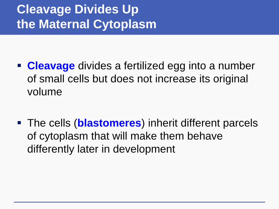
Cleavage Divides Up
the Maternal Cytoplasm
Cleavage divides a fertilized egg into a number
of small cells but does not increase its original
volume
The cells (blastomeres) inherit different parcels
of cytoplasm that will make them behave
differently later in development
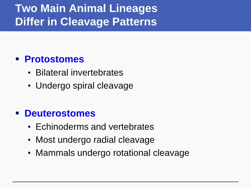
Two Main Animal Lineages
Differ in Cleavage Patterns
Protostomes
• Bilateral invertebrates
• Undergo spiral cleavage
Deuterostomes
• Echinoderms and vertebrates
• Most undergo radial cleavage
• Mammals undergo rotational cleavage

Variations in Cleavage Patterns

Effects of Yolk Size
on Cleavage Patterns
Structure of the Blastula
Blastula
• Cells produced by cleavage
• Structure varies with species’ cleavage pattern
Blastocyst (mammalian blastula)
• Outer cells secrete fluid into the cavity
• Inner cells, clustered against the cavity wall,
develop into the embryo

43.3 From Blastula to Gastrula
Gastrulation
• Developmental process during which cells
rearrange themselves into primary tissue layers
Most animals have three primary tissue layers
• Outermost layer (ectoderm)
• Middle layer (mesoderm)
• Inner layer (endoderm)

Gastrulation in a Fruit Fly

Initiation of Gastrulation
Gastrulation occurs when certain cells of the
blastula make and release short-range signals
that cause nearby cells to move about, either
singly or as a cohesive group
Embryonic induction
• The fate of one group of embryonic cells is
affected by its proximity to another group of cells

Experiment: Embryonic Induction
Transplanted cells of the dorsal lip of the
blastula (descended from the zygote’s gray
crescent) induced gastrulation in salamanders

43.4 Specialized
Tissues and Organs Form
Cell differentiation
• Process by which cell lineages become
specialized
• Lays the groundwork for formation of specialized
tissues and organs
• Based on selective gene expression
Signaling molecules contribute to differentiation
Morphogens
Morphogens
• Signaling molecules encoded by master genes
• Diffuse from a source and form a concentration
gradient throughout the embryo
• Have different effects depending on their
concentration in each region

Morphogenesis
Morphogenesis
• Process by which tissues and organs form
• Some cells migrate to new locations
• Sheets of cells change shapes to form organs
• Apoptosis shapes body parts such as fingers
Apoptosis
• Cells die on cue; signals from cells cause other
cells to self-destruct

Morphogenesis: Neural Tube Formation
Pattern Formation
Pattern formation
• Process by which body parts form in a specific
place
Example: Limb bud formation in chicks
• AER at the tips of limb buds induces the
mesoderm beneath to form a limb
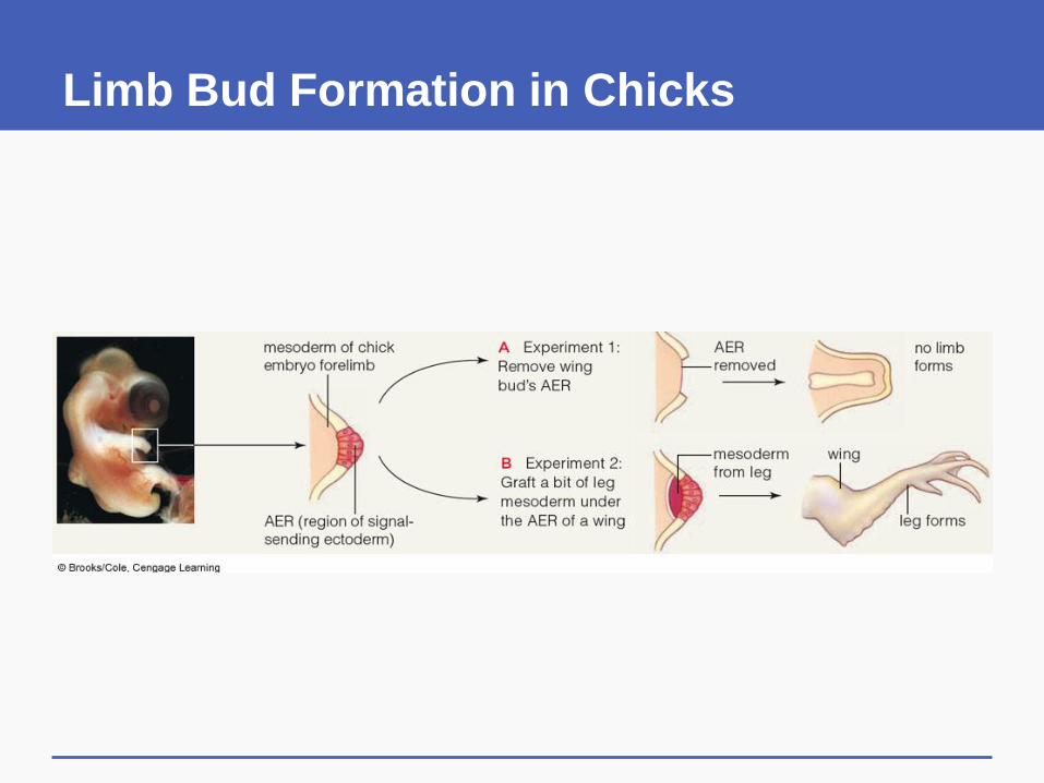
Limb Bud Formation in Chicks

43.5 An Evolutionary
View of Development
Similarities in developmental pathways among
animals are evidence of common ancestry
Cytoplasmic localization in the egg induces
expression of localized master genes
Concentration gradients of master gene
products cause embryonic cells to form tissues
and organs at certain locations
Homeotic Genes
Positional information established by
concentration gradients of master gene products
affects expression of homeotic genes, which
regulate development of specific body parts

Developmental Constraints
and Modifications
Physical constraints
• Surface-to-volume ratio
Architectural constraints
• Existing body frameworks, such as four limbs
Phyletic constraints
• Master genes determine basic body form

Developmental Constraints
and Modifications
Mutations that alter the effects of master genes
are often lethal
Example: Development of somites
• Mesoderm on either side of the neural tube
divides into blocks of cells that will develop into
bones and muscles

Lethal Mutation Affecting Somites

43.1-43.5 Key Concepts
Principles of Animal Embryology
Animals develop through cleavage, gastrulation,
organ formation, and then growth and tissue
specialization
Cleavage parcels out material stored in different
parts of the egg cytoplasm into different cells,
thus starting the process of cell specialization
43.6 Overview of Human Development
Humans begin life as a single cell and go
through a series of developmental stages
• Second week: Blastocyst is embedded in the
mother’s uterus, where it develops
• Embryonic period (first 8 weeks): All organs form
• Fetal period (9 weeks to birth): Organs of the
fetus grow and specialize
• Postnatal growth (after birth): Organ growth and
maturation continues until adulthood
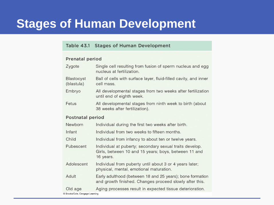
Stages of Human Development

Prenatal and Postnatal Changes

43.7 Early Human Development
Cleavage of a zygote produces a cluster of 16
cells (morula) by the time it reaches the uterus
By the fifth day, a blastocyst forms, consisting
of an outer layer, a fluid-filled cavity (blastocoel)
and an inner cell mass
• Inner cell mass will form the embryo
• Outer cells will form supportive tissues

Implantation
Implantation
• The blastocyst ruptures the zona pellucida and
burrows into the lining (endometrium) of the
mother’s uterus
• In ectopic pregnancy, the blastocyst implants
outside the uterus

Extraembryonic Membranes
The outer layer of the blastocyst gives rise to
four external membranes
• Amnion encloses and protects the embryo in a
fluid-filled cavity
• Yolk sac gives rise to blood and germ cells
• Chorion extends into maternal tissues and
becomes part of the placenta
• Allantois gives rise to blood vessels of placenta

The Placenta
Placenta
• An organ that functions in exchange of materials
between the bloodstreams of a mother and her
developing child
• Forms from projections of chorion that extend into
blood-filled maternal tissues, and blood vessels of
allantois

Human Extraembryonic Membranes

Early Hormone Production
Human chorionic gonadotropin (HCG)
• Released by blastula after implantation
• Causes corpus luteum to keep secreting
progesterone and estrogens to maintain the
uterine lining
The placenta takes over secretion of HCG after
about three months

Fertilization to Implantation
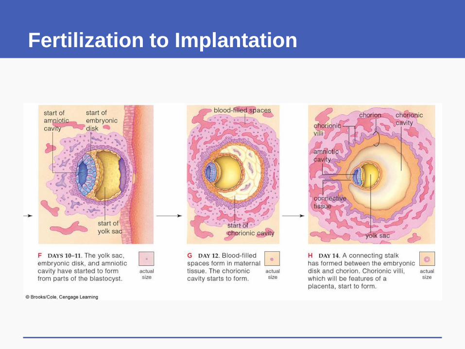
Fertilization to Implantation
43.8 Emergence of
the Vertebrate Body Plan
Two weeks after fertilization, the inner cell mass
of a blastocyst is a two layered embryonic disc
Gastrulation occurs in the third week, forming an
embryo with three germ layers: ectoderm,
mesoderm, and endoderm
• Primitive streak, neural tube and notochord form
• Somites appear on either side of the neural tube

Derivatives of Human Germ Layers
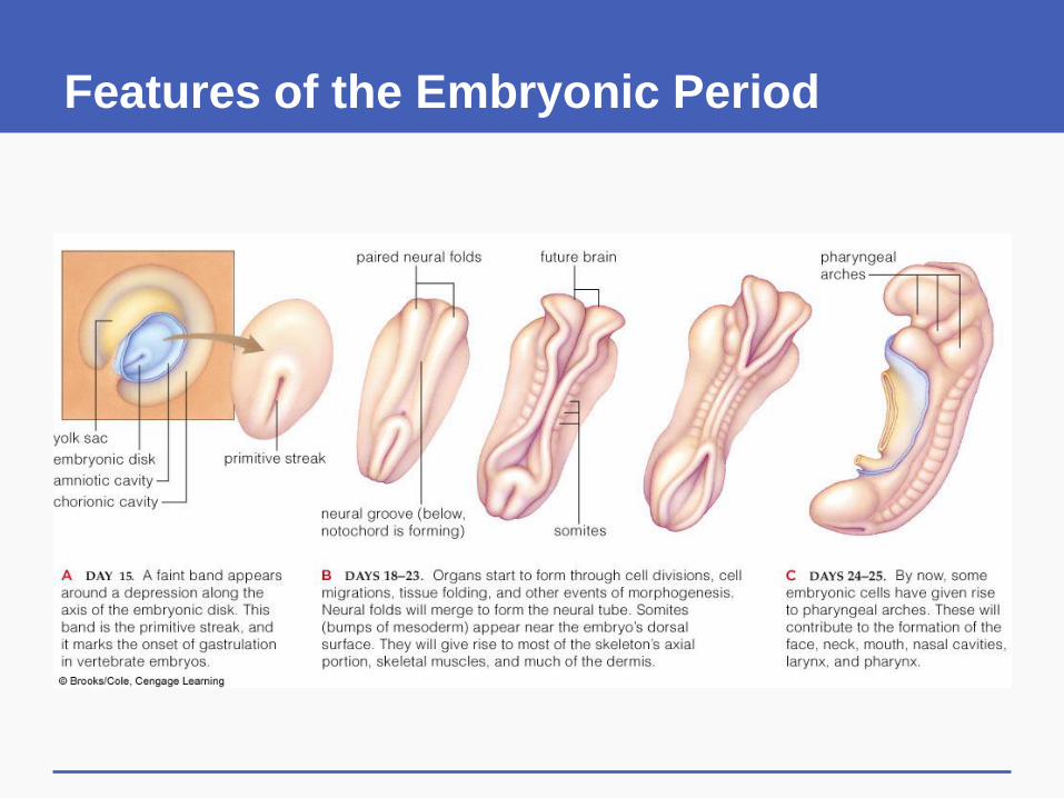
Features of the Embryonic Period
43.6-43.8 Key Concepts
Human Development Begins
A pregnancy starts with fertilization and
implantation of a blastocyst in the uterus
After implantation, a three-layered embryo forms
and organ formation begins
All organs have formed by the end of the eighth
week

43.9 The Function of the Placenta
Maternal and embryonic blood do not mix
• Vessels of the embryo’s circulatory system
extend through the umbilical cord to the placenta,
where they run through pools of maternal blood
• Substances diffuse across membranes between
maternal and embryonic bloodstreams
Placental hormones maintain the uterine lining

The Placenta
43.9 Key Concepts
Function of the Placenta
The placenta allows substances to diffuse
between bloodstreams of a mother and her
developing child
It also produces hormones that help sustain the
pregnancy

43.10 Emergence of
Distinctly Human Features
Embryonic features disappear and the fetus
takes on human appearance about 8th week
Heartbeat and movements are detected in the
second trimester
In the third trimester, the brain is formed and
functioning

Development of the Human Embryo

Development of the Human Embryo

43.11 Mother as Provider and Protector
A developing human depends on its mother to
supply the nutrients it requires to grow and develop
• Proteins, carbohydrates, and lipids
• Vitamins and minerals
Dietary deficiencies affect many developing organs

Teratogens
The embryo/fetus is also subjected to any toxins
or pathogens to which the mother is exposed
Teratogens
• Toxic or infectious agents that interfere with
development
• Effects vary with the timing of exposure

Teratogens
Infectious agents
• Viral diseases (such as rubella), toxoplasmosis
Alcohol and caffeine
• Fetal alcohol syndrome, miscarriage
Smoking
• Affects growth and development
Prescription drugs
• Some medications cause severe birth defects
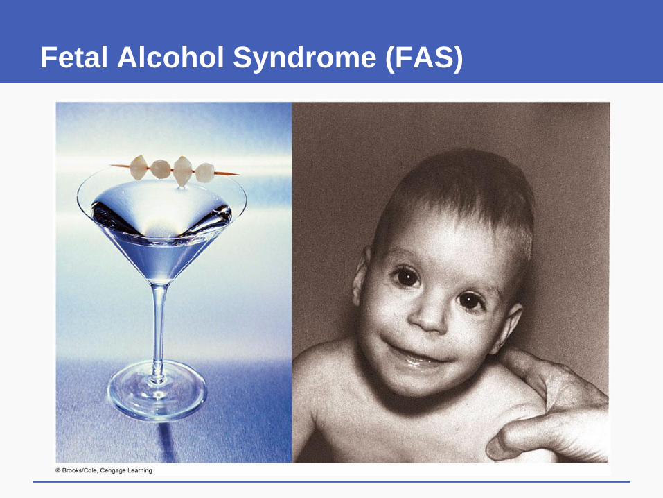
Fetal Alcohol Syndrome (FAS)

Teratogen Sensitivity

43.10-43.11 Key Concepts
Later Human Development
By the time the fetal period begins, the
developing individual appears distinctly human
Harmful substances that get into a mother’s
blood can cross the placenta and cause birth
defects in the developing embryo or fetus
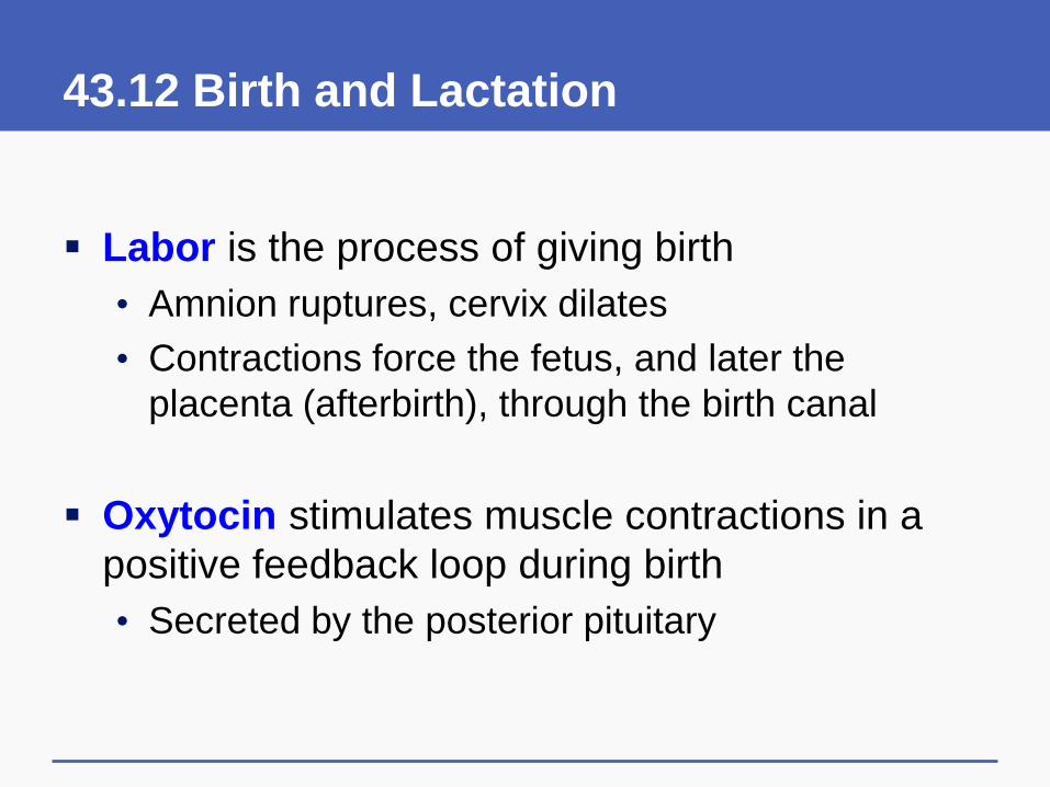
43.12 Birth and Lactation
Labor is the process of giving birth
• Amnion ruptures, cervix dilates
• Contractions force the fetus, and later the
placenta (afterbirth), through the birth canal
Oxytocin stimulates muscle contractions in a
positive feedback loop during birth
• Secreted by the posterior pituitary

Birth and Afterbirth

Nourishing the Newborn
Newborn humans are nourished with milk
secreted by the mother’s mammary glands
Hormonal control of lactation (milk production)
• Prolactin, secreted by the anterior pituitary,
triggers milk synthesis
• Declines in progesterone and estrogen
production after birth increase milk production
• Oxytocin stimulates release of milk into milk ducts

Lactation and Mammary Glands
43.12 Key Concepts
Birth and Lactation
Positive feedback control plays a role in the
process of labor, or childbirth
After birth, the newborn is nourished by milk
secreted by mammary glands