Anesthesia of the cardiac patient for non-cardiac surgery
15
2013.12.08. 1 Acute coronary syndrome and acute left /right heart failure in the ICU Krisztina Madách Department of Anesthesiology and Intensive Therapy 2 Mosaics of the lecture • ACS – PCI guidelines – Periop. management of PCI, BMS, DES patients • Heart failure – Pathophysiology – Pharmacological management – Mechanical management 3 Acute Coronary Syndrome • Endemic disease • Invasive investigation – majority of ACS patients • Anticipated life expectancy at birth increased with 5,5 years • Death of MI has halved! • Indications – new guidelines – Revascularisation guideline (2010) – ESC NSTE-ACS guideline (2011) – ESC STEMI guideline (2008, 2012) 4 Symptoms ACUTE CORONARY SYNDROME STEMI NSTE-ACS ECG Persisting ST elevation ST-T deviation No/ aspecific ECG change Biomarker pos neg Diagnosis STEMI NSTEMI Unstable angina NSTE-ACS Diagnosis
Transcript of Anesthesia of the cardiac patient for non-cardiac surgery

2013.12.08.
1
Acute coronary syndrome and acute left
/right heart failure in the ICU
Krisztina Madách
Department of Anesthesiology and Intensive Therapy
2
Mosaics of the lecture
• ACS
– PCI guidelines
– Periop. management of PCI, BMS,
DES patients
• Heart failure
– Pathophysiology
– Pharmacological management
– Mechanical management
3
Acute Coronary Syndrome
• Endemic disease
• Invasive investigation
– majority of ACS patients
• Anticipated life expectancy at birth increased
with 5,5 years
• Death of MI has halved!
• Indications – new guidelines – Revascularisation guideline (2010)
– ESC NSTE-ACS guideline (2011)
– ESC STEMI guideline (2008, 2012)
4
Symptoms ACUTE CORONARY SYNDROME
STEMI NSTE-ACS
ECG Persisting ST
elevation
ST-T
deviation
No/ aspecific
ECG change
Biomarker pos neg
Diagnosis STEMI NSTEMI Unstable angina
NSTE-ACS
Diagnosis

2013.12.08.
2
5
• On-site diagnosis
– Typical chest pain
– ST elevation or newly developed LBBB
IS ENOUGH FOR PRIMER DIAGNOSIS
– Elevated necroenzymes – not necessary to wait for the
result
– Echocardiography: to disclose other origin of acute
chest pain
ESC STEMI guideline 2008
Early diagnosis
6
• Within 12 hours: STEMI or chest pain and unknown LBBB
(IA)
• After 12 hours: persisting chest pain and ST elevation (IIa)
• 12 - 24 hours: patient with no complaints (IIb)
• After 3 days: routine opening of occluded coronary arteries
responsible for MI may be harmful if there is no viability!
6
PCI indication-STEMI
ESC Revasc. Guideline 2010
7
PCI indication-NSTEMI
Risk factors
• troponin elevation
• dynamic ST-T changes
• diabetes mellitus
• renal insufficiency
• decreased left ventricular function (EF<40%)
• early post-MI angina
• PCI within 6 month
• previous CABG
• medium or high (>140) GRACE risk
8
PCI indication-NSTEMI
• <2 hours: very high ischemic risk
• <24 hours: GRACE score >140, multiple risk factors
• 72 hours <: GRACE score <140, but relapsing
symptomes, positive stress test.

2013.12.08.
3
9
The problem
• 1990’: increasing periop. morbidity/mortality in patients
with PCI going for non-cardiac surgery
• 90% of patients going for PCI get one or several stent/s
• 5% will have non-cardiosurgical intervention within 1 year!
• Periop. dilemma: bleeding or coagulation?
• 63% of anesthesiologists do not know the guideline!
Vicenzi MN et al. Br J Anaesth 2006;96:686-93
Patterson L et al. Can J Anesthesiol 2005;52:440-1
10
PCI, BMS, DES
The traps: restenosis or incomplete
endothelisation and thrombosis?
Venkatesan S. Why should a stent elute drug ?
http//:drsvenkatesan.files.wordpress.com 2008
11
Platelet formation
inhibiting therapies:
What?
For how long?
Mardows TA et al. Circ Res 2007;100:1261 12
Change in endothelisation of BMS and DES stents (intravascular US, autopsy)
Joner M et al. J Am Coll Cardiol 2006;48:193-202

2013.12.08.
4
13
Consequences of abrupt discontinuation of antiplatelet
drugs in the perioperative period
Myocardial infarction
Death
Stent thrombosis
Incompletely endothelized stents-prothrombotic state
Surgical intervention
Increased prothrombotic and inflammatory state
•Increased cytokine, neuroendocrine, inflammatory mediator release
•Increased platelet adhesiveness and persistently high platelet counts
•Increased release of procoagulant factors
•Decreased/impaired fibrinolysis
Rebound effect:
•Significantly increased inflammatory prothrombotic state
•Significantly increased platelet adhesion and aggregation
•Excessive TXA2 activity (for 3-4 days)
Abrupt discontinuation of
clopidrogel
Abrupt discontinuation of aspirin
Discontinuation of antiplatelet drugs: 5-10x increased perioperative myocardial
death?
Chassot PG et al.. Br J Anaesth 2007;99:316-28
14
Clopidrogel use and long-term outcomes with BMS and DES
Eisenstein EL. JAMA 2007;297:159-68
7,29
(p=0.02)
6.0
5.5
3.1
15
Risk of major adverse cardiac events after stent implantation
in non-cardiac surgery
Popescu WM. Current Opinion in Anaesthesiology 2010; 23:109-115
BMS
DES
16
Perioperative bleeding
• ASA
– 1,5x increased bleeding risk
– BUT! Except intracranial operation and TUR it did not increase perioperative morbidity/mortality
• Clopidrogel+ASA (mainly cardiac surgery)
– Bleeding↑, reexplorations, transfusions, increased ICU/hospital stay, no difference in mortality (?)
– BUT! Long-term (10 years) mortality was 80% in patients transfused with 1 U rbc vs. 63% in not transfused patients!
Burger W et al. J Intern Med 2005;257:399-414
Koch CG et al. Ann Thorac Surg 2006;81:1650-7

2013.12.08.
5
17
• UFH
• LMWH
• Direct thrombin inhibitors
• GP IIb/IIIa inhibitors
Assessment of the risk of bleeding,
prevention, treatment
• TEG/ROTEM
• Renal function
• Bridge therapy?
• Antidote: platelet
transfusion
18
Required length of APT
TIMING of anticipated surgery
• Dilatation without stenting: 2-4 weeks dual APT
– Delay for noncardiac surgery: 2-4 weeks
• PCI and BMS: min 4-6 weeks dual APT
– Delay for noncardiac surgery : > 6 weeks
• PCI and DES: 12 months dual APT
– Delay for noncardiac surgery : > 12 months
• Aspirin lifelong
2007 AHA/ACC Science Advisory and Society of Cardiovascular Angiography DES Task Force
Recommendations for Timing of Noncardiac Surgery after PCI
19
Overview: prevention of periop.stent thrombosis
• Avoid preop. coronary stenting
• Stent selection (BMS-DES)
• Delay surgery
• There is no bridge therapy for PCI
patients, timing is crucial
• Education, interdisciplinary teamwork
• Surgery only where PCI is available
Brilakis et al. JACC 2007;49,22:2145-50 20
HEART FAILURE
• > 65 age: most frequent cause of hospital admission
• The incidence of hospitalization and the incidence of
mortality due to heart failure keeps increasing since 1968
– Despite the decrease in overall mortality of cardiac diseases
• 5 year mortality ~ 50%
• Patient going under emergency laparotomy with untreated
heart failure has a mortality of 20-30 %
Telford R. Oxford handbook of anaesthesia 2007;3:48-51

2013.12.08.
6
21
Definition
• Heart failure is a complex clinical syndrome,
characterized by
– Insufficient ventricular performance
– Intolerance of workload
– Frequent ventricular arrhythmias
– Reduced anticipated lifetime.
• Classification:
– Diastolic dysfunction (relaxation problem of the left ventricule)
– Systolic dysfunction (can involve either ventricules)
22
Etiology
• Heart failure is the final common pathway of primer
cardiovascular diseases
– Acut/chronic
• Causes:
– Most frequent: coronary disease, hypertension
– Other: diabetes, valve disease, non-ischemic myopathies
• Indipendent risk factors:
– Man, hypertension, coronary disease, diabetes, age
23
Pathophysiology
„The heart has no wrinkles.” Madame de Sévigné
But nevertheless it ages:
• Size of the ventricle, wall thickness, stiffness,
myocyte loss, fibrosis, collagen bridges
• Max. frequency and CO decreases, SVR,
systolic BP increases
24
Pathophysiology
• Left ventricular relaxation disorder :
– isovolaemic relaxation time increases,
– sarcoplasmatic reticulum Ca++ excretion decreases
• Left ventricular diastolic filling becomes more
dependent on atrial contractions
2013.12.08.
7
25
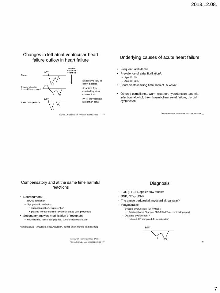
Changes in left atrial-ventricular heart
failure ouflow in heart failure
Magner J, Royston D. Br J Anaesth 2004;93:74-85
E: passive flow in
early diastole
A: active flow
created by atrial
contraction
IVRT: isovolaemic
relaxation time
26
Underlying causes of acute heart failure
• Frequent: arrhythmia
• Prevalence of atrial fibrillation1:
– Age 60: 5%
– Age 90: 22%
• Short diastolic filling time, loss of „A wave”
• Other: ↓ compliance, warm weather, hypertension, anemia,
infection, alcohol, thromboembolism, renal failure, thyroid
dysfunction
1Aronow WS et al. J Am Geriatr Soc 1996;44:521-3
27
Compensatory and at the same time harmful
reactions
• Neurohumoral:
– RAAS activation
– Sympathetic activation:
• vasoconstriction, Na retention
• plasma norepinephrine level correlates with prognosis
• Secondary answer: modification of receptors
– endotheline, natriuretic peptide, tumour necrosis factor
Pre/afterload-, changes in wall tension, direct toxic effects, remodelling
1Aronow W. Heart Dis.2003;5: 279-94
2Cohn JN. Engl J Med 1984;311:819-23 28
Diagnosis
• TOE (TTE), Doppler flow studies
• BNP, NT-proBNP
• The cause pericardial, myocardial, valvular?
• If myocardial:
– Systolic dysfunction (EF<40%) ?
• Fractional Area Change= EDA-ESA/EDA (~ventriculography)
– Diastolic dysfunction ?
• reduced „E”, elongated „E” deceleration)

2013.12.08.
8
29
Therapy of heart failure
• Most cases can not be cured, but can be improved
symptomatically, physical activity and quality of life / duration
can be increased.
• End-stage heart failure: heart transplantation is currently the
only therapy
• And until then?
– Pharmacological
– Non-pharmacological possibilities
30
Therapy of heart failure
1. Treat the underlying cause
2. Elimination of associated factors further impairing
heart failure (infection, hyperthyreosis, stb)
3. Treat heart failure itself
31
Factors determining cardiac output
diastolic
compliance
venous
backflow
preload
contractility
Stroke
volume (SV)
Sympathic/ parasympathic tone
Heart frequency
Cardiac output
(CO)
afterload SVR +
+
+
+
+/-
-
+
32
Restrictive cardiomyopathy
• Difficult to manage
• Wall stiffness: decreased ventricular diastolic filling
• Right heart failure is common
• Cardiac arrest can be provoked by:
– Myocardium depression
– Vasodilatation
– Decreased venous back-flow
– Increased intrathoracal pressure (IPPV)
• Recommended: low intrathoracal pressure, elevated right
ventricular pressure: fluid therapy
Bovill J. Semin Cardiothorac Vasc Anaesth 2003;7:49-54

2013.12.08.
9
33
Dialatative cardiomyopathy
• Low contractility, stroke volume is maintained by
increased LV ebddiastolic volume
• Functional MI, TI
• Frequent arrhythmia → amiodarone
• Avoid myocardium depressive drugs
• Inotrops might be needed
Bovill J. Semin Cardiothorac Vasc Anaesth 2003;7:49-54 34
Hypertrophic cardiomyopathy
• Outflow obstruction (septal hypertrophy)
• Diastolic dysfunction
• Diastolic filling is atrial systole dependent: sinus rhythm!
– Might be even 75% of end-diastolic volume!
• Tachycardia reduces diastolic filling time
• Inotrops can deteriorate outflow obstruction and oxygen
demand of the myocardium, and increase wall tension
• Vasodilators not inducing tachycardia are recommended
Bovill J. Semin Cardiothorac Vasc Anaesth 2003;7:49-54
35
Basic pharmacological treatment
• ACE inhibitors: pre- and afterload↓, neurohormonal blockade, improve
survival
• Βeta blockers: myocardium oxygen demand↓
• Diuretics: spironolacton improves mortality when given with ACE
inhibitor in severe heart failure (EF<25%)
• Cardiac glycosides: digoxin (frequency control, sympathetic
hyperactivity↓)
• Avoid: NSAID, Ca channel blockers (except. amlodipine)
Lonn E. Drug treatment in heart failure. BMJ 2000;320:1188-92 36
Inotrops
Magner J, Royston D. Br J Anaesth 2004;93:74-85
milrinone: Corotrope
dobutamine
trimetazidine: Preductal,
Moduxin
levosimendan: Simdax
Calcium
sensitizer
2013.12.08.
10
37
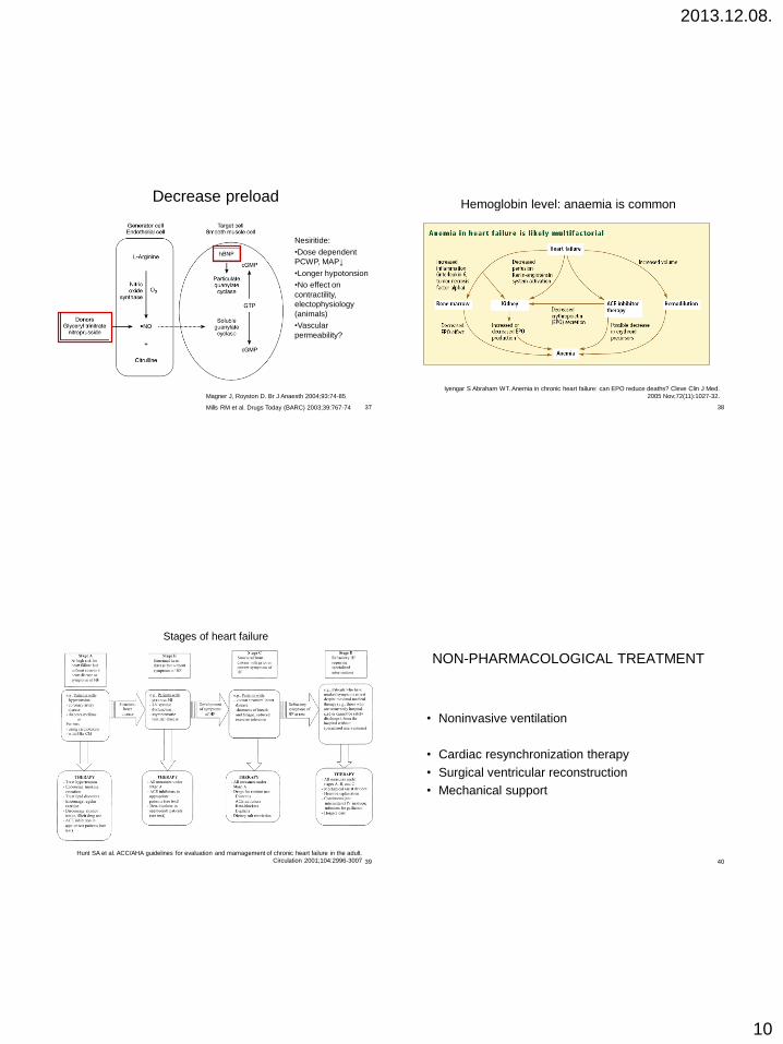
Decrease preload
Magner J, Royston D. Br J Anaesth 2004;93:74-85
Mills RM et al. Drugs Today (BARC) 2003;39:767-74
Nesiritide:
•Dose dependent
PCWP, MAP↓
•Longer hypotonsion
•No effect on
contractility,
electophysiology
(animals)
•Vascular
permeability?
38
Hemoglobin level: anaemia is common
Iyengar S Abraham WT. Anemia in chronic heart failure: can EPO reduce deaths? Cleve Clin J Med.
2005 Nov;72(11):1027-32.
39
Stages of heart failure
Hunt SA et al. ACC/AHA guidelines for evaluation and mamagement of chronic heart failure in the adult.
Circulation 2001;104:2996-3007 40
NON-PHARMACOLOGICAL TREATMENT
• Noninvasive ventilation
• Cardiac resynchronization therapy
• Surgical ventricular reconstruction
• Mechanical support

2013.12.08.
11
Is there a role for noninvasive positive
pressure ventilation (NIV) in acute cardiogenic
pulmonary oedema (ACPE)?
42
Effectivity of NIV in ACPE
• Predictors of success: ABG (pH, pCO2), vital signs
• Patients like it
32 (5)
29 (7)
PaCO2
(mmHg)
NIPPV
Spont.
n=46
87 (15) 109 (13) 16 (8)
126(21) 67 (6) 28 (7)
HR PaO2
(mmHg) Resp.rate (f)
43
Cardiac resynchronization therapy
• Sudden death due to arrhythmia is common
• Sinus rhythm is essential
• Delay in interventricular conduct : non-
synchronized ventricular contraction deteriorates
LV systolic dysfunction
– 30% of heart failure patients!
Abraham WT et al. N Engl J Med 2002;346:1845-53
44
Cardiac resynchronization therapy
• Atrium synchronized biventricular pacing
• EF↑, PCWP↓, diastolic ventricular filling time↑, mitral
regurgitation↓, quality of life ↑, workload↑
• Mortality was reduced by 20%
• If ICD: mortality was reduced by 40%
Chow AW et al. BMJ 2003;326:1073-7
MUSTIC study. Linde C.et al. J Am Coll Cardiol 2002;40:111-18
COMPANION study. Bristow MR et al. J Card FAil. 2000;6:276-85
2013.12.08.
12
45
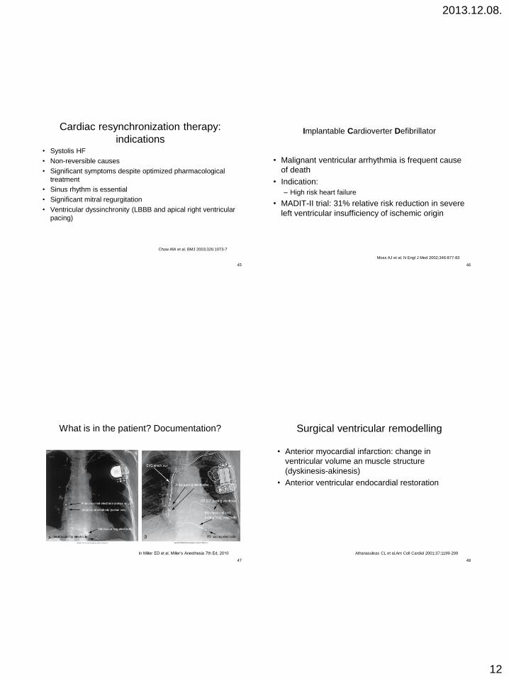
Cardiac resynchronization therapy:
indications • Systolis HF
• Non-reversible causes
• Significant symptoms despite optimized pharmacological
treatment
• Sinus rhythm is essential
• Significant mitral regurgitation
• Ventricular dyssinchronity (LBBB and apical right ventricular
pacing)
Chow AW et al. BMJ 2003;326:1073-7
46
Implantable Cardioverter Defibrillator
• Malignant ventricular arrhythmia is frequent cause
of death
• Indication:
– High risk heart failure
• MADIT-II trial: 31% relative risk reduction in severe
left ventricular insufficiency of ischemic origin
Moss AJ et al. N Engl J Med 2002;346:877-83
47
What is in the patient? Documentation?
In Miller ED et al. Miller’s Anesthesia 7th Ed. 2010
48
Surgical ventricular remodelling
• Anterior myocardial infarction: change in
ventricular volume an muscle structure
(dyskinesis-akinesis)
• Anterior ventricular endocardial restoration
Athanasuleas CL et al.Am Coll Cardiol 2001;37:1199-209

2013.12.08.
13
49
Mechanical assist devices: MAD
• Reduce mechanical work of the heart,
ameliorate coronary perfusion
• Difficult decision:
– Can the heart improve? MODS?
– Can this be influenced by MAD?
• Wide range of devices
50
Mechanical assist devices: MAD
• IABP
– Short term support
– Ambulatory, longer term application? 1
– Overall morbidity (limb ischemia, bleeding) 3-5%
• VAD-Ventricular Assist Device
– End stage (lack of donor organs)
– FDA: Bridge to transplant
– REMATCH trial: patients inappropriate for transplantation2
• 1 year survival 52% contra 25%
• Morbidity 2,35x of pharmacologically treated group: bleeding, infection,
malfunction
1Cochran RP et al. Ann Thorac Surg 2002;74:746-51
2 Rose EA et al. N Engl J Med 2001;345:1435-43
51
Ventricular Assist Device: VAD
• Indication: postcardiotomy shock, AMI, myocarditis,
ACS-cardiogen shock
• „Bridge to recovery”
• „Bridge to transplant”
– MODS can be improved
• „Bridge to destination”
• Timing is crucial
Williams M et al. Cardiac assist devices for end-stage heart failure. Heart Dis 2001;3:109-15
52
Ventricular Assist Device: VAD
• Choice of equipment: duration, body surface,
uni/biventricular support
• Extra-, intracorporal
• According to flow generator: centrifugal, axial,
diaphragmatic
• Considerations: anticoagulation, ICU/outpatient,
mobilization
Williams M et al. Cardiac assist devices for end-stage heart failure. Heart Dis 2001;3:109-15

2013.12.08.
14
53
54
55
56

2013.12.08.
15
57
58
Summary
• Death of myocardial infarction has halved due to invasive investigations/interventions (ESC guidelines)
• PCI (BMS, DES stents): restenosis, incomplete endothelization, thrombosis is a problem
• Platelet aggregation inhibitors and bleeding risk: there is no bridge therapy, timing is crucial
• Heart failure is the final common pathway of primer cardiovascular diseases with increasing incidence
• Pharmacological and novel non-pharmacological therapies can improve life quality and expectancy of heart failure.
59