anemia
56
Anemia Anemia Robb Friedman, MD Robb Friedman, MD Modified by Sean Hesselbacher, MD, Modified by Sean Hesselbacher, MD, Eyal Oren, MD, David Antonetti, MD Eyal Oren, MD, David Antonetti, MD and Cathy Okuliar, MD and Cathy Okuliar, MD
Transcript of anemia

AnemiaAnemiaRobb Friedman, MDRobb Friedman, MD
Modified by Sean Hesselbacher, MD, Modified by Sean Hesselbacher, MD, Eyal Oren, MD, David Antonetti, MD Eyal Oren, MD, David Antonetti, MD
and Cathy Okuliar, MDand Cathy Okuliar, MD

What is Anemia?What is Anemia? ANEMIA IS NEVER NORMALANEMIA IS NEVER NORMAL Reduction below normal in the mass of Reduction below normal in the mass of
red blood cells in the circulationred blood cells in the circulation Hemoglobin concentration, hematocrit, Hemoglobin concentration, hematocrit,
RBC countRBC count Men: HGB < 14 or HCT < 41%Men: HGB < 14 or HCT < 41% Women: HGB < 12.0 or HCT < 36%Women: HGB < 12.0 or HCT < 36%

Anemia and Volume StatusAnemia and Volume Status HGB and HCT are CONCENTRATIONSHGB and HCT are CONCENTRATIONS Therefore dependent upon plasma volumeTherefore dependent upon plasma volume Acute bleeds not reflected for 24-36 hrsAcute bleeds not reflected for 24-36 hrs
– Due to volume deficit being slowly repaired via Due to volume deficit being slowly repaired via movement of fluid from extravascular space to movement of fluid from extravascular space to intravascularintravascular
Anemic patients who are dehydrated will not Anemic patients who are dehydrated will not appear anemicappear anemic
Pregnant women expand RBCs 25% but plasma Pregnant women expand RBCs 25% but plasma volume increases 50%, producing “physiologic volume increases 50%, producing “physiologic anemia”anemia”

Anemia: Special CasesAnemia: Special Cases ErythrocytosisErythrocytosis
– People who live at high altitude have greater RBC People who live at high altitude have greater RBC volumevolume
– Smokers have increased HCT – impairs the ability of Smokers have increased HCT – impairs the ability of the RBCs to deliver O2the RBCs to deliver O2
African-American HGBs are 0.5 to 1.0g/dL lower African-American HGBs are 0.5 to 1.0g/dL lower than Caucasiansthan Caucasians
Athletes (increased plasma volume, Fe Athletes (increased plasma volume, Fe deficiency, hemolysis, polycythemia, use of deficiency, hemolysis, polycythemia, use of performance enhancing agents)performance enhancing agents)

Anemia and the ElderlyAnemia and the Elderly Multiple studies show that the elderly do Multiple studies show that the elderly do
notnot have a “lower normal range” have a “lower normal range” Anemia, while common in the elderly, is Anemia, while common in the elderly, is
still abnormalstill abnormal HGB < 13 in males and < 12 in females HGB < 13 in males and < 12 in females
associated with an increased relative risk associated with an increased relative risk of mortality (1.6 and 2.3 respectively)of mortality (1.6 and 2.3 respectively)

Anemia: HistoryAnemia: History Is the patient bleeding?Is the patient bleeding?
– NSAIDs, ASANSAIDs, ASA– Menstrual history, if applicable (include older women)Menstrual history, if applicable (include older women)– Prior intestinal surgery?Prior intestinal surgery?– Hx of hemorrhoids, hematochezia, or melena?Hx of hemorrhoids, hematochezia, or melena?
Past medical history of anemia? Family history?Past medical history of anemia? Family history? Alcohol, nutritional questionsAlcohol, nutritional questions Liver, renal diseasesLiver, renal diseases EthnicityEthnicity Environmental/work toxins (ie lead)Environmental/work toxins (ie lead)

Symptoms of AnemiaSymptoms of Anemia Decreased O2 deliveryDecreased O2 delivery Hypovolemia if acute lossHypovolemia if acute loss Exertional dyspnea, fatigue, palpitations, Exertional dyspnea, fatigue, palpitations,
lightheadednesslightheadedness Severe: heart failure, anginaSevere: heart failure, angina ““Pica”– craving for clay or paper productsPica”– craving for clay or paper products Pagophagia– craving for icePagophagia– craving for ice

Signs of AnemiaSigns of Anemia Tachycardia, tachypnea, orthostasisTachycardia, tachypnea, orthostasis PallorPallor JaundiceJaundice MurmurMurmur Koilonychia or “Spoon nails”Koilonychia or “Spoon nails” Splenomegaly, lymphadenopathySplenomegaly, lymphadenopathy Petechiae, ecchymosesPetechiae, ecchymoses Atrophy of tongue papillaeAtrophy of tongue papillae Heme + stoolHeme + stool

The Four Causes of AnemiaThe Four Causes of Anemia
Decreased red blood cell Decreased red blood cell productionproduction
Increased red blood cell Increased red blood cell destructiondestruction
Red blood cell lossRed blood cell lossRed blood cell sequestrationRed blood cell sequestration
* Underlying disorder is abnormal production vs. premature loss

Decreased RBC productionDecreased RBC production Deficiency of iron, B12, folateDeficiency of iron, B12, folate Marrow is dysfunctional from Marrow is dysfunctional from
myelodysplasia, tumor infiltration, aplastic myelodysplasia, tumor infiltration, aplastic anemia, etc.anemia, etc.
Bone marrow is suppressed by Bone marrow is suppressed by chemotherapy or radiationchemotherapy or radiation
Low levels of erythropoeitin, thyroid Low levels of erythropoeitin, thyroid hormone, or androgenshormone, or androgens

Increased RBC destructionIncreased RBC destruction RBCs live about 100 daysRBCs live about 100 days Acquired: autoimmune hemolytic anemia, Acquired: autoimmune hemolytic anemia,
TTP-HUS, DIC, malariaTTP-HUS, DIC, malaria Inherited: spherocytosis, sickle cell, Inherited: spherocytosis, sickle cell,
thalassemiathalassemia

RBC LossRBC Loss Bleeding!Bleeding! Obvious vs occultObvious vs occult Iatrogenic: venesection e.g. daily CBC, Iatrogenic: venesection e.g. daily CBC,
surgical, hemodialysissurgical, hemodialysis RetroperitonealRetroperitoneal

Approach to AnemiaApproach to AnemiaCBC
Reticulocyte countMCV
RI < 2% RI > 2%
Underproduction Increased destruction or loss
MCV < 80Microcytic
MCV > 100Macrocytic
MCV 81 – 99Normocytic
MCV Further work upBased on history, Physical, other

Approach to AnemiaApproach to Anemia LOOK AT THE LOOK AT THE
SMEAR!!!!SMEAR!!!! Convenient to separate Convenient to separate
into three classes based into three classes based on the size of the RBCon the size of the RBC
MCV and RDWMCV and RDW Microcytosis: < 80 fLMicrocytosis: < 80 fL Normocytosis: 80-100 fLNormocytosis: 80-100 fL Macrocytosis: >100 fLMacrocytosis: >100 fL CBC, reticulocyte count, CBC, reticulocyte count,
Fe, Ferritin, TIBC, folate, Fe, Ferritin, TIBC, folate, B12, LDH, CMP, ESR…B12, LDH, CMP, ESR…

ReticulocytesReticulocytes Nucleated RBCs – form in marrow where they Nucleated RBCs – form in marrow where they
mature for 3 days and then spend 1 day in mature for 3 days and then spend 1 day in circulation (before maturing to RBC)circulation (before maturing to RBC)
Given avg life span of RBC of 100 days, 1% of Given avg life span of RBC of 100 days, 1% of RBCs are destroyed each dayRBCs are destroyed each day
Retics form 1% of circulating RBCs qdRetics form 1% of circulating RBCs qd Nl RBC count is 5 million/uL so marrow makes Nl RBC count is 5 million/uL so marrow makes
50,000 reticulocytes/uL blood qd50,000 reticulocytes/uL blood qd– With epo, can increase to 250,000 retics/uL blood qd With epo, can increase to 250,000 retics/uL blood qd
(given nl marrow and replete iron, folate, b12)(given nl marrow and replete iron, folate, b12)

Reticulocyte CountReticulocyte Count
Assess adequacy of bone marrow response to anemiaAssess adequacy of bone marrow response to anemia Must adjust for the degree of anemia, use Reticulocyte Must adjust for the degree of anemia, use Reticulocyte
Production IndexProduction Index RI = (measured retic) x (Hct/45) / (Correction Factor)RI = (measured retic) x (Hct/45) / (Correction Factor)
– CF: Hct 41-50 (1); 30-40 (1.5); 20-29 (2); 10-19 (2.5) CF: Hct 41-50 (1); 30-40 (1.5); 20-29 (2); 10-19 (2.5) – Reflects increased circulating time for retics as Epo pushes them out Reflects increased circulating time for retics as Epo pushes them out
of the marrow earlierof the marrow earlier RI < 1.0 is abnormally low and indicates inadequate marrow RI < 1.0 is abnormally low and indicates inadequate marrow
responseresponse

Microcytic AnemiaMicrocytic Anemia Iron Deficiency Iron Deficiency
AnemiaAnemia Thallasemia Thallasemia Anemia of chronic Anemia of chronic
diseasedisease Sideroblastic anemia Sideroblastic anemia

Iron Deficiency AnemiaIron Deficiency Anemia The definitive test is serum ferritinThe definitive test is serum ferritin Low serum ferritin (<12 ug/L)is Low serum ferritin (<12 ug/L)is
diagnostic of iron deficiencydiagnostic of iron deficiency Although ferritin is an acute phase Although ferritin is an acute phase
reactant, it will still be low in iron reactant, it will still be low in iron deficiencydeficiency
Also, high TIBCAlso, high TIBC– Fe saturation = Fe/TIBC < 10% in Fe saturation = Fe/TIBC < 10% in
Fe deficiencyFe deficiency– If ferritin is indeterminateIf ferritin is indeterminate
Low serum Fe is not in itself Low serum Fe is not in itself diagnostic, neither is marrow diagnostic, neither is marrow stainingstaining
Anisocytosis (heterogeneous in Anisocytosis (heterogeneous in shape) and poikilocytosis shape) and poikilocytosis (abnormal shape)(abnormal shape)
Reactive thrombocytosisReactive thrombocytosis

Iron deficiencyIron deficiency

ThalassemiaThalassemia Decreased production of either Decreased production of either αα-globin or -globin or
ββ-globin chains-globin chains Abnormal hemoglobin electrophoresisAbnormal hemoglobin electrophoresis Polychromasia (dark staining retics), Polychromasia (dark staining retics),
target cells, basophilic stipplingtarget cells, basophilic stippling Normal/increased RBC massNormal/increased RBC mass Mentzer index: MCV/RBC ct < 13Mentzer index: MCV/RBC ct < 13

AlphaAlpha-Thalassemia-Thalassemia AlphaAlpha-Thalassemia: 4 genes-Thalassemia: 4 genes 1/4: silent carrier1/4: silent carrier 2/4: 2/4: AlphaAlpha-Thalassemia trait, microcytosis -Thalassemia trait, microcytosis
and mild anemiaand mild anemia 3/4: excess 3/4: excess BetaBeta-chains form tetramers, -chains form tetramers,
results in severe anemia and microcytosisresults in severe anemia and microcytosis 4/4: hydrops fetalis4/4: hydrops fetalis Most common in SE Asian populationsMost common in SE Asian populations

Basophilic stipplingBasophilic stippling

BetaBeta-Thalassemia-Thalassemia 2 genes2 genes 1/2 mutation: 1/2 mutation: BetaBeta-Thal trait, increased -Thal trait, increased
Hgb A2, rarely anemic, mild microcytosisHgb A2, rarely anemic, mild microcytosis 2/2 mutation: 2/2 mutation: BetaBeta-Thalassemia disease, -Thalassemia disease,
Hgb F, microcytosis, anemiaHgb F, microcytosis, anemia Usually found in people of African or Usually found in people of African or
Mediterranean descent but has world-wide Mediterranean descent but has world-wide distributiondistribution

Beta - Beta - ThalassemiaThalassemia

Sideroblastic AnemiaSideroblastic Anemia
Failure of synthesis of porphyrin ring
Hereditary
Acquired (INH, EtOH, B6 deficiency, Lead)
Smear: sideroblasts and basophilic stippling

Macrocytic Anemia (MCV>100)Macrocytic Anemia (MCV>100) Drug Induced (hydroxyurea, AZT, MTX, Drug Induced (hydroxyurea, AZT, MTX,
chemotherapy, anticonvulsants)chemotherapy, anticonvulsants) B12 / folate deficiencyB12 / folate deficiency Myelodysplastic syndromeMyelodysplastic syndrome Liver diseaseLiver disease Alcohol abuseAlcohol abuse Reticulocytes Reticulocytes HypothyroidismHypothyroidism

Folate and B12Folate and B12 Serum folate usually sufficient, but if folate level Serum folate usually sufficient, but if folate level
is normal but folate deficiency is suspected, is normal but folate deficiency is suspected, check serum homocysteine (elevated because check serum homocysteine (elevated because of impaired folate dependent conversion of of impaired folate dependent conversion of homocysteine to methionine) or RBC-folate.homocysteine to methionine) or RBC-folate.
B12 can be spuriously low– a more sensitive and B12 can be spuriously low– a more sensitive and specific test is serum methylmalonic acid level, specific test is serum methylmalonic acid level, will be increased if B12 is low.will be increased if B12 is low.
Classically check Schilling Test for B12 Classically check Schilling Test for B12 deficiency (parietal cell antibody or Intrinsic deficiency (parietal cell antibody or Intrinsic Factor antibody)Factor antibody)


B12 and Folate DeficiencyB12 and Folate Deficiency

Myelodysplastic SyndromeMyelodysplastic Syndrome Primary bone marrow Primary bone marrow
disorder, often found disorder, often found in elderlyin elderly
Macrocytosis, anemiaMacrocytosis, anemia Pseudo-Pelger-Huet Pseudo-Pelger-Huet
abnormality– the abnormality– the bilobed nucleusbilobed nucleus

Normocytic AnemiaNormocytic Anemia Large and complicated group of disorders!Large and complicated group of disorders! Hemolytic anemiasHemolytic anemias Anemia of chronic diseaseAnemia of chronic disease Bone marrow disorderBone marrow disorder Nutritional (early Fe, B12, folate Nutritional (early Fe, B12, folate
deficiency)deficiency) Renal insufficiencyRenal insufficiency

Nutritional AnemiasNutritional Anemias Iron deficiency and B12/folate deficiency Iron deficiency and B12/folate deficiency
can present with normocytic anemia– esp. can present with normocytic anemia– esp. if both deficiencies are concurrent.if both deficiencies are concurrent.
Check iron studies and B12, folate levels.Check iron studies and B12, folate levels.

Anemia of Renal InsufficiencyAnemia of Renal Insufficiency Unremarkable peripheral blood smearUnremarkable peripheral blood smear Inappropriately normal erythropoietin levelInappropriately normal erythropoietin level Anemia usually severe and symptomatic Anemia usually severe and symptomatic
when Cr > 3.0when Cr > 3.0 Mild to moderate anemia found in Cr 1.5-Mild to moderate anemia found in Cr 1.5-
3.03.0 Tx: Epogen or similar, Fe (oral, IV) if iron Tx: Epogen or similar, Fe (oral, IV) if iron
stores are found to be lowstores are found to be low

Hemolytic AnemiasHemolytic Anemias

Evaluation of HemolysisEvaluation of Hemolysis LDH: increasesLDH: increases Indirect bilirubin increases Indirect bilirubin increases
(increased Hgb catabolism)(increased Hgb catabolism) Haptoglobin decreasesHaptoglobin decreases Reticulocyte count increasesReticulocyte count increases Urine hemosiderin test = present Urine hemosiderin test = present
in intravascular, absent in in intravascular, absent in extravascular hemolysis!extravascular hemolysis!
Coombs test:Coombs test:– (+) = autoimmune hemolytic (+) = autoimmune hemolytic
anemiaanemia– (-) consider PNH (abnormal GPI (-) consider PNH (abnormal GPI
protein, send flow for CD55 and protein, send flow for CD55 and CD59)CD59)

Hemolytic Anemia: Intrinsic causesHemolytic Anemia: Intrinsic causesSpherocytosis, Sickle CellSpherocytosis, Sickle Cell

More hemolytic anemiasMore hemolytic anemias

Anemia of Chronic DiseaseAnemia of Chronic Disease Thought to be a cytokine mediated process Thought to be a cytokine mediated process
which inhibits red blood cell production or which inhibits red blood cell production or interferes with action of erythropoietininterferes with action of erythropoietin– Therefore, the disease needs to be inflammatoryTherefore, the disease needs to be inflammatory
Decreased iron utilization/mobilizationDecreased iron utilization/mobilization Seen with rheumatologic diseases, chronic Seen with rheumatologic diseases, chronic
infections, malignancyinfections, malignancy Indices: Low Fe, Low TIBC, Nl/increased FerritinIndices: Low Fe, Low TIBC, Nl/increased Ferritin May be seen in conjunction with Fe-deficiencyMay be seen in conjunction with Fe-deficiency

Anemia due to Primary Bone Anemia due to Primary Bone Marrow DisorderMarrow Disorder
Myelodysplastic Myelodysplastic syndromesyndrome
Bone marrow infiltration: Bone marrow infiltration: nucleated red blood cells nucleated red blood cells found in circulationfound in circulation
Might see “rouleaux” Might see “rouleaux” formation in multiple formation in multiple myelomamyeloma
WBC, plts often abnormalWBC, plts often abnormal Bone marrow biopsyBone marrow biopsy

Anemia: TreatmentsAnemia: Treatments ““Transfusion triggers”Transfusion triggers”
– CAD: Hgb > 10CAD: Hgb > 10– All pts: Hgb > 7.0All pts: Hgb > 7.0
Iron supplementationIron supplementation Erythropoietin analogsErythropoietin analogs B12, folateB12, folate

What in the world is a Howell-Jolly What in the world is a Howell-Jolly Body?Body?

Acanthocytes vs EchinocytesAcanthocytes vs Echinocytes
Acanthocytes: “spur cells” found in liver diseaseAcanthocytes: “spur cells” found in liver disease Echinocytes: “burr cells” found in renal diseaseEchinocytes: “burr cells” found in renal disease
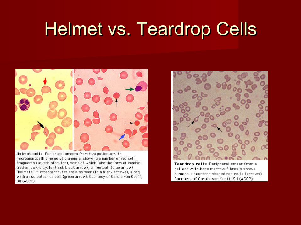
Helmet vs. Teardrop CellsHelmet vs. Teardrop Cells

Anemia: SummaryAnemia: Summary ANEMIA IS NEVER NORMALANEMIA IS NEVER NORMAL Determine if ACUTE or CHRONICDetermine if ACUTE or CHRONIC CONSIDER THE FOUR CAUSESCONSIDER THE FOUR CAUSES CALCULATE the RETIC INDEXCALCULATE the RETIC INDEX LOOK AT THE SMEARLOOK AT THE SMEAR CONSIDER THE ETIOLOGY BASED ON RBC CONSIDER THE ETIOLOGY BASED ON RBC
MORPHOLOGY AND LABORATORY STUDIESMORPHOLOGY AND LABORATORY STUDIES TREAT APPROPRIATELYTREAT APPROPRIATELY

MKSAP QuestionsMKSAP Questions

An 80-year-old man who had a hemicolectomy for colon cancer is An 80-year-old man who had a hemicolectomy for colon cancer is evaluated because of a 4-month history of diarrhea, anorexia, and evaluated because of a 4-month history of diarrhea, anorexia, and fatigue. He had a remote history of alcoholism. fatigue. He had a remote history of alcoholism.
On physical examination, he is cachectic and mildly confused. His On physical examination, he is cachectic and mildly confused. His pulse rate is 70/min, and blood pressure is 140/85 mm Hg. His pulse rate is 70/min, and blood pressure is 140/85 mm Hg. His tongue is smooth. The abdomen is soft; there are no palpable tongue is smooth. The abdomen is soft; there are no palpable masses or hepatosplenomegaly. A stool specimen is negative for masses or hepatosplenomegaly. A stool specimen is negative for occult blood. Neurologic examination shows loss of position sense occult blood. Neurologic examination shows loss of position sense in the feet. He has a wide-based gait. The Romberg test is positive. in the feet. He has a wide-based gait. The Romberg test is positive. His hemoglobin is 9.4 g/dL, reticulocyte count is 2.5%, mean His hemoglobin is 9.4 g/dL, reticulocyte count is 2.5%, mean corpuscular volume is 125 fL, and serum lactate dehydrogenase is corpuscular volume is 125 fL, and serum lactate dehydrogenase is 400 U/L. 400 U/L.
Which of the following is the most l ikely cause for his Which of the following is the most l ikely cause for his symptoms?symptoms?
( A ) Alcoholic cerebellar degeneration( A ) Alcoholic cerebellar degeneration( B ) Vitamin B12 deficiency( B ) Vitamin B12 deficiency( C ) Brain metastases( C ) Brain metastases( D ) Folate deficiency( D ) Folate deficiency( E ) Liver metastases( E ) Liver metastases

Crit iqueCrit ique (Correct Answer = (Correct Answer = BB))
The patient most likely has vitamin B12 deficiency, based on the degree of macrocytosis and neurologic The patient most likely has vitamin B12 deficiency, based on the degree of macrocytosis and neurologic findings. An elevated serum lactate dehydrogenase level, due to intramarrow cell death from ineffective findings. An elevated serum lactate dehydrogenase level, due to intramarrow cell death from ineffective erythropoiesis, is consistent with this diagnosis. erythropoiesis, is consistent with this diagnosis.
Severe macrocytosis (mean corpuscular volume > 120 fL) is often associated with vitamin B12 deficiency or Severe macrocytosis (mean corpuscular volume > 120 fL) is often associated with vitamin B12 deficiency or folate deficiency (megaloblastic anemia), usually seen in conjunction with “oval” macrocytes. The presence folate deficiency (megaloblastic anemia), usually seen in conjunction with “oval” macrocytes. The presence of frequent hypersegmented neutrophils (> 5 segments) is strongly suggestive of vitamin B12 or folate of frequent hypersegmented neutrophils (> 5 segments) is strongly suggestive of vitamin B12 or folate deficiency. deficiency.
Bone marrow morphology in patients with vitamin B12 or folate deficiency is referred to as “megaloblastic” Bone marrow morphology in patients with vitamin B12 or folate deficiency is referred to as “megaloblastic” and is characterized by the presence of large cells with immature nuclear chromatin but maturing erythrocyte and is characterized by the presence of large cells with immature nuclear chromatin but maturing erythrocyte cytoplasm (nuclear-cytoplasmic dissociation). Anemia accompanies this process; hence the term “ineffective cytoplasm (nuclear-cytoplasmic dissociation). Anemia accompanies this process; hence the term “ineffective erythropoiesis.” The intramarrow death of megaloblastic cells causes the serum lactate dehydrogenase level erythropoiesis.” The intramarrow death of megaloblastic cells causes the serum lactate dehydrogenase level to rise. If a patient has a low serum vitamin B12 or folate level, a bone marrow examination is probably to rise. If a patient has a low serum vitamin B12 or folate level, a bone marrow examination is probably unnecessary. However, the physician should determine the cause of the deficiency. If a patient has a normal unnecessary. However, the physician should determine the cause of the deficiency. If a patient has a normal serum vitamin B12 or folate level, a bone marrow examination is frequently helpful to exclude serum vitamin B12 or folate level, a bone marrow examination is frequently helpful to exclude myelodysplastic syndromes or other infiltrative marrow disorders. myelodysplastic syndromes or other infiltrative marrow disorders.
Folate deficiency can induce megaloblastosis within weeks to months, whereas vitamin B12 deficiency Folate deficiency can induce megaloblastosis within weeks to months, whereas vitamin B12 deficiency requires years to cause megaloblastosis since stores of vitamin B12 persist for years in the liver and other requires years to cause megaloblastosis since stores of vitamin B12 persist for years in the liver and other tissues. In patients with vitamin B12 or folate deficiency, parenteral or oral repletion of vitamin B12 or folate tissues. In patients with vitamin B12 or folate deficiency, parenteral or oral repletion of vitamin B12 or folate reverses some morphologic abnormalities within hours. Serum folate levels fluctuate quickly with changes in reverses some morphologic abnormalities within hours. Serum folate levels fluctuate quickly with changes in dietary consumption. Low erythrocyte folate levels often reflect prior nutritional depletion. In patients who are dietary consumption. Low erythrocyte folate levels often reflect prior nutritional depletion. In patients who are hospitalized and are begun on regular diets, the erythrocyte folate test may provide a better assessment of hospitalized and are begun on regular diets, the erythrocyte folate test may provide a better assessment of tissue folate levels than determination of the serum folate level. The erythrocyte folate test often requires a tissue folate levels than determination of the serum folate level. The erythrocyte folate test often requires a special laboratory, and results often are not quickly available. special laboratory, and results often are not quickly available.
In patients with megaloblastic anemias, erythrocyte production is diminished and a “corrected” reticulocyte In patients with megaloblastic anemias, erythrocyte production is diminished and a “corrected” reticulocyte count is inappropriately low for the degree of anemia. This patient had a corrected reticulocyte count of 1% count is inappropriately low for the degree of anemia. This patient had a corrected reticulocyte count of 1% (inappropriately low for a hemoglobin level of 9.4 g/dL). (inappropriately low for a hemoglobin level of 9.4 g/dL).
In addition to changes in the blood, the epithelial cells in patients with megaloblastic anemias may become In addition to changes in the blood, the epithelial cells in patients with megaloblastic anemias may become atrophic and cause a smooth tongue and cheilosis. Posterior column dysfunction, particularly in patients with atrophic and cause a smooth tongue and cheilosis. Posterior column dysfunction, particularly in patients with vitamin B12 deficiency, may lead to changes in vibratory or position sense, causing ataxia. Signs of vitamin B12 deficiency, may lead to changes in vibratory or position sense, causing ataxia. Signs of dementia may appear. However, neurologic dysfunction is very uncommon in adults with folate deficiency. dementia may appear. However, neurologic dysfunction is very uncommon in adults with folate deficiency.
Alcoholic cerebellar degeneration results in ataxia but not position loss. Although liver metastases are Alcoholic cerebellar degeneration results in ataxia but not position loss. Although liver metastases are possible in a patient with a history of colon cancer, their presence would not account for the neurological possible in a patient with a history of colon cancer, their presence would not account for the neurological findings in this patient. Brain metastases would most likely produce focal neurological findings and also findings in this patient. Brain metastases would most likely produce focal neurological findings and also would not account for the blood findings. would not account for the blood findings.

A 26-year-old man is evaluated because of progressive fatigue, A 26-year-old man is evaluated because of progressive fatigue, dyspnea on exertion, and orthostatic dizziness for the past 2 to 3 dyspnea on exertion, and orthostatic dizziness for the past 2 to 3 weeks. He takes no medications. Physical examination is normal weeks. He takes no medications. Physical examination is normal except for pallor. except for pallor.
Laboratory Studies: Laboratory Studies: Hematocrit 13%Leukocyte count 8300/μL; Hematocrit 13%Leukocyte count 8300/μL; normal differential. Reticulocyte count: 0, Platelet count 320,000/μL. normal differential. Reticulocyte count: 0, Platelet count 320,000/μL. A routine biochemical profile, including liver function tests, is normal. A routine biochemical profile, including liver function tests, is normal. A chest radiograph shows normal lung fields and a widened A chest radiograph shows normal lung fields and a widened mediastinum, suggestive of an anterior mediastinal mass. Bone mediastinum, suggestive of an anterior mediastinal mass. Bone marrow biopsy shows absent erythrocyte precursors, normal marrow biopsy shows absent erythrocyte precursors, normal megakaryocytes, and normal leukocyte numbers and maturation. megakaryocytes, and normal leukocyte numbers and maturation.
Which of the fol lowing is the most l ikely cause of the Which of the fol lowing is the most l ikely cause of the mediastinal mass and anemia?mediastinal mass and anemia?
( A ) Hodgkin’s disease( A ) Hodgkin’s disease( B ) Non-Hodgkin’s lymphoma( B ) Non-Hodgkin’s lymphoma( C ) Thyroid carcinoma( C ) Thyroid carcinoma( D ) Thymoma( D ) Thymoma( E ) Germ cell carcinoma( E ) Germ cell carcinoma

Crit iqueCrit ique (Correct Answer = (Correct Answer = DD ))
Each of the listed neoplasms may present as an anterior Each of the listed neoplasms may present as an anterior mediastinal mass and may be associated with anemia of mediastinal mass and may be associated with anemia of chronic disease. However, pure red cell aplasia (which this chronic disease. However, pure red cell aplasia (which this patient has) is often associated with a benign or invasive patient has) is often associated with a benign or invasive thymoma. Approximately 5% to 15% of thymomas occur in thymoma. Approximately 5% to 15% of thymomas occur in patients with pure red cell aplasia. Other thymoma-associated patients with pure red cell aplasia. Other thymoma-associated autoimmune disorders include myasthenia gravis, systemic autoimmune disorders include myasthenia gravis, systemic lupus erythematosus, thrombocytopenia, and, rarely, lupus erythematosus, thrombocytopenia, and, rarely, malabsorption states. A careful search by CT or MRI is always malabsorption states. A careful search by CT or MRI is always warranted in patients with newly diagnosed or relapsing red cell warranted in patients with newly diagnosed or relapsing red cell aplasia or myasthenia. aplasia or myasthenia.
The other listed entities are also included in the differential The other listed entities are also included in the differential diagnosis for an anterior mediastinal mass. Germ cell tumors diagnosis for an anterior mediastinal mass. Germ cell tumors have not been associated with pure red cell aplasia, and have not been associated with pure red cell aplasia, and Hodgkin’s disease, non-Hodgkin’s lymphoma, and thyroid Hodgkin’s disease, non-Hodgkin’s lymphoma, and thyroid carcinoma are rarely associated with this disorder. Chronic carcinoma are rarely associated with this disorder. Chronic lymphocytic leukemia is also commonly associated with red cell lymphocytic leukemia is also commonly associated with red cell aplasia and may present with variable degrees of aplasia and may present with variable degrees of lymphadenopathy but not with an isolated anterior mediastinal lymphadenopathy but not with an isolated anterior mediastinal mass, as in the patient discussed here. mass, as in the patient discussed here.

A 36-year-old black man with known sickle cell anemia is evaluated A 36-year-old black man with known sickle cell anemia is evaluated because of a 2-week history of fever, a macular rash on his trunk, because of a 2-week history of fever, a macular rash on his trunk, and arthralgias. Subsequently, he developed weakness and and arthralgias. Subsequently, he developed weakness and dyspnea on exertion. Several of his children had febrile illnesses dyspnea on exertion. Several of his children had febrile illnesses with associated rashes and fatigue over the past month. These with associated rashes and fatigue over the past month. These illnesses resolved spontaneously without sequelae. illnesses resolved spontaneously without sequelae.
On physical examination, his temperature is 38.8 °C (101.8 °F), On physical examination, his temperature is 38.8 °C (101.8 °F), pulse rate is 100/min, and blood pressure is 160/70 mm Hg. A pulse rate is 100/min, and blood pressure is 160/70 mm Hg. A maculopapular, truncal rash is noted. There is conjunctival pallor. maculopapular, truncal rash is noted. There is conjunctival pallor. The remainder of his examination is unremarkable. The remainder of his examination is unremarkable.
Laboratory StudiesLaboratory Studies Hemoglobin 5.2 g/dLLeukocyte count Hemoglobin 5.2 g/dLLeukocyte count 5000/μLReticulocyte count 0%Platelet count 130,000/μLSerum 5000/μLReticulocyte count 0%Platelet count 130,000/μLSerum lactate dehydrogenase 622 U/Llactate dehydrogenase 622 U/LWhich of the following is the Which of the following is the most l ikely diagnosis?most l ikely diagnosis?
( A ) Paroxysmal nocturnal hemoglobinuria( A ) Paroxysmal nocturnal hemoglobinuria( B ) Parvovirus infection( B ) Parvovirus infection( C ) Glucose-6-phosphate dehydrogenase deficiency( C ) Glucose-6-phosphate dehydrogenase deficiency( D ) Aplastic anemia( D ) Aplastic anemia

Crit iqueCrit ique (Correct Answer = (Correct Answer = BB))
Patients with hemolytic disorders may occasionally present with reticulocytopenia and an “aplastic crisis.” Patients with hemolytic disorders may occasionally present with reticulocytopenia and an “aplastic crisis.” This patient has sickle cell anemia with parvovirus infection, which is causing an aplastic crisis. Parvovirus This patient has sickle cell anemia with parvovirus infection, which is causing an aplastic crisis. Parvovirus may infect patients with hemolytic anemias (for example, patients with hereditary spherocytosis, sickle cell may infect patients with hemolytic anemias (for example, patients with hereditary spherocytosis, sickle cell disease, or thalassemia). In children with sickle cell anemia, over 80% of aplastic crises may be attributed to disease, or thalassemia). In children with sickle cell anemia, over 80% of aplastic crises may be attributed to parvovirus infections. In adults, the usual presenting features are rash, arthritis, and anemia. The “slapped parvovirus infections. In adults, the usual presenting features are rash, arthritis, and anemia. The “slapped cheek” syndrome is rarely a presenting feature. There is usually a complete suppression of erythropoiesis to cheek” syndrome is rarely a presenting feature. There is usually a complete suppression of erythropoiesis to a reticulocyte level of 0%. The bone marrow shows giant dysplastic (megaloblastoid) erythroblasts, a reticulocyte level of 0%. The bone marrow shows giant dysplastic (megaloblastoid) erythroblasts, occasionally with viral inclusions. The diagnosis is usually made by demonstrating IgM antibodies to the occasionally with viral inclusions. The diagnosis is usually made by demonstrating IgM antibodies to the virus. IgG antibodies appear later during the course of the infection and persist. Parvovirus in the blood may virus. IgG antibodies appear later during the course of the infection and persist. Parvovirus in the blood may be detected by the polymerase chain reaction, which is the definitive diagnostic method. Occasionally, other be detected by the polymerase chain reaction, which is the definitive diagnostic method. Occasionally, other blood components such as leukocytes and platelets are affected and result in mild to moderate blood components such as leukocytes and platelets are affected and result in mild to moderate pancytopenia. pancytopenia.
The diagnosis of paroxysmal nocturnal hemoglobinuria (PNH) should be considered in patients with bone The diagnosis of paroxysmal nocturnal hemoglobinuria (PNH) should be considered in patients with bone marrow failure or aplasia, unusual location of thromboses, and unexplained hemolysis. The anemia may be marrow failure or aplasia, unusual location of thromboses, and unexplained hemolysis. The anemia may be severe, and patients with PNH typically have reticulocytopenia. There is no characteristic finding on bone severe, and patients with PNH typically have reticulocytopenia. There is no characteristic finding on bone marrow examination, although the bone marrow of patients with PNH may demonstrate myelodysplastic marrow examination, although the bone marrow of patients with PNH may demonstrate myelodysplastic changes. The diagnosis is based on demonstration of exquisite sensitivity to complement-mediated lysis by changes. The diagnosis is based on demonstration of exquisite sensitivity to complement-mediated lysis by the sucrose lysis test or the acidified serum lysis test (Ham’s test). the sucrose lysis test or the acidified serum lysis test (Ham’s test).
Glucose-6-phosphate dehydrogenase (G6PD) deficiency is another cause of hemolysis that occasionally is Glucose-6-phosphate dehydrogenase (G6PD) deficiency is another cause of hemolysis that occasionally is associated with reticulocytopenia. In patients with G6PD deficiency, erythrocytes are subject to oxidative associated with reticulocytopenia. In patients with G6PD deficiency, erythrocytes are subject to oxidative stresses. Hemoglobin becomes oxidized and precipitates within the erythrocytes, which then undergo stresses. Hemoglobin becomes oxidized and precipitates within the erythrocytes, which then undergo destruction by the reticuloendothelial system. G6PD deficiency is an autosomal recessive disorder that destruction by the reticuloendothelial system. G6PD deficiency is an autosomal recessive disorder that predominantly affects males. After a hemolytic episode, qualitative assays may be normal because only predominantly affects males. After a hemolytic episode, qualitative assays may be normal because only erythrocytes that are resistant to G6PD remain. The African variant of G6PD is associated with a mild form of erythrocytes that are resistant to G6PD remain. The African variant of G6PD is associated with a mild form of hemolysis, whereas the Mediterranean variant is usually severe. Causes include infectious stresses, drugs hemolysis, whereas the Mediterranean variant is usually severe. Causes include infectious stresses, drugs such as quinidine and sulfonamides, or, in the Mediterranean variant, favism (consumption of fava beans). such as quinidine and sulfonamides, or, in the Mediterranean variant, favism (consumption of fava beans). Therapy requires avoiding certain medications and supportive care in crisis situations. Therapy requires avoiding certain medications and supportive care in crisis situations.
In contrast to this patient’s presentation, patients with aplastic anemia have pancytopenia with severe In contrast to this patient’s presentation, patients with aplastic anemia have pancytopenia with severe anemia, reticulocytopenia, thrombocytopenia, and granulocytopenia. In patients with severe aplastic anemia, anemia, reticulocytopenia, thrombocytopenia, and granulocytopenia. In patients with severe aplastic anemia, the bone marrow examination shows less than 5% cellularity with only residual lymphocytes and plasma the bone marrow examination shows less than 5% cellularity with only residual lymphocytes and plasma cells. The abnormal cells described above that are attributable to parvovirus infection are not seen. cells. The abnormal cells described above that are attributable to parvovirus infection are not seen.

A 36-year-old man is evaluated because of fatigue. He has had two A 36-year-old man is evaluated because of fatigue. He has had two episodes of acute gouty arthritis over the past 6 months. He has a 10-year episodes of acute gouty arthritis over the past 6 months. He has a 10-year history of significant alcohol use, but he quit drinking 4 months ago. He history of significant alcohol use, but he quit drinking 4 months ago. He works in a factory making battery products. A complete blood count works in a factory making battery products. A complete blood count obtained prior to elective hernia repair surgery 4 years ago was normal. He obtained prior to elective hernia repair surgery 4 years ago was normal. He takes no medications. takes no medications.
On physical examination, his temperature is 37.3 °C (99.1 °F), pulse is On physical examination, his temperature is 37.3 °C (99.1 °F), pulse is 60/min, and blood pressure is 135/70 mm Hg. His skin is normal. There is 60/min, and blood pressure is 135/70 mm Hg. His skin is normal. There is slight scleral icterus. There is a blue line at the edge of his gums. slight scleral icterus. There is a blue line at the edge of his gums.
The remainder of the examination is normal. Stool specimens are negative The remainder of the examination is normal. Stool specimens are negative for blood on three occasions. for blood on three occasions.
Laboratory StudiesLaboratory Studies Hemoglobin 7.5 g/dLMean corpuscular volume 71 Hemoglobin 7.5 g/dLMean corpuscular volume 71 flLeukocyte count 9400/µLReticulocyte count 5.3%Platelet count flLeukocyte count 9400/µLReticulocyte count 5.3%Platelet count 435,000/µLSerum lactate dehydrogenase 553 U/LSerum uric acid 11 435,000/µLSerum lactate dehydrogenase 553 U/LSerum uric acid 11 mg/dLA peripheral blood smear is shown. mg/dLA peripheral blood smear is shown.
Which of the following diagnostic studies is most useful for Which of the following diagnostic studies is most useful for determining the cause of this patient’s anemia?determining the cause of this patient’s anemia?
( A ) Serum iron, total iron-binding capacity, and ferritin levels( A ) Serum iron, total iron-binding capacity, and ferritin levels( B ) Serum lead levels( B ) Serum lead levels( C ) Direct and indirect antiglobulin tests( C ) Direct and indirect antiglobulin tests( D ) Hemoglobin A2 quantitation( D ) Hemoglobin A2 quantitation( E ) Serum ethanol and folic acid levels( E ) Serum ethanol and folic acid levels


Crit iqueCrit ique (Correct Answer = (Correct Answer = BB))
The patient has chronic lead intoxication that can be confirmed by measuring serum lead levels. The patient has chronic lead intoxication that can be confirmed by measuring serum lead levels. He has a hypochromic, microcytic anemia with coarse basophilic stippling and reticulocytosis. He has a hypochromic, microcytic anemia with coarse basophilic stippling and reticulocytosis. He also has evidence of hemolytic anemia with increased serum lactate dehydrogenase and He also has evidence of hemolytic anemia with increased serum lactate dehydrogenase and indirect bilirubin levels. His physical examination is remarkable for gingival “lead lines.” Bone indirect bilirubin levels. His physical examination is remarkable for gingival “lead lines.” Bone marrow examination shows erythroid hyperplasia and ringed sideroblasts. The anemia of lead marrow examination shows erythroid hyperplasia and ringed sideroblasts. The anemia of lead poisoning fits this description. Sideroblastic anemia with hypochromic indices is typical. poisoning fits this description. Sideroblastic anemia with hypochromic indices is typical. Hemolysis is common, and basophilic stippling, blue staining polyribosomal aggregates with Hemolysis is common, and basophilic stippling, blue staining polyribosomal aggregates with mitochondrial fragments in the erythrocytes, is frequently seen. Lead inhibits pyrimidine 5′-mitochondrial fragments in the erythrocytes, is frequently seen. Lead inhibits pyrimidine 5′-nucleotidase which normally clears ribosomal fragments. Occupational exposures to lead are nucleotidase which normally clears ribosomal fragments. Occupational exposures to lead are relatively uncommon today. However, workers who produce batteries or are exposed to paint, relatively uncommon today. However, workers who produce batteries or are exposed to paint, particularly those who remove leaded paint from old buildings, are at greatest risk if they are not particularly those who remove leaded paint from old buildings, are at greatest risk if they are not protected from inhalation of paint particles during the sanding process. Other manifestations of protected from inhalation of paint particles during the sanding process. Other manifestations of lead toxicity in adults include peripheral neuropathy, abdominal colic, and saturnine gout lead toxicity in adults include peripheral neuropathy, abdominal colic, and saturnine gout (effects of lead on renal tubules that prevent the excretion of uric acid). Chelation therapy is (effects of lead on renal tubules that prevent the excretion of uric acid). Chelation therapy is indicated for patients with serum lead levels exceeding 70 µg/dL and should be continued until indicated for patients with serum lead levels exceeding 70 µg/dL and should be continued until lead levels fall below 40 µg/dL. Agents such as EDTA or dimercaprol may also be effective. lead levels fall below 40 µg/dL. Agents such as EDTA or dimercaprol may also be effective.
This patient is unlikely to have iron deficiency since his reticulocytes are increased. In addition, This patient is unlikely to have iron deficiency since his reticulocytes are increased. In addition, basophilic stippling usually is not seen in patients with iron deficiency. basophilic stippling usually is not seen in patients with iron deficiency.
Thalassemia is associated with a microcytic anemia, reticulocytosis, and basophilic stippling. Thalassemia is associated with a microcytic anemia, reticulocytosis, and basophilic stippling. However, a normal complete blood count 4 years ago rules out this possibility. Therefore, However, a normal complete blood count 4 years ago rules out this possibility. Therefore, quantitative studies to measure hemoglobin A2 are not necessary. quantitative studies to measure hemoglobin A2 are not necessary.
Autoimmune hemolytic anemia should be excluded by performing a direct antiglobulin test in Autoimmune hemolytic anemia should be excluded by performing a direct antiglobulin test in any patient who has evidence of hemolysis on a peripheral blood smear. However, the “lead any patient who has evidence of hemolysis on a peripheral blood smear. However, the “lead lines” on this patient’s gingivae are classic for lead poisoning, and autoimmune hemolytic lines” on this patient’s gingivae are classic for lead poisoning, and autoimmune hemolytic anemia therefore is less likely. anemia therefore is less likely.
Alcoholism may cause a transient sideroblastic anemia, which resolves with cessation of Alcoholism may cause a transient sideroblastic anemia, which resolves with cessation of alcohol intake. Folic acid deficiency may complicate alcoholism but usually presents with alcohol intake. Folic acid deficiency may complicate alcoholism but usually presents with macrocytosis. macrocytosis.

A 22-year-old man is evaluated in the intensive care unit because of A 22-year-old man is evaluated in the intensive care unit because of bleeding immediately after scoliosis surgery. During surgery, 12 bleeding immediately after scoliosis surgery. During surgery, 12 units of packed red blood cells and 12 units of fresh frozen plasma units of packed red blood cells and 12 units of fresh frozen plasma were transfused. There is no history of a bleeding disorder or of were transfused. There is no history of a bleeding disorder or of drug administration that could affect platelet function. drug administration that could affect platelet function.
On physical examination, the patient is afebrile. His pulse rate is On physical examination, the patient is afebrile. His pulse rate is 100/min, and blood pressure is 110/72 mm Hg. Petechiae are 100/min, and blood pressure is 110/72 mm Hg. Petechiae are present on his arms, and blood is oozing from the drains. present on his arms, and blood is oozing from the drains.
Laboratory StudiesLaboratory Studies Hemoglobin 9.0 g/dLPlatelet count Hemoglobin 9.0 g/dLPlatelet count 43,000/µLProthrombin time 12 sActivated partial thromboplastin 43,000/µLProthrombin time 12 sActivated partial thromboplastin time 32 sPlasma fibrinogen 400 g/dLd-Dimers Negativetime 32 sPlasma fibrinogen 400 g/dLd-Dimers NegativeWhich of Which of the following is the most l ikely cause of the the following is the most l ikely cause of the thrombocytopenia and bleeding?thrombocytopenia and bleeding?
( A ) Dilutional thrombocytopenia( A ) Dilutional thrombocytopenia( B ) Incompatible blood transfusion( B ) Incompatible blood transfusion( C ) Posttransfusion purpura( C ) Posttransfusion purpura( D ) Septic transfusion reaction( D ) Septic transfusion reaction

Crit iqueCrit ique (Correct Answer = (Correct Answer = AA ))
Approximately a 50% reduction in the platelet count occurs when Approximately a 50% reduction in the platelet count occurs when transfusions of 1.5 to 2 times the blood volume are given over 4 to 8 hours. transfusions of 1.5 to 2 times the blood volume are given over 4 to 8 hours. Blood stored for more that 2 to 3 days has essentially no platelets, which Blood stored for more that 2 to 3 days has essentially no platelets, which explains the thrombocytopenia. Fresh frozen plasma contains all of the explains the thrombocytopenia. Fresh frozen plasma contains all of the clotting proteins in normal concentrations. This patient received one unit of clotting proteins in normal concentrations. This patient received one unit of fresh frozen plasma for each unit of transfused red blood cells, which fresh frozen plasma for each unit of transfused red blood cells, which resulted in no measurable alteration in the prothrombin time or activated resulted in no measurable alteration in the prothrombin time or activated partial thromboplastin time. Incompatible red blood cell transfusions can partial thromboplastin time. Incompatible red blood cell transfusions can lead to the development of disseminated intravascular coagulation, which is lead to the development of disseminated intravascular coagulation, which is characterized by thrombocytopenia. The other hallmarks of disseminated characterized by thrombocytopenia. The other hallmarks of disseminated intravascular coagulation are absent in this patient, namely, he has a normal intravascular coagulation are absent in this patient, namely, he has a normal fibrinogen, prothrombin time, activated partial thromboplastin time, and fibrinogen, prothrombin time, activated partial thromboplastin time, and negative d-dimers. Posttransfusion purpura is characterized by profound negative d-dimers. Posttransfusion purpura is characterized by profound thrombocytopenia that develops 5 to 7 days after a transfusion. It occurs in thrombocytopenia that develops 5 to 7 days after a transfusion. It occurs in patients who are negative for the PLA-1 human platelet antigen and have patients who are negative for the PLA-1 human platelet antigen and have been transfused or pregnant in the past. Posttransfusion purpura almost been transfused or pregnant in the past. Posttransfusion purpura almost always occurs in women. always occurs in women.
Septic transfusion reactions can be associated with thrombocytopenia when Septic transfusion reactions can be associated with thrombocytopenia when either bacteremia or endotoxemia causes disseminated intravascular either bacteremia or endotoxemia causes disseminated intravascular coagulation. This patient has no indication of either sepsis or disseminated coagulation. This patient has no indication of either sepsis or disseminated intravascular coagulation. intravascular coagulation.