Redalyc.Visuospatial and visuoperceptual impairment in ... · cial Recognition Test y se obtuvieron...
15
Anuario de Psicología ISSN: 0066-5126 [email protected] Universitat de Barcelona España García-Díaz, Anna Isabel; Segura, Bárbara; Baggio, Hugo; Junqué, Carme Visuospatial and visuoperceptual impairment in relation to global atrophy in Parkinson’s disease Anuario de Psicología, vol. 43, núm. 2, septiembre-, 2013, pp. 147-158 Universitat de Barcelona Barcelona, España Available in: http://www.redalyc.org/articulo.oa?id=97029454001 How to cite Complete issue More information about this article Journal's homepage in redalyc.org Scientific Information System Network of Scientific Journals from Latin America, the Caribbean, Spain and Portugal Non-profit academic project, developed under the open access initiative
-
Upload
hoangquynh -
Category
Documents
-
view
214 -
download
0
Transcript of Redalyc.Visuospatial and visuoperceptual impairment in ... · cial Recognition Test y se obtuvieron...

Anuario de Psicología
ISSN: 0066-5126
Universitat de Barcelona
España
García-Díaz, Anna Isabel; Segura, Bárbara; Baggio, Hugo; Junqué, Carme
Visuospatial and visuoperceptual impairment in relation to global atrophy in Parkinson’s disease
Anuario de Psicología, vol. 43, núm. 2, septiembre-, 2013, pp. 147-158
Universitat de Barcelona
Barcelona, España
Available in: http://www.redalyc.org/articulo.oa?id=97029454001
How to cite
Complete issue
More information about this article
Journal's homepage in redalyc.org
Scientific Information System
Network of Scientific Journals from Latin America, the Caribbean, Spain and Portugal
Non-profit academic project, developed under the open access initiative
psicologíaanuario de
The UB Journal of Psychology
Volumen 43Número 2Septiembre 2013
facultad de psicologíauniversidad de barcelona
ISSN: 0066-5126ISSN: 1988-5253
Vol.
43, n
º 2
an
uario
de
psic
olog
ía /
The
UB
Jou
rnal
of P
sych
olog
y
Sep
tiem
bre
2013
AP42-2 [email protected] 13/9/12 07:20 Pagina 1
Compuesta
C M Y CM MY CY CMY K
AP42-2 [email protected] 13/9/12 07:20 Pagina 1
Compuesta
C M Y CM MY CY CMY K
P.V.P.: 14€ (I.V.A. incluido)
Artículos
Anna Isabel García-Díaz, Bárbara Segura, Hugo Baggio y Carme JunquéVisuospatial and visuoperceptual impairment in relation to global atrophy in Parkinson’s disease
Vanessa Renau Ruiz, Ursula Oberst y Xavier Carbonell-SánchezConstrucción de la identidad a través de las redes sociales online:una mirada desde el construccionismo social
Maria Forns, Teresa Kirchner, Laia Soler y Claudia ParetillaSpanish/Catalan version of the Juvenile Victimization Questionnaire (JVQ): Psychometric properties
Viviana LemosLa operacionalización de constructos psicológicos en la infancia: di� cultades y propuestas de superación
Marina Romeo, Montserrat Yepes-Baldó, Tomeu Vidal y Joan Guàrdia-OlmosWhy do they separate it or not? Attitudes and behaviors towards organic waste separation
Pilar Monreal-Bosch, Arantza del Valle y Santiago PereraActivando municipios para la promoción de la salud: un estudio de caso en comunidades rurales
Eva Aguilar MediavillaComparative analysis of the acquisition of syllabic structure and errors in preschool children with SLI
Alicia Martínez-Ramos, Maribel Peró-Cebollero, Teresita Villaseñor-Cabrera y Joan Guàrdia-Olmos Adaptación y validación del test Torre de Londres en mexicanos adultos mayores de 60 años
anuario de psicología / The UB Journal of Psychology
anuario_psicologia_43_2.indd 1 24/07/13 14:37
Anuario de Psicología/The UB Journal of Psychology 2013, vol. 43, nº 2, 147-158 © 2013, Facultat de Psicologia Universitat de Barcelona
Visuospatial and visuoperceptual impairment in relation to global atrophy in Parkinson’s disease*
Anna Isabel García-Díaz Bárbara Segura Hugo Baggio Carme Junqué Universitat de Barcelona
Parkinson’s disease (PD) patients differed from controls of similar age in visuospatial and visuoperceptual functions at diagnosis moment, and these deficits have been shown to be neuropsychological markers of evolution to dementia. The aim of this study was to relate these dysfunctions with measures of brain. The sample of this study consisted of 92 PD patients and 36 healthy subjects matched by age, sex and education. All subjects were evaluated with Judgment of Line Orientation, Visual Form Discrimination and Facial Recog-nition Tests and magnetic resonance imaging at 3 Tesla. We found significant differences between patients and controls in all three tests and in the mean of cortical thickness, gray matter volume and ventricular system. All visuospatial and visuoperceptual tests correlated with the measures of global atrophy sug-gesting that they are reflecting the brain degeneration associated to PD.
Keywords: Parkinson’s disease, visuospatial/visuoperceptual, MRI, cortical thickness, brain atrophy.
Alteraciones visuoespaciales y visuoperceptivas en la enfermedad de Parkinson y su relación con parámetros de atrofia global
Los pacientes con enfermedad de Parkinson (EP) comparados con contro-les de similar edad muestran alteraciones visuoespaciales y visuoperceptivas desde el momento del diagnóstico, y se ha demostrado que estas alteraciones
* Acknowledgements: We acknowledge the funding of PSI2010-16174 and 2009SGR0941CJ in the contribution to this work. Correspondence: Carme Junqué. Departament de Psiquiatría i Psicobiologia Clínica. Universitat de Barcelona.. C/ Casanova, 143 (Ala Norte; 5ª planta). E-mail: [email protected].

148 Visuospatial impairment and atrophy in PD
Anuario de Psicología/The UB Journal of Psychology, vol. 43, nº 2, septiembre 2013, pp. 147-158
© 2013, Universitat de Barcelona, Facultat de Psicologia
actúan como marcadores neuropsicológicos de la evolución a demencia. El objetivo de este estudio es relacionar estas alteraciones con las medidas glo-bales de atrofia cerebral. La muestra consta de 92 EP y 36 sujetos sanos em-parejados por edad, sexo y educación. Todos los sujetos fueron evaluados mediante el Judgment of Line Orientation, Visual Form Discrimination y Fa-cial Recognition Test y se obtuvieron datos de neuroimagen estructural con un aparato de Resonancia Magnética de 3 tesla. Se observaron diferencias en-tre los pacientes y los controles en los tres tests usados y en las medidas de grosor cortical total, volumen de sustancia gris y sistema ventricular. Todos los tests visuoespaciales y visuoperceptivos correlacionaron con medidas de atrofia global, lo cual sugiere que son indicadores útiles de la degeneración cerebral asociada a la EP.
Palabras clave: Enfermedad de Parkinson, imágenes de resonancia magnética, grosor cortical, alteraciones visuoespaciales y visuoperceptivas, atrofia cerebral.
Introducción Parkinson’s disease (PD) is considered to be one of the most common neuro-logical diseases associated with aging. The prevalence ranges from 1-3% in the general population (Galvin, Pollack, & Morris, 2006; Riedel et al., 2008). Classi-cally, PD was regarded as a motor disease, but it is now recognized as a systemic neurodegenerative illness. Cognitive dysfunction and impairment as well as mood disorders and apathy have been increasingly viewed as important symptoms that accompany the disease even in preclinical stages. It has been stated that, after 20 years, 80% of PD patients evolve to dementia, whereas only 25% of them are recognized by clinicians in routine daycare (Svenningsson, Westman, Ballard, & Aarsland, 2012). The disease may course with impaired executive function (Wil-liams-Gray, Foltynie, Brayne, Robbins, & Barker, 2007; Williams- Gray et al., 2009; Aarsland et al., 2009), visuospatial and visuoperceptual dysfunctions (Wil-liams-Gray et al., 2007-2009; Aarsland, Brønnick, Larsen, Tysnes, & Alves, 2009) and verbal memory impairment (Aarsland et al., 2009) but relatively spared language, except for semantic fluency (Williams-Gray et al., 2007; 2009). Visuospatial and visuoperceptual deficits can be seen early in the disease course, but their main interest is their power to predict subsequent evolution to dementia (Williams-Gray et al., 2009; Svenningsson et al., 2012). Several studies have assessed the differential impairment of these functions in subcortical demen-tias, and PD has been stated to have a distinct pattern of alteration compared with other types of dementias, such as Alzheimer’s disease (Ala, Hughes, Kyrouac, Ghobria, & Elble, 2001; Cormack, Aarsland, Ballard, & Tovée, 2004; Tiraboschi et al., 2006; Saka & Elibol, 2009). For this reason, they have been regarded as screening tools for cognitive impairment and evolution to dementia in clinical settings (Williams-Gray et al., 2009; Helmes, 2013).

A. I. García-Díaz, B. Segura, H. Baggio y C. Junqué 149
Anuario de Psicología/The UB Journal of Psychology, vol. 43, nº 2, septiembre 2013, pp. 147-158 © 2013, Universitat de Barcelona, Facultat de Psicologia
Previous neuroimaging research has focused on describing the progress of global atrophy in PD patients using total gray matter and ventricular measures. Gray matter changes have been related to cognitive decline and dementia (Ibarretxe-Bilbao et al., 2012; Zarei et al., 2013; Camicioli et al., 2011) and recent research has demonstrated that gray matter decrease holds a high discriminant power in accurately identifying demented PD patients (Zarei et al., 2013). Nevertheless, progressive gray matter reductions have already been described in early stages of the disease (Ibarretxe-Bilbao et al., 2012). Gray and white matter losses are related to enlargement of the ventricular system in PD patients. Apostolova et al. (2010) found that left and right ventricles were significantly larger in PD patients with dementia compared with healthy subjects and their non-demented peers. Also, it has been shown that ventricular volumes are significantly increased in PD patients with MCI and correlate with memory performance (Dalaker et al., 2011). In sum, visuospatial and visuoperceptual impairment and atrophy measures have been associated with cognitive decline and exhibit a predictive value in the evolution to dementia, but hitherto and to our knowledge, the relationship between these two measures has not been studied. Thus, the aim of this study is to assess the neuropsychological performance in these cognitive functions and its relation with global structural changes in a large sample of non-demented PD patients. Methods Subjects This study included 121 patients, recruited from an outpatient movement disorders clinic (Parkinson’s Disease and Movement Disorders Unit, Service of Neurology, Hospital Clínic, Barcelona, Spain), and 49 healthy subjects, recruited from the Institut de l’Envelliment (Barcelona, Spain). Controls were matched for age, sex and years of education to their patient peers. This study was approved by the institutional ethics committee; all subjects agreed to participate voluntarily and written informed consent was obtained after full explanation of the procedures. Inclusion criteria for patients consisted of meeting the diagnostic criteria for PD established by the UK PD Society Brain Bank (Daniel & Lees, 1993). The following exclusion criteria were considered: [1] presence of dementia according to the Move-ment Disorders Society criteria (Dubois, Burn, Goetz, & Aarsland; 2007), [2] Hoehn and Yahr scale score ≥3, [3] young-onset PD, [4] presence of psychiatric and/or neurologic comorbidity, [5] low IQ score (scaled score score <8 on the Vocabu-lary subtest from the WAIS-IV -Wechsler Adults Intelligence Scale), [6] Mini-Mental State Examination (MMSE) score <24, [7] claustrophobia, [8] pathologi-cal findings non compatible with PD in MRI and [9] MRI artifacts. (demographic data of patients and healthy controls are summarized in table 1).

150 Visuospatial impairment and atrophy in PD
Anuario de Psicología/The UB Journal of Psychology, vol. 43, nº 2, septiembre 2013, pp. 147-158
© 2013, Universitat de Barcelona, Facultat de Psicologia
TABLE 1. DEMOGRAPHIC AND CLINICAL DATA OF THE SAMPLE.
Note: Values are mean ± Standard Deviation (SD); aStudent t test statistics; bChi squared statistics.
Neuropsychological Assessment The neuropsychological assessment comprised the evaluation of all cognitive functions, and the assessment of visuospatial and visuoperceptual functioning was obtained through the following neuropsychological tests: [1] Benton’s Judgment of Line Orientation (JLO; Benton, Hannay, & Varney; 1975), [2] Benton’s Visual Form Discrimination (VFD; Benton, Sivan, & Hamsher; 1994), and [3] Benton’s Facial Recognition (FRT; Benton et al.,1994) tests. Global measures of atrophy analysis FreeSurfer software (version 5.1; available at http://surfer.nmr.harvard.edu) was used to assess global brain atrophy. Along the process, cortical surface 3D model of cortical thickness (CTh) is created using intensity and continuity infor-mation, as described in detail by Fischl & Dale (2000). Independent steps are per-formed in the initial preprocessing of images for each subject: removal of non-brain tissue, automated Talairach transformation, intensity normalization, tessellation of the gray matter / white matter boundary, automated topology correction and surface deformation to optimally place the gray matter / white matter and gray matter / cere-brospinal fluid (CSF) boundaries. The resulting representation of CTh is calculated as the distance between tissue boundaries. All surface models in our study were vi-sually inspected for accuracy and were carefully manually corrected if necessary. Segmentation of brain volume and cortical thickness parcellations were ob-tained based on an automatic procedure included in Freesurfer 5.1 (Desikan et al.,
Demographics Patients (n=92) Controls (n=36) ta, χb p value
Age 63.98 ± 11.10 63.36 ± 10.53 -0.287a 0.775
Sex (male/female) 37/55 19/17 0.521b 0.300
Education, years 10.61 ± 5.36 11.36 ± 4.34 0.750a 0.454
Age at onset 56.04 ± 12.19
Evolution, years 8.39 ± 5.91
Patients with hallucinations n=19

A. I. García-Díaz, B. Segura, H. Baggio y C. Junqué 151
Anuario de Psicología/The UB Journal of Psychology, vol. 43, nº 2, septiembre 2013, pp. 147-158 © 2013, Universitat de Barcelona, Facultat de Psicologia
2006; Fisch et al., 2002). Variables used in order to calculate global measurement of atrophy were: bilateral mean thickness, total cortex volume, total gray matter volume, and the lateral ventricular system (left and right lateral ventricles). Statistical Analyses Statistical analyses of visuospatial and visuoperceptual functions, global atrophy, and neuropsychological and clinical variables were carried out using the statistical package PASW-20 (2011; SPSS, Inc., Chicago, IL). Z scores were obtained in order to assess patients’ performance in visuospatial and visuoperceptual tests. Clinical and demographical variables and brain atrophy group differences were analyzed with Student t test statistics or ANCOVA in case of volumetric global measures of atrophy. Cohen’s d was used to measure effect size in clinical and demographical variables (Ellis, 2009). Correlations analyses were performed among visuospatial and visuoperceptual tests and those with global atrophy measures. All the volu-metric analyses were controlled for intracranial volume (ICV). Results Clinical and demographical analyses Raw scores significantly differed between PD patients and healthy subjects in MMSE, NPI (Cumming’s Neuropsychiatric Inventory) and BDI (Beck Depres-sion Inventory-II), and all effect sizes indicated a large effect (see table 2). Age and education showed correlations with visuospatial and visuoperceptual tests, which were higher in all cases for PD patients than for controls. The highest cor-relations were seen between age and VFD, and education and FRT.
TABLE 2. CLINICAL DATA FOR PATIENTS AND CONTROL SUBJECTS.
Note: MMSE: Mini-Mental State Examination; NPI: Cummings’ Neuropsychiatric Inventory; BDI: Beck De-pression Inventory; UPDRS-III: Unified Parkinson’s Disease Rating Scale-III; H&Y: Hoehn & Yahr; LEDD: Levodopa Equivalent Daily Dose. a Cohen’s d statistics (Group 1=Patients/Group 2=Control subjects); bGroup 1=Control subjects/Group 2=Patients.
Patients Controls t p value da
MMSE 29.05 ± 1.11 29.69 ± 0.47 4.581 .0001 .75b
NPI 5.67 ± 7.39 1.68 ± 3.13 -4.124 .0001 .70a BDI 10.87 ± 6.15 6.15 ± 5.64 -2.845 .0001 .80a
UPDRS-III 17.60 ± 10.92 H&Y 1.89 ± 0.57
LEDD 803.70 ± 494.18
152 Visuospatial impairment and atrophy in PD
Anuario de Psicología/The UB Journal of Psychology, vol. 43, nº 2, septiembre 2013, pp. 147-158
© 2013, Universitat de Barcelona, Facultat de Psicologia
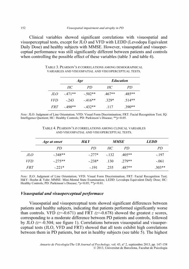
Clinical variables showed significant correlations with visuospatial and visuoperceptual tests, except for JLO and VFD with LEDD (Levodopa Equivalent Daily Dose) and healthy subjects with MMSE. However, visuospatial and visuoper-ceptual performance was still significantly different between patients and controls when controlling the possible effect of these variables (table 3 and table 4).
TABLE 3. PEARSON’S R CORRELATIONS AMONG DEMOGRAPHICAL VARIABLES AND VISUOSPATIAL AND VISUOPERCEPTUAL TESTS.
Note: JLO: Judgment of Line Orientation; VFD: Visual Form Discrimination; FRT: Facial Recognition Test; IQ: Intelligence Quotient; HC: Healthy Controls; PD: Parkinson’s Disease; **p<0.05.
TABLE 4. PEARSON’S R CORRELATIONS AMONG CLINICAL VARIABLES AND VISUOSPATIAL AND VISUOPERCEPTUAL TESTS.
Age at onset H&Y MMSE LEDD
PD PD HC PD PD
JLO -.348** -.277* -.132 .405** -.197
VFD -.275** -.238* .130 .279** -.061
FRT -.221* -.191 .235 .487** .303** Note: JLO: Judgment of Line Orientation; VFD: Visual Form Discrimination; FRT: Facial Recognition Test; H&Y: Hoehn & Yahr; MMSE: Mini-Mental State Examination; LEDD: Levodopa Equivalent Daily Dose; HC: Healthy Controls; PD: Parkinson’s Disease; *p<0.05; **p<0.01.
Visuospatial and visuoperceptual performace Visuospatial and visuoperceptual tests showed significant differences between patients and healthy subjects, indicating that patients performed significantly worse than controls. VFD (z=-0.671) and FRT (z=-0.678) showed the greatest z scores, corresponding to a moderate difference between PD patients and controls, followed by JLO (z=-0.504; see figure 1). Correlations between visuospatial and visuoper-ceptual tests (JLO, VFD and FRT) showed that all tests exhibit high correlations between them in PD patients, but not in healthy subjects (see table 5). The highest
Age Education
HC PD HC PD
JLO -.471** -.502** .467** .485**
VFD -.243 -.416** .329* .514**
FRT -.498** -.432** .117 .390**

A. I. García-Díaz, B. Segura, H. Baggio y C. Junqué 153
Anuario de Psicología/The UB Journal of Psychology, vol. 43, nº 2, septiembre 2013, pp. 147-158 © 2013, Universitat de Barcelona, Facultat de Psicologia
correlations were seen between JLO and FRT, followed by JLO and VFD and, lastly, VFD and FRT.
Note: JLO: Judgment of Line Orientation; VFD: Visual Form Discrimination; FRT: Facial Recognition Test. Figure 1. Graphic representation of visuospatial and visuoperceptual z scores.
TABLE 5. PEARSON’S R CORRELATIONS BETWEEN VISUOSPATIAL AND VISUOPERCEPTUAL TESTS.
Note: JLO: Judgment of Line Orientation; VFD: Visual Form Discrimination; FRT: Facial Recognition Test; HC: Healthy Controls; PD: Parkinson’s Disease; **p<0.01
Global atrophy measures Mean group differences in global atrophy analysis showed significant de-creases in PD patients (see table 6) in the left lateral ventricular system, cortex volume, total gray matter volume and mean cortical thickness.
JLO VFD FRT
HC PD HC PD HC PD
JLO 1 1 .237 .467** .260 .493**
VFD .237 .467** 1 1 .143 .423**
FRT .260 .493** .143 .423** 1 1

154 Visuospatial impairment and atrophy in PD
Anuario de Psicología/The UB Journal of Psychology, vol. 43, nº 2, septiembre 2013, pp. 147-158
© 2013, Universitat de Barcelona, Facultat de Psicologia
Global atrophy correlations analyses (see table 7) showed, except for JLO and right inferior lateral ventricle, significant results between JLO, VFD and FRT and mean thickness bilaterally, lateral ventricular system, total gray matter volume and cortex volume.
TABLE 6. MEAN VALUES AND STANDARD DEVIATIONS OF GLOBAL ATROPHY MEASURES FOR PD PATIENTS AND HEALTHY SUBJECTS. MEAN DIFFERENCES BETWEEN GROUPS ASSESSED WITH STUDENT T TEST STATISTICS OR F IN ANCOVA.
Note: a All values are mm. b All values are mm3. SD: Standard Deviation. c t: Student t test statistics. F: AN-COVA (volumetric measures of global atrophy were controlled by total intracranial volume). *p<0.05 **p<0.01
TABLE 7. PEARSON’S R CORRELATIONS BETWEEN VISUOSPATIAL AND VISUOPERCEPTUAL TESTS AND GLOBAL MEASURES OF ATROPHY.
Test
JLO VFD FRT
r r r
Mean Thickness .294** .226* .314** Cortex Volume .402** .304** .269** Total Gray Matter Volume .411** .282** .303** Left Lateral Ventricle -.224* -.257** -.316** Left Inferior Lateral Ventricle -.184* -.253** -.280** Right Lateral Ventricle -.296** -.305** -.368** Right Inferior Lateral Ventricle -.151 -.190* -.284** Total Ventricular volume -.266** -.290** -.354**
Note: JLO: Judgment of Line Orientation; VFD: Visual Form Discrimination; FRT: Facial Recognition Test. *p<0.05; **p<0.01.
Patients Controls tc/F
Mean SD Mean SD
Mean Thicknessa 2.44 0.11 2.49 0.09 2.610c** Cortex Volumeb 428325.38 48131.76 442560,56 292443.84 5.028* Total Gray Matter Volumeb 601545.31 64185.97 611615.79 54335.30 2.027 Left Lateral Ventricleb 12783.52 7770.29 9886.78 4422.71 4.452* Left Inferior Lateral Ventricleb 512.58 354.91 372 262.28 4.396* Right Lateral Ventricleb 11549.65 6464.13 9388.94 4141.44 3.270 Right Inferior Lateral Ventricleb 383.96 264.12 328.25 181.67 1.132 Total Ventricular Systemb 25229.71 14297.25 19975.97 8436.63 4.219*
A. I. García-Díaz, B. Segura, H. Baggio y C. Junqué 155
Anuario de Psicología/The UB Journal of Psychology, vol. 43, nº 2, septiembre 2013, pp. 147-158 © 2013, Universitat de Barcelona, Facultat de Psicologia
Discussion In this study, we have shown that PD patients had significantly lower per-formance in Benton’s tests than healthy subjects, and they exhibited significant correlations with measures of global atrophy and clinical variables. Thus, these results suggest that visuospatial and visuoperceptual performance reflects the structural deterioration that PD patients show from early stages of the disease. Previous studies have shown that visuospatial and visuoperceptual dysfunc-tions are already present in non-demented PD patients (Foltynie, Brayne, Rob-bins, & Barker, 2004; Muslimovic, Post, Speelman, & Schmand, 2005; Aarsland et al., 2009; Elgh et al., 2009), and they have been related to gray matter volumet-ric changes using voxel-based-morphometry (VBM), mainly in posterior cortex and bilateral parietal regions (Pereira et al., 2009), but this relationship has not been studied using screening measures of global atrophy. From a classical point of view, the visual system has been postulated to be divided into two streams (Ungerlieder & Mishkin, 1982; Goodale & Milner, 1992), namely, a dorsal pathway, related to spatial aspects of visual processing, and a ventral stream, which refers to the perceptual features of visual processing. However, a recent review has postulated the implication of widespread regions, suggesting the involvement of functional subdivisions, including parieto-premotor, parieto-temporal and parieto-prefrontal pathways. In addition, the au-thors of this review have highlighted the relevance of common areas between these pathways, and the possible convergence of this information in posterior temporal cortical regions (Kravitz, Saleem, Bake, & Mishkin; 2011). In agree-ment with this study, we have found that visuospatial and visuoperceptual tests show high intercorrelations, suggesting that they have similar substrates. Visuospatial and visuoperceptual performance and global atrophy have in-creasingly been proposed as predictors of cognitive impairment and progression to dementia in PD patients (Apostolova et al., 2011; Williams-Gray et al., 2007-2013; Zarei et al., 2013). Recently, in a 10-year population-based longitudinal study, it has been shown that the pentagon item from MMSE, as a measure of these functions, has a predictive value in the evolution to dementia (Williams-Gray et al., 2013). In addition, Compta et al. (2013) also found that visuospatial and visuoperceptual dysfunction was a significant baseline dementia predictor in an 18-month follow up study. The authors also report the evolution of cortical thinning in PD patients to be a biomarker of dementia-risk and disease progression. Our results suggest that Benton’s visuospatial and visuoperceptual tests could be regarded as useful screeners of cognitive decline and global brain atrophy. How-ever, longitudinal studies implying both measures are needed to assess their value in the prediction of dementia. On the other hand, the correlations between neuropsychological tests and age and education were also significant in PD subjects in all cases, with a moderate to

156 Visuospatial impairment and atrophy in PD
Anuario de Psicología/The UB Journal of Psychology, vol. 43, nº 2, septiembre 2013, pp. 147-158
© 2013, Universitat de Barcelona, Facultat de Psicologia
large size effect, indicating that these results could probably be affected by these co-variates. Indeed, age (Fjell et al., 2012; Williams-Gray et al., 2013) and education (Cohen et al., 2007) have been reported to have a differential role and an effect themselves on the progression of the disease. Whereas early onset PD patients show more rapid cognitive decline and poorer prognosis (Reid, Hely, Morris, Loy, & Holiday; 2011), advanced age and late PD onset has been associated with higher dementia risk (Dubois, Pillon, Sternic, Lhermitte, & Agid; 1990). Also, higher levels of education have been stated to modulate the cognitive performance in PD (Cohen et al., 2007); thus, this set of evidence suggests these two variables could exert a confounding effect in neuropsychological performance and that further research is needed to assess it. In conclusion, our results indicate that global atrophy is related to visuospatial and visuoperceptual deficits detected by routine clinical practice in the assessment of cognitive performance in non-demented PD patients. REFERENCES Aarsland, D., Brønnick, K., Larsen, J. P., Tysnes, O.B., & Alves, G. (2009). Cognitive impairment
in incident, untreated Parkinson disease. The Norwegian ParkWest Study. Neurology, 72,1121-1126.
Ala T.A., Hughes, L.F., Kyrouac, G.A., Ghobrial, M.W., & Elble, R.J. (2001). Pentagon copying is more impaired in dementia with Lewy bodies than in Alzheimer’s disease. Journal of Neurol-ogy Neurosurgery & Psychiatry, 70, 483-488.
Apostolova, L.G., Beyer, M., Green, A.E., Hwang, K.S., Morra, J.H., Chou, Y.Y. ... Thompson, P. M. (2010). Hippocampal, caudate and ventricular changes in Parkinson’s disease with and without dementia. Movement Disorders, 25(6), 687-695.
Benton, A.L., Hannay, H.J., & Varney, N.R. (1975). Visual perception of line direction in patients with unilateral brain disease. Neurology, 25, 907-910.
Benton, A. L., Sivan, A. B., & Hamsher, K. (1994). Contributions to neuropsychological assess-ment. A clinical manual (2nd Ed). New York: Oxford University Press.
Compta, Y., Pereira, J.B., Ríos, J., Ibarretxe-Bilbao, N., Junqué, C., Bargalló, N. ... Martí, M.J. (2013). Combined dementia-risk biomarkers in Parkinson’s disease: A prospective longitudi-nal study. Parkinsonism and Related Disorders, 1-8.
Camicioli, R., Sabino, J., Gee, M., Bouchard T., Fisher, N., Hanstock, C. ... Martin, W.R. (2011). Ventricular dilatation and brain atrophy in patients with Parkinson’s disease with incipient dementia. Movement Disorders, 26, 1443-1450.
Cohen, O.S., Vakil, E., Tanne, D. , Nitsan, Z. , Schwartz, R., & Hassin-Baer, S. (2007). Educational level as a modulator of cognitive performance and neuropsychiatric features in Parkinson’s disease. Cognitive & Behavioral Neurology, 20(1), 68-72.
Cormack, F., Aarsland, D., Ballard, C., & Tovée, M. (2004). Pentagon drawing and neuropsychological performance in Dementia with Lewy Bodies, Alzheimer’s disease, Parkinson’s disease and Par-kinson’s disease with dementia. International Journal of Geriatric Psychiatry, 19, 371-377.
Dalaker, T.O., Zivadinov, R., Ramasamy, D.P., Beyer, M.K., Alves, G., Bronnick, K. ... Larsen, J.P. (2011). Ventricular enlargement and mild cognitive impairment in early Parkinson’s disease. Movement Disorders, 26(2), 297-301.
A. I. García-Díaz, B. Segura, H. Baggio y C. Junqué 157
Anuario de Psicología/The UB Journal of Psychology, vol. 43, nº 2, septiembre 2013, pp. 147-158 © 2013, Universitat de Barcelona, Facultat de Psicologia
Daniel, S.E., & Lees, A.J. (1993). Parkinson’s Disease Society Brain Bank, London: Overview and research. Journal of Neural Transmission, 39, 165-172.
Desikan, R.S., Segonne, F., Fischl, B., Quinn, B.T., Dickerson, B.C., Blacker, D. ... Killiany, R. J. (2006). An automated labeling system for subdividing the human cerebral cortex on MRI scans into gyral based regions of interest. Neuroimage, 31(3), 968-980.
Dubois, B., Burn, D., Goetz, C., Aarsland, D. et al., (2007). Diagnostic procedures for Parkinson’s disease dementia: Recommendations from the Movement Disorder Society Task Force. Movement Disorders, 22(16), 2314-2324.
Dubois, B., Pillon, B., Sternic, N., Lhermitte, F., Agid, Y. (1990). Age-induced cognitive disturb-ances in Parkinson’s disease. Neurology, 40(138), 145.167.
Elgh, E., Domellöf, M., Linder, J., Edström, M., Stenlund, H., & Forsgren, L. (2009). Cognitive function in early Parkinson’s disease: A population-based study. European Journal of Neurolo-gy, 16(12), 1278-1284.
Ellis, P.D. (2009). “Effect size calculators”, website [http://www.polyu.edu.hk/mm/effectsizefaqs/ calculator/calculator.html] accessed on [6th May, 12:47].
Fischl, B., Salat, D.H., Busa, E., Albert, M., Dieterich, M., Haselgrove, C. ... Dale, A.M. (2002). Whole brain segmentation: Automated labeling of neuroanatomical structures in the human brain. Neuron, 33, 341-355.
Fjell, A.M., Westlye, L.T., Grydeland, H., Amlien, I., Espeseth, T., Reinvang, I. ... Walhovd, K.B. (2012). Accelerating cortical thinning: Unique to dementia or universal in aging? Cerebral Cortex, 22(12), 1-16.
Foltynie, T., Brayne, C.E.G., Robbins, T.W., & Barker, R. A. (2004). The cognitive ability of an incident cohort of Parkinson’s disease patients in the UK. The CamPaIGN study. Brain, 127, 550-560.
Galvin, J.E., Pollack, J., & Morris, J.C. (2006). Clinical phenotype of Parkinson disease dementia. Neurology, 67, 1605-1611.
Goodale, M.A., & Milner A.D. (1992). Separate visual pathways for perception and action. Trends in Neurosciences, 15, 20-25.
Helmes, E. (2013). Cognitive screening of older adults: The utility of pentagon drawing. Internatio-nal Psychogeriatrics, 25(3), 413-419.
Ibarretxe-Bilbao, N., Junqué, C., Segura, B., Baggio, H. C., Martí, M. J., Valldeoriola ... Tolosa, E. (2012). Progression of cortical thinning in early Parkinson’s disease. Movement Disorders, 27(14), 1746-1753.
Kravitz, D.J., Saleem, K.S., Baker, C.I., & Mishkin, M. (2011). A new neural framework for visuospatial processing. Nature Neuroscience, 12, 217-230.
Muslimovic, D., Post, B., Speelman, J.D., & Schmand, B. (2005). Cognitive profile of patients with newly diagnosed Parkinson’s disease. Neurology, 65(8), 1239-1245.
Pereira, J.B., Junqué, C., Martí, M.J., Ramírez-Ruiz, B., Bargalló, N., & Tolosa E. (2009). Neuroana-tomical substrate of visuospatial and visuoperceptual impairment in Parkinson’s disease. Movement Disorders, 24(8), 1193-1199.
Reid, W.G.J., Hely, M.A., Morris, J.G.L., Loy, C., & Halliday, G.M. (2011). Dementia in Parkin-son’s disease: A 20-year neuropsychological study (Sydney Multicentre Study). Journal of Neurology, Neurosurgery & Psychiatry, 82, 1033-1037.
Riedel, O., Klotsche, J., Spottke, A., Deuschl, G., Förstl, H., Henn, F. ... Wittchen, H.U. (2008). Cognitive impairment in 873 patients with idiopathic Parkinson’s disease. Journal of Neurol-ogy, 255, 255-264.
Saka, E., & Elibol, B. (2009). Enhanced cued recall and clock drawing test performances differ in Parkinson’s and Alzheimer’s disease-related cognitive dysfunction. Parkinsonism and Related Disorders, 15, 688-691.

158 Visuospatial impairment and atrophy in PD
Anuario de Psicología/The UB Journal of Psychology, vol. 43, nº 2, septiembre 2013, pp. 147-158
© 2013, Universitat de Barcelona, Facultat de Psicologia
Svenningsson, P., Westman, E., Ballard, C., & Aarsland, D. (2012). Cognitive impairment in pa-tients with Parkinson’s disease: Diagnosis biomarkers and treatment. Lancet Neurology, 11, 697-707.
Tiraboschi P., Salmon, D.P., Hansen, L.A., Hofstetter, R.C., Thal, L.J., & Corey-Bloom, J. (2006). What best differentiates Lewy body from Alzheimer’s disease in early-stage dementia? Brain, 129, 729-735.
Ungerlieder, L.G., & Mishkin, M. (1982). Two cortical visual systems. In M. Ingle, A. Goodale, & R.J.W. Mansfield (Eds.). Analysis of visual behavior (pp. 549-586). Cambridge, MA: MIT Press.
Williams-Gray C.H., Foltynie, T., Brayne, C.E.G., Robbins, T.W., & Barker, R.A. (2007). Evolution of cognitive dysfunction in an incident Parkinson’s disease cohort. Brain, 130, 1787-1798.
Williams-Gray, C.H., Evans, J.R., Goris, A., Foltynie, T., Ban, M., Robbins, T.W. ... Barker R.A. (2009). The distinct cognitive syndroms of Parkinson’s disase: A 5 year follow-up of the CamPaIGN cohort. Brain, 132, 2958-2969.
Williams-Gray, C.H., Mason, S.L., Evans, J.R., Foltynie, T., Brayne, C., Robbins, T.W., Barker, R.A. (2013). The CamPaIGN study of Parkinson’s disease: 10-year outlook in an incident popula-tion-based cohort. Journal of Neurology, Neurosurgery & Psychiatry, 84(6), 1-7.
Zarei, M., Ibarretxe-Bilbao, N., Compta, Y., Hough, M., Junqué, C., Bargalló, N. ... Martí, M.J. (2013). Cortical thinning is associated with disease stages and dementia in Parkinson’s dis-ease. Journal of Neurology, Neurosurgery and Psychiatry, 5, 1-7.

psicologíaanuario de
The UB Journal of Psychology
Volumen 43Número 2Septiembre 2013
facultad de psicologíauniversidad de barcelona
ISSN: 0066-5126ISSN: 1988-5253
Vol.
43, n
º 2
an
uario
de
psic
olog
ía /
The
UB
Jou
rnal
of P
sych
olog
y
Sep
tiem
bre
2013
AP42-2 [email protected] 13/9/12 07:20 Pagina 1
Compuesta
C M Y CM MY CY CMY K
AP42-2 [email protected] 13/9/12 07:20 Pagina 1
Compuesta
C M Y CM MY CY CMY K
P.V.P.: 14€ (I.V.A. incluido)
Artículos
Anna Isabel García-Díaz, Bárbara Segura, Hugo Baggio y Carme JunquéVisuospatial and visuoperceptual impairment in relation to global atrophy in Parkinson’s disease
Vanessa Renau Ruiz, Ursula Oberst y Xavier Carbonell-SánchezConstrucción de la identidad a través de las redes sociales online:una mirada desde el construccionismo social
Maria Forns, Teresa Kirchner, Laia Soler y Claudia ParetillaSpanish/Catalan version of the Juvenile Victimization Questionnaire (JVQ): Psychometric properties
Viviana LemosLa operacionalización de constructos psicológicos en la infancia: di� cultades y propuestas de superación
Marina Romeo, Montserrat Yepes-Baldó, Tomeu Vidal y Joan Guàrdia-OlmosWhy do they separate it or not? Attitudes and behaviors towards organic waste separation
Pilar Monreal-Bosch, Arantza del Valle y Santiago PereraActivando municipios para la promoción de la salud: un estudio de caso en comunidades rurales
Eva Aguilar MediavillaComparative analysis of the acquisition of syllabic structure and errors in preschool children with SLI
Alicia Martínez-Ramos, Maribel Peró-Cebollero, Teresita Villaseñor-Cabrera y Joan Guàrdia-Olmos Adaptación y validación del test Torre de Londres en mexicanos adultos mayores de 60 años
anuario de psicología / The UB Journal of Psychology
anuario_psicologia_43_2.indd 1 24/07/13 14:37