and Other Health Care Professionals Eye Care for the Aging Eye · Eye Care for the Aging Eye...
50
Karla J. Johns, MD Executive Editor Eye Care Skills: Presentations for Physicians and Other Health Care Professionals Version 3.0 Eye Care for the Aging Eye Speaker Notes Copyright © 2009 American Academy of Ophthalmology. All rights reserved.
Transcript of and Other Health Care Professionals Eye Care for the Aging Eye · Eye Care for the Aging Eye...
Karla J. Johns, MDExecutive Editor
Eye Care Skills: Presentations for Physiciansand Other Health Care Professionals Version 3.0
Eye Care for the Aging EyeSpeaker Notes
Copyright © 2009 American Academy of Ophthalmology. All rights reserved.
Eye Care for the Aging Eye 1
Developed by Anne L. Coleman, MD, in conjunction with the Ophthalmology Liaisons Committee of the American Academy of Ophthalmology Reviewer, 2009 Revision Carla J. Siegfried, MD Executive Editor, 2009 Revision Karla J. Johns, MD Ophthalmology Liaisons Committee Carla J. Siegfried, MD, Chair Donna M. Applegate, COT James W. Gigantelli, MD, FACS Kate Goldblum, RN Karla J. Johns, MD Miriam T. Light, MD Mary A. O'Hara, MD Judy Petrunak, CO, COT David Sarraf, MD Samuel P. Solish, MD Kerry D. Solomon, MD
The Academy gratefully acknowledges the contributions of numerous past reviewers and advisory committee members who have played a role in the development of previous editions of the Eye Care Skills slide-script. Academy Staff Richard A. Zorab Vice President, Ophthalmic Knowledge Barbara Solomon Director of CME, Programs & Acquisitions Susan R. Keller Program Manager, Ophthalmology Liaisons Laura A. Ryan Editor Debra Marchi Permissions
The authors state that they have no significant financial or other relationship with the manufacturer of any commercial product or provider of any commercial service discussed in the material they contributed to this publication or with the manufacturer or provider of any competing product or service. The American Academy of Ophthalmology provides this material for educational purposes only. It is not intended to represent the only or best method or procedure in every case, or to replace a physician’s own judgment or to provide specific advice for case management. Including all indications, contraindications, side effects, and alternative agents for each drug or treatment is beyond the scope of this material. All information and recommendations should be verified, prior to use, using current information included in the manufacturer’s package inserts or other independent sources, and considered in light of the patient’s condition and history. Reference to certain drugs, instruments, and other products in this publication is made for illustrative purposes only and is not intended to constitute an endorsement of such. Some materials may include information on applications that are not considered community standard that reflect indications not included in approved FDA labeling, or that are approved for use only in restricted research settings. The FDA has stated that it is the responsibility of the physician to determine the FDA status of each drug or device he or she wishes to use, and to use them with appropriate patient consent in compliance with applicable law.
The Academy specifically disclaims any and all liability for injury or other damages of any kind, from negligence or otherwise, for any and all claims that may arise from the use of any recommendations or other information contained herein. Slides 18–21 are reproduced courtesy of Peter A. D. Rubin, MD. Slide 25 is adapted from the Age-Related Eye Disease Study (AREDS), with permission from the National Eye Institute, National Institutes of Health. Slides 28, 31, and 32 are reprinted, with permission, from The Eye Over Time, San Francisco, California: American Academy of Ophthalmology; 2005. Slide 45 is reproduced courtesy of W. K. Kellogg Eye Center. Slide 49 is used with permission from Pfizer Inc. All rights reserved. Slides 57 and 59 are reproduced courtesy of Gary Brown, MD. Slide 58 is reproduced courtesy of John Paschal, MD.
Eye Care for the Aging Eye 2
CONTENTS
A GUIDE TO PRESENTING EYE CARE FOR THE AGING EYE ........................... 3 SPECIAL CONSIDERATIONS IN GERIATRIC CARE ............................................ 4
Assessing and Treating Older Patients ...............................................................................6 Summary: Special Considerations in Geriatric Care ..........................................................8
PRINCIPAL PROBLEMS OF THE AGING EYE ..................................................... 9 Evaluation .........................................................................................................................12 Eyelid Problems ................................................................................................................14
Blepharitis .................................................................................................................14 Entropion and Ectropion ...........................................................................................15 Ptosis and Dermatochalasis .......................................................................................16 Basal Cell Carcinoma of the Lid ...............................................................................17
Cornea/External Eye Problems .........................................................................................18 Dry Eyes....................................................................................................................18 Herpes Zoster Ophthalmicus .....................................................................................19
Age-Related Macular Degeneration .................................................................................19 Symptoms and Diagnosis ..........................................................................................22 Treatment ..................................................................................................................24
Glaucoma ..........................................................................................................................26 Primary Open-Angle Glaucoma ...............................................................................27 Angle-Closure Glaucoma ..........................................................................................28
Age-Related Cataract ........................................................................................................30 Diabetic Retinopathy ........................................................................................................33 Retinal Vascular Disorders ...............................................................................................35 Posterior Vitreous Detachment .........................................................................................39 Retinal Detachment ...........................................................................................................39 Neuro-Ophthalmic Disorders ............................................................................................40
Cranial Nerve Palsies ................................................................................................40 Ischemic Optic Neuropathy ......................................................................................42
Temporal Arteritis .............................................................................................................42 Low Vision........................................................................................................................44
CONCLUSION ....................................................................................................... 45 REFERENCES ...................................................................................................... 46 APPENDIX 1: Resources ..................................................................................... 47 APPENDIX 2: Eye Care Referral Recommendations for Older Patients .......... 48 APPENDIX 3: Principal Side Effects of Common Glaucoma Medications ...... 49
Eye Care for the Aging Eye 3
A GUIDE TO PRESENTING Eye Care for the Aging Eye
The population of the United States that is 65 years and older grew from 3.1 million in 1900 to 35 million in 2000. Aging baby boomers are expected to increase the older population from 40 to 70 million between 2010 and 2030. Because visual acuity tends to decline with age and older patients are more likely to be affected by eye disease, primary care providers can expect to encounter increasing numbers of older patients with eye diseases
Eye Care for the Aging Eye provides primary care providers with the latest information about eye diseases that affect older patients. Specifically, disorders of the eyelid and cornea, age-related macular degeneration, glaucoma, cataract, retinal disorders, and neuro-ophthalmic problems are discussed, with pertinent information about signs, symptoms, and treatment.
Approximate Running Time 50–90 minutes Suggested Audience • Internists • Geriatricians • Family physicians • Emergency physicians • Medical students, interns, residents • Nurse practitioners • Emergency room personnel (non-MD) • State and local meetings of the national medical societies: AGS, AAFP, AAP, ACP, ACEP
Eye Care for the Aging Eye 4
SPECIAL CONSIDERATIONS IN GERIATRIC CARE
SLIDE
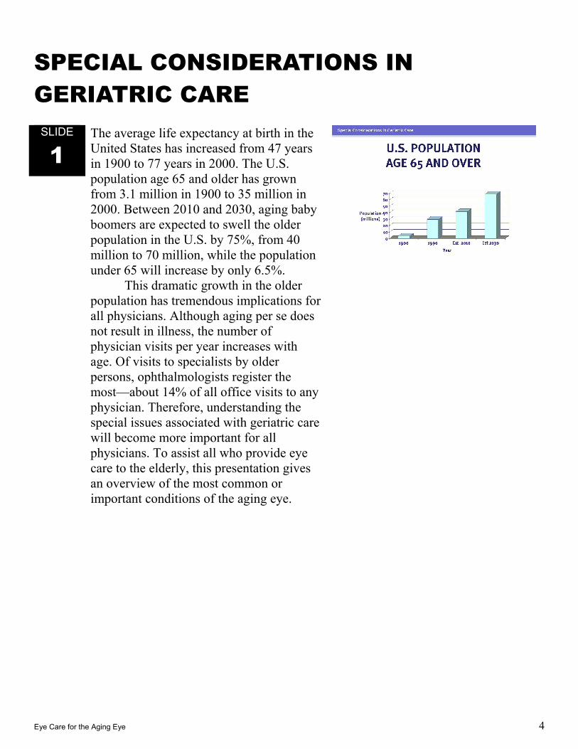
1 The average life expectancy at birth in the United States has increased from 47 years in 1900 to 77 years in 2000. The U.S. population age 65 and older has grown from 3.1 million in 1900 to 35 million in 2000. Between 2010 and 2030, aging baby boomers are expected to swell the older population in the U.S. by 75%, from 40 million to 70 million, while the population under 65 will increase by only 6.5%. This dramatic growth in the older population has tremendous implications for all physicians. Although aging per se does not result in illness, the number of physician visits per year increases with age. Of visits to specialists by older persons, ophthalmologists register the most—about 14% of all office visits to any physician. Therefore, understanding the special issues associated with geriatric care will become more important for all physicians. To assist all who provide eye care to the elderly, this presentation gives an overview of the most common or important conditions of the aging eye.
Eye Care for the Aging Eye 5
SLIDE
2 As people age they may have physiologic, medical, and social differences from younger patients that affect their medical care. Eighty-five percent of Americans aged 65 and over have one or more chronic illness. These individuals are therefore more likely to be taking multiple medications. With aging, physiologic changes occur that result in altered pharmacokinetic and pharmacodynamic effects, so patients 65 and over are more susceptible to adverse effects from medications. Such physiologic changes also probably contribute to the higher frequency of altered presentations of illness in the elderly. Older patients may also have cognitive or functional limitations, which increase the importance of their social support, particularly family.
SLIDE
3 These unique characteristics mean that older patients may have unique goals of care. Because older people may suffer primarily from chronic illness, cure is often not possible. Therefore, improvements in or maintenance of functional abilities and the overall quality of life become the goal. In addition, avoidance of illness in general, and iatrogenic illness in particular (which is common in elderly patients), is an important part of geriatric care. Because older patients commonly have combinations of medical, functional, socioeconomic, cognitive, and affective problems, a multidisciplinary or interdisciplinary team is often necessary to provide care. In addition, incorporating family members into the care process, with permission of the patient, can be essential to ensuring compliance.
Eye Care for the Aging Eye 6
Assessing and Treating Older Patients
SLIDE
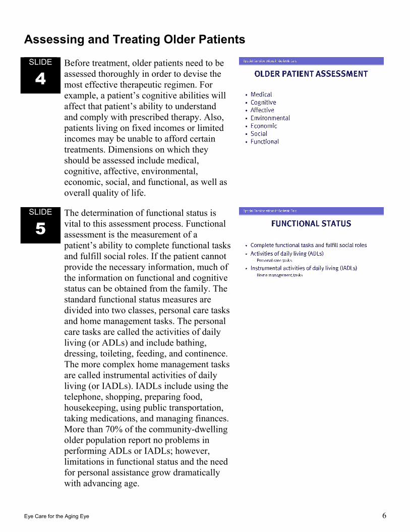
4 Before treatment, older patients need to be assessed thoroughly in order to devise the most effective therapeutic regimen. For example, a patient’s cognitive abilities will affect that patient’s ability to understand and comply with prescribed therapy. Also, patients living on fixed incomes or limited incomes may be unable to afford certain treatments. Dimensions on which they should be assessed include medical, cognitive, affective, environmental, economic, social, and functional, as well as overall quality of life.
SLIDE
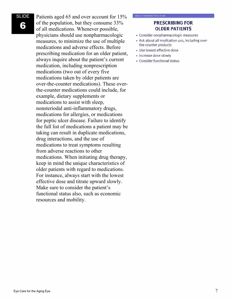
5 The determination of functional status is vital to this assessment process. Functional assessment is the measurement of a patient’s ability to complete functional tasks and fulfill social roles. If the patient cannot provide the necessary information, much of the information on functional and cognitive status can be obtained from the family. The standard functional status measures are divided into two classes, personal care tasks and home management tasks. The personal care tasks are called the activities of daily living (or ADLs) and include bathing, dressing, toileting, feeding, and continence. The more complex home management tasks are called instrumental activities of daily living (or IADLs). IADLs include using the telephone, shopping, preparing food, housekeeping, using public transportation, taking medications, and managing finances. More than 70% of the community-dwelling older population report no problems in performing ADLs or IADLs; however, limitations in functional status and the need for personal assistance grow dramatically with advancing age.
Eye Care for the Aging Eye 7
SLIDE
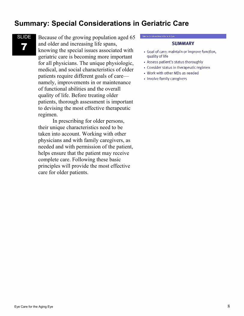
6 Patients aged 65 and over account for 15% of the population, but they consume 33% of all medications. Whenever possible, physicians should use nonpharmacologic measures, to minimize the use of multiple medications and adverse effects. Before prescribing medication for an older patient, always inquire about the patient’s current medication, including nonprescription medications (two out of every five medications taken by older patients are over-the-counter medications). These over-the-counter medications could include, for example, dietary supplements or medications to assist with sleep, nonsteriodal anti-inflammatory drugs, medications for allergies, or medications for peptic ulcer disease. Failure to identify the full list of medications a patient may be taking can result in duplicate medications, drug interactions, and the use of medications to treat symptoms resulting from adverse reactions to other medications. When initiating drug therapy, keep in mind the unique characteristics of older patients with regard to medications. For instance, always start with the lowest effective dose and titrate upward slowly. Make sure to consider the patient’s functional status also, such as economic resources and mobility.
Eye Care for the Aging Eye 8
Summary: Special Considerations in Geriatric Care
SLIDE
7 Because of the growing population aged 65 and older and increasing life spans, knowing the special issues associated with geriatric care is becoming more important for all physicians. The unique physiologic, medical, and social characteristics of older patients require different goals of care—namely, improvements in or maintenance of functional abilities and the overall quality of life. Before treating older patients, thorough assessment is important to devising the most effective therapeutic regimen.
In prescribing for older persons, their unique characteristics need to be taken into account. Working with other physicians and with family caregivers, as needed and with permission of the patient, helps ensure that the patient may receive complete care. Following these basic principles will provide the most effective care for older patients.
Eye Care for the Aging Eye 9
PRINCIPAL PROBLEMS OF THE AGING EYE
SLIDE
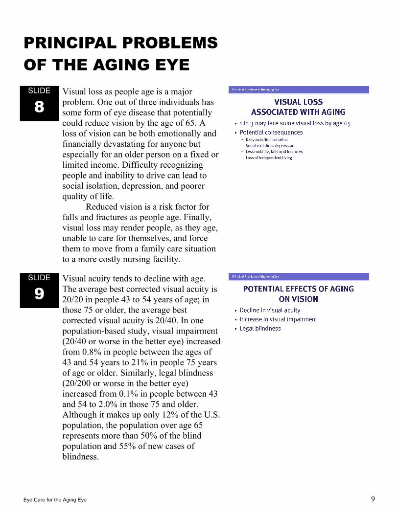
8 Visual loss as people age is a major problem. One out of three individuals has some form of eye disease that potentially could reduce vision by the age of 65. A loss of vision can be both emotionally and financially devastating for anyone but especially for an older person on a fixed or limited income. Difficulty recognizing people and inability to drive can lead to social isolation, depression, and poorer quality of life.
Reduced vision is a risk factor for falls and fractures as people age. Finally, visual loss may render people, as they age, unable to care for themselves, and force them to move from a family care situation to a more costly nursing facility.
SLIDE
9 Visual acuity tends to decline with age. The average best corrected visual acuity is 20/20 in people 43 to 54 years of age; in those 75 or older, the average best corrected visual acuity is 20/40. In one population-based study, visual impairment (20/40 or worse in the better eye) increased from 0.8% in people between the ages of 43 and 54 years to 21% in people 75 years of age or older. Similarly, legal blindness (20/200 or worse in the better eye) increased from 0.1% in people between 43 and 54 to 2.0% in those 75 and older. Although it makes up only 12% of the U.S. population, the population over age 65 represents more than 50% of the blind population and 55% of new cases of blindness.
Eye Care for the Aging Eye 10
SLIDE
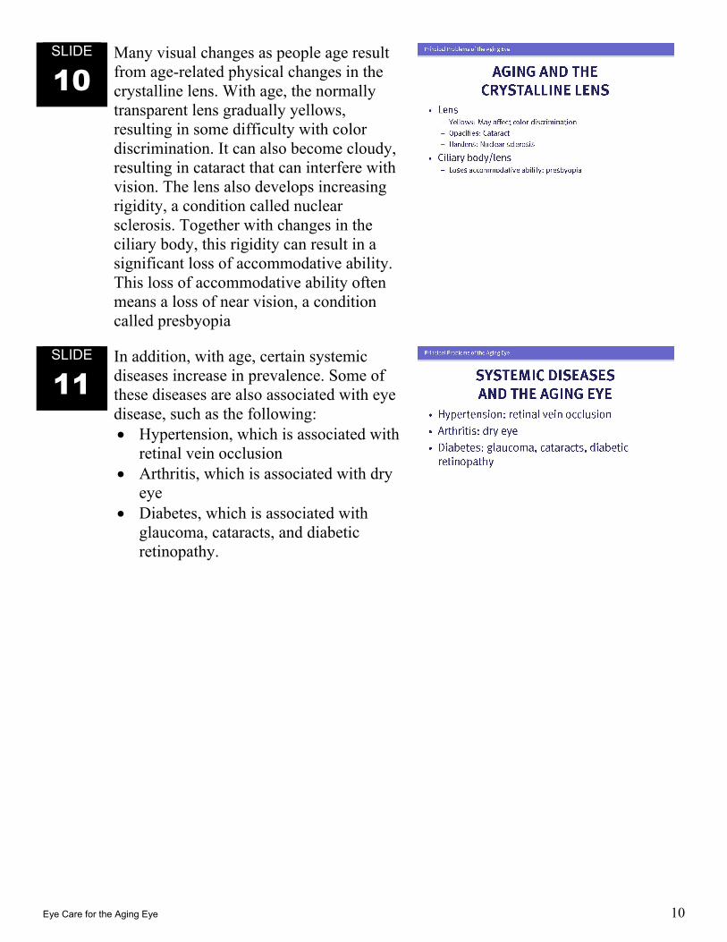
10 Many visual changes as people age result from age-related physical changes in the crystalline lens. With age, the normally transparent lens gradually yellows, resulting in some difficulty with color discrimination. It can also become cloudy, resulting in cataract that can interfere with vision. The lens also develops increasing rigidity, a condition called nuclear sclerosis. Together with changes in the ciliary body, this rigidity can result in a significant loss of accommodative ability. This loss of accommodative ability often means a loss of near vision, a condition called presbyopia
SLIDE
11 In addition, with age, certain systemic diseases increase in prevalence. Some of these diseases are also associated with eye disease, such as the following: • Hypertension, which is associated with
retinal vein occlusion • Arthritis, which is associated with dry
eye • Diabetes, which is associated with
glaucoma, cataracts, and diabetic retinopathy.
Eye Care for the Aging Eye 11
SLIDE
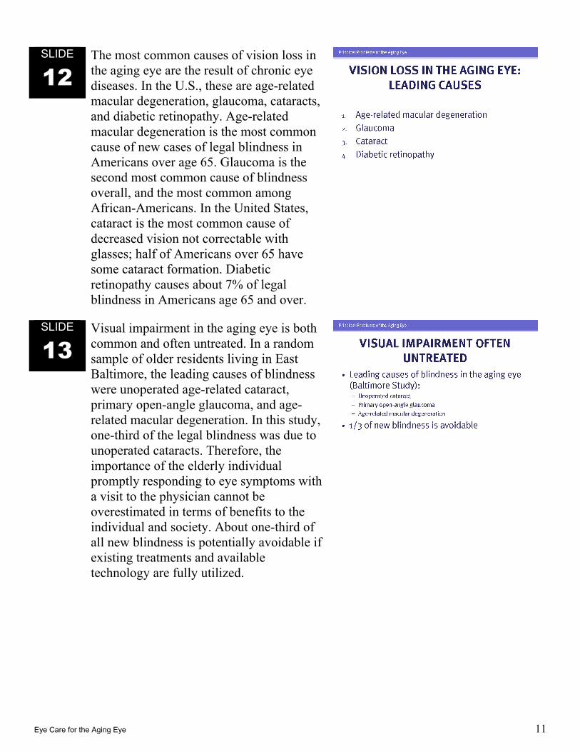
12 The most common causes of vision loss in the aging eye are the result of chronic eye diseases. In the U.S., these are age-related macular degeneration, glaucoma, cataracts, and diabetic retinopathy. Age-related macular degeneration is the most common cause of new cases of legal blindness in Americans over age 65. Glaucoma is the second most common cause of blindness overall, and the most common among African-Americans. In the United States, cataract is the most common cause of decreased vision not correctable with glasses; half of Americans over 65 have some cataract formation. Diabetic retinopathy causes about 7% of legal blindness in Americans age 65 and over.
SLIDE
13 Visual impairment in the aging eye is both common and often untreated. In a random sample of older residents living in East Baltimore, the leading causes of blindness were unoperated age-related cataract, primary open-angle glaucoma, and age-related macular degeneration. In this study, one-third of the legal blindness was due to unoperated cataracts. Therefore, the importance of the elderly individual promptly responding to eye symptoms with a visit to the physician cannot be overestimated in terms of benefits to the individual and society. About one-third of all new blindness is potentially avoidable if existing treatments and available technology are fully utilized.
Eye Care for the Aging Eye 12
Evaluation SLIDE
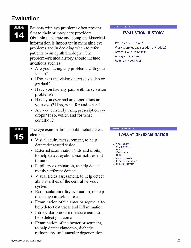
14 Patients with eye problems often present first to their primary care providers. Obtaining accurate and complete historical information is important in managing eye problems and in deciding when to refer patients to an ophthalmologist. The problem-oriented history should include questions such as: • Are you having any problems with your
vision? • If so, was the vision decrease sudden or
gradual? • Have you had any pain with these vision
problems? • Have you ever had any operations on
your eyes? If so, what for and when? • Are you currently using prescription eye
drops? If so, which and for what condition?
SLIDE
15 The eye examination should include these elements: • Visual acuity measurement, to help
detect decreased vision • External examination (lids and orbits),
to help detect eyelid abnormalities and tumors
• Pupillary examination, to help detect relative afferent defects
• Visual fields assessment, to help detect abnormalities of the central nervous system
• Extraocular motility evaluation, to help detect eye muscle paresis
• Examination of the anterior segment, to help detect cataracts and inflammation
• Intraocular pressure measurement, to help detect glaucoma
• Examination of the posterior segment, to help detect glaucoma, diabetic retinopathy, and macular degeneration.
Eye Care for the Aging Eye 13
SLIDE
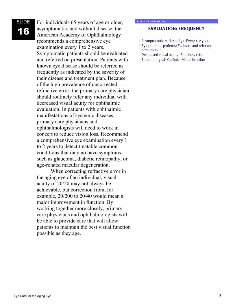
16 For individuals 65 years of age or older, asymptomatic, and without disease, the American Academy of Ophthalmology recommends a comprehensive eye examination every 1 to 2 years. Symptomatic patients should be evaluated and referred on presentation. Patients with known eye disease should be referred as frequently as indicated by the severity of their disease and treatment plan. Because of the high prevalence of uncorrected refractive error, the primary care physician should routinely refer any individual with decreased visual acuity for ophthalmic evaluation. In patients with ophthalmic manifestations of systemic diseases, primary care physicians and ophthalmologists will need to work in concert to reduce vision loss. Recommend a comprehensive eye examination every 1 to 2 years to detect treatable common conditions that may no have symptoms, such as glaucoma, diabetic retinopathy, or age-related macular degeneration.
When correcting refractive error in the aging eye of an individual, visual acuity of 20/20 may not always be achievable, but correction from, for example, 20/200 to 20/40 would mean a major improvement in function. By working together more closely, primary care physicians and ophthalmologists will be able to provide care that will allow patients to maintain the best visual function possible as they age.
Eye Care for the Aging Eye 14
Eyelid Problems Blepharitis
SLIDE
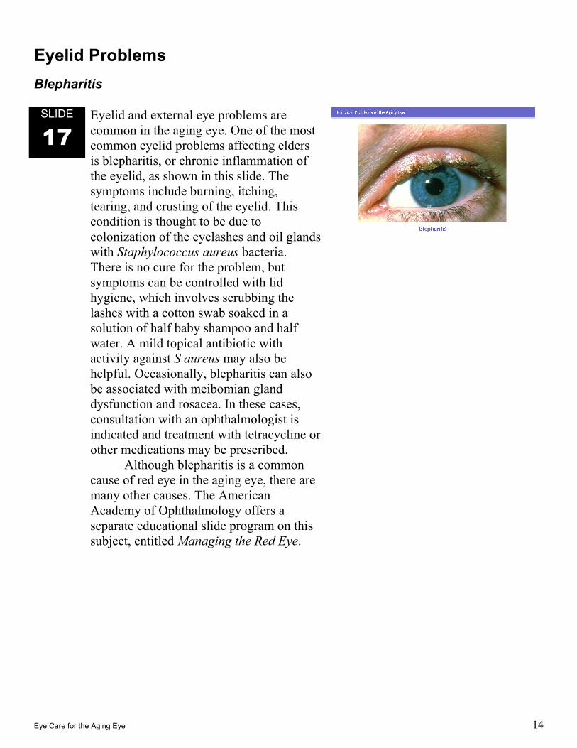
17 Eyelid and external eye problems are common in the aging eye. One of the most common eyelid problems affecting elders is blepharitis, or chronic inflammation of the eyelid, as shown in this slide. The symptoms include burning, itching, tearing, and crusting of the eyelid. This condition is thought to be due to colonization of the eyelashes and oil glands with Staphylococcus aureus bacteria. There is no cure for the problem, but symptoms can be controlled with lid hygiene, which involves scrubbing the lashes with a cotton swab soaked in a solution of half baby shampoo and half water. A mild topical antibiotic with activity against S aureus may also be helpful. Occasionally, blepharitis can also be associated with meibomian gland dysfunction and rosacea. In these cases, consultation with an ophthalmologist is indicated and treatment with tetracycline or other medications may be prescribed.
Although blepharitis is a common cause of red eye in the aging eye, there are many other causes. The American Academy of Ophthalmology offers a separate educational slide program on this subject, entitled Managing the Red Eye.
Eye Care for the Aging Eye 15
Entropion and Ectropion
SLIDE
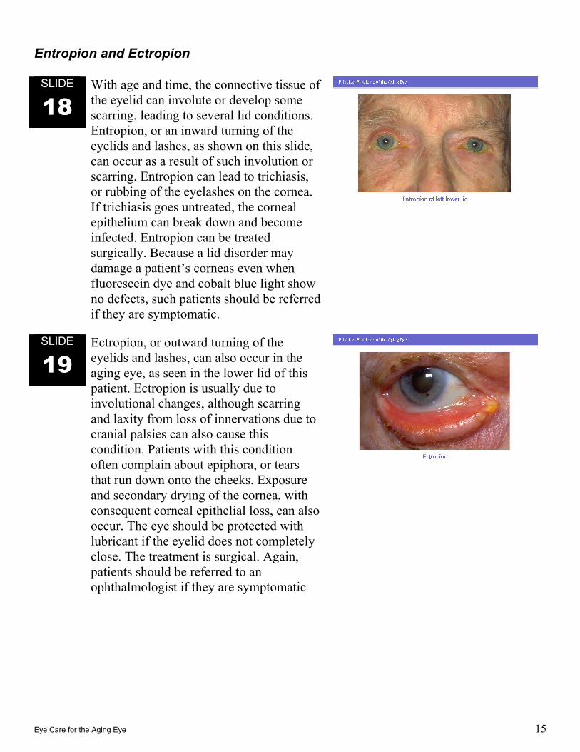
18 With age and time, the connective tissue of the eyelid can involute or develop some scarring, leading to several lid conditions. Entropion, or an inward turning of the eyelids and lashes, as shown on this slide, can occur as a result of such involution or scarring. Entropion can lead to trichiasis, or rubbing of the eyelashes on the cornea. If trichiasis goes untreated, the corneal epithelium can break down and become infected. Entropion can be treated surgically. Because a lid disorder may damage a patient’s corneas even when fluorescein dye and cobalt blue light show no defects, such patients should be referred if they are symptomatic.
SLIDE
19 Ectropion, or outward turning of the eyelids and lashes, can also occur in the aging eye, as seen in the lower lid of this patient. Ectropion is usually due to involutional changes, although scarring and laxity from loss of innervations due to cranial palsies can also cause this condition. Patients with this condition often complain about epiphora, or tears that run down onto the cheeks. Exposure and secondary drying of the cornea, with consequent corneal epithelial loss, can also occur. The eye should be protected with lubricant if the eyelid does not completely close. The treatment is surgical. Again, patients should be referred to an ophthalmologist if they are symptomatic
Eye Care for the Aging Eye 16
Ptosis and Dermatochalasis
SLIDE
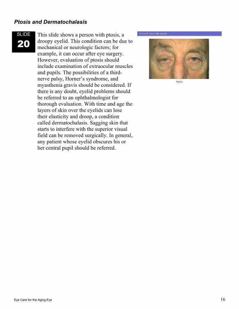
20 This slide shows a person with ptosis, a droopy eyelid. This condition can be due to mechanical or neurologic factors; for example, it can occur after eye surgery. However, evaluation of ptosis should include examination of extraocular muscles and pupils. The possibilities of a third-nerve palsy, Horner’s syndrome, and myasthenia gravis should be considered. If there is any doubt, eyelid problems should be referred to an ophthalmologist for thorough evaluation. With time and age the layers of skin over the eyelids can lose their elasticity and droop, a condition called dermatochalasis. Sagging skin that starts to interfere with the superior visual field can be removed surgically. In general, any patient whose eyelid obscures his or her central pupil should be referred.
Eye Care for the Aging Eye 17
Basal Cell Carcinoma of the Lid
SLIDE
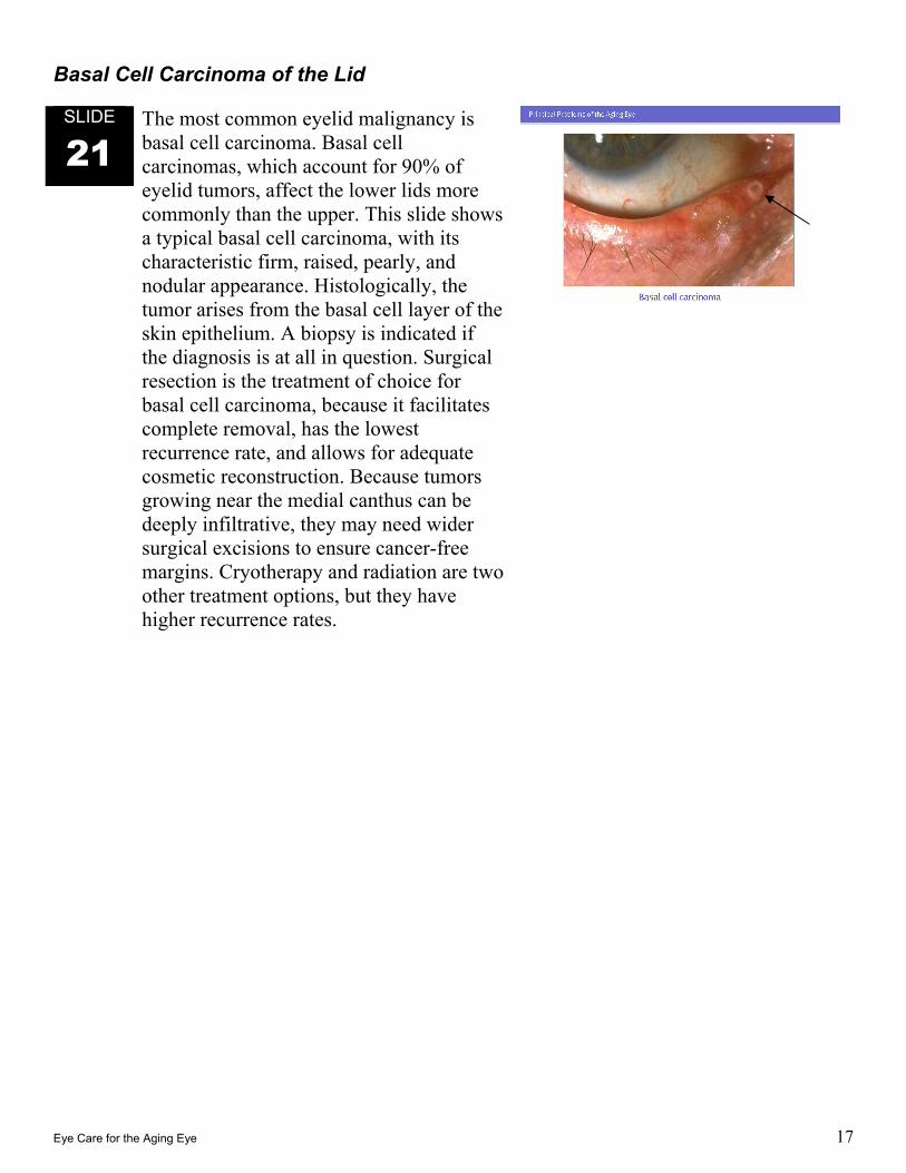
21 The most common eyelid malignancy is basal cell carcinoma. Basal cell carcinomas, which account for 90% of eyelid tumors, affect the lower lids more commonly than the upper. This slide shows a typical basal cell carcinoma, with its characteristic firm, raised, pearly, and nodular appearance. Histologically, the tumor arises from the basal cell layer of the skin epithelium. A biopsy is indicated if the diagnosis is at all in question. Surgical resection is the treatment of choice for basal cell carcinoma, because it facilitates complete removal, has the lowest recurrence rate, and allows for adequate cosmetic reconstruction. Because tumors growing near the medial canthus can be deeply infiltrative, they may need wider surgical excisions to ensure cancer-free margins. Cryotherapy and radiation are two other treatment options, but they have higher recurrence rates.
Eye Care for the Aging Eye 18
Cornea/External Eye Problems Dry Eyes
SLIDE
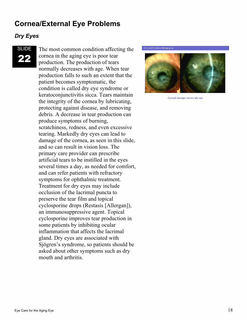
22 The most common condition affecting the cornea in the aging eye is poor tear production. The production of tears normally decreases with age. When tear production falls to such an extent that the patient becomes symptomatic, the condition is called dry eye syndrome or keratoconjunctivitis sicca. Tears maintain the integrity of the cornea by lubricating, protecting against disease, and removing debris. A decrease in tear production can produce symptoms of burning, scratchiness, redness, and even excessive tearing. Markedly dry eyes can lead to damage of the cornea, as seen in this slide, and so can result in vision loss. The primary care provider can prescribe artificial tears to be instilled in the eyes several times a day, as needed for comfort, and can refer patients with refractory symptoms for ophthalmic treatment. Treatment for dry eyes may include occlusion of the lacrimal puncta to preserve the tear film and topical cyclosporine drops (Restasis [Allergan]), an immunosuppressive agent. Topical cyclosporine improves tear production in some patients by inhibiting ocular inflammation that affects the lacrimal gland. Dry eyes are associated with Sjögren’s syndrome, so patients should be asked about other symptoms such as dry mouth and arthritis.
Eye Care for the Aging Eye 19
Herpes Zoster Ophthalmicus
SLIDE
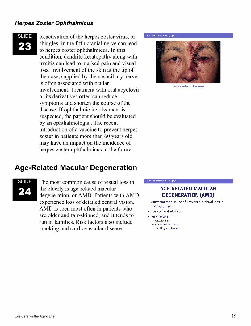
23 Reactivation of the herpes zoster virus, or shingles, in the fifth cranial nerve can lead to herpes zoster ophthalmicus. In this condition, dendrite keratopathy along with uveitis can lead to marked pain and visual loss. Involvement of the skin at the tip of the nose, supplied by the nasociliary nerve, is often associated with ocular involvement. Treatment with oral acyclovir or its derivatives often can reduce symptoms and shorten the course of the disease. If ophthalmic involvement is suspected, the patient should be evaluated by an ophthalmologist. The recent introduction of a vaccine to prevent herpes zoster in patients more than 60 years old may have an impact on the incidence of herpes zoster ophthalmicus in the future.
Age-Related Macular Degeneration
SLIDE
24 The most common cause of visual loss in the elderly is age-related macular degeneration, or AMD. Patients with AMD experience loss of detailed central vision. AMD is seen most often in patients who are older and fair-skinned, and it tends to run in families. Risk factors also include smoking and cardiovascular disease.
Eye Care for the Aging Eye 20
SLIDE
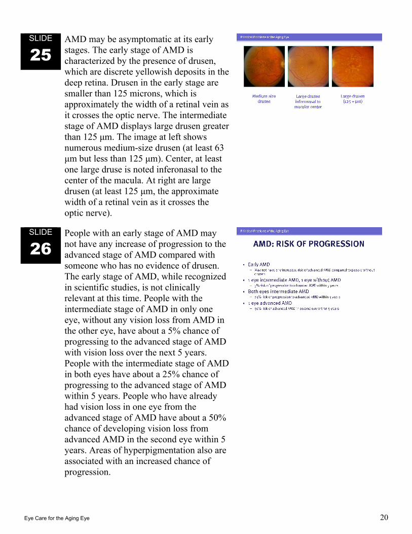
25 AMD may be asymptomatic at its early stages. The early stage of AMD is characterized by the presence of drusen, which are discrete yellowish deposits in the deep retina. Drusen in the early stage are smaller than 125 microns, which is approximately the width of a retinal vein as it crosses the optic nerve. The intermediate stage of AMD displays large drusen greater than 125 μm. The image at left shows numerous medium-size drusen (at least 63 μm but less than 125 μm). Center, at least one large druse is noted inferonasal to the center of the macula. At right are large drusen (at least 125 μm, the approximate width of a retinal vein as it crosses the optic nerve).
SLIDE
26 People with an early stage of AMD may not have any increase of progression to the advanced stage of AMD compared with someone who has no evidence of drusen. The early stage of AMD, while recognized in scientific studies, is not clinically relevant at this time. People with the intermediate stage of AMD in only one eye, without any vision loss from AMD in the other eye, have about a 5% chance of progressing to the advanced stage of AMD with vision loss over the next 5 years. People with the intermediate stage of AMD in both eyes have about a 25% chance of progressing to the advanced stage of AMD within 5 years. People who have already had vision loss in one eye from the advanced stage of AMD have about a 50% chance of developing vision loss from advanced AMD in the second eye within 5 years. Areas of hyperpigmentation also are associated with an increased chance of progression.
Eye Care for the Aging Eye 21
SLIDE
27 AMD is considered to be at an advanced stage when the changes of AMD are associated with loss of vision. This includes an atrophic advanced stage (sometimes called “dry” AMD), and a neovascular advanced stage (sometimes called “wet” AMD). The atrophic advanced stage of AMD is usually associated with a gradual vision loss due to atrophy of the photoreceptors overlying retinal pigment epithelium and atrophy of the underlying choriocapillaris, in the central macular area. The neovascular stage of advanced AMD is often associated with more sudden visual loss from the ingrowth of new blood vessels, from the underlying choriocapillaris through breaks in Bruch’s membrane between the retina and the choroid. The neovascularization may be associated with elevation of the retinal pigment epithelium and hemorrhage and fluid within and beneath the retina. Because the neovascularization represents ingrowth of the fibrovascular tissue, scarring also may become evident, often called a disciform scar.
Eye Care for the Aging Eye 22
Symptoms and Diagnosis
SLIDE
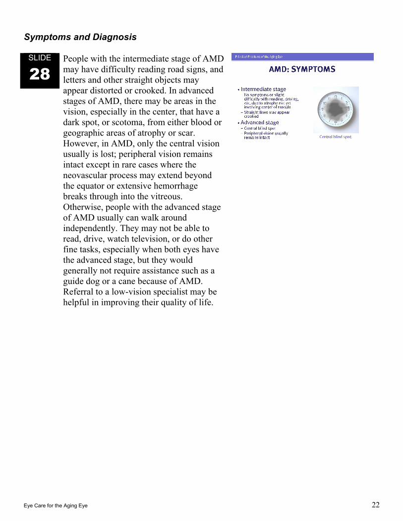
28 People with the intermediate stage of AMD may have difficulty reading road signs, and letters and other straight objects may appear distorted or crooked. In advanced stages of AMD, there may be areas in the vision, especially in the center, that have a dark spot, or scotoma, from either blood or geographic areas of atrophy or scar. However, in AMD, only the central vision usually is lost; peripheral vision remains intact except in rare cases where the neovascular process may extend beyond the equator or extensive hemorrhage breaks through into the vitreous. Otherwise, people with the advanced stage of AMD usually can walk around independently. They may not be able to read, drive, watch television, or do other fine tasks, especially when both eyes have the advanced stage, but they would generally not require assistance such as a guide dog or a cane because of AMD. Referral to a low-vision specialist may be helpful in improving their quality of life.
Eye Care for the Aging Eye 23
SLIDE
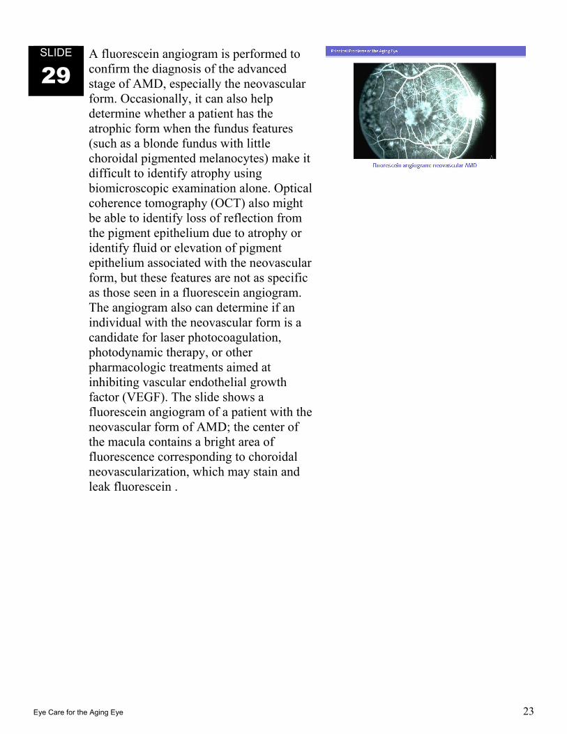
29 A fluorescein angiogram is performed to confirm the diagnosis of the advanced stage of AMD, especially the neovascular form. Occasionally, it can also help determine whether a patient has the atrophic form when the fundus features (such as a blonde fundus with little choroidal pigmented melanocytes) make it difficult to identify atrophy using biomicroscopic examination alone. Optical coherence tomography (OCT) also might be able to identify loss of reflection from the pigment epithelium due to atrophy or identify fluid or elevation of pigment epithelium associated with the neovascular form, but these features are not as specific as those seen in a fluorescein angiogram. The angiogram also can determine if an individual with the neovascular form is a candidate for laser photocoagulation, photodynamic therapy, or other pharmacologic treatments aimed at inhibiting vascular endothelial growth factor (VEGF). The slide shows a fluorescein angiogram of a patient with the neovascular form of AMD; the center of the macula contains a bright area of fluorescence corresponding to choroidal neovascularization, which may stain and leak fluorescein .
Eye Care for the Aging Eye 24
Treatment
SLIDE
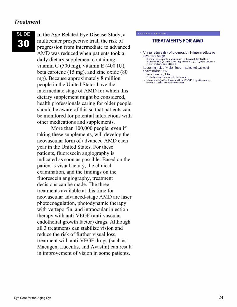
30 In the Age-Related Eye Disease Study, a multicenter prospective trial, the risk of progression from intermediate to advanced AMD was reduced when patients took a daily dietary supplement containing vitamin C (500 mg), vitamin E (400 IU), beta carotene (15 mg), and zinc oxide (80 mg). Because approximately 8 million people in the United States have the intermediate stage of AMD for which this dietary supplement might be considered, health professionals caring for older people should be aware of this so that patients can be monitored for potential interactions with other medications and supplements.
More than 100,000 people, even if taking these supplements, will develop the neovascular form of advanced AMD each year in the United States. For these patients, fluorescein angiography is indicated as soon as possible. Based on the patient’s visual acuity, the clinical examination, and the findings on the fluorescein angiography, treatment decisions can be made. The three treatments available at this time for neovascular advanced-stage AMD are laser photocoagulation, photodynamic therapy with verteporfin, and intraocular injection therapy with anti-VEGF (anti-vascular endothelial growth factor) drugs. Although all 3 treatments can stabilize vision and reduce the risk of further visual loss, treatment with anti-VEGF drugs (such as Macugen, Lucentis, and Avastin) can result in improvement of vision in some patients.
Eye Care for the Aging Eye 25
SLIDE
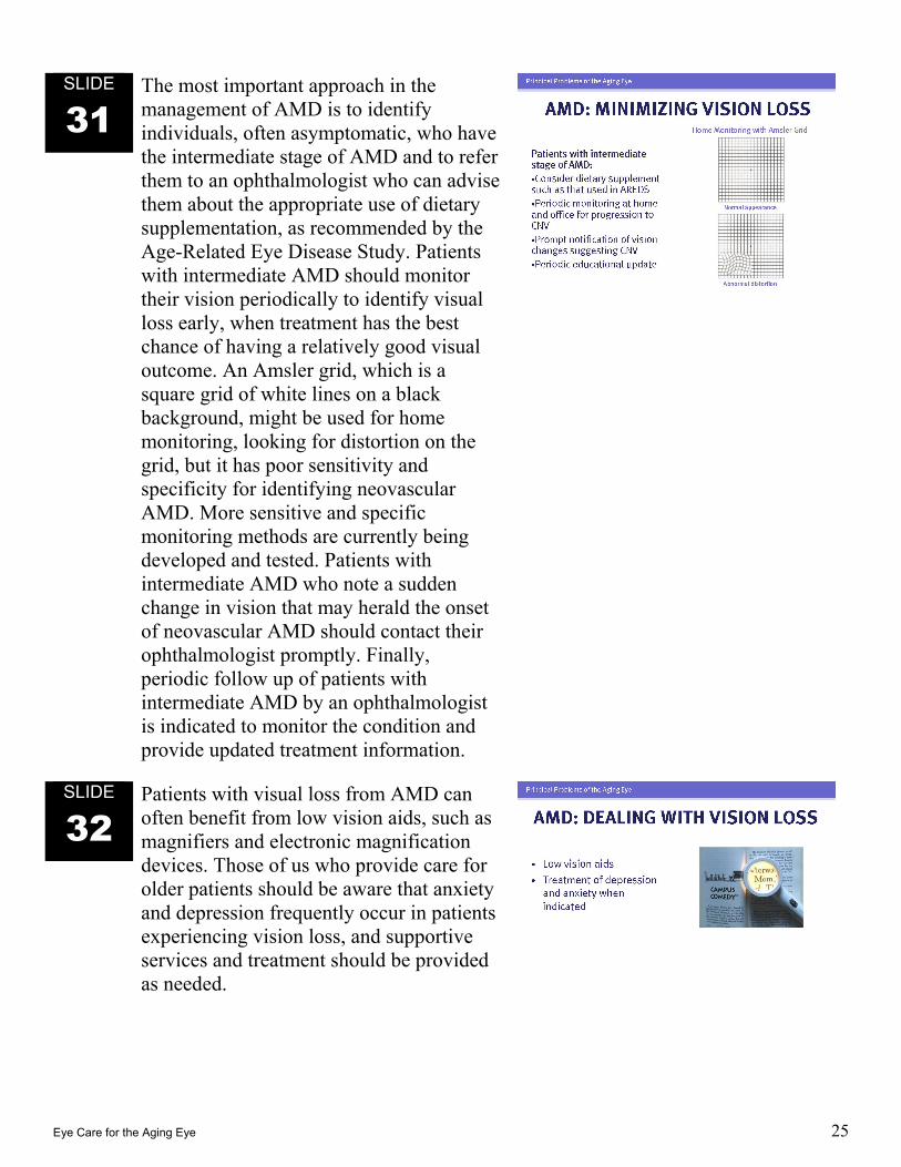
31 The most important approach in the management of AMD is to identify individuals, often asymptomatic, who have the intermediate stage of AMD and to refer them to an ophthalmologist who can advise them about the appropriate use of dietary supplementation, as recommended by the Age-Related Eye Disease Study. Patients with intermediate AMD should monitor their vision periodically to identify visual loss early, when treatment has the best chance of having a relatively good visual outcome. An Amsler grid, which is a square grid of white lines on a black background, might be used for home monitoring, looking for distortion on the grid, but it has poor sensitivity and specificity for identifying neovascular AMD. More sensitive and specific monitoring methods are currently being developed and tested. Patients with intermediate AMD who note a sudden change in vision that may herald the onset of neovascular AMD should contact their ophthalmologist promptly. Finally, periodic follow up of patients with intermediate AMD by an ophthalmologist is indicated to monitor the condition and provide updated treatment information.
SLIDE
32 Patients with visual loss from AMD can often benefit from low vision aids, such as magnifiers and electronic magnification devices. Those of us who provide care for older patients should be aware that anxiety and depression frequently occur in patients experiencing vision loss, and supportive services and treatment should be provided as needed.
Eye Care for the Aging Eye 26
Glaucoma
SLIDE
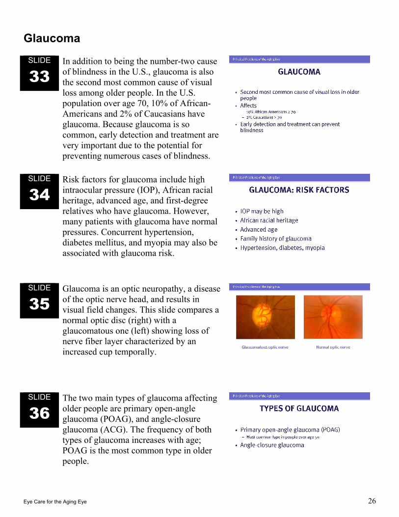
33 In addition to being the number-two cause of blindness in the U.S., glaucoma is also the second most common cause of visual loss among older people. In the U.S. population over age 70, 10% of African-Americans and 2% of Caucasians have glaucoma. Because glaucoma is so common, early detection and treatment are very important due to the potential for preventing numerous cases of blindness.
SLIDE
34 Risk factors for glaucoma include high intraocular pressure (IOP), African racial heritage, advanced age, and first-degree relatives who have glaucoma. However, many patients with glaucoma have normal pressures. Concurrent hypertension, diabetes mellitus, and myopia may also be associated with glaucoma risk.
SLIDE
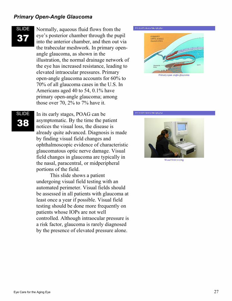
35 Glaucoma is an optic neuropathy, a disease of the optic nerve head, and results in visual field changes. This slide compares a normal optic disc (right) with a glaucomatous one (left) showing loss of nerve fiber layer characterized by an increased cup temporally.
SLIDE
36 The two main types of glaucoma affecting older people are primary open-angle glaucoma (POAG), and angle-closure glaucoma (ACG). The frequency of both types of glaucoma increases with age; POAG is the most common type in older people.
Eye Care for the Aging Eye 27
Primary Open-Angle Glaucoma
SLIDE
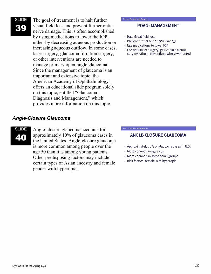
37 Normally, aqueous fluid flows from the eye’s posterior chamber through the pupil into the anterior chamber, and then out via the trabecular meshwork. In primary open-angle glaucoma, as shown in the illustration, the normal drainage network of the eye has increased resistance, leading to elevated intraocular pressures. Primary open-angle glaucoma accounts for 60% to 70% of all glaucoma cases in the U.S. In Americans aged 40 to 54, 0.1% have primary open-angle glaucoma; among those over 70, 2% to 7% have it.
SLIDE
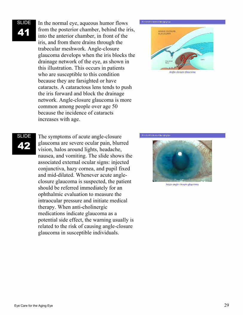
38 In its early stages, POAG can be asymptomatic. By the time the patient notices the visual loss, the disease is already quite advanced. Diagnosis is made by finding visual field changes and ophthalmoscopic evidence of characteristic glaucomatous optic nerve damage. Visual field changes in glaucoma are typically in the nasal, paracentral, or midperipheral portions of the field.
This slide shows a patient undergoing visual field testing with an automated perimeter. Visual fields should be assessed in all patients with glaucoma at least once a year if possible. Visual field testing should be done more frequently on patients whose IOPs are not well controlled. Although intraocular pressure is a risk factor, glaucoma is rarely diagnosed by the presence of elevated pressure alone.
Eye Care for the Aging Eye 28
SLIDE
39 The goal of treatment is to halt further visual field loss and prevent further optic nerve damage. This is often accomplished by using medications to lower the IOP, either by decreasing aqueous production or increasing aqueous outflow. In some cases, laser surgery, glaucoma filtration surgery, or other interventions are needed to manage primary open-angle glaucoma. Since the management of glaucoma is an important and extensive topic, the American Academy of Ophthalmology offers an educational slide program solely on this topic, entitled “Glaucoma: Diagnosis and Management,” which provides more information on this topic.
Angle-Closure Glaucoma
SLIDE
40 Angle-closure glaucoma accounts for approximately 10% of glaucoma cases in the United States. Angle-closure glaucoma is more common among people over the age 50 than it is among young patients. Other predisposing factors may include certain types of Asian ancestry and female gender with hyperopia.
Eye Care for the Aging Eye 29
SLIDE
41 In the normal eye, aqueous humor flows from the posterior chamber, behind the iris, into the anterior chamber, in front of the iris, and from there drains through the trabecular meshwork. Angle-closure glaucoma develops when the iris blocks the drainage network of the eye, as shown in this illustration. This occurs in patients who are susceptible to this condition because they are farsighted or have cataracts. A cataractous lens tends to push the iris forward and block the drainage network. Angle-closure glaucoma is more common among people over age 50 because the incidence of cataracts increases with age.
SLIDE
42 The symptoms of acute angle-closure glaucoma are severe ocular pain, blurred vision, halos around lights, headache, nausea, and vomiting. The slide shows the associated external ocular signs: injected conjunctiva, hazy cornea, and pupil fixed and mid-dilated. Whenever acute angle-closure glaucoma is suspected, the patient should be referred immediately for an ophthalmic evaluation to measure the intraocular pressure and initiate medical therapy. When anti-cholinergic medications indicate glaucoma as a potential side effect, the warning usually is related to the risk of causing angle-closure glaucoma in susceptible individuals.
Eye Care for the Aging Eye 30
SLIDE
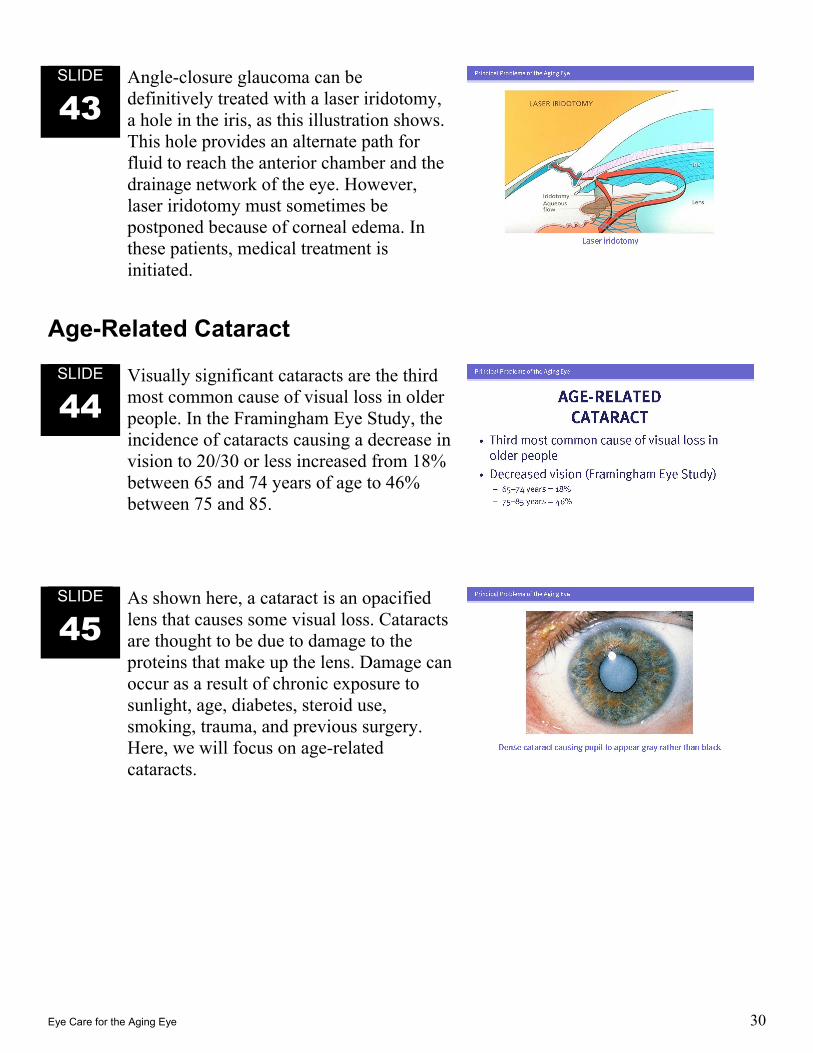
43 Angle-closure glaucoma can be definitively treated with a laser iridotomy, a hole in the iris, as this illustration shows. This hole provides an alternate path for fluid to reach the anterior chamber and the drainage network of the eye. However, laser iridotomy must sometimes be postponed because of corneal edema. In these patients, medical treatment is initiated.
Age-Related Cataract
SLIDE
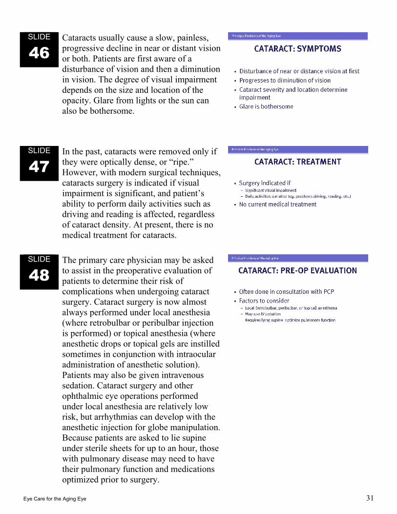
44 Visually significant cataracts are the third most common cause of visual loss in older people. In the Framingham Eye Study, the incidence of cataracts causing a decrease in vision to 20/30 or less increased from 18% between 65 and 74 years of age to 46% between 75 and 85.
SLIDE
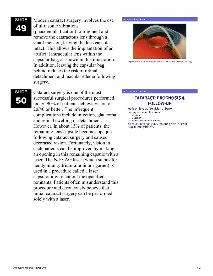
45 As shown here, a cataract is an opacified lens that causes some visual loss. Cataracts are thought to be due to damage to the proteins that make up the lens. Damage can occur as a result of chronic exposure to sunlight, age, diabetes, steroid use, smoking, trauma, and previous surgery. Here, we will focus on age-related cataracts.
Eye Care for the Aging Eye 31
SLIDE
46 Cataracts usually cause a slow, painless, progressive decline in near or distant vision or both. Patients are first aware of a disturbance of vision and then a diminution in vision. The degree of visual impairment depends on the size and location of the opacity. Glare from lights or the sun can also be bothersome.
SLIDE
47 In the past, cataracts were removed only if they were optically dense, or “ripe.” However, with modern surgical techniques, cataracts surgery is indicated if visual impairment is significant, and patient’s ability to perform daily activities such as driving and reading is affected, regardless of cataract density. At present, there is no medical treatment for cataracts.
SLIDE
48 The primary care physician may be asked to assist in the preoperative evaluation of patients to determine their risk of complications when undergoing cataract surgery. Cataract surgery is now almost always performed under local anesthesia (where retrobulbar or peribulbar injection is performed) or topical anesthesia (where anesthetic drops or topical gels are instilled sometimes in conjunction with intraocular administration of anesthetic solution). Patients may also be given intravenous sedation. Cataract surgery and other ophthalmic eye operations performed under local anesthesia are relatively low risk, but arrhythmias can develop with the anesthetic injection for globe manipulation. Because patients are asked to lie supine under sterile sheets for up to an hour, those with pulmonary disease may need to have their pulmonary function and medications optimized prior to surgery.
Eye Care for the Aging Eye 32
SLIDE
49 Modern cataract surgery involves the use of ultrasonic vibrations (phacoemulsification) to fragment and remove the cataractous lens through a small incision, leaving the lens capsule intact. This allows the implantation of an artificial intraocular lens within the capsular bag, as shown in this illustration. In addition, leaving the capsular bag behind reduces the risk of retinal detachment and macular edema following surgery.
SLIDE
50 Cataract surgery is one of the most successful surgical procedures performed today: 90% of patients achieve vision of 20/40 or better. The infrequent complications include infection, glaucoma, and retinal swelling or detachment. However, in about 15% of patients, the remaining lens capsule becomes opaque following cataract surgery and causes decreased vision. Fortunately, vision in such patients can be improved by making an opening in this remaining capsule with a laser. The Nd:YAG laser (which stands for neodymium:yttrium-aluminum-garnet) is used in a procedure called a laser capsulotomy to cut out the opacified remnants. Patients often misunderstand this procedure and erroneously believe that initial cataract surgery can be performed solely with a laser.
Eye Care for the Aging Eye 33
Diabetic Retinopathy
SLIDE
51 Diabetic retinopathy (DR) is the leading cause of blindness among working-age Americans and the fourth most common cause of visual loss in people over the age of 55 in the United States. Older patients are more likely to get type II diabetes, in which macular edema is much more common. Because diabetic retinopathy is an important cause of visual loss, the Academy offers a slide program solely on this subject, Diabetes and Eye Disease, so it will be covered only briefly here.
SLIDE
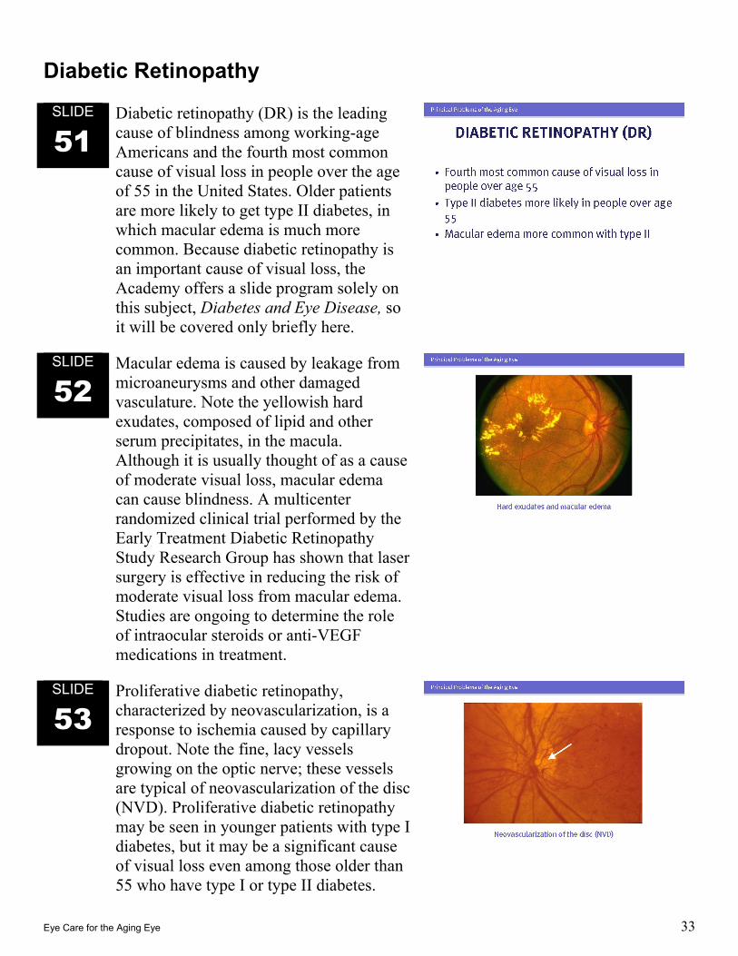
52 Macular edema is caused by leakage from microaneurysms and other damaged vasculature. Note the yellowish hard exudates, composed of lipid and other serum precipitates, in the macula. Although it is usually thought of as a cause of moderate visual loss, macular edema can cause blindness. A multicenter randomized clinical trial performed by the Early Treatment Diabetic Retinopathy Study Research Group has shown that laser surgery is effective in reducing the risk of moderate visual loss from macular edema. Studies are ongoing to determine the role of intraocular steroids or anti-VEGF medications in treatment.
SLIDE
53 Proliferative diabetic retinopathy, characterized by neovascularization, is a response to ischemia caused by capillary dropout. Note the fine, lacy vessels growing on the optic nerve; these vessels are typical of neovascularization of the disc (NVD). Proliferative diabetic retinopathy may be seen in younger patients with type I diabetes, but it may be a significant cause of visual loss even among those older than 55 who have type I or type II diabetes.
Eye Care for the Aging Eye 34
SLIDE
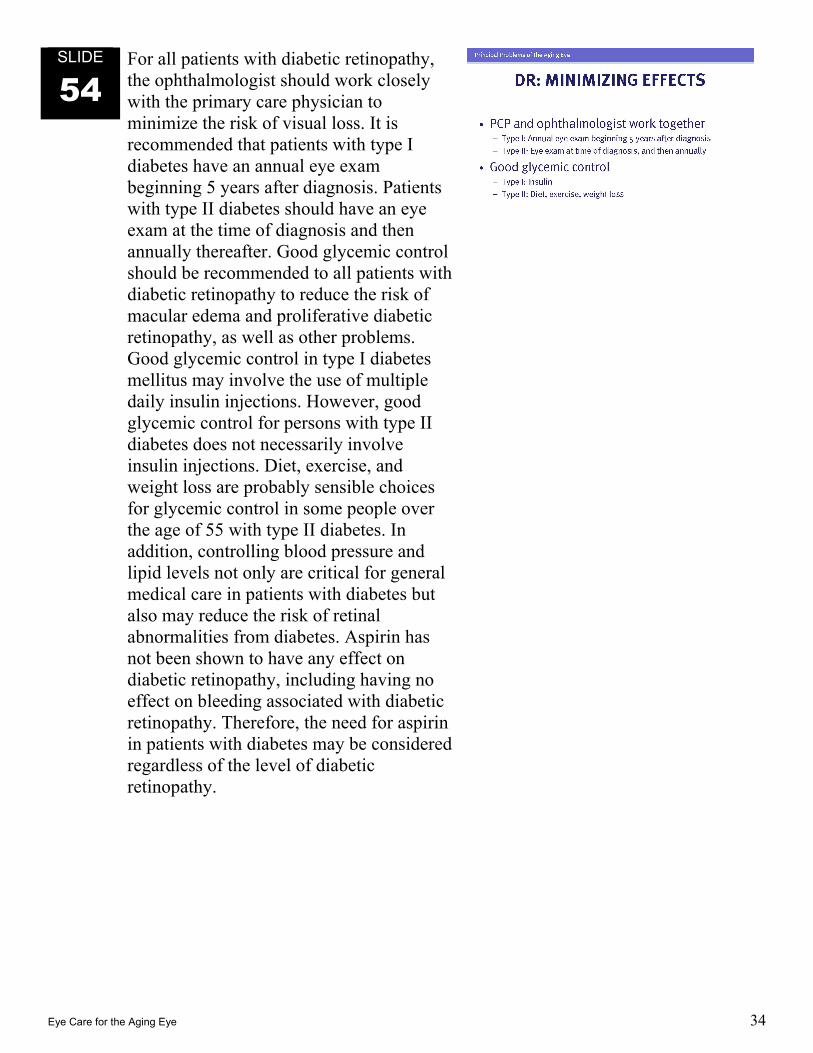
54 For all patients with diabetic retinopathy, the ophthalmologist should work closely with the primary care physician to minimize the risk of visual loss. It is recommended that patients with type I diabetes have an annual eye exam beginning 5 years after diagnosis. Patients with type II diabetes should have an eye exam at the time of diagnosis and then annually thereafter. Good glycemic control should be recommended to all patients with diabetic retinopathy to reduce the risk of macular edema and proliferative diabetic retinopathy, as well as other problems. Good glycemic control in type I diabetes mellitus may involve the use of multiple daily insulin injections. However, good glycemic control for persons with type II diabetes does not necessarily involve insulin injections. Diet, exercise, and weight loss are probably sensible choices for glycemic control in some people over the age of 55 with type II diabetes. In addition, controlling blood pressure and lipid levels not only are critical for general medical care in patients with diabetes but also may reduce the risk of retinal abnormalities from diabetes. Aspirin has not been shown to have any effect on diabetic retinopathy, including having no effect on bleeding associated with diabetic retinopathy. Therefore, the need for aspirin in patients with diabetes may be considered regardless of the level of diabetic retinopathy.
Eye Care for the Aging Eye 35
Retinal Vascular Disorders
SLIDE
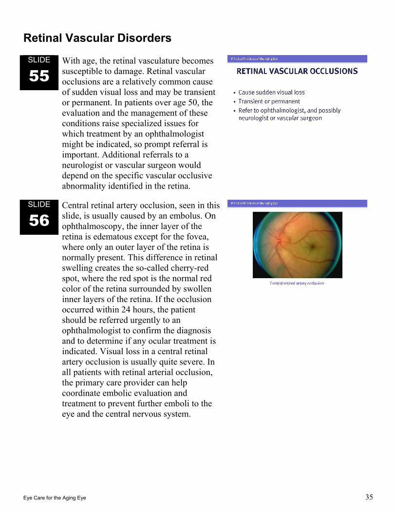
55 With age, the retinal vasculature becomes susceptible to damage. Retinal vascular occlusions are a relatively common cause of sudden visual loss and may be transient or permanent. In patients over age 50, the evaluation and the management of these conditions raise specialized issues for which treatment by an ophthalmologist might be indicated, so prompt referral is important. Additional referrals to a neurologist or vascular surgeon would depend on the specific vascular occlusive abnormality identified in the retina.
SLIDE
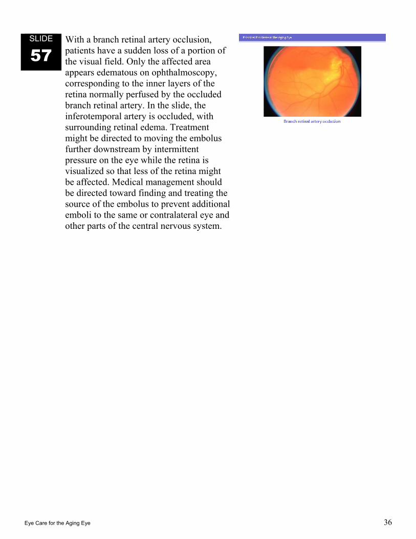
56 Central retinal artery occlusion, seen in this slide, is usually caused by an embolus. On ophthalmoscopy, the inner layer of the retina is edematous except for the fovea, where only an outer layer of the retina is normally present. This difference in retinal swelling creates the so-called cherry-red spot, where the red spot is the normal red color of the retina surrounded by swollen inner layers of the retina. If the occlusion occurred within 24 hours, the patient should be referred urgently to an ophthalmologist to confirm the diagnosis and to determine if any ocular treatment is indicated. Visual loss in a central retinal artery occlusion is usually quite severe. In all patients with retinal arterial occlusion, the primary care provider can help coordinate embolic evaluation and treatment to prevent further emboli to the eye and the central nervous system.
Eye Care for the Aging Eye 36
SLIDE
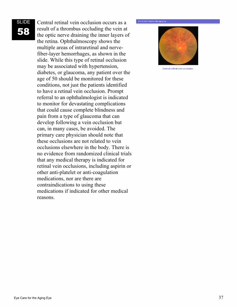
57 With a branch retinal artery occlusion, patients have a sudden loss of a portion of the visual field. Only the affected area appears edematous on ophthalmoscopy, corresponding to the inner layers of the retina normally perfused by the occluded branch retinal artery. In the slide, the inferotemporal artery is occluded, with surrounding retinal edema. Treatment might be directed to moving the embolus further downstream by intermittent pressure on the eye while the retina is visualized so that less of the retina might be affected. Medical management should be directed toward finding and treating the source of the embolus to prevent additional emboli to the same or contralateral eye and other parts of the central nervous system.
Eye Care for the Aging Eye 37
SLIDE
58 Central retinal vein occlusion occurs as a result of a thrombus occluding the vein at the optic nerve draining the inner layers of the retina. Ophthalmoscopy shows the multiple areas of intraretinal and nerve-fiber-layer hemorrhages, as shown in the slide. While this type of retinal occlusion may be associated with hypertension, diabetes, or glaucoma, any patient over the age of 50 should be monitored for these conditions, not just the patients identified to have a retinal vein occlusion. Prompt referral to an ophthalmologist is indicated to monitor for devastating complications that could cause complete blindness and pain from a type of glaucoma that can develop following a vein occlusion but can, in many cases, be avoided. The primary care physician should note that these occlusions are not related to vein occlusions elsewhere in the body. There is no evidence from randomized clinical trials that any medical therapy is indicated for retinal vein occlusions, including aspirin or other anti-platelet or anti-coagulation medications, nor are there are contraindications to using these medications if indicated for other medical reasons.
Eye Care for the Aging Eye 38
SLIDE
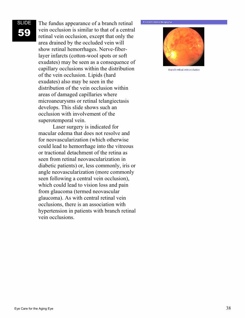
59 The fundus appearance of a branch retinal vein occlusion is similar to that of a central retinal vein occlusion, except that only the area drained by the occluded vein will show retinal hemorrhages. Nerve-fiber-layer infarcts (cotton-wool spots or soft exudates) may be seen as a consequence of capillary occlusions within the distribution of the vein occlusion. Lipids (hard exudates) also may be seen in the distribution of the vein occlusion within areas of damaged capillaries where microaneurysms or retinal telangiectasis develops. This slide shows such an occlusion with involvement of the superotemporal vein.
Laser surgery is indicated for macular edema that does not resolve and for neovascularization (which otherwise could lead to hemorrhage into the vitreous or tractional detachment of the retina as seen from retinal neovascularization in diabetic patients) or, less commonly, iris or angle neovascularization (more commonly seen following a central vein occlusion), which could lead to vision loss and pain from glaucoma (termed neovascular glaucoma). As with central retinal vein occlusions, there is an association with hypertension in patients with branch retinal vein occlusions.
Eye Care for the Aging Eye 39
Posterior Vitreous Detachment
SLIDE
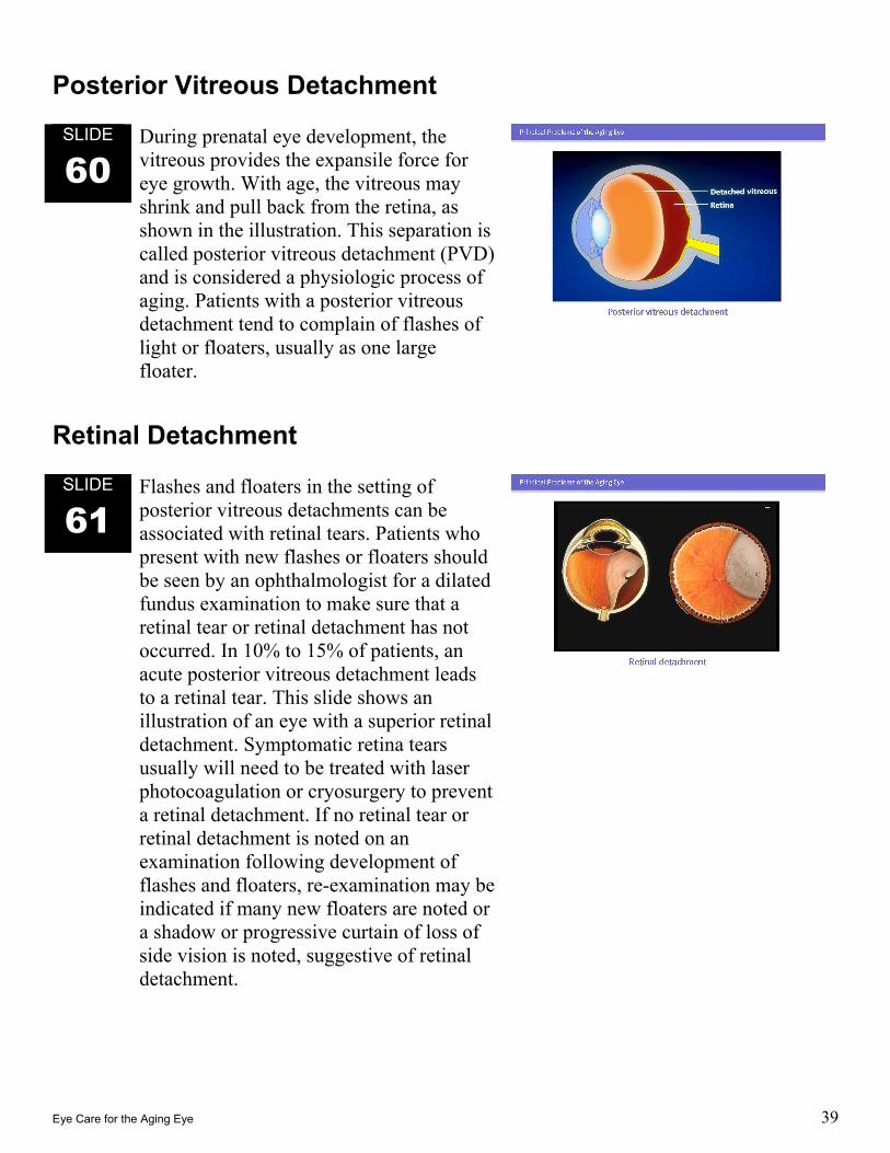
60 During prenatal eye development, the vitreous provides the expansile force for eye growth. With age, the vitreous may shrink and pull back from the retina, as shown in the illustration. This separation is called posterior vitreous detachment (PVD) and is considered a physiologic process of aging. Patients with a posterior vitreous detachment tend to complain of flashes of light or floaters, usually as one large floater.
Retinal Detachment
SLIDE
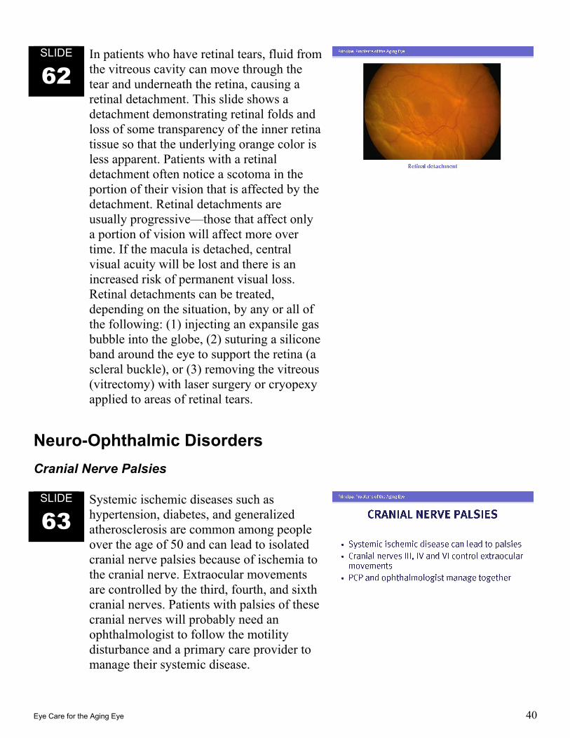
61 Flashes and floaters in the setting of posterior vitreous detachments can be associated with retinal tears. Patients who present with new flashes or floaters should be seen by an ophthalmologist for a dilated fundus examination to make sure that a retinal tear or retinal detachment has not occurred. In 10% to 15% of patients, an acute posterior vitreous detachment leads to a retinal tear. This slide shows an illustration of an eye with a superior retinal detachment. Symptomatic retina tears usually will need to be treated with laser photocoagulation or cryosurgery to prevent a retinal detachment. If no retinal tear or retinal detachment is noted on an examination following development of flashes and floaters, re-examination may be indicated if many new floaters are noted or a shadow or progressive curtain of loss of side vision is noted, suggestive of retinal detachment.
Eye Care for the Aging Eye 40
SLIDE
62 In patients who have retinal tears, fluid from the vitreous cavity can move through the tear and underneath the retina, causing a retinal detachment. This slide shows a detachment demonstrating retinal folds and loss of some transparency of the inner retina tissue so that the underlying orange color is less apparent. Patients with a retinal detachment often notice a scotoma in the portion of their vision that is affected by the detachment. Retinal detachments are usually progressive—those that affect only a portion of vision will affect more over time. If the macula is detached, central visual acuity will be lost and there is an increased risk of permanent visual loss. Retinal detachments can be treated, depending on the situation, by any or all of the following: (1) injecting an expansile gas bubble into the globe, (2) suturing a silicone band around the eye to support the retina (a scleral buckle), or (3) removing the vitreous (vitrectomy) with laser surgery or cryopexy applied to areas of retinal tears.
Neuro-Ophthalmic Disorders Cranial Nerve Palsies
SLIDE
63 Systemic ischemic diseases such as hypertension, diabetes, and generalized atherosclerosis are common among people over the age of 50 and can lead to isolated cranial nerve palsies because of ischemia to the cranial nerve. Extraocular movements are controlled by the third, fourth, and sixth cranial nerves. Patients with palsies of these cranial nerves will probably need an ophthalmologist to follow the motility disturbance and a primary care provider to manage their systemic disease.
Eye Care for the Aging Eye 41
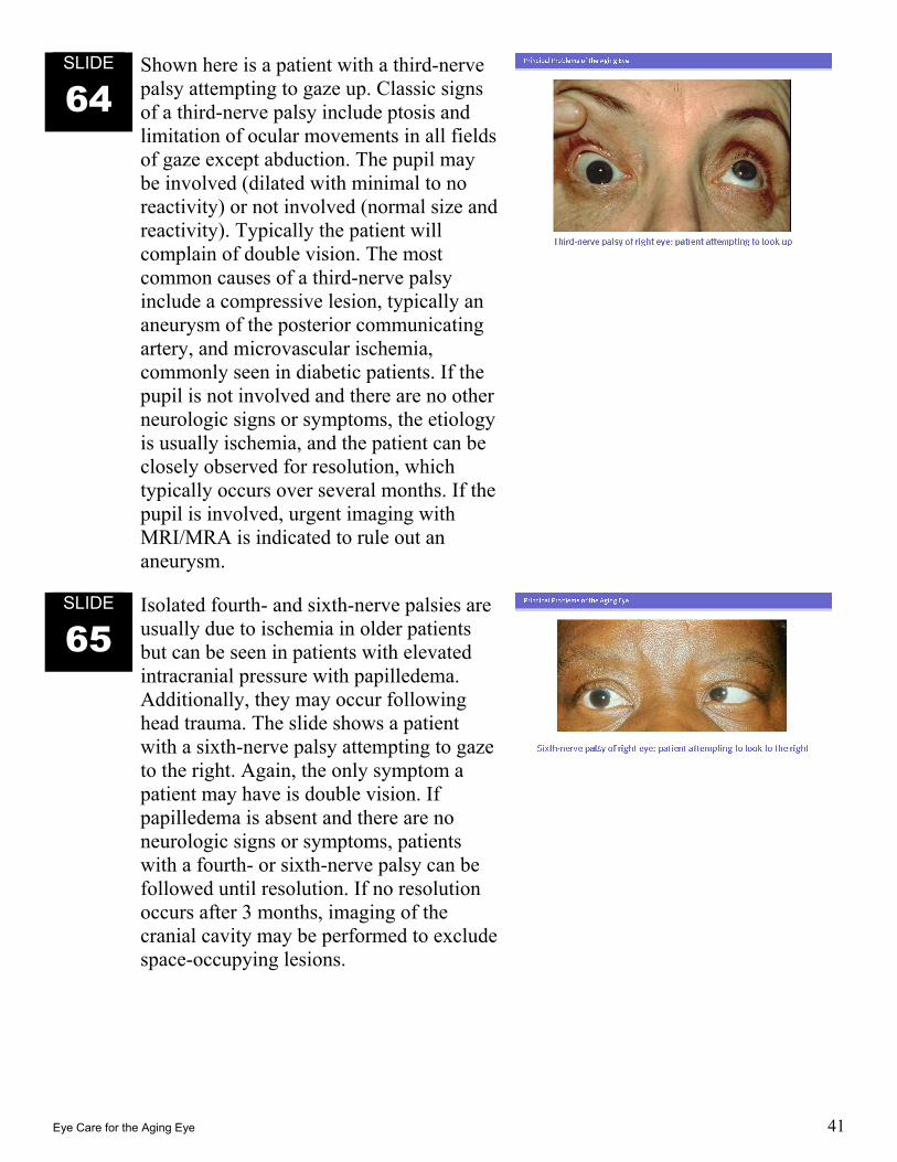
SLIDE
64 Shown here is a patient with a third-nerve palsy attempting to gaze up. Classic signs of a third-nerve palsy include ptosis and limitation of ocular movements in all fields of gaze except abduction. The pupil may be involved (dilated with minimal to no reactivity) or not involved (normal size and reactivity). Typically the patient will complain of double vision. The most common causes of a third-nerve palsy include a compressive lesion, typically an aneurysm of the posterior communicating artery, and microvascular ischemia, commonly seen in diabetic patients. If the pupil is not involved and there are no other neurologic signs or symptoms, the etiology is usually ischemia, and the patient can be closely observed for resolution, which typically occurs over several months. If the pupil is involved, urgent imaging with MRI/MRA is indicated to rule out an aneurysm.
SLIDE
65 Isolated fourth- and sixth-nerve palsies are usually due to ischemia in older patients but can be seen in patients with elevated intracranial pressure with papilledema. Additionally, they may occur following head trauma. The slide shows a patient with a sixth-nerve palsy attempting to gaze to the right. Again, the only symptom a patient may have is double vision. If papilledema is absent and there are no neurologic signs or symptoms, patients with a fourth- or sixth-nerve palsy can be followed until resolution. If no resolution occurs after 3 months, imaging of the cranial cavity may be performed to exclude space-occupying lesions.
Eye Care for the Aging Eye 42
Ischemic Optic Neuropathy
SLIDE
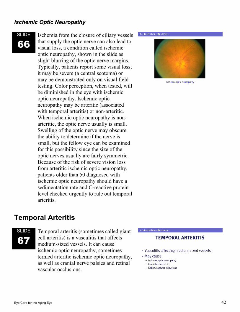
66 Ischemia from the closure of ciliary vessels that supply the optic nerve can also lead to visual loss, a condition called ischemic optic neuropathy, shown in the slide as slight blurring of the optic nerve margins. Typically, patients report some visual loss; it may be severe (a central scotoma) or may be demonstrated only on visual field testing. Color perception, when tested, will be diminished in the eye with ischemic optic neuropathy. Ischemic optic neuropathy may be arteritic (associated with temporal arteritis) or non-arteritic. When ischemic optic neuropathy is non-arteritic, the optic nerve usually is small. Swelling of the optic nerve may obscure the ability to determine if the nerve is small, but the fellow eye can be examined for this possibility since the size of the optic nerves usually are fairly symmetric. Because of the risk of severe vision loss from arteritic ischemic optic neuropathy, patients older than 50 diagnosed with ischemic optic neuropathy should have a sedimentation rate and C-reactive protein level checked urgently to rule out temporal arteritis.
Temporal Arteritis
SLIDE
67 Temporal arteritis (sometimes called giant cell arteritis) is a vasculitis that affects medium-sized vessels. It can cause ischemic optic neuropathy, sometimes termed arteritic ischemic optic neuropathy, as well as cranial nerve palsies and retinal vascular occlusions.
Eye Care for the Aging Eye 43
SLIDE
68 Patients with temporal arteritis often complain of headaches, generalized malaise, night sweats, weight loss, and jaw claudication. There is also an association with polymyalgia rheumatica.
SLIDE
69 Temporal arteritis is a clinical diagnosis. Laboratory signs may include an elevated sedimentation rate, but because this test is neither sensitive nor specific, the diagnosis should not be based on an elevated rate. The C-reactive protein level may also be elevated. A finding of giant cell infiltration of the artery on temporal artery biopsy can confirm the diagnosis, but infiltration is not always present. All patients suspected to have temporal arteritis should be referred emergently to an ophthalmologist, because left untreated they could rapidly lose their vision.
SLIDE
70 Treatment of temporal arteritis consists of high doses of steroids tapered over many months, usually given orally, but if severe visual loss from ischemic optic neuropathy is present, intravenous steroid treatment should be considered. If ischemic optic neuropathy has already occurred in one eye, steroid treatment should be started to protect the fellow eye as soon as a temporal-arteritis diagnosis is suspected. Steroid treatment should not be delayed while awaiting temporal artery biopsy, as such treatment will not affect the biopsy results provided that the procedure is performed within about a week of the beginning treatment.
Eye Care for the Aging Eye 44
Low Vision
SLIDE
71 Problems of low vision are common in people aged 50 and older. Moderate low vision can be defined as visual acuity of approximately 20/70 to 20/160 in the better-seeing eye. For functioning indoors, these individuals can be provided increased lighting for ambulation, and bright lighting with optical or electronic magnification aids for reading and other near-vision tasks. Patients with such vision can be fitted with refractive devices such as special spectacles, contact lenses, and telescopes to enhance their distance or near vision, depending on their need.
SLIDE
72 Severe low vision can be defined as visual acuity of 20/200 (legal blindness) to 20/400 or worse (blindness according the World Health Organization) in the better-seeing eye. For patients with such vision, once all refractive errors have been corrected as much as possible, handheld or mounted magnifiers or electronic magnification might improve vision sufficiently for reading and writing and other near-vision tasks In addition, there are special household applications such as “talking” clocks that announce the time and computers that can speak and record the human voice as written text.
Eye Care for the Aging Eye 45
CONCLUSION
SLIDE
73 Visual loss in the aging eye is a major problem that has many aspects of which the primary case physician should be aware. Many common eye conditions and diseases affect older people. Vision can often be improved or maintained by the primary care physician referring patients appropriately to an ophthalmologist for evaluation and management, even when patients are asymptomatic, since many of these conditions can be prevented or treated before vision loss has occurred. Systemic diseases that affect the visual system can best be managed by a team effort between primary care physicians and ophthalmologists. In addition, periodic updates on this topic should be reviewed, since a tremendous amount of research is underway, providing hope that the management and treatment of vision loss in the aging eye will continue to improve over the coming years. As we ourselves look forward to longer lives, we should ensure that independence and joy in living are curtailed as little as possibly by limitations in vision.
Eye Care for the Aging Eye 46
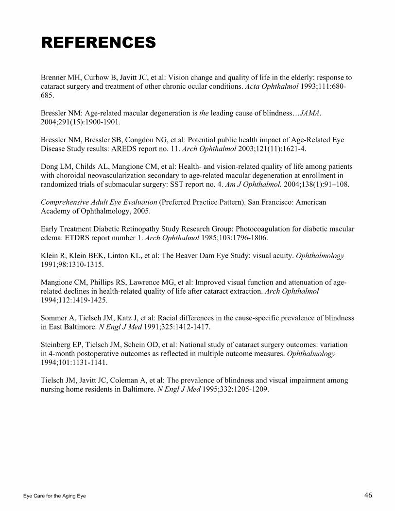
REFERENCES Brenner MH, Curbow B, Javitt JC, et al: Vision change and quality of life in the elderly: response to cataract surgery and treatment of other chronic ocular conditions. Acta Ophthalmol 1993;111:680-685. Bressler NM: Age-related macular degeneration is the leading cause of blindness…JAMA. 2004;291(15):1900-1901. Bressler NM, Bressler SB, Congdon NG, et al: Potential public health impact of Age-Related Eye Disease Study results: AREDS report no. 11. Arch Ophthalmol 2003;121(11):1621-4. Dong LM, Childs AL, Mangione CM, et al: Health- and vision-related quality of life among patients with choroidal neovascularization secondary to age-related macular degeneration at enrollment in randomized trials of submacular surgery: SST report no. 4. Am J Ophthalmol. 2004;138(1):91–108. Comprehensive Adult Eye Evaluation (Preferred Practice Pattern). San Francisco: American Academy of Ophthalmology, 2005. Early Treatment Diabetic Retinopathy Study Research Group: Photocoagulation for diabetic macular edema. ETDRS report number 1. Arch Ophthalmol 1985;103:1796-1806. Klein R, Klein BEK, Linton KL, et al: The Beaver Dam Eye Study: visual acuity. Ophthalmology 1991;98:1310-1315. Mangione CM, Phillips RS, Lawrence MG, et al: Improved visual function and attenuation of age-related declines in health-related quality of life after cataract extraction. Arch Ophthalmol 1994;112:1419-1425. Sommer A, Tielsch JM, Katz J, et al: Racial differences in the cause-specific prevalence of blindness in East Baltimore. N Engl J Med 1991;325:1412-1417. Steinberg EP, Tielsch JM, Schein OD, et al: National study of cataract surgery outcomes: variation in 4-month postoperative outcomes as reflected in multiple outcome measures. Ophthalmology 1994;101:1131-1141. Tielsch JM, Javitt JC, Coleman A, et al: The prevalence of blindness and visual impairment among nursing home residents in Baltimore. N Engl J Med 1995;332:1205-1209.
Eye Care for the Aging Eye 47
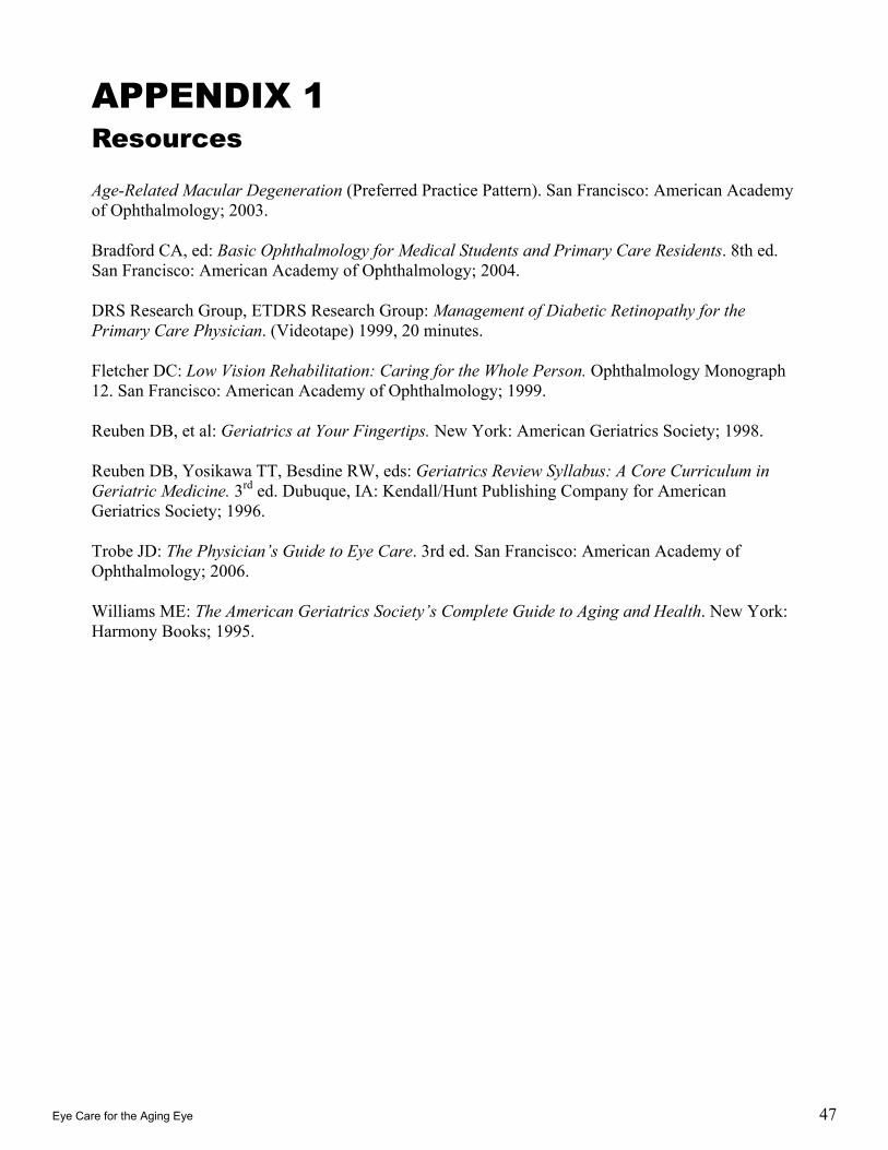
APPENDIX 1 Resources Age-Related Macular Degeneration (Preferred Practice Pattern). San Francisco: American Academy of Ophthalmology; 2003. Bradford CA, ed: Basic Ophthalmology for Medical Students and Primary Care Residents. 8th ed. San Francisco: American Academy of Ophthalmology; 2004. DRS Research Group, ETDRS Research Group: Management of Diabetic Retinopathy for the Primary Care Physician. (Videotape) 1999, 20 minutes. Fletcher DC: Low Vision Rehabilitation: Caring for the Whole Person. Ophthalmology Monograph 12. San Francisco: American Academy of Ophthalmology; 1999. Reuben DB, et al: Geriatrics at Your Fingertips. New York: American Geriatrics Society; 1998. Reuben DB, Yosikawa TT, Besdine RW, eds: Geriatrics Review Syllabus: A Core Curriculum in Geriatric Medicine. 3rd ed. Dubuque, IA: Kendall/Hunt Publishing Company for American Geriatrics Society; 1996. Trobe JD: The Physician’s Guide to Eye Care. 3rd ed. San Francisco: American Academy of Ophthalmology; 2006. Williams ME: The American Geriatrics Society’s Complete Guide to Aging and Health. New York: Harmony Books; 1995.
Eye Care for the Aging Eye 48
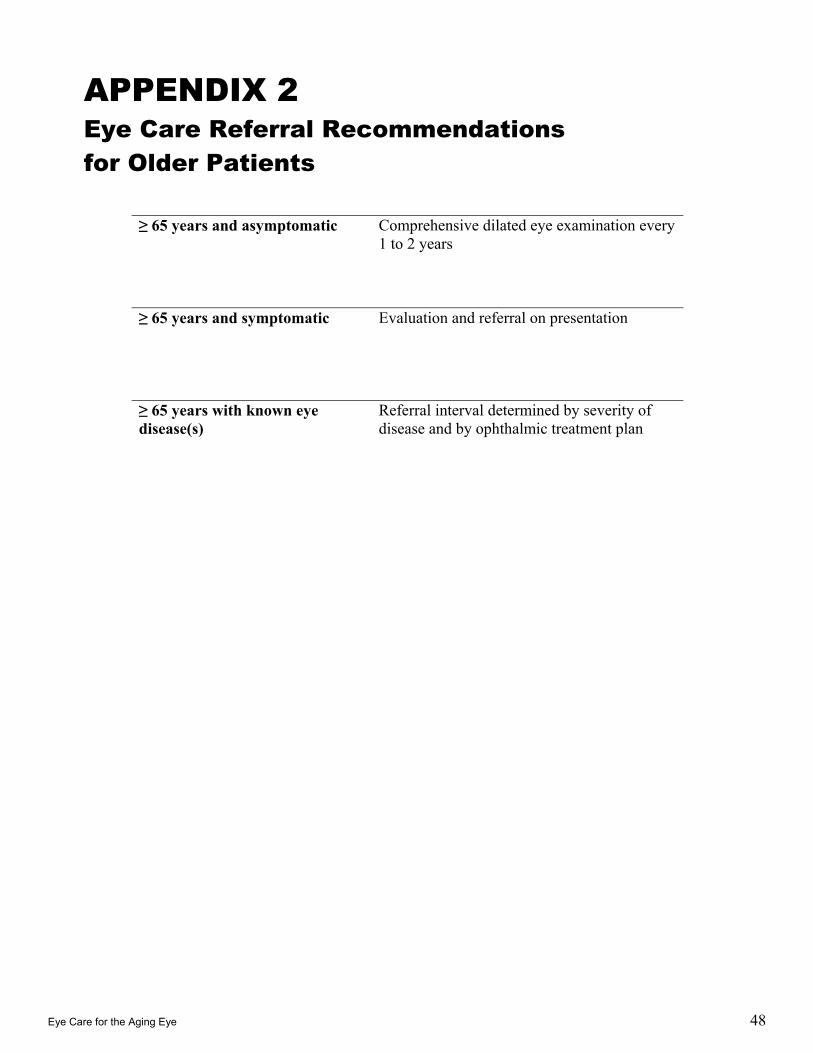
APPENDIX 2 Eye Care Referral Recommendations for Older Patients
≥ 65 years and asymptomatic Comprehensive dilated eye examination every 1 to 2 years
≥ 65 years and symptomatic Evaluation and referral on presentation
≥ 65 years with known eye disease(s)
Referral interval determined by severity of disease and by ophthalmic treatment plan
Eye Care for the Aging Eye 49
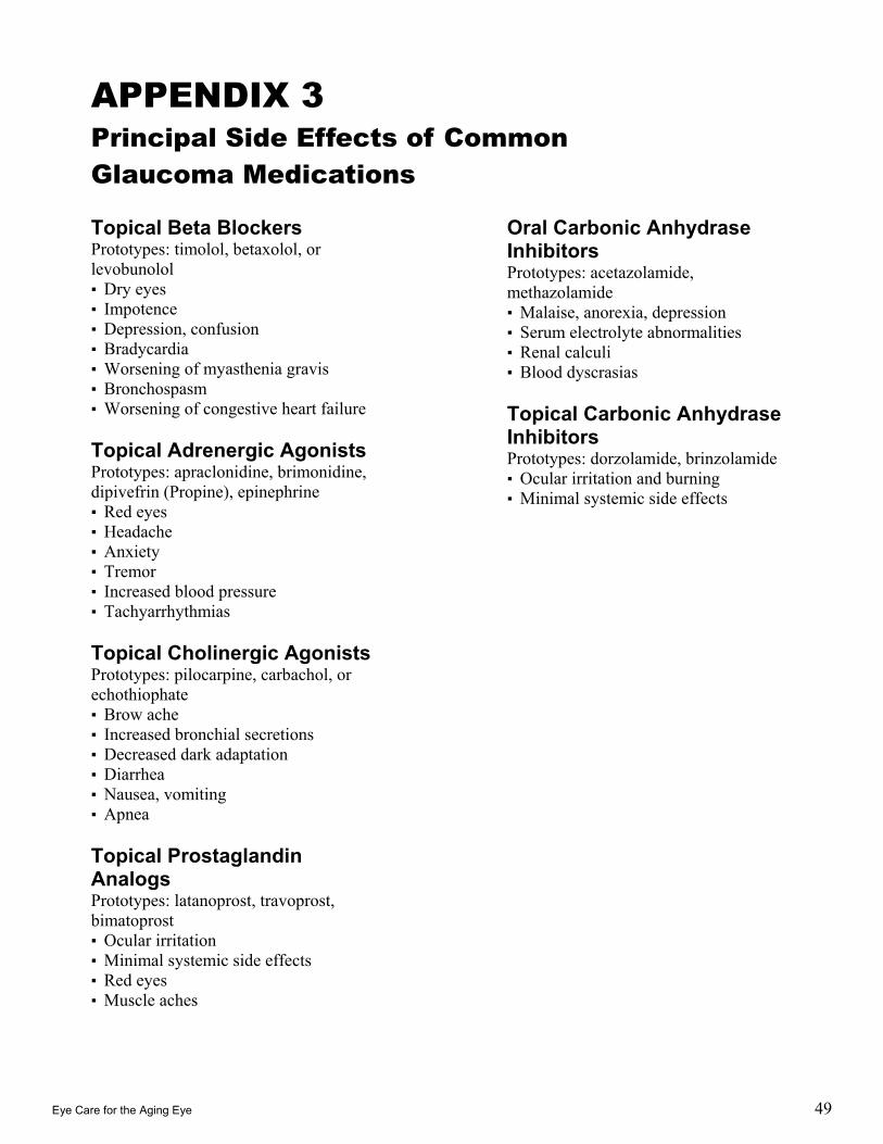
APPENDIX 3 Principal Side Effects of Common Glaucoma Medications
Topical Beta Blockers Prototypes: timolol, betaxolol, or levobunolol ▪ Dry eyes ▪ Impotence ▪ Depression, confusion ▪ Bradycardia ▪ Worsening of myasthenia gravis ▪ Bronchospasm ▪ Worsening of congestive heart failure Topical Adrenergic Agonists Prototypes: apraclonidine, brimonidine, dipivefrin (Propine), epinephrine ▪ Red eyes ▪ Headache ▪ Anxiety ▪ Tremor ▪ Increased blood pressure ▪ Tachyarrhythmias Topical Cholinergic Agonists Prototypes: pilocarpine, carbachol, or echothiophate ▪ Brow ache ▪ Increased bronchial secretions ▪ Decreased dark adaptation ▪ Diarrhea ▪ Nausea, vomiting ▪ Apnea Topical Prostaglandin Analogs Prototypes: latanoprost, travoprost, bimatoprost ▪ Ocular irritation ▪ Minimal systemic side effects ▪ Red eyes ▪ Muscle aches
Oral Carbonic Anhydrase Inhibitors Prototypes: acetazolamide, methazolamide ▪ Malaise, anorexia, depression ▪ Serum electrolyte abnormalities ▪ Renal calculi ▪ Blood dyscrasias Topical Carbonic Anhydrase Inhibitors Prototypes: dorzolamide, brinzolamide ▪ Ocular irritation and burning ▪ Minimal systemic side effects