and - Aetna Medicaid · Aetna considers the use of banked human tissue graft (e.g., Alloderm) to...
30
1 of 28 Number: 0419 Policy *Please see amendment for Pennsylvania Medicaid at the end of this CPB. I. Aetna considers orbital decompression surgery, and eye muscle surgery or eyelid surgery (e.g., marginal myotomy of levator palpebrae muscle, lateral tarsal canthoplasty, mullerectomy (resection of the Müller muscle), eyelid spacer grafts, and recession of the lower eyelid retractors) medically necessary for members with severe Graves' ophthalmopathy (especially individuals with marked proptosis and optic neuropathy) when both of the following measures have not been successful: Last Review 04/27/2017 Effective: 05/12/2000 Next Review: 04/26/2018 Review History Definitions A. A trial of conservative measures, such as elevating the head at night, cool compresses, sunglasses, lubricating eyedrops, and if the member has strabismus, prisms for glasses; and B. A trial of medications, such as diuretics, methimazole, prednisone, and propylthiouracil. Note: According to available literature, surgical treatment should not be undertaken until stability of the thyroid‐ related orbitopathy (TRO) has been demonstrated. One of the advantages of waiting for stability of TRO is that some cosmetic problems may resolve or improve without Clinical Policy Bulletin Notes
Transcript of and - Aetna Medicaid · Aetna considers the use of banked human tissue graft (e.g., Alloderm) to...

1 of 28
Number: 0419
Policy
*Please see amendment for Pennsylvania Medicaid at the
end of this CPB.
I. Aetna considers orbital decompression surgery, and eye
muscle surgery or eyelid surgery (e.g., marginal myotomy of
levator palpebrae muscle, lateral tarsal canthoplasty,
mullerectomy (resection of the Müller muscle), eyelid spacer
grafts, and recession of the lower eyelid retractors) medically
necessary for members with severe Graves' ophthalmopathy
(especially individuals with marked proptosis and optic
neuropathy) when both of the following measures have not
been successful:
Last Review 04/27/2017
Effective: 05/12/2000
Next Review: 04/26/2018
Review History
Definitions
A. A trial of conservative measures, such as elevating the head
at night, cool compresses, sunglasses, lubricating eyedrops,
and if the member has strabismus, prisms for glasses; and
B. A trial of medications, such as diuretics, methimazole,
prednisone, and propylthiouracil.
Note: According to available literature, surgical treatment
should not be undertaken until stability of the thyroid‐
related orbitopathy (TRO) has been demonstrated. One of
the advantages of waiting for stability of TRO is that some
cosmetic problems may resolve or improve without
Clinical Policy Bulletin Notes

2 of 28
intervention. Fat pad removal is commonly requested with
surgery for exophthalmos and is generally cosmetic in
nature, and therefore, is considered not medically
necessary.
II. Aetna considers orbital radiotherapy medically necessary for
the treatment of members with severe Graves'
ophthalmopathy when both of the afore‐mentioned criteria
are met.
III. Aetna considers the use of banked human tissue graft (e.g.,
Alloderm) to elevate the lower eyelids in members with lower
eyelid retraction associated with Graves ophthalmopathy
experimental and investigational because there is insufficient
evidence to support this approach.
IV. Aetna considers the following interventions experimental and
investigational for the treatment of Graves' ophthalmopathy
because their effectiveness for this indication has not been
established.
Celecoxib
Intravenous immunoglobulins
Pioglitazone
Rituximab
Somatostatin analogs (e.g., lanreotide and octreotide)
Teprotumumab
Tocilizumab
Tumor necrosis factor‐alpha inhibitors (e.g., etanercept
and infliximab).
Background
Graves' disease (also known as Parry's or Basedow's disease) is a
complex disease whose pathogenesis is believed to be
autoimmune. It is a disorder that affects mainly females, and
although it may occur at any age, has a peak incidence in the 3rd
and 4th decades. Graves' disease has 3 principal manifestations:
(i) hyperthyroidism with diffuse goiter, (ii) ophthalmopathy, and
(iii) dermopathy; however, they do not necessarily appear
simultaneously.

3 of 28
Graves' ophthalmopathy, also known as thyroid‐associated
ophthalmopathy (TAO), occurs in 2 to 7 % of patients with Graves'
disease with the major manifestations being proptosis,
ophthalmoplegia, optic neuropathy, and/or eyelid retraction.
Thyroid‐associated ophthalmopathy is the commonest cause of
proptosis in adults. The term exophthalmos is used exclusively to
describe the proptosis of TAO; exophthalmos may be unilateral
early but usually becomes bilateral with time. The term
exophthalmic ophthalmoplegia refers to the ocular muscle
weakness that results in impaired upward gaze and convergence
and strabismus with varying degree of diplopia.
Physicians recommend treatment of Graves' ophthalmopathy
according to each patient's symptoms. Sometimes combinations
of the following procedures are used:
Elevating the head at night, cool compresses, sunglasses,
lubricating eyedrops, or prisms for glasses;
Eye muscle surgery, eyelid surgery, or both;
Medications or radiation to shrink tissue;
Orbital decompression surgery.
An assessment by the National Institute for Health and Clinical
Excellence (2005) found that retrobulbar irradiation to be an
effective procedure in patients for whom other treatments are
inadequate or are associated with significant side effects.
Orbital Decompression Surgery
In orbital decompression surgery, the bone between the orbit and
the sinuses is removed. A successful procedure improves vision
and provides room for the eye to slip back into the orbit's
protection. Orbital decompression is indicated in patients with
severe ophthalmopathy refractory to medications and
radiotherapy, especially in the presence of marked proptosis and
optic neuropathy.
Eye Muscle Surgery
Diplopia often occurs because the eyes are misaligned. Usually,

4 of 28
mis‐alignment is caused by 1 or more eye muscles that are too
short or “tight” due to scar tissue from Graves' ophthalmopathy.
This scar tissue results from changes surrounding the eye because
of swelling. The goal of eye muscle surgery is to attain single
vision when looking straight ahead and looking down when
reading. During eye muscle surgery, the muscle is cut from its
attachment to the eyeball and re‐attached further back on the
eye. Usually eye muscle surgery does not require an over‐night
stay in the hospital. The physician evaluates the final results
about 2 months later. More than 1 operation is sometimes
required.
If orbital decompression and eye muscle surgery are to be
performed, the orbital decompression surgery generally is carried
out first.
Eyelid Surgery
Graves' ophthalmopathy generally causes the eyelids to open
more widely. The front surface of the eyeball becomes exposed
beyond the eyelid and causes excessive tearing and discomfort.
Lid retraction may be improved by orbital decompression,
especially the lower lid. However, the backward and downward
movement of the globe following decompression may accentuate
upper lid retraction. Surgical re‐positioning (recession) of the
upper lid retractors may have to be performed as an adjunct.
If orbital decompression, eye muscle, and eyelid surgery are
required, the eyelid procedure is generally performed as the final
operation in a series.
Acellular human dermis is being investigated for elevating the
lower eyelids in lower eyelid retraction associated with Graves
ophthalmopathy. However, current evidence in the peer‐
reviewed medical literature is limited to case reports and small,
retrospective case series.
Repositioning of the canthus may be necessary for orbital
decompression when surgery is being done for lid retraction
and lid lengthening by myotomy of the lid retractors is
5 of 28
insufficient.
Leone (1984) noted that from a series of 124 consecutive
patients, parameters were developed in the management of
Graves' disease. Patients who were stable with symptomatic
treatment were observed and followed. Oral steroids, 40 to 80
mg prednisone daily, were moderately effective in reducing
progressive soft tissue inflammatory signs, but were less effective
in controlling myopathy and lid retraction. Radiotherapy, 1,000
rad from each lateral port, was most effective in halting the
progressive inflammatory component, particularly in those who
had a recent rapid rate of change. Dysthyroid optic neuropathy
required high dose steroids; if it was not effective, decompression
of the orbit was carried out. Once the disease became stable,
myopathy, lid retraction, and exophthalmos were surgically
treated on an elective basis. The techniques utilized were
tarsorrhaphy, lateral canthoplasty, upper and lower eyelid
retractor release, and 1‐ to 3‐wall orbital decompression.
A Wikipedia review on “Graves' ophthalmopathy” (Last modified
July 20, 2015) states that “Eyelid surgery is the most common
surgery performed on Graves ophthalmopathy patients. Lid‐
lengthening surgeries can be done on upper and lower eyelid to
correct the patient’s appearance and the ocular surface exposure
symptoms. Marginal myotomy of levator palpebrae muscle can
reduce the palpebral fissure height by 2 to 3 mm. When there is
a more severe upper lid retraction or exposure keratitis, marginal
myotomy of levator palpebrae associated with lateral tarsal
canthoplasty is recommended. This procedure can lower the
upper eyelid by as much as 8 mm. Other approaches include
mullerectomy (resection of the Müller muscle), eyelid spacer
grafts, and recession of the lower eyelid retractors”.
Orbital Radiation
Zoumalan and colleagues (2007) noted that thyroid eye disease
(TED, Graves' ophthalmopathy, thyroid ophthalmopathy) is the
most common cause of orbital inflammation and proptosis in
adults. There is no agreement on its management although
corticosteroids and external beam orbital radiation have

6 of 28
traditionally been believed to provide benefit in active
inflammation. A review of the published literature in English
disclosed an overall corticosteroid‐mediated treatment response
of 66.9 % in a total of 834 treated patients who had moderate or
severe TED. Intravenous (IV) corticosteroids used in repeated
weekly pulses were more effective (overall favorable response =
74.6 %, n = 177) and had fewer side effects than daily oral
corticosteroids (overall favorable response = 55.5 %, n = 265). A
combination of corticosteroid and radiation therapy seemed to be
more effective than corticosteroids alone. However, the authors
stated that these conclusions are tempered by a notable lack of
standardization within and between study designs, treatment
protocols, and outcome measures. Accordingly, the North
American Neuro‐Ophthalmology Society, American Society of
Ophthalmic Plastic and Reconstructive Surgery and the Orbital
Society, in conjunction with Neuro‐Ophthalmology Research and
Development Consortium, will investigate the design and funding
of a multi‐center controlled trial.
A technology assessment on orbital radiation for Graves
ophthalmopathy by the American Academy of Ophthalmology
(Bradley et al, 2008) examined if orbital radiation offers effective
and safe treatment for Graves ophthalmopathy. Medical
literature databases were searched to identify all published
reports relating to orbital radiation treatment for Graves
ophthalmopathy. To be included in the technology assessment,
reports had to provide original data, to report on a case series or
uncontrolled trial of at least 100 subjects or a randomized clinical
trial (RCT) of any size, to focus on orbital radiation for the
treatment of Graves ophthalmopathy, and to follow‐up patients
for at least 3 months. Abstracted data included study
characteristics, patient characteristics, treatment response, and
safety information. A total of 14 studies were included in the
technology assessment: 5 observational studies and 9 RCTs.
Three of the observational studies reported on treatment
response, with overall favorable outcomes for 40 % to 97 % of
patients. Three of the observational studies provided
intermediate‐term safety data. The risk of definite radiation
retinopathy is 1 % to 2 % within 10 years after treatment.
Patients treated with orbital radiation did not have an increased
7 of 28
risk of secondary malignancy or premature death. The 9 RCTs
were qualitatively heterogeneous. Patients with optic
neuropathy generally were excluded from participating in the
RCTs. Three of the RCTs were sham‐controlled. None of these
studies showed that orbital radiation was more effective than
sham irradiation for improving proptosis, lid fissure, or soft tissue
changes such as eyelid swelling. Two of the 3 sham‐controlled
RCTs demonstrated improved vertical range of motion in
radiation‐treated subjects compared with controls. The authors
concluded that systematic review of the effect of orbital radiation
on Graves ophthalmopathy is limited by the lack of
standardization and variable quality of published reports. Extra‐
ocular motility impairment may improve with radiotherapy,
although the evidence of a treatment effect is mixed in clinical
trials. Future studies are needed to determine if a potentially
beneficial motility effect results in improved patient function and
quality of life. Level I evidence indicates that proptosis, eyelid
retraction, and soft tissue changes do not improve with radiation
treatment. The effectiveness of orbital radiation for compressive
optic neuropathy resulting from Graves ophthalmopathy has not
been investigated in clinical trials and merits further study.
Radiation retinopathy, although rare, is a risk of orbital radiation,
even in patients without diabetes who receive appropriate
radiation dose and delivery.
Guidance on retrobulbar irradiation for thyroid eye disease from
the National Institute for Health and Clincial Excellence (NICE,
2005) concluded: "Current evidence on the safety and efficacy of
retrobulbar irradiation for thyroid eye disease appears adequate
to support the use of this procedure in patients for whom other
treatments are inadequate or associated with significant side
effects."
Other Treatments
Bartalena and Tanda (2009) noted that RCT have not shown a
benefit of somatostatin analogs (e.g., lanreotide and octreotide)
for Graves' ophthalmopathy. They stated that there are also few
data to support the use of intravenous immune globulin for this
condition. This is in agreement with the consensus statement of

8 of 28
the European Group on Graves' orbitopathy on the management
of Graves' orbitopathy (Bartalena et al, 2008), which stated that
treatments of marginal or unproven value include somatostatin
analogs and intravenous immunoglobulins.
In a systematic review and meta‐analysis on treatment modalities
for Graves' ophthalmopathy, Stiebel‐Kalish et al (2009) concluded
that current evidence demonstrates the effectiveness
of intravenous corticosteroids in decreasing CAS in patients with
moderate‐to‐severe Graves' ophthalmopathy. Intravenous pulse
corticosteroids therapy has a small but statistically significant
advantage oral therapy and causes significantly fewer adverse
events. Somatostatin analogs have marginal clinical efficacy. The
efficacy of orbital radiotherapy as single therapy remains unclear,
whereas the combination of radiotherapy with corticosteroids has
better efficacy than either radiotherapy or oral corticosteroids
alone. Rituximab is not listed as a therapeutic option.
Furthermore, Hegedus (2009) stated that no data as yet support
the routine use of biological therapies (e.g., rituximab). The
author stated that prospective, randomized trials
comparing available and any novel therapeutic options
for Graves' disease are needed.
Bartalena et al (2010) stated that non‐surgical treatments for
moderate to severe and active Graves' orbitopathy (systemic
glucocorticoids with or without orbital radiotherapy) have limited
effects on the underlying autoimmune process causing the
disease. Although the clinical responses to treatment are often
good, at least one‐third of patients with Graves' orbitopathy are
eventually dissatisfied with the treatment outcome. Advent
in the understanding of the autoimmune basis of Graves'
orbitopathy (although still incomplete) made it possible, similar
to other autoimmune disorders, to envision the use of novel
immunomodulating drugs. Among the currently available
biologic agents, the CD20+ B cell‐depleting agent, rituximab, and
tumor necrosis factor‐alpha inhibitors (e.g., etanercept and
infliximab) are presently the drugs that have the best chance of
being employed in the future for the treatment of Graves'
orbitopathy. However, the authors noted that RCTs to support
their use are needed.

9 of 28
Viani et al (2012) evaluated the effectiveness of radiotherapy (RT)
with total dose of 20 Gy (RT 20 Gy) in the treatment of Graves'
ophthalmopathy. A systematic review and meta‐analysis of RCTs
was performed comparing RT 20 Gy with or without
glucocorticoid to clinical treatments for Graves' ophthalmopathy.
The MEDLINE, EMBASE, Cochrane Library databases and recent
relevant journals were searched. Relevant reports were reviewed
by 2 reviewers. Response to radiotherapy was defined as clinical
success according to each trial. These investigators also evaluated
the quality of life and whether RT to produce fewer side
effects than other treatments. A total of 8 RCTs (439
patients) were identified. In the subgroup analysis, the overall
response to treatment rates was better for: RT 20 Gy plus
glucocorticoid versus glucocorticoids alone, OR = 17.5 (95 %
confidence interval [CI]: 1.85 to 250, p = 0.04), RT 20 Gy versus
sham RT, OR = 3.15 (95 % CI: 1.59 to 6.23, p = 0.003) and RT 20Gy
plus intravenous glucocorticoid versus RT 20Gy plus oral
glucocorticoid, OR = 4.15(95 % CI: 1.34 to 12.87, p = 0.01). There
were no differences between RT 20 Gy versus other fractionations
and RT 20 Gy versus glucocorticoid alone. Radiotherapy 20 Gy
with or without glucocorticoids showed an improvement in
diplopia grade, visual acuity, optic neuropathy, lid width,
proptosis and ocular motility. No difference was seen for costs,
intra‐ocular pressure and quality of life. The authors concluded
that these findings showed that RT 20 Gy should be offered as a
valid therapeutic option to patients with moderate‐to‐severe
ophthalmopathy. The effectiveness of orbital radiotherapy can be
increased by the synergistic interaction with glucocorticoids.
Moreover, RT 20 Gy is useful to improve a lot of ocular
symptoms, excluding intra‐ocular pressure, without any
difference in quality of life and costs.
Tanda and Bartalena (2012) examined the safety and
effectiveness of orbital radiotherapy (OR) for graves' orbitopathy
(GO). The major source of data acquisition included PubMed
strategies. Original articles, systemic reviews and meta‐analyses,
and other relevant citations were screened. Randomized clinical
trials evaluating the effectiveness of OR are limited. However,
available data suggest that OR is a safe treatment, which seems
to be effective particularly on ocular motility impairment,

10 of 28
especially if it is of recent onset. Orbital radiotherapy seems to
be effective also on soft tissue changes, whereas exophthalmos
and long‐standing extra‐ocular muscle dysfunction are poorly
affected. The effectiveness of OR on dysthyroid optic neuropathy
is uncertain. The combination of OR and oral glucocorticoids
(GCs) is more effective than either treatment alone, suggesting a
synergistic effect of the 2 treatments. There is no available
evidence that the addition of OR to intravenous GCs provides an
advantage over intravenous GCs alone. The authors concluded
that OR can be considered a safe second‐line treatment for
patients with moderate‐to‐severe and active GO but less effective
than GCs. A possible strategy may include its use in combination
with intravenous GCs in patients whose GO has only partially
responded to a first‐course of intravenous GCs alone and is still
active.
Melcescu et al (2014) noted that GO often remains a major
diagnostic and therapeutic challenge. It has become increasingly
important to classify patients into categories based on disease
activity at initial presentation. A Hertel exophthalmometer
measurement of greater than 2 mm above normal for race
usually categorizes a patient as having moderate‐to‐severe GO.
Encouraging smoking cessation and achieving euthyroidism in the
individual patient are important. Simple treatment measures
such as lubricants for lid retraction, nocturnal ointments for
incomplete eye closure, prisms in diplopia, or botulinum toxin
injections for upper‐lid retraction can be effective in mild cases of
GO. Glucocorticoids, orbital radiotherapy, and
decompression/rehabilitative surgery are generally indicated for
moderate‐to‐severe GO and for sight‐threatening optic
neuropathy. Future therapies, including rituximab aimed at
treating the molecular and immunological basis of GO, are under
investigation and hold promise for the future.
Salvi (2014) noted that in recent years, immunosuppressive
therapy, as an alternative to corticosteroids, has been proposed
as novel agents that target the various antigens involved in the
pathogenesis of Graves' ophthalmopathy. Although the lack of
randomized and controlled studies suggests caution in
generalizing results, some data show interesting results. Potential

11 of 28
targets for immune therapy in Graves' ophthalmopathy are the
antigens expressed on the target organ of inflammation, namely
the receptor and the insulin growth factor 1 (IGF‐1) receptor on
fibroblasts, inflammatory cytokines, and B and T cells. The most
promising results are observed with small thyroid stimulating
hormone receptor molecules interacting with the receptor on
thyrocytes and fibroblasts and with the anti‐IGF‐1 receptor
antibody teprotumumab. A recent open study with tocilizumab,
an anti‐soluble interleukin‐6 receptor, has shown inactivation of
Graves' ophthalmopathy. Consistent reports on the efficacy of
rituximab will have to be confirmed by RCTs, which are now in
progress. The author concluded that current clinical practice for
Graves' ophthalmopathy will greatly benefit from the availability
of immunosuppressors that act as disease‐modifying drugs, as
compared to steroids, the current standard treatment for Graves'
ophthalmopathy. Rituximab seems to be a good candidate, as
preliminary results from ongoing randomized trials suggest good
efficacy with a relative well‐tolerated profile.
In a prospective, interventional, non‐randomized study, Perez‐
Moreiras et al (2014) examined the effectiveness of tocilizumab in
thyroid eye disease patients who were refractory to multiple
intravenous steroids. This study enrolled active GO (defined by
CAS greater than or equal to 4) patients resistant to previous
intravenous steroids treated with tocilizumab. Snellen visual
acuity, Hertel exophthalmometry, CAS evaluation, TSI levels,
ocular motility, and side effects were registered at a 4‐week
interval. A total of 18 patients were included with a mean age of
47.9 ± 8.63 years. All patients had a significant progressive CAS
improvement (mean CAS score reduction 5.89 ± 1.41 points, p <
0.00027). Mean TSI levels were significantly lower at the end of
the treatment (mean of ‐76.18 % ± 17.80 %, p = 0.00007).
Thirteen patients (72.22 %) reduced proptosis a mean of ‐3.92 ±
1.54 mm (p = 0.002); 15 patients (83.33 %) had an improvement
in extra‐ocular motility, and 7 patients of 13 resolved their
diplopia (53.85 %). No severe side effects or relapse of active GO
were observed at the end of follow‐up. The authors concluded
that the findings of this study suggested that intravenous
tocilizumab may be effective on reducing activity in patients with
thyroid eye disease refractory to intravenous steroids. These
12 of 28
preliminary findings need to be validated by well‐designed
studies.
Radioiodine Therapy:
Ren et al (2015) integrated the evidence to provide hierarchies of
the comparative effectiveness of 4 treatments (radioiodine,
radioiodine+prednisone, anti‐thyroid drugs and surgery). These
researchers conducted a Bayesian‐framework network meta‐
analysis of RCTs to compare 4 treatments in patients with Graves'
disease. The eligible RCTs were identified by searching Amed, the
British Nursing Index, Embase, PubMed, the Cochrane Central
Register of Controlled Trials (CENTRAL), Google scholar, SIGLE, the
National Technical Information Service, the National Research
Register (UK) and the Current Controlled Trials databases. The
data for 2 outcomes (e.g., ophthalmopathy and recurrence) were
independently extracted by 2 authors. A total of 4 RCTs were
ultimately included. Radioiodine+prednisone therapy showed
statistical significance in reducing the incidence of new or
deteriorative ophthalmopathy comparing with the other 3
therapies. Compared with radioiodine, therapy with anti‐thyroid
drugs therapy as well as surgery significantly decreased the
incidence of new or deteriorative ophthalmopathy. Radioiodine
therapy significantly reduced the rate of recurrence when
compared to therapy with anti‐thyroid drugs or surgery. For
decreasing the incidence of new or deteriorative
ophthalmopathy, the 4 treatments were ranked as follows:
radioiodine+prednisone therapy, therapy with anti‐thyroid drugs,
surgery and radioiodine therapy. For reducing the rate of
recurrence, 3 treatments were ranked as follows: radioiodine
therapy, therapy with anti‐thyroid drugs and surgery. The authors
concluded that radioiodine+prednisone therapy might have the
least probability of leading to an exacerbation or new appearance
of ophthalmopathy, and radioiodine therapy might have the least
probability of causing a recurrence.
In a Cochrane review, Ma and colleagues (2016) evaluated the
effects of radioiodine therapy versus anti‐thyroid medications for
Graves' disease. These researchers performed a systematic
literature search in the Cochrane Library, MEDLINE and EMBASE
13 of 28
and the trials registers ICTRP Search Portal and ClinicalTrials.gov.
The date of the last search was September 2015 for all
databases. Randomized controlled trials comparing the effects of
radioiodine therapy versus anti‐thyroid medications for Graves'
disease with at least 2 years follow‐up were selected for analysis.
Two authors independently screened titles and abstracts for
relevance. One author carried out screening for inclusion, data
extraction and “Risk of bias” assessment and a second author
checked this. They presented data not suitable for meta‐analysis
as descriptive data, and analyzed the overall quality of evidence
utilizing the GRADE instrument. These investigators included 2
RCTs involving 425 adult participants with Graves' disease in this
review. Altogether 204 participants were randomized to
radioiodine therapy and 221 to methimazole therapy. A single
dose of radioiodine was administered. The duration of
methimazole medication was 18 months. The period of follow‐up
was at least 2 years, depending on the outcome measured. For
most outcome measures risk of bias was low; for the outcomes
health‐related quality of life as well as development and
worsening of Graves' ophthalmopathy risks of performance bias
and detection bias were high in at least 1 of the 2 RCTs. Health‐
related quality of life appeared to be similar in the radioiodine
and methimazole treatment groups, however no quantitative
data were reported (425 participants; 2 trials; low quality
evidence). The development and worsening of Graves'
ophthalmopathy was observed in 76 of 202 radioiodine‐treated
participants (38 %) and in 40 of 215 methimazole‐treated
participants (19 %): risk ratio (RR) 1.94 (95 % CI: 1.40 to 2.70); 417
participants; 2 trials; low quality evidence. A total of 35 % to 56
% of radioiodine‐treated participants and 42 % of participants
treated with methimazole were smokers, which is associated with
the risk of worsening or development of Graves' ophthalmopathy.
Euthyroidism was not achieved by any participant being treated
with radioiodine compared with 64/68 (94 %) of participants after
methimazole treatment (112 participants; 1 trial). In this trial
thyroxine therapy was not introduced early in both treatment
arms to avoid hypothyroidism. Recurrence of hyperthyroidism
(relapse) in favor of radioiodine treatment showed a RR of 0.20
(95 % CI: 0.01 to 2.66); p = 0.22; 417 participants; 2 trials; very
low quality evidence. Heterogeneity was high (I² = 91 %) and the

14 of 28
RRs were 0.61 or 0.06 with non‐overlapping CIs. Adverse events
other than development of worsening of Graves' ophthalmopathy
for radioiodine therapy were hypothyroidism (39 of 41
participants (95 %) compared with 0 % of participants receiving
methimazole, however thyroxine treatment to avoid
hypothyroidism was not introduced early in the radioiodine group
‐‐ 104 participants; 1 trial; very low quality evidence) and drug‐
related adverse events for methimazole treatment (23 of 215
participants (11 %) reported adverse effects likely related to
methimazole therapy ‐‐ 215 participants; 2 trials; very low quality
evidence). The outcome measures all‐cause mortality and bone
mineral density were not reported in the included trials. One trial
(174 participants) reported socio‐economic effects: costs based
on the official hospital reimbursement system in Sweden for
patients without relapse and methimazole treatment were US$
1,126/1,164 (young/older methimazole group) and for
radioiodine treatment US$ 1,862. Costs for patients with relapse
and methimazole treatment were US$ 2,284/1,972 (young/older
methimazole group) and for radioiodine treatment US$ 2,760.
The authors concluded that the only anti‐thyroid drug
investigated in the 2 included trials was methimazole, which
might limit the applicability of these findings with regard to other
compounds such as propylthiouracil. Results from 2 RCTs
suggested that radioiodine treatment is associated with an
increased risk of Graves' ophthalmopathy. They noted that these
findings suggested some benefit from radioiodine treatment for
recurrence of hyperthyroidism (relapse) but there is uncertainty
about the magnitude of the effect size.
Rituximab:
In a retrospective, interventional case series, Khanna and
colleagues (2010) examined the effectiveness of rituximab in
patients with severe, corticosteroid (CS)‐resistant TAO. Responses
to rituximab therapy were graded using standard clinical
assessment and flow cytometric analysis of peripheral
lymphocytes. Main outcome measures were clinical activity score
(CAS), proptosis, strabismus, treatment side effects, and
quantification of regulatory T cells; 6 patients were studied.
Systemic CS failed to alter clinical activity in all patients (mean
15 of 28
CAS +/‐ standard deviation, 5.3 +/‐ 1.0 before versus 5.5 +/‐ 0.8
during therapy for 7.5 +/‐ 6.4 months; p = 1.0). However, after
rituximab therapy, CAS improved from 5.5 +/ ‐0.8 to 1.3 +/‐ 0.5 at
2 months after treatment (p < 0.03) and remained quiescent in all
patients (CAS, 0.7 +/‐ 0.8; p < 0.0001) at a mean follow‐up of 6.2
+/‐ 4.5 months. Vision improved bilaterally in all 4 patients with
dysthyroid optic neuropathy (DON). None of the 6 patients
experienced disease relapse after rituximab infusion, and
proptosis remained stable (Hertel measurement, 24 +/‐ 3.7 mm
before therapy and 23.6 +/‐ 3.7 mm after therapy; p = 0.17). The
abundance of T regulatory cells, assessed in 1 patient, increased
within 1 week of rituximab and remained elevated at 18 months
of follow‐up. The authors concluded that in progressive, CS‐
resistant TAO, rapid and sustained resolution of orbital
inflammation and DON followed treatment with rituximab.
In a Cochrane review, Minakaran and Ezra (2013) examined the
effectiveness and safety of rituximab for the treatment of TAO.
These investigators searched CENTRAL (which contains the
Cochrane Eyes and Vision Group Trials Register) (the Cochrane
Library 2013, Issue 3), Ovid MEDLINE, Ovid MEDLINE In‐Process
and Other Non‐Indexed Citations, Ovid MEDLINE Daily, Ovid
OLDMEDLINE, (January 1950 to April 2013), EMBASE (January
1980 to April 2013), Latin American and Caribbean Literature on
Health Sciences (LILACS) (January 1982 to April 2013), OpenGrey
(System for Information on Grey Literature in Europe)
(www.opengrey.eu/), the metaRegister of Controlled Trials
(mRCT) (www.controlled‐trials.com), ClinicalTrials.gov
(www.clinicaltrials.gov), the WHO International Clinical Trials
Registry Platform (ICTRP) (www.who.int/ictrp/search/en) and the
EU Clinical Trials Register (www.clinicaltrialsregister.eu). They did
not use any date or language restrictions in the electronic
searches for trials. They last searched the electronic databases
on April 15, 2013. These researchers manually searched
references of review articles and used the Science Citation Index
to identify additional studies citing trials. They contacted the lead
investigators of relevant trials on ClinicalTrials.gov and the WHO
ICTRP for information and data from as yet unpublished clinical
trials. They contacted experts in the field for information about
any ongoing trials; and contacted the manufacturers of rituximab

16 of 28
for details of any sponsored trials. These researchers included
RCTs of rituximab treatment by intravenous infusion for the
treatment of patients with TAO, compared with placebo or
intravenous glucocorticoid treatment. Two review authors
independently scanned titles and abstracts, as well as
independently screened the full reports of the potentially
relevant studies. At each stage, the results were compared and
disagreements were solved by discussion. No studies were
identified that met the inclusion criteria. There are 3 ongoing
studies that are likely to meet inclusion criteria once published,
and thus be included in future updates of this review. The
authors concluded that there is currently insufficient evidence to
support the use of rituximab in patients with TAO. There is a
need for large RCTs, investigating rituximab versus placebo or
corticosteroids in patients with active TAO to make adequate
judgment on the safety and effectiveness of this novel therapy for
this condition.
An UpToDate review on “Treatment of Graves' orbitopathy
(ophthalmopathy)” (Davies, 2015) states that “Rituximab ‐‐ A
number of reports have indicated that some patients with severe
Graves’ orbitopathy may respond dramatically to B cell depletion
induced by rituximab, which is a monoclonal antibody directed
against the B cell CD20 molecule. Rituximab induces a fall in TSH
receptor antibody levels and depletion of B cells in the retro‐
orbital tissues, not just the periphery. Although high doses of this
antibody may be associated with severe side effects from the
profound immunosuppression that ensues, it is likely that much
lower doses may be effective in Graves’ orbitopathy and allow
such effects to be avoided. This approach to the treatment of
severe eye disease is currently undergoing larger trials;
preliminary results from these trials have been mixed and are not
yet published …. Somatostatin analogs ‐‐ Somatostatin analogs
have been explored as a potential therapy for Graves'
ophthalmopathy, based upon the observations that orbital
fibroblasts have somatostatin receptors and the activity of
orbitopathy correlates with activity on octreotide scintigrams.
One randomized, placebo‐controlled trial of a long‐acting
octreotide preparation reported improvement in clinical activity
scores and median lid fissure width with octreotide compared

17 of 28
with placebo. In contrast, two other similar trials reported
limited benefit with octreotide. In a meta‐analysis of four trials,
somatostatin analogs resulted in a slightly lower clinical activity
score than placebo, but had no advantages for other important
outcomes (diplopia, proptosis, lid aperture). Octreotide has no
role in the routine treatment of Graves' ophthalmopathy”.
Furthermore, this review does not mention tocilizumab as a
therapeutic option.
Ladsous (2016) stated that the use of rituximab in GO is appealing
but its exact role in the therapeutic arsenal remains to be
clarified, and its safety profile also needs to be confirmed on a
larger scale.
Celecoxib and Pioglitazone:
Cheng and colleagues (2016) examined the role of extra‐ocular
muscles (EOM) myoblasts in GO pathology and the effect of a
cyclooxygenase (COX)‐2 inhibitor and a peroxisome proliferator‐
activated receptor (PPAR)‐γ agonist in its treatment. Myoblasts
were isolated and cultured from EOM of 10 patients with GO and
4 without (non‐GO). The cultured myoblasts were treated with
interferon‐gamma (IFN‐γ), insulin‐like growth factor (IGF)‐1,
interleukin (IL)‐1β, and tumor‐necrosis‐factor‐alpha (TNF‐α), and
the effect on PPAR‐γ, COX‐2, TGF‐β, and thyroid stimulating
hormone receptor (TSHR) expressions were assessed using real‐
time polymerase chain reaction (RT‐PCR), enzyme‐linked
immunosorbent assay (ELISA), and Western blot. The effect of a
COX‐2 inhibitor and a PPAR‐γ agonist on the expression of TGF‐β,
hyaluronan synthases (HAS)‐1, ‐2, and ‐3, and hyaluronan (HA)
were further evaluated. Real‐time PCR showed significant up‐
regulation in PPAR‐γ, COX‐2, TGF‐β, and TSHR messenger RNA
(mRNA) expression in GO myoblasts when treated with TNF‐α but
not in the non‐GO. While IFN‐γ and IGF‐1 had no significant
effect, IL‐1β did up‐regulate COX‐2 expression. These results
were further confirmed by ELISA and Western blotting. Tu mor
necrosis factor α‐induced TGF‐β in turn significantly increased HA
expression and HAS3 level, but not HAS1 and HAS2. The
cyclooxygenase 2 inhibitor and PPAR‐γ agonist substantially
diminished this TNF‐α‐induced TGF‐β, HA, and HAS3 expression.

18 of 28
The authors concluded that these findings showed the role of
EOM myoblasts in the pathogenesis of GO. They stated that the
cyclooxygenase 2 inhibitor and PPAR‐γ agonist in this study are
potential treatments for GO due to their ability to suppress TNF‐
α‐induced TGF‐β, HAS, and HA up‐regulation.
Teprotumumab:
Smith and Janssen (2017) noted that the pathogenesis of orbital
Graves' disease (GD), a process known as TA O, remains
incompletely understood. The TSHR represents the central
autoantigen involved in GD and has been proposed as the thyroid
antigen shared with the orbit that could explain the infiltration of
immune cells into tissues surrounding the eye. Another cell
surface protein, IGF‐I receptor (IGF‐IR), has recently been
proposed as a second antigen that participates in TA O by virtue of
its interactions with anti‐IGF‐IR antibodies generated in GD, its
apparent physical and functional complex formation with TSHR,
and its necessary involvement in TSHR post‐receptor signaling.
The proposal that IGF‐IR is involved in TA O has provoked
substantial debate. Furthermore, several studies from different
laboratory groups, each using different experimental models,
have yielded conflicting results. These researchers summarized
the biological characteristics of IGF‐IR and TSHR. They also
reviewed the evidence supporting and refuting the postulate that
IGF‐IR is a self‐antigen in GD and that it plays a potentially
important role in TA O. The putative involvement of IGF‐IR in
disease pathogenesis carries substantial clinical implications.
Specifically, blocking this receptor with monoclonal antibodies
can dramatically attenuate the induction by TSH and pathogenic
antibodies generated in GD of pro‐inflammatory genes in cultured
orbital fibroblasts and fibrocytes. These cell types appear critical
to the development of TA O. The authors stated that these
observations have led to the conduct of a now‐completed multi‐
center therapeutic trial of a fully human monoclonal anti‐IGF‐IR
blocking antibody (teprotumumab or RV001) in moderate‐to‐
severe, active TA O.
Wiersinga (2017) stated that corticosteroids have been the
mainstay of treatment for GO, but new evidence about immune

19 of 28
mechanisms has provided a basis to explore other drug classes; IV
methylprednisolone pulses are more effective and better
tolerated than oral prednisone in the treatment of active,
moderate‐to‐severe GO. Rituximab has also been suggested as a
possible replacement for IV corticosteroids. Two RCTs of
rituximab reached seemingly contradictory conclusions ‐‐
rituximab was not better with respect to the primary outcome
(clinical activity score) than placebo in 1 trial (which, however,
was confounded by rather long GO duration), but was slightly
better than IV methylprednisolone pulses in the other (disease
flare‐ups occurred only in the latter group). The author stated
that on the basis of evidence published so far, rituximab cannot
replace IV methylprednisolone pulses, but could have a role in
corticosteroid‐resistant cases. Open‐label studies of TNF‐α
blockade had limited efficacy, but other studies showed that
interleukin (IL)‐6 receptor antibodies were effective. Results of
RCTs investigating the effectiveness of the IGF‐1 receptor
antibody teprotumumab and the IL‐6 receptor antibody
tocilizumab are expected shortly. Approaches that target the
causal mechanism of GO (antibodies or antagonists that block
TSHRs) also look promising.
Smith (2017) reviewed the fundamental characteristics of the
TSHR, its role in GD and TA O, and its relationship to IGF‐IR. Strong
evidence supports the concept that the 2 receptors form a
physical and functional complex and that IGF‐IR activity is needed
for some of the down‐stream signaling initiated through TSHR.
The author also reviewed recently developed small molecules
and monoclonal antibodies that block TSHR and IGF‐IR signaling
in the narrow context of their potential utility as therapeutics in
GD and TA O. The PubMed database was searched from its
inception for relevant publications. The author concluded that
agents that can interrupt the TSHR and IGF‐IR pathways possess
the potential for offering more specific and better tolerated
treatments of both hyperthyroidism and TA O.
CPT Codes / HCPCS Codes / ICD‐10 Codes

20 of 28
Information in the [brackets] below has been added for clarification
purposes. Codes requiring a 7th character are represented by "+":
CPT codes covered if selection criteria are met:
15820 Blepharoplasty, lower eyelid
15822 Blepharoplasty, upper eyelid
61330 Decompression of orbit only, transcranial approach
67311 ‐
67343
Strabismus surgery
67414 Orbitotomy without bone flap (frontal or
transconjunctival approach); with removal of bone for
decompression
67445 Orbitotomy with bone flap or window, lateral
approach (e.g., Kroenlein); with removal of bone for
decompression
67901 ‐
67908
Repair of blepharoptosis
67950 Cathoplasty [lateral tarsal canthoplasty]
67961 ‐ Excision and repair of eyelid, involving lid margin,
67966 tarsus, conjunctiva, canthus, or full thickness, may
include preparation for skin graft or pedicle flap with
adjacent tissue transfer or rearrangement
[mullerectomy (resection of the Müller muscle), eyelid
spacer grafts]
67909 Reduction of overcorrection of ptosis
67911 Correction of lid retraction
77789 Surface application of low dose rate radionuclide
source
CPT codes not covered for indications listed in the CPB:
15821 Blepharoplasty, lower eyelid; with extensive herniated
fat pad
15823 Blepharoplasty, upper eyelid; excessive skin weighting
down lid
90281 Immune globulin (Ig), human, for intramuscular use
90283 Immune globulin (IgIV), human, for intravenous use
21 of 28
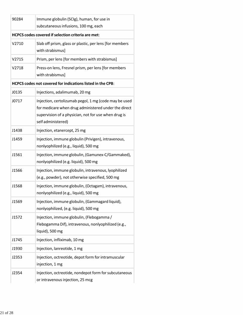
90284 Immune globulin (SCIg), human, for use in
subcutaneous infusions, 100 mg, each
HCPCS codes covered if selection criteria are met:
V2710 Slab off prism, glass or plastic, per lens [for members
with strabismus]
V2715 Prism, per lens [for members with strabismus]
V2718 Press‐on lens, Fresnel prism, per lens [for members
with strabismus]
HCPCS codes not covered for indications listed in the CPB:
J0135 Injections, adalimumab, 20 mg
J0717 Injection, certolizumab pegol, 1 mg (code may be used
for medicare when drug administered under the direct
supervision of a physician, not for use when drug is
self administered)
J1438 Injection, etanercept, 25 mg
J1459 Injection, immune globulin (Privigen), intravenous,
nonlyophilized (e.g., liquid), 500 mg
J1561 Injection, immune globulin, (Gamunex‐C/Gammaked),
nonlyophilized (e.g. liquid), 500 mg
J1566 Injection, immune globulin, intravenous, lyophilized
(e.g., powder), not otherwise specified, 500 mg
J1568 Injection, immune globulin, (Octagam), intravenous,
nonlyophilized (e.g., liquid), 500 mg
J1569 Injection, immune globulin, (Gammagard liquid),
nonlyophilized, (e.g. liquid), 500 mg
J1572 Injection, immune globulin, (Flebogamma /
Flebogamma Dif), intravenous, nonlyophilized (e.g.,
liquid), 500 mg
J1745 Injection, infliximab, 10 mg
J1930 Injection, lanreotide, 1 mg
J2353 Injection, octreotide, depot form for intramuscular
injection, 1 mg
J2354 Injection, octreotide, nondepot form for subcutaneous
or intravenous injection, 25 mcg

22 of 28
J2792 Injection, Rho D immune globulin, intravenous,
human, solvent detergent, 100 IU
J3262 Injection, tocilizumab, 1 mg
J9310 Injection, rituximab, 100 mg
S9338 Home infusion therapy, immunotherapy,
administrative services, professional pharmacy
services, care coordination, and all necessary supplies
and equipment (drugs and nursing visits coded
separately), per diem
Other HCPCS codes related to the CPB:
S9338 Home infusion therapy, immunotherapy, adminstrative
services, professional pharmacy services, care
coordination, and all necessary supplies and
equipment (drugs and nursing visits coded separately),
per diem
S9359 Home infusion therapy, anti‐tumor necrosis factor
intravenous therapy, (e.g., Infliximab); administrative
services, professional pharmacy services, care
coordination, and all necessary supplies and
equipment (drug and nursing visits coded separately),
per diem
ICD‐10 codes covered if selection criteria are met:
E05.00 ‐
E05.01
Thyrotoxicosis with diffuse goiter [with/without
thyrotoxic crisis or storm]
H05.89 Other disorders of orbit [thyrotoxic exophtalmos,
exophthalmic ophthalmoplegia]
The above policy is based on the following references:
1. Bartalena L, Pinchera A, Marcocci C. Management of
Graves' ophthalmopathy: Reality and perspectives. Endocr
Rev. 2000;21(2):168‐199.
2. Pliego‐Maldonado A, Miranda‐Ruiz R, Vargas‐Aguayo A, et
al. Orbit decompression surgery in patients with
exophthalmos caused by Graves‐Basedow disease. Gac
Med Mex. 2000;136(1):11‐15.
23 of 28
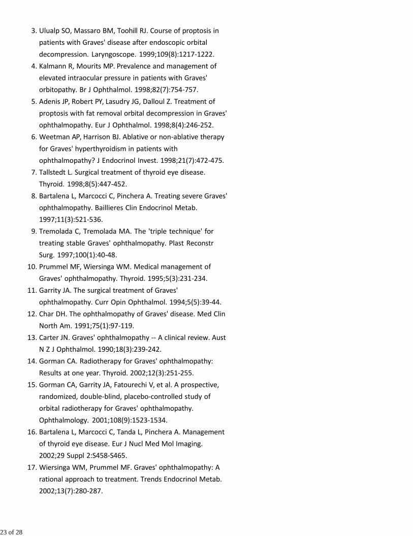
3. Ulualp SO, Massaro BM, Toohill RJ. Course of proptosis in
patients with Graves' disease after endoscopic orbital
decompression. Laryngoscope. 1999;109(8):1217‐1222.
4. Kalmann R, Mourits MP. Prevalence and management of
elevated intraocular pressure in patients with Graves'
orbitopathy. Br J Ophthalmol. 1998;82(7):754‐757.
5. Adenis JP, Robert PY, Lasudry JG, Dalloul Z. Treatment of
proptosis with fat removal orbital decompression in Graves'
ophthalmopathy. Eur J Ophthalmol. 1998;8(4):246‐252.
6. Weetman AP, Harrison BJ. Ablative or non‐ablative therapy
for Graves' hyperthyroidism in patients with
ophthalmopathy? J Endocrinol Invest. 1998;21(7):472‐475.
7. Tallstedt L. Surgical treatment of thyroid eye disease.
Thyroid. 1998;8(5):447‐452.
8. Bartalena L, Marcocci C, Pinchera A. Treating severe Graves'
ophthalmopathy. Baillieres Clin Endocrinol Metab.
1997;11(3):521‐536.
9. Tremolada C, Tremolada MA. The 'triple technique' for
treating stable Graves' ophthalmopathy. Plast Reconstr
Surg. 1997;100(1):40‐48.
10. Prummel MF, Wiersinga WM. Medical management of
Graves' ophthalmopathy. Thyroid. 1995;5(3):231‐234.
11. Garrity JA. The surgical treatment of Graves'
ophthalmopathy. Curr Opin Ophthalmol. 1994;5(5):39‐44.
12. Char DH. The ophthalmopathy of Graves' disease. Med Clin
North Am. 1991;75(1):97‐119.
13. Carter JN. Graves' ophthalmopathy ‐‐ A clinical review. Aust
N Z J Ophthalmol. 1990;18(3):239‐242.
14. Gorman CA. Radiotherapy for Graves' ophthalmopathy:
Results at one year. Thyroid. 2002;12(3):251‐255.
15. Gorman CA, Garrity JA, Fatourechi V, et al. A prospective,
randomized, double‐blind, placebo‐controlled study of
orbital radiotherapy for Graves' ophthalmopathy.
Ophthalmology. 2001;108(9):1523‐1534.
16. Bartalena L, Marcocci C, Tanda L, Pinchera A. Management
of thyroid eye disease. Eur J Nucl Med Mol Imaging.
2002;29 Suppl 2:S458‐S465.
17. Wiersinga WM, Prummel MF. Graves' ophthalmopathy: A
rational approach to treatment. Trends Endocrinol Metab.
2002;13(7):280‐287.
24 of 28
18. Siracuse‐Lee DE, Kazim M. Orbital decompression: Current
concepts. Curr Opin Ophthalmol. 2002;13(5):310‐316.
19. Dotsch J, Rascher W, Dorr HG. Graves disease in childhood:
A review of the options for diagnosis and treatment.
Paediatr Drugs. 2003;5(2):95‐102.
20. Soares‐Welch CV, Fatourechi V, Bartley GB, et al. Optic
neuropathy of Graves disease: Results of transantral orbital
decompression and long‐term follow‐up in 215 patients.
Am J Ophthalmol. 2003;136(3):433‐441.
21. Cruz AA, Leme VR. Orbital decompression: A comparison
between trans‐fornix/transcaruncular inferomedial and
coronal inferomedial plus lateral approaches. Ophthal Plast
Reconstr Surg. 2003;19(6):440‐445; discussion 445.
22. Kalpadakis P, Rudolph G, Mueller A, Boergen KP. Muscle
surgery in patients with Graves' disease using topical
anesthesia. Ophthalmology. 2004;111(8):1563‐1568.
23. Hintschich C, Haritoglou C. Full thickness eyelid
transsection (blepharotomy) for upper eyelid lengthening in
lid retraction associated with Graves' disease. Br J
Ophthalmol. 2005;89(4):413‐416.
24. Taban M, Douglas R, Li T, et al. Efficacy of 'thick' acellular
human dermis (AlloDerm) for lower eyelid reconstruction:
Comparison with hard palate and thin AlloDerm grafts.
Arch Facial Plast Surg. 2005;7(1):38‐44.
25. Wakelkamp IM, Baldeschi L, Saeed P, et al. Surgical or
medical decompression as a first‐line treatment of optic
neuropathy in Graves' ophthalmopathy? A randomized
controlled trial. Clin Endocrinol (Oxf). 2005;63(3):323‐328.
26. Goh MS, McNab AA. Orbital decompression in Graves'
orbitopathy: Efficacy and safety. Intern Med J.
2005;35(10):586‐591.
27. National Institute for Health and Clinical Excellence (NICE).
Retrobulbar irradiation for thyroid eye disease.
Interventional Procedure Guidance 148. London, UK: NICE;
2005.
28. Durairaj VD, Bartley GB, Garrity JA. Clinical features and
treatment of graves ophthalmopathy in pediatric patients.
Ophthal Plast Reconstr Surg. 2006;22(1):7‐12.
29. Sherman J, Thompson GB, Lteif A, et al. Surgical
management of Graves disease in childhood and

25 of 28
adolescence: An institutional experience. Surgery.
2006;140(6):1056‐1061; discussion 1061‐1062.
30. Wiersinga WM. Management of Graves' ophthalmopathy.
Nat Clin Pract Endocrinol Metab. 2007;3(5):396‐404.
31. Zoumalan CI, Cockerham KP, Turbin RE, et al. Efficacy of
corticosteroids and external beam radiation in the
management of moderate to severe thyroid eye disease. J
Neuroophthalmol. 2007;27(3):205‐214.
32. Bradley EA, Gower EW, Bradley DJ, et al. Orbital radiation
for graves ophthalmopathy: A report by the American
Academy of Ophthalmology. Ophthalmology.
2008;115(2):398‐409.
33. Stålberg P, Svensson A, Hessman O, et al. Surgical treatment
of Graves' disease: Evidence‐based approach. World J Surg.
2008;32(7):1269‐1277.
34. Bartalena L, Baldeschi L, Dickinson A, et al; European Group
on Graves' Orbitopathy (EUGOGO). Consensus statement of
the European Group on Graves' orbitopathy (EUGOGO) on
management of GO. Eur J Endocrinol. 2008;158(3):273‐285.
Available at: http://www.eje‐online.org/cgi/content
/long/158/3/273 (http://www.eje‐online.org/cgi/content
/long/158/3/273). Accessed May 1, 2009.
35. Bartalena L, Tanda ML. Clinical practice. Graves'
ophthalmopathy. N Engl J Med. 2009;360(10):994‐1001.
36. Stiebel‐Kalish H, Robenshtok E, Hasanreisoglu M, et al.
Treatment modalities for Graves' ophthalmopathy:
Systematic review and metaanalysis. J Clin Endocrinol
Metab. 2009;94(8):2708‐2716.
37. European Group on Graves' Orbitopathy (EUGOGO),
Mourits MP, Bijl H, Altea MA, et al. Outcome of orbital
decompression for disfiguring proptosis in patients with
Graves' orbitopathy using various surgical procedures. Br J
Ophthalmol. 2009;93(11):1518‐1523.
38. Hegedüs L. Treatment of Graves' hyperthyroidism:
Evidence‐based and emerging modalities. Endocrinol
Metab Clin North Am. 2009;38(2):355‐371, ix.
39. Leong SC, White PS. Outcomes following surgical
decompression for dysthyroid orbitopathy (Graves'
disease). Curr Opin Otolaryngol Head Neck Surg.
2010;18(1):37‐43.

26 of 28
40. Khanna D, Chong KK, Afifiyan NF, et al. Rituximab treatment
of patients with severe, corticosteroid‐resistant thyroid‐
associated ophthalmopathy. Ophthalmology.
2010;117(1):133‐139.
41. Abraham P, Avenell A, McGeoch SC, Clark LF, Bevan JS.
Antithyroid drug regimen for treating Graves'
hyperthyroidism Cochrane Database of Systematic Reviews:
Reviews 2010;(1):CD003490.
42. Bartalena L, Lai A, Sassi L, et al. Novel treatment modalities
for Graves' orbitopathy. Pediatr Endocrinol Rev. 2010;7
Suppl 2:210‐216.
43. Hegedüs L, Smith TJ, Douglas RS, Nielsen CH. Targeted
biological therapies for Graves' disease and thyroid‐
associated ophthalmopathy. Focus on B‐cell depletion with
rituximab. Clin Endocrinol (Oxf). 2011;74(1):1‐8.
44. Zang S, Ponto KA, Kahaly GJ. Clinical review: Intravenous
glucocorticoids for Graves' orbitopathy: Efficacy and
morbidity. J Clin Endocrinol Metab. 2011;96(2):320‐332.
45. Viani GA, Boin AC, De Fendi LI, et al. Radiation therapy for
Graves' ophthalmopathy: A systematic review and meta‐
analysis of randomized controlled trials. Arq Bras Oftalmol.
2012;75(5):324‐332.
46. Tanda ML, Bartalena L. Efficacy and safety of orbital
radiotherapy for graves' orbitopathy. J Clin Endocrinol
Metab. 2012;97(11):3857‐3865.
47. Salvi M, Vannucchi G, Beck‐Peccoz P. Potential utility of
rituximab for Graves' orbitopathy. J Clin Endocrinol Metab.
2013;98(11):4291‐4299.
48. Minakaran N, Ezra DG. Rituximab for thyroid‐associated
ophthalmopathy. Cochrane Database Syst Rev.
2013;5:CD009226.
49. Melcescu E, Horton WB, Kim D, et al. Graves orbitopathy:
Update on diagnosis and therapy. South Med J.
2014;107(1):34‐43.
50. Salvi M. Immunotherapy for Graves' ophthalmopathy. Curr
Opin Endocrinol Diabetes Obes. 2014;21(5):409‐414.
51. Perez‐Moreiras JV, Alvarez‐Lopez A, Gomez EC. Treatment
of active corticosteroid‐resistant Graves' orbitopathy.
Ophthal Plast Reconstr Surg. 2014;30(2):162‐167.
52. Bhatti MT, Dutton JJ. Thyroid eye disease: Therapy in the

27 of 28
active phase. J Neuroophthalmol. 2014;34(2):186‐197.
53. Gao G, Dai J, Qian Y, Ma F. Meta‐analysis of
methylprednisolone pulse therapy for Graves'
ophthalmopathy. Clin Experiment Ophthalmol.
2014;42(8):769‐777.
54. Davies TF. Treatment of Graves' orbitopathy
(ophthalmopathy). UpToDate [online serial]. Waltham, MA:
UpToDate; reviewed February 2015.
55. Leone CR Jr. The management of ophthalmic Graves'
disease. Ophthalmology. 1984;91(7):770‐779.
56. Mou P, Jiang LH, Zhang Y, et al. Common
immunosuppressive monotherapy for Graves'
ophthalmopathy: A meta‐analysis. PLoS One.
2015;10(10):e0139544.
57. Ren Z, Qin L, Wang JQ, et al. Comparative efficacy of four
treatments in patients with Graves' disease: A network
meta‐analysis. Exp Clin Endocrinol Diabetes.
2015;123(5):317‐322.
58. Ma C, Xie J, Wang H, et al. Radioiodine therapy versus
antithyroid medications for Graves' disease. Cochrane
Database Syst Rev. 2016;2:CD010094.
59. Ladsous M. Therapeutic innovations in endocrine diseases ‐
Part 5: Rituximab and graves' orbitopathy. Presse Med.
2016;45(6 Pt 2):e221‐e224.
60. Cheng AM, Yin HY, Chen A, et al. Celecoxib and pioglitazone
as potential therapeutics for regulating TGF‐β‐induced
hyaluronan in dysthyroid myopathy. Invest Ophthalmol Vis
Sci. 2016;57(4):1951‐1959.
61. Wang J, Qin L. Radioiodine therapy versus antithyroid drugs
in Graves' disease: A meta‐analysis of randomized
controlled trials. Br J Radiol. 2016 Jun 27 [Epub ahead of
print].
62. Precausta F, Arsene S2, Renoult‐Pierre P, et al. Treatment by
rituximab on six Grave's ophthalmopathies resistant to
corticosteroids. Ann Endocrinol (Paris). 2017;78(1):20‐26.
63. Smith TJ, Janssen JA. Building the case for insulin‐like
growth factor receptor‐I involvement in thyroid‐associated
ophthalmopathy. Front Endocrinol (Lausanne). 2017;7:167.
64. Wiersinga WM. Advances in treatment of active, moderate‐
to‐severe Graves' ophthalmopathy. Lancet Diabetes

28 of 28
Endocrinol. 2017;5(2):134‐142.
65. Smith TJ. TSHR as a therapeutic target in Graves' disease.
Expert Opin Ther Ta rgets. 2017 Feb 6:1‐6 [Epub ahead of
print].
29 of 28
Copyright Aetna Inc. All rights reserved. Clinical Policy Bulletins are developed by Aetna to assist in administering plan
benefits and constitute neither offers of coverage nor medical advice. This Clinical Policy Bulletin contains only a
partial, general description of plan or program benefits and does not constitute a contract. Aetna does not provide
health care services and, therefore, cannot guarantee any results or outcomes. Participating providers are
independent contractors in private practice and are neither employees nor agents of Aetna or its affiliates. Treating
providers are solely responsible for medical advice and treatment of members. This Clinical Policy Bulletin may be
updated and therefore is subject to change.
Copyright © 2001‐2017 Aetna Inc.
AETNA BETTER HEALTH® OF PENNSYLVANIA
Amendment to Aetna Clinical Policy Bulletin Number: 0419 Graves' Ophthalmopathy Treatments
There are no amendments for Medicaid.
www.aetna betterhealth.com/pennsylvania revised 06/23/2017