AnAutism-Associated Mutation Impairs Neuroligin-4 ...
16
Cellular/Molecular An Autism-Associated Mutation Impairs Neuroligin-4 Glycosylation and Enhances Excitatory Synaptic Transmission in Human Neurons Thomas P. Cast, 1 Daniel J. Boesch, 1 Kim Smyth, 2 Alisa E. Shaw, 1 Michael Ghebrial, 3 and Soham Chanda 1,4 1 Biochemistry & Molecular Biology, Colorado State University, Fort Collins, Colorado 80523, 2 Pediatric Neurology, University of Calgary, Calgary, Alberta T3B 6A8, Canada, 3 Biological Science, California State University Fullerton, Fullerton, California 92831, and 4 Molecular, Cellular & Integrated Neurosciences, Colorado State University, Fort Collins, Colorado 80523 Neuroligins (NLGNs) are a class of postsynaptic cell adhesion molecules that interact with presynaptic neurexins (NRXNs) and regulate synapse function. NLGN4 is a member of the NLGN family and consists of a unique amino acid sequence in humans that is not evolutionarily well conserved in rodents. The human-specific NLGN4 gene has been reported to be mutated in many patients with autism and other neurodevelopmental disorders. However, it remained unclear how these mutations might alter the molecular properties of NLGN4 and affect synaptic transmission in human neurons. Here, we describe a severely autistic male patient carrying a single amino acid substitution (R101Q) in the NLGN4 gene. When expressed in HEK293 cells, the R101Q mutation in NLGN4 did not affect its binding affinity for NRXNs or its capacity to form homodimers. This mutation, however, impaired the maturation of NLGN4 protein by inhibiting N-linked glycosylation at an adjacent residue (N102), which is conserved in all NLGNs. As a result, the R101Q substitution significantly decreased the surface trafficking of NLGN4 and increased its retention in the endoplasmic reticulum and Golgi apparatus. In human neurons derived from male stem cell lines, the R101Q mutation also similarly reduced the synaptic localization of NLGN4, resulting in a loss-of-function phenotype. This mutation-induced trafficking defect substantially diminished the ability of NLGN4 to form excitatory synapses and modulate their functional properties. Viewed together, our findings suggest that the R101Q mutation is pathogenic for NLGN4 and can lead to synaptic dysfunction in autism. Key words: AMPA receptor; autism; glycosylation; neuroligin-4; synaptic transmission; trafficking Significance Statement Several single amino-acid substitutions in the X-linked neuroligin 4 (NLGN4) gene (NLGN4X) have been identified in autistic patients. However, it remained unclear how these point mutations might affect NLGN4 properties and influence synapse func- tion in humans. Here, we analyzed the mechanisms of an autism-associated R101Q variant in NLGN4. We demonstrate that this mutation can directly impair the post-translational modification of NLGN4 by inhibiting N-linked glycosylation. These immaturely glycosylated products exhibit categorical deficits in surface trafficking and synaptic localization, and thus repre- sent a loss-of-function phenotype. In reprogrammed human neurons, the loss of NLGN4 function by R101Q mutation resulted in enhanced excitatory synaptic transmission. Our results highlight the pathologic reactions of a NLGN4 point mutation, which may lead to future mechanism-based therapy. Introduction Autism spectrum disorder (ASD) is an early neurodevelopmental syndrome associated with impaired social interaction, delayed speech and nonverbal communication, and repetitive and re- strictive behavior (American Psychiatric Association, 2013; Volkmar, 2013). Genetic studies suggest that a large cohort of autistic probands carry either familial or de novo mutations in var- ious synaptic proteins (Jacquemont et al., 2006; Sebat et al., 2007). One of these genes that exhibit high penetrance for ASD is the X chromosome-linked neuroligin 4 (NLGN4), a postsynaptic cell ad- hesion molecule (CAM; for review, see Südhof, 2008). More than 50 distinct mutations in NLGN4 have been reported in ASD patients with a nearly 100% penetrant phenotype (Jamain et Received Feb. 19, 2020; revised Nov. 18, 2020; accepted Nov. 20, 2020. Author contributions: T.P.C., D.J.B., K.S., and S.C. designed research; T.P.C., D.J.B., K.S., A.E.S., M.G., and S. C. performed research; T.P.C., D.J.B., K.S., A.E.S., M.G., and S.C. analyzed data; T.P.C., D.J.B., and S.C. wrote the paper. This study was supported by a start-up fund from Colorado State University to S.C. We thank the affected individual and family for participating in this study. We also thank the laboratory members of Dr. James Bamburg for helpful discussion and some experimental reagents, including cryopreserved mouse glia. The authors declare no competing financial interests. Correspondence should be addressed to Soham Chanda at [email protected]. https://doi.org/10.1523/JNEUROSCI.0404-20.2020 Copyright © 2021 the authors 392 • The Journal of Neuroscience, January 20, 2021 • 41(3):392–407
Transcript of AnAutism-Associated Mutation Impairs Neuroligin-4 ...

Cellular/Molecular
An Autism-Associated Mutation Impairs Neuroligin-4Glycosylation and Enhances Excitatory SynapticTransmission in Human Neurons
Thomas P. Cast,1 Daniel J. Boesch,1 Kim Smyth,2 Alisa E. Shaw,1 Michael Ghebrial,3 and Soham Chanda1,41Biochemistry & Molecular Biology, Colorado State University, Fort Collins, Colorado 80523, 2Pediatric Neurology, University of Calgary, Calgary,Alberta T3B 6A8, Canada, 3Biological Science, California State University Fullerton, Fullerton, California 92831, and 4Molecular, Cellular &Integrated Neurosciences, Colorado State University, Fort Collins, Colorado 80523
Neuroligins (NLGNs) are a class of postsynaptic cell adhesion molecules that interact with presynaptic neurexins (NRXNs)and regulate synapse function. NLGN4 is a member of the NLGN family and consists of a unique amino acid sequence inhumans that is not evolutionarily well conserved in rodents. The human-specific NLGN4 gene has been reported to bemutated in many patients with autism and other neurodevelopmental disorders. However, it remained unclear how thesemutations might alter the molecular properties of NLGN4 and affect synaptic transmission in human neurons. Here, wedescribe a severely autistic male patient carrying a single amino acid substitution (R101Q) in the NLGN4 gene. Whenexpressed in HEK293 cells, the R101Q mutation in NLGN4 did not affect its binding affinity for NRXNs or its capacity toform homodimers. This mutation, however, impaired the maturation of NLGN4 protein by inhibiting N-linked glycosylationat an adjacent residue (N102), which is conserved in all NLGNs. As a result, the R101Q substitution significantly decreasedthe surface trafficking of NLGN4 and increased its retention in the endoplasmic reticulum and Golgi apparatus. In humanneurons derived from male stem cell lines, the R101Q mutation also similarly reduced the synaptic localization of NLGN4,resulting in a loss-of-function phenotype. This mutation-induced trafficking defect substantially diminished the ability ofNLGN4 to form excitatory synapses and modulate their functional properties. Viewed together, our findings suggest that theR101Q mutation is pathogenic for NLGN4 and can lead to synaptic dysfunction in autism.
Key words: AMPA receptor; autism; glycosylation; neuroligin-4; synaptic transmission; trafficking
Significance Statement
Several single amino-acid substitutions in the X-linked neuroligin 4 (NLGN4) gene (NLGN4X) have been identified in autisticpatients. However, it remained unclear how these point mutations might affect NLGN4 properties and influence synapse func-tion in humans. Here, we analyzed the mechanisms of an autism-associated R101Q variant in NLGN4. We demonstrate thatthis mutation can directly impair the post-translational modification of NLGN4 by inhibiting N-linked glycosylation. Theseimmaturely glycosylated products exhibit categorical deficits in surface trafficking and synaptic localization, and thus repre-sent a loss-of-function phenotype. In reprogrammed human neurons, the loss of NLGN4 function by R101Q mutation resultedin enhanced excitatory synaptic transmission. Our results highlight the pathologic reactions of a NLGN4 point mutation,which may lead to future mechanism-based therapy.
IntroductionAutism spectrum disorder (ASD) is an early neurodevelopmentalsyndrome associated with impaired social interaction, delayedspeech and nonverbal communication, and repetitive and re-strictive behavior (American Psychiatric Association, 2013;Volkmar, 2013). Genetic studies suggest that a large cohort ofautistic probands carry either familial or de novomutations in var-ious synaptic proteins (Jacquemont et al., 2006; Sebat et al., 2007).One of these genes that exhibit high penetrance for ASD is the Xchromosome-linked neuroligin 4 (NLGN4), a postsynaptic cell ad-hesion molecule (CAM; for review, see Südhof, 2008). More than50 distinct mutations in NLGN4 have been reported in ASDpatients with a nearly 100% penetrant phenotype (Jamain et
Received Feb. 19, 2020; revised Nov. 18, 2020; accepted Nov. 20, 2020.Author contributions: T.P.C., D.J.B., K.S., and S.C. designed research; T.P.C., D.J.B., K.S., A.E.S., M.G., and S.
C. performed research; T.P.C., D.J.B., K.S., A.E.S., M.G., and S.C. analyzed data; T.P.C., D.J.B., and S.C. wrotethe paper.This study was supported by a start-up fund from Colorado State University to S.C. We thank the affected
individual and family for participating in this study. We also thank the laboratory members of Dr. JamesBamburg for helpful discussion and some experimental reagents, including cryopreserved mouse glia.The authors declare no competing financial interests.Correspondence should be addressed to Soham Chanda at [email protected]://doi.org/10.1523/JNEUROSCI.0404-20.2020
Copyright © 2021 the authors
392 • The Journal of Neuroscience, January 20, 2021 • 41(3):392–407

al., 2003; Laumonnier et al., 2004; Yan et al., 2005;Chocholska et al., 2006; Talebizadeh et al., 2006; Macarov etal., 2007; Lawson-Yuen et al., 2008; Pampanos et al., 2009).
NLGN4 belongs to the family of NLGN genes (NLGN1–4)that participate in synapse organization and maintenance, andassign synaptic properties (Südhof, 2008). NLGNs generally forma trans-synaptic complex with presynaptic neurexins (NRXNs;Ichtchenko et al., 1995; Varoqueaux et al., 2006; Arac et al., 2007;Fabrichny et al., 2007; Koehnke et al., 2008; Ko et al., 2009) and es-tablish cis-interaction with postsynaptic scaffolding molecules(e.g., PSD-95, SHANK3, and Gephyrin; Irie et al., 1997; Meyer etal., 2004; Varoqueaux et al., 2004; Nam and Chen, 2005; Mondinet al., 2011). Different NLGN genes differentially regulate postsy-naptic clustering of neurotransmitter receptors at excitatory versusinhibitory synapses (Song et al., 1999; Varoqueaux et al., 2004;Budreck and Scheiffele, 2007; Heine et al., 2008; Chanda et al.,2017; Jiang et al., 2017).
Although NLGN1/2/3 are evolutionarily conserved, NLGN4exhibits low abundance and poor sequence conservation inrodents (Bolliger et al., 2008; Maxeiner et al., 2020). No NLGN4homolog was identified in Rattus norvegicus. Moreover, themouse ortholog (termed NLGN4-like in Mus musculus) showsconsiderable variability across different strains and contains only;51% sequence similarity with human NLGN4 (Bolliger et al.,2008; Maxeiner et al., 2020). Therefore, to understand the func-tion of human NLGN4, we have previously used autopsiedhuman brain samples and cultured human neurons differenti-ated from embryonic stem (ES) cells. We demonstrated thathuman NLGN4 is expressed in the cerebral cortex, preferentiallylocalizes at glutamatergic postsynapses, and specifically modu-lates excitatory synaptic transmission (Marro et al., 2019).
Given this regulatory role of NLGN4 at human synapses, adefinitive loss of NLGN4 function by deleterious mutations sug-gest a causative connection between synaptic dysfunction andASD. In fact, several internal deletions and insertions, frameshiftmutations, premature nonsense codons, and copy number varia-tions (CNVs) constitute the majority of NLGN4 mutations iden-tified in autistic probands (Marshall et al., 2008).
However, a number of ASD patients also carry NLGN4 mis-sense mutations with relatively unknown pathologic consequen-ces (Fabrichny et al., 2007; Schepici et al., 2019). Although someof these amino acid substitutions have been shown to disrupt thestructural integrity of NLGN4 (e.g., R87W; Zhang et al., 2009) orenhance its binding affinity for synaptic receptors (e.g., R704;Chanda et al., 2016; Marro et al., 2019), other variants might sim-ply represent genetic polymorphisms without any functionalsignificance (e.g., G84R, G99S, Q162K, A283T; Fabrichny et al.,2007; Xu et al., 2017). Therefore, it remained unclear whetherthere are other pathogenic mechanisms by which point mutationscan affect NLGN4 properties and impair synapse function in ASD.
In this current study, we analyze the potential role of aNLGN4 missense variant in ASD by investigating the case of anautistic boy carrying R101Q substitution in the NLGN4 gene.We show that this mutation impairs NLGN4 maturation,decreases its surface trafficking, and inhibits NLGN4-mediatedsilencing of excitatory synapses in reprogrammed human neu-rons. Our report draws a direct correlation between the deleteri-ous effects of a NLGN4 point mutation and clinical symptomsobserved in a human patient.
Materials and MethodsClinical evaluationThe proband is a boy with autism and intellectual disability. He exhibitssubstantially delayed language skills, executive functions, and aberrant
behavior. His social skills are substantially delayed in all domains (cogni-tive, behavior, and adaptive) compared with his peers. However, he isfully aware of his functional abilities as well as social limitations; he canexpress himself and answer prompted questions through independent(i.e., unassisted) use of a letterboard and/or computer keyboard. His hear-ing is normal, but vision is impaired by a lazy eye and a congenital cataractfor which he wears corrective glasses. The patient is not dysmorphic; hehas difficulty falling asleep and is a restless sleeper.
This individual has never had witnessed clinical seizures, but persis-tent abnormalities were observed in his electroencephalogram (EEG)recordings dating back to 2013, including multifocal independent epilep-tiform discharges (bilateral frontal, temporal, central, parietal, and occi-pital regions), occasional generalized epileptiform discharges, and rarebursts of generalized paroxysmal fast activity. These EEG patterns are in-dicative of a predisposition toward focal onset and generalized onsetseizures. Between 2013 and 2017, discharges were seen frequently duringboth wakefulness and sleep. However, no electrographic seizures or con-tinuous discharges (e.g., continuous spike-wave discharge during slow-wave sleep) were ever recorded. Epileptiform discharges decreased infrequency from 2018 and now only occur rarely. Formerly (2014–2016),the patient was prescribed Trileptal (oxcarbazepine) daily for seizureprevention. In addition, he also took L-carnitine, leucovorin (folinicacid), and Namenda (memantine). He currently (2016 to present) takesLamictal (lamotrigine) and L-carnitine, as recommended.
Neuropsychological testsAll interviews for neuropsychological assessments were administeredwith full permission of the patient family.
Social responsiveness scales. The Social Responsiveness Scale (SRS;Constantino, 2005) test and its revised second edition (SRS-2; Constantinoand Gruber, 2012) are self-reported questionnaires, and were filled out byparents of the affected individual when he was 4 and 14years old. The SRS/SRS-2 scale is designed to identify the presence and severity of social impair-ments associated with ASD and to differentiate them from clinical groupswith other neurologic disorders. The test scores (T-scores) are normalizedto healthy children matching the patient’s age, sex, ethnicity, and education.T-scores.60 are generally considered as moderate autism symptoms, whileT-scores.75 indicate severe ASD symptoms. Nonautistic children typicallyscore between 0 and 35 (Bölte et al., 2011).
Aberrant behavior checklists. The Aberrant Behavior Checklist(ABC-2; Aman and Singh, 1994, 2017) test is a parental questionnairethat measures a list of problematic secondary behaviors for multiple in-tellectual and learning disorders. These subdomain scores (range, 0–3)are categorized into five subscales that are empirically derived by princi-pal component analysis. Final scores rate how the tested person com-pares to age-matched and gender-matched healthy people, with greaterscores representing more severity overall.
Vineland adaptive behavior scales. The Vineland Adaptive BehaviorScale (VABS; Sparrow and Cicchetti, 1985; Sparrow et al., 2016) testmeasures the ability of an individual to live independently and followsocial rules. The Vineland-3 test is typically used to track functional,day-to-day abilities of an autistic patient and progress along plannedintervention programs. The three major domains of the Vineland-3 testinclude communication (one’s capacity to both understand and effec-tively communicate), daily living skills (self-reliability in a community),and socialization (ability to form interpersonal relationships). The com-posite score of a human subject is compared with those of healthy, age-matched individuals (normalized to 100). A low VABS score indicatessevere deficits.
Childhood autism rating scales. The Childhood Autism Rating Scale(CARS-2; Schopler and Van Bourgondien, 2010) test provides a diagnos-tic and severity measurement for ASD. A professional observer rates thesubject’s behavior in 14 different categories that described emotional,visual, and listening responses, as well as verbal and nonverbal commu-nications. Scores from each category ranged from 0 to 4, with higherscores representing more severe maladaptation and likelihood of ASDdiagnosis. Recommended final cutoff scores vary by age groups and in-tellectual development, and was set at 28 in this study.
Cast et al. · Mutant NLGN4 Inhibits Excitatory Synapse Silencing J. Neurosci., January 20, 2021 • 41(3):392–407 • 393

Whole exome sequencingNo human tissues or bodily fluids were directly handled in this study.The patient family privately conducted genetic analysis of patient sam-ples. This article only reports this genetic information along with a briefdescription of the analysis methods (see below), with full knowledge ofthe Colorado State University Institutional Review Board and permis-sion from the parents of this child.
The blueprint genetics (BpG) whole exome family plus test (version 2)was performed, which included whole exome sequencing (WES) analysiscoupled with whole exome deletion/duplication (CNV) analysis for theproband and unaffected family members. The test targeted all protein cod-ing exons, exon–intron boundaries (620 bp), and clinically relevant non-coding variants, and also detected single nucleotide substitutions andsmall insertions and deletions. The analysis of the WES test was primarilyfocused on established genes (total,.3750) that have been previouslyassociated with genetic disorders, curated from BpG diagnostic panels,clinical genomics database, and the developmental disorders genotype–phenotype database. Variants classified as potentially deleterious were fur-ther confirmed by bidirectional Sanger sequencing.
Structural prediction of NLGN4The amino acid sequence of human NLGN4 was uploaded to the Phyre2web portal and fit against the previously resolved rat NLGN1 crystalstructure with 100% confidence. The resulting PDB file was modeledand colored using Chimera (University of California, San Francisco). A40% transparency was applied to the surface model to visualize its sec-ondary structures. The a-helix bundle involved in NLGN4 dimerizationand the residues creating NRXN1 contact sites were highlighted withpurple and green, respectively (Leone et al., 2010). The location of theR101Q mutation and adjacent N-linked glycosylation site (N102) werecolored coded blue and red, respectively.
General experimental designAll experiments were conducted with the approval of the ColoradoState University Institutional Biosafety Committee (protocol 19-059B). Experiments (except for the Western blots and inducedneurogenesis) were conducted in “blinded” fashion (i.e., theexperimenters were unaware of the sample types being analyzed).
Vector designReliable antibodies for NLGN4 do not exist. Therefore, we introduced ahemagglutinin (HA) or FLAG epitope-tag in wild-type (WT) humanNLGN4X coding sequence (CDS) after its N-terminal signal peptide (1–41 aa) cleavage site, which does not interfere with the surface traffickingof NLGN4 (Chanda et al., 2016; Marro et al., 2019). The R101Q andN102A mutations were introduced in HA-NLGN4X CDS using site-directed mutagenesis. Similar strategies were implemented for generat-ing HA-tagged rat NLGN1/2 and mouse NLGN3 expression cassettescarrying R101Q-equivalent mutations. These constructs were theninserted into a vector preceded by cytomegalovirus promoter, a Kozaksequence, and followed by an internal ribosome entry site (IRES)-drivenfluorescent protein, either enhanced green fluorescent protein (EGFP)or mOrange (see Fig. 2A). The HA-tagged rat NRXN1b without splice-site #4 (NRXN1b -SS4) was cloned and expressed using a similarapproach (see Fig. 2G).
NLGN immunoblottingHuman embryonic kidney 293 (HEK293) cells were transfected with re-spective vectors using polyethylenimine (PEI) and were allowed toexpress the constructs for 48 h with media exchanges at 8 and 24 h. Cellswere collected by scraping and lysed in RIPA buffer (150 mM NaCl, 5mM EDTA, 25 mM Tris pH 7.4, 1% Nonidet P-40 substitute, 0.5% so-dium deoxycholate) supplemented with Halt protease inhibitor cocktail(PIC; catalog #78429, Thermo Fisher Scientific). Lysates were mixedwith 2.5–5% SDS loading buffer, run on a 7.5% PAGE, and transferredto a nitrocellulose membrane. Membranes were blocked with 1–3% bo-vine serum albumin in Tris-buffered saline and then immunostainedovernight at 4°C with mouse anti-HA (1:1000; catalog #h3663, Sigma-Aldrich) or rabbit anti-HA (1:3000; catalog #51 064–2-AP, Proteintech)
antibodies. Membranes were stained with fluorescent secondary anti-bodies and imaged using the LI-COR Odyssey CLx imaging system.Mouse anti-GAPDH primary antibodies (1:60,000; catalog #60 004–1-Ig,Proteintech; or 1:10,000; catalog #MAB374, Sigma-Aldrich) were used aswell loading control, whereas a mouse anti-GFP antibody (1:1000; catalog#AE012, ABClonal) was used as transfection control for quantifying rela-tive protein expressions (NLGNs/EGFP intensity). Normalized ratios ofmature versus immature NLGNs (see Fig. 4C–E) were calculated as corre-sponding fractions of total (mature1 immature) NLGN1/2/3 levels.
Cell-aggregation assayHEK293 cells (60–70% confluent) were separately transfected with eitherEGFP-only control or NLGN4(WT/R101Q)-IRES-EGFP and NRXN1b -IRES-mOrange constructs. After 48 h, cells were washed once with PBS anddissociated with EDTA (1 mM in PBS). Cells were triturated and resus-pended at a concentration of 1� 106 cells/ml in DMEM media (GeneseeScientific) supplemented with 10 mM CaCl2. NRXN-expressing cells(300ml) were mixed with appropriate combinations of control versusNLGN4 WT versus R101Q mutant-expressing cells (300ml) in a tube rota-tor for 2 h at room temperature. Cell mixtures were imaged using aKeyence BZ-X710 microscope and analyzed using Mander’s coefficient ofcolocalization.
ImmunoprecipitationTo monitor NLGN4 dimerization, lysates from HEK293 cells expressingboth HA-tagged and FLAG-tagged NLGN4 constructs (WT/R101Q)were collected in RIPA buffer and incubated with mouse anti-HA anti-body (1:250; catalog #h3663, Sigma-Aldrich) or nonspecific mouse IgGantibody as an isotype control (1:250; catalog #PP100, Sigma-Aldrich) ina tube rotator overnight at 4°C. The lysate and antibody mix were incu-bated with protein G-Sepharose beads (catalog #6511–1, BioVision)while rotating for 4 h at 4°C. These mixes were then washed three timeswith RIPA buffer supplemented with PIC. Next, the beads were resus-pended in 25ml of SDS loading buffer and placed in boiling water for20min. The mixture was vortexed, spun (13,000 rpm for 5min), and theeluate was collected to run on an SDS-PAGE. The gels were transferredto a nitrocellulose membrane and blotted with mouse anti-HA (1:1000;catalog #h3663, Sigma-Aldrich) and rabbit anti-FLAG (1:2000; catalog#F7425, Sigma-Aldrich) antibodies.
Glycosidase treatmentFor deglycosylation experiments, HA-tagged NLGN4 WT versus R101Qmutant proteins were immunoprecipitated using anti-HA antibody andprotein G-Sepharose beads, as described above. The beads were then spundown at 13,000 rpm for 2min, resuspended in 20ml RIPA 1 PIC, andtreated with either Endoglycosidase H (Endo H; catalog #P0702S, NewEngland Biolabs) or PNGase F (catalog #P0704S, New England Biolabs)per manufacturer specifications for 1 h at 37°C in a tube rotator. NonidetP-40 substitute was omitted from PNGase F treatments because of itspresence in RIPA buffer. These deglycosylated products were collected bycentrifugation, the supernatants were removed, and 15ml of SDS loadingbuffer was added. Tubes were boiled for 10min, spun again to removeany aggregates, and the eluates were collected for Western blot analysis.
Lentivirus productionFor neuronal expression, the EGFP-only control or NLGN4(WT/R101Q)-IRES-EGFP cassettes were cloned into a lentiviral vector under humanSynapsin (hSyn1) promoter, followed by a Woodchuck regulatory ele-ment, and flanked by 59 and 39 long terminal repeats. A similar policy wasadopted to generate lentiviruses for NLGN4(WT/R101Q)-IRES-mOrangeand SEP-GluA1 [AMPA receptor (AMPAR) subunit GluA1 fused tosuper ecliptic pHluorin (SEP), a pH-sensitive variant of EGFP] constructs.The viral vectors used for Neurogenin-2 (Ngn2)-induced neuronalreprogramming of ES cells included Ngn2-T2A-PuromycinResistance andrtTA (driven by Tet-on and Ubiquitin promoters, respectively).
Three helper plasmids (pRSV-REV, pMDLg/pRRE, and VSV-G; 7mgeach) and corresponding expression vectors (20mg) were cotransfectedwith PEI into 70–80% confluent HEK293T (containing SV40 T-antigento facilitate virus production) cells plated on 10 cm dishes. At 8–9 hpost-transfection, the culture medium was exchanged completely, and
394 • J. Neurosci., January 20, 2021 • 41(3):392–407 Cast et al. · Mutant NLGN4 Inhibits Excitatory Synapse Silencing

the supernatant containing viral particles was collected after 36 and 60 h.The supernatant was then pooled and spun at 1200 rpm for 6min toremove any HEK293 cell debris. The supernatant was then spun at30,000 rpm for 2 h at 4°C (Beckman L8-70 M ultracentrifuge equippedwith SW41Ti rotor). The viral pellet was resuspended overnight in;100ml DMEM media, subsequently aliquoted, and frozen at �80°Cbefore use.
Generation of human neuronsHuman ES cells (male H1-line, WiCell WA01) were maintained underfeeder-free conditions in mTeSR Plus media (StemCell Technologies).Media were changed every day. When cell density reached ;70%, theywere dissociated with PBS-EDTA (0.5 mM) and plated at a 1:6 dilutiononto Matrigel (BD Biosciences)-coated wells. During passaging, the cul-tures were additionally supplemented with ROCK-inhibitor Y-27 632(2.5mM; MedChemExpress) overnight.
Differentiation of human ES cells into neural stem cell (NSC) mono-layer was achieved by dual SMAD inhibition (Chambers et al., 2009)with minor modifications as described previously (Chanda et al., 2019).In brief, Oct3/4-positive H1-ES cells were treated with LDN193189 (100nM; STEMCELL Technologies) and SB431542 (10 mM; STEMCELLTechnologies) for 6 d to induce them into Nestin-positive NSCs (see Fig.5C). The NSCs were then passaged and expanded by a 1:6 split, and sub-sequently treated with Cytarabine (Ara-C; 2 mM) to prevent further pro-liferation. During this period, the NSCs spontaneously differentiatedinto doublecortin (Dcx)-positive neurons. The cells were finally dissoci-ated with Accutase (Innovative Cell Technologies) and replated withprimary mouse glia (passages 1–2, derived from C57BL/6 strain) onMatrigel-coated coverslips at a 1:50 ratio. These cells were cultured inN3 media [DMEM/F12 (Thermo Fisher Scientific), N2 (Thermo FisherScientific), and B27 (Thermo Fisher Scientific), supplemented with insu-lin (20mg/ml, Sigma-Aldrich) and penicillin/streptomycin (ThermoFisher Scientific)].
Transcription factor (TF)-mediated direct neuronal reprogrammingwas achieved as described previously (Zhang et al., 2013; Chanda et al.,2019). In brief, the H1-ES cells were coinfected with lentiviruses express-ing rtTA and Ngn2-t2A-PuromycinResistance, induced with doxycycline(2mg/ml), selected using puromycin (1mg/ml), and cultured togetherwith mouse glia in N3 media (see Fig. 7A). The NSC-derived or Ngn2-induced human neurons were additionally infected with control orNLGN4(WT/R101Q)-expressing lentiviruses, as needed.
ImmunostainingAfter 48 h of transfection, the HEK293 cells were washed once with PBSand immediately fixed with 4% paraformaldehyde (PFA) for 20–30minat room temperature, followed by an additional wash with PBS. Humanneurons expressing NLGN constructs were similarly fixed with 4% PFA.Cells were blocked in 10% cosmic calf serum for 1 h at 37°C, incubatedwith primary antibodies for 1–2 h at 37°C, washed four times withblocking buffer, followed by incubation with fluorophore-conjugatedsecondary antibodies for 1 h at 37°C. Cells were then washed four timeswith PBS and mounted on glass slides using Fluoromount-G (SouthernBiotech). For immunostaining experiments where permeabilization wasrequired, 0.1% Triton X-100 was added to the blocking buffer and for allsubsequent steps including washes or antibody dilution. Cells were alsonuclear stained with DAPI (1:50,000; catalog #D1306, Thermo FisherScientific) for 10min when applicable. Images were acquired using aninverted microscope (model IX83, Olympus) equipped with LaserStackunits (model 3iL33, Intelligent Imaging Innovations) and spinning diskconfocal scanner (model CSU22, Yokogawa), or a STELLARIS 5 (LeicaMicrosystems) laser-scanning microscope. A series of z-projections wereobtained with 0.5–1mm optical thickness using either a 20� dry objec-tive or 40–63� oil-immersion objectives.
Primary antibodies for immunolabeling included mouse anti-HA(1:1000; catalog #h3663, Sigma-Aldrich), rabbit anti-calnexin (1:500; cat-alog #PA5-34 754, Thermo Fisher Scientific), rabbit anti-GM130 (1:300;catalog #A5344, ABClonal), chicken anti-GFP (1:500; catalog #GFP-1020, Aves Labs), mouse anti-Oct3/4 (1:200; catalog #sc-5279, SantaCruz Biotechnology), mouse anti-Nestin (1:500; catalog #MAB1259,
R&D Systems), goat anti-Dcx (1:500; mixture of catalog #sc-8066 and#8067, Santa Cruz Biotechnology), mouse anti-Tuj1 (1:1000; catalog #801202, BioLegend), chicken anti-MAP2 (1:400; catalog #ab5392,Abcam), mouse anti-MAP2 (1:500; catalog #M1406, Sigma-Aldrich),rabbit anti-Synapsin1/2 (1:500; catalog #106002, Synaptic Systems), rab-bit anti-vGAT (1:200; catalog #131003, Synaptic Systems), rabbit anti-VGluT1/2 (1:500; YENZYM ANTIBODIES, mixture of catalog#YZ6089, #6093, #6102), and Alexa Fluor 488/555/647-conjugated sec-ondary antibodies (Thermo Fisher Scientific). A mouse anti-GFP anti-body (1:500; catalog #AE012, ABclonal) was used to further amplifysurface SEP–GluA1 signals under nonpermeabilized condition.
Image analysisAll images were analyzed using FIJI-ImageJ (NIH). Images of HA-NLGN surface localization in HEK293 cells were converted into maxi-mum-intensity projections (two to three optical slices) and the mean sig-nal intensity per cell was normalized to corresponding fluorescentreporter protein intensity. Images of NLGN4 colocalization at endoplas-mic reticulum (ER) and Golgi were appropriately thresholded and ana-lyzed using Mander’s coefficient within the JACoP plugin. To quantifysynaptic puncta along neuronal processes, images were obtained withmaximum intensity projection, and channels containing neurite arborswere converted into ROIs to specify analysis areas for synaptic markers.The ROI masks were then skeletonized, and the total pixel length ofthese skeletons was measured for normalization.
ElectrophysiologyWhole-cell patch-clamp recordings were performed similarly to thosedescribed previously (Chanda et al., 2013). In brief, reprogrammedhuman neurons were patched using an internal solution containing (forcurrent clamp; in mM) 130 KMeSO3, 10 NaCl, 2 MgCl2, 0.5 EGTA, 0.16CaCl2, 4 Na2ATP, 0.4 NaGTP, 14 Tris-creatine phosphate, and 10HEPES-KOH, pH 7.3, 310 mOsm; or (for voltage clamp; in mM) 135CsCl2, 1 EGTA, 1 NaGTP, 2 QX-314, and 10 HEPES-CsOH, pH 7.4,310 mOsm. The extracellular solution contained the following (in mM):140 NaCl, 5 KCl, 2 CaCl2, 1 MgCl2, 10 glucose, and 10 HEPES-NaOH,pH 7.4. Recordings were conducted using an integrated patch-clamp amplifier (Sutter Instrument) with a customized Igor Pro(WaveMetrics) data acquisition system. Current-induced actionpotential (AP) recordings were performed at approximately�60mV, usinga small holding current to adjust the membrane potential accordingly.Voltage-clamp recordings for AMPAR-mediated EPSCs and GABAA recep-tor (GABAAR)-mediated IPSCs were conducted at a holding potential(Vhold) of �70mV. The evoked synaptic currents were triggered by fieldstimulation using a matrix electrode (catalog #MX21AEW-RT2, FHC)connected to an A365RC isolated pulse stimulator (World PrecisionInstruments). AMPAR-mediated or GABAAR-mediated synaptic currentswere isolated using, respectively, picrotoxin (100 mM; GABAAR/GlycineRBlocker, Tocris Bioscience) or CNQX (AMPAR blocker; 25 mM; TocrisBioscience). Tetrodotoxin (2 mM; Ascent Scientific) was additionally addedto the external solution during all miniature EPSC (mEPSC) and miniatureIPSC (mIPSC) recordings, to avoid presynaptic release induced by sponta-neous APs.
Quantitative RT-PCRNgn2-induced human neurons from control, NLGN4 WT, and NLGN4R101Q conditions were washed with ice-cold PBS and collected in 500mlTRIzol reagent. Immediately, 250 ml chloroform was added to the celllysate, vortexed vigorously, centrifuged at 12,000 � g for 15min, aque-ous phase collected, and RNA precipitated by adding 250ml of isopropa-nol and centrifuging at 12,000 � g for 10min. The RNA pellets werewashed with 70% ethanol, air dried, and dissolved in nanopure water.
cDNA was generated from 200 to 400ng to total RNA using InvitrogenSuperScript III First-Strand Synthesis SuperMix (catalog #11752–050, Thermo Fisher Scientific) following the manufacturer protocol.Quantitative PCR (qPCR) was performed on a CFX-96 (Bio-Rad)machine using SYBR Green Master Mix (catalog #M-915, GoldBiotechnology). Two independent primer sets were designed thatspanned two NLGN4 exons (set 1: forward, 59-GAAGCCCGTCAT
Cast et al. · Mutant NLGN4 Inhibits Excitatory Synapse Silencing J. Neurosci., January 20, 2021 • 41(3):392–407 • 395

GGTCTATATC-39; reverse, 59-AGTATTCCCAGACGGTAGTTAATG-39; set 2: forward, 59-ACCTGGATGAGAGATCCTTACT-39;reverse, 59-CGTGGGCACGTAGATGTTTA-39). A human GAPDHprimer set (forward, 59-TTGAGGTCAATGAAGGGGTC-39; reverse, 59-GAAGGTGAAGGTCGGAGTCA-39) was used as an internal control tonormalize the NLGN4 expression levels per sample.
Data presentationFor batchwise comparisons (i.e., immunoblots and qPCR), the totalnumber of experimental replicates are indicated with corresponding av-erage values. For all other experiments (i.e., electrophysiology and imag-ing), the average values are presented with X/Y, where “X” representsthe total number of neurons recorded (electrophysiology) or field ofviews analyzed (imaging) from “Y” number of independent batches.All average data indicate the mean 6 SEM (SD of parameter tested di-vided by the square root of number of samples). In most cases, statisti-cal comparisons between conditions were made using unpaired (pairedfor batchwise comparisons), two-tailed, Student’s t test [pppp, 0.005;ppp, 0.01; pp, 0.05; ns, not significant (p. 0.05)]. In some cases,one-way ANOVA (for group assessments) or Kolmogorov–Smirnov(KS; for cumulative distribution) tests were performed, as mentionedin the figure legends.
ResultsAutistic individual carries a missense mutation in NLGN4The patient we describe in this study is a male child who beganto manifest signs of pervasive developmental abnormality at;1 year of age and was diagnosed with autism when he was2 years old. He was first seen in a neurology clinic for the evalua-tion of recurring unresponsive staring spells. At 8 years of age,EEG recordings identified abnormal activity with independentepileptiform discharges in the cerebral cortex. He was sincetreated with Trileptal and Lamictal, and subsequently showedsubjectively enhanced social response including elevated non-verbal interactions and spontaneous greetings. He is partiallycapable of communicating with others using rapid promptingmethod aids and often demonstrates emotional intelligence andthe ability to comprehend social cues by responding accord-ingly. However, despite these improvements, the subject con-tinues to lack verbal expression, and struggles with repetitivebehavior and social detachment.
At the age of 4 years, his social responsiveness was evaluatedusing SRS scales, and his overall test score met the rating criteriafor severe autism (Fig. 1Ai). By 14 years of age, he showedmoderate improvement in social awareness and cognition butremained equally impaired for social communication, placing himinto the moderate autism category (Fig. 1Ai). The patient was alsoassessed for his aberrant behavior in residential-, educational-, andcommunity-based facilities. ABC-2 test subdomains ranked himas severely autistic, as he continued to struggle with hyperactivity,irritability, and repetitive interest or stereotyped behavior (Fig.1Aii). The Vineland-3 test was performed to evaluate his adaptivebehavior skills. The composite score suggested that his communi-cation, socialization, and daily living skills range well below theexpected age classification (Fig. 1Aiii). In addition, when assessedby the CARS-2 test, he scored beyond the cutoff value to be char-acterized as severely autistic (Fig. 1Aiv).
To determine whether any underlying genetic mutationscould help explain the clinical symptoms of this child, a high-throughput WES test was conducted privately in a clinic. Thisgenomic analysis followed by bidirectional Sanger sequencingidentified the presence of two mutations in the patient exome; amaternally inherited missense variant 302G.A creating a hemi-zygous R101Q substitution in NLGN4 CDS, and a paternallyinherited frameshift variant causing heterozygous deletion in
GLI2 (Fig. 1B). Both parents are heterozygous for their corre-sponding mutations and are asymptomatic, suggesting compen-satory effects from their second alleles. Although the malepatient is also heterozygous for his autosomal GLI2 deletion, helacks a second allele to compensate for the X-linked NLGN4mutation. Therefore, we hypothesized that the NLGN4X mis-sense mutation could manifest pathogenic potential. Moreover,several mutations in NLGN4X gene have been previouslyreported in autistic probands, making the R101Q substitution alikely candidate leading to ASD diagnosis in this individual.
The R101 residue is located within the esterase homology do-main of NLGN4, adjacent to a consensus N-linked glycosylationsequon (N102), and proximal to a downstream cysteine-loop(Fig. 1C). Interestingly, the R101Q mutation is also situated nextto a region with multiple previously identified ASD mutations(e.g., G84R, R87W, and G99S), some with mostly unknownmechanistic consequences (Fig. 1C; Fabrichny et al., 2007; Zhanget al., 2009; Xu et al., 2017; Schepici et al., 2019; Nguyen et al.,2020). A structural model of the extracellular domain of NLGN4predicted that the R101Q mutation is spatially oriented oppositeto its dimerization domain and away from its NRXN contact site(Fig. 1D; Fabrichny et al., 2007; Leone et al., 2010).
R101Qmutation impairs NLGN4 maturation and itstrafficking to cell surfaceWe first inquired whether R101Q substitution has any impacton the maturation of NLGN4 protein. We transfected HEK293cells with constructs expressing HA-tagged NLGN4 WT versusR101Q variant followed by either an IRES-EGFP or IRES-mOrange reporter (Fig. 2A). We extracted total cell lysate pro-teins after 48 h and performed quantitative immunoblottingwith HA antibody. The molecular weight (MW) of WT NLGN4is;92 kDa, but it generated a predominantly high MW prod-uct (.100 kDa) which likely reflected a maturely glycosylatedversion, as described for other NLGNs (Fig. 2B; also seeBoucard et al., 2005; Budreck and Scheiffele, 2007; Zhang et al.,2009; Poulopoulos et al., 2012). The WT NLGN4 also generateda minor, low MW (,100 kDa) version, which indicated animmaturely glycosylated product that was possibly undergoingpost-translational modifications (PTMs) in intracellular organ-elles (Fig. 2B; also see Zhang et al., 2009; Poulopoulos et al.,2012). We noticed that both products were similarly present inR101Q condition, however, their relative abundance was signif-icantly altered (Fig. 2B). R101Q substitution reduced the totalNLGN4 protein content, considerably decreased its mature ver-sion, and increased the immature form, when normalized tocoexpressed EGFP levels (Fig. 2C).
Because the R101Q mutation partially inhibited NLGN4 mat-uration, we next asked whether it influenced the surface traffick-ing of NLGN4, an important feature of all synaptic CAMs. Weimmunostained transfected HEK293 cells after 48 h with HAantibody under nonpermeabilized conditions and normalizedthe signal to a coexpressed intracellular mOrange intensity. Wenoticed that R101Q substitution considerably diminished thelocalization of NLGN4 at the cell surface, suggestive of a majortrafficking defect (Fig. 2D).
We next asked whether R101Q mutation alters other criticalproperties of NLGN4, e.g., dimerization or NRXN-binding. Wefirst confirmed the specificity of HA antibody as it failed to probeany similar MW product in cell lysates from untransfected con-trol; moreover, both NLGN4 WT and R101Q variants did notexhibit any nonspecific binding when immunoprecipitated withthe HA antibody versus a control IgG (Fig. 2Ei). To monitorNLGN4 dimerization, we next coexpressed FLAG-tagged and
396 • J. Neurosci., January 20, 2021 • 41(3):392–407 Cast et al. · Mutant NLGN4 Inhibits Excitatory Synapse Silencing

HA-tagged NLGN4 in HEK293 cells, lysed them after 48 h,immunoprecipitated NLGN4 (WT vs R101Q mutant) using HAantibody, and immunolabeled the products with HA and FLAGantibodies of two different species (Fig. 2Eii). We observed thatboth mature and immature versions of NLGN4 WT and R101Qmutant were able to form homodimers in similar proportions, asevidenced by HA and FLAG colabeling (Fig. 2Eii,F).
To examine whether R101Q substitution affects the ability ofNLGN4 to bind NRXN, we transfected HEK293 cells with aNRXN1b -SS4 construct followed by IRES-mOrange reporter,and EGFP-only control, NLGN4 WT, or R101Q mutant con-structs followed by IRES-EGFP reporter (Fig. 2G). At 48 hpost-transfection, we dissociated the NRXN-expressing andNLGN4-expressing cells, mixed them together, and evaluatedtheir capacity to form aggregates via molecular interaction. Wefound that, with respect to control condition, expression of bothNLGN4 WT and R101Q mutant substantially increased the for-mation of multicellular clusters, as identified by colocalization ofmOrange and EGFP-positive cells (Fig. 2H,I). This result suggeststhat the NLGN4 R101Q variant can interact with NRXN.However, when compared withWT, the R101Q condition showeda small but significant reduction in cell aggregation, likely becauseof the lower surface export of mutant protein (Fig. 2H,I).
In summary, our data revealed that the R101Q mutation ispathogenic, inhibits maturation of NLGN4 protein and its trans-port to the cell surface, but does not severely alter its ability todimerize or its affinity for NRXN.
R101Qmutation causes glycosylation defects in NLGN4 andincreases its retention at endoplasmic reticulum and GolgiThe vicinity of the R101 residue of NLGN4 to a potential N-glycosylation sequon (N102; Fig. 1C,D) and the appearanceof a lower MW product in R101Q conditions (Fig. 2B) madeus hypothesize that the R101Q mutation could indirectly impairglycosylation at the adjacent N102 residue. To probe this, we firstsubstituted the N102 residue for an alanine (N102A) to directlyeliminate its glycosylation (Fig. 3A). We expressed this HA-taggedmutant construct in HEK293 cells for 48 h and performedWestern blot with HA antibody under a denaturing condition.We found that the N102A mutation similarly generated a promi-nent immature product, and phenocopied the R101Q variant bysubstantially decreasing mature NLGN4 level (Fig. 3B).
To further characterize the glycosylation profile of matureversus immature NLGN4 products, we next treated the WT andR101Q variant with glycosidases: Endo H (which cleaves onlythe ER-associated, immature, high-mannose oligosaccharidesfrom N-linked glycoproteins) or PNGase F (which removes allN-linked oligosaccharides from glycoproteins, including bothcomplex and hybrid forms as well as immature high-mannose ol-igosaccharides). We noticed that Endo H treatment specificallydownshifted the immature NLGN4 bands of lower MW withoutaffecting the position of mature NLGN4 bands of higher MW;whereas PNGase F treatment displaced both high- and low-MWNLGN4 products for both the WT and R101Q conditions (Fig.
Figure 1. Identification of NLGN4 missense mutation in an autistic patient. Ai–iii, Behavior assessments leading to ASD diagnosis in the proband. Subdomain scores for social responsivenesstests (SRS and SRS-2, respectively conducted at ages 4 and 14 years using parental questionnaire; severe autism score, �76, moderate autism score, 66–75; mild deficit score, 60–65;Constantino, 2005; Constantino and Gruber, 2012; i), aberrant behavior checklist (ABC-2 test performed at age 14 years using the parental questionnaire; severe autism score, 75; Aman andSingh, 2017; ii), adaptive behavior scales (Vineland-3 assessment at age 14 years via professional observation; results are represented as 90% confidence interval 6 SD; below-averagescore,,100; Sparrow et al., 2016; iii), and childhood autism rating (CARS-2 test at age 14 years via professional observation; severe autism score, �35; Schopler and Van Bourgondien, 2010;iv). B, WES report listing genetic alterations in NLGN4X and GLI2. Sequencing shows that the patient is hemizygous for a maternally inherited missense mutation in the NLGN4X gene, produc-ing the Arg101Gln (R101Q) protein variant. The GLI2 heterozygous mutation originated from an asymptomatic father. C, Annotated map of NLGN4 with domain composition and correspondingamino acid numbers (top, not to scale): signal peptide (SP; gray), NRXN-binding domain (NRXN; green), dimerization residues (Dm; purple), O-glycosylation site (O-Gly; teal), transmembraneregion (TMR; black), Gephyrin-binding domain (GPHN; cyan), and PDZ-binding sequence (PDZ; gold). The R101Q mutation is located within the esterase homology domain of NLGN4 (bottom,magnified view), neighboring a consensus site for N-linked glycosylation (N-Gly; yellow), and proximal to a cysteine-loop structure (Cys-loop; blue). The R101Q variant is also adjacent to previ-ously reported ASD mutations (e.g., G99S, G84R, and R87W). D, Model of NLGN4 extracellular domain indicating spatial orientation of the R101Q mutation (blue), proximal to N-glycosylation(red) residue, and distal from dimerization (purple) and NRXN-contacting (green) regions.
Cast et al. · Mutant NLGN4 Inhibits Excitatory Synapse Silencing J. Neurosci., January 20, 2021 • 41(3):392–407 • 397

3C,D). Therefore, the low- and high-MW bands,respectively, correspond with immaturely andmaturely glycosylated versions of NLGN4, and theratio between these two was significantly altered byR101Q substitution.
Because R101Q mutation impaired the mat-uration of NLGN4 and prevented its transportto the cell surface (Fig. 2), we postulated thatthe mutant protein might accumulate in intra-cellular organelles (e.g., ER and Golgi, whichplay major roles in PTM pathways includingglycosylation). We transfected HEK293 cellswith HA-tagged NLGN4 WT versus R101Qfollowed by an IRES-mOrange reporter, andcoimmunostained them for ER-specific markercalnexin and HA antibody under permeabil-ized conditions. We noticed that the R101Qvariant exhibited higher localization at ER,when compared with WT NLGN4 (Fig. 3E). Wealso observed a similar increase in colocalizationbetween the NLGN4 R101Q variant and the Golgi-specific marker GM130 (Fig. 3F). These data indi-cate that the R101Q substitution inhibits the glyco-sylation of NLGN4 and increases its retention atintracellular compartments.
R101Q-equivalent mutations triggertrafficking defects in all NLGNs with conservedglycosylation sitesThe R101 residue and its adjacent glycosylation siteare highly conserved among all NLGN genes fromdifferent species, except mouse NLGN4 (also
Figure 2. R101Q mutation prevents maturation and surface localization of NLGN4. A, Design of NLGN4 (WT/R101Q) expression vectors (for details, see Materials and Methods). B, Representative immunoblot of HA-taggedNLGN4 (top) collected from HEK293 cells expressing NLGN4 WT or R101Q variant; arrowheads indicate mature(green) versus immature (purple) NLGN4 products. GAPDH was used as a loading control (middle), and EGFP wasused as a transfection control (bottom). C, Bar graphs summarize relative expression of total (mature 1 imma-ture; left), mature (middle), or immature (right) products in WT versus R101Q conditions, when normalized to cor-responding EGFP levels. D, Sample images (left) of HEK293 cells transfected with NLGN4 WT (top) and R101Q
/
(bottom) constructs coexpressing mOrange (red), immunolabeled withHA antibody under nonpermeabilized condition (green), and counter-stained for DAPI (blue). Yellow versus white arrowheads point at trans-fected versus nontransfected cells, respectively. Summary graph (right)of HA-NLGN4 surface expression normalized to mOrange signal intensity.Ei, Immunoprecipitation experiment from HEK293 cells in untransfectedcontrol (Ctrl) or those expressing HA-tagged NLGN4 (WT vs R101Q) con-structs: sample immunoblots (IBs) with anti-HA antibody for 10% input(left; with GAPDH as a loading control), or when immunoprecipitated(IP) with anti-HA antibody versus IgG control (right); arrowheads pointat mature (green) versus immature (purple) NLGN4 products, IgG heavychains (blue) or IgG light chains (red), and asterisk indicates a nonspe-cific (ns) “sticky” band. Eii, To assess NLGN4 dimerization, both HA- andFLAG-tagged versions of NLGN4 were coexpressed in HEK cells, immuno-precipitated with anti-HA antibody (right), and the same lanes wereimmunolabeled with mouse anti-HA versus rabbit anti-FLAG antibodies.F, Summary graph indicates the relative intensity of immunoprecipitatedFLAG-NLGN4 compared with HA-NLGN4, for mature versus immatureproducts in WT versus R101Q conditions. G, Experimental strategy forprobing NLGN4–NRXN interaction (for details, see Materials andMethods). H, I, Average bar graphs (H) and example images (I) ofaggregates (white arrowheads) formed by HEK cells expressing NRXNwith mOrange reporter, and either control vector or NLGN4 (WT/R101Q)with EGFP reporter. Quantification of cell aggregates were performedusing Mander’s coefficient of colocalization, and normalized to controlcondition. All numerical data are presented as the mean 6 SEM, withtotal number of batches analyzed (for Western blots) or field of viewsanalyzed/independent batches (for imaging). Statistical significance wasevaluated by two-tailed, unpaired (paired for batchwise comparisons, C,F), Student’s t test or one-way ANOVA (across all conditions, F).pppp, 0.005; pp, 0.05; ns, not significant (p. 0.05).
398 • J. Neurosci., January 20, 2021 • 41(3):392–407 Cast et al. · Mutant NLGN4 Inhibits Excitatory Synapse Silencing

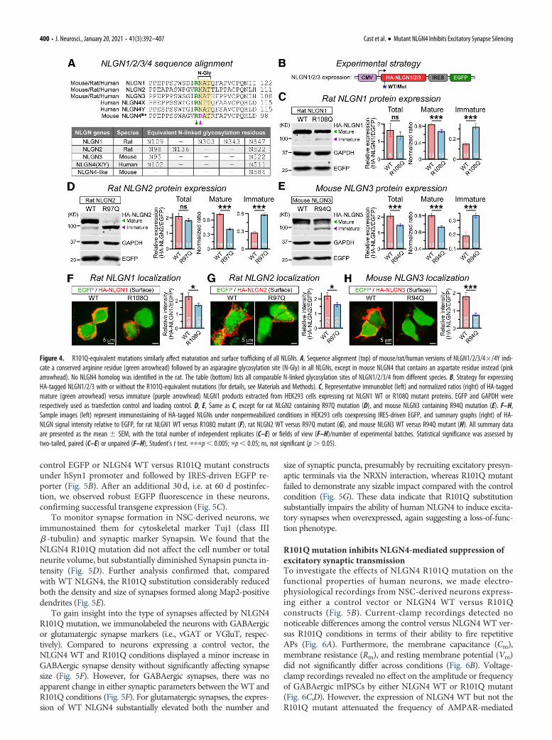
referred to as NLGN4-like), which contains an aspartic acid resi-due (D85) instead of asparagine in the equivalent location, elimi-nating the consensus sequence for N-linked glycosylation (Fig.4A). Therefore, we next introduced R101Q-equivalent mutationsin HA-tagged versions of rat NLGN1, rat NLGN2, and mouseNLGN3, and asked whether that can similarly affect their molecu-lar properties (Fig. 4B). Our immunoblotting experiments fromHEK293 cells transfected with NLGN1 R108Q mutant indicated adefinitive increase in immature product and a decrease in matureproduct (Fig. 4C). This effect was similarly reproduced by R97Qmutation in NLGN2 and R94Q mutation in NLGN3 (Fig. 4D,E).Of note, R101Q-equivalent mutations also decreased the total pro-tein content of NLGN3 (similar to NLGN4; Fig. 2B), but not thatof NLGN1 or NLGN2 (Fig. 4C–E), which contained a highernumber of N-glycosylation sites (Fig. 4A). Thus, R101Q-equiva-lent mutations similarly affected the maturation of all NLGNs andalso impaired the stability of different NLGNs depending on theirpotential glycosylation profiles.
To further assess whether these mutation-induced deficien-cies in NLGN1/2/3 maturation can also affect their ability tolocalize at the cell surface, we conducted immunostaining ofNLGN-expressing HEK293 cells under nonpermeabilized condi-tions. We found that NLGN1 R108Q, NLGN2 R97Q, andNLGN3 R94Q substitutions displayed significant reduction insurface intensity, with respect to their corresponding WT ver-sions (Fig. 4F–H). These results demonstrate that the N102-equivalent glycosylation sites play a critical role in the maturationand surface trafficking of all NLGNs, and that the R101-equiva-lent upstream residues can strongly influence this process.
R101Qmutation blocks the ability of NLGN4 to induceexcitatory synaptogenesisAfter we used HEK293 cells as a reduced system to establish thatR101Q substitution is not benign and directly impairs the molec-ular properties of NLGN4, we next inquired whether this patho-genic mutation affects the morphologic and/or functionalproperties of human neurons. To this end, we first performeddual-SMAD inhibition in human ES cells to generate NSCs, thendifferentiated the NSCs into neurons, and subsequently cocul-tured them with mouse primary astrocytes, as described previ-ously (Fig. 5A; Chambers et al., 2009; Chanda et al., 2019).During this period, the cells gradually changed their identity interms of relevant marker expression, from Oct3/4-positive EScells to Nestin-positive NSCs to Dcx-positive neurons (Fig. 5C).We infected the neurons with lentivirus expressing either a
Figure 3. R101Q mutation blocks NLGN4 glycosylation and retains it at intracellular com-partments. A, Alanine substitution of NLGN4 N102 residue to prevent N-linked glycosylation(N-Gly). B, Western blot (left) illustrating mature (green arrowhead) versus immature (purplearrowhead) HA-NLGN4 products harvested from HEK293 cells expressing NLGN4 WT versusN102A mutant, with EGFP as a transfection control and GAPDH as a loading control.Summary graphs (right) of total, mature, and immature NLGN4 normalized to coexpressed
/
EGFP levels. C, Example immunoblot (C) for NLGN4 WT and R101Q variant, before (untreated)and after enzymatic deglycosylation with glycosidases (i.e., Endo H and PNGase F). D, Bargraphs represent the relative positions of mature (left) and immature (right) NLGN4 products.To compare across multiple experimental batches with slightly variable mobility shifts, the posi-tion of all bands was normalized (dotted lines) to the corresponding distance between themature and immature bands in the untreated R101Q condition. E, Representative images (left)of HEK293 cells expressing NLGN4 WT (top) or R101Q mutant (bottom), as visualized by anIRES-driven mOrange reporter, and stained with DAPI, HA antibody, and calnexin antibody (re-spective channels) under permeabilized conditions. Arrowheads point at intracellular HA-NLGN4colocalized with soluble mOrange and ER marker calnexin. Summary graphs (right) quantify thecorrelation between HA-NLGN4 and calnexin signals, measured via Mander’s coefficients. F,Same as E, except the cells were counterstained with GM130 antibody. The values on bargraphs represent the mean 6 SEM for number of experimental batches (immunoblots) orfields of view analyzed (imaging)/number of independent batches. Statistical significance wasweighed by two-tailed, paired (for immunoblots) or unpaired (for imaging), Student’s t test.pppp, 0.005; ppp, 0.01; pp, 0.05; ns, not significant (p. 0.05).
Cast et al. · Mutant NLGN4 Inhibits Excitatory Synapse Silencing J. Neurosci., January 20, 2021 • 41(3):392–407 • 399
control EGFP or NLGN4 WT versus R101Q mutant constructsunder hSyn1 promoter and followed by IRES-driven EGFP re-porter (Fig. 5B). After an additional 30 d, i.e. at 60 d postinfec-tion, we observed robust EGFP fluorescence in these neurons,confirming successful transgene expression (Fig. 5C).
To monitor synapse formation in NSC-derived neurons, weimmunostained them for cytoskeletal marker Tuj1 (class IIIb -tubulin) and synaptic marker Synapsin. We found that theNLGN4 R101Q mutation did not affect the cell number or totalneurite volume, but substantially diminished Synapsin puncta in-tensity (Fig. 5D). Further analysis confirmed that, comparedwith WT NLGN4, the R101Q substitution considerably reducedboth the density and size of synapses formed along Map2-positivedendrites (Fig. 5E).
To gain insight into the type of synapses affected by NLGN4R101Q mutation, we immunolabeled the neurons with GABAergicor glutamatergic synapse markers (i.e., vGAT or VGluT, respec-tively). Compared to neurons expressing a control vector, theNLGN4 WT and R101Q conditions displayed a minor increase inGABAergic synapse density without significantly affecting synapsesize (Fig. 5F). However, for GABAergic synapses, there was noapparent change in either synaptic parameters between theWT andR101Q conditions (Fig. 5F). For glutamatergic synapses, the expres-sion of WT NLGN4 substantially elevated both the number and
size of synaptic puncta, presumably by recruiting excitatory presyn-aptic terminals via the NRXN interaction, whereas R101Q mutantfailed to demonstrate any sizable impact compared with the controlcondition (Fig. 5G). These data indicate that R101Q substitutionsubstantially impairs the ability of human NLGN4 to induce excita-tory synapses when overexpressed, again suggesting a loss-of-func-tion phenotype.
R101Qmutation inhibits NLGN4-mediated suppression ofexcitatory synaptic transmissionTo investigate the effects of NLGN4 R101Q mutation on thefunctional properties of human neurons, we made electro-physiological recordings from NSC-derived neurons express-ing either a control vector or NLGN4 WT versus R101Qconstructs (Fig. 5B). Current-clamp recordings detected nonoticeable differences among the control versus NLGN4 WT ver-sus R101Q conditions in terms of their ability to fire repetitiveAPs (Fig. 6A). Furthermore, the membrane capacitance (Cm),membrane resistance (Rm), and resting membrane potential (Vm)did not significantly differ across conditions (Fig. 6B). Voltage-clamp recordings revealed no effect on the amplitude or frequencyof GABAergic mIPSCs by either NLGN4 WT or R101Q mutant(Fig. 6C,D). However, the expression of NLGN4 WT but not theR101Q mutant attenuated the frequency of AMPAR-mediated
Figure 4. R101Q-equivalent mutations similarly affect maturation and surface trafficking of all NLGNs. A, Sequence alignment (top) of mouse/rat/human versions of NLGN1/2/3/4�/4Y indi-cate a conserved arginine residue (green arrowhead) followed by an asparagine glycosylation site (N-Gly) in all NLGNs, except in mouse NLGN4 that contains an aspartate residue instead (pinkarrowhead). No NLGN4 homolog was identified in the rat. The table (bottom) lists all comparable N-linked glycosylation sites of NLGN1/2/3/4 from different species. B, Strategy for expressingHA-tagged NLGN1/2/3 with or without the R101Q-equivalent mutations (for details, see Materials and Methods). C, Representative immunoblot (left) and normalized ratios (right) of HA-taggedmature (green arrowhead) versus immature (purple arrowhead) NLGN1 products extracted from HEK293 cells expressing rat NLGN1 WT or R108Q mutant proteins. EGFP and GAPDH wererespectively used as transfection control and loading control. D, E, Same as C, except for rat NLGN2 containing R97Q mutation (D), and mouse NLGN3 containing R94Q mutation (E). F–H,Sample images (left) represent immunostaining of HA-tagged NLGNs under nonpermeabilized conditions in HEK293 cells coexpressing IRES-driven EGFP, and summary graphs (right) of HA-NLGN signal intensity relative to EGFP, for rat NLGN1 WT versus R108Q mutant (F), rat NLGN2 WT versus R97Q mutant (G), and mouse NLGN3 WT versus R94Q mutant (H). All summary dataare presented as the mean 6 SEM, with the total number of independent replicates (C–E) or fields of view (F–H)/number of experimental batches. Statistical significance was assessed bytwo-tailed, paired (C–E) or unpaired (F–H), Student’s t test. pppp, 0.005; pp, 0.05; ns, not significant (p. 0.05).
400 • J. Neurosci., January 20, 2021 • 41(3):392–407 Cast et al. · Mutant NLGN4 Inhibits Excitatory Synapse Silencing

mEPSCs and augmented their interevent inter-val without affecting mEPSC amplitude (Fig.6E,F).
The kinetic parameters of mEPSC eventswere not considerably altered by NLGN4 WT/R101Q expression, suggesting no apparentimpact on AMPAR composition (Fig. 6G).Finally, the overexpression of NLGN4 WT butnot the R101Q mutant also suppressed the am-plitude of AMPAR-evoked EPSCs withoutaffecting presynaptic release probability [as esti-mated by paired-pulse ratios (PPRs)], whenmeasured under similar recording conditions[as assessed by series resistance (Rs) parameters;Fig. 6H]. These results suggest that NLGN4overexpression can negatively modulate thestrength of excitatory synapses in human neu-rons by a potentially postsynaptic mechanism,and the R101Q substitution causes a major lossof this NLGN4-induced synaptic phenotype.
To test whether our conclusions apply toneurons generated by a different protocol, wetransdifferentiated human ES cells into corti-cal glutamatergic neurons by ectopic expres-sion of a single TF, Ngn2 (Fig. 7A; Zhang etal., 2013). We coinfected the neurons withlower-titer lentivirus encoding either a con-trol vector or HA-tagged NLGN4 WT versusR101Q mutant, and detected no noticeabledifference in their cell viability (Fig. 7A,B).We next identified infected neurons by coex-pressed fluorescent reporters and madepatch-clamp recordings to evaluate theirfunctional properties (Fig. 7A,C). Similar toNSC-derived neurons, the Ngn2-inducedhuman neurons expressing WT NLGN4showed a significantly lower amplitude ofAMPAR-mediated evoked EPSCs; whereasthe R101Q variant largely reversed this phe-notype (Fig. 7D). Furthermore, R101Q substi-tution in NLGN4 also elevated the AMPARmEPSC frequency compared with WT ver-sion, with a relatively minor effect on mEPSCamplitude (Fig. 7E,F). Thus, both NLGN4WT and R101Q variant had highly consistentand reproducible effects on the excitatorysynaptic transmission in human neurons irre-spective of their differentiation or reprogram-ming methods.
R101Qmutation prevents NLGN4-mediated silencing of excitatorypostsynapsesWe aimed to understand how loss of NLGN4function by R101Q mutation can contributeto aberrant synaptic transmission. We first
Figure 5. R101Q mutation inhibits NLGN4-induced excitatory synapse formation in NSC-derived human neurons. A,NSCs were induced from H1-ES cells by dual-SMAD inhibition. The NSC-derived neurons were cocultured with glia,infected with lentivirus-expressing transgenes, and analyzed at indicated time points (arrowheads). B, Design of lentiviralvectors expressing NLGN4 WT or R101Q variant under hSyn1 promoter followed by an IRES-EGFP construct. An EGFP-onlyvector was used as infection control (Ctrl). C, Images illustrate cellular identities, when immunolabeled for ES cell marker(Oct3/4), NSC marker (Nestin), or neuronal marker (Dcx), at different stages of the differentiation protocol. DAPI wasused for nuclear stain. At day 60, elaborate neuronal morphology was confirmed by EGFP signal. Arrowheads point atneurons (yellow) and glial cells (white) in the culture. D, Sample images (top) depict NSC-derived neurons expressingNLGN4 WT versus R101Q variant, when immunostained for cytoskeletal marker Tuj1 and synaptic marker Synapsin. Bargraphs (below) represent normalized counts of cell bodies per field of view (left), total neurite volume (middle), andintegrated signal intensity of Synapsin (right), for WT versus R101Q conditions. E, Representative images (top) of MAP2-positive dendritic segments counterstained for Synapsin, and summary graphs (bottom) of the density and size ofSynapsin puncta, as quantified from cells expressing NLGN4 WT versus R101Q mutant. F, Example images (left) ofMAP2-positive dendritic branches from cells in control, NLGN4 WT, and R101Q mutant conditions, as counterstained foran inhibitory synapse marker vGAT. Summary graphs (right) of the density and size of vGAT puncta. G, Same as F, exceptthe neurons were immunolabeled for VGluT, an excitatory synapse marker. Averages indicate the mean 6 SEM, with
/
the total number of fields of view analyzed/number of batches.Statistical significance was tested by two-tailed, unpaired,Student’s t test. pppp, 0.005; ppp, 0.01; pp, 0.05; ns,not significant (p. 0.05).
Cast et al. · Mutant NLGN4 Inhibits Excitatory Synapse Silencing J. Neurosci., January 20, 2021 • 41(3):392–407 • 401

used IRES-driven EGFP fluorescence to visualize the morphol-ogy of Ngn2-induced neurons from control, WT, and R101Qconditions, but failed to observe any difference in their somasize, primary dendrite number, or total neurite length (Fig. 8A).To determine NLGN4 levels in these neurons, we performedqPCR analysis, and found that cells overexpressing WT versusR101Q versions displayed an equivalent increase in mRNA levelsrelative to control condition (Fig. 8B). We next tracked the distri-bution of NLGN4 protein along dendrites by immunostainingthe cells with HA antibody under nonpermeabilized conditions,and then permeabilized them to counterstain for Synapsin. Wenoticed that R101Q mutation caused a substantial reduction in
the density of NLGN4 clusters along the surface of dendriticbranches (Fig. 8C) and thus decreased the colocalization ofNLGN4 with the Synapsin signal (Fig. 8D). Therefore, R101Qsubstitution inhibited the surface trafficking of NLGN4 in humanneurons, even to a greater extent than our observations in HEK293cells (compare with Fig. 2D) and simultaneously diminished its tar-geting at synapses.
To further evaluate the consequences of reduced NLGN4 con-tent at excitatory synapses, we immunostained Ngn2-inducedneurons with Synapsin and VGluT antibodies. We found that,compared with control condition, the overexpression of WTNLGN4 but not the R101Q version caused a considerable increase
Figure 6. NLGN4 WT but not the R101Q variant suppresses AMPAR-mediated synaptic transmission in NSC-derived human neurons. A, Recordings were conducted from NSC-derived neuronsexpressing either a control vector (Ctrl; EGFP only) or NLGN4 WT versus R101Q mutant constructs followed by an IRES-driven EGFP reporter. Sample traces (left) of APs induced by step current injec-tions (protocol is shown on top, Vhold =�60mV), and summary plot (right) of AP numbers as a function of injected current–amplitude. B, Average values of Cm (left), Rm (middle), and Vm (right),as measured from control, NLGN4 WT, and R101Q conditions. C, D, Example traces of GABAAR-mediated mIPSCs (C) and cumulative plots with average bar graphs (D) for mIPSC event frequency andamplitude, as recorded from control, NLGN4 WT, and R101Q conditions. E, F, Same as C and D, except for AMPAR-mediated mEPSC events. G, Superimposed mEPSC waveforms (left) from control,NLGN4 WT, and R101Q conditions. Summary graphs (right) of mEPSC rise time and decay kinetics. H, Representative traces (left) of AMPAR-mediated evoked EPSCs stimulated in pairs(Dt=50ms); arrowheads indicate stimulation artifacts. Summary graphs (right) of EPSC amplitude, PPR (EPSC2/EPSC1) as an indirect measure of presynaptic release probability, and Rs as a measureof recording quality. All quantifications are the mean 6 SEM. All summary data include the number of cells patched/experimental batches. Statistical significance was calculated by two-tailed,unpaired, Student’s t test (all bar graphs), one-way ANOVA (summary plot; A), or KS test (WT vs R101Q probability plots; D–F). pppp, 0.005; pp, 0.05; ns, not significant (p. 0.05).
402 • J. Neurosci., January 20, 2021 • 41(3):392–407 Cast et al. · Mutant NLGN4 Inhibits Excitatory Synapse Silencing

in both Synapsin and VGluT intensities at dendritic branches (Fig.8E,F), mimicking our observations in NSC-derived neurons (Fig.5G). Although these results suggest that overexpression of NLGN4WT but not the R101Q mutant can induce the formation of gluta-matergic synapses, their function depends on the availability ofpostsynaptic AMPARs. To directly assess that, we infected Ngn2-induced neurons with lentiviruses expressing SEP-GluA1, and ei-ther control or NLGN4 WT versus R101Q constructs followed byan IRES-mOrange reporter. We noticed that, compared with con-trol condition, WT NLGN4 significantly reduced the localizationand signal intensity of SEP-GluA1 at Synapsin-positive synapticclusters, whereas the R101Q variant alleviated these phenotypes(Fig. 8G). Together, these results indicate that R101Q substitutionincreases excitatory synaptic transmission (Fig. 7C–F) by prevent-ing NLGN4-induced loss of postsynaptic AMPARs.
DiscussionThe X chromosome-linked human NLGN4 gene has been foundto be mutated in several patients with autism and other
neuropsychiatric disorders (Jamain et al., 2003; Laumonnier etal., 2004; Yan et al., 2005; Chocholska et al., 2006; Talebizadeh etal., 2006; Macarov et al., 2007; Lawson-Yuen et al., 2008;Pampanos et al., 2009). A majority of these mutations representCNVs, truncations, frameshifts, and premature stop codons, sug-gesting an ultimate loss of NLGN4 expression. However, a frac-tion of NLGN4 mutations also include single nucleotidepolymorphisms creating missense variants of relatively unknownphysiological consequence. Earlier studies indicate that whilesome of these missense mutations do exhibit pathogenic pheno-types, other variants may not lead to significant functional inacti-vation (Xu et al., 2017). In this study, we describe a severelyautistic patient carrying a single amino acid substitution (i.e.,R101Q) in NLGN4 (Figs. 1, 9A). We demonstrate that theR101Q mutation prevents NLGN4 maturation by causingincomplete glycosylation (Figs. 2, 3, 9B). As a result, this muta-tion blocks the surface trafficking of NLGN4 by enhancing itsretention at intracellular compartments (Figs. 3, 9B). R101Q-equivalent mutations also affect the maturation of other NLGNswith conserved glycosylation sites (Fig. 4). This mutation-
Figure 7. NLGN4 WT but not the R101Q variant suppresses AMPAR-mediated synaptic transmission in Ngn2-induced human neurons. A, Neurogenesis was induced in H1-ES cells by ectopicexpression of Ngn2 using high-titer lentivirus, and cells were selected using puromycin and cocultured with mouse glia for an extended period (timeline; top). In addition, cells were also sparselyinfected with lower-titer lentiviruses expressing either a control vector or an HA-tagged NLGN4 WT versus R101Q variant under hSyn1 promoter (abbreviated construct maps; bottom), each followedby an IRES-EGFP or IRES-mOrange reporter (R). B, Representative images (left) depict Ngn2-induced neurons coinfected with lentivirus expressing a control vector or NLGN4 WT versus R101Q variant,when immunostained for dendritic marker MAP2; bar graph (right) represents normalized counts of cell bodies per field of view. Arrowheads point at neurons (yellow) and glial cells (white) in thesecocultures. C, Sample image of an Ngn2-induced human neuron expressing NLGN4 transgene, as visualized by EGFP (yellow arrowhead) and patched using a recording pipette (Rec). White arrow-head points at an uninfected cell. D, Representative traces (left) of AMPAR-mediated evoked EPSCs with arrowheads indicating stimulation artifacts. Summary graph (right) of EPSC amplitude, asmeasured from Ngn2-induced neurons expressing either a control vector or NLGN4 WT versus R101Q mutant constructs, while the recording quality was monitored by Rs measurements. E, F,Example traces of AMPAR-mediated mEPSCs (E) and cumulative probability plots with average bar graphs (F) for mEPSC event frequency and amplitude, as recorded from control, NLGN4 WT, andNLGN4 R101Q conditions. Data are presented as the mean 6 SEM, with corresponding number of cells patched (for electrophysiology) or frames analyzed (for imaging)/number of experiments.Statistical significance was assessed by two-tailed, unpaired, Student’s t test (all bar graphs), or KS test (probability plots; F). pppp, 0.005; ppp, 0.01; ns, not significant (p. 0.05).
Cast et al. · Mutant NLGN4 Inhibits Excitatory Synapse Silencing J. Neurosci., January 20, 2021 • 41(3):392–407 • 403

Figure 8. R101Q mutation impairs NLGN4 synaptic localization and inhibits NLGN4-mediated reduction in synaptic AMPAR clusters. A, Representative images (left) of Ngn2-induced humanneurons expressing either a control (Ctrl) vector or NLGN4 WT versus R101Q variant, as their morphologic parameters were assessed using an EGFP reporter. Summary graphs (right) of somasize, primary dendrite number, and average neurite length per cell. B, NLGN4 mRNA levels were estimated by quantitative real-time PCR and normalized to those of GAPDH (internal control)for Ngn2-induced neurons expressing either a control vector, or NLGN4 WT versus R101Q mutant constructs. The average bar graphs combine values from two independent primer sets (set 1:circles; n= 6 Ctrl, 6 WT, and 5 for R101Q; and set 2: squares; n= 6 Ctrl, 6 WT, and 5 for R101Q). C, Sample images (left) of EGFP-expressing dendritic segments (pseudocolored for better visibil-ity) from NLGN4 WT (top) versus R101Q (bottom) conditions, when immunostained with anti-HA antibody (arrowheads) under nonpermeabilized state. Summary graphs (right) of surface HA-NLGN4 puncta density and size. D, Representative images (left) of EGFP-labeled dendritic segments from WT (top) versus R101Q (bottom) conditions, when incubated with anti-HA antibodyunder nonpermeabilized state, then permeabilized and coimmunostained with Synapsin antibody. The channel colors were artificially reassigned to provide better visibility for synaptic signals;arrowheads point at synaptic puncta. Summary graphs (right) indicate synaptic distribution of NLGN4, characterized using normalized Mander’s colocalization coefficients between HA-NLGN4and Synapsin signals. E, F, Dendritic sections of mOrange-expressing human neurons from control, NLGN4 WT, and NLGN4 R101Q conditions immunostained with either Synapsin (E, left) orVGluT (F, left) antibody, and integrated intensity of Synapsin (E, right) or VGluT (F, right) puncta normalized with respect to the dendrite length. G, Neurons expressed either a control vectoror NLGN4 WT versus R101Q mutant followed by mOrange reporter. Sample images (left; pseudocolor) of dendritic branches immunolabeled for surface AMPARs (SEP-GluA1) using anti-GFP anti-body under nonpermeabilized condition, subsequently permeabilized and stained with anti-Synapsin antibody. Arrowheads indicate synaptic clusters; note that many lack copresence of SEP-
404 • J. Neurosci., January 20, 2021 • 41(3):392–407 Cast et al. · Mutant NLGN4 Inhibits Excitatory Synapse Silencing

induced trafficking deficiency was already visible in reduced sys-tems (e.g., HEK293 cells, Figs. 2–4) and inhibited the synapticproperties of NLGN4 even more prominently in human neuronswhen overexpressed for a prolonged time period (Figs. 5–8).R101Q substitution causes a major reduction of NLGN4 localiza-tion at human synapses, and largely inhibits NLGN4-dependenteffects on synapse formation and synaptic transmission (Figs. 5,6, 9C). These phenotypes were highly reproducible and mediatedby altered synaptic density of AMPARs (Figs. 7, 8, 9C). Theseresults confirm that the R101Q mutation is not benign, as itimpairs the synaptic properties of NLGN4 by a partial loss-of-function mechanism.
The R101Q mutation is located in close proximity to a num-ber of previously identified NLGN4 mutations. Of these, anASD-associated R87W substitution has been shown to disruptthe nucleation site for the overall folding of NLGN4, leading toits degradation and substantial loss in total protein content(Zhang et al., 2009). Unlike R87W mutation, the R101Q variantonly moderately reduced NLGN4 protein level, but interruptedN-linked glycosylation at an adjacent N102 residue (Figs. 2B,3A–D). This mutant protein, although immaturely glycosylated,was fully capable of forming homodimers and interacting withpresynaptic NRXNs, indicating that its secondary structure likelyremained intact (Fig. 2E–I). These findings are in accordancewith previous studies with recombinant proteins suggesting thatdeglycosylated versions of soluble NLGN1 fragments can bind toNRXNs, often with a higher affinity (Comoletti et al., 2003).However, in a cellular environment, mutation-induced lack ofglycosylation also modulated the availability of NLGN4 at thecell surface and indirectly reduced its ability to bind NRXNs
when compared with an adequately glycosylated WT product(Fig. 2D,G–I). Thus, the pathogenic effects of R101Q differed sig-nificantly from R87W in that the former mutation affectedNLGN4 properties primarily by impairing post-translationalmodification with a relatively minor effect on the stability of thisprotein. These results highlight divergent mechanisms of variousmissense mutations that can directly or indirectly trigger the lossof NLGN4 function. Moreover, although the pathologic mecha-nisms of other missense mutations (e.g., G99S, G84R) remainuncertain or even controversial, at least some variants may alsoalter NLGN4 properties by gain-in-function mechanisms (e.g.,R704C; Marro et al., 2019).
All NLGNs are heavily modified by N-glycosylation at multi-ple putative residues located in their corresponding extracellulardomains (Comoletti et al., 2003; Bemben et al., 2015; Jeong et al.,2017). While some of these glycosylation sites are specific forindividual NLGNs, the N102 site of human NLGN4 is highlyconserved among all other NLGN genes (NLGN1/2/3) regardlessof species, and is positioned next to an equally conserved R101residue (Fig. 4A). We found that R101Q-equivalent substitutionsalso affected the protein processing and surface trafficking ofNLGN1/2/3 to variable degrees (Fig. 4B–H). This is in contrastto the N303 glycosylation site of NLGN1, which actively modu-lates its NRXN binding, but is not conserved in NLGN2/3/4 toaffect their properties (Fig. 4A; Comoletti et al., 2003; Lee et al.,2010). Although our data suggest that the N102 glycosylation sitecould be necessary for NLGN function in general, further investi-gation is required to comprehend its mechanistic contributionrelative to other specific and/or conserved glycosylation sites.Interestingly, the evolutionarily divergent mouse NLGN4 com-pletely lacks this additional N102 glycosylation site. This contrastin glycosylation profile further highlights the differences betweenrodent and human versions of NLGN4, which might also contrib-ute to their differential effects on synapse regulation. Moreover,it was recently shown that the Y chromosome linked humanNLGN4 (NLGN4Y) only produces an immature and low-MWproduct (Nguyen et al., 2020). Although both N102 and N511 gly-cosylation sites are preserved in NLGN4Y (Fig. 4A), subtle but in-herent differences in NLGN4X versus NLGN4Y amino acid
/
GluA1 in WT condition. Summary graphs (right) of normalized Mander’s coefficient indicatinga Synapsin (Syn) puncta fraction containing SEP-GluA1 signal and the intensity of SEP-GluA1puncta colocalized with Synapsin. Average bar graphs represent the mean6 SEM, with thenumber of frames analyzed/number of experiments, or only the number of independentbatches (for B). Statistical significance was assessed by two-tailed, unpaired (one-tailed andpaired for B), Student’s t test. pppp, 0.005; pp, 0.05; ns, not significant (p. 0.05).
Figure 9. Schematic diagram of NLGN4 R101Q mutation-induced defects in cellular models. A, Summary of clinical evaluations performed on an autistic patient carrying R101Q substitutionin NLGN4. B, In HEK cells, R101Q mutation directly impairs N-linked glycosylation of NLGN4, increases its accumulation in ER and Golgi, and, as a result, indirectly decreases its surface traffickingto the cell membrane. C, In stem cell -derived human neurons, R101Q mutation-mediated reduction in surface trafficking relieves NLGN4-induced inhibition of AMPAR clustering at the excita-tory postsynapses, and thus enhances neuronal excitability.
Cast et al. · Mutant NLGN4 Inhibits Excitatory Synapse Silencing J. Neurosci., January 20, 2021 • 41(3):392–407 • 405

residues might indirectly influence their accessibility for PTMs,differentially affecting their maturation and surface localization.
We noticed that the overexpression of WT NLGN4Xenhanced synaptogenesis in NSC-derived human neurons(Fig. 5). However, this increase in synapse number and sizewas mainly restricted to excitatory synapses, with only minorchanges observed at inhibitory synapses (Fig. 5F,G).Furthermore, the overexpression of WT NLGN4 also unam-biguously affected the functional properties of excitatorysynapses (Fig. 6E–H). The NLGN4-mediated induction insynapse formation and modulation of synaptic transmissionwas substantially impaired by the R101Q mutation, support-ing a primarily loss-of-function hypothesis (Figs. 5, 6). Thesephenotypes, in conjunction with previous findings, supportthe notion that the role of human NLGN4 could be limitedto excitatory synapses (Chanda et al., 2016; Marro et al.,2019). These characteristics of human NLGN4 were surpris-ingly different from its mouse counterpart, because earlierelegant studies demonstrated that mouse NLGN4 preferen-tially localizes at glycinergic and/or GABAergic synapses andregulates inhibitory synaptic transmission (Hoon et al., 2011;Unichenko et al., 2018). Given the striking disparity betweenthe amino acid compositions of human versus mouseNLGN4, it is possible that they diverge in synaptic localiza-tion and/or function. Indeed, in differentiated human neu-rons, an epitope-tagged version of endogenous NLGN4preferably localized at excitatory synapses and only a subsetof inhibitory synapses (Marro et al., 2019).
Similar to earlier findings, the overexpression of WT NLGN4in human neurons significantly reduced the frequency ofAMPAR-mediated mEPSCs without affecting their amplitude(Fig. 6E,F; also see Marro et al., 2019). Although the mechanismof this effect remains unclear, molecular overcrowding by overex-pressed NLGN4 might outcompete endogenous NLGN1/3 homo-dimers and heterodimers for NRXN or PSD-95 binding, whichplay critical role in recruiting AMPARs (NMDA receptor depend-ent or independent) at postsynaptic density (Fig. 9C). It is possiblethat, compared to NLGN1/3, human NLGN4 could be a relativelyweak inducer of postsynaptic AMPAR clustering. However, in theabsence of other NLGNs (e.g. in NLGN1/2/3 knockdown cellswith severely impaired basal excitatory synaptic transmission), theoverexpressed NLGN4 homodimers might enhance EPSC strengthby partially substituting for NLGN1/3 (Nguyen et al., 2020).
Interestingly, overexpression of the R101Q variant inhibitedNLGN4-induced suppression of synaptic strength, again support-ing our hypothesis that this mutation operates via an obligatoryloss-of-function mechanism. These phenotypes were independentof differentiation protocol and equally observed in neuronsreprogrammed by different procedures (Fig. 7D–F). The NLGN4-mediated suppression of mEPSC frequency could result from (1)the decrease in presynaptic release probability, (2) the reduction intotal synapse numbers, or (3) the silencing of excitatory synapses.Analysis of AMPAR-mediated evoked EPSCs indicated that theoverexpression of WT NLGN4 substantially attenuated basal syn-aptic strength without changing PPRs, arguing against any effecton release probability (Fig. 6H). In addition, overexpression ofWT NLGN4 increased the number of excitatory synapses insteadof decreasing it (Figs. 5E–G, 8E,F). Therefore, an alternative hy-pothesis suggests that overexpressed WT NLGN4 might inducesilencing of excitatory synapses, and R101Q mutation could pre-vent this process by eliminating NLGN4 localization from dendri-tic surfaces and synapses. In support of this prediction, we foundthat R101Q mutation decreased dendritic clusters of NLGN4,
apparently reduced its synaptic localization (Fig. 8D), and concur-rently facilitated the synaptic localization of AMPARs as moni-tored by SEP-GluA1 (Fig. 8G). Based on these results, we proposethat the autism-associated R101Q mutation interrupts NLGN4-mediated silencing of excitatory synapses, and thus increases cellu-lar excitability in human neurons.
ReferencesAman MG, Singh NN (1994) Aberrant behavior checklist. New York:
Slosson Educational Publications.Aman MG, Singh NN (2017) Aberrant behavior checklist, Ed 2. East Aurora,
NY: Slosson Educational Publications.Arac D, Boucard AA, Ozkan E, Strop P, Newell E, Südhof TC, Brunger AT
(2007) Structures of neuroligin-1 and the neuroligin-1/neurexin-1 betacomplex reveal specific protein-protein and protein-Ca21 interactions.Neuron 56:992–1003.
American Psychiatric Association (2013) DSM-5. Diagnostic and statisticalmanual of mental disorders, Ed 5. Washington, DC: AmericanPsychiatric Association.
Bemben MA, Shipman SL, Nicoll RA, Roche KW (2015) The cellular andmolecular landscape of neuroligins. Trends Neurosci 38:496–505.
Bolliger MF, Pei J, Maxeiner S, Boucard AA, Grishin NV, Südhof TC (2008)Unusually rapid evolution of Neuroligin-4 in mice. Proc Natl Acad Sci US A 105:6421–6426.
Boucard AA, Chubykin AA, Comoletti D, Taylor P, Südhof TC (2005) Asplice code for trans-synaptic cell adhesion mediated by binding of neu-roligin 1 to alpha- and beta-neurexins. Neuron 48:229–236.
Budreck EC, Scheiffele P (2007) Neuroligin-3 is a neuronal adhesion proteinat GABAergic and glutamatergic synapses. Eur J Neurosci 26:1738–1748.
Bölte S, Westerwald E, Holtmann M, Freitag C, Poustka F (2011) Autistictraits and autism spectrum disorders: the clinical validity of two measurespresuming a continuum of social communication skills. J Autism DevDisord 41:66–72.
Chambers SM, Fasano CA, Papapetrou EP, Tomishima M, Sadelain M,Studer L (2009) Highly efficient neural conversion of human ES and iPScells by dual inhibition of SMAD signaling. Nat Biotechnol 27:275–280.
Chanda S, Marro S, Wernig M, Südhof TC (2013) Neurons generated bydirect conversion of fibroblasts reproduce synaptic phenotype caused byautism-associated neuroligin-3 mutation. Proc Natl Acad Sci U S A110:16622–16627.
Chanda S, Aoto J, Lee SJ, Wernig M, Südhof TC (2016) Pathogenic mecha-nism of an autism-associated neuroligin mutation involves alteredAMPA-receptor trafficking. Mol Psychiatry 21:169–177.
Chanda S, Hale WD, Zhang B, Wernig M, Südhof TC (2017) Unique versusredundant functions of neuroligin genes in shaping excitatory and inhibi-tory synapse properties. J Neurosci 37:6816–6836.
Chanda S, Ang CE, Lee QY, Ghebrial M, Haag D, Shibuya Y, Wernig M,Südhof TC (2019) Direct reprogramming of human neurons identifiesMARCKSL1 as a pathogenic mediator of valproic acid-induced teratoge-nicity. Cell Stem Cell 25:103–119.e6.
Chocholska S, Rossier E, Barbi G, Kehrer-Sawatzki H (2006) Molecular cyto-genetic analysis of a familial interstitial deletion Xp22.2-22.3 with a highlyvariable phenotype in female carriers. Am J Med Genet A 140:604–610.
Comoletti D, Flynn R, Jennings LL, Chubykin A, Matsumura T, HasegawaH, Südhof TC, Taylor P (2003) Characterization of the interaction of arecombinant soluble neuroligin-1 with neurexin-1beta. J Biol Chem278:50497–50505.
Constantino JN (2005) Social responsiveness scale. Torrance, CA: WesternPsychological Services.
Constantino JN, Gruber CP (2012) Social responsiveness scale: SRS-2, Ed 2.Torrance, CA: Western Psychological Services.
Fabrichny IP, Leone P, Sulzenbacher G, Comoletti D, Miller MT, Taylor P,Bourne Y, Marchot P (2007) Structural analysis of the synaptic proteinneuroligin and its beta-neurexin complex: determinants for folding andcell adhesion. Neuron 56:979–991.
Heine M, Thoumine O, Mondin M, Tessier B, Giannone G, Choquet D(2008) Activity-independent and subunit-specific recruitment of func-tional AMPA receptors at neurexin/neuroligin contacts. Proc Natl AcadSci U S A 105:20947–20952.
HoonM, Soykan T, Falkenburger B, HammerM, Patrizi A, Schmidt KF, Sassoè-Pognetto M, Löwel S, Moser T, Taschenberger H, Brose N, Varoqueaux F
406 • J. Neurosci., January 20, 2021 • 41(3):392–407 Cast et al. · Mutant NLGN4 Inhibits Excitatory Synapse Silencing

(2011) Neuroligin-4 is localized to glycinergic postsynapses and regulates in-hibition in the retina. Proc Natl Acad Sci U S A 108:3053–3058.
Ichtchenko K, Hata Y, Nguyen T, Ullrich B, Missler M, Moomaw C, SüdhofTC (1995) Neuroligin 1: a splice site-specific ligand for beta-neurexins.Cell 81:435–443.
Irie M, Hata Y, Takeuchi M, Ichtchenko K, Toyoda A, Hirao K, Takai Y,Rosahl TW, Südhof TC (1997) Binding of neuroligins to PSD-95. Science277:1511–1515.
Jacquemont ML, Sanlaville D, Redon R, Raoul O, Cormier-Daire V, LyonnetS, Amiel J, Le Merrer M, Heron D, de Blois MC, Prieur M, Vekemans M,Carter NP, Munnich A, Colleaux L, Philippe A (2006) Array-based com-parative genomic hybridisation identifies high frequency of cryptic chro-mosomal rearrangements in patients with syndromic autism spectrumdisorders. J Med Genet 43:843–849.
Jamain S, Quach H, Betancur C, Råstam M, Colineaux C, Gillberg IC,Soderstrom H, Giros B, Leboyer M, Gillberg C, Bourgeron T (2003)Mutations of the X-linked genes encoding neuroligins NLGN3 andNLGN4 are associated with autism. Nat Genet 34:27–29.
Jeong J, Paskus JD, Roche KW (2017) Posttranslational modifications of neu-roligins regulate neuronal and glial signaling. Curr Opin Neurobiol45:130–138.
Jiang M, Polepalli J, Chen LY, Zhang B, Südhof TC, Malenka RC (2017)Conditional ablation of neuroligin-1 in CA1 pyramidal neurons blocksLTP by a cell-autonomous NMDA receptor-independent mechanism.Mol Psychiatry 22:375–383.
Ko J, Zhang C, Arac D, Boucard AA, Brunger AT, Südhof TC (2009)Neuroligin-1 performs neurexin-dependent and neurexin-independentfunctions in synapse validation. EMBO J 28:3244–3255.
Koehnke J, Jin X, Budreck EC, Posy S, Scheiffele P, Honig B, Shapiro L(2008) Crystal structure of the extracellular cholinesterase-like domainfrom neuroligin-2. Proc Natl Acad Sci U S A 105:1873–1878.
Laumonnier F, Bonnet-Brilhault F, Gomot M, Blanc R, David A, MoizardMP, Raynaud M, Ronce N, Lemonnier E, Calvas P, Laudier B, Chelly J,Fryns JP, Ropers HH, Hamel BC, Andres C, Barthélémy C, Moraine C,Briault S (2004) X-linked mental retardation and autism are associatedwith a mutation in the NLGN4 gene, a member of the neuroligin family.Am J HumGenet 74:552–557.
Lawson-Yuen A, Saldivar JS, Sommer S, Picker J (2008) Familial deletionwithin NLGN4 associated with autism and Tourette syndrome. Eur JHumGenet 16:614–618.
Lee H, Dean C, Isacoff E (2010) Alternative splicing of neuroligin regulatesthe rate of presynaptic differentiation. J Neurosci 30:11435–11446.
Leone P, Comoletti D, Ferracci G, Conrod S, Garcia SU, Taylor P, Bourne Y,Marchot P (2010) Structural insights into the exquisite selectivity of neu-rexin/neuroligin synaptic interactions. EMBO J 29:2461–2471.
Macarov M, Zeigler M, Newman JP, Strich D, Sury V, Tennenbaum A,Meiner V (2007) Deletions of VCX-A and NLGN4: a variable phenotypeincluding normal intellect. J Intellect Disabil Res 51:329–333.
Marro SG, Chanda S, Yang N, Janas JA, Valperga G, Trotter J, Zhou B,Merrill S, Yousif I, Shelby H, Vogel H, Kalani MYS, Südhof TC, WernigM (2019) Neuroligin-4 regulates excitatory synaptic transmission inhuman neurons. Neuron 103:617–626.e6.
Marshall CR, Noor A, Vincent JB, Lionel AC, Feuk L, Skaug J, Shago M,Moessner R, Pinto D, Ren Y, Thiruvahindrapduram B, Fiebig A,Schreiber S, Friedman J, Ketelaars CEJ, Vos YJ, Ficicioglu C, KirkpatrickS, Nicolson R, Sloman L, et al. (2008) Structural variation of chromo-somes in autism spectrum disorder. Am J HumGenet 82:477–488.
Maxeiner S, Benseler F, Krasteva-Christ G, Brose N, Sudhof TC (2020)Evolution of the autism-associated neuroligin-4 gene reveals broad erosionof pseudoautosomal regions in rodents. Mol Biol Evol 37:1243–1258.
Meyer G, Varoqueaux F, Neeb A, Oschlies M, Brose N (2004) The complex-ity of PDZ domain-mediated interactions at glutamatergic synapses: acase study on neuroligin. Neuropharmacology 47:724–733.
Mondin M, Labrousse V, Hosy E, Heine M, Tessier B, Levet F, Poujol C,Blanchet C, Choquet D, Thoumine O (2011) Neurexin-neuroligin
adhesions capture surface-diffusing AMPA receptors through PSD-95scaffolds. J Neurosci 31:13500–13515.
Nam CI, Chen L (2005) Postsynaptic assembly induced by neurexin-neuroli-gin interaction and neurotransmitter. Proc Natl Acad Sci U S A102:6137–6142.
Nguyen TA, Wu K, Pandey S, Lehr AW, Li Y, Bemben MA, Badger JD 2nd,Lauzon JL, Wang T, Zaghloul KA, Thurm A, Jain M, Lu W, Roche KW(2020) A cluster of autism-associated variants on X-linked NLGN4Xfunctionally resemble NLGN4Y. Neuron 106:759–768.
Pampanos A, Volaki K, Kanavakis E, Papandreou O, Youroukos S,Thomaidis L, Karkelis S, Tzetis M, Kitsiou-Tzeli S (2009) A substitutioninvolving the NLGN4 gene associated with autistic behavior in the Greekpopulation. Genet Test Mol Biomarkers 13:611–615.
Poulopoulos A, Soykan T, Tuffy LP, Hammer M, Varoqueaux F, Brose N(2012) Homodimerization and isoform-specific heterodimerization ofneuroligins. Biochem J 446:321–330.
Schepici G, Cavalli E, Bramanti P, Mazzon E (2019) Autism spectrum disor-der and miRNA: an overview of experimental models. Brain Sci 9:265.
Schopler E, Van Bourgondien ME (2010) CARS2: the childhood autism rat-ing scale, Ed 2. Los Angeles: Western Psychological Services.
Sebat J, Lakshmi B, Malhotra D, Troge J, Lese-Martin C, Walsh T, YamromB, Yoon S, Krasnitz A, Kendall J, Leotta A, Pai D, Zhang R, Lee Y-H,Hicks J, Spence SJ, Lee AT, Puura K, Lehtimäki T, Ledbetter D, et al.(2007) Strong association of de novo copy number mutations with au-tism. Science 316:445–449.
Song JY, Ichtchenko K, Südhof TC, Brose N (1999) Neuroligin 1 is a postsy-naptic cell-adhesion molecule of excitatory synapses. Proc Natl Acad SciU S A 96:1100–1105.
Sparrow S, Cicchetti D, Saulnier C, Doll EA (2016) Vineland adaptive behav-ior scales, Ed 3. Minneapolis, MN: Pearson Assessments.
Sparrow SS, Cicchetti DV (1985) Diagnostic uses of the Vineland AdaptiveBehavior Scales. J Pediatr Psychol 10:215–225.
Südhof TC (2008) Neuroligins and neurexins link synaptic function to cogni-tive disease. Nature 455:903–911.
Talebizadeh Z, Lam DY, Theodoro MF, Bittel DC, Lushington GH, ButlerMG (2006) Novel splice isoforms for NLGN3 and NLGN4 with possibleimplications in autism. J Med Genet 43:e21.
Unichenko P, Yang JW, Kirischuk S, Kolbaev S, Kilb W, Hammer M,Krueger-Burg D, Brose N, Luhmann HJ (2018) Autism relatedNeuroligin-4 knockout impairs intracortical processing but not sensoryinputs in mouse barrel cortex. Cereb Cortex 28:2873–2886.
Varoqueaux F, Jamain S, Brose N (2004) Neuroligin 2 is exclusively localizedto inhibitory synapses. Eur J Cell Biol 83:449–456.
Varoqueaux F, Aramuni G, Rawson RL, Mohrmann R, Missler M, GottmannK, Zhang W, Südhof TC, Brose N (2006) Neuroligins determine synapsematuration and function. Neuron 51:741–754.
Volkmar FR (2013) Encyclopedia of autism spectrum disorders, Ed 1. NewYork: Springer.
Xu X, Hu Z, Zhang L, Liu H, Cheng Y, Xia K, Zhang X (2017) Not all neuro-ligin 3 and 4X missense variants lead to significant functional inactiva-tion. Brain Behav 7:e00793.
Yan J, Oliveira G, Coutinho A, Yang C, Feng J, Katz C, Sram J, Bockholt A,Jones IR, Craddock N, Cook EH Jr, Vicente A, Sommer SS (2005)Analysis of the neuroligin 3 and 4 genes in autism and other neuro-psychiatric patients. Mol Psychiatry 10:329–332.
Zhang C, Milunsky JM, Newton S, Ko J, Zhao G, Maher TA, Tager-FlusbergH, Bolliger MF, Carter AS, Boucard AA, Powell CM, Südhof TC (2009) Aneuroligin-4 missense mutation associated with autism impairs neuroligin-4 folding and endoplasmic reticulum export. J Neurosci 29:10843–10854.
Zhang Y, Pak C, Han Y, Ahlenius H, Zhang Z, Chanda S, Marro S, Patzke C,Acuna C, Covy J, Xu W, Yang N, Danko T, Chen L, Wernig M, SüdhofTC (2013) Rapid single-step induction of functional neurons fromhuman pluripotent stem cells. Neuron 78:785–798.
Cast et al. · Mutant NLGN4 Inhibits Excitatory Synapse Silencing J. Neurosci., January 20, 2021 • 41(3):392–407 • 407
![A-level€¦ · [3 marks] Turn over for the next question 0 1 . 3 0 .1 4 8 . 6 Some autism spectrum disorders (ASDs) are associated with a mutation affecting the neuroligin-3 gene.](https://static.fdocuments.in/doc/165x107/5ecdbea2f43998436e2bb686/a-level-3-marks-turn-over-for-the-next-question-0-1-3-0-1-4-8-6-some-autism.jpg)