Anatomy of optic nerve and its clinical significance
66
Anatomy of optic nerve and its clinical Significance • Presented By • Pabita Dhungel • B. Optometry • 2 nd year • Institute of Medicine 1
-
Upload
pabita-dhungel -
Category
Health & Medicine
-
view
521 -
download
4
Transcript of Anatomy of optic nerve and its clinical significance

Anatomy of optic nerve and its clinical Significance
• Presented By• Pabita Dhungel• B. Optometry• 2nd year• Institute of Medicine
1

References
• Walsh and Hoyt’s clinical Ophthalmology• Wolf’s anatomy• Duane’s Ophthalmology-2005• AAO – Fundamentals & Principles of
Ophthalmology-sec 2• Embryology – Duke Elder• Anataomy and physiology of Eye – A.K
Khurana 2nd edition
2

Presentation layout
• Embryology of optic nerve • Introduction•Blood supply•Few optic nerve diseases•Summary
3

Formation of optic vesicle and optic stalk
• Neural plate destined to form prosencephalon depresses to form optic sulcus•Neural plate converts into prosencephalic vesicle•Optic sulcus deepens and prosencephalon bulge out to form optic vesicle•Proximal part of optic vesicle becomes constricted and enlongated to form optic stalk
4

5

Embryology of optic nerve
Optic nerve headThe optic nerve head is formed late in the embryonic period as the optic nerve stalk encloses the hyaloid artery(8th week,20-mm stage)From the hyaloid artery ,the vascular bud develops(13th week,96-mm) within Bergmeister papilla and through it into the nerve fibre layer of the retina
6

Contd…
• Glial cells form the sheaths of the vessels• Eventually hyaloid artery disapears before
birth, Bergmeister’s papilla becomes atrophic, and the physiological cup of the optic disc develops at 15th week of gestation
• Optic nerve• Develops from embryonic optic stalk at 4th
week and connects the optic vesicle to forebrain
7
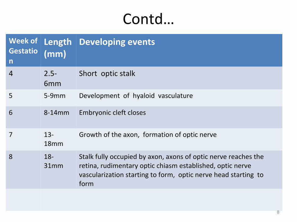
Contd…Week of Gestation
Length (mm)
Developing events
4 2.5-6mm
Short optic stalk
5 5-9mm Development of hyaloid vasculature
6 8-14mm Embryonic cleft closes
7 13-18mm
Growth of the axon, formation of optic nerve
8 18-31mm
Stalk fully occupied by axon, axons of optic nerve reaches the retina, rudimentary optic chiasm established, optic nerve vascularization starting to form, optic nerve head starting to form
8

Contd…Week of gestation
Length (mm)
Developing events
11 65-73mm
Vascular-connective septa invade the nerve
12 80mm Pia mater, arachnoid, and dura mater distinguishable, glial filaments appear
14 105mm Subarachnoid spaces appears
15 117-123mm
Physiological cup starts to form
18 160mm Vascularization of the optic nerve completed
23 220mm Myelinization starts
9

Embryology contd…
• Glial elements are formed from 9th week (45mm stage) to 18th week
• Vasculature develops at 11th week(65-73mm) in the same way as CNS
10

Anatomy of optic nerve
• Optic nerve- more than 1 million axons.• Starts from optic disc upto optic chiasma.• Backward continuation of the nerve fiber
layer of retina.• Consisting of axons originating from ganglion
cells.• Contains the afferent fibers of light reflex.• Elongated tract of white matter • Not covered by neurilemma.
11

Contd…
• Optic nerve divided in topographic areas.Intraocular portion of optic nerve- optic disc or nerve head,pre laminar and post
laminar.Intraorbital portion.Intracanalicular portion.Intracranial portion.
12

13

Intraocular optic nerve
• 1 mm in length.• 1.5 mm diameter.• Which expands approximately 3-4 mm behind
the sclera.• Optic nerve head divided in 4 parts(ant to
post)
14

Intraorbital optic nerve
Relation of ophthalmic arteryInitially infero-lateral-mediallyAt the optic foramen-inferior and lateral
Lateral to optic nerve (in posterior orbit)-Inferior division of 3rd nerve-Nasociliary artery-Sixth Nerve-Ciliary ganglion
15

16
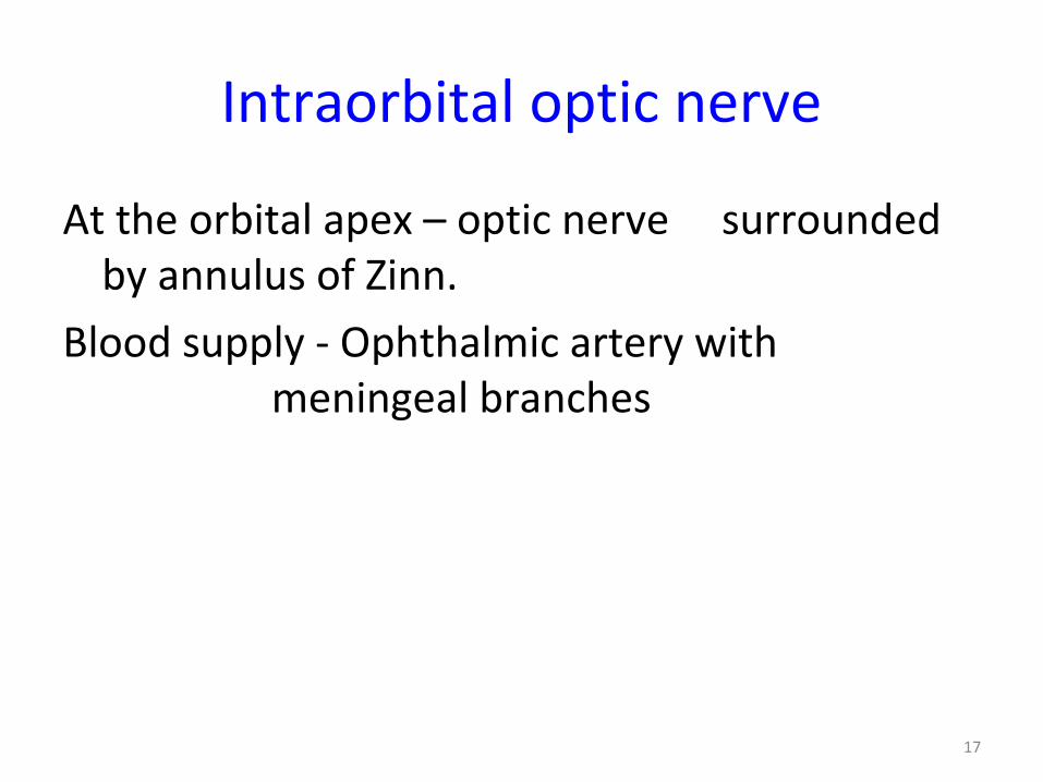
Intraorbital optic nerve
At the orbital apex – optic nerve surrounded by annulus of Zinn.
Blood supply - Ophthalmic artery with meningeal branches
17

Intracanalicular optic nerve
• 9 mm• Tightly fixed within the canal (compressive
optic neuropathy)• Blunt trauma • Optic nerve edema• Blood supply - pial branches from ophthalmic
artery.
18

Intracranial optic nerve
• Length-10mm• Dm-4.5mmExtends post & medially ascending at an angle
of 45º to join the chiasmaBlood supply- pial vessels arising from ICA
branches from ant cerebral and anterior communicating artery
19

Optic nerve head
4 layers of Optic Nerve Head:• Surface nerve fiber layer.• Prelaminar layer consisting of retinal nerve
fibres angled posteriorly from the plane of retina visible only within the central cup.
• Laminar layer: Lamina cribrosa and nerve fibre bundles running through it.
• Retro laminar layer.
20

21

Optic nerve head
• Lamina Cribrosa - Consists of series of plates of collagenous connective tissues.
• -Perforated by 200 - 400 openings.• -Superficial openings – appear as grey
dots deep within the optic cup.• -Large pores have thin connective
tissue supports and contain large nerve fibres.
22
Contd…
• Optic Cup - 3-dimensional pale depression in the centre. Size of the cup related to dm of the disc.
• Neuroretinal rim- tissue between outer edge of the cup and outer margin of the disc.
• Orange or pink colour• Uniform width• Retinal blood vessels
23

24

25

Optic chaisma
Nerve fibres:-Lower nasal fibres traverse the chiasma low and anteriorly (vulnerable-expanding intrasellar lesions)
Upper nasal fibres traverse high and posteriorly (craniopharyngioma)
Crossing fibres from infero-nasal quadrant loop ant into post part of contralateral optic nerve –Wilbrand’s Knee
26

27
Optic tract
• Each optic tract contains ipsilateral temporal & contralateral nasal fibres from optic nerves.
• Nerve fibres rotates a 90º so that superior fiber – medially & inferior – laterally .
• Macular fibres - dorsolateral orientation. • Blood supply - anterior choroidal artery &
branches from posterior communicating artery.
28

Contd…
• Visual field defects in optic tract lesion • incongruous homonymous hemianopia • Bilateral retinal layer atrophy / optic atrophy • Pupillary reflex – RAPD in contralateral eye
29

Lateral geniculate bodyPart of Thalamus• Site of termination of afferent fibres• 6 major layers:-
Magnocellular (1 ,2 )Parvocellular (3,4,5,6 )
• Visual field defect in lesions of LGB• Incongrous homonymous hemianopia• No pupillary change
30

Contd…
• Primary target of optic tract is dorsal lateral geniculate body(dLGB), a thalamic neuclei
• dLGB consists of 3 distinct regions• Dorsal four layers, consisting small neurons
called parvo, or P-cells, are parvocelluar layers• Larger neurons called magno or M- cells
comprises two ventral magnocellular layers• Intermediately lies small konio cells
31

Contd…
• Parvo, magno and konio pathways are referred to as parallel pathways
• Parvo pathway encodes detail and color while magno pathway encodes fast movement
• The role of konio pathway is less well understood
32

Optic radiation• 3 main groups:-
- Inferior portion (serving superior V.F.)
- Superior portion (serving inferior V.F.)
- Central portion (macular fibres)• As they exit from LGB the rotation of fibres
occurs in reverse 90º so superior fibres lie superiorly and inferior fibres lie inferiorly.
• Blood supply:- anterior choroidal artery & posterior cerebral artery
33

Visual cortex
• Thinnest portion of cerebral cortex• Area - 20-45 sq cm• Occupies 3-5% of brain• Situated along calcarine fissure on medial
side• Macular fibres – tip of occipital lobe and tip of
lateral side• Ocular Dominance Column
34

35

Visual cortex
• Blood supply: Middle cerebral artery Posterior cerebral artery
• Visual field defect - occlusion of the Posterior cerebral artery - congruous homogeneous hemianopia
• Head injury, gun shot injury, cerebral tumours.
36

1 lesion of optic nerve
2..lesion through thru proximal part
3.Sagittal (central) lesion of chaisma
4.Lateral chaismal lesion
5. Lesions of optic tract
6. Lesions of LGB
7. Lesion of optic radiation in temporal lobe
8. Lesions of optic radiation in parietal lobe
9.Optic radiation
10.Visual cortex sparing macula
11.Visual cortex only macula 37

Blood supply
• Intraocular part- a)surface nerve fiber-capillaries from retinal
artery. b)prelaminar-short post ciliary artery. c)lamina cribrosa-short post ciliary artery and
circle of Zinn-Haller. d)retrolaminar-ciliary and retinal circulation.
38

39

Contd…
• Intraorbital part- a)periaxial system-internal carotid artery. ophthalmic artery long posterior ciliary artery short post ciliary artery lacrimal artery central artery of retina circle of Zinn
40

Contd…
b)axial system-
intraneural branches of central retinal artery
central collateral arteries
central artery of optic nerve
41
Contd…
• Intracanalicular part-
ophthalmic artery &
periaxial system
42

Contd…
• Intracranial part-
periaxial & pial plexus(4 sources)
internal carotid artery
anterior cerebral artery
ophthalmic artery
anterior communicating
artery
43

44
Venous drainage
• Primarily by central retinal vein.• Orbital part –pial plexus and central retinal
vein.• Intracranial part-anterior cerebral and basal
vein.
45

•Clinical significance of optic nerve
46

• Optic nerve is formed by axons of 1.2 million ganglion cells of retina
• Measures about 1.5mm in diameter and lies betn 10-15 degrees from fixation in the nasal retina
47

Contd…
• A healthy disc is normally pink to orange in color, with well delineated margins and a small cup to disc ratio (<0.3)
48

Why the normal disc is pink?
• Thickness and the cytoarchitecture of fiber bundles passing between glial columns containing capillaries
49

Contd…
• Pathologies of the optic nerve, even though not always detected on ophthalmoscopic exam, may compromise its function and cause the following sign;
• Reduced VA• Afferent pupillary defects• Visual field defects• Dischromatopsia(impaired CV)
50

Contd…
• Diminished light brightness sensitivity• Diminished contrast sensitivity• Optic disc changes on fundoscopy can include
disc edema, hyperemia, paleness and atrophy• Many a times though the disc looks
completely normal yet it harbors a major underlying disc
51
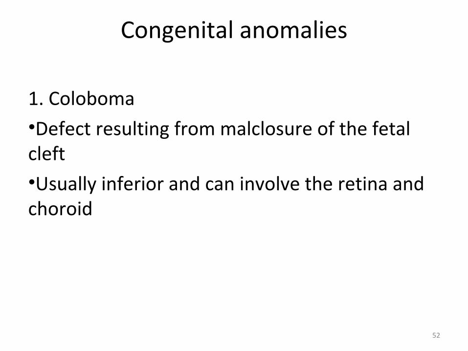
Congenital anomalies
1. Coloboma•Defect resulting from malclosure of the fetal cleft•Usually inferior and can involve the retina and choroid
52

Optic disc showing coloboma (rt)
53

Contd…
2. Hypoplasia •Smaller disc that carries fewer axon than normal and may be associated with poor VA, field defects, strabismus.•Intake of alcohol, steroids and insulin by mother during first trimester of pregnancy increases the risk
54

55
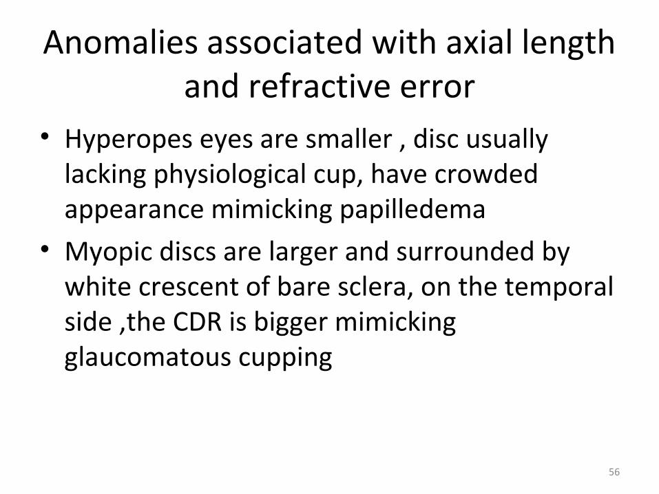
Anomalies associated with axial length and refractive error
• Hyperopes eyes are smaller , disc usually lacking physiological cup, have crowded appearance mimicking papilledema
• Myopic discs are larger and surrounded by white crescent of bare sclera, on the temporal side ,the CDR is bigger mimicking glaucomatous cupping
56

Myelinated nerve fibre
• Myelinating process which is completed by the 9th month of gestation sometimes extends onto the surface of the disc or surrounding retina in a radiating fashion causing feathery patches
• Condition is usually benign
57

58

Drusens
• Yellow opalescent hyaline excrescences derived from calcified axonal debris present on the surface of the disc or burried in it
• Optic nerve head is full and small mimicking papilledema
• May be associated with RP • Autofluoresence before FFA may help in
diagnosis
59

60

Optic nerve head swelling
• Common causes• Congenital: ex drusens• Systemic diseases: ex; anemia, hypoxemia,
uremia, HTN• Tumors: ex; hemangiomas, orbital glioma,
meningioma• Infiltrative diseases: ex; lymphoma
61

Severe disc oedema in multiple sclerosis
62

Contd…
• Vascular diseases: ex; AION, giant cell arteritis and other autoimmune vasculitides, DR
• Ocular diseases: ex; uveitis, hypotony, CRVO• Inflammatory diseases: optic neuritis, papillitis
and neuroretinitis• elevated ICP (intracranial pressure) : ex; mass
occupying lesions, pseudotumor cerebri...
63

Common causes of optic nerve edema
Papilledema Optic neuritis AIONNon inflammatory edema secondary to increased ICP
Inflammatory swelling Vascular accident (occlusion of short posterior ciliary artery causing retrolaminar nerve infarction
Brain tumors , abscesses, hematomas,meningitis might be underlying etiology
Multiple Sclerosis is highly associated
Hypertension, giant cell arteritis, hypercoagulable state are possible factors
Bilateral , may be asymmetric
Unilateral Unilateral
64

Contd…Papilledema Optic neuritis AIONHeadache, nausea, vomittingNo visual loss usually, only enlarged blind spot and possible hyperopia
Retrobulbar pain, especially on ocular movement, early central scotoma, decreased acuity, impaired color vision,presence o APD
Acute painless visual loss, usually hemialtitudinal defect involving the lower visual field
Variable degree of disc swelling, hemorrhages and cystoid infarcts
Fewer hemorrhages and cotton wool spots
Pale segmental swelling and splinter hemorrhages at its margins
Prognosis usually good if primary cause of increased ICP is treated
Vision usually returns to normal
Poorer prognosis with permanent . loss. Second eye is ultimately involved in one third of idiopathic cases.
65

66