Anatomy of nerve_injuries_lower_limb
73
Anatomy of Nerve Injuries Anatomy of Nerve Injuries Lower Limb Lower Limb By- Dr. Armaan Singh By- Dr. Armaan Singh
-
Upload
dr-armaan-singh -
Category
Health & Medicine
-
view
282 -
download
1
Transcript of Anatomy of nerve_injuries_lower_limb

Anatomy of Nerve Injuries Anatomy of Nerve Injuries Lower LimbLower Limb
By- Dr. Armaan SinghBy- Dr. Armaan Singh
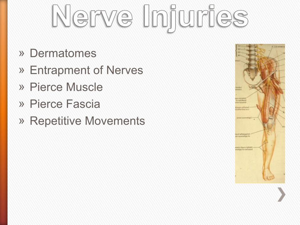
» Dermatomes» Entrapment of Nerves» Pierce Muscle» Pierce Fascia» Repetitive Movements

» Must know the course of nerve» Dermatomes» Entrapment of nerves» Pierce muscle» Pierce fascia» Repetitive movements

Nerves supply
» Skin
» Muscles (group)
» Tendons
» Bones
» Joints
» Blood vessels

» External forces » Fibro-osseous tunnels, tether
the nerve» Oedema» Callus formation as a result of a
fracture » External compression due to
specific movements» Mechanical compression » Compartment syndromes» The nerve is tender at the site
of compression

» Fibrous bands» Accessory muscles» Spurs» Narrow notches» Anatomical variations of the nerve
itself

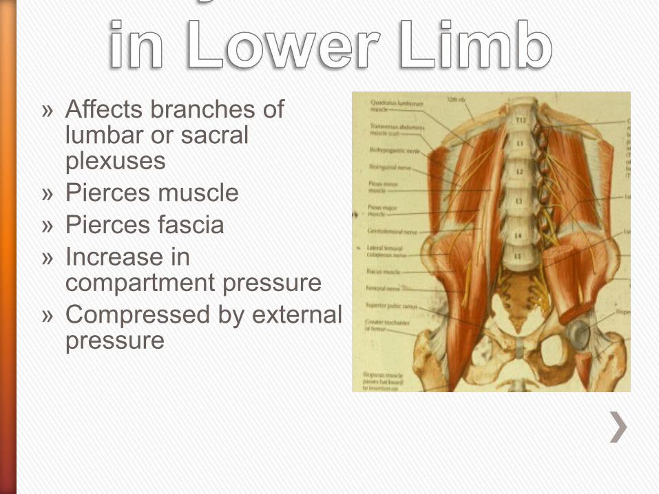
» Affects branches of lumbar or sacral plexuses
» Pierces muscle» Pierces fascia» Increase in
compartment pressure» Compressed by external
pressure
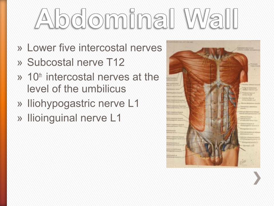
» Lower five intercostal nerves» Subcostal nerve T12» 10th intercostal nerves at the
level of the umbilicus» Iliohypogastric nerve L1» Ilioinguinal nerve L1
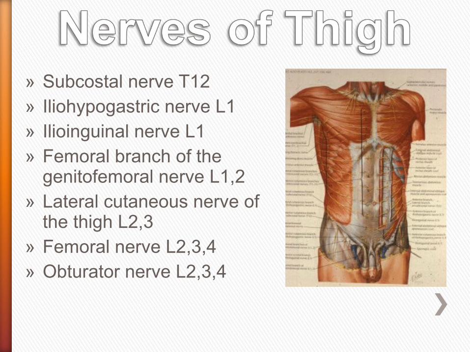
» Subcostal nerve T12» Iliohypogastric nerve L1» Ilioinguinal nerve L1» Femoral branch of the
genitofemoral nerve L1,2» Lateral cutaneous nerve of
the thigh L2,3» Femoral nerve L2,3,4» Obturator nerve L2,3,4

Cutaneous Nerves

» Branch of lumbar plexus» Lateral border of psoas» Anterior to quadratus
lumborum» Neurovascular plane
between internal oblique and transversus
» Lateral cutaneous supplies upper part of buttock

» Pierces internal oblique above anterior superior iliac spine
» Pierces aponeurosis of external oblique an inch
above superficial ring » Supplies skin over
lower part of rectus sheath
» Can be trapped piercing aponeurosis
Iliohypogastric Nerve
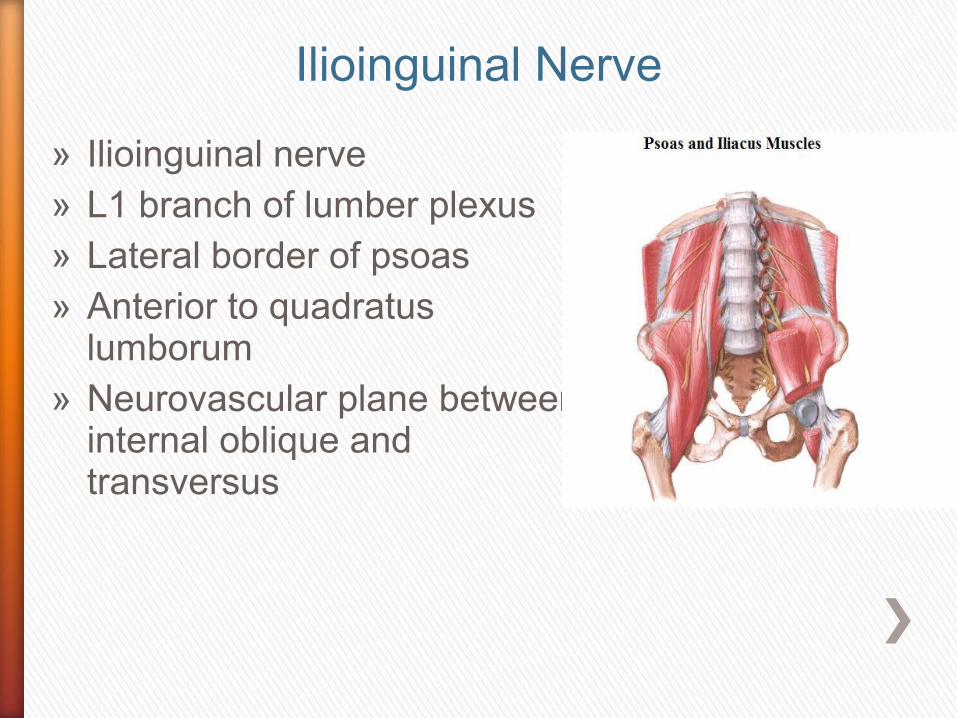
» Ilioinguinal nerve» L1 branch of lumber plexus» Lateral border of psoas» Anterior to quadratus
lumborum» Neurovascular plane between
internal oblique and transversus
Ilioinguinal Nerve
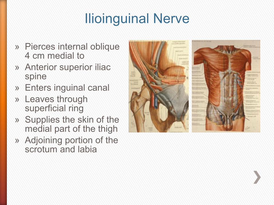
» Pierces internal oblique 4 cm medial to
» Anterior superior iliac spine
» Enters inguinal canal» Leaves through
superficial ring» Supplies the skin of the
medial part of the thigh» Adjoining portion of the
scrotum and labia
Ilioinguinal Nerve

» May be trapped post surgery, due to adhesions
» Poor tone in abdominal muscles
» Pain increased by increased tension in the anterior abdominal wall
» Hyperextension of hip» Tenderness 4 cm from
anterior superior iliac spine
Ilioinguinal Nerve

» Pain increased» Increased tension in the
anterior abdominal wall» Hyperextension of hip» Tenderness 4 cm medial to
anterior superior iliac spine
Ilioinguinal Nerve Entrapment
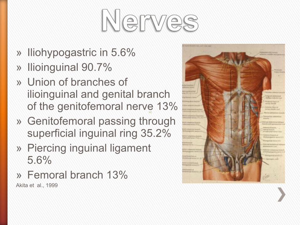
» Iliohypogastric in 5.6%» Ilioinguinal 90.7%» Union of branches of
ilioinguinal and genital branch of the genitofemoral nerve 13%
» Genitofemoral passing through superficial inguinal ring 35.2%
» Piercing inguinal ligament 5.6%
» Femoral branch 13% Akita et al., 1999

» Lumbar plexus L1,2» Anterior aspect of the
psoas» Genital branch enters the
deep inguinal ring» Femoral branch lies on the
lateral side of femoral artery in the femoral sheath

» Enters thigh on lateral aspect of femoral artery in femoral sheath
» Pierces anterior wall of the sheath
» Supplies skin a hands breath below the inguinal ligament

» Union with ilioinguinal nerve on anterior aspect of spermatic cord
» Supplies ventral aspect of scrotum and adductor region
» Cutaneous branch on the dorsal-caudal aspect
» May also supply dorsal scrotum
Akita et al., 1999
Genitofemoral Nerve

Genitofemoral Nerve

» Lumbar plexus in psoas» Lateral aspect of psoas» Pierces inguinal ligament» Lies in fibrous tunnel» Divides into two» Pierces deep fascia
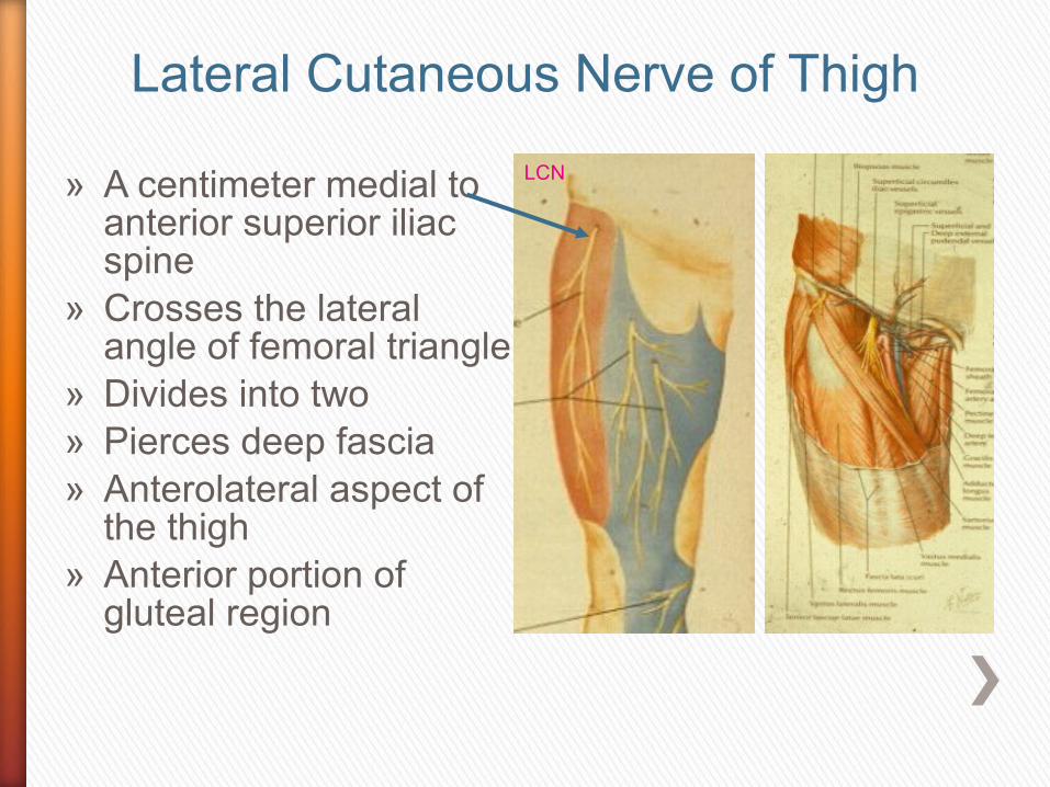
» A centimeter medial to anterior superior iliac spine
» Crosses the lateral angle of femoral triangle
» Divides into two» Pierces deep fascia» Anterolateral aspect of
the thigh» Anterior portion of
gluteal region
LCN
Lateral Cutaneous Nerve of Thigh
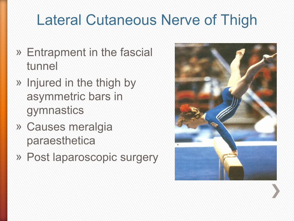
» Entrapment in the fascial tunnel
» Injured in the thigh by asymmetric bars in gymnastics
» Causes meralgia paraesthetica
» Post laparoscopic surgery
Lateral Cutaneous Nerve of Thigh
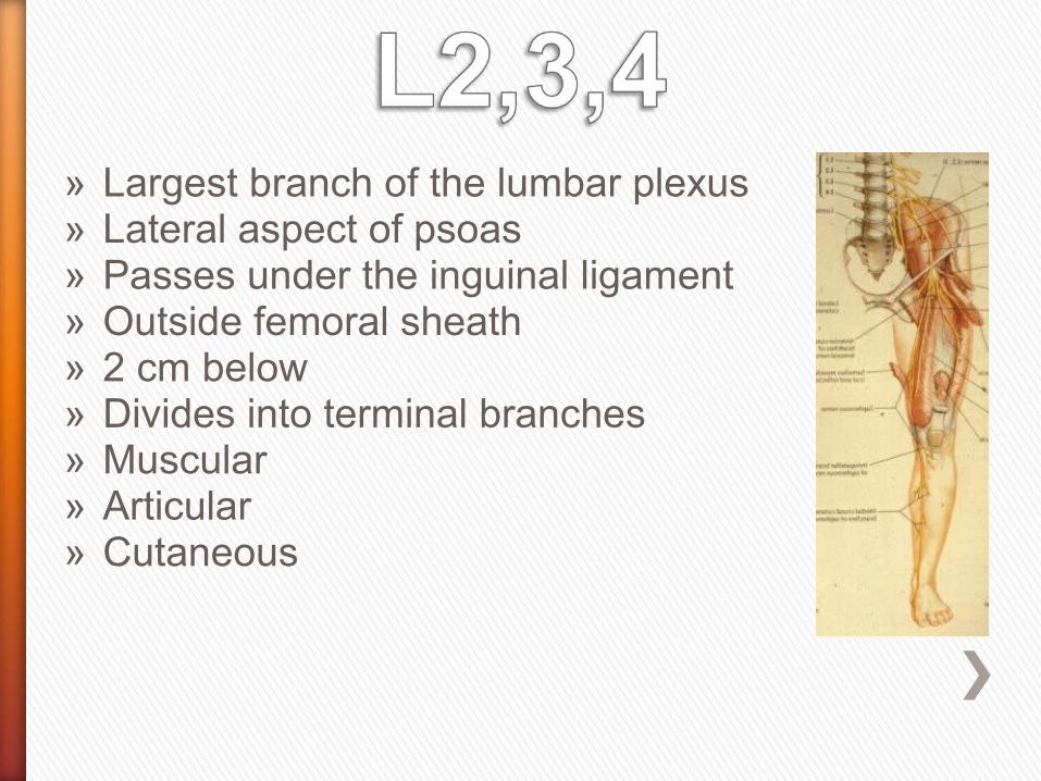
» Largest branch of the lumbar plexus» Lateral aspect of psoas» Passes under the inguinal ligament» Outside femoral sheath» 2 cm below» Divides into terminal branches» Muscular » Articular» Cutaneous
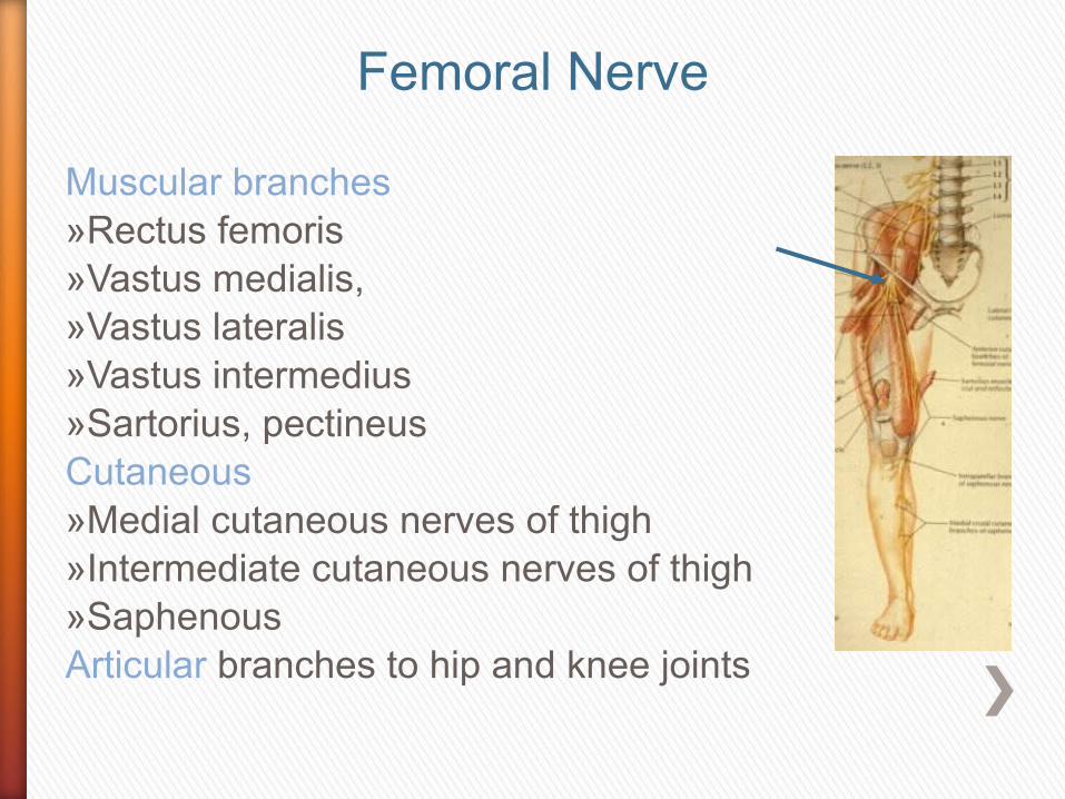
Muscular branches»Rectus femoris»Vastus medialis,»Vastus lateralis»Vastus intermedius»Sartorius, pectineusCutaneous »Medial cutaneous nerves of thigh»Intermediate cutaneous nerves of thigh»Saphenous Articular branches to hip and knee joints
Femoral Nerve

» Dancers may stretch the nerve by prolonged hyperextension of the hip
» Compress the nerve under the inguinal ligament
» Nerve may also be compressed due to a haematoma following a partial tear of the iliacus
O’Brien, 1997
Femoral Nerve

Femoral nerve
Saphenous
Femoral Nerve
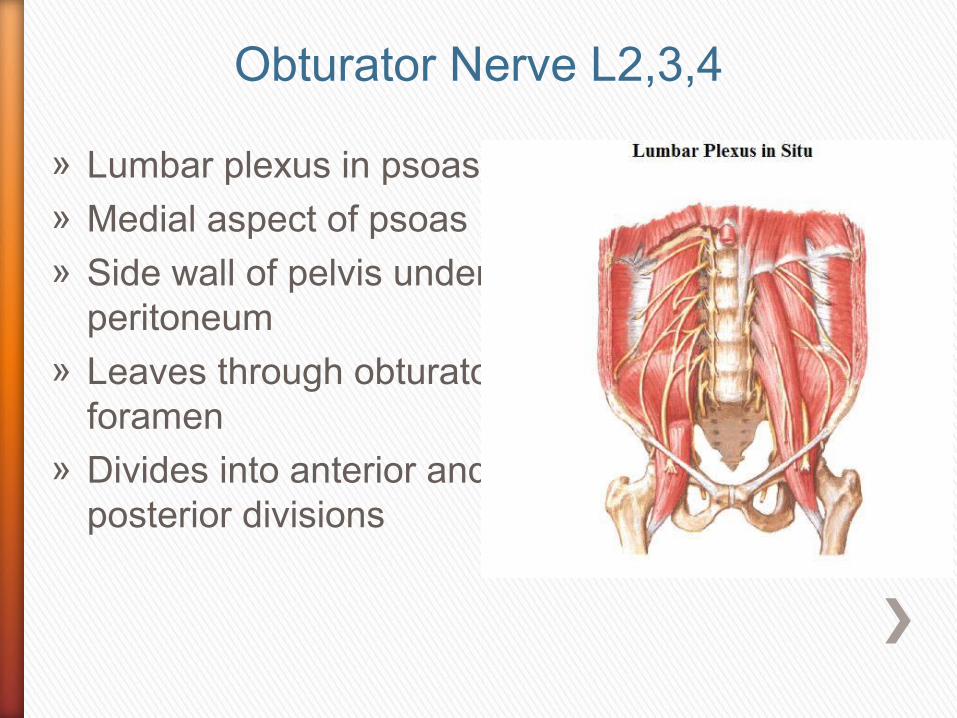
» Lumbar plexus in psoas» Medial aspect of psoas» Side wall of pelvis under
peritoneum» Leaves through obturator
foramen» Divides into anterior and
posterior divisions
Obturator Nerve L2,3,4
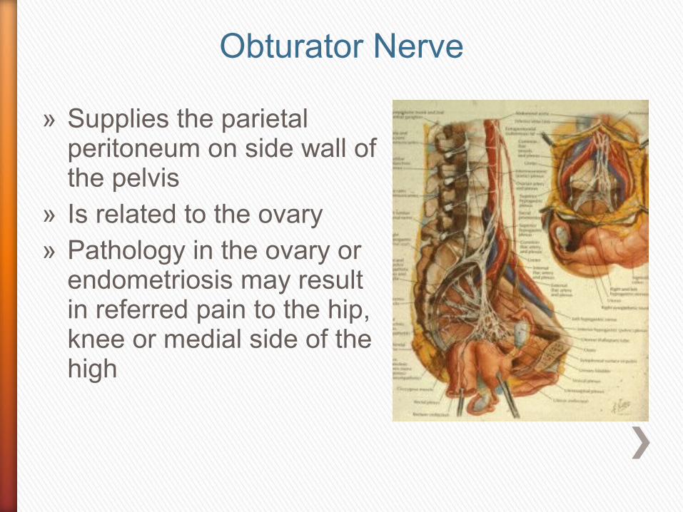
» Supplies the parietal peritoneum on side wall of the pelvis
» Is related to the ovary» Pathology in the ovary or
endometriosis may result in referred pain to the hip, knee or medial side of the high
Obturator Nerve

» Anterior division of the obturator leaves pelvis
» Anterior to obturator externus
» Descends in front of adductor brevis
» Behind pectineus and adductor longus
Obturator nerve

» Adductor longus » Adductor brevis» Gracilis» It gives an articular twig
to the hip joint» Skin on the medial side
of the thigh
Anterior Division Obturator

Obturator Nerve
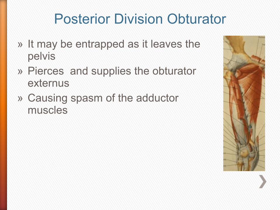
» It may be entrapped as it leaves the pelvis
» Pierces and supplies the obturator externus
» Causing spasm of the adductor muscles
Posterior Division Obturator

» Supplies adductor portion of adductor magnus, above hiatus
» Articular twig to knee joint and cruciate ligaments
» Causing spasm of the adductor muscles
» It may be entrapped as it leaves the pelvis or between fascial planes
Posterior Division Obturator
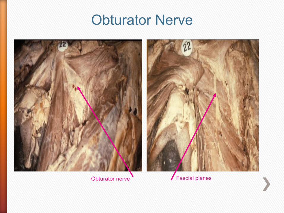
Obturator nerve
Obturator Nerve
Fascial planes
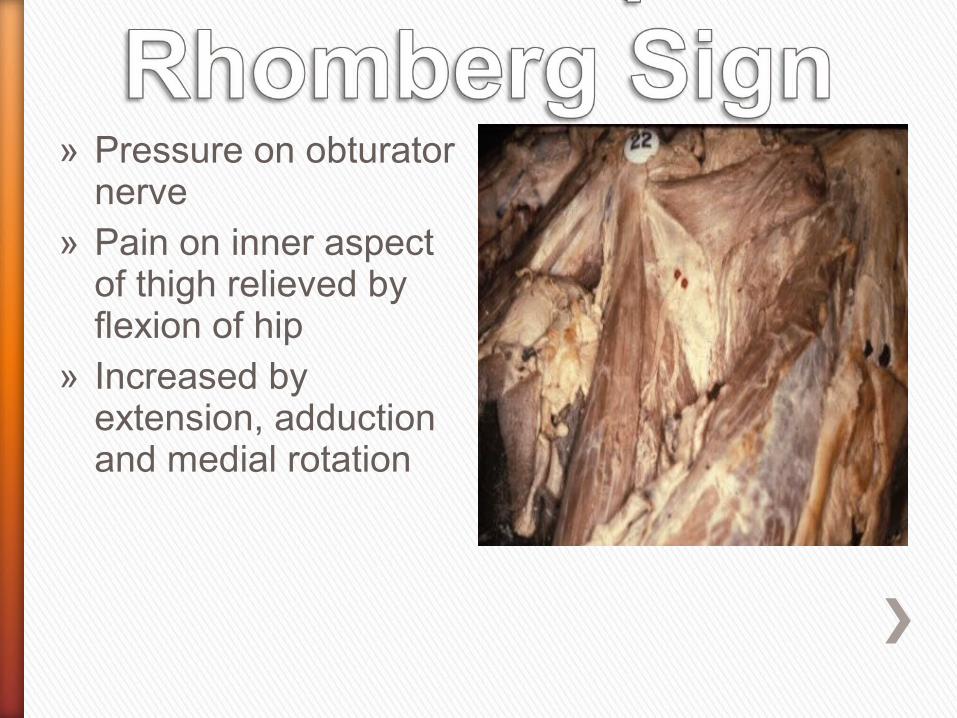
» Pressure on obturator nerve
» Pain on inner aspect of thigh relieved by flexion of hip
» Increased by extension, adduction and medial rotation

Obturator Nerve


» Compression of pudendal nerve in cyclists due to saddle
» History of change of saddle» Compressing dorsal nerve of
penis
Pudendal Nerve

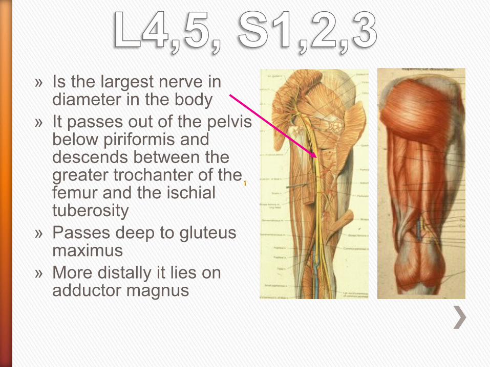
» Is the largest nerve in diameter in the body
» It passes out of the pelvis below piriformis and descends between the greater trochanter of the femur and the ischial tuberosity
» Passes deep to gluteus maximus
» More distally it lies on adductor magnus
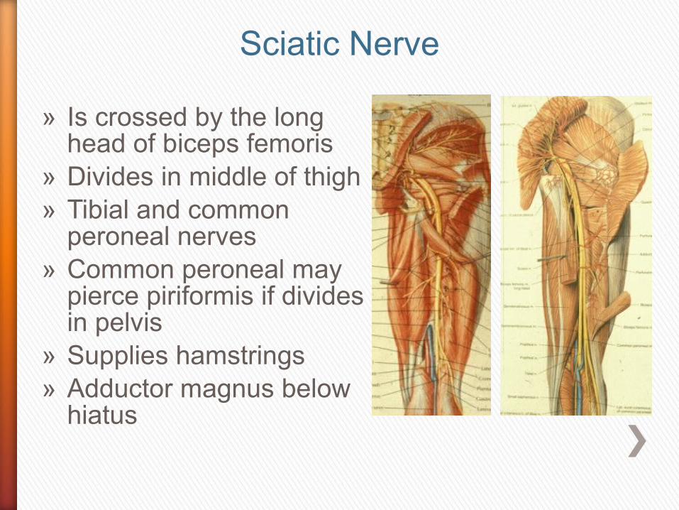
» Is crossed by the long head of biceps femoris
» Divides in middle of thigh» Tibial and common
peroneal nerves» Common peroneal may
pierce piriformis if divides in pelvis
» Supplies hamstrings » Adductor magnus below
hiatus
Sciatic Nerve
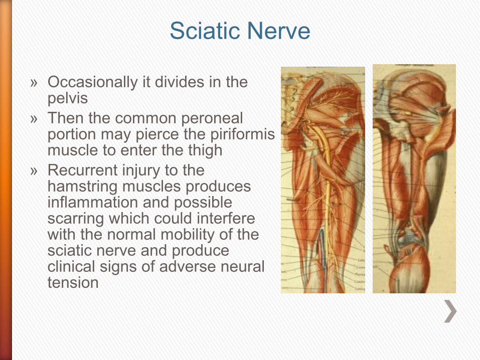
» Occasionally it divides in the pelvis
» Then the common peroneal portion may pierce the piriformis muscle to enter the thigh
» Recurrent injury to the hamstring muscles produces inflammation and possible scarring which could interfere with the normal mobility of the sciatic nerve and produce clinical signs of adverse neural tension
Sciatic Nerve
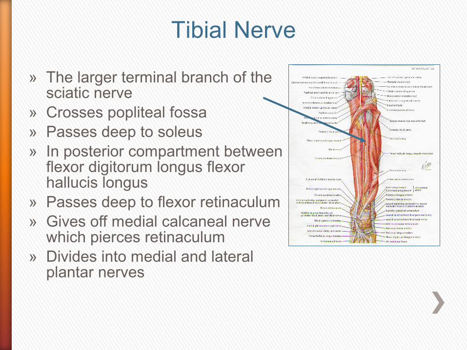
» The larger terminal branch of the sciatic nerve
» Crosses popliteal fossa» Passes deep to soleus» In posterior compartment between
flexor digitorum longus flexor hallucis longus
» Passes deep to flexor retinaculum » Gives off medial calcaneal nerve
which pierces retinaculum» Divides into medial and lateral
plantar nerves
Tibial Nerve

» The tibial nerve supplies all the muscles of the posterior compartment of calf
» In popliteal fossa gives off » Superomedial, middle and inferomedial
genicular branches» Nerve to medial and lateral heads of
gastronemii» Plantaris» Popliteus » Soleus» Sural nerve
Tibial Nerve

» Medial side of psoas» Side wall of pelvis» Obturator canal» Divides anterior posterior
division

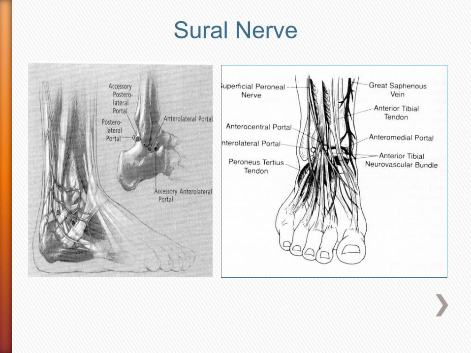
» Sural nerve is joined by sural communicating from commom peroneal
» Pierces deep fascia » Supplies posterior and
lateral portion of calf» Lateral border of foot» Entrapment occurs most
frequently in runners with a history of ankle sprain
Sural Nerve
Sural Nerve

» Deep fascia from medial malleolus to medial margin of calcaneus
Anterior to posterior» Tibialis posterior» Flexor digitorum longus» Posterior tibial artery » Tibial nerve» Both give off medial calcaneal artery and nerve» Then both divide into medial and lateral plantar
branches» Flexor hallucis longus

» Gives off the medial calcaneal nerve under cover of the retinaculum
» It then pierces the flexor retinaculum to supply the posterior and medial aspect of the heel
Tibial Nerve
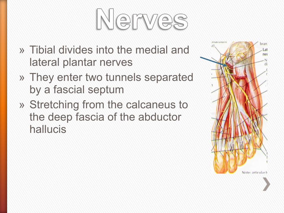
» Tibial divides into the medial and lateral plantar nerves
» They enter two tunnels separated by a fascial septum
» Stretching from the calcaneus to the deep fascia of the abductor hallucis

» Passes under the abductor hallucis
» Then runs on the plantar surface of the flexor digitorum longus
» Dividing into its digital branches
» Sensory to the plantar aspect of the medial three and a half toes
Medial Plantar Nerve

» Motor to the abductor hallucis» Flexor hallucis brevis» Flexor digitorum brevis » First or unipennate lumbrical
Medial Plantar Nerve

» Crosses the sole of the foot deep to the abductor hallucis, flexor digitorum brevis and the abductor digiti minimi
» To the base of 5th metatarsal
» Superficial to flexor hallucis longus, flexor digitorum longus and flexor accessorius
» Lateral plantar nerve supplies the lateral one-and-a-half toes
» Supplies all the other intrinsic muscles of the foot
Medial Plantar Nerve
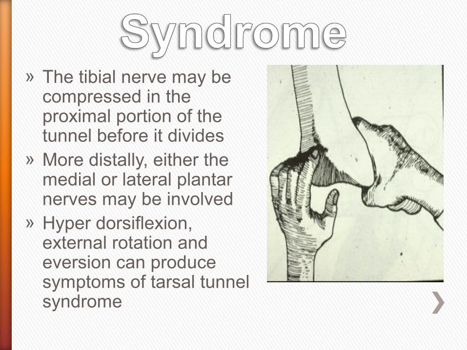
» The tibial nerve may be compressed in the proximal portion of the tunnel before it divides
» More distally, either the medial or lateral plantar nerves may be involved
» Hyper dorsiflexion, external rotation and eversion can produce symptoms of tarsal tunnel syndrome
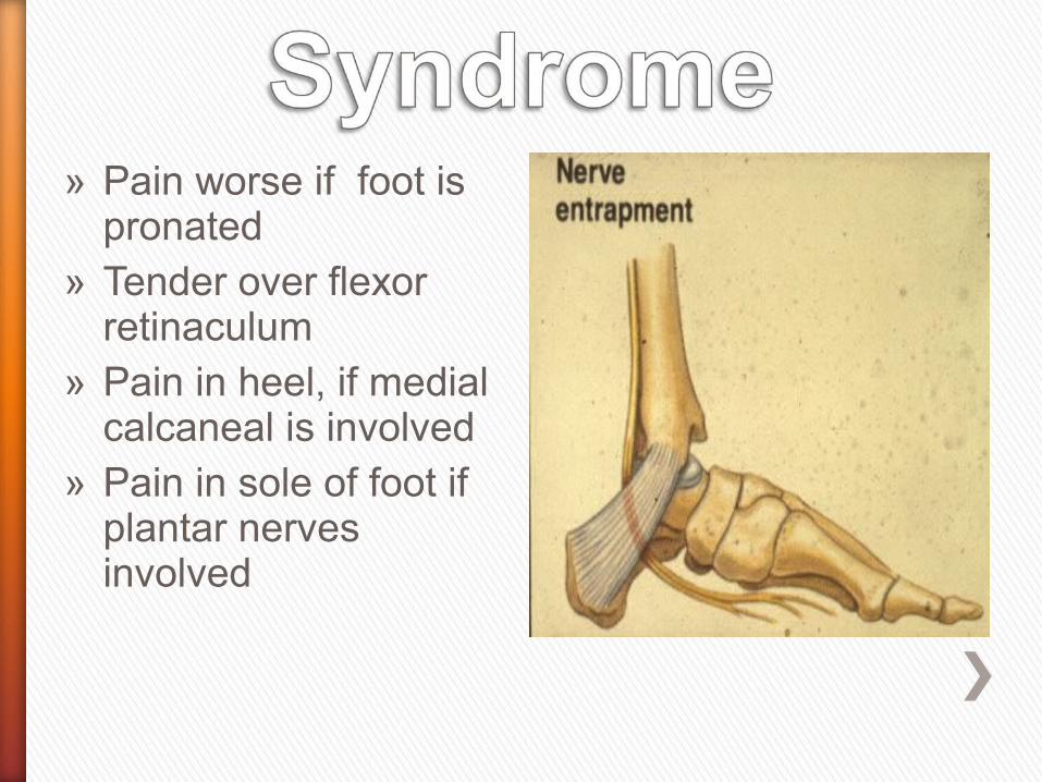
» Pain worse if foot is pronated
» Tender over flexor retinaculum
» Pain in heel, if medial calcaneal is involved
» Pain in sole of foot if plantar nerves involved
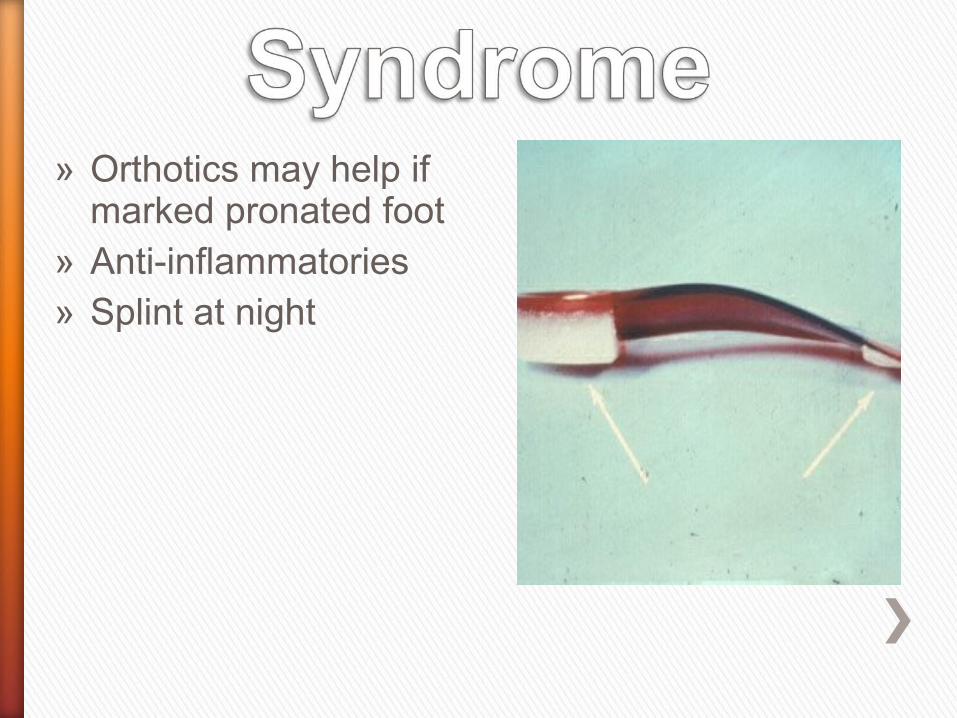
» Orthotics may help if marked pronated foot
» Anti-inflammatories» Splint at night


» Depressed transverse arch in runners and ballet dancers with mobile first ray
» Neuroma on digital nerve to the second cleft or third cleft
morton neuroma.jpg
Morton’s Foot

» Pronated foot» Depressed transverse
arch at heads of metatarsals
» Pain worse with tight shoes on
» Relieved by removing shoes
Medial Plantar

» If sciatic nerve divides inside the pelvis
» Common peroneal nerve pierces the piriformis
» May be entrapped» In popliteal fossa» The common peroneal
nerve lies between the tendon of biceps femoris and the lateral head of gastrocnemius

» In popliteal fossa gives off» Lateral cutaneous of calf» Sural communicating» Superior lateral, inferior lateral
genicular nerves» Leaves fossa at lateral angle» Crosses neck of fibula deep to
peroneus longus » Gives off recurrent genicular, deep
and superficial peroneal» Vulnerable to injury as it winds around neck of
fibula» Foot drop, plantar flexed, inverted
Common Peroneal

» Nerves can be compressed in compartments
» Anterior compartment deep peroneal nerve
» Lateral compartment superficial peroneal
» Posterior compartment tibial nerve

» Branch of the common peroneal at the neck of the fibula
» Pierces the lateral inter-muscular septum to enter the anterior compartment
» Supplies all muscles in anterior compartment tibialis anterior, extensor hallucis longus, extensor digitorum longus, peroneus tertius and extensor digitorum brevis
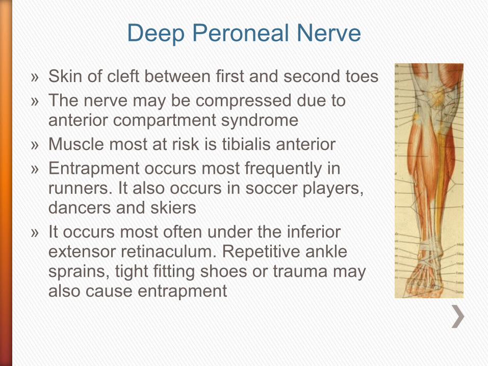
» Skin of cleft between first and second toes» The nerve may be compressed due to
anterior compartment syndrome» Muscle most at risk is tibialis anterior» Entrapment occurs most frequently in
runners. It also occurs in soccer players, dancers and skiers
» It occurs most often under the inferior extensor retinaculum. Repetitive ankle sprains, tight fitting shoes or trauma may also cause entrapment
Deep Peroneal Nerve

» Runs in the lateral compartment of the calf
» Between the peroneus longus and brevis supplying both these muscles
» Pierces the deep fascia 10–12 cm above the lateral malleolus supplies most of the dorsum
Superficial Peroneal Nerve

» Divides 6 cm above the lateral malleolus into branches, which supply the dorsum of the foot
» The first cleft is supplied by the deep peroneal
» The lateral border is supplied by the sural nerve
» Medial border to ball of big toe saphenous
» Rest superficial peroneal

» Entrapment occurs where the superficial peroneal pierces the deep fascia
» Particularly if there is herniation of the muscle due to fascial defects
» Chronic ankle strains also stretch the nerve

» Grade III ankle injuries have a high incidence of traction injuries to both the peroneal and posterior tibial nerves
Taunton & Fricker, 1996