Anatomy of face
19
Anatomy of face Dr. Firas Kassab Page 1 Anatomy of face Dr. Firas Kassab
-
Upload
feras-kassab -
Category
Health & Medicine
-
view
140 -
download
0
Transcript of Anatomy of face

Anatomy of face
Dr. Firas Kassab Page 1
Anatomy of
face
Dr. Firas Kassab

Anatomy of face
Dr. Firas Kassab Page 2
Anatomy of face
FACIAL MUSCLES
Arrangement produces tension lines on the skin that are directed at right angles
to the plane of the muscle fiber.
These tension lines (Langer’s cleavage lines) deepen with the aging process as
the skin loses its elasticity.

Anatomy of face
Dr. Firas Kassab Page 3
Scalp and forehead
A series of muscles that allow it to be
moved in an upward or downward
direction.
The frontal belly fibers of the occipitofrontal muscle have no bony
attachment.
They are continuous with the
procerus, corrugator, and
orbicularis oculi.
The fibers are directed
vertically, thinning before
inserting into an aponeurosis
termed the galea
aponeurotica, which they
share with the occipital belly
fibers.
The occipital belly arises
from bone and periosteum
along the superior nuchal line
before its fibers join into the aponeurosis.
The occipital frontal muscles acting together draw the scalp backward, raising the
eyebrows and wrinkling the forehead.
The frontal bellies, acting alone, raise the eyebrows.
The corrugator supercilii, arises from the orbital rim near the medial canthus
and then directs its fibers superiorly and laterally to insert into the deep surface
of the frontal belly of the occipitofrontal muscle.
Contraction of the corrugator supercilii draws the brow medially, producing the
oblique frown lines over the glabellar region.

Anatomy of face
Dr. Firas Kassab Page 4
Scalp structure: skin, connective
tissue, aponeurosis, loose connective
tissue, and periosteum.
The majority of blood vessels and
cutaneous nerves are located in the
dense connective tissue space
superficial to the galea aponeurotica.
An important exception to this rule is
the origin of the supraorbital and
supratrochlear neurovascular bundle
at the rim of the orbit.
As these vessels emerge from the orbit, they must traverse the loose connective
space and galea aponeurotica before penetrating into the dense connective
tissue plane.
The term danger space as applied to the loose connective tissue layer refers not
to this particular traumatic event but to the fact that infection may spread readily
in this space and be subsequently transmitted intracranially by emissary veins.
Orbital region and eyelid

Anatomy of face
Dr. Firas Kassab Page 5
The orbicular eye muscle, a series of concentric rings, originates either from
the medial palpebral ligament or from bone on the medial orbital wall.
Three parts of the muscle are usually described:
The large orbital region, which covers the superior and inferior limits of the orbit;
The palpebral part, which is adjacent to the upper or lower eyelids, and
The lacrimal portion attached to the posterior lacrimal crest.
Closure of the entire orbital region and lid is accomplished by coordinated
contraction of the entire muscle group, whereas blinking is limited to the
palpebral region of the muscle covering the lid.
The palpebral portion of the muscle passes posterior to the medial palpebral
ligament adjacent to the lacrimal sac.
When the eye is closed, these fibers can exert traction on the lacrimal sac,
serving as a pump to aid in the drainage of the tears.
The orbital portion is attached to the skin where it broadens over the anterior
temporal and malar region.
These skin attachments give rise in the aging face to the radially oriented skin
creases known as crow’s feet.
The palpebral portion does not attach to the overlying skin or underlying orbital
septum, but rather is separated from these structures by a layer of areolar
connective tissue.
This relationship accounts for the ease in developing a cutaneous or
musculocutaneous flap during blepharoplasty.
In the central part of the upper lid just above the margin of the eyelash, the
layers of the eyelid are skin, orbicular eye muscle, tarsal plate, and conjunctival
sac. SOTC
More peripherally the layers are skin, orbicular eye muscle, orbital septum,
preaponeurotic space, levator aponeurosis, postaponeurotic space, superior tarsal
muscle, and conjunctival layer. SOSPLPSC
Three muscles, therefore, are represented in the upper lid.
The orbicular eye muscle is a striated muscle supplied by CN VII, which is
responsible for lid closure.
The levator palpebrae superioris muscle, also striated, functions to elevate
the upper lid and is supplied by CN III.
Superior tarsal muscle is smooth muscle supplied by the sympathetic nervous
system and has only a minor role in supporting the lid.
Nevertheless, loss of function in either CN III or the sympathetic nervous system
can result in ptosis of the upper lid.

Anatomy of face
Dr. Firas Kassab Page 6
Interruption of sympathetic fibers along any point from the spinal cord to the
point of innervation of the muscle will result in ptosis and myosis, which are part
of Horner’s syndrome.
The additional clinical findings of Horner’s syndrome, anhidrosis and
vasodilatation of the ipsilateral face, occur only if the fibers are disrupted in the
neck at the level of the superior cervical sympathetic ganglion.
Clinically, the septum orbitale is a particularly important structure in that it
represents a line of continuity between periosteum and periorbita, and technically
separates the intraorbital contents from the extraorbital structures.
Preseptal infections of the eyelid are known as periorbital cellulitis, and are more
common and less serious than postseptal orbital cellulitis.
Nose
The muscles of the nose include the procerus muscle and the nasal muscle.
The procerus muscle is a small muscle overlying the bridge of the nose and in
part continuous with the frontal belly fibers of the occipitofrontal muscle.

Anatomy of face
Dr. Firas Kassab Page 7
This muscle actually works with the frontal belly fibers in drawing the skin of the
forehead downward during the act of squinting.
Because of its vertically arranged muscle fibers, it forms transverse frown lines in
the skin between the eyes.
During rhinoplasty, avulsion of the procerus muscle is sometimes performed in
attempts to deepen the nasofrontal angle of the nose.
The nasalis muscle is frequently described as having two portions: a
transverse portion and an alar portion.
The transverse fibers, also termed the compressor naris, form an inverted U over
the bridge of the nose.
As the name implies, these fibers compress the naris by compressing or flexing
the upper lateral nasal cartilage at its joint with the alar or lower lateral nasal
cartilage.
Dilatation of the naris is accomplished by the alar portion, which elevates the
lower lateral or alar cartilage.
Mouth region (cheek and lips)

Anatomy of face
Dr. Firas Kassab Page 8
The buccinator muscle arises from the pterygomandibular raphe and the
tuberosity of the maxilla.
The fibers of the muscle cross anteriorly in the substance of the cheek and then
blend at the corner of the mouth with the fibers of the orbicular mouth muscle.
The essential function of the bucinator muscle is to maintain food between the
teeth during the masticatory process.
The orbicular mouth muscle is a series of concentric rings surrounding the
mouth, which, when contracted, purse the lips.
Many of the fibers interlace with the buccinator muscle, as well as with muscles
that elevate or depress the lip.
Elevation of the upper lip is accomplished by either the levator labii or the levator
anguli oris muscle.
The levator labii muscle elevates the lip itself, whereas the levator anguli oris
muscle moves the angle of the lip.
A complementary pair of muscles is found in the lower lip: the depressor labii and
depressor anguli oris muscles.
The greater and lesser zygomatic muscles are located more superficially
than the levators of the lip.
Both the greater and lesser zygomatic muscles arise from the zygomatic bone
lateral to the infraorbital foramen.
The lesser zygomatic attaches into the orbicular mouth muscle near the ala of
the nose, whereas the greater zygomatic attaches at the corner of the mouth.
Both are elevators of the lip, although the lesser zygomatic muscle is important in
producing the melolabial fold.
Chin and neck
The mentalis
(levator labii
inferioris) is a flat
band of fibers arising
from the mandible
near the roots of the
incisors and inserting
into the skin near the

Anatomy of face
Dr. Firas Kassab Page 9
midline.
The action of these fibers is to tense the skin of the chin as well as aid in
protruding the lower lip.
Although the platysma may be thought of as a muscle of the neck region, it is in
fact part of the facial musculature.
CN VII exits the skull via the stylomastoid foramen.
The first branch to arise from CN VII after it emerges from the stylomastoid
foramen is the posterior auricular nerve.
This nerve courses inferior to the auricle, ascending superficially on the mastoid
process to supply eventually the occipital belly of the occipitofrontal muscle.
In relation to the auricle, some sensory fibers may supply the skin of the external
auditory meatus.
The evidence for this is the clinical presentation of vesicles in herpes zoster oticus
(Ramsay-Hunt syndrome).

Anatomy of face
Dr. Firas Kassab Page 10
Also arising from the major trunk of CN VII in this region are direct motor
branches to the posterior belly of the diagastric muscle and to the stylohyoid
muscle.
The remaining trunk of CN VII forms two or three divisions with five major
branches: temporal, zygomatic, buccal, mandibular, and cervical.
The temporal branch of CN VII arises from the anterior aspect of the parotid
gland and courses superficial to the zygomatic arch.
It provides motor innervation to the frontal belly of the occipitofrontal muscle, as
well as to the orbicular eye muscle and the corrugator supercilii muscle.
The zygomatic branch ascends to the lateral canthus, where it also supplies the
orbicular eye muscle and forms an anastomotic network with the temporal
branch.
The buccal branch crosses transversely on the face to supply the central muscles
of the face, including the greater and lesser zygomatic muscles, the levator
anguli oris and levator labii superioris muscles, and all the small muscles
associated with the surface of the nose.
The buccal branch overlaps with the zygomatic branch in its supply of muscles in
the central part of the face.
Principally, it supplies the buccinator muscle and the orbicular eye muscle.
The mandibular branch (marginal mandibular) crosses inferior to the angle of the
mandible into the submandibular triangle and then ascends, crossing the
mandible a second time to supply the muscles over the surface of the chin.
The cervical branch of CN VII emerges from the inferior tip of the parotid gland,
following the deep surface of the platysma muscle, which it innervates.
Area where the temporal branch crosses the zygomatic arch represents the most
superficial location of any of the major divisions of the facial nerve, and is
considered a “danger zone” for facelift surgery.
Terminals of the zygomatic branch at the point where they innervate the
orbicular eye muscle are essentially at the level of the dermis.
Even shallow incisions (for removal of moles, for example) may divide these
nerves, producing paralysis of the orbicular eye muscle and an inability to close
the eye.
Another particularly vulnerable site is the region of the submandibular triangle.
Although the mandibular branch of CN VII is under the cover of the platysma,
the site is a common surgical approach to the submandibular gland.
Interruption of the mandibular branch of CN VII results in an inability to depress
the lower lip.

Anatomy of face
Dr. Firas Kassab Page 11
In these patients the angle of the lip on the affected side is slightly elevated,
whereas the lower portion of the lip on the affected side is pulled to the opposite
side by tonic activity of the nonparalyzed muscles.
There are certain anatomic relationships between branches of CN VII and
adjacent structures that are important to the surgeon performing parotidectomy,
repairing cheek lacerations that have injured branches of the nerve, or
performing other types of facial surgery.
The zygomatic branch lies approximately 1 cm below the zygomatic arch in the
region of the parotid gland, a fact that can be used as a guide in identifying this
branch.
The buccal branch crosses the parotid duct on its superficial surface, running
from superior to inferior before turning anteriorly to be distributed to the facial
musculature.
By virtue of this relationship, injuries usually involve both structures
concomitantly.
The mandibular branch usually lies below the point at which the facial artery and
vein emerge from the submandibular gland.
In such instances, the commonly used maneuver in which the vascular structures
are identified, divided, and elevated along with the overlying platysma and skin,
does not represent a safe method of protecting the mandibular nerve.
The nerve does, however, lie external to the fascial capsule of the submandibular
gland and rarely extends below its inferior margin.
Incising the capsular fascia at the inferior margin of the gland and elevating it
along with the overlying tissues will provide protection for the nerve.
The cervical branch of CN VII has an important relationship to the posterior
division of the retromandibular vein, which, together with the posterior auricular
vein, forms the external jugular vein.
The nerve lies immediately on the lateral aspect of the posterior division of the
retromandibular vein.
Knowledge of this relationship can be used in performing retrograde dissection to
the main trunk of CN VII.
This is accomplished by identifying the external jugular vein, which is an easily
located surgical landmark in the neck, and dissecting the vein upward to the
posterior division of the retromandibular vein, at which point the cervical branch
is encountered.
The branch can be dissected retrograde to the inferior division of CN VII.
Lesions of the seventh cranial nerve

Anatomy of face
Dr. Firas Kassab Page 12
A lower motor neuron lesion is a deficit of the motor neuron in the CN VII
nucleus or at any point distal to the nucleus.
Thus, all motor branches, whether from the intracranial or facial parts of CN VII,
would be affected.
If the lesion is complete, a total hemiparesis of the face will result. Bell’s palsy is
an example of such a lesion, although in this case the site of injury is the bony
facial canal in the temporal bone.
In contrast, should a lesion affect the upper motor neuron at any point from the
motor cortex or along the length of the axon before it synapses with the facial
nucleus, a different set of physical findings will result.
Paralysis as a result of this type of lesion spares the muscles of the upper portion
of the face (that is, the orbicular eye and occipitofrontal muscles). However, the
muscles of the central and lower portions of the face are paralyzed.
This sparing occurs because the muscles of the upper part of the face receive
motor supply from both cerebral cortices.
Thus in a patient with a stroke lesion in the cerebral cortex or internal capsule,
the orbicular eye and occipitofrontal muscles will be spared on the contralateral
side because they receive secondary innervation from the ipsilateral cerebral
cortex.
Thus, the usual finding in stroke patients is paralysis of the muscles of the nose
and mouth on the contralateral side of the cortical lesion.
CUTANEOUS FACIAL INNERVATION

Anatomy of face
Dr. Firas Kassab Page 13
A line projected from the tip of the chin to the vertex of the skull will define a
plane in which the trigeminal nerve supplies all skin lying anterior to this plane.
In contrast, the cervical plexus innervates all skin lying posterior to this plane.
The ophthalmic division of the trigeminal begins at the semilunar
ganglion of CN V in the middle cranial fossa.
After traversing the cavernous sinus, it enters the orbital cavity through the
supraorbital fissure.
At this point it divides into several branches, some of which eventually reach the
skin of the nose and periorbital region.
The three subdivisions of the ophthalmic branch within the orbit are the lacrimal,
the nasociliary, and the frontal. NLF
The lacrimal nerve follows along the lateral and superior wall of the orbit and
supplies a small area of skin near the upper lateral portion of the eyelid.
The nasociliary nerve supplies two areas of skin: one via the infratrochlear
nerve to the skin near the medial canthus of the upper lid and the other via the
external nasal branch to the skin over the bridge of the nose.
The external nasal nerve is not a direct branch of the nasociliary nerve; rather, it
is the terminal branch of the anterior ethmoidal nerve after it supplies the
ethmoid sinuses.
The lacrimal, infratrochlear, and external nasal nerves supply only small areas of
skin on the face.
The major area of skin in the periorbital and frontal regions is supplied via
branches of the frontal nerve from the ophthalmic division.
The frontal nerve is given off soon after the ophthalmic division enters the
orbit.
Traversing the superior part of the orbital wall, the frontal division divides into
the supraorbital and supratrochlear nerves, which emerge from the orbit through
the supraorbital notch.
In some individuals the notch is closed to form a bony foramen.
From this point the nerve pierces the loose connective tissue and aponeurotic
layers of the scalp to travel upward in the dense connective tissue layer of the
scalp.
The maxillary division of the trigeminal nerve begins at the
semilunar ganglion and enters the orbital region via the round foramen.

Anatomy of face
Dr. Firas Kassab Page 14
As it enters the floor of the orbit, a zygomatic branch is given off that abruptly
subdivides into two branches: the zygomaticotemporal and the zygomaticofacial.
These nerves supply skin over the temporal and malar regions, respectively.
After the zygomatic branch is given off, the maxillary nerve enters the
infraorbital groove and traverses the infraorbital canal, emerging on the face via
the infraorbital foramen.
At this point three named branches of the infraorbital nerve appear: the
palpebral branch, which supplies the lower lid; the external nasal branch, which
supplies the lateral and alar portions of the nasal skin; and the labial branch,
which supplies the upper lid and cheek skin. PEL
In orbital blow-out fractures that disrupt the orbital floor, the zygomatic branch
of the maxillary nerve is usually spared, because the injury is distal to its origin.
However, the infraorbital branch of the maxillary nerve can be completely
involved, leaving the patient with an anesthetized area over the lower lid, side of
the nose, and upper lip and cheek.
Furthermore, because the anterosuperior alveolar nerves arise from the
infraorbital nerve, the anterior teeth (incisors and canines) are also numb.
These nerves descend from the infraorbital nerve through small bony canals
located in the anterior wall of the maxillary sinus.

Anatomy of face
Dr. Firas Kassab Page 15
Caldwell-Luc and other surgical procedures that remove part or all of the anterior
wall of the maxillary sinus will result in some numbness of these teeth as well.
The mandibular nerve exits the middle cranial fossa via the oval foramen.
Here the nerve enters the infratemporal fossa and divides into several named
branches, three of which are important to the innervation of the face.
One is the auriculotemporal nerve, which courses laterally around the neck of the
mandible and ascends the side of the skull in the skin of the scalp anterior to the
ear.
In addition to being a sensory nerve, the auriculotemporal nerve carries both
parasympathetic postganglionic fibers from the otic ganglion to the parotid gland,
and sympathetic fibers distributed via the carotid artery to innervate sweat
glands of the skin in the area of the nerve distribution.
Parotidectomy results in division of some of the branches of the auriculotemporal
nerve, and may give rise to faulty reinnervation of sweat gland secretomotor
receptors by parotid gland secretomotor fibers, resulting in the auriculotemporal
(Frey) syndrome of gustatory sweating.
The second important sensory branch of the mandibular nerve is the buccal
branch, which traverses the infratemporal fossa and supplies part of the buccal
mucosa, as well as the skin over the cheek region.
The third branch is the inferior alveolar nerve, which, after entering the
mandibular canal and supplying all of the mandibular teeth, emerges as the
mental nerve, supplying skin over the point of the chin.
Anatomy of face
Dr. Firas Kassab Page 16
While all of these nerves may be affected by lesions involving the proximal trunk
of the mandibular division, each of these nerves individually may be injured by
operative procedures or trauma.
In the case of the auriculotemporal nerve, it may be injured by fractures of the
neck of the mandible or during face-lifting procedures when the skin anterior to
the ear is being elevated over the path of the nerve.
Mandibular fractures through the mandibular canal almost always produce
impairment of the mental nerve, as well as numbness of the teeth.
Injury to the mental nerve can also occur during the elevation of skin or mucosal
flaps in the region of the mental foramen.
Another consideration with respect to the anatomy of the trigeminal nerve is in
regard to herpes zoster.
The distribution of vesicles on the skin usually follows the pattern of one of the
trigeminal divisions.
Herpetic involvement of the ophthalmic division often causes more discomfort to
patients because of the distribution of the nasociliary branch to the cornea and
conjunctiva, resulting in excessive tearing, burning sensations, and shooting
pains over the eye.
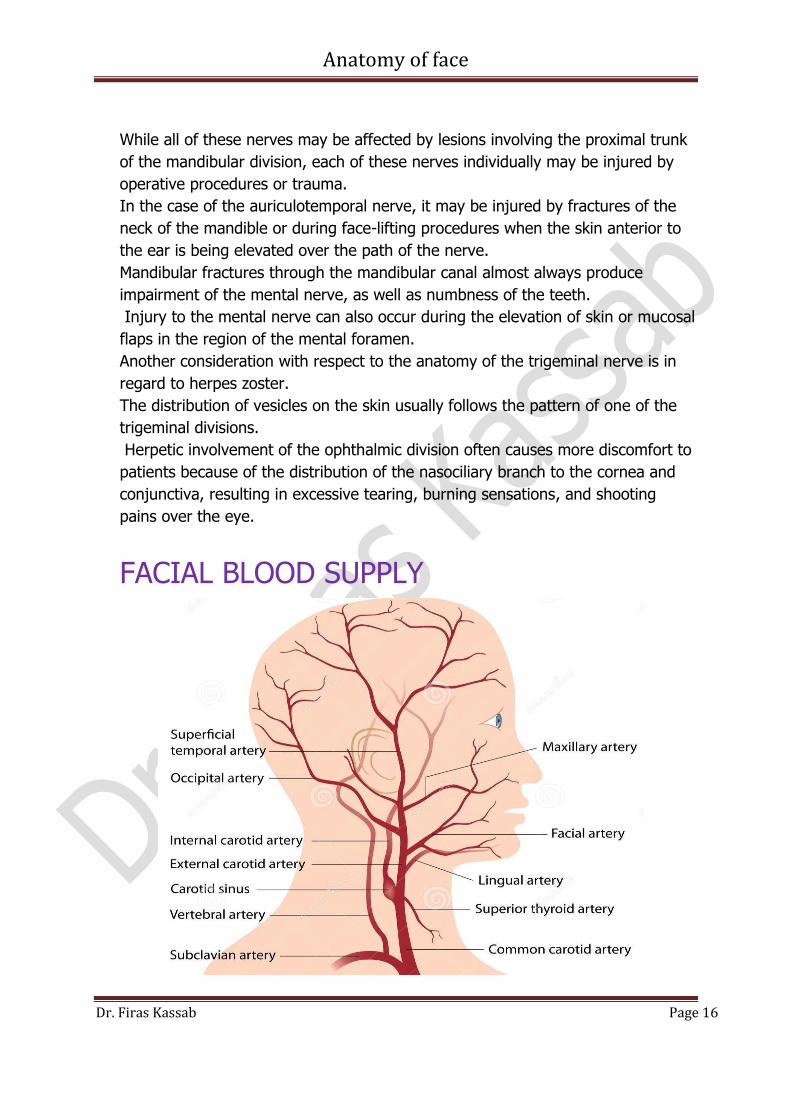
FACIAL BLOOD SUPPLY
Anatomy of face
Dr. Firas Kassab Page 17
Both the internal and external carotid branches supply the face.
The internal carotid gives off an ophthalmic branch at the level of the circle of
Willis.
This vessel traverses the optic canal, where it enters the orbit and is concerned
principally with supplying the eye and intraorbital tissues.
From this plexus of intraorbital arteries, two branches emerge; the supraorbital
and the supratrochlear emerge at the orbital rim to parallel the course of the
supraorbital and supratrochlear nerves.
These form a neurovascular bundle in the dense connective layer of the scalp. In
summary, the internal carotid supplies the periorbital and scalp tissues of the
forehead.
Two branches of the external carotid contribute to the blood supply of the face
along with the internal carotid. These are the superficial temporal artery and the
facial artery itself.
The superficial temporal artery is the terminal branch of the external
carotid.
It begins in the substance of the parotid, exiting at the superior pole of the
gland, where it joins the auriculotemporal nerve to lie just under the skin anterior
to the tragus of the ear.
It is also vulnerable during the elevation of skin flaps for face-lifting procedures.
After entering the upper portion of the scalp, the superficial temporal artery
anastomoses in the region of the forehead with the supraorbital and
supratrochlear branches of the ophthalmic artery.
There is also an anastomotic pattern over the occipital area with posterior
auricular branches of the external carotid.
The facial artery, a branch of the external carotid artery, supplies the major
portion of the face. After its origin from the external carotid, the facial artery
ascends deep to the posterior belly of the digastric muscle and crosses the
mandible at the anterior margin of the masseter muscle.
Here, the artery lies deep to the plane of the facial muscles and is protected by
them until it reaches the angle of the mouth, where the artery divides into
inferior and superior labial arteries.
The main trunk continues into the nasolabial angle and, after coursing deep to
the lesser zygomatic muscle, is no longer covered by a muscle sheath.
The artery terminates by sending small branches to the lateral aspect of the nose
and finally anastomosing in small networks with the plexus of vessels about the
orbital region.

Anatomy of face
Dr. Firas Kassab Page 18
This effectively provides an anastomotic link between the facial and ophthalmic
arteries (external and internal carotid systems, respectively).
In some individuals the facial artery may end effectively at the angle of the
mouth.
In this case the transverse facial artery originates from the superficial temporal
artery within the parotid gland.
This vessel exits the parotid, following a course similar to the zygomatic branch
of the facial nerve. When this vessel is large, it replaces the angular portion of
the facial artery.
FACIAL VENOUS DRAINAGE

Anatomy of face
Dr. Firas Kassab Page 19
The principal venous drainage of the face occurs via the facial and
retromandibular veins.
The facial vein begins as the angular vein near the medial canthus of the
eye, coursing inferiorly in the nasolabial angle.
The vessel receives branches from the nose and region of the lip.
Paralleling the course of the facial artery, it descends on the face, crossing the
angle of the mandible, where it joins the common facial vein, which empties into
the internal jugular vein.
The common facial vein also receives the retromandibular vein, which begins in
the scalp as the superficial temporal vein.
When the superficial temporal vein enters the superior portion of the parotid
gland, it is joined by the maxillary vein from the infratemporal fossa.
The union of these two vessels forms the retromandibular vein before it exits the
parotid at its inferior pole to join the common facial vein at the angle of the
mandible.
In addition to joining the common facial vein, the retromandibular vein also
anastomoses with the external jugular vein.
In this manner the anterior and more lateral aspects of the face drain to both the
internal and external jugular veins.
The veins of the orbital region and lower portion of the forehead-scalp are
tributaries of the ophthalmic veins within the orbital cavity.
Usually there are superior and inferior branches of the orbital vein, which collect
blood from the orbital region.
These veins usually drain posteriorly through the apex of the orbit, becoming
tributaries of the cavernous sinus.