ANATOMY HANDBOOK - Semmelweis...
14
Semmelweis University Department of Anatomy, Histology and Embryology Faculty of Medicine 2 nd year 1 st semester ANATOMY HANDBOOK September 2017 Dr. Andrea D. Székely Associate Professor Course Director of the English Language Program Dr. Ágoston Szél Full Professor Head of Deaprtment, Rector
Transcript of ANATOMY HANDBOOK - Semmelweis...
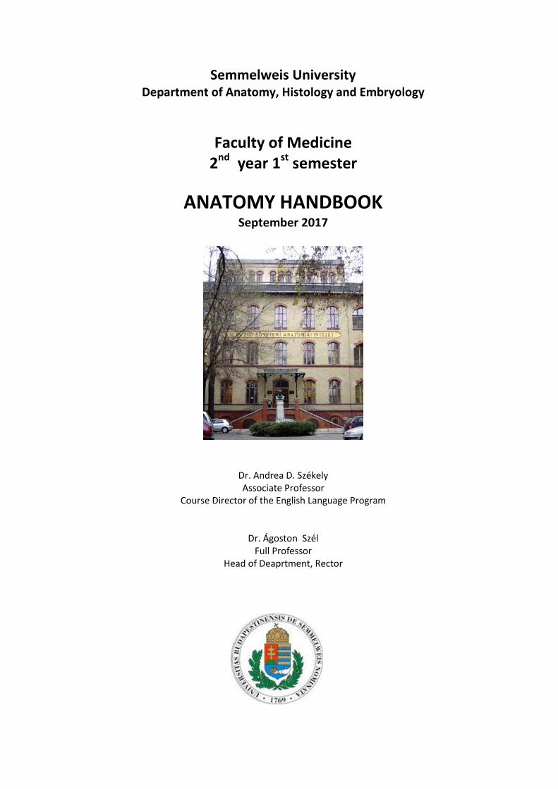
Semmelweis University Department of Anatomy, Histology and Embryology
Faculty of Medicine 2nd year 1st semester
ANATOMY HANDBOOK September 2017
Dr. Andrea D. Székely Associate Professor
Course Director of the English Language Program
Dr. Ágoston Szél
Full Professor Head of Deaprtment, Rector
Anatomy, Histology and Embryology for EM students
TEACHING DEPARTMENT: SEMMELWEIS UNIVERSITY Department of Anatomy, Histology and Embryology Budapest, Tűzoltó utca 58. H-1094 Budapest http://semmelweis.hu/anatomia
LEARNING OBJECTIVES
Aims of the lectures in anatomy: Presentation of the important and/or complicated chapters such as
introductory chapters, thorax, pelvis, hand, foot, skull, heart, chapters of the visceral organs, central nervous
system, organs of special senses, topographical anatomy.
Aims of the lectures in cell biology and histology: Presentation of the cell, basic principles in cell
biology (mitosis, cytoskeleton, cellular motility), detailed presentation of the basic tissues (epithelial,
connective, muscle and nervous). Completing the gross anatomy with the detailed presentation of the fine
structure of the organs, including the ultrastructural details as well as the molecular arrangement. Important
chapters: basic tissues, viscera, central nervous system.
Aims of the lectures in embryology: Presentation of the early development from the differentiation of
the germ cells to the formation of the human embryo (general embryology). Presentation of the
development of the organs and functional systems parallel with the gross anatomical and histological
lectures including the frequently occurring malformations.
Aims of the practical sessions in the dissecting room: Based on the weekly programs the students
study the preparations (bones, joints, muscles, viscera, brain) and dissect (parts of or an entire cadaver). They
are aided by the lab instructors. Bones, joints, muscles and peripheral nervous system will be taught primarily
in the dissecting room.
Aims of the practical sessions in the histology room: Facilitate the understanding of the basic tissues
(epithelial, connective, muscle and nervous) and the fine structure of the organs through the observation and
interpretation of histological specimens.
Discussion of the more complicated chapters of the embryology is presented on small group
discussions connected to the practical sessions in the dissecting room.
The knowledge of the students will be checked by mid-term tests.
Lectures: first semester: 3x 45 min; second semester: 3x 45 min; third semester: 3x 45 min; fourth
semester: 1x 45 min.
Topics of the lectures:
First semester: Gross anatomy of the bones, joints and muscles, basic cytology, basic histology, basic
embryology, development of the skull, spine and limbs.
Second semester: Heart and vessels, lymphatic organs, viscera and body cavities; integrated gross
anatomy, cytology, histology and embryology.
Third semester: Central and peripheral nervous system, organs of special senses, endocrine organs;
integrated gross anatomy, cytology, histology and embryology.
Fourth semester: Topographical anatomy of the head, neck and body cavities (thorax, abdomen,
pelvis), cross sectional anatomy.
Practical course
6x 45 min; second semester: 6x 45 min; third semester: 4x 45 min; fourth semester: 2x 45 min;
First semester: Gross anatomy of the bones, joints and muscles, basic cytology, basic histology, basic
embryology, development of the skull, spine and limbs.
Second semester: Heart and vessels, lymphatic organs, viscera, topography of body cavities; integrated
gross anatomy, cytology, histology and embryology. Topographical anatomy of the ventral regions of limbs
and the trunk.
Third semester: Central and peripheral nervous system, organs of special senses, endocrine organs;
integrated gross anatomy, cytology, histology and embryology. Topographical anatomy of the dorsal regions
of limbs and the trunk, including spinal cord.
Fourth semester: Topographical anatomy of the head, neck and body cavities (thorax, abdomen,
pelvis), cross sectional anatomy. Review of the subjects taught and studied during the four semesters.
Type of exams: first semester: semifinal; second semester: semifinal; third semester: semifinal; fourth
semester: final exam from the subjects of the four semesters..
ECTS credits: four semesters together: 28 (first semester: 9; second semester: 9; third semester: 7;
fourth semester: 3)
EM II ANNOUNCEMENTS
Evaluation is made using a five-grade scale (1-5).
Signing of the lecture book: active participation in lectures, dissection room and histology lab sessions is
obligatory. Students should attend at least 75% of the scheduled hours to gain a signature proving the validity of the
semester (and see below *). Absences are therefore limited in 25%.
Midterm examinations: During the semester, both practical and theoretical knowledge will regularly be
evaluated. The midterm tests may be oral or written exams and cannot be done at a different time. Anatomy mid-
terms include both identification of several structures on the specimen and theoretical questions related to the subject.
The second midterm (Microscopy of the CNS) is obligatory. Without a valid (i.e. passing) mark gained in this midterm
the semester is not accepted*. The results of all tests will appear on the personal achievement cards.
A semester practical mark is calculated from the midterm marks together with the personal achievement mark
given by the group instructor. This practical mark will be counted into the semifinal examination and will be written on
the personal achievment cards.
Competition – only in the 3rd semester we offer the possibility of a competition (held during the 13th study week)
upon invitation to those students whose midterm marks are at least 4 (good). Registration for the competition is open
in the 12th week. Topic: Material of the semester, written test, including slides of macroscopical and microscopical
specimen together with relevant theoretical questions. Students who reach the required 80% at the competition are
exempted from the semifinal examination with a mark 5 (excellent).
Semifinal examinations are composed of the following parts:
1. written pretest,
2. oral examination (practical and theoretical questions in Macroscopy together with the
identification/description of two histological specimen.
Notebooks should be used regularly in histology lab sessions in order to prepare schematic drawings of the
histological specimens. Students will be asked to present their histology notebooks, containing the drawings, during the
semifinal exams.
Obligatory dissection work – during the 4th semester, every student is required to produce a fully dissected
specimen to be exempted from the dissection part of the final examination. The specimen will be evaluated, marked
and counted in the result of the final examination.
N.B. – In case, neither the first nor the repeated takes of a semifinal exam have been successful
and so the exam has to be postponed to the following, exam period (i.e. ’CV’ exam), only those
students will be allowed to go for a CV exam, parallel to continuing their studies whose average score
of the midterm tests) is equal, or higher than 2.00
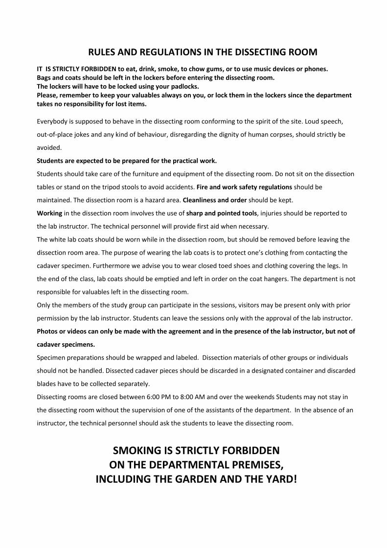
RULES AND REGULATIONS IN THE DISSECTING ROOM
IT IS STRICTLY FORBIDDEN to eat, drink, smoke, to chow gums, or to use music devices or phones. Bags and coats should be left in the lockers before entering the dissecting room. The lockers will have to be locked using your padlocks. Please, remember to keep your valuables always on you, or lock them in the lockers since the department takes no responsibility for lost items. Everybody is supposed to behave in the dissecting room conforming to the spirit of the site. Loud speech,
out-of-place jokes and any kind of behaviour, disregarding the dignity of human corpses, should strictly be
avoided.
Students are expected to be prepared for the practical work.
Students should take care of the furniture and equipment of the dissecting room. Do not sit on the dissection
tables or stand on the tripod stools to avoid accidents. Fire and work safety regulations should be
maintained. The dissection room is a hazard area. Cleanliness and order should be kept.
Working in the dissection room involves the use of sharp and pointed tools, injuries should be reported to
the lab instructor. The technical personnel will provide first aid when necessary.
The white lab coats should be worn while in the dissection room, but should be removed before leaving the
dissection room area. The purpose of wearing the lab coats is to protect one’s clothing from contacting the
cadaver specimen. Furthermore we advise you to wear closed toed shoes and clothing covering the legs. In
the end of the class, lab coats should be emptied and left in order on the coat hangers. The department is not
responsible for valuables left in the dissecting room.
Only the members of the study group can participate in the sessions, visitors may be present only with prior
permission by the lab instructor. Students can leave the sessions only with the approval of the lab instructor.
Photos or videos can only be made with the agreement and in the presence of the lab instructor, but not of
cadaver specimens.
Specimen preparations should be wrapped and labeled. Dissection materials of other groups or individuals
should not be handled. Dissected cadaver pieces should be discarded in a designated container and discarded
blades have to be collected separately.
Dissecting rooms are closed between 6:00 PM to 8:00 AM and over the weekends Students may not stay in
the dissecting room without the supervision of one of the assistants of the department. In the absence of an
instructor, the technical personnel should ask the students to leave the dissecting room.
SMOKING IS STRICTLY FORBIDDEN ON THE DEPARTMENTAL PREMISES,
INCLUDING THE GARDEN AND THE YARD!
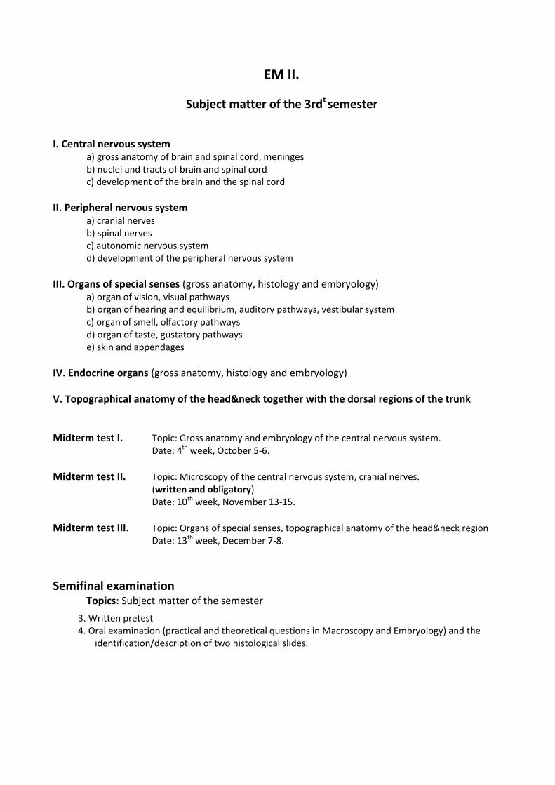
EM II.
Subject matter of the 3rdt semester
I. Central nervous system a) gross anatomy of brain and spinal cord, meninges b) nuclei and tracts of brain and spinal cord c) development of the brain and the spinal cord
II. Peripheral nervous system a) cranial nerves b) spinal nerves c) autonomic nervous system d) development of the peripheral nervous system
III. Organs of special senses (gross anatomy, histology and embryology) a) organ of vision, visual pathways b) organ of hearing and equilibrium, auditory pathways, vestibular system c) organ of smell, olfactory pathways d) organ of taste, gustatory pathways e) skin and appendages
IV. Endocrine organs (gross anatomy, histology and embryology)
V. Topographical anatomy of the head&neck together with the dorsal regions of the trunk
Midterm test I. Topic: Gross anatomy and embryology of the central nervous system. Date: 4th week, October 5-6.
Midterm test II. Topic: Microscopy of the central nervous system, cranial nerves. (written and obligatory) Date: 10th week, November 13-15.
Midterm test III. Topic: Organs of special senses, topographical anatomy of the head&neck region Date: 13th week, December 7-8.
Semifinal examination Topics: Subject matter of the semester
3. Written pretest 4. Oral examination (practical and theoretical questions in Macroscopy and Embryology) and the
identification/description of two histological slides.
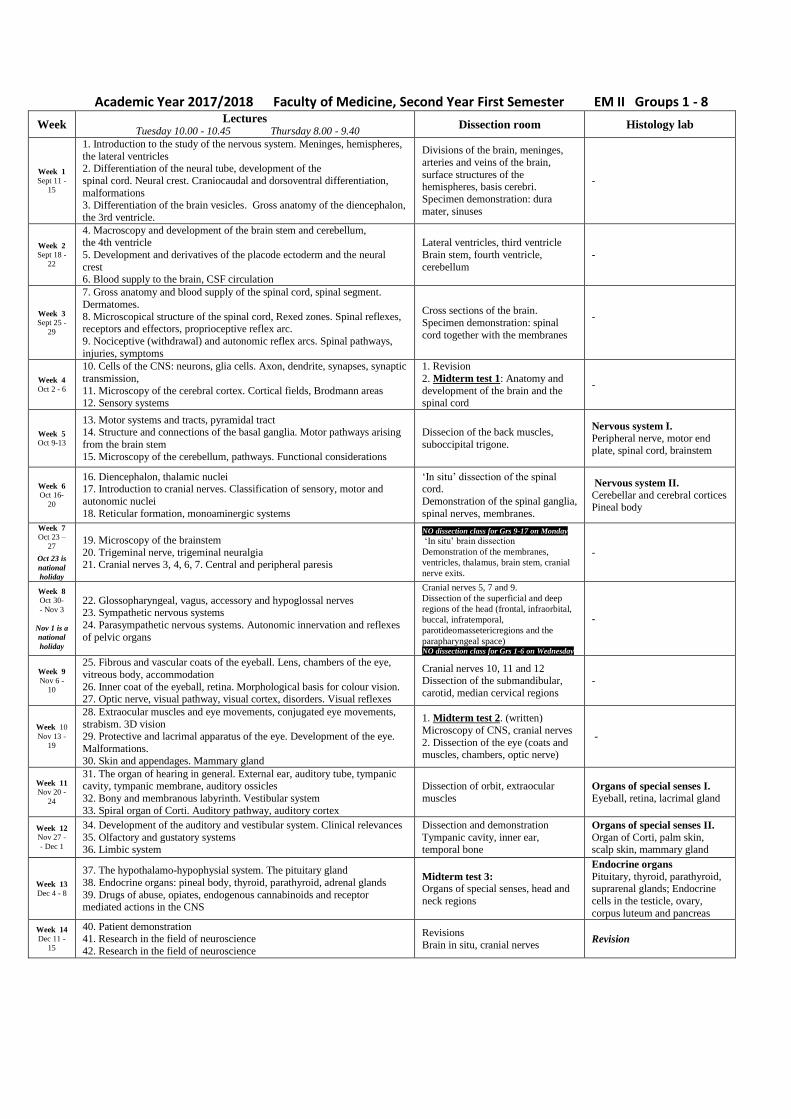
Academic Year 2017/2018 Faculty of Medicine, Second Year First Semester EM II Groups 1 - 8
Week Lectures
Tuesday 10.00 - 10.45 Thursday 8.00 - 9.40 Dissection room Histology lab
Week 1
Sept 11 -
15
1. Introduction to the study of the nervous system. Meninges, hemispheres,
the lateral ventricles 2. Differentiation of the neural tube, development of the
spinal cord. Neural crest. Craniocaudal and dorsoventral differentiation,
malformations 3. Differentiation of the brain vesicles. Gross anatomy of the diencephalon,
the 3rd ventricle.
Divisions of the brain, meninges,
arteries and veins of the brain,
surface structures of the hemispheres, basis cerebri.
Specimen demonstration: dura
mater, sinuses
-
Week 2
Sept 18 -
22
4. Macroscopy and development of the brain stem and cerebellum, the 4th ventricle
5. Development and derivatives of the placode ectoderm and the neural
crest 6. Blood supply to the brain, CSF circulation
Lateral ventricles, third ventricle
Brain stem, fourth ventricle,
cerebellum
-
Week 3
Sept 25 -
29
7. Gross anatomy and blood supply of the spinal cord, spinal segment.
Dermatomes.
8. Microscopical structure of the spinal cord, Rexed zones. Spinal reflexes, receptors and effectors, proprioceptive reflex arc.
9. Nociceptive (withdrawal) and autonomic reflex arcs. Spinal pathways,
injuries, symptoms
Cross sections of the brain.
Specimen demonstration: spinal
cord together with the membranes
-
Week 4
Oct 2 - 6
10. Cells of the CNS: neurons, glia cells. Axon, dendrite, synapses, synaptic
transmission,
11. Microscopy of the cerebral cortex. Cortical fields, Brodmann areas 12. Sensory systems
1. Revision
2. Midterm test 1: Anatomy and
development of the brain and the spinal cord
-
Week 5
Oct 9-13
13. Motor systems and tracts, pyramidal tract 14. Structure and connections of the basal ganglia. Motor pathways arising
from the brain stem
15. Microscopy of the cerebellum, pathways. Functional considerations
Dissecion of the back muscles,
suboccipital trigone.
Nervous system I.
Peripheral nerve, motor end plate, spinal cord, brainstem
Week 6
Oct 16-
20
16. Diencephalon, thalamic nuclei 17. Introduction to cranial nerves. Classification of sensory, motor and
autonomic nuclei
18. Reticular formation, monoaminergic systems
‘In situ’ dissection of the spinal cord.
Demonstration of the spinal ganglia,
spinal nerves, membranes.
Nervous system II.
Cerebellar and cerebral cortices Pineal body
Week 7
Oct 23 –
27
Oct 23 is
national
holiday
19. Microscopy of the brainstem
20. Trigeminal nerve, trigeminal neuralgia 21. Cranial nerves 3, 4, 6, 7. Central and peripheral paresis
NO dissection class for Grs 9-17 on Monday
‘In situ’ brain dissection
Demonstration of the membranes,
ventricles, thalamus, brain stem, cranial
nerve exits.
-
Week 8
Oct 30-
- Nov 3
Nov 1 is a
national
holiday
22. Glossopharyngeal, vagus, accessory and hypoglossal nerves 23. Sympathetic nervous systems
24. Parasympathetic nervous systems. Autonomic innervation and reflexes
of pelvic organs
Cranial nerves 5, 7 and 9.
Dissection of the superficial and deep
regions of the head (frontal, infraorbital,
buccal, infratemporal,
parotideomassetericregions and the
parapharyngeal space) NO dissection class for Grs 1-6 on Wednesday
-
Week 9
Nov 6 -
10
25. Fibrous and vascular coats of the eyeball. Lens, chambers of the eye,
vitreous body, accommodation
26. Inner coat of the eyeball, retina. Morphological basis for colour vision. 27. Optic nerve, visual pathway, visual cortex, disorders. Visual reflexes
Cranial nerves 10, 11 and 12 Dissection of the submandibular,
carotid, median cervical regions
-
Week 10
Nov 13 -
19
28. Extraocular muscles and eye movements, conjugated eye movements,
strabism. 3D vision 29. Protective and lacrimal apparatus of the eye. Development of the eye.
Malformations.
30. Skin and appendages. Mammary gland
1. Midterm test 2. (written)
Microscopy of CNS, cranial nerves
2. Dissection of the eye (coats and muscles, chambers, optic nerve)
-
Week 11
Nov 20 -
24
31. The organ of hearing in general. External ear, auditory tube, tympanic cavity, tympanic membrane, auditory ossicles
32. Bony and membranous labyrinth. Vestibular system
33. Spiral organ of Corti. Auditory pathway, auditory cortex
Dissection of orbit, extraocular
muscles Organs of special senses I. Eyeball, retina, lacrimal gland
Week 12
Nov 27 -
- Dec 1
34. Development of the auditory and vestibular system. Clinical relevances
35. Olfactory and gustatory systems 36. Limbic system
Dissection and demonstration
Tympanic cavity, inner ear, temporal bone
Organs of special senses II. Organ of Corti, palm skin, scalp skin, mammary gland
Week 13
Dec 4 - 8
37. The hypothalamo-hypophysial system. The pituitary gland
38. Endocrine organs: pineal body, thyroid, parathyroid, adrenal glands
39. Drugs of abuse, opiates, endogenous cannabinoids and receptor mediated actions in the CNS
Midterm test 3:
Organs of special senses, head and
neck regions
Endocrine organs
Pituitary, thyroid, parathyroid, suprarenal glands; Endocrine
cells in the testicle, ovary,
corpus luteum and pancreas
Week 14
Dec 11 -
15
40. Patient demonstration 41. Research in the field of neuroscience
42. Research in the field of neuroscience
Revisions
Brain in situ, cranial nerves Revision
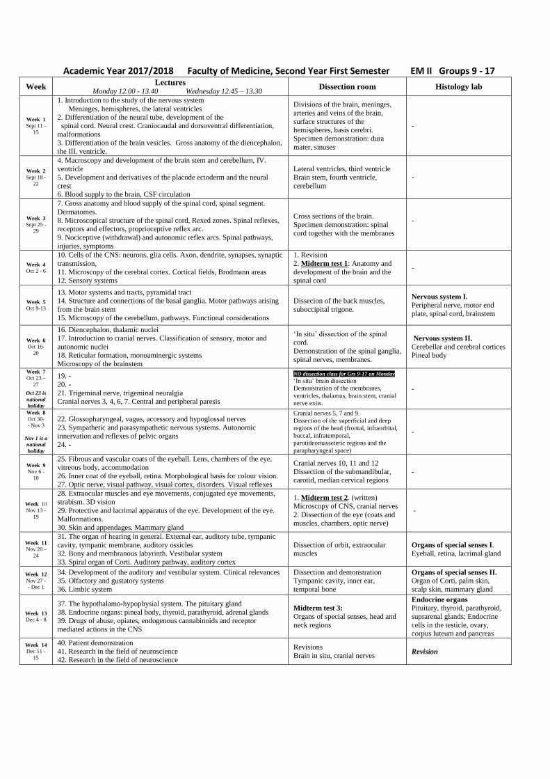
Academic Year 2017/2018 Faculty of Medicine, Second Year First Semester EM II Groups 9 - 17
Week Lectures
Monday 12.00 - 13.40 Wednesday 12.45 – 13.30 Dissection room Histology lab
Week 1
Sept 11 -
15
1. Introduction to the study of the nervous system
Meninges, hemispheres, the lateral ventricles 2. Differentiation of the neural tube, development of the
spinal cord. Neural crest. Craniocaudal and dorsoventral differentiation,
malformations 3. Differentiation of the brain vesicles. Gross anatomy of the diencephalon,
the III. ventricle.
Divisions of the brain, meninges,
arteries and veins of the brain,
surface structures of the hemispheres, basis cerebri.
Specimen demonstration: dura
mater, sinuses
-
Week 2
Sept 18 -
22
4. Macroscopy and development of the brain stem and cerebellum, IV. ventricle
5. Development and derivatives of the placode ectoderm and the neural
crest 6. Blood supply to the brain, CSF circulation
Lateral ventricles, third ventricle
Brain stem, fourth ventricle,
cerebellum
-
Week 3
Sept 25 -
29
7. Gross anatomy and blood supply of the spinal cord, spinal segment.
Dermatomes.
8. Microscopical structure of the spinal cord, Rexed zones. Spinal reflexes, receptors and effectors, proprioceptive reflex arc.
9. Nociceptive (withdrawal) and autonomic reflex arcs. Spinal pathways,
injuries, symptoms
Cross sections of the brain.
Specimen demonstration: spinal
cord together with the membranes
-
Week 4
Oct 2 - 6
10. Cells of the CNS: neurons, glia cells. Axon, dendrite, synapses, synaptic
transmission,
11. Microscopy of the cerebral cortex. Cortical fields, Brodmann areas 12. Sensory systems
1. Revision
2. Midterm test 1: Anatomy and
development of the brain and the spinal cord
-
Week 5
Oct 9-13
13. Motor systems and tracts, pyramidal tract 14. Structure and connections of the basal ganglia. Motor pathways arising
from the brain stem
15. Microscopy of the cerebellum, pathways. Functional considerations
Dissecion of the back muscles,
suboccipital trigone.
Nervous system I.
Peripheral nerve, motor end plate, spinal cord, brainstem
Week 6
Oct 16-
20
16. Diencephalon, thalamic nuclei
17. Introduction to cranial nerves. Classification of sensory, motor and
autonomic nuclei 18. Reticular formation, monoaminergic systems
Microscopy of the brainstem
‘In situ’ dissection of the spinal cord.
Demonstration of the spinal ganglia,
spinal nerves, membranes.
Nervous system II.
Cerebellar and cerebral cortices Pineal body
Week 7
Oct 23 –
27
Oct 23 is
national
holiday
19. -
20. -
21. Trigeminal nerve, trigeminal neuralgia Cranial nerves 3, 4, 6, 7. Central and peripheral paresis
NO dissection class for Grs 9-17 on Monday
‘In situ’ brain dissection
Demonstration of the membranes,
ventricles, thalamus, brain stem, cranial
nerve exits.
-
Week 8
Oct 30-
- Nov 3
Nov 1 is a
national
holiday
22. Glossopharyngeal, vagus, accessory and hypoglossal nerves
23. Sympathetic and parasympathetic nervous systems. Autonomic
innervation and reflexes of pelvic organs
24. -
Cranial nerves 5, 7 and 9.
Dissection of the superficial and deep
regions of the head (frontal, infraorbital,
buccal, infratemporal,
parotideomasseteric regions and the
parapharyngeal space)
-
Week 9
Nov 6 -
10
25. Fibrous and vascular coats of the eyeball. Lens, chambers of the eye, vitreous body, accommodation
26. Inner coat of the eyeball, retina. Morphological basis for colour vision.
27. Optic nerve, visual pathway, visual cortex, disorders. Visual reflexes
Cranial nerves 10, 11 and 12
Dissection of the submandibular, carotid, median cervical regions
-
Week 10
Nov 13 -
19
28. Extraocular muscles and eye movements, conjugated eye movements,
strabism. 3D vision
29. Protective and lacrimal apparatus of the eye. Development of the eye. Malformations.
30. Skin and appendages. Mammary gland
1. Midterm test 2. (written) Microscopy of CNS, cranial nerves
2. Dissection of the eye (coats and
muscles, chambers, optic nerve)
-
Week 11
Nov 20 -
24
31. The organ of hearing in general. External ear, auditory tube, tympanic
cavity, tympanic membrane, auditory ossicles 32. Bony and membranous labyrinth. Vestibular system
33. Spiral organ of Corti. Auditory pathway, auditory cortex
Dissection of orbit, extraocular muscles
Organs of special senses I. Eyeball, retina, lacrimal gland
Week 12
Nov 27 -
- Dec 1
34. Development of the auditory and vestibular system. Clinical relevances 35. Olfactory and gustatory systems
36. Limbic system
Dissection and demonstration Tympanic cavity, inner ear,
temporal bone
Organs of special senses II. Organ of Corti, palm skin,
scalp skin, mammary gland
Week 13
Dec 4 - 8
37. The hypothalamo-hypophysial system. The pituitary gland
38. Endocrine organs: pineal body, thyroid, parathyroid, adrenal glands 39. Drugs of abuse, opiates, endogenous cannabinoids and receptor
mediated actions in the CNS
Midterm test 3:
Organs of special senses, head and
neck regions
Endocrine organs
Pituitary, thyroid, parathyroid,
suprarenal glands; Endocrine
cells in the testicle, ovary, corpus luteum and pancreas
Week 14
Dec 11 -
15
40. Patient demonstration
41. Research in the field of neuroscience 42. Research in the field of neuroscience
Revisions
Brain in situ, cranial nerves Revision
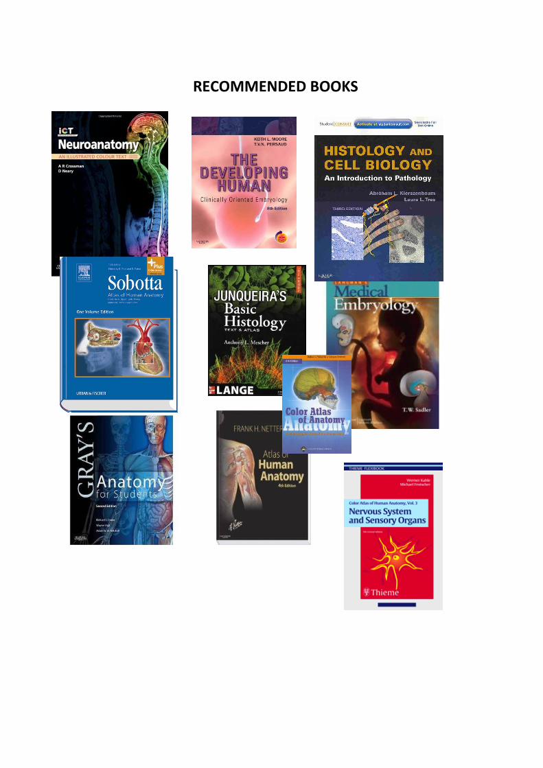
RECOMMENDED BOOKS
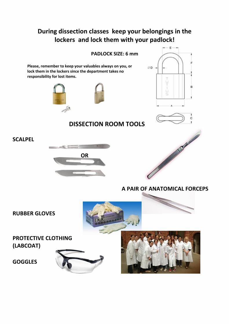
During dissection classes keep your belongings in the lockers and lock them with your padlock!
PADLOCK SIZE: 6 mm
Please, remember to keep your valuables always on you, or lock them in the lockers since the department takes no responsibility for lost items.
DISSECTION ROOM TOOLS
SCALPEL
OR
A PAIR OF ANATOMICAL FORCEPS
RUBBER GLOVES
PROTECTIVE CLOTHING (LABCOAT)
GOGGLES
TOPICS OF THE SEMIFINAL EXAM
EM II. Third semester
Dissection room Topics of the 1st and 3rd MIDTERM examinations
Histology exam Description of histological specimen
Microscopical structure of the central and peripheral nervous systems, different staining techniques (cerebral and cerebellar cortices, neurons, supporting cells, ganglia, receptors and effectors, sysnapses)
Other relevant topics
Endocrine organs Gross and microscopical anatomy of the pituitary gland; development of the posterior lobe Blood supply, histology and development of the anterior and intermediate lobes of the pituitary gland Gross and microscopical anatomy of the pineal gland Gross and microscopical anatomy and the development of the thyroid gland Gross and microscopical anatomy and the development of the parathyroid gland Gross and microscopical anatomy and the development of the suprarenal gland Histology of the Langerhans islets Endocrine cells and function of the male and female gonads Microscopical structure of the eyeball Histology of the lacrimal gland Microscopical structure of the cochlea Microscopical structure of the skin (scalp and palm) Histology of the mammary gland (lactating and non-lactating)
Macroscopical structure of the CNS Gross anatomy of hemispheres
Cranial dura mater and its sinuses Arachnoid and pia mater. Subarachnoid cisterns. Cerebrospinal fluid Arteries, circle of Willis and veins of the brain Gross anatomy of the lateral ventricles Gross anatomy of the third ventricle Gross anatomy of the fourth ventricle Gross anatomy of the large comissural pathways Gross anatomy of the internal capsule Gross and microscopic anatomy of basal nuclei Gross and microscopic anatomy of thalamus Anatomy, blood supply and development of the diencephalon Gross and microscopic anatomy of midbrain Gross and microscopic anatomy of pons Gross and microscopic anatomy of medulla oblongata Gross and microscopic anatomy of cerebellum Cranial nerve exits Gross anatomy of spinal cord. Spinal segment. Spinal nerve. Blood supply and meninges of the spinal cord Microscopical structure and development of the central nervous system Development and primary differentiation of the neural tube Development of the spinal cord; neurohistogenesis Differentiation of the prosencephalon vesicle; development of the hemispheres and the lateral ventricle Differentiation of the diencephalon vesicle, development of the third ventricle Differentiation of the mesencephalon and rhombencephalon vesicles, development of the fourth ventricle Roots, branches and components of the spinal nerves; spinal segment Fine structure (microscopy) of the spinal cord Neurons and function of the spinal proprioceptive (strech) reflex Neurons and function of the spinal flexion (withdrawal) reflex Neurons and functions of the visceral reflexes Microscopical anatomy of the medulla Microscopical anatomy of the pons Microscopical anatomy of the midbrain Nuclei of the cranial nerves Microscopical anatomy of the cerebellum Afferent and efferent cerebellar connections Microscopical anatomy of thalamus, divisions, connections and transmitters Hypothalamus, hypothalamo-hypophyseal systems Microscopical anatomy of the basal ganglia, divisions, connections and transmitters Histology of the cerebral cortex; cortical fields Internal capsule, divisions Tracts of the protopathic sensibility (anterolateral system) Tracts of the epicritic sensibility (posterior funiculus/medial lemniscus) Corticospinal tract (pyramidal tract) Extrapyramidal system
Limbic system (nuclei and tracts) Gross anatomy and development of the peripheral nervous system Development, fate and differentiation of the cells in the neural crest Development of the peripheral nervous system Nuclei and branches of the IIIrd, IVth and VIth cranial nerves Nuclei of the trigeminal nerve; course and fiber composition of the branches of the ophthalmic (V/1) nerve Course and fiber composition of the branches of the maxillary nerve (V/2) Course and fiber composition of the branches of the mandibular nerve (V/3) Nuclei, course and fiber composition of the branches of the facial nerve (VII) Nuclei, course and fiber composition of the branches of the glossopharyngeal nerve (IX) Nuclei, course and fiber composition of the branches of the vagus nerve (X) Nuclei, course and fiber composition of the branches of the accessory (XI) and hypoglossal nerves (XII) Cervical plexus and its branches Brachial plexus and its branches (including the short branches to the neck and shoulder girdle) Lumbar plexus and its branches Sacral plexus and its branches General organization of the autonomic nervous system The sympathetic trunk Cranial part of the parasympathetic nervous system Sacral part of the parasympathetic nervous system Gross anatomy, histology and embryology of the organs of special senses Gross anatomy and microscopic structure of the fibrous coat of the eye ball (cornea, sclera) Gross anatomy and microscopic structure of the vascular coat of the eye ball (choroid, ciliary body, iris) Gross anatomy, microscopic structure and development of the nervous coat of the eye ball (retina) Neurons of the visual pathways; localization and microscopic structure of the visual cortex Gross anatomy, microscopic structure and development of the lens, accomodation Gross anatomy and content of the chambers of the eye, circulation of the aqueous humor; gross anatomy of the vitreous body Gross anatomy and function of the external ocular muscles. Visual reflexes Gross anatomy, microscopic structure of the eye lids; conjunctiva, Tennon's capsule and periorbit Gross anatomy, microscopic structure and development of the lacrimal apparatus Gross anatomy and development of the external ear and the tympanic membrane Gross anatomy and development of the tympanic cavity and the auditory tube Gross anatomy and development of the auditory ossicles; joints, muscles, and the mucous membrane of the tympanic cavity Sensory innervation and blood supply of the tympanic cavity Internal acoustic meatus Gross anatomy of the bony labyrinth
Gross anatomy and development of the labyrinth Morphology, development and divisions of the vestibular apparatus Receptors and neuronal connections of the vestibular system Gross anatomy, microscopic structure and development of the cochlear duct and the organ of Corti Neurons of the auditory pathways Organ and pathways of olfaction Organ and pathways of taste Gross anatomy and microscopic structure of the fibrous coat of the eye ball (cornea, sclera) Gross anatomy and microscopic structure of the vascular coat of the eye ball (choroid, ciliary body, iris) Gross anatomy, microscopic structure and development of the nervous coat of the eye ball (retina) Neurons of the visual pathways; localization and microscopic structure of the visual cortex Gross anatomy, microscopic structure and development of the lens, accomodation Gross anatomy and content of the chambers of the eye, circulation of the aqueous humor; gross anatomy of the vitreous body Gross anatomy and function of the external ocular muscles. Visual reflexes Gross anatomy, microscopic structure of the eye lids; conjunctiva, Tennon's capsule and periorbit Gross anatomy, microscopic structure and development of the lacrimal apparatus Gross anatomy and development of the external ear and the tympanic membrane Gross anatomy and development of the tympanic cavity and the auditory tube Gross anatomy and development of the auditory ossicles; joints, muscles, and the mucous membrane of the tympanic cavity Sensory innervation and blood supply of the tympanic cavity Internal acoustic meatus Gross anatomy of the bony labyrinth Gross anatomy and development of the labyrinth Morphology, development and divisions of the vestibular apparatus Receptors and neuronal connections of the vestibular system Gross anatomy, microscopic structure and development of the cochlear duct and the organ of Corti Neurons of the auditory pathways Organ and pathways of olfaction Organ and pathways of taste