Anatomy 2-Connection-of-bones
35
The Department of Human anatomy The Department of Human anatomy General doctrine General doctrine about connection of about connection of the bones the bones
Transcript of Anatomy 2-Connection-of-bones
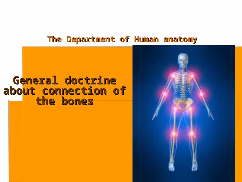
The Department of Human anatomyThe Department of Human anatomy
General doctrine about General doctrine about connection of the connection of the
bonesbones

PLANPLAN Basic data of the bone articulationsBasic data of the bone articulations.. The classification of the bones articulationsThe classification of the bones articulations.. The types of continuous articulations.The types of continuous articulations. Intermediate articulationsIntermediate articulations.. Synovial articulationsSynovial articulations.. The structure of articulationThe structure of articulation.. The analysis of movements of the The analysis of movements of the
articulations.articulations.
Joint is a junction between two or more Joint is a junction between two or more bones or cartilages. It is a device to bones or cartilages. It is a device to permit movements in a hard and rigid permit movements in a hard and rigid skeleton. Joints are formed in such a skeleton. Joints are formed in such a way that they keep a balance between way that they keep a balance between the movement, stability and strength of the movement, stability and strength of human skeleton.human skeleton.
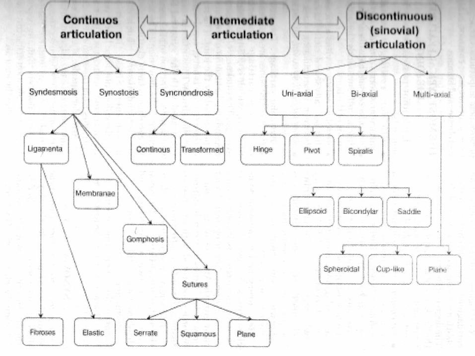
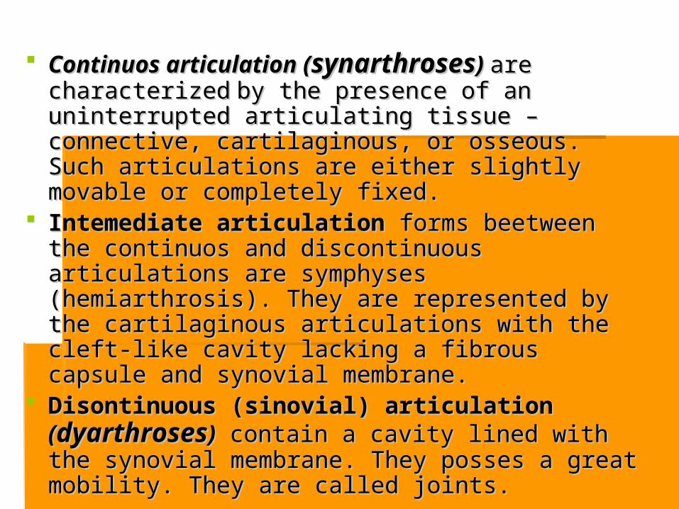
Continuos articulation (Continuos articulation (synarthrosessynarthroses) ) are are characterizedcharacterized by the presence of an uninterrupted by the presence of an uninterrupted articulating tissue – connective, cartilaginous, or articulating tissue – connective, cartilaginous, or osseous. Such articulations are either slightly osseous. Such articulations are either slightly movable or completely fixed.movable or completely fixed.
Intemediate articulation Intemediate articulation forms beetweenforms beetween the the continuos and discontinuous articulations are continuos and discontinuous articulations are symphyses (hemiarthrosis). They are represented symphyses (hemiarthrosis). They are represented by the cartilaginous articulations with the cleft-like by the cartilaginous articulations with the cleft-like cavity lacking a fibrous capsule and synovial cavity lacking a fibrous capsule and synovial membrane.membrane.
Disontinuous (sinovial) articulation Disontinuous (sinovial) articulation ((dyarthrosesdyarthroses)) contain a cavity lined with the contain a cavity lined with the synovial membrane. They posses a great mobility. synovial membrane. They posses a great mobility. They are called joints.They are called joints.
Syndesmosys- the most common type of Syndesmosys- the most common type of fibrous articulations, which include :fibrous articulations, which include :
ligamentsligaments membranesmembranes suturessutures gomphosisgomphosis fontanellefontanelle
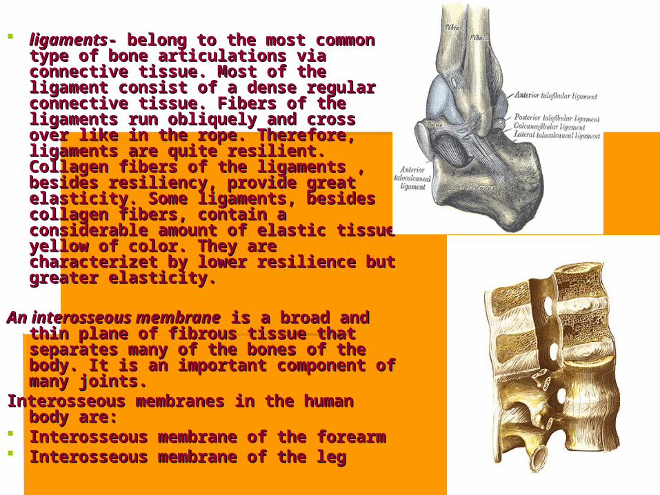
ligamentsligaments- belong to the most common - belong to the most common type of bone articulations via connective type of bone articulations via connective tissue. Most of the ligament consist of a tissue. Most of the ligament consist of a dense regular connective tissue. Fibers dense regular connective tissue. Fibers of the ligaments run obliquely and cross of the ligaments run obliquely and cross over like in the rope. Therefore, ligaments over like in the rope. Therefore, ligaments are quite resilient. Collagen fibers of the are quite resilient. Collagen fibers of the ligaments , besides resiliency, provide ligaments , besides resiliency, provide great elasticity. Some ligaments, besides great elasticity. Some ligaments, besides collagen fibers, contain a considerable collagen fibers, contain a considerable amount of elastic tissue yellow of color. amount of elastic tissue yellow of color. They are characterizet by lower resilience They are characterizet by lower resilience but greater elasticity. but greater elasticity.
An interosseous membraneAn interosseous membrane is a broad and is a broad and
thin plane of fibrous tissue that separates thin plane of fibrous tissue that separates many of the bones of the body. It is an many of the bones of the body. It is an important component of many joints.important component of many joints.
Interosseous membranes in the human Interosseous membranes in the human body are:body are:
Interosseous membrane of the forearmInterosseous membrane of the forearm Interosseous membrane of the legInterosseous membrane of the leg

Sutures:Sutures: fibrous fibrous articulation between the articulation between the bones of the skull, which bones of the skull, which contain a thin layer of contain a thin layer of connective tissue, and are connective tissue, and are immovable. According to immovable. According to the shape of the bony the shape of the bony margin the sutures may margin the sutures may be;be; Plane suturesPlane sutures Serrate suturesSerrate sutures Denticulate suturesDenticulate sutures Squamous suturesSquamous sutures Limbous suturesLimbous sutures SchindylesisSchindylesis
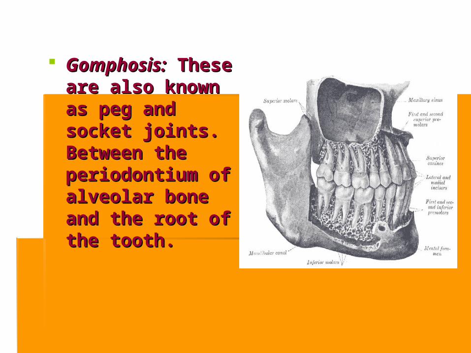
Gomphosis:Gomphosis: These These are also known as are also known as peg and socket peg and socket joints. Between the joints. Between the periodontium of periodontium of alveolar bone and alveolar bone and the root of the the root of the tooth.tooth.

FontanellesFontanelles are soft spots on a are soft spots on a
baby's head which, during baby's head which, during birth, enable the bony plates birth, enable the bony plates of the skull to flex, allowing of the skull to flex, allowing the child's head to pass the child's head to pass through the birth canal. through the birth canal. The The ossificationossification of the bones of the bones of the skull causes the of the skull causes the fontanelles to close over by fontanelles to close over by a child's second birthday. a child's second birthday. The closures eventually form The closures eventually form the sutures of the the sutures of the neurocranium. There are neurocranium. There are such unpair: the anterior and such unpair: the anterior and posterior fontanelles, and posterior fontanelles, and two pair: the mastoid two pair: the mastoid fontanelle and fontanelle and the sphenoidal fontanelle.the sphenoidal fontanelle.

Synostosis is the joining of two bones by the Synostosis is the joining of two bones by the ossification of connecting tissues. Example is hip ossification of connecting tissues. Example is hip bone where the three bones (ilium, ischium and pubis) bone where the three bones (ilium, ischium and pubis) fuse together after 16-18 years.fuse together after 16-18 years.

Synchondrosis.Synchondrosis. In this In this subtype the bones are united subtype the bones are united by a plate of hyaline by a plate of hyaline cartilage so that the joint is cartilage so that the joint is immovable and strong. immovable and strong. There are two types of There are two types of synchondrosis: synchondrosis: temporary temporary and permanentand permanent. .
TemporaryTemporary synchondroses synchondroses include the articulation include the articulation between the sacral vertebra, between the sacral vertebra, between the epiphyses and between the epiphyses and diaphyses of long bones, diaphyses of long bones, and spheno-occipital and spheno-occipital synchondrosis.synchondrosis.
Permanent Permanent synchondroses synchondroses are located between the first are located between the first rib and the sternum. The rib and the sternum. The skull has such skull has such synchondrosis like synchondrosis like sphenopetrosal and sphenopetrosal and petrooccipital.petrooccipital.
In several areas of the sceleton In several areas of the sceleton cartilage persists only until a certain cartilage persists only until a certain age providing an opportunity for age providing an opportunity for their growth. After the termination of their growth. After the termination of growth, such temporary growth, such temporary synchondrosis become subtituted synchondrosis become subtituted with the osseus tissues and with the osseus tissues and transform into synostoses.transform into synostoses.
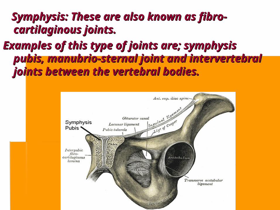
Symphysis: These are also known as fibro-Symphysis: These are also known as fibro-cartilaginous joints. cartilaginous joints.
Examples of this type of joints are; symphysis Examples of this type of joints are; symphysis pubis, manubrio-sternal joint and intervertebral pubis, manubrio-sternal joint and intervertebral joints between the vertebral bodies.joints between the vertebral bodies.

Disontinuous (sinovial) articulationDisontinuous (sinovial) articulation Synovial joints are most evolved and therefore most mobile type of Synovial joints are most evolved and therefore most mobile type of
joints. joints. They possess the following characteristic features;They possess the following characteristic features;
There articular surfaces are covered with hyaline cartilage. This There articular surfaces are covered with hyaline cartilage. This articular cartilage is avascular, non nervous and elastic. articular cartilage is avascular, non nervous and elastic. Lubricated with synovial fluid, the cartilage forms slippery Lubricated with synovial fluid, the cartilage forms slippery surfaces for free movements.surfaces for free movements.
Between the articular surfaces there is a joint cavity filled with Between the articular surfaces there is a joint cavity filled with synovial fluid. The cavity may be partially or completely synovial fluid. The cavity may be partially or completely subdivided by an articular disc or meniscus.subdivided by an articular disc or meniscus.
The joint is surrounded by an articular capsule which is fibrous The joint is surrounded by an articular capsule which is fibrous in nature and is lined by synovial membrane. Because of its rich in nature and is lined by synovial membrane. Because of its rich nerve supply the fibrous capsule is sensitive to stretches nerve supply the fibrous capsule is sensitive to stretches imposed by movements.imposed by movements.
The synovial membrane lines the entire joint except the articular The synovial membrane lines the entire joint except the articular surfaces covered by hyaline cartilage. It is this membrane that surfaces covered by hyaline cartilage. It is this membrane that secretes the slimy fluid called synovial fluid which lubricates the secretes the slimy fluid called synovial fluid which lubricates the joint and nourishes the articular cartilage.joint and nourishes the articular cartilage.

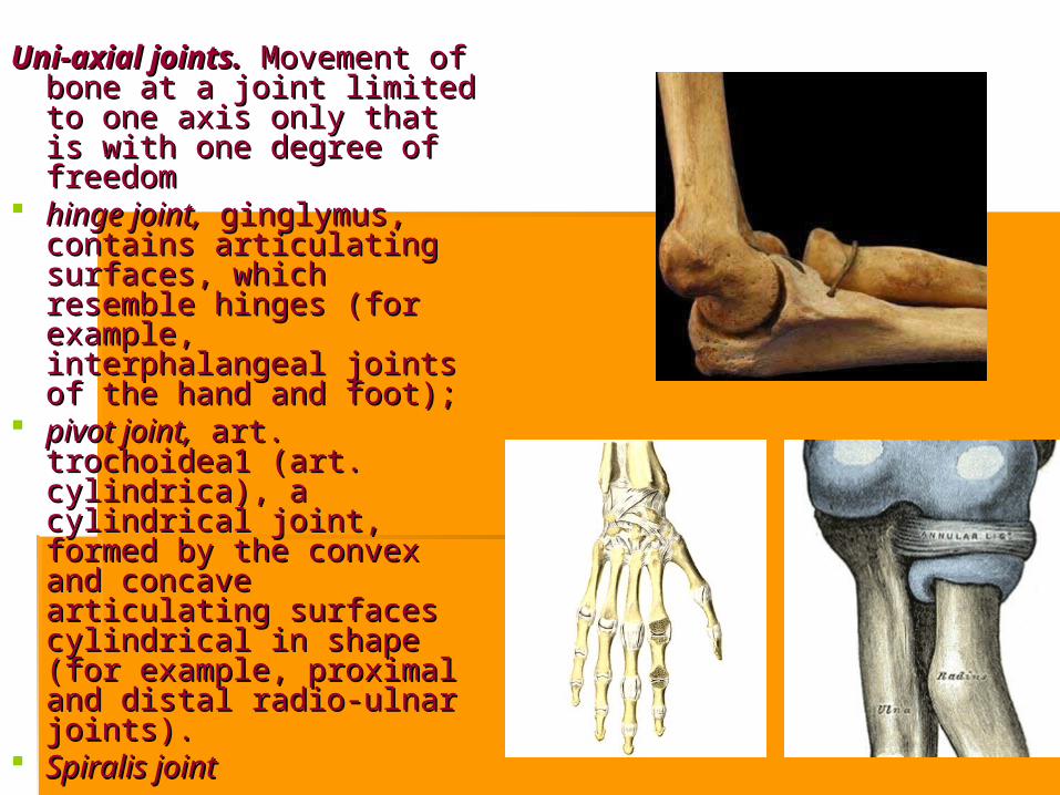
Uni-axial joints.Uni-axial joints. Movement of Movement of bone at a joint limited to bone at a joint limited to one axis only that is with one axis only that is with one degree of freedomone degree of freedom
hinge joint,hinge joint, ginglymus, ginglymus, contains articulating contains articulating surfaces, which resemble surfaces, which resemble hinges (for example, hinges (for example, interphalangeal joints of the interphalangeal joints of the hand and foot);hand and foot);
pivot joint,pivot joint, art. trochoidea1 art. trochoidea1 (art. cylindrica), a cylindrical (art. cylindrica), a cylindrical joint, formed by the convex joint, formed by the convex and concave articulating and concave articulating surfaces cylindrical in surfaces cylindrical in shape (for example, shape (for example, proximal and distal radio-proximal and distal radio-ulnar joints).ulnar joints).
Spiralis jointSpiralis joint

Bi-axial joints.Bi-axial joints. These joints permit movements around These joints permit movements around
two axes, which are perpendicular to two axes, which are perpendicular to each other. The following each other. The following bi-axial bi-axial joints joints are distinguished based on are distinguished based on their shape:their shape:
ellipsoid joint,ellipsoid joint, art. ellipsoidea, art. ellipsoidea, contains elliptical (egg-shaped) contains elliptical (egg-shaped) articulating surfaces; for example, articulating surfaces; for example, radiocarpal joint;radiocarpal joint;
bicondylar joint,bicondylar joint, art. bicondylaris2, art. bicondylaris2, consists of a pair of convex, consists of a pair of convex, ellipsoid, articulating surfaces, which ellipsoid, articulating surfaces, which are rather prominent. Hence, they are rather prominent. Hence, they are known as condyles (for example, are known as condyles (for example, knee joint);knee joint);
saddle joint,saddle joint, art. sellaris, art. sellaris, contains contains two articulating surfaces each of two articulating surfaces each of which resembles a saddle (for which resembles a saddle (for example, carpometacarpal joint of example, carpometacarpal joint of the thumb). the thumb).

Multi-axial jointsMulti-axial joints include spheroidal joints and their variations. include spheroidal joints and their variations.
Even though, in principle, movements in Even though, in principle, movements in these joints are possible around multiple these joints are possible around multiple axes drawn through the joint, rotation axes drawn through the joint, rotation occurs only around three axis:occurs only around three axis:
spheroidal joint (ball-and-socket spheroidal joint (ball-and-socket joint),joint), art. spheroidea, consists of art. spheroidea, consists of a globular head and a a globular head and a corresponding concavity. A corresponding concavity. A typical example is the shoulder typical example is the shoulder joint. joint.
cup-like joint (cup-like joint ( art. cotylica); art. cotylica); Variation of the ball- and-socket Variation of the ball- and-socket joint is the cotyloid joint. Its head joint is the cotyloid joint. Its head resides in a deep articular socket, resides in a deep articular socket, consequently, f.e. hip jointconsequently, f.e. hip joint
plane joint,plane joint, art. plana, contains art. plana, contains two flat surfaces, which can be two flat surfaces, which can be viewed as surviewed as surffaces of a sphere aces of a sphere with a large diameter. Because of with a large diameter. Because of the small size difference of both the small size difference of both articulating surfaces, only slight articulating surfaces, only slight translations are possible in plane translations are possible in plane joints (for example, sacro-iliac joints (for example, sacro-iliac joint). joint).

Functional classification:Functional classification:
Immovable joints: (Synarthroses) – Immovable joints: (Synarthroses) – continuos jointscontinuos joints
Partially moveable joints: Partially moveable joints: (Amphiarthroses) – intermediate joints(Amphiarthroses) – intermediate joints
Freely moveable joints: (Diarthroses) – Freely moveable joints: (Diarthroses) – discontinous jointsdiscontinous joints
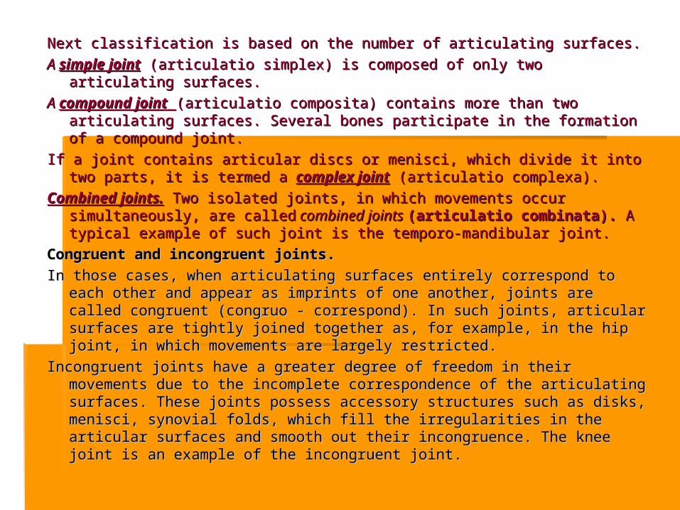
NextNext classification is based on the number of classification is based on the number of articulating surfaces.articulating surfaces. A A simple jointsimple joint (articulatio simplex) (articulatio simplex) is composed of is composed of only two articulating only two articulating
surfaces.surfaces. A A compound jointcompound joint (articulatio composita)(articulatio composita) contains more than two articulating contains more than two articulating
surfaces. Several bones participate in the formation of a compound joint. surfaces. Several bones participate in the formation of a compound joint. If a joint contains articular discs or menisci, which divide it into two parts, it is If a joint contains articular discs or menisci, which divide it into two parts, it is
termed a termed a complex jointcomplex joint (articulatio complexa). (articulatio complexa). Combined joints.Combined joints. Two isolated joints, in which movements occur Two isolated joints, in which movements occur
simultaneously, are calledsimultaneously, are called combined joints combined joints (articulatio combinata).(articulatio combinata). A typical A typical example of such joint is the temporoexample of such joint is the temporo--mandibular joint.mandibular joint.
Congruent and incongruent joints. Congruent and incongruent joints. In those cases, when articulating surfaces entirely correspond to each other and In those cases, when articulating surfaces entirely correspond to each other and
appear as imprints of one another, joints are called congruent (congruo - appear as imprints of one another, joints are called congruent (congruo - correspond). In such joints, articular surfaces are tightly joined together as, correspond). In such joints, articular surfaces are tightly joined together as, for example, in the hip joint, in which movements are largely restricted.for example, in the hip joint, in which movements are largely restricted.
Incongruent joints have a greater degree of freedom in their movements due to Incongruent joints have a greater degree of freedom in their movements due to the incomplete correspondence of the articulating surfaces. These joints the incomplete correspondence of the articulating surfaces. These joints possess accessory structures such as disks, menisci, synovial folds, which possess accessory structures such as disks, menisci, synovial folds, which fill the irregularities in the articular surfaces and smooth out their fill the irregularities in the articular surfaces and smooth out their incongruence. The knee joint is an example of the incongruent joint.incongruence. The knee joint is an example of the incongruent joint.

Anatomical terms for describing Anatomical terms for describing movements:movements:
Flexion:Flexion: A movement by which A movement by which the angle of a joint is decreasedthe angle of a joint is decreased
Extension: Extension: A movement by A movement by which the angle of a joint is which the angle of a joint is increasedincreased
Adduction:Adduction: Movement toward Movement toward the central axisthe central axis
Abduction: Abduction: Movement away Movement away from the central axisfrom the central axis
Pronation: Pronation: This movement This movement occurs in the forearm whereby occurs in the forearm whereby the palm is turned forwardsthe palm is turned forwards
Supination:Supination: This movement also This movement also occurs in the forearm whereby occurs in the forearm whereby the palm is turned backwardsthe palm is turned backwards

THE MOST TYPICAL FEATURES OF THE THE MOST TYPICAL FEATURES OF THE JOINTS.JOINTS.
Each joint possesses four basic elements:Each joint possesses four basic elements: articular cartilage,articular cartilage, cartilago articularis, cartilago articularis, forms forms
articular surfaces;articular surfaces; articular capsule,articular capsule, capsula articularis, capsula articularis, like a cuff, like a cuff,
surrounds the articular ends of the bone;surrounds the articular ends of the bone; articular cavity,articular cavity, cavitas articularis, cavitas articularis, hermetically hermetically
sealed, cleft-like cavity between the articulating sealed, cleft-like cavity between the articulating bones;bones;
synovial fluid,synovial fluid, synovia, synovia, fills articular cavity. fills articular cavity.
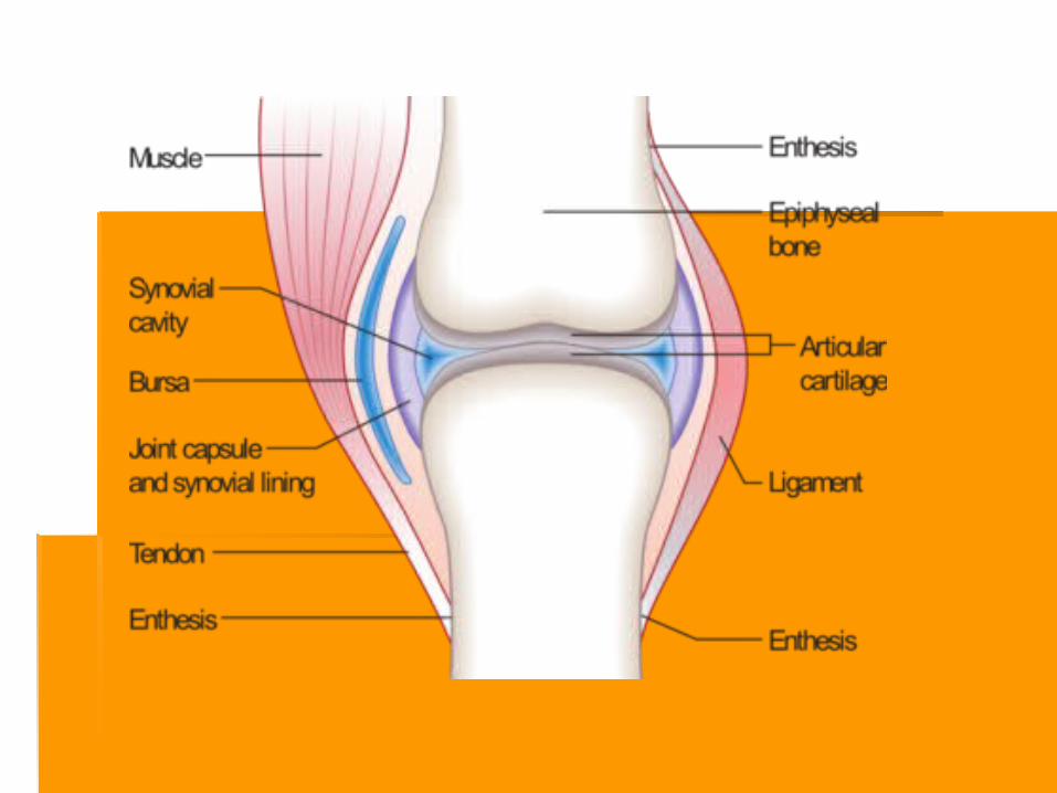

Articular cartilage.Articular cartilage. The articular surfaces of the The articular surfaces of the bones are covered with the hyaline cartilage bones are covered with the hyaline cartilage (sometimes, fibrous), which is 2.5 mm thick on (sometimes, fibrous), which is 2.5 mm thick on average. The thickness of the cartilage depends average. The thickness of the cartilage depends on the amount of compression experienced by on the amount of compression experienced by the joint. The surface of the cartilage is not the joint. The surface of the cartilage is not covered with the perichondrium. The surface is covered with the perichondrium. The surface is smooth, lubricated with the synovial fluid, which smooth, lubricated with the synovial fluid, which reduces friction to minimum and facilitates the reduces friction to minimum and facilitates the movement. The articular cartilage is fairly elastic. movement. The articular cartilage is fairly elastic. It softens strokes and absorbs shock during It softens strokes and absorbs shock during movements. Since the articular cartilage does movements. Since the articular cartilage does not have blood vessels, its nutrition occurs via not have blood vessels, its nutrition occurs via diffusion of nutrients from the synovial fluid.diffusion of nutrients from the synovial fluid.
Articular capsule.Articular capsule. The capsule of the joint The capsule of the joint attaches to the edges of articular surfaces attaches to the edges of articular surfaces or somewhat over them. or somewhat over them.
The articular capsule hermetically seals the The articular capsule hermetically seals the articular cavity and consists of two layers -articular cavity and consists of two layers - external fibrous layer, external fibrous layer, membrana fibrosa,membrana fibrosa, andand internal synovial layer, internal synovial layer, membrana membrana synovialis.synovialis.
Synovial folds and bursae.Synovial folds and bursae. The synovial The synovial membrane is well developed and, in the majority of membrane is well developed and, in the majority of the joints forms largethe joints forms large folds, folds, plicae synoviales, plicae synoviales, which contain adipose tissue. They go into the which contain adipose tissue. They go into the articular cavity, filling its potential spaces and articular cavity, filling its potential spaces and forming cushions, which absorb shock during forming cushions, which absorb shock during motion.motion.
In certain regions, most frequently in the areas of In certain regions, most frequently in the areas of muscular tendons, the synovial membrane muscular tendons, the synovial membrane protrudes through the clefts in the fibrous layer and protrudes through the clefts in the fibrous layer and forms synovial bursae. They serve to reduce forms synovial bursae. They serve to reduce friction during tendons' movement and can be friction during tendons' movement and can be attributed to the accessory muscular apparatus.attributed to the accessory muscular apparatus.
TThe synovial layer bears numerous, microscopic,he synovial layer bears numerous, microscopic, synovial villi, synovial villi, villi synoviales,villi synoviales, which significantly which significantly increase its surface area, contain capillary increase its surface area, contain capillary networks, and produce the synovia] fluid. networks, and produce the synovia] fluid.

Synovial fluidSynovial fluid.. In the articular cavity there is a In the articular cavity there is a small amount of pale, thick, synovial fluid. It is small amount of pale, thick, synovial fluid. It is produced by the synoviaproduced by the synoviall membrane. The synovial membrane. The synovial fluid lubricates the articular surfaces and reduces fluid lubricates the articular surfaces and reduces friction between them.friction between them.
The synovial fluid constitutes the internal The synovial fluid constitutes the internal environment of the joint. It provides nutrition to the environment of the joint. It provides nutrition to the internal structures of the joint and participates in internal structures of the joint and participates in its metabolism.its metabolism.
Synovial cavity.Synovial cavity. It iIt is a thin cleft, bounded by the s a thin cleft, bounded by the synovial membrane and the articular surfaces. synovial membrane and the articular surfaces. Between the latter, there is a thin layer of synovial Between the latter, there is a thin layer of synovial fluid, which separates articulating surfaces. In the fluid, which separates articulating surfaces. In the pathological processes, the synovial cavity can pathological processes, the synovial cavity can accumulate large amounts of inflammatory liquid accumulate large amounts of inflammatory liquid and greatly increases in size.and greatly increases in size.

Accessory structures of joints. Accessory structures of joints. Synovial joints may Synovial joints may possess several accessory elements, which facilitate possess several accessory elements, which facilitate movement to a certain extent. Such accessory elements movement to a certain extent. Such accessory elements include cartilaginous articular discs, menisci, glenoidal include cartilaginous articular discs, menisci, glenoidal labralabraee, and intraarticular ligaments:, and intraarticular ligaments:
articular- disc,articular- disc, discus articularis, discus articularis, a cartilaginous plate, a cartilaginous plate, which completely separates the joint into two regions;which completely separates the joint into two regions;
articular meniscus,articular meniscus, meniscus arti cularis, meniscus arti cularis, cartilaginous cartilaginous semilunar plate, which partially separates the synovial semilunar plate, which partially separates the synovial cavity;cavity;
glenoid labium,glenoid labium, labrum glenoidale, labrum glenoidale, a cartilaginous rim, a cartilaginous rim, which deepens the articular socket;which deepens the articular socket;
intraarticular ligaments,intraarticular ligaments, ligg. intraarticularia, ligg. intraarticularia, connect connect bones within the joint; they are covered with the synovial bones within the joint; they are covered with the synovial membrane.membrane.

Clinical applications.Clinical applications. Pathological process can develop in the joints, among Pathological process can develop in the joints, among
which infectious arthritis is the most common which infectious arthritis is the most common (particularly, rheumatic or tuberculous arthritis). (particularly, rheumatic or tuberculous arthritis). Inflammations of the synovial membrane — synovitis Inflammations of the synovial membrane — synovitis and bursitis are also fairly common. Joint deformations, and bursitis are also fairly common. Joint deformations, contraction of the synovial cleft, and restriction of contraction of the synovial cleft, and restriction of movements accompanied by pain syndrome can occur movements accompanied by pain syndrome can occur as a consequence of degenerative changes (arthrosis). as a consequence of degenerative changes (arthrosis). Traumas can cause various joint dislocations with Traumas can cause various joint dislocations with stretched and ruptured ligaments. Physician of different stretched and ruptured ligaments. Physician of different specialties often have to deal with the diseases of specialties often have to deal with the diseases of jointsjoints

Dislocation of joint:Dislocation of joint:
This is a condition in which the This is a condition in which the articular surfaces of the joint are articular surfaces of the joint are abnormally displaced so that one abnormally displaced so that one surface loses its contact surface loses its contact completely with the other. If a completely with the other. If a partial contact is still retained, it is partial contact is still retained, it is better called subluxation rather better called subluxation rather than dislocation. Dislocation is than dislocation. Dislocation is commonly caused by trauma and commonly caused by trauma and is characterized by pain, is characterized by pain, deformity and loss of function. deformity and loss of function. X-X-ray is used for confirmation.ray is used for confirmation.

Sprain:Sprain: It is the severe pain It is the severe pain
in the joint caused by in the joint caused by ligamentous tear, but ligamentous tear, but without any without any associated associated dislocation or dislocation or fracture. The tear fracture. The tear leads to effusion into leads to effusion into the ligament and joint the ligament and joint causing great pain.causing great pain.

Arthritis:Arthritis: It is the inflammation of one or more joints. It can be It is the inflammation of one or more joints. It can be
caused by a variety of diseases but the common types caused by a variety of diseases but the common types of arthritis are rheumatic, rheumatoid, osteoarthritis of arthritis are rheumatic, rheumatoid, osteoarthritis and tuberculoses. The involved joint is commonly and tuberculoses. The involved joint is commonly swollen and its movements are restricted and painful.swollen and its movements are restricted and painful.

Osteoarthritis:Osteoarthritis: It represents the ageing It represents the ageing
process. In old age the process. In old age the articular cartilage shows articular cartilage shows degenerative changes in degenerative changes in the center (fibrillation of the center (fibrillation of cartilage) and proliferative cartilage) and proliferative changes around the edges. changes around the edges. Due to this lips are formed Due to this lips are formed around the edges of joints.around the edges of joints.

Stiffness of joints related to weather:Stiffness of joints related to weather:The viscosity of synovial fluid increases with fall in The viscosity of synovial fluid increases with fall in
temperature. This accounts for stiffness of the joints in temperature. This accounts for stiffness of the joints in cold weather. Mobility of the joint itself is an important cold weather. Mobility of the joint itself is an important factor in promoting lubrication. Thus the stiffness of the factor in promoting lubrication. Thus the stiffness of the joints experienced in the morning gradually passes off joints experienced in the morning gradually passes off as the movements are resumed.as the movements are resumed.
Neuropathic joint:Neuropathic joint:It is the result of complete denervation of the joint so that It is the result of complete denervation of the joint so that
all reflexes are eliminated and the joint is left all reflexes are eliminated and the joint is left unprotected and liable to mechanical damage. A unprotected and liable to mechanical damage. A neuropathic joint shows painless swelling, excessive neuropathic joint shows painless swelling, excessive mobility and bony destruction. It is commonly caused mobility and bony destruction. It is commonly caused by leprosy, tabes dorsalis and syringomyelia.by leprosy, tabes dorsalis and syringomyelia.
Thank you for attention!Thank you for attention!
![Anatomy and Physiology-Bones and Joints[1]](https://static.fdocuments.in/doc/165x107/577d25f71a28ab4e1e9ff20a/anatomy-and-physiology-bones-and-joints1.jpg)