Anatomie Holsteinerhof a - biomedizin.unibas.ch · Microbial biofi lms ... Calorimetry is a...
59
3 | 08 Microbial biofilms – a special form of life on medical implants | Developmental Neurobiology: What drives and stops growing dendrites and axons? | Werner Kübler über Zeit, Chancen und Visionen Periodisches Informationsblatt des Departementes Biomedizin Periodical Information of the Department of Biomedicine Flugplatz Anatomie Pestalozzistrasse 20 Infektiologie Hebelstrasse 20 Holsteinerhof Basler Jura DBM Facts
Transcript of Anatomie Holsteinerhof a - biomedizin.unibas.ch · Microbial biofi lms ... Calorimetry is a...

3 | 08
Microbial biofi lms – a special form of life on medical implants | Developmental Neurobiology: What drives and stops growing dendrites and axons? | Werner Kübler über Zeit, Chancen und Visionen
Periodisches Informationsblatt des Departementes BiomedizinPeriodical Information of the Department of Biomedicine Flugplatz
AnatomiePestalozzistrasse 20
InfektiologieHebelstrasse 20
Holsteinerhof
Basler Jura
DBM Facts

INHALTCONTENTSEditorial
1
Erratum18
Publikationen | Publications19
Art29
Auszeichnungen | Congratulations30
Mitarbeitende | Colleagues32
Rätsel | Enigma35
Wandern36
Rezepte44
Das DBM stellt sich vor48
Uninacht50
Konkret | Concrete53
2
40Microbial biofilms – special form of life on medical implants from Andrea Steinhuber and Andrej Trampuz
Faszination Fliegenvon Michael Heberer
7
12
Developmental Neurobiology: What drives and stops growing dendrites and axons?from Josef Kapfhammer
Werner Kübler über Zeit, Chancen und Visionen
46
Impressionen vom Spitalfest am 22. August 2008
DBM Facts 3/2008 Departement Biomedizin
IMPRESSUM
RedaktionHeidi Hoyermann (Textredaktion)Verena Jäggin (Bildredaktion, Layout)
ÜbersetzungenPeter Mullen/Brigitte Schneider/ Carolyn King
LayoutThomas Stebler, Basel
DruckDruckerei Morf + Co AG, Basel
AnschriftRedaktion DBM FactsDepartement BiomedizinHebelstrasse 204031 [email protected]

DBM Facts 3/2008 Department of Biomedicine
VORSCHAUPREVIEWIn der nächsten Ausgabe . . .
... wohnen wir einer indischen Hochzeit bei
... stellt uns Bernhard Bettler seine Forschungsgruppe Synaptic Plasticity vor
... erzählt uns Daniel Haag-Wacker-nagel von «Tauben, Menschen und Schweinen»
... nimmt uns Bettina Burger mit auf die Reise durch die Forschung der Dermatologie
... lernen wir Advents- und Weih-nachtsbräuche aus aller Herren Län-der kennen

DBM Facts 3|2008 Department of Biomedicine
EDITORIAL
Liebe Leserinnen und Leser
«Forschen liegt in der Natur des Menschen», hält Werner Kübler, Spitaldirektor des Universitätsspital Basel, im Interview mit den DBM Facts auf Seite 12 fest. Dem stimmen neben uns wahrscheinlich auch andere gerne zu: Andrej Trampuz, der uns einlädt, seiner Forschungsgruppe «Infectious Diseases» über die Schulter zu schauen (Seite 2), Josef Kapfham-mer, der uns in die «Developmental Neurobiology» einführt, und wohl auch die Autoren der zahlreichen neuen Publika-tionen aus dem DBM (ab Seite 19).
Ganz anders erforschen lassen sich der Basler Jura (Seite 36), die Welt über den Wolken (Seite 40) oder die ganz verschie-denen Geschmacksrichtungen des Herbstgemüses Kürbis (Seite 44).
Das und vieles mehr finden Sie in der neuesten Ausgabe der DBM Facts.
Ich wünsche Ihnen eine abwechslungsreiche Lektüre.Radek Skoda
Radek SkodaLeiter DBM
Dear Readers
“Research is part of human nature” - Werner Kübler, Director of the University-Hospital Basel, claims in his interview on page 12 of the current DBM Facts. Besides us, some others might agree as well: Andrej Trampuz, who invites us to look over the shoulders of his research group “Infectious Diseases” (page 2) as well as Josef Kapfhammer, who gives us an introduction into “Developmental Neurobiology” and all authors of the new DBM originated publications (page 19ff).
Totally different research tools are necessary to explore the Basler Jura (page 36), the world above the clouds (page 40) or the different tastes of pumpkin, a typical autumn vegetable (page 44).
These topics and more you will find in the actual issue of the DBM Facts.
I wish you all an exciting read.Radek Skoda

DBM Facts 3|2008 Departement Biomedizin
2 WISSENSCHAFT | SCIENCE DEPARTEMENT BIOMEDIZIN USB
Microbial biofilms – a special form of life on medical implants“Infectious Diseases” is a developing research group at the Department of Biomedicine, esta-blished in August 2006, which continues experimental work on biofilm infections initiated 25 years ago by Prof. Werner Zimmerli. Highly motivated team members (see below) investigate the behavior of bacteria growing on the surfaces of foreign bodies and search for new and in-novative methods for diagnosing and treating implant-associated infections and combating multiresistant pathogens, such as methicillin-resistant Staphylococcus aureus (MRSA).
From left to right:Daniela Baldoni, Andrea Steinhuber,
Brigitte Schneider, Anne-Kathrin John, Andrej Trampuz.
Not on the photo: Heinz Hermann

DBM Facts 3|2008 Department of Biomedicine
DEPARTMENT OF BIOMEDICINE USB WISSENSCHAFT | SCIENCE 3
Bacterial biofilms – a survival strategy of
microorganisms
In 2.5 billion years of evolution, bacteria have developed
a specialized form of life, which allows them to survive in
an unfavorable environment. This complex and highly or-
ganized structure is known as a biofilm (Figure 1) and repre-
sents the preferred mode of microbial growth in its natural
environment. Bacteria have “learned” to live in biofilms in
coexistence with the microbial community, attached to the
surface and embedded in an extracellular matrix. In this
multicellular structure, primitive circulatory and communi-
cation systems develop. The metabolic rate of bacteria in
biofilms decreases and cells enter into a slow- or non-grow-
ing (stationary) phase.
With the increased use of implants in modern medi-
cine, interest in biofilms also increased in the medical field
(1). In the biofilm, bacteria are more resistant to antibiot-
ics and host immune defenses, and represent a treatment
challenge for modern medicine involving implants. Various
implants are used to replace missing function or anatomic
structure and exist either as short-term devices (e.g. urinary
or vascular catheters, osteosynthetic material) or perma-
nent devices (e.g. prosthetic joints, artificial cardiac valves,
pacemakers, neurosurgical shunts, breast implants).
After implantation of a device, an interface is created
between the human tissue and prosthetic material, which
is associated with an increased risk of infection. If infec-
tion occurs, it is both difficult to diagnose and difficult to
treat. As a consequence, implant infections are frequently
diagnosed late, when chronic inflammation of the sur-
rounding tissue causes implant failure, usually requiring
removal of the device (2). Repeated surgical interventions
and prolonged antimicrobial treatment are often required
Figure 1Ultrastructure of microbial biofilms on the surface of an implant.
When microorganisms attach to a surface, they change their growth characteristics from a free-floating (planktonic) to a sessile (biofilm) mode.
to control the infection. This causes high morbidity and
consumes a substantial proportion of healthcare expendi-
tures. Therefore, research projects on biofilms, which lead
to novel and innovative strategies for early detection and
effective treatment may result in a significant improve-
ment of patient management and generate new insights in
the pathogenesis of implant-associated infections (3).
Diagnosis of infection – non-invasive detection of
bacteria with vitamin B12
Infections are preferably diagnosed before surgery using
non-invasive methods. Current imaging techniques (e.g.
nuclear imaging, magnetic resonance imaging) have insuf-
ficient sensitivity or specificity for diagnosing infections,
especially when implants with a low-grade infection are
involved. Therefore, microbe-specific tracers are currently
being investigated by several research groups, in order
to achieve an accurate diagnosis of infection and, impor-
tantly, to differentiate infection from sterile inflammation
due to healing process, tissue remodeling or degenerative
changes. Such microbe-specific tracers include radiola-
beled antibacterial and antifungal substances (e.g. cipro-
floxacin, fluconazol), antimicrobial peptides that bind to
specific bacterial antigens (e.g. ubiciquidin, human neutro-
phil peptide) and vitamins or bacterial growth factors that
are essential for bacterial growth.
Vitamin B12 is an attractive candidate for a microbe-
specific substance due to its universal uptake (consumed
by 99.999% and produced by only 0.001% of all species)
and efficient transport systems for intracellular accumula-
tion. Rapidly dividing cells, such as tumor cells or bacteria,
consume high amounts of vitamin B12. Our research group
specifically investigates the potential of radiolabeled vi-

DBM Facts 3|2008 Departement Biomedizin
4 WISSENSCHAFT | SCIENCE DEPARTEMENT BIOMEDIZIN USB
tamin B12 for the diagnosis of infection in its natural form
(57Co-cyanocobalamin) and as cobalamin derivatives (99mTc-
labeled). We are evaluating the in vitro uptake and specific
binding of vitamin B12-derivatives using medically signifi-
cant bacteria that frequently cause infections. Of special
interest among the new derivatives are those that do not
bind to transport proteins (e.g. transcobalamin II). These
non-binders do not accumulate in eukaryotic cells and
therefore have the potential to detect and discriminate be-
tween bacteria.
Vitamin B12 derivatives are tested as potential microbe-
specific tracers in vivo using a subcutaneous cage infection
mouse model. Mice receive a vitamin B12- and folate-free
diet for 4 weeks, followed by subcutaneous implantation
of a cylinder shaped Teflon cage, perforated by 130 regu-
larly spaced holes 1 mm in diameter. After the healing of
surgical wounds, a localized, persistent and reproducible
infection is induced by injection of Staphylococcus aureus
or Escherichia coli in the cage. The radiotracer under inves-
tigation is injected intraperitoneally or intravenously or by
injection directly into the cage, followed by tri-dimensional
imaging using single-photon emission computed tomog-
raphy (SPECT) in combination with computed tomography
(CT) for anatomic localization. Figure 2 shows data from
an experiment in which the cage was injected with Staphy-
lococcus aureus, lipopolysaccharide (LPS) to induce sterile
inflammation or sterile saline as a control. 48 hours after
injection of 99mTc-labeled cobalamin derivative in the cage
(20 μCi), the infected cage showed a positive SPECT-signal,
whereas non-infected cages (with LPS or saline) were nega-
tive. This animal model is further validated for testing of
other radiotracers, which are potential candidates for mi-
crobe-specific detection of infection. In addition, repeated
aspiration of the cage fluid after intraperitoneal or intrave-
nous injection allows for pharmacokinetic investigations of
the radiotracer.
Calorimetry – measuring bacterial heat for early
diagnosis of infection and investigating the
physiology of living cells
Calorimetry is a nonspecific technique which allows direct
measurement of heat generated by cell metabolism and
microbial replication. This method is well-suited to study-
ing biological processes within bacterial, fungal or parasitic
cells, as well as the effect of antimicrobial substances (4, 5).
For such heat measurements, an ultra-sensitive isothermal
batch calorimeter was purchased, which can continuously
measure up to 48 samples in parallel and can detect heat
flow of less than 1 μW within minutes to hours. Microor-
ganisms have the property to replicate exponentially in an
appropriate medium, so the tiny heat produced by each
cell, in average 1–3 pW, increases exponentially too and
leads to a characteristic heat flow curve over time. Besides
microorganisms and other cells that replicate in the batch
sample, such as protozoa or tumor cells, the technique can
also be used to measure metabolic differences between
samples of non-replicating cells, such as lymphocytes,
macrophages, mast cells, spermatozoa etc.
Figure 2Imaging of infection by SPECT/CT using a radiolabeled vitamin
B12-derivative in a mouse model.
The cage was either infected with Staphylococcus aureus (left), injected with lipopolysachharide (LPS) to induce sterile inflammation (middle) or injected by sterile saline as a control (right). 48 hours after in-cage
injection of 99mTc-labeled cobalamin derivative (20 μCi), the infected cage showed a positive signal, whereas non-infected cages were negative.

DBM Facts 3|2008 Department of Biomedicine
DEPARTMENT OF BIOMEDICINE USB WISSENSCHAFT | SCIENCE 5
Calorimetry is an innovative approach for rapid detec-
tion, identification and quantification of microorganisms in
clinical specimens. The basic principles of the invention are
that (a) microorganisms produce heat as a consequence
of metabolic activity and replication activity, (b) the heat
signal produced in a specimen containing small numbers
of such organisms is readily, quickly and continuously de-
tectable by calorimetry, (c) comparison of the nature of
variation in the heat signal over time potentially provides a
means for quickly identifying the type and amount of mi-
croorganism present.
Our group investigates the application of calorimetry
as a tool for accurate and rapid diagnosis of infection in
various body fluids (blood, blood products, synovial fluid,
cerebrospinal fluid, urine, peritoneal dialysis). Medically im-
portant bacteria divide every 20–30 min under permissive
conditions, making calorimetry a particularly attractive
method for their detection. First, the calorimetric detec-
tion of microorganisms is optimized in artificially contami-
nated growth media, material from animal studies and
body fluids obtained from healthy persons. Then, the op-
timized method is evaluated on clinically relevant patient
specimens.
We hypothesize that early detection of the causative
pathogen, particularly when combined with rapid deter-
mination of its antimicrobial susceptibility, will significantly
improve the outcome for patients with severe infections
(e.g. sepsis, arthritis, meningitis, peritonitis). In clinical
practice, we estimate that calorimetry will detect 20% more
bacterial and 40% more fungal infections in patients with
fever, on average 1–3 days earlier than conventional cul-
tures. Serious infections may be diagnosed (or excluded)
within hours, which may significantly improve patient out-
come by rapid administration of appropriate antimicrobial
treatment. In contrast, excluding an infection prevents the
overuse of antibiotics, saves costs and limits development
of drug resistance in microorganisms. Calorimetry may fur-
ther improve medical practice by enabling rapid determi-
nation of antimicrobial resistance and microbial identifica-
tion by specific characteristics of heat flow curves.
Testing of new antibiotics against implant-
associated infections
An additional focus of the research group is the evaluation
of new antimicrobial agents and combinations there of
against implant-associated infections in vitro and in animal
models.
Implant-associated infections are particularly challeng-
ing for physicians. On the one hand, artificial devices display
surfaces that can easily be colonized by microorganisms.
Figure 3Isothermal 48 channel batch calorimeter.
The calorimetric analyses are performed using a 48 calorimeter at 37°C (Model 3102 TAM III, TA Instruments, New Castle, DE, USA). Sample vials
consist of 4 ml glass ampoules that are air-tightly sealed and introduced into the calorimeter. Any heat produced or absorbed by the sample is
recorded in real time by comparison with an internal reference.

DBM Facts 3|2008 Departement Biomedizin
References:Zimmerli W, Trampuz A, Ochsner PE. Prosthetic-joint infections. N Engl J Med 2004, 851: 1645–1654.
Trampuz A, Zimmerli W. New strategies for the treatment of infections associated with prosthetic joints. Curr Opin Investig Drugs 2005; 6: 185–90.
Trampuz A, Piper KE, Jacobson MJ, Hanssen AD, Unni KK, Osmon DR, Man-drekar JN, Cockerill FR, Steckelberg JM, Greenleaf JF, Patel R. Sonication of removed hip and knee prostheses for improved diagnosis of infection. N Engl J Med 2007; 357: 654–663.
Trampuz A, Steinhuber A, Wittwer M, Leib SL. Rapid diagnosis of experi-mental meningitis by bacterial heat production in cerebrospinal fluid. BMC Infect Dis 2007; 7: 116.
Trampuz A, Salzmann S, Antheaume J, Daniels AU. Microcalorimetry – A novel method for detection of microbial contamination in platelet prod-ucts. Transfusion 2007; 47: 1643–1650.
Trampuz A, Murphy CK, Rothstein DM, Widmer AF, Landmann R, Zimmerli W. Efficacy of a novel rifamycin ABI-0043 against Staphylococcus aureus in an experimental model of a foreign-body infection. Antimicrob Agents Chemother 2007; 51: 2540–2545.
On the other hand, increasing numbers of artificial devices
are implanted every year worldwide. After initial attach-
ment a so-called biofilm forms a condition in which they
form multilayered clusters, embedded in a self-produced
extracellular matrix. Bacteria growing in a biofilm reach
high local concentrations and display an exceptional resis-
tance towards both antibiotics and the immune system.
For our research purposes, a guinea pig model with
subcutaneously implanted tissue cages is used to deter-
mine the pharmacokinetic parameters and antimicrobial
treatment efficacy against infections caused by bacteria
growing in a biofilm. The model is in general the same as
the above described mouse model, but in guinea pigs four
cages instead of one are implanted. After complete healing
of wounds experiments are started by injecting a defined
quantity of the test microorganism into each cage. Infec-
tion is confirmed by quantitative cultures of the cage fluid
before beginning of treatment.
Antibiotics are administered intraperitoneally and the
pharmacokinetic parameters are determined in serum and
cage fluid using an agar diffusion bioassay or high-pres-
sure liquid chromatography (HPLC). The treatment efficacy
is evaluated by cultures of aspirated cage fluid (effect on
planktonic bacteria) and cultures of removed cages (bio-
film bacteria). Recently, we tested various antibiotics active
against methicillin-resistant Staphylococcus aureus (MRSA)
and enterococci, including rifamycin derivatives, linezolid,
daptomycin and dalbavancin (6). In addition to antibiotics,
inorganic substances with antimicrobial activity (such as
gallium maltolate) or antimicrobial agents bonded on vita-
min B12 are tested in a modified model.
Conclusion
New diagnostic and treatment approaches may improve
future management of implant-associated infections in
patients. With better diagnostic methods, pathogens may
be identified in a substantial proportion of samples that
fail to be detected by standard diagnosis, including diffi-
cult-to-detect organisms. Innovative diagnostic and treat-
ment approaches may radically change the current surgical
approach to implant-associated infections and may help
to prevent the rapid development of antimicrobial resis-
tance.
The public health relevance of this work is significant
since these findings will improve our current understand-
ing of the etiology, pathogenesis and treatment of im-
plant-associated infections, which will inturn impact the
current diagnostic approach directed against biofilms.This
may result in fewer unnecessary implant replacements. In
addition these findings are likely to be extended to other
medical devices such as vascular, urinary and peritoneal
catheters, cardiac valves, pacemaker electrodes, neurovas-
cular shunts, stents and grafts.
Andrea Steinhuber and Andrej Trampuz
Infectious Diseases, Department of Biomedicine USB
Figure 4Guinea-pig model of implant-associated infection.
Each animal is subcutaneously implanted with 4 teflon cylinders (tissue cages). Interstitial tissue cage fluid (TCF) is aspirated at different time points for analysis of planktonic bacteria.
6 WISSENSCHAFT | SCIENCE DEPARTEMENT BIOMEDIZIN USB

DBM Facts 3|2008 Department of Biomedicine
Developmental Neuro-biology: What drives and stops growing dendrites and axons?
Why is the outgrowth of neuronal processes
interesting?
For neurons, the outgrowth of processes is a crucial step
in their development. It is only through these processes,
called axons and dendrites, that neurons communicate
with other neurons and cells. Thorough knowledge about
the regulation of the outgrowth of neuronal processes is
important for understanding the developmental mecha-
From left to right: Markus Saxer, Mark Ji, Josef Kapfhammer, Olivia Gugger. Not on the photo: Brenda Bonnici.
Our group is located in the Institute of Anatomy at Pestalozzistrasse 20. We are interested in the mechanisms and molecules that drive and regulate the outgrowth of neuronal processes.
INSTITUTE OF ANATOMY, PESTALOZZISTR ASSE 20 WISSENSCHAFT | SCIENCE 7

DBM Facts 3|2008 Departement Biomedizin
nisms that shape the nervous system during embryonic
development. It is also key to interventions that enhance
or inhibit changes of neuronal processes as they often
occur in disease situations, a process called neuronal
plasticity. Process outgrowth is highly regulated, being
controlled by molecules in the environment of the grow-
ing process and by the intrinsic gene expression pattern
within the neuronal cell. In our group we aim to identify
molecules which control process outgrowth in nerve
cells (1, 2). At the moment we are studying two specific
systems: the dendritic growth of cerebellar Purkinje cells
and the regeneration of axons in the central nervous sys-
tem (CNS).
Organotypic slice cultures
In many of our experiments we use organotypic slice cul-
tures in order to study process outgrowth in an accessible
in vitro system. This culture method is particularly attrac-
tive for our studies because, unlike in regular dissociated
cultures, a whole thick tissue slice of a little less than 0.5
mm thickness is cultured. This means that the cellular en-
vironment and the neighbor-to-neighbor relations of the
cells are maintained in such a culture (3, 4). Slice cultures
of nervous tissue can be derived from mouse pups in the
early postnatal period, and in these cultures the majority
of developmental processes occur in very similar manner
as in vivo. Most importantly for us, the possible culture
periods cover the phase of dendritic differentiation and
development. Similarly, axonal projections develop in
these cultures and allow us to also use them as a tool to
study axonal outgrowth and regeneration. These cultures
can be used as a model system that preserves many as-
pects of the in vivo situation, while affording easier and
more accurate experimental manipulation that in pos-
sible in vivo.
Purkinje cell dendritic development
The outgrowth of a dendritic tree is an important step
in the differentiation of every neuron, and when we talk
about different types of neurons (Pyramidal cells, gran-
ule cells, etc.) these neurons are defined and identified
by the pattern of their dendritic tree. We are interested
in the mechanisms which determine the development of
Purkinje cell dendritic trees. Purkinje cells are the largest
and the most important cells in the cerebellar cortex, and
they have an impressive dendritic tree. The development
of this large dendritic tree starts rather late, when the
mouse pups have already been born. This is because the
afferents (the so-called parallel fibers) which make con-
tact with these dendrites originate from the cerebellar
granule cells, which are among the last cells to develop in
the whole brain. Therefore, it is possible to study the de-
velopment of the Purkinje cell dendritic tree very well in
the early postnatal period in the mouse. After two weeks
in cerebellar slice cultures, Purkinje cells develop a size-
able dendritic tree (Fig.1) although only a very small tree
was present at the beginning of the culture period. Most
of the dendritic tree has, therefore, developed during the
culture period in vitro.
We became interested in Purkinje cell dendritic de-
velopment when we noticed, by chance, that treatment
of the cultures with the phorbol ester PMA, a strong ac-
tivator of the signaling molecule protein kinase C (PKC),
in organotypic slice cultures resulted in stunted dendritic
growth that yielded a very small dendritic tree with short
primary dendrites and few side branches. Further work
showed that modulation of dendritic growth is not re-
stricted to an inhibition of growth but also can stimulate
dendritic growth and branching (Fig. 2). Using PKC antag-
onists we could show that the Purkinje cell dendritic ar-
bors were increased in size and showed a strong increase
in dendritic branching (5, 6). These experiments demon-
strated that PKC is an important regulator of Purkinje cell
dendritic arbor growth and development.
PKC consists of a family of closely related isoforms
which have diverse and specific expression patterns in
Figure 1: View of one folium of the cerebellum in an organotypic slice culture after two weeks in culture. The Purkinje cells (in white) are located along the folium, with their dendritic trees extending in the Molecular layer (ML) of the folium. The axons grow away from the dendrites and form a fiber bundle in the cerebellar white matter (WM).
8 WISSENSCHAFT | SCIENCE INSTITUTE OF ANATOMY, PESTALOZZISTR ASSE 20

DBM Facts 3|2008 Department of Biomedicine
INSTITUTE OF ANATOMY, PESTALOZZISTR ASSE 20 WISSENSCHAFT | SCIENCE 9
the cerebellum. We have investigated which of these iso-
forms are involved in Purkinje cell dendritic growth (7, 8).
PKC signaling in Purkinje cells is known to be impor-
tant in the signaling pathways of glutamate receptors. We
have, therefore, investigated the role of neurotransmitter
receptor activity for Purkinje cell dendritic development.
Using different pharmacological blockers of glutamate
receptors we have suppressed glutamate receptor activ-
ity during outgrowth of Purkinje cell dendritic arbors in
slice cultures. Somewhat surprisingly, this had very little
effect on the development of the Purkinje cell dendritic
tree, indicating that excitatory postsynaptic potentials
mediated by glutamate receptors are not required for vir-
tually normal dendritic development of Purkinje cells in
slice cultures (9, 10).
Recently we have studied the role of glutamate re-
ceptor activation for Purkinje cell dendritic development
(11). While AMPA and NMDA receptor activation had little
or no effect on Purkinje cell dendritic development, the
situation was different for activation of metabotropic
glutamate receptors (mGluR). The activation of mGluR
resulted in Purkinje cells with very small dendritic trees
(11). This effect could be completely blocked by co-ap-
plication of a nonspecific mGluR receptor antagonist (Fig.
3). mGluR are a class of receptors which are not coupled
to ion channels but instead are linked to signaling path-
ways. We are currently trying to identify the signaling
pathway that may be involved in the transmission of the
stop signal for dendritic growth from the mGluR receptor.
Interestingly, blocking the classical pathway which goes
via phospholipase C, IP3 and PKC does not prevent the
dendritic changes after mGluR activation. One of our fu-
ture goals will be to establish whether chronic activation
of PKC in Purkinje cells will induce neurodegeneration in
vivo.
Axonal regeneration in organotypic slice cultures
The second major interest of our lab is axonal regenera-
tion. Axonal projections build the neuronal connections
within the CNS. They can be destroyed by traumatic le-
sions (e.g. spinal or head injury) vascular insults and neu-
rodegenerative diseases. A complete recovery of function
after such lesions can only be achieved by regenerative
axonal growth. The focus of our group is on using organ-
otypic slice cultures as a model system to study axonal
regeneration and evaluate strategies to promote axonal
Figure 2: PKC activity modulates Purkinje cell dendritic morphology. With PKC activation, Purkinje cell den-drites shrink, with PKC inhibition they expand and show increased branching.

DBM Facts 3|2008 Departement Biomedizin
growth through a lesion site. In this context, we have
used the model of entorhino-hippocampal slice cultures
in order to test pharmacological compounds for their po-
tential to improve regeneration. Furthermore, we have
developed a novel slice culture model of longitudinal spi-
nal cord slice cultures which will allow us to extend our
studies to regeneration in a spinal cord environment.
In the entorhino-hippocampal system we have stud-
ied the potential for axonal regeneration in relation to the
postnatal maturation of the tissue. In these experiments
the entorhino-hippocampal projection was lesioned af-
ter different time periods in vitro in slices derived from
mice pups at postnatal day 6. We showed that there was
a sharp decline in the regeneration of this projection be-
tween days 4 and day 6 of postnatal development (12).
Furthermore, we have searched for compounds which
may promote axonal growth. Among others, we have dis-
covered that inhibitors of PKC activity are potent stimula-
tors of axonal growth through the lesion in this system,
and currently we are evaluating several pharmacological
compounds that affect signal transduction pathways for
their potential to promote axonal regeneration in this
model system.
Additional work from our group has identified the en-
torhino-hippocampal slice culture system as a useful tool
to study the repair of axonal projections by transplanta-
tion of immature neuronal precursor cells. When post-
natal hippocampal slices were combined with immature
embryonic slices from various brain areas, a specific pro-
jection developed not only when the hippocampal slice
was cocultured with embryonic entorhinal cortex (the
appropriate partner), but also after coculture with em-
bryonic cortex from other, inappropriate, cortical regions
(13). This indicates that the axons of immature cortical
tissue are able to respond to the guidance cues present
in the dentate gyrus. It supports the concept that imma-
ture neuronal cells are able to differentiate in response to
environmental cues and that the transplantation of such
cells is a potential strategy for the repair of axonal projec-
tions.
The most important and most widely used CNS re-
gion for the study of axonal regeneration is the spinal
cord, because it is clinically the most relevant due to the
high prevalence and severe impact of spinal cord injury.
Previously, no slice culture model for the study of axo-
nal regeneration in the spinal cord has been available. We
Figure 3: Activation of mGluR (by the drug
DHPG) also shrinks Purkinje cell dendrites (B and C). This effect
can be blocked by an antagonist (MCPG) of mGluR receptors.
10 WISSENSCHAFT | SCIENCE INSTITUTE OF ANATOMY, PESTALOZZISTR ASSE 20

DBM Facts 3|2008 Department of Biomedicine
INSTITUTE OF ANATOMY, PESTALOZZISTR ASSE 20 WISSENSCHAFT | SCIENCE 11
have developed such a slice culture model by using spinal
cord slices cut in the sagittal longitudinal plane, in parallel
to the extension of axonal projections in the spinal cord
(14). In these cultures, most neurons survive, including
the motoneurons, and the basic cytoarchitectonic fea-
tures of the spinal cord are preserved (Fig. 4). In a first
set of experiments we showed that these cultures are
feasible for testing pharmacological compounds which
might promote axonal regeneration and we will take ad-
vantage of this novel culture system to evaluate known
and to identify new pharmacological approaches to pro-
mote axonal regeneration.
Conclusion
Achieving a better understanding of the mechanisms
and the molecular regulation of the outgrowth and ex-
tension of axons and dendrites in the CNS is a fascinating
and demanding. For most types of neurons and projec-
tions, research is still at the basic stage of trying to iden-
tify relevant molecules and putting together pieces of
the puzzle in order to get a rough idea about what is go-
ing on. Nevertheless, this is a very rewarding and exciting
research field because process outgrowth and rearrange-
ment is crucial both for the formation of the nervous sys-
tem during development and for the rearrangement of
neuronal connections (neuronal plasticity) in adulthood,
which is the basis for repair mechanisms in the injured
and diseased CNS.
Josef Kapfhammer
Institute of Anatomy, Pestalozzistrasse 20
Figure 4: Spinal cord slice cultures maintain many aspects of intact spinal cord. The dorsal (D) and ventral (V) domains of the spinal cord are present in spinal cord slice cultures (B) similar as in intact spinal cord (A). High magnification shows the presence of motoneuron like cells in the cultured spinal cord (C).
References:Kapfhammer, J.P., Xu, H., Raper, J.A. (2007). The detection and quantifica-tion of growth cone collapsing activities. Nature Protoc. 2, 2005–2011.
Hofsaess, U., Kapfhammer, J.P. (2003). Identification of numerous genes differentially expressed in rat brain during postnatal development by suppression subtractive hybridization and expression analysis of the novel rat gene rMMS2. Mol. Brain Res. 113, 13–27.
Kapfhammer, J.P. (2005). Cerebellar slice cultures. Biovalley Monogr. 1, 74–81.
Bendfeldt, K., Radojevic, V., Kapfhammer, J.P., Nitsch, C. (2007). Basic fibroblast growth factor modulates density of blood vessels and pre-serves tight junctions in organotypic cortical cultures of mice – a new in vitro model of the blood brain barrier. J. Neurosci. 27, 3260–3267.
Metzger, F., Kapfhammer, J.P. (2000). Protein kinase C activity modulates dendritic differentiation of rat Purkinje cells in cerebellar slice cultures. Eur. J. Neurosci. 12, 1993–2005.
Metzger, F., Kapfhammer, J.P. (2003). Protein kinase C: Its role in activity-dependent Purkinje cell dendritic development and plasticity. Cerebel-lum 2, 206–214.
Schrenk, K., Kapfhammer, J.P., Metzger, F. (2002). Altered dendritic devel-opment of cerebellar Purkinje cells in slice cultures from protein kinase C γ- deficient mice. Neuroscience 110, 675–689.
Gundlfinger, A., Kapfhammer, J.P., Kruse, F., Leitges, M., Metzger F. (2003). Different regulation of Purkinje cell dendritic development in cerebellar slice cultures by protein kinase C α and β. J. Neurobiol. 57, 95–109.
Adcock, K.H., Metzger, F., Kapfhammer, J.P. (2004). Purkinje cell dendritic tree development in the absence of excitatory neurotransmission and of brain-derived neurotrophic factor in organotypic slice cultures. Neuroscience, 127, 137–145.
Kapfhammer, J.P. (2004). Cellular and molecular control of dendritic growth and development of cerebellar Purkinje cells. Progr. Histochem. Cytochem. 39, 131–182.
Sirzen-Zelenskaya, A., Zeyse, J., Kapfhammer, J.P. (2006). Activation of class I metabotropic glutamate receptors limits dendritic growth of Pur-kinje cells in organotypic slice cultures. Europ. J. Neurosci. 24, 2978–2986.
Prang, P., Del Turco, D., Kapfhammer, J.P. (2001). Regeneration of entorhi-nal fibers in mouse slice cultures is age dependent and can be stimulated by NT-4, GDNF and modulators of G-proteins and protein kinase C. Exp. Neurol. 169, 135–147.
Radojevic, V., Kapfhammer, J.P. (2004). Repair of the entorhino-hippo-campal projection in vitro. Exp. Neurol. 188, 11–19.
Bonnici, B., Kapfhammer, J.P. (2008). Spontaneous regeneration of intrin-sic spinal cord axons in a novel spinal cord slice culture model. Eur. J. Neurosci. 27, 2483–2492.

DBM Facts 3|2008 Departement Biomedizin
12 INTERVIEW DEPARTEMENT BIOMEDIZIN USB
Werner Kübler über Zeit, Chancen und VisionenDie DBM Redaktion traf den Spitaldirektor zum Gespräch
DBM: Seit der Personalinformationsveranstaltung kurz
nach Ihrer Wahl zum Spitaldirektor haben viele das Gefühl,
Sie seien ein Direktor «zum Anfassen». Haben Sie selbst
auch dieses Empfinden?
WK: Ja, ich versuche, greifbar zu sein, soweit mir das von der
zeitlichen Belastung her möglich ist. Häufig ist es so, dass
ich von morgens um acht bis abends um acht durchgebucht
bin. Aber wenn man ein Anliegen hat und es wirklich eine
Linienthematik ist, kann man mich ruhig ansprechen.
Sie sind seit Januar im Amt. Was war der Grund für Sie, die
Stelle anzunehmen? Und zweite Frage: Warum gibt man
seine Tätigkeit als Arzt auf, um ins Management zu gehen?
Also zu erst einmal zur Frage Arzt – Management. Das hat
sich vor zwanzig Jahren so ergeben. Ich war damals in Afri-
ka tätig und bin dort ins Non-profit-Management eingetre-
ten mit dem Gedanken, das machst Du für eine bestimmte
Zeit. Ich hatte den Eindruck, gute organisatorische Arbeit ist
auch wichtig, um gewisse Ziele zu erreichen. Obwohl ich da-
mals noch entsprechende Pläne und Stellen hatte, bin ich
schliesslich nicht mehr in die Klinik zurückgekehrt.
Fehlt Ihnen etwas?
Ich denke nicht. Nein.
Und am Anfang ...
... habe ich mich manchmal gefragt. Ich wäre als klinischer
Arzt glücklich geworden und bin es hier jetzt auch.
Und die zweite bzw. erste Frage ...
Warum ich mich auf diese Stelle beworben habe? Das hat
sich ergeben. Die Möglichkeit, wieder in einem Spital zu ar-
beiten, war von meinen Erfahrungen her ideal für mich. Vor
fünf Jahren bin ich von ausserhalb des Gesundheitswesens

DBM Facts 3|2008 Department of Biomedicine
DEPARTMENT OF BIOMEDICINE USB INTERVIEW 13
in die Spitalleitung gekommen. Damals konnte ich nicht da-
mit rechnen, dass die Direktion bereits jetzt frei würde. Ich
habe mich natürlich gefragt, ob ich das Risiko der In-House
Bewerbung auf mich nehmen solle. Ich kam zur Ueberzeu-
gung, ich könnte die Aufgabe wahrscheinlich erfüllen. Es
wäre sicher spannend. Und ich bin auch von verschiedenen
Menschen ermutigt worden, diesen Schritt zu tun.
Was war bisher Ihr positivstes Erlebnis?
Wie positiv die Menschen auf mich reagiert haben. Und dass
wir an einem Spital sind, an dem wir gemeinsam mit dem
teilintegrierten Departement Biomedizin, der Fakultät und
der Universität einen weiteren wichtigen Schritt in Richtung
Zukunft machen können, so dass wir künftig gut positio-
niert sind.
Und was war weniger erfreulich?
Ich bin eigentlich nicht negativ überrascht worden. Es ist
bekannt, dass in diesen Funktionen der Zeitaufwand, sich
mit schwierigen, komplexen Problemen zu beschäftigen,
immer grösser ist, als man wahrhaben will (lacht). Was mich
dagegen positiv überrascht hat, war die grosse Bereitschaft,
diese Probleme miteinander zu lösen.
Wo sehen Sie die grössten Baustellen im Moment?
Wir müssen viel in die Abläufe investieren, die die Behand-
lung von Patienten betreffen. Wir müssen auf ein gesundes
Niveau kommen: qualitativ gut und kosteneffizient. Patien-
ten wünschen vermehrt Terminsicherheit – da müssen wir
uns positionieren, da dürfen wir nicht zurückfallen trotz un-
serer guten medizinischen Leistungen.
Das heisst, das Sparen ist noch nicht zu Ende.
Nein, das Sparen wird eine Dauerbaustelle sein. Und zwar,
weil die Öffentlichkeit – ich sage bewusst nicht «die Poli-
tik» – will, dass das Gesundheitswesen billig ist. Jeder Prä-
mien- und jeder Steuerzahler ist der Meinung, es müsste
günstiger gehen, aber die Leistung muss besser werden.
In diesem Clinch bewegen wir uns, das heisst, wir müssen
qualitativ gute Leistungen bringen, kosteneffizient arbeiten,
innovativ und in bestimmten Bereichen in der Forschung
vorne bleiben.
In Deutschland hat man sich kaputt gespart ...
Ich habe die Hoffnung, dass wir in der Schweiz von Deutsch-
land lernen, aber ich bin sicher, dass der Spardruck auch bei
uns eher grösser wird. Und der Wettbewerbsdruck wird här-
ter. Vielen ist noch nicht so bewusst, dass sich mit dem neu-
en Krankenversicherungsgesetz die Kantonsgrenzen öffnen
und die Patientinnen und Patienten ihre Spitäler frei evalu-
ieren werden können. Wir müssen im Wettbewerb beste-
hen, das ist anspruchsvoll. Mehr Geld auszugeben als durch
Mehrerträge erwirtschaftet wird, ist nicht realistisch.
Die risikoreicheren Fälle kommen in die staatlichen Spi-
täler, wer kein grosses Risiko hat und es sich leisten kann,
geht ins Privatspital.
Es ist heikel, wo der Trend wirklich hingeht, aber sicher ist,
andere Spitäler können mehrfach erkrankte Patienten nicht
unbedingt ideal behandeln, da man bei diesen eine moder-
ne, investitionsintensive Technologie benötigt. Aber es ist
denkbar, dass grössere Häuser bei der Behandlung von klas-
sischen Tiefrisikopatienten am Schluss schlechter mithalten
können als kleinere Kliniken. Bei kleinen Operationen bei
«gesunden» Patienten sind wir möglicherweise in zehn Jah-
ren nicht mehr der Anbieter der Wahl. Da wird es vielleicht
andere geben, die in diesem Bereich kostengünstiger und
eventuell sogar angenehmer für den Patienten arbeiten. Bei
uns wird das Programm oftmals kurzfristig durch Notfälle
verändert oder der Professor ist als Dozent noch in der Vor-
lesung engagiert, das sind Themen, die das System aus Sicht
der Patienten stören. Diese Entwicklung zeichnet sich ab.
Ist das die grösste Herausforderung für die Zukunft?
Ja, das ist klar. Wie positionieren wir uns auf dem Markt?
Wie können wir erreichen, dass wir mit den Leistungen, in
denen wir uns stark sehen, auch Erfolg haben? Es wird im-
mer mehr darauf ankommen, die Qualität auch wirklich zu
halten, die wir versprechen. Man wird Qualitätsindikatoren
vermehrt messen und wir werden die Ergebnisse immer
transparenter zeigen müssen. Über Internet und Publikati-
onen wird jede Patientin und jeder Patient eine riesige Fül-
le von Qualitätsinformationen zur Verfügung haben, über
deren Aussagekraft übrigens in den nächsten fünf Jahren
noch viele Diskussionen zu führen sein werden. Entspre-
chend liegt für uns in den Prozessen und in der Qualität ein
spannendes Entwicklungsfeld, da sind alle Mitarbeiterinnen
und Mitarbeiter beteiligt, von der Forschung bis zum Pati-
ententransport.

DBM Facts 3|2008 Departement Biomedizin
14 INTERVIEW DEPARTEMENT BIOMEDIZIN USB
Wo sehen Sie denn das USB in zehn Jahren? Oder wo wür-
den Sie es gerne sehen?
Es gibt drei Ebenen, auf denen wir gut aufgestellt sein wer-
den: Erstens: sehr gut vernetzt mit der Universität, mit den
entsprechenden fakultären Schwerpunkten und Forschern,
in Bezug auch auf die Nähe Klinik und Forschung; zweitens:
sehr gut vernetzt mit der ganzen Schweiz in der hochspezi-
alisierten Medizin, drittens: sehr gut vernetzt mit der Regi-
on. Mit diesen Voraussetzungen können wir gut bestehen.
Region heisst auch Frankreich und Deutschland?
Die Region ist die Nordwestschweiz und das nähere Ausland.
Kooperieren ist in diesem Feld einerseits wirklich eine Visi-
on, aber andererseits auch bereits die Konsequenz daraus in
der Umsetzung. Kooperieren kann heissen, dass wir die Auf-
gaben untereinander aufteilen, je nachdem, was jeder wirk-
lich besser und effizienter kann. Kooperation zu verlangen
ist einfach, aber in der Realisierung sehr anspruchsvoll. Da
gibt es noch manches Sträusschen zu binden und manchen
Vertrag auszuhandeln, um halt manchmal hinterher wieder
festzustellen, so geht es doch nicht (lacht). Dranbleiben ist
das Erfolgsrezept.
Zusammenarbeit schweizweit heisst Basel-Bern?
Die Zusammenarbeit mit Bern steht absolut im Vordergrund.
Die Medizinische Allianz Basel-Bern müssen wir zum Erfolg
führen. Aber wenn Sie mich nach einer längerfristigen Vision
fragen, denke ich, wird man in der Schweiz flächendeckend
ein Netzwerk von Universitätsspitälern haben. Dazu gehö-
ren in zehn Jahren auch Zürich, die welschen Spitäler und
gewisse grosse Kantonsspitäler.
Immer mit entsprechender Spezialisierung.
Ja. Die richtige Mischung zwischen Spezialisierung und brei-
terem Angebot wird viel zu diskutieren geben in den nächs-
ten zehn Jahren.
Sie sind in einer Sandwichposition. Auf der einen Seite die
Mitarbeitenden des USB, auf der anderen Seite das Ge-
sundheitsdepartement. Ist es schwierig, in dieser Position
zu leben?
In der jetzigen Konstellation sehe ich das Sandwich, das
«oben» und «unten» impliziert, nicht als Problem. Das Ge-
sundheitswesen hat einen Komplexitätsgrad wie nicht viele
andere Themen und es gibt einfach sehr viele unterschied-
liche Einflussfaktoren und Interessen, die sich teils auch klar
widersprechen. Damit müssen wir leben. Natürlich ist das
anspruchsvoll, aber ist es auch nichts Neues. Das Gesund-
heitsdepartement ist diesen vielen Einflussfaktoren genau-
so ausgesetzt wie wir als Spital, und wir versuchen gemein-
sam, uns darin möglichst erfolgreich zu bewegen.
Sie müssen den Druck von oben ja weitergeben.
Es ist eine von meinen Aufgaben zu überlegen, wie man
in dem von extrem vielen Faktoren beeinflussten System
funktionieren kann, so dass man möglichst gut existiert.
Prof. Jürg Sommer, hier am WWZ tätig, hat vor einigen Jah-
ren einmal ein Papier mit dem Titel: «Muddling Through Ele-
gantly» publiziert (auf Deutsch: sich elegant durchwursch-
teln) (lacht).
Es gibt so Sprüche (lacht weiter), es ist effektiv so, dass
wir uns überlegen müssen, wie wir da navigieren können. Es
gibt nicht nur viele Interessen, sondern auch abrupte Brüche
auf der Zeitachse. Im Moment sind wir eine Dienstabteilung
des Staates und 2012 haben wir sicher einen eigenen Rech-
nungskreis. Mit der neuen Spitalfinanzierung ändern sich
plötzlich sehr viele finanzielle und Steuerungs-Parameter.
Wir müssen versuchen, so damit umzugehen, dass die Aus-
wirkungen auf die Organisation und ihre Leistungsfähigkeit
möglichst klein bleiben. Aber derartige schockartigen Effek-
te werden unvermeidlich sein. Es werden Widersprüchlich-
keiten und Probleme auftauchen. Diese müssen wir lösen.
Werner Kübler, geb. 13. Dezember 1962wohnhaft in Otelfingen ZHverheiratet, 3 Kinder
– Mittelschule, Medizinstudium und Doktorat in experimenteller Immunologie in Zürich
– nach dem Studienabschluss Tätigkeiten in der 3. Welt und im Management von Non-Profit-Organi-sationen
– Zweitabschluss als MBA, dann berufliche Stationen als Mitglied der Geschäftsleitung eines Bundes-amtes und als Berater / Manager in der Privatwirt-schaft
– seit 2003 Mitglied der Spitalleitung am Universitäts-spital Basel, seit 2008 als Spitaldirektor

DBM Facts 3|2008 Department of Biomedicine
DEPARTMENT OF BIOMEDICINE USB INTERVIEW 15
Müssen wir uns dann selbst tragen, wenn wir einen eige-
nen Rechnungskreis haben?
Die neue Spitalfinanzierung ist eine Leistungsfinanzierung
über Preise anstelle der heute von Gesetzes wegen vor-
geschriebenen Defizitfinanzierung durch die Kantone. Mit
der neuen Finanzierung kommt die DRG (Diagnosis Related
Groups)-Fallpauschale, ein Gesamtpreis inklusive Abgeltung
für die Investitionen. Die Investitionen, die der Kanton in die
Spitalinfrastruktur tätigt, sind bis heute nicht eingerechnet
und werden nicht durch die Krankenkassen oder durch das
Defizit gedeckt, sondern der Kanton finanziert diese über
separate Investitionskredite. In Zukunft wird man einen
Fallpreis haben, der alles deckt. Das Gesetz schreibt in Zu-
kunft vor, welchen Anteil der Kanton mindestens an einen
Fall zahlen muss und die Krankenkassen höchstens. Als
Spital erhalten wir den gesamten Betrag, das heisst an sich
sind unsere ganzen Kosten gedeckt. Und es gibt noch einen
wichtigen Unterschied: in Zukunft generieren wir das Geld
zu 100% mit den Patientinnen und Patienten, und nicht über
den Spitalträger. Wenn uns genügend Patienten als Spital ih-
rer Wahl aussuchen, sind wir finanziert, sonst fehlt uns das
Geld. Deshalb habe ich vorhin gesagt, dass Mehrausgaben
ohne entsprechende Mehrerträge keine Option sind.
Aber das hat doch auch zur Folge, wenn jemand ein
schwieriger Fall ist, muss er frühzeitig nach Hause gehen,
weil die Kosten nicht gedeckt sind.
Das ist eine Frage der Ausgestaltung des DRG Systems. Es
besteht ein gewisses Risiko für solche sogenannten «bluti-
gen Entlassungen», aber es gibt auch Möglichkeiten, diese
zu unterbinden. Bei den Pauschalen gibt es immer auch Fäl-
le, die günstiger behandelt werden können – dieser Mix ist
die Idee der Pauschale. Aber: Es gibt nach den Erfahrungen
aus Deutschland ein gewisses Risiko, dass Unispitäler unter
dem DRG-System schlechter finanziert sind. Deshalb berei-
ten wir uns gut auf das System vor. Das USB gehörte zu den
ersten Spitälern, die systematische und vollständige Zahlen
in die DRG-Datenbank geliefert haben.
Waren Sie selbst schon einmal in der Forschung tätig?
(lacht) Ich habe eine Dissertation in experimenteller Im-
munologie geschrieben. In dem Sinne ja. Ich weiss in etwa,
wie ein Wetlab funktioniert. Aber das ist zwanzig Jahre her
(lacht). Die Highlights waren damals Western Blots und mo-
noklonale Antikörper (lacht).
Und wie lange waren Sie da?
Dies dauerte gut ein Jahr während des Studiums, inklusive
der Semesterferien vorher und nachher.
Welchen Stellenwert hat die Forschung am USB für Sie?
Sie ist sehr wichtig. Wer in der Forschung allzu viele Kom-
promisse macht, präsentiert sich als Unispital ebenso wenig
gut, wie wenn man zu wenig Patienten hat. Die Abstimmung
zwischen Spital und Forschung im Sinne einer guten Zu-
sammenarbeit ist ein Schlüssel zum Erfolg. Wir müssen uns
gegenseitig stützen und wir müssen uns auch wehren für
unsere Forschung. Wir müssen der Forschung weiter min-
destens in dieser Grössenordnung Raum geben, denn die
Nähe zwischen Spital und zum universitären Forschungs-
umfeld kann das Erfolgsmodell für die Zukunft sein.
Wo sehen Sie Verbesserungsbedarf seitens des USB?
Einerseits besteht Handlungsbedarf darin, den strategischen
Wachstumsbedarf an Laborfläche zu sichern, andererseits
muss es uns gelingen, im inneruniversitären Wettbewerb
die Mittel für die klinische Lehre und Forschung mindestens
im Gleichschritt mit der Entwicklung der Mittel der ganzen
Universität Basel aufrecht zu erhalten. Da rede ich von den
universitären Mitteln und nicht von Drittmitteln.
Manche Labors finanzieren sich bei uns ganz über Dritt-
mittel.
Das ist sehr positiv und ist kein Widerspruch zum Gesagten.
Ich bin glücklich, dass wir Forscher haben, die sich ganz über
Drittmittel finanzieren können.
Und dass wir ganz vielen jungen Wissenschaftlern keine
Perspektive bieten können? Es gibt viele, die sind ein paar
Jahre da, und dann müssen sie in der Wirtschaft etwas
finden ...
Das ist sicher nicht erfreulich. Wie wir hier bessere Möglichkei-
ten schaffen, müssen wir wirklich mit der Uni zusammen klä-
ren. Insgesamt können wir aber feststellen: Die Produktivität,
die wir hier in Basel haben, lässt sich sehen. Ob die Struktur-
finanzierung an anderen Universitäten wirklich viel besser ist,
weiss ich noch zu wenig. Aber wir werden alle den Auftrag ha-
ben, die Lage zu verbessern. Die Verteilung von Forschungs-
geldern ist sicher genauso anspruchsvoll wie der Verteilkampf
im Gesundheitswesen. Ich versuche, meinen Beitrag dazu im
Rahmen meiner Zusammenarbeit mit der Fakultät zu leisten.

DBM Facts 3|2008 Departement Biomedizin
16 INTERVIEW DEPARTEMENT BIOMEDIZIN USB
Was würden Sie sich wünschen von den Forschenden am
Departement Biomedizin?
Was ich sehe, das muss ich vorausschicken, darüber bin ich
erfreut. Ich glaube, wenn wir die kreative Spannung zwi-
schen Klinik und Forschung, mit allen Reibungsflächen und
aufwändigen Interaktionen, aufrecht erhalten können, ist
das eine gute Investition für die Zukunft. Es besteht sonst
das Risiko, dass man langsam auseinanderdriftet, d.h. dass
man langfristig nur noch mit Klinikern zu tun hat, die kaum
Forschungserfahrung gesammelt haben, und andererseits
mit Forschern, die noch nie in einem Spital tätig gewesen
sind. Dies scheint mir in unserer Situation nicht optimal.
Es gibt natürlich Menschen, die aus dem Departement Bio-
medizin gerne ein zweites Biozentrum machen würden ...
Was meinen Sie damit?
Publikationen mit höherem Impactfactor, mehr Grundla-
genforschung ...
Sie müssen mir noch erklären, zweites Biozentrum, geht es
um Selbständigkeit, örtliche Eigenständigkeit oder was ist
die Idee?
Nein, wie gesagt, dass die Publikationen auf einem höhe-
ren Level sind, mehr Grundlagenforschung, man möchte
teilweise schon weiter oben mitmischen und daran hin-
dern schon manchmal die Kliniker, die nicht das Know how
haben. Es gibt bei uns diese Diskrepanz zwischen Klinik
angewandter Forschung und Grundlagenforschung. Aber
wenn Sie die Nähe zur Klinik hervorheben, dann möchten
Sie kein zweites Biozentrum ...
Ich denke, dass wir mit einem kliniknahen DBM-Teil einen
strategischen Vorteil haben. Wie man das Departement kon-
figurieren kann, dass die Impactfaktoren nach oben gehen,
dass man in der klinischen Community in einem gewissen
Sinn auch anspruchsvollere Forschungsarbeiten vollbringen
kann, das ist eine Baustelle, auf der wir uns sicherlich wei-
ter verbessern müssen. Vielleicht ist das die Botschaft, dass
wir uns mit gegenseitiger Unterstützung verbessern sollen,
ohne das eine gegen das andere auszuspielen. Biomedizi-
nische Forschung ohne Translation in die klinische Anwen-
dung hat gesamtgesellschaftlich gesehen langfristig auch
einen Grenznutzen von null.
Was wünschen Sie sich von der Medizinischen Fakultät?
Ich wünsche mir, dass die Zusammenarbeit mit langfristi-
ger Perspektive und mit einer konstruktiven Ernsthaftigkeit
weitergeführt werden kann.
Der Dekan, Prof. Albert Urwyler, hat vor kurzem in einem
DBM Facts-Interview mit Prof. Regine Landmann geäus-
sert, dass er der Fakultät die Macht geben möchte.
Eine der grossen Herausforderungen wird es sein, die de-
mokratische Abstützung der fakultären Entscheide zu er-
halten und gleichzeitig strategiefähig zu sein. Wir müssen
uns bewusst sein, dass immer wieder strategische Ent-
scheide notwendig sind, in Zukunft noch mehr als in der
Vergangenheit. Das ist ein anspruchsvoller und oftmals
auch schmerzhafter Prozess, in gewissen Bereichen müs-
sen wir uns stärker positionieren, in anderen sollten wir
uns vielleicht besser zurückhalten, obwohl die Thematik
sicherlich auch interessant wäre. Das Gleiche gilt für das
Spital. Die Schwerpunkte miteinander abzustimmen, ist
die grosse Herausforderung. Wenn es gelingt, in der Fakul-
tät die notwendigen Schritte demokratisch abgestützt zu
vollziehen, um nicht plötzlich unter dem Druck von aussen

DBM Facts 3|2008 Department of Biomedicine
DEPARTMENT OF BIOMEDICINE USB INTERVIEW 17
ohne Handlungsfreiheit entscheiden zu müssen, dann hat
die Fakultät viel erreicht.
Sie haben einmal gesagt, «Jesus von Nazareth» sei ihr
grosses Vorbild.
Eines meiner grossen Vorbilder, ja.
Prof. Gesine Schwan, Präsidentin der Europa-Universität
Viadrina Frankfurt/Oder und Kandidatin für das Amt der
Bundespräsidentin in Deutschland, hat einen interessan-
ten Vortrag gehalten zum Thema «Braucht Wissenschaft
Religion?» Sie geht davon aus, dass insbesondere in der
Medizin und den Naturwissenschaften die Prioritäten
derjenigen Personen und Institutionen wirken, die Wissen-
schaft finanzieren und nicht die individuelle Neugier der
Wissenschaftler. Aufgrund der leeren öffentlichen Kassen
gäbe es immer mehr private Finanzierung, aber auch die
öffentlichen Geldgeber erwarteten durch die Leistung
der Universitäten eine Ankurbelung von Wirtschaft und
Arbeitsmarkt. Hat sie dann Recht, wenn sie sagt: «Religi-
on befreit Wissenschaft dann und in dem Masse, wie sie
gegen Partikularinteressen, die entmündigende Unter-
werfung unter einen weltumspannenden Selbstlauf und
die Partialisierung in Spezialwissen sowie darauf ausge-
richtete Karrieremuster durch die Verpflichtung auf einen
transzendenten Wahrheitsanspruch schützt»?
Das ist eine sehr theologische und philosophische Frage,
für die ich fachlich eigentlich nicht qualifiziert bin. Ich per-
sönlich glaube, dass Religion diese Rolle wahrnehmen kann,
da sie letztendlich vor einer Überwertung des Individuellen
und Partikulären schützt. Von daher würde ich Frau Schwan
Recht geben - so ganz grob und spontan gesagt. Um gute
Naturwissenschaft zu betreiben, ist Religion aber keine
zwingende Voraussetzung. Von daher könnte man auch sa-
gen: Nein. Aber zum Vorankommen der Gesamtgesellschaft
ist eine religiöse, transzendente Dimension meines Erach-
tens von Vorteil. Sie ist ein Gegengewicht zu den Dysfunkti-
onalitäten und heiklen Beobachtungen, die Sie zitiert haben.
Letztendlich muss jeder für sich selbst entscheiden, das gilt
genauso für jedes andere Feld der Gesellschaft. Wenn es
nur noch um den persönlichen Profit und Status geht, dann
kippt fast jedes System. Religion hat wahrscheinlich eine
gewisse Fähigkeit, dies zu verhindern. Aber ich möchte sie
auch nicht instrumentalisiert wissen. Wenn man die Schöp-
fung untersucht, ist nicht die Grundaussage entscheidend,
ob es einen Schöpfer geben kann oder nicht. Ein religiöser
Mensch ist nicht der bessere Wissenschaftler.
Die andere Frage ist, wo ist die ethische Grenze.
Beim Klonen?
Etwas zu klonen bedeutet eigentlich nicht etwas zu schöp-
fen, es ist immer noch eher kopieren als designen.
Aber darf man?
Ich bin der Auffassung, dass man dort in Bereiche vorstösst,
in denen man mit allergrösster Vorsicht vorgehen sollte,
um nicht zu sagen, vielleicht besser die Finger davon lassen
sollte. Aber in der Geschichte hat man nie gute Erfahrungen
mit Forschungs- und Denkverboten gemacht, oder? (lacht)
Ich glaube, Forschen liegt in der Natur des Menschen. Es
kommt auf die Haltung der Menschen an, die es tun.
Ich selbst habe massive Probleme mit Tierversuchen. Prof.
Christoph Rehmann-Sutter, Präsident der Ethikkommission
in Bern und Professor für Bioethik an der Universität Basel,
ist der Meinung, dass jeder Tierversuch – selbst bei aller
Notwendigkeit – ein moralischer Skandal bleibt.
Ich kann das nachvollziehen. Ich habe selbst keine klassi-
schen Tierversuche gemacht, aber für molekulare Arbeiten
haben wir auch Tiere benötigt. Es ist so, Tierversuche sind
an der Grenze zum moralischen Skandal. Gleichzeitig ist es
unbestritten, dass wir vorangekommen sind, und ohne Tier-
versuche vermutlich nicht so weit gekommen wären. Es ist
ein enorm schwieriges Thema und ich bin kein Experte. Ich
beobachte die Entwicklung mit Interesse. Aber ich denke,
das Bewusstsein, dass eine Transzendenz existieren könnte,
verhilft vielleicht in manchen heiklen Situationen dazu, sich
zuerst einmal zurückzulehnen und zu überlegen, ob man
die Verantwortung für das eigene Handeln auch vor diesem
Hintergrund wahrnehmen kann. Und das ist ein inhärenter
Schutzmechanismus.
Herr Kübler, herzlichen Dank für dieses Gespräch.
Das Interview führte Heidi Hoyermann-Welinsky
Photos: Verena Jäggin

DBM Facts 3|2008 Departement Biomedizin
18 WISSENSCHAFT | SCIENCE DEPARTEMENT BIOMEDIZIN USB
ErratumIn the article Oxidative Stress by the Myocardial Research Group which ap-peared in DBM Facts 2/08 (pp 10–13), the figure legends were accidentally omitted. We apologize for this error and print the legends below.
Figure 1Myocardial remodeling. Progression of myocardial remodeling as it is characteristic of pressure-overload conditions (e.g. arterial hyperten-sion, valve stenosis). Normal heart (a). Chronic pressure overload induces hypertrophy (b) that further progresses to ventricular dilation (c) and heart failure.
Figure 2Cellular redox balance. Perturbation of the cellular redox balance leads to impaired cell signaling or direct cellular injury, along with cell death, disease and premature aging. Therapeutic strategies to counterbalance oxidative stress may include enhancement of antioxidant capacities, inhibition of sources of reactive oxygen species or protection of areas that are targets for reactive oxygen species.
Figure 3Working model focusing on NADPH oxidase as a potential source of regulatory reactive oxygen species in cardiomyocytes (CMC). In CMC, the integrin receptor is made up of an αx- and β1 subunit, and is activated upon binding of a ligand (L)/matrix component (e.g. laminin, fibronectin). Integrin co-operates with Gq-protein-coupled receptor (GPCR) stimula-tion [via phenylephrine (PE), endothelin-1 (ET-1) or angiotensin II (AngII)] to induce CMC hypertrophy. We hypothesize that NADPH oxidase-derived reactive oxygen species regulate β1-integrin in response to GPCR stimu-lation, and that β1-integrin itself may regulate NADPH oxidase activity. Figure by B. Rosc-Schlüter.

DBM Facts 3|2008 Department of Biomedicine
DEPARTMENT OF BIOMEDICINE WISSENSCHAFT | SCIENCE 19
The Journal of Experimental Medicine 205, 841–852, 2008 IF 15.6
Abstract: Immunosuppression is required for BK viremia and polyomavirus BK–as-
sociated nephropathy (PVAN) in kidney transplants (KTs), but the role of
viral determinants is unclear. We examined BKV noncoding control re-
gions (NCCR), which coordinate viral gene expression and replication. In
286 day–matched plasma and urine samples from 129 KT patients with
BKV viremia, including 70 with PVAN, the majority of viruses contained
archetypal (ww-) NCCRs. However, rearranged (rr-) NCCRs were more fre-
quent in plasma than in urine samples (22 vs. 4%; P < 0.001), and were
associated with 20-fold higher plasma BKV loads (2.0 x 10⁴/ml vs. 4.4 x
Polyomavirus BK with rearranged noncoding control region emerge in vivo in renal transplant patients and increase viral replication and cytopathology
R. Gosert1, C. H. Rinaldo2, G. A. Funk1, A. Egli1, E. Ramos3, C. B. Drachenberg4, and H. H. Hirsch1,5
105/ml; P < 0.001). Emergence of rr-NCCR in plasma correlated with du-
ration and peak BKV load (R2 = 0.64; P < 0.001). This was confirmed in
a prospective cohort of 733 plasma samples from 227 patients. For 39
PVAN patients with available biopsies, rr-NCCRs were associated with
more extensive viral replication and inflammation. Cloning of 10 rr-NC-
CRs revealed diverse duplications or deletions in different NCCR subre-
gions, but all were sufficient to increase early gene expression, replication
capacity, and cytopathology of recombinant BKV in vitro. Thus, rr-NCCR
BKV emergence in plasma is linked to increased replication capacity and
disease in KTs.
Criteria for selecting papers presented in “DBM Facts”Please submit articles as pdf files to the Departmental Assistant, Manuela Bernasconi: [email protected]
We will try to include as many articles as possible in each issue. However, there are page constrains, which may force us to make a selection. The final decision will be made by the chair of the Department of Biomedicine according to following criteria:
• First priority will be given to articles published in high ranked journals that are authored by members of the Depart-ment of Biomedicine (first author, senior author and corresponding author from DBM).
• Articles published in low impact journals and articles where members of the Department of Biomedicine are only co-authors may also be included, but will receive lower priority.
The following publications do not qualify for inclusion in “DBM Facts” (=most frequent reasons for rejection):• Articles “in press” (please wait until the pdf is available with the correct volume, page numbers etc)• Articles without mentioning of the DBM affiliation • Articles where neither first author, nor senior author, nor corresponding author are from DBM• Articles with purely clinical work without a clear contribution of the DBM laboratories• Review articles (with very few exceptions, e.g. reviews in Cell, Science, Nature etc.)• Book chapters
Please note that each issue of DBM Facts will have a deadline for the submission of articles. Deadline for the next issue is November 14, 2008.Radek Skoda
1 Transplantation Virology and Molecular Diagnostic Laboratory, Institute for Medical Microbiology, Department of Biomedicine, University of Basel, CH-4003 Basel, Switzerland
2 Microbiology and Infection Control, University Hospital of North Norway, 9038 Tromsø, Norway3 Department of Medicine and 4 Department of Pathology, University of Maryland Transplant
Center, Baltimore, MD 212015 Infectious Diseases and Hospital Epidemiology, University Hospital Basel, 4031 Basel,
Switzerland

20 PUBLICATIONS DEPARTEMENT BIOMEDIZIN
DBM Facts 3|2008 Departement Biomedizin
Abstract: We have studied a patient with acute myeloid leukemia (AML) and
t(10;11)(q23;p15) as the sole cytogenetic abnormality. Molecular analy-
sis revealed a translocation involving nucleoporin 98 (NUP98) fused to
the DNA-binding domain of the hematopoietically expressed homeobox
gene (HHEX). Expression of NUP98/HHEX in murine bone marrow cells
leads to aberrant self-renewal and a block in normal differentiation that
depends on the integrity of the NUP98 GFLG repeats and the HHEX home-
odomain. Transplantation of bone marrow cells expressing NUP98/HHEX
leads to transplantable acute leukemia characterized by extensive infil-
tration of leukemic blasts expressing myeloid markers (Gr1+) as well as
markers of the B-cell lineage (B220+). A latency period of 9 months and its
Blood 111, 5672–5682, 2008 IF 10,9
Blood 112, 626–634, 2008 IF 10,9
Abstract: The thymus constitutes the primary lymphoid organ responsible for the
generation of naive T cells. Its stromal compartment is largely composed
of a scaffold of different subsets of epithelial cells that provide soluble
and membrane-bound molecules essential for thymocyte maturation
and selection. With senescence, a steady decline in the thymic output of
T cells has been observed. Numeric and qualitative changes in the stromal
compartment of the thymus resulting in reduced thymopoietic capacity
have been suggested to account for this physiologic process. The precise
cellular and molecular mechanisms underlying thymic senescence are,
however, only incompletely understood. Here, we demonstrate that TGF-
β signaling in thymic epithelial cells exerts a direct influence on the cell‘s
capacity to support thymopoiesis in the aged mouse as the physiologic
process of thymic senescence is mitigated in mice deficient for the ex-
pression of TGF-βRII on thymic epithelial cells. Moreover, TGF-β signaling
in these stromal cells transiently hinders the early phase of thymic recon-
stitution after myeloablative conditioning and hematopoietic stem cell
transplantation. Hence, inhibition of TGF-β signaling decelerates the pro-
cess of age-related thymic involution and may hasten the reconstitution
of regular thymopoiesis after hematopoietic stem cell transplantation.
TGF-β signaling in thymic epithelial cells regulates thymic involution and post-irradiation reconstitution
M. M. Hauri-Hohl1, S. Zuklys1, M. P. Keller1, L. T. Jeker1, T. Barthlott1, A. M. Moon2, J. Roes3, and G. A. Holländer1
1 Laboratory of Pediatric Immunology, Center for Biomedicine, Department of Clinical-Biological Sciences, University of Basel and The University Children‘s Hospital (UKBB), Basel, Switzerland
2 Division of Pediatric Critical Care, Utah Health Sciences Center, The University of Utah, Salt Lake City
3 Department of Immunology and Molecular Pathology, University College London, London, United Kingdom
Leukemogenic mechanisms and targets of a NUP98/HHEX fusion in acute myeloid leukemia
D. Jankovic1, P. Gorello2, T. Liu1, S. Ehret1, R. La Starza2, C. Desjobert3, F. Baty4, M. Brutsche4, P.S. Jayaraman3,5, A. Santoro6, C. Mecucci2, and J. Schwaller1
1 Department of Research, University Hospital Basel, Basel, Switzerland 2 Hematology and Bone Marrow Transplantation Unit, University of Perugia, Perugia, Italy3 Department of Biochemistry, University of Bristol, Bristol, United Kingdom4 Department for Internal Medicine, University Hospital Basel, Basel, Switzerland 5 Division of Immunity and Infection, University of Birmingham, Birmingham, United Kingdom 6 Ospedale V. Cervello, Divisione di Ematologia, Palermo, Italy
clonal character suggest that NUP98/HHEX is necessary but not sufficient
for disease induction. Expression of EGFP-NUP98/HHEX fusions showed a
highly similar nuclear localization pattern as for other NUP98/homeodo-
main fusions, such as NUP98/HOXA9. Comparative gene expression pro-
filing in primary bone marrow cells provided evidence for the presence
of common targets in cells expressing NUP98/HOXA9 or NUP98/HHEX.
Some of these genes (Hoxa5, Hoxa9, Flt3) are deregulated in NUP98/
HHEX-induced murine leukemia as well as in human blasts carrying this
fusion and might represent bona fide therapeutic targets.

DEPARTMENT OF BIOMEDICINE PUBLICATIONS 21
DBM Facts 3|2008 Department of Biomedicine
Abstract: Skeletal muscle constitutes ≈40% of the human body mass, and altera-
tions in muscle mass and strength may result in physical disability. There-
fore, the elucidation of the factors responsible for muscle force devel-
opment is of paramount importance. Excitation–contraction coupling
(ECC) is a process during which the skeletal muscle surface membrane is
depolarized, causing a transient release of calcium from the sarcoplasmic
reticulum that activates the contractile proteins. The ECC machinery is
complex, and the functional role of many of its protein components re-
PNAS 104,20108–20113, 2007 IF 9,6
PNAS 105, 11406–11411, 2008 IF 9,6
Abstract: Agrin and its receptor MuSK are required for the formation of the post-
synaptic apparatus at the neuromuscular junction (NMJ). In the current
model the local deposition of agrin by the nerve and the resulting local
activation of MuSK are responsible for creating and maintaining the post-
synaptic apparatus including clusters of acetylcholine receptors (AChRs).
Concomitantly, the release of acetylcholine (ACh) and the resulting depo-
larization disperses those postsynaptic structures that are not apposed by
the nerve and thus not stabilized by agrin-MuSK signaling. Here we show
that a miniaturized form of agrin, consisting of the laminin-binding and
MuSK-activating domains, is sufficient to fully restore NMJs in agrin mu-
tant mice when expressed by developing muscle. Although miniagrin is
expressed uniformly throughout muscle fibers and induces ectopic AChR
clusters, the size and the number of those AChR clusters contacted by
the motor nerve increase during development. We provide experimental
evidence that this is due to ACh, because the AChR agonist carbachol sta-
bilizes AChR clusters in organotypic cultures of embryonic diaphragms.
In summary, our results show that agrin function in NMJ development
requires only two small domains, and that this function does not depend
on the local deposition of agrin at synapses. Finally, they suggest a novel
local function of ACh in stabilizing postsynaptic structures
Muscle-wide secretion of a miniaturized form of neural agrin rescues focal neuromuscular innervation in agrin mutant mice
S. Lin1, M. Maj1, G.Bezakova1, J. P. Magyar3, H. R. Brenner2, and M. A. Ruegg1
Loss of skeletal muscle strength by ablation of the sarcoplasmic reticulum protein JP45
O. Delbono2, J. Xia1,S. Treves1, Z. M. Wang2, R. Jimenez-Moreno2, A. M. Payne2, M. L. Messi2, A. Briguet3, F. Schaerer3, M. Nishi4, H. Takeshima4, and F. Zorzato1,5
1 Departments of Anaesthesia and Research, Basel University Hospital, Hebelstrasse 20, 4031 Basel, Switzerland
2 Departments of Physiology, Pharmacology, and Internal Medicine; Gerontology; and Geriatric Medicine, Wake Forest University School of Medicine, Winston-Salem, NC 27157
3 Santhera Pharmaceuticals, CH-4410 Liestal, Switzerland4 Department of Biological Chemistry, Graduate School of Pharmaceutical Sciences,
Kyoto University, Kyoto 606-8501, Japan5 Department of Experimental and Diagnostic Medicine, General Pathology Section, University of
Ferrara, Via Borsari 46, 44100 Ferrara, Italy
mains elusive. This study demonstrates that deletion of the gene encod-
ing the sarcoplasmic reticulum protein JP45 results in decreased muscle
strength in young mice. Specifically, this loss of muscle strength in JP45
knockout mice is caused by decreased functional expression of the volt-
age-dependent Ca2+ channel Cav1.1, which is the molecule that couples
membrane depolarization and calcium release from the sarcoplasmic re-
ticulum. These results point to JP45 as one of the molecules involved in
the development or maintenance of skeletal muscle strength.
1 Biozentrum Department of Biomedicine, University of Basel, Klingelbergstrasse 70, 4056 Basel, Switzerland
2 Institute of Physiology, Department of Biomedicine, University of Basel, Klingelbergstrasse 70, 4056 Basel, Switzerland
3 Santhera Pharmaceuticals, Hammerstrasse 47, 4410 Liestal, Switzerland

22 PUBLICATIONS DEPARTEMENT BIOMEDIZIN
DBM Facts 3|2008 Departement Biomedizin
Abstract: Biological substitutes for autologous bone flaps could be generated by
combining flap pre-fabrication and bone tissue engineering concepts.
Here, we investigated the pattern of neotissue formation within large
pre-fabricated engineered bone flaps in rabbits. Bone marrow stromal
cells from 12 New Zealand White rabbits were expanded and uniformly
seeded in porous hydroxyapatite scaffolds (tapered cylinders, 10–20 mm
diameter, 30 mm height) using a perfusion bioreactor. Autologous cell-
scaffold constructs were wrapped in a panniculus carnosus flap, covered
by a semipermeable membrane and ectopically implanted. Histological
analysis, substantiated by magnetic resonance imaging (MRI) and micro-
Journal of Cellular and Molecular Medicine 12, 1238–1249, 2008 IF 6,8
Journal of Hepatology 49, 429–440, 2008 IF6,6
Abstract: Background/Aims: Hepatitis C virus (HCV) infection disturbs glucose
and lipid metabolism contributing to the development of liver steatosis,
insulin resistance and type 2 diabetes mellitus. On the other hand, insulin
resistance and steatosis have been found to be associated with increased
rates of fibrosis progression and lower rates of response to interferon
therapy in chronic hepatitis C (CHC). The molecular mechanisms con-
tributing to insulin resistance in CHC are not well understood. We have
shown previously that protein phosphatase 2A (PP2A) is over-expressed
in biopsies from patients with CHC. In this study, we tested if PP2A over-
expression leads to insulin resistance.
Methods: We studied insulin signalling in cell lines that allow the regu-
lated over-expression of HCV proteins and of the PP2A catalytic subunit
(PP2Ac). Insulin signalling and PP2Ac expression were also studied in HCV
transgenic mice and in liver biopsies from patients with CHC.
Results: Over-expression of PP2Ac in cells inhibited insulin signalling by
dephosphorylation of PKB/Akt. PP2Ac over-expression and impaired in-
sulin signalling were found in the liver of HCV transgenic mice and in liver
biopsies of patients with CHC.
Conclusions: HCV-induced over-expression of PP2A in the liver contrib-
utes to the pathogenesis of insulin resistance in patients with CHC.
Virus-induced over-expression of protein phosphatase 2A inhibits insulin signalling in chronic hepatitis C
C. Bernsmeier1, F. H. T. Duong1, V. Christen1, P. Pugnale3, F. Negro3,4, L. Terracciano2, M. H. Heim1
1 Department of Biomedicine, Division of Gastroenterology and Hepatology, University Hospital Basel, Hebelstrasse 20, CH-4031 Basel, Switzerland
2 Institute of Pathology, University Hospital Basel, Switzerland3 Division of Clinical Pathology, University Hospital, Geneva, Switzerland4 Division of Gastroenterology and Hepatology, University Hospital, Geneva, Switzerland
Spatial and temporal patterns of bone formation in ectopically pre-fabricated, autologous cell-based engineered bone flaps in rabbits
O. Scheufler1, D. J. Schaefer1, C. Jaquiery1, A. Braccini1,D. J. Wendt1, J. A. Gasser2, R. Galli1, G. Pierer1, M. Heberer1, I. Martin1
1 Departments of Surgery and of Research, University Hospital Basel, Basel, Switzerland 2 Novartis Institutes for Biomedical Research, Musculoskeletal Diseases, Basel, Switzerland
computerized tomography scans, indicated three distinct zones: an outer
one, including bone tissue; a middle zone, formed by fibrous connective
tissue; and a central zone, essentially necrotic. The depths of connective
tissue and of bone ingrowth were consistent at different construct diam-
eters and significantly increased from respectively 3.1 ± 0.7 mm and 1.0
± 0.4 mm at 8 weeks to 3.7± 0.6 mm and 1.4 ± 0.6 mm at 12 weeks. Bone
formation was found at a maximum depth of 1.8 mm after 12 weeks. Our
findings indicate the feasibility of ectopic pre-fabrication of large cell-
based engineered bone flaps and prompt for the implementation of strat-
egies to improve construct vascularization, in order to possibly accelerate
bone formation towards the core of the grafts.

DEPARTMENT OF BIOMEDICINE PUBLICATIONS 23
DBM Facts 3|2008 Department of Biomedicine
Abstract: With the ultimate goal to generate suitable materials for the repair of
osteochondral defects, in this work we aimed at developing composite
osteochondral scaffolds organized in different integrated layers, with
features which are biomimetic for articular cartilage and subchondral
bone and can differentially support formation of such tissues. A bio-
logically inspired mineralization process was first developed to nucleate
Mg-doped hydroxyapatite crystals on type I collagen fibers during their
self-assembling. The resulting mineral phase was non-stoichiometric and
amorphous, resembling chemico-physical features of newly deposited,
natural bone matrix. A graded material was then generated, consisting of
(i) a lower layer of the developed biomineralized collagen, corresponding
to the subchondral bone, (ii) an upper layer of hyaluronic acid-charged
collagen, mimicking the cartilaginous region, and (iii) an intermediate
Biomaterials 29, 3539–3546, 2008 IF 6,3
Clinical Cancer Research 14, 3132–3140, 2008 IF 6,3
Abstract: Purpose: High-grade gliomas are difficult to treat due to their location
behind the blood-brain barrier and to inherent radioresistance and che-
moresistance.
Experimental Design: Because tumorigenesis is considered a multistep
process of accumulating mutations affecting distinct signaling pathways,
combinations of compounds, which inhibit nonoverlapping pathways,
are being explored to improve treatment of gliomas. Histone deacetylase
inhibitors (HDI) have proven antitumor activity by blocking cell prolifera-
tion, promoting differentiation, and inducing tumor cell apoptosis.
Results: In this report, we show that the HDIs trichostatin A, sodium bu-
tyrate, and low nanomolar doses of LAQ824 combined with the glycolysis
inhibitor 2-deoxy-D-glucose induce strong apoptosis in cancer cell lines
of brain, breast, and cervix in a p53-independent manner. HDIs up-regu-
late p21, which is blocked by concomitant administration of 2-deoxy-D-
glucose.
Conclusions: We propose simultaneous blockade of histone deacety-
lation and glycolysis as a novel therapeutic strategy for several major
cancers.
Histone Deacetylase Inhibition and Blockade of the Glycolytic Pathway Synergistically Induce Glioblastoma Cell Death
V. Egler, S. Korur, M. Failly, J. L. Boulay, R. Imber, M. M. Lino and A. Merlo
Laboratory of Molecular Neuro-oncology, Departments of Research and Surgery, UniversityHospitals, Basel, Switzerland
Design of graded biomimetic osteochondral composite scaffolds
A. Tampieri1, M. Sandri1, E. Landi1, D. Pressato2, S. Francioli4, R. Quarto3 and I. Martin4
1 Institute of Science and Technology for Ceramic, National Research Council, Via Granarolo 64, Faenza 48018, Italy
2 FIN-CERAMICA Biomedical solution SpA, Via Ravegnana, Faenza 48018, Italy 3 Advanced Biotechnology Center, Stem Cell Laboratory, University of Genova, Largo R. Benzi 10,
Genova 16132, Italy 4 Departments of Surgery and of Biomedicine, University Hospital of Basel, Hebelstrasse 20,
Basel 4031, Switzerland
layer of the same nature as the biomineralized collagen, but with a lower
extent of mineral, resembling the tidemark. The layers were stacked and
freeze-dried to obtain an integrated monolithic composite. Culture of
the material for 2 weeks after loading with articular chondrocytes yielded
cartilaginous tissue formation selectively in the upper layer. Converse-
ly, ectopic implantation in nude mice of the material after loading with
bone marrow stromal cells resulted in bone formation which remained
confined within the lower layer. In conclusion, we developed a composite
material with cues which are biomimetic of an osteochondral tissue and
with the capacity to differentially support cartilage and bone tissue gen-
eration. The results warrant testing of the material as a substitute for the
repair of osteochondral lesions in orthotopic animal models.

24 PUBLICATIONS DEPARTEMENT BIOMEDIZIN
DBM Facts 3|2008 Departement Biomedizin
Clinical Chemistry 53, 1609–1614, 2007 IF 4,8
Abstract: Background: Increased concentrations of cell-free DNA have been found
in several disorders and have been interpreted as evidence of increased
rates of cell death or turnover. Evidence from in vitro and animal experi-
ments suggests that DNA may play a role in the pathogenesis of rheuma-
toid arthritis (RA).
Methods: We measured cell-free DNA in plasma and serum from patients
with RA and healthy controls by use of quantitative PCR for glyceralde-
hyde-3-phosphate dehydrogenase (GAPDH) DNA. We used protein G
SepharoseTM bead adsorption of plasma and elution to isolate antibody-
bound DNA.
Results: In paired plasma and serum samples of 16 healthy controls the
median GAPDH copies were 4500 genome equivalents (GE)/mL plasma
(range 319–21 000) and in 26 RA patients 17 000 GE/mL plasma (2100–2
375 000, P = 0.0001). In the serum from normal controls the median GAP-
DH copies were 35 000 GE/mL (1700–239 000) and from RA patients 222
000 GE/mL (21 000–2 375 000, P = 0.004). A median of 81% of the cell-
free DNA in RA was associated with antibody compared with 9% in healthy
controls (P = 0.001). The concentrations of DNA did not vary with the type
of therapy patients received.
Conclusions: These results provide new evidence for a role of cell-free
DNA-antibody complexes in the etiology of RA, suggest new avenues for
basic research, and may prove to be relevant to diagnosis and assessment
of therapy.
Increased Concentrations of Antibody-Bound Circulatory Cell-Free DNA in Rheumatoid Arthritis
X.Y. Zhong1, I. von Mühlenen2, Y. Li1, A. Kang1, A. Kumar Gupta1, A. Tyndall2, W. Holzgreve1, S. Hahn1, and P. Hasler2,3
1 Laboratory for Prenatal Medicine, University Women’s Hospital, Department of Research, University Hospital Basel, Basel, Switzerland.
2 Departments of Rheumatology and Research, Felix Platter Spital and University Hospital Basel, Basel, Switzerland.
3 Rheumaklinik, Kantonsspital Aarau, Tellstrasse, Aarau, Switzerland
Cardiovascular Research 80, 106–113, 2008 IF 6,1
Abstract: Aims: Angiotensin converting enzyme (ACE) inhibition reduces heart dis-
ease and vascular stiffness in hypertension and leads to kinin accumula-
tion. In this study, we analysed the role and importance of two kinin re-
ceptor subtypes in angiogenesis during ACE inhibition in an in vitro model
of angiogenesis of the mouse heart.
Methods and results: First, we analysed the angiogenic properties of
bradykinin and enalapril on wild-type C57Bl/6 and B2 receptor–/– mouse
heart under normoxia (21% O2) and hypoxia (1% O
2) in vitro and the con-
tribution of B1 and B2 kinin receptors to this effect. Bradykinin induced
dose-dependent endothelial sprout formation in vitro in adult mouse
heart only under hypoxia (1.7 fold, n = 6, P < 0.05). The B2 receptor medi-
ated sprouting that was induced by bradykinin and vascular endothelial
growth factor (VEGF164; n = 6, P < 0.05), but did not mediate sprouting
that was induced by growth factors bFGF or PDGF-BB. Enalapril induced
sprouting through both the B1 and B2 kinin receptors, but it required the
presence of the B2 receptor in both scenarios and was dependent on BK
synthesis. B1-receptor agonists induced sprout formation via the B1 re-
ceptor (2.5 fold, n = 6, P < 0.05), but it required the presence of the B2
receptor for them to do so. Both B2-receptor and B1-receptor agonist-
induced angiogenesis required nitric oxide biosynthesis.
Conclusion: The kinin B2 receptor plays a crucial role in angiogenesis
that is induced by different vasoactive molecules, namely bradykinin,
ACE inhibitors, B1-stimulating kinin metabolites, and VEGF164 in an in
vitro model of angiogenesis of mouse heart under hypoxia. Therapeutic
treatment of hypertensive patients by using ACE inhibitors may poten-
tially benefit the ischaemic heart through inducing B2-dependent heart
neovascularization.
B2-kinin receptor plays a key role in B1-, angiotensin converting enzyme inhibitor-, and vascular endothelial growth factor-stimulated in vitro angio-genesis in the hypoxic mouse heart
L. Sanchez de Miguel1, S. Neysari1, S. Jakob1, M. Petrimpol1, N. Butz1, A. Banfi2, C. E. Zaugg3, R. Humar1,4 and E. J. Battegay1,4
1 Department of Biomedicine, Vascular Biology, Basel, Switzerland2 Department of Biomedicine, Cell and Gene Therapy, Basel, Switzerland3 Department of Biomedicine, Cardiology, University Hospital, CH-4031 Basel, Switzerland4 Division of Internal Medicine, University Hospital, Rämistrasse 100, CH-8091 Zürich, Switzerland

DEPARTMENT OF BIOMEDICINE PUBLICATIONS 25
DBM Facts 3|2008 Department of Biomedicine
Abstract: Dendritic cells (DC) can be activated by proinflammatory cytokines or
upon toll-like receptor (TLR) triggering. These stimuli induce specific pat-
terns of phenotypic modulation and gene expression profiles. We inves-
tigated whether TLR triggering represents an indispensable requirement
for the induction of T cell responses by human DC generated upon cul-
ture of monocytes in the presence of granulocyte macrophage colony-
stimulating factor and interferon-α (IFN-DC). As model stimulator we
chose imidazoquinolone (3M-001), a synthetic TLR7 agonist used in the
treatment of skin infections and tumors and as experimental adjuvant. At
difference with DC generated upon culture of monocytes in the presence
of granulocyte macrophage colony-stimulating factor and interleukin (IL-
Journal Immunotherapy 31, 466–474, 2008 IF 4,8
Obstetrics & Gynecology 110, 1358–1363, 2007 IF 4,3
Abstract: Objective: To estimate whether potential clinical applications of cell-free
fetal and total DNA in the field of noninvasive prenatal diagnosis need to
be adjusted for maternal smoking status.
Methods: In this study, using 344 maternal blood samples from the sec-
ond trimester of pregnancy, circulating cell-free DNA in maternal plasma
samples, specific for the SRY and DYS14 loci (representing fetal DNA) and
GAPDH sequence (representing total genomic DNA) were quantified by
real-time polymerase chain reaction.
Results: Fetal sex determination was 100% accurate using a combination
of probes for SRY and DYS14. The levels of DYS14 and SRY detected were
significantly correlated (r=0.884, P<.001). No significant difference was
seen between the quantitative levels of cell-free male fetal DNA between
the smoking groups and control group. Similarly, no significant difference
was seen in the amount of total cell-free DNA in the study population.
Conclusion: In contrast to first- and second-trimester screening assays
for Down syndrome, where smoking status significantly affect levels of
maternal serum analytes, smoking status does not affect quantitative lev-
els of cell-free fetal DNA or total cell-free DNA in maternal plasma.
Maternal Smoking: Effect on Circulating Cell-Free Fetal and Total DNA Levels in Maternal Plasma From the Second Trimester
O. Lapaire1, T. Volgmann2, D.Huang1, S. Hahn1, W. Holzgreve1 and Y. X. Zhong1
1 University of Basel, Department of Obstetrics and Gynecology/Department of Research, Basel, Switzerland
2 Medical Practice for Prenatal Medicine, Greifswald, Germany
Efficient stimulation of T cell responses by human IFN–α–induced dendritic cells does not require Toll-like receptor triggering
L. Bracci, R. Schumacher, M. Provenzano, M. Adamina, R. Rosenthal, C. Groeper, P. Zajac, G. Iezzi, E. Proietti, F. Belardelli, G.C. Spagnoli
Institute for Surgical Research and Hospital Management, DBM, University Hospital, Basel,Switzerland
4) (IL-4-DC), IFN-DC display a semimature phenotype. Furthermore, IFN-
DC, but not IL-4-DC are able to induce CD4+ and CD8+ T cell responses, in
steady state, for example, in the absence of TLR triggering. 3M-001 treat-
ment induces up-regulation of the surface expression of costimulatory
molecules and „de novo“ production of IL-12 and IL-6 in IFN-DC. How-
ever, TLR7 triggering fails to significantly enhance the capacity of IFN-DC
to induce antigen-specific cytotoxic T lymphocytes and to stimulate al-
logeneic CD4+ T cells. These data indicate that TLR engagement and IL-12
production do not represent indispensable prerequisites for optimal anti-
gen-presenting cell function in IFN-DC, qualifying these cells as powerful
cellular reagents of potential use in active specific immunotherapy.

26 PUBLICATIONS DEPARTEMENT BIOMEDIZIN
DBM Facts 3|2008 Departement Biomedizin
Best Practice & Research Clinical Haematology 21, 119–128, 2008 IF 3,6
Abstract: A favorable outcome of allogeneic hematopoietic stem-cell transplan-
tation (HSCT) depends on the complete reconstitution of the host‘s im-
mune system. While recovery of peripheral T cells occurs in transplant
recipients via both thymus-dependent and thymus-independent path-
ways, the regeneration of a population of phenotypically naive T cells
with a broad T-cell receptor (TCR) repertoire relies entirely on the de novo
generation of T cells in the thymus. However, preclinical models and clini-
cal studies of allogeneic HSCT have identified the thymus as a target of
graft-versus-host disease (GVHD). The present review focuses on recent
insight into how GVHD affects thymic function and how this knowledge
aides the design of new strategies to improve immune reconstitution fol-
lowing allogeneic HSCT.
The thymus in GVHD pathophysiology
W. Krenger, G.A. Holländer
Laboratory of Pediatric Immunology, Department of Biomedicine, University of Basel, BaselSwitzerland
Clinical Immunology 128, 409–414, 2008 IF 3,6
Abstract: Autoantibodies against complement C1q (anti-C1q) strongly correlate
with the occurrence of severe lupus nephritis. Recent data suggest that
anti-C1q might also correlate with more severe forms of acute post-strep-
tococcal glomerulonephritis (APSGN). Therefore, we prospectively inves-
tigated the role of anti-C1q in 50 children with newly diagnosed APSGN.
Associations between anti-C1q and disease manifestations as well as
serum complement concentrations were analyzed. Nineteen of the 50
children (38%) with APSGN were positive for anti-C1q compared to 0 /
40 healthy controls. Levels of anti-C1q correlated negatively with serum
C1q and C3 concentrations. Anti-C1q positive patients had significantly
higher proteinuria and serum creatinine as well as more often oliguria,
hypertension and delayed resolution of the disease than patients with-
out anti-C1q. The data point to a potential pathogenic role of anti-C1q in
APSGN. Determination of anti-C1q might help to identify patients at risk
for prolonged courses of the disease.
Autoantibodies against complement C1q in acute post-streptococcal glomerulonephritis
I. Kozyro1, L. Korosteleva2, D. Chernoshej3, D. Danner4, A. Sukalo1 and M. Trendelenburg4,5
1 Department of Pediatrics, 2nd Children‘s Hospital, Belarus State University, Minsk, Belarus 2 Republic Centre of Oncohematology, Minsk, Belarus 3 Department of Microbiology, Immunology and Virology, Belarus State University, Minsk, Belarus 4 Clinical Immunology, Department of Research, University Hospital Basel, Basel, CH, Switzerland 5 Internal Medicine, University Hospital Basel, Basel, CH, Switzerland

DEPARTMENT OF BIOMEDICINE PUBLICATIONS 27
DBM Facts 3|2008 Department of Biomedicine
Abstract: Autoantibodies against complement C1q (anti-C1q) have been well de-
scribed in patients with systemic lupus erythematosus, where they cor-
relate with the occurrence of severe lupus nephritis. However, data on
anti-C1q in organ-specific autoimmune diseases are scarce. In order to
determine the prevalence of anti-C1q in patients with autoimmune thy-
roid disorders (AITD) and a possible association with thyroid function,
we measured prospectively anti-C1q in 23 patients with Graves‘ disease
(GD) and 52 patients with Hashimoto‘s thyroiditis (HT). Anti-C1q levels
were correlated with parameters of thyroid function and autoantibodies
against thyroperoxidase, thyroglobulin and thyroid stimulating hormone
(TSH) receptor. Twenty-one patients with multi-nodular goitre and 72
normal blood donors served as controls. We found elevated concentra-
tions of anti-C1q more frequently in patients with AITD than in controls:
Clinical & Experimental Immunology 153, 96–101, 2008 IF 2,6
Cryobiology 57, 37–45, 2008 IF 1,9
Abstract: Background: We have shown previously that cryopreservation of human
internal mammary arteries activates protein kinase C and enhances intra-
cellular Ca2+ [Ca2+]i. We now present evidence that in human saphenous
veins (HSV) cryoinjury is associated with activation of the Rho/Rho kinase
signaling pathways and enhanced [Ca2+]i.
Methods: HSV were investigated in vitro either unfrozen within 12 h after
removal or after storage at −196 °C in a cryomedium containing 1.8 M di-
methyl sulfoxide and 0.1 M sucrose as cryoprotectant additives.
Results: Cryostorage diminished responses to receptor-mediated con-
tractile agonists such as noradrenaline, 5-HT and endothelin-1 by up to
30% whereas responses to KCl were attenuated by about 50%. Concen-
tration–response curves for CaCl2 on unfrozen and cryopreserved HSV
revealed similar inhibitory activities of both blocking 1,4-dihydropyridine
derivatives nifedipine and the (−)-(R) enantiomer of SDZ 202-791 whereas
the Ca2+ channel activating (+)-(S) enantiomer of SDZ 202-791 was 10
times less effective at enhancing contractions to CaCl2 when tested after
cryostorage. These functional effects were reflected by changes in [Ca2+]i
as demonstrated by fluorescence of Fluo-3AM loaded veins. The dimin-
ished activity of (+)-(S) SDZ 202-791 in cryopreserved HSV was reversed
partially when the potassium channel opener pinacidil (1 μM) was pres-
ent during the freezing/thawing process. Blockade of Rho kinase by HA-
1077 proved to be significantly more effective at attenuating contractile
responses to both endothelin-1 and KCl after cryostorage.
Conclusions: Data suggested that cryopreservation modified [Ca2+]i of
venous smooth muscle cells (1) through depolarization-induced changes
in Ca2+ influx and (2) through activation of Rho kinase signaling path-
ways.
Activated Rho/Rho kinase and modified calcium sensitivity in cryopreserved human saphenous veins
E. Müller-Schweinitzer1,2, D. C. Reineke3, E. Glusa4, A. B. Ebeigbe5, M.T.R. Grapow1,2 and T. P. Carrel1,2,3
1 Department of Cardiac Surgery, University Hospital, CH-4031 Basel, Switzerland 2 Department of Biomedicine, University Hospital, CH-4031 Basel, Switzerland 3 Department of Cardiovascular Surgery, University Hospital, CH-3010 Bern, Switzerland 4 Institute for Pharmacy, Friedrich Schiller-University Jena, D-07743 Jena, Germany 5 Department of Pharmacology & Toxicology, University of Benin, Benin, Nigeria
1 3rd Clinic of Medicine, General Teaching Hospital and 1st Faculty of Medicine, Charles University, Prague, Czech Republic
2 Institute of Clinical Biochemistry and Laboratory Diagnostics, Laboratory of Clinical Allergology and Immunology, General Teaching Hospital and 1st Faculty of Medicine, Charles University, Prague, Czech Republic
3 Internal Medicine B and Laboratory of Clinical Immunology, Department of Research, University Hospital Basel, Basel, Switzerland
seven of 23 (30%) patients with GD and 11 of 52 (21%) patients with HT,
compared with one of 21 (5%) patients with multi-nodular goitre and six
of 72 (8%) normal controls. Anti-C1q levels did not correlate with thyroid
autoantibodies. However, in GD absolute levels of anti-C1q correlated
negatively with TSH and positively with free thyroxine (FT4) and triio-
dothyronine (FT3). In contrast, in HT, anti-C1q correlated positively with
TSH levels. No correlation between TSH and thyroid autoantibodies was
found. In conclusion, we found an increased prevalence of anti-C1q in
patients with AITD and their levels correlated with the thyroid function
in both GD and HT. This correlation seems to be independent of thyroid
autoantibodies. Therefore, anti-C1q might point to a pathogenic mecha-
nism involved in the development of AITD that is independent of classical
thyroid autoantibodies.
Autoantibodies against complement C1q correlate with the thyroid function in patients with autoimmune thyroid disease
E. Potlukova1, J. Jiskra1, Z. Limanova1, P. Kralikova2, D. Smutek1, H. Mareckova2, M. Antosova1, and M. Trendelenburg3

28 PUBLICATIONS DEPARTEMENT BIOMEDIZIN
DBM Facts 3|2008 Departement Biomedizin
European Journal of Wildlife Research 54, 715–721, 2008 IF N.N
Abstract: Feral pigeons (Columba livia, Gmelin 1789) cause different problems for
building owners when using structures for daytime perching, sleeping,
and breeding. Problems include fouling of building facades and pave-
ments, transmission of allergens and pathogenic microorganisms, and
infestations with ectoparasites emanating from breeding sites. Owners
are primarily interested in keeping away unwanted pigeons from their
property. Pest control companies offer different deterrent systems, of
widely varying efficacy, for proofing buildings against feral pigeons. A
better solution is avoiding attractive structures during building design or
subsequent alterations of existing structures used by feral pigeons. With
our study, we elaborate the relevant structural data to help to maintain
a building free of pigeons. We performed experiments with free ranging
feral pigeons in a feral pigeon loft in the City of Basel, Switzerland. The
maximum outlet width a pigeon is not able to pass through is 4 cm; the
respective outlet height is 5 cm and a pigeon-safe square opening is not
larger than 6 × 6 cm. The maximum ledge width a pigeon is not able to
sit on is 4 cm. The pigeon-safe angle of inclination for smooth construc-
tion materials (tinplate, glass, plastics) is 25°, for medium rough materials
(wood, plane concrete) 35°, and for rough materials (sandstone, rough
concrete) at least 50°. Additionally, we studied the behavioral strategies
used by feral pigeons to surmount our experimental constructional re-
strictions, ledge width, and ledge inclinations. Our data provide the es-
sential data to prevent feral pigeons from using building structures.
Protecting buildings against feral pigeons
D. Haag-Wackernagel and I. Geigenfeind
Department of Biomedicine, Institute of Anatomy, University of Basel, Pestalozzistrasse 20,4056 Basel, Switzerland
Clinical Chemistry and Laboratory Medicine 46, 299–305, 2008 IF 1,7
Abstract: BACKGROUND: Single nucleotide polymorphisms (SNPs) of mitochondri-
al DNA (mtDNA) are involved in physiological and pathological conditions.
We developed a rapid, accurate, highly sensitive and high-throughput ap-
proach with low cost to identify mtDNA SNPs.
METHODS: Matrix-assisted laser desorption/ionization time-of-flight
mass spectrometry (MALDI-TOF MS) was used to detect 18 SNPs of mtD-
NA by uniplex and multiplex assays. The sensitivity and specificity of the
MALDI-TOF MS were evaluated. The accuracy of the approach was validat-
ed by the comparison of using the robust sequencing analysis.
RESULTS: The detection limit achieved with the assays corresponded
to the identification of five-genome equivalence of mtDNA per reaction
after first round PCR amplification. The testing system enabled the dis-
crimination of as little as 5% of mtDNA polymorphism in the predomi-
nating background of mtDNA not containing the SNP. No false positive
and false negative results were obtained using the uniplex and multiplex
MALDI-TOF MS assays for the analysis of the 18 SNPs compared with those
obtained by sequencing analysis.
CONCLUSIONS: Possible fields which could benefit from this powerful
and sensitive tool include forensic medicine, tracing of matrilineage,
transplantation immunology, transfusion medicine, the diagnosis of
mtDNA mutation related disorders, and the research regarding aging,
apoptosis and carcinogenesis based on physiologic and pathogenic al-
terations of mtDNA for the analysis of large-scale samples, multiple SNPs
or rare mtDNA.
A rapid and accurate approach to identify single nucleotide polymorphisms of mitochondrial DNA using MALDI-TOF mass spectrometry
A. Xiu-Cheng Fan1, H.S. Garritsen2, S.E. Tarhouny3, M. Morris4, S. Hahn5, W. Holzgreve6, X.Y. Zhong7
Department of Research, University of Basel, Basel, Switzerland

DBM Facts 3|2008 Department of Biomedicine
Augustiner-Brunnen
Augustinergasse 1 / Rheinsprung (nahe dem Naturhistorischen Museum)
1468 erscheint bereits ein «Augustinergassebrunnen» in den Akten.
Dieser wurde 1530 mit einem Brunnstock samt Basilisk, der auf das
Basel-Schild hinunterblickt, geschmückt. Er hiess «Alter Augustiner-
brunnen» und wurde 1846 durch den heutigen «Augustinerbrunnen»
ersetzt.
Das Original des Brunnstocks befindet sich im Historischen Museum. Der
Brunnen verfügt über einen rechteckigen Trog. Die dunkelrote Säule, aus
der ein Wasserspeier ragt, ist mit Engelsköpfen verziert.
Die Augustiner waren eine katholische Ordensgemeinschaft, die Ende
des 13. Jahrhunderts in Basel lebte.
DEPARTMENT OF BIOMEDICINE ART 29

DissertationenIm Juli 2007 hat Simon Ströbel von der Forschungs-gruppe Tissue Engineering (ICFS/Departement Biome-dizin USB) seine Dissertation erfolgreich abgeschlossen. Der Titel seiner Doktorarbeit lautete: «Engineering of cartilage tissue constructs in a 3D bioreactor culture sy-tem under controlled oxygen tension».
Mit der Dissertationsprüfung am 29. November 2007 endete die Doktorandenzeit erfreulich für Séverine Louvel von der Forschungsgruppe Research in Transla-tional Medicine (Institut für Mikrobiologie). Sie hat sich in ihrer Dissertation mit dem Thema: «Validation of replica-tive phenotyping to detect and assign HIV-1 resistance in clinical specimens» beschäftigt.
Seit dem 10. Juli 2008 darf sich auch Chantal Schlat-ter-Häner von der Forschungsgruppe Clinical Pharma-cology (Departement Biomedizin USB) Frau Dr. nennen. Sie befasste sich in ihrer Dissertation mit dem Thema «Dose Adaptation of Drugs in Patients with Liver Disease».
Mit der Doktorprüfung am 1. Juli 2008 schloss Steven Knecht von der Forschungsgruppe Endocrinology (De-
partement Biomedizin USB) seine Dissertandenzeit ab. Das Thema seiner Doktorarbeit lautete: «Development of new tags for solid-phase peptide synthesis».
Am 17. Juli 2008 konnte Jean-Philippe Bapst von der Forschungsgruppe Endocrinology (Departement Biome-dizin USB) seine Dissertation mit Erfolg beenden. Er hat sich in seiner Dissertation mit der Problemstellung: «No-vel DOTA-α-melanocyte-stimulating hormone analogs for melanoma targeting: the impact of dimerization, carbo-hydration and negative charges on the in vivo biodistri-bution» befasst.
Am 19. August 2008 hat sich Andres Buser von der Forschungsgruppe Neurobiology (Departement Biome-dizin USB) erfolgreich den Fragen des Dissertationskomi-tees gestellt. Er hat sich in seiner Doktorarbeit mit «The septin cytockeleton is associated with distinct myelin structures of the central and peripheral nervous system» auseinandergesetzt.
Herzlichen Glückwunsch an alle!
HabilitationenVenia legendi für Claude JaquieryDr. med. Dr. dent. Claude Jaquiery von der Forschungs-gruppe Tissue Engineering (ICFS/Departement Biomedi-zin USB) hat sich an der Medizinischen Fakultät habilitiert
und am 4. April 2008 seine Antrittsvorlesung gehalten. Er hat sich mit der Thematik «Die Regeneration knöcher-ner Defekte im Kiefer und Gesichtsbereich» beschäftigt.
30 AUSZEICHNUNGEN | CONGR ATUL ATIONS DEPARTEMENT BIOMEDIZIN
GastprofessurenPD Dr. Xaio Yan Zhong von der Forschungsgruppe Prenatal Medicine and Gynecological Oncology (Departe-ment Biomedizin USB) ist von den chinesischen Univer-sitäten Xi’an Jiao Tong University, Sichuan University, Nanjing University und der Chinese Academy of Sciences jeweils zur Gastprofessorin ernannt worden.
Prof. Sinuhe Hahn von der Forschungsgruppe Pre-natal Medicine and Gynecological Oncology (Departe-ment Biomedizin USB) hat eine Guest Research Profes-sorship der University of Singapore erhalten.
DBM Facts 3|2008 Departement Biomedizin

PreiseSGG- und SASL-Preis an Magdalena Sarasin-FilipowiczMagdalena Sarasin-Filipowicz von der Forschungs-gruppe Hepatology (Departement Biomedizin USB) hat am 28.8.2008 an der gemeinsamen Jahrestagung der Schweizerischen Gesellschaft für Gastroenterologie (SGG), der Swiss Association for the Study of the Liver (SASL) und der Schweizerischen Gesellschaft für Vis-zeralchirurgie (SGVC) den «Senior Hepatology Prize» der Schweizerischen Gesellschaft für Gastroenterologie (SGG) und der Swiss Association for the Study of the Liver (SASL) erhalten. Der Preis ist mit 12 500 CHF dotiert und wurde ihr für ihre kürzlich im PNAS publizierte Arbeit «Interferon signaling and treatment outcome in chronic hepatitis C» verliehen.
SGC-Preis an Christian CandrianDer Preis der Schweizerischen Gesellschaft für Chirur-gie 2008 zur Förderung der Chirurgischen Forschung für wissenschaftliche Arbeiten in angewandter Grundla-
gen- oder klinischer Forschung ist an Christian Candrian von der Forschungsgruppe Tissue Engineering (ICFS/Departement Biomedizin) für seine Arbeit «Engineered cartilage generated by nasal chondrocytes is responsive to physical forces resembling joint loading» (Candrian et al., Arthritis Rheum 2008; 58: 197–208) vergeben wor-den. Das Preisgeld beträgt 5000 CHF.
SFAS-Award an Jennifer FrühDen SFAS (Foot & Ankle and Soccer)-Award 2008 für die beste wissenschaftliche Veröffentlichung erhielten Jen-nifer Früh, Christian Candrian, Elisa Bonacina, Dieter Wirz, Michael Heberer, Ivan Martin und Andrea Barbero für die Veröffentlichung «Intra-individual Comparison of Human Ankle and Knee Chondrocytes in vitro: Relevan-ce for Talar Cartilage Repair». Der Preis ist mit 1000 CHF dotiert.
An alle herzliche Gratulation!
EURO 2008: Von uns lag niemand richtig ...
andere freuten sich umso mehr ...
Nobody of us had the right result ...
the others were lucky all the more ...
ESPAÑA
CAMPÉON
DBM Facts 3|2008 Department of Biomedicine
DEPARTMENT OF BIOMEDICINE AUSZEICHNUNGEN | CONGRATUL ATIONS 31

32 EINTRIT TE |NEW COLLEAGUES DEPARTEMENT BIOMEDIZIN
INSTITUTFÜR
PHYSIOLOGIE
DEPARTEMENT BIOMEDIZIN
USB
Virginie Galati-FournierTranspl. Immunology
Albert NeutznerOcular Pharmol. & Physiol.
Sandra BirrerMolecular Nephrology
Julia AleteSynapse Formation
Alfred ZippeliusMedical Oncology
Jonas SieberMolecular Nephrology
Celeste ScottiTissue Engineering
Karoliina PelttariTissue Engineering
Adrian MeyerOncology Surgery
Jürg LerchMetabolism
DBM Facts 3|2008 Departement Biomedizin

DEPARTMENT OF BIOMEDICINE MUTATIONEN | MUTATIONS 33
DEPARTEMENT BIOMEDIZIN USB
Donato D’Alonzo, Gynecological EndocrinologyMarco Fischer, Metabolism Franco Gambazzi, Oncology SurgeryNathalie Schmid, Infectious Diseases Yves Brand, Inner Ear Research
INSTITUT FÜR PHYSIOLOGIE
Sabrina Gatti, Synapse Formation
INSTITUT FÜR ANATOMIE
André Leumann, Musculoskeletal research group
INSTITUT FÜR MEDIZINISCHE MIKRO- BIOLOGIE
Bartolomé Pérez, Technical SupportSarah Wagner, Molecular Diagnostics
Ausserdem haben angefangen:
DEPARTEMENT BIOMEDIZIN USB
Sandrine Aeppli, Infectious Diseases Jean-Philippe Bapst, EndocrinologyAlessandra Braccini, Tissue EngineeringChiara Giovenzana, Oncology SurgeryAnne Géraldine Guex, Tissue EngineeringHeike Gutmann, Clinical PharmacologyHatice Karaüzüm, Infection BiologyKirsten Laule, PneumologySimon Ströbel, Tissue EngineeringJinyu Xia, Perioperative Patient SafetyCaroline Möller-Dossenbach, AdministrationStephanie Eckert, Clinical Pharmacology
Barbara Lüscher, Clinical Pharmacology Regina Krattinger, EndocrinologyClaudia Bittel, Clinical PharmacologyNadine Vogt, Clinical PharmacologyAnne-Sophie Benischke, Clinical PharmacologyEva Seiler, Infection Biology Vincent Mosimann,Molecular NephrologyLouis Hofstetter, Infection Biology
INSTITUT FÜR ANATOMIE
Heidi Schaller, AdministrationLukas Landmann, Cytomorphology Susanne Drews, Musculoskeletal research groupEva Tiecke, Developmental Genetics
INSTITUT FÜR BIOCHEMIE UND GENETIK
Patric Urfer, Molecular Genetics
INSTITUT FÜR MEDIZINISCHE MIKRO- BIOLOGIE
Anja Catregn, Administration
INSTITUT FÜR PHYSIOLOGIE
Amyaouch Bradaia, Synaptic PlasticityLei Zhang, Synapse Formation
Austritte:
INSTITUT FÜR ANATOMIE
Jingmin Ji, Developmental Neurobiology
Interne Wechsel:
DBM Facts 3|2008 Department of Biomedicine

Congratulations
Herzlich willkommen,
allerseits!
Sherin Rahel Limacher-ReberGeboren am 30.6.2008
Nicolas Manuel Girard-RossiGeboren am 27.6.2008
Alfred Robert Pádraig Baumann-CullenGeboren am 30.7.2008
Matthieu Boulay-MiotGeboren am 1.7.2008
Das DBM gratuliert ganz herzlich!
34 BABYS DEPARTEMENT BIOMEDIZIN
DBM Facts 3|2008 Departement Biomedizin

DBM Facts-RätselZwei Ehepaare, Inge und Viktor sowie Julie und Karl, leben in einem kleinen Dorf in Mathe-matanien, in dem nur äl-tere Eheleute ohne ihre Kinder wohnen. Ordnet man die Einwohnerliste dem Alter nach (abstei-gend, also der Älteste zuerst), so kann man dies auf drei Arten ma-chen:• Dem Alter der Ehe-
männer nach: Da steht das Ehepaar Karl und Julie an siebter Stelle, unmittelbar gefolgt von Viktor und Inge.
• Dem Alter der Ehefrauen nach: Jetzt steht das Ehepaar Karl und Julie an achter Stelle, unmittelbar hinter Viktor und Inge.
• Dem Gesamtalter beider Ehepartner nach: Jetzt stehen Karl und Julie insgesamt an erster Stelle und Viktor und Inge insgesamt an letzter Stelle.
Wie viele Ehepaare leben in diesem Dorf?
Tipp: Überlege zuerst, was das Ergebnis von Reihung drei für die einzelnen Partner jedes anderen Einwohnerehepaa-res bedeutet.
EnigmaTwo couples, Viktor and Inge and Karl and Julie, live in a small village in Mathematania where only old couples without children live. If you wan-ted to compile a list of residents according to their age, starting with the eldest couple, you have three possibilities.• According to the age
of the husbands. You will find Karl and Julie in 7th position, direct-ly followed by Viktor and Inge.
• According to the age of the wives. Now you will find Karl and Julie in 8th position, directly behind Viktor and Inge.
• According to the cumulative age of a couple. Now you will find Karl and Julie in 1st position and Viktor and Inge in last position.
How many couples live in the village?
Hint: First think about the result of point 3 and its meaning for the single partners of the other couple.
Whoever else would like to win 2 cin-ema tickets must correctly solve our next puzzle. The editoral office look forward to your replies, which should be sent to [email protected].
The closing date for submission is 14.11.2008.
The mystery is unraveledThe solution:
First of all, the ‘consumption per head’ per cow has to be
calculated for one week. The variable ‘w’ is the additional
growth of the grazing land during this week. The results are
equalized:
10/(16 x 12) + w/12 = 10/(8 x 18) + w/18
The answer is: w = 5/8.
Then ‘k’ (number of cows needed) for 6 weeks on 10 hect-
ares can be calculated with the following equation:
10/(6 x k) + (5/8)k = 10/192 + (5/8)/12
The answer is: k = 22.
That means that you need 88 cows for 40 hectares.
Just one of the entries we received had the correct answer.
Gerry Brunner has won the two cinema tickets. Congratu-
lation from DBM Facts. We would also like to thank Pathé
Küchlin AG for sponsoring the tickets.
DEPARTMENT OF BIOMEDICINE EINTRIT TE |NEW COLLEAGUES 35
DBM Facts 3|2008 Department of Biomedicine
Illu
stra
tion
: Rok
Hu
mar

DBM Facts 3|2008 Departement Biomedizin
36 FEUILLETON DEPARTEMENT BIOMEDIZIN USB
Herbstzeit – Wanderzeit. Raus aus dem Nebelmeer in die strahlende Kraft der Sonne. Und das alles fast vor der Haustür. Auf den folgenden Seiten die schöns-ten Touren:
Wanderungen im Basler Jura
Ausgesucht von Verena Jäggin

DBM Facts 3|2008 Department of Biomedicine
Beschreibung: Abenteuerliche Schluchtenwanderung mit unzähligen kleinen Brücken. Stege mit Handläufen sind an Felswänden angebracht, wo sonst kein Durchgang wäre. Landschaftlich abwechslungsreich, mit glatt geschliffenen Strudellöchern, Wasserfällen und Felsnischen.
Anreise: Bahn nach Hägendorf SO (bei Olten)Rückreise: Bahn ab Olten Wanderkarte: Wanderkarte Schweiz: Blatt 4 Basel-Aarau 1:60 000Einkehrmöglichkeit: Bergwirtschaft Allerheiligenberg (Montag-Mittwoch geschlossen), Bergwirtschaft Rumpel (Montag geschlossen)
Kurzvariante: Hägendorf – Tüfelsschlucht – AllerheiligenbergReine Gehzeit: 1.30 h
Rückreise mit Bus Allerheiligenberg Höhenklinik – Hägendorf – Olten
Hägendorf – Tüfelsschlucht – Belchen – Olten
Reine Gehzeit: 5.30 h
Route: Hägendorf (428 müM) – Tüfelsschlucht – Allerheiligenberg – Gwidemhöchi – Belchen (1045 müM) – Richtung Hauenstein – General Wille Haus – Homberglücke – Rumpelhöchi – Rumpel – Olten (396 müM)
Mein Tipp: Beim Wegweiser Belchen den Abstecher zur Belchenflue (1098 müM) machen (grandiose Rundsicht!), 10 Min Aufstieg.
DEPARTMENT BIOMEDICINE USB FEUILLETON 37

DBM Facts 3|2008 Departement Biomedizin
38 FEUILLETON DEPARTEMENT BIOMEDIZIN USB
Beschreibung: Tour für den kulturinteressierten Wanderer. Der Dom mitten im historischen Ortskern von Arlesheim und die Ermitage, einer der bedeutendsten historischen Land-schaftsgärten der Schweiz, keine zehn Gehminuten entfernt, laden zur Besinnung und zum Verweilen. Auf dem Gempen-turm kann man einen herrlichen Ausblick über das Laufental, das Dorneck und Basel mit seiner Agglomeration bis zum Schwarzwald geniessen.
Anreise: Tram 10 bis Arlesheim DorfRückreise: von Grellingen und Duggingen mit Bahn S3 nach Basel Wanderkarte: Wanderkarte Schweiz: Blatt 4 Basel-Aarau 1:60 000Einkehrmöglichkeit: Restaurant Schönmatt (Montag/Dienstag geschlossen), Restaurant Gempenturm Restaurant Herrenmatt
Kurzvariante: Arlesheim (294 müM) – Eremitage – Schönmatt – Stollen – GempenZurück mit dem Bus nach Dornach – ArlesheimReine Gehzeit: 2 h
Lange Variante: Arlesheim (294 müM) – Burg Reichenstein – Schönmatt – Stollen – Schartenfluh – Gempenturm (768 müM) – Gempen – Hochwald – Uf dr Hollen – Herrenmatt – Falkenflue – Duggingen (etwas kürzer) oder – Grellingen (326 müM)Reine Gehzeit: 6 h
Arlesheim – Gempenturm – Hochwald – Duggingen oder Grellingen
Reine Gehzeit: 6 h
Mein Tipp: Rundsicht vom Gempenturm geniessen.Einkehren Gempen Dorf im Café, hausgemachte Kuchen und Sandwiches.

DBM Facts 3|2008 Department of Biomedicine
DEPARTMENT BIOMEDICINE USB FEUILLETON 39
Beschreibung: Reigoldswil ist bekannt als Ausgangs-punkt für die Wasserfallen, dem höchsten Aussichts-punkt in Baselland. Oben findet man ein tolles Wander-gebiet, sei es auf den Jurahöhenwegen zum Passwang, seien es die beiden Rundwanderwege oder die verschie-denen Abstiegsvarianten nach Reigoldswil.
Reigoldswil – Rundwanderung Wasserfallen
Reine Gehzeit: 2.30h
Anreise: Bus 70 ab AeschenplatzRückreise: Bus 70 Wanderkarte: Wanderkarte Schweiz: Blatt 4 Basel-Aarau 1:60 000Einkehrmöglichkeit: Restaurant Hintere Wasserfallen (Montag geschlossen) Bergwirtschaft Vogelberg (Montag/Dienstag geschlossen)
Zusatzvariante: Abstieg zu Fuss auf dem Jägerweg –Bergstation Wasserfallen – Bürtenflue – Schelmenloch – Eiset – Chilchli – Talstation Wasserfallenbahn – Dorfplatz Reigoldswil Reine Gehzeit 1.15 h
Route: Reigoldswil Dorfplatz (509 müM) – Talstation Gondelbahn – Bergstation Wasserfallen (920 müM) – Hintere Wasserfallen – Huerewägeli nach Bürten – Grauboden – Vogelberg – Wiesengrat Passwang (1165 müM) – Hintere Wasserfallen – Bergstation Wasserfallen
Mein Tipp: Sonntags vor der Wanderung Bauernfrühstück im Restaurant Hintere Wasserfallen.Von der Bergstation Gondelbahn mit dem Trottinett nach Reigoldswil (Trottinett und Helm kann bei der Bergstation gemietet werden).
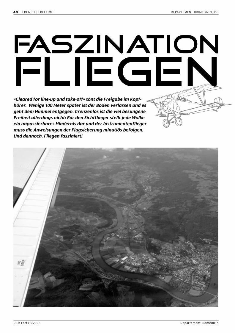
DBM Facts 3|2008 Departement Biomedizin
40 FREIZEIT | FREETIME DEPARTEMENT BIOMEDIZIN USB
«Cleared for line-up and take-off» tönt die Freigabe im Kopf-hörer. Wenige 100 Meter später ist der Boden verlassen und es geht dem Himmel entgegen. Grenzenlos ist die viel besungene Freiheit allerdings nicht: Für den Sichtflieger stellt jede Wolke ein unpassierbares Hindernis dar und der Instrumentenflieger muss die Anweisungen der Flugsicherung minutiös befolgen. Und dennoch, Fliegen fasziniert!

DBM Facts 3|2008 Department of Biomedicine
DEPARTMENT OF BIOMEDICINE USB FREIZEIT | FREETIME 41
Technik und Natur
Bewährte Triebwerke bringen das
Flugzeug in die Luft, die Redundanz
konventioneller Funknavigation
und moderner GPS-Einrichtungen
sichern die Orientierung und der
Autopilot steuert Höhen und Kurse
nach Programm. Schöne Tage bie-
ten unvergessliche Eindrücke von
Bergen und Seen, von Himmel und
Wolken sowie von Sonnenauf- und
Sonnenuntergängen. Aber schnell
erfliegt man auch die Grenzen der
Technik: Eine Wetterfront, ein Ge-
witter oder ein Schneesturm lassen
auch solide Reiseflugzeuge zum
Spielball der Natur werden. Auf- und
Abwinde können die Motorenkraft
ganz deutlich übersteigen, Tragflä-
chen und Propeller können nur un-
ter Einsatz aller technischer und na-
vigatorischer Mittel eisfrei gehalten
werden, und wenn Turbulenzen das
Flugzeug schütteln, dann sind die
fliegerischen Fähigkeiten gefordert.
Man erfliegt dann die Erfahrung,
dass die Gewalten der Natur nur
durch Anpassung überlebt werden
können.
Spannung und Entspannung
Technik und Natur können jederzeit
überraschende Herausforderungen
bieten, auch wenn die Vorbereitun-
gen auf Wetter und Flugzeug stan-
dardisiert sind. Veränderungen von
Wind und Sicht sind kurzfristig und
nicht genau vorhersehbar. Einfache
Flüge gibt es deshalb nicht, und die
Anspannung ist immer spürbar, be-
sonders bei Start und Landung. In
diesen Phasen erfordern gleichzeiti-
ge Navigation, Triebwerkssteuerung
und Kommunikation hohe Konzen-
tration. Die Beschäftigung mit dem
Fliegen lässt dann keinen Raum für
anderen Gedanken: Der Alltag wird
von einer faszinierend andersar-
tigen Anspannung verdrängt, die
sicherlich auch Taucher, Segler und
Athleten kennen. Nach der Landung
verbindet sich eine wunderschöne
Entspannung mit den Erinnerun-
gen und dem Gefühl einer Leistung.
Der Wunsch nach Wiederholung ist
deshalb immer da: Fliegen macht
schon auch abhängig.
Transport und Freizeit
Aber der Reiseflug ist auch nützlich:
Fünf Chirurgen, die ein neues Ope-
rationsverfahren in Salzburg ken-
nen lernen wollen (Abbildungen),
können kaum schneller und günsti-
ger als mit dem Individualflug trans-
portiert werden. Dass man dabei die
Flugzeiten frei wählen kann, ist ein
Vorteil. Dass man Warteschlangen
an Schaltern und Security vermei-
det, ist komfortabel. Bei vielen Rei-
sezielen, die durch die Konzentra-
tion des Linienverkehrs nicht mehr
Anflug auf den Flughafen Salzburg
Seite 40: Anflug auf Basel, mit Blick auf Bad Säckingen

DBM Facts 3|2008 Departement Biomedizin
42 FREIZEIT | FREETIME DEPARTEMENT BIOMEDIZINE USB
direkt erreichbar sind, gibt es ob-
jektive Vorteile des Individualflugs.
Aber natürlich: Die Faszination Flie-
gen bleibt das wichtigste Argument
für den Motorflug. Wenn man dabei
aber Transport und Freizeit verbin-
den kann, dann ist das eine beson-
ders schöne Kombination.
Fliegerei und Medizin
Dass die Medizin von der Fliegerei
lernen kann, ist bei uns in Basel bes-
tens bekannt: Das Critical Incidence
Reporting System (CIRS), das die
Basler Anästhesie unter der Leitung
von Prof. Daniel Scheidegger in die
Medizin eingeführt hat, stammt aus
der Luftfahrt und wurde gemein-
sam mit der damaligen Swissair ent-
wickelt. Insbesondere Beinahezwi-
schenfälle, die sonst eher verdeckt
als bekannt würden, werden ano-
nym rapportiert und für Verbesse-
rungen genutzt. Aber wir können
noch mehr von der Luftfahrt ler-
nen, die alle Kommunikationssys-
teme auf Feed-back abgestellt hat:
Die Freigabe „Cleared for line-up
and take-off“ muss vom Piloten
wörtlich zurückbestätigt werden.
Wie wäre es, wenn wir in der Medi-
zin Informationen an Patientinnen
und Patienten erst dann akzeptie-
ren würden, wenn diese einwand-
frei zurückbestätigt würden?
Flughafen und Flugschule Basel
Basel ist für die Fliegerei ein beson-
derer Ort: Nicht nur der trinatio-
nale Flughafen auf französischem
Terrain ist eine Besonderheit. Auch
die Nähe dieses perfekt ausgestat-
teten Verkehrsflughafens zur Stadt
ist einzigartig. Ebenso die Infra-
Das Chirurgenteam: Daniel Oertli (erster von links),
Oleg Heizmann (zweiter von links), Roger Schmid (zweiter von rechts),
Michael Heberer (erster von rechts).

DBM Facts 3|2008 Department of Biomedicine
DEPARTMENT OF BIOMEDICINE USB FREIZEIT | FREETIME 43
struktur mit einer hervorragenden
Flugschule, die schon Generationen
von Piloten auf allen Niveaus aus-
gebildet hat. Auch viele Mitarbeiter
unseres Universitätsspitals haben
dort ihre fliegerische Ausbildung er-
halten. In dieser Stadt macht fliegen
und fliegen lernen besondere Freu-
de. Die Faszination Fliegen hat hier
eine einzigartige Basis.
Michael Heberer
Blick auf den Bodensee bei Radolfzell

DBM Facts 3|2008 Departement Biomedizin
44 KULINARISCHES | CULINARY DEPARTEMENT BIOMEDIZIN USB
Pumpkins e a s o nAutumn: the days get shorter, the heat disappears and, on
menus, the summer’s fruits make way for this season’s ve-
getables. Now it’s time for the pumpkin. Visible everywhe-
re, in fields and markets, the pumpkin displays its great
variety of shapes and colours. It comes in orange, yellow
or even light blue, and the pumpkin is perfect in an equally
wide range of dishes. You can use it with nearly all spices,
herbs, fruits and many other ingredients to create a deli-
cious dinner. Here are a few ideas to help you prepare a
pumpkin meal, but we’d be happy to hear your recipe sug-
gestions too. How do you prepare pumpkin in your coun-
try?
Brigitte Schneider
MexicanPumpkin
1 pumpkin of approx. 1.5 kg
400 g minced meat (beef)
3 onions
1 tin tomatoes or 2-3 fresh tomatoes
1 tin corn
2 green peppers
1 hot chili
100 g boiled rice 1⁄2 bundle of cilantro/coriander1⁄2 teaspoon cinnamon
1 teaspoon chopped oregano
100 ml vegetable broth
100 g clotted cream
100 g goat’s cheese
2–3 tablespoons hot chili sauce
Cut a lid from the top of the pumpkin and carefully remove
the seeds from inside. Scrape off some of the pumpkin pulp,
but leave enough to keep the pumpkin stable.
The meat and the finely sliced onions are fried in a little bit
of olive oil until it becomes brownish, then add the vegetable
broth and cook gently for 10 min. Cut green pepper and chili
in thin slices, tomatoes in pieces and add to the meat. Chop
the cilantro leaves, crumble the cheese and cut the removed
pumpkin into little squares and add together with the corn,
the clotted cram and the chili sauce. Fill the pumpkin with this
mixture, place it into a casserole dish and cover with a sheet
of aluminium foil. Bake in the oven for 50–60 min at 200°C. In
the last 10 min replace the foil with the pumpkin-lid.

DBM Facts 3|2008 Department of Biomedicine
DEPARTMENT OF BIOMEDICINE USB KULINARISCHES | CULINARY 45
Pumpkin-Plum-Salad
500 g pumpkin pulp
2 apples, not too sweet
1 pear
Grate the pumpkin, apples and pear roughly, cut the plums
into fine slices, chop the parsley and peppermint and mix all
together. For the dressing, mix all the ingredients smoothly and
add to the fruits. “Incubate at room temperature” for about
30 min before serving.
Pumpkin and Coconut Soup1 liter water
1 bundle parsley
2 of lemongrass sticks
3 chopped onions
salt, saffron
1 kg pumpkin pulp
2 peeled potatoes
2 pieces of leek
juice from 1 lemon
salt, pepper
1 tin coconut cream
Cut parsley and 1 lemongrass stick into fine slices and boil
together with the onions, salt and saffron in approx. 1 l wa-
ter for about 30 min. In the meantime, cut the pumpkin pulp
and the potatoes into fine squares. Cut the lemongrass and
the leek into fine rings and fry in a bit of oil or butter. Add
the pumpkin and the potatoes and stir. Fill up the pan with
the vegetable broth prepared before and the coconut cream
and cook for about 30 min on a low heat. Flavour with lemon
juice, salt and pepper and mash very finely.
Leave to cool and serve with fresh parsley and crou-
tons.
Pumpkin-Gratinwith Hot Plums
1 kg pumpkin pulp
200 g sugar
a bit of salt
a pinch of grinded cloves
a pinch of cardamom
1 tablespoon chipped peppermint leaves
100 g semolina
200 g roasted walnut pieces
50 ml orange juice or white wine
50 g butter
500 g plums
Grate pumpkin pulp and mash together with orange juice or
wine. Add sugar, spices and semolina, mix and fill into a but-
tered casserole dish. Sprinkle walnuts on top and add the butter
in little flakes. Bake in the oven for 45–60 min at 200 °C.
In the meantime cut plums in slices, cook together with
orange juice or wine, chili and pepper and serve with the gratin.
100 ml orange juice or red wine1⁄2 teaspoon chili-flakes
freshly grinded black pepper
300 g plums
1 bunch of parsley
1 bunch of peppermint
Dressing:
100 g yoghurt1⁄2 teaspoon mustard
1 tablespoon lemon juice
2 tablespoons olive oil
sugar, salt, pepper, ginger

DBM Facts 3|2008 Departement Biomedizin
46 FREIZEIT | FREETIME DEPARTEMENT BIOMEDIZIN
Von Feuerschluckern, Trommlern und balinesi-schen TänzerinnenImpressionen vom Spitalfest am 22. August 2008
Auch viele Mitarbeitende des Departements Biomedizin waren dabei und genossen die tolle Atmosphäre ...
Foto
s: M
arc W
eile
r, Ph
otog
raph
y
Foto
cred
it w
ww.
mar
cwei
ler.c
h

DBM Facts 3|2008 Department of Biomedicine
DEPARTMENT OF BIOMEDICINE FREIZEIT | FREETIME 47

DBM Facts 3|2008 Departement Biomedizin
Wie beginnt eine Laufbahn als
Wissenschaftler?
Liebe Kollegen, heute bin ich Privat-
dozent in Molekularer Medizin, aber
eigentlich hat während meiner frühen
Jugendzeit wenig auf eine wissen-
schaftliche Laufbahn hingewiesen.
Ich bin in einer Oberbaselbieter Ge-
meinde als Sohn einer Landwirtsfami-
lie aufgewachsen und habe tatsäch-
lich einige meiner Jugendjahre auch
jetzt noch «in der Nase». Während die
anderen Schulkollegen sich in der Badi
oder sonstwo vergnügten, misteten
mein Bruder und ich wahlweise den
Kuhstall aus, fütterten die Schwei-
ne oder pflegten das Federvieh, von
den Pferden, Kaninchen, Katzen und
Hunden ganz zu schweigen. Heute
bin ich meinen Eltern dankbar, eine
solche unbeschwerte, bewegungsrei-
che und naturverbundene Jugendzeit
habe verbringen zu dürfen. Was sich
heute in meinem persönlichen Rück-
blick nach Bauernhofromantik anfühlt
war tatsächlich eine manchmal nicht
angenehm riechende Knochenarbeit.
Zu jener Zeit, jung und rastlos wie ich
war und – zumindest in meinen eige-
nen Augen – zur Landarbeit gänzlich
untauglich geraten, suchte ich Wege,
dem Bauernalltag auszuweichen. Da
für mich die Schule keine Mühe son-
dern eher Lust war, kam eigentlich nur
der gymnasiale Weg in Frage (dies ob-
wohl mich meine Eltern schon für die
Landwirtschaftsschule vorgemerkt
hatten).
Ich konnte während der Gymnasi-
alzeit – wie vielleicht doch zu erwar-
ten war – nicht völlig aus meiner Haut
schlüpfen und so wurde Biologie mein
Lieblingsfach. Als wenn es noch einen
weiteren Anstoss gebraucht hätte,
legte mir mein Biologielehrer ans
Herz, dieses Fach doch an der Uni zu
studieren. Nichts sonderlich Weltbe-
wegendes heute, war dies damals tat-
sächlich keine leichte Entscheidung,
da nun niemand den elterlichen Hof
weiterführen würde (mein älterer Bru-
der hatte sich bereits früher entschie-
den, ein Elektroingenieurstudium zu
absolvieren). Im Jahr 1977 habe ich
dann das Biologiestudium im ehr-
würdigen Zoologischen Institut am
Rheinsprung und am mit Entschie-
denheit noch ehrwürdigeren Botani-
schen Institut an der Schönbeinstras-
se begonnen. Nach dem Diplom 1982
(ja, so hiess das früher noch; heute
wär’s ein Master) begann ich im Jahr
1983 meine Dissertation bei Prof. C.G.
Honegger im 3. Stock des ZLF an der
Hebelstrasse. Mein Fach- und Interes-
sensgebiet war in der Neurobiologe
anzusiedeln und in jener Zeit konn-
te ich viele wertvolle Kontakte mit
Freunden und Kollegen knüpfen, wel-
che auch heute noch wichtig für mich
sind. In meiner Dissertation fasste ich
1988 meine Forschungsergebnisse
über die Experimental Autoimmune
Encephalomyelitis (EAE) zusammen
und feierte meinen Abschluss natür-
lich ausgiebig.
Werner Krenger:Pädiatrische Immunologie
Blick vom Elternhaus auf die Sissacherflueh
Gruppe C.G. Honegger im Jahr 1988
48 DAS DBM STELLT SICH VOR DEPARTEMENT BIOMEDIZIN USB

DBM Facts 3|2008 Department of Biomedicine
Wanderjahre
Nach Abschluss meiner Doktorarbeit
wollte ich als nächstes den neurobi-
ologischen Aspekt der EAE um die
immunologische Fazette erweitern.
Dieses Ansinnen wurde nach gebüh-
render Investition meinerseits durch
die Harvard Medical School in Boston
honoriert und so reiste ich am 1. März
1989 – es fühlt sich an wie gestern –
mit Sack und Pack in einer real nicht
mehr existierenden Swissair Maschine
in die USA und habe quasi ein neues
Leben begonnen. Ursprünglich geför-
dert für ein Jahr vom SNF erwarb ich
am Brigham & Women’s Hospital im
Center for Neurologic Diseases bei
Prof. Howard L. Weiner das immuno-
logische Rüstzeug, die Mechanismen
der EAE studieren zu können. Die Ar-
beit interessant, die Ergebnisse gut,
die Freunde mehr und mehr, die Le-
bensweise der liberalen Ostküstena-
merikaner emulierend, wurde aus
dem einem Jahr deren zwei, dann drei,
dann vier. Da ich mich bestens einge-
lebt hatte, weder abhängig noch mit
Familie versehen, entschied ich mich
im Jahr 1993, nochmals eine neue He-
rausforderung in Form einer zweiten
Postdoc/Instructor Stelle zu suchen.
Am Dana Farber Cancer Institute in
Boston begann ich unter Leitung von
Prof. James L. Ferrara auf demjenigen
Thema zu forschen, auf dem ich auch
heute noch tätig bin, die Graft-vs.-
Host Disease (GVHD). Diese Erkran-
kung ist eine unangenehme Beikost
einer Hämatopoietischen Stammzell-
transplantation, welche heutzutage
sehr oft zur Therapie von Leukämien
eingesetzt wird. Das Thema faszinier-
te mich von Anfang an und so verlän-
gerte sich meine Instruktortätigkeit an
der Harvard University um interessan-
te, lehrintensive und sozial äusserst
verträgliche weitere vier Jahre.
Der Kreis schliesst sich
Nun, man bleibt nicht das ganze Le-
ben Postdoc/Instruktor und so stell-
te sich mir die Frage nach dem «Wie
weiter?». Aus einer Zufallsbegegnung
am Dana Farber ist nun eine mehr
als 10-jährige Arbeitsbeziehung ge-
worden. Georg A. Holländer – sei-
nes Zeichens zu jener Zeit ebenfalls
Postoc in den USA – war eine Stelle
am (damaligen) Kantonsspital Basel
offeriert worden. Er ein Spezialist in
Thymusbiologie, ich in der Stamm-
zelltransplantation – zusammen ge-
baren wir nach unzähligen Diskussi-
onen, Bieren, Fondues bei und mit
Freunden die Idee, in der Schweiz
ein gemeinsames Projekt auf die
Beine zu stellen. Tatsächlich wurde
unser SNF-Antrag gutgeheissen und
so reiste ich – ziemlich schweren
Herzens und viele Freunde zurück-
lassend – nach Basel zurück, um An-
fang Januar 1997 meine neue Arbeit
zu beginnen. Der Untertitel dieses
Abschnittes besagt es: Ich kam nach
acht Jahren in der Ferne nach Basel
zurück, jedoch nicht an irgendeinen
Ort, sondern in genau dasjenige La-
bor, in dem ich exakt 15 Jahre zuvor
meine Diplomarbeit begonnen hatte.
Ungewöhnlich, wie sich der Kreis ge-
schlossen hatte, und ich wieder viele
der früheren Freunde und Kollegen
antreffen durfte.
Heute
Von 1997–2004 forcierte ich am ZLF
in unterschiedlichen Funktionen,
mal Projektleiter, dann Oberassistent,
dann Kollaborator, mal wieder Projekt-
leiter, dann als Angestellter des UKBB
und als Mitglied der Forschungsgrup-
pe «Pädiatrische Immunologie» die
Identifikation der immunologischen
Grundlagen der Stammzelltransplan-
tation. Im September 2004 erfolgte
dann der Umzug (ungewollt, muss
ich sagen) von der Hebelstrasse an
die Mattenstrasse in das umgebaute
Zentrum für Biomedizin. Meine For-
schungshauptrichtung konzentriert
sich nach wie vor auf die Rolle des
Thymus in der T-Zell-Bildung nach
Transplantation/GVHD (siehe Exhibit
A, welches eine typisch abnorme Or-
ganisation von Thymusgewebe zeigt).
Im Jahr 2006 erhielt ich die Venia do-
cendi der Universität Basel, welches
mir erlaubte, meine Vorlesungstätig-
keit in Molekularer Medizin und meine
Ausbildnertätigkeit für Masterstuden-
ten des Biologielehrganges zu inten-
sivieren. Neben meiner beruflichen
Tätigkeit existiert noch ein reiches
Privatleben, weil .... oh, da seh’ ich die
DBM Facts Redaktoren verzweifelt
winken. Mein Platz ist aufgebraucht,
hätte mich kürzer fassen sollen. Nun,
zu spät. Wen’s interessiert, kann ger-
ne mit mir reden, für heut’ reicht’s
aber. Herzliche Grüsse und ich sehe
Euch sicher demnächst im ZLF, an der
Mattenstrasse oder im Pharmazent-
rum, ....oder auf dem Tennisplatz, der
Skipiste, vielleicht auch dem Wander-
weg oder ...
Euer Werner Krenger
Harvard Medical School im Jahr 1989
DEPARTMENT OF BIOMEDICINE USB DAS DBM STELLT SICH VOR 49

DBM Facts 3|2008 Departement Biomedizin
Uninacht 2008
Biozentrum und Pharmazentrum mit der Chemie und
Physik zu einem grossen Campus zu verschmelzen, war
eine der vielen Ideen, der Uninacht 2008 ein besonderes
Gesicht zu geben. Die Pestalozzistrasse, die den Campus
Naturwissenschaften in zwei Teile trennt, blieb für 24 Stun-
den gesperrt und stattdessen stand dort ein riesiges Zelt
für mehrere hundert Personen und eine grosse Musikbüh-
ne. Das Zelt füllte sich ab 18 Uhr rasch bis auf den letzten
Platz. Noch mehr Leute suchten Einlass zur legendären
Sprengvorlesung im Chemiehörsaal, wo Doktorierende
am Experimentiertisch alle Trickkisten der anorganischen
Chemie öffneten und dem staunenden Publikum ein vi-
suelles und akustisches Feuerwerk boten, derweil unten
in der Halle Kinder verschiedenste Versuche durchführen
und ein Juniordiplom in Chemie erwerben konnten.
Die Uninacht 2008 lockte Tausende an
die rund 25 verschiedenen Besuchs- und
Festorte zwischen Münsterplatz und
Schällenmätteli. Die sieben Fakultäten
der Universität Basel und ihre zentralen
Einrichtungen und Museen bereiteten
zum ersten Mal zusammen mit Alumni
und Skuba ein Programm mit über 300
Veranstaltungen vor, speziell auch sol-
che für Kinder und Jugendliche, um ei-
nen Querschnitt durch alle Fachgebiete
der 20 Departemente und rund 110 In-
stitute und Seminare der Universität Ba-
sel zu präsentieren.
50 FREIZEIT | FREETIME DEPARTEMENT BIOMEDIZIN
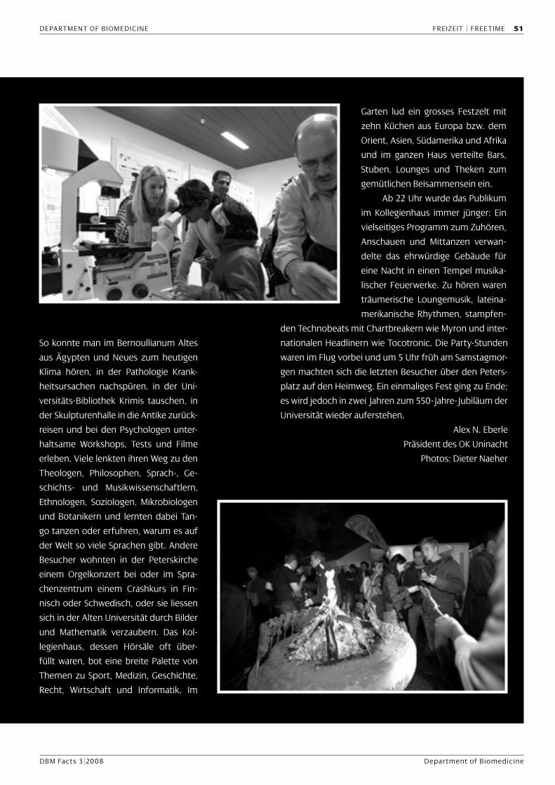
DBM Facts 3|2008 Department of Biomedicine
So konnte man im Bernoullianum Altes
aus Ägypten und Neues zum heutigen
Klima hören, in der Pathologie Krank-
heitsursachen nachspüren, in der Uni-
versitäts-Bibliothek Krimis tauschen, in
der Skulpturenhalle in die Antike zurück-
reisen und bei den Psychologen unter-
haltsame Workshops, Tests und Filme
erleben. Viele lenkten ihren Weg zu den
Theologen, Philosophen, Sprach-, Ge-
schichts- und Musikwissenschaftlern,
Ethnologen, Soziologen, Mikrobiologen
und Botanikern und lernten dabei Tan-
go tanzen oder erfuhren, warum es auf
der Welt so viele Sprachen gibt. Andere
Besucher wohnten in der Peterskirche
einem Orgelkonzert bei oder im Spra-
chenzentrum einem Crashkurs in Fin-
nisch oder Schwedisch, oder sie liessen
sich in der Alten Universität durch Bilder
und Mathematik verzaubern. Das Kol-
legienhaus, dessen Hörsäle oft über-
füllt waren, bot eine breite Palette von
Themen zu Sport, Medizin, Geschichte,
Recht, Wirtschaft und Informatik. Im
Garten lud ein grosses Festzelt mit
zehn Küchen aus Europa bzw. dem
Orient, Asien, Südamerika und Afrika
und im ganzen Haus verteilte Bars,
Stuben, Lounges und Theken zum
gemütlichen Beisammensein ein.
Ab 22 Uhr wurde das Publikum
im Kollegienhaus immer jünger: Ein
vielseitiges Programm zum Zuhören,
Anschauen und Mittanzen verwan-
delte das ehrwürdige Gebäude für
eine Nacht in einen Tempel musika-
lischer Feuerwerke. Zu hören waren
träumerische Loungemusik, lateina-
merikanische Rhythmen, stampfen-
den Technobeats mit Chartbreakern wie Myron und inter-
nationalen Headlinern wie Tocotronic. Die Party-Stunden
waren im Flug vorbei und um 5 Uhr früh am Samstagmor-
gen machten sich die letzten Besucher über den Peters-
platz auf den Heimweg. Ein einmaliges Fest ging zu Ende;
es wird jedoch in zwei Jahren zum 550-Jahre-Jubiläum der
Universität wieder auferstehen.
Alex N. Eberle
Präsident des OK Uninacht
Photos: Dieter Naeher
DEPARTMENT OF BIOMEDICINE FREIZEIT | FREETIME 51

DBM Facts 3|2008 Departement Biomedizin
Z Basel isch Mäss!25. Oktober bis 9. November
52 FREIZEIT | FREETIME DEPARTEMENT BIOMEDIZIN

DBM Facts 3|2008 Department of Biomedicine
RNA profiling of MS brain tissuesThe introduction of RNA profiling using microarray technology has recently helped to elucidate gene expression changes in diseased tissue samples from postmortem human brains. This is especially the case in the field of multiple sclerosis research, where microarray-based RNA pro-filing has been applied in the hope of identifying disease-specific alterations. The lack of good biomarkers for diagnostic and prognostic purposes, the need for new drug targets and for a better understanding of the pathophysiology makes this technique a valuable tool.
Microarrays
There are several different platforms available for RNA
profiling. They differ in the number of gene sequences
covered by the array, in the length of the cDNA probes,
and the way the source RNA is processed for probe la-
beling. The most commonly used microarrays use short
oligonucleotide sequences (25- to 60-mers), which are
covalently bound to a small glass surface (e.g. Affime-
trix, Illumina). Nowadays, these arrays cover the whole
genome sequence of the species to be analyzed. From
a selected biological sample total or poly A+ RNA is ex-
tracted, converted into cDNA, and finally fluorescently
labelled cRNA is generated. Another type of array uses
radioactive-labeled cDNA as a highly sensitive probe for
detecting expressed gene sequences (e.g. Clontech Atlas
arrays). The combination of long cDNA fragments (300–
500 bp) attached to a nylon membrane and the use of
radioactivity for labeling cDNA probes makes this type
of microarray very specific and sensitive. A disadvantage
of this system is the lower number of gene sequences
(500–5000) covered by one array. After hybridization of
the labeled cDNA probe, the hybridization signal of each
sequence spot is determined either by laser scanning
of the fluorescent signal or by autoradiography of the
radioactive signal (Phosphor Imager). With 1μg of total
RNA (or as little as 10 ng RNA) the gene expression of the
whole genome can be analyzed at the same time.
Microarrays in MS
The first study applying microarray technology on MS
brain tissue was published in 1999 by Whitney and col-
leagues. Since then, several studies using microarrays
have been performed to identify differential gene ex-
pression in a variety of tissues of MS patients, as well as
in experimental autoimmune encephalomyelitis (EAE),
an animal model of MS.
The studies differ, but also partially overlap, con-
cerning experimental setup and the results obtained.
The heterogeneity of the experimental setups makes it
difficult to compare these studies retrospectively. Sev-
eral points have to be considered when a microarray
study is planned (Fig. 1). At the beginning of each gene-
expression study the scientific question determines the
experimental setup. In MS research a wide range of sci-
entific questions have been addressed using microarray
experiments. Several studies aimed to identify biomark-
ers for diagnostic purposes, whereas other studies were
designed to identify the molecular mechanisms of MS
pathophysiology or to discover new drug targets.
An important issue in using microarray technology in MS
research is tissue selection and sampling. This includes
different populations of peripheral blood cells (periph-
eral blood mononuclear cells and CD3+ cells), different
types of lesioned white matter (e.g. acute, active and
silent lesions) and nonlesioned normal-appearing white
DEPARTMENT OF BIOMEDICINE USB KONKRET | CONCRETE 53

DBM Facts 3|2008 Departement Biomedizin
matter. These studies differ in the number of MS cases
and type of MS disease course (relapsing remitting, pri-
mary progressive or secondary progressive). Different
expression array platforms are available and have been
recently reviewed.
Another major step is the analysis of the hybridiza-
tion signals and there are a large variety of software tools
available for statistical analysis to identify the differen-
tially expressed genes. The choice of the normalization
parameters largely depends on the system used. For
example, ‘global normalization’ is often applied to mi-
croarrays covering thousands of sequences, whereas hy-
bridization signals from microarrays, which cover a cho-
sen selection of sequences, will instead be normalized
with one or a set of house-keeping gene sequences. Still,
the final interpretation of the obtained results remains
one of the most challenging steps in analyzing data from
gene chips. Currently, the need to develop new tools to
convert the enormous amount of generated microarray
data into biological significant interpretations is a topic
of much interest. The identified differentially expressed
genes have to be verified using other techniques, such as
quantitative RT-PCR, in situ hybridization, Western blot
analysis and/or immunohistochemistry. Validation using
an independent cohort of patients would be optimal.
The diversity in experimental design used in the
past partially reflects the different approaches address-
ing distinct scientific questions. Among all these issues,
sample and case selection is one of the major criteria
for successful analyzing gene expression alterations in
MS brain tissues, and we therefore now discuss them in
more detail.
Sample and case selection
In neurological diseases affecting the central nervous
system, such as MS, the availability of affected tissue is
limited. The studies are mostly limited to those tissues
obtained during postmortem examinations because
brain biopsies for analytical purposes are only rarely tak-
en. Therefore, analysing autopsies is mostly a snapshot
of gene expression at late stages of the disease course.
RNA degrades relatively quickly after death, and thus
only samples obtained a short time after death contain
high-quality RNA that can be used for expression profil-
ing. Although, full-length total RNA can be isolated up
to 20 hours after death, a short postmortem time does
not always guarantee intact RNA, since other mecha-
nisms, which are not controllable, lead to RNA degra-
dation after death. Nevertheless, postmortem tissue is
a useful source for gene expression studies to analyze
pathomechanisms, and to identify possible drug targets.
The selection of the tissue sample is an especially impor-
tant issue in the experimental setup of MS studies. To
ensure that the identified changes in gene expression
are due to the parameter to be analyzed (e.g. diseased
vs. control) it is important to avoid false results due to
a bias in case and tissue selection. Potential sources of
bias in case selection are sex, age, treatment, disease
course, additional confounding diseases and the length
of time after death that tissue was collected.
In MS research there is a variety of sources of bio-
logical samples when analyzing gene expression in the
brain. Most of the studies used brains samples from
MS and healthy control patients obtained at postmor-
Figure 1: The main issues to be considered when performing RNA profiling of MS brains using microarray analysis. The first step consists of defining the scientific question that should be answered by the output data. The working hypothesis will help to design a study, and to select the patient cohort and the type of tissue samples. The next step includes the actual microarray experiment by which the gene expression data are obtained. After statistical analysis and generation of an expression profile the data should be verified by alternative methods and, optimally, validated using an independent cohort of patients.
54 KONKRET | CONCRETE DEPARTEMENT BIOMEDIZIN USB

DBM Facts 3|2008 Department of Biomedicine
tem (Fig. 2A), but also brain and spinal cord tissues of
EAE animals have been used (Fig. 2B). To maintain com-
parability between MS cases and controls, the selected
brain areas (e.g. subcortical white matter, periventricular
white matter) used for the analysis should be analogous.
The sample heterogeneity can be reduced by isolating
tissue from defined brain regions to avoid false results
due to regional differences. Another important issue in
Figure 2: The various types of brain tissues analyzed in MS research. Different types of brain tissue have been used for microarray studies in the field of MS research (A). Additionally, an animal model for MS, EAE, has been used to isolate tissue from the central nervous system for microar-ray experiments (B). An important issue in identifying differentially expressed genes is the cellular composition of the tissue to be compared. White matter is mainly populated by myelinating oligodendrocytes, astrocytes and microglia (C). In lesions the cellular composition is even more complex due to infiltrating immune cells, such as T-cells, B-cells and peripheral macrophages (D−F). Therefore, tissues with different cellular compositions (e.g. lesioned MS tissue vs. tissue from control cases) must be interpreted with this special focus and awareness.
identifying differentially expressed genes is the cellular
composition of the tissue to be compared (Fig. 2C−F).
To identify genes which are up- or downregulated by a
specific cell type, a monotypic cell population as source
for a microarray experiment would be optimal. For most
heterogeneous tissues, like the central nervous system,
this is not the case. White matter is mainly populated by
myelinating oligodendrocytes, astrocytes and microglia
(Fig. 2C). Additionally, interstitial subcortical neurons,
endothelial cells forming blood vessel walls, and even
axonal transported transcripts can be sources of RNA.
Therefore, the obtained expression profile of white mat-
ter tissue is a mixture of all these cell types. When using
lesioned tissue as a source for RNA, the cellular compo-
sition is even more complex due to infiltrating immune
cells like T-cells, B-cells and peripheral macrophages (Fig.
2D−F). Therefore, the comparison of tissue with different
cellular composition (e.g. lesioned MS tissue vs. tissue
from control cases) must be interpreted with this special
focus and awareness. Certainly, it is nearly impossible to
fulfil all these criteria, but nevertheless they have to be
considered when analyzing and interpreting the results.
Conclusion
The use of cDNA microarrays in RNA expression profiling
studies provided new insights in the pathomechanism of
MS. Several microarray studies in MS research revealed
promising results which may lead to the identification of
new therapeutic targets and might help to design more
specific treatments.
Jochen Kinter, Thomas Zeis and
Nicole Schaeren-Wiemers
Neurobiology, Department of Biomedicine USB
Full article:
RNA profiling of MS brain tissues.
Int MS J (2008), 15,51–58
DEPARTMENT OF BIOMEDICINE USB KONKRET | CONCRETE 55

DBM Facts 3|2008 Departement Biomedizin
DBM Christmas Party Tuesday 16th December 2008
56 FREIZEIT | FREETIME DEPARTEMENT BIOMEDIZIN
The attractions:
Dance to the music of our live band • Eat what you like
from the buffet • Meet people from the whole department •
Enjoy the atmosphere of an old vault.
We meet at 17.00 at Holsteinerkeller, Hebelstrasse 32