An anatomical study of variations in termination of brachial

Accepted Manuscript
Anatomical Assessment of Variations in Kambin’s Triangle: A surgical and cadaverstudy
Ali Fahir Ozer, Prof. MD, Tuncer Suzer, Prof. MD, Halil Can, MD, Mani Falsafi, MD,Murat Aydin, MD, Mehdi Sasani, Assoc. Prof. MD, Tunc Oktenoglu, Assoc. Prof. MD
PII: S1878-8750(17)30080-3
DOI: 10.1016/j.wneu.2017.01.057
Reference: WNEU 5154
To appear in: World Neurosurgery
Received Date: 19 July 2016
Revised Date: 12 January 2017
Accepted Date: 16 January 2017
Please cite this article as: Ozer AF, Suzer T, Can H, Falsafi M, Aydin M, Sasani M, Oktenoglu T,Anatomical Assessment of Variations in Kambin’s Triangle: A surgical and cadaver study, WorldNeurosurgery (2017), doi: 10.1016/j.wneu.2017.01.057.
This is a PDF file of an unedited manuscript that has been accepted for publication. As a service toour customers we are providing this early version of the manuscript. The manuscript will undergocopyediting, typesetting, and review of the resulting proof before it is published in its final form. Pleasenote that during the production process errors may be discovered which could affect the content, and alllegal disclaimers that apply to the journal pertain.

MANUSCRIP
T
ACCEPTED
ACCEPTED MANUSCRIPT Endoscopic extraforaminal approach, Kambin’s triangle
1
Anatomical Assessment of Variations in Kambin’s Triangle: A surgical and cadaver study
Ali Fahir Ozer, Prof. MD1; Tuncer Suzer, Prof. MD1; Halil Can, MD2; Mani Falsafi, MD3;
Murat Aydin,MD4 , Mehdi Sasani, Assoc. Prof. MD1; Tunc Oktenoglu, Assoc. Prof. MD1
1 Koc University Medical School, Neurosurgery Department, Istanbul-Turkey
2 Private Pendik Yüzyıl Hospital, Neurosurgery Department, Istanbul-Turkey/ Research fellow in Koc
University Medical School Neurosurgery Department, Istanbul-Turkey
3 Iran University of Medical Science, Hazrat Rasoul Medical Complex, Spine Surgery Division, Tehran-
Iran/ Research fellow in Koc University Medical School Neurosurgery Department, Istanbul-Turkey
4 Izmir Bozyaka Research and Training Hospital, Neurosurgery Department, Izmir- Turkey/ Research
fellow in Koc University Medical School Neurosurgery Department, Istanbul-Turkey
Correspondence: Ali Fahir Ozer, Prof MD
Koc University School of Medicine Department of Neurosurgery
Rumelifeneri Yolu Sarıyer, Istanbul 34450 Turkey
Ph: 90 (212) 338-1401
Fax: 90 (212) 338-1559
E-mail: [email protected]
Conflict of Interest: None.
Disclosure of Funding: None
Running head: Endoscopic extraforaminal approach, Kambin’s triangle

MANUSCRIP
T
ACCEPTED
ACCEPTED MANUSCRIPT Endoscopic extraforaminal approach, Kambin’s triangle
2
Abstract
Background. The relationship of exiting root and Kambin’s triangle is discussed in this article.
Objective. Transforaminal endoscopic surgery as the gold standard of less invasive lumbar disc
surgeries is performed through Kambin’s triangle. Existing root damage is one of the most
important complication for this type of surgery. Anatomical variations in Kambin’s triangle may
be the main reason for nerve root damage during endoscopic lumbar disc surgery.
Method. Kambin’s triangle was investigated with surgical views and cadaver studies.
34 patients with farlateral disc herniation were treated via extraforaminal approach under the
microscope. On the other hand, 48 Kambin’s triangles were dissected on 8 cadavers.Three main
types of triangle were identified, and patients were grouped according to these three types of the
triangle.
Results. Only 6 of the 34 patients had type 3 triangles, which is the wide classical triangle
described by Kambin, however 17 patients had type 2, with a narrow space in the triangle, and 11
patients had type 1, with no space inside the triangle. Cadaver results were similar; only 10 of the
48 specimens had the type 3 classical triangle while 23 had type 2, and 15 had type 1 triangles.
Our results disclosed narrowed or no space in 82,4% of the patients and 79,2% of the cadavers.
Conclusion. We observed that, a wide and safe room of the triangle may not be exist in some
patients. Therefore, more care must be taken during endoscopic lumbar disc surgery to avoid
nerve damage.
Key Words. Endoscopic discectomy, Extraforaminal approach, Kambin’s triangle

MANUSCRIP
T
ACCEPTED
ACCEPTED MANUSCRIPT Endoscopic extraforaminal approach, Kambin’s triangle
3
INTRODUCTION
Endoscopic surgery has been accepted as a less invasive procedure for lumbar disc herniation and
was introduced by Kambin and Hijikata in the 1970’s.1 For this purpose, Kambin described a safe
area called Kambin’s triangle: the exiting root is its hypotenuse, the inferior border of the lower
vertebral body creates the base, and the height is formed by the articular process and superior
articulating facet of the caudal vertebra. Using this path,there is a safe pathway near the upper
part of the root for passing the cannulas and endoscopic instruments for nucleotomy or
endoscopic fragment resection.2,3
The major advantage of this type of surgery is that the ligaments, muscles, and bones are better
preserved, limiting instability, facet arthropathy and disc space narrowing.4-10 Further, less
manipulation of the epidural venous system may prevent scarring, edema and chronic neural
fibrosis formation.11,12 This technique also allows a better view based on magnification and light.
However, there are some complications that have reduced the popularity of this type of surgery
during recent years. The most important complication is an existing nerve root damage, which
adds a new symptom to the symptoms that the patient experienced before surgery.13-16 Damage to
radicular arteries is another complication, which could cause epidural hematoma or cord ischemia
if the Adamkiewicz artery has been damaged.17,18 Sairyo et al reported 2% nerve irritation, 1%
epidural hemotoma in 100 cases of patient series.19 These types of damage may be caused by
problems with the path of approach, the cannula diameter, the maneuvers used during endoscopy
or normal variations in the Kambin’s triangle.
The objective of this study was to evaluate the normal anatomy and the variations of the
Kambin’s triangle. The shape and size of the area was investigated with microsurgical findings as
well as cadaver study findings.
METHODS

MANUSCRIP
T
ACCEPTED
ACCEPTED MANUSCRIPT Endoscopic extraforaminal approach, Kambin’s triangle
4
Subjects
An anatomical assay was conducted from to assess the normal variations in Kambin’s triangle in
vivo in lumbar disc herniation patients scheduled for a microsurgical lateral extraforaminal
approach. Patients included in the assay are lumbar radiculopathy cases who did not respond to
conservative treatment, showing lateral intra- or extraforaminal protrusion or extrusion. Patients
with degenerative facet disease and facet hypertrophy have been excluded. The variables
compiled by the surgeon include the patient’s age, gender, date of surgery, level of involvement
and the anatomical variation of Kambin’s triangle. The video recordings of the patient’s
operation was reviewed by the surgeon after surgery for typing the triangle.
The variability of the results due to different observers is an important concern for this kind of
investigations. The measurements were performed by same author to prevent this problem.
An ethics committee approval or patient consent for operative recording have not been necessary,
because all the operations were routine spinal procedures and video recording of the operations is
done in all operations.
Operative technique
The operation was performed under microscopic visualization by two neurosurgeons specializing
in spine surgery. After applying general anesthesia, the patient was positioned in a prone posture.
Fluoroscopy was used to determine the correct level. After preparation and draping of the skin, a
lateral skin incision was performed 2 cm from the midline. After undermining the subcutaneous
tissue, the fascia was incised, and a blunt longitudinal dissection of the paraspinal muscles was
used to find the facet joint and the transverse process of the desired level. After exposure of the
“U”-shaped connection of the superior and inferior transverse processes and the facet joint, the
intertransverse ligament was removed. Under the ligament, we can follow path of the root, which
usually begins at the junction of the superior transverse process and the lateral surface of facet
joint and extends inferiorly, laterally and anteriorly.
Kambin’s triangle is defined anteriorly by the exiting root, medially by the facet joint and
inferiorly by the superior border of the inferior vertebral body and the inferior pedicle. After
assessment of triangle variations and anatomy, extraforaminal discectomy was performed.

MANUSCRIP
T
ACCEPTED
ACCEPTED MANUSCRIPT Endoscopic extraforaminal approach, Kambin’s triangle
5
We used one dose of prophylactic antibiotic during surgery. An anti-hemorrhagic agent and drain
were used to prevent hematoma formation. We used one dose of prophylactic antibiotic during
surgery. An anti-hemorrhagic agent and drain were used to prevent hematoma formation from
bleeding of muscles, and drain was removed in the first morning after surgery.
Cadaver study
On the other hand anatomy of Kambin’s triangle was also studied in cadavers. Eight cadavers
were dissected and Kambin’s triangles were found and photographed. L5-S1 levels were
excluded and L2-3, L3-4 and L4-5 levels were studied bilaterally. The shape and size of a total
number of 48 triangles were investigated. It is very important to make a correct evaluation of the
triangle in the cadaveric specimens, thus the processing of the cadavers were performed by same
investigator with maximum attention to prevent any mistake.
Classification of Kambin’s triangle
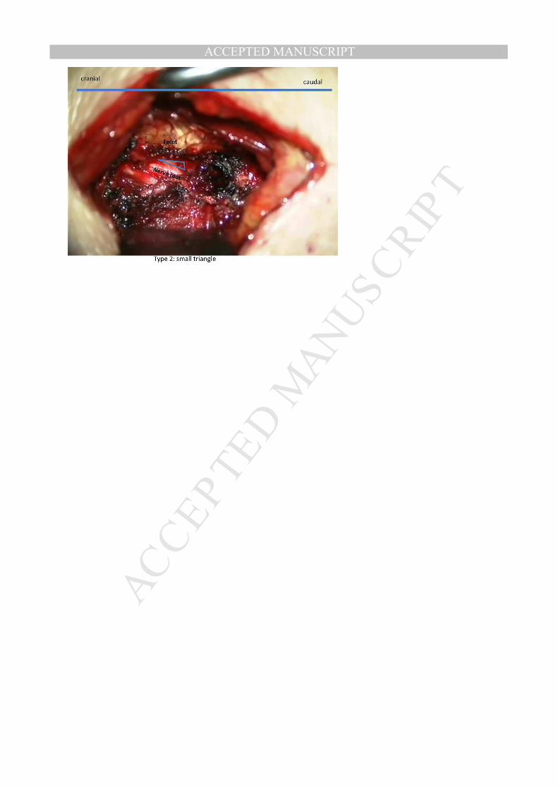
In order to provide standardization for the Kambin triangle the angle between the edge of facet
joints and existing root were measured in all patients. In this way, standardization was achieved
in the image captured from different sizes. This angle is an important indicator that provides
information about what type of triangle will be formed. We found the mean degree of angles
type 1: 5,6°, type 2: 13,5° and type 3: 39,3°. Cadaver results were similar type 1: 5,1°, type 2:
13,2°, type 3: 38,6°.
Consequently we classified Kambin’s triangle into three types according to the surgical view
appearances and cadaver study findings (Figure 1). The first type is a closed triangle without any
available space between the triangle elements, the second type is a narrowed triangle and the
third type is the normal triangle as described by Kambin.
RESULTS

MANUSCRIP
T
ACCEPTED
ACCEPTED MANUSCRIPT Endoscopic extraforaminal approach, Kambin’s triangle
6
Operative Results:
The summary of the patients’ data and the type of the triangle which determined during surgery
were summarized in Table 1. There were 34 patients of lumbar lateral discopathy (23 male, 11
female) in this study that met our inclusion criteria between 1999 and 2015. The patients’ age
range was 17-71 years old, with a mean age of 49,4 years.
Among all the cases, only 6 (17,6%) had the type 3 triangle, which is the normal triangle
described by Kambin, with a wide area between the sides of the triangle. However, 17 cases
(50%) had type 2 triangles, with a narrow space in the triangle, and 11 (32,3%) cases had type 1
triangles, with no space in the triangle (Figures 2-4). These data showed that 82,4% of the
patients had a narrow space or there is no space in this triangle.
Cadavers Results:
There were 8 cadavers (6 male, 2 female) selected at random. The age range was 20- 57 years old
with a mean age of 43,2 years.
A total number of 48 triangles between L2-5 were studied. Among all the cadavers, only 10
(20.8%) had a type 3 triangle, which is the normal triangle described by Kambin, with a wide
area between the sides of the triangle. However, we found 23 (48%) type 2 triangles with a
narrow space in the triangle, and 15 (31,2%) type 1 triangles, with no space in the triangle
(Figures 5-7). As a result, we found abnormal space in the triangle in 79.2% of the cadavers
similar with operative results.
DISCUSSION
Lumbar radiculopathy is a common cause of disability, occurring in 13-40% of people.20 The
main cause of pain in these patients is root compression or chemical irritation due to disc material
extrusion, making open decompression surgery a reasonable solution. For a less invasive
technique, Lyman and Smith invented chemonucleolysis in 1964.21
Kambin described a safe area called Kambin’s triangle: the exiting root is its hypotenuse, the
inferior border of the lower vertebral body creates the base, and the height is formed by the
articular process and superior articulating facet of the caudal vertebra. Considering this triangle,
the exiting nerve root goes inferiorly, anteriorly and laterally, so there is a safe pathway near the

MANUSCRIP
T
ACCEPTED
ACCEPTED MANUSCRIPT Endoscopic extraforaminal approach, Kambin’s triangle
7
upper part of the root for passing the cannulas and endoscopic instruments for nucleotomy or
endoscopic fragment resection.2,3
The major advantage of this type of surgery is that the ligaments, muscles, and bones are better
preserved, limiting instability, facet arthropathy and disc space narrowing. 8-10 Further, less
manipulation of the epidural venous system may prevent scarring, edema and chronic neural
fibrosis formation. 13,14 This technique also allows a better view based on magnification and light.
We have been performing endoscopic disc surgery since 1998. During this period, we have
encountered a few cases of root damage or post-surgical long-term paresthesia (in 4 cases out of
66),22 and other research shows similar results.16 Further, there are some reports of damage to the
radicular artery during endoscopic surgery followed by in situ hematoma formation or cord
ischemia.17,18
To assess the cause of these complications, we evaluated the normal anatomy and variations of
the root in Kambin’s triangle in in-vivo cases that are candidates for a lateral extraforaminal
surgical approach. In this approach, after removal of the intertransverse ligament, the root
pathway and the pattern of Kambin’s triangle were clearly visible. All of the patients who
underwent surgery had obvious nerve root compression. We performed microsurgical disc
removal and video recording, and evaluated the anatomy of the triangle. This investigation
showed that the type 3 classical Kambin’s triangle was present in only 6 of the 34 patients
(17,6%). However, the space was too narrow or absent in 28 patients (82,4%). Therefore, we
aimed to investigate the anatomy of Kambin’s triangle in normal spine on a cadaver study. This
evaluation also disclosed that the space of triangle was too narrow or absent in 38 of the 48
specimens (79,2%), while the type 3 normal triangle was determined only in 10 cadaver
specimens (20,8%).
Cadaver results were compared with the findings of the operation. Surprisingly cadaver and
patient outcomes were overlapping. In patients out of the 34 cases 32,3% had type 1 triangles,
which are completely closed triangles, 50% had type 2 triangles, which have narrow openings,
and only 17,6% had type 3 triangles, which are the triangles previously described by
Kambin.2,5,6,23,24 Cadaver results were similar; 15 (31,2%) had type 1 triangles, which are
completely closed triangles, 23 (48%) had type 2 triangles, which have narrow openings, and
only 10 (20,8%) had type 3 triangles.

MANUSCRIP
T
ACCEPTED
ACCEPTED MANUSCRIPT Endoscopic extraforaminal approach, Kambin’s triangle
8
Even the nerve root may be compressed and pushed from the bottom due to herniated disc, root
will slide laterally and normal triangle would be expected to be seen in greater proportion but the
results did not show that in both surgical and cadaver groups.
We know that the medial border of the triangle is composed of bone tissue (articular process and
superior articulating facet of the caudal vertebra) and nervous tissue (traversing root and dura)
under the bone. However, in most cases, even in young people without facet hypertrophy, the real
working area is small and the bone tissue covers the triangle. It is necessary to remove a part of
articular process to create enough space for endoscopic discectomy (Figure 8). This bone
removal can be easily performed through the working channel and it is not necessary to use a
different tubular system. This could be accompanied by the risk of damaging the nerve root, the
radicular artery, dura and traversing root during surgery.
Consequently, although endoscopic disc surgery is a popular type of disc surgery today, we
suggest more caution during surgery. This operation may be performed under local anesthesia so
that the patient can communicate when the surgeon is working just close the nerve root. Neural
monitoring would also be a useful addition. Further, we recommend fewer maneuvers during disc
removal, bearing in mind the proximity of the root to the instruments during surgery.
We believe that it is necessary to expand this study with more cases and cadaver studies to be
able to evaluate the frequency of each type of triangle at different levels, ages, and primary
disorders such as disc space narrowing and degenerative disease. This may allow the
identification of risk factors and the improvement of guidelines for this type of surgery.
MANUSCRIP
T
ACCEPTED
ACCEPTED MANUSCRIPT Endoscopic extraforaminal approach, Kambin’s triangle
9
Figure Legends
Figure 1: Types of extraforaminal triangle.
Figure 2: Type 1, no apparent triangle in operative view.
Figure 3: Type 2, small triangle in operative view.
Figure 4: Type 3, Kambin’s original triangle, operative view.
Figure 5: Type 1, no apparent triangle in cadaver study.
Figure 6: Type 2, small triangle in cadaver study.
Figure 7: Type 3, normal triangle in cadaver study.
Figure 8: After facet joint removal (yellow oval line), a wide area can be created between two
nerve roots. The original Kambin’s triangle (blue triangle) is obviously smaller than the created
wide area after bone removal (green line).

MANUSCRIP
T
ACCEPTED
ACCEPTED MANUSCRIPT Endoscopic extraforaminal approach, Kambin’s triangle
10
References:
01-Hijikata S, Yamagishi M, Nakayama T, Omori K. Percutaneous nucleotomy: a new
treatment method for lumbar disc herniation. J Toden Hosp. 1975;5:5-13.
02-Kambin P, Brager MD. Percutaneous posterolateral discectomy. Anatomy and
mechanism. Clin Orthop Relat Res. 1987;223:145-154.
03-Onik GM, Kambin P, Chang MK. Minimally invasive disc surgery: nucleotomy versus
fragmentectomy. Spine (Phila Pa 1976). 1997;22:827-828.
04-Iida Y, Kataoka O, Sho T, Sumi M, Hirose T, Bessho Y, Kobayashi D.et al. Postoperative
lumbar spinal instability occurring or progressing secondary to laminectomy. Spine (Phila Pa
1976). 1990;15:1186-1189.
05-Kambin P, Cohen LF, Brooks M, Schaffer JL. Development of degenerative spondylosis
of the lumbar spine after partial discectomy. Comparison of laminotomy, discectomy, and
posterolateral discectomy. Spine (Phila Pa 1976). 1995;20:599-607.
06-Kambin P, O'Brien E, Zhou L, Schaffer JL. Arthroscopic microdiscectomy and selective
fragmentectomy. Clin Orthop Relat Res 1998;347:150-167.
07-Mochida J, Toh E, Nomura T, Nishimura K. The risks and benefits of percutaneous
nucleotomy for lumbar disc herniation. A 10-year longitudinal study. J Bone Joint Surg Br.
2001;83:501-505.
08-Natarajan RN, Andersson GB, Patwardhan AG, Andriacchi TP. Study on effect of graded
facetectomy on change in lumbar motion segment torsional flexibility using three-
dimensional continuum contact representation for facet joints. J Biomech Eng. 1999;121:215-
221.
09-Weber BR, Grob D, Dvorák J, Müntener M. Posterior surgical approach to the lumbar
spine and its effect on the multifidus muscle. Spine (Phila Pa 1976). 1997;22:1765-1772.
10-Zander T, Rohlmann A, Klöckner C, Bergmann G. Influence of graded facetectomy and
laminectomy on spinal biomechanics. Eur Spine J. 2003;12:427-434.
11-Cooper RG, Mitchell WS, Illingworth KJ, Forbes WS, Gillespie JE, Jayson MI. The role
of epidural fibrosis and defective fibrinolysis in the persistence of postlaminectomy back
pain. Spine (Phila Pa 1976). 1991;16:1044-1048.
MANUSCRIP
T
ACCEPTED
ACCEPTED MANUSCRIPT Endoscopic extraforaminal approach, Kambin’s triangle
11
12-Ross JS, Robertson JT, Frederickson RC, Petrie JL, Obuchowski N, Modic MT,
deTribolet Ne et al. Association between peridural scar and recurrent radicular pain after
lumbar discectomy: magnetic resonance evaluation. Neurosurgery. 1996;38:855-861.
13-Ahn Y, Lee SH, Park WM, Lee HY, Shin SW, Kang HY. Percutaneous endoscopic
lumbar discectomy for recurrent disc herniation: surgical technique, outcome, and prognostic
factors of 43 consecutive cases. Spine (Phila Pa 1976). 2004;29:326-332
14-Cho JY, Lee SH, Lee HY. Prevention of development of postoperative dysesthesia in
transforaminal percutaneous endoscopic lumbar discectomy for intracanalicular lumbar disc
herniation: floating retraction technique. Minim Invasive Neurosurg Neurosurg.2011;54:214-
218.
15-Ruetten S, Komp M, Merk H, Godolias G. Full-endoscopic interlaminar and
transforaminal lumbar discectomy versus conventional microsurgical technique: a
prospective, randomized, controlled study. Spine (Phila Pa 1976). 2008;33:931-939.
16-Yeung AT, Tsou PM. Posterolateral endoscopic excision for lumbar disc herniation:
surgical technique, outcome, and complications in 307 consecutive cases. Spine (Phila Pa
1976). 2002;27:722-731.
17-Ahn Y. Transforaminal percutaneous endoscopic lumbar discectomy: technical tips to
prevent complications. Expert Rev Med Devices. 2012; 9: 361-366.
18-Mayer HM, Brock M. Percutaneous endoscopic discectomy: surgical technique and
preliminary results compared to microsurgical discectomy. J Neurosurg. 1993;78:216-225.
19- Sairyo K, Matsuura T, Higashino K, Sakai T, Takata Y, Goda Y, Suzue N, et al. Surgery
related complications in percutaneous endoscopic lumbar discectomy under local anesthesia.
J Med Invest 2014;61:264-269.
20-Park KD, Lee J, Jee H, Park Y. Kambin triangle versus the supraneural approach for the
treatment of lumbar radicular pain. Am J Phys Med Rehabil. 2012;91:1039-1050, 2012.
21-Smith L. Chemonucleolysis. Clin Orthop Relat Res. 1969;67:72-80.
22-Sasani M, Ozer AF, Oktenoglu T, Canbulat N, Sarioglu AC. Percutaneous endoscopic
discectomy for far lateral lumbar disc herniations: prospective study and outcome of 66
patients. Minim Invasive Neurosurg. 2007;50:91-97.
23-Kambin P, Casey K, O'Brien E, Zhou L. Transforaminal arthroscopic decompression of
lateral recess stenosis. J Neurosurg. 1996;84: 462-467.

MANUSCRIP
T
ACCEPTED
ACCEPTED MANUSCRIPT Endoscopic extraforaminal approach, Kambin’s triangle
12
24-Kambin P, Zhou L. History and current status of percutaneous arthroscopic disc surgery.
Spine (Phila Pa 1976). 1996;21:57-61.

MANUSCRIP
T
ACCEPTED
ACCEPTED MANUSCRIPTTable 1: A summary of three Types of Kambin’s Triangle in 34 Patients.
Age Gender Type Level 1 48 M 2 L3-4 2 17 F 2 L3-4 3 62 M 3 L3-4 4 46 F 1 L4-5 5 37 F 2 L5-S1 6 37 M 2 L5-S1 7 70 M 1 L3-4 8 64 F 1 L4-5 9 50 M 3 L4-5 10 50 M 2 L4-5 11 52 M 2 L3-4 12 63 M 1 L4-5 13 32 F 1 L4-5 14 54 F 3 L3-4 15 43 M 2 L4-5 16 64 F 1 L4-5 17 60 F 3 L4-5 18 70 M 2 L4-5 19 45 M 2 L4-5 20 58 M 2 L5-S1 21 38 M 1 L5-S1 22 49 M 2 L1-2 23 55 F 2 L4-5 24 44 M 1 L3-4 25 39 F 2 L4-5 26 71 M 2 L4-5 27 40 F 3 L4-5 28 41 M 2 L3-4 29 60 M 1 L2-3 30 45 M 1 L5-S1 31 35 M 2 L3-4 32 40 M 3 L4-5 33 65 M 1 L5-S1 34 37 M 2 L4-5

MANUSCRIP
T
ACCEPTED
ACCEPTED MANUSCRIPT

MANUSCRIP
T
ACCEPTED
ACCEPTED MANUSCRIPT
MANUSCRIP
T
ACCEPTED
ACCEPTED MANUSCRIPT

MANUSCRIP
T
ACCEPTED
ACCEPTED MANUSCRIPT

MANUSCRIP
T
ACCEPTED
ACCEPTED MANUSCRIPT

MANUSCRIP
T
ACCEPTED
ACCEPTED MANUSCRIPT

MANUSCRIP
T
ACCEPTED
ACCEPTED MANUSCRIPT

MANUSCRIP
T
ACCEPTED
ACCEPTED MANUSCRIPT

MANUSCRIP
T
ACCEPTED
ACCEPTED MANUSCRIPT
Highlights
• Transforaminal endoscopic surgery is the gold standard. • It is performed through Kambin’s triangle. • Every patient has not a wide Kambin’s triangle as described. • Only 20% of the patients have enough safe space in the triangle. • To be aware of anatomical variations is important for succesfull endoscopic
surgery.