ANALYSIS OF THE HAEMOLYMPH OF ARION ATER L
12
J. Exp. Biol. (1963), 40, 613-623 613 With 2 text-figures Printed in Great Britain ANALYSIS OF THE HAEMOLYMPH OF ARION ATER L. (GASTROPODA: PULMONATA) BY D. K. ROACH Department of Zoology, University College of South Wales and Monmouthshire, Cardiff {Received 29 April 1963) INTRODUCTION Pulmonate smooth muscle structures are well suited for physiological investigation, since they are normally bathed directly by haemolymph and many of them are rhyth- mically contractile. Among the muscular organs investigated have been the heart (Cardot, 1921; Hogben, 1925; Arvanitaki & Cardot, 1931; Jullien et al. 1955; Krijgsman & Divaris, 1955; Jaeger, 1961), the buccal retractor (Bozler, 1930; Ramsay, 1940; Graham & Heathcote, 1952), the flagellum (Bachrach & Cardot, 1921), penis retractor (Jaeger, 1962) and alimentary tract (Wells, 1928; Graham & Heathcote, 1952). The buccal mass, crop and stomach are particularly suitable for kymograph studies and preparatory to an investigation of the motor activity of the alimentary tract of Arion ater it was realized that a blood analysis was necessary to form a basis for the preparation of a physiological saline for this animal. A number of pulmonate salines have been prepared (Krijgsman & Divaris, 1955; Lockwood, 1961), mostly for various Helix species, though also for Lymnaea (Carriker, 1946) and Strophocheilos (Jaeger, 1961). Very few salines, however, have been based on blood analyses. Salines for Helix species, based on limited analyses, have been produced by Bernard & Bonnet (1930), Arvanitaki & Cardot (1931) and Jullien et al. (1955), and further analyses of pulmonate blood have been made by Bernard (1931), Holtz & von Brand (1940), Saxena (1957) and Michon & Alaphilippe (1958). It is known that there is a con- siderable variation in water content from day to day in terrestrial pulmonates (Arvani- taki & Cardot, 1931; Kamada, 1933; Howes & Wells, 1934; Martin, Harrison, Huston & Stewart, 1958) and it might be supposed that an exact knowledge of the blood composition is not necessary to prepare a suitable saline. However, interchanging of salines between pulmonate species has not proved successful, and in some instances (Bozler, 1930; Ramsay, 1940) pooled haemolymph has had to be employed. Further- more, it has been pointed out repeatedly (Cardot, 1921; Wells, 1928; Bernard & Bonnet, 1930; Ramsay, 1940; Hughes & Kerkut, 1956; Kerkut & Taylor, 1956) that the normal functioning of the muscular and nervous tissues of molluscs is considerably affected by variation both of the concentration of individual ions and of the overall osmotic concentration, making a knowledge of blood composition very desirable. Experimental methods are now available for a full analysis of very small volumes of blood and have been used here to find the haemolymph concentrations of sodium, potassium, calcium, magnesium, chloride, sulphate, phosphate, bicarbonate, protein and total electrolytes, as well as freezing-point depression (A) and pH, for the slug Arion ater.
Transcript of ANALYSIS OF THE HAEMOLYMPH OF ARION ATER L
J. Exp. Biol. (1963), 40, 613-623 613With 2 text-figures
Printed in Great Britain
ANALYSIS OF THE HAEMOLYMPH OF ARION ATER L.(GASTROPODA: PULMONATA)
BY D. K. ROACH
Department of Zoology, University College of South Wales and Monmouthshire, Cardiff
{Received 29 April 1963)
INTRODUCTION
Pulmonate smooth muscle structures are well suited for physiological investigation,since they are normally bathed directly by haemolymph and many of them are rhyth-mically contractile. Among the muscular organs investigated have been the heart(Cardot, 1921; Hogben, 1925; Arvanitaki & Cardot, 1931; Jullien et al. 1955;Krijgsman & Divaris, 1955; Jaeger, 1961), the buccal retractor (Bozler, 1930; Ramsay,1940; Graham & Heathcote, 1952), the flagellum (Bachrach & Cardot, 1921), penisretractor (Jaeger, 1962) and alimentary tract (Wells, 1928; Graham & Heathcote,1952). The buccal mass, crop and stomach are particularly suitable for kymographstudies and preparatory to an investigation of the motor activity of the alimentarytract of Arion ater it was realized that a blood analysis was necessary to form a basisfor the preparation of a physiological saline for this animal. A number of pulmonatesalines have been prepared (Krijgsman & Divaris, 1955; Lockwood, 1961), mostlyfor various Helix species, though also for Lymnaea (Carriker, 1946) and Strophocheilos(Jaeger, 1961). Very few salines, however, have been based on blood analyses. Salinesfor Helix species, based on limited analyses, have been produced by Bernard & Bonnet(1930), Arvanitaki & Cardot (1931) and Jullien et al. (1955), and further analyses ofpulmonate blood have been made by Bernard (1931), Holtz & von Brand (1940),Saxena (1957) and Michon & Alaphilippe (1958). It is known that there is a con-siderable variation in water content from day to day in terrestrial pulmonates (Arvani-taki & Cardot, 1931; Kamada, 1933; Howes & Wells, 1934; Martin, Harrison, Huston& Stewart, 1958) and it might be supposed that an exact knowledge of the bloodcomposition is not necessary to prepare a suitable saline. However, interchanging ofsalines between pulmonate species has not proved successful, and in some instances(Bozler, 1930; Ramsay, 1940) pooled haemolymph has had to be employed. Further-more, it has been pointed out repeatedly (Cardot, 1921; Wells, 1928; Bernard &Bonnet, 1930; Ramsay, 1940; Hughes & Kerkut, 1956; Kerkut & Taylor, 1956) thatthe normal functioning of the muscular and nervous tissues of molluscs is considerablyaffected by variation both of the concentration of individual ions and of the overallosmotic concentration, making a knowledge of blood composition very desirable.Experimental methods are now available for a full analysis of very small volumes ofblood and have been used here to find the haemolymph concentrations of sodium,potassium, calcium, magnesium, chloride, sulphate, phosphate, bicarbonate, proteinand total electrolytes, as well as freezing-point depression (A) and pH, for the slugArion ater.
614 D. K. ROACH
MATERIALS AND METHODS
The animals, large garden slugs of the species Arion ater L. (subspecies rufus),were collected locally from gardens and maintained in wide, shallow terraria fittedwith wire-mesh lids, with a plentiful supply of food and water. Cabbage and lettucewere provided, as well as supplementary artificial diets (BOCM 'Chick mash' andOxoid ' Diet 41'). Haemolymph was collected by inserting the needle of a hypodermicsyringe through the pneumostome into the mantle cavity, then through the postero-lateral wall of the cavity into the visceral haemocoele. Between 0-2 and 0-5 ml. of theopalescent, blue body fluid could then be withdrawn, uncontaminated by body-wallmucus, and without apparent harm to the animal. After 3 or 4 days a further sampleof fluid could be taken from the same slug.
After withdrawal, the haemolymph sample was placed in a small Pyrex glass tube(i\ x \ in.) and centrifuged at 1500 g for 20 min. to remove debris and cells. The clearsupernatant was then pipetted off, using a waxed pipette, into a clean tube which wasfinally sealed with a Polythene cap secured by a rubber ring. When the fluid samples werenot used immediately they were stored by being frozen solid and kept at — 50 C.
Haemolymph samples were not pooled for the analyses. Determinations weremade separately on samples from many slugs and, in the cases of chloride and bi-carbonate measurements, were repeated on individual samples. 'Analar' gradechemicals were used throughout the investigation.
ANALYSIS OF CATIONS
Sodium, potassium and calcium were determined by flame photometry. A pre-liminary analysis was made to estimate the approximate values for the three ions, andthen two standard solutions were made up. The first contained 0-05 mii/l. NaCl, andwas used for sodium estimations. The other contained 0-75 mM/1. NaCl, 0-05 mM/1.KC1 and 0-05 mM/1. CaCl2, and was the standard solution for potassium and calciumestimations, the added sodium chloride eliminating interference errors, o-i ml. ofthe haemolymph sample was diluted to 10 ml. (dilution x 100) and was used, withthe appropriate filters, in an EEL flame photometer for analysis of potassium andcalcium. 1 ml. of the diluted sample was then diluted further to 25 ml. (total dilutionx 2500) and employed for sodium analysis. The two separate dilutions were made tomatch the highest sensitivity range of the photometer for the different ions.
Since flame photometry gives the total ion concentration, and some at least of thesodium, potassium and particularly calcium might exist in a bound form, the analyseswere repeated on blood samples freed of protein by precipitation, either with 10%trichloroacetic acid or with a zinc-alkali mixture (Somogyi, 1945). In either case, theprecipitate was centrifuged down and 0-2 ml. of the supernatant was pipetted off anddiluted as above.
Magnesium was estimated colorimetrically, by the formation and measurement ofthe alkaline Titan yellow complex (method of Orange & Rhein, as modified byCroghan, 1958). Trichloroacetic acid-precipitated haemolymph was used, o-i ml. beingadded to 1-5 ml. of very dilute Titan yellow solution and 1-5 ml. of N-NaOH added.After mixing, the extinction was read at 520 m/x, in a Unicam SP 600 spectrophoto-
Haemolymph of Arion ater 615
Tneter. Magnesium concentrations were read off a calibration curve constructed fromstandard solutions of 1-10 mM/1. MgCl2, the relationship being linear from 3 to 9 mM/1.
Results
An attempt has been made in each case to evaluate the variation to be expected fromthe experimental methods. These are expressed in Table 1 as average standarddeviations and as average coefficients of variation, related to the mean haemolymphconcentration of the ion in question. In the case of magnesium the average of thestandard deviations (s.D.) of the values for the standard solutions from the expected(and corrected) straight line has been used. For the flame photometry results three
Table 1. Variation resulting from analytical technique
Ion measured
SodiumPotassiumCalciumMagnesium
Table 2
Ion measured
SodiumUntreated haemolymphTrichloroacetic precipitatedZinc-alkali precipitated
PotassiumUntreated haemolymphTrichloroacetic precipitatedZinc-alkali precipitated
CalciumUntreated haemolymphTrichloroacetic precipitatedZinc-alkali precipitated
MagnesiumTrichloroacetic precipitated
Average s.D.due to errorof method
(HIM)
±096±006±0-17±0-27
:. Results of cation
No. ofsamples
2788
2711
8
281 2
8
2 0
Averagecoefficient
of variationrelative to
meanhaemolymphconcentration
(%)
1 6
2 - 2
6-0
4-5
analysis
Concentration(rnM)
6149650661-92
2 6 8
2-552-69
2'8l2-972-30
S-68
S.D.
±5-72±8-17±8-79
±o-8o±078+ 0-98
±0-48±0-66±o-S3
±0-87
Coefficientof variation
(%)
9 312-514-0
2 9 9
30-S36-S
17-12 2 2
23-O
I5'3
readings had been taken for each diluted sample, alternating with measurement ofthe standard solution, so that it was possible to use the variations occurring betweenrepeat measurements on individual samples as a measure of this ' reproducibility'value. Thus, by comparing the s.D. and coefficients of variation columns in Table 2with those in Table 1, it is possible to estimate the extent to which the variation foundin the results is due to error of the method.
Statistical analysis of the results shows that there is no significant difference(P > 0-5) between the untreated and protein-free bloods as far as sodium and
6i6 D. K. ROACH
potassium are concerned—i.e. all the potassium and sodium present is in simplesolution. For the calcium, however, it is quite clear that about 20 % exists in boundform (P < 0-02) and the amount actually in solution is only 2*30 mM/1.
ANALYSIS OF ANIONS
The method described by Wigglesworth (1938) was used as the basis for the chlorideanalysis, i.e. addition of silver nitrate and back-titration with sodium thiocyanate withiron alum as indicator. 0*05 ml. of protein-free haemolymph (zinc-alkali precipitated)was pipetted on to a small Polythene dish, an excess of 50 % nitric acid was added,
1 0 1 -
0-24 6 8 10 12 14 16
Burette reading (units of BaCb)18
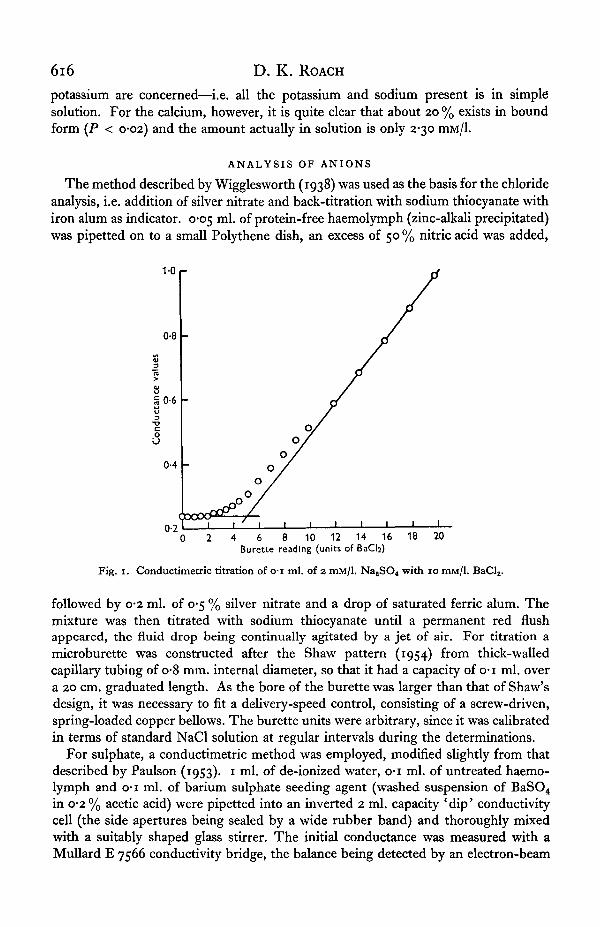
Fig. i. Conductimetric titration of o-i ml. of 2 mM/1. NaaSO4 with 10 mM/1. Bad, .
followed by 0-2 ml. of 0-5 % silver nitrate and a drop of saturated ferric alum. Themixture was then titrated with sodium thiocyanate until a permanent red flushappeared, the fluid drop being continually agitated by a jet of air. For titration amicroburette was constructed after the Shaw pattern (1954) from thick-walledcapillary tubing of o-8 mm. internal diameter, so that it had a capacity of o-i ml. overa 20 cm. graduated length. As the bore of the burette was larger than that of Shaw'sdesign, it was necessary to fit a delivery-speed control, consisting of a screw-driven,spring-loaded copper bellows. The burette units were arbitrary, since it was calibratedin terms of standard NaCl solution at regular intervals during the determinations.
For sulphate, a conductimetric method was employed, modified slightly from thatdescribed by Paulson (1953). 1 ml. of de-ionized water, o-i ml. of untreated haemo-lymph and o-i ml. of barium sulphate seeding agent (washed suspension of BaSO4
in 0-2% acetic acid) were pipetted into an inverted 2 ml. capacity 'dip' conductivitycell (the side apertures being sealed by a wide rubber band) and thoroughly mixedwith a suitably shaped glass stirrer. The initial conductance was measured with aMullard E 7566 conductivity bridge, the balance being detected by an electron-beam
Haemolymph of Arion ater 617
indicator (' magic eye'). Known volumes of 10 mM/1. BaCl2 standard solution were thenrun in from the microburette described above and the conductance was re-measuredafter each addition, again after thorough stirring. The end-point was determinedgraphically (Fig. 1) by plotting conductance values against units of added titrant andextrapolating the two straight lines of initial and subsequent conductance values totheir intersection. The microburette was calibrated by repeating the titration onstandard 2 mM/1. Na2SO4 and K2SO4 solutions and the system was also tested byestimating the sulphate content of a mixture after successive additions of knownvolumes of standard sulphate solutions (sulphate recovery test), when full recoverywas demonstrated.
Phosphate was estimated colorimetrically, as the molybdenum blue complex.Stannous chloride proved inconsistent as a reducing agent, but a method modifiedfrom Courgill & Pardee (1957) and Croghan (1958) worked consistently at the verylow concentrations of phosphate encountered. 12-5 ml. of a filtered stock solution ofreducing agent (0-5 g. of i-amino-2-naphthol-4-sulphonic acid in 244 ml. of 15 %sodium bisulphite and 6 ml. of 20 % sodium sulphite) and 25 ml. of stock molybdicacid solution (5 g. ammonium molybdate in 222 ml. water and 28 ml. sulphuric acid)were mixed and diluted to 250 ml. immediately before use. 5 ml. of this mixture werepipetted into each of a series of tubes and 0*2 ml. of trichloroacetic acid-precipitatedhaemolymph was added to each tube. 10 min. after mixing, the extinction was readat 775 m ,̂ and the phosphate concentration was read off a calibration curve con-structed from standard 0-2-1-0 mM/1. Na2HPO4 solutions.
For the purposes of devising a physiological saline it was assumed that the totalalkali reserve of the blood was made up of bicarbonate and phosphate ions, and, sincethe concentration of the latter was very low, the titratable alkalinity was taken as ameasure of the bicarbonate concentration. 0-05 ml. of untreated (fresh) haemolymphwas pipetted on to a waxed, white Perspex square and 0-05 N-HCl was titrated againstit, using the microburette. For convenience and consistency of results the acid titrantwas made up to contain 10 ml. of B.D.H. '4-5' indicator per 100 ml. The square wasgently rotated by hand to mix the solutions, and the end-point was clearly given bythe sharp colour change from blue, through grey to the first orange flush. The systemwas calibrated against 25 mM/1. NaHCO3 solution. Stored, well-sealed frozen samplesgave results very similar to those obtained with fresh haemolymph. The phosphateconcentration (as m-equiv./l.) was subtracted from the alkalinity value to give the' bicarbonate' concentration.
Results
As for the cation analytical methods, a measure of the reproducibility of the methodhas been calculated. In the case of phosphate analysis, average standard deviationsof the readings for standard solutions from the corrected calibration curve were used,while variations between repeat determinations of the standard solutions wereemployed for chloride, sulphate and bicarbonate (see Tables 3 and 4).
FURTHER ANALYSES
Protein was estimated by the method of Folin (1922), modified by Danielson (1953).0-05 ml. samples were diluted to 1 ml. and 5*5 ml. of mixed reagents added. The
6i8 D. K. ROACH
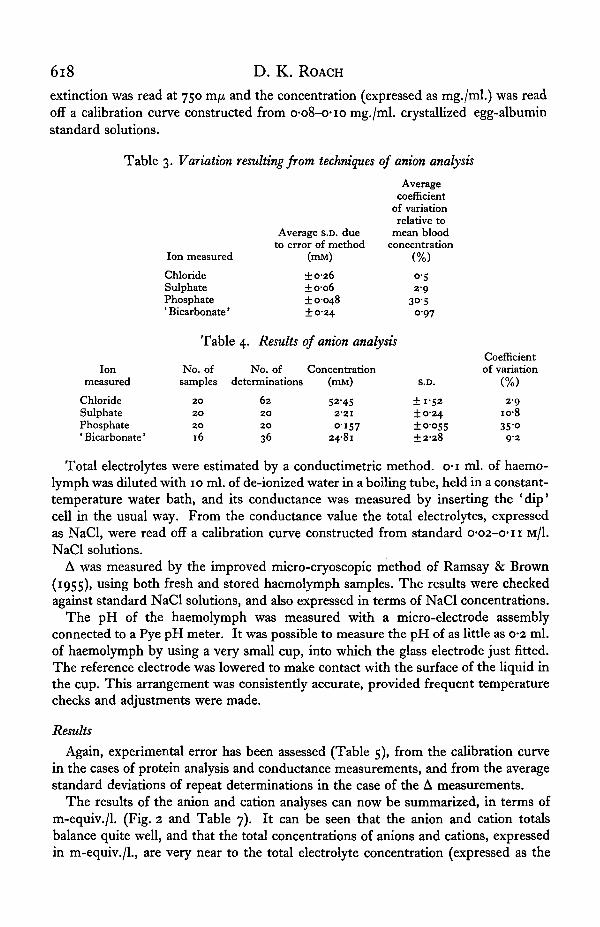
extinction was read at 750 m/x and the concentration (expressed as mg./ml.) was readoff a calibration curve constructed from o-o8-c-io mg./ml. crystallized egg-albuminstandard solutions.
Table 3. Variation resulting from techniques of onion analysis
Ionmeasured
ChlorideSulphatePhosphate' Bicarbonate'
Ion measured
ChlorideSulphatePhosphate'Bicarbonate'
Table
No. of
Average S.D. dueto error of method
(mia)
±0-26±006± 0-048±0-24
Averagecoefficient
of variationrelative to
mean bloodconcentration
(%)
o-52 9
30-50 9 7
4. Results of onion analysis
No. of Concentrationsamples determinations (min)
2 0
2 0
2 0
16
62 52-4520 2'2I2O O-I5736 24-8l
S.D.
±1-52±0-24±0-055±2-28
Coefficientof variation
(%)
2 9io-8350
9-2
Total electrolytes were estimated by a conductimetric method, o-i ml. of haemo-lymph was diluted with 10 ml. of de-ionized water in a boiling tube, held in a constant-temperature water bath, and its conductance was measured by inserting the 'dip'cell in the usual way. From the conductance value the total electrolytes, expressedas NaCl, were read off a calibration curve constructed from standard O'02-c-n M/1.NaCl solutions.
A was measured by the improved micro-cryoscopic method of Ramsay & Brown(1955), using both fresh and stored haemolymph samples. The results were checkedagainst standard NaCl solutions, and also expressed in terms of NaCl concentrations.
The pH of the haemolymph was measured with a micro-electrode assemblyconnected to a Pye pH meter. It was possible to measure the pH of as little as 0-2 ml.of haemolymph by using a very small cup, into which the glass electrode just fitted.The reference electrode was lowered to make contact with the surface of the liquid inthe cup. This arrangement was consistently accurate, provided frequent temperaturechecks and adjustments were made.
Results
Again, experimental error has been assessed (Table 5), from the calibration curvein the cases of protein analysis and conductance measurements, and from the averagestandard deviations of repeat determinations in the case of the A measurements.
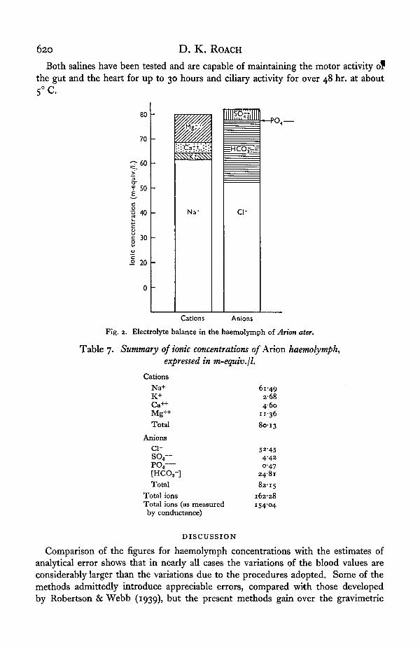
The results of the anion and cation analyses can now be summarized, in terms ofm-equiv./l. (Fig. 2 and Table 7). It can be seen that the anion and cation totalsbalance quite well, and that the total concentrations of anions and cations, expressedin m-equiv./l., are very near to the total electrolyte concentration (expressed as the
Haemolymph of Arion ater 619
Sum of the Na+ and Cl~ concentrations in m-equiv./l.) as measured by the conductanceof the haemolymph. These two facts lend support to the validity of the analyticalresults.
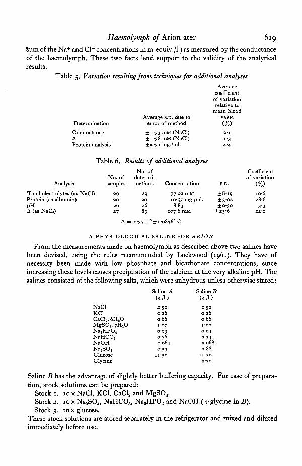
Table 5. Variation resulting from techniques for additional analyses
Determination
ConductanceAProtein analysis
Average S.D. due toerror of method
±i-33mM (NaCl)±i-38mM (NaCl)+ 0-31 mg./ml.
Table 6. Results of additional analyses
No. ofAnalysis samples
Total electrolytes (as NaCl) 29Protein (as albumin) 20pH 26A (as NaCl) 27
A =
No. ofdetermi-nations Concentration
29 77-02 mM20 10-55 mg./ml.26 88383 1076 mM
= 0-3711° ±0-0836° C.
Averagecoefficientof variationrelative to
mean bloodvalue<%)2-11-34-4
S.D.
±8-19±3-02±0-30
±23-6
Coefficientof variation
(%)
io-628-6
3-3220
A PHYSIOLOGICAL SALINE FOR ARION
From the measurements made on haemolymph as described above two salines havebeen devised, using the rules recommended by Lockwood (1961). They have ofnecessity been made with low phosphate and bicarbonate concentrations, sinceincreasing these levels causes precipitation of the calcium at the very alkaline pH. Thesalines consisted of the following salts, which were anhydrous unless otherwise stated:
Saline A Saline B
NaClKC1CaCla.6H2OMgSO4.7H2ONa,HPO.NaHCOsNaOHNaaSO4
GlucoseGlycine
2-520-26o-66i-oo0-030-760-064O-53
11-50
2-520-26o-66i-oo0-03o-340068o-88
11-500-30
Saline B has the advantage of slightly better buffering capacity. For ease of prepara-tion, stock solutions can be prepared:
Stock 1. 10 x NaCl, KC1, CaCl2 and MgSO4.Stock 2. 10 x Na2SO4, NaHC03, Na2HPO4 and NaOH ( +glycine in B).Stock 3. 10 x glucose.
These stock solutions are stored separately in the refrigerator and mixed and dilutedimmediately before use.
620 D . K. ROACH
Both salines have been tested and are capable of maintaining the motor activity ofthe gut and the heart for up to 30 hours and ciliary activity for over 48 hr. at about5°C
80
70(m
-equ
iv./l
O
<
C
! »
° 20
0
-
-
-
-
-
is
Na'
llllnflTllHI
=HCO; -—
ci-
- P O 4
Cations Anions
Fig. 2. Electrolyte balance in the haemolymph of Arion ater.
Table 7. Summary of ionic concentrations of Arion haemolymph,expressed in m-equiv.jl.
CationsNa+K+Ca++Mg++Total
Anionsc i -
so,—PO4—[HCO3-]Total
Total ionsTotal ions (as measuredby conductance)
DISCUSSION
61-492 6 84-60
11-3680-13
52-454-420-47
248182-15
162-28i54'°4
Comparison of the figures for haemolymph concentrations with the estimates ofanalytical error shows that in nearly all cases the variations of the blood values areconsiderably larger than the variations due to the procedures adopted. Some of themethods admittedly introduce appreciable errors, compared with those developedby Robertson & Webb (1939), but the present methods gain over the gravimetric
Haemolymph of Arion ater 621
'procedures in speed and convenience of use, as well as only requiring o-1 ml. of haemo-lymph instead of 1 ml. The phosphate figures are least reliable, but here the concen-trations encountered are extremely low (some 5 parts of phosphorus per million).
Even when account is taken of the errors introduced by the analytical methods used,it can be seen that there is considerable variation in the haemolymph of Arion ater.This is particularly noticeable in the case of the A, which shows a coefficient of variationof 22 %; the actual values ranged from c-180 to 0-43° C. Agriolimax haemolymph hasbeen shown to vary from 0-2° to o-8° C. (calculated from Hughes & Kerkut, 1956),while Duval (1930) found variation in Helix from 0-37° to 0-43° C. in hibernatinganimals and o^o0 to 0-40° C. in active ones, the two ranges overlapping considerably.Kamada (1933), for the same animal, found 0*41° C. for 'hibernating' blood and0-30° C. for 'active' blood. These figures for haemolymph variation agree well withobservations on weight fluctuations by Howes & Wells (1934) and Dainton (1954),and also with the figures of Martin et al. (1958) for variation of the haemolymphvolume of Arion from about 25 to 47 % of the wet weight of the animal, while Arvani-taki & Cardot (1931) for Helix pomatia showed that the haemolymph volume was10-7% of the total weight in hibernating animals and 16-2% in active animals. Inview of this extreme variability it is somewhat surprising to find that the activity ofthe nervous system is considerably affected by osmotic pressure changes (Hughes &Kerkut, 1956; Kerkut & Taylor, 1956; Duncan, 1961) and so is the activity of theheart (Cardot, 1921).
The results seem to indicate that the greater part of this osmotic fluctuation is dueto changes in concentration of the non-electrolytes in the blood, rather than of theelectrolytes. The smaller variation of the latter (shown by the conductance measure-ments, supported by individual ion analyses) indicates that they are gained or lostwith the water, whereas the non-electrolytes (as demonstrated by A and proteinanalysis) tend to remain in the haemolymph. This is of interest as many investigatorshave reported the disturbances of function in a number of nervous and musculartissues when electrolyte ratios and concentrations have been altered (Bachrach &Cardot, 1921; Hogben, 1925; Wells, 1928; Bernard & Bonnet, 1930; Arvanitaki &Cardot, 1931; Ramsay, 1940; Krijgsman & Divaris, 1955; Duncan, 1961; Jaeger,1961). In view of these findings, the relatively large variations of Ca++ and K+ levelsin the haemolymph are of some significance. Dainton (1954) and Howes & Wells(1934) have suggested that the activity of slugs, or at least the degree of responsivenessto stimuli, is regulated by the degree of hydration of the animal. As the gain and lossof water apparently results in a change of concentration of Ca++ and K+, and as boththese ions have profound effects on nervous and muscular irritability, this is a possiblemechanism causing the change of activity of the animal, in addition to the directosmotic effect.
Because of the wide variations encountered in the haemolymph of Arion, it isprobable that cycles of activity occur in the individual organs and tissues of the body,as well as in the behaviour of the animal as a whole. The physiological salines, then,are to be considered as maintaining, at best, an ' average' activity of the isolated organs,rather than a 'normal' one. This is inevitably an artificial condition, which is unableto illustrate the cycles of activity evident in the intact animal.
The constancy of the pH is all the more remarkable in contrast with the variation40 Exp. Biol. 40, 4
622 D. K. ROACH
of most other aspects of the haemolymph, though Arvanitaki & Blanc (1934) havedescribed the profound effects of pH changes on the heart of Helix. It is, too, aremarkably alkaline pH, far more so than for almost any other animal studied in thisrespect. Most animals have a blood pH just on the alkaline side of neutrality, butvalues of 7-8-8-08 have been recorded for Anodonta and 8-4 for Helix (Lockwood,1961), although 7-6-7-9 is generally quoted for the haemolymph of Helix pomatia;Michon & Alaphilippe (1958) give pH 7-8 for Achatina, and Saxena (1957) gives 7-86for Pilaglobosa. The alkaline haemolymph of Arum has been the cause of difficulty inmaking up the salines, as the calcium tends to precipitate very readily at this pH,and it has meant that the phosphate concentration has had to be kept low, with conse-quent reduction of buffering power.
The most constant of the haemolymph constituents, then, are chloride, sodium andalkali reserve. This is at least suggestive of a mechanism for regulating these factorsin the haemolymph when water is gained or lost by the animal, lending support tothe concept of ionic regulation expressed by Holtz & von Brand (1940), who foundevidence of this in the haemolymph of Helix pomatia. Saxena (1957) has recordedthat chloride was the most stable constituent of the haemolymph of Pila. More in-formation on this possible regulatory mechanism should be obtainable by followingthe changes in composition and volume of the haemolymph of individual slugs throughtheir cycles of loss and gain of weight.
SUMMARY
1. Methods are described for the collection of slug haemolymph and its analysisfor cations, anions, total electrolytes, protein, A and pH.
2. Of the cations measured, potassium and calcium show the greatest variation,being two to three times as variable as sodium, the most constant. About 20 % of theblood calcium exists in a bound form.
3. Of the anions measured, chloride is the most concentrated and least variable.Sulphate and alkalinity are about equally variable, three times the chloride fluctuation.The phosphate concentration is very low.
4. The variation of total electrolytes is only half that of the total solutes, as measuredby A, while protein is very variable.
5. The pH is remarkably alkaline but varies only within narrow limits.6. Formulae are given for two physiological salines, capable of maintaining both
muscular and ciliary activity.
My sincere thanks are due to Dr W. A. L. Evans for much advice and for readingthe manuscript. The investigation was carried out under a D.S.I.R. ResearchStudentship.
REFERENCES
ARVANITAKI, A. & BLANC, H. (1934). Action des ions H et des ions OH sur la survie, la rythmicite,le tonus, le potential d'activite du myocarde de l'Escargot. J. Physiol. Path. gin. 32, 775-91.
ARVANITAKI, A. & CARDOT, H. (1931). Solutions equilibrees pour le coeur des Helix, en rapport avecla composition de l'hemolymphe. C.R. Soc. Biol., Paris, 106, 185-7.
BACHRACH, E. & CARDOT, H. (1921). Contractilite et excitability du flagelle de l'Escargot. C.R. Soc.Biol., Paris, 85, 170-2.
BERNARD, A. (1931). Sur la composition minerale de rhemolymphe de differents Helix. C.R. Soc. Biol.,Paris, 106, 183-4.

Haemolymph of Arion ater 623BERNARD, A. & BONNET, V. (1930). Composition minerale de l'hemolymphe et 6tude d'une solution
physiologique pour l'Escargot. C.R. Soc. Biol., Paris, 103, 1119-20.BOZLER, E. (1930). Untersuchungen zur Physiologie der Tonusmuskeln. Z. vergl. Physiol. 12, 579-602.CARDOT, H. (1921). Action des solutions de Ringer hypertoniques sur le coeur isole d'Helix pomatia.
C.R. Soc. Biol., Paris, 85, 813-16.CARRIKER, M. R. (1946). Observations on the functioning of the alimentary system of the snail Lymnaea
stagnalis appressa Say. Biol. Bull., Woods Hole, 91, 88-111.COURGILL, R. W. & PARDEE, A. B. (1957). In Experiments in Biochemical Research Techniques,
p. 177. New York: Wiley and Sons.CROGHAN, P. C. (1958). The osmotic and ionic regulation oiArtemia salina L. J. Exp. Biol. 35, 219-33.DAINTON, B. H. (1954). The activity of slugs. I. J. Exp. Biol. 31, 165-87.DANIELSON, I. S. (1953). Amino-nitrogen in blood and its determination. J. Biol. Chem. IOI, 505-22.DUNCAN, C. J. (1961). Spontaneous activity in the isolated nerves of pulmonate molluscs. Comp.
Biochem. Physiol. 3, 42-51.DUVAL, M. (1930). Quoted in KAMADA (1933).FOLIN, O. (1922). A system of blood analysis. III. A new colorimetric method for the determination
of amino-nitrogen in blood. J. Biol. Chem. 51, 377—91.GRAHAM, J. D. P. & HEATHCOTE, R. St A. (1952). Pharmacological investigation of the nature of the
innervation of the muscles of Helix aspersa. J. Exp. Biol. 29, 192-5.HOGBEN, L. T. (1925). Studies on the comparative physiology of contractile tissues. I. The action of
electrolytes on invertebrate muscle. Quart. J. Exp. Physiol. 15, 263-312.HOLTZ, F. & VON BRAND, T. (1940). Quantitative studies upon some blood constituents of Helix pomatia.
Biol. Bull, Woods Hole, 79, 423-31.HOWES, N. H. & WELLS, G. P. (1934). Water relations of snails and slugs. J. Exp. Biol. 11, 327-51.HUGHES, G. M. & KERKUT, G. A. (1956). Electrical activity in a slug ganglion in relation to the con-
centration of Locke solution. J. Exp. Biol. 33, 282-94.JAEGER, C. P. (1961). Physiology of Mollusca. I. Action of acetylcholine on the heart of Strophocheilos
oblongus. Comp. Biochem. Physiol. 4, 30-2.JAEGER, C. P. (1962). Physiology of Mollusca. III. Action of acetylcholine on the penis retractor muscle
of Strophocheilos oblongus. Comp. Biochem. Physiol. 7, 63-9.JULLIEN, A., ACOLAT, L., RIPPLINGER, J., JOLY, M. & VIELLE-CESSAY, C. (1955). La teneur en ions Na,
K et Ca de l'hemolymphe d^terminee au photometre a flamme et ses rapports avec la compositionde solutions artificielles aptes a assurer une activity de longue dur£e au coeur isol6 chez les Helicides.C.R. Soc. Biol., Paris, 149, 723-5.
KAMADA, T. (1933). The vapour pressure of the blood of the edible snail. J. Exp. Biol. 10, 75-8.KERKUT, G. A. & TAYLOR, B. J. R. (1956). The sensitivity of the pedal ganglion of the slug to osmotic
pressure changes. J. Exp. Biol. 33, 493-501.KRIJGSMAN, B. J. & DIVARIS, G. A. (1955). Contractile and pacemaker mechanisms in the heart of
molluscs. Biol. Rev. 30, 1—36.LOCKWOOD, A. P. M. (1961). 'Ringer' solutions and some notes on the physiological basis of their
ionic composition. Comp. Biochem. Physiol. 2, 241-89.MARTIN, A. W., HARRISON, F. M., HUSTON, M. J. & STEWART, D. M. (1958). The blood volumes of some
representative molluscs. J. Exp. Biol. 35, 260-79.MICHON, J. & ALAPHILIPPE, F. (1958). Comparison de quelques constantes biologiques de l'hemolymphe
d'individus normaux de Achatina marginata (Gast^ropode Pulmone) et d'individus pr^sentant uneanomalie de croissance. C.R. Soc. Biol., Paris, 152, 1349—52.
PAULSON, S. (1953). Biophysical and physiological investigations on cartilage and other mesenchymaltissues. IV. A semimicro method for conductimetric determination of sulfur. Acta chem. scand. 7,325-8.
RAMSAY, J. A. (1940). A nerve-muscle preparation from the snail. J. Exp. Biol. 17, 96-115.RAMSAY, J. A. & BROWN, R. H. J. (1955). Simplified apparatus and procedure for freezing-point
determinations upon small volumes of fluid. J. Sci. Instrum. 32, 372-5.ROBERTSON, J. D. & WEBB, D. A. (1939). The microestimation of sodium, potassium, calcium, mag-
nesium, chloride and sulphate in sea-water and the body fluids of marine animals. J. Exp. Biol. 16,155-77-
SAXENA, B. B. (1957). Inorganic ions in the blood of Pila globosa (Swainson). Physiol. Zool. 30, 161-4.SHAW, J. (1954). A simple procedure for the study of ionic regulation in small animals. J. Exp. Biol.
32, 321-9.SOMOGYI, M. (1945). Determination of blood sugar. J. Biol. Chem. 160, 69-73.WELLS, G. P. (1928). The action of potassium on muscle preparations from invertebrates. J. Exp. Biol.
5, 258-82.WIGGLESWORTH, V. B. (1938). A simple method of volumetric analysis for small quantities of fluid:
estimation of chloride in o-3 /il. of tissue fluid. Biochem. J. 31, 1719—22.
40-2