Analysis of the genetic diversity of influenza A … · Analysis of the genetic diversity of...
24
See discussions, stats, and author profiles for this publication at: http://www.researchgate.net/publication/273470093 Analysis of the genetic diversity of influenza A viruses using next-generation DNA sequencing ARTICLE in BMC GENOMICS · FEBRUARY 2015 Impact Factor: 4.04 · DOI: 10.1186/s12864-015-1284-z · Source: PubMed CITATION 1 DOWNLOADS 23 VIEWS 53 4 AUTHORS: Silvie Van den Hoecke Ghent University 3 PUBLICATIONS 3 CITATIONS SEE PROFILE Judith Verhelst Vlaams Instituut voor Biotechnologie 8 PUBLICATIONS 42 CITATIONS SEE PROFILE Marnik Vuylsteke Ghent University 72 PUBLICATIONS 2,812 CITATIONS SEE PROFILE Xavier Saelens Vlaams Instituut voor Biotechnologie 103 PUBLICATIONS 3,971 CITATIONS SEE PROFILE Available from: Silvie Van den Hoecke Retrieved on: 10 July 2015
Transcript of Analysis of the genetic diversity of influenza A … · Analysis of the genetic diversity of...

Seediscussions,stats,andauthorprofilesforthispublicationat:http://www.researchgate.net/publication/273470093
AnalysisofthegeneticdiversityofinfluenzaAvirusesusingnext-generationDNAsequencing
ARTICLEinBMCGENOMICS·FEBRUARY2015
ImpactFactor:4.04·DOI:10.1186/s12864-015-1284-z·Source:PubMed
CITATION
1
DOWNLOADS
23
VIEWS
53
4AUTHORS:
SilvieVandenHoecke
GhentUniversity
3PUBLICATIONS3CITATIONS
SEEPROFILE
JudithVerhelst
VlaamsInstituutvoorBiotechnologie
8PUBLICATIONS42CITATIONS
SEEPROFILE
MarnikVuylsteke
GhentUniversity
72PUBLICATIONS2,812CITATIONS
SEEPROFILE
XavierSaelens
VlaamsInstituutvoorBiotechnologie
103PUBLICATIONS3,971CITATIONS
SEEPROFILE
Availablefrom:SilvieVandenHoecke
Retrievedon:10July2015

Van den Hoecke et al. BMC Genomics (2015) 16:79 DOI 10.1186/s12864-015-1284-z
RESEARCH ARTICLE Open Access
Analysis of the genetic diversity of influenza Aviruses using next-generation DNA sequencingSilvie Van den Hoecke1,2, Judith Verhelst1,2, Marnik Vuylsteke3 and Xavier Saelens1,2*
Abstract
Background: Influenza viruses exist as a large group of closely related viral genomes, also called quasispecies.The composition of this influenza viral quasispecies can be determined by an accurate and sensitive sequencingtechnique and data analysis pipeline. We compared the suitability of two benchtop next-generation sequencers forwhole genome influenza A quasispecies analysis: the Illumina MiSeq sequencing-by-synthesis and the Ion TorrentPGM semiconductor sequencing technique.
Results: We first compared the accuracy and sensitivity of both sequencers using plasmid DNA and different ratios ofwild type and mutant plasmid. Illumina MiSeq sequencing reads were one and a half times more accurate than thoseof the Ion Torrent PGM. The majority of sequencing errors were substitutions on the Illumina MiSeq and insertions anddeletions, mostly in homopolymer regions, on the Ion Torrent PGM. To evaluate the suitability of the two techniquesfor determining the genome diversity of influenza A virus, we generated plasmid-derived PR8 virus and grew this virusin vitro. We also optimized an RT-PCR protocol to obtain uniform coverage of all eight genomic RNA segments. Thesequencing reads obtained with both sequencers could successfully be assembled de novo into the segmentedinfluenza virus genome. After mapping of the reads to the reference genome, we found that the detection limit forreliable recognition of variants in the viral genome required a frequency of 0.5% or higher. This threshold exceeds thebackground error rate resulting from the RT-PCR reaction and the sequencing method. Most of the variants in the PR8virus genome were present in hemagglutinin, and these mutations were detected by both sequencers.
Conclusions: Our approach underlines the power and limitations of two commonly used next-generation sequencersfor the analysis of influenza virus gene diversity. We conclude that the Illumina MiSeq platform is better suitedfor detecting variant sequences whereas the Ion Torrent PGM platform has a shorter turnaround time. The dataanalysis pipeline that we propose here will also help to standardize variant calling in small RNA genomes based onnext-generation sequencing data.
Keywords: Influenza virus, Quasispecies, Next-generation sequencing, Illumina MiSeq, Ion Torrent PGM, RT-PCR
BackgroundViruses outnumber all other known life forms on earth.Furthermore, viruses in general and RNA viruses inparticular have a huge genetic diversity, which is thedriving force of their evolutionary success. Viral genomicdiversity is well captured in the term ‘quasispecies’. Theterm ‘quasispecies theory’ was first introduced by ManfredEigen as a theoretical model to study molecular evo-lution by mutation and selection in self-reproducing
* Correspondence: [email protected] of Medical Protein Research, VIB, B-9052 Ghent, Belgium2Department of Biomedical Molecular Biology, Ghent University, B-9052Ghent, BelgiumFull list of author information is available at the end of the article
© 2015 Van den Hoecke et al.; licensee BioMeCreative Commons Attribution License (http:/distribution, and reproduction in any mediumDomain Dedication waiver (http://creativecomarticle, unless otherwise stated.
macromolecules [1,2]. Later, the term was also used todescribe an RNA virus population consisting of a mixtureof related genomes [3-6]. A viral quasispecies is defined asa proliferating population of non-identical but closelyrelated viral genomes in a mutation-prone environmentsubjected to continuous competition and selection [5,7].Biologically, the quasispecies is the level at which selectiontakes place [8]. Human influenza viruses represent aprototypical example of rapid virus evolution facilitatedby error-prone genome replication combined with theselection pressure imposed by host immune responses.This situation favors the emergence of fit mutant virusesthat escape the herd immunity induced by infection withparental viruses or by vaccination [9,10].
d Central. This is an Open Access article distributed under the terms of the/creativecommons.org/licenses/by/4.0), which permits unrestricted use,, provided the original work is properly credited. The Creative Commons Publicmons.org/publicdomain/zero/1.0/) applies to the data made available in this

Van den Hoecke et al. BMC Genomics (2015) 16:79 Page 2 of 23
Influenza is an acute and highly contagious viral diseaseof the respiratory tract in humans. It is caused by influ-enza A and B viruses and occasionally by influenza Cvirus. These viruses represent three of the five genera ofthe Orthomyxoviridae family, which is characterized byenveloped viruses that have a segmented, single-stranded,negative sense RNA genome [11]. Replication of the RNAgenome of influenza viruses is associated with a relativelyhigh mutation rate (2.3 × 10⁻⁵) because the viral RNA-dependent RNA polymerase lacks 3′-5′-exonucleaseactivity and therefore has no proof-reading function[12,13]. Mutations that are introduced during replicationare tolerated because they are neutral for virus fitness in aparticular environment, rapidly lost because they reducefitness, or expanded because they are advantageous [5].The mutation rate of influenza A viruses has been
traditionally determined by sequencing different cDNAclones obtained from multiple plaques descending froma plaque-purified influenza A virus [14]. In other words,viral genomes that are fit enough to generate plaqueswere sequenced. This approach revealed a mutation rateof approximately 1.5 × 10⁻⁵ per nucleotide per infectiouscycle. Sequence analysis of multiple clones of cDNAfragments derived from one or more gene segments hasalso been used to study sequence variation of influenzavirus derived from clinical samples [15,16]. In addition,deep amplicon sequencing of one or two gene segmentsfrom avian H7N1 and equine H3N8 influenza viruseshas been applied to study within and between hostgenetic variation [17,18]. However, identification of theextent of genetic variation in a viral quasispecies under agiven condition requires a highly accurate sequencingmethod that does not rely on molecular cloning, or aphenotypic selection method such as plaque generation.Next-generation sequencing (NGS) seems to fulfill thisrequirement [19-21]. However, experimental errors areintroduced during the preparatory steps, i.e. reversetranscription and PCR amplification, and the NGS methoditself is also an error-prone process [22].NGS enables sequencing of multiple gigabases of DNA
in a single run; the output size depends on the sequencinginstrument [23]. Consequently, because the influenzagenome consists of only 13,000 ribonucleotides, it isstraightforward to sequence it at high coverage (i.e. thenumber of times the genome is sequenced). However,its segmented RNA genome makes it technically chal-lenging to obtain full genome coverage. StoichiometricRT-PCR amplification of each of the eight genomicRNA segments is difficult, in particular when startingfrom ex vivo samples such as nasal swabs or bronchoal-veolar lavage from experimentally infected animals.NGS studies of influenza virus reported to date did notstart from the amplification of all eight full-length gen-omic segments in sufficient amounts in a single reaction,
and homogeneous coverage across all eight segments wasnot always obtained [24-29].Here, we compared the suitability of two NGS methods
to determine the influenza A virus quasispecies diversity.We deep-sequenced A/Puerto Rico/8/34 (PR8) influenzavirus, which is used extensively in many research labora-tories for in vitro and mouse experiments. In addition,PR8 virus is used as a donor to generate egg-grown reas-sortant viruses for seasonal influenza vaccine production.Importantly, we also took advantage of the availableplasmid-based reverse genetics system for PR8 virusbecause it is a genetically stable equivalent of the virus[30]. We compared the quality of the primary sequencedata, the read length, the coverage across the viral gen-ome, the method-associated error rate, and the sensitivityof two modern NGS platforms: the Illumina MiSeqsequencing-by-synthesis and the Ion Torrent PGM semi-conductor sequencing technique. For both sequencingplatforms, we used the latest available software and themost recent chemistries available.
ResultsHigh-throughput sequencing of plasmid samplesOur aim was twofold: (1) to compare the performanceof two high-throughput sequencing instruments; (2) todetermine the complexity of an influenza A virus quasis-pecies (to count the number of nucleotide variants presentin a swarm of genomes of that virus). We selected theIllumina MiSeq and the Ion Torrent PGM sequencingplatforms because the accuracy of single nucleotidepolymorphism (SNP) identification of these two popularNGS platforms is unclear. A study by Quail and colleaguesconcluded that the overall SNP calling rate is slightlyhigher for the data generated by Ion Torrent PGM thanfor Illumina MiSeq data [21], whereas Loman and col-leagues reported a lower substitution error rate for theIllumina MiSeq [20].We first compared the accuracy and sensitivity of these
two sequencers. We used plasmid DNA to compare theaccuracy of the sequencing output because it is geneticallyvery stable. We also generated a plasmid with two tracermutations, which allowed us to prepare mixtures withdifferent, defined amounts of wild type and mutant plas-mid before sequence analysis, in order to determine thesensitivity of the sequencers for picking out the occur-rence of the introduced SNPs. For this comparison, wechose plasmids that also allowed us to generate PR8 viruswith or without the introduced tracer mutations [30,31].We generated a mutated version of plasmid pHW197-M
(pHW197-Mmut). This mutant has two silent mutationsin the influenza virus M1 open reading frame (ORF) thatserved as tracers when mixing pHW197-Mmut andpHW197-M plasmids at different ratios. Because weintended to perform such mixing experiments with both

Van den Hoecke et al. BMC Genomics (2015) 16:79 Page 3 of 23
plasmids and viruses generated from these plasmids, wecarefully selected two silent mutations that most likelywould not affect virus fitness. We chose these mutationsbased on their prevalence in human H1N1 virus isolates(see Methods). We selected two silent mutations in M1,which at the same time also added a restriction site tofacilitate screening (Figure 1A). These mutations were intro-duced in pHW197-M at positions 797 (C797T, pHW197-M numbering; C354T, segment 7 numbering) and 1088(A1088T, pHW197-M numbering; A645T, segment 7numbering). So the resulting plasmid, pHW197-Mmut,had additional HindIII and PvuII restriction sites. Thepresence of these mutations was verified by restrictionanalysis and conventional Sanger sequencing (Figure 1B).
Sequence read lengthAssuming an equal error rate per base, longer readlengths are preferred for the de novo sequence assembly.
Figure 1 Introduction of synonymous tracer mutations in genesegment 7 of PR8 virus. (A) Schematic representation of theinfluenza M segment present in pHW197-Mmut. The open readingframes of M1 (yellow, starting at position 489, relative to the upstreamCMV promoter (not depicted)) and M2 (orange, starting at position 489and ending at position 1470) are indicated. The resulting HindIII andPvuII restriction sites are indicated. (B) Fluorograms showing thesynonymous substitutions in pHW197-Mmut relative to pHW197-Mat positions 797 (C to T) and 1088 (A to T). The predicted amino acidsequence is shown underneath the nucleotide sequence.
In addition, longer read lengths increase the likelihoodthat one can conclude whether mutations observed in agenomic segment are linked or not. The two pointmutations that we introduced in the M gene segmentare 291 nucleotides apart. Therefore, to confirm thepresence of these two mutations on the same DNAmolecule, read lengths after processing should be at least291 nucleotides long. Such a length should be obtainedwhen using the Ion Torrent PGM 400 base-pair sequen-cing kits. The length distribution of the sequencing readsof the plasmid samples generated by both sequencers isshown in black in Figure 2. Plasmid samples werefragmented with Nextera XT transposase for IlluminaMiSeq and mechanically sheared by Covaris, followed byadaptor ligation before Ion Torrent PGM sequencing.Nearly 70% of the unprocessed reads obtained on theIllumina MiSeq (2x250 bp sequencing) have a length of250 bp, and the mean read length is 233.70 bp ± 1.65 bp(Figure 2A). The length of the unprocessed reads gener-ated by the Ion Torrent PGM (400-bp sequencing onIon 318 chip v2) follows a Gaussian distribution with apeak around 280 bp and a mean read length of 261.06bp ± 2.51 bp (Figure 2B). These values are lower thanexpected since the Ion PGM Template OT2 400 Kit, IonPGM Sequencing 400 Kit and Ion 318 chip v2 (revision2.0) that we used should offer sequence reads of 400 bpaccording to their manuals. As analyzed on a HighSensitivity DNA Chip on the Agilent Bioanalyzer, thepeak fragment size before emulsion PCR (emPCR) wassituated around 450 bp (data not shown), indicatingthat Covaris shearing and subsequent size selection didnot account for this relatively short average sequencelength. We note that Junemann et al. also obtainedfragments with the OT2 400 kit that were shorter thanexpected [19].
In silico processing of the sequencing readsAccurate analysis of viral quasispecies composition hasto be based on high quality reads to ensure that SNPsand insertions and deletions (indels) can be confidentlycounted, because low quality reads could lead to over-interpretation of the number of mutations. In addition,high quality reads will lead to a higher accuracy of denovo sequence assembly. Therefore, we performed aquality control using the CLC Genomics Workbenchsoftware; we also propose a NGS data analysis pipelinethat is generally applicable (Figure 3). First, we removedadaptor contamination and the low quality ends of thesequencing reads from the data generated by the twodeep sequencing techniques. It was recently reportedthat applying a Phred score of 20 or higher to filterIllumina MiSeq NGS data dramatically reduces the noisein SNP calling [32]. Hence, we applied this qualitythreshold to all our plasmid-derived sequencing reads. A

Figure 2 Quality of sequencing reads obtained on the Illumina MiSeq and Ion Torrent PGM platforms. The pHW197-M and pHW197-Mmutplasmids (= 7) were fragmented with the Nextera XT DNA sample preparation kit (Illumina MiSeq) or with Covaris mechanical shearing followed byadaptor ligation (Ion Torrent PGM). Distribution of the read lengths obtained on the Illumina MiSeq (A) and Ion Torrent PGM (B) before processing (inblack, output files of sequencer) and after processing (in orange) the obtained sequencing reads. Processing implies removal of adaptor contamination,quality trimming (> Q20), the removal of ambiguous bases and removal of reads shorter than 50 bases. For the Illumina MiSeq reads, broken pairs afterread processing were also removed during the processing. Error bars represent the standard deviation. (C, D) Per-base quality distribution of sequencingreads. The Phred score distribution (Y-axis) relative to the processed reads obtained after sequencing on the Illumina MiSeq (C) and Ion Torrent PGM(D). x% ile = xth percentile of quality scores observed at that position.
Van den Hoecke et al. BMC Genomics (2015) 16:79 Page 4 of 23
Phred score is logarithmically related to the base-callingerror probabilities. When a Phred score of 20 is assignedto a base, it means that the chance that this base iscalled incorrectly is 1 in 100. We also discarded ambigu-ous bases and read lengths below 50 bases, whichfurther reduces the background because such shortreads are often mapped inaccurately. This quality trim-ming and read length filtering retained 94.89% ± 0.55%of the Illumina MiSeq and 95.26% ± 0.57% of the IonTorrent PGM reads. On the other hand, 85.99% ± 0.72%of the bases sequenced on the Illumina MiSeq and78.99% ± 1.22% of the bases sequenced on the Ion Tor-rent PGM were retained. This indicates that the greatestloss of bases was due to quality trimming rather thanread length filtering and that Illumina MiSeq sequencingprovides higher sequencing quality than Ion TorrentPGM. The resulting read length distribution after this in
silico filtering is shown in orange in Figure 2, where themean read length is 211.78 bp ± 2.18 bp on the IlluminaMiSeq and 216.43 bp ± 1.15 bp on the Ion Torrent PGMafter processing of the reads.
Quality of the sequencing readsThe per-base quality distribution on both sequencers,using the plasmid samples as template, is shown inFigure 2. Bases with a Phred score of 30 (chance of awrong base call of 1 in 1000) are a measure of high qual-ity data. For the processed reads obtained on the IlluminaMiSeq, the 25th percentile of the Phred scores is ≥ 33until position 251, and thus most of the sequencingreads are without sequencing error (Figure 2C). For thereads obtained on the Ion Torrent PGM, the median ofthe Phred scores is ≥ 30 until position 266 (Figure 2D).Therefore, we conclude that the overall sequencing

Figure 3 Next generation sequencing data analysis pipeline. Schematic representation of the analysis pipeline for in silico processing ofnext-generation sequencing data.
Van den Hoecke et al. BMC Genomics (2015) 16:79 Page 5 of 23
quality of the reads obtained on the Illumina MiSeq ishigher than that obtained on the Ion Torrent PGM.
Mapping of the sequencing readsTo evaluate the accuracies of both sequencers, the proc-essed reads were mapped to the plasmid referencesequence (Table 1). The percentage of unmapped baseswas lower for the Illumina MiSeq (0.17% ± 0.02%) thanfor the Ion Torrent PGM (1.14% ± 0.10%). This is due tothe lower quality of the Ion Torrent PGM sequencingreads, which reflects the intrinsic sequencing errors thatlead to reduced alignment and a higher number of un-mapped bases, particularly at the ends of the longerreads.For both sequencers, we observed a striking fluctu-
ation in coverage depth (times a nucleotide is sequencedplotted against the position in the genome) (Figure 4).The largest fluctuation was seen for the Illumina MiSeq(Figure 4B). It is known that Illumina MiSeq and IonTorrent PGM sequencers perform rather poorly when
sequencing DNA with very low or very high GC content,which leads to low sequencing coverage of AT and GCrich regions [33,34]. In addition, the Nextera transposon-based fragmentation that we used for the samplessequenced on the Illumina MiSeq has some sequencepreference, which can lead to a fragmentation bias,particularly in small genomes [35].Since the plasmid reference sequence is known, we
were confident that any mismatching nucleotide variantcould be reported as a sequencing error. The error rateper read position was 0.08% ± 0.01% for the IlluminaMiSeq and 0.12% ± 0.01% for the Ion Torrent PGM. Theerror rate increases slightly with the read length for bothsequencers, with a pronounced rise at the end of thereads on the Ion Torrent PGM (data not shown). Forthe Illumina MiSeq, substitutions are the dominant errortype with A-to-C and T-to-G being the most prevalent(Figure 5A), which is consistent with an earlier report[36]. In contrast, indels are dominant on the Ion TorrentPGM (Figure 5B), and most of them are single nucleotide

Table 1 Alignment metrics for Illumina MiSeq and Ion Torrent PGM sequencing runs
Illumina MiSeq Ion Torrent PGM
pHW197-M pHW197-Mmut pHW197-M pHW197-Mmut
S1 S2 S1 S2 S1 S2 S1 S2
Minimum coverage 683 744 815 609 3532 4510 3995 4830
Maximum coverage 27389 28589 32802 26275 15716 17632 14664 18196
Average coverage 15369 16315 18236 14610 11525 13236 11118 13636
Standard deviation 6739 7120 7888 6315 3323 3499 2853 3562
Unmapped reads (%) 0.20 0.16 0.21 0.22 1.06 1.05 1.28 1.19
Unmapped bases (%) 0.17 0.14 0.19 0.19 1.07 1.05 1.26 1.18
Wild type (pHW197-M) and mutant (pHW197-Mmut) plasmids were sequenced in duplicate (S1 and S2) on both sequencers and the processed reads weremapped to the plasmid reference sequence.
Van den Hoecke et al. BMC Genomics (2015) 16:79 Page 6 of 23
insertions or deletions (data not shown). Nearly all ofthese indels occur in homopolymeric regions. Since theseregions require multiple incorporations of identical nucle-otides, this increases the chance of non-linearity betweenthe signal intensity and homopolymer length, explainingthe higher indel error rate of the Ion Torrent PGM.
Variant detectionWe considered the frequency of a given nucleotide sig-nificant (a real mutation) when it was higher than twicethe sequencing error background, i.e. above 0.16% forthe Illumina MiSeq and above 0.24% for the Ion TorrentPGM. Since we are dealing with proportions very closeto zero, the proportion of variants that could be mis-called at this threshold was estimated using the Agresti-Coull interval as an approximate binomial confidenceinterval [37]. Setting twice the background error rate asupper bound of the binomial confidence interval, only0.0041% and 0.00002% of the variants are expected to bemiscalled as true variant on the Illumina MiSeq and IonTorrent PGM, respectively. Despite this stringent cut-off, false positive errors were still detected, mostly as aconsequence of the sequence specific error profiles ofboth sequencers (Table 2, [21,38,39]). The largest num-ber of variants was deduced from the Ion Torrent PGMdata, and all of them were indels (Table 2). In contrast,the variant calls on the Illumina MiSeq were mainlySNPs (Table 2). To eliminate false positive variants, weapplied extra in silico filtering parameters. We set theforward/reverse balance between 0.25 and 0.75, mean-ing that the minimum ratio between the number of for-ward and reverse reads that support the surmisedvariant should be at least 0.25. In addition, a nucleotidevariant should be counted at least 10 times independentlyand should have an average Phred score of at least 20(based on [40]) (Figure 3). Applying these variant filtersremoved most of the false positive variant calls andretained one variant from the Illumina MiSeq and six orfive variants from the Ion Torrent PGM data (Table 2). Soapplying the variant filtering parameters has the largest
impact on removing false positive variants detected in theIon Torrent PGM data. Regardless of the sequencingmethod used, all false positive indels were present in ho-mopolymer regions (at least two consecutive identicalbases in the plasmid reference sequence). These variantscan be excluded by using a homopolymer indel filter.However, homopolymeric regions are also the siteswere the viral RNA polymerase may have the highesterror rate. Therefore, applying this homopolymer indelfilter to analyze viral RNA sequences (see below) couldlead to underestimation of the number of variant ge-nomes. Alternatively, the number of called variants basedon the Ion Torrent PGM data can be reduced in order toexclude likely false positive variants, by increasing theaverage Phred score for a registered variant to 30. How-ever, this also increased the number of false negative vari-ant calls (data not shown).To determine the sensitivity for variant calling, we mixed
pHW197-M and pHW197-Mmut plasmids in ratios of95:5, 99:1 and 99.9:0.1 (v:v) and then sequenced themixtures on both platforms. On both sequencers, thecalculated frequency of pHW197-M or pHW197-Mmutbased on the output data closely resembled the usedratios (Table 3). Nevertheless, the average quality (averagePhred score) of the tracer mutations was higher on theIllumina MiSeq (37.97 ± 0.09) than on the Ion TorrentPGM (30.72 ± 1.07), making the detected variants on theIllumina MiSeq more reliable. Since the mutations arephysically linked on one plasmid, both mutations shouldbe present at similar frequencies in a single sample. Thiswas indeed the case: the observed frequencies of thelinked tracer mutations varied only slightly with on aver-age 0.18% ± 0.26% on the mapped Illumina MiSeq readsand 0.22% ± 0.15% on the mapped Ion Torrent PGMreads. Finally, we found that the 99.9:0.1 plasmid inputratio could not be resolved because it is too close to theintrinsic error rate of both sequencers. Overall, theIllumina MiSeq is more accurate than the Ion TorrentPGM sequencer but they have similar sensitivities fordetection of SNPs in plasmid DNA.

Figure 4 Next generation sequence analysis of pHW197-M. (A)Schematic representation of pHW197-M. HCMV: human cytomegaloviruspromoter, T7: T7 RNA polymerase promoter, M1: matrix protein 1 openreading frame, M2: matrix protein 2 open reading frame (interrupted byan intron), hPolI: human RNA polymerase I promoter, pMB1 ori: origin ofreplication, AmpR: ampicillin resistance gene. (B) Mean sequencing depthafter mapping the processed reads (n = 2) to the reference plasmidgenome. The pHW197-M plasmid was fragmented with the Nextera XTDNA sample preparation kit before Illumina MiSeq sequence analysis orby Covaris mechanical shearing, followed by adaptor ligation before IonTorrent PGM sequence analysis. (C) Percentage GC distribution in thepHW197-M plasmid reference sequence. The peak after position 2000corresponds to the origin of replication.
Van den Hoecke et al. BMC Genomics (2015) 16:79 Page 7 of 23
Sequencing of influenza virus samplesTo compare the efficacy of the sequencers for detectingmutations in an influenza A virus sample, we generatedinfluenza virus starting from eight plasmids, includingpHW197-M or pHW197-Mmut. This resulted in wildtype PR8 and mutant PR8 (PR8mut), respectively, thelatter carrying two silent mutations in the M1 ORF(C354T and A645T, segment 7 numbering). These muta-tions did not seem to affect viral fitness because PR8and PR8mut replicated equally well in vitro (Figure 6).In addition, Sanger sequencing and restriction analysisof the mutant M segment after RT-PCR revealed thatthe introduced tracer mutations in PR8mut were uni-formly present in the stock preparation (data notshown). These viral samples were sequenced in duplicate(i.e. from each RT-PCR sample two libraries of DNAfragments were generated in parallel) to evaluate theconsistency of the two NGS methods. In addition, wildtype and mutant viruses were mixed at a ratio of 99:1before RNA isolation to compare the accuracy of the twoNGS sequencing methods to resolve this ratio. Finally, wealso wanted to quantify the number of differences, if any,between the plasmid encoded influenza virus informationand the in vitro cultured virus samples. This quantificationwould reflect the baseline quasispecies diversity, in theabsence of exogenous selection pressure.
Amplification of the genomic influenza virus segmentsEnsuring sufficient coverage across all segments requiresan RT-PCR protocol that amplifies all eight influenzagenome segments with equal efficiency. We used anRT-PCR protocol based on the conserved termini of theinfluenza genome segments, which allowed us to amplifyall eight segments in sufficient amounts (Figure 7A)[41-43]. Surprisingly, next to the eight genomic segments,an unexpected band with a length of about 850 bp wasalso amplified. This band was identified by conventionalSanger sequencing after blunt-end cloning in pBlueScriptand corresponded to the first 847 nucleotides of HA. Itsamplification in the RT-PCR reaction was probably due to

Figure 5 Comparison of nucleotide variants revealed by Illumina MiSeq and Ion torrent PGM sequencing. The pHW197-M and pHW197-Mmutplasmids were fragmented with the Nextera XT DNA sample preparation kit (Illumina MiSeq) or by Covaris mechanical shearing, followed by adaptorligation (Ion Torrent PGM). The samples were sequenced in duplicate and the sequence reads were processed (adaptor removal, Q20 trimming,removal of ambiguous bases and removal of reads shorter than 50 bases). For reads obtained on the Illumina MiSeq: broken pairs after read processingwere also removed. The relative percentages of substitutions, insertions and deletions were determined after mapping the processed Illumina MiSeq(A) and Ion Torrent PGM (B) sequencing reads to the pHW197-M (n = 2) or pHW197-Mmut (n = 2) reference sequence. Bars represent averages from4 samples and error bars represent the standard deviation.
Van den Hoecke et al. BMC Genomics (2015) 16:79 Page 8 of 23
partial overlap of the CommonUni12G primer with anine-nucleotide perfect match in the coding region of HA(GCCGGAGCTCTGCAGATATCAGCGAAAGCAGG,match in bold). By lowering the concentration of theCommonUni12G primer, we could avoid this extra bandand obtained the eight amplicons of the expected size(Figure 7B). Overall, these results show that this RT-PCRprotocol based on the conserved termini of the influenzaA genome segments is suitable for amplifying all eightsegments simultaneously and efficiently.
De novo assembly of sequencing reads derived from viral RNAAccurate de novo nucleotide sequence assembly is essentialto identify the viral quasispecies that is present in (clinical)samples. The viral RT-PCR products were purified andsubjected to NGS on the Illumina MiSeq and the IonTorrent PGM platforms. Before assembly, the reads
Table 2 Number of detected variants in the pHW197-Msample before and after filtering
Illumina MiSeq Ion Torrent PGM
Before Aftera Before Aftera
S1b S2b S1b S2b S1b S2b S1b S2b
SNPc 4 4 0 0 0 0 0 0
Insertion 0 0 0 0 14 12 3 1
Deletion 0 2 0 1 71 66 3 4aThe filtering parameters used were average quality threshold > Q20, forward/reverse balance > 0.25, and independent counts of variant > 10.bSequencing was performed in duplicate (S1 and S2).cSNP = single nucleotide polymorphism.
were processed in silico as described above for theplasmid-derived sequences (Figure 3). Afterwards, thesequencing reads were assembled de novo using deBruijn graphs [44]. This assembly method is ideallysuited for high coverage next-generation sequencingdata since the computational burden is lowered by firstsubdividing all sequencing reads in all possible subse-quences with a certain short length (k), followed bylooking for all neighbors with k-1 overlap. The consensussequence is then constructed as being the alignment of k-mers that follows the shortest path connecting all overlapsequences [45]. In this way, 99.90% ± 0.02% of the readson the Illumina MiSeq and 99.65% ± 0.16% of the reads onthe Ion Torrent PGM were assembled in eight contigscorresponding to the eight genome segments of the PR8virus. These eight contigs had a mean coverage depth of23020 ± 3504 on the Illumina MiSeq and 13768 ± 394 onthe Ion Torrent PGM. All viral genome segments werealmost completely covered by the consensus contigs(Table 4). Only the extreme 3′ and 5′ ends of eachsegment were not covered in all consensus sequences.This is partly due to the high sequence similarity andpartial complementarity of the 5′ and 3′ ends of theinfluenza virus genome, making those reads moredifficult to assemble de novo. In addition, the transposase-based fragmentation and tagging of the samples sequencedon the Illumina MiSeq disfavors coverage of free ends,making de novo assembly at these ends more difficult. Forthe Ion Torrent PGM samples, the adaptors were ligated tothe DNA fragments that had been generated by sonication,

Table 3 Sensitivity of Illumina MiSeq and Ion Torrent PGM
Illumina MiSeq Ion Torrent PGM
797 1088 797 1088
pHW197-M pHW197-Mmut C T A T C T A T
0 100 < d.l. 99.97 < d.l. 99.96 < d.l. 99.56 < d.l. 99.89
0 100 < d.l. 99.95 < d.l. 99.94 < d.l. 99.62 < d.l. 99.96
95 5 94.84 5.14 95.40 4.58 95.22 4.75 95.02 4.96
99 1 98.78 1.19 98.93 1.06 98.97 1.02 98.96 1.02
99.9 0.1 99.80 0.17 99.85 < d.l. 99.93 < d.l. 99.87 < d.l.
The observed mutation frequencies (%) after mapping the reads to the reference sequence of pHW197-M are shown. < d.l. = mutation frequency falls below detectionlimit (< 2*error rate, < 0.16% for Illumina MiSeq and < 0.24% for Ion Torrent PGM). The pHW197-Mmut plasmid contains the tracer mutations C797T and A1088T.
Van den Hoecke et al. BMC Genomics (2015) 16:79 Page 9 of 23
with the free ends of the influenza genome DNA segmentsfavoring adaptor ligation, resulting in higher coverageof the segment termini and making full-length de novoassembly easier. Nevertheless, in all viral contigs, thecoding sequences were highly covered and entirely present.In summary, both sequencers are equally suited for de novo
Figure 6 Comparison of the in vitro replication of PR8 and PR8mutinfluenza viruses. (A) Individual plaques of PR8 and PR8mut. Plaqueswere revealed by immunostaining with an anti-M2 ectodomain-specificmonoclonal antibody. (B) Multi-cycle growth analysis of PR8 and PR8mutviruses. MDCK cells were infected in triplicate at a MOI of 0.01 of PR8 orPR8mut virus. Every twelve hours after infection, samples in the cellsupernatant were analyzed for the presence of infectious virus by plaqueassay. Error bars represent the standard deviation.
assembly of the influenza virus genome, and transposase-based fragmentation should be avoided when high coverageof the influenza virus genome ends is desired.
Mapping of sequencing readsMapping of the above-mentioned reads to the viralreference genome (based on the eight plasmids used togenerate the recombinant PR8 virus, with addition ofthe extra 20 nucleotides present at the 5′ site in the RT-PCR primers) resulted in sufficient full-length coverageof the entire influenza genome (Figure 8 and Table 5).This allowed us to study the viral quasispecies, i.e. todetermine the number of variable nucleotides at eachposition in the viral genome. When mapping was donewith the Illumina MiSeq data, we noticed a significantcoverage dip near the middle of the NP segment as wellas a dip around position 600 of the PA segment, but thisdid not occur when the Ion Torrent PGM data wereused (Figure 8). These parts of NP and PA are not par-ticularly GC-rich or AT-rich, and these coverage dipstherefore likely reflect a sequence dependency of theNextera transposase [35,46]. Indeed, when we usedmechanical shearing to fragment the RT-PCR productsbefore Illumina MiSeq sequencing, coverage of the NPand PA segments was high and consistent over the entirelength of all PR8 genome segments (Figure 9, orange). Forthe viral samples sequenced on the Ion Torrent PGM,the sequencing depth is more homogenous across thesegments, and the regions close to the ends of the viralsegments are slightly overrepresented. This overrepre-sentation is probably due to mechanical shearing andsubsequent adaptor ligation. The inadvertent RT-PCRamplification of the 847-bp HA fragment mentionedearlier was clearly reflected in the sequence read cover-age of that segment, which showed a higher coveragefor the 5′ half of this segment (Figure 8). Moreover, thegradual versus steep drop of coverage near position 847in the HA segment reflects the different chemistries ofthe Nextera transposase and the Covaris shearing/adapterligation methods. Homogenous coverage across the HA
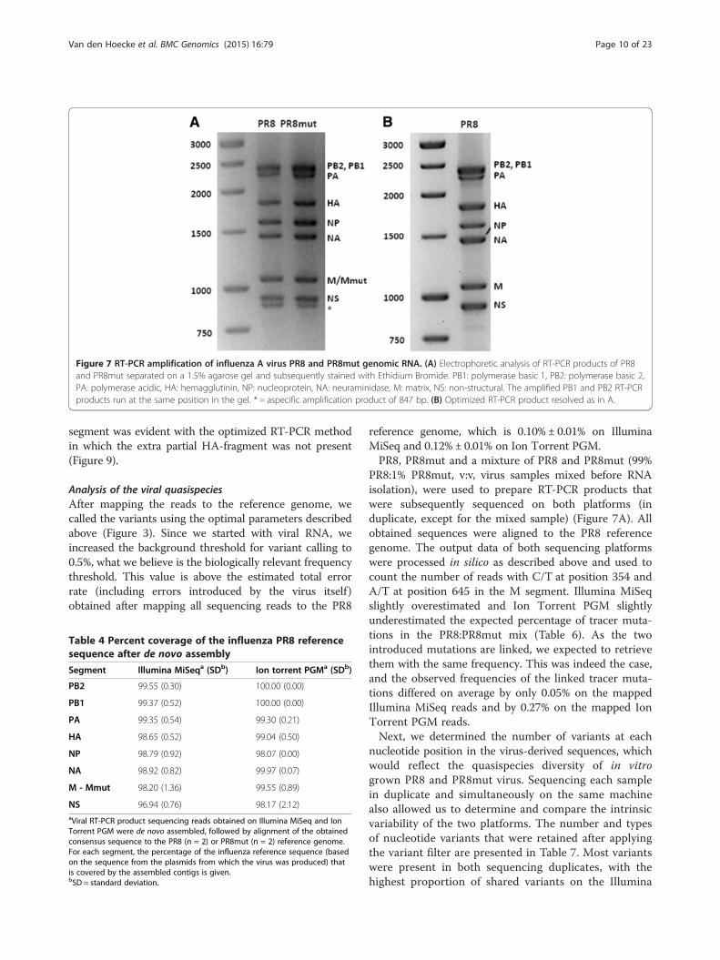
Figure 7 RT-PCR amplification of influenza A virus PR8 and PR8mut genomic RNA. (A) Electrophoretic analysis of RT-PCR products of PR8and PR8mut separated on a 1.5% agarose gel and subsequently stained with Ethidium Bromide. PB1: polymerase basic 1, PB2: polymerase basic 2,PA: polymerase acidic, HA: hemagglutinin, NP: nucleoprotein, NA: neuraminidase, M: matrix, NS: non-structural. The amplified PB1 and PB2 RT-PCRproducts run at the same position in the gel. * = aspecific amplification product of 847 bp. (B) Optimized RT-PCR product resolved as in A.
Van den Hoecke et al. BMC Genomics (2015) 16:79 Page 10 of 23
segment was evident with the optimized RT-PCR methodin which the extra partial HA-fragment was not present(Figure 9).
Analysis of the viral quasispeciesAfter mapping the reads to the reference genome, wecalled the variants using the optimal parameters describedabove (Figure 3). Since we started with viral RNA, weincreased the background threshold for variant calling to0.5%, what we believe is the biologically relevant frequencythreshold. This value is above the estimated total errorrate (including errors introduced by the virus itself)obtained after mapping all sequencing reads to the PR8
Table 4 Percent coverage of the influenza PR8 referencesequence after de novo assembly
Segment Illumina MiSeqa (SDb) Ion torrent PGMa (SDb)
PB2 99.55 (0.30) 100.00 (0.00)
PB1 99.37 (0.52) 100.00 (0.00)
PA 99.35 (0.54) 99.30 (0.21)
HA 98.65 (0.52) 99.04 (0.50)
NP 98.79 (0.92) 98.07 (0.00)
NA 98.92 (0.82) 99.97 (0.07)
M - Mmut 98.20 (1.36) 99.55 (0.89)
NS 96.94 (0.76) 98.17 (2.12)aViral RT-PCR product sequencing reads obtained on Illumina MiSeq and IonTorrent PGM were de novo assembled, followed by alignment of the obtainedconsensus sequence to the PR8 (n = 2) or PR8mut (n = 2) reference genome.For each segment, the percentage of the influenza reference sequence (basedon the sequence from the plasmids from which the virus was produced) thatis covered by the assembled contigs is given.bSD = standard deviation.
reference genome, which is 0.10% ± 0.01% on IlluminaMiSeq and 0.12% ± 0.01% on Ion Torrent PGM.PR8, PR8mut and a mixture of PR8 and PR8mut (99%
PR8:1% PR8mut, v:v, virus samples mixed before RNAisolation), were used to prepare RT-PCR products thatwere subsequently sequenced on both platforms (induplicate, except for the mixed sample) (Figure 7A). Allobtained sequences were aligned to the PR8 referencegenome. The output data of both sequencing platformswere processed in silico as described above and used tocount the number of reads with C/T at position 354 andA/T at position 645 in the M segment. Illumina MiSeqslightly overestimated and Ion Torrent PGM slightlyunderestimated the expected percentage of tracer muta-tions in the PR8:PR8mut mix (Table 6). As the twointroduced mutations are linked, we expected to retrievethem with the same frequency. This was indeed the case,and the observed frequencies of the linked tracer muta-tions differed on average by only 0.05% on the mappedIllumina MiSeq reads and by 0.27% on the mapped IonTorrent PGM reads.Next, we determined the number of variants at each
nucleotide position in the virus-derived sequences, whichwould reflect the quasispecies diversity of in vitrogrown PR8 and PR8mut virus. Sequencing each samplein duplicate and simultaneously on the same machinealso allowed us to determine and compare the intrinsicvariability of the two platforms. The number and typesof nucleotide variants that were retained after applyingthe variant filter are presented in Table 7. Most variantswere present in both sequencing duplicates, with thehighest proportion of shared variants on the Illumina

Figure 8 Sequence coverage of the influenza virus genome. Sequence coverage for the different genome segments of wild type PR8 virussequenced on Illumina MiSeq (2x250 bp, black lines, n = 2) or Ion Torrent PGM (Ion 318 chip v2, orange lines, n = 2). The obtained sequenceswere mapped to the reference genome (based on the pHW plasmids that were used to generate the virus, with addition of the extra 20nucleotides present at the 5′ site in the RT-PCR primers).
Van den Hoecke et al. BMC Genomics (2015) 16:79 Page 11 of 23

Table 5 Alignment metrics for Illumina MiSeq and Ion Torrent PGM sequencing runs
Illumina MiSeq
PR8 S1
Segment Length Mapped reads Minimum coverage Maximum coverage Average coverage
PB2 2381 159869 12 20525 15057
PB1 2381 126244 6 16513 11960
PA 2273 107490 7 14883 10533
HA 1815 213169 6 58709 25756
NP 1605 149599 9 29927 19883
NA 1453 139858 5 29353 21256
M 1067 180592 13 56656 37788
NS 930 140785 4 47293 31651
PR8 S2
Segment Length Mapped reads Minimum coverage Maximum coverage Average coverage
PB2 2381 163969 14 20266 14923
PB1 2381 128791 9 16043 11750
PA 2273 110954 5 14733 10486
HA 1815 222513 5 57511 25860
NP 1605 150831 11 29497 19330
NA 1453 135597 14 27006 19834
M 1067 177520 13 54233 35854
NS 930 136505 12 44068 29591
Ion torrent PGM
PR8 S1
Segment Length Mapped reads Minimum coverage Maximum coverage Average coverage
PB2 2381 93676 6396 11399 8765
PB1 2381 72187 4016 10132 6471
PA 2273 70492 4735 9315 6940
HA 1815 148242 4613 39585 17544
NP 1605 94509 8518 19617 12324
NA 1453 77561 7918 14959 11904
M 1067 119301 15170 31854 24331
NS 930 112041 16425 33280 25428
PR8 S2
Segment Length Mapped reads Minimum coverage Maximum coverage Average coverage
PB2 2381 84783 5612 10775 7947
PB1 2381 65635 3442 9253 5900
PA 2273 63994 4529 8607 6290
HA 1815 139240 4438 37662 16517
NP 1605 88966 8283 18590 11625
NA 1453 74318 7629 14494 11453
M 1067 115512 15395 30397 23553
NS 930 109661 16936 32481 24950
Wild type PR8 virus was sequenced in duplicate (S1 and S2) on both sequencers and the processed reads were mapped to the reference sequence (based on thesequence obtained from the plasmids from which the virus was produced, with addition of the extra 20 nucleotides present at the 5′ site in the RT-PCR primers).
Van den Hoecke et al. BMC Genomics (2015) 16:79 Page 12 of 23

Figure 9 Coverage of PR8 virus genome with the optimized RT-PCR protocol. Sequence coverage for the different genome segments of wildtype PR8 virus sequenced on Illumina MiSeq (2x250 bp) using two different fragmentation methods: Nextera XT transposase-based fragmentation(black lines) and mechanical Covaris shearing followed by adaptor ligation (orange lines). The obtained sequences were mapped to the referencegenome (based on the plasmids used to generate the virus).
Van den Hoecke et al. BMC Genomics (2015) 16:79 Page 13 of 23
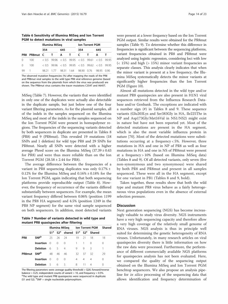
Table 6 Sensitivity of Illumina MiSeq and Ion TorrentPGM to detect mutations in viral samples
Illumina MiSeq Ion Torrent PGM
354 645 354 645
PR8 PR8mut C T A T C T A T
0 100 < 0.5 99.96 < 0.5 99.95 < 0.5 99.61 < 0.5 99.95
0 100 < 0.5 99.96 < 0.5 99.95 < 0.5 99.62 < 0.5 99.95
99 1 98.21 1.77 98.31 1.64 98.90 0.76 98.95 0.90
The observed mutation frequencies (%) after mapping the reads of the PR8and PR8mut viral samples to the wild type PR8 viral reference genome (basedon the sequence from the plasmids from which the virus was produced) areshown. The PR8mut virus contains the tracer mutations C354T and A645T.
Van den Hoecke et al. BMC Genomics (2015) 16:79 Page 14 of 23
MiSeq (Table 7). However, the variants that were identifiedin only one of the duplicates were actually also detectablein the duplicate sample, but just below one of the fourvariant filtering parameters. As for the plasmid samples, allof the indels in the samples sequenced on the IlluminaMiSeq and most of the indels in the samples sequenced onthe Ion Torrent PGM were present in homopolymer re-gions. The frequencies of the sequencing variants detectedby both sequencers in duplicate are presented in Tables 8(PR8) and 9 (PR8mut). This revealed 19 mutations (18SNPs and 1 deletion) for wild type PR8 and 29 SNPs forPR8mut. Nearly all SNPs were detected with a higheraverage Phred score on the Illumina MiSeq (37.39 ± 0.43for PR8) and were thus more reliable than on the IonTorrent PGM (28.58 ± 2.44 for PR8).The average difference between the frequencies of a
variant in PR8 sequencing duplicates was only 0.17% ±0.12% for the Illumina MiSeq and 0.16% ± 0.18% for theIon Torrent PGM, again indicating that both sequencingplatforms provide reproducible output (Table 8). How-ever, the frequency of occurrence of the variants differedsubstantially between sequencers. For example, the meanvariant frequency differed between 0.06% (position 1199in the PR8 HA segment) and 4.5% (position 1249 in thePR8 NP segment) for the same viral sample sequencedon both sequencers. In addition, most detected variants
Table 7 Number of variants detected in wild type andmutant PR8 quasispecies after filtering
Illumina MiSeq Ion Torrent PGM Shared
S1a S2a shared S1a S2a Shared
PR8 SNPb 25 26 24 19 21 18 18
Insertion 0 0 0 1 1 0 0
Deletion 6 5 4 9 9 3 1
PR8mut SNPb 48 46 46 32 37 32 29
Insertion 0 0 0 4 4 4 0
Deletion 5 6 5 8 11 4 0
The filtering parameters were: average quality threshold > Q20, forward/reversebalance > 0.25, independent counts of variant > 10, and frequency > 0.5%.aThe wild type and mutant PR8 quasispecies were sequenced in duplicate(S1 and S2). bSNP = single nucleotide polymorphism.
were present at a lower frequency based on the Ion TorrentPGM output. Similar results were obtained for the PR8mutsamples (Table 9). To determine whether this difference infrequencies is significant between the sequencing platforms,variant frequencies obtained in PR8 and PR8mut wereanalyzed using logistic regression, considering loci with low(< 15%) and high (> 15%) minor variant frequencies asseparate classes. This analysis clearly indicates that whenthe minor variant is present at a low frequency, the Illu-mina MiSeq systematically detects the minor variants atsignificantly higher frequencies than the Ion TorrentPGM (Figure 10).Almost all mutations detected in the wild type and/or
mutant PR8 quasispecies are also present in H1N1 viralsequences retrieved from the Influenza Research Data-base and/or Genbank. The exceptions are indicated witha number sign (#) in Tables 8 and 9. These sequencevariants (Glu203Lys and Ser383Gly in HA, Ile225Thr inNP and Asp173Gly/Met16Val in NS1/NS2) might existin nature but have not been reported yet. Most of thedetected mutations are present in the HA segment,which is also the most variable influenza protein innature [70]. Most of the detected mutations were substi-tutions occurring at a frequency < 5%. However, threemutations in HA and one in NP of PR8 as well as fourmutations in HA and one in NS of PR8mut were presentat a frequency > 10% (based on Illumina MiSeq data)(Tables 8 and 9). Of all detected variants, only seven (fivenon-synonymous and two synonymous) were sharedby both PR8 and PR8mut and present in all samplessequenced. These were all in the HA segment, exceptfor one variant in PB1 (Tables 8 and 9, bold).Taken together, these results show that both the wild
type and mutant PR8 virus behave as a fairly heteroge-neous virus populations even in the absence of externalselection pressure.
DiscussionNext generation sequencing (NGS) has become increas-ingly valuable to study virus diversity. NGS instrumentshave a very high sequencing capacity and therefore allowa very high coverage of the relatively small genome ofRNA viruses. NGS analysis is thus in principle wellsuited for determining the genetic heterogeneity of RNAviruses. Unfortunately, in many research articles on viralquasispecies diversity there is little information on howthe raw data were processed. Furthermore, the perform-ance of different commercially available NGS platformsfor quasispecies analysis has not been evaluated. Here,we compared the quality of the sequencing outputobtained on the Illumina MiSeq and Ion Torrent PGMbenchtop sequencers. We also propose an analysis pipe-line for in silico processing of the sequencing data thatallows identification and frequency determination of

Table 8 Wild type PR8 quasispecies sequenced in duplicate on both Illumina MiSeq and Ion Torrent PGM
Frequency (in %)
Segment Position Type Reference Allele aa change IlluminaMiSeq
Ion TorrentPGM
Function/location
PB1 1482 Deletion A - frameshift 1.87 2.19 3.18 2.88
PB1 1486 SNP A G Lys481Arg 2.32 2.62 1.91 1.91 K481 crucial for polymerase functionin vivo, not in vitro [47]
PA 539 SNP A G silent 1.37 1.42 0.56 0.54 /
HA 607 SNP A G silent 1.60 1.56 2.02 1.85 /
HA# 659 SNP G A Glu203Lys 1.13 1.23 0.65 0.60 enhanced receptor binding activity [48]
HA 660 SNP A G Glu203Gly 3.11 3.02 1.76 1.55 slightly increased α2-6 and decreased α2-3 binding [49]
HA 747 SNP A G Glu232Gly 11.56 11.43 7.29 7.19 receptor specificity [50]
HA 764 SNP G A Asp238Asn 0.83 0.80 0.65 0.60 enables binding to α2.3- andα2.6-linked sialic acids [51]
HA 765 SNP A G Asp238Gly 39.73 39.43 35.33 35.00 enables binding to α2.3- andα2.6-linked sialic acids [52,53]
HA 768 SNP A G Gln239Arg 2.81 3.12 1.43 1.23 preferential binding to α-2,3-linked glycans [52]
HA 823 SNP A G Ile257Met 1.76 1.54 0.72 0.74 located in head domain close to Sa antigenic site [54]
HA# 1199 SNP A G Ser383Gly 1.41 1.15 1.14 1.29 located in stem domain
HA 1330 SNP A G silent 1.59 1.50 1.02 1.20 /
HA 1424 SNP G A Val458Met 95.25 95.67 97.85 97.77 located in stem domain
HA 1440 SNP A G Glu463Gly 1.91 1.75 0.58 0.56 located in stem domain
HA 1451 SNP A G Ser467Gly 0.70 0.81 0.62 0.63 located in stem domain
NP 212 SNP C T silent 1.80 1.76 0.83 0.64 /
NP 1249 SNP A G Asn395Ser 10.71 11.01 5.97 6.76 located in NP-NP and NP-PB2 interactiondomain [55,56]
NP 1324 SNP T G Phe420Cys 3.43 3.41 1.31 1.15 located in the hypervariable NP418-426 CTL epitope [57]#not present in Genbank or Influenza Research Database.Bold = variant also present in PR8mut quasispecies.HA segment = numbering of HA amino acid residues is based on the PR8 HA open reading frame with the starting methionine as position = 1.
Van den Hoecke et al. BMC Genomics (2015) 16:79 Page 15 of 23
nucleotide variants in the influenza A virus (Figure 3).This analysis pipeline will help to standardize variantcalling in small RNA genomes based on NGS data.To determine the influenza genome diversity by NGS
technology, different hurdles have to be overcome. First, itis technically challenging to obtain high quality full-lengthRT-PCR products that cover the complete segmentedRNA genome of influenza viruses. We optimized anRT-PCR protocol with primers based on the conserved3′ (Uni12) and 5′ ends (Uni13) of the eight genomesegments [42,43,71]. Critical steps in this protocol areprimer concentration and annealing and elongationtimes. Because the sequence of these segment ends isconserved, this RT-PCR should be applicable to differentinfluenza A virus strains.A second hurdle is to distinguish between mutations
that truly represent the viral genome diversity fromerrors introduced by RT-PCR amplification and the NGSchemistry. The first step is to filter the output sequencedata in silico to retain only high quality reads. However,the available software and filtering parameters vary and
are not always clearly described in the literature, makingcomparison of results very difficult. To reduce false posi-tive variant calls introduced by the sequencing method,we applied specific trimming, filtering and variant callingparameters in the CLC Genomics Workbench software.We first applied this bioinformatics analysis pipeline tosequencing reads derived from plasmid DNA samples.We removed adaptor contamination, ambiguous nucleo-tides and trimmed low quality bases at the end of thereads by applying a Phred score of 20. Then, we excludedreads shorter than 50 bases to avoid unspecific mappingof these short reads. Trimming eliminated relatively morebases from the Ion Torrent PGM, meaning that the basequality of sequencing reads from the Ion Torrent PGM islower than that from the Illumina MiSeq. In other words,the potential advantage of longer read lengths obtainedwith the Ion Torrent machine was cancelled by theirrelatively low quality. Together this resulted in a higherrelative loss of bases for the Ion Torrent PGM data thanfor the Illumina MiSeq data (21.01% versus 14.01%respectively). Furthermore, the Phred score distribution

Table 9 Mutant PR8 quasispecies sequenced in duplicate on both Illumina MiSeq and Ion Torrent PGM
Frequency (in %)
Segment Position Type Reference Allele aa change IlluminaMiSeq
Ion TorrentPGM
Function/location
PB2 416 SNP A G silent 1.55 1.30 0.59 0.57 /
PB1 1486 SNP A G Lys481Arg 2.52 2.80 1.79 2.37 K481 crucial for polymerase functionin vivo, not in vitro [47]
PA 212 SNP G T Glu56Asp 5.72 4.85 2.09 2.18 located in endonuclease domain [58,59]
PA 1139 SNP G T Gln365His 2.50 2.40 1.00 1.05 located in PB1 interacting domain [60]
HA 524 SNP A C Ser158Arg 13.17 12.93 9.70 9.63 Compensatory mutation in [61],located in Ca antigenic site [54]
HA 524 SNP A T Ser158Cys 0.98 0.90 0.61 0.63 located in variable Ca antigenic site [54]
HA 607 SNP A G silent 1.54 1.43 2.23 2.47 /
HA 747 SNP A G Glu232Gly 39.95 40.14 36.74 36.02 receptor specificity [50]
HA 765 SNP A G Asp238Gly 3.17 3.07 1.50 1.44 enables binding to α2,3- andα2,6-linked sialic acids [52,53]
HA 823 SNP A G Ile257Met 2.92 3.08 1.49 1.64 located in head domain close to Sa antigenic site [54]
HA 828 SNP A G Glu259Gly 5.17 4.96 2.09 1.95 located on surface head domainclose to Sa antigenic site [54]
HA 1088 SNP T A Phe346Ile 6.99 6.76 3.75 4.18 located in fusion peptide [62]
HA 1090 SNP T G Phe346Leu 1.28 1.09 0.59 0.69 located in fusion peptide [62,63]
HA 1109 SNP A G Ile353Val 59.69 60.02 62.48 61.74 described as fusion peptide pseudorevertant [62,64]
HA# 1199 SNP A G Ser383Gly 1.13 1.18 0.97 1.05 located in stem domain
HA 1330 SNP A G silent 1.59 1.67 1.94 1.14 /
HA 1424 SNP G T Val458Leu 3.43 3.13 1.63 1.64 located in stem domain, not surface exposed
HA 1430 SNP A G Asn460Asp 10.08 9.60 5.82 6.10 present in the PR8 quasispeciesgrown on MDCK cells [65]
HA 1431 SNP A G Asn460Ser 14.79 14.62 10.15 10.29 located in stem domain
HA 1487 SNP G A Gly479Arg 1.59 1.39 0.61 0.57 located in stem domain, not surface exposed
NP 635 SNP G A silent 4.44 3.93 2.45 1.87 /
NP# 739 SNP T C Ile225Thr 1.11 1.15 0.52 0.56 surface exposed, in NP-NP interaction domain [56]
NA 476 SNP T A Cys146Ser 6.85 6.71 4.33 3.85 located in head domain, involved in couplingof subunits [66]
NA 994 SNP C T silent 1.03 1.00 0.60 0.65 /
M 354 SNP C T introduced 99.96 99.96 99.61 99.62 /
M 645 SNP A T introduced 99.95 99.95 99.95 99.95 /
NS 409 SNP G T NS1: Gln121His 40.37 39.54 31.96 32.61 situated next to the NS1122–130 CTL epitope [67]
NS 549 SNP G A NS1: Gly168Glu 1.27 1.20 0.71 0.59 NS1: located in effector domain [68]
NS2: N-terminal domainNS2: Asp11Asn
NS# 564 SNP A G NS1: Asp173Gly 1.05 1.10 0.65 0.64 NS1: located in effector domain [68]
NS2: Met16Val NS2: Met16 is involved in nuclear export NP [69]#not present in Genbank or Influenza Research Database.Bold = variants also present in PR8 quasispecies.HA segment = numbering of HA amino acid residues is based on the PR8 HA open reading frame with the starting methionine as position = 1.
Van den Hoecke et al. BMC Genomics (2015) 16:79 Page 16 of 23
across the reads, a measure of the intrinsic sequencingquality, was higher for the Illumina MiSeq data than forthe Ion Torrent PGM data, resulting in a lower error rate.After this quality control, the sequencing reads weremapped to the reference sequence, resulting in a higherpercentage of mapped reads for the Illumina MiSeq. The
total mapping error rate of the Illumina MiSeq (mainlynucleotide substitutions) was lower than that of the IonTorrent PGM (mainly indels). This finding is in agreementwith Loman and colleagues [20]. However, for plasmidDNA analysis the substitution error rate on the IonTorrent PGM appeared to be lower than that of Illumina

Figure 10 Low frequency minor alleles are detected atsignificantly higher frequencies by Illumina MiSeq compared to IonTorrent PGM. Nucleotide variants were subdivided in two frequencyclasses: high (frequency minor allele > 15%, n = 4) and low (frequencyminor allele: < 15%, n = 42). Mean proportions ± s.e. of the minor variantsdetected in PR8 and PR8mut viral samples by the Illumina MiSeq and IonTorrent PGM are shown. Minor allele proportions were analyzed bylogistic regression (link function = logit). Significance levels of pairwisecomparisons were assessed by a Fisher’s protected least significancedifference test * = p < 0.05, ** = p < 0.01.
Van den Hoecke et al. BMC Genomics (2015) 16:79 Page 17 of 23
MiSeq (Figure 5). After variant calling, the resulting hitswere filtered based on frequency, forward/reverse balance,average quality, and independent counts to remove falsepositive variants. After filtering, both sequencers detectedthe tracer mutations we had introduced with excellentaccuracy and sensitivity. Nevertheless, the average quality(Phred score) of the detected variants was higher on theIllumina MiSeq than on the Ion Torrent PGM, makingthe variants detected on the Illumina MiSeq more reliable.The number of false positive variants can be furtherreduced by cross-platform replication, but the differentbiases of the sequencing platforms may cause many truevariants to be overlooked when cross-platform replicatesare compared [72,73].We then applied the analysis pipeline outlined in
Figure 3 to PR8 and PR8mut virus, which were gener-ated by a plasmid-based reverse genetics system andamplified in MDCK cells. In our opinion, variants in theinfluenza virus genome that appear with a frequencybelow 0.5% are very difficult to distinguish from thebackground noise that is cumulatively introduced by RT-PCR and the inherent variation due to the chemistry ofcurrently available Illumina and Ion Torrent sequencers.We propose that a similar threshold of 0.5% should beapplied to interpret the genetic diversity of RNA viruses.Nevertheless, mutations with a frequency as low as0.05 − 0.2% in Chikungunya virus have been reported inthe literature as meaningful based on Illumina GAIIX
sequencing [74]. Given the error rate of the influenzavirus polymerase, resulting in approximately one mutationper 10.000 nucleotides, together with the errors intro-duced during RT-PCR and the technical backgrounderror rate of the NGS platforms applied in this study, itis not straightforward for both the Illumina MiSeq andthe Ion Torrent PGM to identify each variant in theviral quasispecies. Nevertheless, even with the thresholdof 0.5% proposed here, NGS will enable studying of theviral diversity in much more detail than in the past.Our analysis showed that the de novo assembled PR8
and PR8mut sequences correspond very well to theplasmid-derived reference genome. We detected 19 muta-tions in PR8 and 29 mutations (including the two tracermutations) in PR8mut with a frequency of 0.5% or higher.When a variant was present at low frequency (< 15%), theIllumina MiSeq detected it with significantly higher fre-quency than the Ion Torrent PGM. Most of the detectedmutations were transitions and appeared with a frequencybelow 5%. However, three mutations in HA and one inNP of PR8, as well as four mutations in HA and one inNS of PR8mut, were present at a frequency > 10% (basedon Illumina MiSeq data) (Tables 8 and 9). We detectedonly one single nucleotide deletion in the PR8 virus. Thisdeletion was in a homopolymer at position 1482 in PB1but was detected with a frequency of 2 − 3% by bothsequencers, in both duplicates of PR8 virus. In addition,this deletion was also detected with a similar frequency inboth PR8mut samples sequenced on the Illumina MiSeqand in one of the duplicate samples sequenced on the IonTorrent PGM. This deletion disrupts the open readingframe, leading to premature termination of PB1. Thisdetrimental mutation is in line with the finding of Brookeand colleagues, who showed that most of the infectiousinfluenza A virions fail to express detectable levels of oneor more viral proteins [75].We focused on the mutations detected by both
sequencers with a frequency > 5% and on the mutationsthat appeared in both wild type and mutant PR8 viruses.There are three such mutations in the HA head domainof PR8 and four in the HA head domain of PR8mut,and all of them are part of or close to the antigenic sites(Figure 11A). The shared Asp238Gly mutation (Asp225Glyfor H3 numbering) is associated with enhanced virionbinding to the avian-type Sia(α2-3)Gal and was reportedpreviously as a position that is selected by egg-adaptationof influenza viruses [76]. The Ser158Arg mutation (Ser145Arg for H3 numbering) in PR8mut has been describedas a compensatory mutation in PR8 virus possessingthe Lys165Glu mutation in HA (H3 numbering), whichdecreases the receptor binding avidity and replicationkinetics of the virus [61]. The two mutations in thestem domain are relatively conservative (Ser383Gly andVal458Met; Ser40Gly and Val115Met for H3 numbering

Figure 11 Position of variants present in PR8 and PR8mut quasispecies in the HA, NP and NS1. The variants in HA (hemagglutinin), NP(nucleoprotein) and NS1 (non-structural protein 1) detected in the PR8 and PR8mut quasispecies were modeled with PyMol (Delano Scientific,http://www.pymol.org), using the HA from A/Puerto Rico/8/1934 (H1N1) (PDB code: 1RVX), the NP from A/Wilson-Smith/1933 (H1N1) (PDB code:2IQH) and the effector domain of NS1 from A/Puerto Rico/8/1934 (H1N1) (PDB code: 3RVC). (A) Top (left) and lateral (right) view of the surfaceexposed amino acids of the HA trimer. The Cb, Ca, Sa and Sb antigenic sites are shown in green. The mutations that are present in both PR8 andPR8mut are shown in red or in magenta if they overlap with the antigenic sites. Mutations in PR8mut that are present at a frequency > 5% areshown in blue or in yellow when overlapping with the antigenic sites or in purple when overlapping with the fusion peptide (orange). Themature H3 amino acid numbering of the variants is provided in superscript. (B) Lateral view of the NP monomer with the N395S mutationpresent in PR8 shown in brown. (C) The effector domain of NS1 with the Q121H mutation in PR8mut shown in blue.
Van den Hoecke et al. BMC Genomics (2015) 16:79 Page 18 of 23
of HA2) and therefore might not affect virus replication.Remarkably, the G-to-A substitution at position 1424,leading to the Val458Met change in HA, had a frequencyclose to 100% in the PR8 HA segment but was absent inPR8mut (although a Val458Leu change is present in asmall percentage of PR8mut). This mutation was probablyfixed in the wild type virus genome at a very early step,e.g. during plaque purification of the PR8 seed virus weused to prepare stock virus. We also picked up twoother codon changes in the HA stem region of PR8mut:Asn460Asp (5 − 10%) and Asn460Ser (10 − 15%) (Asn117Asp and Asn117Ser for H3 numbering of HA2). Basedon pyrosequencing of the HA segment, the Asn460Aspmutation has been observed in 12.2% in PR8 virus grownon MDCK cells [65]. In addition, the PR8mut caries theIle353Val (Ile10Val for H3 numbering of HA2) mutationin the HA fusion peptide at a frequency of about 60%.A valine at this position has been observed in a PR8pseudo-revertant after introducing the Ile10Ala mutation.A valine at this position is compatible with the α-helicalstructure of the fusion peptide [64]. Both PR8 viruses alsocontain mutations in other segments. For example, bothviruses share the conservative Lys481Arg mutation inPB1. This lysine at position 481 is crucial for the polymer-ase function of PB1 in vivo but mutating it to alanine wastolerated in vitro [47]. In wild type PR8, the Asn395Servariant in NP is in a domain involved in NP −NP and NP− PB2 interactions (Figure 11B) [77]. The Gln121Hisvariant detected in NS1 of PR8mut is situated just beforea human CTL epitope (Figure 11C) [67]. Remarkably,none of the variants we observed correspond to the vari-ants described in an earlier study, in which a PR8 strain
(originally adapted for growth on embryonated chickeneggs) was adapted for growth on MDCK cells [78].However, we used MDCK cells only to expand ourvirus stock, which corresponds to about six cycles ofPR8 virus replication. Furthermore, we generated ourPR8 virus starting from eight plasmids, indicating thatthe passaging history is a determinant of the variantsdetected in an influenza virus quasispecies.Both sequencers are highly effective for accurate detec-
tion of low frequency mutations, but each one has itsadvantages and limitations. On the one hand, the IlluminaMiSeq platform has about three times higher outputcapacity than the Ion Torrent PGM, enabling sequencingof more samples in parallel on the Illumina MiSeq. On theother hand, the Ion Torrent PGM is significantly faster: itstime from sample preparation to data analysis is one dayless than for the Illumina MiSeq. After the in silico qualitycontrol, the two sequencers produced reads of compar-able lengths. The Illumina MiSeq had a higher intrinsicsequencing quality than the Ion Torrent PGM, presum-ably because detecting incorporated bases based on acoupled fluorescent dye (Illumina) gives less noise thana change in pH caused by release of a proton afterincorporation of a base (Ion Torrent). However, the IonTorrent PGM had a lower false-positive rate for detectingSNPs. Another interesting observation is the lowercoverage of the ends of the viral segments on the IlluminaMiSeq due to the transposase-based fragmentation.Nextera transposase-based fragment library preparationis convenient and fast but results in low coverage ofsegment termini. We also noticed some sequence bias ofthis transposase-based fragmentation approach (Figures 8

Van den Hoecke et al. BMC Genomics (2015) 16:79 Page 19 of 23
and 9). Mechanical fragmentation followed by adaptorligation enables comparable coverage of all bases of theinfluenza virus genome, and is therefore the preferredmethod for library preparation (Figures 8 and 9).The proposed RT-PCR protocol and subsequent ana-
lysis pipeline for influenza viruses is widely applicable, e.g.to study vaccine composition, analyze virus evolutionunder selection pressure, monitor mutations associatedwith antiviral resistance, and assemble the referencegenome of new viral isolates. For clinical samples, theshorter turnaround time of the Ion Torrent PGM (samplepreparation, sequencing and analysis in about 2 days) isclearly advantageous to the Illumina MiSeq (about 3 days).In contrast, when analyzing many viral samples at highcoverage, the greater output of the Illumina MiSeq is animportant advantage.
ConclusionOur study underlines the power and limitations of twocommonly used next-generation sequencers for theanalysis of influenza gene diversity. We propose an insilico pipeline for selecting high quality reads obtainedby NGS platforms. This pipeline is also more widelyapplicable. Due to the lower total error rate and thehigher sequencing quality of the reads, we conclude thatthe Illumina MiSeq platform is more suited than the IonTorrent PGM for detecting variant sequences, whereasthe Ion Torrent platform has a shorter turnaround time.In addition, we found that the detection limit for reliablerecognition of variants in the viral genome required afrequency of 0.5% or higher.
MethodsCell linesMDCK and HEK293T cells were cultured in Dulbecco’sModified Eagle medium (DMEM) supplemented with 10%fetal calf serum, non-essential amino acids, 2 mM L-glu-tamine, 0.4 mM sodium pyruvate, 100 U/ml penicillin and0.1 mg/ml streptomycin at 37°C in 5% CO2.
Generation and production of plasmids with tracermutationsReverse genetics plasmids for PR8 virus were kindlyprovided by Dr. Robert G. Webster (St. Jude Children’sResearch Hospital, Memphis, USA) [31]. We introducedtwo silent mutations in the M coding gene, a C-to-Tsubstitution at position 797 (numbering relative to thehuman cytomegalovirus promoter in the pHW197-Mplasmid) and an A-to-T substitution at position 1088 inpHW197-M. These two positions were selected as follows.First, we generated a consensus sequence of the M-genebased on all full-length segment 7 sequences of humanH1N1 viruses present in the Influenza Virus ResourceDatabase (NCBI) on September 11th, 2011. Next, we
aligned the consensus sequence to the M segment of PR8(present in pHW197-M) and selected two synonymousmutations in the M1 open reading frame at positionsC354T and A645T (segment 7 numbering). These twomutations were introduced by two consecutive rounds ofquickchange site-directed mutagenesis (Stratagene) atpositions C797T and A1088T in pHW197-M to generatepHW197-Mmut. The two mutations also introduced aHindIII and a PvuII restriction site, respectively. Theseplasmids and the plasmids encoding the other seven PR8genome segments were transformed and amplified in E.coli DH5α. Plasmid DNA was isolated with the PlasmidMidi Kit (Qiagen) according to the manufacturer’s instruc-tions. The resulting air-dried pellet was dissolved in 50 μlof sterile ultrapure water. The presence of the introducedmutations in pHW197-Mmut was confirmed by restrictionanalysis and Sanger sequencing on a capillary sequencer(Applied Biosystems 3730XL DNA Analyzer).
Generation of recombinant PR8 and PR8mut virusesTo generate recombinant wild type PR8 virus and PR8virus with the two tracer mutations in the M gene(PR8mut), 1 μg of pHW191-PB2, pHW192-PB1, pHW193-PA, pHW194-HA, pHW195-NP, pHW196-NA and pHW198-NS, together with 1 μg of pHW197-M (wild type PR8)or pHW197-Mmut (PR8mut) was transfected usingcalcium phosphate co-precipitation into a HEK293T-MDCK cell co-culture in Opti-MEM (3 × 10⁵ HEK293Tand 2 × 10⁵ MDCK cells in a 6-well plate). After 30 h,L-1-tosylamide-2-phenylethyl chloromethyl ketone (TPCK)-treated trypsin (Sigma) was added to a final concentra-tion of 2 μg/ml. After 72 h, the culture medium wascollected and the presence of virus was confirmed byhemagglutination of chicken red blood cells. Reversegenetics-generated PR8 and PR8mut viruses were plaque-purified on MDCK cells as follows. Confluent MDCK cellsin a six-well plate were infected with a serial dilutionseries of virus. After 1 h, an overlay of low melting agarose(Type VII agarose, Sigma; final concentration 1%) inserum-free cell culture medium containing 2 μg/mlTPCK-treated trypsin (Sigma) was added. After 56 h,cytopathic effect was checked, agar overlaying viralplaques were selected with a pipette tip, and virus wasallowed to diffuse from the agar for 24 h at 4°C inserum-free medium. Afterwards, virus derived from oneplaque was amplified on MDCK cells in serum-free cellculture medium in the presence of 2 μg/ml TPCK-treatedtrypsin (Sigma). After 96 h, the culture medium wascollected, and cell debris was removed by centrifugationfor 10 min at 2500 g at 4°C, and the virus was pelletedfrom the supernatants by overnight centrifugation at16,000 g at 4°C. The pellet was dissolved in sterile 20%glycerol in PBS, aliquoted and stored at −80°C. The infec-tious titer of the obtained PR8 and PR8mut virus stocks

Van den Hoecke et al. BMC Genomics (2015) 16:79 Page 20 of 23
was determined by plaque assay on MDCK cells, on threedifferent aliquots each performed in triplicate. The pres-ence of the introduced mutations in the M segment ofPR8mut was confirmed by segment-7-specific RT-PCRfollowed by purification from 1% agarose gel (High PurePCR Product Purification Kit, Roche) and conventionalSanger sequencing of the amplified PCR fragment.
Plaque assayMDCK cells were seeded in complete DMEM in 12-wellplates at 3 × 10⁵ cells per well. After 18 h, the cells werewashed once with serum-free medium and incubated (intriplicate) with a two-fold dilution series of the virus(made in serum-free cell culture medium containing0.1% BSA) in 500 μl medium. After 1 h incubation at37°C, an overlay of 500 μl of 1.6% Avicel RC-591 (FMCBiopolymer) in serum-free medium with 4 μg/ml TPCK-treated trypsin (Sigma) was added. After incubation at37°C for 48 h, the overlay was removed and the cellswere fixed with 4% paraformaldehyde and permeabilizedwith 20 mM glycine and 0.5% (v/v) Triton X-100. Plaqueswere stained with an anti-M2e IgG1 mouse monoclonalantibody (final concentration 0.5 μg/ml) followed by asecondary anti-mouse IgG horseradish peroxidase (HRP)-linked antibody (GE Healthcare). After washing, TrueBlueperoxidase substrate (KPL) was used to visualize theplaques.
RNA isolationRNA was isolated with the High Pure RNA Isolation Kit(Roche) according to the manufacturer’s instructions,excluding the DNase I digestion step. In brief, a 200-μlsample containing 1 x 10⁷ PFU of stock virus in serum-free cell culture medium with 0.1% BSA was combinedwith 400 μl lysis-binding buffer and mixed by vortexing.The mixture was loaded on a two-layered glass fibercolumn. After binding to the column and washing, theRNA was eluted in 50 μl elution buffer (water, PCRgrade).
RT-PCRPrimers used for cDNA synthesis and PCR were designedbased on the 5′ and 3′ conserved ends of the influenza Agenomic segments and contain an additional sequenceof 20 nucleotides at their 5′ end necessary for PCRamplification [41-43,79]. cDNA was generated using theTranscriptor First Strand cDNA Synthesis Kit (Roche).Reverse transcription was performed with the TranscriptorReverse Transcriptase (10 U, Roche), using 12.5 μl RNA,2.5 μM CommonUni12G primer (GCCGGAGCTCTGCAGATATCAGCGAAAGCAGG), 1x Transcriptor ReverseTranscriptase Reaction Buffer, 20 U Protector RNAseinhibitor and 4 mM dNTPs, in a total volume of 20 μl. Thecomponents were mixed, and the reaction was incubated
for 15 min at 42°C, 15 min at 55°C, 5 min at 60°C, andfinally 5 min at 85°C to inactivate the reverse tran-scriptase. Ten microliters of the resulting cDNA samplewas amplified in a 100-μl PCR reaction using 2 U PhusionHigh Fidelity polymerase (Thermo Scientific), 0.2 μMCommonUni12G and CommonUni13 (GCCGGAGCTCTGCAGATATCAGTAGAAACAAGG), 0.2 mM dNTPs,and 1× High-Fidelity buffer. Thermocycling was per-formed in a PTC-200 Thermal Cycler (MJ Research)with the following conditions: initial denaturation for30 s at 98°C, 25 cycles of 10 s at 98°C followed by7.5 min at 72°C, and a final elongation step of 7 min at72°C. PCR products were purified using the High PurePCR Product Purification kit (Roche) according to themanufacturer’s instructions, and the product was elutedin 50 μl sterile ultrapure water (preheated to 65°C). Onemicrogram of the product was analyzed by agarose gelelectrophoresis (1.5% agarose gel) followed by ethidiumbromide staining.
Illumina MiSeq sequence determinationWe used 0.5 ng of purified plasmid or RT-PCR sampleand the Nextera XT DNA Sample Preparation Kit(Illumina) according to the manufacturer’s instructionsto generate multiplexed paired-end sequencing librar-ies. Sequencing libraries were generated in duplicate,meaning that from each plasmid or RT-PCR sampletwo libraries were prepared in parallel and sequencedon the same Illumina MiSeq sequencing chip. In brief,DNA samples were fragmented and tagged with adaptersby Nextera XT transposase. These adaptor ligated DNAfragments were amplified by a limited-cycle PCR program(12 cycles) to add the barcodes and sequences requiredfor subsequent cluster formation. The resulting fragmentswere purified and simultaneously size-selected by using0.6× AMpure beads. Fragments were analyzed on aHigh Sensitivity DNA Chip on the Bioanalyzer (AgilentTechnologies) before loading on the sequencing chip.The fragment lengths showed a negatively skewed dis-tribution with a peak at approximately 700–1000 bases.From the optimized RT-PCR products, also 500 ng wassheared with an M220 focused-ultrasonicator (Covaris)set to obtain peak fragment lengths of 300–400 bp.Next, the NEBNext Ultra DNA Library Preparation kit(New England Biolabs) was used to repair the ends andto add Illumina MiSeq-compatible barcode adapters to100 ng of fragmented DNA. The resulting fragmentswere size-selected using Agencourt AMPure XP beadsizing (Beckman Coulter). Afterwards, indexes were addedin a limited-cycle PCR (10 cycles), followed by purificationon Agencourt AMpure XP beads. Fragments were ana-lyzed on a High Sensitivity DNA Chip on the Bioanalyzer(Agilent Technologies) before loading on the sequencingchip. Equimolar amounts of normalized libraries were

Van den Hoecke et al. BMC Genomics (2015) 16:79 Page 21 of 23
combined and diluted 25-fold in hybridization buffer. Themultiplex sample was heat denatured for 2 min at 96°Cbefore loading on the MiSeq chip. After the 2×250 bpMiSeq paired-end sequencing run, the data were basecalled and reads with the same barcode were collectedand assigned to a sample on the instrument, whichgenerated Illumina FASTQ files (Phred +64 encoding).These files were imported in the CLC Genomics Work-bench software (CLC Bio, Qiagen). During import in CLCGenomics Workbench, the uncallable ends of the MiSeqreads (B in input file) were automatically trimmed and thefailed reads (Y in header information for the quality score)were removed.
Ion Torrent PGM 318 chip sequence determinationSamples for sequence analysis were generated in duplicate,meaning that from each plasmid or RT-PCR sample twolibraries were prepared in parallel for sequencing on thesame Ion Torrent PGM 318 sequencing chip. From eachplasmid or RT-PCR product, 100 ng was sheared with anM220 focused-ultrasonicator (Covaris) set to obtain peakfragment lengths of 400–500 bp. After shearing, bluntends were created using the end repair enzyme fromthe Ion Plus Fragment Library kit (Life Technologies).Next, the fragments were ligated to Ion Torrent PGM-compatible barcode adapters. Since the adaptors arenot 5′ phosphorylated, the nick repair polymerase inthe kit repairs subsequently the nick on one strand ateach ligation site, in order to minimize adaptor-dimerformation. We purified and simultaneously size-selectedthe adapter-ligated library using Agencourt AMPure XPbead sizing (Beckman Coulter). Fragments were analyzedon a High Sensitivity DNA Chip on the Bioanalyzer(Agilent Technologies); the fragment length peak wassituated around 450 bp. Barcoded libraries were pooledin equimolar amounts. From the resulting diluted mul-tiplexed library, 20 μl was loaded on an Ion OneTouch2 instrument (Life Technologies) to perform emulsionPCR on Ion Sphere particles using the Ion PGM Tem-plate OT2 400 kit. We used the Ion PGM sequencing400 kit (Life Technologies) to sequence templated ionsphere particles deposited in the Ion 318 chip v2(revision 2.0, Life Technologies). The Ion Torrent Suiteversion 4.6 (Life Technologies) was used with thedefault parameters for base calling and assigning of thereads to a sample based on their barcode. The defaultsettings in the Ion Torrent Suite already filter and trimthe sequencing reads to some extent. These defaulttrimming parameters are not stringent and remove onlyvery low quality 3′ ends (mean Phred score of at least15 in a base window of 30) and adaptor contamination.The resulting FASTQ files were imported into CLCGenomics Workbench for further analysis.
Analysis of sequencing dataCLC Genomics Workbench version 7.0.3 (CLC Bio,Qiagen) was used to analyze and process the sequencingreads of both the Ion Torrent PGM and the IlluminaMiSeq. First, adaptor contamination was removed fromthe reads. Next, the sequencing reads were trimmed fromboth sides using the modified Mott trimming algorithm toreach a Q20 score, which means that the chance that aparticular base in the sequence is called incorrectly by thesequencer is 1 in 100. Afterwards, all ambiguous (N) baseswere trimmed from the reads. We also removed the readswith a read length below 50. For the Illumina MiSeq, thebroken pairs resulting from trimming and filtering werealso removed. The remaining reads were assembled usingdefault settings for de novo assembly. In addition, theprocessed reads were also aligned with the pHW197-Mplasmid reference sequence or the influenza PR8 referencegenome (based on the sequences encoding the eightsegments in the pHW vectors, determined by Sangersequencing, with addition of the extra 20 nucleotidespresent at the 5′ site in the RT-PCR primers) using localalignment. For this, the following default penalties wereused: match = +1, mismatch = −2, insertion/deletion = −3,filtering threshold: length fraction = 0.9 and similarityfraction = 0.8. Non-specific matches, defined as readsaligning to more than one position with an equallygood score, were ignored. Sequence variants werecalled using all available sequencing data that coveredeach nucleotide at least 100 times and had a centralbase quality score of Q20 or greater. The A-to-Gvariant introduced by the primer at position 24 inthe HA, NP, NA, M and NS segments was not takeninto account during the influenza quasispecies variantanalysis. All numerical data mentioned in the text arepresented as averages with their standard deviations(± SD).
Statistical analysisSequence variants with the lowest proportion wereconsidered as minor alleles. Analysis of minor alleleproportions was performed by fitting a logistic regressionmodel of the form logit(p) = constant + PLATFORM*-VIRUS*CLASS + error, where p indicates the minorallele proportion, PLATFORM refers to the sequencingplatform, VIRUS refers to virus population, and CLASSrefers to class of loci having either low (< 15%) or high(> 15%) minor variant frequencies. Significance of thefixed PLATFORM, VIRUS and CLASS effects wasassessed by an F-test. Significance of pair-wise com-parisons between mean proportions was assessed by aFisher’s protected least significance difference test. Thelogistic regression and assessment of significance wasperformed in Genstat v16.

Van den Hoecke et al. BMC Genomics (2015) 16:79 Page 22 of 23
Sequencing dataThe output sequencing reads obtained on the IlluminaMiSeq and Ion Torrent PGM were submitted to NCBI’sSequence Read Archive and can be found under projectnumbers SRP052608 (plasmid samples) and SRP052225(viral samples).
Competing interestsThe authors declared that they have no competing interests.
Authors’ contributionsSVDH performed the experiments and performed the data analysis. SVDHand XS designed the experiments. MV performed the statistical analysis. XSand JV carried out scientific supervision. XS carried out project design. SVDH,JV and XS co-wrote the manuscript. All authors read and approved thefinal manuscript.
AcknowledgementsThis work was supported by a PhD student fellowship from Fonds voorWetenschappelijk Onderzoek Vlaanderen to SVDH, by Fonds voorWetenschappelijk Onderzoek Vlaanderen [grant number 3G052412] and byVIB TechWatch. JV was supported by a Ghent University Special ResearchGrant [grant number BOF12/GOA/014].We thank VIB Nucleomics Core (www.nucleomics.be) for performing theIllumina MiSeq and Ion Torrent PGM sequencing runs. We thank the VIBTech Watch team for co-funding the Illumina MiSeq and Ion Torrent PGMsequencing runs. We thank Dr. Robert G. Webster (St. Jude Children’sResearch Hospital, Memphis, USA) for providing us the reverse geneticsplasmids for PR8 virus. We thank Dr. Walter Fiers for helpful discussionsand Dr. Amin Bredan for critically reading the manuscript.
Author details1Department of Medical Protein Research, VIB, B-9052 Ghent, Belgium.2Department of Biomedical Molecular Biology, Ghent University, B-9052Ghent, Belgium. 3Gnomixx, Onafhankelijkheidslaan 38, B-9000 Ghent,Belgium.
Received: 31 October 2014 Accepted: 26 January 2015
References1. Eigen M. Molecular self-organization and the early stages of evolution.
Experientia. 1971;27(11):149–212.2. Eigen M, Schuster P. The hypercycle. A principle of natural self-organization.
Part A: emergence of the hypercycle. Naturwissenschaften. 1977;64(11):541–65.3. Eigen M. Viral quasispecies. Sci Am. 1993;269(1):42–9.4. Eigen M. On the nature of virus quasispecies. Trends Microbiol. 1996;4(6):216–8.5. Lauring AS, Andino R. Quasispecies theory and the behavior of RNA viruses.
PLoS Pathog. 2010;6(7):e1001005.6. Domingo E, Martinez-Salas E, Sobrino F, de la Torre JC, Portela A, Ortin J,
et al. The quasispecies (extremely heterogeneous) nature of viral RNAgenome populations: biological relevance–a review. Gene. 1985;40(1):1–8.
7. Nowak MA. What is a quasispecies? Trends Ecol Evol. 1992;7(4):118–21.8. Vignuzzi M, Stone JK, Arnold JJ, Cameron CE, Andino R. Quasispecies
diversity determines pathogenesis through cooperative interactions in aviral population. Nature. 2006;439(7074):344–8.
9. Hensley SE, Das SR, Bailey AL, Schmidt LM, Hickman HD, Jayaraman A, et al.Hemagglutinin receptor binding avidity drives influenza A virus antigenicdrift. Science. 2009;326(5953):734–6.
10. Schmolke M, Garcia-Sastre A. Evasion of innate and adaptive immune responsesby influenza A virus. Cell Microbiol. 2010;12(7):873–80.
11. Wright P, Webster R. In: Knipe D, Howley P, editors. Fields Virology. 4th ed.Philadelphia: Lippincott Williams & Wilkins; 2001. p. 1533–79.
12. Nobusawa E, Sato K. Comparison of the mutation rates of human influenzaA and B viruses. J Virol. 2006;80(7):3675–8.
13. Sanjuan R, Nebot MR, Chirico N, Mansky LM, Belshaw R. Viral mutation rates.J Virol. 2010;84(19):9733–48.
14. Parvin JD, Moscona A, Pan WT, Leider JM, Palese P. Measurement of themutation rates of animal viruses: influenza A virus and poliovirus type 1.J Virol. 1986;59(2):377–83.
15. Baz M, Abed Y, McDonald J, Boivin G. Characterization of multidrug-resistantinfluenza A/H3N2 viruses shed during 1 year by an immunocompromisedchild. Clin Infect Dis. 2006;43(12):1555–61.
16. Robertson JS, Nicolson C, Bootman JS, Major D, Robertson EW, Wood JM.Sequence analysis of the haemagglutinin (HA) of influenza A (H1N1) virusespresent in clinical material and comparison with the HA of laboratory-derived virus. J Gen Virol. 1991;72(Pt 11):2671–7.
17. Iqbal M, Xiao H, Baillie G, Warry A, Essen SC, Londt B, et al. Within-hostvariation of avian influenza viruses. Philos Trans R Soc Lond Ser B Biol Sci.2009;364(1530):2739–47.
18. Murcia PR, Baillie GJ, Daly J, Elton D, Jervis C, Mumford JA, et al. Intra- andinterhost evolutionary dynamics of equine influenza virus. J Virol. 2010;84(14):6943–54.
19. Junemann S, Sedlazeck FJ, Prior K, Albersmeier A, John U, Kalinowski J, et al.Updating benchtop sequencing performance comparison. Nat Biotechnol.2013;31(4):294–6.
20. Loman NJ, Misra RV, Dallman TJ, Constantinidou C, Gharbia SE, Wain J, et al.Performance comparison of benchtop high-throughput sequencing platforms.Nat Biotechnol. 2012;30(5):434–9.
21. Quail MA, Smith M, Coupland P, Otto TD, Harris SR, Connor TR, et al. A tale ofthree next generation sequencing platforms: comparison of Ion Torrent. PacificBiosciences and Illumina MiSeq sequencers. BMC Genomics. 2012;13:341.
22. Robasky K, Lewis NE, Church GM. The role of replicates for error mitigationin next-generation sequencing. Nat Rev Genet. 2014;15(1):56–62.
23. Glenn TC. Field guide to next-generation DNA sequencers. Mol Ecol Resour.2011;11(5):759–69.
24. Croville G, Soubies SM, Barbieri J, Klopp C, Mariette J, Bouchez O, et al. Fieldmonitoring of avian influenza viruses: whole-genome sequencing and trackingof neuraminidase evolution using 454 pyrosequencing. J Clin Microbiol.2012;50(9):2881–7.
25. Hoper D, Hoffmann B, Beer M. A comprehensive deep sequencing strategyfor full-length genomes of influenza A. PLoS One. 2011;6(4):e19075.
26. Lin Z, Farooqui A, Li G, Wong GK, Mason AL, Banner D, et al. Next-generationsequencing and bioinformatic approaches to detect and analyze influenzavirus in ferrets. J Infect Dev Ctries. 2014;8(4):498–509.
27. Rutvisuttinunt W, Chinnawirotpisan P, Simasathien S, Shrestha SK, Yoon IK,Klungthong C, et al. Simultaneous and complete genome sequencing ofinfluenza A and B with high coverage by Illumina MiSeq Platform. J VirolMethods. 2013;193(2):394–404.
28. Wilker PR, Dinis JM, Starrett G, Imai M, Hatta M, Nelson CW, et al. Selectionon haemagglutinin imposes a bottleneck during mammalian transmissionof reassortant H5N1 influenza viruses. Nat Commun. 2013;4:2636.
29. Zhou B, Lin X, Wang W, Halpin RA, Bera J, Stockwell TB, et al. Universalinfluenza B virus genomic amplification facilitates sequencing, diagnostics,and reverse genetics. J Clin Microbiol. 2014;52(5):1330–7.
30. Hoffmann E, Neumann G, Kawaoka Y, Hobom G, Webster RG. A DNAtransfection system for generation of influenza A virus from eight plasmids.Proc Natl Acad Sci U S A. 2000;97(11):6108–13.
31. Hoffmann E, Krauss S, Perez D, Webby R, Webster RG. Eight-plasmid system forrapid generation of influenza virus vaccines. Vaccine. 2002;20(25–26):3165–70.
32. Del Fabbro C, Scalabrin S, Morgante M, Giorgi FM. An extensive evaluation ofread trimming effects on Illumina NGS data analysis. PLoS One. 2013;8(12):e85024.
33. Dohm JC, Lottaz C, Borodina T, Himmelbauer H. Substantial biases inultra-short read data sets from high-throughput DNA sequencing. NucleicAcids Res. 2008;36(16):e105.
34. Ross MG, Russ C, Costello M, Hollinger A, Lennon NJ, Hegarty R, et al.Characterizing and measuring bias in sequence data. Genome Biol.2013;14(5):R51.
35. Goryshin IY, Miller JA, Kil YV, Lanzov VA, Reznikoff WS. Tn5/IS50 targetrecognition. Proc Natl Acad Sci U S A. 1998;95(18):10716–21.
36. Minoche AE, Dohm JC, Himmelbauer H. Evaluation of genomic high-throughputsequencing data generated on Illumina HiSeq and genome analyzer systems.Genome Biol. 2011;12(11):R112.
37. Agresti A, Coull BA. Approximate is better than “exact” for intervalestimation of binomial proportions. Am Stat. 1998;52(2):119–26.
38. Nakamura K, Oshima T, Morimoto T, Ikeda S, Yoshikawa H, Shiwa Y, et al.Sequence-specific error profile of Illumina sequencers. Nucleic Acids Res.2011;39(13):e90.
39. Allhoff M, Schonhuth A, Martin M, Costa IG, Rahmann S, Marschall T.Discovering motifs that induce sequencing errors. BMC Bioinformatics.2013;14 Suppl 5:S1.

Van den Hoecke et al. BMC Genomics (2015) 16:79 Page 23 of 23
40. Altshuler D, Pollara VJ, Cowles CR, Van Etten WJ, Baldwin J, Linton L, et al.An SNP map of the human genome generated by reduced representationshotgun sequencing. Nature. 2000;407(6803):513–6.
41. Adeyefa CA, Quayle K, McCauley JW. A rapid method for the analysis ofinfluenza virus genes: application to the reassortment of equine influenzavirus genes. Virus Res. 1994;32(3):391–9.
42. Watson SJ, Welkers MR, Depledge DP, Coulter E, Breuer JM, de Jong MD,et al. Viral population analysis and minority-variant detection using shortread next-generation sequencing. Philos Trans R Soc Lond Ser B Biol Sci.2013;368(1614):20120205.
43. Zhou B, Donnelly ME, Scholes DT, St George K, Hatta M, Kawaoka Y, et al.Single-reaction genomic amplification accelerates sequencing and vaccineproduction for classical and Swine origin human influenza A viruses. J Virol.2009;83(19):10309–13.
44. Zerbino DR, Birney E. Velvet: algorithms for de novo short read assemblyusing de Bruijn graphs. Genome Res. 2008;18(5):821–9.
45. Compeau PE, Pevzner PA, Tesler G. How to apply de Bruijn graphs togenome assembly. Nat Biotechnol. 2011;29(11):987–91.
46. Adey A, Morrison HG, Asan, Xun X, Kitzman JO, Turner EH, et al. Rapid,low-input, low-bias construction of shotgun fragment libraries byhigh-density in vitro transposition. Genome Biol. 2010;11(12):R119.
47. Dreger M, Leung BW, Brownlee GG, Deng T. A quantitative strategy todetect changes in accessibility of protein regions to chemical modificationon heterodimerization. Protein Sci. 2009;18(7):1448–58.
48. Gen F, Yamada S, Kato K, Akashi H, Kawaoka Y, Horimoto T. Attenuation ofan influenza A virus due to alteration of its hemagglutinin-neuraminidasefunctional balance in mice. Arch Virol. 2013;158(5):1003–11.
49. Chen LM, Blixt O, Stevens J, Lipatov AS, Davis CT, Collins BE, et al. In vitroevolution of H5N1 avian influenza virus toward human-type receptorspecificity. Virology. 2012;422(1):105–13.
50. Nobusawa E, Ishihara H, Morishita T, Sato K, Nakajima K. Change inreceptor-binding specificity of recent human influenza A viruses (H3N2):a single amino acid change in hemagglutinin altered its recognition ofsialyloligosaccharides. Virology. 2000;278(2):587–96.
51. Kong W, Liu L, Wang Y, Gao H, Wei K, Sun H, et al. Hemagglutinin mutationD222N of the 2009 pandemic H1N1 influenza virus alters receptor specificitywithout affecting virulence in mice. Virus Res. 2014;189:79–86.
52. Mochalova L, Gambaryan A, Romanova J, Tuzikov A, Chinarev A, Katinger D,et al. Receptor-binding properties of modern human influenza virusesprimarily isolated in Vero and MDCK cells and chicken embryonated eggs.Virology. 2003;313(2):473–80.
53. Zhang W, Shi Y, Qi J, Gao F, Li Q, Fan Z, et al. Molecular basis of thereceptor binding specificity switch of the hemagglutinins from both the1918 and 2009 pandemic influenza A viruses by a D225G substitution.J Virol. 2013;87(10):5949–58.
54. Caton AJ, Brownlee GG, Yewdell JW, Gerhard W. The antigenic structure ofthe influenza virus A/PR/8/34 hemagglutinin (H1 subtype). Cell.1982;31(2 Pt 1):417–27.
55. Biswas SK, Boutz PL, Nayak DP. Influenza virus nucleoprotein interacts withinfluenza virus polymerase proteins. J Virol. 1998;72(7):5493–501.
56. Elton D, Medcalf E, Bishop K, Digard P. Oligomerization of the influenzavirus nucleoprotein: identification of positive and negative sequenceelements. Virology. 1999;260(1):190–200.
57. Rimmelzwaan GF, Kreijtz JH, Bodewes R, Fouchier RA, Osterhaus AD.Influenza virus CTL epitopes, remarkably conserved and remarkably variable.Vaccine. 2009;27(45):6363–5.
58. Dias A, Bouvier D, Crepin T, McCarthy AA, Hart DJ, Baudin F, et al. Thecap-snatching endonuclease of influenza virus polymerase resides in the PAsubunit. Nature. 2009;458(7240):914–8.
59. Yuan P, Bartlam M, Lou Z, Chen S, Zhou J, He X, et al. Crystal structure of anavian influenza polymerase PA(N) reveals an endonuclease active site.Nature. 2009;458(7240):909–13.
60. Obayashi E, Yoshida H, Kawai F, Shibayama N, Kawaguchi A, Nagata K, et al.The structural basis for an essential subunit interaction in influenza virusRNA polymerase. Nature. 2008;454(7208):1127–31.
61. Myers JL, Wetzel KS, Linderman SL, Li Y, Sullivan CB, Hensley SE.Compensatory hemagglutinin mutations alter antigenic properties ofinfluenza viruses. J Virol. 2013;87(20):11168–72.
62. Skehel JJ, Waterfield MD. Studies on the primary structure of the influenzavirus hemagglutinin. Proc Natl Acad Sci U S A. 1975;72(1):93–7.
63. Daniels RS, Downie JC, Hay AJ, Knossow M, Skehel JJ, Wang ML, et al.Fusion mutants of the influenza virus hemagglutinin glycoprotein. Cell.1985;40(2):431–9.
64. Cross KJ, Wharton SA, Skehel JJ, Wiley DC, Steinhauer DA. Studies oninfluenza haemagglutinin fusion peptide mutants generated by reversegenetics. EMBO J. 2001;20(16):4432–42.
65. Roedig JV, Rapp E, Hoper D, Genzel Y, Reichl U. Impact of host cell lineadaptation on quasispecies composition and glycosylation of influenza Avirus hemagglutinin. PLoS One. 2011;6(12):e27989.
66. Xu X, Zhu X, Dwek RA, Stevens J, Wilson IA. Structural characterization of the1918 influenza virus H1N1 neuraminidase. J Virol. 2008;82(21):10493–501.
67. Man S, Newberg MH, Crotzer VL, Luckey CJ, Williams NS, Chen Y, et al.Definition of a human T cell epitope from influenza A non-structural protein1 using HLA-A2.1 transgenic mice. Int Immunol. 1995;7(4):597–605.
68. Qian XY, Alonso-Caplen F, Krug RM. Two functional domains of the influ-enza virus NS1 protein are required for regulation of nuclear export ofmRNA. J Virol. 1994;68(4):2433–41.
69. Neumann G, Hughes MT, Kawaoka Y. Influenza A virus NS2 proteinmediates vRNP nuclear export through NES-independent interaction withhCRM1. EMBO J. 2000;19(24):6751–8.
70. Ellebedy AH, Webby RJ. Influenza vaccines. Vaccine. 2009;27 Suppl 4:D65–8.71. Baillie GJ, Galiano M, Agapow PM, Myers R, Chiam R, Gall A, et al.
Evolutionary dynamics of local pandemic H1N1/2009 influenza viruslineages revealed by whole-genome analysis. J Virol. 2012;86(1):11–8.
72. Lam HY, Clark MJ, Chen R, Chen R, Natsoulis G, O’Huallachain M, et al.Performance comparison of whole-genome sequencing platforms. NatBiotechnol. 2012;30(1):78–82.
73. Ratan A, Miller W, Guillory J, Stinson J, Seshagiri S, Schuster SC. Comparisonof sequencing platforms for single nucleotide variant calls in a humansample. PLoS One. 2013;8(2):e55089.
74. Lee CY, Kam YW, Fric J, Malleret B, Koh EG, Prakash C, et al. Chikungunyavirus neutralization antigens and direct cell-to-cell transmission are revealedby human antibody-escape mutants. PLoS Pathog. 2011;7(12):e1002390.
75. Brooke CB, Ince WL, Wrammert J, Ahmed R, Wilson PC, Bennink JR, et al.Most influenza A virions fail to express at least one essential viral protein.J Virol. 2013;87(6):3155–62.
76. Gambaryan AS, Robertson JS, Matrosovich MN. Effects of egg-adaptation onthe receptor-binding properties of human influenza A and B viruses. Virology.1999;258(2):232–9.
77. Li Z, Watanabe T, Hatta M, Watanabe S, Nanbo A, Ozawa M, et al.Mutational analysis of conserved amino acids in the influenza A virusnucleoprotein. J Virol. 2009;83(9):4153–62.
78. de Wit E, Spronken MI, Bestebroer TM, Rimmelzwaan GF, Osterhaus AD,Fouchier RA. Efficient generation and growth of influenza virus A/PR/8/34from eight cDNA fragments. Virus Res. 2004;103(1–2):155–61.
79. Lycett S, McLeish NJ, Robertson C, Carman W, Baillie G, McMenamin J, et al.Origin and fate of A/H1N1 influenza in Scotland during 2009. The Journal ofGeneral Virology. 2012;93(Pt 6):1253–60.
Submit your next manuscript to BioMed Centraland take full advantage of:
• Convenient online submission
• Thorough peer review
• No space constraints or color figure charges
• Immediate publication on acceptance
• Inclusion in PubMed, CAS, Scopus and Google Scholar
• Research which is freely available for redistribution
Submit your manuscript at www.biomedcentral.com/submit