Analysis of anatomical variations of intrapelvic vessels ...
7
RESEARCH ARTICLE Open Access Analysis of anatomical variations of intrapelvic vessels for advanced pelvic surgery Atsushi Hamabe * , Takashi Harino, Takayuki Ogino, Tsukasa Tanida, Shingo Noura, Shunji Morita and Keizo Dono Abstract Background: In pelvic surgery, it is important to anticipate potential anatomic variations, which may be unknown, and inter-relationships among intrapelvic vessels. Here, we comprehensively analyzed intrapelvic vessel patterns. Method: This retrospective analysis included 81 patients that underwent colorectal surgery in our institution in 2016. A total of 162 half-pelvises were imaged with contrast-enhanced computed tomography. We scrutinized thin- slice images. Results: We found variations in the number of internal iliac veins. In 47.5% of cases, one internal iliac vein drained into the ipsilateral common iliac vein in both halves of the pelvis. In the other cases, several internal iliac veins were observed in one or both halves of the pelvis. We analyzed the inter-relationships between the superior gluteal artery and the sacral nerve plexus in pelvic halves. Superior gluteal arteries ran between the 5th lumbar nerve and 1st sacral nerves, in 82% of halves, and lateral to the 5th lumbar nerve, in 17% of halves. Dorsally, the superior gluteal artery ran on the medial side of the internal iliac vein in 15% of halves. In 28% of half-pelvises, two superior gluteal veins were observed. Superior gluteal veins passed through the sacral nerve plexus lateral to 5th lumbar, between 5th lumbar and 1st sacral, and between 1st and 2nd sacral nerve, in 42.0, 47.5, and 37.7% of halves, respectively. We evaluated the rate of symmetric pelvic anatomies, and found that all anatomic variations formed symmetrically, except the number of internal iliac veins. Conclusion: This study clarified the anatomical variations of intrapelvic vessels and their inter-relationships. These findings will benefit our understanding of pelvic anatomy and enhance the safety of radical surgery for treating pelvic diseases. Keywords: Pelvic anatomy, Iliac artery, Iliac vein, Rectal cancer Background Many organs and tissues lie in complicated juxtaposition in the pelvic cavity, including vessels, nerves, muscles, uro- genital organs, and the rectum. The spatial configurations of these organs are difficult to comprehend for many sur- geons. Recent studies have demonstrated that three- dimensional models or three-dimensional simulations could be helpful in understanding this complexity [1, 2]. In performing advanced surgery for rectal cancer and can- cers that arise in urological or gynecological organs, it is necessary to have an accurate, comprehensive understand- ing of pelvic anatomy to perform safe, oncologically ap- propriate surgery. However, in addition to the complexity of the pelvic cavity, many anatomical variations, particu- larly vascular patterns, make it even more difficult to understand. Only a few studies have demonstrated the morphological variability in the courses of the internal © The Author(s). 2020 Open Access This article is licensed under a Creative Commons Attribution 4.0 International License, which permits use, sharing, adaptation, distribution and reproduction in any medium or format, as long as you give appropriate credit to the original author(s) and the source, provide a link to the Creative Commons licence, and indicate if changes were made. The images or other third party material in this article are included in the article's Creative Commons licence, unless indicated otherwise in a credit line to the material. If material is not included in the article's Creative Commons licence and your intended use is not permitted by statutory regulation or exceeds the permitted use, you will need to obtain permission directly from the copyright holder. To view a copy of this licence, visit http://creativecommons.org/licenses/by/4.0/. The Creative Commons Public Domain Dedication waiver (http://creativecommons.org/publicdomain/zero/1.0/) applies to the data made available in this article, unless otherwise stated in a credit line to the data. * Correspondence: [email protected] Department of Surgery, Toyonaka Municipal Hospital, 4-14-1 Shibahara-cho, Toyonaka, Osaka 560-8565, Japan Hamabe et al. BMC Surgery (2020) 20:47 https://doi.org/10.1186/s12893-020-00711-0
Transcript of Analysis of anatomical variations of intrapelvic vessels ...

RESEARCH ARTICLE Open Access
Analysis of anatomical variations ofintrapelvic vessels for advanced pelvicsurgeryAtsushi Hamabe*, Takashi Harino, Takayuki Ogino, Tsukasa Tanida, Shingo Noura, Shunji Morita and Keizo Dono
Abstract
Background: In pelvic surgery, it is important to anticipate potential anatomic variations, which may be unknown,and inter-relationships among intrapelvic vessels. Here, we comprehensively analyzed intrapelvic vessel patterns.
Method: This retrospective analysis included 81 patients that underwent colorectal surgery in our institution in2016. A total of 162 half-pelvises were imaged with contrast-enhanced computed tomography. We scrutinized thin-slice images.
Results: We found variations in the number of internal iliac veins. In 47.5% of cases, one internal iliac vein drainedinto the ipsilateral common iliac vein in both halves of the pelvis. In the other cases, several internal iliac veins wereobserved in one or both halves of the pelvis. We analyzed the inter-relationships between the superior glutealartery and the sacral nerve plexus in pelvic halves. Superior gluteal arteries ran between the 5th lumbar nerve and1st sacral nerves, in 82% of halves, and lateral to the 5th lumbar nerve, in 17% of halves. Dorsally, the superiorgluteal artery ran on the medial side of the internal iliac vein in 15% of halves. In 28% of half-pelvises, two superiorgluteal veins were observed. Superior gluteal veins passed through the sacral nerve plexus lateral to 5th lumbar,between 5th lumbar and 1st sacral, and between 1st and 2nd sacral nerve, in 42.0, 47.5, and 37.7% of halves,respectively. We evaluated the rate of symmetric pelvic anatomies, and found that all anatomic variations formedsymmetrically, except the number of internal iliac veins.
Conclusion: This study clarified the anatomical variations of intrapelvic vessels and their inter-relationships. Thesefindings will benefit our understanding of pelvic anatomy and enhance the safety of radical surgery for treatingpelvic diseases.
Keywords: Pelvic anatomy, Iliac artery, Iliac vein, Rectal cancer
BackgroundMany organs and tissues lie in complicated juxtapositionin the pelvic cavity, including vessels, nerves, muscles, uro-genital organs, and the rectum. The spatial configurationsof these organs are difficult to comprehend for many sur-geons. Recent studies have demonstrated that three-dimensional models or three-dimensional simulations
could be helpful in understanding this complexity [1, 2].In performing advanced surgery for rectal cancer and can-cers that arise in urological or gynecological organs, it isnecessary to have an accurate, comprehensive understand-ing of pelvic anatomy to perform safe, oncologically ap-propriate surgery. However, in addition to the complexityof the pelvic cavity, many anatomical variations, particu-larly vascular patterns, make it even more difficult tounderstand. Only a few studies have demonstrated themorphological variability in the courses of the internal
© The Author(s). 2020 Open Access This article is licensed under a Creative Commons Attribution 4.0 International License,which permits use, sharing, adaptation, distribution and reproduction in any medium or format, as long as you giveappropriate credit to the original author(s) and the source, provide a link to the Creative Commons licence, and indicate ifchanges were made. The images or other third party material in this article are included in the article's Creative Commonslicence, unless indicated otherwise in a credit line to the material. If material is not included in the article's Creative Commonslicence and your intended use is not permitted by statutory regulation or exceeds the permitted use, you will need to obtainpermission directly from the copyright holder. To view a copy of this licence, visit http://creativecommons.org/licenses/by/4.0/.The Creative Commons Public Domain Dedication waiver (http://creativecommons.org/publicdomain/zero/1.0/) applies to thedata made available in this article, unless otherwise stated in a credit line to the data.
* Correspondence: [email protected] of Surgery, Toyonaka Municipal Hospital, 4-14-1 Shibahara-cho,Toyonaka, Osaka 560-8565, Japan
Hamabe et al. BMC Surgery (2020) 20:47 https://doi.org/10.1186/s12893-020-00711-0

iliac artery, vein, or obturator vessels. Moreover, most ofthose studies were based on a small series or theyaddressed the variability of an individual artery or vein[3–9]. During surgery, it is also quite important to under-stand the inter-relationships between intrapelvic organs,in addition to individual variability, and these issues arerarely studied. Based on this background, the currentstudy was undertaken to provide a comprehensive analysisof intrapelvic vascular anatomic variability, to elucidatethe distribution of different pelvic vascular pattern varia-tions, and to determine systematically the inter-relationships among intrapelvic vessels.
MethodsPatientsThis retrospective analysis included 81 patients thatunderwent colorectal surgery for colorectal cancers inour institution in 2016. These patients had undergonecontrast-enhanced computed tomography (CT) in a pre-operative work-up. All CT images were acquired with a64-detector row CT scanner (Revolution GSI and Revo-lution EVO, GE Healthcare, Milwaukee, WI, USA). TheCT scan was started at 70 s after an injection of non-ionic contrast agent with iodine. In our institution, weroutinely reconstructed thin-slice CT images (1.25-mmthick or occasionally 0.625-mm thick) for rectal cancercases that required lateral pelvic lymph node dissection.
Interpretation of intrapelvic vascular anatomyA total of 162 pelvic halves in 81 patients were exam-ined. CT images were interpreted in detail, independ-ently, by two surgeons (AH, a specialist in colorectal
surgery, and TH). In cases of disagreement, the final in-terpretation was based on a mutual agreement betweenthe two surgeons. After this process, all images werecarefully confirmed again (by AH).We examined the locations of spinal nerves, 5th lumbar
nerve (L5), 1st sacral nerve (S1), and 2nd sacral nerve (S2),which join together to form the sacral nerve plexus. Wealso examined the locations of arteries, including the com-mon, external, and internal iliac arteries (CIA, EIA, andIIA, respectively), the superior and inferior gluteal arteries(SGA and IGA, respectively), and the internal pudendalartery (IPA). We also examined the locations of veins, in-cluding the common, external, and internal iliac veins(CIV, EIV, and IIV, respectively), the superior gluteal vein(SGV), and the aberrant obturator vein.In the current study, variations in the branching pat-
tern of the IIV were classified as follows (Fig. 1): in typeI, one IIV drained into the ipsilateral CIV in both halvesof the pelvis; in type II, two IIVs drained into the ipsilat-eral CIV in one or both halves of the pelvis; in type III,one of the two IIVs drained into the contralateral CIV,and the other drained into the ipsilateral CIV; and typeIV comprised all variations in IIV patterns that did notfit into types I-III. Additionally, type II was subclassifiedinto three subtypes: in type IIa, two IIVs were present inthe left half of the pelvis; in type IIb, two IIVs werepresent in the right half of the pelvis; and in type IIc,two IIVs were present in both halves of the pelvis. Simi-larly, type III was classified into two subtypes: in typeIIIa, one IIV draining toward the ipsilateral cavity ranfrom the right cavity into the left CIV; in type IIIb, it ranfrom the left cavity into the right CIV.
Fig. 1 Variations in the branching pattern of the internal iliac vein. IVC, inferior vena cava; CIV, common iliac vein; IIV, internal iliac vein; EIV, externaliliac vein
Hamabe et al. BMC Surgery (2020) 20:47 Page 2 of 7
Statistical analysisAll statistical analyses were performed with JMP pro13.0.0 software (SAS Institute, Cary, NC, USA). A kappascore was calculated to evaluate whether the pelvic anat-omy was symmetric.
ResultsPatient backgroundThe cohort of 81 patients included 42 (51.9%) males and39 (48.1%) females, with a median age of 73 years (range40 to 84 years). The primary tumors were located in theright-side colon (from the cecum to the transversecolon) in 19 patients (23.5%); in the left-side colon (fromthe descending to the sigmoid colon) in 30 patients(37.0%); and in the rectum in 32 patients (39.5%). CTslices were 0.675-mm thick in 7 cases (8.6%) and 1.25-mm thick in 74 cases (91.4%).
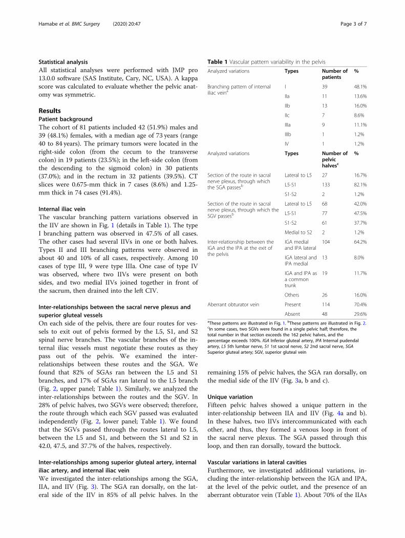
Internal iliac veinThe vascular branching pattern variations observed inthe IIV are shown in Fig. 1 (details in Table 1). The typeI branching pattern was observed in 47.5% of all cases.The other cases had several IIVs in one or both halves.Types II and III branching patterns were observed inabout 40 and 10% of all cases, respectively. Among 10cases of type III, 9 were type IIIa. One case of type IVwas observed, where two IIVs were present on bothsides, and two medial IIVs joined together in front ofthe sacrum, then drained into the left CIV.
Inter-relationships between the sacral nerve plexus andsuperior gluteal vesselsOn each side of the pelvis, there are four routes for ves-sels to exit out of pelvis formed by the L5, S1, and S2spinal nerve branches. The vascular branches of the in-ternal iliac vessels must negotiate these routes as theypass out of the pelvis. We examined the inter-relationships between these routes and the SGA. Wefound that 82% of SGAs ran between the L5 and S1branches, and 17% of SGAs ran lateral to the L5 branch(Fig. 2, upper panel; Table 1). Similarly, we analyzed theinter-relationships between the routes and the SGV. In28% of pelvic halves, two SGVs were observed; therefore,the route through which each SGV passed was evaluatedindependently (Fig. 2, lower panel; Table 1). We foundthat the SGVs passed through the routes lateral to L5,between the L5 and S1, and between the S1 and S2 in42.0, 47.5, and 37.7% of the halves, respectively.
Inter-relationships among superior gluteal artery, internaliliac artery, and internal iliac veinWe investigated the inter-relationships among the SGA,IIA, and IIV (Fig. 3). The SGA ran dorsally, on the lat-eral side of the IIV in 85% of all pelvic halves. In the
remaining 15% of pelvic halves, the SGA ran dorsally, onthe medial side of the IIV (Fig. 3a, b and c).
Unique variationFifteen pelvic halves showed a unique pattern in theinter-relationship between IIA and IIV (Fig. 4a and b).In these halves, two IIVs intercommunicated with eachother, and thus, they formed a venous loop in front ofthe sacral nerve plexus. The SGA passed through thisloop, and then ran dorsally, toward the buttock.
Vascular variations in lateral cavitiesFurthermore, we investigated additional variations, in-cluding the inter-relationship between the IGA and IPA,at the level of the pelvic outlet, and the presence of anaberrant obturator vein (Table 1). About 70% of the IIAs
Table 1 Vascular pattern variability in the pelvis
Analyzed variations Types Number ofpatients
%
Branching pattern of internaliliac veina
I 39 48.1%
IIa 11 13.6%
IIb 13 16.0%
IIc 7 8.6%
IIIa 9 11.1%
IIIb 1 1.2%
IV 1 1.2%
Analyzed variations Types Number ofpelvichalvesc
%
Section of the route in sacralnerve plexus, through whichthe SGA passesb
Lateral to L5 27 16.7%
L5-S1 133 82.1%
S1-S2 2 1.2%
Section of the route in sacralnerve plexus, through which theSGV passesb
Lateral to L5 68 42.0%
L5-S1 77 47.5%
S1-S2 61 37.7%
Medial to S2 2 1.2%
Inter-relationship between theIGA and the IPA at the exit ofthe pelvis
IGA medialand IPA lateral
104 64.2%
IGA lateral andIPA medial
13 8.0%
IGA and IPA asa commontrunk
19 11.7%
Others 26 16.0%
Aberrant obturator vein Present 114 70.4%
Absent 48 29.6%aThese patterns are illustrated in Fig. 1. bThese patterns are illustrated in Fig. 2.cIn some cases, two SGVs were found in a single pelvic half; therefore, thetotal number in that section exceeds the 162 pelvic halves, and thepercentage exceeds 100%. IGA Inferior gluteal artery, IPA Internal pudendalartery, L5 5th lumbar nerve, S1 1st sacral nerve, S2 2nd sacral nerve, SGASuperior gluteal artery; SGV, superior gluteal vein
Hamabe et al. BMC Surgery (2020) 20:47 Page 3 of 7

Fig. 2 Variations in inter-relationships between the spinal nerves and the superior gluteal artery and vein. (Upper panel) Patterns of the superiorgluteal artery (SGA). (Lower panel) Patterns of the superior gluteal vein (SGV). IIA, internal iliac artery IIV, internal iliac vein; L5, 5th lumbar nerve;S1, 1st sacral nerve; S2, 2nd sacral nerve
Fig. 3 Inter-relationships among the superior gluteal artery (SGA) and the internal iliac artery and vein. (a) The SGA (red) runs dorsal, on thelateral side of the internal iliac vein (blue). (b) The SGA runs dorsal, on the medial side of the internal iliac vein. (c) Representative photograph ofthe right side of the pelvic cavity, which shows the pattern described in (b). Photograph was acquired during laparoscopic surgery for a laterallymph node dissection. Asterisk (*) represents the internal iliac vein. IIA, internal iliac artery IIV, internal iliac vein; L5, 5th lumbar nerve; S1, 1stsacral nerve; S2, 2nd sacral nerve; SGA, superior gluteal artery
Hamabe et al. BMC Surgery (2020) 20:47 Page 4 of 7

divided into IPA and IGA branches in the pelvic cavity.In 12% of the pelvic halves, the IIAs passed out of pelvisas a common trunk. In pelvic halves where IPA and IGAwere divided in pelvic cavity, the IGA exited the pelvison the medial side more frequently than on the lateralside of the IPA. The remaining cases had other types ofIIA divisions; for example, sometimes, the IGA branchedoff the SGA outside the pelvis and ran toward the but-tock. An aberrant obturator vein was present in 70% ofpelvic halves.
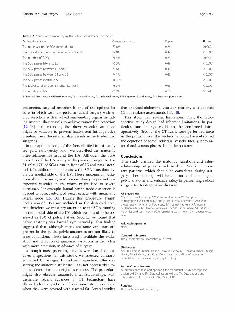
Anatomic symmetry in the lateral pelvic cavitiesThe rate of pelvic anatomic symmetry, or the concord-ance rate of pelvic anatomies, was analyzed for the fol-lowing variations; the route that the SGA passedthrough; a SGA that ran dorsally, on the medial side ofthe IIV; the number of SGVs; the route that the SGVpassed through; the presence of an aberrant obturatorvein; and the number of IIVs (Table 2). We found thatall anatomical variations, other than the number of IIVs,were symmetric.
DiscussionThis study revealed details about the inter-relationshipsamong pelvic vessels and their anatomic variations inpelvic cavity. Furthermore, several unique anatomical
variations were observed in a few cases. This study wasthe first to elucidate these patterns, and our findingshave important implications for surgeons.Since Adachi first classified the IIA branching varia-
tions into five types [3], several studies have investigatedthe distributions of branching patterns in different popu-lations [4–6]. Although those studies analyzed arterialvariations in detail, they only assessed the IIA, independ-ent of other vessels. Information other than arterial vari-ations have not been available, due to the scarcity ofstudies that investigated vein, muscle, or nerve anat-omies. Therefore, the present study addressed the unmetneed for a comprehensive analysis of pelvic vascularanatomy, including anatomic inter-relationships andvariations.Current textbooks on human anatomy illustrate intra-
pelvic vessels and their inter-relationships as invariant[10, 11]. Typically, the SGA branches off the IIA, runsposteriorly, on the lateral side of the IIV, passes betweenthe L5 and S1 nerves, and runs toward the buttock. Inaddition, the IIV branches off the CIV, one on each side.Many surgeons probably consider this configuration tobe typical pelvic anatomy, and they might implementsurgery based on that assumption. However, the presentstudy demonstrated that not all pelvic vessels form thosestereotypic patterns. In locally recurrent rectal cancer
Fig. 4 A unique variation of the inter-relationship between the internal iliac artery (IIA) and vein (IIV). (a) The schematic figure illustrates thispattern. (b) Representative intra-operative photograph shows this pattern on the right side of the pelvic cavity. Photograph was acquired duringlaparoscopic surgery for a lateral lymph node dissection. Asterisk (*) represents the medial internal iliac vein (IIV); dagger (†) represents the lateralIIV; double dagger (‡) represents the internal iliac artery (IIA); hash sign (#) represents the venous loop between the two IIVs, which has formed infront of the sacral nerve plexus; and the section sign (§) represents the external iliac vein. IIA, internal iliac artery IIV, internal iliac vein; L5, 5thlumbar nerve; S1, 1st sacral nerve; S2, 2nd sacral nerve; SGA, superior gluteal artery
Hamabe et al. BMC Surgery (2020) 20:47 Page 5 of 7
treatments, surgical resection is one of the options forcure, in which we must perform radical surgery with enbloc resection with involved surrounding organs includ-ing internal iliac vessels to achieve tumor-free resection[12–14]. Understanding the above vascular variationsmight be valuable to prevent inadvertent intraoperativebleeding from the internal iliac vessels in such advancedsurgeries.In our opinion, some of the facts clarified in this study
are quite noteworthy. First, we described the anatomicinter-relationships around the IIA. Although the SGAbranches off the IIA and typically passes through the L5-S1 split, 17% of SGAs run in front of L5 and pass lateralto L5. In addition, in some cases, the SGA runs dorsally,on the medial side of the IIV. These uncommon varia-tions should be recognized preoperatively to prevent un-expected vascular injury, which might lead to severeoutcomes. For example, lateral lymph node dissection isneeded to resect advanced rectal cancer with metastaticlateral node [15, 16]. During this procedure, lymphnodes around IIVs are included in the dissected area,and therefore we must pay attention to the SGA runningon the medial side of the IIV which was found to be ob-served in 15% of pelvic halves. Second, we found thatpelvic anatomy was formed symmetrically. This findingsuggested that, although many anatomic variations arepresent in the pelvis, pelvic anatomies are not likely toarise at random. These facts might facilitate the evalu-ation and detection of anatomic variations in the pelviswith more precision, in advance of surgery.Although most preceding studies were based on ca-
daver inspections, in this study, we assessed contrast-enhanced CT images. In cadaver inspection, after dis-secting the anatomic structures, it is not necessarily sim-ple to determine the original structure. The proceduremight also obscure anatomic inter-relationships. Fur-thermore, recent advances in CT technology haveallowed clear depictions of anatomic structures evenwhen they were covered with visceral fat. Several studies
that analyzed abdominal vascular anatomy also adoptedCT for making assessments [17, 18].This study had several limitations. First, the retro-
spective study design had inherent limitations. In par-ticular, our findings could not be confirmed intra-operatively. Second, the CT scans were performed oncein the portal phase; this technique could have obscuredthe depiction of some individual vessels. Ideally, both ar-terial and venous phases should be obtained.
ConclusionsThis study clarified the anatomic variations and inter-relationships of pelvic vessels in detail. We found somerare patterns, which should be considered during sur-gery. These findings will benefit our understanding ofpelvic anatomy and enhance safety in performing radicalsurgery for treating pelvic diseases.
AbbreviationsCIA: Common iliac artery; CIV: Common iliac vein; CT: Computedtomography; EIA: External iliac artery; EIV: External iliac vein; IGA: Inferiorgluteal artery; IIA: Internal iliac artery; IIV: Internal iliac vein; IPA: Internalpudendal artery; IVC: Inferior vena cava; L5: 5th lumbar nerve; S1: 1st sacralnerve; S2: 2nd sacral nerve; SGA: Superior gluteal artery; SGV: Superior glutealvein
AcknowledgementsNone.
Competing interestThe authors declare no conflict of interest.
DisclosuresAtsushi Hamabe, Takashi Harino, Takayuki Ogino, MD, Tsukasa Tanida, ShingoNoura, Shunji Morita, and Keizo Dono have no conflicts of interest orfinancial ties to disclosure regarding this study.
Authors’ contributionsAll authors have read and approved the manuscript. Study concept anddesign: AH, SN and SM. Data collection: AH and TH. Data analysis andinterpretation: AH, TH, TO, TT, SN, SM and KD.
FundingThis study received no funding.
Table 2 Anatomic symmetry in the lateral cavities of the pelvis
Analyzed variations Concordance rate Kappa P value
The route where the SGA passes through 77.8% 0.26 0.0069
SGA runs dorsally, on the medial side of the IIV 84.0% 0.39 < 0.0001
The number of SGVs 70.4% 0.28 0.0057
The SGV passes lateral to L5 75.3% 0.49 < 0.0001
The SGV passes between L5 and S1 71.6% 0.43 < 0.0001
The SGV passes between S1 and S2 74.1% 0.45 < 0.0001
The SGV passes medial to S2 100.0% 1 < 0.0001
The presence of an aberrant obturator vein 76.5% 0.45 < 0.0001
The number of IIVs 61.7% 0.13 0.1061
IIV Internal iliac vein, L5 5th lumbar nerve, S1 1st sacral nerve, S2 2nd sacral nerve, SGA Superior gluteal artery, SGV Superior gluteal vein
Hamabe et al. BMC Surgery (2020) 20:47 Page 6 of 7

Availability of data and materialsThe datasets used and/or analyzed during the current study are availablefrom the corresponding author on reasonable request.
Ethics approval and consent to participateAll procedures performed in studies involving human participants were inaccordance with the ethical standards of the institutional and/or nationalresearch committee and with the 1964 Declaration of Helsinki and its lateramendments or comparable ethical standards. This study was approved bythe institutional Ethics Committee in Toyonaka Municipal Hospital, and awritten informed consent was obtained from all individual participantsbefore surgery for collection and analysis of the data. This article does notcontain any studies with animals performed by any of the authors.
Consent for publicationNot applicable.
Competing interestsThe authors declare that they have no competing interests.
Received: 15 December 2019 Accepted: 5 March 2020
References1. Sora MC, Jilavu R, Matusz P. Computer aided three-dimensional
reconstruction and modeling of the pelvis, by using plastinated crosssections, as a powerful tool for morphological investigations. Surg RadiolAnat. 2012;34:731–6.
2. Hamabe A, Ito M. A three-dimensional pelvic model made with a three-dimensional printer: applications for laparoscopic surgery to treat rectalcancer. Tech Coloproctol. 2017;21:383–7.
3. Watanabe K, Shoja MM, Loukas M, et al. Buntaro Adachi (1865-1945):Japanese master of human anatomic variation. Clin Anat. 2012;25:957–60.
4. Braithwaite JL. Variations in origin of the parietal branches of the internaliliac artery. J Anat. 1952;86:423–30.
5. Roberts WH, Krishingner GL. Comparative study of human internal iliacartery based on Adachi classification. Anat Rec. 1967;158:191–6.
6. Sakthivelavan S, Aristotle S, Sivanandan A, et al. Variability in the branchingpattern of the internal iliac artery in Indian population and its clinicalimportance. Anat Res Int. 2014;2014:597103.
7. Krishnamurthy A, Nayak SR, Khan S, et al. Anomalous channels draining theinternal iliac veins: embryological and clinical significance. Rom J MorpholEmbryol. 2007;48:71–3.
8. Oto A, Akpinar E, Surucu HS, et al. Right internal iliac vein joining the leftcommon iliac vein: case report demonstrated by CT angiography. SurgRadiol Anat. 2003;25:339–41.
9. Lotz PR, Seeger JF. Normal variations in iliac venous anatomy. AJR Am JRoentgenol. 1982;138:735–8.
10. Netter F. Atlas of human anatomy. 7th ed. Amsterdam: Elsevier; 2018.11. Agur AMR, Dalley AF. Grant's Atlas of Anatomy. 13th ed. Philadelphia:
Lippincott Williams & Wilkins; 2012.12. Alberda WJ, Verhoef C, Schipper ME, et al. The importance of a minimal
tumor-free resection margin in locally recurrent rectal Cancer. Dis ColonRectum. 2015;58:677–85.
13. Mirnezami AH, Sagar PM, Kavanagh D, Witherspoon P, Lee P, Winter D.Clinical algorithms for the surgical management of locally recurrent rectalcancer. Dis Colon Rectum. 2010;53:1248–57.
14. Troja A, El-Sourani N, Abdou A, Antolovic D, Raab HR. Surgical options forlocally recurrent rectal cancer--review and update. Int J Colorectal Dis. 2015;30:1157–63.
15. Kim TG, Park W, Choi DH, et al. Factors associated with lateral pelvicrecurrence after curative resection following neoadjuvantchemoradiotherapy in rectal cancer patients. Int J Colorectal Dis. 2014;29:193–200.
16. Kusters M, Slater A, Muirhead R, et al. What to do with lateral nodal diseasein low locally advanced rectal Cancer? A call for further reflection andresearch. Dis Colon Rectum. 2017;60:577–85.
17. Ogino T, Takemasa I, Horitsugi G, et al. Preoperative evaluation of venousanatomy in laparoscopic complete mesocolic excision for right coloncancer. Ann Surg Oncol. 2014;21(Suppl 3):S429–35.
18. Hamabe A, Park S, Morita S, et al. Analysis of the vascular interrelationshipsamong the first Jejunal vein, the superior mesenteric artery, and the middlecolic artery. Ann Surg Oncol. 2018;25:1661–7.
Publisher’s NoteSpringer Nature remains neutral with regard to jurisdictional claims inpublished maps and institutional affiliations.
Hamabe et al. BMC Surgery (2020) 20:47 Page 7 of 7