![Diet-related factors in the conservation of kiwi (Apteryx mantelli) · Captive kiwi, brown teal (Anas chlorotis) and takahe (Porphyrio [Notornis] mantelli) had a greater diversity](https://static.fdocuments.in/doc/165x107/606907501ded6d024b41d79e/diet-related-factors-in-the-conservation-of-kiwi-apteryx-mantelli-captive-kiwi.jpg)
Analysis of an associated Cretoxyrhina mantelli dentition ... · Cretoxyrhina dentition while it...
18
Analysis of an associated Cretoxyrhina mantelli dentition from the Late Cretaceous (Smoky Hill Chalk, Late Coniacian) of western Kansas Jim Bourdon 1 and michael J. everhart 2 1. Nordica Drive, Croton-on-Hudson, New York – [email protected] 2. Sternberg Museum of Natural History, Fort Hays State University, Hays, Kansas [email protected] Fossil shark teeth are common but are usually represented by shed examples and are seldom in associated or articulated groups. Complete dentition reconstruction from isolated teeth cannot be certain, even when large quantities are available from a single location and horizon. The Smoky Hill Chalk (Late Cretaceous) of western Kansas, U.S.A. has yielded a number of associated, and sometimes articulated, tooth sets of Cretoxyrhina mantelli Agassiz. Teeth from articulated sets are rarely removed from matrix which limits the understanding of positional characteristics to a single perspective. In this paper, we analyze and described an associated set of disarticulated Cretoxyrhina teeth. These teeth were arranged and compared with other disarticulated associated sets, then compared with a known articulated tooth set; resulting in a multi-perspective Cretoxyrhina tooth set. This reconstruction provides characteristics that permit upper and lower lateroposterior teeth to be differentiated and raise questions regarding the number of anterior tooth positions present. Keywords: Cretoxyrhinidae, lamniform shark, Niobrara Formation t ransactions of the Kansas academy of science Vol. 114, no. 1-2 p. 15-32 (2011) introduction The Late Cretaceous Smoky Hill Chalk of western Kansas has produced the most complete specimens known of the large lamniform shark Cretoxyrhina mantelli, including articulated tooth sets and the calcified cartilage of cranial elements, jaws, vertebral centra and fins. While most shark fossils consist of isolated shed teeth, the associated and reasonably complete dentitions preserved within several of these “mummified” remains provide the data to study Cretoxyrhina in greater detail than most other fossil sharks. Here we present a detailed analysis of an associated, disarticulated tooth set from the Late Coniacian of Gove County, Kansas; supplemented with observations from other disarticulated tooth sets from the Smoky Hill Chalk. Cretoxyrhina was a cosmopolitan pelagic predator that is documented from the Albian to the Early Campanian in North America and which persisted even later in Europe (Siverson and Lindgren 2005). The presence of Cretoxyrhina mantelli is well documented (Shimada 1997f) during the deposition of the Smoky Hill Chalk (Late Coniacian-Early Campanian) in the Western Interior Seaway. The species is represented by; skeletons (Shimada 1997c; Shimada et al. 2006), associated tooth sets (Eastman 1894; Williston 1900; Siverson 1996; Shimada 1997b), interactions with other taxa (Shimada, 1997e; Everhart 1999; Shimada and Everhart 2004; Everhart 2004; Everhart 2005b; Everhart and Hamm 2005, and; Everhart and Ewell 2006) and isolated teeth (Shimada et al. 2006; Siverson and Lindgren 2005; Shimada 2008; and Shimada and Nagrodski 2010). Based on
Transcript of Analysis of an associated Cretoxyrhina mantelli dentition ... · Cretoxyrhina dentition while it...

Analysis of an associated Cretoxyrhina mantelli dentition from the Late Cretaceous (Smoky Hill Chalk, Late Coniacian) of western KansasJim Bourdon1 and michael J. everhart2
1. Nordica Drive, Croton-on-Hudson, New York – [email protected]. Sternberg Museum of Natural History, Fort Hays State University, Hays, Kansas [email protected]
Fossil shark teeth are common but are usually represented by shed examples and are seldom in associated or articulated groups. Complete dentition reconstruction from isolated teeth cannot be certain, even when large quantities are available from a single location and horizon. The Smoky Hill Chalk (Late Cretaceous) of western Kansas, U.S.A. has yielded a number of associated, and sometimes articulated, tooth sets of Cretoxyrhina mantelli Agassiz. Teeth from articulated sets are rarely removed from matrix which limits the understanding of positional characteristics to a single perspective. In this paper, we analyze and described an associated set of disarticulated Cretoxyrhina teeth. These teeth were arranged and compared with other disarticulated associated sets, then compared with a known articulated tooth set; resulting in a multi-perspective Cretoxyrhina tooth set. This reconstruction provides characteristics that permit upper and lower lateroposterior teeth to be differentiated and raise questions regarding the number of anterior tooth positions present.
Keywords: Cretoxyrhinidae, lamniform shark, Niobrara Formation
transactions of the Kansas academy of science
Vol. 114, no. 1-2 p. 15-32 (2011)
introduction
The Late Cretaceous Smoky Hill Chalk of western Kansas has produced the most complete specimens known of the large lamniform shark Cretoxyrhina mantelli, including articulated tooth sets and the calcified cartilage of cranial elements, jaws, vertebral centra and fins. While most shark fossils consist of isolated shed teeth, the associated and reasonably complete dentitions preserved within several of these “mummified” remains provide the data to study Cretoxyrhina in greater detail than most other fossil sharks. Here we present a detailed analysis of an associated, disarticulated tooth set from the Late Coniacian of Gove County, Kansas; supplemented with observations from other disarticulated tooth sets from the Smoky Hill Chalk.
Cretoxyrhina was a cosmopolitan pelagic predator that is documented from the Albian to the Early Campanian in North America and which persisted even later in Europe (Siverson and Lindgren 2005). The presence of Cretoxyrhina mantelli is well documented (Shimada 1997f) during the deposition of the Smoky Hill Chalk (Late Coniacian-Early Campanian) in the Western Interior Seaway. The species is represented by; skeletons (Shimada 1997c; Shimada et al. 2006), associated tooth sets (Eastman 1894; Williston 1900; Siverson 1996; Shimada 1997b), interactions with other taxa (Shimada, 1997e; Everhart 1999; Shimada and Everhart 2004; Everhart 2004; Everhart 2005b; Everhart and Hamm 2005, and; Everhart and Ewell 2006) and isolated teeth (Shimada et al. 2006; Siverson and Lindgren 2005; Shimada 2008; and Shimada and Nagrodski 2010). Based on

16 Bourdon and Everhart
the fossil evidence, Cretoxyrhina mantelli was an apex predator capable of feeding on large marine prey and scavenging floating carcasses of terrestrial dinosaurs.
The species was first described by Agassiz 1843 as Oxyrhina mantelli from the Late Cretaceous of England. Currently, the oldest reported occurrence is from the basal Upper Albian Gault Clay of England (Ward 2010). The youngest reports of the genus in Kansas are of Early Campanian age from the Smoky Hill Chalk Member of the Niobrara Formation (Stewart 1990; Shimada 1997f; Carpenter 2003; Everhart 2005a; Siverson and Lindgren 2005).
Stewart (1990) suggests that a general cooling of the Western Interior Sea led to its extirpation in North America, while Everhart (2005a) noted that the decline of Cretoxyrhina coincides with the rise of giant marine lizards called mosasaurs (e.g. Tylosaurus proriger) that would have competed with this large shark as an apex predator. Since Cretoxyrhina is also regarded as a pelagic shark (Shimada 1997f), the shallowing and narrowing of the seaway during the regressive phase of the Niobrara cyclothem may have eliminated its preferred environment within the Western Interior Sea, but that does not explain its worldwide extinction later in the Campanian.
In North America, the first documented report of Oxyrhina mantelli is by Gibbes (1849, p. 202, pl. 27, fig. 158) from the Cretaceous of Alabama. In 1867, John LeConte collected fossils in western Kansas while leading a railroad survey party. One of the specimens (ANSP 5399; “Lamna”) donated to the Academy of Natural Sciences of Philadelphia was among the first Cretoxyrhina teeth collected and documented from Kansas (Leidy 1873, pl.18, fig. 44). On the same plate, Leidy figured other “Lamna” teeth (figs. 21-25) collected by Dr. George M. Sternberg, and described Oxyrhina extenta (p. 302) as a broad-form tooth of the O. mantelli design from Kansas. Cope (1875, p. 296) included
this species from the “Niobrara epoch of Smoky Hill”. Eastman (1894) first described an associated Oxyrhina mantelli tooth set from the Smoky Hill Chalk (Upper Coniacian) of southeastern Gove County, Kansas. Although originally articulated (at least in part) when collected (see Sternberg 1900, 1906, 1909), Eastman removed all teeth from matrix and arranged them (pl. 16) following the mako dentition-design – he would also synonymize O. extenta with O. mantelli. Eastman’s perception of the dentition-design would go unchallenged in North America for a century. Partially-based on Russian platform material, Glikman (1958) erected the family Cretoxyrhinidae and genus Cretoxyrhina with C. mantelli as type species. Meyer (Unpub. PhD dissertation, 1974, p. 243-254) considered the Cretoxyrhina dentition-design similar to Isurus oxyrinchus and proposed two subspecies based on captured metrics – C. mantelli oxyrhinoides (Sauvage, 1870, Cenomanian-Turonian) and C. m. extenta (Leidy 1873; Santonian-Campanian).
Using radiographic examination, Shimada (1997b) documented an articulated Cretoxyrhina dentition while it was still in matrix. His paper provides single-perspective documentation of the teeth and their arrangement – one that shows a significant difference in dentition-design between Cretoxyrhina and Isurus. However, he could not provide multi-perspective images of each tooth. In our paper, we will use associated but disarticulated teeth in order to document the diagnostic details available when considering characteristics displayed by different perspectives.
stratigraphy and depositional environment
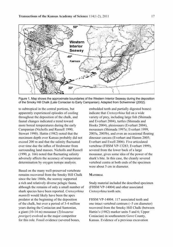
The Smoky Hill Chalk Member of the Niobrara Formation was deposited on the eastern shelf of the Western Interior Seaway from the Late Coniacian through the beginning of Early Campanian time (87-82 MA; Obradovich 1993). The seaway was warm temperate
Transactions of the Kansas Academy of Science 114(1-2), 2011 17
to subtropical in the central portions, but apparently experienced episodes of cooling throughout the deposition of the chalk, and faunal changes indicated a trend toward more boreal temperatures during the early Campanian (Nicholls and Russell 1990; Stewart 1990). Hattin (1982) noted that the maximum depth over Kansas probably did not exceed 200 m and that the salinity fluctuated over time due the influx of freshwater from surrounding land masses. Nicholls and Russell (1990, p. 166) noted that fluctuating salinity adversely affects the accuracy of temperature determination by oxygen isotope analysis.
Based on the many well-preserved vertebrate remains recovered from the Smoky Hill Chalk since the late 1860s, the seaway supported a rich and relatively diverse pelagic fauna, although the remains of only a small number of shark species have been reported. Cretoxyrhina mantelli would likely have been the apex predator at the beginning of the deposition of the chalk, but over a period of 3-4 million years during the Coniacian and Santonian, a giant (10-14 m) mosasaur (Tylosaurus proriger) evolved as the major competitor for this role. Fossil evidence (severed bones,
embedded teeth and partially digested bones) indicate that Cretoxyrhina fed on a wide variety of prey, including large fish (Shimada and Everhart 2004), turtles (Shimada and Hooks 2004), plesiosaurs (Everhart 2004), mosasaurs (Shimada 1997e; Everhart 1999, 2005a, 2005b), and even an occasional floating dinosaur carcass (Everhart and Hamm 2005; Everhart and Ewell 2006). Five articulated vertebrae (FHSM VP-13283; Everhart 1999), severed from the lower back of a large mosasaur, gives some idea of the power of the shark’s bite. In this case, the cleanly severed vertebral centra at both ends of the specimen were about 5 cm in diameter. material
Study material included the described specimen (FHSM VP-14004) and other associated Cretoxyrhina tooth sets.
FHSM VP-14004. 117 associated teeth and one intact vertebral centrum (~5 cm diameter) recovered from the Smoky Hill Chalk (between Hattin’s (1982) marker units 5 and 6; Upper Coniacian) in southeastern Gove County, Kansas. Evidence of a previous excavation
Figure 1, Map shows the approximate boundaries of the Western Interior Seaway during the deposition of the Smoky Hill Chalk (Late Coniacian to Early Campanian). Adapted from Schwimmer (2002).

18 Bourdon and Everhart
showed that a fairly complete shark specimen, including a portion of the dentition, had been hurriedly collected (poached). The disturbed matrix around the excavation was searched and screened for additional remains for two years following the discovery to collect additional teeth. The specimen (FHSM VP-14004) was placed in the Sternberg Museum collection in 2002. As a side note, the specimen included 10 Squalicorax “falcatus” teeth (FHSM VP-14005) that are assumed to represent scavenging on the Cretoxyrhina carcass. Two broken teeth that were mis-assembled in the field were digitally repaired for this paper.
To serve as a collateral source for comparison and contrasting, material from two private collections and a published tooth set were employed.
HC-02 - Hubbell collection (MP/KS-53506). An associated set of 100 teeth from the Smoky Hill Chalk, Gove/Logan Co., Kansas.
SC-01 - Swiatovy collection. An associated set of 217 (92 loose) teeth from the Smoky Hill Chalk (MU 2-3; Upper Coniacian), Martin’s Canyon, Southeaster Gove County, Kansas. Observations derived from single-perspective images provided by Ed Swiatovy.
MPM-01. During C.H. Sternberg’s 1890-92 collecting campaign on behalf of K.A. von Zittel (Sternberg 1900; 1909, 113), an associated set of 280 teeth was collected from Gove County, Kansas, placed in the collection of the Museum of Palaeontology in Munich (MPM), and described by Eastman (1894). The detailed illustrations of this material allowed for its comparative usage; the museum and its collections were destroyed the night of April
24-25th, 1944 by Allied bombing (Smith, et al., 2006). Eastman (1894, p. 157) noted that the teeth were removed from matrix and that 214 appeared articulated. Eastman deemed these teeth to follow a lamnid tooth-design (Oxyrhina gomphodon Müller & Henle 1839 = Isurus oxyrinchus Rafinesque 1810) and arranged them accordingly, but made no comments as to their articulated positioning. In rearranging them for this current study, three tooth-positions were deemed to be missing and included as silhouettes; one of these fell within the “comparative” group (lCP8; see below). This tooth set is particularly useful both as it is the set with the largest teeth (ontogenetic considerations) and by including the tooth that most favorably compares with Agassiz’s (1843, pl. 33, fig 8) best specimen.
FHSM VP-2187 – This specimen of an articulated dentition, the subject of the Shimada (1997b) study of the Cretoxyrhina dentition, was not directly contrasted in our study; rather, Shimada’s conclusions on tooth arrangement were compared with our results. More in-depth positional comparisons between FHSM VP-2187 and FHSM VP-14004 are required in the future.
terminology
This paper follows the general tooth-design terminology of Cappetta (1987). Positional nomenclatures, as proposed by Shimada (1997b, as emended 2002) and Siverson (1999) are largely different and would add a level of subjective determinations above the underlying tooth-positions. Rather than choose between the two for descriptive purposes, teeth will simply be numbered one-up.
Table 1. Positional nomenclature applied in this paper compared to that used by Siverson (1999) and Shimada (2002) for various comparative positions.
Transactions of the Kansas Academy of Science 114(1-2), 2011 19
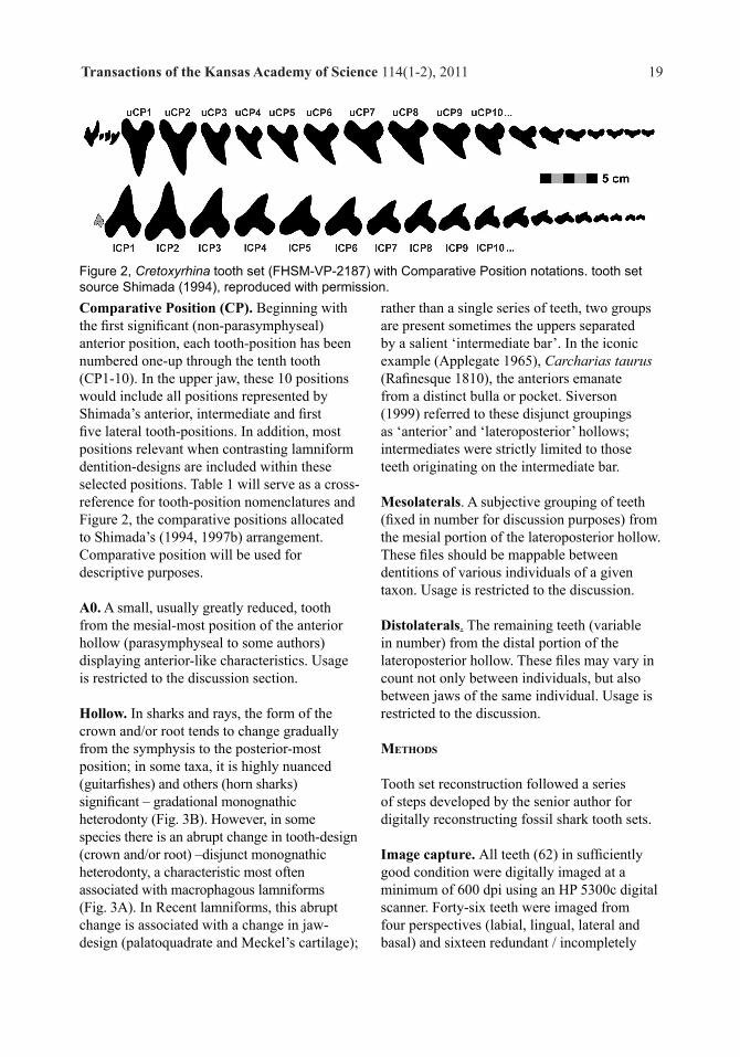
Comparative Position (CP). Beginning with the first significant (non-parasymphyseal) anterior position, each tooth-position has been numbered one-up through the tenth tooth (CP1-10). In the upper jaw, these 10 positions would include all positions represented by Shimada’s anterior, intermediate and first five lateral tooth-positions. In addition, most positions relevant when contrasting lamniform dentition-designs are included within these selected positions. Table 1 will serve as a cross-reference for tooth-position nomenclatures and Figure 2, the comparative positions allocated to Shimada’s (1994, 1997b) arrangement. Comparative position will be used for descriptive purposes.
A0. A small, usually greatly reduced, tooth from the mesial-most position of the anterior hollow (parasymphyseal to some authors) displaying anterior-like characteristics. Usage is restricted to the discussion section.
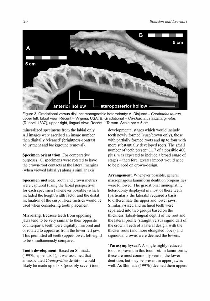
Hollow. In sharks and rays, the form of the crown and/or root tends to change gradually from the symphysis to the posterior-most position; in some taxa, it is highly nuanced (guitarfishes) and others (horn sharks) significant – gradational monognathic heterodonty (Fig. 3B). However, in some species there is an abrupt change in tooth-design (crown and/or root) –disjunct monognathic heterodonty, a characteristic most often associated with macrophagous lamniforms (Fig. 3A). In Recent lamniforms, this abrupt change is associated with a change in jaw-design (palatoquadrate and Meckel’s cartilage);
rather than a single series of teeth, two groups are present sometimes the uppers separated by a salient ‘intermediate bar’. In the iconic example (Applegate 1965), Carcharias taurus (Rafinesque 1810), the anteriors emanate from a distinct bulla or pocket. Siverson (1999) referred to these disjunct groupings as ‘anterior’ and ‘lateroposterior’ hollows; intermediates were strictly limited to those teeth originating on the intermediate bar.
Mesolaterals. A subjective grouping of teeth (fixed in number for discussion purposes) from the mesial portion of the lateroposterior hollow. These files should be mappable between dentitions of various individuals of a given taxon. Usage is restricted to the discussion.
Distolaterals. The remaining teeth (variable in number) from the distal portion of the lateroposterior hollow. These files may vary in count not only between individuals, but also between jaws of the same individual. Usage is restricted to the discussion.
methods
Tooth set reconstruction followed a series of steps developed by the senior author for digitally reconstructing fossil shark tooth sets.
Image capture. All teeth (62) in sufficiently good condition were digitally imaged at a minimum of 600 dpi using an HP 5300c digital scanner. Forty-six teeth were imaged from four perspectives (labial, lingual, lateral and basal) and sixteen redundant / incompletely
Figure 2, Cretoxyrhina tooth set (FHSM-VP-2187) with Comparative Position notations. tooth set source Shimada (1994), reproduced with permission.
20 Bourdon and Everhart
mineralized specimens from the labial only. All images were ascribed an image number then digitally ‘cleaned’ (brightness-contrast adjustment and background removal).
Specimen orientation. For comparative purposes, all specimens were rotated to have the crown-root contacts at the lateral margins (when viewed labially) along a similar axis.
Specimen metrics. Tooth and crown metrics were captured (using the labial perspective) for each specimen (whenever possible) which included the height/width factor and the distal inclination of the cusp. These metrics would be used when considering tooth placement.
Mirroring. Because teeth from opposing jaws tend to be very similar to their opposite counterparts, teeth were digitally mirrored and or rotated to appear as from the lower left jaw. This permitted all teeth (upper-lower, left-right) to be simultaneously compared.
Tooth development. Based on Shimada (1997b; appendix 1), it was assumed that an associated Cretoxyrhina dentition would likely be made up of six (possibly seven) tooth
developmental stages which would include teeth newly formed (cusp/crown only), those with partially formed roots and up to four with more substantially developed roots. The small number of teeth present (117 of a possible 400 plus) was expected to include a broad range of stages – therefore, greater import would need to be placed on crown-design.
Arrangement. Whenever possible, general macrophagous lamniform dentition propensities were followed. The gradational monognathic heterodonty displayed in most of these teeth (particularly the laterals) required a basis to differentiate the upper and lower jaws. Similarly-sized and inclined teeth were separated into two groups based on the thickness (labial-lingual depth) of the root and the lateral profile (straight versus sigmoidal) of the crown. Teeth of a lateral design, with the thicker roots (and more elongated lobes) and sigmoidal crowns were deemed the lowers.
‘Parasymphyseal’. A single highly reduced tooth is present in this tooth set. In lamniforms, these are most commonly seen in the lower dentition, but may be present in upper jaw as well. As Shimada (1997b) deemed them uppers
Figure 3, Gradational versus disjunct monognathic heterodonty: A. Disjunct – Carcharias taurus, upper left, labial view, Recent – Virginia, USA; B. Gradational – Carcharhinus albimarginatus (Rüppell 1837), upper right, lingual view, Recent – Taiwan. Scale bar = 5 cm.

Transactions of the Kansas Academy of Science 114(1-2), 2011 21
based on an articulated dentition, this paper will follow that positional determination.
The Reconstructed tooth sets.The 4-perspective study-set (FHSM VP-14004) was first arranged on the basis of labial tooth-shape and basal profile; uppers and lowers were segregated using lateral profile. The set with 3-perspective images (HC-02) was also arranged on a similar basis and the two sets then harmonized. The collateral set (SC-01) fortunately included articulated teeth from the first two anterior positions (Fig. 4), interpreted as lowers, which permitted these first two positions (lCP1-2) to be identified and differentiated from their upper counterparts. The final collateral set (MPM-01) was then matched to the first three arrangements. The resulting arrangements (Fig. 5) show a broad consistency in uCP1-10 and lCP1-10 positional-designs between the four sets of teeth, both uppers and lowers. Teeth are primarily of a cutting design, but the large number of teeth per file (as noted by Shimada 1997b) and thick roots suggest the anteriors may have served a clutching purpose as well (more than one functional row); anteriors with dignathic and laterals largely displaying gradational monognathic heterodonty. In profile, the crowns of lower lateral teeth are thicker and the lobes more labially extended, relative to their upper counterparts. The upper tooth series is markedly different from the lower in the CP3-5 positions, wherein the
uppers are significantly reduced and the lowers more evenly-sized. The first position (CP1) is relatively upright while remaining positions are distally inclined to varying degrees.
Positional Descriptions.As included below, the upper and lower positions are described using the FHSM VP-14004 arrangement (Fig. 6) and supplemented with observations from the collateral tooth sets. The general design includes: a somewhat narrow subtriangular cusp (less narrow and more triangular in HC-02) with a complete cutting edge and smooth enameloid, the neck is moderately wide, the basal labial face is broadly V-shaped and weakly extends beyond the root medially. In profile, the lingual face is convex and the labial, nearly flat to weakly convex; the crown of the lowers tends to be thicker than the uppers, and the labial root face more labially directed in the lowers. The roots are bilobate, becoming more splayed distally; lobes are broad and rounded and the moderate lingual protuberance lacks a nutritive groove and bears a single foramen. From a basal
Figure 4, Cretoxyrhina mantelli, lower positions CP1-4, SC-01, all specimens similarly scaled: A-B articulated anteriors: A. right lCP1), labial view; B. right lCP1, left lCP1, left lCP2 and right lCP2 positions in matrix; C-F isolated right-hand positions 1-4, lingual perspective: C. right lCP1; D. right lCP2; E. right lCP3; F. right lCP4. Scale bar = 3 cm.
FACING PAGE: Figure 5, Cretoxyrhina mantelli, comparative tooth sets, all specimen files individually scaled relative to Figure D: A, A1 MPM-01, labial view, x .73; A, upper files, A1, lower files; B, B1, HC-02, lingual view, x.88; B, upper files, B1, lower files; C, C1, SC-01, lingual view, x.92; C, upper files, C1, lower files; D, D1, FHSM VP-14004, labial view, D, upper files, D1, lower files. Scale bar = 5 cm.
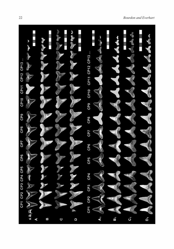
22 Bourdon and Everhart
Transactions of the Kansas Academy of Science 114(1-2), 2011 23

24 Bourdon and Everhart
perspective, lower teeth have thicker roots than their upper counterparts. A single reduced tooth in FHSM VP-14004 was assigned to the upper jaw following Shimada (1994, 1997b) and could be interpreted as a reduced A0 or parasymphyseal; because they are not present in two of the tooth sets (HC-02 and SC-01) and represented by two or three positions in MPM-01, they have not been included as a comparative position.
Position CP1. This position (and CP2) is clearly an anterior and would be considered such by both Shimada (1997b; 2002) and Siverson (pers. comm. 2008). The CP1 is erect or slightly inclined (usually mesially), the distal edge may be mesially curved, particularly in the uppers and there are no distinct shoulders. In profile, the FHSM VP-14004 crown is relatively straight in both jaws (Fig. 7D,J); in the HC-01 examples, the upper is labially curved and the lower lingually curved (Fig. 7A,G). The root is thick, more so in the lower; the lower CP1 has higher less splayed lobes when compared with the upper.
Of all the positions in the comparative tooth sets (Fig. 5), the CP1 is the least consistent in cusp orientation. The proposed arrangements placed priority in the root shape over cusp inclination and shape (Fig. 7A,D,G,J). This is the authors’ hypothesis and can only be substantiated with multiple articulated tooth sets with anteriors removed from matrix.
Position CP2. The CP2 is higher and broader than the CP1. The upper CP2 cusp is more distally inclined and mesially recurved than the lower. The lower CP2 tends to be the tallest tooth with the thickest root in the jaw. Uppers and lowers both lack mesial shoulders, but display weak distal shoulders. In FHSM VP-14004, the upper has higher, more angular lobes than the lower; however, this likely is attributable to the worn / not fully-developed condition of the specimen’s root; in profile, the upper is straight and the lower weakly sigmoidal (Fig. 7E,K). In HC-01, the upper is labially curved and the lower is relatively straight with a slight labial curvature of the tip (Fig. 7B,H).
Position CP3. This position is markedly different between upper and lower jaws although each is more inclined than their more-mesial counterparts. In the upper, the tooth is reduced in size (height and width), yet the root remains labiolingually thick. The mesial edge lacks a distinct shoulder but includes an obtuse distal one. In the lower jaw, the CP3 crown is slightly shorter in height and narrower relative to the CP2; shoulders are present on both edges, more salient on the distal. The root is broader overall and the mesially lobe more elongate than the distal. In profile, both uppers and lowers are relatively straight (Fig. 6E,F); in HC-02, there is a slight labial curvature of
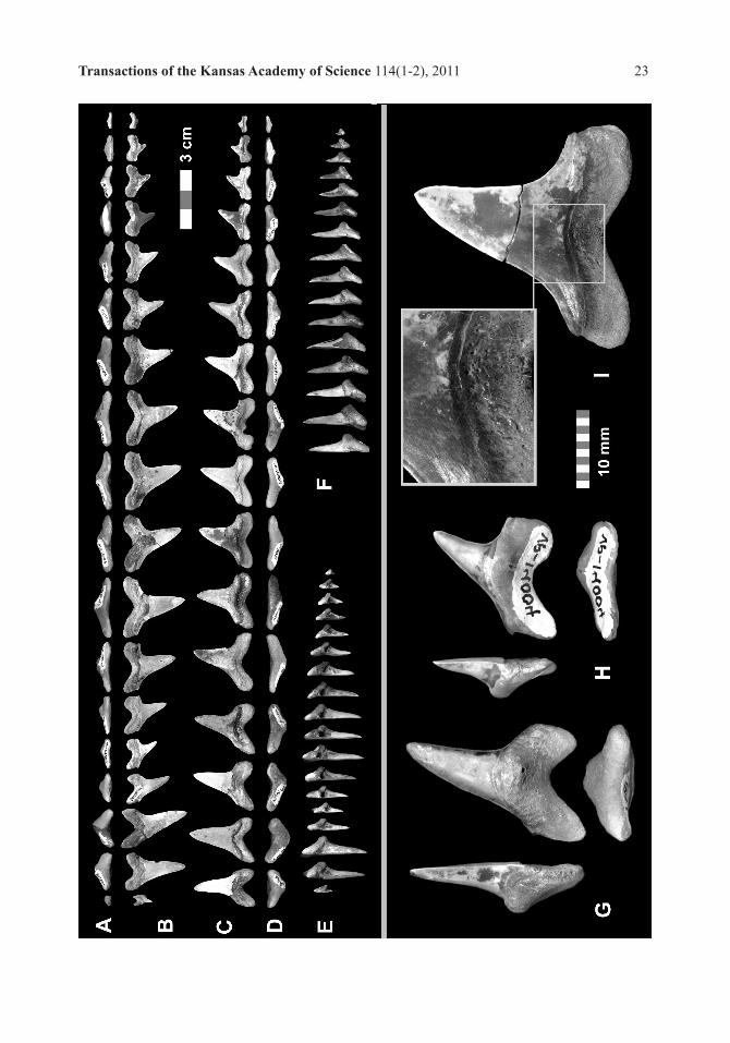
FACING PAGE - Figure 6, Cretoxyrhina mantelli, FHSM VP-14004, tooth set and examples, individually scaled by group: A-F, Tooth set, 3-perspectives, similarly scaled. Scale bar = 3 cm; A, Upper files, basal view; B, Upper files, labial view; C, Lower files, labial view; D, Lower files, basal view; E, Upper files, lateral view; F, Lower files, lateral view; G-H, Positional examples, 3-perspectives, similarly scaled, Scale bar = 10 cm; G, FHSM VP-14004.04, Upper CP3 (uA3), lateral, lingual and basal views, mirrored; H, FHSM VP-14004.05, Upper CP4 (uLP1), lateral, lingual and basal views, mirrored; I, FHSM VP-14004.42, Lower CP7 (lLP3), lingual with 2x enlargement.
RIGHT - Figure 7, Cretoxyrhina mantelli anterior positional comparisons, FHSM VP-14004 and HC-02, lingual, lateral and basal perspectives, all specimens presented as right-hand files and similarly-scaled. A-C, HC-02 upper anteriors; A, HC-02.1191.a, UA1; B, HC-02.1191 b, UA2; C, HC-02.1191.d, UA3; D-F, FHSM VP-14004 upper anteriors; D, FHSM VP-14004.02, UA1; E, FHSM VP-14004.03, UA2 (mirrored); F, FHSM VP-14004.04, UA3; G-I, HC-02 lower anteriors; G, HC-02.1192.a, LA1 (mirrored); H, HC-02.1192.b, LA2; I, HC-02.1191.c, LA4 (mirrored); J-L, FHSM VP-14004 lower anteriors; J, FHSM VP-14004.36. LA1 (mirrored); K, FHSM VP-14004.37, LA2; and L, FHSM VP-14004.39, LA4 (mirrored). Scale bar = 3 cm.

Transactions of the Kansas Academy of Science 114(1-2), 2011 25
26 Bourdon and Everhart
the tip (Fig. 7C). The relative cusp height of the FHSM VP-14004 upper CP3 (Fig. 7F) is significantly higher than those of the other tooth sets (i.e., HC-02, Fig. 7C).
Position CP4. The upper CP4 is the most reduced Comparative Position of the upper jaw until the CP13 position (posterior); the narrow cusp differentiates it from posterior positions. There is a hint of a mesial shoulder and a well-developed distal. The root is labiolingually compressed and the mesial lobe is elongate and compressed. In contrast, the lower CP4 is nearly as high and the root as thick as the lower CP3. Both shoulders are present, the distal less obtuse than the mesial. The mesial cusp edge is straight and the distal has a slight mesial curvature. In profile, CP4s from both tooth sets are relatively straight (Fig. 7L) with the exception of the lower CP4 form HC-02 which is lingually directed. (Fig. 7I)
Position CP5. The upper CP5 reflects an increase in size within this jaw; a mesial shoulder begins to develop and the distal is well-formed. Noteworthy is the asymmetrical root from a basal perspective (Fig. 6A); the mesial lobe is elongate and labiolingually compressed. The lower CP5 is quite different from the preceding lower position: the cusp is slightly higher (slightly lower in HC-02), the mesial edge broadly curved rather than straight, the mesial shoulder long, the root lower, broadest in the jaw and the lobes splayed; from a basal perspective, the mesial lobe is labiolingually compressed. In profile, the crown is weakly sigmoidal except for the FHSM VP-14004 lower which is labially curved.
Position CP6. The upper tooth is higher than the preceding position; a mesial shoulder is now present, obtusely extending from the cusp; the distal is sharply defined. Viewed basally, the root is asymmetrical but the mesial lobe is not as compressed or elongate as the CP5. The lower CP6 differs in a few respects from the CP5. It is a slightly smaller tooth, more erect
than adjacent positions and the distal edge may be mesially curved. Viewed basally, the root is rather symmetrical and the mesial lobe is not compressed labiolingually. In profile, FHSM VP-14004.41 (lower CP6) is weakly sigmoidal; in contrast, the HC-02 upper has a weak labial and the lower a slight lingual, curvature.
Position CP7. In the upper jaw, this is the largest lateral tooth; the cusp is more erect than preceding and succeeding files, the shoulders long and low; the root is high but the lobes still obtusely splayed and from a basal perspective, approaches symmetrical. The lower CP7 is smaller than the CP6 and returns to the cusp inclination of the CP5 (no mesial recurvature). Two elongate shoulders are present; the root is low and splayed. FHSM VP-14004 teeth have a weak sigmoidal profile, more so in the lower; in HC-02, both teeth are very straight.
Position uCP8-17. The upper CP8 is similar to the CP7 except that the cusp is slightly lower and more inclined, the mesial shoulder merges more obtusely, the root is lower and when viewed basally, more symmetrical. Succeeding teeth (CP9-17) follow this trend as teeth become smaller, cusps are lower and narrower, mesial shoulder more obtuse and root lower – there is little change in cusp inclination into distolateral positions
Position lCP8-16. The lower CP8 is a smaller version of the CP7, except that the cusp is slightly lower, narrower and more inclined; the mesial shoulder merges more obtusely and the mesial edge is straighter. Succeeding teeth (CP9-16) follow this trend as teeth continue to become smaller and cusps and roots lower.
discussion
The dentition-design resulting from this collection of positional designs is not comparable with taxa other than that described for Cretoxyrhina by Shimada (1997b). Siverson (1999) argued that macrophagous lamniforms needed to be evaluated on the basis

Transactions of the Kansas Academy of Science 114(1-2), 2011 27
of the two hollows present in each side of the jaw, the anterior and lateroposterior; and that teeth be numbered accordingly. This paper follows Siverson’s nomenclature for discussion purposes; however, conclusions drawn by us from these specimens does not necessarily represent Siverson’s interpretation of the same data.
Hollow Determinations.Allocating upper and lower teeth to particular hollows is a critical aspect of Siverson (1999) positional nomenclature and the resulting usage of hollow tooth formulas for diagnostic purposes. His nomenclature relied on Recent dentitions and numbering positions from front to back in each hollow. In fossil associated tooth sets (articulated or not), the allocation of teeth to particular hollows of an extinct family can be subjective – often weighing conflicting propensities while considering the possibility of pathological conditions or the noise created by contaminants from another individual. Due to the gradational monognathic propensity of tooth hollows, the crown and particularly the root can provide evidence for certain groupings of teeth. The reliability of root design, particularly with associated tooth sets, is often lessened by lobes that are in varying stages of development or poorly preserved. One reviewer raised this issue when evaluating our interpretation of four lower anteriors, suggesting better images of better specimens were required. It is acknowledged that better specimens are always preferable, however the availability of and consistency between multiple tooth sets helps mitigate specimen condition.
Upper jaw. There is an abrupt change in tooth morphology between CP3 and CP4 (Fig. 6G,H): crown inclination, mesial and distal shoulder design, root height, root thickness and mesial lobe compression. In each case, the CP4 more closely compares with the CP5. Other than its small size, there are no odontological features that argue that the CP4 might be deemed an intermediate (sensu Applegate 1965), a tooth
position that may be present in odontaspidids and mitsukurinids. Therefore, in FHSM VP-14004 (Fig. 6), CP1-3 have been deemed upper anteriors (uA1-A3) and CP4-17, lateroposteriors (uLP1-14). As a point of clarification, the small CP3-5 positions emanate from the distal-most position of the anterior hollow or the mesial-most positions of the lateroposterior hollow; there is no evidence of a well-defined bar separating the two hollows as present in Carcharias or Odontaspis.
Lower jaw. The most distinct break in positional-design occurs between CP4 and CP5. The CP4 has a straight mesial edge and curved distal, while the CP5 has a straight distal and curved mesial. Secondly, the mesial shoulder is significantly longer on CP5 when compared with CP4. The root is higher and the lobes higher and more angular in CP4 while lower and more obtusely splayed in CP5. Lastly, in basal view, the CP5 has a thinner, more elongate root and a labiolingually compressed mesial lobe. Because CP4 is more comparable with CP3 and CP5 with CP6, the former is deemed an anterior (A4) and the latter a lateroposterior (LP1). Therefore, in FHSM VP-14004 (Fig. 6) CP1-4 have been deemed anteriors (A1-A4) and CP5-16, lateroposteriors (lLP1-12). As noted earlier, one reviewer took exception to this interpretation (the presence of an A4), which should be viewed at this time as the authors’ hypothesis.
Dentition Characteristics - FHSM VP-14004.Three sets of characteristics can be determined based on the tooth set reconstruction, positional designs and hollow interpretations.Tooth Design. The generalized design includes: a crown with smooth enameloid, sub-triangular cusp that is distally inclined in all but the A1 positions, complete cutting edge, similarly-sized shoulders on lateral teeth with no accessory cusplets, and in cross-section, a convex lingual and nearly flat to weakly convex labial face. The neck is moderately broad, wider in anteriors; the basal labial
28 Bourdon and Everhart
margin, which is broadly V-shaped, extends beyond the root medially. The root is bilobate, high in anterior positions and lower with more splayed lobes in lateroposterior positions; lobes are broad and rounded. The protuberance is well-developed and lacks a nutritive groove; one somewhat small lingual foramen is present (Fig. 6G,H). The root, below the labial crown face, is perforated by multiple very small foramina (Fig. 6I).
From a lateral perspective, the crowns are relatively straight but may vary between slightly labially curved, lingually curved or sigmoidal; distolaterals tend to be lingually directed. The roots of the lateroposteriors are quite distinctive between jaws: in the uppers, the labial root face is on a plane similar to the labial face of the crown; however, in lowers, the root lobes are deflected, directed labial to the crown face (compare lateral profiles of Fig 6E and 6F).
Hollow Design. All Comparative Position teeth of VP-14004 (and the collateral tooth sets HC02, SC01 and MPM-01) can be attributed to one of the four lamniform hollows.
Upper Anteriors. Excluding a very small parasymphyseal or A0, three tooth-positions are present: a relatively erect and large A1, a larger, distally directed A2 and a small distally-inclined A3. The roots are relatively thicker than corresponding lateroposteriors and the A1 roots more mesiodistally compressed than the A2.
Upper Lateroposteriors. As seen in other macrophagous lamniforms and depending on individual or jaw, a non-fixed number of teeth may be present in this hollow. In VP-14004, a fixed number (7) were deemed mesolaterals (mL) and the remaining (7) distolaterals (dL). All lateroposteriors are distally inclined; mL1-3 increase in height and become more erect through the mL4 position; mL5-7 gradually decrease in height but maintain a similar cusp inclination. Beginning in the LP8 position, the distolaterals decrease in size at a greater rate.
The mL1-3 positions have elongate mesial lobes while other upper mesolaterals and distolaterals tend to have more symmetrical lobes. In lateral profile, the first seven lateroposterior positions have a straight to slightly recurved (labial) profile; beginning in the eighth position, the cusp begins to become lingually directed (weakly).
Lower Anteriors. Four large tooth-positions are interpreted as present in this hollow. The A1 is relatively erect with a slight mesial inclination in FHSM VP-14004.36 (Fig. 7J) and a somewhat mesiodistally compressed root when compared to the others. In other positions the mesial lobe is more elongate and the A3 and A4 position crowns similarly inclined. In fully-developed teeth, the lowers have longer lobes than corresponding uppers, the longest being in the A2 position. The A3 tends to have longer and less obtusely splayed lobes than the A4; the A3 and A4 crowns may be very similar in design, requiring the root to play an important role in position determination; an option not available with isolated specimens.
Lower Lateroposteriors. Similar to its upper counterpart, the lower hollow usually incorporates a non-fixed number of tooth files. In FHSM VP-14004, and for discussion purposes, the first seven have been deemed mesolaterals and the remaining five, distolaterals. The tallest crown is in the first position and those of succeeding teeth progressively lower. The first (mL1=lCP5) is distinguished by its labiolingually compressed mesial lobe. In the next position (mL2) the distal edge of the cusp is mesially curved. Remaining crowns are progressively shorter but similarly inclined. In lateral profile, the first seven positions have weakly sigmoidal profiles; remaining teeth become more lingually directed or lack the labial recurvature of the crown tip. Compared with the uppers, the base of the crown is thicker and the roots extend labially more than corresponding uppers.
Transactions of the Kansas Academy of Science 114(1-2), 2011 29
Dentition. Based on FHSM VP-14004, several conclusions can be drawn on dentition-design. Lacking an articulated specimen, combined root widths will serve as a surrogate for the hollow length; this introduces an error factor associated with possible presence of diastema between teeth (and hollows); in extant macrophagous lamniforms this would have little impact on anterior teeth and slightly more relevance in some (i.e. Odontaspis) laterals.
Upper vs. lower jaws. The upper tooth count (18, including the parasymphyseal / A0) outnumber the lower (16) but the upper’s cumulative root widths (37.1 cm) is nearly equal the lowers (37.2 cm). Using the first ten ‘comparative positions’ only, the uppers total 23.8 and lowers 26.5 cm. The small difference in full row totals between jaws suggests no appreciable diastema between upper hollows is likely present; however, the primary purpose is for contrasting this specimen with others.
Upper vs. lower anteriors. The most salient feature of this dentition is the significant difference between these two hollows. The combined width of the UA0-A3 (6.2 cm) differs substantially (65%) from the lower anteriors (LA1-LA4, 9.6 cm). One reviewer suggested that we included one too many lower anteriors, but we did not find sufficient evidence to limit that hollow to three teeth. In addition, the overall size of the lower anterior teeth is greater than the uppers. The thick roots of anterior teeth and numerous stages (up to seven, Shimada 1997b) of development suggest that more than one row was functional at a given time.
Upper vs. lower laterals. These two hollows are largely the same except for the slightly shorter mesiodistal length (18.2 vs. 16.9 cm; 93%) of the lower. The roots are sufficiently compressed (labiolingually) that a single tooth from each file was likely functional at a given time.
The evidence provided by VP-14004 suggests cutting-clutching anteriors and cutting laterals. The inordinate difference between upper and lower anterior hollows cannot be correlated with extant lamniforms.
conclusion
The overall dentition-design, as represented by these associated sets, is similar to that as reported by Shimada (1997b) for an articulated Cretoxyrhina set. This dentition is dissimilar to those of extant lamniforms and the interpreted presence of four lower anteriors would further differentiate the family. Monognathic heterodonty is well-expressed in the upper jaw of Cretoxyrhina mantelli while the lower might best be described as gradational. Dignathic heterodonty is strong in the anteriors but weak in most lateroposterior positions. Ontogenetic heterodonty is reflected in the shortening and broadening of the crown, relative to root width, as teeth get larger (Fig. 5). The oversized UA3 (Fig. 7F) and differences in lateral profiles between FHSM VP-14004 and HC-02 (Fig. 7) may represent sexual dimorphism or just individual variations.
acKnowledgments
Pat Young was kind enough to proof read and make suggestions for the text. We would also like to thank Mikael Siverson for his valuable comments regarding the tooth set reconstructions. Gordon Hubbell provided access to his collection to document his associated specimens. Ed Swiatovy provided images of his associated tooth set and René Kindlimann provided translations of Eastman’s archaic German.
Literature Cited
Agassiz, L. 1833-1845. Recherches sur les Poissons Fossiles, 5 volumes, and atlas 5 volumes, Imprimerie Petitpierre, Neuchatel, France, 1420 pp.

30 Bourdon and Everhart
Applegate, S.P. 1965. Tooth terminology and variation in sharks with special reference to the sand tiger, Carcharias taurus Rafinesque. Los Angeles County Museum Contributions in Science 86:1-18.
Cappetta, H. 1987. Chondrichthyes II. Mesozoic and Cenozoic Elasmobranchii. in Handbook of Paleoichthyologie, Volume 3b, Gustav Fischer Verleg, Stuttgart, 193 pp.
Carpenter, K. 2003. Vertebrate Biostratigraphy of the Smoky Hill Chalk (Niobrara Formation) and the Sharon Springs Member (Pierre Shale). pp. 421-437 In Harries, P. J. (Ed.), Approaches in High-Resolution Stratigraphic Paleontology, Kluwer Academic Publishers, The Netherlands.
Cope, E.D. 1875 Vertebrata of the Cretaceous Formations of the West. Report of the United States Geological Survey of the Territories, Vol. II. 302 pp.
Eastman, C. 1894. Beiträge zur Kenntniss der Gattung Oxyrhina, mit besonderer Berücksichtigung von Oxyrhina mantelli AGASSIZ. Palaeontographica, Band XLI, pp. 149-192, pls. xvi-xviii.
Everhart, M.J. 1999. Evidence of feeding on mosasaurs by the late Cretaceous lamniform shark, Cretoxyrhina mantelli. Journal of Vertebrate Paleontology 17(Supplement to 3):43A-44A.
Everhart, M.J. 2004. Late Cretaceous interaction between predators and prey. Evidence of feeding by two species of shark on a mosasaur. PalArch, Vertebrate Palaeontology 1(1):1-7.
Everhart, M.J. 2005a. Oceans of Kansas - A Natural History of the Western Interior Sea. Indiana University Press, 322 pp.
Everhart, M.J. 2005b. Bite marks on an elasmosaur (Sauropterygia; Plesiosauria) paddle from the Niobrara Chalk (Upper Cretaceous) as probable evidence of feeding by the lamniform shark, Cretoxyrhina mantelli. PalArch, Vertebrate Paleontology 2(2):14-24.
Everhart, M. J. and Ewell, K. 2006. Shark-bitten dinosaur (Hadrosauridae) vertebrae from the Niobrara Chalk (Upper Coniacian) of western Kansas. Kansas Academy of Science, Transactions, 109(1-2):27-35.
Everhart, M. J. and Hamm, S.A. 2005. A new nodosaur specimen (Dinosauria: Nodosauridae) from the Smoky Hill Chalk (Upper Cretaceous) of western Kansas. Kansas Academy of Science, Transactions 108(1-2):15-21.
Gibbes, R. 1849. Monograph of the fossil Squalidae of the United States. Journal of the Academy of Natural Sciences of Philadelphia, 1(3):191-206, pls 25-27.
Glikman, L. 1958. Rates of evolution in lamnoid sharks [in Russian]. Doklady Akademij Nauk SSSR 123:568-71.
Hattin, D. E. 1982. Stratigraphy and depositional environment of the Smoky Hill Chalk Member, Niobrara Chalk (Upper Cretaceous) of the type area, western Kansas. Kansas Geological Survey Bulletin 225, 108 pp.
Leidy, J. 1873 Contributions to the Extinct Vertebrate Fauna of the Western Territories. Report of the United States Geographical and Geological Survey of the Territories (Hayden) 1:1-358.
Meyer, R.L. 1974. Late Cretaceous elasmobranchs from the Mississippi and East Texas Embayments of the Gulf Coastal Plain. Unpublished PhD dissertation, Southern Methodist University, Dallas, xiv+419 p.
Müller, J. and Henle, J. 1838-1841, Systematische Beschreibung der Plagiostomen: Berlin [unpaginated].
Nicholls, E. L. and Russell, A.P. 1990. Paleobiogeography of the Cretaceous Western Interior Seaway of North America: the vertebrate evidence. Palaeogeography, Palaeoclimatology, Palaeoecology 79:149-169.
Obradovich, J.D. 1993. A Cretaceous time scale. pp. 379–396 in Caldwell, W.G.E. and Kaufmann, E.G. (eds.), Evolution of the Western Interior Basin, Geologic Association of Canada. Special Paper 39.

Transactions of the Kansas Academy of Science 114(1-2), 2011 31
Rafinesque, C.S. 1810. Caratteri di alcuni nuovi generi e nouvi spedie di animali e piante della Sicilia, con varie osservazione sopra i medesimi. Palermo, 105 pp.
Rüppell, W.P.E.S. 1837-1838 Neue Wirbelthiere zu der Faunavon von Abyssinien gehörig entdeckt und beschreiben von Dr. Eduard Ruppel. Fische des rothen Meeres. Frankfurt-am-Main. Pp. 1-11, 1-148, pls 1-33.
Sauvage, H.E. 1870. Recherches sur les poissons fossiles des terrains Crétacés de la Sarthe. Annales des Sciences Géologiques 2(7):1-44, pls. XVI, XVII, Paris.
Schwimmer, D.A. 2002. King of the Crocodylians - The Paleobiology of Deinosuchus. Indiana University Press, Bloomington, IN., 220 pp.
Shimada, K. 1994. Paleobiology of the Late Cretaceous shark, Cretoxyrhina mantelli (Lamniformes: Cretoxyrhinidae), from Kansas. Unpublished M.S. thesis, Fort Hays State University, Hays, Kansas, 169pp.
Shimada, K. 1997b. Dentition of the Late Cretaceous lamniform shark Cretoxyrhina mantelli, from the Niobrara Chalk of Kansas, Journal of Vertebrate Paleontology 17(2):269-279.
Shimada, K. 1997c. Skeletal anatomy of the Late Cretaceous lamniform shark, Cretoxyrhina mantelli, from the Niobrara Chalk in Kansas. Journal of Vertebrate Paleontology 17:642–652.
Shimada, K. 1997d. Gigantic lamnoid shark vertebra from the Lower Cretaceous Kiowa Shale of Kansas. Journal of Paleontology 7(13):522-524.
Shimada, K. 1997e. Paleoecological relationships of the Late Cretaceous lamniform shark, Cretoxyrhina mantelli (Agassiz), Journal of Paleontology 71(5):926-933
Shimada, K. 1997f. Stratigraphic record of the Late Cretaceous lamniform shark, Cretoxyrhina mantelli (Agassiz), in Kansas. Kansas Academy of Science, Transactions 100(3-4):139-149.
Shimada, K. 2002. Dental homologies in lamniform sharks (Chondrichthyes: Elasmobranchii). Journal of Morphology 251:38-72.
Shimada, K. 2008. Ontogenetic parameters and life history strategies of the Late Cretaceous Lamniform shark, Cretoxyrhina mantelli, base on vertebral growth increments. Journal of Vertebrate Paleontology 28(1):21–33.
Shimada, K., Cumbaa, S. L. and Van Rooyen, D. 2006. Caudal fin skeleton of the Late Cretaceous shark, Cretoxyrhina mantelli (Lamniformes: Cretoxyrhinidae) from the Niobrara Chalk of Kansas. Bulletin of the New Mexico Museum of Natural History 35:185-192.
Shimada, K. and Everhart, M.J. 2004. Shark-bitten Xiphactinus audax (Teleostei: Ichthyodectiformes) from the Niobrara Chalk (Upper Cretaceous) of Kansas. Mosasaur 7:35-39.
Shimada, K., and Hooks, G.E. III. 2004. Shark-bitten protostegid turtles from the Upper Cretaceous Mooreville Chalk, Alabama. Journal of Paleontology 78(1):205-210.
Shimada, K. and Nagrodski, M. 2010. Occurrence of the fossil lamniform shark, Cretoxyrhina mantelli, from the Upper Cretaceous Hartland Shale, central Kansas. Kansas Academy of Science, Transactions 113(3-4):235-236.
Shimada, K., Schumacher, B.A., Parkin, J.A. and Palermo, J.M. 2006. Fossil marine vertebrates from the lowermost Greenhorn Limestone (Upper Cretaceous: middle Cenomanian) in southeastern Colorado. Journal of Paleontology Memoir 63:1-45.
Siverson, M. 1996. Lamniform sharks of the mid-Cretaceous Alinga Formation and Beedagong Claystone, Western Australia. Palaeontology 39:813–849.
Siverson, M. 1999. A new large lamniform shark from the uppermost Gearle Siltstone (Cenomanian, Late Cretaceous) of Western Australia. Transactions of the Royal Society of Edinburgh, Earth Sciences 90:49-65.

32 Bourdon and Everhart
Siverson, M. and Lindgren, J. 2005. Late Cretaceous sharks Cretoxyrhina and Cardabiodon from Montana, USA. Acta Palaeontologica Polonica 50(2):301-314.
Smith, J.B., Lamana, M.C., Mayer, H. and Lacovaran, K.J. 2006. New information regarding the holotype of Spinosaurus aegypticus Stromer, 1915. Journal of Paleontology 80(2):400-406.
Sternberg, C.H. 1900. The sharks of Kansas. Popular Science News 34:38.
Sternberg, C.H. 1906. Some animals discovered in the fossil beds of Kansas. Kansas Academy of Science, Transactions 20:122-124.
Sternberg, C.H. 1909. The Life of a Fossil Hunter. Henry Holt and Company, 286 pp.
Stewart, J.D. 1990. Niobrara Formation vertebrate stratigraphy. pp. 19-30 in Bennett, S. C. (ed.), Niobrara Chalk Excursion Guidebook, The University of Kansas Museum of Natural History and the Kansas Geological Survey, Open-file Report 90-60.
Ward, D.J. 2010. Sharks and Rays. pp. 275-299 (Chapter 21) in Young, J.R., Gale, A.S., Knight, R.I. and Smith, A.B. (eds.), Fossils of the Gault Clay. Palaeontological Association Field Guide to Fossils 12, The Palaeontological Association, London.
Williston, S. 1900. Some fish teeth from the Kansas Cretaceous. Kansas University Quarterly 9(1):27-42, pl. VI-XIV.
Zhelezko, V. 2000. The evolution of the dental systems of the sharks of the genus Pseudoisurus GLIKMAN 1957 the largest Cretaceous pelagic sharks of Eurasia. in Chuvashov, B. (ed.), Material on stratigraphy and palaeontology of the Urals, Urals Branch Russian Academy of Science. Publication House, Ekaterinburg 4:136-141. (In Russian)