Analyses of intramolecular disulfide bonds in proteins by polyacrylamide gel electrophoresis...
9
ANALYTICAL BIOCHEMISTRY 168, 193-20 1 ( 1988) Analyses of Intramolecular Disulfide Bonds in Proteins by Polyacrylamide Gel Electrophoresis following Two-Step Alkylation’ MASAAKI HIROSE,~ NOBUYUKI TAKAHASHI, HIDEO OE, AND ETSUSHIRO DOI The Research Institute for Food Science, Kyoto University, Uji, Kyoto 61 I, Japan Received June 8, 1987 A method that makes use of polyacrylamide gel electrophoresis was developed for the analysis of intramolecular disulfide bonds in proteins. Proteins with different numbers of cleaved disul- fide bonds are alkylated with iodoacetic acid or iodoacetamide as the first step. The disulfide bonds remaining were reduced by excess dithiothreitol, and the newly generated free sulthydryl groups were alkylated with the reagent not yet used (iodoacetamide, iodoacetic acid, or vinyl- pyridine) as the second step. This treatment made it possible for lysozyme (M,, 14,ooO; 4 disulfides), the N-terminal half-molecule of conalbumin (M,, 36,000; 6 disulfides), the C-termi- nal half-molecule of conalbumin (Mr, 40,000; 9 disulfides), and whole conalbumin (M,, 78,000; 15 disulfides) to be separated by acid-urea polyactylamide gel electrophoresis into distinct bands depending on the number of disulfide bonds cleaved. The method allowed us to deter- mine the total number of disulfide bonds in native proteins and to assess the cleaved levels of disulfide bonds in partially reduced proteins. Two-step alkylation used in combination with radioautography was especially useful for the analysis of disulfide bonds in proteins synthesized in COmpkX biological SyStemS. Q 1988 Academic PBS, IIIC. KEY WORDS: protein gel electrophoresis; electrophoresis; protein disulfides; protein alkyla- tion; protein processing; protein structure. Established methods for the quantitative determination of protein disulfides, such as amino acid analyses and spectrophotometic methods (I), are not always suitable for stud- ies of biological translation systems, in which only limited amounts of tested proteins and a great number of nonspecific cellular com- ponents are included. Gel electrophoresis is one of the most useful techniques for protein analyses in such complex biological systems, since it requires very small amounts of pro- teins which can be visualized by selective methods of gel staining. However, an electro- phoretic method for the quantitative deter- mination of protein disulfides remained to be established. The pathways of intramolecular disulfide bond formation in chemically reduced im- munoglobulin light chain (2) ribonucleases (3,4), and trypsin inhibitors (5-7) have been analyzed by PAGE.3 The method used in ex- tensive studies from Creighton’s laboratory includes trapping by IAA or IAM of the free sulfhydryl groups in proteins (3-7). By PAGE, different intermediates formed dur- ing the refolding process can be resolved de- pending on differences in their net charge and conformation. This method is excellent for identifying the intermediate states quali- tatively and for following the kinetic behav- ior of the intermediates during the refolding. ’ This work was supported in part by a grant-in-aid for 3 Abbreviations used: PAGE, polyacrylamide gel elec- scientific research from The Ministry of Education, trophoresis; IAA, iodoacetic acid, IAM, iodoacetamide; Science and Culture of Japan. Hepes, 4-(2-hydroxyethyl)-I-piperazineethanesulfonic * To whom correspondence should be addressed. acid. 193 0003-2697188 $3.00 Copyright 0 1988 by Academic Press, Inc. All rights of reprcduction in any form reserved.
-
Upload
masaaki-hirose -
Category
Documents
-
view
212 -
download
0
Transcript of Analyses of intramolecular disulfide bonds in proteins by polyacrylamide gel electrophoresis...

ANALYTICAL BIOCHEMISTRY 168, 193-20 1 ( 1988)
Analyses of Intramolecular Disulfide Bonds in Proteins by Polyacrylamide Gel Electrophoresis following Two-Step Alkylation’
MASAAKI HIROSE,~ NOBUYUKI TAKAHASHI, HIDEO OE, AND ETSUSHIRO DOI
The Research Institute for Food Science, Kyoto University, Uji, Kyoto 61 I, Japan
Received June 8, 1987
A method that makes use of polyacrylamide gel electrophoresis was developed for the analysis of intramolecular disulfide bonds in proteins. Proteins with different numbers of cleaved disul- fide bonds are alkylated with iodoacetic acid or iodoacetamide as the first step. The disulfide bonds remaining were reduced by excess dithiothreitol, and the newly generated free sulthydryl groups were alkylated with the reagent not yet used (iodoacetamide, iodoacetic acid, or vinyl- pyridine) as the second step. This treatment made it possible for lysozyme (M,, 14,ooO; 4 disulfides), the N-terminal half-molecule of conalbumin (M,, 36,000; 6 disulfides), the C-termi- nal half-molecule of conalbumin (Mr, 40,000; 9 disulfides), and whole conalbumin (M,, 78,000; 15 disulfides) to be separated by acid-urea polyactylamide gel electrophoresis into distinct bands depending on the number of disulfide bonds cleaved. The method allowed us to deter- mine the total number of disulfide bonds in native proteins and to assess the cleaved levels of disulfide bonds in partially reduced proteins. Two-step alkylation used in combination with radioautography was especially useful for the analysis of disulfide bonds in proteins synthesized in COmpkX biological SyStemS. Q 1988 Academic PBS, IIIC.
KEY WORDS: protein gel electrophoresis; electrophoresis; protein disulfides; protein alkyla- tion; protein processing; protein structure.
Established methods for the quantitative determination of protein disulfides, such as amino acid analyses and spectrophotometic methods (I), are not always suitable for stud- ies of biological translation systems, in which only limited amounts of tested proteins and a great number of nonspecific cellular com- ponents are included. Gel electrophoresis is one of the most useful techniques for protein analyses in such complex biological systems, since it requires very small amounts of pro- teins which can be visualized by selective methods of gel staining. However, an electro- phoretic method for the quantitative deter- mination of protein disulfides remained to be established.
The pathways of intramolecular disulfide bond formation in chemically reduced im- munoglobulin light chain (2) ribonucleases (3,4), and trypsin inhibitors (5-7) have been analyzed by PAGE.3 The method used in ex- tensive studies from Creighton’s laboratory includes trapping by IAA or IAM of the free sulfhydryl groups in proteins (3-7). By PAGE, different intermediates formed dur- ing the refolding process can be resolved de- pending on differences in their net charge and conformation. This method is excellent for identifying the intermediate states quali- tatively and for following the kinetic behav- ior of the intermediates during the refolding.
’ This work was supported in part by a grant-in-aid for 3 Abbreviations used: PAGE, polyacrylamide gel elec- scientific research from The Ministry of Education, trophoresis; IAA, iodoacetic acid, IAM, iodoacetamide; Science and Culture of Japan. Hepes, 4-(2-hydroxyethyl)-I-piperazineethanesulfonic
* To whom correspondence should be addressed. acid.
193 0003-2697188 $3.00 Copyright 0 1988 by Academic Press, Inc. All rights of reprcduction in any form reserved.

194 HIROSE ET AL.
In the present study, the effects of protein conformation were minimized by two-step alkylation so that the intramolecular disul- fide bonds in proteins could be quantita- tively determined. The first alkylation in- cluded trapping by IAA or IAM of the free sulthydryl groups in the proteins tested. In the second step, the disulfides remaining were fully reduced by an excess of dithio- threitol, and then the newly generated free sulthydryl groups were blocked by another alkylation reagent. We report that by subse- quent acid-urea PAGE, at least with tested proteins, intramolecular disulfide bonds can be quantitatively analyzed in complex bio- synthesis systems as well as in purified pro- tein systems.
MATERIALS AND METHODS
Materials. Egg-white lysozyme was pur- chased from Sigma. Conalbumin was puri- fied from fresh egg white as described before (8). The N-terminal and C-terminal half- molecules of conalbumin were prepared by treatment of iron-saturated conalbumin with trypsin (manuscript in preparation). L-[35S]- Methionine (1340 Ci/mmol) was purchased from Amersham. Anti-conalbumin IgG was produced in a rabbit by two immunizations of 2 mg of conalbumin with adjuvant. IgG was purified by ammonium sulfate fraction- ation and DEAE-cellulose chromatography as described elsewhere (9). The purified IgG was immobilized onto Affi-Gel 10 (Bio-Rad) as directed by the manufacturer. The cation- exchange resin, Bio-Rex 70, was purchased from Bio-Rad.
Alkylation and polyacrylamide gel electro- phoresis. Proteins (1 mg/ml) were reduced by incubation at 37°C for 30 min in 0.5 ml of Buffer A (8 M urea, 50 mM Tris-HCl, pH 8.2, 1 mM Na-EDTA) in the presence of dif- ferent concentrations of dithiothreitol. In some experiments, portions of the sample were withdrawn and analyzed for free sulf- hydryl levels with 5,5-dithiobis(2-nitroben-
zoic acid) as described (8). The first step of alkylation was accomplished by the addition of 0.5 M IAA or IAM to the sample at a final concentration of 50 mM. After 15 min of incubation at 37°C the proteins were precip- itated by the addition of, 5 ml of cold ace- tone/l N HCl (98/2) and centrifugation at 2000g for 5 min. The precipitates were washed three times by repeated suspension in cold acetone/l N HC1/H20 solution (98/2/ 10) and centrifugation. The proteins dissolved in 0.5 ml of Buffer A were fully reduced by incubation at 37°C for 30 min with 1 .O mM dithiothreitol. The second step of alkylation was incubation of the samples at 37°C for 15 min with 50 mM IAM or IAA. Whole conalbumin alkylated with IAA in the first step was alkylated in the second step with 4-vinylpyridine. To 0.5 ml of the fully reduced conalbumin, 2 ~1 of 9.5 M 4-vinyl- pyridine was added three times at 20-min intervals. The total incubation time was 60 min at 37°C. The proteins were electropho- resed on a discontinuous acrylamide slab gel ( 13.5 cm long) in the presence of 8 M and the buffer system described by Reisfeld et al. (10). The concentration of acrylamide in the separating gel was 7.5% for the analysis of conalbumin and its half-molecules, and 15% for lysozyme. Proteins were run at constant currents (lo-20 mA) for 16-22 h at 8°C and were stained with Coomassie blue.
Analysis of egg-white proteins synthesized in isolated chick oviduct explants. Female White Leghorn chicks were stimulated with estrogen for 14 days by subcutaneous im- plantation of one tube (2.5 cm long, Silastic medical-grade tubing, Dow Corning) con- taining 65 mg of diethylstilbestrol per chick. Oviducts were removed, minced into small pieces (about 1 mm3), and washed twice with Buffer B (20 mM Hepes-HCl, pH 7.7, Hanks’ salt). The egg-white proteins were la- beled by incubation of the tissues (0.3 g, wet wt) at 37°C for 15 min in 0.5 ml of Buffer B containing 220 Ci/ml of [35S]methionine. The tissues were washed twice with cold

ELECTROPHORETIC ANALYSIS OF PROTEIN DISULFIDE BONDS 195
Buffer B containing 50 mM IAA or IAM, and immediately homogenized in Buffer C (25 mM Tris-HCl, pH 7.5, 25 mM NaCl, 0.25% Triton X-100, and 0.25% sodium deoxycho- late) containing 50 mM IAA or IAM as pre- viously described (11). Homogenates were then centrifuged at 12,000g for 15 min. The first-step alkylation was done by incubation of the supernatant at 37°C for 15 min. The sample was chilled on ice, and free IAA or IAM was removed by passing the sample through a Sephadex G-25 column (NAP- 10, Pharmacia) equilibrated with Buffer C.
We purified conalbumin synthesized in vitro by putting a portion (one-tenth) of the sample on an immobilized anti-conalbumin column (0.8 cm diameter X 0.5 cm long) and by eluting it with 0.1 N HCl. The sample was neutralized with Tris-HCl buffer (pH 8.0) at the final concentration of 0.2 M and concen- trated with a microconcentrator (Centricon- 10, Amicon).
Lysozyme was purified essentially as re- ported earlier ( 12). The same portion of sam- ple treated with Sephadex G-25 was placed on a Bio-Rex 70 column (0.8 cm diameter X 1.0 cm long) equilibrated with 0.1 M Na- phosphate buffer (pH 8.0). Lysozyme was eluted with 1.0 M of the same buffer and concentrated in the same way.
The samples diluted with Buffer A were fully reduced by dithiothreitol, alkylated with IAM or IAA in the second step of alkyl- ation, and electrophoresed on acid-urea gel in the same way. Radiolabeled conalbumin and lysozyme were made visible by fluorog- raphy with the use of Kodak X-Omat AR film and Enlighting (New England Nuclear).
RESULTS
Analyses of Egg- White Lysozyme
Lysozyme from hen egg has a molecular weight of 14,300 and contains four intramo- lecular disulfide bonds. To determine the ef- fects of the two-step alkylation on results from PAGE, we first studied this less com-
a b c d e f g
FIG. 1. Acid-urea PAGE of egg white lysozyme. Lysozyme was incubated in buffer A in the presence of 5 mM (lanes d and e) dithiothreitol or in its absence (lanes b and c) at 37°C for 30 min. Samples were alkylated with IAA in the first step and with IAM in the second step (lanes b and d) or with IAM in the first step and with IAA in the second step (lanes c and e). In lane f, lysozyme was incubated with different molar ratios of dithiothreitol to protein (0, 1.5, 3, and 50) and alkylated with the combi- nations of IAA and IAM in the two alkylation steps. The samples were mixed and electrophoresed. In lanes a and g, lysozyme was fully reduced by incubation at 37°C for 30 min with 5 mM dithiothreitol in Buffer A and alkyl- ated with mixtures of five different ratios of IAA to IAM (1AA:IAM = lO:O, 7:1, 3:1, l:l, 0:lO); the samples were mixed and electrophoresed. The number of IAA mole- cules introduced is shown on the right.
plicated egg-white protein. A previous report has shown that when free sulfhydryl groups in fully reduced proteins are incubated with mixtures of different ratios of IAA to IAM, less complicated proteins such as bovine pancreatic trypsin inhibitor, ribonuclease, and lysozyme can be separated by acid-urea gel electrophoresis into distinct bands of (n + l), where 12 is the integral number of total half-cystines (13). In the present study, to calibrate the relationship between the mobil- ity and the number of IAA molecules intro- duced, lysozyme that was modified competi- tively with IAA and IAM was used. Lyso- zyme that had been fully reduced and modified with mixtures of different ratios of IAA to IAM was separated into nine distinct bands, as expected from the occurrence of four disulfides; hence there was a total of eight half-cystines (Fig. 1, lanes a and g). When nonreduced lysozyme and fully re- duced lysozyme were alkylated in the first step with IAA and IAM, and in the second step with IAM and IAA, respectively, the

196 HIROSE ET AL.
number of IAA molecules introduced was zero (lanes b and e). Eight molecules of IAA were introduced into the lysozyme when the nonreduced and fully reduced lysozymes were modified with IAM and IAA in the first step and with IAA and IAM in the second step, respectively (lanes c and d). These data indicated that at both alkylation steps, IAA and IAM reacted specifically with the free sulfhydryl groups of the protein.
To determine how many IAA molecules were introduced into partially reduced lyso- zyme, the protein was incubated with differ- ent concentrations of dithiothreitol and al- kylated with IAA in the first step and IAM in the second step. When the mixture of the samples was electrophoresed (Fig. 1, lane f), lysozyme was separated into five bands, each of which corresponded to the number of IAA molecules introduced: 0, 2, 4, 6, 8. There- fore, in the two-step alkylation method, two molecules of IAA are introduced for each di- sulfide bond cleaved. We expected the band number to be five, which it was, because of the occurrence of four disuifide bonds in ly- sozyme, since the total band number should be (m + I), where m is the total number of intramolecular disulfide bonds.
N-Terminal and C-Terminal Half- Molecules of Conalbumin
To determine whether the two-step alkyla- tion was useful for the analysis of proteins of higher molecular weights and greater num- bers of disulfide bonds than those of lyso- zyme, the two half-molecules of conalbumin were examined. The N-terminal half-mole- cule has a molecular weight of 36,000 and six intramolecular disulfide bonds; the C-termi- nal half-molecule has a molecular weight of 40,000 and nine intramolecular disulfide bonds (14,15). There are no free sullhydryl groups in the two half-molecules (15). Each of the half-molecules was reduced by differ- ent concentrations of dithiothreitol and al- kylated with IAM in the first step and IAA in
the second step. As the ratio of dithiothreitol to the proteins increased, the mobility in- creased with decreased number of IAA mole- cules introduced (Fig. 2). The sample mix- tures were separated into 7 bands with the N-terminal half-molecule and into 10 bands with the C-terminal half-molecule. These band numbers were consistent with the num- ber (m + l), where m is the number of the intramolecular disulfide bonds in the conal- bumin half-molecules. Therefore, the two- step alkylation method allows the two half- molecules to be separated into distinct bands corresponding to different numbers of disul- fide bonds cleaved in the first step of alkyla- tion.
A
abcdef
B
dli
abcdefg
FIG. 2. Analysis of N-terminal and C-terminal half- molecules of conalbumin. (A) The N-terminal half-mol- ecule of conalbumin was incubated at 37°C for 30 min with different molar ratios of dithiothreitol to half-mole- cule: 0 in lane a, 1.5 in lane b, 3.0 in lane c, 4.5 in lane d, and 70 in lane e. The samples were alkylated with IAM in the first step and with IAA in the second step. (B) The C-terminal half-molecule was reduced in the molar ratios for dithiothreitol to protein of 0 in lane a, 1 in lane b, 3 in lane c, 5 in lane d, 7 in lane e, and 70 in lane f. The samples were alkylated with IAM in the first step and with IAA in the second step. Lane fin (A) and lane g in (B) represent the electrophoretic profiles of the mixtures of the five samples (a-e) of the N-terminal half and of the six samples (a-f) of the C-terminal half of conalbumin, respectively. The numbers of uncleaved disullide bonds are shown on the right.

ELECTROPHORETIC ANALYSIS OF PROTEIN DISULFIDE BONDS 197
Analysis of Intact Conalbumin
We tested the possibility of disulfide bond analysis by the two-step alkylation method with a more complex protein. Intact conal- bumin has a molecular weight of 78,000 and contains 15 intramolecular disulfide bonds (15). It has no free sullhydryl groups (15). After conalbumin was incubated with differ- ent concentrations of dithiothreitol, the sam- ples were modified with IAA in the first step and with IAM in the second step, or by use of IAA and IAM in reverse order in the two steps. Nonreduced conalbumin was alkyl- ated with IAA in the first step and IAM in the second step (Fig. 3A, lane b), and fully re- duced conalbumin alkylated with the two re- agents in reverse order (lane k) had the same mobility as the protein in which all the disul- fide bonds were cleaved and modified with IAM alone (the band with the greatest mobil- ity in lane m). However, nonreduced conal- bumin that was alkylated with IAM and IAA in that order (lane g) and fully reduced pro- tein that was alkylated with IAA and IAM in the reverse order (lane f) had the same mo-
bility as the protein modified with IAA alone (the band with the lowest mobility in lane m). Partially reduced conalbumin that was alkylated with the two reagents used in either order migrated at a speed between these two extremes. The changes in electrophoretic mobility were in direct proportion to the amount of free sulfhydryl groups, which were assayed spectrophotometrically using the Ellman reagent. However, when the mixture of the samples in each pair of alkyl- ations was electrophoresed, a ladder of 16 bands was not found. Therefore, when con- albumin was alkylated with IAA and IAM in the two steps, the extent of disulfide cleavage could be measured from the relative mobili- ties.
Sulthydryl groups in proteins are specifi- cally alkylated with a basic reagent, 4 vinyl- pyridine (16). Conalbumin incubated with different concentrations of dithiothreitol was alkylated with IAA in the first step and fully reduced in the same way. As the second step, the samples were alkylated with 4-vinylpyri- dine. With the increase in the dithiothreitol to conalbumin ratio, the protein mobilities
A
abcdefghijklm SHlConalbumin
FIG. 3. Two-step alkylation of conalbumin with IAA and IAM. (A) Whole conalbumin was incubated with different molar ratios of dithiothreitol to protein: 0 in lanes b and g, 4 in lanes c and h, 8 in lanes d and i, 12 in lanes e and j, and 50 in lanes f and k. Portions of the samples were withdrawn and analyzed for free sullhydryls in the protein by use of 5,5dithiobis(2-nitrobenzoic acid). The rest of the samples were alkylated with IAA in the first step and IAM in the second step (lanes b to f) or with IAM in the first step and IAA in the second step (lanes g to k). The mixtures of lanes b to f and lanes g to k were electrophoresed in lane a and lane 1, respectively. In lane m, conalbumin was fully reduced by the incubation with a 50-fold molar excess of dithiothreitol and alkylated with 50 mM IAA, with 50 mM IAM, or with the mixture of 37.5 mM IAA and 12.5 mM IAM. The three samples were mixed and electrophoresed. (B) The ordinate shows relative mobilities of individual bands to that of the protein in which all of the cleaved disulfides are modified with IAM. The abscissa shows the number of free sullhydryls per molecule of conalbumin as found spectrophotometrically by use of 5,5dithiobis(2-nitrobenzoic acid). Open circles and closed circles correspond to data of lanes b to f and lanes g to k, respectively.

198 HIROSE ET AL.
a b c d e f g h
FIG. 4. Two-step alkylation with IAA and vinylpyri- dine of conalbumin. Whole conalbumin was incubated with different molar ratios of dithiothreitol to protein: 0 in lane b, 3.3 in lane c, 6.5 in lane d, 10 in lane e, 13 in lane f, and 40 in lane g. The samples were alkylated with IAA in the first step and with 4-vinylpyridine in the second step. In lanes a and h, the mixture of the samples was electrophoresed. The numbers of disulfide bonds cleaved are shown on the left.
decreased (Fig. 4). The mixture of the sam- ples separated into the 16 distinct bands, as expected from the number of intramolecular disulfide bonds in conalbumin. Although band-to-band distances were decreased with increased number of vinylpyridine mole- cules introduced, the disulfide bonds in whole conalbumin could be analyzed as in- tegral numbers by two-step alkylation with IAA and vinylpyridine.
Egg- White Proteins Synthesized in Isolated Chick Oviduct Explants
The preceding data showed that in the two-step alkylation method, proteins mi- grated at a speed in direct proportion to the ratio of molecules of IAA introduced to those of IAM introduced, which reflected the numbers of disulfide bonds in the proteins tested. Proteins fully alkylated with IAA or IAM can be prepared with any number of disulfide bonds cleaved, if the proteins are modified by the same reagent at the two al- kylation steps. If the protein tested contains some cleaved disulfides, the mobility of the protein modified with different agents at the two steps should be between the two ex- tremes. Therefore, if the proteins tested are modified with the four different combina- tions of the two alkylating agents in the two
steps (IAA/IAA, IAA/IAM, IAM/IAA, and IAM/IAM), the sulfhydryl-to-disulfide ratio can be found by comparison of the mobilities of the four samples.
Lysozyme and conalbumin synthesized in chick oviduct explants were analyzed after two-step alkylation with the four different combinations of IAA and IAM. Egg-white proteins were labeled with [35S]methionine in isolated chick oviduct explants, solubi- lized from the tissues, and immediately al- kylated with IAA or IAM in the first step. Lysozyme and conalbumin were purified, fully reduced with an excess of dithiothreitol, and alkylated with IAA or IAM in the second step. Proteins were analyzed by gel electro- phoresis and fluorography. The mobility of radiolabeled lysozyme modified with IAA in the first step and with IAM in the second step of alkylation (Fig. 5A, lane d) was exactly the same as that of the sample modified with IAM alone in the two steps of alkylation (lane e), and the mixture of these two sam- ples gave a single band (lane f). When radio- labeled lysozyme was modified with IAM and IAA in that order (lane c), its mobility was the same as that of the sample modified with IAA alone in both steps (lane b). A mixture of the samples used in lanes b and c gave a single band (lane a). Therefore, the radiolabeled lysozyme, when modified with different reagents in the two steps, migrated depending only on the reagent used in the second step. The same was true with radiola- beled conalbumin (Fig. 4B). These data indi- cated that at least under the conditions used here the formation of intramolecular disul- fide bonds in both lysozyme and conalbumin was completed.
DISCUSSION
In this study, a method that makes use of PAGE was devised for the quantitative anal- ysis of intramolecular disulfide bonds in pro- teins. The technique includes two-step alky- lation both to give different net charges to the
ELECTROPHORETIC ANALYSIS OF PROTEIN DISULFIDE BONDS 199
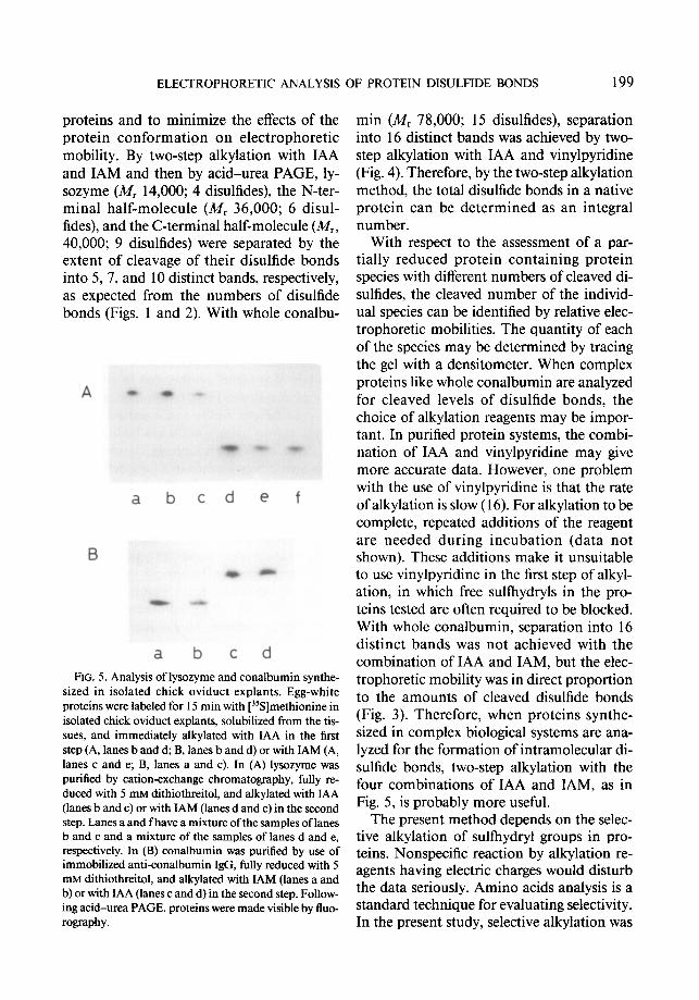
proteins and to minimize the effects of the protein conformation on electrophoretic mobility. By two-step alkylation with IAA and IAM and then by acid-urea PAGE, ly- sozyme (Mr 14,000; 4 disulfides), the N-ter- minal half-molecule (M, 36,000; 6 disul- fides), and the C-terminal half-molecule (M,, 40,000; 9 disulfides) were separated by the extent of cleavage of their disulfide bonds into 5, 7, and 10 distinct bands, respectively, as expected from the numbers of disulfide bonds (Figs. 1 and 2). With whole conalbu-
A *
a b
B
a
P
cd e
b c d
f
FIG. 5. Analysis of lysozyme and conalbumin synthe- sized in isolated chick oviduct explants. Egg-white proteins were labeled for 15 min with [35S]methionine in isolated chick oviduct explants, solubilized from the tis- sues, and immediately alkylated with IAA in the first step (A, lanes b and d; B, lanes b and d) or with IAM (A, lanes c and e; B, lanes a and c). In (A) lysozyme was purified by cation-exchange chromatography, fully re- duced with 5 mM dithiothreitol, and alkylated with IAA (lanes b and c) or with IAM (lanes d and e) in the second step. Lanes a and f have a mixture of the samples of lanes b and c and a mixture of the samples of lanes d and e, respectively. In (B) conalbumin was purified by use of immobilized anti-conalbumin IgG, fully reduced with 5 mM dithiothreitol, and alkylated with IAM (lanes a and b) or with IAA (lanes c and d) in the second step. Follow- ing acid-urea PAGE. proteins were made visible by Iluo- rography.
min (Mr 78,000; 15 disulfides), separation into 16 distinct bands was achieved by two- step alkylation with IAA and vinylpyridine (Fig. 4). Therefore, by the two-step alkylation method, the total disulfide bonds in a native protein can be determined as an integral number.
With respect to the assessment of a par- tially reduced protein containing protein species with different numbers of cleaved di- sulfides, the cleaved number of the individ- ual species can be identified by relative elec- trophoretic mobilities. The quantity of each of the species may be determined by tracing the gel with a densitometer. When complex proteins like whole conalbumin are analyzed for cleaved levels of disulfide bonds, the choice of alkylation reagents may be impor- tant. In purified protein systems, the combi- nation of IAA and vinylpyridine may give more accurate data. However, one problem with the use of vinylpyridine is that the rate of alkylation is slow ( 16). For alkylation to be complete, repeated additions of the reagent are needed during incubation (data not shown). These additions make it unsuitable to use vinylpyridine in the first step of alkyl- ation, in which free sulfhydryls in the pro- teins tested are often required to be blocked. With whole conalbumin, separation into 16 distinct bands was not achieved with the combination of IAA and IAM, but the elec- trophoretic mobility was in direct proportion to the amounts of cleaved disulfide bonds (Fig. 3). Therefore, when proteins synthe- sized in complex biological systems are ana- lyzed for the formation of intramolecular di- sulfide bonds, two-step alkylation with the four combinations of IAA and IAM, as in Fig. 5, is probably more useful.
The present method depends on the selec- tive alkylation of sullhydryl groups in pro- teins. Nonspecific reaction by alkylation re- agents having electric charges would disturb the data seriously. Amino acids analysis is a standard technique for evaluating selectivity. In the present study, selective alkylation was
200 HIROSE ET AL.
confirmed with IAA. Lysozyme was fully re- duced by dithiothreitol in Buffer A, alkylated with 50 mM IAA, and analyzed for its amino acid composition. Half-cystine was quantita- tively detected as S-carboxymethylcysteine, and little loss of other amino acids was seen (data not shown). With newly analyzed pro- teins, total half-cystine data provided by amino acid analysis would be also helpful for the evaluation of results obtained from the two-step alkylation method. Another simple approach may be more practical for testing selectivity. Native proteins are incubated with different concentrations of an alkyla- tion reagent having an electric charge, and the samples are electrophoresed on the acid- urea gel. Protein mobilities are compared with the control (no treatment with the al- kylation reagent). If nonspecific reactions occur, protein bands having mobilities dif- ferent from those of the control should ap- pear. By this approach, we also confirmed that under the conditions used in the present study, IAA and vinylpyridine react specifi- cally with sulfhydryl groups in the proteins (data not shown).
Although the present study suggests that most proteins, including those of high molec- ular weight and with a large number of disul- fides, can be analyzed for intramolecular di- sulfide bonds by two-step alkylation and subsequent acid-urea PAGE, their physical and chemical properties vary greatly. If a highly acidic protein is analyzed, the PAGE buffer system may have to be replaced by another system, such as the high pH discon- tinuous system ( 17). The use of gel electrofo- cusing ( 18) would be also effective. If a native protein contains free sullhydryl groups, the groups should be blocked by IAM prior to the two-step alkylation. Proteins containing heterogeneous numbers of groups with elec- tric charges, such as phosphorus and sialic acid, cannot be analyzed without the re- moval of the groups by chemical or enzy- matic means. We have not tested the analy- ses of disulfide bonds in proteins which are
more complex than whole conalbumin in terms of molecular weight and the number of disulfide bonds.
Compared to the established methods for quantitative analyses of protein disulfides, such as amino acid analysis and spectropho- tometric methods (l), our two-step alkyla- tion technique has the advantages that the quantity of disulfide bonds in a native pro- tein can be determined as an integral number and that with a partially reduced protein, mixtures of protein species with different numbers of cleaved disulfides can be ana- lyzed separately. The two-step alkylation method seems to be especially useful for the analysis of proteins synthesized in complex biological systems. First, biosynthesized pro- teins can be specifically characterized by au- toradiography using specific antibodies (as in Fig. 5) or by Western blotting. Second, pro- teins are electrophoresed in a fully denatured form in which all the disulfide bonds are re- duced and alkylated. Therefore, drastic iso- lation procedures, such as elution from a IgG column with urea, acid, or alkali, are un- likely to affect protein mobility. Finally, pro- teins synthesized in biological systems are sometimes different from naturally occur- ring proteins, as with preproteins that con- tain a signal peptide. This makes it difficult to prepare standard proteins for calibration of the relationship between mobility and numbers of disulfide bonds. In the two-step alkylation method, if the proteins tested are modified as in Fig. 5 with four different com- binations of IAA and IAM in the two steps, the extent of disulfide bond formation can be determined by comparing the mobilities of the four samples without any additional standard protein.
ACKNOWLEDGMENT We thank Miss Y. Nishizawa for her excellent techni-
cal assistance.
REFERENCES 1. Cecil, R. (I 963) in The Proteins (Neurath, H. Ed.),
Vol. 1, pp. 379-476, Academic Press, New York.

2.
3. 4.
5. 6. 7.
8.
9.
10.
11.
ELECTROPHORETIC ANALYSIS OF PROTEIN DISULFIDE BONDS 201
Goto, Y., and Hamaguchi, K. (1981) J. Mol. Biol. 12. 146,321-340.
Creighton, T. E. (1977) J. Mol. Biol. 113,329-341. Pace, C. N., and Creighton, T. E. (1986) J. Mol.
Biol. 188,477-486. Creighton, T. E. (1974) J. Mol. Biol. 87, 579-602. Creighton, T. E. (1974) J. Mol. Biol. 87,603-624. Hollecker, M., and Creighton, T. E. (1983) J. Mol.
Biol. S&409-437.
13.
14.
15 ’
Oe, H., Hirose, M., and Doi, E. (1986) Agric. Biol. Chem. 50,2469-2475. 16.
Hurn, B. A. L., and Chantler, S. M. (1980) in Methods in Enzymology (Van Vunakis, H., and 17. Langone, J. J., Eds.), Vol. 70, pt A, pp. 104-142, Academic Press, New York.
Reisfeld, R. A., Lewis, U. J., and Williams, D. E. 18. (1962) Nature (London) 195,28 l-283.
Hirose, M., Sarui, K., and Sunagawa, A. (1985) J. Biochem. 97,781-789.
Tallan, H. H., and Stein, W. H. (195 1) J. Amer. Chem. Sot. 73,2976-2977.
Creighton, T. E. (1980) Nature (London) 284, 487-489.
Evans, R. W., and Williams, J. (1978) Biochem. J. 173,543-552.
Williams, J., Elleman, T. C., Kingston, I. B., Wil- kins, A. G., and Kuhn, K. A. (1982) Eur. J. Bio- them. 122,297-303.
Friedman, M., Krull, L. H., and Cavins, J. F. ( 1970) J. Biol. Chem. 245,3868-387 1.
Hames, B. D. ( 198 1) in Gel Electrophoresis of Pro- teins (Hames, B. D., and Rickwood, D., Eds.), pp. l-9 1, IRL Press, Washington, DC.
der Lan, B. A., and Chrambach, A. (1981) in Gel Electrophoresis of Proteins (Hames, B. D., and Rickwood, D., Eds.), pp. 157-187, IRL Press, Washington, DC.