Anaerobic bacterial endophthalmitis in the...
4
Reports Anaerobic Bacterial Endophthalmitis in the Rabbit David Ormerod,* Kothy Koh,* Ricardo S. Juarez,* Martha A. C. Edelsrein.f Lawrence L. Rife,* Sydney M. Finegold.f and Ronald E. Smith* Anaerobic bacterial endophthalmitis was studied in rabbits following intravitreal injection of live Fusobacterium necro- phorum. Clinical response, bacterial recovery, and histopa- thology were studied. An inoculum of approximately 50 or- ganisms produced endophthalmitis in 59% of injected eyes, while 1000 or more organisms produced endophthalmitis in 100% of injected eyes. The course and severity of disease seemed to be independent of the concentration of bacteria above a minimal inoculum size. Affected eyes showed pro- gressive endophthalmitis. Histopathologic changes corre- sponded to the clinical gradation of endophthalmitis, including progressive retinal necrosis. Invest Ophthalmol Vis Sci 27: 115-118,1986 Endophthalmitis is a catastrophic complication of intraocular surgery, penetrating injury, and endoge- nous infection. The etiologic agent in many cases is unclear and routine aerobic bacterial cultures are often negative. Only recently has there been any emphasis on the role of anaerobic bacteria in endophthalmitis. Since anaerobic bacteria are present in some normal as well as infected conjunctivae, 1 ' 2 anaerobic bacterial infection should be considered in cases of endophthal- mitis. In this report, we describe the production of anaer- obic bacterial endophthalmitis following intravitreal injection of live Fusobacterium necrophorum in rabbits. After preliminary studies to determine a clinical grad- ing system and an optimal concentration of inoculum, eyes were injected and studied for clinical progression of disease, bacterial recovery and histopathology. Clin- ical and histopathologic features of experimental an- aerobic endophthalmitis were characterized. Materials and Methods. Clinical isolates of F. nec- rophorum from the blood of patients with septicemia were stored at — 70°C in skim milk and used later for intravitreal injection. F. necrophorum was selected be- cause it is a strict anaerobe and a virulent pathogen. While it would be expected to produce clinical disease, it would also serve as a stringent test of our technique for the preparation and recovery of anaerobic bacteria. The isolates were thawed and then subcultured onto Brucella blood agar at least twice prior to inoculation into thioglycolate broth for 24-48 hr. The broth culture was diluted in sterile, pyrogen-free, non-bacteriostatic buffer (. 15 M NaCl; .02 M phosphate buffer) to a tur- bidity corresponding to a number 1 MacFarland stan- dard [10 8 colony forming units (CFU)/ml]. Aliquots of serial dilutions made in the sterile buffer were used for both the determination of viable counts (rotator- pipet method 3 ) and intravitreal injection. The injection aliquots were transplanted in Hungate style screw cap tubes containing an oxygen-free atmosphere and glass beads (to facilitate mixing). Male, New Zealand albino rabbits (5-5.5 lbs) were obtained from ABC Laboratories (Pomona, CA). Our investigations utilizing animals conformed to the ARVO Resolution on the Use of Animals in Research. For intravitreal injections, animals were anesthetized with intramuscular injections of Xylazine HC1 (3 mg/ kg) and ketamine HC1 (15 mg/kg). Corneas were anes- thesized with topical proparacaine HC1 (0.5%). For vi- treal aspirates and enucleations, animals were euthan- ized with sodium pentobarbitol (6 gr/ml). To prevent loss of inoculum, intraocular pressure was reduced by removal of 0.1 ml aqueous humor. Intravitreal injec- tions were through the pars plana, using a 30-gauge needle on a tuberculin syringe, and ranged from 5 X 1 0 ' to 1 X 10 7 bacteria per 0.1 ml per eye. Control eyes were injected with either 0.1 ml saline or thioglycolate broth diluted 1:4 in buffer. Ocular examinations were performed daily using an indirect ophthalmoscope and a Haag Streit slit lamp and the eyes graded according to Table 1. As endophthalmitis progressed, animals were randomly selected at each grade for vitreous as- pirates and enucleation. Enucleated eyes were partially opened at the equator and fixed in 10% neutral buffered formalin. After 48 hr, the globe was bisected around the equator and one half of the posterior hemisphere paraffin embedded, sectioned, and stained with he- matoxylin and eosin (H & E). The vitreal aspirates were transported in Port-a-cul vials (Baltimore Biological Laboratory; McConkeyville, MD.). After vigorous vortexing, each was subcultured in an anaerobic 115 Downloaded From: https://iovs.arvojournals.org/pdfaccess.ashx?url=/data/journals/iovs/933126/ on 08/20/2018
-
Upload
trinhnguyet -
Category
Documents
-
view
222 -
download
0
Transcript of Anaerobic bacterial endophthalmitis in the...

Reports
Anaerobic Bacterial Endophthalmitis in the Rabbit
David Ormerod,* Kothy Koh,* Ricardo S. Juarez,* Martha A. C. Edelsrein.fLawrence L. Rife,* Sydney M. Finegold.f and Ronald E. Smith*
Anaerobic bacterial endophthalmitis was studied in rabbitsfollowing intravitreal injection of live Fusobacterium necro-phorum. Clinical response, bacterial recovery, and histopa-thology were studied. An inoculum of approximately 50 or-ganisms produced endophthalmitis in 59% of injected eyes,while 1000 or more organisms produced endophthalmitis in100% of injected eyes. The course and severity of diseaseseemed to be independent of the concentration of bacteriaabove a minimal inoculum size. Affected eyes showed pro-gressive endophthalmitis. Histopathologic changes corre-sponded to the clinical gradation of endophthalmitis, includingprogressive retinal necrosis. Invest Ophthalmol Vis Sci 27:115-118,1986
Endophthalmitis is a catastrophic complication ofintraocular surgery, penetrating injury, and endoge-nous infection. The etiologic agent in many cases isunclear and routine aerobic bacterial cultures are oftennegative. Only recently has there been any emphasison the role of anaerobic bacteria in endophthalmitis.Since anaerobic bacteria are present in some normalas well as infected conjunctivae,1'2 anaerobic bacterialinfection should be considered in cases of endophthal-mitis.
In this report, we describe the production of anaer-obic bacterial endophthalmitis following intravitrealinjection of live Fusobacterium necrophorum in rabbits.After preliminary studies to determine a clinical grad-ing system and an optimal concentration of inoculum,eyes were injected and studied for clinical progressionof disease, bacterial recovery and histopathology. Clin-ical and histopathologic features of experimental an-aerobic endophthalmitis were characterized.
Materials and Methods. Clinical isolates of F. nec-rophorum from the blood of patients with septicemiawere stored at — 70°C in skim milk and used later forintravitreal injection. F. necrophorum was selected be-cause it is a strict anaerobe and a virulent pathogen.While it would be expected to produce clinical disease,it would also serve as a stringent test of our techniquefor the preparation and recovery of anaerobic bacteria.The isolates were thawed and then subcultured onto
Brucella blood agar at least twice prior to inoculationinto thioglycolate broth for 24-48 hr. The broth culturewas diluted in sterile, pyrogen-free, non-bacteriostaticbuffer (. 15 M NaCl; .02 M phosphate buffer) to a tur-bidity corresponding to a number 1 MacFarland stan-dard [108 colony forming units (CFU)/ml]. Aliquotsof serial dilutions made in the sterile buffer were usedfor both the determination of viable counts (rotator-pipet method3) and intravitreal injection. The injectionaliquots were transplanted in Hungate style screw captubes containing an oxygen-free atmosphere and glassbeads (to facilitate mixing).
Male, New Zealand albino rabbits (5-5.5 lbs) wereobtained from ABC Laboratories (Pomona, CA). Ourinvestigations utilizing animals conformed to theARVO Resolution on the Use of Animals in Research.For intravitreal injections, animals were anesthetizedwith intramuscular injections of Xylazine HC1 (3 mg/kg) and ketamine HC1 (15 mg/kg). Corneas were anes-thesized with topical proparacaine HC1 (0.5%). For vi-treal aspirates and enucleations, animals were euthan-ized with sodium pentobarbitol (6 gr/ml). To preventloss of inoculum, intraocular pressure was reduced byremoval of 0.1 ml aqueous humor. Intravitreal injec-tions were through the pars plana, using a 30-gaugeneedle on a tuberculin syringe, and ranged from 5X10 'to 1 X 10⁷ bacteria per 0.1 ml per eye. Control eyeswere injected with either 0.1 ml saline or thioglycolatebroth diluted 1:4 in buffer. Ocular examinations wereperformed daily using an indirect ophthalmoscope anda Haag Streit slit lamp and the eyes graded accordingto Table 1. As endophthalmitis progressed, animalswere randomly selected at each grade for vitreous as-pirates and enucleation. Enucleated eyes were partiallyopened at the equator and fixed in 10% neutral bufferedformalin. After 48 hr, the globe was bisected aroundthe equator and one half of the posterior hemisphereparaffin embedded, sectioned, and stained with he-matoxylin and eosin (H & E). The vitreal aspirates weretransported in Port-a-cul vials (Baltimore BiologicalLaboratory; McConkeyville, MD.). After vigorousvortexing, each was subcultured in an anaerobic
115
Downloaded From: https://iovs.arvojournals.org/pdfaccess.ashx?url=/data/journals/iovs/933126/ on 08/20/2018
116 INVESTIGATIVE OPHTHALMOLOGY & VISUAL SCIENCE / January 1986 Vol. 27
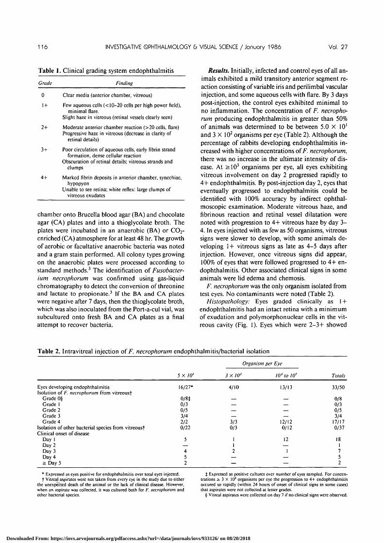
Table 1. Clinical grading system endophthalmitis
Grade Finding
0 Clear media (anterior chamber, vitreous)
1+ Few aqueous cells (< 10-20 cells per high power field),minimal flare.
Slight haze in vitreous (retinal vessels clearly seen)
2+ Moderate anterior chamber reaction (>20 cells, flare)Progressive haze in vitreous (decrease in clarity of
retinal details)
3+ Poor circulation of aqueous cells, early fibrin strandformation, dense cellular reaction
Obscuration of retinal details; vitreous strands andclumps
4+ Marked fibrin deposits in anterior chamber, synechiae,hypopyon
Unable to see retina; white reflex: large clumps ofvitreous exudates
chamber onto Brucella blood agar (BA) and chocolateagar (CA) plates and into a thioglycolate broth. Theplates were incubated in an anaerobic (BA) or CO2-enriched (CA) atmosphere for at least 48 hr. The growthof aerobic or facultative anaerobic bacteria was notedand a gram stain performed. All colony types growingon the anaerobic plates were processed according tostandard methods.3 The identification of Fusobacter-ium necrophorum was confirmed using gas-liquidchromatography to detect the conversion of threonineand lactate to propionate.3 If the BA and CA plateswere negative after 7 days, then the thioglycolate broth,which was also inoculated from the Port-a-cul vial, wassubcultured onto fresh BA and CA plates as a finalattempt to recover bacteria.
Results. Initially, infected and control eyes of all an-imals exhibited a mild transitory anterior segment re-action consisting of variable iris and perilimbal vascularinjection, and some aqueous cells with flare. By 3 dayspost-injection, the control eyes exhibited minimal tono inflammation. The concentration of F. necropho-rum producing endophthalmitis in greater than 50%of animals was determined to be between 5.0 X 10'and 3 X 10² organisms per eye (Table 2). Although thepercentage of rabbits developing endophthalmitis in-creased with higher concentrations of F. necrophorum,there was no increase in the ultimate intensity of dis-ease. At >103 organisms per eye, all eyes exhibitingvitreous involvement on day 2 progressed rapidly to4+ endophthalmitis. By post-injection day 2, eyes thateventually progressed to endophthalmitis could beidentified with 100% accuracy by indirect ophthal-moscopic examination. Moderate vitreous haze, andfibrinous reaction and retinal vessel dilatation werenoted with progression to 4+ vitreous haze by day 3 -4. In eyes injected with as few as 50 organisms, vitreoussigns were slower to develop, with some animals de-veloping 1+ vitreous signs as late as 4-5 days afterinjection. However, once vitreous signs did appear,100% of eyes that were followed progressed to 4+ en-dophthalmitis. Other associated clinical signs in someanimals were lid edema and chemosis.
F. necrophorum was the only organism isolated fromtest eyes. No contaminants were noted (Table 2).
Histopathology: Eyes graded clinically as 1 +endophthalmitis had an intact retina with a minimumof exudation and polymorphonuclear cells in the vit-reous cavity (Fig. 1). Eyes which were 2-3+ showed
Table 2. Intravitreal injection of F. necrophorum endophthalmitis/bacterial isolation
Eyes developing endophthalmitisIsolation of F. necrophorum from vitreousf
Grade 0§Grade 1Grade 2Grade 3Grade 4
Isolation of other bacterial species from vitreousfClinical onset of disease
Day 1Day 2Day 3Day 4S: Day 5
5 X 7 0 '
16/27*
0/8J0/30/53/42/20/22
5—452
3X10²
4/10
————3/30/3
112
——
Organism per Eye
103 to 107
13/13
————
12/120/12
12—
1——
Totals
33/50
0/80/30/53/4
17/170/37
181752
* Expressed as eyes positive for endophthalmitis over total eyes injected.t Vitreal aspirates were not taken from every eye in the study due to either
the unexpected death of the animal or the lack of clinical disease. However,when an aspirate was collected, it was cultured both for F. necrophorum andother bacterial species.
X Expressed as positive cultures over number of eyes sampled. For concen-trations ^ 3 X 10² organisms per eye the progression to 4+ endophthalmitisoccured so rapidly (within 24 hours of onset of clinical signs in some cases)that aspirates were not collected at lesser grades.
§ Vitreal aspirates were collected on day 7 if no clinical signs were observed.
Downloaded From: https://iovs.arvojournals.org/pdfaccess.ashx?url=/data/journals/iovs/933126/ on 08/20/2018
No. 1 Reporrs
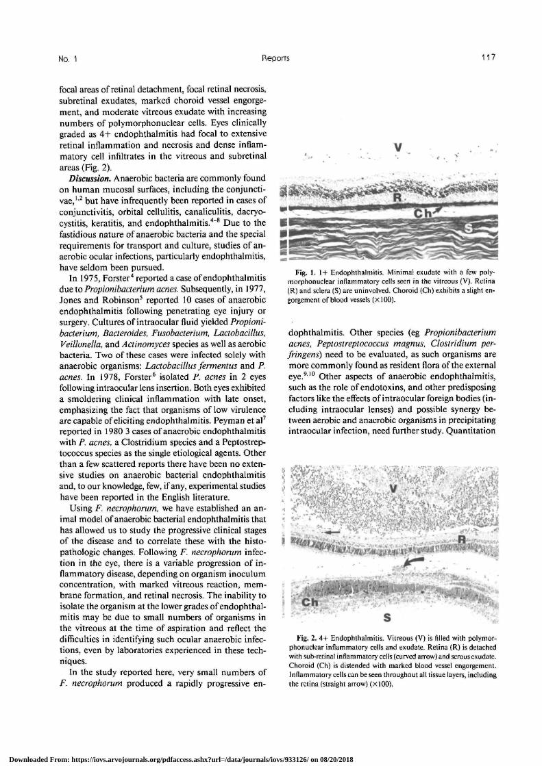
focal areas of retinal detachment, focal retinal necrosis,subretinal exudates, marked choroid vessel engorge-ment, and moderate vitreous exudate with increasingnumbers of polymorphonuclear cells. Eyes clinicallygraded as 4+ endophthalmitis had focal to extensiveretinal inflammation and necrosis and dense inflam-matory cell infiltrates in the vitreous and subretinalareas (Fig. 2).
Discussion, Anaerobic bacteria are commonly foundon human mucosal surfaces, including the conjuncti-vae,1-2 but have infrequently been reported in cases ofconjunctivitis, orbital cellulitis, canaliculitis, dacryo-cystitis, keratitis, and endophthalmitis.4"8 Due to thefastidious nature of anaerobic bacteria and the specialrequirements for transport and culture, studies of an-aerobic ocular infections, particularly endophthalmitis,have seldom been pursued.
In 1975, Forster4 reported a case of endophthalmitisdue to Propionibacterium acnes. Subsequently, in 1977,Jones and Robinson5 reported 10 cases of anaerobicendophthalmitis following penetrating eye injury orsurgery. Cultures of intraocular fluid yielded Propioni-bacterium, Bacteroides, Fusobacterium, Lactobacillus,Veillonella, and Actinomyces species as well as aerobicbacteria. Two of these cases were infected solely withanaerobic organisms: Lactobacillus fermentus and P.acnes. In 1978, Forster6 isolated P. acnes in 2 eyesfollowing intraocular lens insertion. Both eyes exhibiteda smoldering clinical inflammation with late onset,emphasizing the fact that organisms of low virulenceare capable of eliciting endophthalmitis. Peyman et al7
reported in 1980 3 cases of anaerobic endophthalmitiswith P. acnes, a Clostridium species and a Peptostrep-tococcus species as the single etiological agents. Otherthan a few scattered reports there have been no exten-sive studies on anaerobic bacterial endophthalmitisand, to our knowledge, few, if any, experimental studieshave been reported in the English literature.
Using F. necrophorum, we have established an an-imal model of anaerobic bacterial endophthalmitis thathas allowed us to study the progressive clinical stagesof the disease and to correlate these with the histo-pathologic changes. Following F. necrophorum infec-tion in the eye, there is a variable progression of in-flammatory disease, depending on organism inoculumconcentration, with marked vitreous reaction, mem-brane formation, and retinal necrosis. The inability toisolate the organism at the lower grades of endophthal-mitis may be due to small numbers of organisms inthe vitreous at the time of aspiration and reflect thedifficulties in identifying such ocular anaerobic infec-tions, even by laboratories experienced in these tech-niques.
In the study reported here, very small numbers ofF. necrophorum produced a rapidly progressive en-
Fig. 1. 1+ Endophthalmitis. Minimal exudate with a few poly-morphonuclear inflammatory cells seen in the vitreous (V). Retina(R) and sclera (S) are uninvolved. Choroid (Ch) exhibits a slight en-gorgement of blood vessels (X100).
dophthalmitis. Other species (eg Propionibacteriumacnes, Peptostreptococcus magnus, Clostridium per-fringens) need to be evaluated, as such organisms aremore commonly found as resident flora of the externaleye.910 Other aspects of anaerobic endophthalmitis,such as the role of endotoxins, and other predisposingfactors like the effects of intraocular foreign bodies (in-cluding intraocular lenses) and possible synergy be-tween aerobic and anaerobic organisms in precipitatingintraocular infection, need further study. Quantitation
1 I R * & i ^ i ^ ^ ^
Fig. 2. 4+ Endophthalmitis. Vitreous (V) is filled with polymor-phonuclear inflammatory cells and exudate. Retina (R) is detachedwith sub-retinal inflammatory cells (curved arrow) and serous exudate.Choroid (Ch) is distended with marked blood vessel engorgement.Inflammatory cells can be seen throughout all tissue layers, includingthe retina (straight arrow) (X)00).
Downloaded From: https://iovs.arvojournals.org/pdfaccess.ashx?url=/data/journals/iovs/933126/ on 08/20/2018

118 INVESTIGATIVE OPHTHALMOLOGY 6 VISUAL SCIENCE / January 1986 Vol. 27
of bacteria from ocular aspirates will become an im-
portant aspect of future studies.
Key words: anaerobic bacteria, Fusobacterium necrophorum,experimental endophthalmitis, rabbits
Acknowledgments. The authors thank Ann Dawson, AnnGuild, and Ellen Narver for technical assistance in performingthese studies and in the preparation of this manuscript.
From the Department of Ophthalmology, University of SouthernCalifornia, and the Estelle Doheny Eye Foundation* and InfectiousDisease Section of VA Wadsworth Medical Center and Departmentof Medicine, UCLA,f Los Angeles, California. Supported in part byResearch to Prevent Blindness, New York, New York. Submittedfor publication: March 11, 1985. Reprint requests: Ronald E. Smith,MD, Department of Ophthalmology, Estelle Doheny Eye Foundation,1355 San Pablo Street, Los Angeles, CA 90033.
References1. McNatt J, Allen SD, Wilson LA, and Dowell VR Jr: Anaerobic
flora of the normal human conjunctival sac. Arch Ophthalmol96:1448, 1978.
2. Matsura H: Anaerobes in the conjunctiva and outer eye diseases.Jpn J Clin Ophthalmol 25:1391, 1971.
3. Sutter VL, Citron DM, Edelstein MAC, and Finegold SM:Wadsworth Anaerobic Bacteriology Manual. Starr PublishingCompany. Fourth Edition, 1985 (in press).
4. Forster RK, Zachary 1G, Cottingham Jr AJ, and Norton EWD:Further observations on the diagnosis, cause, and treatment ofendophthalmitis. Am J Ophthalmol 81:52, 1976.
5. Jones DB and Robinson NM: Anaerobic ocular infections. TransPa Acad Ophthalmol Otolaryngol 83:309, 1977.
6. Forster RK: Etiology and diagnosis of bacterial postoperativeendophthalmitis. Ophthalmology 85:320, 1978.
7. Peyman GA, Raichand M, and Bennett TO: Management ofendophthalmitis with pars plana vitrectomy. Br J Ophthalmol64:472, 1980.
8. Perry LD, Brinser JH, and Kolodner H: Anaerobic corneal ulcers.Ophthalmology,89:636, 1981.
9. McCulley JP, Dougherty JM, and Deneau DG: Classification ofchronic blepharitis. Ophthalmology 89:1173, 1982.
10. Perkins RE, Kundsin RB, Pratt MV, Abrahamson I, and Lei-bowitz HM: Bacteriology of normal and infected conjunctiva. JClin Microbiology 1:147, 1975.
Inexpensive Stereoscopic CCRG Comera for Lens/CataractPhotography In Vitro
Leo T. Chylock, Jr. and William H. Tung
In 1978 the American Cooperative Cataract Research Group(CCRG) adopted a stereoscopic camera system for photo-graphing human cataracts in vitro based upon a Zeiss OPMI1 operating microscope (Carl Zeiss, Inc.; Oberkochen, WestGermany). Photographs obtained with this system were usedto classify human cataractous change according to the stan-dardized CCRG protocol. Classification data correlated withlaboratory data furthered the attempt to define the biochem-ical or biophysical basis for specific types of cataractouschange. Presently, the high cost of this camera (exceeding$20,000) precludes its use by many laboratories wishing todo human lens research. This study describes an inexpensive(less than $2,200) alternative camera for this type of pho-tography. Adjacent frames on the film strip constitute stereopairs which can be viewed in a modified stereo viewer. In theoriginal CCRG camera both members of a stereo pair wereincluded in the same frame. The quality of the stereo imagesobtained with this new system nearly equals that with theoriginal Zeiss system. It is hoped that this inexpensive systemwill allow more scientists to participate in CCRG-related re-search and increase the supply of intracapsularly extractedcataracts available to all collaborating CCRG scientists. InvestOphthalmol Vis Sci 27:118-122, 1986
In 1978 Chylack1 published a system of cataractclassification, and in 19802 it was adopted by the
American Cooperative Cataract Research Group(CCRG). High quality, artifact-free, color, stereoscopicphotographs were obtained with the system's modifiedOPMI 1 operating microscope with a Zeiss-Urban ste-reoscopic adaptor (Carl Zeiss, Inc.; Oberkochen, WestGermany). The cataract classification format has beenimproved, simplified,3'4 and employed in studies of theassociations among age, sex, nuclear color and opaci-fication,4 and the structure5'6 and light scattering prop-erties of human cataracts.7'8
Originally it was hoped that some of the in vivo cat-aract classification methods could be validated bypostoperative photography and in vitro classificationof intracapsularly extracted lenses. However, the de-cline in availability of intracapsular cataract extractionin the medical centers processing the original CCRGclassification apparatus makes it unlikely that thesesame centers can complete such a study. The need fora light, inexpensive portable classification camera mayfacilitate the completion of such studies, either in theUnited States or elsewhere.
In spite of the acknowledged usefulness of the orig-inal CCRG photographic apparatus, its present highcost (more than $20,000) precludes its widespread use.It is the purpose of this study to describe a compara-
Downloaded From: https://iovs.arvojournals.org/pdfaccess.ashx?url=/data/journals/iovs/933126/ on 08/20/2018