an W, Pogosyan a, Hyam JA, Jenkinson N, Foltynie T, Limousin P, Bogdanovic M, Zrinzo L, Green AL,...
13
BRAIN A JOURNAL OF NEUROLOGY Alpha oscillations in the pedunculopontine nucleus correlate with gait performance in parkinsonism Wesley Thevathasan, 1,2 Alek Pogosyan, 1 Jonathan A. Hyam, 3 Ned Jenkinson, 3 Tom Foltynie, 4 Patricia Limousin, 4 Marko Bogdanovic, 2 Ludvic Zrinzo, 4 Alexander L. Green, 3 Tipu Z. Aziz 3 and Peter Brown 1,2 1 Nuffield Departments of Clinical Neuroscienc es, University of Oxford OX3 9DU, UK 2 Departme nt of Clinical Neurology, John Radcliffe Hospit al, Oxford OX3 9DU, UK 3 Nuffield Depar tment of Surgical Scie nces, University of Ox ford OX3 9DU, UK 4 Sobell Departme nt of Motor Neuroscience and Movemen t Disorders, Institute of Neurology, Lond on WC1N 3BG, UK Correspondence to: Peter Brown, Department of Clinical Neurology University of Oxford Level 6, West Wing John Radcliffe Hospital Oxford OX3 9DU, UK E-mail: peter.brown @clneuro.o x.ac.uk The pedunculoponti ne nucleu s, a compone nt of the retic ular formation , is topographic ally organized in animal models and impli cated in locomot or control . In Parkin son’s disease, pedunculo pontine nucleus stimulati on is an emergi ng treatment for gait freezing. Local field potentials recorded from pedunculopontine nucleus electrodes in such patients have demonstrated oscilla- tions in the alpha and beta frequency bands, reactive to self-paced movement. Whether these oscillations are topographically organized or relevant to locomotion is unknown. Here, we recorded local field potentials from the pedunculopontine nucleus in parkinsonian patients during rest and unconstrained walking. Relative gait speed was assessed with trunk accelerometry. Peaks of alpha power were present at rest and during gait, when they correlated with gait speed. Gait freezing was associated with attenuation of alpha activity. Beta peaks were less consistently observed across rest and gait, and did not correlate with gait speed. Alpha power was maximal in the caudal pedunculopontine nucleus region and beta power was maximal rostrally. These results indicate a topographic distribution of neuronal activity in the pedunculopontine nucleus region and concur with animal data suggesting that the caudal subregion has particular relevance to gait. Alpha synchronization, proposed to suppress ‘task irrelevant’ distraction, has previously been demonst rated to correlate with performance of cognit ive tasks. Here, we demonstrate a correlation between alpha oscillations and improved gait performance. The results raise the possibility that stimulation of caudal and rostral pedunculopontine nucleus regions may differ in their clinical effects. Keywords: Parkinson’s disease; gait freezing; pedunculopontine nucleus; deep brain stimulation; neuronal oscillations Abbreviations: FzCz = frontal zero central zero; PPN = pedunculopontine nucleus; UPDRS = Unified Parkinson’s Disease Rating Scale doi:10.1093/brain/awr315 Brain 2012: 135; 148–160 | 148 Received August 3, 2011. Revised September 7, 2011. Accepted September 22, 2011. Advance Access publication January 9, 2012 ß The Author (2012). Published by Oxford University Press on behalf of the Guarantors of Brain. This is an Open Access article distributed under the terms of the Creative Commons Attribution Non-Commercial License (http://creativecommons.org/licenses/by-nc/3.0), which permits unrestrict ed non-commerc ial use, distribution, and reprodu ction in any medium, provided the origina l work is properl y cited.
Transcript of an W, Pogosyan a, Hyam JA, Jenkinson N, Foltynie T, Limousin P, Bogdanovic M, Zrinzo L, Green AL,...
8/3/2019 an W, Pogosyan a, Hyam JA, Jenkinson N, Foltynie T, Limousin P, Bogdanovic M, Zrinzo L, Green AL, Aziz TZ, Brown P
http://slidepdf.com/reader/full/an-w-pogosyan-a-hyam-ja-jenkinson-n-foltynie-t-limousin-p-bogdanovic 1/13
BRAINA JOURNAL OF NEUROLOGY
Alpha oscillations in the pedunculopontinenucleus correlate with gait performance inparkinsonismWesley Thevathasan,1,2 Alek Pogosyan,1 Jonathan A. Hyam,3 Ned Jenkinson,3 Tom Foltynie,4
Patricia Limousin,4 Marko Bogdanovic,2 Ludvic Zrinzo,4 Alexander L. Green,3 Tipu Z. Aziz3 andPeter Brown1,2
1 Nuffield Departments of Clinical Neurosciences, University of Oxford OX3 9DU, UK
2 Department of Clinical Neurology, John Radcliffe Hospital, Oxford OX3 9DU, UK
3 Nuffield Department of Surgical Sciences, University of Oxford OX3 9DU, UK
4 Sobell Department of Motor Neuroscience and Movement Disorders, Institute of Neurology, London WC1N 3BG, UK
Correspondence to: Peter Brown,
Department of Clinical Neurology
University of Oxford
Level 6, West Wing
John Radcliffe Hospital
Oxford OX3 9DU, UK
E-mail: [email protected]
The pedunculopontine nucleus, a component of the reticular formation, is topographically organized in animal models and
implicated in locomotor control. In Parkinson’s disease, pedunculopontine nucleus stimulation is an emerging treatment for gait
freezing. Local field potentials recorded from pedunculopontine nucleus electrodes in such patients have demonstrated oscilla-tions in the alpha and beta frequency bands, reactive to self-paced movement. Whether these oscillations are topographically
organized or relevant to locomotion is unknown. Here, we recorded local field potentials from the pedunculopontine nucleus in
parkinsonian patients during rest and unconstrained walking. Relative gait speed was assessed with trunk accelerometry. Peaks
of alpha power were present at rest and during gait, when they correlated with gait speed. Gait freezing was associated with
attenuation of alpha activity. Beta peaks were less consistently observed across rest and gait, and did not correlate with gait
speed. Alpha power was maximal in the caudal pedunculopontine nucleus region and beta power was maximal rostrally. These
results indicate a topographic distribution of neuronal activity in the pedunculopontine nucleus region and concur with animal
data suggesting that the caudal subregion has particular relevance to gait. Alpha synchronization, proposed to suppress ‘task
irrelevant’ distraction, has previously been demonstrated to correlate with performance of cognitive tasks. Here, we demonstrate
a correlation between alpha oscillations and improved gait performance. The results raise the possibility that stimulation of
caudal and rostral pedunculopontine nucleus regions may differ in their clinical effects.
Keywords: Parkinson’s disease; gait freezing; pedunculopontine nucleus; deep brain stimulation; neuronal oscillations
Abbreviations: FzCz = frontal zero central zero; PPN = pedunculopontine nucleus; UPDRS = Unified Parkinson’s Disease Rating Scale
doi:10.1093/brain/awr315 Brain 2012: 135; 148–160 | 148
Received August 3, 2011. Revised September 7, 2011. Accepted September 22, 2011. Advance Access publication January 9, 2012
ß The Author (2012). Published by Oxford University Press on behalf of the Guarantors of Brain.
This is an Open Access article distributed under the terms of the Creative Commons Attribution Non-Commercial License (http://creativecommons.org/licenses/by-nc/3.0),
which permits unrestricted non-commercial use, distribution, and reproduction in any medium, provided the original work is properly cited.

8/3/2019 an W, Pogosyan a, Hyam JA, Jenkinson N, Foltynie T, Limousin P, Bogdanovic M, Zrinzo L, Green AL, Aziz TZ, Brown P
http://slidepdf.com/reader/full/an-w-pogosyan-a-hyam-ja-jenkinson-n-foltynie-t-limousin-p-bogdanovic 2/13
IntroductionThe pedunculopontine nucleus (PPN) is a reticular collection of
neurons, located at the junction of midbrain and pons
(Jacobsohn, 1911; Olszewski and Baxter, 1954; Alam et al.,
2011) The PPN, at least in animal models, appears to be topo-
graphically organized, with -aminobutyric acid expressing neu-
rons predominating rostrally and cholinergic and glutamatergicneurons caudally (Martinez-Gonzalez et al., 2011). In
Parkinson’s disease, cholinergic PPN neurons degenerate and this
cell loss has been associated with gait dysfunction (Hirsch et al.,
1987; Zweig et al., 1989; Rinne et al., 2008; Karachi et al., 2010).
PPN neurons in Parkinson’s disease may also be disrupted through
their reciprocal connectivity with the basal ganglia (Breit et al.,
2001; Mena-Segovia et al., 2004). In patients with Parkinson’s
disease, deep brain stimulation of the PPN region at low frequen-
cies is an emerging treatment for postural instability and gait
freezing (Plaha and Gill, 2005; Ferraye et al., 2009; Moro et al.,
2010; Thevathasan et al., 2011a).
The control of locomotion likely involves coordination be-
tween spatially segregated nervous system regions in order
to modulate the rhythmic activity produced by spinal central
pattern generators (Grillner et al., 2008). Synchronized oscillatory
neuronal activity is proposed to bind together such neuronal
assemblies and enhance information representation—whilst also
suppressing task irrelevant or competing processes (Fries, 2005;
Schoffelen et al., 2005; Jensen and Mazaheri, 2010). Local field
potential recordings from deep brain stimulation electrodes im-
planted in the PPN in parkinsonian patients have variably been
reported to demonstrate alpha or beta band oscillations, reactive
to self-paced movement (Androulidakis et al., 2008a, b; Tsang
et al., 2010). Whether these different oscillatory patterns conform
to any topographic distribution or are relevant to locomotion isunknown.
In this study, we assessed local field potentials from parkinso-
nian patients implanted with PPN electrodes and assessed the spa-
tial pattern of oscillatory activity in the region and its relationship
to the performance of gait.
Materials and methods
Subjects and clinical assessmentsSeven patients with Parkinson’s disease implanted with deep brain
stimulation electrodes in the PPN region were assessed (Table 1).
Following their initial implantation, electrodes were attached to special
extension cables ‘externalized’ through the scalp to permit connectionwith recording equipment. Final implantation of deep brain stimulation
hardware was therefore deferred for the period of ‘externalization’
(3–7 days). Eleven PPN electrodes were recorded from the seven pa-
tients. Patients were recruited from hospitals in Oxford and London,
UK. Local ethics committee approval was obtained from both centres
and participants gave written informed consent. Reaction time results
from one of the patients have been reported previously (Thevathasan
et al., 2010, 2011b).
The indication for PPN stimulation was severe gait freezing and
postural instability, persisting even ‘ON medication’ and either causing
frequent falls or precluding walking. In Parkinson’s disease, gait freez-
ing and postural instability become more frequent and less responsive
to medication with disease progression (Giladi et al., 2001; Bloem
et al., 2004). The overall prevalence of gait freezing and posturalinstability in Parkinson’s disease is $50% (Macht et al., 2007).
However, severe medication resistant gait freezing and postural
instability as the predominant issue is unusual in Parkinson’s disease
and raises the question of atypical pathologies (Factor, 2008; Jankovic,
2008). As there is no definitive test in life, we stress that the diagnosis
of Parkinson’s disease in our series is presumptive.
Preoperative assessments included the motor subsection (part III) of
the Unified Parkinson’s Disease Rating Scale (UPDRS; score/108). OFF
medication assessments occurred after overnight withdrawal (412h)
of dopaminergic therapy. UPDRS was segmented into items 27–30
(IT27/30, score/16) assessing posture, gait and balance and residual
items 1–26 (R-UPDRS, score/92) assessing bradykinesia, rigidity and
tremor. Patients treated in Oxford also prospectively completed the
Gait and Falls Questionnaire (score/64) which assesses Parkinsonian
gait disturbance including gait freezing, festination and falls
(Giladi et al., 2000). The Freezing of Gait Questionnaire (score/24)
and Falls Question (score/4) are components of the Gait and Falls
questionnaire (Giladi et al., 2000, 2009). For all motor scales, higher
scores indicate worse function. The London patient was assessed with
Table 1 Clinical details of the study participants
Patient Age(years)
Parkinson’sdiseaseduration(years)
UPDRSIII OFF/ONmedication(score/108)
R-UPDRSOFF/ONmedication(score/92)
IT27-30OFF/ONmedication(score/16)
GFQ(score/64)
FOGQ(score/24)
FallsQ(score/4)
L-dopa doseequivalent (mg/day)
Supportive for UK Brain Bankcriteriaa
1 55 14 35/24 28/18 7/6 55 22 4 1600 A, P
2 76 16 34/25 25/16 9/9 38 20 1 600 D, A, P
3 55 25 33/22 27/17 6/5 36 15 3 300 D, A, P
4 68 9 40/26 29/18 11/8 49 24 2 1650 A, P
5 70 20 35/22 29/17 6/5 36 13 3 900 D, A, T, P
6 71 20 37/19 27/14 10/5 NA NA 2 1450 D, A, T, P
7 54 20 53/19 47/14 6/5 38 14 4 800 D, A, T, P
All patients were operated in Oxford except Patient 6 (London). All patients were male. FOGQ = Freezing of Gait Questionnaire (score/24); Falls Q = Falls Questionnaire
(score/4); GFQ = Gait and Falls questionnaire; NA = not assessed. For all motor scales, higher scores indicate worse function. A = asymmetry persistent; D = dyskinesias;
P = progressive disease course; T = tremor at rest.
a Additional to disease duration and L-dopa response as documented elsewhere in the table.
PPN oscillations correlate with gait Brain 2012: 135; 148–160 | 149
8/3/2019 an W, Pogosyan a, Hyam JA, Jenkinson N, Foltynie T, Limousin P, Bogdanovic M, Zrinzo L, Green AL, Aziz TZ, Brown P
http://slidepdf.com/reader/full/an-w-pogosyan-a-hyam-ja-jenkinson-n-foltynie-t-limousin-p-bogdanovic 3/13
UPDRS (part II) items assessing gait, freezing and falls (combinedscore/16).
Techniques to target and implant electrodes in the PPN have been
described previously (Pereira et al., 2008; Zrinzo et al., 2008; Foltynie
and Hariz, 2010). In two early studies of stimulation in this vicinity,
electrodes appeared to be placed lateral to the lemniscal system—in
the peripeduncular nucleus rather than the PPN (Mazzone et al., 2005;
Stefani et al., 2007; Yelnik, 2007; Zrinzo and Hariz, 2007; Zrinzo
et al., 2007). In the current study, the PPN was targeted medial to
the lemniscal system and lateral to the superior cerebellar peduncle
and its decussation. Electrodes (Medtronic) were configured with
four active contacts each spanning 1.5mm. Electrodes in six patients
were model 3389 (0.5 mm spacing between contacts) and in one pa-
tient were model 3387 (1.5mm spacing between contacts). Contact
coordinates were assessed on postoperative MRI or postoperativeCT fused with preoperative MRI and transformed onto Montreal
Neurological Institute (MNI) space using the fMRIB Software Library
(Smith et al., 2004). Coordinates were calculated in millimetres from
midline (laterality), ventrodorsal distance (d ) from floor of the fourth
ventricle and rostrocaudal distance (h) from a pontomesencephalic line
connecting the pontomesencephalic junction to the caudal end of the
inferior colliculi, as described previously (Ferraye et al., 2009).
Electrode locations are summarized in Fig. 1A.
Experiments and recordingsExperiments took place 2–6 days after electrode implantation.
Assessments were performed ‘OFF medication’, after overnight with-drawal of dopaminergic medication, to limit variance from fluctuating
dopaminergic state and to maximize gait disturbance.
There were two conditions: rest and gait. For rest recordings, pa-
tients sat comfortably with eyes open for 2–3 min. For gait recordings,
patients walked at their preferred speed along an unobstructed path
10 m long, 10–30 times (depending on speed and fatigue).
Local field potentials were recorded in bipolar configuration from
consecutive contacts (01, 12 and 23) of each electrode (Fig. 2A).
Local field potentials were band-pass filtered between 0.5 and
500 Hz. Single channel EEG was recorded from FzCz (frontal zero cen-
tral zero). In Patients 1–6, a triaxial accelerometer (TMSI) was firmly
fixed with tape over the spinous processes at the upper thoracic level
to record trunk acceleration. A portable battery powered amplifier
(Porti amplifier, TMSI) was attached with a waist belt. Data were
sampled at 2048Hz and recorded on a laptop (Porti32 software) via
a long fibre-optic cable.
Data analysis and parametersData were analysed in Spike 2 (Cambridge electronic design) using
routines therein. Unless stated otherwise, all frequency spectral ana-
lyses employed non-overlapping blocks of 4 s duration that afforded a
resolution of 0.25Hz (fast Fourier transform, Hanning window). The
mean (ÆSEM) duration of recordings at rest was 222 Æ 38s and for
gait was 253 Æ 68 s.
Gait assessment with trunk accelerometer
Three dimensional trunk accelerometry is a validated method to assess
spatiotemporal parameters of gait in healthy subjects and patients with
Parkinson’s disease (Dijkstra et al., 2008; Lord et al., 2008; Senden
et al., 2009). Acceleration in the anteroposterior plane is predicted by
an inverted pendulum model (MacKinnon and Winter, 1993). Duringsingle support after mid stance, forward acceleration increases as the
body falls forwards and downwards (Zijlstra, 2004). Acceleration peaks
at the point of foot contact after which there is sharp deceleration and
ultimately reversal of body motion to upwards and backwards along
with contralateral foot elevation. Thus the biggest transients in antero-
posterior trunk acceleration oscillate in time with stepping.
Gait speed can be reliably estimated from the amplitudes of antero-
posterior acceleration related to stepping (Cappozzo, 1982;
Moe-Nilssen, 1998; Zijlstra and Hof, 2003; Zijlstra, 2004). The pre-
dominant frequency of stepping (cadence) within 4 s blocks is identi-
fiable as the peak in spectral power over 1–3 Hz, corresponding to the
‘locomotor frequency band’ (Ichinoseki-Sekine et al., 2006). This fre-
quency band captures the typical ranges of cadences reported during
unconstrained walking in healthy and parkinsonian subjects
(MacDougall and Moore, 2005; Sofuwa et a l., 2005; Mirelman
et al., 2011). Relative gait speed can be derived from the power of
the spectral peaks indicating cadence, as gait speed has a quadratic
relationship with amplitudes of anteroposterior acceleration
(Moe-Nilssen, 1998). The estimation of relative gait speed was con-
sidered sufficient for our purposes as we sought correlations between
this parameter and local field potential activity within subjects.
Estimation of absolute gait speed requires further assumptions, such
as leg length helping predict step length. Such accelerometer based
methods have been validated in both healthy subjects and parkinso-
nian patients and are substantially more accurate than the threshold
crossing algorithms employed by pedometers (Ichinoseki-Sekine et al.,
2006; Dijkstra et al., 2008; Lord et al., 2008; Speelman et al., 2011).In accordance with the above, relative gait speed was computed as
follows. The accelerometer channel detecting acceleration in the an-
terior–posterior axis was selected for analysis. Periods marked during
experiments as other than unconstrained walking (e.g. turning, stand-
ing) were spliced out. Cadence was identified for every non-
overlapping 4 s block as the peak frequency in acceleration over
1–3Hz. In each patient, depending on the variability of cadence, a
span of 4–6 spectral bins (1.0–1.5Hz) was identified to cover the
predominant spectrum of cadences over all blocks. Power within this
individualized frequency band computed for every 4 s block of walking
yielded the relative gait speed.
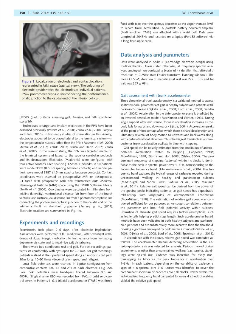
Figure 1 Localization of electrodes and contact locations
represented in MNI space (sagittal view). The colouring of
electrode tips identifies the electrodes of individual patients.
PM = pontomesencephalic line connecting the pontomesence-
phalic junction to the caudal end of the inferior colliculi.
150 | Brain 2012: 135; 148–160 W. Thevathasan et al.
8/3/2019 an W, Pogosyan a, Hyam JA, Jenkinson N, Foltynie T, Limousin P, Bogdanovic M, Zrinzo L, Green AL, Aziz TZ, Brown P
http://slidepdf.com/reader/full/an-w-pogosyan-a-hyam-ja-jenkinson-n-foltynie-t-limousin-p-bogdanovic 4/13
8/3/2019 an W, Pogosyan a, Hyam JA, Jenkinson N, Foltynie T, Limousin P, Bogdanovic M, Zrinzo L, Green AL, Aziz TZ, Brown P
http://slidepdf.com/reader/full/an-w-pogosyan-a-hyam-ja-jenkinson-n-foltynie-t-limousin-p-bogdanovic 5/13
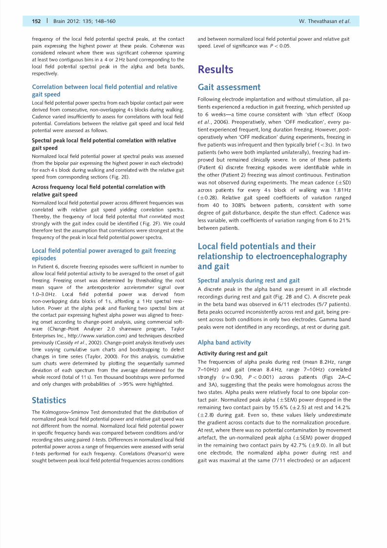
frequency of the local field potential spectral peaks, at the contact
pairs expressing the highest power at these peaks. Coherence was
considered relevant where there was significant coherence spanning
at least two contiguous bins in a 4 or 2 Hz band corresponding to the
local field potential spectral peak in the alpha and beta bands,
respectively.
Correlation between local field potential and relative
gait speedLocal field potential power spectra from each bipolar contact pair were
derived from consecutive, non-overlapping 4 s blocks during walking.
Cadence varied insufficiently to assess for correlations with local field
potential. Correlations between the relative gait speed and local field
potential were assessed as follows.
Spectral peak local field potential correlation with relative
gait speed
Normalized local field potential power at spectral peaks was assessed
(from the bipolar pair expressing the highest power in each electrode)
for each 4 s block during walking and correlated with the relative gait
speed from corresponding sections (Fig. 2E).
Across frequency local field potential correlation with
relative gait speed
Normalized local field potential power across different frequencies was
correlated with relative gait speed yielding correlation spectra.
Thereby, the frequency of local field potential that correlated most
strongly with the gait index could be identified (Fig. 2F). We could
therefore test the assumption that correlations were strongest at the
frequency of the peak in local field potential power spectra.
Local field potential power averaged to gait freezingepisodes
In Patient 6, discrete freezing episodes were sufficient in number to
allow local field potential activity to be averaged to the onset of gait
freezing. Freezing onset was determined by thresholding the root
mean square of the anteroposterior accelerometer signal over 1.0–3.0Hz. Local field potential power was derived from
non-overlapping data blocks of 1 s, affording a 1 Hz spectral reso-
lution. Power at the alpha peak and flanking two spectral bins at
the contact pair expressing highest alpha power was aligned to freez-
ing onset according to change-point analysis, using commercial soft-
ware (Change-Point Analyser 2.0 shareware program, Taylor
Enterprises Inc., http://www.variation.com) and techniques described
previously (Cassidy et al., 2002). Change-point analysis iteratively uses
time varying cumulative sum charts and bootstrapping to detect
changes in time series (Taylor, 2000). For this analysis, cumulative
sum charts were determined by plotting the sequentially summed
deviation of each spectrum from the average determined for the
whole record (total of 11 s). Ten thousand bootstraps were performed
and only changes with probabilities of 495% were highlighted.
StatisticsThe Kolmogorov–Smirnov Test demonstrated that the distribution of
normalized peak local field potential power and relative gait speed was
not different from the normal. Normalized local field potential power
in specific frequency bands was compared between conditions and/or
recording sites using paired t -tests. Differences in normalized local field
potential power across a range of frequencies were assessed with serial
t -tests performed for each frequency. Correlations (Pearson’s) were
sought between peak local field potential frequencies across conditions
and between normalized local field potential power and relative gait
speed. Level of significance was P5 0.05.
Results
Gait assessment
Following electrode implantation and without stimulation, all pa-
tients experienced a reduction in gait freezing, which persisted up
to 6 weeks—a time course consistent with ‘stun effect’ (Koop
et al., 2006). Preoperatively, when ‘OFF medication’, every pa-
tient experienced frequent, long duration freezing. However, post-
operatively when ‘OFF medication’ during experiments, freezing in
five patients was infrequent and then typically brief (53s). In two
patients (who were both implanted unilaterally), freezing had im-
proved but remained clinically severe. In one of these patients
(Patient 6) discrete freezing episodes were identifiable while in
the other (Patient 2) freezing was almost continuous. Festination
was not observed during experiments. The mean cadence (ÆSD)
across patients for every 4 s block of walking was 1.81Hz(Æ0.28). Relative gait speed coefficients of variation ranged
from 40 to 308% between patients, consistent with some
degree of gait disturbance, despite the stun effect. Cadence was
less variable, with coefficients of variation ranging from 6 to 21%
between patients.
Local field potentials and their relationship to electroencephalographyand gait
Spectral analysis during rest and gait
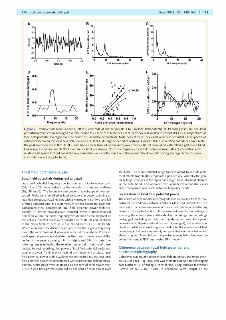
A discrete peak in the alpha band was present in all electroderecordings during rest and gait (Fig. 2B and C). A discrete peak
in the beta band was observed in 6/11 electrodes (5/7 patients).
Beta peaks occurred inconsistently across rest and gait, being pre-
sent across both conditions in only two electrodes. Gamma band
peaks were not identified in any recordings, at rest or during gait.
Alpha band activity
Activity during rest and gait
The frequencies of alpha peaks during rest (mean 8.2Hz, range
7–10Hz) and gait (mean 8.4 Hz, range 7–10Hz) correlated
strongly (r = 0.90, P50.001) across patients (Figs 2A–C
and 3A), suggesting that the peaks were homologous across thetwo states. Alpha peaks were relatively focal to one bipolar con-
tact pair. Normalized peak alpha (ÆSEM) power dropped in the
remaining two contact pairs by 15.6% (Æ2.5) at rest and 14.2%
(Æ2.8) during gait. Even so, these values likely underestimate
the gradient across contacts due to the normalization procedure.
At rest, where there was no potential contamination by movement
artefact, the un-normalized peak alpha (ÆSEM) power dropped
in the remaining two contact pairs by 42.7% (Æ9.0). In all but
one electrode, the normalized alpha power during rest and
gait was maximal at the same (7/11 electrodes) or an adjacent
152 | Brain 2012: 135; 148–160 W. Thevathasan et al.

8/3/2019 an W, Pogosyan a, Hyam JA, Jenkinson N, Foltynie T, Limousin P, Bogdanovic M, Zrinzo L, Green AL, Aziz TZ, Brown P
http://slidepdf.com/reader/full/an-w-pogosyan-a-hyam-ja-jenkinson-n-foltynie-t-limousin-p-bogdanovic 6/13
(3/11 electrodes) contact pair. Normalized peak alpha power did
not significantly differ between rest and gait.
Localization
Normalized alpha peak power during gait was significantly greater
at caudal (deeper than 2 mm below the pontomesencephalic junc-
tion) compared with rostral recording sites [t (31) = 4.183,
P50.001; Fig. 4A]. During rest, normalized alpha peak power
was also significantly greater in caudal compared with rostral re-
cording sites [t (31) = 3.359, P50.001].For rest recordings, which did not require normalization, we
were able to follow un-normalized log alpha peak power across
recording depth (Fig. 4B). The resting un-normalized log alpha
peak power was divided according to the depth of each recording
site into subregions spanning 4 mm along a rostrocaudal axis. An
ANOVA revealed a significant difference in log alpha peak power
across these subregions [F (4,28) = 7.873, P50.001) (Fig. 4B).
Resting un-normalized log alpha peak power was maximal in
the caudal PPN subregion between 2 and 6 mm below the
pontomesencephalic junction, falling significantly at rostral
subregions [À2 to À6 mm versus À2 to + 2mm, t (10) = 3.177,
P = 0.010 and À2 to À6mm versus +2 to +6mm t (8) = 4.793,
P = 0.003] and caudal subregions [À2 to À6 mm versus À6 to
À10mm, t (14) = 2.845, P = 0.013 and À2 to À6 mm versus
À10 to À14mm t (11) = 2.667, P = 0.022]. These sub-
regions along the rostrocaudal axis are represented in Fig. 4C
in standard MNI space. For Patient 3’s left electrode (used as
the individual example for local field potentials in Fig. 2) the high-
est resting peak alpha power was at 2.5 and 4.2 mm below the
pontomesencephalic line—reflecting findings from the grouped
analysis. For this patient, to give an example of the anatomy of
the region expressing highest alpha power, axial MRI slices across
this and flanking regions are displayed in the Supplementary
material.
Coherence with EEG
In 7/11 electrodes, significant local field potential EEG coherence
spanning at least two consecutive 1 Hz bins was observed within
the alpha peak range during rest and/or walking (Fig 2D).
Figure 3 (A) Relationship between the peak rest local field potential (LFP) and peak gait local field potential. The strong correlation
(r = 0.90, P50.001) suggests that the two local field potential peaks may be related. (B) Relationship between the peak rest local field
potential and the frequency of the peak in the local field potential–relative gait speed (rGS) correlation spectra. The strong correlation
(r = 0.779, P = 0.013) suggests that the peak rest local field potential may be relevant to the performance of gait. (C) Group average
un-normalized local field potential and relative gait speed correlation spectra demonstrating a peak in correlation in the alpha band. Dotted
line is the average correlation and continuous line is the 6-point (backwards) moving-average. ( D) Group averaged normalized (across
5–40 Hz) local field potential and relative gait speed correlation spectra demonstrating persistence of the peak in correlation in the alphaband. Dotted line is the average correlation and continuous line is the 6-point (backwards) moving-average. Correlations were Fisher
transformed prior to averaging in C and D.
PPN oscillations correlate with gait Brain 2012: 135; 148–160 | 153

8/3/2019 an W, Pogosyan a, Hyam JA, Jenkinson N, Foltynie T, Limousin P, Bogdanovic M, Zrinzo L, Green AL, Aziz TZ, Brown P
http://slidepdf.com/reader/full/an-w-pogosyan-a-hyam-ja-jenkinson-n-foltynie-t-limousin-p-bogdanovic 7/13
Relationship with relative gait speed
In eight of nine electrodes where local field potential and trunk
acceleration were simultaneously recorded, normalized peak alpha
power correlated significantly with the relative gait speed. In these
electrodes, the mean correlation was r = 0.433, P50.01 (see
Fig. 2E for an example from a single electrode). Local field poten-
tial relative gait speed correlation spectra in all electrodes revealed
that the correlation peaked in the alpha band (see Fig. 2F for an
example from a single electrode). The frequency of this peak
correlated strongly with the peak local field potential during gait
(r = 0.782, P = 0.013) and peak local field potential at rest
(r = 0.779, P = 0.013; Fig. 3B). When averaged across recordings,
spectra of the Fisher transformed correlations between local field
potential power and relative gait speed demonstrated a discrete
positive peak in correlation at 7–10 Hz, regardless of whether local
field potential power was raw (Fig. 3C) or normalized (Fig. 3D).
Note that normalization inevitably induced a negative correlation
at frequencies below 7 Hz.
Figure 4 (A) Normalized power (mean Æ SEM) of alpha peaks during gait and beta peaks during rest or gait grouped according to caudal
(deeper than 2 mm below the pontomesencephalic junction) or rostral recording site. Alpha peak power was greater caudally and beta
power was greater rostrally. (B) Log alpha peak power at rest (mean Æ SEM) divided according to recording site depth into 4-mm
subregions (denoted on thex
-axis in mm relative to the pontomesencephalic junction). Log alpha peak power is maximal in theÀ
2 toÀ6 mm region, falling significantly at surrounding sites. *P50.05. In grey has been superimposed a plot of the variation in per cent
improvement in the Gait and Falls questionnaire (GFQ) postoperatively according to stimulation depth at most recent follow-up [depth
selected blinded to the local field potential (LFP) data]. One data point was available from each subject (Table 2), with the exception of the
patient from London, where the Gait and Falls questionnaire was not assessed. Three of these data points fell À6 to À10 mm below the
pontomesencephalic junction, so that here the SEM is also displayed. (C) Representation of the rostrocaudal location of peak
un-normalized alpha power at rest, which correlated strongly in frequency and location with peak alpha power that correlated with gait
(but which required normalization to remove movement artefact thereby also diminishing power gradients). Relative log alpha power is
represented in grey-scale intensity whereby black is highest and white lowest alpha power. Regions relative to the pontomesencephalic
line are numbered as follows: 1, + 2 to + 6 mm; 2, À2 to + 2 mm; 3, À6 to À2 mm; 4, À10 to À6 mm; and 5, À14 to À10 mm. Alpha
power was maximal at location 3 (À6 to À2 mm below the pontomesencephalic line). Beta power was highest in regions 1 and 2
combined. Note: no inference is made regarding location in ventrodorsal or mediolateral planes. DBS = deep brain stimulation; GFQ = Gait
and Falls questionnaire; LFP = local field potential; PM = pontomesencephalic line connecting the pontomesencephalic junction to the
caudal end of the inferior colliculi.
154 | Brain 2012: 135; 148–160 W. Thevathasan et al.

8/3/2019 an W, Pogosyan a, Hyam JA, Jenkinson N, Foltynie T, Limousin P, Bogdanovic M, Zrinzo L, Green AL, Aziz TZ, Brown P
http://slidepdf.com/reader/full/an-w-pogosyan-a-hyam-ja-jenkinson-n-foltynie-t-limousin-p-bogdanovic 8/13
Averaged to gait freezing episodes
Patient 6 had 24 discrete freezing episodes identifiable during
recordings and a local field potential alpha peak during gait at8 Hz. Local field potential power over 7–9Hz averaged to the
onset of the freezing episodes is represented in Fig. 5. There is
a significant associated attenuation in alpha power, which begins
around 1 s prior to the onset of freezing and continues for over 2 s
thereafter.
Beta band activity
Activity during rest or gait
Beta peaks (mean 21.1 Hz, range 17.3–28.5) were relatively focal
to one bipolar contact pair, with normalized peak beta (ÆSEM)
power dropping by 15.4 (Æ2.4)% at the remaining two contact
pairs of each electrode. At rest, the un-normalized peak beta(ÆSEM) power dropped in the remaining two contact pairs by
45.6 (Æ11.1)%.
Localization
In the six electrodes demonstrating a beta peak (during rest or
gait), normalized beta peak power was significantly greater in ros-
tral (higher than 2 mm beneath the pontomesencephalic junction)
than caudal contacts [t (16) = À2.232, P = 0.040; Fig. 4]. There
were insufficient beta peaks at rest to compare un-normalized
beta peak power with location.
Coherence with EEG
In only one of the six electrodes demonstrating a beta peak, wasthere significant coherence spanning at least two consecutive 1 Hz
bins between the local field potential at beta peak frequency and
FzCz.
Relationship with relative gait speed
Normalized beta peak power did not significantly correlate with
relative gait speed. Similarly, spectra of correlations between nor-
malized local field potential at each frequency and relative gait
speed did not reveal discrete peaks in correlation at the frequen-
cies corresponding to beta peaks.
Therapeutic impact of chronicstimulation on gait
Therapeutic outcomes from chronic PPN region stimulation are
presented in Table 2. Pre- and postoperative Gait and Falls ques-
tionnaire scores were available in all six cases operated in Oxford,
affording six estimates of therapeutic impact. Gait and Falls ques-
tionnaire scores in these six patients improved with PPN stimula-
tion [mean 42.0–29.8, t (5) = 4.425, P = 0.007]. The location
where stimulation was applied along the rostrocaudal axis was
calculated relative to the pontomesencephalic line. Where stimu-
lation was bilateral, stimulation depth was the mean from both
sides. The overall improvement of Gait and Falls questionnaire
scores with PPN stimulation was only moderate. However, closer
examination within this small sample suggests a possible relation-
ship between stimulation depth and percentage improvement inthe Gait and Falls questionnaire with the best outcome being
achieved when stimulation was applied at the level of maximal
alpha activity (Fig. 4B). Thus, a relationship was sought between
power in the alpha band where stimulation was applied and thera-
peutic outcomes. With monopolar stimulation, alpha power was
taken as the mean power recorded from the two flanking bipolar
pairs. A correlation between mean log alpha power at rest and per
cent postoperative Gait and Falls questionnaire improvement con-
firmed a correspondence between these measures (r = 0.85,
P = 0.033). The picture was similar if the per cent improvement
(20%) in UPDRS II items scoring freezing, falls and gait was
included from the London patient in whom these measures were
used to assess outcome (r = 0.81, P = 0.027).
DiscussionIn parkinsonian patients, we found alpha oscillations in a network
involving the caudal subregion of the PPN and the cerebral cortex.
Synchronization of PPN alpha activity in the caudal PPN region
correlated with the performance of gait. Beta oscillations were
found in the rostral PPN subregion but this activity did not correl-
ate with gait.
Figure 5 Change-point analysis of the time series of mean local field potential power over 7–9 Hz averaged to onset (0 s) of freezing
episodes (n = 24) in Case 6. Horizontal dotted lines are the 95% confidence limit of the whole record and the grey blocks represent stable
periods between changes in power as defined by change-point analysis. There is a significant drop in 7–9 Hz power $1 s before the onset
of freezing, and 7–9 Hz activity continues to be attenuated for just over 2 s thereafter.
PPN oscillations correlate with gait Brain 2012: 135; 148–160 | 155

8/3/2019 an W, Pogosyan a, Hyam JA, Jenkinson N, Foltynie T, Limousin P, Bogdanovic M, Zrinzo L, Green AL, Aziz TZ, Brown P
http://slidepdf.com/reader/full/an-w-pogosyan-a-hyam-ja-jenkinson-n-foltynie-t-limousin-p-bogdanovic 9/13
Topographical distribution ofoscillations
A rostrocaudal topographical organization of the PPN is supported
by extensive data from animal research (Martinez-Gonzalez et al.,
2011). Rostral PPN neurons predominantly express -aminobutyric
acid and are strongly interconnected with the basal ganglia includ-
ing subthalamic nucleus and internal pallidum (Mena-Segovia
et al., 2004; Ros et al., 2010; Martinez-Gonzalez et al., 2011),
which exhibit beta activity in untreated Parkinson’s disease
(Hammond et al., 2007). Caudal PPN neurons express predomin-
antly acetylcholine and glutamate (Martinez-Gonzalez et al.,
2011). Cholinergic neurons arborize widely, including to cortex
and locomotor centres (Skinner et al., 1990; Mena-Segovia
et al., 2008). It is therefore congruent that we found beta oscil-
lations in the rostral PPN region and a distinct oscillatory activity in
the caudal PPN region, alpha activity, coherent with cortex and
associated with locomotion. This interpretation is also supported
by the one previous study in which PPN local field potentials were
assessed in parkinsonian patients according to recording site depth
(Weinberger et al., 2008). In that study, beta oscillations were
recorded in an area corresponding to what is here defined as
the rostral PPN region. In an example local field potential record-
ing, a spectral peak can also be identified in the alpha band in
what is defined here as the caudal PPN region (1–4 mm below the
inferior colliculus). Of further interest, neurons considered by the
authors as likely cholinergic (having long duration action potentials
and low firing rates) were found predominantly in the caudal PPNregion (Weinberger et al., 2008). Such topography could explain
why different centres have reported different dominant oscillatory
patterns, in the alpha or beta frequency bands, from PPN
recordings—as this would be determined by the depth of the im-
planted electrodes (Androulidakis et al., 2008a, b; Weinberger
et al., 2008; Tsang et al., 2010)
The origin of the focal alpha activity in the PPN region needs
consideration. The distribution of electrode contacts in this study
was broad—and this facilitated the relationship between oscillatory
power and location to be recognized. Some contacts lie more
caudal than any described location of the PPN but these were
not the ones expressing alpha at highest power across patients.
Across patients, un-normalized peak alpha power at rest, whichcorrelated strongly in frequency and location with alpha power
during gait, was maximal between 2 mm and 6 mm beneath the
pontomesencephalic junction. The spatial accuracy of this finding
is limited by the bipolar nature of the local field potential record-
ings and the averaging of data across patients. However, with this
limitation in mind, a key question is whether this region corres-
ponds to the PPN. The PPN comprises a well-defined ‘pars com-
pacta’ subregion but also a reticular ‘pars dissipata’ with indistinct
boundaries (Olszewski and Baxter, 1954). The atlas of Olszewski
and Baxter (1982) identifies the PPN based on cytoarchitectural
methods. Later, cholinergic PPN neurons were identified in
humans by immunohistochemical labelling of choline acetyltrans-
ferase (Mesulam et al., 1989; Manaye et al., 1999). Althoughthese latter studies do not provide stereotactic coordinates, their
relevance has been heightened by the involvement of cholinergic
PPN neurons in gait and its dysfunction (Karachi et al., 2010) and
several observations are of localizing value. While cholinergic PPN
neurons were clustered most densely in the pars compacta, the
pars dissipata accounts for the greater proportion of cholinergic
neurons (Manaye et al., 1999). Cholinergic PPN neurons span at
least 7 mm in the rostrocaudal plane being at highest density
2.5 mm under the rostral pole (as defined by choline acetyltrans-
ferase staining, so potentially missing any rostral component not
expressing choline acetyltransferase; Manaye et al., 1999).
Cholinergic PPN neurons were noted to lie where the ‘superior
cerebellar peduncle ascends towards the dorsolateral pons towardsits decussation’ with the pars compacta located at the level of
the decussation (Mesulam et al., 1989; Manaye et al., 1999).
Identifying these anatomical landmarks on the axial images of
Patient 3 suggests that the levels 2–4 mm below the pontomesen-
cephalic line would therefore correspond to the caudal PPN
region. Other gait related entities also exist in the PPN region
including the cuneiform and subcuneiform nuclei, which together
with the PPN comprise the mesencephalic locomotor region (Alam
et al., 2011). Both the PPN and cuneiform nucleus are activated
during fast imagined gait in functional MRI studies in healthy
Table 2 Pedunculopontine nucleus region stimulation and therapeutic outcomes
Patients Rostrocaudalstimulation location(mm to PM line)
Stimulationsettings(Hz/V/ms)
PreoperativeGFQ (score/64)
PostoperativeGFQ (score/64)
Improvement inGFQ (%)
Timepost operation(months)
1 À7.65 Bilateral 35/3.5/60 55 38 30.9 2
2 À12.7 Unilateral 35/2.3/60 38 34 10.5 13
3aÀ5.5 Bilateral 30/2.5/60 36 15 58.3 9
4À
1.5 Bilateral 40/1.8/60 49 42 14.3 75 À8.9 Bilateral 35/2.2/60 36 28 22.2 2
6b + 0.4 Unilateral 30/3.0/60 NA NA NAb NA
7 À7.0 Bilateral 20/2.5/60 38 22 42.1 2
Where bilateral stimulation was applied, voltage reflects the mean of both sides.
a Stimulation remains under titration and cycled between off and on, with postoperative GFQ score the best result so far.
b Outcome in this patient was assessed with UPDRS II items scoring freezing, falls and gait with the combined score being 5/16 preoperatively and 4/16 postoperatively (on
medication).
GFQ = Gait and Falls questionnaire; NA = not available; PM = pontomesencephalic.
156 | Brain 2012: 135; 148–160 W. Thevathasan et al.
8/3/2019 an W, Pogosyan a, Hyam JA, Jenkinson N, Foltynie T, Limousin P, Bogdanovic M, Zrinzo L, Green AL, Aziz TZ, Brown P
http://slidepdf.com/reader/full/an-w-pogosyan-a-hyam-ja-jenkinson-n-foltynie-t-limousin-p-bogdanovic 10/13
subjects (Karachi et al., 2010). Microelectrode recordings have
suggested that neurons that modulate firing in response to ima-
gined gait actually tend to be located in the subcuneiform region
dorsal to the PPN (Piallat et al., 2009). However, boundaries
between these various nuclei are indistinct—potentially confound-
ing precise determination of the source of neuronal recordings and
of the structures responsible for clinical effects of stimulation in
this region. On the other hand neurons of the PPN, but not the
cuneiform or subcuneiform, are reported to degenerate in associ-
ation with falls in Parkinson’s disease (Karachi et al., 2010).
Furthermore, cytoarchitectural atlases suggest that the cuneiform
and subcuneiform nuclei do not extend as caudally as the zone
where we recorded maximal alpha band activity (Olszewski and
Baxter, 1982).
The distribution of PPN oscillatory activity concurs with previous
work in Parkinson’s disease suggesting that alpha and beta oscil-
latory networks are segregated (Litvak et al., 2010). The function-
al nature of alpha and beta oscillations in Parkinson’s disease also
appears distinct. Beta band activity is pathologically increased in
Parkinson’s disease, is suppressed by L-dopa and high-frequency
subthalamic stimulation and correlates with deficits of bradykinesiaand rigidity (Silberstein et al., 2005; Kuhn et al., 2006; Eusebio
et al., 2010). Alpha power in the PPN tends to increase with L-
dopa, suggesting that it could be pathologically attenuated in
Parkinson’s disease (Androulidakis et al., 2008a, b). It now also
appears that attenuation of PPN alpha activity is associated with
gait freezing whilst increases in PPN alpha power in Parkinson’s
disease correlated with improved gait.
Pedunculopontine nucleus alphaoscillations and locomotor function
A potential confound needs consideration; that the correlation be-tween alpha power and gait speed could be due to movement
artefact. Several factors militate against this. First, our local field
potential recordings were bipolar, so a signal common across con-
tacts should have been subtracted out. Second, the relative gait
speed correlated maximally with alpha oscillations (7–10 Hz),
which differed in frequency from the accelerometer frequencies
used to derive gait speed (1–3Hz). Third, correlation with the
relative gait speed was relatively specific to alpha local field po-
tential activity, evidenced by the peak in correlation spectra at
alpha frequencies and the lack of any correlation with beta
power. Fourth, correlations were present even when local field
potential power was normalized to broad band activity, which
effectively eliminated any common artefact from movement.An important qualification is that the correlation between gait
speed and alpha power does not necessarily imply causation and
the relationship could be epiphenomenal. For example, alpha
power may be permissive for higher gait speeds or reactive to it.
However, correlations were found within a brain area that is func-
tionally relevant to gait disturbance, as evidenced by the potential
for improvement upon PPN stimulation. Furthermore, the positive
nature of the correlation between alpha synchronization and
increasing gait speed, concurs with the synchronization of alpha
power observed with L-dopa (Androulidakis et al., 2008a, b),
which also tends to improve gait. Yet, even if PPN alpha oscilla-
tions are mechanistically important in gait disturbance, the rela-
tionship does not appear obligatory or exclusive. The mean
correlation was 0.43, so that fluctuations in alpha power only
predicted $20% (r 2 = 0.19) of the variance in relative gait
speed. A further question is whether this alpha activity also relates
specifically to gait freezing. Due to the reduction in freezing post-
operatively due to a possible stun effect, we could only compare
freezing episodes with alpha power in a single patient, who was
implanted unilaterally. This demonstrated an attenuation of alpha
activity related to the onset of freezing. This suggests that higher
gait speeds could have, at least partly, resulted from relief of
freezing related deficits even if discrete freezing episodes did not
frequently occur. For example, patients with gait freezing are re-
ported to have continuous background deficits in gait, including
reduced gait velocity, step length and increased spatiotemporal
variability (Hausdorff et al., 2003b; Chee et al., 2009; Snijders
et al., 2011). It is notable that attenuation of alpha local field
potential power preceded freezing onset. However, this latter find-
ing should be interpreted cautiously given the limited accuracy in
determining when exactly freezing begins.Thus, a particular limitation of the current study was the limited
characterization of gait performance in terms of relative gait speed
and the analysis of freezing in only one patient. Gait dysfunction
in Parkinson’s disease is complex, with varying involvement of
features such as gait initiation, turning, postural instability and
gait velocity (Bloem, 1992; Morris et al., 1994; Hausdorff et al.,
2003b; Sofuwa et al., 2005). The failure to capture these features
might also help explain why fluctuations in alpha power only pre-
dicted $20% of the variance in relative gait speed.
By what mechanism could PPN alpha activity relate to gait per-
formance, as indexed by relative gait speed? Gait speed reduces in
healthy subjects, elderly fallers and in Parkinson’s disease during
the performance of a second, unrelated task (‘dual tasking’)(Hausdorff et al., 2003a; Springer et al., 2006; Lamoth et al.,
2011). In elderly subjects, an inability to ‘walk whilst talking’ pre-
dicts falls (Lundin-Olsson et al., 1997). Such findings implicate
‘attention’ and the effective allocation of processing resources
that flows from it, as a potentially important factor influencing
gait speed. In Parkinson’s disease, attentional deficits are
common and there is also impaired automaticity of movement
so that processing demands are higher (Wu and Hallett, 2005,
2008). Parkinsonian patients with gait freezing are reported to
have even more attentional deficits than those without gait
freezing (Amboni et al., 2008; Yogev-Seligmann et al., 2008).
Dual tasking can worsen gait freezing, as can other precipitants
that are thought to ‘distract’ attention away from gait (Giladi andHausdorff, 2006).
There is increasing evidence that alpha activity has an important
role in attention and the allocation of processing resources. The
synchronization of occipital alpha with eye closure was once in-
terpreted to reflect passive ‘idling’ (Berger, 1929; Pfurtscheller
et al., 1996). However, alpha activity is now considered to support
active suppression of task irrelevant processes (Jensen and
Mazaheri, 2010). For example, during working memory tasks, cor-
tical alpha power in visual and motor–sensory areas increases and
the degree of synchronization correlates with the number of items
PPN oscillations correlate with gait Brain 2012: 135; 148–160 | 157

8/3/2019 an W, Pogosyan a, Hyam JA, Jenkinson N, Foltynie T, Limousin P, Bogdanovic M, Zrinzo L, Green AL, Aziz TZ, Brown P
http://slidepdf.com/reader/full/an-w-pogosyan-a-hyam-ja-jenkinson-n-foltynie-t-limousin-p-bogdanovic 11/13
recalled (Jensen et al., 2002; Haegens et al., 2010). Correlation of
performance with oscillatory power in task irrelevant regions sug-
gests suppression rather than mere idling. It has been proposed
that within the motor system, suppression of competing processes
with alpha could aid the smooth execution of motor programmes
(Pfurtscheller and Neuper, 1994; Suffczynski et al., 2001). Regions
such as the subthalamic nucleus and caudal PPN that have dis-
tributed functional connectivity, including with cerebral corticalareas, would be placed to operationalize this putative role. Thus
PPN deep brain stimulation is able to increase blood flow in an
extensive network of subcortical and cortical areas involved in
balance and motor control (Strafella et al., 2008; Ballanger
et al., 2009).
Clinical relevance to pedunculopontinenucleus stimulation
Gait freezing improved following electrode implantation in the ab-
sence of any stimulation in all patients, although this was less
pronounced in patients implanted unilaterally. Gait freezing is no-torious for improving during medical assessments, perhaps due to
attentional or placebo effects. However, we observed that the
improvement in gait freezing persisted for up to 6 weeks
post-implantation—a time course arguably more consistent with
a stun effect. In the subthalamic nucleus, the ‘stun’ or ‘microle-
sion’ effect of surgery is usually attributed to suppression of beta
activity in the target nucleus by acute tissue disruption from elec-
trode implantation (Chen et al., 2006; Koop e t al., 2006).
However, in the caudal PPN, alpha activity persisted despite the
apparent stun effect and correlated positively with gait perform-
ance. Accordingly, we speculate that stun effects in PPN surgery
might predominately arise from microlesioning inhibitory suprasp-
inal influences along the electrode trajectory. Excessive inhibition
of the PPN (for example from the internal pallidum) has been
considered a pathophysiological factor causing gait and postural
disturbance in Parkinson’s disease, in addition to PPN neuronal
degeneration (Aziz et al., 1998). Consistent with this reasoning,
in the 1-methyl-4-phenyl-1,2,3,6-tetrahydropyridine (MPTP)
primate model of Parkinson’s disease, the microinjection of a
-aminobutyric acid antagonist into the PPN improved mobility
(Nandi et al., 2002). Microlesioning inhibitory afferents to the
PPN would be expected to have analogous effects. If this reason-
ing is correct, then the occurrence of a possible stun effect would
not necessarily indicate accurate electrode placement within the
PPN, nor necessarily portend benefit from stimulation.The topographic arrangement of oscillations in the PPN region
along the rostrocaudal axis raises the possibility that caudal and
rostral PPN stimulation could have different therapeutic effects.
Alpha and beta oscillatory activity could provide functional bio-
markers for these different subregions. Alpha and beta activity in
the PPN region were focal, evidenced by un-normalized oscillatory
power at rest dropping by an average of 42% for alpha activity
and 45% for beta activity across bipolar recording sites. This is
similar to the gradients of beta activity reported across the dorso-
lateral subthalamic nucleus that can be detected in intraoperative
and postoperative recordings and used to guide deep brain stimu-
lation electrode implantation (Chen et al., 2006).
Only the alpha activity recorded in the caudal PPN region was
demonstrated to be functionally related to gait and gait freezing.
Therapeutic outcomes from PPN region stimulation were variable
between patients as were the locations along the rostrocaudal axis
where stimulation was applied. Interestingly, the best outcome
was achieved when stimulation was applied at the level of max-
imal alpha activity, 2–6 mm below the pontomesencephalic junc-
tion, so that per cent improvement in Gait and Falls questionnaire
score at different depths closely correlated with the alpha power
recorded at the same depths. We take this to be an encouraging
preliminary finding, noting that patient numbers were small, uni-
lateral and bilateral stimulation were mixed, postoperative periods
were relatively short and our outcome measure was not objective.
Accordingly, it is important that future studies examine whether
the efficacy of PPN stimulation relates to stimulation location
‘within’ patients, preferably using objective gait measures. Still,
the current study is important in providing physiological evidence,
supported by consistent clinical findings, that the site of stimula-
tion along the rostrocaudal axis of the PPN region might be afactor determining therapeutic outcomes.
AcknowledgementsWe thank Prof Brian Day for assistance in recording gait in patient
six, Prof Peter Silburn for advice on anatomical targeting and Ms
Beth Forrow for assistance with clinical assessments.
FundingNational Institute of Health Research Oxford Biomedical Research
Centre, Medical Research Council (UK) and Rosetrees Trust.
Supplementary materialSupplementary material is available at Brain online.
ReferencesAlam M, Schwabe K, Krauss JK. The pedunculopontine nucleus
area: critical evaluation of interspecies differences relevant for itsuse as a target for deep brain stimulation. Brain 2011; 134 (Pt 1):
11–23.
Amboni M, Cozzolino A, Longo K, Picillo M, Barone P. Freezing of gait
and executive functions in patients with Parkinson’s disease. Mov
Disord 2008; 23: 395–400.
Androulidakis AG, Khan S, Litvak V, Pleydell-Pearce CW, Brown P,
Gill SS. Local field potential recordings from the pedunculopontine
nucleus in a Parkinsonian patient. Neuroreport 2008a; 19: 59–62.
Androulidakis AG, Mazzone P, Litvak V, Penny W, Dileone M,
Gaynor LM, et al. Oscillatory activity in the pedunculopontine
area of patients with Parkinson’s disease. Exp Neurol 2008b; 211:
59–66.
158 | Brain 2012: 135; 148–160 W. Thevathasan et al.

8/3/2019 an W, Pogosyan a, Hyam JA, Jenkinson N, Foltynie T, Limousin P, Bogdanovic M, Zrinzo L, Green AL, Aziz TZ, Brown P
http://slidepdf.com/reader/full/an-w-pogosyan-a-hyam-ja-jenkinson-n-foltynie-t-limousin-p-bogdanovic 12/13
Aziz TZ, Davies L, Stein J, France S. The role of descending basal ganglia
connections to the brain stem in parkinsonian akinesia. Br J Neurosurg
1998; 12: 245–9.
Ballanger B, Lozano AM, Moro E, van Eimeren T, Hamani C, Chen R,
et al. Cerebral blood flow changes induced by pedunculopontine nu-
cleus stimulation in patients with advanced Parkinson’s disease: a
[(15)O] H2O PET study. Hum Brain Mapp 2009; 30: 3901–9.
Berger H. U ¨ ber das elektroenkephalogramm des menschen. Arch
Psychiatr Nervenkr 1929; 87: 527–70.
Bloem BR. Postural instability in Parkinson’s disease. Clin NeurolNeurosurg 1992; 94 (Suppl): S41–5.
Bloem BR, Hausdorff JM, Visser JE, Giladi N. Falls and freezing of gait in
Parkinson’s disease: a review of two interconnected, episodic phenom-
ena. Mov Disord 2004; 19: 871–84.
Breit S, Bouali-Benazzouz R, Benabid AL, Benazzouz A. Unilateral
lesion of the nigrostriatal pathway induces an increase of neuronal
activity of the pedunculopontine nucleus, which is reversed by the
lesion of the subthalamic nucleus in the rat. Eur J Neurosci 2001;
14: 1833–42.
Cappozzo A. Low frequency self-generated vibration during ambulation
in normal men. J Biomech 1982; 15: 599–609.
Cassidy MJ, Brown P. Hidden Markov based autoregressive analysis of
stationary and non-stationary electrophysiological signals for functional
coupling studies. J Neurosci Methods 2002; 116: 35–53.
Chee R, Murphy A, Danoudis M, Georgiou-Karistianis N, Iansek R. Gaitfreezing in Parkinson’s disease and the stride length sequence effect
interaction. Brain 2009; 132 (Pt 8): 2151–60.
Chen CC, Pogosyan A, Zrinzo LU, Tisch S, Limousin P, Ashkan K, et al.
Intra-operative recordings of local field potentials can help localize the
subthalamic nucleus in Parkinson’s disease surgery. Exp Neurol 2006;
198: 214–21.
Dijkstra B, Zijlstra W, Scherder E, Kamsma Y. Detection of walking per-
iods and number of steps in older adults and patients with Parkinson’s
disease: accuracy of a pedometer and an accelerometry-based
method. Age Ageing 2008; 37: 436–41.
Eusebio A, Thevathasan W, Doyle Gaynor L, Pogosyan A, Bye E,
Foltynie T, et al. Deep brain stimulation can suppress pathological
synchronisation in parkinsonian patients. J Neurol Neurosurg
Psychiatry 2011; 82: 569–573.
Factor SA. The clinical spectrum of freezing of gait in atypical parkinson-
ism. Mov Disord 2008; 23 (Suppl 2): S431–8.
Ferraye MU, Debu B, Fraix V, Goetz L, Ardouin C, Yelnik J, et al. Effects
of pedunculopontine nucleus area stimulation on gait disorders in
Parkinson’s disease. Brain 2009; 133 (Pt 1): 205–14.
Foltynie T, Hariz MI. Surgical management of Parkinson’s disease. Expert
Rev Neurother 2010; 10: 903–14.
Fries P. A mechanism for cognitive dynamics: neuronal communication
through neuronal coherence. Trends Cogn Sci 2005; 9: 474–80.
Giladi N, Hausdorff JM. The role of mental function in the pathogenesis
of freezing of gait in Parkinson’s disease. J Neurol Sci 2006; 248:
173–6.
Giladi N, McDermott MP, Fahn S, Przedborski S, Jankovic J, Stern M,
et al. Freezing of gait in PD: prospective assessment in the DATATOP
cohort. Neurology 2001; 56: 1712–21.
Giladi N, Shabtai H, Simon ES, Biran S, Tal J, Korczyn AD. Construction
of freezing of gait questionnaire for patients with parkinsonism.Parkinsonism Relat Disord 2000; 6: 165–70.
Giladi N, Tal J, Azulay T, Rascol O, Brooks DJ, Melamed E, et al.
Validation of the freezing of gait questionnaire in patients with
Parkinson’s disease. Mov Disord 2009; 24: 655–61.
Grillner S, Wallen P, Saitoh K, Kozlov A, Robertson B. Neural bases of
goal-directed locomotion in vertebrates—an overview. Brain Res Rev
2008; 57: 2–12.
Grosse P, Cassidy M, Brown P. EEG-EMG, MEG-EMG and EMG-EMG
frequency analysis: physiological principles and clinical applications.
Clin Neurophysiol 2002; 113: 1523–31.
Haegens S, Osipova D, Oostenveld R, Jensen O. Somatosensory working
memory performance in humans depends on both engagement and
disengagement of regions in a distributed network. Hum Brain Mapp
2010; 31: 26–35.
Hammond C, Bergman H, Brown P. Pathological synchronization in
Parkinson’s disease: networks, models and treatments. Trends
Neurosci 2007; 30: 357–64.
Hausdorff JM, Balash J, Giladi N. Effects of cognitive challenge on gait
variability in patients with Parkinson’s disease. J Geriatr Psychiatry
Neurol 2003a; 16: 53–8.
Hausdorff JM, Schaafsma JD, Balash Y, Bartels AL, Gurevich T, Giladi N.
Impaired regulation of stride variability in Parkinson’s disease subjectswith freezing of gait. Exp Brain Res 2003b; 149: 187–94.
Hirsch EC, Graybiel AM, Duyckaerts C, Javoy-Agid F. Neuronal loss in
the pedunculopontine tegmental nucleus in Parkinson disease and in
progressive supranuclear palsy. Proc Natl Acad Sci USA 1987; 84:
5976–80.
Ichinoseki-Sekine N, Kuwae Y, Higashi Y, Fujimoto T, Sekine M,
Tamura T. Improving the accuracy of pedometer used by the elderly
with the FFT algorithm. Med Sci Sports Exerc 2006; 38: 1674–81.
Jacobsohn L. Uber die Kerne des menschlichen Hirnstamms: (Medulla
oblongata, Pons, und Pedunculus cerebri). Anhang zuden
Abhandlungen der Kgl Preuss. Akad d Wiss 1911.
Jankovic J. Parkinson’s disease: clinical features and diagnosis. J Neurol
Neurosurg Psychiatry 2008; 79: 368–76.
Jensen O, Gelfand J, Kounios J, Lisman JE. Oscillations in the alpha band
(9-12 Hz) increase with memory load during retention in a short-termmemory task. Cereb Cortex 2002; 12: 877–82.
Jensen O, Mazaheri A. Shaping functional architecture by oscillatory
alpha activity: gating by inhibition. Front Hum Neurosci 2010; 4: 186.
Karachi C, Grabli D, Bernard FA, Tande D, Wattiez N, Belaid H, et al.
Cholinergic mesencephalic neurons are involved in gait and postural
disorders in Parkinson disease. J Clin Invest 2010; 120: 2745–54.
Koop MM, Andrzejewski A, Hill BC, Heit G, Bronte-Stewart HM.
Improvement in a quantitative measure of bradykinesia after micro-
electrode recording in patients with Parkinson’s disease during deep
brain stimulation surgery. Mov Disord 2006; 21: 673–8.
Kuhn AA, Kupsch A, Schneider GH, Brown P. Reduction in subthalamic
8-35 Hz oscillatory activity correlates with clinical improvement in
Parkinson’s disease. Eur J Neurosci 2006; 23: 1956–60.
Lamoth CJ, van Deudekom FJ, van Campen JP, Appels BA, de Vries OJ,
Pijnappels M. Gait stability and variability measures show effects of
impaired cognition and dual tasking in frail people. J Neuroeng Rehabil
2011; 8: 2.
Lord S, Rochester L, Baker K, Nieuwboer A. Concurrent validity of accel-
erometry to measure gait in Parkinsons disease. Gait Posture 2008; 27:
357–9.
Litvak V, Jha A, Eusebio A, Oostenveld R, Foltynie T, Limousin P, et al.
Resting oscillatory cortico-subthalamic connectivity in patients with
Parkinson’s disease. Brain 2011; 134: 359–74.
Lundin-Olsson L, Nyberg L, Gustafson Y. “Stops walking when talking”
as a predictor of falls in elderly people. Lancet 1997; 349: 617.
MacDougall HG, Moore ST. Marching to the beat of the same drummer:
the spontaneous tempo of human locomotion. J Appl Physiol 2005;
99: 1164–73.
Macht M, Kaussner Y, Moller JC, Stiasny-Kolster K, Eggert KM,
Kruger HP, et al. Predictors of freezing in Parkinson’s disease: a
survey of 6,620 patients. Mov Disord 2007; 22: 953–6.MacKinnon CD, Winter DA. Control of whole body balance in the front-
al plane during human walking. J Biomech 1993; 26: 633–44.
Manaye KF, Zweig R, Wu D, Hersh LB, De Lacalle S, Saper CB, et al.
Quantification of cholinergic and select non-cholinergic mesopontine
neuronal populations in the human brain. Neuroscience 1999; 89:
759–70.
Martinez-Gonzalez C, Bolam JP, Mena-Segovia J. Topographical
organization of the pedunculopontine nucleus. Front Neuroanat
2011; 5: 22.
Mazzone P, Lozano A, Stanzione P, Galati S, Scarnati E, Peppe A, et al.
Implantation of human pedunculopontine nucleus: a safe and clinically
relevant target in Parkinson’s disease. Neuroreport 2005; 16: 1877–81.
PPN oscillations correlate with gait Brain 2012: 135; 148–160 | 159
8/3/2019 an W, Pogosyan a, Hyam JA, Jenkinson N, Foltynie T, Limousin P, Bogdanovic M, Zrinzo L, Green AL, Aziz TZ, Brown P
http://slidepdf.com/reader/full/an-w-pogosyan-a-hyam-ja-jenkinson-n-foltynie-t-limousin-p-bogdanovic 13/13
Mena-Segovia J, Bolam JP, Magill PJ. Pedunculopontine nucleus and
basal ganglia: distant relatives or part of the same family? Trends
Neurosci 2004; 27: 585–8.
Mena-Segovia J, Sims HM, Magill PJ, Bolam JP. Cholinergic brainstem
neurons modulate cortical gamma activity during slow oscillations.
J Physiol 2008; 586: 2947–60.
Mesulam MM, Geula C, Bothwell MA, Hersh LB. Human reticular for-
mation: cholinergic neurons of the pedunculopontine and laterodorsal
tegmental nuclei and some cytochemical comparisons to forebrain
cholinergic neurons. J Comp Neurol 1989; 283: 611–33.Mirelman A, Gurevich T, Giladi N, Bar-Shira A, Orr-Urtreger A,
Hausdorff JM. Gait alterations in healthy carriers of the LRRK2
G2019S mutation. Ann Neurol 2011; 69: 193–7.
Moe-Nilssen R. A new method for evaluating motor control in gait under
real-life environmental conditions. Part 2: Gait analysis. Clin Biomech
1998; 13: 328–35.
Moro E, Hamani C, Poon YY, Al-Khairallah T, Dostrovsky JO,
Hutchison WD, et al. Unilateral pedunculopontine stimulation im-
proves falls in Parkinson’s disease. Brain 2010; 133 (Pt 1): 215–24.
Morris ME, Iansek R, Matyas TA, Summers JJ. The pathogenesis of gait
hypokinesia in Parkinson’s disease. Brain 1994; 117 (Pt 5): 1169–81.
Nandi D, Aziz TZ, Giladi N, Winter J, Stein JF. Reversal of akinesia in
experimental parkinsonism by GABA antagonist microinjections in the
pedunculopontine nucleus. Brain 2002; 125 (Pt 11): 2418–30.
Olszewski J, Baxter D. Cytoarchitecture of the human brain stem.
1st edn. Philadelphia: Lippencott; 1954.
Olszewski J, Baxter D. Cytoarchitecture of the human brain stem. Basel:
S Karger AG; 1982.
Pereira EA, Muthusamy KA, De Pennington N, Joint CA, Aziz TZ. Deep
brain stimulation of the pedunculopontine nucleus in Parkinson’s dis-
ease. Preliminary experience at Oxford. Br J Neurosurg 2008; 22
(Suppl 1): S41–4.
Pfurtscheller G, Neuper C. Event-related synchronization of mu rhythm
in the EEG over the cortical hand area in man. Neurosci Lett 1994;
174: 93–6.
Pfurtscheller G, Stancak A Jr, Neuper C. Event-related synchronization
(ERS) in the alpha band—an electrophysiological correlate of cortical
idling: a review. Int J Psychophysiol 1996; 24: 39–46.
Piallat B, Chabardes S, Torres N, Fraix V, Goetz L, Seigneuret E, et al.
Gait is associated with an increase in tonic firing of the sub-cuneiform
nucleus neurons. Neuroscience 2009; 158: 1201–5.Plaha P, Gill SS. Bilateral deep brain stimulation of the pedunculopontine
nucleus for Parkinson’s disease. Neuroreport 2005; 16: 1883–7.
Rinne JO, Ma SY, Lee MS, Collan Y, Roytta M. Loss of cholinergic
neurons in the pedunculopontine nucleus in Parkinson’s disease is
related to disability of the patients. Parkinsonism Relat Disord 2008;
14: 553–7.
Ros H, Magill PJ, Moss J, Bolam JP, Mena-Segovia J. Distinct types of
non-cholinergic pedunculopontine neurons are differentially modulated
during global brain states. Neuroscience 2010; 170: 78–91.
Schoffelen JM, Oostenveld R, Fries P. Neuronal coherence as a mech-
anism of effective corticospinal interaction. Science 2005; 308: 111–3.
Senden R, Grimm B, Heyligers IC, Savelberg HH, Meijer K.
Acceleration-based gait test for healthy subjects: reliability and refer-
ence data. Gait Posture 2009; 30: 192–6.
Silberstein P, Pogosyan A, Kuhn AA, Hotton G, Tisch S, Kupsch A, et al.Cortico-cortical coupling in Parkinson’s disease and its modulation by
therapy. Brain 2005; 128 (Pt 6): 1277–91.
Skinner RD, Kinjo N, Henderson V, Garcia-Rill E. Locomotor projections
from the pedunculopontine nucleus to the spinal cord. Neuroreport
1990; 1: 183–6.
Smith SM, Jenkinson M, Woolrich MW, Beckmann CF, Behrens TE,
Johansen-Berg H, et al. Advances in functional and structural MR
image analysis and implementation as FSL. Neuroimage 2004; 23
(Suppl 1): S208–19.
Snijders AH, Leunissen I, Bakker M, Overeem S, Helmich RC, Bloem BR,
et al. Gait-related cerebral alterations in patients with Parkinson’s dis-
ease with freezing of gait. Brain 2011; 134 (Pt 1): 59–72.
Sofuwa O, Nieuwboer A, Desloovere K, Willems AM, Chavret F,
Jonkers I. Quantitative gait analysis in Parkinson’s disease: comparison
with a healthy control group. Arch Phys Med Rehabil 2005; 86:
1007–13.
Speelman AD, van Nimwegen M, Borm GF, Bloem BR, Munneke M.
Monitoring of walking in Parkinson’s disease: Validation of an ambu-
latory activity monitor. Parkinsonism Relat Disord 2011; 17: 402–4.
Springer S, Giladi N, Peretz C, Yogev G, Simon ES, Hausdorff JM.
Dual-tasking effects on gait variability: the role of aging, falls, and
executive function. Mov Disord 2006; 21: 950–7.Stefani A, Lozano AM, Peppe A, Stanzione P, Galati S, Tropepi D, et al.
Bilateral deep brain stimulation of the pedunculopontine and subtha-
lamic nuclei in severe Parkinson’s disease. Brain 2007; 130 (Pt 6):
1596–607.
Strafella AP, Lozano AM, Ballanger B, Poon YY, Lang AE, Moro E. rCBF
changes associated with PPN stimulation in a patient with Parkinson’s
disease: a PET study. Mov Disord 2008; 23: 1051–4.
Suffczynski P, Kalitzin S, Pfurtscheller G, Lopes da Silva FH.
Computational model of thalamo-cortical networks: dynamical control
of alpha rhythms in relation to focal attention. Int J Psychophysiol
2001; 43: 25–40.
Taylor WA. Change-point analysis: a powerful new tool for detecting
changes. Deerfield, IL: Baxter Healthcare Corporation 2000.
Thevathasan W, Coyne TJ, Hyam JA, Kerr G, Jenkinson N, Aziz TZ, et al.
Pedunculopontine nucleus stimulation improves gait freezing inParkinson’s disease. Neurosurgery 2011a; 69: 1248–54.
Thevathasan W, Pogosyan A, Hyam JA, Jenkinson N, Bogdanovic M,
Coyne TJ, et al. A block to pre-prepared movement in gait freezing,
relieved by pedunculopontine nucleus stimulation. Brain 2011b; 134:
2085–95.
Thevathasan W, Silburn PA, Brooker H, Coyne TJ, Khan S, Gill SS, et al.
The impact of low-frequency stimulation of the pedunculopontine nu-
cleus region on reaction time in parkinsonism. J Neurol Neurosurg
Psychiatry 2010; 81: 1099–104.
Tsang EW, Hamani C, Moro E, Mazzella F, Poon YY, Lozano AM, et al.
Involvement of the human pedunculopontine nucleus region in volun-
tary movements. Neurology 2010; 75: 950–9.
Weinberger M, Hamani C, Hutchison WD, Moro E, Lozano AM,
Dostrovsky JO. Pedunculopontine nucleus microelectrode recordings
in movement disorder patients. Exp Brain Res 2008; 188: 165–74.Wu T, Hallett M. A functional MRI study of automatic movements in
patients with Parkinson’s disease. Brain 2005; 128 (Pt 10): 2250–9.
Wu T, Hallett M. Neural correlates of dual task performance in patients
with Parkinson’s disease. J Neurol Neurosurg Psychiatry 2008; 79:
760–6.
Yelnik J. PPN or PPD, what is the target for deep brain stimulation in
Parkinson’s disease? Brain 2007; 130 (Pt 9): e79; author reply e80.
Yogev-Seligmann G, Hausdorff JM, Giladi N. The role of executive func-
tion and attention in gait. Mov Disord 2008; 23: 329–42; quiz 472.
Zijlstra W. Assessment of spatio-temporal parameters during uncon-
strained walking. Eur J Appl Physiol 2004; 92: 39–44.
Zijlstra W, Hof AL. Assessment of spatio-temporal gait parameters from
trunk accelerations during human walking. Gait Posture 2003; 18:
1–10.
Zrinzo L, Hariz M. The peripeduncular nucleus: a novel target for deep
brain stimulation? Neuroreport 2007; 18: 1631–2; author reply 2–3.
Zrinzo L, Zrinzo LV, Hariz M. The pedunculopontine and peripeduncular
nuclei: a tale of two structures. Brain 2007; 130 (Pt 6): e73; author
reply e4.
Zrinzo L, Zrinzo LV, Tisch S, Limousin PD, Yousry TA, Afshar F, et al.
Stereotactic localization of the human pedunculopontine nucleus:
atlas-based coordinates and validation of a magnetic resonance
imaging protocol for direct localization. Brain 2008; 131 (Pt 6):
1588–98.
Zweig RM, Jankel WR, Hedreen JC, Mayeux R, Price DL. The
pedunculopontine nucleus in Parkinson’s disease. Ann Neurol 1989;
26: 41–6.
160 | Brain 2012: 135; 148–160 W. Thevathasan et al.