An Update on the Role of PCSK9 in Atherosclerosis
10
909 J Atheroscler Thromb, 2020; 27: 909-918. http://doi.org/10.5551/jat.55400 The official journal of the Japan Atherosclerosis Society and the Asian Pacific Society of Atherosclerosis and Vascular Diseases Review Atherosclerosis is initiated by functional changes in the endothelium accompanied by accumulation, oxida- tion, and glycation of LDL-cholesterol in the inner layer of the arterial wall and continues with the expression of adhesion molecules and release of chemoattractants. PCSK9 is a proprotein convertase that increases circulating LDL levels by directing hepatic LDL receptors into lysosomes for degradation. The effects of PCSK9 on hepatic LDL receptors and contribution to atherosclerosis via the induction of hyperlipidemia are well defined. Mono- clonal PCSK9 antibodies that block the effects of PCSK9 on LDL receptors demonstrated beneficial results in cardiovascular outcome trials. In recent years, extrahepatic functions of PCSK9, particularly its direct effects on atherosclerotic plaques have received increasing attention. Experimental trials have revealed that PCSK9 plays a significant role in every step of atherosclerotic plaque formation. It contributes to foam cell formation by increas- ing the uptake of LDL by macrophages via scavenger receptors and inhibiting cholesterol efflux from macro- phages. It induces the expression of inflammatory cytokines, adhesion molecules, and chemoattractants, thereby increasing monocyte recruitment, inflammatory cell adhesion, and inflammation at the atherosclerotic vascular wall. Moreover, low shear stress is associated with increased PCSK9 expression. PCSK9 may induce endothelial cell apoptosis and autophagy and stimulate the differentiation of smooth muscle cells from the contractile phe- notype to synthetic phenotype. Increasing evidence indicates that PCSK9 is a molecular target in the develop- ment of novel approaches toward the prevention and treatment of atherosclerosis. This review focuses on the molecular roles of PCSK9 in atherosclerotic plaque formation. rowing of the arteries causing ischemic complaints or plaque may become unstable, leading to acute athero- thrombotic events. Among the various risk factors of atherosclerosis, dyslipidemia—particularly LDL cholesterol—plays the main role in the initiation and progression of the atherosclerotic process 4) . As confirmed by several experimental and clinical studies, there is a clear, log– linear relationship between the LDL cholesterol level and atherosclerosis 5, 6) Circulating LDL cholesterol level is mainly determined by the number of hepatic LDL cholesterol receptors (LDLR) and the expression or the activity of proprotein convertase subtilisin kexin type 9 (PCSK9) enzyme 7) . Proprotein convertases are a family of proteins that are responsible for post-translational modifica- Atherosclerosis is a chronic condition that starts early in life and then continues lifelong. Atheroscle- rotic process begins with functional changes in the endothelium accompanied by accumulation, oxida- tion, and glycation of low-density lipoprotein (LDL) cholesterol in the inner layer of the arterial wall and continues with the expression of adhesion molecules and the release of chemoattractants 1) . Monocytes and T cells are recruited in the intimal space, where mono- cytes engulf the oxidized LDL and become foam cells 2) . Fatty streaks, the earliest lesions of atheroscle- rosis, can be visualized in the aorta from the first decade of life. They appear in the coronary arteries in the second decade and in the cerebral arteries from the third or fourth decades 3) . Fatty streaks develop into atherosclerotic plaques, which can lead to luminal nar- Copyright©2020 Japan Atherosclerosis Society This article is distributed under the terms of the latest version of CC BY-NC-SA defined by the Creative Commons Attribution License. Address for correspondence: Ece Yurtseven, Department of Cardiology, Koc University School of Medicine, Istanbul, Turkey. Koc University Hospital, Davutpaşa Caddesi No:4 34010 Topkapı, Istanbul, Turkey E-mail: [email protected] Received: January 23, 2020 Accepted for publication: May 14, 2020 Key words: PCSK9, Atherosclerosis, Form cell, Inflammation, Vascular wall An Update on the Role of PCSK9 in Atherosclerosis Ece Yurtseven 1 , Dilek Ural 1 , Kemal Baysal 2 and Lale Tokgözoğlu 3 1 Department of Cardiology, Koç University School of Medicine, Istanbul, Turkey 2 Department of Biochemistry and Research Center for Translational Medicine, Koç University School of Medicine, Istanbul, Turkey 3 Department of Cardiology, Hacettepe University Faculty of Medicine, Ankara, Turkey
Transcript of An Update on the Role of PCSK9 in Atherosclerosis

909
J Atheroscler Thromb, 2020; 27: 909-918. http://doi.org/10.5551/jat.55400
The official journal of the Japan Atherosclerosis Society and the Asian Pacific Society of Atherosclerosis and Vascular Diseases
Review
Atherosclerosis is initiated by functional changes in the endothelium accompanied by accumulation, oxida-tion, and glycation of LDL-cholesterol in the inner layer of the arterial wall and continues with the expression of adhesion molecules and release of chemoattractants. PCSK9 is a proprotein convertase that increases circulating LDL levels by directing hepatic LDL receptors into lysosomes for degradation. The effects of PCSK9 on hepatic LDL receptors and contribution to atherosclerosis via the induction of hyperlipidemia are well defined. Mono-clonal PCSK9 antibodies that block the effects of PCSK9 on LDL receptors demonstrated beneficial results in cardiovascular outcome trials. In recent years, extrahepatic functions of PCSK9, particularly its direct effects on atherosclerotic plaques have received increasing attention. Experimental trials have revealed that PCSK9 plays a significant role in every step of atherosclerotic plaque formation. It contributes to foam cell formation by increas-ing the uptake of LDL by macrophages via scavenger receptors and inhibiting cholesterol efflux from macro-phages. It induces the expression of inflammatory cytokines, adhesion molecules, and chemoattractants, thereby increasing monocyte recruitment, inflammatory cell adhesion, and inflammation at the atherosclerotic vascular wall. Moreover, low shear stress is associated with increased PCSK9 expression. PCSK9 may induce endothelial cell apoptosis and autophagy and stimulate the differentiation of smooth muscle cells from the contractile phe-notype to synthetic phenotype. Increasing evidence indicates that PCSK9 is a molecular target in the develop-ment of novel approaches toward the prevention and treatment of atherosclerosis. This review focuses on the molecular roles of PCSK9 in atherosclerotic plaque formation.
rowing of the arteries causing ischemic complaints or plaque may become unstable, leading to acute athero-thrombotic events.
Among the various risk factors of atherosclerosis, dyslipidemia—particularly LDL cholesterol—plays the main role in the initiation and progression of the atherosclerotic process4). As confirmed by several experimental and clinical studies, there is a clear, log–linear relationship between the LDL cholesterol level and atherosclerosis5, 6) Circulating LDL cholesterol level is mainly determined by the number of hepatic LDL cholesterol receptors (LDLR) and the expression or the activity of proprotein convertase subtilisin kexin type 9 (PCSK9) enzyme7).
Proprotein convertases are a family of proteins that are responsible for post-translational modifica-
Atherosclerosis is a chronic condition that starts early in life and then continues lifelong. Atheroscle-rotic process begins with functional changes in the endothelium accompanied by accumulation, oxida-tion, and glycation of low-density lipoprotein (LDL) cholesterol in the inner layer of the arterial wall and continues with the expression of adhesion molecules and the release of chemoattractants1). Monocytes and T cells are recruited in the intimal space, where mono-cytes engulf the oxidized LDL and become foam cells2). Fatty streaks, the earliest lesions of atheroscle-rosis, can be visualized in the aorta from the first decade of life. They appear in the coronary arteries in the second decade and in the cerebral arteries from the third or fourth decades3). Fatty streaks develop into atherosclerotic plaques, which can lead to luminal nar-
Copyright©2020 Japan Atherosclerosis SocietyThis article is distributed under the terms of the latest version of CC BY-NC-SA defined by the Creative Commons Attribution License.
Address for correspondence: Ece Yurtseven, Department of Cardiology, Koc University School of Medicine, Istanbul, Turkey. Koc University Hospital, Davutpaşa Caddesi No:4 34010 Topkapı, Istanbul, Turkey E-mail: [email protected]
Received: January 23, 2020 Accepted for publication: May 14, 2020
Key words: PCSK9, Atherosclerosis, Form cell, Inflammation, Vascular wall
An Update on the Role of PCSK9 in Atherosclerosis
Ece Yurtseven1, Dilek Ural1, Kemal Baysal2 and Lale Tokgözoğlu3
1Department of Cardiology, Koç University School of Medicine, Istanbul, Turkey2Department of Biochemistry and Research Center for Translational Medicine, Koç University School of Medicine, Istanbul, Turkey3Department of Cardiology, Hacettepe University Faculty of Medicine, Ankara, Turkey

Yurtseven et al.
910
levels, whereas PCSK9 overexpression did not cause an increase in plasma lipids but in atherosclerotic lesion size18, 19). The Atheroma IVUS study revealed that the necrotic core volume of the atherosclerotic plaque increases proportionately to serum PCSK9 level independent of LDL cholesterol20). A 10 year follow-up study showed that the carotid plaque area and the formation of new carotid plaques are related to LDL levels and also independent to increased PCSK9 levels17). Moreover, STAINLAS study cohort revealed that increased PCSK9 level is found to be related to carotid plaque formation21). These trials, showing the relation of PCSK9 with atherosclerosis, have drawn attention to direct effects of PCSK9 on atherosclerotic plaques.
Vascular smooth muscle cells (SMCs) are the main PCSK9 secreting cells in the vascular wall. Despite conflicting reports, many studies have shown that endothelial cells also express PCSK9, albeit less than SMC22, 23). Macrophages and human atheroscle-rotic plaques are other sources of PCSK9 expression. The expression of PCSK9 in various components of the arterial wall contributes to its direct effects in the initiation and progression of the atherosclerotic pro-cess. This review summarizes current data on the role of PCSK9 in atherosclerotic plaque formation.
Effects of PCSK9 in Oxidized LDL Uptake and Cholesterol Efflux
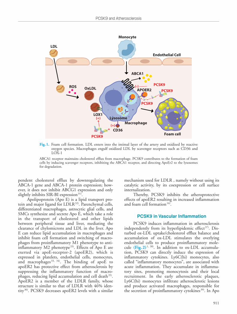
Atherosclerosis begins with the formation of foam cells, which are macrophages that engulf oxi-dized LDL (ox-LDL) (Fig.1). Ox-LDL enters into the monocytes/macrophages and also SMCs and fibro-blasts by scavenger receptors (SRs), including class A SR, class B SR type I, CD36, macrosialin (CD68), and lectin-like ox-LDL (LOX-1)24, 25). LOX-1 is the major receptor in the engulfment of ox-LDL by mac-rophages and also increases the production of cell sur-face adhesion molecules and is involved in the process of oxidative stress26-28). PCSK9 induces expression of all SRs, but mostly, LOX-1 on monocytes and SMCs24). In inflammatory milieu, PCSK9 and LOX1 induce each other, leading to an increased ox-LDL uptake29).
Excess intracellular free cholesterol is toxic for macrophages from which they protect themselves by cholesterol efflux to extracellular acceptors30). Inhibit-ing cholesterol efflux in macrophages causes an increase in foam cell formation. ATP Binding Cassette A1 (ABCA1), ATP Binding Cassette G1 (ABCG1), and Scavenger Receptor Class B Type I (SR-BI) are membrane transporters that provide most cholesterol efflux in macrophages. PCSK9 inhibits ABCA-1-de-
tions of many functional proteins, including proteo-lytic cleavage, and activation of most polypeptide hor-mones such as proinsulin8). Hepatic PCSK9 is the cir-culating protein that regulates the half-life of LDLR in the liver. In the absence of PCSK9, LDLR binds to LDL on the hepatocyte surface, transfers it into endo-somes inside the hepatocyte, and recycles back to the cell surface to bind and internalize new LDL particles. Binding with PCSK9 routes the LDLR to lysosomal degradation, and the recycling of the LDLR is blocked9). Endocytosis of PCSK9-mediated LDLRs occurs both via clathrin- and caveolae-dependent pathways, whereas recycling of LDLR and PCSK9 complex occurs in clathrin-dependent pathway and degradation occurs in caveolae-dependent pathway. LDLR and PCSK9 complex interaction with adenyl cyclase associated protein-1 (CAP1) leads to caveolae-dependent endocytosis10).
PCSK9, synthesized as a 74 kDa zymogen with 692 amino acids, comprises four domains, namely, the N-terminal pro-domain, signal peptide, the catalytic domain, and a C-terminal domain. Autocatalysis occurs between its pro-domain and catalytic domain in endoplasmic reticulum11). After autocatalysis, the pro-domain remains non-covalently attached to its catalytic pocket, which is unique to this enzyme12). N-terminal of pro-domain inhibits convertase activity of PCSK9, preventing it from interacting with sur-rounding substrates. Therefore, the pro-domain acts to modulate the effect of PCSK9 by releasing the cata-lytic domain. A short segment of amino acids in the catalytic domain of PCSK9 (amino acids 367 to 380) interacts with the epidermal growth factor-like A domain of LDLR to divert it to endosomes and lyso-somes for degradation13, 14). The catalytic domain activity of PCSK9 is not required for its effect on LDLR. PCSK9 also escorts very low-density lipopro-tein receptor (VLDLR), apolipoprotein E receptor 2 (ApoER2), and LDLR-related protein-1 (LRP1) to lysosomal degradation15, 16).
PCSK9’s Role in Atherosclerotic Process
The atherogenic effects of PCSK9 were initially explained through its role in lipid metabolism; how-ever, plasma PCSK9 level is only modestly correlated with the plasma lipid levels17). In recent years, it has been recognized that beyond its effects on lipid metab-olism, PCSK9 also has direct atherosclerotic effects on the vascular wall. Several preclinical and clinical stud-ies showed that the relation of PCSK9 with athero-sclerosis can be partly independent of its hyperlipid-emic effects. In Apo E knock-out mice, PCSK9 accel-erated atherosclerosis without affecting plasma lipid
PCSK9 and Atherosclerosis
911
mechanism used for LDLR , namely without using its catalytic activity, by its coexpression or cell surface internalization.
Thereby, PCSK9 inhibits the atheroprotective effects of apoER2 resulting in increased inflammation and foam cell formation16).
PCSK9 in Vascular Inflammation
PCSK9 induces inflammation in atherosclerosis independently from its hyperlipidemic effect37). Dis-turbed ox-LDL uptake/cholesterol efflux balance and accumulation of ox-LDL stimulates the overlying endothelial cells to produce proinflammatory mole-cule (Fig.2)1, 38). In addition to ox-LDL accumula-tion, PCSK9 can directly induce the expression of inflammatory cytokines. Ly6C(hi) monocytes, also called “inflammatory monocytes”, are associated with acute inflammation. They accumulate in inflamma-tory sites, promoting monocytosis and their local recruitment. In the early atherosclerotic plaques, Ly6C(hi) monocytes infiltrate atherosclerotic lesions and produce activated macrophages, responsible for the secretion of proinflammatory cytokines39). In Apo
pendent cholesterol efflux by downregulating the ABCA-1 gene and ABCA-1 protein expression; how-ever, it does not inhibit ABCG1 expression and only slightly inhibits SIR-BI expression31).
Apolipoprotein (Apo E) is a lipid transport pro-tein and major ligand for LDLR32). Parenchymal cells, differentiated macrophages, astrocytic glial cells, and SMCs synthesize and secrete Apo E, which take a role in the transport of cholesterol and other lipids between peripheral tissue and liver, mediating the clearance of chylomicrons and LDL in the liver. Apo E can reduce lipid accumulation in macrophages and inhibit foam cell formation and switching of macro-phages from proinflammatory M1 phenotype to anti-inflammatory M2 phenotype33). Effects of Apo E are exerted via apoE-receptor-2 (apoER2), which is expressed in platelets, endothelial cells, monocytes, and macrophages33, 34). The binding of apoE to apoER2 has protective effect from atherosclerosis by suppressing the inflammatory function of macro-phages, reducing lipid accumulation and cell death35). ApoER2 is a member of the LDLR family, whose structure is similar to that of LDLR with 46% iden-tity36). PCSK9 decreases apoER2 levels with a similar
ROS OxLDL
LDL
Monocyte
Endothelial Cell
CD36
PCSK9
APOER2
ABCA1
LOX1
PCSK9
Macrophage
Foam cell
Lysosome
PCSK9PCSK9
(+)
(+)
(-)
Fig.1. Foam cell formation. LDL enters into the intimal layer of the artery and oxidized by reactive oxygen species. Macrophages engulf oxidized LDL by scavenger receptors such as CD36 and LOX-1
ABCA1 receptor maintains cholesterol efflux from macrophage. PCSK9 contributes to the formation of foam cells by inducing scavenger receptors, inhibiting the ABCA1 receptor, and directing ApoEr2 to the lysosomes for degradation.

Yurtseven et al.
912
tory effects on macrophages are mostly via LDLRs40, 42).The nuclear factor kappa B (NF-KB) controls
the transcription of many genes that have roles in inflammation and atherosclerosis, including cytokines, chemokines, adhesion molecules, acute phase proteins, regulators of apoptosis, and cell proliferation44). NF-KB, mitogen-activated protein kinase, and dam-aged mitochondrial DNA have significant roles in PCSK9-mediated ox-LDL uptake29). NF-KB nuclear translocation is induced by PCSK9 overexpression in macrophages, increasing the mRNA levels of proin-flammatory cytokines and toll-like receptor 4 (TLR4) expression. Inversely, NF-KB inhibition decreases LPS, ox-LDL, and TNF-α induced PCSK9 expres-sion43). Therefore, it is worth noting that NF-KB has an important signaling role in inflammatory stimulus mediated by PCSK9 expression and PCSK9 acceler-ates atherosclerotic plaque inflammation through the
E knock-out mice (Apo E KO), PCSK9 increases Ly6C(hi) monocytes in spleen and enhance their recruitment in arterial wall40). It increases macrophage response to lipopolysaccharide (LPS) leading to exces-sive secretion of inflammatory cytokines, including tumor necrosis factor-α (TNF-α), interleukin-1β (IL-1β), and interleukin-6 (IL-6) and decreases anti-inflammatory cytokines, including interleukin-10 (IL10) and arginase41, 42). Silencing PCSK9 decreases the expression of TNF-α, IL-1b, and monocyte che-moattractant protein 1 (MCP1) in the atherosclerotic aortas of Apo E KO mice43). MCP-1 and chemokine (C-X-C motif ) ligand 2 (CXCL2) are chemokines related to monocyte recruitment. PCSK9 can directly induce expression of these molecules in macro-phages42). These proinflammatory effects of PCSK9 are independent of cholesterol but not observed in LDLR −/− mice, suggesting that PCSK9 inflamma-
ROS
OxLDL
LOX1
LDL
MonocyteMonocyte
Endothelial Cell
Foam cell
VCAM1
NFκB
Macrophage
IκB PCSK9(+)
PCSK9 PCSK9
(+)(+)
(+)
PCSK9
(+) In ammatorymRNA
In ammatoryCytokines
- IL1- TNFα- IL6
Smooth Muscle Cell
Fig.2. Inflammation
PCSK9 induces the expression of endothelial adhesion molecules. NF-KB nuclear translocation is induced by PCSK9, leading to an increase in mRNA levels and secretion of inflammatory cytokines.

PCSK9 and Atherosclerosis
913
concentration-dependently (Fig.3) 46). In ox-LDL treated HUVECs, expressions of proapoptotic protein Bax and the activity of caspase9 and caspase3 increase, whereas anti-apoptotic protein Bcl-2 expression decreases46). The silencing of PCSK9, with its siRNA, reverses the apoptotic effects of ox-LDL and inhibits apoptosis in endothelial cells46). Therefore, it is sug-gested that PCSK9 increases apoptosis of HUVECs by Bcl/Bax- caspase9-caspase3 pathway41, 46). PCSK9 inhibition also decreases the phosphorylation of c-Jun N-terminal kinase and p38, which play an important role in cell apoptosis that is induced by oxidative dam-age, IL1, TNF-α, and G protein-coupled receptors41).
Autophagy is a protective cellular process of self-digestion of the misfolded proteins and dysfunctional organelles. It aims to promote cell survival under unfavorable conditions such as inflammation and oxi-dative stress. In the early stages of atherosclerosis, autophagy is protective against macrophage engulf-ment and local inflammation. However, in late stages, it leads to macrophage death, accelerated inflamma-tion, and thinning of fibrous cap by collagen degrada-tion and can cause acute coronary events47). The mammalian target of rapamycin (mTOR) is an inhibi-tor of autophagy, whereas mTOR inhibitor rapamycin is the most commonly used introducer of autophagy, providing a possible anti-atherosclerotic effect48). PCSK9 upregulation induces mTOR expression in vascular SMC cultures dose-dependently, whereas inhibition of PCSK9 with its siRNA promotes
activation of TLR4/NF-KB pathway29, 43). The vascular cell adhesion molecule 1 (VCAM-1)
is a protein whose expression increases in inflamma-tory conditions, mediates the adhesion of lympho-cytes, monocytes, eosinophils, and basophils to the vascular wall. PCSK9 induces VCAM-1 expression in vascular SMCs, and PCSK9 inhibition in cultured vascular SMCs significantly decreases VCAM-1 expression29).
Dendritic cells that play a role in endothelial inflammation stimulate T cell proliferation and differ-entiation of naive CD4+T cells into Th1 and Th17 in the existence of ox-LDL. Ox-LDL induces the PCSK9 expression in dendritic cells and increases secretion of TNF-α, IL-1, IL-6, transforming growth factor beta and IL-10 from dendritic cells45). Differentiation and proliferation of T cells are reduced when PCSK9 is inhibited in dendritic cells45).
PCSK9-Induced Apoptosis
Endothelial cell apoptosis promotes endothelial dysfunction, which is a foundation in the atheroscle-rosis development. Intrinsic pathway of apoptosis is regulated by B cell lymphoma-2 (Bcl-2) family pro-teins. Bax and Bcl2 proteins are members of this fam-ily with opposite roles: Bax induces apoptosis, whereas Bcl2 inhibits apoptosis. Ox-LDL induces apoptosis of human umbilical vein endothelial cells (HUVECs) and upregulates the expression of PCSK9 and LOX-1
Mitochondria
Bax
Cytc
Caspase
Bcl2
(-)(+) (-)PCSK9 PCSK9
Fig.3. Apoptosis
PCSK9 induces apoptosis of HUVECs by inducing Bax and inhibiting Bcl2.

Yurtseven et al.
914
at low shear stress23).
PCSK9’s Effects on Vascular SMCs
Vascular SMCs within adult blood vessels exhibit a low rate of proliferation and low synthetic activity and express contractile proteins. SMCs in the media express contraction proteins such as alpha-smooth muscle actin, myosin heavy chain II, and calponin, whereas intimal SMCs synthesize extracellular matrix, proteases, and cytokines and have higher migratory and proliferative capacity53, 54). Contractile SMCs can differentiate to a synthetic phenotype with athero-genic stimuli such as shear stress, cytokines, and ROS55). Synthetic SMCs proliferate and migrate more rapidly, synthesize more collagen, increase lipid syn-thesis, and express higher levels of SRs; therefore, dif-ferentiation to synthetic SMC contributes to the for-mation of foam cells56). PCSK9, which can be induced by resistin, insulin, and hemodynamic shear stress in SMCs, induces SMC differentiation to synthetic phe-notype and increases migration and proliferation of synthetic type SMCs23, 57, 58).
PCSK9 deficiency protects arteries partially from neointimal formation59). There is an increased number of the synthetic phenotype of SMCs and increased PCSK9 expression in acute aortic dissection sam-ples60). In aortic dissection with medial aortic calcifi-cation (MAC), PCSK9 expression is higher than in aortic dissection without MAC60). Thus, it can be sug-gested that PCSK9 plays a role in aortic dissection by contributing to the loss of structural integrity of aorta60).
Micro-RNAs Targeting PCSK9 Expression
Micro-RNAs (miRNAs) are short non-coding RNAs (18–22 nucleotides) that function in RNA silencing and controlling post-transcriptional regula-tion of gene expression61). They initiate the degrada-tion or inhibit the translation of their target mRNA by binding to the recognition element on the 3′-UTR62). By affecting gene expression, they participate in the regulation of gene expression. Being specific for a given cell and tissue, they influence the phenotype of the cell in which they are expressed. They can be found free in the circulation, bound to proteins or high density lipoprotein (HDL), and in exosomes. By entering body fluids such as blood or urine, they relay signals from the producer cell to distant targets. Their expression has been correlated with various diseases, including coronary artery disease, and they have been reported to be involved in all phases of atherosclerosis, particularly immune cell recruitment and cytokine
autophagy and cellular vitality via decreasing mTOR phosphorylation49). Moreover, mTOR pathway is a regulator of LDLR pathway leading to upregulated LDLR expression and inhibited PCSK9 transcription when activated by various signals such as insulin and to decreased LDLR expression and increased PCSK9 expression when inhibited with rapamycin49).
Reactive Oxygen Species-Induced PCSK9 Expression
Reactive oxygen species (ROS) are oxygen-con-taining reactive molecules and can be produced in oxi-dation reactions in mitochondria and other cellular locations. In inflammatory states, such as atherosclero-sis, increase in mitochondrial ROS production pro-vokes endothelial dysfunction, induces the infiltration and activation of inflammatory cells, and increases apoptosis of endothelial and vascular SMCs. Elevated ROS production increases PCSK9 levels23). Mitochon-drial ROS leads to PCSK9 and LOX1 increase; the interrelationship of PCSK9 with ROS is strongly sup-ported by the observation that shows a decrease in mtROS in response to LOX-1 or PCSK9 knock-down29). Although ROS inducers pyocyanin and superoxide inducer antimycin-A increase PCSK9 expression, ROS inhibitors such as diphenylene iodo-nium and apocynin decrease PCSK9 expression in macrophages24).Increased H2O2 production and Nox2 activation are seen in the platelets that are incubated with PCSK9 50).
Shear Stress-Induced PCSK9 Expression
Vascular tone and diameter are under the control of vascular SMCs through the mechanism of contrac-tion51). Vascular SMCs are highly specialized cells of which principal functions are contraction and regula-tion of blood vessel tone, blood pressure, and blood flow. Local susceptibility to atherosclerosis is regulated by blood flow patterns. Endothelial shear stress is one of the mechanical factors that initiate atherosclerotic plaques. It is the tangential stress derived from the friction of the flowing blood on the endothelial sur-face of the arterial wall. Low shear stress, which typi-cally occurs in regions of branching or bifurcation regions of the arteries, results in high EC turnover, increased accumulation of LDL, increased oxidative stress, increased DNA synthesis, and higher expression of proinflammatory adhesion molecules; therefore, low shear stress is related to atherosclerosis52). It has been shown that low shear stress (3–6 dyne/cm2) increases PCSK9 expression mostly in the SMCs, and lipid-induced increase in PCSK9 expression is higher

PCSK9 and Atherosclerosis
915
Despite the rapidly accumulating data on its athero-genic effects, much is still unknown, including its sig-naling pathways and receptors in atherosclerosis, its acting mechanisms on vascular cells in atherosclerosis, its role in vascular diseases such as aneurysms, its interaction and signal transduction through receptors other than LDLR, the extent of miRNA regulation of PCSK9, and its relevance. Furthermore, it should not be overlooked that, although PCSK9 is a member of the proprotein convertase family, its substrate for con-vertase activity and why it exists are still unknown. To clarify its role in atherosclerosis and its reason for exis-tence, further research is required.
Acknowledgements
The authors gratefully acknowledge use of the services and facilities of the Koç University Research Center for Translational Medicine (KUTTAM), funded by the Presidency of Turkey, Presidency of Strategy and Budget. The content is solely the respon-sibility of the authors and does not necessarily repre-sent the official views of the Presidency of Strategy and Budget.
Conflicts of Interest
None.
References1) Libby P, Buring JE, Badimon L, Hansson GK, Deanfield
J, Bittencourt MS, Tokgözoğlu L and Lewis EF: Athero-sclerosis. Nat Rev Dis Primers, 2019; 5: 56
2) Chistiakov DA, Melnichenko AA, Myasoedova VA, Grechko AV and Orekhov AN: Mechanisms of foam cell formation in atherosclerosis. J Mol Med (Berl), 2017; 95: 1153-1165
3) Lusis AJ: Atherosclerosis. Nature, 2000; 407: 233-2414) Yusuf S, Hawken S, Ounpuu S, Dans T, Avezum A, Lanas
F, McQueen M, Budaj A, Pais P, Varigos J and Lisheng L: Effect of potentially modifiable risk factors associated with myocardial infarction in 52 countries (the INTER-HEART study): case-control study. Lancet, 2004; 364: 937-952
5) Fernández-Friera L, Fuster V, López-Melgar B, Oliva B, García-Ruiz JM, Mendiguren J, Bueno H, Pocock S, Ibáñez B, Fernández-Ortiz A and Sanz J: Normal LDL-Cholesterol Levels Are Associated With Subclinical Ath-erosclerosis in the Absence of Risk Factors. J Am Coll Cardiol, 2017; 70: 2979-2991
6) Nordestgaard BG, Chapman MJ, Humphries SE, Gins-berg HN, Masana L, Descamps OS, Wiklund O, Hegele RA, Raal FJ, Defesche JC, Wiegman A, Santos RD, Watts GF, Parhofer KG, Hovingh GK, Kovanen PT, Boileau C, Averna M, Borén J, Bruckert E, Catapano AL, Kuiven-hoven JA, Pajukanta P, Ray K, Stalenhoef AF, Stroes E,
production63, 64).Many miRNAs may affect PCSK9 expression65).
MiR-17-5p, a potential biomarker for the diagnosis of coronary atherosclerosis, inhibits VLDLR expression in vascular SMCs via direct binding to its 3’-UTR66). Inhibition of miR-17-5p induces upregulation of VLDLR and downregulation of PCSK9 in atheroscle-rotic apoE −/− mice66). miR-191, miR-222, and miR-224 can bind the 3′ end of the PCSK9 mRNA and thus regulate PCSK9 expression67). When cells are transfected with vectors overexpressing miR-191, miR-222, and miR-224, significant downregulation of PCSK9 is observed67).
Hypomethylation and elevated expression of miR-191 promote epithelial to mesenchymal transi-tion in hepatocellular carcinoma68). EGR1 (early growth response protein 1) is a zinc finger transcrip-tion factor that has a binding site on the PCSK9 pro-moter. miR-191 targets EGR1 and can regulate PCSK9 expression in different physiological states69).
In a study by Vickers et al., miR-222 level in cir-culating HDL was found to be 8.2-fold higher in familial hypercholesterolemia patients compared with HDL from normal subjects64). miR-222 regulates insulin sensitivity, and miR-222 levels are higher in plasma of obese human patients70). It is an important regulator in lipid metabolic pathways and a negative regulator of adipocyte differentiation71). It has been reported that miR-222 plays important roles in many physiological and pathological processes in the cardio-vascular system; it also plays a role in atherosclerosis and plaque formation72). Moreover, miR-222 has been reported to decrease the PCSK9 expression67).
miR-27a induces an increase in PCSK9 and con-sequently decreases LDLR levels by 40% via enhanc-ing LDLR degradation73). Silencing PCSK9 repressed miR-27a and, to a lesser extent, let-7c 44).
Further studies are required to delineate the role of miRNAs in PCSK9 regulation.
Conclusion
After its discovery in 2003, knowledge on PCSK9 has gone from bench-to-bedside in less than 9 years, and PCSK9 monoclonal antibodies entered in dyslipidemia guidelines since 2016. Although the role of PCSK9 in LDL metabolism is well defined, recent studies have revealed that it has various effects on many metabolic pathways, including inflammation, sepsis, sodium metabolism, glucose metabolism, and atherosclerosis74-76). Signals of these metabolic path-ways also affect PCSK9’s level and function77).
It is now clear that PCSK9 contributes to every step of the molecular pathway of atherosclerosis.

Yurtseven et al.
916
27819) Denis M, Marcinkiewicz J, Zaid A, Gauthier D, Poirier S,
Lazure C, Seidah NG and Prat A: Gene inactivation of proprotein convertase subtilisin/kexin type 9 reduces ath-erosclerosis in mice. Circulation, 2012; 125: 894-901
20) Cheng JM, Oemrawsingh RM, Garcia-Garcia HM, Boersma E, van Geuns RJ, Serruys PW, Kardys I and Akkerhuis KM: PCSK9 in relation to coronary plaque inflammation: Results of the ATHEROREMO-IVUS study. Atherosclerosis, 2016; 248: 117-122
21) Ferreira JP, Xhaard C, Lamiral Z, Borges-Canha M, Neves JS, Dandine-Roulland C, LeFloch E, Deleuze JF, Bacq-Daian D, Bozec E, Girerd N, Boivin JM, Zannad F and Rossignol P: PCSK9 Protein and rs562556 Polymorphism Are Associated With Arterial Plaques in Healthy Middle-Aged Population: The STANISLAS Cohort. J Am Heart Assoc, 2020; 9: e014758
22) Ferri N, Tibolla G, Pirillo A, Cipollone F, Mezzetti A, Pacia S, Corsini A and Catapano AL: Proprotein conver-tase subtilisin kexin type 9 (PCSK9) secreted by cultured smooth muscle cells reduces macrophages LDLR levels. Atherosclerosis, 2012; 220: 381-386
23) Ding Z, Liu S, Wang X, Deng X, Fan Y, Sun C, Wang Y and Mehta JL: Hemodynamic shear stress via ROS modu-lates PCSK9 expression in human vascular endothelial and smooth muscle cells and along the mouse aorta. Anti-oxid Redox Signal, 2015; 22: 760-771
24) Ding Z, Liu S, Wang X, Theus S, Deng X, Fan Y, Zhou S and Mehta JL: PCSK9 regulates expression of scavenger receptors and ox-LDL uptake in macrophages. Cardiovasc Res, 2018; 114: 1145-1153
25) Mehta JL, Sanada N, Hu CP, Chen J, Dandapat A, Suga-wara F, Satoh H, Inoue K, Kawase Y, Jishage K, Suzuki H, Takeya M, Schnackenberg L, Beger R, Hermonat PL, Thomas M and Sawamura T: Deletion of LOX-1 reduces atherogenesis in LDLR knockout mice fed high choles-terol diet. Circ Res, 2007; 100: 1634-1642
26) Kume N, Murase T, Moriwaki H, Aoyama T, Sawamura T, Masaki T and Kita T: Inducible expression of lectin-like oxidized LDL receptor-1 in vascular endothelial cells. Circ Res, 1998; 83: 322-327
27) Ding Z, Liu S, Wang X, Khaidakov M, Dai Y and Mehta JL: Oxidant stress in mitochondrial DNA damage, autophagy and inflammation in atherosclerosis. Sci Rep, 2013; 3: 1077
28) Sawamura T, Kume N, Aoyama T, Moriwaki H, Hoshi-kawa H, Aiba Y, Tanaka T, Miwa S, Katsura Y, Kita T and Masaki T: An endothelial receptor for oxidized low-den-sity lipoprotein. Nature, 1997; 386: 73-77
29) Ding Z, Liu S, Wang X, Deng X, Fan Y, Shahanawaz J, Shmookler Reis RJ, Varughese KI, Sawamura T and Mehta JL: Cross-talk between LOX-1 and PCSK9 in vas-cular tissues. Cardiovasc Res, 2015; 107: 556-567
30) Zanotti I, Favari E and Bernini F: Cellular cholesterol efflux pathways: impact on intracellular lipid trafficking and methodological considerations. Curr Pharm Biotech-nol, 2012; 13: 292-302
31) Adorni MP, Cipollari E, Favari E, Zanotti I, Zimetti F, Corsini A, Ricci C, Bernini F and Ferri N: Inhibitory effect of PCSK9 on Abca1 protein expression and choles-terol efflux in macrophages. Atherosclerosis, 2017; 256:
Taskinen MR and Tybjærg-Hansen A: Familial hypercho-lesterolaemia is underdiagnosed and undertreated in the general population: guidance for clinicians to prevent cor-onary heart disease: consensus statement of the European Atherosclerosis Society. Eur Heart J, 2013; 34: 3478-3490a
7) Schulz R and Schlüter KD: PCSK9 targets important for lipid metabolism. Clin Res Cardiol Suppl, 2017; 12: 2-11
8) Seidah NG, Abifadel M, Prost S, Boileau C and Prat A: The Proprotein Convertases in Hypercholesterolemia and Cardiovascular Diseases: Emphasis on Proprotein Conver-tase Subtilisin/Kexin 9. Pharmacol Rev, 2017; 69: 33-52
9) Shapiro MD, Tavori H and Fazio S: PCSK9: From Basic Science Discoveries to Clinical Trials. Circ Res, 2018; 122: 1420-1438
10) Jang HD, Lee SE, Yang J, Lee HC, Shin D, Lee H, Lee J, Jin S, Kim S, Lee SJ, You J, Park HW, Nam KY, Lee SH, Park SW, Kim JS, Kim SY, Kwon YW, Kwak SH, Yang HM and Kim HS: Cyclase-associated protein 1 is a bind-ing partner of proprotein convertase subtilisin/kexin type-9 and is required for the degradation of low-density lipoprotein receptors by proprotein convertase subtilisin/kexin type-9. Eur Heart J, 2020; 41: 239-252
11) Naureckiene S, Ma L, Sreekumar K, Purandare U, Lo CF, Huang Y, Chiang LW, Grenier JM, Ozenberger BA, Jacobsen JS, Kennedy JD, DiStefano PS, Wood A and Bingham B: Functional characterization of Narc 1, a novel proteinase related to proteinase K. Arch Biochem Biophys, 2003; 420: 55-67
12) Seidah NG, Benjannet S, Wickham L, Marcinkiewicz J, Jasmin SB, Stifani S, Basak A, Prat A and Chretien M: The secretory proprotein convertase neural apoptosis-reg-ulated convertase 1 (NARC-1): liver regeneration and neuronal differentiation. Proc Natl Acad Sci U S A, 2003; 100: 928-933
13) Seidah NG, Awan Z, Chrétien M and Mbikay M: PCSK9: a key modulator of cardiovascular health. Circ Res, 2014; 114: 1022-1036
14) Seidah NG and Prat A: The biology and therapeutic tar-geting of the proprotein convertases. Nat Rev Drug Dis-cov, 2012; 11: 367-383
15) Canuel M, Sun X, Asselin MC, Paramithiotis E, Prat A and Seidah NG: Proprotein convertase subtilisin/kexin type 9 (PCSK9) can mediate degradation of the low den-sity lipoprotein receptor-related protein 1 (LRP-1). PLoS One, 2013; 8: e64145
16) Poirier S, Mayer G, Benjannet S, Bergeron E, Marcinkie-wicz J, Nassoury N, Mayer H, Nimpf J, Prat A and Sei-dah NG: The proprotein convertase PCSK9 induces the degradation of low density lipoprotein receptor (LDLR) and its closest family members VLDLR and ApoER2. J Biol Chem, 2008; 283: 2363-2372
17) Ridker PM, Rifai N, Bradwin G and Rose L: Plasma pro-protein convertase subtilisin/kexin type 9 levels and the risk of first cardiovascular events. Eur Heart J, 2016; 37: 554-560
18) Tavori H, Giunzioni I, Predazzi IM, Plubell D, Shivinsky A, Miles J, Devay RM, Liang H, Rashid S, Linton MF and Fazio S: Human PCSK9 promotes hepatic lipogenesis and atherosclerosis development via apoE- and LDLR-mediated mechanisms. Cardiovasc Res, 2016; 110: 268-

PCSK9 and Atherosclerosis
917
PCSK9 siRNA inhibits HUVEC apoptosis induced by ox-LDL via Bcl/Bax-caspase9-caspase3 pathway. Mol Cell Biochem, 2012; 359: 347-358
47) Sun Y and Guan X-r: Autophagy: A new target for the treatment of atherosclerosis. Frontiers in Laboratory Med-icine, 2018; 2: 68-71
48) Cai Z, He Y and Chen Y: Role of Mammalian Target of Rapamycin in Atherosclerosis. Curr Mol Med, 2018; 18: 216-232
49) Ding Z, Liu S, Wang X, Mathur P, Dai Y, Theus S, Deng X, Fan Y and Mehta JL: Cross-Talk Between PCSK9 and Damaged mtDNA in Vascular Smooth Muscle Cells: Role in Apoptosis. Antioxid Redox Signal, 2016; 25: 997-1008
50) Cammisotto V, Pastori D, Nocella C, Bartimoccia S, Cas-tellani V, Marchese C, Scavalli AS, Ettorre E, Viceconte N, Violi F, Pignatelli P and Carnevale R: PCSK9 Regu-lates Nox2-Mediated Platelet Activation via CD36 Recep-tor in Patients with Atrial Fibrillation. Antioxidants (Basel), 2020; 9: 296
51) Chistiakov DA, Orekhov AN and Bobryshev YV: Vascu-lar smooth muscle cell in atherosclerosis. Acta Physiol (Oxf ), 2015; 214: 33-50
52) Chatzizisis YS, Coskun AU, Jonas M, Edelman ER, Feld-man CL and Stone PH: Role of endothelial shear stress in the natural history of coronary atherosclerosis and vascu-lar remodeling: molecular, cellular, and vascular behavior. J Am Coll Cardiol, 2007; 49: 2379-2393
53) Campbell JH and Campbell GR: The role of smooth muscle cells in atherosclerosis. Curr Opin Lipidol, 1994; 5: 323-330
54) Worth NF, Rolfe BE, Song J and Campbell GR: Vascular smooth muscle cell phenotypic modulation in culture is associated with reorganisation of contractile and cytoskel-etal proteins. Cell Motil Cytoskeleton, 2001; 49: 130-145
55) Orr AW, Hastings NE, Blackman BR and Wamhoff BR: Complex regulation and function of the inflammatory smooth muscle cell phenotype in atherosclerosis. J Vasc Res, 2010; 47: 168-180
56) Campbell JH, Popadynec L, Nestel PJ and Campbell GR: Lipid accumulation in arterial smooth muscle cells. Influ-ence of phenotype. Atherosclerosis, 1983; 47: 279-295
57) Melone M, Wilsie L, Palyha O, Strack A and Rashid S: Discovery of a new role of human resistin in hepatocyte low-density lipoprotein receptor suppression mediated in part by proprotein convertase subtilisin/kexin type 9. J Am Coll Cardiol, 2012; 59: 1697-1705
58) Costet P, Cariou B, Lambert G, Lalanne F, Lardeux B, Jarnoux AL, Grefhorst A, Staels B and Krempf M: Hepatic PCSK9 expression is regulated by nutritional sta-tus via insulin and sterol regulatory element-binding pro-tein 1c. J Biol Chem, 2006; 281: 6211-6218
59) Ferri N, Marchianò S, Tibolla G, Baetta R, Dhyani A, Ruscica M, Uboldi P, Catapano AL and Corsini A: PCSK9 knock-out mice are protected from neointimal formation in response to perivascular carotid collar place-ment. Atherosclerosis, 2016; 253: 214-224
60) Iida Y, Tanaka H, Sano H, Suzuki Y, Shimizu H and Urano T: Ectopic Expression of PCSK9 by Smooth Mus-cle Cells Contributes to Aortic Dissection. Ann Vasc Surg, 2018; 48: 195-203
61) Liu N and Olson EN: MicroRNA regulatory networks in
1-632) Mahley RW: Apolipoprotein E: from cardiovascular dis-
ease to neurodegenerative disorders. J Mol Med (Berl), 2016; 94: 739-746
33) Bai XQ, Peng J, Wang MM, Xiao J, Xiang Q, Ren Z, Wen HY, Jiang ZS, Tang ZH and Liu LS: PCSK9: A potential regulator of apoE/apoER2 against inflammation in atherosclerosis? Clin Chim Acta, 2018; 483: 192-196
34) Leeb C, Eresheim C and Nimpf J: Clusterin is a ligand for apolipoprotein E receptor 2 (ApoER2) and very low density lipoprotein receptor (VLDLR) and signals via the Reelin-signaling pathway. J Biol Chem, 2014; 289: 4161-4172
35) Waltmann MD, Basford JE, Konaniah ES, Weintraub NL and Hui DY: Apolipoprotein E receptor-2 deficiency enhances macrophage susceptibility to lipid accumulation and cell death to augment atherosclerotic plaque progres-sion and necrosis. Biochim Biophys Acta, 2014; 1842: 1395-1405
36) Reddy SS, Connor TE, Weeber EJ and Rebeck W: Simi-larities and differences in structure, expression, and func-tions of VLDLR and ApoER2. Mol Neurodegener, 2011; 6: 30
37) Ruscica M, Tokgözoğlu L, Corsini A and Sirtori CR: PCSK9 inhibition and inflammation: A narrative review. Atherosclerosis, 2019; 288: 146-155
38) Leiva E, Wehinger S, Guzmán L and Orrego R: Role of Oxidized LDL in Atherosclerosis. In: Hypercholesterol-emia, 2015
39) Swirski FK, Libby P, Aikawa E, Alcaide P, Luscinskas FW, Weissleder R and Pittet MJ: Ly-6Chi monocytes domi-nate hypercholesterolemia-associated monocytosis and give rise to macrophages in atheromata. J Clin Invest, 2007; 117: 195-205
40) Giunzioni I, Tavori H, Covarrubias R, Major AS, Ding L, Zhang Y, DeVay RM, Hong L, Fan D, Predazzi IM, Rashid S, Linton MF and Fazio S: Local effects of human PCSK9 on the atherosclerotic lesion. J Pathol, 2016; 238: 52-62
41) Li J, Liang X, Wang Y, Xu Z and Li G: Investigation of highly expressed PCSK9 in atherosclerotic plaques and ox-LDL-induced endothelial cell apoptosis. Mol Med Rep, 2017; 16: 1817-1825
42) Ricci C, Ruscica M, Camera M, Rossetti L, Macchi C, Colciago A, Zanotti I, Lupo MG, Adorni MP, Cicero AFG, Fogacci F, Corsini A and Ferri N: PCSK9 induces a pro-inflammatory response in macrophages. Sci Rep, 2018; 8: 2267
43) Tang ZH, Peng J, Ren Z, Yang J, Li TT, Li TH, Wang Z, Wei DH, Liu LS, Zheng XL and Jiang ZS: New role of PCSK9 in atherosclerotic inflammation promotion involving the TLR4/NF-κB pathway. Atherosclerosis, 2017; 262: 113-122
44) De Winther MPJ, Kanters E, Kraal G, Hofker MH: Nuclear factor κB signaling in atherogenesis. Arterioscler Thromb Vasc Biol, 2005; 25(5): 904-914
45) Liu A and Frostegård J: PCSK9 plays a novel immunolog-ical role in oxidized LDL-induced dendritic cell matura-tion and activation of T cells from human blood and ath-erosclerotic plaque. J Intern Med, 2018; 284: 193-210
46) Wu CY, Tang ZH, Jiang L, Li XF, Jiang ZS and Liu LS:

Yurtseven et al.
918
tus. PLoS Genet, 2013; 9: e100331170) Ortega FJ, Mercader JM, Catalán V, Moreno-Navarrete
JM, Pueyo N, Sabater M, Gómez-Ambrosi J, Anglada R, Fernández-Formoso JA, Ricart W, Frühbeck G and Fernández-Real JM: Targeting the circulating microRNA signature of obesity. Clin Chem, 2013; 59: 781-792
71) Price NL and Fernández-Hernando C: miRNA regulation of white and brown adipose tissue differentiation and function. Biochim Biophys Acta, 2016; 1861: 2104-2110
72) Bazan HA, Hatfield SA, O’Malley CB, Brooks AJ, Light-ell D, Jr. and Woods TC: Acute Loss of miR-221 and miR-222 in the Atherosclerotic Plaque Shoulder Accom-panies Plaque Rupture. Stroke, 2015; 46: 3285-3287
73) Alvarez ML, Khosroheidari M, Eddy E and Done SC: MicroRNA-27a decreases the level and efficiency of the LDL receptor and contributes to the dysregulation of cholesterol homeostasis. Atherosclerosis, 2015; 242: 595-604
74) Walley KR, Thain KR, Russell JA, Reilly MP, Meyer NJ, Ferguson JF, Christie JD, Nakada TA, Fjell CD, Thair SA, Cirstea MS and Boyd JH: PCSK9 is a critical regula-tor of the innate immune response and septic shock out-come. Sci Transl Med, 2014; 6: 258ra143
75) Sharotri V, Collier DM, Olson DR, Zhou R and Snyder PM: Regulation of epithelial sodium channel trafficking by proprotein convertase subtilisin/kexin type 9 (PCSK9). J Biol Chem, 2012; 287: 19266-19274
76) Mbikay M, Sirois F, Mayne J, Wang GS, Chen A, Dew-pura T, Prat A, Seidah NG, Chretien M and Scott FW: PCSK9-deficient mice exhibit impaired glucose tolerance and pancreatic islet abnormalities. FEBS Lett, 2010; 584: 701-706
77) Spolitu S, Okamoto H, Dai W, Zadroga JA, Wittchen ES, Gromada J and Ozcan L: Hepatic Glucagon Signaling Regulates PCSK9 and Low-Density Lipoprotein Choles-terol. Circ Res, 2019; 124: 38-51
cardiovascular development. Dev Cell, 2010; 18: 510-52562) Filipowicz W, Bhattacharyya SN and Sonenberg N:
Mechanisms of post-transcriptional regulation by microR-NAs: are the answers in sight? Nat Rev Genet, 2008; 9: 102-114
63) Navickas R, Gal D, Laucevičius A, Taparauskaitė A, Zdanytė M and Holvoet P: Identifying circulating microRNAs as biomarkers of cardiovascular disease: a sys-tematic review. Cardiovasc Res, 2016; 111: 322-337
64) Martens CR, Bansal SS and Accornero F: Cardiovascular inflammation: RNA takes the lead. J Mol Cell Cardiol, 2019; 129: 247-256
65) Momtazi AA, Banach M, Pirro M, Stein EA and Sahebkar A: MicroRNAs: New Therapeutic Targets for Familial Hypercholesterolemia? Clin Rev Allergy Immunol, 2018; 54: 224-233
66) Tan L, Meng L, Shi X and Yu B: Knockdown of microRNA-17-5p ameliorates atherosclerotic lesions in ApoE(-/-) mice and restores the expression of very low density lipoprotein receptor. Biotechnol Lett, 2017; 39: 967-976
67) Naeli P, Mirzadeh Azad F, Malakootian M, Seidah NG and Mowla SJ: Post-transcriptional Regulation of PCSK9 by miR-191, miR-222, and miR-224. Front Genet, 2017; 8: 189
68) He Y, Cui Y, Wang W, Gu J, Guo S, Ma K and Luo X: Hypomethylation of the hsa-miR-191 locus causes high expression of hsa-mir-191 and promotes the epithelial-to-mesenchymal transition in hepatocellular carcinoma. Neoplasia, 2011; 13: 841-853
69) Di Leva G, Piovan C, Gasparini P, Ngankeu A, Taccioli C, Briskin D, Cheung DG, Bolon B, Anderlucci L, Alder H, Nuovo G, Li M, Iorio MV, Galasso M, Santhanam R, Marcucci G, Perrotti D, Powell KA, Bratasz A, Garofalo M, Nephew KP and Croce CM: Estrogen mediated-acti-vation of miR-191/425 cluster modulates tumorigenicity of breast cancer cells depending on estrogen receptor sta-