An Invisible Killer - hksccm.org
90
An Invisible Killer HKSCCM Inter-hospital Meeting TKOH ICU Dr. Edward Chung 23 Nov 2010
Transcript of An Invisible Killer - hksccm.org

An Invisible Killer
HKSCCM Inter-hospital MeetingTKOH ICUDr. Edward Chung 23 Nov 2010

History
M/37 Non smoker Good past health Owner of optical shop

History of Present Illness
Increased SOB since evening Dry cough Minimal sputum No runny nose / sore throat No hemoptysis No chest pain

History of Present Illness
No avian contact No recent travel history No recent air travel Not on chronic medications No habit of recreational drug use

Physical Examination
T 35.7º BP 116/86 P 120 RR 36 SpO2 87% RA GCS 15/15

Physical Examination
Diffuse crepitations over both sides of lungs
HS normal, no murmur heard No ankle edema Abodmen soft

Laboratory
WBC 44.3 Neut 88% Lymp 4% Eosin 0%
Hb 20.6 Plt 353 Cr 166 LFT normal Clotting normal

Progress
Increase in dyspnea Put on 100% oxygen mask at AED

ABG
pH 7.17 PaCO2 5.2 PaO2 9.7 HCO3 13.8 BE -13.9 Cl 96 / Na 136 AG = 26

CXR (AED)

Progress
ICU consulted Transferred to ICU for management

Arrived ICU
BP 100/70 Pulse 120 T 39º SpO2 85% on 100% O2 mask RR 38 GCS 15/15
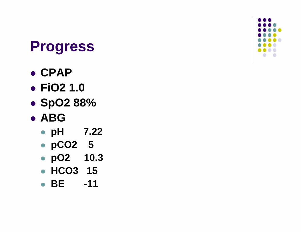
Progress CPAP FiO2 1.0 SpO2 88% ABG pH 7.22 pCO2 5 pO2 10.3 HCO3 15 BE -11

Progress
Patient agreed to intubation

Mechanical Ventilation
Intubated and sedated ACPC mode FiO2 1.0 Pi 20 PEEP 12

CXR (Post-intubation, Day 1)

Laboratory
WCC 31 Neut 90%
Hb 19 RLFT normal Troponin T –ve
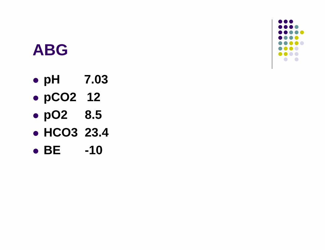
ABG
pH 7.03 pCO2 12 pO2 8.5 HCO3 23.4 BE -10

Short Summary
Young man with good past health Sudden onset of severe respiratory failure Fever Leucocytosis with neutrophil
predominance Diffuse bilateral lung infiltrates in
pulmonary edema pattern

Acute Pulmonary Edema Cardiogenic
Left ventricular dysfunction Acute myocarditis Ischemic disease Arrhythmia Valvular disease Constrictive pericarditis or
acute tamponade Hypertensive emergency Volume overload High output state
Intracardiac or extracardiac shunt Anemia Sepsis Thyrotoxicosis
Substances abuse
Non cardiogenic (ARDS) Sepsis
Pneumonia Extrapulmonary source
Trauma TRALI Aspiration

Echocardiogram
EF 60% (by four chambers view) No RWMA No Pericardial effusion / Cardiac
tamponade No significant valvular lesion No intra-cardiac shunt No dilated RV / significant TR

Progress
Bronchoscopy Airways clear No sputum retention No pulmonary hemorrhage No endobronchial lesion
Bronchoscopic lavage done
Preliminary diagnosis
ARDS ?underlying cause
Treatment
Rocephin + Azithromycin Noradrenaline for hypotension Hydrocortisone
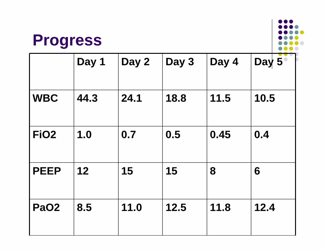
ProgressDay 1 Day 2 Day 3 Day 4 Day 5
WBC 44.3 24.1 18.8 11.5 10.5
FiO2 1.0 0.7 0.5 0.45 0.4
PEEP 12 15 15 8 6
PaO2 8.5 11.0 12.5 11.8 12.4


CXR (Day 2)

CXR (Day 3)

CXR (Day 4)

CXR (Day 5)

CXR (Day 6)

Progress
To General ward on Day 6
Autoimmune Markers
ANA -ve ANCA -ve Anti-GBM <2 RF -ve C3/4 normal

Microbiology
Streptococcus pneumoniae Ag -ve Legionella Ag -ve Influenza A/B -ve Anti-HIV -ve Sputum c/st -ve

Bronchoscopic Lavage
WBC trace Bacterial c/st -ve AFB -ve No PCP Viral c/st -ve Cytology -ve

Clues?

The Hint
Patient is owner of an optical shop “I broke a glass of nitric acid which is for
cleaning the metal parts of glasses in the afternoon”

Internet Search……

Three young men died of rapidly progressive pulmonary edema of delayed onset after inhalation of fumes from an accidental nitric acid explosion.
Chest 1990;97;487-89

A case of acute inhalation injury of nitric acid in a 56-year old white male. The patient presented conscious and dyspneic at the emergency department after cleaning a copper chandelier with nitric acid. He had to be intubated 2 h after admission and mechanically ventilated because of fulminant respiratory insufficiency
Resuscitation35 (1997) 33-36

As all sources of mechanical ventilation failed, ECMO had to be established 7 h after admission. With the additional use of surfactant, patient could be stabilised for 3 days and lung function improved temporarily. Despite all efforts the patient died at the fourth day from refractory respiratory failure.
Resuscitation35 (1997) 33-36
A rare case of survival following inhalation of nitric acid fumes and its decomposition products, which resulted in severe pulmonary edema and ARDS. Successful outcome followed ventilatorysupport and use of steroids.
IJCCM 2005;9;244-7

Diagnosis
Acute pulmonary edema induced by nitric acid fume inhalation

Nitric Acid

History First synthesised at
8th century Described as “Aqua
Fortis” (Strong Water) Obtained by calcining
a mixture of niter, alum and blue vitriol

Nitric Acid Nitric acid is a potent
oxidant and corrosive agent
Used in various industries Metal refining and
cleaning Electroplating Production of
explosives Production of dyes Oxidizer in rocket fuel
Nitric Acid
Nitric acid (HNO3) is colourless
Commercially used nitric acid is typically a solution of 52-68% HNO3 in water

Nitric Acid
Fumes of HNO3 contain a mixture of HNO3, nitric oxide (NO) and nitrogen dioxide (NO2)
Depends on temperature, humidity, or contact with organic materials or metals

Nitric Oxides
NO and NO2 are naturally occurring products of biological metabolism
Created by fires, volcanoes, fossil fuel combustion and lightning

Nitric Oxides
In small amounts, oxides of nitrogen(NO/NO2) are harmless
NO is beneficial in improving oxygenation in refractory ARDS
Significant exposure to nitrogen oxides isfatal

Nitrogen Dioxide
NO2 is liberated when HNO3 contacts with organic materials or metals
NO2 is a reactive free radical
Causes extensive damage to mucous membranes and the surface lining of lungs

Nitric Acid Inhalation
Toxic standards
NO 25 ppmNO2 5 ppm

Nitric Acid Inhalation
At levels of 100-150 ppm toxicity occurs within 30-60 min
At levels of 200-700 ppm fatalities result shortly after exposure
Clinical Presentation
Depends on duration and intensity of exposure
From mild irritation of upper respiratory tract to fulminant pulmonary edema

Triphasic Pattern
Phase 1
Mild irritation of the upper respiratory tract
Within several hours mild cases may stay asymptomatic
Triphasic Pattern
Phase 2
After a latency of 3-24 h, typical symptoms of pulmonary edema may appear, resulting in acute respiratory failure

Triphasic Pattern
Phase 3
In severe exposures, progressive pulmonary edema develops instantaneously and the patients may not survive for more than 24 h
Fulminant Pulmonary Edema
Our patient developed fulminantpulmonary edema after accidental nitric acid inhalation
Toxicity of nitric acid (HNO3) on the respiratory tract is mediated by nitrogen oxides (NO, NO2)

Pathogenesis
Nitrogen oxides (NO, NO2) are insoluble in water
Less irritating to conjunctiva and oropharynx
Victims may be unaware of exposure

Pathogenesis
NO and NO2 are potent oxidants
Local tissue inflammation
Damage distal airways

Pathogenesis
Form free radicals
Initiate lipid peroxidation and oxidation of cellular proteins
Inhibit cellular metabolism

Pathogenesis
Increase membrane permeability
Lead to swelling and disintegration of intracellular organelles
Impair lung surfactant activity
Induce collagen degradation

Pathogenesis
Type I and type II alveolar cells are affected
Type I cells at the junction of the terminal airways and gas exchange tissue being affected the greatest

Symptoms
Symptoms can be generalized into three phases
Acute
Subacute
Delayed
Acute Symptoms
Related to HNO3 exposure Cough Dyspnea Chest tightness Nausea and vomiting Laryngospasm Bronchospasm
Subacute Symptoms
Non-specific
Dyspnea, cough, headache, fatigue, somnolence, and nausea
Can persist for up to 2 weeks

Delayed Symptoms
Can begin from 4 to 12 h after exposure
Include dyspnea, tachypnea, cyanosis, bronchospasm, hemoptysis, tachycardia, and substernal chest pain
Associated with development of chemical pneumonitis or ARDS
Delayed Symptoms
A 66-year-old white man developed delayed-onset pulmonary edema, ARDS, and fatal circulatory collapse 53 h after occupational exposure to HNO3. He was put on mechanical ventilator and inotropes support. Methylprednisolonewas also started.
The journal of emergency medicine Vol.39, pp. 39-43, 2010

Delayed Symptoms He expired on hospital day 3. Autopsy
examination revealed edematous lungs with stiff parenchyma. Microscopic evaluation demonstrated congestion of alveolar capillaries and larger vessels and focal areas of intra-alveolar proteinaceous debris and mononuclear cells. Cause of death was determined to be pulmonary edema due to inhalation of nitric acid.
The journal of emergency medicine Vol.39, pp. 39-43, 2010

Treatment
Few case reports published
No RCT
Mainly supportive
Observe for at least 24 h even if initially asymptomatic
Treatment
Support respiratory failure Mechanical Ventilation ECLS

ECLS Two Koreans presented with potentially fatal
pulmonary edema after accidental exposure to nitric and hydrofluoric acid fumes during electroplating. Despite aggressive respiratory support, one succumbed 3.5 h after inhalation. The other patient also rapidly progressed to respiratory failure. Extracorporeal life support (ECLS) was started 5 h after exposure at the ED. During ECLS, hypoxia improved, but pulmonary edema shown by radiography became aggravated.
Resuscitation volume 75, Issue 1, October 2007, Pages 184-188

ECLS N-Acetyl cysteine was given i.v. on the first day
of admission and nebulised for 48 h after exposure. Pulmonary secretions were significantly reduced 24 h after the nebulisingtherapy began. Ultimately, the patient was discharged without serious pulmonary or neurological complications after 28 days of hospitalisation. In this case, early ECLS, nebulised antioxidant and antidote were available to treat potentially fatal pulmonary edema after exposure to nitric and hydrofluoric acid fumes.
Resuscitation volume 75, Issue 1, October 2007, Pages 184-188

Treatment
Steroids, surfactants and nebulised anti-oxidants have been mentioned in case reports
Steroids in HNO3 inhalation
Patients with exposure to high level of nitrogen dioxide have been reported to develop bronchiolitis obliterans
Steroids reduce inflammation in bronchiolitis obliterans

Prognosis
The long-term sequelae not fully understood
Bronchiolitis obliterans can occur up to 1 month after acute illness
Patients with minor upper airway symptoms recover completely

Risky Occupation
Manufacturer of dyes Fertilizers Lacquers Welding Glass blowing Food bleaching Firemen Farmers

Silo Filler’s Disease A silo is a structure
for storing grain
Occupational disease resulting from exposure to nitrogen oxides produced in silos

Silo Filler’s Disease
First recorded incidence of death was in 1914 when 3 men fell into a silo and were asphyxiated by an unknown gas
The term silo filler's disease was coined in 1956

Silo Filler’s Disease
Toxic level of nitrogen dioxide forms in farm silos filled with fresh organic material (eg, corn, grains)
Prevalent during the harvest months
Associated with ARDS/ bronchiolitisobliterans

Safety Precautions

NO In ARDS
Inhaled NO (INO) was first used in clinical practice in 1991
Suggested dose of NO => 5 to 10 ppm

Theorectical Benefits
Selective pulmonary vasodilatation in well-ventilated lung units
Improving ventilation-perfusion mismatch
Reducing pulmonary hypertension

Potential Harm NO is rapidly converted to active
intermediates, e.g. nitrogen dioxide
Accumulation of its degradation products result in lung tissue damage
High dose NO (> 80 ppm) not recommended due to risk of production of toxic level of nitrogen dioxide (NO2)
NO In ARDS
A systematic review of 14 trials with 1303 patients with acute hypoxaemicrespiratory failure (AHRF)
Found no benefits of NO on survival Transiently improve oxygenation for the
first 24 hours Increases the risk of renal failure
Cochrane Database of Systematic Reviews Issue: Volume (8), 2010

Our Patient
Discharged from general ward on Day 9

CXR (Day 9)

Follow Up
HRCT showed complete resolution of pulmonary edema
Blood test results were completely normal Totally asymptomatic Exercise tolerance back to normal Resume his work

Take Away Messages
Occupational history is important Observe at least 24 h in toxic fume
inhalation cases Early ICU care and supportive treatment Consider ECLS if failed mechanical
ventilator support Consider steroid if HNO3 inhalation injury
suspected

Thank You