An Investigation into UV-Curable Gel Formulations for ...
244
1 | Page An Investigation into UV-Curable Gel Formulations for Topical Nail Medicines Laxmi Valji Kerai A thesis submitted for the degree of Doctor of Philosophy University College London School of Pharmacy December 2016
Transcript of An Investigation into UV-Curable Gel Formulations for ...

1 | P a g e
An Investigation into UV-Curable Gel Formulations
for Topical Nail Medicines
Laxmi Valji Kerai
A thesis submitted for the degree of Doctor of Philosophy
University College London
School of Pharmacy
December 2016

2 | P a g e
Declaration
‘I, Laxmi Valji Kerai confirm that the work presented in this thesis is my own. Where
information has been derived from other sources, I confirm that this has been indicated
in the thesis.'
December 2016

3 | P a g e
In loving memory of my grandmother, Prembai Shivji Kerai (1928 – 2013),
who encouraged me to start this Ph.D.

4 | P a g e
Acknowledgements
Firstly I would like to thank my Ph.D. supervisors Dr Sudaxshina Murdan and Dr Stephen
Hilton for the guidance provided at the start of and throughout my Ph.D. journey. I
would also like to thank Basma Hossin, Katrien Van Bocxlaer, Sejal Ranmal, Asma Buanz,
Isabel Goncalves, and the Erasmus lads Josep Bardés and Álvaro Santos, as without their
help and support I would not have been able to complete this journey.
I would like to thank UCL School of Pharmacy and Birkbeck, University of London for
funding this work. I am also grateful to Marie-josee Maugueret-Minerve, Bilkis Kazi, Jane
Nicklin and Sanjib Bhakta for their help provided in the microbiology sector of my work,
and to the volunteers who donated their nail clippings and participated in the in vivo
studies. Furthermore, a special shout-out goes to John Frost, Alison Dolling, Kate Keen
and David McCarty for the help provided with some of the equipment used.
I would also like to thank my fellow lab mates, Seen-wai Lavelle and Pierfrancesco
Lanzilotti, the guys in office 308 (in particular Brenda Sanchez-Vazquez, Francisco Pereira
and Nicola Parisi) and my lunch mates (Jessica Soto, Mary Malamatari, Sara Hanning,
Nicholas Hobson, Avnish Patel and Choon Fu Goh) who made my journey enjoyable and
most definitely unforgettable.
On a more personal note, I would really like to thank my fellow Mount Kilimanjaro 2016
climbers, which I now call my “Kili family”. I met them in October 2015 for the first time,
after which we met up regularly to train for the climb that awaited us in June 2016.
Training with the group helped me cope mentally and provided me with the buzz I
needed to complete writing this Thesis. Thanks to them I now see life in a completely
different perspective, one which will help me to continue to climb mountains
figuratively and literally.
Finally, I owe the most gratitude to my grandfather, dad, mum, brother, sister and my
little birdy, as their constant support is what kept me going on a day-to-day basis.

5 | P a g e
Abstract
UV gels are nail cosmetics which are applied on the nail plate surface and polymerised
by placing the nail under a UVA nail lamp. The polymeric film formed can reside on the
nail plate for up to 3 weeks without developing any visible defects. Using such a
formulation as a drug carrier for the treatment of nail diseases, e.g. fungal infections,
could address current issues with topical formulations, such as the failure to maintain a
drug depot at the desired site and the need for frequent applications.
The aim of this thesis was thus to formulate pharmaceutical UV-curable gels, using
antifungals as test drugs, and characterise the resulting film’s properties, such as
morphology, thickness, macro- and micro-structure, residual monomer content,
stability, in vivo nail residence, drug release, ungual drug permeation and efficacy.
The gels formulated contained diurethane dimethacrylate, different reactive diluent
(meth)acrylate-based monomers, a photoinitiator, antifungal drugs (amorolfine HCl or
terbinafine HCl), solvents and penetration enhancers. The diluent monomer, solvent and
penetration enhancer choice and amount varied in the gel in order to develop an
optimised formulation.
Upon application to a nail model and exposure to UVA for 2 minutes, the pharmaceutical
gels polymerised and formed smooth, transparent, thin and highly cross-linked films,
containing negligible levels of residual monomers. The formulations were stable and
were able to reside on the nails of volunteers for up to two weeks. Despite drug release
studies showing incomplete release from the formulations developed, the amount of
drug that permeated across the nail was sufficient to arrest the growth of the fungus
Trichophyton rubrum (the most common fungus causing nail infections) in an in vitro
model.
In conclusion, it has been demonstrated that the UV-curable gel formulations show
potential as drug carriers for the topical treatment of nail diseases, in this instance for
fungal infections.

6 | P a g e
Table of Contents
Declaration .............................................................................................................................................. 2
Acknowledgements .................................................................................................................................. 4
Abstract ................................................................................................................................................... 5
Table of Contents ..................................................................................................................................... 6
List of Tables ........................................................................................................................................... 12
List of Figures .......................................................................................................................................... 15
Abbreviations .......................................................................................................................................... 19
Chapter 1: Introduction to the human nail, its diseases, current treatments, and UV-curable gels ......... 21
1.1 The structure of the human nail ......................................................................................................... 21
1.1.1 The nail plate ............................................................................................................................... 22
1.1.1.1 Nail plate layers .................................................................................................................. 23
1.1.1.2 Nail plate composition ....................................................................................................... 23
1.2 The function of the human nail ........................................................................................................... 24
1.3 Nail diseases and current treatments ................................................................................................. 24
1.3.1 Onychomycosis ............................................................................................................................ 25
1.3.1.1 Etiology ............................................................................................................................... 25
1.3.1.2 Disease features and classification ..................................................................................... 25
1.3.1.3 Why treat onychomycosis? ................................................................................................ 27
1.3.1.4 The current armamentarium for onychomycosis treatment ............................................. 28
1.3.2 Nail psoriasis ................................................................................................................................ 35
1.3.2.1 Disease features ................................................................................................................. 35
1.3.2.2 Options available for the management of nail psoriasis .................................................... 36
1.4 Topical drug delivery ........................................................................................................................... 41
1.4.1 The barriers to a successful therapeutic response ...................................................................... 41
1.4.1.1 Permeant properties .......................................................................................................... 41
1.4.1.2 Diseased nail plate properties ............................................................................................ 43
1.4.2 Enhancing the nail permeability of topically applied drugs ......................................................... 43
1.4.2.1. Physical approaches to enhance ungual drug delivery ..................................................... 44
1.4.2.2 Chemical approaches to enhance ungual drug delivery .................................................... 45
1.5 Topical drug vehicles ........................................................................................................................... 47
1.5.1 Nail solutions ............................................................................................................................... 47
1.5.2 Nail lacquers ................................................................................................................................ 48
1.5.3 Semi-solids ................................................................................................................................... 50
1.5.3.1 Gels ..................................................................................................................................... 50
1.5.3.2 Ointments ........................................................................................................................... 51
1.5.3.3 Creams ................................................................................................................................ 52
1.5.4 Hot-melt extruded films .............................................................................................................. 53
1.5.5 Patches ........................................................................................................................................ 53

7 | P a g e
1.5.6 Colloidal drug carrier systems ..................................................................................................... 55
1.5.7 The need for a new nail medicine ............................................................................................... 56
1.6. UV-curable gels as potential topical nail medicines........................................................................... 57
1.6.1 Artificial nail enhancements – an introduction ........................................................................... 57
1.6.1.1 Nail wraps ........................................................................................................................... 57
1.6.1.2 Liquid and powders ............................................................................................................ 58
1.6.1.3 UV gels ................................................................................................................................ 59
1.6.2. Artificial nail enhancement - safety concerns ............................................................................ 60
1.6.2.1 Allergic contact dermatitis ................................................................................................. 60
1.6.2.2 Worn down nails ................................................................................................................ 64
1.6.2.3 UVA radiation exposure ..................................................................................................... 64
1.6.3 Why investigate UV-curable gels as topical nail medicines? ....................................................... 65
Chapter 2: Optimisation of the UV-curable gel formulation and of the polymerisation process .............. 66
2.1 Introduction ........................................................................................................................................ 66
2.2 Aims .................................................................................................................................................... 69
2.3 Materials ............................................................................................................................................. 70
2.4 Methods .............................................................................................................................................. 70
2.4.1 Identification of UV-curable gel components and proportions to be used ................................. 70
2.4.1.1 Determination of drug solubility in monomers and in solvents ......................................... 71
2.4.2 UV-curable gel preparation ......................................................................................................... 71
2.4.3 Assessment of the UV-curable gel properties ............................................................................. 73
2.4.3.1 Viscosity of gel .................................................................................................................... 73
2.4.3.2 Stability of gel ..................................................................................................................... 73
2.4.4 UV-curing of formulations ........................................................................................................... 74
2.4.5 Assessment of the polymerisation process ................................................................................. 74
2.4.5.1 Mass yield from monomer to cured polymer film ............................................................. 74
2.4.5.2 FT-IR spectroscopy ............................................................................................................. 74
2.4.5.3 The levels of residual monomers in the polymer film ........................................................ 75
2.4.6 Analytical methods ...................................................................................................................... 75
2.4.6.1 High-performance liquid chromatography (HPLC) ............................................................. 75
2.4.6.2 Gas chromatography (GC) .................................................................................................. 76
2.4.7 Statistical analyses ....................................................................................................................... 76
2.5 Results and discussion ........................................................................................................................ 77
2.5.1 Components of cosmetic UV gels ................................................................................................ 77
2.5.2 Components of the pharmaceutical UV-curable gel formulation ............................................... 83
2.5.2.1 The monomers ................................................................................................................... 83
2.5.2.2 The photoinitiator .............................................................................................................. 84
2.5.2.3 The antifungals and solvents .............................................................................................. 84
2.5.3 Proportions of the different gel components .............................................................................. 86
2.5.3.1 Monomer proportions ........................................................................................................ 86

8 | P a g e
2.5.3.2 Determination of the proportion of the photoinitiator ..................................................... 86
2.5.3.3 Determination of the proportion of the solvent ................................................................ 89
2.5.4 Preparation of gel formulation .................................................................................................... 91
2.5.5 Properties of the gel formulation ................................................................................................ 91
2.5.5.1 Drug-loading ....................................................................................................................... 91
2.5.5.2 Gel viscosity ........................................................................................................................ 92
2.5.5.3 Stability of antifungals in the gel formulation .................................................................... 95
2.5.6 Preparation of UV-cured film....................................................................................................... 96
2.5.6.1 Selection of UVA lamp and duration of UV exposure for polymerisation .......................... 96
2.5.6.2 Polymerisation process ...................................................................................................... 98
2.5.7 Assessment of the polymerisation process ............................................................................... 100
2.5.7.1 Removal of the oxygen inhibition layer and mass yield from monomer mixture to polymer film ...................................................................................................................................................... 100
2.5.7.2 Degree of conversion and amount of residual monomers in cured polymer film ........... 103
2.6 Conclusions ....................................................................................................................................... 107
Chapter 3: UV-cured film characterisation ............................................................................................ 109
3.1 Introduction ...................................................................................................................................... 109
3.2 Aims .................................................................................................................................................. 112
3.3 Materials ........................................................................................................................................... 113
3.4 Methods ............................................................................................................................................ 113
3.4.1 Determining film thickness and uniformity of thickness ........................................................... 114
3.4.2 UV-cured film’s swelling in toluene as an indication of its cross-link extent ............................. 114
3.4.3 Determination of UV-cured polymer film’s porosity using gas adsorption/desorption ............ 115
3.4.4 Imaging using Scanning Electron Microscopy (SEM) ................................................................. 115
3.4.5 Maximum drug-load determination .......................................................................................... 115
3.4.5.1 Polarised Light Microscopy (PLM) .................................................................................... 115
3.4.5.2 X-Ray Diffraction (XRD) ..................................................................................................... 115
3.4.6 Quantification of drug in UV-cured polymer film ...................................................................... 116
3.4.7 Drug-polymer interactions ........................................................................................................ 116
3.4.8 Thermal properties of UV-cured polymer films ......................................................................... 116
3.4.8.1 Thermal Gravimetric Analysis (TGA) ................................................................................. 116
3.4.8.2 Dynamic Mechanical Analysis (DMA) ............................................................................... 116
3.4.8.3 Differential Scanning Calorimetry (DSC) ........................................................................... 117
3.4.9 Occlusivity of UV-cured polymer films ...................................................................................... 117
3.4.10 UV-cured polymer film’s adhesivity ......................................................................................... 118
3.4.10.1 Cross-cut test .................................................................................................................. 118
3.4.10.2 Pull off test using a texture analyser .............................................................................. 119
3.4.11 UV-cured polymer film’s sensitivity to water .......................................................................... 120
3.4.12 In vivo fingernail residence of UV-cured polymer films ........................................................... 121
3.4.13 Mass change of UV-cured polymer films with time and drug stability in film ......................... 122
3.4.13.1 Mass change ................................................................................................................... 122

9 | P a g e
3.4.13.2 Drug stability in film ....................................................................................................... 122
3.4.14 Statistical analyses ................................................................................................................... 122
3.5 Results and discussion ...................................................................................................................... 123
3.5.1 Morphology, thickness and structure of UV-cured films ........................................................... 123
3.5.1.1 Morphology and thickness ............................................................................................... 123
3.5.1.2 Structure of UV-cured films .............................................................................................. 125
3.5.2 Drug-load in UV-cured film and drug-polymer interactions ...................................................... 135
3.5.2.1 Drug-load .......................................................................................................................... 135
3.5.2.2. Any drug-polymer interactions? ...................................................................................... 138
3.5.3 Thermal properties of UV-cured films ....................................................................................... 140
3.5.3.1 Polymer degradation ........................................................................................................ 140
3.5.3.2 Glass transition temperatures of UV-cured films and the influence of film components 142
3.5.4 Occlusivity of UV-cured polymer films ...................................................................................... 147
3.5.5 UV-cured polymer film’s adhesivity ........................................................................................... 149
3.5.5.1 Cross-cut test .................................................................................................................... 149
3.5.5.2 Pull-off test ....................................................................................................................... 150
3.5.6 UV-cured polymer film’s sensitivity to water ............................................................................ 151
3.5.7 In vivo fingernail residence profiles of UV-cured polymer films ................................................ 154
3.5.8 Stability of UV-cured polymer films - mass change of film with time and drug stability in film 156
3.5.8.1 Mass change ..................................................................................................................... 156
3.5.8.2 Drug stability in film ......................................................................................................... 159
3.6 Conclusions ....................................................................................................................................... 161
Chapter 4: UV-cured film’s drug release, ungual drug permeation and efficacy against Trichophyton rubrum .................................................................................................................................................. 162
4.1 Introduction ...................................................................................................................................... 162
4.2 Aims .................................................................................................................................................. 164
4.3 Materials ........................................................................................................................................... 164
4.4 Isolate ................................................................................................................................................ 164
4.5 Methods ............................................................................................................................................ 164
4.5.1 Drug release from UV-cured polymer films ............................................................................... 165
4.5.1.1 Mass & polarised light microscopy examination of polymer films .................................. 166
4.5.1.2 Mathematical modelling of drug release ......................................................................... 166
4.5.2 Determination of ungual drug permeation ............................................................................... 167
4.5.2.1 Extraction of drug from nail following the permeation study .......................................... 168
4.5.2.2 Calculating the steady-state flux, permeability coefficient, lag time & diffusion coefficient ............................................................................................................................................................ 169
4.5.3 Determination of the antifungal efficacy of UV-curable films against T. rubrum ...................... 169
4.5.3.1 Preparation of media ....................................................................................................... 169
4.5.3.2 Preparation of test plates ................................................................................................. 169
4.5.3.3 Preparation of nails .......................................................................................................... 170
4.5.3.4 Testing of formulations .................................................................................................... 171

10 | P a g e
4.5.4 Statistical analyses ..................................................................................................................... 173
4.6 Results and discussion ...................................................................................................................... 174
4.6.1 Drug release from UV-cured polymer films and from the control Curanail® ............................. 174
4.6.1.1 Drug release profile of Curanail® ...................................................................................... 174
4.6.1.2 Drug release profiles of amorolfine HCl–loaded UV-cured polymer films ....................... 174
4.6.1.3 Drug release profiles of terbinafine HCl–loaded UV-cured polymer films ....................... 175
4.6.1.4 Influence of film components on drug release ................................................................ 176
4.6.1.5 Amount of drug released by day 30 ................................................................................. 177
4.6.2 Ungual drug permeation profiles of UV-cured polymer films and Curanail® ............................. 181
4.6.2.1 Amorolfine HCl–loaded UV-cured polymer films ............................................................. 181
4.6.2.2 Terbinafine HCl–loaded UV-cured polymer films ............................................................. 182
4.6.2.3 Amorolfine HCl–loaded vs. terbinafine HCl–loaded UV-cured polymer films .................. 183
4.6.2.4 UV-curable gels vs. Curanail® nail lacquer ........................................................................ 183
4.6.3 Antifungal efficacy of formulations against T. rubrum .............................................................. 188
4.7 Conclusions ....................................................................................................................................... 191
Chapter 5: Optimising UV-curable gel formulations with the use of penetration enhancers ................. 192
5.1 Introduction ...................................................................................................................................... 192
5.2 Aims .................................................................................................................................................. 194
5.3 Materials ........................................................................................................................................... 195
5.4 Methods ............................................................................................................................................ 195
5.4.1 UV-curable gel preparation ....................................................................................................... 195
5.4.2 UV-curing of formulations and UV-cured film characteristics ................................................... 196
5.4.3 Statistical analyses ..................................................................................................................... 197
5.5 Results and discussion ...................................................................................................................... 198
5.5.1 Assessment of the polymerisation process ............................................................................... 198
5.5.1.1 Mass yield from monomer gel to polymer film and thickness of the resulting polymer film ............................................................................................................................................................ 198
5.5.1.2 Degree of conversion (DC) and amount of residual monomers in cured polymer film ... 201
5.5.2 Structure & microstructure of UV-cured polymer film .............................................................. 204
5.5.3 Drug-load in UV-cured polymer film .......................................................................................... 207
5.5.4 Thermal properties of UV-cured polymer films ......................................................................... 207
5.5.4.1 Polymer degradation ........................................................................................................ 207
5.5.4.2 Glass transition ................................................................................................................. 208
5.5.5 UV-cured polymer films’ sensitivity to water ............................................................................ 209
5.5.6 Drug release profiles of UV-cured polymer films ...................................................................... 211
5.5.6.1 Drug release profiles of amorolfine HCl–loaded UV-cured polymer films ....................... 211
5.5.6.2 Drug release profiles of terbinafine HCl–loaded UV-cured polymer films ....................... 212
5.5.7 Ungual drug permeation profiles of UV-cured polymer films ................................................... 217
5.5.7.1 Amorolfine HCl–loaded UV-cured polymer films ............................................................. 217
5.5.7.2 Terbinafine HCl–loaded UV-cured polymer films ............................................................. 218
5.5.7.3 Amorolfine HCl–loaded vs. terbinafine HCl–loaded UV-cured polymer films .................. 219

11 | P a g e
5.6 Conclusions ....................................................................................................................................... 224
Chapter 6: Conclusions and future work ............................................................................................... 225
6.1 Introduction ...................................................................................................................................... 225
6.2 Summary of key findings ................................................................................................................... 226
6.2.1 Influence of reactive diluent monomer choice on UV-cured film properties............................ 227
6.2.2 Influence of monomer ratio on UV-cured film properties ........................................................ 227
6.2.3 Influence of ethanol incorporation on UV-cured film properties .............................................. 228
6.2.4 Influence of drug nature and presence on UV-cured film properties ....................................... 228
6.2.5 Influence of penetration enhancer on UV-cured film properties .............................................. 229
6.2.6 UV-curable gels vs. nail lacquers ............................................................................................... 228
6.3 Considerations for future work ......................................................................................................... 229
6.3.1 Physical approach to enhance the ungual drug delivery of pharmaceutical UV-curable gels ... 230
6.3.2 3D printed pens as a delivery device for UV-curable gel formulations to improve formulation application procedure ........................................................................................................................ 231
6.4 Conclusions ....................................................................................................................................... 231
References ............................................................................................................................................ 232
Appendices………………………………………………………………………………………………………………………………………..245
Publications……………………………………………………………………………………………………………………………………….283

12 | P a g e
List of Tables
Chapter 1
Table 1.1 Onychomycosis treatment options in the UK as detailed in the British National Formulary (BNF)
71 (March 2016). ........................................................................................................................................... 29
Table 1.2 Mechanism of action, spectrum of activity and mycological cure rates of systemic antifungals
used for onychomycosis treatment............................................................................................................... 30
Table 1.3 Mechanism of action, spectrum of activity and mycological cure rates of topical antifungals used
for onychomycosis treatment. ...................................................................................................................... 32
Table 1.4 Products available OTC from UK pharmacies (Boots, Lloyds and Tesco) for the topical treatment
of fungal nail infections. ................................................................................................................................ 34
Table 1.5 Topical therapies for nail psoriasis management, their efficacies and side-effects. ..................... 37
Table 1.6 Treatment recommendations for four clinical nail psoriasis scenarios ......................................... 40
Table 1.7 Excipients of topical solutions currently approved for treating nail diseases (onychomycosis). .. 48
Table 1.8 Excipients of the nail lacquers currently approved for treating nail diseases (onychomycosis). .. 49
Table 1.9 Excipients of the ointments tested for management of nail psoriasis. ......................................... 52
Table 1.10 Excipients of the creams tested or available for the management of onychomycosis. .............. 53
Table 1.11 Case reports of allergic contact dermatitis following the use of artificial nail enhancements.. . 61
Chapter 2
Table 2.1 Excipients and their quantities for the different types of UV-curable gel formulations prepared.
....................................................................................................................................................................... 72
Table 2.2 HPLC method for the quantification of amorolfine HCl and terbinafine HCl in samples. ............. 76
Table 2.3 Composition of some of the UV gel products manufactured by NSI, Kinetics – Professional Nail
Systems and Jessica Cosmetics UK. ............................................................................................................... 78
Table 2.4 Chemical structures and molecular weights of (meth)acrylate monomers identified in
commercially available UV gels.. ................................................................................................................... 81
Table 2.5 The physical form, chemical structure, UV/VIS absorption peaks (nm) in methanol and quantum
yields of dissociation of 1-hydroxycyclohexyl phenyl ketone and 2-hydroxy-2-methylpropiophenone, along
with the structure of the free radicals they form upon irradiation with UVA light. ..................................... 82
Table 2.6 Chemical structure and molecular weight of amorolfine hydrochloride and terbinafine
hydrochloride. ............................................................................................................................................... 85
Table 2.7 Solubilities of amorolfine HCl and terbinafine HCl in methacrylate monomers used in the gel
formulations. ................................................................................................................................................. 86

13 | P a g e
Table 2.8 Chemical structures and molecular weights of the solvents used in the gel formulations and the
solubilities of amorolfine HCl and terbinafine HCl in these solvents. ........................................................... 86
Table 2.9 Theoretical and actual drug-load in UV-curable gel formulations. ............................................... 92
Table 2.10 Viscosities of gel components and UV-curable gel formulations. ............................................... 94
Table 2.11 Mass yield of formulations after UV-curing and removal of oxygen inhibition layer ............... 101
Table 2.12 Percentage DC from monomers to polymer. ............................................................................ 105
Table 2.13 Concentration of residual DUDMA and EMA, IBOMA or HEMA in the UV-cured polymer films
..................................................................................................................................................................... 106
Chapter 3
Table 3.1 Excipients and their quantities required to produce the different drug-loaded UV-curable gel
formulations ................................................................................................................................................ 113
Table 3.2 Scoring of cross-cut test results (ISO 2409:2013). ....................................................................... 119
Table 3.3 Thickness of UV-cured polymer films. ......................................................................................... 124
Table 3.4 Uniformity of thickness (%) within and among three UV-cured polymer films. ......................... 124
Table 3.5 Classification of pores by the IUPAC .. ......................................................................................... 128
Table 3.6 Scanning electron micrographs of top surfaces (i.e. exposed to UV light), under surfaces (i.e. in
contact with the support), and cross-sections of UV-cured films. .............................................................. 132
Table 3.7 Polarised light micrographs and corresponding XRD patterns of drug-loaded UV-cured films. . 136
Table 3.8 Antifungal drug concentration in the gel formulations prior to curing and in UV-cured film
following the removal of the oxygen inhibition layer. ................................................................................ 137
Table 3.9 FT-IR spectra of UV-cured films. .................................................................................................. 139
Table 3.10 Tg values of UV-cured films. ...................................................................................................... 146
Table 3.11 Polarised light microscopy images of drug-loaded (amorolfine HCl or terbinafine HCl) UV-cured
polymer films with time. ............................................................................................................................. 159
Chapter 4
Table 4.1 R² values obtained for drug release modelling using zero order, first order and Higuchi models.
..................................................................................................................................................................... 180
Table 4.2 Lag time, steady-state flux, permeability coefficient, diffusion coefficient and amount of drug in
nail clippings. ............................................................................................................................................... 187
Table 4.3 Photographic images (at day 30) of T. rubrum inoculated SDA plates containing nail clippings
with drug-free and drug-loaded formulations cured on the surface .......................................................... 190
14 | P a g e
Chapter 5
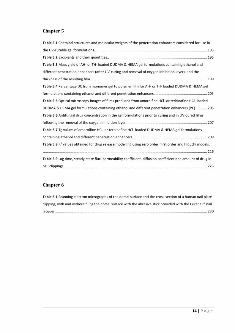
Table 5.1 Chemical structures and molecular weights of the penetration enhancers considered for use in
the UV-curable gel formulations. ................................................................................................................ 193
Table 5.2 Excipients and their quantities.. .................................................................................................. 195
Table 5.3 Mass yield of AH- or TH- loaded DUDMA & HEMA gel formulations containing ethanol and
different penetration enhancers (after UV-curing and removal of oxygen inhibition layer), and the
thickness of the resulting film ..................................................................................................................... 199
Table 5.4 Percentage DC from monomer gel to polymer film for AH- or TH- loaded DUDMA & HEMA gel
formulations containing ethanol and different penetration enhancers. .................................................... 203
Table 5.5 Optical microscopy images of films produced from amorolfine HCl- or terbinafine HCl- loaded
DUDMA & HEMA gel formulations containing ethanol and different penetration enhancers (PE) ............ 205
Table 5.6 Antifungal drug concentration in the gel formulations prior to curing and in UV-cured films
following the removal of the oxygen inhibition layer. ................................................................................ 207
Table 5.7 Tg values of amorolfine HCl- or terbinafine HCl- loaded DUDMA & HEMA gel formulations
containing ethanol and different penetration enhancers . ......................................................................... 209
Table 5.8 R² values obtained for drug release modelling using zero order, first order and Higuchi models.
..................................................................................................................................................................... 216
Table 5.9 Lag time, steady-state flux, permeability coefficient, diffusion coefficient and amount of drug in
nail clippings. ............................................................................................................................................... 223
Chapter 6
Table 6.1 Scanning electron micrographs of the dorsal surface and the cross-section of a human nail plate
clipping, with and without filing the dorsal surface with the abrasive stick provided with the Curanail® nail
lacquer. ........................................................................................................................................................ 230

15 | P a g e
List of Figures
Chapter 1
Fig. 1.1 Schematic diagram of the nail unit – external appearance and cross-section ................................. 22
Fig. 1.2 Scanning electron micrograph of (A) the dorsal surface of a human nail plate and (B) the cross-
section of a human nail clipping. ................................................................................................................... 23
Fig. 1.3 Schematic diagram of the nail unit cross-section highlighting the sites of infection and hence types
of onychomycosis .......................................................................................................................................... 27
Fig. 1.4 Schematic diagram of the nail unit cross-section highlighting the main clinical features of nail
psoriasis and the site of nail involvement .................................................................................................... 36
Chapter 2
Fig. 2.1 Schematic diagram highlighting the importance of excipient choice and quantity. ........................ 67
Fig. 2.2 Typical rheogram of UV-curable gel formulation ............................................................................. 73
Fig. 2.3 Chemical structure of acrylate and methacrylate-based monomers. .............................................. 83
Fig. 2.4 Effect of photoinitiator concentration on mass yield from monomer to cured polymer film. ........ 88
Fig. 2.5 Effect of photoinitiator concentration on percentage DC from monomer to polymer. ................... 88
Fig. 2.6 Effect of photoinitiator concentration on concentration of residual DUDMA and EMA monomers
extracted from UV-cured polymer film ......................................................................................................... 88
Fig. 2.7 Percentage DC from monomer to polymer for formulations containing between 0 and 50% v/v
ethanol .......................................................................................................................................................... 90
Fig. 2.8 Concentration of residual monomers in the UV-cured polymer films produced from formulations
containing between 0 and 50% v/v ethanol. ................................................................................................. 90
Fig. 2.9 Percentage mass yield of films produced by UV-curable gel formulations containing between 0 and
50% v/v ethanol............................................................................................................................................. 90
Fig. 2.10 Water sensitivity (at 48 hour incubation) of films produced by UV-curable gel formulations
containing between 0 and 50% v/v ethanol .................................................................................................. 90
Fig. 2.11 Stability of amorolfine HCl in UV-curable gels over time. .............................................................. 95
Fig. 2.12 Stability of terbinafine HCl in UV-curable gels over time. .............................................................. 95
Fig. 2.13 Photographic image of the UVA nail lamp used for curing the gel formulations. .......................... 96
Fig. 2.14 Effect of UVA cure-time on mass yield from monomer to cured polymer film .............................. 97
Fig. 2.15 Effect of UVA cure-time on percentage DC from monomer to polymer. ....................................... 97
Fig. 2.16 Effect of UVA cure-time on concentration of residual monomers in the cured polymer films.. .... 97
Fig. 2.17 Suggested synthetic pathway for diurethane dimethacrylate & ethyl methacrylate copolymer . 99
Fig. 2.18 Basic chemical structure of DUDMA & IBOMA copolymer and DUDMA & HEMA copolymer. .... 100

16 | P a g e
Fig. 2.19 FT-IR spectra of the gel (containing DUDMA, EMA and photoinitiator) and of the resulting
polymer film after UV-curing (but before removal of the oxygen inhibition layer). ................................... 100
Chapter 3
Fig. 3.1 Photographic image of UV-cured film produced from a DUDMA & EMA containing gel formulation
(drug-free and solvent-free). ....................................................................................................................... 109
Fig. 3.2 Photographic image of the TOWL instrument (Aquaflux) used to conduct film occlusivity tests. . 118
Fig. 3.3 Schematic of Instron set up for pull-off test ................................................................................... 120
Fig. 3.4 Steps to apply UV-curable gel formulations.. ................................................................................. 121
Fig. 3.5 Weight gain (%) for UV-cured films produced from DUDMA & EMA, IBOMA or HEMA containing
gels (± solvent and ± drug) at 48 hours ....................................................................................................... 126
Fig. 3.6 Influence of degree of conversion (%) from monomer gel to polymer film on the resulting film’s
swelling ability, and hence cross-linking extent. ......................................................................................... 127
Fig. 3.7 Relative pressure vs volume adsorbed for UV-cured films produced from DUDMA & EMA, IBOMA
or HEMA containing gels (drug-free, solvent-free, with a DUDMA: diluent monomer ratio of 85:15 % v/v
unless otherwise stated). ............................................................................................................................ 128
Fig. 3.8 Total pore volume of UV-cured films produced from DUDMA & EMA, IBOMA or HEMA containing
gels (± solvent and ± drug) .......................................................................................................................... 128
Fig. 3.9 Polarised light micrographs & corresponding XRD patterns of amorolfine HCl & terbinafine HCl 135
Fig. 3.10 XRD patterns of DUDMA & EMA, IBOMA or HEMA copolymer films (drug-free and solvent-free
with a DUDMA: diluent monomer ratio of 85:15% v/v). ............................................................................. 135
Fig. 3.11 FT-IR spectra of DUDMA & EMA, IBOMA or HEMA copolymer films (drug-free and solvent-free
with a DUDMA: diluent monomer ratio of 85:15% v/v). ............................................................................. 138
Fig. 3.12 TGA profiles of films produced from drug-free UV-curable gel formulations containing EMA,
IBOMA and HEMA . ..................................................................................................................................... 141
Fig. 3.13 Log [storage modulus] and tan δ from DMA and heat flow from DSC vs. temperature for UV-
cured films produced from DUDMA & EMA gel formulations without solvent and without drug ............. 145
Fig. 3.14 Log [storage modulus] and tan δ from DMA and heat flow from DSC vs. temperature for UV-
cured films produced from the DUDMA & EMA gel formulations with solvent (ethanol or NMP), but
without drug ................................................................................................................................................ 145
Fig. 3.15 Reduction in TOWL following application of a formulation on the nail plate surface. ................. 148
Fig. 3.16 Influence of film thickness on film occlusivity .............................................................................. 148
Fig. 3.17 Cross-cut scores for films produced from gels containing DUDMA & EMA, IBOMA or HEMA (±
solvent and ± drug) ...................................................................................................................................... 150
Fig. 3.18 Peak adhesive force (A) and work of adhesion (B) readings obtained for the UV-cured films
produced from gels containing DUDMA & EMA, IBOMA or HEMA (± solvent and ± drug) ........................ 151

17 | P a g e
Fig. 3.19 Water sensitivity score of UV-cured films produced from DUDMA & EMA gels (± solvent and ±
drug). ........................................................................................................................................................... 153
Fig. 3.20 Water sensitivity score of UV-cured films produced from DUDMA & IBOMA gels (± solvent and ±
drug) ............................................................................................................................................................ 153
Fig. 3.21 Water sensitivity score of UV-cured films produced from DUDMA & HEMA gels (± solvent and ±
drug) ............................................................................................................................................................ 153
Fig. 3.22 Water sensitivity score of UV-cured films produced from DUDMA & HEMA gels (± drug) with a
DUDMA: HEMA ratio of 75:25 % v/v ........................................................................................................... 153
Fig. 3.23 Area under the curve values calculated from the curves in Fig. 3.19 – Fig. 3.22 for the UV-cured
films produced from DUDMA & EMA, IBOMA or HEMA containing gels (± solvent and ± drug) ................ 153
Fig. 3.24 In vivo residence profile of UV-cured films (± solvent and ± drug) and a commercially available
nail lacquer on the ten fingernails in six volunteers.................................................................................... 156
Fig. 3.25 % Mass change of UV-cured films produced from DUDMA & EMA gels (± solvent and ± drug) .. 158
Fig. 3.26 % Mass change of UV-cured films produced from DUDMA & IBOMA gels (± solvent and ± drug).
..................................................................................................................................................................... 158
Fig. 3.27 % Mass change of UV-cured films produced from DUDMA & HEMA gels (± solvent and ± drug) 158
Fig. 3.28 Stability of amorolfine HCl in UV-curable films over time. ........................................................... 160
Fig. 3.29 Stability of terbinafine HCl in UV-curable films over time ............................................................ 160
Chapter 4
Fig. 4.1 Franz diffusion cell set up for release studies................................................................................. 166
Fig. 4.2 Modified Franz diffusion cell used for permeation studies.. .......................................................... 168
Fig. 4.3 Photographic images of T.rubrum inoculated test plates set up on day 0 and day 3..................... 172
Fig. 4.4 Cumulative % drug release from the amorolfine HCl loaded UV-cured films and Curanail®, their
corresponding polarised light microscopy images (pre- and post-release) and the amount of drug released
by day 30.. ................................................................................................................................................... 178
Fig. 4.5 Cumulative % drug release from the terbinafine HCl loaded UV-cured films, their corresponding
polarised light microscopy images (pre- and post-release) and the amount of drug released by day 30. . 179
Fig. 4.6 Weight change (%) for the drug-loaded UV-cured films produced from DUDMA & EMA, IBOMA or
HEMA containing gels following drug-release studies ................................................................................ 180
Fig. 4.7 Cumulative amount of amorolfine HCl permeated across the nail with time from the UV-cured and
Curanail® films, and the % of amorolfine HCl permeated across the nail and remaining in the nail at day
30.. ............................................................................................................................................................... 185
Fig. 4.8 Cumulative amount of terbinafine HCl permeated across the nail with time from the UV-cured
films, and the % of terbinafine HCl permeated across the nail and remaining in the nail at day 30 .......... 186

18 | P a g e
Chapter 5
Fig. 5.1 Influence of penetration enhancer incorporation on concentration of residual monomers in AH- or
TH- loaded UV-cured DUDMA & HEMA copolymer films ............................................................................ 203
Fig. 5.2 Scanning electron micrographs of the cross-sectional surfaces of UV-cured films produced from
amorolfine HCl-loaded DUDMA & HEMA gels containing ethanol and (A) no enhancer, (B) water, (C) MPE,
(D) NMP and (E) PEG 200. ........................................................................................................................... 206
Fig. 5.3 Water sensitivity scores for amorolfine HCl-loaded UV-cured films produced from DUDMA &
HEMA gels containing ethanol and different penetration enhancers......................................................... 210
Fig. 5.4 Water sensitivity scores for terbinafine HCl-loaded UV-cured films produced from DUDMA &
HEMA gels containing ethanol and different penetration enhancers ........................................................ 210
Fig. 5.5 Area under the curve values calculated from the curves in Fig. 5.3 & Fig. 5.4 for the UV-cured films
produced from the drug-loaded DUDMA & HEMA gels containing ethanol and different penetration
enhancers ................................................................................................................................................... 210
Fig. 5.6 Cumulative % drug release from the 4% w/v amorolfine HCl-loaded DUDMA & HEMA UV-cured
films containing ethanol and different penetration enhancers, their corresponding polarised light
microscopy images (pre- and post-release) and the amount of drug released by day 30. ......................... 214
Fig. 5.7 Cumulative % drug release from the 6% w/v terbinafine HCl-loaded DUDMA & HEMA UV-cured
films containing ethanol and different penetration enhancers, their corresponding polarised light
microscopy images (pre- and post-release) and the amount of drug released by day 30. ......................... 215
Fig. 5.8 Weight change (%) for UV-cured films produced from drug-loaded DUDMA & HEMA gels
containing ethanol and different penetration enhancers following drug-release studies. ........................ 216
Fig. 5.9 Influence of degree of conversion from monomer gel to polymer film on amorolfine HCl release
from the polymer film. ................................................................................................................................ 216
Fig. 5.10 Cumulative amount of amorolfine HCl permeated across the nail with time from the 4% w/v
amorolfine HCl-loaded DUDMA & HEMA UV-cured films containing ethanol and different penetration
enhancers , and the % of amorolfine HCl permeated across the nail and remaining in the nail at day 30 221
Fig. 5.11 Cumulative amount of terbinafine HCl permeated across the nail with time from the 6% w/v
terbinafine HCl-loaded DUDMA & HEMA UV-cured films containing ethanol and different penetration
enhancers, and the % of terbinafine HCl permeated across the nail and remaining in the nail at day 30.. 222
Chapter 6
Fig. 6.1 Schematic diagram highlighting the investigations to assess the pharmaceutical potential of UV-
curable gel formulations. ............................................................................................................................ 226

19 | P a g e
Abbreviations
AH Amorolfine hydrochloride
ANOVA Analysis of variance
ASTM American Society for Testing and Materials
BA Butyl acrylate
1,4-BDA 1,4-Butanediol acrylate
1,4-BDDMA 1,4-Butanediol dimethacrylate
BMA Butyl methacrylate
BNF British national formulary
CIR Cosmetic ingredient review
CND Creative Nail Design, Inc.
DC Degree of conversion
DEGDA Diethylene glycol diacrylate
DMA Dynamic mechanical analysis
DSC Differential scanning calorimetry
DUDMA Diurethane dimethacrylate
EA Ethyl acrylate
ECA Ethyl cyanoacrylate
EGDMA Ethylene glycol dimethacrylate
EMA Ethyl methacrylate
ETOH Ethanol
FID Flame ionisation detector
Fig. Figure
FT-IR Fourier transform infrared
GC Gas chromatography
HEA Hydroxyethyl acrylate
HEMA 2-Hydroxyethyl methacrylate
HDPE High density polyethylene
HPLC High-performance liquid chromatography
HPMA Hydroxypropyl methacrylate
20 | P a g e
IBOMA Isobornyl methacrylate
ICH International Council for Harmonisation of Technical Requirements for Pharmaceuticals for Human Use
ISO International Organisation for Standardisation
IUPAC International Union of Pure and Applied Chemistry
LoD Limit of detection
LoQ Limit of quantification
M Monomer
MIC Minimum inhibitory concentration
MMA Methyl methacrylate
MPE 2-Mercaptoethanol
NF Not formulated
NMP 1-Methyl-2-pyrrolidinone
NSI Nail Systems International
PAF Peak adhesive force
PBS Phosphate buffer solution
PE Penetration enhancer
PEG 200 Poly(ethylene glycol) 200
PI Photoinitiator
PLM Polarised light microscopy
SDA Sabouraud dextrose agar
SEM Scanning electron microscopy
Tg Glass transition temperature
TGA Thermal gravimetric analysis
TH Terbinafine hydrochloride
TOWL Transonychial water loss
TREGDA Triethylene glycol diacrylate
TREGDMA Triethylene glycol dimethacrylate
TRPGDA Tripropylene glycol diacrylate
UDA Urethane diacrylate
XRD X-ray diffraction
21 | P a g e
Chapter 1: Introduction to the human nail, its diseases, current
treatments, and UV-curable gels
In order to fully appreciate the need for a new topical nail medicine, knowledge
regarding nail diseases, why these diseases require treatment, current treatment
options, and the barriers faced for a successful response is required. This in turn requires
an understanding of the nail’s structure and function. This chapter therefore covers
these bases, and eventually provides a detailed overview of UV-curable gels and why
they have been researched extensively, in this thesis, as a candidate for topical nail
therapy.
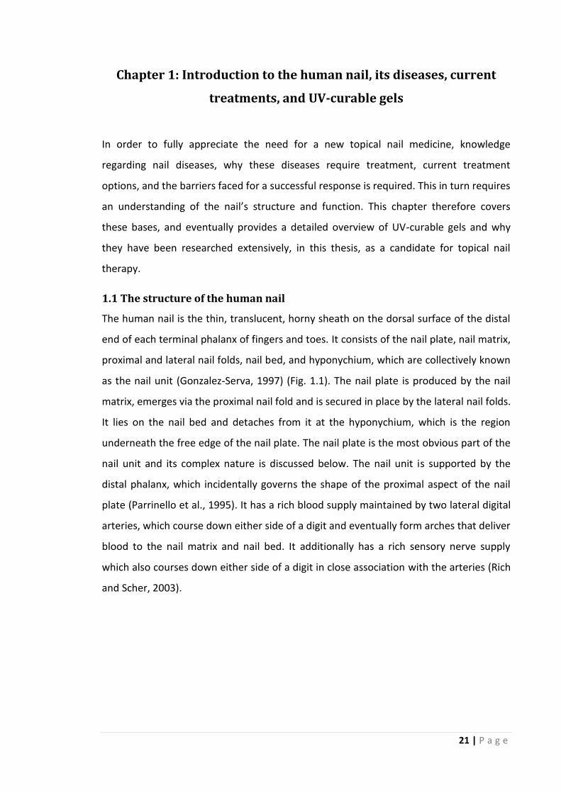
1.1 The structure of the human nail
The human nail is the thin, translucent, horny sheath on the dorsal surface of the distal
end of each terminal phalanx of fingers and toes. It consists of the nail plate, nail matrix,
proximal and lateral nail folds, nail bed, and hyponychium, which are collectively known
as the nail unit (Gonzalez-Serva, 1997) (Fig. 1.1). The nail plate is produced by the nail
matrix, emerges via the proximal nail fold and is secured in place by the lateral nail folds.
It lies on the nail bed and detaches from it at the hyponychium, which is the region
underneath the free edge of the nail plate. The nail plate is the most obvious part of the
nail unit and its complex nature is discussed below. The nail unit is supported by the
distal phalanx, which incidentally governs the shape of the proximal aspect of the nail
plate (Parrinello et al., 1995). It has a rich blood supply maintained by two lateral digital
arteries, which course down either side of a digit and eventually form arches that deliver
blood to the nail matrix and nail bed. It additionally has a rich sensory nerve supply
which also courses down either side of a digit in close association with the arteries (Rich
and Scher, 2003).

22 | P a g e
Fig. 1.1 Schematic diagram of the nail unit – external appearance and cross-section. (Adapted from Jiaravuthisan et al., Journal of the American Academy of Dermatology, 57, 1, 2007.)
1.1.1 The nail plate
The nail plate is the end-product of the keratinocyte differentiation in the nail matrix.
The nail plate grows throughout life, but with varying rates among individuals
(Fleckman, 2005). On average, it grows at a rate of approximately 3 mm per month for
fingernails, and a significantly slower 1 mm per month for toenails (Rich and Scher,
2003). Therefore fingernails take 6 months to grow out completely, while toenails take
between 12 – 18 months. Furthermore, the thickness of the fingernail plates
(approximately 0.5 mm at the distal edge) is around half the thickness of the big toe (up
to 1 mm at the distal edge) (Hamilton et al., 1955).
23 | P a g e
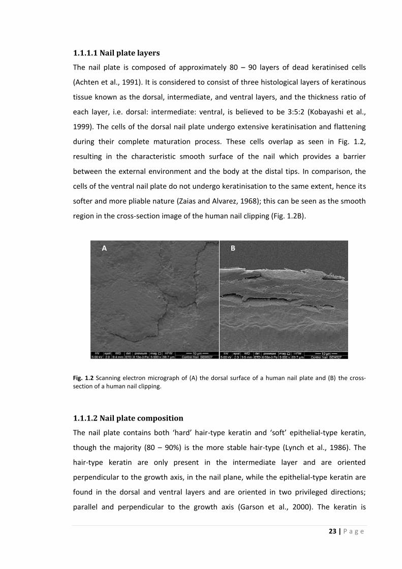
1.1.1.1 Nail plate layers
The nail plate is composed of approximately 80 – 90 layers of dead keratinised cells
(Achten et al., 1991). It is considered to consist of three histological layers of keratinous
tissue known as the dorsal, intermediate, and ventral layers, and the thickness ratio of
each layer, i.e. dorsal: intermediate: ventral, is believed to be 3:5:2 (Kobayashi et al.,
1999). The cells of the dorsal nail plate undergo extensive keratinisation and flattening
during their complete maturation process. These cells overlap as seen in Fig. 1.2,
resulting in the characteristic smooth surface of the nail which provides a barrier
between the external environment and the body at the distal tips. In comparison, the
cells of the ventral nail plate do not undergo keratinisation to the same extent, hence its
softer and more pliable nature (Zaias and Alvarez, 1968); this can be seen as the smooth
region in the cross-section image of the human nail clipping (Fig. 1.2B).
Fig. 1.2 Scanning electron micrograph of (A) the dorsal surface of a human nail plate and (B) the cross-section of a human nail clipping.
1.1.1.2 Nail plate composition
The nail plate contains both ‘hard’ hair-type keratin and ‘soft’ epithelial-type keratin,
though the majority (80 – 90%) is the more stable hair-type (Lynch et al., 1986). The
hair-type keratin are only present in the intermediate layer and are oriented
perpendicular to the growth axis, in the nail plane, while the epithelial-type keratin are
found in the dorsal and ventral layers and are oriented in two privileged directions;
parallel and perpendicular to the growth axis (Garson et al., 2000). The keratin is
A B

24 | P a g e
thought to be held together by globular, cysteine rich proteins, whose disulphide links
behave like an adhesive (Fleckman, 1997). This ‘sandwich’ structure and the strong
intercellular junctions give the nail great mechanical rigidity and hardness, both in the
transverse direction and in the growth direction.
The nail plate also contains 0.1% calcium, but this barely contributes to its hardness
(Rich and Scher, 2003). It contains water at 10 – 30%, which is directly related to the
environmental relative humidity and is important for nail flexibility and elasticity
(Forslind, 1970, Baden et al., 1973), while lipids account for less than 5% of the nail’s
content (Helmdach et al., 2000).
1.2 The function of the human nail
It is evident that the nail unit is a complex unique structure; it is to no surprise then that
it serves numerous functions. These include protecting the distal digits, improving fine
touch sensation, assisting in picking up and manipulating small objects, scratching and
grooming, and enhancing the aesthetic appearance of the hands and feet. It is
commonly used as a cosmetic organ, and by some, to communicate social class
(Gonzalez-Serva, 1997, Rich and Scher, 2003). However it is often also abused by biting,
which is thought to contribute to providing a sense of relief and alleviating boredom
(Williams et al., 2007).
1.3 Nail diseases and current treatments
Unfortunately, the nail can suffer from a very wide range of disorders, from benign, for
example, nail discoloration following the administration of drugs such as chloroquine, to
extremely painful and serious, for example, malignant tumours of the nail apparatus.
The majority of nail disorders arise from two nail diseases - onychomycosis and nail
psoriasis. These diseases are therefore explored in the following sections.

25 | P a g e
1.3.1 Onychomycosis
Onychomycosis is a fungal nail infection, with a mean prevalence of 4.3% in Europe and
North America (Sigurgeirsson and Baran, 2014). Over 50 million people worldwide
suffered from the disease in 2012, and it is forecasted that cases of onychomycosis
across the US, France, Germany, Italy, Spain, UK and Japan will increase by 15% over the
next decade (De Angelis, 2013). Onychomycosis is more frequent amongst the elderly,
diabetics, individuals with peripheral artery disease, and sports-active individuals. Its
increase in occurrence is in part a reflection of the rise in individuals in these categories,
together with the increasing numbers of individuals who are immunodeficient (resulting
from an increased use of immunosuppressant drugs), lifestyle factors such as wearing ill-
fitting footwear, and improved detection and greater public awareness (Piérard, 2001,
Gupta et al., 1998, Gupta et al., 2000b, Caputo et al., 2001, Scher, 1996).
1.3.1.1 Etiology
The dermatophytes, particularly Trichophyton rubrum and Trichophyton
mentagrophytes, yeasts such as Candida albicans, and non-dermatophytes such as
Scytalidium dimidiatum, Scytalidium hyalinum, and Fusarium species have all been
identified as causative agents for the disease (Midgley et al., 1994). In a recent study,
the main causative agent was found to be a dermatophyte, accounting for 65% of cases,
with Trichophyton rubrum identified as the single most common fungus (45%). Yeasts
were found on average in 21% of cases, and non-dermatophyte moulds in 13% of cases
(Sigurgeirsson and Baran, 2014). Onychomycosis predominately affects toenails
compared to fingernails, with the ratio of toenail to fingernail onychomycosis ranging
from 4:1 to 19:1 (Gupta et al., 2014). Onychomycosis can adopt several clinical patterns
depending on the causative organism and site of infection, and is classified accordingly
as explained in the following section.
1.3.1.2 Disease features and classification
The five main categories of onychomycosis are (A) distal and lateral subungual
onychomycosis, (B) proximal subungual onychomycosis, (C) superficial white
onychomycosis, (D) endonyx onychomycosis, and (E) total dystrophic onychomycosis,
depending on where the infection begins (Ameen et al., 2014) (Fig. 1.3).

26 | P a g e
(A) Distal and lateral subungual onychomycosis
Distal and lateral subungual onychomycosis is the most common form, in which the nail
bed is invaded by fungal (mainly T. rubrum) penetration through the distal or lateral
margins. The affected nail can become thick, discoloured, and onycholysis may present
to an extent. The infection can be confined to one side of the nail or spread to involve
the whole nail bed. Over time the nail plate can therefore become friable and break.
(B) Proximal subungual onychomycosis
Proximal subungual onychomycosis is usually caused by T. rubrum, and can originate in
the proximal nail fold, with subsequent penetration into the newly forming nail plate or
beneath the proximal nail plate. It produces a white discolouration in the area of the
lunula, and as the nail plate grows, the white discolouration moves distally.
(C) Superficial white onychomycosis
White superficial onychomycosis arises as the surface of the nail plate is invaded by the
fungal organism, usually T. mentagrophytes. Crumbling white lesions appear on the nail
surface, and the infection can spread to the deeper layers to involve the entire nail
plate.
(D) Endonyx onychomycosis
Endonyx onychomycosis is most commonly caused by T. soudanense and T. violaceum,
which invade the nail by immediately penetrating the nail plate keratin. It causes the nail
plate to be discoloured white with the absence of onycholysis and subungual
hyperkeratosis.
(E) Total dystrophic onychomycosis
Total dystrophic onychomycosis results from the unchecked progression of any of the
other forms of onychomycosis, especially of distal and lateral subungual onychomycosis.
It causes the nail plate to become hyperkeratotic and crumble, therefore the nail plate is
essentially completely destroyed.

27 | P a g e
Mixed pattern onychomycosis
Occasionally, the same individual may present with different patterns of nail plate
infection, with the most common combinations including proximal subungual
onychomycosis with superficial white onychomycosis, and distal and lateral subungual
onychomycosis with superficial white onychomycosis (Hay and Baran, 2011).
Fig. 1.3 Schematic diagram of the nail unit cross-section highlighting the sites of infection and hence types of onychomycosis (left), and corresponding images of its clinical appearance (right). (A) Distal and lateral subungual onychomycosis; (B) proximal subungual onychomycosis; (C) superficial white onychomycosis; (D) endonyx onychomycosis. (Adapted from Hay, R. J. and Baran, R., Baran & Dawber’s Diseases of the Nails and their Management, Forth Edition, 2012.)
1.3.1.3 Why treat onychomycosis?
There are numerous reasons as to why the successful treatment of onychomycosis is an
absolute necessity. For one, it can prevent the progression of the disease to its total
dystrophic form and help restore the nails’ natural beauty. While this may appear as
tackling what looks to be merely a cosmetic nuisance, the reality is in fact far from this,
as in addition to causing obvious changes to the appearance of the nail unit,
onychomycosis presents with physical, psychosocial, and occupational consequences
(Scher, 1996, Lubeck et al., 1993, Drake et al., 1999, Elewski, 1997, Whittam and Hay,
1997, Turner and Testa, 2000). One study which surveyed a total of 258 patients with
confirmed onychomycosis, found that the disease caused nail-trimming problems (76%),
embarrassment (74%), pain (48%), nail pressure (40%), discomfort wearing shoes (38%),

28 | P a g e
and an impaired ability to pick up small objects by those with fingernail involvement
(41%). Furthermore, during a 6-month period, more than 58 onychomycosis-related sick
days and 468 medical visits (1.8 per subject) were reported (Drake et al., 1998). A more
recent article on the burden of onychomycosis revealed that up to 93% of patients
believe that other people are repulsed by the site of their infected nails, and that nearly
one-fifth of patients avoid various social activities due to the disease (Daniel, 2013).
If left untreated, an onychomycotic nail not only progresses to its total dystrophic form,
but acts like a fungal reservoir with the potential to spread to other nails, body sites
such as the groin, skin and scalp, and to other people (Ameen et al., 2014). It has been
suggested that the presence of sensitising dermatophyte antigens in an infected nail unit
may predispose to clinical conditions such as erythema nodosum, urticaria, atopic
dermatitis and asthma or sensitisation of the bronchia and upper airways (Hicks, 1977,
Weary and Guerrant, 1967, Wilson et al., 1993, Ward et al., 1989, Schwartz and Ward,
1995, Gumowski et al., 1987, Ameen et al., 2014). An onychomycotic nail can also
disrupt the integrity of the skin providing an entry port for bacteria, and this may explain
why diabetic patients with onychomycosis have a high predisposition to foot ulceration
and gangrene (Joseph, 2002, Ameen et al., 2014).
The rise in the incidence of onychomycosis is of significant importance as it is further
increasing the pool of infection. This urgently needs to be controlled and reversed,
through education for prevention and treatment for elimination. The next section walks
through the treatment options currently available for onychomycosis.
1.3.1.4 The current armamentarium for onychomycosis treatment
A range of treatment options are available for onychomycosis, with the main aim to
permanently eliminate the infecting organism as demonstrated by culture and
microscopy. In the UK, the options include eliminating the infectious agent by topical or
systemic therapy; details of the antifungal prescription-only–medicines (POMs) available
can be found in Table 1.1. There are further topical therapies for onychomycosis
treatment which are available outside the UK, and all the available systemic and topical
therapies are discussed in detail below. Laser and light therapies are also available
outside the UK; however at the time of writing they had FDA clearance for only a

29 | P a g e
‘temporary increase in clear nail in patients with onychomycosis’ rather than cure, and
will therefore not be discussed further.
Table 1.1 Onychomycosis treatment options in the UK as detailed in the British National Formulary (BNF) 71 (March 2016).
Therapy Formulation Dose Duration
Systemic
Terbinafine (as hydrochloride) 250 mg tablet
250 mg daily 6 weeks – 3 months (occasionally longer in toenail infections).
Itraconazole 100 mg capsule 200 mg daily 3 months
200 mg twice daily 7 days, repeated after 21 day interval; fingernails 2 courses, toenails 3 courses.
Griseofulvin 500 mg tablet 500 – 1000 mg daily
6 – 9 months for fingernails and 12 – 18 months for toenails.
Topical
Amorolfine (as hydrochloride) 5% w/v nail lacquer
Applied to infected nails 1 – 2 times weekly after filing and cleansing.
6 months for fingernails and 9 – 12 months for toenails (review at intervals of 3 months).
Tioconazole 28% w/v solution (Trosyl®)
Applied to nails and surrounding skin twice daily.
Up to 6 months (may be extended to 12 months).
Systemic therapies
The BNF states that for onychomycosis treatment, a systemic antifungal is more
effective than topical therapy. These agents enter the systemic circulation following oral
administration and absorption, and diffuse from the blood vessels into the nail plate via
the nail bed. The two most widely used drugs are terbinafine and itraconazole, with
terbinafine considered the drug of choice, and itraconazole reserved for Candida and
non-dermatophyte infections (Sá et al., 2014). Terbinafine and itraconazole both
concentrate and persist in the nail plate following therapy cessation. Itraconazole can
persist for up to 6 – 9 months, compared to the 3 – 6 months for terbinafine (Baran et
al., 2008a, Baran et al., 2008b), and this explains the success of the ‘pulsed’
administration of itraconazole. Griseofulvin is rarely considered for onychomycosis
treatment given its lower efficacy, higher relapse rates, longer treatment duration and
hence lower compliance rates, and greater risk of drug interactions when compared to
the others. Fluconazole is not licensed for the treatment of onychomycosis, but may be
a useful alternative for patients who cannot tolerate terbinafine or itraconazole (Ameen

30 | P a g e
et al., 2014). These antifungals’ mechanism of actions, spectrum of activity and
mycological cure rates are detailed in Table 1.2.
Table 1.2 Mechanism of action, spectrum of activity and mycological cure rates of systemic antifungals used for onychomycosis treatment.
Unfortunately, systemic therapy can cause adverse reactions such as headache,
gastrointestinal symptoms, taste disturbances, and more rarely, liver and kidney
Antifungal
Mode of action
Spectrum of activity
Mycological cure rate (toenail infections)
Terbinafine Terbinafine is an allylamine which inhibits the enzyme squalene epoxidase of the ergosterol synthetic pathway. Intracellular accumulation of squalene initiates a cascade of events that leads to the fungicidal activity, whereas the depletion of ergosterol (needed for integrity of the fungal cell membranes) leads to fungistatic activity (Ryder and Favre, 1997).
Terbinafine has a broad and potent fungicidal effect against dermatophytes, particularly T. rubrum and T. mentagrophytes, but a lower fungistatic activity against Candida species (Bueno et al., 2010).
74 – 82% (Goodfield, 1992, Drake et al., 1997).
Itraconazole Itraconazole is a triazole which inhibits the fungal cytochrome P450-mediated 14-alpha-lanosterol demethylation, an essential step in fungal ergosterol biosynthesis. This results in the depletion of ergosterol and fungistatic activity (Vanden Bossche et al., 1989).
Itraconazole is active against a range of fungi including yeasts, dermatophytes and some non-dermatophyte moulds. When compared to terbinafine, it is not as active in vitro against dermatophytes – it has a 10 times greater minimum inhibitory concentration (MIC) (Bueno et al., 2010).
Pulse therapy: 88.2 – 90.6% (Gupta et al., 2001, Gupta et al., 2006).
Griseofulvin Griseofulvin acts by inhibiting fungal nucleic acid synthesis, arresting cell division and inhibiting fungal cell wall synthesis (Roobol et al., 1977).
Griseofulvin is weakly fungistatic against species of Trichophyton, Microsporum,
and Epidermophyton. Studies comparing griseofulvin therapy with terbinafine and itraconazole have demonstrated lower cure rates for griseofulvin (Faergemann et al., 1995, Haneke et al., 1995, Korting et al., 1993).
30 – 40% (Davies et al., 1967).
Fluconazole Fluconazole is also a triazole and therefore its use results in the depletion of ergosterol in a similar manner to itraconazole.
Fluconazole has some activity against dermatophytes and some Candida species. However, it is less effective than itraconazole or terbinafine (Arca et al., 2002).
47 – 62% (Scher et al., 1998).
31 | P a g e
disturbances. Furthermore, while terbinafine displays minimal drug–drug interactions,
itraconazole and fluconazole can inhibit the cytochrome P450 3A4 isoenzyme system,
with fluconazole additionally inhibiting CYP2C9. They therefore have the potential to
increase the plasma concentration of other concomitantly administered drugs that are
metabolised by these enzyme systems, which can cause major problems. Oral therapy
with standard antifungal drugs is also subject to a consistent failure rate of 20%, as well
as a relapse rate of 25% (Roberts, 1999, Gupta, 2012). Reasons identified for treatment
failure include poor patient compliance, misdiagnosis, low bioavailability, lack of drug
penetration into the nail due to extensive onycholysis for example, and the presence of
dormant, drug-resistant fungal spores (Pierard et al., 2000, Baran et al., 2008a, Baran et
al., 2008b) .
Topical therapies
Therapy with topical antifungals is limited to mild cases of distal and lateral subungual
onychomycosis, for superficial white onychomycosis, or where there are
contraindications to systemic therapy. As shown in Table 1.1, amorolfine (as
hydrochloride) 5% w/v nail lacquer and tioconazole 28% w/v solution (Trosyl®) are
treatment options in the UK, with amorolfine 5% w/v nail lacquer considered as the
more superior agent due to its greater efficacy against onychomycosis-causing
pathogens. While both are available as POMs, amorolfine nail lacquer is also available to
the public over-the-counter (OTC) for the treatment of mild cases of distal and lateral
subungual onychomycosis caused by dermatophytes, yeasts and mould; subject to
treatment of a maximum of two nails (BNF 71, March 2016).
Outside the UK, two ciclopirox 8% w/v nail lacquers are available, Penlac® (also
marketed as Batrafen®, Loprox® and Mycoster®) and Ciclopoli® (also marketed as
RejuveNail®). Both should be applied once daily, for up to 24 weeks on the fingernails
and up to 48 weeks on the toenails. Two recently FDA approved formulations for toenail
onychomycosis – Jublia® (efinaconazole) 10% topical solution and Kerydin® (Tavaborole)
5% topical solution – are also available. These are applied to the affected toenails once
daily for 48 weeks. These antifungals’ mechanism of actions, spectrum of activity and
mycological cure rates are shown in Table 1.3.

32 | P a g e
Table 1.3 Mechanism of action, spectrum of activity and mycological cure rates of topical antifungals used for onychomycosis treatment.
Formulation Antifungal’s mode of action Spectrum of activity Mycological cure rate
Amorolfine 5% w/v nail lacquer
Amorolfine is a morpholine antifungal agent that inhibits the delta 14 reductase and delta 8 and delta 7 isomerase enzymes in the ergosterol biosynthetic pathway which leads to fungistatic and fungicidal activities (Ameen et al., 2014).
It exhibits a broad spectrum of activity against dermatophytes, non-dermatophyte moulds and yeasts, and is fungicidal against C. albicans and T. mentagrophytes (Ameen et al., 2014).
52% for toenail onychomycosis (Zaug, 1995).
Tioconazole 28% w/v solution (Trosyl®)
Tioconazole is an imidazole derivative which inhibits the CYP450 dependent enzyme lanosterol 14 alpha-demethylase, involved in the ergosterol biosynthetic pathway. This results in ergosterol depletion and fungistatic activity.
It exhibits a broad spectrum of activity against common dermatophytes and C. albicans (Jevons et al., 1979).
22% (Hay et al., 1985).
Ciclopirox 8% w/v nail lacquer (Penlac®, Batrafen®, Loprox®, Mycoster® Ciclopoli® or RejuveNail® )
Ciclopirox is a hydroxypyridone and by binding to trivalent cations such as Fe3+, it inhibits several metal-dependent enzymes such as cytochromes. It has been shown to affect the energy production of mitochondrial electron transport processes, to reduce the activity of catalase and peroxidase which are responsible for the degradation of toxic peroxides, and to affect the nutrient uptake and associated synthesis of protein and nucleic acid. Ciclopirox is therefore fungicidal and also has anti-inflammatory and anti-allergic properties (Bohn and Kraemer, 2000).
Ciclopirox is active against dermatophytes, as well as yeasts and non-dermatophyte moulds (Bohn and Kraemer, 2000).
29 – 36% for toenail onychomycosis (Gupta et al., 2000a).
Efinaconazole 10% topical solution (Jublia®)
Efinaconazole is a triazole antifungal which inhibits ergosterol biosynthesis by inhibiting the enzyme 14 alpha- demethylase. The resulting ergosterol depletion and accumulation of precursor sterols may secondarily affect cell membrane integrity and function (Tatsumi et al., 2013).
Efinaconazole has shown activity against Trichophyton, Microsporum, Epidermophyton, Acremonium, Fusarium, Paecilomyces, Pseudallescheria, Scopulariopsis, Aspergillus, Cryptococcus, Trichosporon and Candida (Jo Siu et al., 2013).
53 – 55% for toenail onychomycosis (Elewski et al., 2012).
Tavaborole 5% topical solution (Kerydin®)
Tavaborole is a novel oxaborole antifungal drug that exerts its antifungal activity by blocking cellular protein synthesis via inhibition of fungal cytoplasmic leucyl-aminoacyl transfer RNA (tRNA) synthetase. Formation of a stable tRNALeu – tavaborole adduct inhibits leucyl-tRNALeu synthesis, thus blocking protein synthesis (Markham, 2014).
Tavaborole is fungicidal against T. rubrum and T. mentagrophytes. It is also active against Trichophyton tonsurans, Epidermophyton floccosum, Microsporum audouinii, Microsporum canis and Microsporum gypseum, Aspergillus fumigatus, Candida spp., Cryptococcus neoformans, Fusarium solani, Malassezia spp. and Saccharomyces cerevisiae (Markham, 2014).
31 – 36% for toenail onychomycosis (Elewski et al., 2015).

33 | P a g e
As can be seen when comparing Tables 1.2 and 1.3, the mycological cure rates are lower
with topical antifungals than systemic antifungals, and this justifies why systemic
therapy is ideal when infections become more complex.
Another product available for fungal nail infections in the UK and mentioned in the BNF
71 (March 2016) is a Phytex® paint. It contains salicylic acid 1.46%, tannic acid 4.89% and
boric acid 3.12% (as borotannic complex), and should be applied twice daily after
washing. However it is considered by the Joint Formulary Committee to be less suitable
for prescribing, as there is limited data to support its use as a therapy for onychomycosis
(Ameen et al., 2014).
There is a vast range of other products (in addition to the amorolfine 5% w/v nail
lacquer) that are available OTC for the treatment of mild fungal nail infections, examples
of which are shown in Table 1.4. While some contain known actives such as urea,
miconazole, and benzalkonium chloride, published studies regarding the efficacy of
these products are lacking and their use cannot therefore be recommended.

34 | P a g e
Table 1.4 Products available OTC from UK pharmacies (Boots, Lloyds and Tesco) for the topical treatment of fungal nail infections (these products were available on 11
th August 2015).
Boots Advanced Footcare Fungal Nail Treatment Solution
Contains pentylene glycol, dimethyl isosorbide, olive-leaf active-TM, hydrolyzed
silk fibroin, 2-phenoxyethanol and
water.
Canespro® Fungal Nail Treatment Set
Contains one 10g urea ointment tube (40% urea),
22 specially designed waterproof plasters, and one
plastic scraper.
Daktarin® 2% Cream
Contains miconazole nitrate, PEG-6, PEG-32, glycol stearate, oleoyl macroglycerides, liquid paraffin, benzoic acid (E210),
butylated hydroxyanisole (E320) and purified water.
Excilor® Treatment for Fungal Nail Infections
Contains ethyllactate, acetic acid, penetration enhancer, film-forming agent, water,
glycerol, polysorbate 80, cetyl acetate, acetylated lanolin
alcohols, biotin and preservatives (contains no
parabens).
Profoot Mycosan Fungal Nail Treatment
Contains aqua, rye ferment filtrate, pentylene glycol, dimethyl isosorbide and hydroxyethylcellulose.
Nailner Repair Brush
Contains ethyl lactate, glycerin, aqua, lactic acid and
citric acid.
Scholl Fungal Nail Treatment
Contains an ‘advanced liquid’. Neither the packaging, nor the
bottle nor the enclosed instruction leaflet gives any
indication as to what the product's active ingredients
are.
ClearZal® BAC Antimicrobial Solution
Contains benzalkonium
chloride (0.1%), purified water (aqua),
cocamidopropylbetaine, cocamidopropylamine oxide, hypromellose, centrimonium chloride, organic aloe vera, didecyldimonium chloride,
diazolidinyl urea, quaternium-15, methylisothiazolinone,
phenoxyethanol,
triethanolamine and citric acid.
Systemic and topical therapy combination
Some studies suggest a topical and systemic antifungal therapy combination for severe
onychomycosis, as this can achieve a higher cure rate, reduces the oral drug intake, and
demonstrates a better cure per cost ratio (Baran et al., 2000, Lecha, 2001, Avner et al.,
2005, Baran et al., 2007). However, there are also a few studies which have failed to find

35 | P a g e
statistical differences between topical and systemic antifungal combination therapies
and oral alone therapies (Rigopoulos et al., 2003, Jaiswal et al., 2007). There appears to
be a need for further rigorously conducted, randomised, controlled trails in order to
draw firm conclusions regarding the efficacy of combination therapy.
1.3.2 Nail psoriasis
Psoriasis is a chronic, immune-mediated inflammatory disease of the skin which typically
causes red, flaky, crusty patches covered with silvery scales. At the cellular level, it is
characterised by epidermal hyperproliferation, incomplete differentiation and
accumulation of glycogen, and it affects between 1.5 – 1.9% of the UK population (Rosa
et al., 2012). As skin appendages, nails can be affected by psoriasis, and the life time
incidence of nail involvement in psoriatic patients is as high as 80 – 90% (Tripuraneni and
Kerdel, 2014). Nail psoriasis without any cutaneous involvement is rare but does occur,
the reasons for which are unknown, and it accounts for 1 – 3% of cases (Wozel, 2008).
1.3.2.1 Disease features
The main features of nail psoriasis are pitting, discoloration, onycholysis, subungual
hyperkeratosis, nail plate dystrophy, and splinter hemorrhages, which can be correlated
with the site of nail involvement (Holzberg and Baran, 2012) (Fig. 1.4). Proximal nail fold
involvement results in its psoriatic scaling accompanied with soft tissue swelling.
Involvement of the matrix causes changes which are reflected in the outgrowing nail
plate, and these changes depend on disease duration as well as location. For instance,
lesions may be limited to nail pitting and transverse furrows reflecting transient matrix
dysfunction, while superficial pits indicate matrix disease in the proximal region; trapped
parakeratotic cells in the plate (which cause the clinical appearance of leukonychia –
white discoloration of the nail plate) reflect matrix disease in the distal region, and
sustained nail abnormalities such as nail plate thickening or nail loss reflect persistence
disease. Nail bed involvement results in splinter hemorrhages (which are longitudinal
collections of extravasated blood under the nail plate), an oil-droplet like discoloration
(which is caused by serum glycoprotein accumulation in and under the nail), subungual
hyperkeratosis and/or onycholysis.

36 | P a g e
Fig. 1.4 Top – schematic diagram of the nail unit cross-section highlighting: (i) the main clinical features of nail psoriasis – A. pitting; B – C. discoloration (leukonychia (B) and “oil-drop” (C)); D. onycholysis; E. subungual hyperkeratosis, and (ii) the site of nail involvement – proximal portion of the matrix, distal portion of the matrix and nail bed. Bottom – corresponding images of clinical appearances. (Adapted from Camisa, C., Handbook of Psoriasis, Second Edition, 2007 and Holzberg, M. and Baran R., The Nail in Dermatological Disease, 2012).
Psoriatic nail disease can lead to significant functional impairment, pain, an altered
sense of touch, and psychological stress. It can also have a negative impact on social and
work activities, and hence quality of life (van der Velden et al., 2014, Baran, 2010, de
Jong et al., 1996). The management of nail psoriasis is therefore necessary, and in the
next section the options available are discussed.
1.3.2.2 Options available for the management of nail psoriasis
Unfortunately, a standardised therapeutic regimen for nail psoriasis does not currently
exist (Crowley et al., 2015). However, based on the studies available for its management,
the options could include topical, intralesional, radiation, and systemic therapies, which
are discussed below.

37 | P a g e
Topical therapies
Topical therapies can be applied to the nail plate, nail fold and/or nail bed depending on
the location of the psoriatic lesion. In order to facilitate a successful therapeutic
response, nails with onycholysis present are trimmed to the point where it separates
from the nail bed, bulky nails are chemically avulsed, and patients may be advised to
cover the topical formulation with an occlusive dressing, such as non-porous tape or
plastic gloves, to enhance drug movement into the nail unit (Holzberg and Baran, 2012).
Therapies with drugs such as glucocorticoids, vitamin D3 analogues, 5-flourouracil,
dithranol, tazarotene and ciclosporin in vehicles such as creams, ointments, gels,
solutions and lacquers have all been investigated, and Table 1.5 provides a summary of
these. In general, topical therapies appear to be an effective form for treating subungual
hyperkeratosis, but lack convincing evidence for other dystrophies. Glucocorticoids and
vitamin D3 analogues are the more popular choices of topical therapy, however as
clinical outcomes are not standardized, comparison among treatment options is virtually
impossible.
Table 1.5 Topical therapies for nail psoriasis management, their efficacies and side-effects.
Topical active Formulation and efficacy Side-effects
Glucocorti-
coids
Betamethasone dipropionate (64 mg/g) with salicylic acid (3%) in an ointment base (Diprosalic® ointment in UK): Reduced subungual hyperkeratosis by 52% when used twice a day for 5 months (Tosti et al., 1998).
Erythema, skin atrophy and tapering of the distal phalange on the treated digit with prolonged use.
Clobetasol-17-propionate (8%) nail lacquer (not available in the UK): Daily use for an average of 2.5 months lead to a 69% improvement, while using the formulation 2 – 3 times per week for an average of 7 months lead to an 80% improvement, particularly in nail pitting and onycholysis (Holzberg and Baran, 2012).
Vitamin D3 analogues
Calcipotriol (50 μg/g) ointment (also available as Dovonex®): Reduced subungual hyperkeratosis by 49% for fingernails and 41% for toenails when used twice a day for 5 months (Tosti et al., 1998).
Erythema, periungual irritation, burning at the site of application, and diffuse urticaria.
Calcipotriol (50 μg/g) cream (no longer marketed in the UK): 3 months use showed improvement of subungual hyperkeratosis and onycholysis in five of seven patients (Holzberg and Baran, 2012).
Tacalcitol (4 μg/g) ointment (Bonalfa® ointment, ISDIN S.A, marketed as Curatoderm® in the UK): Found to be effective in all nail parameters, especially onycholysis and subungual hyperkeratosis, followed by oil-drop discoloration and pitting. The trial included 15 patients and the ointment was used daily at night in occlusion (with cotton gloves) over a 6-month period (Márquez Balbás et al., 2009).

38 | P a g e
Glucocorti-coids and Vitamin D3 analogues combination
Betamethasone dipropionate and calcipotriol ointment (Dovobet® ointment in UK): Application once daily for 12 weeks in a study involving 25 patients resulted in a significant improvement for hyperkeratosis and onycholysis, moderate improvement for oil drops and slight improvement for pitting (Rigopoulos et al., 2009).
Refer to side-effects for glucocorticoids and vitamin D3 analogues.
Combination of calcipotriol cream (no longer marketed in the UK) and clobetasol propionate cream (Dermovate® in UK): Use of calcipotriol cream daily during the weekday and clobetasol propionate cream daily over the weekend for 6 months, followed by the use of only clobetasol propionate cream daily over the weekend for a further 6 months, was found to improve subungual hyperkeratosis by 77% at 12 months in a study involving 60 patients (Rigopoulos et al., 2002).
5-Fluorouracil Fluorouracil (1%) solution (not available at this strength in the UK): Twice daily application for 6 months around the margin of the nail improved pitting and hyperkeratosis by 75% in 17 of 20 patients, however onycholysis worsened (Frederiksson, 1974).
Transient rhabdomyoly-sis, yellow nail discoloration, pain, swelling, discoloration, inflammation, onycholysis and nail perforations.
Dithranol Anthralin (0.4 – 2.0%) ointment (Dithranol ointment, BP in the UK): In a study of 20 patients, 5 months of 30-min short contact dithranol 0.4 – 2.0% ointment showed a fair response in 60% of patients for onycholysis and subungual hyperkeratosis and, to a lesser extent, pitting (Yamamoto et al., 1998).
Irritation and considerable temporary staining.
Tazarotene Tazarotene (0.1%) gel (Zorac® in the UK): One study found a statistically significant improvement in onycholysis (occluded and non-occluded nails) and nail pitting (if applied under occlusion) following daily evening application for 24 weeks (Scher et al., 2001). In another study 19 of 25 patients, showed a statistically significant improvement in onycholysis, hyperkeratosis, pitting and oil spots, following daily evening application for 12 weeks. However the effects started to lapse after 24 weeks (Bianchi et al., 2003).
Proximal nail fold peeling and erythema, periungual irritation, paronychia and skin irritation.
Ciclosporin 70% of ciclosporin (Neoral® oral solution) and 30% of maize oil: This solution was applied to affected fingernails with a little brush twice daily for a period of 12 weeks. A mean improvement of 77% was observed, with the best results obtained for onycholysis and hyperkeratosis (Cannavò et al., 2003). However, to date, finding an effective vehicle for topical cyclosporine delivery has been challenging (Prins et al., 2007, Holzberg and Baran, 2012).
No side-effects were noted.
Intralesional therapy
An intralesional corticosteroid injection (most commonly of triamcinolone acetonide) is
considered effective owing to the anti-inflammatory and anti-proliferative effect of the
corticosteroid which is commonly administered through a 28 gauge needle directly to
the target site (via the nail folds, usually the proximal nail folds). Clinicians typically
determine frequency of injection by response and often taper the regimen over a 12-

39 | P a g e
month period. The therapy is more effective in treating ridging, nail thickening and
subungual hyperkeratosis. The main disadvantage is pain; however a local anaesthetic
may be used to overcome this. Other adverse effects include subungual haemorrhage,
reversible atrophy at the injection sites, periungual hypopigmentation and potential
atrophy of the underlying bone (Holzberg and Baran, 2012).
Radiation therapies
Radiation therapy is not routinely used in the treatment of psoriatic nails, however a
number of therapies have been investigated for their potential use. One form of
radiation therapy is photochemotherapy with UVA in addition to the photosensitizer
psoralen (PUVA). PUVA has been found to improve subungual hyperkeratosis,
onycholysis, discoloration, nail crumbling and proximal nail fold symptoms. However,
complete clearing is uncommon and pitting may not respond. Moreover there is a
potential for adverse side-effects such as subungual haemorrhage, photo-onycholysis
and local pigmentation (Holzberg and Baran, 2012). Other types of radiation therapies
include Grenz rays, superficial radiotherapy and electron beam therapy. However, there
are a limited number of studies available assessing their efficacy, from which the
response rates have been found to be variable (Jiaravuthisan et al., 2007).
Systemic therapies
The systemic therapies available include acitretin, methotrexate, cyclosporine, and
biological immunomodulators such as adalimumab, etanercept, golimumab, infliximab
and ustekinumab. For disease limited to the nails, the toxicity of the medication used
against the benefits of treatment need to be weighed, while also considering the degree
of debilitation of the patient’s nail psoriasis. Therefore due to the associated toxicities
and cost of the systemic therapies available, systemic treatment is only recommended
when there is widespread skin or joint involvement in addition to nail psoriasis (Holzberg
and Baran, 2012).

40 | P a g e
Best practice recommendations from the medical board of the National Psoriasis
Foundation/ USA
In order to help guide treatment decisions for clinicians who are treating patients with
nail psoriasis, treatment recommendations for four clinical nail psoriasis scenarios have
recently been developed based on a review of evidence and expert opinion of the
Medical Board of the National Psoriasis Foundation (Crowley et al., 2015). The
recommendations are summarised in Table 1.6 and are consistent with the discussions
above.
Table 1.6 Treatment recommendations for four clinical nail psoriasis scenarios (Crowley et al., 2015).
Clinical nail psoriasis scenario Treatment Recommendation
Disease limited to the nails High-potency topical corticosteroids with or without calcipotriol. Intralesional corticosteroids can also be considered.
Significant nail disease and where topical therapy has failed
Adalimumab, etanercept, intralesional corticosteroids, ustekinumab, methotrexate, and acitretin.
Significant skin and nail disease Adalimumab, etanercept, and ustekinumab are strongly recommended, and methotrexate, acitretin, infliximab, and apremilast (not licensed in the UK) are recommended.
Significant nail, skin, and joint disease
Adalimumab, etanercept, ustekinumab, infliximab, methotrexate, apremilast, and golimumab.

41 | P a g e
1.4 Topical drug delivery
As discussed in the previous section, nail diseases are mostly treated with therapeutic
agents administered systemically or topically. Systemic therapy can be seen to be more
efficacious; however it is associated with a number of serious side effects, drug
interactions, contraindications, high recurrence rates, and can be considerably costly.
Topical therapies bypass the side effects, drug interactions and contraindications
associated with systemic therapy; it is therefore the ideal mode of therapy. However
topical therapies for nail diseases such as onychomycosis and nail psoriasis remain
unsatisfactory. The following sections explore the barriers to a successful therapeutic
response following topical therapy, and past and current attempts to improve their
efficacy.
1.4.1 The barriers to a successful therapeutic response
The efficacy of therapy with topical therapeutic agents can be limited by several factors,
one of which is poor patient compliance (Zhou et al., 2011) due to the requirement of
regular topical drug application until all the affected nail tissue has grown out, which can
take up to several months. The other more important factor, (which comes into play
even when adherence is not an issue), is the limited penetration of the therapeutic
agent through the nail plates’ firmly bound layers of dead keratinous cells and compact
dorsal structure. One study actually found the total drug uptake into the nail to be less
than 0.2% of the applied dose after 6 weeks of application twice daily (vanHoogdalem et
al., 1997). Other factors dig deeper and include those that affect the ungual permeability
of the permeant, such as the permeants’ molecular weight, partition coefficient, keratin
affinity, and ionisation, in combination with the diseased nail plate properties, all of
which are discussed briefly below.
1.4.1.1 Permeant properties
The most important factor affecting ungual permeation is molecular size, as the extent
of permeation is significantly decreased when the molecular weight of the permeant
increases (Kobayashi et al., 2004, Mertin and Lippold, 1997c). This is why it has been
suggested that antifungal drugs considered for the topical management of
onychomycosis should have a molecular weight not larger than 350 g mol-1 (Elsayed,
2015). The permeant’s partition coefficient was previously thought to also be an

42 | P a g e
important factor for ungual permeation, given that the nail plate is believed to behave
like a hydrophilic gel membrane rather than a lipophilic partition membrane. As
expected, a decrease in nail permeability with increasing lipophilicity of the permeant
had been found (Walters et al., 1983, Mertin and Lippold, 1997b, Kobayashi et al., 2004),
but this association was disregarded and instead attributed to the increasing molecular
weight of the permeant. It should not be neglected altogether however, as the effect of
the permeant lipophilicity is expected to be prominent for permeants with similar
molecular weights and wide lipophilicity differences.
The permeant’s affinity to keratin also limits its nail permeability. This is particularly
important to consider for onychomycosis treatment as in vitro antifungal assays have
demonstrated than keratin binds to and reduces the antifungal activity, i.e. increases the
MICs (minimum inhibitory concentrations) or the MFCs (minimum fungicidal
concentrations), of amorolfine and terbinafine. However this binding is often reversible
(Tatsumi et al., 2002), and keratin can thus act as a reservoir that prolongs antifungal
drug effect.
Finally, vehicle pH and permeant ionisation is expected to affect the nail permeability of
a permeant. The isoelectric point of nail keratins is in the range of 4.0 – 5.0 (Elsayed,
2015), and they therefore carry a net positive charge at a pH below their isoelectric
point and a net negative charge at a pH above their isoelectric point. The apparent pH at
the nail plate surface is 5.0 and at the nail plate interior is 4.1 (Murdan et al., 2011). The
nail plate is therefore almost neutral under normal conditions, while at pH 7.4 the nail
plate surface charge is negative. As expected the nail permeability of water and non-
ionisable compounds are unaffected by varying the formulation pH in the range of 2 –
11. However, charges on ionised permeant molecules are expected to electrostatically
interact with the charges on nail keratins, thus enhancing or reducing nail permeability.
Furthermore, permeant ionisation can increase its aqueous solubility which in turn is
expected to increase ungual flux, while ion hydration and the consequent increase of the
permeant’s apparent molecular weight can decrease the nail diffusivity and permeability
(Elsayed, 2015). Disappointingly, contradictory results have been reported in studies
investigating the effect of vehicle pH and permeant ionisation on nail permeability
(Mertin and Lippold, 1997b, Walters et al., 1985a, Kobayashi et al., 2004).

43 | P a g e
1.4.1.2 Diseased nail plate properties
The nail plate’s diseased state can also affect nail permeability with the direction and
extent of change dependent on the clinical features of the affected nail. For instance, an
onychomycotic or psoriatic nail plate can be much thicker than a healthy nail plate, and
its permeability is therefore expected to be considerably lower (Kobayashi et al., 2004).
In an onychomycotic nail, the presence of dermatophytoma, which is a dense focus of
fungi with thick shortened hyphae, is thought to reduce the success of oral therapy
(Roberts and Evans, 1998), and topical therapy could potentially follow a similar fate as
drug diffusion within the dermatophytoma is expected to be reduced. An onychomycotic
or psoriatic nail plate can also present with onycholysis, thus presenting a barrier for
drug movement from the formulation to the nail bed. However, in such cases, if suitable,
the formulation can be applied to the exposed portion of the nail bed. On the other
hand, a ‘crumbly’ onychomycotic nail could have greater porosity and thereby increase
drug permeability. Likewise, pitting, a symptom of psoriatic nail disease, may enhance
drug permeability in a similar manner that microporation of the nail plate does (which is
discussed in the next section); however, this requires investigation.
1.4.2 Enhancing the nail permeability of topically applied drugs
As previously mentioned, the nail plate behaves as a hydrophilic gel membrane, and
when the nail plate is hydrated, the nail keratin networks contain pores filled with
aqueous solution. Using a cylindrical pore model, the effective size of the pores radius in
a fully hydrated nail has been estimated to be around 0.7 nm (Hao and Li, 2008), which
is the same order of magnitude as the Stokes-Einstein radii of most drugs. When the
pore dimensions are of the same order as those of permeants, the permeants’ transport
(via diffusion and convection) through the aqueous pore pathways can be significantly
restricted (Deen, 1987, Hao and Li, 2008), and this explains why the molecular size and
shape of the permeant is an important factor in transungual transport. At the same time
the pore size and nail porosity are equally important, with nail permeability boosted by
enlarging the nails’ effective pore size and/or increasing its porosity by enlarging pre-
existing pores or creating larger pores, both of which can be achieved by hydrating the
nail and/or treating the nail with ungual penetration enhancers. When considering the
nail plate layers, the dorsal layer is the main barrier to permeant transport through the
44 | P a g e
nail plate into the nail bed (Nair et al., 2009a, Kobayashi et al., 1999). Ungual permeation
of topically applied drugs can therefore also be enhanced by disrupting the dorsal layer
by physical means, and the following section explores the more popular physical and
chemical approaches to enhance ungual penetration. It should be noted at this point
that the drug permeation into an intact nail plate may be encouraged by formulating the
drug within a vehicle which enables drug partition out of the vehicle and into the nail
plate; however topical vehicles will be covered extensively in Section 1.5.
1.4.2.1. Physical approaches to enhance ungual drug delivery
Physical techniques can enhance ungual permeation by disrupting the dorsal nail plate
layer prior to the application/re-application of drug-loaded formulations, and simple
techniques such as nail filing using an abrasive have shown success (Pittrof et al., 1992,
Lauharanta, 1992). This is why amorolfine 5% w/v nail lacquer is provided with an
abrasive stick and why filing the nail is recommended prior to formulation
application/re-application. However, despite this, as shown in Table 1.3, the mycological
cure rate following the use of this nail lacquer remains inadequate.
Etching the dorsal nail plate surface with brief applications of 20% tartaric acid solution
or a 10% phosphoric acid gel prior to the application of drug-loaded topical formulations
has shown to enhance the formulations’ adhesion to the nail plate as well as drug
permeation. Etching with acid increases the surface roughness and hence surface area of
the nail, providing a greater opportunity for drug-loaded films to bond with the nail
plate, and permeation is enhanced by the decreased membrane thickness of the etched
nail plate (Repka et al., 2002, Repka et al., 2004).
Other physical approaches to disrupt the dorsal nail plate surface include abrasion of the
nail plate surface in a more aggressive manner (i.e. by using electrical equipment and
dental drills), ablation of the nail plate using pulsed lasers, microporation of the nail
plate, and application of low-frequency ultrasound (Murdan, 2008). While these
approaches have been shown to increase ungual drug penetration, little has been done
to examine how these techniques can be used in practice.
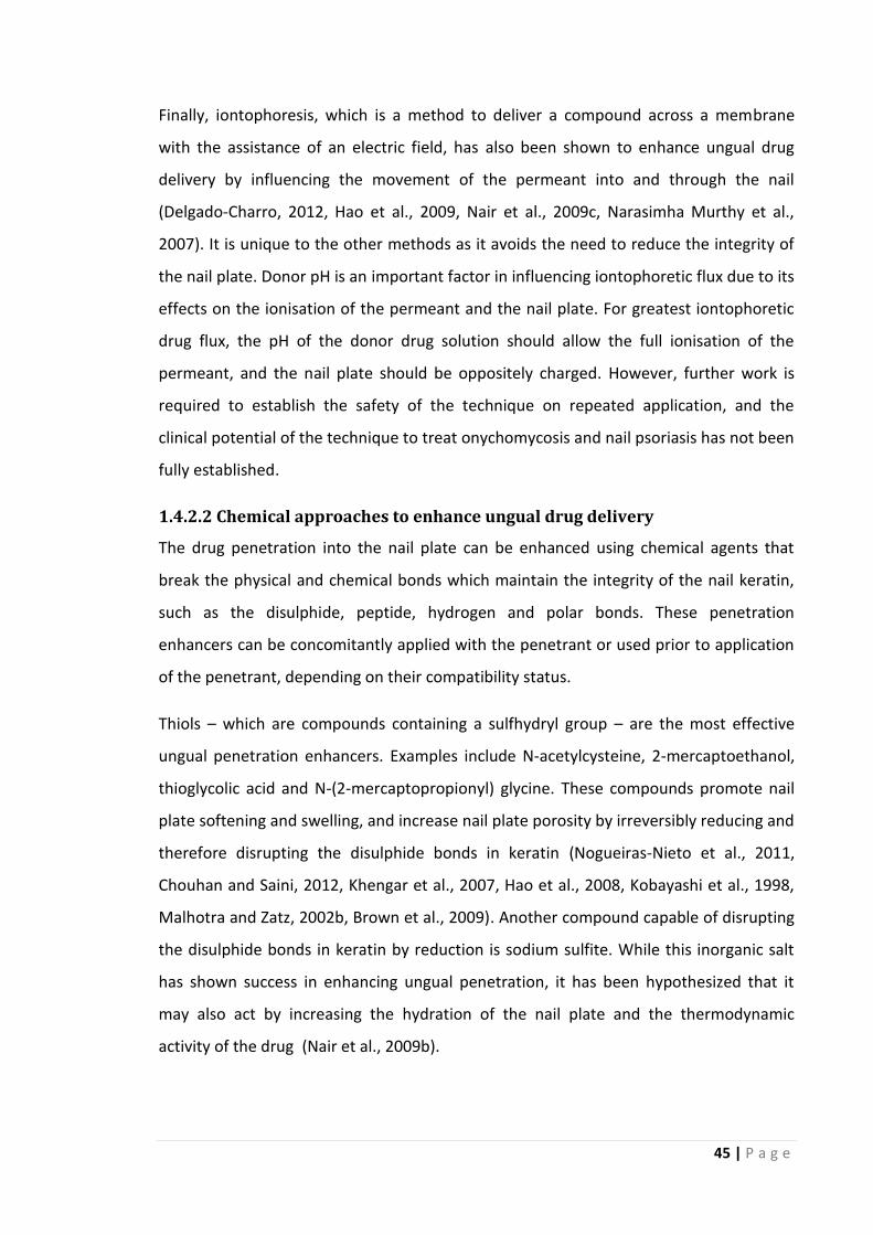
45 | P a g e
Finally, iontophoresis, which is a method to deliver a compound across a membrane
with the assistance of an electric field, has also been shown to enhance ungual drug
delivery by influencing the movement of the permeant into and through the nail
(Delgado-Charro, 2012, Hao et al., 2009, Nair et al., 2009c, Narasimha Murthy et al.,
2007). It is unique to the other methods as it avoids the need to reduce the integrity of
the nail plate. Donor pH is an important factor in influencing iontophoretic flux due to its
effects on the ionisation of the permeant and the nail plate. For greatest iontophoretic
drug flux, the pH of the donor drug solution should allow the full ionisation of the
permeant, and the nail plate should be oppositely charged. However, further work is
required to establish the safety of the technique on repeated application, and the
clinical potential of the technique to treat onychomycosis and nail psoriasis has not been
fully established.
1.4.2.2 Chemical approaches to enhance ungual drug delivery
The drug penetration into the nail plate can be enhanced using chemical agents that
break the physical and chemical bonds which maintain the integrity of the nail keratin,
such as the disulphide, peptide, hydrogen and polar bonds. These penetration
enhancers can be concomitantly applied with the penetrant or used prior to application
of the penetrant, depending on their compatibility status.
Thiols – which are compounds containing a sulfhydryl group – are the most effective
ungual penetration enhancers. Examples include N-acetylcysteine, 2-mercaptoethanol,
thioglycolic acid and N-(2-mercaptopropionyl) glycine. These compounds promote nail
plate softening and swelling, and increase nail plate porosity by irreversibly reducing and
therefore disrupting the disulphide bonds in keratin (Nogueiras-Nieto et al., 2011,
Chouhan and Saini, 2012, Khengar et al., 2007, Hao et al., 2008, Kobayashi et al., 1998,
Malhotra and Zatz, 2002b, Brown et al., 2009). Another compound capable of disrupting
the disulphide bonds in keratin by reduction is sodium sulfite. While this inorganic salt
has shown success in enhancing ungual penetration, it has been hypothesized that it
may also act by increasing the hydration of the nail plate and the thermodynamic
activity of the drug (Nair et al., 2009b).

46 | P a g e
Keratolytic agents, such as urea and salicylic acid, also promote nail softening and
swelling (Chouhan and Saini, 2012, Hao et al., 2008, Kobayashi et al., 1998), but via
keratin denaturation. Unfortunately, when used alone these agents are unable to
enhance ungual drug flux through the nail (Kobayashi et al., 1998, Malhotra and Zatz,
2002b, Quintanar-Guerrero et al., 1998). However a combination of two keratolytic
agents or a keratolytic agent and a thiol can enhance ungual drug flux by supposedly
working synergistically, with urea, for example, capable of augmenting the transungual
permeation enhancing effects of N-(2-mercaptopropionyl) glycine and N-acetyl-cysteine
(Malhotra and Zatz, 2002b, Quintanar-Guerrero et al., 1998). Hydrogen peroxide, which
is an oxidising agent, is thought to oxidise and therefore cleave disulphide bonds. When
complexed with urea, it promotes nail plate softening and swelling (Khengar et al.,
2007), and similar to keratolytic agents, it augments the transungual permeation
enhancing effect of thiols and is ineffective when used alone (Brown et al., 2009).
Finally, keratolytic enzymes which have the ability to hydrolyze nail keratins are
expected to weaken the nail plate’s barrier properties and enhance transungual
permeation. A keratinase enzyme from the fungus Paecilomyces marquandii did
precisely that and enhanced the permeability of a model permeant through a model nail
plate (Mohorcic et al., 2007). However, further investigations are required to
demonstrate the efficacy of keratinase on human nail plates.
Other ungual penetrations enhancers have also been identified and include 2-n-nonyl-
1,3-dioxolane (Hui et al., 2003), hydroxypropyl-β-cyclodextrin (Chouhan and Saini, 2014),
hydrophobins (Vejnovic et al., 2010b, Vejnovic et al., 2010a), sodium phosphate (Nair et
al., 2009b), and solvents such as water (Walters and Flynn, 1983, Walters et al., 1985b),
dimethyl sulfoxide (DMSO) (Stüttgen and Bauer, 1982, Franz, 1992, Hui et al., 2002,
Vejnovic et al., 2010b), methanol (Vejnovic et al., 2010b), low molecular weight
polyethylene glycol (PEG) (Nair et al., 2010, Ahn et al., 2013) and N-methyl-2-pyrrolidone
(NMP) (Ahn et al., 2013, Myoung and Choi, 2003). These enhancers were tested as
excipient in formulations and do not necessarily work by breaking bonds which maintain
the integrity of the nail keratin, but rather promote nail hydration, increase the aqueous
solubility of the drug, possibly induce nail lipid depletion, promote a higher affinity of

47 | P a g e
the drug towards the nail surface or promote an improved adhesion of the formulation
to the nail plate.
1.5 Topical drug vehicles
It was previously mentioned that drug permeation into an intact nail plate may be
encouraged by formulating the drug within a vehicle which enables drug partition out of
the vehicle and into the nail plate. Even when factors that can enhance permeation are
considered, such as disrupting the dorsal nail plate layer prior to formulation application
or using chemical enhancers, if the vehicle itself is inadequate, the success rate following
therapy will reflect this. There were various topical nail formulations for onychomycosis
and nail psoriasis which had been discussed earlier which included solutions, lacquers,
gels, ointments and creams. Other formulations investigated or under investigation
include films, patches, and colloidal carriers. These are all discussed in detail below in
order to highlight why there remains a need for a new topical nail medicine.
1.5.1 Nail solutions
Nail solutions are highly concentrated solutions of a drug in a suitable solvent. When the
nail solution is a concentrated aqueous or alcoholic antimicrobial solution in a volatile
vehicle, it is called a paint (Shivakumar et al., 2013). Following application of the nail
solution on the nail surface, the solvent evaporates to leave a highly concentrated
deposit of drug on the nail (not necessarily in the form of a film), which is expected to
penetrate and diffuse through the nail plate. The depth of penetration of topically
applied therapeutic agents depends on the solvents used to make up the nail solution
(Hui et al., 2002), and Table 1.7 shows the excipients used in some of the topical
solutions mentioned in previous sections. Unfortunately, such a topical dosage form
needs to be applied frequently, and fails to maintain therapeutic drug levels at the
desired site due to its ease of removal.

48 | P a g e
Table 1.7 Excipients of topical solutions currently approved for treating nail diseases (onychomycosis).
Product Active Pharmaceutical Ingredient Solvent base
Trosyl® nail solution Tioconazole 28% w/v Undecylenic acid and ethyl acetate
Jublia® topical solution Efinaconazole 10% w/w
Alcohol, anhydrous citric acid, butylated hydroxytoluene, C12-15 alkyl lactate, cyclomethicone, diisopropyl adipate, disodium edetate and purified water.
Kerydin® topical solution Tavaborole 5% w/w
Alcohol, propylene glycol and edetate calcium disodium
Phytex® paint Salicylic acid 1.46% (total combined), tannic acid 4.89% and boric acid 3.12% (as borotannic complex)
Alcohol and ethyl acetate
1.5.2 Nail lacquers
Nail lacquers contain a therapeutic agent in a suitable lacquer base. Following a nail
lacquer’s application on the nail plate surface, its volatile solvent(s) evaporate leaving
behind a highly concentrated deposit of drug in a polymeric film on the nail, from which
the drug is released and permeates through the nail plate. The polymeric film formed
can reduce transonychial water loss (TOWL), and is therefore likely to hyper-hydrate the
upper nail layers, which can aid the drug permeation into and through the nail plate
(Spruit, 1971, Marty, 1995). This hydration property is useful in onychomycosis
treatment, as the hydration can also facilitate germination of drug-susceptible fungal
hyphae and limit the formation and persistence of drug-resistant fungal spores, thus
limiting the risk of reinfection. The polymeric film formed can additionally inhibit the
adhesion of fungal propagules on and underneath the nail plate, which can prevent
fungal infection/reinfection in its initial step (Flagothier et al., 2005), a useful property
even when non-infectious nail diseases are being treated. Table 1.8 provides a list of
excipients for the nail lacquers mentioned in previous sections, and identifies film-
formers and solvents as key ingredients alongside the pharmaceutical active.

49 | P a g e
Table 1.8 Excipients of the nail lacquers currently approved for treating nail diseases (onychomycosis).
Product Active Pharmaceutical Ingredient
Lacquer base
Amorolfine 5% w/v nail lacquer (Loceryl®)
Amorolfine 5% w/v, as the hydrochloride salt
Water-insoluble film-former: Ammonio methacrylate copolymer A (Eudragit® RL,) Plasticizer: triacetin Solvents: butyl acetate, ethyl acetate and ethanol
Penlac® (also marketed as Batrafen®, Loprox® or Mycoster® ) nail lacquer
Ciclopirox 8% w/v
Water-insoluble film-former: Butyl monoester of poly(methylvinyl ether/maleic acid) Solvents: ethyl acetate and isopropyl alcohol
Ciclopoli® (also marketed as RejuveNail® ) nail lacquer
Ciclopirox 8% w/v
Water-soluble film-former: Hydroxypropyl chitosan Solvents: cetostearyl alcohol, ethyl alcohol, ethyl acetate and purified water
The amount of drug permeated through the nail plate and the drug uptake by the nail
has been shown to be influenced by the solvent constituting the lacquer (Franz, 1992).
Similarly, the drug concentration in the deposited lacquer film following application
influences drug permeation and uptake into the nail plate (Mertin and Lippold, 1997a,
Pittrof et al., 1992), with increasing concentrations showing to be favourable as long as
the drug is maintained in its soluble/amorphous form after evaporation of the
solvent(s). The addition of penetration enhancers such as 2-n-nonyl-1,3-dioxolane or
hydroxypropyl-β-cyclodextrin in a lacquer formulation can further increase ungual flux
(Hui et al., 2003, Chouhan and Saini, 2014).
Finally, the choice of the film-former used in the nail lacquer can affect several factors in
addition to transungual permeation. The water-insoluble films, such as methacrylic
polymer and vinyl resin based films, provide sustained drug release and are resistant to
washing, however, they require weekly removal either by mechanical means or by using
a suitable solvent, which may affect the surrounding skin. Water-soluble films, such as
hydroxypropyl chitosan based films, adhere to the nail plate to a greater extent and
facilitate a greater drug partitioning/ release (by directly promoting nail hydration) when
compared to the water-insoluble films (Monti et al., 2010, Monti et al., 2005), however,
they can be easily washed off from the nail surface. In order to combine the
advantageous occlusiveness and wash-resistance of water-insoluble films with the
adhesion and drug release properties of water-soluble films, a bilayer nail lacquer

50 | P a g e
composed of an underlying drug-loaded hydrophilic layer and overlying hydrophobic
vinyl layer was suggested (Shivakumar et al., 2010). This formulation although promising
complicates the topical drug administration protocol, which requires the application of a
drug-loaded hydrophilic nail lacquer, followed by the application of a hydrophobic nail
lacquer once the hydrophilic layer has dried.
Another novel drug-loaded nail lacquer formulation has recently been suggested to
facilitate an effective topical response. The vehicle was an anhydrous/alcohol
composition with a dual acrylate–silicone hybrid copolymer system that offers film
forming and occlusion properties due to synergistic plasticizing properties. The vehicle
base contained ethanol, polysilicone-8, panthenol, acrylates copolymer, tocopheryl
acetate, phytantriol, butylene glycol, benzophenone-3, calcium chloride, and fragrance
(Hafeez et al., 2013). However in vivo – in vitro correlations are required to define the
clinical relevance of the results obtained.
1.5.3 Semi-solids
Semi-solids such as gels, ointments and creams are also available or have been
investigated for the treatment of nail disorders, as these formulations can provide the
necessary chemical gradient to deliver the drug to the infected site.
1.5.3.1 Gels
Gels can hold a considerable amount of water and therefore have the potential to
hydrate the nail plate upon application, even more so than nail lacquers (Shivakumar et
al., 2013). This in turn can enhance drug permeation due to the formation of a less
dense structural keratin matrix with large pores. One study actually demonstrated the
better efficacy of gel formulations compared to a lacquer in enhancing the permeation
of antifungals across the nail (Hui et al., 2004). The gel used was a Loprox® gel which is
marketed outside the UK for dermatological infections, and contains ciclopirox (0.77%)
in a base consisting of octadecanol, dimethicone copolyol 190, carbomer 980, isopropyl
alcohol, sodium hydroxide, docusate sodium and purified water. The lacquer used was
Penlac® (which contains ciclopirox 8% w/v), and this study highlights how the type of
formulation plays a more crucial role in determining drug penetration than drug
concentration in the formulation.

51 | P a g e
Zorac® 0.1% gel is another gel used for nail diseases. It contains tazarotene (0.1%) in a
gel base consisting of benzyl alcohol, macrogol 400, hexylene glycol, carbomer 974P,
trometamol, poloxamer 407, polysorbate 40, ascorbic acid, butylhydroxyanisole (E320),
butylhydroxytoluene (E321), disodium edetate and purified water. While it has shown to
be of some benefit for nail psoriasis management (Table 1.5), like the Loprox® gel, it was
initially formulated for use on the skin, in this case for mild to moderate plaque psoriasis
affecting up to 10% of the skin area.
There are a few studies available that have investigated gels formulated specifically for
application to the nail plate, but with the overall aim to evaluate the effect of nail
etching or various penetration enhancers on the permeability of the human nail plate
(Repka et al., 2004, Malhotra and Zatz, 2002b, Nair et al., 2009b). These studies suggest
that gels could be the most ideal formulations for etched nails as they could easily flow
into the microporosities at the nail surface and therefore establish good contact at the
microscopic level, and also for incorporating water soluble penetration enhancers such
as sodium phosphate. Gels are also the most suitable formulation for iontophoresis, but
unfortunately, they can be easily wiped off the surface of the nail plate during regular
activities and so their use is not popular.
1.5.3.2 Ointments
Ointments have been tested for use in the topical treatment of various nail disorders.
Table 1.9 shows the collection of excipients that make up the ointment bases for the
formulations which have been tested for nail psoriasis management. Unfortunately the
efficacies of these formulations are below par. Formulations tested for onychomycosis
include a bifonazole (1%) and urea (40%) ointment which has been shown to adequately
soften a diseased nail for atraumatic removal (Hardjoko et al., 1990), and a tolnaftate
(2%) in urea 20% ointment which has shown some success (Ishii et al., 1983). However
these formulations required the use of an occlusive dressing, and the overall success
following therapy was still inadequate. Furthermore, like gels and solutions, ointments
can be easily wiped off the surface of the nail plate during regular activities, and this
factor limits their use.
52 | P a g e
Table 1.9 Excipients of the ointments tested for management of nail psoriasis.
Product Active Pharmaceutical Ingredient
Ointment base
Diprosalic® ointment Betamethasone dipropionate 0.064 % w/w (equivalent to 0.05 % w/w betamethasone) and salicylic acid 3.0 % w/w
Liquid paraffin and white soft paraffin
Dovobet® ointment Betamethasone dipropionate 0.05% and calcipotriol monohydrate 50 μg/g
Liquid paraffin, all-rac-α-tocopherol, polyoxypropylene-11stearyl ether, white soft paraffin and butylhydroxytoluene (E321)
Dovonex® ointment Calcipotriol 50 μg/g Disodium edetate, disodium phosphate dihydrate, DL-α tocopherol, liquid paraffin, macrogol (2) stearyl ether, propylene glycol, purified water and white soft paraffin.
Curatoderm® ointment Tacalcitol monohydrate 4.17 μg/g (tacalcitol 4 μg/g)
White petrolatum, liquid paraffin and diisopropyl adipate
Dithranol ointment, BP Dithranol 0.1 – 2.0% Yellow soft paraffin
1.5.3.3 Creams
Creams are emulsion-based topical formulations and are typically water-in-oil (w/o) or
oil-in-water (o/w) emulsions, depending on the solubilities of the actives that need to be
incorporated. Table 1.10 shows the list of excipients that make up a cream base for
some of the formulations tested for nail diseases. Clinical experts do not find a good
rationale for treating onychomycosis with antifungal creams applied to intact nail, apart
from cases of superficial white onychomycosis (de Berker, 2009), hence creams studied
for onychomycosis have been applied following mechanical reduction of the nail plate or
(non)surgical nail avulsion (Davies, 2006, Baden, 1994). Creams have been studied for
nail psoriasis without compromising the nail plate structure, and the use of calcipotriol
cream daily during the weekday and clobetasol propionate cream daily over the
weekend for 6 months, followed by the use of only clobetasol propionate cream daily
over the weekend for a further 6 months, was found to improve subungual
hyperkeratosis (Rigopoulos et al., 2002). However this treatment regimen can be

53 | P a g e
considered as tedious and response rates are inadequate. Furthermore, creams can also
be easily wiped off the surface of the nail plate during regular activities.
Table 1.10 Excipients of the creams tested or available for the management of onychomycosis.
Product Active Pharmaceutical Ingredient
Cream base
Loprox cream (not available in the UK)
Ciclopirox 0.77% Benzyl alcohol, cetyl alcohol, cocoamide diethanolamine, lactic acid, mineral oil, myristyl alcohol, octyldodecanol, polysorbate 60, purified water, sorbitan monostearate and stearyl alcohol.
Clotrimazole cream Clotrimazole 1% Benzyl alcohol, cetostearyl alcohol, cetyl palmitate, octyldodecanol, polysorbate 60, sorbitan stearate and purified water.
Nizoral® cream Ketoconazole 2% w/w Propylene glycol, stearyl alcohol, cetyl alcohol, sorbitan stearate, polysorbate 60, isopropyl myristate, sodium sulphite anhydrous (E221), polysorbate 80 and purified water.
1.5.4 Hot-melt extruded films
Hot melt extrusion is the process of applying heat and pressure to melt an active
compound embedded in a carrier formulation (consisting of polymeric materials or low
melting point waxes) and forcing it though an orifice in a continuous process. One study
investigated the use of a Killion extruder (Model KLB 100) to produce hot-melt extruded
films for the topical treatment of onychomycosis. The excipients included ketoconazole
(20%), hydroxypropylcellulose, polyethylene oxide and Noveon AA1, and these were
blended thoroughly, dried at 50 ⁰C for 24 hours, and then extruded at 115 – 120⁰C to
form films with good content uniformity, drug content, bioadhesion, and drug
permeation through the nail (Repka et al., 2004). While further work was conducted on
hot-melt extruded films, these mainly revolved around the study of the films’ physio-
chemical and mechanical properties, and drug stability (Mididoddi and Repka, 2007,
Mididoddi et al., 2006), and the efficacy of such a topical therapy was never established.
1.5.5 Patches
A bioadhesive patch was also investigated for the ungual delivery of 5-aminolevulinic
acid (ALA), a naturally occurring precursor of the photosensitiser protoporphyrin IX,
which could be useful in the treatment of onychomycosis. The patch was prepared by a
film-casting technique and was produced from an aqueous solution of ALA, methyl vinyl

54 | P a g e
ether and maleic anhydride copolymer, tripropylene glycol methyl ether and ethanol.
This mixture formed a gel which was casted into a mould consisting of a release liner
placed on a levelled surface and dried under a warm air flow to prepare a 2.5 x 3 cm
patch. A backing layer consisting of medical grade polyvinyl chloride plasticised with
dibutyl phthalate (Plastisol®) was attached to one side of the formed patches. Patch
application for 24 hours allowed drug permeation into the nail, and it was suggested
that drug penetration across nail may be improved using penetration enhancers or by
filing of the impenetrable dorsal surface of the nail (Donnelly et al., 2005).
Sertaconazole nail patches were also investigated for the treatment of onychodystrophy
and onychomycosis. The sertaconazole-containing nail patches were 2.2 cm² in area and
contained sertaconazole, lactate, aerosol and aluminium acetylacetonate in the
adhesion layer and Durotak® was used as the backing layer. Sixteen healthy adults were
treated with this patch in a double-blind study, for a treatment period of 6 weeks with
the patches replaced weekly. The drug was detected in all sertaconazole-treated nail
samples (Susilo et al., 2006), however further work is required to establish the clinical
relevance of the results.
The onychomycotic drug, ciclopirox, was also formulated into a patch to investigate the
effect of various pressure sensitive adhesives on its penetration. It was found that the
patch could enable ciclopirox release and hence permeation, with the highest
permeability from an acrylic adhesive with a hydroxyl functional group, followed by
styrene-isoprene-styrene, styrene-butadiene-styrene, silicone and polyisobutylene
adhesives in order (Myoung and Choi, 2003). More recently, terbinafine was formulated
into a patch to investigate the same parameter. Similar to ciclopirox, terbinafine release
was possible but this time the permeation was highest from the silicone adhesive
matrix, followed by polyisobutylene and most of the acrylic adhesives, polystyrene–
polyisoprene–polystyrene and finally polystyrene–polybutadiene–polystyrene (Ahn et
al., 2013).
While patches address problems associated with the conventional topical dosage forms,
such as the need for frequent applications and failure to maintain therapeutic drug

55 | P a g e
levels at the desired site, they do not have an aesthetically pleasing appearance.
Furthermore, studies have not demonstrated the clinical efficacies of such formulations.
1.5.6 Colloidal drug carrier systems
Colloidal drug carrier systems are well known for their penetration enhancing effects on
the skin, and consist of an oil and water phase stabilized by emulsifiers in a certain ratio
which has to be ascertained. The penetration behavior of a novel highly lipophilic
antifungal agent (EV-086K) for the treatment of onychomycosis was investigated in one
study in four different formulations. The first formulation was a hydrogel consisting of
the active, Tylose H 30.000 P2 PHA hydroxyethylcellulose, glycerol, and distilled water.
The second was a colloidal carrier system which contain the active, propylene glycol and
distilled water (hydrophilic phase), PEG-20 glyceryl oleate and Poloxamer 331
(emulsifiers), and butylphthalimide isopropylphthalimide (oil component). The third was
a nail lacquer consisting of the active, ethanol, Eudragit®E100 (a cationic copolymer
based on dimethylaminoethyl methacrylate, butyl methacrylate and methyl
methacrylate), and the solubiliser diethylene glycol monoethyl ether. The final
formulation was a solution which contained the active, ethanol, distilled water, the
solubiliser diethylene glycol monoethyl ether, the pH adjusters citric acid and sodium
phosphate monobasic, and the antioxidants butylated hydroxytoluene and
ethylenediaminetetraacetic acid. The investigations found that the developed colloidal
drug carrier system significantly increased the penetration rate of an extremely lipophilic
antifungal drug through the human nail plate compared to other formulations (nail
lacquer, solution and hydrogel). The high drug penetration rate following the application
of the colloidal carrier system was down to the presence of propylene glycol in the
system, which improved the solubility of the drug in the hydrophilic phase and therefore
enabled the system to obtain a high drug concentration, and hence high thermodynamic
activity and drug flux. Propylene glycol may have also acted as a penetration enhancer
(Naumann et al., 2014). It should be noted though that while the developed active
penetrated into the human nail plate in high concentrations, it did not completely
penetrate through the nail plate.
NB-002 is an antifungal, oil-in-water nanoemulsion containing highly purified oil,
ethanol, polysorbate 20, water, and cetylpyridinium chloride, with activity against

56 | P a g e
dermatophytes, including spores. It consists of nanodroplets that have an average
diameter of 180 nm which can be taken up through skin pores and hair follicles when
applied to the skin surrounding the nail, where they laterally diffuse to the site of
infection (nail bed). In patients with distal subungual onychomycosis, NB-002 was
applied to all 10 toenails and 5 mm of adjacent skin for 42 weeks. While NB-002 showed
clear antifungal activity with clinically significant nail clearing, at 4 or 8 weeks post-
treatment, the mycological cure rates were low and ranged from 25.0% to 31.7%
(Ijzerman et al., 2010).
Finally, TDT 067, which is terbinafine in soybean phosphatidylcholine – Tween 80
deformable lipid vesicles (Transfersomes®), is a new terbinafine preparation that has
been formulated for topical delivery to the nail, nail bed, and surrounding tissue for
onychomycosis treatment. In vitro studies have shown that TDT 067 has inhibitory and
fungicidal activity against dermatophytes and the non-dermatophytic pathogens of
onychomycosis. It has a higher activity than conventional terbinafine preparations, and
the Transfersome® is believed to accelerate entry of terbinafine into the fungal cells and
enhance its fungicidal effects. A clinical study which involved treating infected nails as
well as its surrounding skin with the TDT 067 liquid spray twice daily for 12 weeks
demonstrated reasonable mycological cure rates and few local reactions, however, an
ongoing Phase-III study is investigating the efficacy and safety of TDT 067 further
(Dominicus et al., 2012, Ghannoum et al., 2011, Ghannoum et al., 2012, Sigurgeirsson
and Ghannoum, 2012).
1.5.7 The need for a new nail medicine
As demonstrated, there is a vast range of topical drug vehicles available for nail diseases
with the aim to deliver a therapeutic level of drug at the desired site. Unfortunately, as
also seen, these vehicles require frequent applications, which reduce patient
compliance, and/or are easily removed from the surface of the nail plate, thus failing to
maintain therapeutic drug levels. There is therefore an urgent need for a nail medicine
which can overcome these limitations, while being aesthetically acceptable in
appearance at the same time.

57 | P a g e
1.6. UV-curable gels as potential topical nail medicines
The concept of using a nail product as a drug delivery vehicle was borrowed from the
cosmetic industry. The cosmetic industry also uses artificial nail enhancements for
decorative purposes or to hide unattractive nail deformities such as discoloration,
splitting, and breaking, which can be a result of median canal dystrophy or splinter
hemorrhages for example. The first artificial nail appeared in the United States around
1935. Throughout the years their quality has improved and as a result they have become
more and more popular. At one point they were almost exclusively applied by
professional trained manicurist in nail salons, which can be quite expensive, however
artificial nail kits designed for home use are now available over-the-counter (Andre et
al., 2014). There are three main types of artificial nail enhancements which include nail
wraps, liquid and powders, and UV gels (Schoon, 2010). These are all more durable than
the conventional nail lacquer (Schoon and Baran, 2012), and the following section
explores these enhancements and their safety, and outlines why UV-curable gels were
investigated as potential topical nail medicines in this thesis.
1.6.1 Artificial nail enhancements – an introduction
1.6.1.1 Nail wraps
Nail wraps are also known as fiberglass wraps, resin wraps, no-light gels, silk or paper
wraps, and are a less popular nail enhancement technique, accounting for about 1 - 2%
of the worldwide market (Jefferson and Rich, 2012, Schoon and Baran, 2012). They are
created from methyl and ethyl cyanoacrylate monomers, with the cyano functional
group believed to provide tremendous adhesion to the natural nail plate, and are
applied onto a clean, dehydrated nail surface, with dehydration achieved by wiping the
nail surface with a nail wipe coated with isopropyl alcohol. Typically, thin layers of a
fibrous substance (silk, linen or fiberglass) are applied within multiple layers of
cyanoacrylate monomers. Alternatively, the cyanoacrylate monomers can be thickened
with polymers such as polymethyl methacrylate and applied without a reinforcing
fibrous substance; such systems are termed no-light gels. Cyanoacrylate monomers are
applied to the nail plate directly from its container’s nozzle and cure upon exposure to
nail plate moisture. The curing process is accelerated to 5 – 10 seconds by solvents
containing a tertiary aromatic amine such as dimethyl tolylamine (0.5 – 1 %), which can

58 | P a g e
be sprayed on, applied with a dropper or impregnated into the woven fabric. Following
the cure, the nail wrap coating can be shaped as desired. This technique is often used to
mend cracks or tears in the nail plate, by using the monomer to adhere a small piece of
fabric over the broken or damaged area of the nail plate (Schoon and Baran, 2012,
Schoon, 2010). Nail wraps are often referred to as three-component systems, as they
commonly require a fabric mesh, resin (cyanoacrylate monomer) and activator (tertiary
aromatic amine) (Newman, 2005b).
Nail wraps are removed using acetone, and the removal process involves placing the
nails in a small bowl and immersing them in the solvent, a process that requires less
than 10 minutes (Schoon and Baran, 2012).
1.6.1.2 Liquid and powders
The liquid and powder system is a two-component system, which is also known as
acrylic, porcelain or solar nails (Schoon and Baran, 2012). It came directly from the
dental industry, was the first enhancement to be used commercially, and is popular in
the UK and the US, holding approximately 50% of the nail market (Newman, 2005b). The
‘liquid’ component is a mixture of ethyl methacrylate (60 – 95%), other di- or tri-
functional methacrylate monomers (3 – 5%), stabilisers such as hydroquinone or methyl
ether hydroquinone (100 – 1000 ppm), catalysts such as dimethyl tolylamine (0.75 –
1.25%), and other additives such as soluble colorants and plasticizers. The ‘powder’
component consists of poly methyl and/or ethyl methacrylate powder beads
(approximately 50 – 80 µm in size) coated with 1 – 2% benzoyl peroxide (which is a
polymerization initiator), and other additives such as colorants and opacifiers (e.g.
titanium dioxide) (Schoon, 2010, Schoon and Baran, 2012, Andre et al., 2014).
Mixing the liquid and the powder components initiates polymerisation; the two
components are therefore mixed before use and immediately applied and smoothed
into shape on the nail surface. The actual procedure for liquid and powder application
involves dipping a brush into the monomer liquid, wiping off the excess and drawing the
tip of the brush through the polymer powder to form a small bead at the end of the
brush. Typically three to six beads are normally smoothed into shape with the brush per
nail plate. The mixed components polymerise by free radical polymerisation and harden

59 | P a g e
on the nail surface within 2 – 3 minutes. Over 95% of the polymerisation occurs in the
first 5 – 10 minutes, however complete polymerisation can take anywhere between 24 –
48 hours. After hardening, the nail is shaped either by hand filing or with an electric file,
and the product formed is highly durable (Schoon, 2010, Schoon and Baran, 2012).
Prior to the application of the liquid and powder system, the natural nail is cleaned with
a disinfected soft-bristled brush to remove surface oil and debris, lightly filed with a low
grit abrasive file (180 – 240 grit) to increase the surface area, wiped with a nail wipe
coated with nail surface dehydrators containing isopropyl alcohol to remove surface
moisture and residual oils, and primers are also applied to aid adhesion by increasing the
surface compatibility between the natural nail and product. The primers usually contain,
for example, mixtures of hydroxylated monomers or oligomers which adhere well to
both the nail plate and product. Removal of the liquid and powder product also requires
acetone, and the removal process is the same as that for the nail wraps, but longer in
duration (30 – 40 minutes) as the polymer is highly cross-linked. However, this duration
can be greatly accelerated by pre-filing to remove the bulk of the system (Schoon, 2010,
Schoon and Baran, 2012).
1.6.1.3 UV gels
UV gels are also known as gel nails (Schoon and Baran, 2012), and have for the most
part, also been derived from the dental industry, where light-cured materials are used
for dental bonding. UV gels are becoming increasing popular, and in some countries
such as Germany, they are favoured over liquid and powder systems (Newman, 2005b).
UV gels are one-component systems which typically contain a urethane methacrylate
based monomer (75 – 85%), mono- and multi-functional (meth)acrylate based
monomers (15 – 25%), and a photoinitiator (1 – 4%) such as 1-hydroxycyclohexyl phenyl
ketone or 2-hydroxy-2-methylpropiophenone. The gel cures under low intensity UVA
light typically in the range of 435 – 325 nm, and this is provided by a UVA nail lamp,
which can hold one to five 6 – 9 W bulbs depending on the design. An alternative UVA
light source is a light-emitting diode lamp, suitable for gels which contain photoinitiators
that respond to a narrower spectrum of UVA light (Newman, 2005b, Jefferson and Rich,
2012, Schoon and Baran, 2012).

60 | P a g e
Prior to gel application, in order to ensure good adhesion, the natural nail is cleaned,
lightly filed, and dehydrated in the same manner as liquid and powder systems. A layer
of gel is then applied on the nail plate surface and the nail is placed under a UVA lamp
for 1 – 3 minutes. The UVA light activates the photoinitiator in the gel to form free
radicals which facilitate a polymerisation process to produce a polymeric film on the nail
plate. Further gel layers are sequentially applied and cured as the UVA light does not
efficiently penetrate more than a few millimetres into the UV gel. Following the curing
process, an occlusive polymer film is produced on the nail surface. This film is wiped
with isopropyl alcohol in order to remove an unreacted monomer layer on its surface,
caused by an oxygen-induced inhibition of polymerisation. The resulting film is highly
durable and can be worn for up to 3 weeks without developing any visible defects
(Schoon, 2010, Jefferson and Rich, 2012, Schoon and Baran, 2012).
Removal of the UV gel requires acetone, and the removal process is the same as that for
the nail wrap and liquid and powder system, but longer in duration (45 – 60 minutes) as
the highly cross-linked urethane methacrylate based polymers have an inherently
greater solvent resistance (Schoon, 2010). Alternatively, it can be removed with a
wooden stick 10 minutes after applying a cotton pad soaked with acetone over the
coating (Schoon and Baran, 2012).
1.6.2. Artificial nail enhancement - safety concerns
1.6.2.1 Allergic contact dermatitis
Unfortunately, the monomers used in artificial nail enhancement products are known
allergens (Lazarov, 2007), and their use poses a risk of developing allergic contact
dermatitis which results from a T-cell mediated, delayed-type hypersensitivity response
(Type IV hypersensitivity) (Gober and Gaspari, 2008). Typical clinical manifestations in
sensitised patients include nail fold dermatitis, while less frequent reactions include
onycholysis, paronychia and nail bed hyperkeratosis. Table 1.11 is a collection of case
reports and identifies monomers which the patients were sensitised to.

61 | P a g e
Table 1.11 Case reports of allergic contact dermatitis following the use of artificial nail enhancements. The monomers in bold were present in the formulation believed to trigger allergic contact dermatitis.
Case
Manicurist or
client
Clinical presentation
Positive reaction to the following (Meth)acrylates
Reference
1 Manicurist Fissure and dryness, followed by exudative, itchy lesions on or around the nails of fingers that had been in contact with a (meth)acrylate resins. The manicurist had previously used liquid-powder systems without any problems.
EMA MMA HEMA
(Conde‐salazar et al., 1986)
2 Client Dermatitis affecting the fingers, dorsa of hands, arms, upper trunk and face, and mild onycholysis of the fingernails. This began 6 months after beginning regular applications of the liquid-powder system.
EMA BMA
Polymethacrylate powder
(Fitzgerald and English, 1994)
3 Client Itching within one hour of applying liquid-powder system. Blisters, erythema and oedema on the fingertips and around the nail folds and paraesthesia in 2 fingers on the left hand the next day. The client had previously used liquid-powder systems without any problems.
MMA EGDMA
(Freeman et al., 1995)
4 Client Nail fold dermatitis following use of liquid-powder system for up to 18 months. Examination showed erythema, scaling and fissuring of the nail folds and tips of the fingers. The right index and left 5
th finger nail
were dystrophic.
MMA (Freeman et al., 1995)
5 Manicurist Erythema on ulnar border of the left hand and mild dermatitis on the nail folds. The manicurist had previously worn artificial acrylic nails for 8 years without problems.
EGDMA (Freeman et al., 1995)
6 Manicurist Dermatitis affecting eyelids and neck. Wearing artificial acrylic nails did not affect the fingers.
EGDMA (Freeman et al., 1995)
7 Client Fingertip rash following application of acrylic nails. Dermatitis subsided after removal of acrylic nail; however persistent paresthesia in all fingertips had been experienced since (3 years).
HEMA HPMA
EGDMA
(Slodownik et al., 2007)
8 Client Severe paronychia, pulps fissure, nail dystrophy, onycholysis and nail bed hyperkeratosis affecting all fingers, 1 week following the application of a liquid-powder system.
EA MMA
(Cruz et al., 2011)
9 Manicurist Dermatitis localised to the face and eyelid 4 months after first contact with liquid-powder system. No hand/periungual or other cutaneous lesions at distant sites present.
HEMA HPMA
(Paula et al., 2012)
10 Manicurist Periungual eczema 4 months after first application of UV gel.
HEMA HPMA
(Paula et al., 2012)
11 Client Periungual eczema 2 months after first application of UV gel.
HEMA HPMA
(Paula et al., 2012)

62 | P a g e
12 Client Severe paresthesia of distal phalanges of all fingers following application of UV gel.
MMA (Fisher, 1990)
13-17 4 Clients and 1
manicurist
Pruritic and painful perionychial and subonychial dermatitis for several months, 6 months to 3 years after first application of UV gels. Two patients also had dermatitis of the lower lids and cheeks. Monthly applications caused strong exacerbation of the dermatitis the next day.
(Meth)acrylated urethanes, TREGDMA
HEMA HPMA
(Hemmer et al., 1996)
18 Manicurist Dermatitis on right thumb within weeks after starting work with UV gels. This spread to both hands, and after stronger exposure led to severe hand and face dermatitis.
BA, EA, MMA, EMA, BMA,
HEMA, HPMA, EGDMA,
TREGDMA, 1,4-BDDMA, 1,4-BDA, DEGDA, TRPGDA,
UDA, TREGDA
(Kanerva et al., 1996)
19 Client Dermatitis of nail folds and fingertips following application of UV gels. The client had been applying it for 3 years previously with no problems.
HEMA HPMA MMA
EGDMA
(Erdmann et al., 2001)
20 Manicurist Dermatitis affected fingertips, nail folds, and eyelids.
HEMA HPMA
EGDMA MMA
(Erdmann et al., 2001)
21-22 2 Clients
Periungual eczema 3-6 months after first application of UV gels.
HEMA HPMA
(Cravo et al., 2008)
23 Manicurist Periungual and hand dermatitis after using UV gels professionally, in spite of having had UV gel nails for 2 years.
HEMA HPMA
(Cravo et al., 2008)
24 Manicurist Eyelid dermatitis 5 months after starting work with no hand/periungual lesions.
TREGDA (Cravo et al., 2008)
25 Manicurist Fingertip dermatitis, nail dystrophy and periungual hyperkeratosis. Symptoms improved after avoiding contact with UV gels.
HEA HEMA
EGDMA
(Vázquez-Osorio et al., 2013)
26 Manicurist Airborne dermatitis (cheeks, eyelids, forehead and temple area) and fingertip dermatitis. Symptoms improved after avoiding contact with UV gels.
HEMA EGDMA
(Vázquez-Osorio et al., 2013)
27 Client Fingertip dermatitis following use of UV gels. Dermatitis of the fingertips resolved following removal of the artificial nail, although nail dystrophy persisted for several months.
HEMA EGDMA
(Vázquez-Osorio et al., 2013)
28 Manicurist Dermatitis of the fingers, dorsa of the hands, face including eyelids, and large areas of the trunk. Symptoms improved after avoiding contact with cyanoacrylate glues and wearing polypropylene gloves while working.
ECA (Fitzgerald et al., 1995)
29 Client Eczematous change over fingertips, subungual hyperkeratosis, and dystrophy of fingernails. Scattered areas of eczema (in a hand-print pattern) over the arms, shoulders, and trunk were also present.
EMA EA
ECA
(Guin et al., 1998)

63 | P a g e
30 Client Subungual hyperkeratosis and eczematous eruptions on the hands, behind the left ear and on the left neck. The client used artificial nails containing fiberglass, which were attached at 2-week intervals.
ECA (Guin et al., 1998)
31 Client Eczema of the tips of digits, especially periungually, elevated and dystrophic nails, and hyperkeratotic eczema in the mid-palmar skin.
ECA (Guin et al., 1998)
Abbreviations: EMA, ethyl methacrylate; MMA, methyl methacrylate; HEMA, 2-hydroxyethyl
methacrylate; BMA, butyl methacrylate; EGDMA, ethylene glycol dimethacrylate; HPMA, hydroxypropyl
methacrylate; TREGDMA, triethylene glycol dimethacrylate; BA, butyl acrylate; EA, ethyl acrylate; 1,4-
BDDMA, 1,4-Butanediol dimethacrylate; 1-4-BDA, 1,4-Butanediol acrylate; DEGDA, diethylene glycol
diacrylate; TRPGDA, tripropylene glycol diacrylate; UDA, urethane diacrylate; TREGDA, triethylene glycol
diacrylate; HEA, hydroxyethyl acrylate; ECA, ethyl cyanoacrylate.
As can be seen from Table 1.11, both the manicurist and those wearing artificial nails
can develop allergic contact dermatitis, and it appears that the most frequent
(meth)acrylates they are sensitised to include ethylene glycol dimethacrylate,
hydroxyethyl methacrylate and hydroxypropyl methacrylate. Unfortunately, the majority
of these case reports cannot identify which (meth)acrylates were present in the
formulation that triggered the allergic contact dermatitis, and the positive
(meth)acrylate sensitivities could therefore be attributed to cross-sensitisation (Kanerva,
2001) .
These allergic responses most frequently arise due to repeated (meth)acrylate monomer
overexposure to the cuticles, lateral nail folds, or hyponychium. Following the
polymerisation reaction, the artificial nail enhancement is not considered to be a
sensitiser, and therefore the risk of developing allergic contact dermatitis can be
overcome by simply avoiding skin contact with the uncured product. This can be
achieved by applying the product to the nail plate ensuring a tiny product-free margin at
the nail folds is kept. As evident from Table 1.11, allergic contact dermatitis can also
affect the neck, face and eyelids, as a result of touching these areas with contaminated
hands. Exposure to filings may also be responsible in such cases, as they can contain
small amounts of unreacted monomers following the shaping of liquid and powder nail
enhancements for example, which as mentioned can take up to 48 hours to cure fully.
To avoid the non-local reactions, hands should be washed thoroughly after application

64 | P a g e
of a nail enhancement and contact with enhancement filings should be avoided (Schoon,
2010).
The safety of methacrylate ester monomers used in nail enhancements has been
assessed by the cosmetic ingredient review (CIR) panel, and they concluded that butyl
methacrylate, t-butyl methacrylate, cyclohexyl methacrylate, ethoxyethyl methacrylate,
2-ethoxy ethoxy ethyl methacrylate, ethyl methacrylate, ethylene glycol dimethacrylate,
hexyl methacrylate, hydroxyethyl methacrylate, di-HEMA trimethylhexyl dicarbamate,
hydroxyethyl methacrylate acetoacetate, hydroxypropyl methacrylate, isobornyl
methacrylate, isobutyl methacrylate, isopropylidenediphenyl bisglycidyl methacrylate,
lauryl methacrylate, methoxydiglycol methacrylate, PEG-4 dimethacrylate, pyromellitic
glycidyl dimethacrylate, tetrahydrofurfuryl methacrylate, triethylene glycol
dimethacrylate, trimethylolpropane trimethacrylate and urethane methacrylate are all
safe to use in nail enhancement products when skin contact is avoided (Escobar and
Yamarik, 2005).
1.6.2.2 Worn down nails
Worn down nails following the removal of a nail enhancement (Wu et al., 2015, Andre et
al., 2014) is the most common non-allergic problem reported. This can be a result of nail
filing with heavy grit abrasives (<180 grit) prior to the application of the nail
enhancement. Avoiding the use of nail files or switching to smoother abrasive files (>180
grit) can avoid such nail damage (Schoon, 2010).
1.6.2.3 UVA radiation exposure
At one point there was a concern that the UVA radiation required for UV gel curing may
be a risk factor for the development of skin cancer, as a case review on two women with
a history of UVA nail light exposure developed squamous cell carcinoma on their dorsal
hands (MacFarlane and Alonso, 2009). However, this concern has been put to rest with
recent research suggesting that two hands (of a person) placed in a UV nail lamp for 10
minutes twice a month is equivalent to that person spending an extra 2.7 minutes in
sunlight each day for a month (Schoon et al., 2010), a person could use a UV nail lamp
for 2.8 hours every day without any requirement for warning or protective measures
(Dowdy and Sayre, 2013), and that the UVA radiation produced by UVA nail lamps

65 | P a g e
specifically designed for UV gels pose a low risk of skin cancer even when used weekly
for over 250 years (Markova and Weinstock, 2013).
1.6.3 Why investigate UV-curable gels as topical nail medicines?
Of the three artificial nail enhancements, UV gels appear to be more favourable as they
are a much simpler and therefore convenient and consistent system to work with, their
polymerisation process is controlled by an external source providing an unlimited
application time, their complete cure time is relatively short (1 – 3 min), and they display
an inherently greater solvent resistance. To add to this, a recent article highlighted that
UV gels could be a valuable tool in improving cosmesis and satisfaction among patients
presenting with nail plate surface abnormalities which included nail pitting (Nanda and
Grover, 2014). These factors in combination with the UV gels’ ability to produce a
natural-looking, long-lasting, occlusive polymeric film on the nail plate surface makes it
worthwhile to consider as a potential topical nail medicine, especially as it addresses
current issues such as the need for frequent applications and failure to maintain
therapeutic drug levels at the desired site.
In this thesis, the pharmaceutical potential of UV-curable gels using antifungals drugs as
the test drugs, (as onychomycosis is the most common of nail diseases), is investigated
by setting out the following aims:
(i) To formulate and characterise UV-curable gels in terms of formulation
components and polymerisation.
(ii) To characterise the resulting film’s properties, such as morphology, thickness
microstructure, thermal and viscoelastic properties, occlusivity, adhesivity, water
sensitivity, drug-loading and stability.
(iii) To follow the release and ungual permeation of the loaded drug, and determine
the efficacy of the formulations against the most common onychomycosis-
causing pathogen.
(iv) To optimise the formulations with penetration enhancers.

66 | P a g e
Chapter 2: Optimisation of the UV-curable gel formulation and of
the polymerisation process
2.1 Introduction
To rationally formulate a UV-curable gel formulation for use as a topical nail medicine, a
more detailed understanding of commercially available UV gels is required. As
mentioned in Chapter 1, a UV gel is typically a blend of urethane methacrylate based
monomers/oligomers, mono- and multi-functional (meth)acrylate monomers and a
photoinitiator. However, a literature search on currently available cosmetic gels is
required to identify the exact components, their proportions and reasoning behind
selection. These excipients could then be screened as potential components for the
pharmaceutical formulation, as their safety as a nail formulation component is expected
to be established. To test the pharmaceutical potential of the formulation, a
pharmaceutical active needs to be incorporated into the gel, which in this case is an
antifungal agent for onychomycosis treatment. While there is a vast range of antifungal
agents available to tackle onychomycosis, the most favourable agents are those that
possess high potencies against onychomycosis-causing dermatophytes and Candida.
With this in mind, at least one agent needed to be selected. As the concentration of this
agent in the UV gel and therefore in the deposited film following application is expected
to influence drug permeation and uptake into the nail plate, a high drug load in the gel is
desired such that drug movement out of the preparation and into the nail along the
thermodynamic activity is favoured. The solubility of the antifungal in the UV gel
component mix is unknown, and hence if poor, a solvent may be necessary. Solvents
miscible with both the drug and the other UV gel components can not only help
incorporate drug into the gel, but also help achieve a high drug load. In this chapter, the
exact components that constitute a UV gel, alongside the potential antifungals and
solvents that could be incorporated into a gel are identified, and the information
available on these excipients are used to aid their selection for the pharmaceutical UV-
curable gel formulation.

67 | P a g e
Alongside excipient choice, the excipient proportion is vital to consider, as both can have
a knock-on effect on the gel properties, curing conditions and film characteristics
detailed in Fig. 2.1.
Fig. 2.1 Schematic diagram highlighting the importance of excipient choice and quantity.
UV gels are marketed in their gel form, and hence determining their properties is
absolutely essential. One vital UV-curable gel property is its viscosity, which is important
to consider as it can influence how well the formulation can be applied on the nail plate,
and whether it easily flows towards the proximal and lateral nail folds – an undesirable
property as repeated skin contact can trigger allergic contact dermatitis. Although, it
should be noted that commercially-available cosmetic UV gels do not have a defined
viscosity as such, with ‘thin to medium’, ‘medium’, ‘medium to thick’, and ‘super thick’
viscosity gels among the options available, and with the selection by a manicurist based
on the desired finished look (Newman, 2005a). Nevertheless it is vital to define from a
pharmaceutical prospective. Another vital UV-curable gel property, particularly for a
pharmaceutical gel, is drug stability, and it is essential to ensure that the drug
Excipient selection
•Choice and quantity
UV-curable gel properties
•Gel viscosity
•Drug stability
UV-curing conditions
•UVA device
•Cure time
UV-cured film characteristics
•Mass yield
•Degree of conversion
•Residual monomer content

68 | P a g e
incorporated in the UV gel does not react with the gel components, readily decompose
upon storage, or precipitate out of the mixture. A stability study which examines these
changes over time is therefore required. The International Council for Harmonisation of
Technical Requirements for Pharmaceuticals for Human Use (ICH) have developed
guidelines for accelerated testing conditions, and recommend that the formulation is
stored at 40⁰C ± 2⁰C/ 75% RH ± 5% RH for a minimum time period of 6 months. With this
in consideration, in this chapter, the UV-curable gel formulated is characterised for its
viscosity and drug stability.
In order for the UV gel to cure, the correct curing conditions, i.e. UVA light source and
cure time, is required, which is interrelated with the photoinitiator choice and
photoinitiator concentration respectively. This is as the photoinitiator has to be capable
of absorbing the wavelength of light emitted by the UVA light source in order to
generate free radicals. With regards to photoinitiator concentration, increasing the
concentration can increase the rate of cure, thus potentially reducing total cure time.
However, there is an optimum concentration, beyond which a large fraction of the
radiation is absorbed in the upper few micrometres of the film that minimal radiation
reaches the lower layers. As the half-life of free radicals is short, they must be generated
within a few nanometres of the depth in the film where they are to initiate
polymerisation (Wicks et al., 2007), and if this is not achieved, an improper cure results.
UV-curing is an exothermic reaction, and increasing the photoinitiator concentration
beyond a certain point can also increase the maximum temperature of the UV gel curing
process, which can “burn” the nail (Schoon, 2005). Another requirement for a UV gel to
cure is an inert atmosphere. This is because as the radicals form by the photolysis of the
initiator, they are rapidly scavenged by O2 molecules and are unreactive towards the
(meth)acrylate C=C bond; they therefore do not initiate or participate in the
polymerisation process (Decker, 1989). However, an inert atmosphere is not practically
feasible; UV gels are therefore cured in the presence of oxygen. As a consequence, the
UV-curable monomers do not achieve 100% conversion due to this oxygen-induced
inhibition of polymerisation, and an unreacted layer forms on the surface of the cured
film which must be removed. The mass yield from monomer to cured polymer is a useful
calculation to help determine loss due to the removal of the oxygen inhibition layer, and

69 | P a g e
can be a valuable tool for determining the effect of excipients, their concentrations and
curing conditions on its formation. The polymerisation reaction itself can also be
assessed by Fourier transform infrared (FT-IR) spectroscopy as the alkene bonds of the
(meth)acrylate monomers are converted to alkane ones, thus enabling a degree of
conversion (DC) percentage from monomer mixture to polymer film to be obtained.
Determining DC can therefore aid in defining the effect of excipients, their
concentrations and curing conditions on the polymerisation process. In this chapter, the
mass yield and DC percentage from monomer mixture to polymer film are determined
to aid in the identification of the optimal excipient concentrations and curing conditions
for a pharmaceutical UV-curable gel.
Unfortunately, residual monomers may exist in a cured film, and this is an undesired
property as they may penetrate into and through the nail plate, triggering either allergic
contact dermatitis or irritation. To determine the risk that the residual monomers may
cause, they can be extracted from the film into a solvent and quantified using analytical
methods such as gas chromatography. Quantifying the residual monomer content in the
film can also aid in the identification of optimal excipient concentrations and curing
conditions, thus keeping the amount of residual monomers in the cured film to an
absolute minimum. In this chapter, the amount of residual monomers in the cured film
is therefore also determined and examined alongside the mass yield and DC percentage
from monomer mixture to polymer film. In addition, these examinations are used to
help determine the effects of excipients, in particular the drug and solvent, on the UV-
curing process, to assess whether a pharmaceutical UV-curable gel formulation for use
as a topical nail medicine is feasible.
2.2 Aims
(i) Select components for the pharmaceutical UV-curable gel formulation.
(ii) Develop a method to prepare the gel formulation and determine its key properties,
i.e. viscosity and stability.
(iii) Develop a method to cure the gel formulation and form a UV-cured film, and assess
the polymerisation process.

70 | P a g e
2.3 Materials
As detailed in Section 2.5.2, the excipients selected for the pharmaceutical UV-curable
gel formulation were diurethane dimethacrylate in combination with either ethyl
methacrylate, isobornyl methacrylate or 2-hydroxyethyl methacrylate, and the
photoinitiator 2-hydroxy-2-methylpropiophenone. The antifungal drugs selected were
amorolfine HCl or terbinafine HCl and the solvents – ethanol or NMP – were also found
to be necessary. The chemical structures for these components can be found in Section
2.5, alongside their properties and reasonings behind selection.
Diurethane dimethacrylate, ethyl methacrylate, isobornyl methacrylate, 2-hydroxyethyl
methacrylate, 2-hydroxy-2-methylpropiophenone, absolute ethanol, methanol, 1-
methyl-2-pyrrolidinone (NMP), propan-2-ol, sodium chloride, triethylamine, phosphoric
acid 85% wt. solution in water and trifluoroacetic acid were purchased from Sigma–
Aldrich (Dorset, UK). Acetonitrile HPLC gradient grade was purchased from Fisher
Scientific (Hertfordshire, UK). Amorolfine HCl was purchased from Ranbaxy Research
Laboratories (Haryana, India) and terbinafine HCl from AK Scientific (CA, USA). The 36
Watt Cuccio Professional UVA nail lamp was purchased from Amazon UK. Nail & Beauty
Emporium lint-free wipes (4 ply) were purchased from Just Beauty UK, an online retailer
specialising in professional beauty, hair and skin products.
2.4 Methods
2.4.1 Identification of UV-curable gel components and proportions to be used
The web pages of four professional nail care product manufacturers, namely Creative
Nail Design, Inc. (CND) (CND, [No date]), Nail Systems International (NSI) (NSI, [No date]),
Kinetics – Professional Nail Systems (Kinetics - Professional Nail Systems, [No date]) and
Jessica Cosmetics International (JESSICA, [No date]), were visited in order to obtain
details of the UV gels they manufacture for salon and/or home cosmetic use. Details of
the formulations’ components and their contents were obtained from their Material
Safety Data Sheets and product profiles where available. This information was used to
identify potential excipients for a pharmaceutical UV-curable gel formulation.

71 | P a g e
2.4.1.1 Determination of drug solubility in monomers and in solvents
Saturation solubility studies were carried out to determine the solubilities of the
antifungal drugs – amorolfine HCl or terbinafine HCl – in the monomers selected, i.e.,
ethyl methacrylate, isobornyl methacrylate and 2-hydroxyethyl methacrylate, and
solvents selected, i.e. ethanol or NMP. The antifungals were added in excess to vials
containing 2 ml of the monomers or solvents. The mixtures were left to stir on a
magnetic stirrer placed in a water bath at 25°C for 72 hours. Subsequently, 1 ml of each
sample was withdrawn from the vial using a syringe and passed through a 0.22µm
MILLEX® GP filter unit into an Eppendorf tube. The samples were then centrifuged using
a bench centrifuge for 20 minutes at a speed of 13.2 x 1000 rpm. The supernatant for
each sample was then collected and diluted by 50, 100 or 1000 times with ethanol (or
NMP – for the solubility determination in NMP) and analysed by HPLC (as per Section
2.4.6). Each test was carried out in triplicate.
2.4.2 UV-curable gel preparation
A total of four types of formulations were prepared (i) drug-free and solvent-free; (ii)
drug-free containing solvent; (iii) drug and solvent containing; and (iv) solvent-free
containing drug, the details for which are shown in Table 2.1. The types of formulations
were further subdivided depending on whether they contained the monomer ethyl
methacrylate, isobornyl methacrylate or 2-hydroxyethyl methacrylate, the solvent
ethanol or NMP, and the drug amorolfine HCl or terbinafine HCl. Furthermore, the ratio
of diurethane dimethacrylate to ethyl methacrylate, isobornyl methacrylate or 2-
hydroxyethyl methacrylate was set at 85:15% v/v (unless otherwise stated) based on the
findings from Section 2.5.1.

72 | P a g e
Table 2.1 Excipients and their quantities for the different types of UV-curable gel formulations prepared. The drug-free & solvent-free formulation and solvent-free formulation containing drug which are shaded in grey were only prepared with 2-hydroxyethyl methacrylate (with the diurethane dimethacrylate to 2-hydroxyethyl methacrylate ratio set at 75:25% v/v). NMP was used as a solvent for formulations containing ethyl methacrylate only. For formulation containing drug, the drug and solvent mixture constituted 25% v/v of the formulation.
Formulation
Excipient
Diurethane dimethacrylate
(% v/v)
Ethyl methacrylate,
isobornyl methacrylate
or 2-hydroxyethyl methacrylate
(% v/v)
2-hydroxy-2-methylpropio
phenone (% v/v)
Ethanol or NMP (% v/v)
Amorolfine HCl or
Terbinafine HCl
(% w/v)
Drug-free & solvent-free
82.45 14.55 3
72.75 24.25 3
Drug-free containing solvent
61.2 10.8 3 25
Drug and solvent containing
61.2 10.8 3 25 1-7
Solvent-free containing drug
72.75 24.25 3 1-7
Drug-free formulations were prepared by mixing the excipients and leaving the mixture
to stir overnight. Drug-loaded formulations were prepared by first dissolving the drug in
the chosen solvent, and then adding the required excipients to the drug solution and
leaving the mixture to stir overnight. Each formulation was prepared in triplicate.
To obtain the optimal photoinitiator concentration, which was found to be 3% v/v,
formulations containing diurethane dimethacrylate, ethyl methacrylate and the
photoinitiator at concentrations of 1, 2, 3 and 4% v/v were prepared. Films were formed
as described below and tested as described in Section 2.4.5.
To determine the optimal concentration of solvent, which was found to be 25% v/v,
formulations containing diurethane dimethacrylate, ethyl methacrylate, a photoinitiator
(3% v/v) and ethanol at concentrations of 5, 10, 15, 20, 25, 30, 35, 40, 45 and 50% v/v
were prepared. Films were formed as described below and tested as described in
Section 2.4.5. The film’s sensitivity to water was also tested as described in Chapter 3
(Section 3.4.11).

73 | P a g e
2.4.3 Assessment of the UV-curable gel properties
2.4.3.1 Viscosity of gel
The viscosity of the UV-curable gel formulations was determined using a Bohlin Gemini
HR nano rheometer (Malvern Instruments, UK). This is a rotational rheometer which
requires 2 ml of sample per test. The experimental temperature was maintained at 25°C
and the experiment was conducted in a dark room to prevent the polymerisation of the
test sample during viscosity measurements. A 4°/40mm cone and plate was selected for
all measurements and a controlled stress from 0.02 – 15.76 Pa was applied. The test
time was 220 seconds, and this produced 22 test points following a logarithmic scale.
From the data generated, a shear rate (1/s) vs shear stress (Pa) plot was produced as
shown in Fig. 2.2. As the samples behaved as a Newtonian fluid where shear stress was
directly proportional to the shear rate, the gradient of the plot was recorded as the
viscosity of the sample.
0 100 200 300 400 500 600 700 800 900 1000
0
2
4
6
8
10
12
14
16
18
Sh
ear
str
ess (
Pa)
Shear rate (s-1)
Fig. 2.2 Typical rheogram of UV-curable gel formulation
2.4.3.2 Stability of gel
The stability of drug-loaded gel formulations was tested over 6 months under
accelerated testing conditions, i.e., 40°C ± 2°C and 75% RH ± 5% RH. This was conducted
by placing the formulations in a desiccator containing a saturated solution of sodium
chloride and placing this in a 40°C oven. At timed intervals (i.e. on Day 0 and every 28
days for 6 months) the formulations were assessed for (i) colour change (visually), (ii)

74 | P a g e
precipitation of loaded drug (by polarised light microscopy as described in Chapter 3
[Section 3.4.5.1]) and (iii) drug concentration over time (by HPLC as per Section 2.4.6).
2.4.4 UV-curing of formulations
The formulation (30 µl) was applied using a pipette tip on a microscope glass slide (to an
area of 15 mm x 15 mm) as a single layer. The glass slide was placed under a 36 Watt
Cuccio Professional UVA nail lamp for 2 minutes. This caused curing of the formulation
and formation of a film. The surface of the film was wiped with propan-2-ol using a 4 ply
lint-free nail wipe to remove the oxygen inhibition layer. This revealed a glossy polymer
film, which was then collected from the glass slide using a scalpel and used in the
assessments described below. The decision to use a 2 minute cure time came from the
assessment of films (as described below) produced by curing formulations containing
diurethane dimethacrylate, ethyl methacrylate and a photoinitiator for 0.5, 1, 2, 3, 4 or 5
minutes.
2.4.5 Assessment of the polymerisation process
2.4.5.1 Mass yield from monomer to cured polymer film
The mass yield from monomer mixture to polymer film was calculated using the
following equation:
Mass yield (%) = (Wt/W0) x 100
where W0 is the weight of the monomer mixture before curing and Wt is the weight of
the UV-cured film after removal of the oxygen inhibition layer.
2.4.5.2 FT-IR spectroscopy
Infrared spectroscopy was used to determine the DC from monomer mixture to polymer
film by following the conversion of alkene bonds to alkane bonds. Spectra of the
uncured and cured formulations were obtained using the OPUS 7.0 software and
recorded by a Bruker Alpha IR Spectrophotometer (Bremen, Germany); taking 24 scans
over the 400–4000 cm-1 range with background subtraction. The DC % was calculated
from the ratio of the height of the absorbance peak of the aliphatic C=C bond (1636 cm-
1) relative to that of the carbonyl group (>C=O, 1702 cm-1), used as an internal standard,
using the following equation:

75 | P a g e
DC (%) =(A1636/A1702)𝟎 − (A1636/A1702)𝐭
(A1636/A1702)𝟎 × 100
where (A1636/A1702)0 and (A1636/A1702)t are relative absorbance of C=C bonds to C=O
before curing and after curing respectively.
2.4.5.3 The levels of residual monomers in the polymer film
The levels of residual monomers in the polymer film were quantified by ultrasonic
extraction using an Elma Transsonic T460/H sonicator (Singen, Germany). Immediately
after curing and removal of the oxygen inhibition layer, one gram of each film was
placed in a glass vial and three millilitres of methanol was added. The mixture was
sonicated for up to 2 hours, after which the solvent was analysed by gas
chromatography (as per Section 2.4.6) to calculate the amount of residual monomers in
the polymer film. This extraction procedure was optimised for volume of extraction
solvent and extraction time (Appendix A3). Furthermore, to ensure that all the residual
monomers had been extracted, the polymer film used in the first extraction was rinsed
with methanol and blotted dry with Kimwipes, before placing in another vial containing
3 ml of methanol. This was also sonicated for 2 hours and the solvent was analysed by
gas chromatography to confirm the absence of residual monomers. Each test was
carried out in triplicate.
2.4.6 Analytical methods
2.4.6.1 High-performance liquid chromatography (HPLC)
The amount of amorolfine HCl and terbinafine HCl in samples was quantified by using a
1260 Infinity Agilent HPLC system equipped with an autosampler and a variable
wavelength absorbance detector (Agilent Technologies, Germany). Elution was
performed using a Luna C18 column (150 x 4.6 mm, 5 µm) at a temperature of 40⁰C.
Table 2.2 shows the HPLC method developed, which was validated for specificity,
calibration and linearity, accuracy and precision, and detection and quantification limit
(Appendix A1).

76 | P a g e
Table 2.2 HPLC method for the quantification of amorolfine HCl and terbinafine HCl in samples.
Drug Mobile Phase Flow rate
(ml/min)
Sample injection
vol. (µl)
Wavelength
(nm)
Retention
time (min)
Amorolfine
HCl
0.1% trifluoroacetic acid:
acetonitrile (55:45 v/v)
1.0 20.0 220 5.8
Terbinafine
HCl
0.012M triethylamine +
0.020M phosphoric acid:
acetonitrile (65:35 v/v)
1.0 20.0 224 8.8
2.4.6.2 Gas chromatography (GC)
GC was conducted to determine the amount of diurethane dimethacrylate and ethyl
methacrylate, isobornyl methacrylate or 2-hydroxyethyl methacrylate in samples using a
7890A GC System (Agilent, USA) equipped with a flame ionisation detector (FID) system.
Chromatographic separation was achieved on a HP-5 column (30 m long x 320 µm inside
diameter with 0.25 µm film thickness). The sample injection volume was 2 µl. The
injector was in the split mode (100:1) and its temperature was maintained at 250°C
throughout the experiments. The column temperature was raised from 45°C (hold 2
min) to 110°C (hold 2 min) at a 10°C/min heating rate, and then from 110⁰C to 280°C
(hold 2 min) at a 20°C/min heating rate. The flow rate of the carrier gas (N2) was 1.5
ml/min. The detection was carried out by the FID with the temperature of 280°C and the
ratio of H2/air at 25/250. Diurethane dimethacrylate, ethyl methacrylate, isobornyl
methacrylate and 2-hydroxyethyl methacrylate were detected at 16.5, 6.5, 16.2 and 5.7
minutes respectively. The method developed was validated for specificity, calibration
and linearity, accuracy and precision, and detection and quantification limit (Appendix
A2).
2.4.7 Statistical analyses
Statistical calculations were conducted using IBM SPSS 22. The data was tested to
determine whether they were normally distributed or not using the Shapiro-Wilk test
and then analysed using either a t-test or Mann-Whiney U test for parametric and non-
parametric data respectively. For multiple comparisons, one-way analysis of variance
(ANOVA) followed by post hoc Tukey or the Kruskal Wallis test with post hoc analysis
performed with Nemenyi’s test was used for parametric and non-parametric data
respectively.

77 | P a g e
2.5 Results and discussion
2.5.1 Components of cosmetic UV gels
A number of urethane-based (meth)acrylates, other (meth)acrylates, and a couple of
photoinitiators were identified upon reviewing the commercial and scant academic
literature on cosmetic UV gels. One of the more commonly used UV gel is the CND®
Shellac® brand system. Unfortunately, Shellac® is a proprietary formula, with butyl
acetate (≤50.0%) listed as the only chemical under composition and ingredient
information. The compositions of other UV gels available, excluding pigments, are listed
in Table 2.3.

78 | P a g e
Table 2.3 Composition of some of the UV gel products manufactured by NSI, Kinetics – Professional Nail Systems and Jessica Cosmetics UK.
Manufacturer
Product
Viscosity
Components
Urethane based monomers/ oligomers
Other functional ((meth)acrylate) monomers
Photoinitiator Other
NSI
Balance Body Builder Gels
Medium – thick
Di-HEMA trimethylhexyl dicarbomate* (85 -95%)
Triethylene glycol dimethacrylate (5 -10 %) and glycerol propoxylate triacrylate (< 5%)
2-Hydroxy-2-methylpropiophenone (1-4%)
Triethylene glycol divinyl ether (< 3%)
Balance Builder Gels
Medium Di-HEMA trimethylhexyl dicarbomate* (80-90%)
Triethylene glycol dimethacrylate (5-10 %) and isopropylidenediphenyl bisoxyhydroxypropyl methacrylate (5-10 %)
2-Hydroxy-2-methylpropiophenone (1-3%)
Triethylene glycol divinyl ether (1-3%)
Balance Colour Gels
Thin – medium
Di-HEMA trimethylhexyl dicarbomate* (70-80%)
2-Hydroxyethyl methacrylate (15-25%)
1-Hydroxycyclohexyl phenyl ketone (1-5%)
Benzophenone (1-5%)
Balance One Step Clear Gel
Thin – medium
Urethane acrylate (55-65%) and Di-HEMA trimethylhexyl dicarbomate* (20-30%)
Isopropylidenediphenyl bisoxyhydroxypropyl methacrylate (5-10%), glycerol propoxylate (1PO/OH) triacrylate (3-5%) and triethylene glycol dimethacrylate (1-3%)
2-Hydroxy-2-methylpropiophenone (<1%)
*Di-HEMA trimethylhexyl dicarbomate is also commonly referred to as urethane dimethacrylate or diurethane dimethacrylate.

79 | P a g e
Manufacturer
Product
Components
Urethane based monomers/ oligomers
Other functional ((meth)acrylate) monomers
Photoinitiator Other
NSI Glaze’n Go Polyurethane acrylate oligomer (70-80%)
PEG-4 dimethacrylate (15-20%) and ethyl methacrylate (5-10%)
1-Hydroxycyclohexyl phenyl ketone (1-3%)
Illusion Colour Gels Di-HEMA trimethylhexyl dicarbomate* (75-90%)
2-Hydroxyethyl methacrylate (5-10%) and triethylene glycol dimethacrylate (1-3%)
2-Hydroxy-2-methylpropiophenone (1-5%)
Triethylene glycol divinyl ether (1-3%)
Polish Pro Now Colours
Di-HEMA trimethylhexyl dicarbomate* (50-60%)
2-Hydroxyethyl methacrylate (15-20%) and hydroxypropyl methacrylate (15-20%)
1-Hydroxycyclohexyl phenyl ketone (<1%)
Trimethylbenzoyl diphenylphoshine oxide (1-3%), butyl acetate (1-5%), cellulose acetate propionate (1-3%), methyl ethyl ketone (<2%), polysilicone-13 (<1%) and isopropyl alcohol (<2%)
Kinetics – Professional Nail Systems
Epsilon BioDefence Gel
Urethane acrylate oligomer (50-80%)
Isobornyl methacrylate (10-30%) and carboxyethyl acrylate (1-10%)
Photoinitiator identity not published (1-10%)
Calcium pantothenate (ppm)
Kinetics SHIELD Coloured Gel Polish
Urethane acrylate (30-40 %) and polyester urethane acrylate (5-10 %)
Hydroxypropyl methacrylate (30-40%), tripropylene glycol diacrylate (<1%) and acrylates copolymer (<1%)
1-Hydroxycyclohexyl phenyl ketone (<1 %)
Nitrocellulose (5-10 %), ethyl acetate (1-5 %), butyl acetate (1-5 %), silica dimethyl silylate (1-5 %), ethyl trimethylbenzoyl phenylphosphinate (1-5 %), phthalic anhydride (<1 %), dilinoleic acid/ glycol copolymer (<1 %), acetyl tributyl citrate (<1 %), isopropyl alcohol (<1%), hydroxyethyl acrylate/ sodium acryloyldimethyl taurate copolymer (<1%) and p-hydroxyanisole (<0.005%).
Jessica Cosmetics UK
GELERATION Soak-Off Gel Polish
Di-HEMA trimethylhexyl dicarbomate* (50-60%)
2-Hydroxyethyl methacrylate (3-7%), hydroxypropyl methacrylate (3-7%) and isobornyl methacrylate (2-6%)
1-Hydroxycyclohexyl phenyl ketone (2-6%)
Butyl acetate (10-13%) and ethyl acetate (9-12%)

80 | P a g e
It can be clearly seen from Table 2.3 that urethane-based (meth)acrylates are the main
components in cosmetic UV gels, (constituting between 50 – 95% of the formulation).
They contain carbamate groups, -NHCOO-, which are also referred to as urethane
groups, in their backbone structure, and are popular due to their favourable properties,
namely high abrasion resistance and durability (Botero et al., 2014). Of the UV gel
products listed, the Balance Body Builder gel is an example of a medium to thick
viscosity gel, the Balance Builder gel is an example of a medium viscosity gel and the
Balance Colour and Balance One Step Clear gels are examples of thin to medium
viscosity gels. Urethane-based (meth)acrylates are viscous monomers, and it can be
seen that it is the amount of this excipient which governs whether a UV gel is a thin or
thick viscosity gel. It can also be seen that the most commonly used urethane-based
(meth)acrylate is diurethane dimethacrylate, the structure and molecular weight for
which is shown in Table 2.4.
Other (meth)acrylate monomers are also included in the gel mix, and their structures
and molecular weights are also shown in Table 2.4. Carboxyethyl acrylate, ethyl
methacrylate, 2-hydroxyethyl methacrylate, hydroxypropyl methacrylate and isobornyl
methacrylate are all monomers with a single polymerisable C=C bond, and these shape
the type and nature of the polymer backbone. They are used primarily as reactive
diluents because of their low viscosities, but are also carefully selected to obtain a
desired glass transition temperature, flexibility, mechanical strength and
hydrophilic/hydrophobic character of the resulting polymer film. Isopropylidenediphenyl
bisoxyhydroxypropyl methacrylate, PEG-4 dimethacrylate, triethylene glycol
dimethacrylate and tripropylene glycol diacrylate are all monomers with two
polymerisable C=C bond sites. These add crosslinks to the polymer architecture, with the
group in between the reactive ends helping to determine the physical and mechanical
attributes of the resulting cross-linked polymer structures. Glycerol propoxylate
triacrylate is a monomer with three polymerisable C=C bond sites, and is typically used
to generate a highly cross-linked polymer structure, with an improved polymer
toughness and solvent resistance (Polysciences, 2011).

81 | P a g e
Table 2.4 Chemical structures and molecular weights of (meth)acrylate monomers identified in commercially available UV gels. The monomers shaded in white contain a single polymerisable C=C bond, those in light blue contain two polymerisable C=C bond sites, and the monomer shaded in dark blue contains three polymerisable C=C bond sites.
(Meth)acrylate ester monomer
Chemical structure & molecular weight (Daltons)
Carboxyethyl acrylate
144.13
Ethyl methacrylate
114.14
Hydroxyethyl methacrylate
130.14
Hydroxypropyl methacrylate
144.17
Isobornyl methacrylate
222.32
Diurethane dimethacrylate
470.56
Isopropylidenediphenyl
bisoxyhydroxypropyl methacrylate
512.59
PEG-4 dimethacrylate
330.37
Triethylene glycol
dimethacrylate 286.32
Tripropylene glycol diacrylate
300.35
Glycerol propoxylate triacrylate
428.48

82 | P a g e
It can also be seen from Table 2.3 that the α-hydroxy ketones – 1-hydroxycyclohexyl
phenyl ketone and 2-hydroxy-2-methylpropiophenone – are popular photoinitiator
choices in cosmetic UV gels. This is as both are capable of absorbing photons upon
irradiation with the UVA light provided by nail lamps. They both therefore undergo
cleavage to produce free radicals to initiate polymerisation, with exceptionally low
concentrations (<1%) being adequate. Table 2.5 shows both the photoinitiators’ physical
form, chemical structure, UV/VIS absorption peaks (nm) in methanol, quantum yields of
dissociation (i.e. the number of dissociated molecules divided by the number of photons
absorbed by the system), and the structure of the free radicals formed by the
photoinitiators upon UVA light exposure (Schwalm, 2007). Both photoinitiators have
similar UV/VIS absorption peaks, the same dissociation quantum yield value, and both
are also useful for non-yellowing applications. Hence, from the cosmetic UV gels
examined, both were equally popular.
Table 2.5 The physical form, chemical structure, UV/VIS absorption peaks (nm) in methanol and quantum yields of dissociation of 1-hydroxycyclohexyl phenyl ketone and 2-hydroxy-2-methylpropiophenone, along with the structure of the free radicals they form upon irradiation with UVA light (Schwalm, 2007).
Photoinitiator
Physical
form
Chemical structure
UV/VIS Absorption peaks (nm)
in methanol
Dissociation
quantum yield
Free radicals
formed
1-hydroxycyclohexyl
phenyl ketone
Powder
246, 280, 333
0.8
2-Hydroxy-2-methylpropiophenone
Liquid
245, 280, 331
0.8
Finally, while other components may be present in the cosmetic UV gels, they are not as
essential for the polymerisation reaction. One exception is the non-(meth)acrylate based
monomer - triethylene glycol divinyl ether - which contains two polymerisable C=C bond
sites, and can thus partake in the polymerisation process. However, it makes up no more
than 3% of the formulation and was not popular in the majority of the cosmetic UV gels
examined. Therefore, only the urethane-based (meth)acrylates, other functional
(meth)acrylates and photoinitiators identified from the cosmetic UV gels were screened

83 | P a g e
as potential components for the pharmaceutical formulation, and the next section walks
through the components selected.
2.5.2 Components of the pharmaceutical UV-curable gel formulation
2.5.2.1 The monomers
The cosmetic UV gels examined had a mixture of acrylate- and methacrylate- based
monomers. (Meth)acrylates are α,β-unsaturated ester monomers, with the
methacrylate esters differing in composition from their corresponding acrylate esters by
the presence of a methyl group substituted at the alpha carbon, as highlighted in Fig.
2.3. This extra methyl group donates electrons to the double bond, thereby stabilising it.
Furthermore it imposes sterile hindrance. Both factors render methacrylates less
reactive than their corresponding acrylates, and this is reflected in their lower toxicity
and lower sensitising capacity (Clemmensen, 1984, Yoshii, 1997, Rietschel et al., 2008).
Due to this lower sensitising potential, methacrylates are generally more favourable
than their corresponding acrylates for nail enhancements (Schoon, 2010).
Acrylate-based monomer
Methacrylate-based monomer
Fig. 2.3 Chemical structure of acrylate and methacrylate-based monomers.
As briefly mentioned in the previous chapter, methacrylate monomers used in nail
enhancements have been assessed for safety by the cosmetic ingredient review panel
(Escobar and Yamarik, 2005). To no surprise, the methacrylate-based monomers
identified in the cosmetic UV gels examined were all regarded as safe to use when skin
contact is avoided, and these methacrylate-based monomers were considered for
inclusion in the pharmaceutical UV-curable gel formulation.
Diurethane dimethacrylate (DUDMA) was chosen as the backbone of the formulation,
similar to cosmetic UV gels, because it is capable of producing a strong cross-linked
polymer which imparts exceptional abrasion resistance and durability as mentioned in
Section 2.5.1. While the addition of further cross-linking monomers such as

84 | P a g e
isopropylidenediphenyl bisoxyhydroxypropyl methacrylate, PEG-4 dimethacrylate and
triethylene glycol dimethacrylate may improve polymer toughness and solvent
resistance, they were not considered as this would compromise polymer flexibility and
increase its brittleness (Daniels, 1989). The addition of a reactive diluent monomer with
one methacrylate functional group was therefore considered instead, with ethyl
methacrylate (EMA), hydroxyethyl methacrylate (HEMA), hydroxypropyl methacrylate
(HPMA) and isobornyl methacrylate (IBOMA) among the choices available. Of these,
three monomers were selected to investigate three different UV-curable gel blends due
to their unique structures which could potentially impact the resulting polymers’
properties: (i) EMA as it can enhance film flexibility, (ii) IBOMA as its cyclic group may
affect the resulting film’s mechanical strength and glass transition temperature, and
finally (iii) HEMA as it contains a hydroxyl group which can affect the
hydrophilic/hydrophobic character of the resulting polymer.
2.5.2.2 The photoinitiator
2-hydroxy-2-methylpropiophenone was selected as the photoinitiator for the
formulation because it shows a high acrylate double bond conversion, is capable of
forming hard films, and is available in a liquid form with good solvency properties
(Segurola et al., 1999).
2.5.2.3 The antifungals and solvents
Two drugs – amorolfine hydrochloride and terbinafine hydrochloride – were selected to
determine the pharmaceutical potential of UV-curable gel formulations. Their chemical
structures and molecular weights are shown in Table. 2.6.

85 | P a g e
Table 2.6 Chemical structure and molecular weight of amorolfine hydrochloride and terbinafine hydrochloride.
Antifungal agent Chemical Structure Molecular Weight (Daltons)
Amorolfine
hydrochloride
353.98
Terbinafine hydrochloride
327.90
Amorolfine hydrochloride was selected since an amorolfine 5% w/v nail lacquer is
licensed in Europe to treat onychomycosis. An amorolfine UV-curable gel formulation
could therefore be compared with this lacquer to assess the vehicle’s equivalence (or
not). Terbinafine hydrochloride’s choice was based upon its high potency against
onychomycosis-causing dermatophytes (Badali et al., 2015), and also as it is sublimable
at room temperature (23-25°C) and at physiological temperatures (37°C) (Polak et al.,
2004, Jäckel et al., 2006). This sublimable ability should in theory enable terbinafine to
overcome air cavities in mycotic lesions and reach tissue layers on the other side of
cavities, thus contributing to its therapeutic efficacy against onychomycosis (Elsayed,
2015).
Unfortunately both drugs are insoluble in DUDMA, while their solubilities in the other
methacrylate monomers are as shown in Table 2.7. The solubility of the drugs in EMA
and IBOMA were extremely poor in comparison to their solubilities in HEMA. This is as
HEMA contains a polar functional group (-OH-) which is capable of facilitating the
dissolution of the antifungals. However, as DUDMA was the main component of the UV
gel, a solvent was included in the UV-curable gel formulation to achieve a reasonable
drug load. Two solvents – ethanol and NMP – were selected for their miscibility with the
monomers and their solvency for the drugs. The solvents’ chemical structures, molecular

86 | P a g e
weights, and the solubilities of amorolfine HCl and terbinafine HCl in the solvents are as
shown in Table 2.8.
Table 2.7 Solubilities of amorolfine HCl and terbinafine HCl in methacrylate monomers used in the gel formulations.
Solubility (mg/ml)
EMA IBOMA HEMA
Amorolfine HCl 0.10 ± 0.01 0.19 ± 0.02 209.3 ± 9.4
Terbinafine HCl 0.66 ± 0.19 0.12 ± 0.02 229.0 ± 9.8
Table 2.8 Chemical structures and molecular weights of the solvents used in the gel formulations and the solubilities of amorolfine HCl and terbinafine HCl in these solvents.
Solvent
Chemical Structure
Molecular Weight
(Daltons)
Amorolfine HCl solubility (mg/ml)
Terbinafine HCl solubility (mg/ml)
Ethanol
46.07 119.5 ± 5.4 141.30 ± 1.7
NMP
99.13
56.2 ± 2.1
106.6 ± 1.3
2.5.3 Proportions of the different gel components
2.5.3.1 Monomer proportions
The proportions of DUDMA and the diluent methacrylate monomers (EMA, IBOMA or
HEMA) chosen were similar to those in cosmetic UV gels (of medium viscosity), with the
ratio of DUDMA to EMA, IBOMA or HEMA kept at 85:15% v/v. While a higher diluent
methacrylate monomer content would enhance its overall effect on the property of the
resulting polymer, it was anticipated that the addition of further excipients to the gel,
particularly the solvent, would reduce the overall proportion of DUDMA, which in turn
could (i) reduce the viscosity of the gel and hence affect its application ability and (ii)
compromise the formulations ability to produce a durable polymer.
2.5.3.2 Determination of the proportion of the photoinitiator
To determine the optimal amount of the photoinitiator, a DUDMA and EMA mix, with
varying concentrations of the photoinitiator (1 – 4% v/v) was cured for 2 minutes, and
the mass yield and DC from gel blend to polymer film, and levels of residual monomers
in the polymer film were determined. It was assumed that the findings would be
87 | P a g e
applicable to formulations containing IBOMA and HEMA, due to their lower overall
content in the formulations in comparison to DUDMA. It was found that the mass yield
and DC from gel blend to polymer film increased as the photoinitiator concentration
increased, while the levels of residual monomers in the polymer film decreased (Fig. 2.4
– 2.6). The decrease in the concentration of unreacted monomers extracted is consistent
with previous findings, whereby the amount of unreacted monomer molecules
decreased with an increase in the photoinitiator concentration and therefore the
availability of radicals (Taki and Nakamura, 2011). Furthermore, it has been found that
increasing the photoinitiator concentration can reduce the thickness of the oxygen
inhibition layer, as the greater amount of radicals formed shifts the balance between the
free radicals quenched by oxygen and the free radicals available for polymerisation (Taki
and Nakamura, 2011); the higher mass yield and DC obtained can be considered to
reflect this. However, the increase in mass yield and DC, and decrease in the
concentration of unreacted monomers extracted was insignificant between a 3% v/v and
4% v/v photoinitiator concentration (p>0.05) (Fig. 2.4 – 2.6). A photoinitiator
concentration of 3% v/v was therefore selected for the pharmaceutical formulations,
(which is also within the concentration range found in cosmetic UV gels). A higher
photoinitiator concentration was not considered, because while it can increase the rate
of polymerisation, there is also a possibility of radical recombination of the
photoinitiator fragments before they are able to initiate polymerisation, as well as early
termination of growing chains due to their reaction with the primary photoinitiator
radicals. Moreover as mentioned, a high concentration can increase the heat released
during the UV gel curing process, which in turn would impact on the application of the
UV gel in practice.

88 | P a g e
0 1 2 3 4
86
87
88
89
90
91
92
93
94
Mas
s y
ield
(%
)
2-Hydroxy-2-methylpropiophenone
concentration (% v/v)
Fig. 2.4 Effect of photoinitiator concentration on mass yield from monomer to cured polymer film. Means and standard deviations are shown, n=3. The Kruskal Wallis test with post hoc analysis performed with Nemenyi’s test showed a significant increase in mass yield (p<0.05) between a 1% and 4% photoinitiator concentration. All other differences were insignificant (p>0.05).
1 2 3 4
46
48
50
52
54
56
58
60
62
DC
(%
)
2-Hydroxy-2-methylpropiophenone
concentration (% v/v)
Fig. 2.5 Effect of photoinitiator concentration on percentage DC from monomer to polymer. Means and standard deviations are shown, n=3. The Kruskal Wallis test with post hoc analysis performed with Nemenyi’s test showed no significant increase in DC % (p>0.05) as the concentration of the photoinitiator increased.
1 2 3 4
0
2
4
6
8
10
12
14
Co
ncen
trati
on
of
resid
ual E
MA
in
UV
-cu
red
po
lym
er
film
s (
% w
/w)
2-Hydroxy-2-methylpropiophenone concentration (% v/v)
Co
ncen
trati
on
of
resid
ual D
UD
MA
in
UV
-cu
red
po
lym
er
film
s (
% w
/w)
0.00
0.05
0.10
0.15
0.20
0.25
0.30
0.35
0.40
Fig. 2.6 Effect of photoinitiator concentration on concentration of residual DUDMA and EMA monomers extracted from UV-cured polymer film. Mean levels of monomers in film and standard deviations are shown, n=3. There was no significant decrease in the amount of residual monomers extracted (p>0.05) between a 3% and 4% photoinitiator concentration. All other differences were significant (p<0.05).

89 | P a g e
2.5.3.3 Determination of the proportion of the solvent
The amount of drug that could be loaded in the gel depended on the amount of solvent
(ethanol/ NMP) that could be incorporated. Formulations were therefore developed
which contained DUDMA, EMA, a photoinitiator and ethanol at concentrations ranging
from 0 – 50% v/v, and the mass yield and DC from gel blend to polymer film, the levels
of residual monomers in the polymer film and water sensitivity of the polymer film were
determined. Once again, it was assumed that the findings would be applicable to
formulations containing IBOMA and HEMA, or the solvent NMP. The % DC from
monomer mixture to polymer film was found to increase as the ethanol concentration
increased up until 25% v/v, after which DC % remained the same (Fig. 2.7). Similarly, the
concentration of unreacted monomers extracted from the UV-cured polymer film
decreased with increasing ethanol concentrations up till 25% v/v, after which no change
was observed (Fig. 2.8). This apparent increase in polymerisation with increasing ethanol
concentration, (which is in fact favourable), could be due to the lowering viscosity of the
polymerising mixture, and a subsequent increase in the mobility of the DUDMA side
chains, a phenomenon which is further discussed in Section 2.5.7.2. However at the
same time, ethanol inclusion in the gel caused the mass yield percentage to decrease
(Fig. 2.9) possibly due to the volatile nature of ethanol. Furthermore, the water-
sensitivity of the UV-cured film increased in a concentration dependent manner (Fig.
2.10), and this could be attributed to the fact that ethanol is miscible in water, and
hence if ethanol is present in the film, it would be expected to negatively affect the
film’s water-resistance. As the water-resistance of a nail preparation is critical to its
residence on the nail (Murdan et al., 2015), a solvent concentration of 25% v/v was
chosen as a compromise between the ability of the formulation to contain sufficient
drug and to be water-resistant.

90 | P a g e
0 10 20 30 40 50
56
58
60
62
64
66
68
70
72
74
DC
(%
)
Ethanol concentration in UV-curable gel formulation
(% v/v)
0 10 20 30 40 50
0.0
0.5
1.0
1.5
2.0
2.5
3.0
Co
ncen
trati
on
of
resid
ual D
UD
MA
in
UV
-cu
red
po
lym
er
film
s (
% w
/w)
Co
ncen
trati
on
of
resid
ual E
MA
in
UV
-cu
red
po
lym
er
film
s (
% w
/w)
Ethanol concentration in UV-curable gel formluation
(% v/v)
0.00
0.02
0.04
0.06
0.08
0.10
0.12
Fig. 2.7 Percentage DC from monomer to polymer for formulations containing between 0 and 50% v/v ethanol. Means and standard deviations are shown, n=3.
Fig. 2.8 Concentration of residual monomers in the UV-cured polymer films produced from formulations containing between 0 and 50% v/v ethanol. Means and standard deviations are shown, n=3.
0 10 20 30 40 50
76
78
80
82
84
86
88
90
92
Mas
s y
ield
(%
)
Ethanol concentration in UV-curable gel formulation
(% v/v)
0 10 20 30 40 50
0.0
0.5
1.0
1.5
2.0
2.5
3.0
3.5
Wa
ter
se
ns
itiv
ity o
f fi
lm (
a.u
.)
Ethanol concentration in UV-curable gel formulation
(% v/v) Fig. 2.9 Percentage mass yield of films produced by UV-curable gel formulations containing between 0 and 50% v/v ethanol. Means and standard deviations are shown, n=3.
Fig. 2.10 Water sensitivity (at 48 hour incubation) of films produced by UV-curable gel formulations containing between 0 and 50% v/v ethanol. Means and standard deviations are shown, n=3.

91 | P a g e
2.5.4 Preparation of gel formulation
Drug-free gel formulations were prepared by mixing DUDMA with EMA, IBOMA or
HEMA, the photoinitiator, with or without the solvent, and leaving the mixture to stir
overnight, which produced a clear homogenous solution. Drug-loaded formulations
were prepared by first dissolving the drug in the chosen solvent, and then adding the
two monomers and photoinitiator to this drug solution and leaving the mixture to stir
overnight. This also produced a clear homogenous solution.
2.5.5 Properties of the gel formulation
2.5.5.1 Drug-loading
Based on the solubility of the two antifungal drugs in the gel components (i.e. the
monomers and solvents), theoretical drug-loadings were calculated (Table 2.9), and the
actual drug load was also determined by visualising the absence/presence of drug
crystals in the gel formulation at various drug concentrations. It was found that inclusion
of ethanol (at 25% v/v) in an EMA-containing gel allowed the loading of 3% w/v
amorolfine HCl or 4% w/v terbinafine HCl. Meanwhile, inclusion of NMP, also at 25% v/v,
allowed the inclusion of 1% w/v of amorolfine HCl or 1% w/v of terbinafine HCl. The
lower drug load enabled by NMP reflects its lower solvency for the drugs as shown in
Table 2.8. Inclusion of ethanol (at 25% v/v) in an IBOMA-containing gel formulation also
allowed the loading of 3% w/v amorolfine HCl or 4% w/v terbinafine HCl, which is as
expected as both EMA and IBOMA have similar solvencies for the drugs. Inclusion of
ethanol (at 25% v/v) in a HEMA-containing gel formulation allowed higher drug loadings
(4% w/v amorolfine HCl or 6% w/v terbinafine HCl), reflecting HEMA’s higher solvency
for the drugs in comparison to the other diluent monomers (as shown in Table 2.7).
Furthermore, as HEMA has a higher solvency for the antifungal drugs in comparison to
ethanol, drug formulations with a DUDMA to HEMA ratio set at 75:25% v/v (without
solvent) were also produced. However, as seen in Table 2.9, the actual drug-load in
these formulations were much lower than anticipated due to the precipitation of drug
upon mixing with DUDMA and the photoinitiator. An exception was seen with the EMA
and IBOMA formulations which contained ethanol and terbinafine HCl, whereby the
actual drug-load was slightly higher than the expected drug-load. This suggests that

92 | P a g e
terbinafine may be more soluble in an ethanol, DUDMA and EMA or IBOMA mix than in
the individual components alone.
The drug-load in the gel formulations was considered acceptable, as both drugs were
present in the formulations at concentrations much greater than their MIC’s against the
most common onychomycosis-causing pathogens (Elsayed, 2015).
Table 2.9 Theoretical and actual drug-load in UV-curable gel formulations.
Backbone
and photoinitiator
Diluent
monomer
Solvent
(25% v/v)
Antifungal
AH or TH Expected Drug load (% w/v)
Actual drug load (% w/v)
DUDMA and
2-hydroxy-2-methylpropiophenone
EMA Ethanol AH 3.0 3
TH 3.5 4
NMP AH 1.4 1
TH 2.7 1
IBOMA Ethanol AH 3.0 3
TH 3.5 4
HEMA None AH 5.1 2
TH 5.6 2
Ethanol AH 5.2 4
TH 6.0 6
Abbreviations: AH, amorolfine HCl; TH, terbinafine HCl
2.5.5.2 Gel viscosity
The viscosities of the UV-curable gels (± solvent and ± drug) are as shown in Table 2.10.
It can be seen that the viscosities of the drug-free, solvent-free gels are between that of
DUDMA and the other monomer, (whose viscosities are also shown in Table 2.10). Of
the drug-free and solvent-free gel blends with a DUDMA to diluent monomer ratio of
85:15% v/v, the formulation containing EMA has the lowest viscosity due to the lower
viscosity of EMA when compared to IBOMA and HEMA. While the formulations
containing IBOMA and HEMA have similar viscosities due to the comparable viscosities
of the components. As expected, the solvent-free HEMA formulation with a greater
concentration of diluent monomer, has a significantly (p<0.05) lower viscosity than the
formulation containing a lower HEMA concentration.
All the formulations containing a solvent – ethanol or NMP – also have significantly
(p<0.05) lower viscosity values when compared to the solvent-free formulations. The
solvent makes up a quarter of the UV-curable gel formulation, and hence in its presence,

93 | P a g e
the viscosity reducing effects of the diluent monomers, while still evident, are
statistically insignificant (p>0.05). Furthermore, in this case, the choice of solvent does
not affect the final viscosity of the gel, possibly as ethanol and NMP have similar
viscosity values (1.08 and 1.67 for ethanol and NMP respectively). Finally, the presence
of drug in the UV-curable gel does not significantly influence the viscosity of the gel
(p>0.05), possibly due to the very low drug levels in the formulation.
In order to deduce whether the viscosity of the drug-loaded UV gels is acceptable for the
purpose of application on the nail plate, one would expect direct comparisons with
cosmetic UV gels. However, this proved to be difficult, because as previously mentioned,
they do not have a defined viscosity as such, and viscosity values for the different
viscosity grade gels are not readily available. When considering other nail technologies
such as nail enamels, a viscosity with a range of 300 – 400 mPas at 25⁰C is desired for
ease of application (Schlossman, 1980). This is because a lower viscosity material is too
thin to remain on the application brush and unable to deposit enough to cover the
average nail, while a higher viscosity material is thick and streaky when applied. The
viscosities obtained for the pharmaceutical UV-curable gels are significantly lower than
this. However, the mode of gel application will be different to cosmetic nail products,
and it is anticipated that the product will be applied on the nail plate in a manner that
mimics application with the aid of a pipette. When taking this application procedure into
consideration, the viscosity values obtained for the drug-loaded gels were considered
acceptable for the purpose of application on the nail plate, particularly as it was found
that the gel could be applied as desired and did not readily flow towards the proximal
and lateral nail folds upon application (as found from in vivo studies described in
Chapter 3 [Section 3.4.12]).

94 | P a g e
Table 2.10 Viscosities of gel components and UV-curable gel formulations. Means ± standard deviations are shown, n=3.
Viscosity (mPas) of gel formulations containing
Excipients DUDMA Solvent Drug EMA IBOMA HEMA
Ethanol NMP
Formulations
DUDMA 85 % v/v : diluent monomer 15 % v/v
None None None 366.5 ± 27.4 672.4 ± 54.5 598.1 ± 34.9
25% v/v
None
None 18.0 ± 4.6 25.3 ± 2.9 24.5 ± 8.7
AH 15.5 ± 4.4 23.2 ± 8.3 23.0 ± 5.5
TH 15.2 ± 4.5 24.3 ± 7.7 22.6 ± 6.1
None
25% v/v
None 18.1 ± 6.7 NF NF
AH 18.7 ± 0.1 NF NF
TH 19.3 ± 1.2 NF NF
DUDMA 75 % v/v :
diluent monomer 25 v/v
None
None
None NF NF 162.1 ± 33.7
AH NF NF 188.9 ± 28.5
TH NF NF 222.8 ± 69.5
Viscosity of excipients (mPas)
8500 1.08 1.67 - 0.62 7.40 6.79
Abbreviations: AH, amorolfine HCl; TH, terbinafine HCl; NF, not formulated.
Notes: DUDMA has a high viscosity of 8500 mPas, imparted by its high hydrodynamic volume, (due to its high molecular weight), and the presence of polar functional groups (-NH-) which form intermolecular hydrogen bonds. EMA, IBOMA and HEMA have viscosities of 0.62 mPas, 7.40 mPas and 6.79 mPas respectively. When compared to EMA, IBOMA and HEMA are significantly more viscous due to their higher molecular weights, and the presence of a polar functional group (-OH-) which can form intermolecular hydrogen bonds in the case of HEMA (Table 2.4).

95 | P a g e
2.5.5.3 Stability of antifungals in the gel formulation
The drug-loaded UV-curable gel formulations were stored under accelerated stability
conditions and assessed for any changes in their appearance and drug concentration
over time. Over a period of six months, the drug-loaded gel formulations showed no
changes in colour, no visible signs of drug precipitation (Appendix A4), and no significant
changes in drug concentration (p>0.05) (Fig. 2.11 - 2.12). The UV-curable gel
formulations were therefore considered to be stable and suitable for further
consideration.
0 4 8 12 16 20 24
0
1
2
3
4
5
DUDMA & EMA gel containing ETOH & 3% w/v AH
DUDMA & IBOMA gel containing ETOH & 3% w/v AH
DUDMA & HEMA gel containing ETOH & 4% w/v AH
DUDMA & HEMA gel containing 2% w/v AH
Co
ncen
trati
on
of
am
oro
lfin
e H
Cl
in
UV
-cu
rab
le g
el fo
rmu
lati
on
(%
w/v
)
Time (weeks)
Fig. 2.11 Stability of amorolfine HCl in UV-curable gels over time. Means ± standard deviations are shown, n=3. Abbreviations: AH, amorolfine HCl; ETOH, ethanol.
0 4 8 12 16 20 24
0
2
4
6
DUDMA & EMA gel containing ETOH & 4% w/v TH
DUDMA & IBOMA gel containing ETOH & 4% w/v TH
DUDMA & HEMA gel containing ETOH & 6% w/v TH
DUDMA & HEMA gel containing 2% w/v TH
Co
nc
en
tra
tio
n o
f te
rbin
afi
ne
HC
l
in U
V-c
ura
ble
ge
l fo
rmu
lati
on
(%
w/v
)
Time (weeks)
Fig. 2.12 Stability of terbinafine HCl in UV-curable gels over time. Means ± standard deviations are shown, n=3. Abbreviations: TH, terbinafine HCl; ETOH, ethanol.

96 | P a g e
2.5.6 Preparation of UV-cured film
2.5.6.1 Selection of UVA lamp and duration of UV exposure for polymerisation
The lamp used for curing was a 36 Watt Cuccio Professional UVA nail lamp (Fig. 2.13)
which uses 4 x 9W UV bulbs, and is capable of emitting low intensity UVA light in the
range of 320 – 400 nm, thus allowing the chosen photoinitiator to absorb the
wavelength of light (331 nm) required for free radical production.
Fig. 2.13 Photographic image of the UVA nail lamp used for curing the gel formulations.
In order to determine the optimal duration of UV exposure, gel formulations containing
DUDMA, EMA and the photoinitiator were exposed to this lamp for either 0.5, 1, 2, 3, 4
or 5 minutes, and the mass yield and DC from gel blend to polymer film, and the levels
of residual monomers in the polymer film were determined. Once again, it was assumed
that the findings would be applicable to formulations containing IBOMA and HEMA. As
the cure time was increased from 0.5 to 1 minute, the mass yield from monomer
mixture to polymer film significantly increased (p<0.05), after which there was no
further increase (p>0.05) (Fig. 2.14). Similarly, the % DC from monomer mixture to
polymer film significantly increased as the cure time was increased from 0.5 to 1 minute
(p<0.05), and beyond a 1 minute cure there was no further increase (p>0.05) (Fig. 2.15).
In terms of the concentration of unreacted monomers extracted from the UV-cured
polymer film, significantly more DUDMA and EMA monomers were extracted from the
films following a 0.5 minute cure compared to higher cure times (p<0.05), and beyond a
2 minute cure time, there was no significant difference in the amount of unreacted
monomers extracted (p>0.05) (Fig. 2.16). It appears that a cure time of less than 2
minutes is insufficient for complete polymerisation, and with this in mind, a 2 minute
cure time was selected for curing the pharmaceutical UV-curable gel formulations. A

97 | P a g e
higher cure time was not considered necessary, given the data in Figs 2.14, 2.15 and
2.16, and in order to keep UVA light exposure to a minimum.
0 1 2 3 4 5
80
82
84
86
88
90
92
94
96
Mass y
ield
(%
)
Cure time (min)
Fig. 2.14 Effect of UVA cure-time on mass yield from monomer to cured polymer film. Means and standard deviations are shown, n=3.
0 1 2 3 4 5
35
40
45
50
55
60
DC
(%
)
Cure time (min)
Fig. 2.15 Effect of UVA cure-time on percentage DC from monomer to polymer. Means and standard deviations are shown, n=3.
0 1 2 3 4 5
0
2
4
6
8
10
12
Co
ncen
trati
on
of
resid
ual D
UD
MA
in
UV
-cu
red
po
lym
er
film
s (
% w
/w)
Co
ncen
trati
on
of
resid
ual E
MA
in
UV
-cu
red
po
lym
er
film
s (
% w
/w)
Cure time (min)
0.05
0.10
0.15
0.20
0.25
0.30
0.35
Fig. 2.16 Effect of UVA cure-time on concentration of residual monomers in the cured polymer films. Means and standard deviations are shown, n=3.

98 | P a g e
2.5.6.2 Polymerisation process
To prepare a UV-cured film, the gel formulation was applied on a glass slide and exposed
to the UVA light provided by the UVA nail lamp. This exposure initiated polymerisation
between DUDMA and the other monomer (EMA, IBOMA or HEMA), where the original
C=C alkene bonds in the acrylate moieties of the monomers were converted to alkane
ones. A suggested polymerisation pathway for UV-curable gel formulations containing
DUDMA and EMA, and the basic structure of the polymer produced is shown in Fig. 2.17.
The polymerisation pathways are expected to be similar when the other monomers –
IBOMA or HEMA – are used instead of EMA, and the basic chemical structures of the
polymers they produce are shown in Fig. 2.18.
This polymerisation process and the conversion of C=C to saturated bonds was
confirmed by FT-IR which showed that the C=C stretching related absorption band at
1636 cm-1 was much weaker upon UV-curing (Fig. 2.19).

99 | P a g e
Fig. 2.17 Suggested synthetic pathway for diurethane dimethacrylate & ethyl methacrylate copolymer: A) Initiation step - formation of free radicals from 2-hydroxy-2-methylpropiophenone upon UVA exposure; B) Propagation steps; C) Example of cross-linked structure (polymer section); D) Basic chemical structure of diurethane dimethacrylate & ethyl methacrylate copolymer. It is possible that not all C=C alkene bonds in the acrylate moieties of the diurethane dimethacrylate convert to form the alkane ones (circled). R = CH2-
CH2-O-CO-NH-CH2-C (CH3)2-CH2-CH (CH3)-CH2-CH2-NH-CO-O-CH2-CH2.

100 | P a g e
A
B
Fig. 2.18 Basic chemical structure of A) DUDMA & IBOMA copolymer and B) DUDMA & HEMA copolymer.
1650 1640 1630 1620 1610 1600
0.00
0.02
0.04
0.06
0.08
0.10
Ab
so
rban
ce
Wavenumber (cm-1)
Gel formulation (prior to UV-curing)
Polymer film (after UV-curing)
Fig. 2.19 FT-IR spectra of the gel (containing DUDMA, EMA and photoinitiator) and of the resulting polymer film after UV-curing (but before removal of the oxygen inhibition layer).
2.5.7 Assessment of the polymerisation process
2.5.7.1 Removal of the oxygen inhibition layer and mass yield from monomer
mixture to polymer film
The existence of the small C=C peak in the UV-cured film shown in Fig. 2.19 indicates
that conversion of the alkene to alkane bonds was not total and is likely due to the
oxygen inhibition layer that is formed on the surface of the polymer film. As practised in
the cosmetic industry, this unpolymerised layer was removed by wiping the film surface
with a super absorbent 4 ply lint-free nail wipe soaked in propan-2-ol. Wiping the
surface reduced the mass yield percentage from monomer mixture to polymer film to
less than 100% as shown in Table 2.11.

101 | P a g e
Table 2.11 Mass yield of formulations after UV-curing and removal of oxygen inhibition layer. Means ± standard deviations are shown, n=3.
Mass yield (%) for gel formulations containing
Ratio of DUDMA to reactive diluent monomer
Solvent Drug DUDMA & EMA DUDMA & IBOMA DUDMA & HEMA
85:15% v/v
None None 90.7 ± 0.3 84.5 ± 0.8 84.8 ± 0.6
Ethanol
None 85.1 ± 1.6 80.1 ± 1.2 80.9 ± 1.7
AH 85.9 ± 1.9 80.9 ± 1.9 80.8 ± 0.5
TH 86.5 ± 1.7 80.7 ± 1.4 80.7 ± 1.1
NMP
None 85.8 ± 1.3 NF NF
AH 85.9 ± 0.5 NF NF
TH 85.5 ± 1.5 NF NF
75:25% v/v
None
None NF NF 81.4 ± 1.7
AH NF NF 81.2 ± 2.0
TH NF NF 81.1 ± 1.3
Abbreviations: AH, amorolfine HCl; TH, terbinafine HCl; NF, not formulated.

102 | P a g e
From Table 2.11, it can be seen from the drug-free and solvent-free gel blends with a
DUDMA to diluent monomer ratio of 85:15% v/v, that the choice of reactive diluent
monomer appears to affect the percentage mass yield from monomer to cured polymer
film, as the EMA formulation shows a higher mass yield percentage than the other two
monomer formulations, which have similar values. This could be attributed to the
viscosities of the formulations. The EMA formulation is less viscous, as shown in Table
2.10, and this property could translate to a less restricted mobility of the reactants
within the gel matrix during UV exposure and curing, thereby perhaps enabling a greater
extent of polymerisation to take place. However, a lower viscosity can also increase
oxygen diffusion during the cure, and this explains why the difference, while evident, is
not significant (p>0.05).
The solvent-free HEMA formulations with a greater concentration of diluent monomer
have a small but significant reduction in mass yield (p<0.05). This could be attributed to
the volatile nature of HEMA, which has a boiling point of 67°C. As previously mentioned,
free radical polymerisation is an exothermic reaction; therefore it is possible that the
heat generated by the reaction coupled with the heat provided by the UVA lamp during
the curing process (Appendix A5), causes some of the HEMA to evaporate off, thus
reducing the mass yield from monomer to cured polymer film. In addition, it is possible
that the lower viscosity of the formulation (as shown in Table 2.10) allows greater
diffusion of oxygen into the formulation, such that a thicker inhibition layer is formed,
and lost when the inhibition layer is removed by wiping with alcohol.
All the UV-curable gels containing the solvent ethanol have significantly lower mass
yields (p<0.05) when compared to the solvent-free formulations. This could be due to
the volatile nature of ethanol, which has a boiling point of 78°C, in combination with the
lower viscosity of the formulation, as explained above.
The EMA formulation containing NMP also has a significantly lower mass yield
percentage when compared to the solvent-free formulation (p<0.05). NMP has a high
boiling point of 202°C, and is therefore unlikely to evaporate off during curing. Hence it
appears that NMP might interact with the free radicals and prevent them from initiating

103 | P a g e
or participating in the polymerisation process, a phenomena which has previously been
suggested following an investigation into the behaviour of free radicals in coal in various
organic solvents (Li et al., 2002). Furthermore, the lower viscosity of the formulation
may also be responsible, as explained above.
Finally, the incorporation of either amorolfine HCl or terbinafine HCl in the UV-curable
gel formulations did not affect the mass yield from monomer to cured polymer film
(p>0.05); possibly due to the very low drug levels in the formulation. Therefore, the
mass yield from monomer mixture to cured polymer film for the pharmaceutical gel was
considerable, and hence acceptable.
2.5.7.2 Degree of conversion and amount of residual monomers in cured
polymer film
Following the removal of the oxygen inhibition layer, the polymer film was re-examined
by FT-IR to measure the extent of polymerisation within the bulk of the film. The % DC –
using the change in the peak of the alkene group – was found to be between 56% and
69% (Table 2.12). Such a DC reflects values (43-73%) reported for photo-activated
methacrylate-based dental composites (Halvorson et al., 2003). To determine whether
the less than 100% DC shown in Table 2.12 was due to the presence of unreacted
monomers or the presence of unreacted groups within reacted monomers, the level of
monomers in polymer films was quantified following their extraction. The amount of
DUDMA and reactive diluent monomers in the polymer films were found to be
extremely low (Table 2.13), which indicates that the 56-69% DC shown in Table 2.12 is
due to the presence of unreacted groups within the polymer, rather than unreacted
monomers. It seems that a significant proportion of the methacrylate groups of the
DUDMA monomer are not involved in the polymerisation reaction, which could be due
to the restricted mobility of some of the side chains during the polymerising reaction.
When considering the drug-free and solvent-free gel blends with a DUDMA to diluent
monomer ratio of 85:15% v/v, it can be seen that the choice of reactive diluent
monomer does not cause any significant changes in the % DC or the amount of
unreacted monomers extracted, possibly indicating that they may have similar levels of

104 | P a g e
reactivity. A previous study has actually measured the reactivity of these methacrylates
(5% w/w) in a RadicalTM artificial nail monomer/polymer system. The reactivity was
determined by using Differential Scanning Calorimetry (DSC) maximum peak exotherms
while the nail enhancement product reacted in the test chamber. The maximum peak
exotherm occurs at the gel point of a curing nail enhancement system, which is reached
when at least 50% of the monomer has reacted and the material has a hardened
surface. The changes in gel point time are directly proportional to the methacrylates’
reactivity, and the readings obtained were 310, 327 and 285 mins for EMA, IBOMA and
HEMA respectively. The reactivity of IBOMA and HEMA was thus considered similar to
EMA (Escobar and Yamarik, 2005). However, a methacrylate concentration of 5% w/w to
test reactivity could be considered low and hence tests conducted using higher
methacrylate concentrations would enable the possibility of more realistic conclusions.
Furthermore, one would expect the formulation containing EMA to have a higher DC %
and lower amount of unreacted monomers in the film when compared to the other
monomers, due to its lower gel viscosity which could increase the mobility of reactants
and hence reactivity.
The HEMA formulation with a greater concentration of diluent monomer has a slightly
(but significantly, p<0.05) higher % DC and much lower amounts of unreacted DUDMA
and HEMA monomers (p<0.05). This increase in polymerisation could possibly be due to
the lower viscosity of the polymerising mixture and a subsequent increase in the
mobility of the chains. The UV-curable gel formulations containing the solvent – ethanol
or NMP – also have significantly higher DC percentages (p<0.05) and lower amounts of
unreacted monomers in their films, also possibly due to their lower viscosities when
compared to the solvent-free formulations. The incorporation of an antifungal in the UV-
curable gel formulations did not affect % DC or amount of unreacted monomers in the
films (p>0.05); probably due to its very low level in the formulation.
The negligible amounts of monomers in drug-containing UV-cured films (Table 2.13)
show that such UV-curable formulations can be used as topical nail medicines, especially
if a formulation-free margin is left around the nail to avoid contact with the skin.

105 | P a g e
Table 2.12 Percentage DC from monomers to polymer. Means ± standard deviations are shown, n=3.
Degree of conversion (%) for gel formulations containing
Ratio of DUDMA to reactive diluent monomer
Solvent Drug DUDMA & EMA DUDMA & IBOMA DUDMA & HEMA
85:15% v/v
None None 57.8 ± 3.0 56.2 ± 4.1 57.4 ± 0.9
Ethanol
None 68.2 ± 0.9 65.7 ± 1.9 64.0 ± 2.2
AH 68.4 ± 0.2 66.9 ± 1.2 64.7 ± 1.6
TH 67.3 ± 2.0 65.8 ± 2.1 63.0 ± 3.2
NMP
None 66.6 ± 12.4 NF NF
AH 68.1 ± 8.5 NF NF
TH 67.0 ± 2.7 NF NF
75:25% v/v
None
None NF NF 61.2 ± 0.5
AH NF NF 61.1 ± 0.9
TH NF NF 60.2 ± 0.8
Abbreviations: AH, amorolfine HCl; TH, terbinafine HCl; NF, not formulated.

106 | P a g e
Table 2.13 Concentration of residual DUDMA and EMA, IBOMA or HEMA in the UV-cured polymer films. Mean levels of monomers in films ± standard deviations are shown, n=3. Residual DUDMA concentrations are those in black, while residual EMA, IBOMA and HEMA concentrations are in blue, red and green respectively.
Residual DUDMA and diluent monomer concentrations (% w/w) in films produced from gel formulations containing
Excipients DUDMA Solvent Drug EMA IBOMA HEMA
Formulations
DUDMA 85 % v/v : diluent monomer 15 % v/v
None None 2.9 ± 0.04 0.1 ± 0.002
3.1 ± 0.3 0.2 ± 0.03
3.2 ± 0.3 0.2 ± 0.03
Ethanol
None 0.7 ± 0.03 0.003 ± 0.0001
0.8 ± 0.1 0.004 ± 0.001
0.8 ± 0.1 0.004 ± 0.001
AH 0.7 ± 0.03 0.003 ± 0.00008
0.8 ± 0.1 0.004 ± 0.001
0.8 ± 0.1 0.004 ± 0.002
TH 0.7 ± 0.03 0.005 ± 0.002
0.7 ± 0.1 0.003 ± 0.001
0.8 ± 0.07 0.004 ± 0.001
NMP
None 0.4 ± 0.009 0.003 ± 0.0001
NF NF
AH 0.4 ± 0.01 0.003 ± 0.00008
NF NF
TH 0.3 ± 0.008 0.002 ± 0.00001
NF NF
DUDMA 75 % v/v : diluent monomer 25 v/v
None
None NF NF 1.0 ± 0.02 0.005 ± 0.0004
AH NF NF 1.0 ± 0.04 0.005 ± 0.0004
TH NF NF 1.0 ± 0.07 0.005 ± 0.0009
Abbreviations: AH, amorolfine HCl; TH, terbinafine HCl; NF, not formulated.

107 | P a g e
2.6 Conclusions
It is possible to formulate a stable pharmaceutical UV-curable gel formulation using
blends of an antifungal – amorolfine HCl or terbinafine HCl – and a solvent – ethanol or
NMP, in combination with excipients commonly used in cosmetic UV gels, i.e. DUDMA
and either EMA, IBOMA or HEMA, and the photoinitiator 2-hydroxy-2-
methylpropiophenone. The solvent, at a concentration of 25% v/v, was found to be
necessary for drug incorporation into the gel, and for such formulations a DUDMA to
diluent monomer ratio of 85:15 % v/v, and a photoinitiator concentration of 3% v/v was
found to be optimal. The ethanol and DUDMA & EMA- or IBOMA- containing gels
allowed the loading of 3% w/v amorolfine HCl or 4% w/v terbinafine HCl, the ethanol
and DUDMA & HEMA-containing gels allowed the inclusion of 4% w/v amorolfine HCl or
6% w/v terbinafine HCl, while the NMP and DUDMA & EMA-containing gels allowed the
inclusion of 1% w/v amorolfine HCl or terbinafine HCl. Formulations containing HEMA,
with a DUDMA to diluent monomer ratio of 75:15% v/v and no solvent, also enabled
drug incorporation. However, the overall drug-load was considerably lower (2% w/v
amorolfine HCl or terbinafine HCl).
The viscosities of the formulations were affected by monomer choice and the
incorporation of a solvent, with formulations containing EMA and either ethanol or NMP
displaying lower viscosities. However, the viscosity reducing effects of EMA was
overpowered by the solvent present in the formulation. Furthermore the presence of
drug in the formulation did not influence its viscosity, and the viscosity of the drug-
loaded formulations was found to be acceptable for application on the human nail plate.
The formulations were able to cure sufficiently under a UVA nail lamp for 2 minutes.
However, the mass yield from monomer mixture to cured polymer film was found to be
affected by the presence of a solvent, with formulations containing either ethanol or
NMP displaying lower mass yields. The presence of drug in the formulation did not
influence this factor and the mass yields for the drug-loaded formulations were
considerable, and therefore acceptable. The DC% and amount of unreacted monomers
extracted from the films were also affected by the presence of solvent in the
formulation, whereby the DC% had increased and the levels of unreacted monomers in
108 | P a g e
the films decreased. Once again, the drug in the formulation was not found to influence
the polymerisation process, and the drug-loaded UV-cured films contained negligible
levels of residual monomers.
A UV-curable gel formulation for use as a topical nail medicine appears feasible, and the
formulations developed appear suitable for further consideration.

109 | P a g e
Chapter 3: UV-cured film characterisation
3.1 Introduction
UV gels can shrink between 12 – 18% upon curing (Schoon, 2005). When applied on the
human nail plate, excessive shrinking can cause discomfort and trauma to the nail bed,
rendering the nail more prone to infection. This shrinkage phenomenon is therefore
minimised in practice by applying multiple thin coatings of UV gels instead of a single
thick coating, which incidentally also avoids the likelihood of increasing the maximum
temperature of the UV gel curing process (Schoon, 2005). For the pharmaceutical UV-
curable gels formulated, the minimum amount of gel required to cover a given area
while still capable of producing an adequate film was 13.3 µl/cm2. Therefore, in order to
minimise shrinkage upon curing, this volume per area ratio was used for assessing the
polymerisation process in Chapter 2. In the cosmetic industry, UV gel layers are
sequentially applied and cured. However, it would be more convenient to apply a
pharmaceutical formulation as a single layer as this is not only simpler, but also reduces
application time and minimises the time a patient has to expose their hand under a UVA
nail lamp. With this in consideration, the pharmaceutical UV gel was designed for
application on the nail plate as a single layer. This single cured layer produced a film
which was visually smooth and transparent, as shown in Fig. 3.1.
Fig. 3.1 Photographic image of UV-cured film produced from a DUDMA & EMA containing gel formulation (drug-free and solvent-free).
In order to assess the formulations’ appropriateness for topical nail drug delivery and
understand the potential drug release and ungual drug permeation patterns, the film
formed requires full characterisation, starting with film thickness and structure. While

110 | P a g e
simply quantifying the thickness of a polymeric film is in itself not crucial, assessing its
thickness uniformity is an essential characteristic to ensure that consistent films are
continually being produced for further determinations. Deducing the film’s structure on
the other hand, in particular the film’s cross-link extent, porosity and microstructure,
can not only help decipher the effect of excipients on the polymer’s architecture, but
can help investigate whether this in turn can affect drug release. In this Chapter, the UV-
cured film’s thickness uniformity and structure is therefore assessed. The extent of
cross-linking in the UV-cured polymer film is determined by measuring the film’s
swelling in a solvent, given that the solvent uptake into the polymer matrix is expected
to decrease with increased cross-linking (Flory and Rehner Jr., 1943).
As mentioned in Chapter 2, a high drug-load in a polymeric film is ideal for optimal drug
release and hence optimal ungual drug penetration (Mertin and Lippold, 1997a, Pittrof
et al., 1992). However, the drug should be maintained in its soluble/amorphous form
following a UV-cure. Furthermore, the absence/presence of drug-polymer interactions
should be confirmed, as it can be an important factor governing drug release patterns
(Puttipipatkhachorn et al., 2001). In this Chapter, the film’s maximum drug-load is
therefore confirmed and the existence of any drug-polymer interactions is identified.
The film’s thermal properties are also worth investigation to determine its thermal
stability and whether there are excipients (such as solvents) remaining within the
polymer film – a factor which may impact other properties including glass transition
temperatures (Belfiore, 2010). The glass transition temperature of a UV-cured polymeric
film could in turn impact the film’s drug release properties. Glass transition
temperatures can be determined by using techniques such as differential scanning
calorimetry (DSC) and dynamic mechanical analysis (DMA). However, DMA is 1000 times
more sensitive for detecting the glass transition temperature compared to DSC
(Chartoff, 1997). DMA involves subjecting the polymer film to an oscillating stress,
usually following a sinusoidal waveform:
σ(t) = αmaxsinωt
where σ(t) is the stress at time t, αmax is the maximum stress and ω is the angular frequency of oscillation.

111 | P a g e
The applied stress produces a corresponding deformation or strain. For an elastic
material, the strain is proportional to the applied stress. A viscous material possesses a
resistance to deformation proportional to the rate of application of strain. Therefore for
a viscoelastic material, the measured strain will lag behind the applied stress by an angle
of δ, the maximum lag being 90⁰. The ratio of peak stress to peak strain gives the
complex modulus which consists of an in-phase component (storage modulus, E’)
corresponding to the sample’s elastic response, and an out-of-phase component (loss
modulus, E’’) corresponding to the sample’s viscous component. The ratio between the
loss and storage moduli (E’’/E’) gives tan δ, which is a measure of the amount of
deformational energy that is dissipated as heat (Price, 2002). At the glass transition
temperature, the storage modulus declines rapidly and the loss modulus and tan δ
curves rise to maximum values, the glass transition temperature can therefore be
calculated as the inflection point in the storage modulus or as the tan δ peak. In this
Chapter, the thermal properties and glass transition temperatures of the films are
determined, and the effects of excipients on these properties are also examined.
An ideal pharmaceutical UV-cured film should be occlusive, a characteristic which is
determined in vivo by measuring the reduction of transonychial water loss (TOWL)
following the formation of the film on the nail plate surface. The occlusive effect of the
formulation is expected to hyper-hydrate the upper nail layers, which can aid drug
permeation into and through the nail plate (Spruit, 1971, Marty, 1995). Another ideal
property is a long residence on the nail plate similar to its cosmetic counterpart, to
minimize application frequency and consequently improve patient compliance,
treatment efficacy and cost. A long residence on the nail plate requires good adhesion
between the nail plate surface and UV-cured film, a factor which is considered to
positively affect ungual drug permeation (Hui et al., 2003, Monti et al., 2005). Tests such
as the cross-cut test to assess the resistance of the polymer films to separation from a
substrate and adhesion studies using a texture analyser are therefore commonly used
for assessing the adhesion properties of topical nail formulations (Murdan et al., 2015,
Shivakumar et al., 2010). Ideally the substrate used in adhesion tests should be human
nail plates. However, due to the small size and scarcity of human nail plates, alternatives
are often used, examples include glass and the smooth side of high density polyethylene

112 | P a g e
(HDPE) sheets (Murdan et al., 2015). In addition to possessing excellent adhesive
properties, an ideal pharmaceutical formulation should be water-resistant, as this is a
major factor which governs the in vivo residence time of formulations on the human nail
plate, and simple in vitro water sensitivity tests can give valuable insight into the in vivo
residence of formulations on the nail plate (Murdan et al., 2015). Therefore, in this
Chapter, the UV-cured films’ occlusivity, adhesivity and water sensitivity are explored,
while considering the influence of excipients on these properties. The in vivo residence
of the UV-cured polymer films on fingernails is also investigated over 28 days in order to
determine how long the films actually reside on the nail plate. Furthermore, the UV gels’
occlusivity and in vivo residence is compared to the commercially available
pharmaceutical nail lacquer – Curanail® – to assess whether the UV gel vehicle offers any
advantages over a nail lacquer regarding these two essential properties.
Finally, the drug incorporated in the UV-cured films should be stable while the film
resides on the nail plate surface in order for it to have a therapeutic effect following
ungual penetration; hence drug stability studies are vital. Determining whether there is
loss of any residual solvent from the films with time is just as important, as this may
cause the drug in the film to crystallise with time and potentially affect its release. In this
Chapter, a stability study is therefore carried out at 30°C ± 2°C/ 50% RH ± 5% RH over
the time period that the film is likely to reside on the nail plate, and the suitability of the
pharmaceutical UV-curable gel for further consideration is deduced.
3.2 Aims
To produce UV-cured films using the gel formulations developed in Chapter 2 and
determine their:
(i) morphology, thickness uniformity and structure
(ii) maximum drug-load and identify any drug-polymer interactions
(iii) viscoelastic and thermal properties
(iv) occlusivity, adhesivity, water sensitivity and in vivo residence time on the
human nail plate
(v) stability
and the influence of drug, solvent and monomer compositions on the above properties.

113 | P a g e
3.3 Materials
The monomers, photoinitiator, solvents and antifungal agents used to make the UV-
curable gel formulations and the UVA nail lamp and nail wipes were obtained as detailed
in Chapter 2 (Section 2.3). Toluene and magnesium nitrate were purchased from Sigma–
Aldrich (Dorset, UK). High density polyethylene (HDPE) sheets were purchased from RS
Components (Corby, UK). Double-sided Sellotape® Sticky Fixer Strips (25mm x 3m) were
purchased from Robert Dyas, UK. Curanail® (5% w/v amorolfine hydrochloride) nail
lacquer was purchased from Boots pharmacy, UK.
3.4 Methods
The UV-cured films produced from formulations containing DUDMA and a diluent
reactive monomer (EMA, IBOMA or HEMA) at a ratio of 85:15% v/v (with and without
ethanol or NMP and with and without amorolfine HCl or terbinafine HCl), as well as the
formulations containing DUDMA and HEMA at a ratio of 75:25% v/v (with and without
amorolfine HCl or terbinafine HCl) were prepared as described in Chapter 2 (Section
2.4.2 and 2.4.4). The concentration of amorolfine HCl and terbinafine HCl in the
formulations was the maximum that could be incorporated in the gel as determined in
Chapter 2 (Section 2.5.5.1), and Table 3.1 highlights this concentration of drug in the
drug-loaded formulations.
Table 3.1 Excipients and their quantities required to produce the different drug-loaded UV-curable gel formulations. Two types of each formulation were prepared: (i) amorolfine HCl-loaded or (ii) terbinafine HCl-loaded.
Formulation
Excipient
DUDMA (% v/v)
EMA, IBOMA or
HEMA (% v/v)
2-hydroxy-2-methylpropiophenone (% v/v)
Solvent (% v/v)
Drug (% w/v)
AH TH
Drug and solvent
containing
DUDMA 85%: EMA 15% v/v
61.2
10.8
3
25% ETOH
3 4
25% NMP
1 1
DUDMA 85%: IBOMA 15% v/v
25% ETOH
3 4
DUDMA 85%: HEMA 15% v/v
25% ETOH
4 6
Solvent-free containing drug
DUDMA 75%: HEMA 25% v/v
72.75 24.25 3 2 2
Abbreviations: AH, amorolfine HCl; TH, terbinafine HCl; ETOH, ethanol

114 | P a g e
The resulting film’s thickness, cross-link extent, porosity, microstructure, maximum
drug-load, thermal properties, occlusivity, adhesivity, water sensitivity, in vivo residence
and stability were characterised as detailed below. For the adhesion and water
immersion studies, the smooth side of a HDPE sheet was used as a nail model, as its
surface energy (35 mJ/m2) (Yaoguang et al., 1993) is similar to that of the nail plate
surface (34.1 ± 5.5 mJ/m2) (Murdan et al., 2012), and surface energy is known to
influence adhesion (Venkatraman and Gale, 1998). Unless otherwise stated, each test
was conducted in triplicate.
3.4.1 Determining film thickness and uniformity of thickness
The thickness of the films produced was measured using a Sealey AK9635D 0–25 mm
Digital External Micrometer (PVR Direct, Bristol, UK). Each film was measured at three
separate points and an average was taken. Precautions were taken to measure the
thickness without compressing the polymer film. The uniformity of thickness within each
film and among films was calculated using the following formula:
% Uniformity = (1 −standard deviation
mean) × 100
3.4.2 UV-cured film’s swelling in toluene as an indication of its cross-link
extent
In order to deduce the UV-cured polymer film’s swelling behaviour when immersed in
toluene, a 15 x 15 mm film was produced and weighed on a precision balance. This film
was then placed in a glass vial containing three millilitres of toluene and left to stand at
room temperature (22 ± 2⁰C) for 48 hours. Subsequently, the solvent was decanted and
the liquid solvent adhering to the film’s surface was blotted dry with Kimwipes. The film
was then reweighed and its swelling (%) was determined using the following formula:
Swelling (%) = polymer mass after toluene immersion − polymer mass before toluene immersion
polymer mass before toluene immersion× 100
The mass at 48 hours was confirmed as the equilibrium swelling mass as soaking the film
for a total duration of 7 days did not alter the mass obtained at 48 hours.

115 | P a g e
3.4.3 Determination of UV-cured polymer film’s porosity using gas
adsorption/desorption
The porosity of the UV-cured films was determined using N2 adsorption/desorption
isotherms obtained by an automated surface area analyser (Beckman Coulter SA-3100
apparatus, Beckman Coulter, Inc., California, USA). Samples (approx. 150 mg of 2 mm x 2
mm sized films) were added to a Beckman Coulter RapiTube sample tube and were
firstly outgassed at 120⁰C for 15 minutes. Subsequently, the sample tube was immersed
in liquid nitrogen and the samples N2 adsorption/desorption isotherms were measured
over a relative pressure (Ps/Po) range between 0 – 1 and back. The total pore volume
was calculated (using the Coulter SA-3100 Surface Area and Pore Size Analyser software
version 2.13) from the amount of nitrogen adsorbed at the relative pressure of 0.98.
3.4.4 Imaging using Scanning Electron Microscopy (SEM)
SEM was used for imaging the surfaces and cross-sections of the polymer films
produced. The samples were gold sputter coated (10 nm) and imaged using a FEI Quanta
200F electron microscope (Eindhoven, The Netherlands).
3.4.5 Maximum drug-load determination
3.4.5.1 Polarised Light Microscopy (PLM)
To determine whether the gel formulations which were saturated with drug produced
films with the drug remaining in their soluble form, the films were examined by PLM for
the absence/presence of drug crystals using a Nikon Microphot-FXA microscope (Tokyo,
Japan) and polarising filters. Images were taken using a Lumenera Infinity 2 digital
camera (Ottawa, Canada) attached to the microscope.
3.4.5.2 X-Ray Diffraction (XRD)
XRD was used: (i) to determine the crystallinity/amorphousness of the UV-cured films
and (ii) to confirm the maximum (soluble) drug-load of the films determined by PLM. X-
ray diffraction spectra of the antifungals and of the polymer films were obtained using a
Rigaku MiniFlex 600 X-ray diffractometer (Tokyo, Japan) equipped with MiniFlex
Guidance software. The samples were scanned over an angular range of 2–60⁰, with a
step size of 0.02⁰ and step duration of 0.5⁰/min.

116 | P a g e
3.4.6 Quantification of drug in UV-cured polymer film
The exact drug-load in UV-cured polymer films was determined by ultrasonic extraction
using an Elma Transsonic T460/H sonicator (Singen, Germany). Ten milligrams of each
film was placed in a glass vial. Five millilitres of ethanol was added to the film and the
mixture was sonicated for up to 2 hours. Subsequently the solvent was analysed by HPLC
(as per Section 2.4.6 [Chapter 2]). The polymer film was rinsed with ethanol and blotted
dry with Kimwipes, before placing in a vial containing 1 ml of ethanol. This was
ultrasonicated for 2 hours and the solvent was analysed by HPLC, while the film
retrieved was placed in another vial containing 1 ml of ethanol for a further two hour
sonication. This extraction procedure was repeated until no further drug was extracted.
The total amount of drug in the polymer film was then calculated.
3.4.7 Drug-polymer interactions
FT-IR spectroscopy was used to assess the UV-cured polymer films’ structure and to
identify any drug-polymer interactions. The equipment used is as detailed in Chapter 2
(Section 2.4.5.2).
3.4.8 Thermal properties of UV-cured polymer films
3.4.8.1 Thermal Gravimetric Analysis (TGA)
TGA was used to determine the thermal stability of the polymer films. It was conducted
using the Discovery TGA Model (TA Instruments–Waters LLC, Delaware, USA). Oxygen-
free nitrogen gas at a flow rate of 25 ml/min was used. The sample mass was
approximately 4 mg, and each sample was heated from 25 to 500⁰C, with a heating rate
of 10⁰C/min. Data analysis was carried out with TA Instruments TRIOS V3.1.0.3538.
3.4.8.2 Dynamic Mechanical Analysis (DMA)
DMA was used to study the viscoelastic behaviour of the polymer films as a function of
temperature. Films with an area of 15 mm x 6.5 mm (produced after curing 13 µl of the
formulation) were prepared and analysed using the Q800 Dynamic Mechanical Analyser
(TA Instruments – Waters LLC, Delaware, USA). The mode was set at DMA multi-
frequency strain using a film tension clamp. The purge gas used was nitrogen, and the
heating range was from room temperature to 200⁰C with a heating rate of 3⁰C/min. The
oscillating frequency was set at 1 Hz. A preload force (0.01 N) was applied to the sample

117 | P a g e
prior to the dynamic oscillating force to prevent the film buckling. The data was
collected using Advantage for Q Series Version 2.8.0.394, and the data analysis was
carried out with TA Instruments Universal Analysis 2000.
3.4.8.3 Differential Scanning Calorimetry (DSC)
DSC was used mainly to confirm the results from DMA, and the thermal properties of
the polymer films were analysed by using the Q2000 TA Instrument (Waters LLC,
Delaware, USA), equipped with TA Universal Analysis 2000 software. Oxygen-free
nitrogen gas with a purge rate of 50 ml/min was used. Approximately 8 mg of sample
was contained within a T-zero pan following seal with a T-zero hermetic lid. Each sample
was heated from -30 ⁰C to 250⁰C with a heating rate of 10 ⁰C/min.
3.4.9 Occlusivity of UV-cured polymer films
The occlusivity of UV-cured films was extrapolated from TOWL measurements taken
from healthy fingernail plates of volunteers before and after the application of the gel
formulations, (which were applied as described in Section 3.4.12). The occlusivity of the
Curanail® lacquer film was also extrapolated in a similar manner with the nail lacquer
applied as instructed on the packaging, (i.e. after filing and cleansing). Prior to TOWL
measurements, the volunteers rested for at least 60 minutes in the laboratory, and
avoided contact with water. TOWL measurements were obtained using a Biox Aquaflux
Model AF200 equipped with an in vivo nail cap (Biox Systems Ltd, London, UK). In order
to obtain readings, the finger was placed on a flat surface for support and the Aquaflux
measurement head was placed on the centre of the nail plate. No pressure was applied
as the weight of the measurement head itself ensured a good seal, and measurements
were collected using AquaFlux version 4.8 (Fig. 3.2). Water vapour flux density vs time
graphs were recorded until a steady-state TOWL reading was obtained (between 90 -
120 seconds, with a mean of 10 points), and five repeats were obtained per formulation
tested (using a different fingernail plate each time). All measurements were conducted
in the same laboratory, where the ambient temperature and air humidity fluctuated
between 22–26⁰C and 38–48% respectively. The % reduction in TOWL was calculated
using the following formula:
Reduction in TOWL % = TOWL (without formulation applied) − TOWL (with formulation applied)
TOWL (without formulation applied)× 100

118 | P a g e
A greater reduction in TOWL indicated a higher occlusivity.
Fig. 3.2 Photographic image of the TOWL instrument (Aquaflux) used to conduct film occlusivity tests.
3.4.10 UV-cured polymer film’s adhesivity
3.4.10.1 Cross-cut test
The cross-cut test (adapted from ISO (International Organisation for Standardisation)
2409:2013 – Paints and Varnishes – Cross-cut Test (ISO-2409, 2013)) was used to assess
the resistance of the polymer films to separation from a substrate (i.e. the films’
adhesion to the substrate) when a right-angle lattice pattern is cut into the film,
penetrating through the substrate. A single layer of the UV-curable gel formulation (1.68
ml) was cured onto a 70 x 180 mm area on the smooth surface of a HDPE sheet and was
left to age for 24 hours. Prior to testing, the thickness of the film was measured using a
micrometre. A cross-hatch pattern was then cut manually into the polymer film using a
scalpel (blade thickness of 0.38 mm, Swann–Morton, Sheffield, UK) by applying uniform
pressure at a uniform cutting rate. A total of six parallel cuts were made in the direction
of gel application, followed by six perpendicular cuts to form a lattice. The cuts made
were 3 mm apart as the film thickness was between 121 and 250 µm. Any loose film was
then removed by brushing the substrate lightly with a soft brush several times
backwards and forwards and several times forwards along each of the diagonals of the
lattice pattern. This cross-hatch pattern was examined visually to assess the extent to
which the polymer film had been removed off the polyethylene sheet. The film’s

119 | P a g e
removal was scored as detailed in Table 3.2. A high score therefore reflected poorer
resistance of the film to removal from the substrate (i.e. poor adhesion). Five repeats
were conducted per formulation.
Table 3.2 Scoring of cross-cut test results (ISO 2409:2013).
Score
Description
Appearance of surface of cross-cut area from
which flaking has occurred
0 The edges of the cuts are completely smooth; none of the squares of the lattice is detached.
1 Detachment of small flakes of the coating at the intersections of the cuts. A cross-cut area not greater than 5 % is affected.
2 The coating has flaked along the edges and/or at the intersections of the cuts. A cross-cut area greater than 5 %, but not greater than 15 %, is affected.
3 The coating has flaked along the edges of the cuts partly or wholly in large ribbons, and/or it has flaked partly or wholly on different parts of the squares. A cross-cut area greater than 15 %, but not greater than 35 %, is affected.
4 The coating has flaked along the edges of the cuts in large ribbons and/or some squares have detached partly or wholly. A cross-cut area greater than 35 %, but not greater than 65 %, is affected.
5
Any degree of flaking that is greater than for a score of 4.
3.4.10.2 Pull off test using a texture analyser
The adhesion of a UV-cured film to a HDPE sheet was determined using Instron 5567
equipped with a 5 kg load cell (Instron®, UK). A single layer of the UV-curable gel
formulation (65.4 µl) was cured on the smooth surface of a circular HDPE sheet (which
had a diameter of 25mm and a height of 3mm). The HDPE sheet with the film attached
was secured to the base of the Instron equipment. Double-sided Sellotape® was
attached to the entire surface of a circular attachment (diameter of 50mm) secured to
the Instron cross-head. The crosshead was lowered from a height of 35 centimetres at a
speed of 1mm/s till the tape touched the surface of the film on the substrate. When the
tape detected the surface of the film, a trigger force of 20 N was applied for 30 seconds.
After the contact time had lapsed, the cross-head was raised away from the surface at a
pre-set speed of 0.5 mm/s, as shown in Fig. 3.3, and the force required to achieve this
was recorded as peak adhesive force (PAF). The energy at break (i.e. work of adhesion)

120 | P a g e
was also determined from the force deflection profiles. The two parameters were
recorded and interpreted using Instron Bluehill software, Version 2.6.
Fig. 3.3 Schematic of Instron set up for pull-off test. The cross-head was lowered at a speed of 1mm/s till the double-sided tape on the surface of an attachment secured to the cross-head touched the surface of the film adhered to the HDPE substrate. When the tape detected the surface of the film, a trigger force of 20N was applied for 30 seconds. After the contact time had lapsed, the cross-head was raised at a pre-set speed of 0.5mm/s. At this point, the UV-cured film was detached from the HDPE substrate and attached to the double-sided Sellotape®.
3.4.11 UV-cured polymer film’s sensitivity to water
The water immersion test (adapted from ASTM (American Society for Testing and
Materials) D870-15 – Standard Practice for Testing Water Resistance of Coatings Using
Water Immersion (ASTM-D870, 2015)) was used to determine the resistance of the
cured films to water. The formulation (140 µl) was cured to a 15 mm by 70 mm area
onto the smooth side of a HDPE sheet. The latter was then placed in a distilled water
bath (Grants Instruments Ltd., Cambridge, England) at room temperature (25°C) such
that half the length of the film strip was immersed in water, while the other half was
outside. At timed intervals, (initially every 10 minutes for 1 hour, then hourly for 8 hours
and finally every 24 hours for two days), the HDPE sheets were taken out of the water
bath to observe the effect of water immersion on the polymer film. The film’s
sensitivity/resistance to water was scored as 0 for no change in film, 1 for ≤25% blister
formation, 2 for >25≤50% blister formation, 3 for >50≤75% blister formation and 4 for
>75% blister formation. Therefore a high score indicated high sensitivity to water. The
extent of blister formation was reported since other factors such as colour, were not
affected.

121 | P a g e
3.4.12 In vivo fingernail residence of UV-cured polymer films
Following approval by the UCL School of Pharmacy's ethics committee for Project
5337/002: ‘Determination of the in vivo residence of UV-cured polymer films to nails’, 12
female volunteers (aged between 18 – 65 years) with healthy fingernails were recruited.
For each volunteer, a single layer of the UV-curable gel formulation was applied on the
fingernail plate using a pipette tip. This was initially done on each of the fingernails on
one hand while leaving a formulation-free margin (approximately 1-2 mm) at the nail
plate perimeter, ensuring that skin contact was avoided. This hand was then placed
under a UVA nail lamp for two minutes, as shown in Fig. 3.4A. Subsequently, the surface
of the cured gel on each nail plate was wiped with propan-2-ol using a super absorbent 4
ply lint-free nail wipe to remove the oxygen inhibition layer, thus leaving behind a thin,
smooth, transparent film, as shown in Fig 3.4B. This procedure was then repeated for
the other hand. The nails were then visually observed daily to estimate the percentage
of UV-cured film remaining on the fingernails. Estimation was facilitated by visually
dividing the nail plate into quadrants which were then further divided into sub-
quadrants. This was continued until the UV-cured films had completely dislodged from
the nail plate or for up to 28 days, whichever occurred sooner. After 28 days, the
remaining film was removed with a wooden stick 10 minutes after applying a cotton pad
soaked with acetone over the film. The experiment was then repeated with a different
formulation. A total of four UV-curable gel formulations (which contained the monomer
EMA, ± solvent and drug) were tested, and each formulation was tested on the 10
fingernails of a minimum of six volunteers.
Fig. 3.4 Steps to apply UV-curable gel formulations. (A) Apply the UV gel on the nail surface and cure it under a UVA lamp for two minutes and (B) wipe the nail surface with a nail wipe soaked with isopropyl alcohol to reveal the polymer film adhered to it.
A B

122 | P a g e
3.4.13 Mass change of UV-cured polymer films with time and drug stability in
film
3.4.13.1 Mass change
The UV-cured films’ mass change was followed over 28 days under the following testing
conditions: 30°C ± 2°C and 50% RH ± 5% RH. These conditions were chosen as they were
considered to resemble conditions the films would encounter when attached on the nail
plate. The experiment was conducted by placing the films in a desiccator containing a
saturated solution of magnesium nitrate and placing this in a 30⁰C oven. The UV-cured
film’s mass was recorded following its formation (day 0). At timed intervals (i.e. day 1, 2,
3, 7, 14, 21 and 28) the mass was recorded and any changes from day 0 calculated.
3.4.13.2 Drug stability in film
The drug stability in the UV-cured films was also tested over 28 days under the same
testing conditions as above. At timed intervals (i.e. every 7 days), the incubated polymer
films were observed for signs of drug crystallisation, after which the drug concentration
in the film was determined. PLM was used to visualise the presence of crystals in the
film, if any, as per Section 3.4.5.1. To determine whether the drug in the film was stable
with time, the drug in the UV-cured polymer film was extracted and quantified as per
Section 3.4.6.
3.4.14 Statistical analyses
Statistical calculations were conducted using IBM SPSS 22. The data was tested to
determine whether they were normally distributed or not using the Shapiro-Wilk test
and then analysed using either a t-test or Mann-Whiney U test for parametric and non-
parametric data respectively. For multiple comparisons, ANOVA followed by post hoc
Tukey or the Kruskal Wallis test with post hoc analysis performed with Nemenyi’s test
was used for parametric and non-parametric data respectively. Lastly, repeated
measures ANOVA was used to determine whether there were differences in the in vitro
water resistance, in vivo nail residence, and mass change of the UV-cured polymers over
the experimental time.

123 | P a g e
3.5 Results and discussion
3.5.1 Morphology, thickness and structure of UV-cured films
3.5.1.1 Morphology and thickness
As mentioned previously (in Section 3.1), the UV-cured films produced were visually
smooth and transparent, and this held true for the films produced by all the gels
formulated containing EMA, IBOMA or HEMA, with/without solvent and with/without
drug (Appendix A6). All formulations were therefore considered to produce films that
were aesthetically acceptable and thus visually suitable as a means for delivering drug,
in this case an antifungal, through the nail plate.
The thicknesses of the different films produced are shown in Table 3.3. As can be seen,
the films produced from the drug-free and solvent-free gel blends with a DUDMA to
diluent monomer ratio of 85:15% v/v are about 210 µm thick, with the choice of reactive
diluent monomer in the gel displaying insignificant alterations in its film thickness
(p>0.05). The drug-free and solvent-free HEMA formulation with a greater concentration
of HEMA produced a slightly but statistically significantly thinner film (p<0.05),
suggesting that the proportion of monomers, rather than diluent monomer choice, in
the gel mixture predominately governs the thickness of the resulting film. Films
produced from formulations containing the solvents ethanol or NMP are significantly
thinner (p<0.05), at about 165 µm, than their solvent-free control counterparts due to
the formulations’ lower monomer content, with solvent choice displaying no impact on
film thickness. Inclusion of the drug amorolfine HCl or terbinafine HCl in the gel also had
no influence on its film thickness (p>0.05), due to their fairly low levels. While these
films could be considered thin, in practice this would translate to a discreet drug-
delivery vehicle, which is great when considering factors which could impact patient
compliance.
Table 3.4 shows the uniformity of thickness within and among UV-cured films. In both
instances, the thickness uniformity was equal to or greater than 95%, making the films
suitable for further investigations.

124 | P a g e
Table 3.3 Thickness of UV-cured polymer films. Means ± standard deviations are shown, n=9. Abbreviations: AH, amorolfine HCl; TH, terbinafine HCl; NF, not formulated.
Film thickness (µm) for formulations containing
Ratio of DUDMA to reactive diluent monomer
Solvent Drug DUDMA & EMA DUDMA & IBOMA DUDMA & HEMA
85:15% v/v
None None 211.1 ± 6.0 210.0 ± 10.0 212.2 ± 8.3
Ethanol
None 161.1 ± 7.8 167.8 ± 6.7 168.9 ± 6.0
AH 163.3 ± 8.7 165.6 ± 5.3 163.3 ± 7.1
TH 164.4 ± 7.3 162.2 ± 6.7 164.4 ± 5.3
NMP
None 165.6 ± 7.3 NF NF
AH 167.8 ± 8.3 NF NF
TH 166.7 ± 7.1 NF NF
75:25% v/v
None
None NF NF 196.7 ± 7.1
AH NF NF 193.3 ± 7.1
TH NF NF 192.2 ± 6.7
Table 3.4 Uniformity of thickness (%) within and among three UV-cured polymer films.
Uniformity of thickness (%) for formulations containing
Ratio of DUDMA to reactive diluent monomer
Solvent Drug DUDMA & EMA DUDMA & IBOMA DUDMA & HEMA
Within film Among films Within film Among films Within film Among films
85:15% v/v
None None 97.3 97.2 97.3 95.2 95.2 96.1
Ethanol
None 96.3 95.1 96.7 96.0 96.5 96.4
AH 96.5 94.7 96.5 96.8 96.5 95.7
TH 96.5 95.6 96.3 95.9 96.5 96.8
NMP
None 96.5 95.6 NF NF
AH 96.7 95.0 NF NF
TH 96.5 95.8 NF NF
75:25% v/v
None
None NF NF 97.1 96.4
AH NF NF 97.0 96.3
TH NF NF 96.9 96.5

125 | P a g e
3.5.1.2 Structure of UV-cured films
Extent of cross-linking
The mean weight gain % of the UV-cured polymer films as a result of solvent-induced
swelling is shown in Fig. 3.5. As mentioned in Section 3.1, the swelling of a polymer film
can yield information on its cross-linking (Hirschl et al., 2013), with a higher weight gain
% indicating that more solvent molecules have imbibed into the film, due to fewer cross-
links in the polymer structure.
From Fig. 3.5, it can be seen that the UV-cured polymer films developed are highly cross-
linked, with the highest weight gain being no greater than 10%. When considering the
drug-free and solvent-free gel blends with a DUDMA to diluent monomer ratio of
85:15% v/v, it can be seen that the choice of reactive diluent monomer does not cause
any significant changes in the swelling of the polymer films (p>0.05), which thus appear
to have similar levels of cross-linking. This suggests that the diluent methacrylate
monomers have a similar level of reactivity, as the proportion of the cross-linking
monomer, i.e. DUDMA, and other monomers was kept constant. The HEMA formulation
with a greater concentration of diluent monomer has a significantly lower weight gain %
(p<0.05), indicating that fewer solvent molecules have imbibed into the film due to more
cross-links. This increase in cross-link extent could possibly be due to the lower viscosity
of the polymerising mixture (Chapter 2, Table 2.10), which would allow greater mobility
of the reactants and hence greater reactivity and cross-linking. The UV-curable gel
formulations containing the solvent ethanol also have a significantly lower weight gain %
(p<0.05), also probably due to their lower viscosities when compared to the ethanol-free
formulations. The incorporation of either amorolfine HCl or terbinafine HCl in the
formulation does not appear to affect the film’s property (p>0.05); possibly due to the
small drug content in the films.
The results obtained follow the same trend as for degree of conversion (Chapter 2, Table
2.12), and in fact show that an increased degree of conversion from monomer gel to
polymer film produces a film with a reduced swelling extent, i.e. increased polymer
cross-linking, and that the swelling reduces exponentially with increased conversion

126 | P a g e
(R2=0.871, p<0.01) (Fig. 3.6). This finding is consistent with previous reports which
suggest that the density of cross-linkages increases markedly at higher monomer to
polymer conversions (Flory, 1947).
However, the UV-curable gel formulations containing the solvent NMP did not swell, and
instead the drug-free, amorolfine HCl–loaded and terbinafine HCl–loaded formulations
obtained mass loss values of -9.6 % ± 2.9 %, -11.4 % ± 3.3 % and -9.4 % ± 2.9 %
respectively. As mentioned in the previous Chapter, NMP has a higher boiling point
compared to ethanol (202⁰C vs 78⁰C); therefore while the ethanol originally present in
the gel formulation may evaporate off during UV-curing, NMP is likely to remain in the
film. Consequently, it appears that when immersed in toluene, the loss of NMP residing
in the film outweighs the amount of solvent molecules that have imbibed into the film;
hence information regarding the film’s cross-link extent cannot be ascertained. This
highlights a limitation of using film swelling in a solvent as an indication of its cross-link
extent. Inverse gas chromatography is an alternative method to measure the cross-link
density of a UV-cured film (Burnett et al., 2007), and should be considered in future
studies for films produced from gel formulations containing a solvent.
0 1 2 3 4 5 6 7 8 9 10
EMA gel (drug-free, solvent-free)
EMA gel containing ETOH
EMA gel containing ETOH & AH
EMA gel containing ETOH & TH
IBOMA gel (drug-free, solvent-free)
IBOMA gel containing ETOH
IBOMA gel containing ETOH & AH
IBOMA gel containing ETOH & TH
HEMA gel (drug-free, solvent-free)
HEMA gel containing ETOH
HEMA gel containing ETOH & AH
HEMA gel containing ETOH & TH
HEMA: DUDMA (25:75%) gel
HEMA: DUDMA (25:75%) & AH gel
HEMA: DUDMA (25:75%) & TH gel
Weight gain (%)
Fig. 3.5 Weight gain (%) for UV-cured films produced from DUDMA & EMA, IBOMA or HEMA containing gels (± solvent and ± drug) at 48 hours. Means and standard deviations are shown, n=3. Higher % weight gain indicates a lower extent of cross-linking. Abbreviations: AH, amorolfine HCl; TH, terbinafine HCl; ETOH, ethanol.

127 | P a g e
56 58 60 62 64 66 68 70
0
2
4
6
8
10
Sw
ellin
g (
%)
Degree of conversion (%)
R2=0.871
Fig. 3.6 Influence of degree of conversion (%) from monomer gel to polymer film on the resulting film’s swelling ability, and hence cross-linking extent.
Porosity - total pore volume analysis from gas adsorption profiles of UV-cured
polymer films
The adsorption of N2 onto the UV-cured polymer film’s surface is governed by the
interaction between N2 and the polymer and also the polymer’s pore size and shape.
The adsorption isotherms obtained by the DUDMA & EMA, IBOMA or HEMA UV-cured
copolymer films (produced from drug-free and solvent-free gels) are shown in Fig. 3.7.
According to the International Union of Pure and Applied Chemistry (IUPAC)
classification of adsorption isotherms, the isotherms obtained are characteristic of
adsorbents which are macroporous (i.e. contains pores with a diameter greater than 50
nm [Table 3.5]) and with weak interactions with the adsorbate (Sing et al., 1985). The
adsorption isotherms obtained by UV-cured films produced from gels containing
DUDMA & EMA, IBOMA or HEMA with drug and/or solvent (Appendix A7) are also
characteristic of adsorbents which are macroporous, and the total pore volume of the
UV-cured films produced from DUDMA & EMA, IBOMA or HEMA containing gels (±
solvent and ± drug) is shown in Fig. 3.8.

128 | P a g e
0.0 0.2 0.4 0.6 0.8 1.0
0.0
0.5
1.0
1.5
2.0
2.5
3.0
3.5
4.0
DUDMA & EMA gel 1
DUDMA & EMA gel 2
DUDMA & EMA gel 3
DUDMA & IBOMA gel 1
DUDMA & IBOMA gel 2
DUDMA & IBOMA gel 3
DUDMA & HEMA gel 1
DUDMA & HEMA gel 2
DUDMA & HEMA gel 3
DUDMA & HEMA (75:25% v/v) gel 1
DUDMA & HEMA (75:25% v/v) gel 2
DUDMA & HEMA (75:25% v/v) gel 3
Vo
lum
e a
ds
orb
ed
cc/g
(S
TP
)
Relative pressure Ps/Po
Fig. 3.7 Relative pressure vs volume adsorbed for UV-cured films produced from DUDMA & EMA, IBOMA or HEMA containing gels (drug-free, solvent-free, with a DUDMA: diluent monomer ratio of 85:15 % v/v unless otherwise stated).
Table 3.5 Classification of pores by the IUPAC (Sing et al., 1985), where width refers to diameter of a cylindrical pore or distance between opposite walls in case of slit pores.
Pore type Width (nm)
Micropore < 2
Mesopore 2 – 50
Macropore > 50
0 2 4 6 8 10 12 14 16
EMA gel (drug-free, solvent-free)
EMA gel containing ETOH
EMA gel containing ETOH & 3% w/v AH
EMA gel containing ETOH & 4% w/v TH
EMA gel containing NMP
IBOMA gel (drug-free, solvent-free)
IBOMA gel containing ETOH
IBOMA gel containing ETOH & 3% w/v AH
IBOMA gel containing ETOH & 4% w/v TH
HEMA gel (drug-free, solvent-free)
HEMA gel containing ETOH
HEMA gel containing ETOH & 4% w/v AH
HEMA gel containing ETOH & 6% w/v TH
HEMA: DUDMA (25:75%) gel
HEMA: DUDMA (25:75%) & 2% w/v AH gel
HEMA: DUDMA (25:75%) & 2% w/v TH gel
Total pore volume (l/g)
Fig. 3.8 Total pore volume of UV-cured films produced from DUDMA & EMA, IBOMA or HEMA containing gels (± solvent and ± drug). Means and standard deviations are shown, n=3. Abbreviations: AH, amorolfine HCl; TH, terbinafine HCl; ETOH, ethanol.
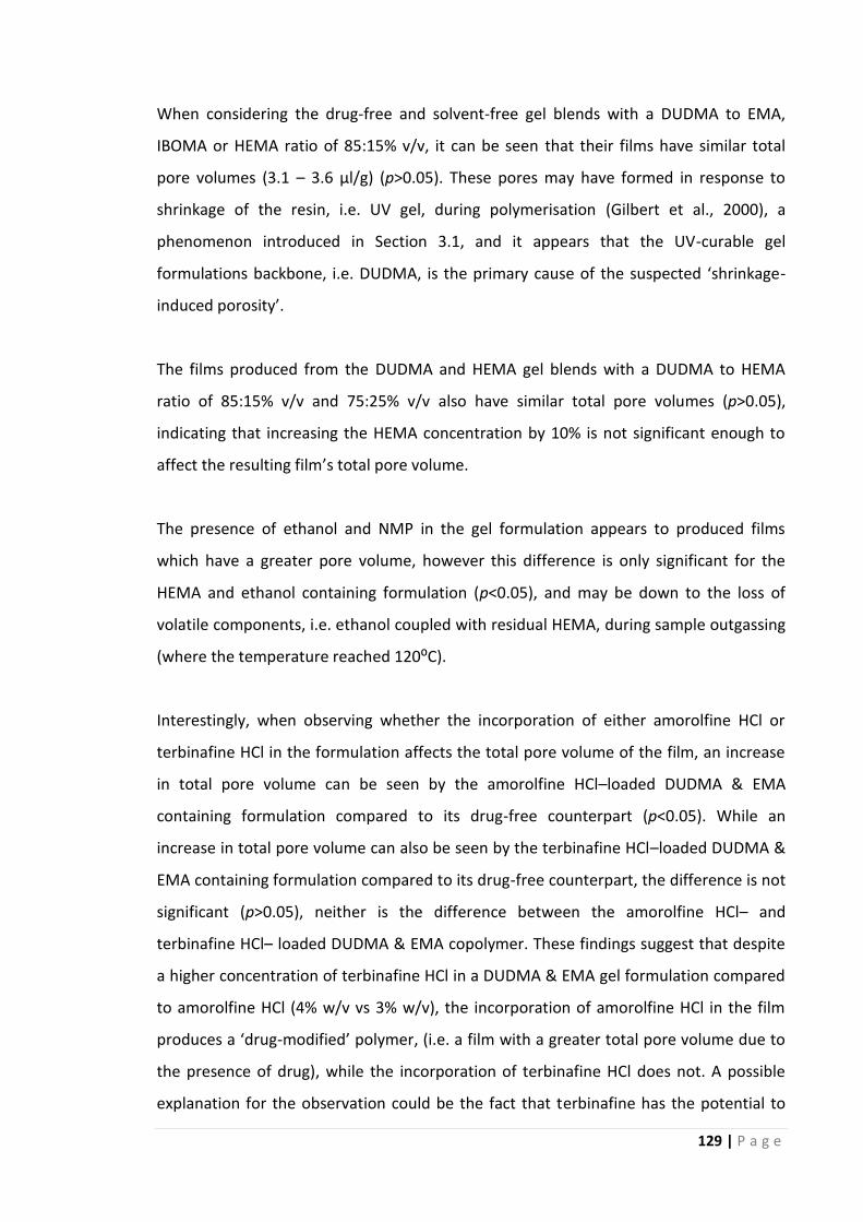
129 | P a g e
When considering the drug-free and solvent-free gel blends with a DUDMA to EMA,
IBOMA or HEMA ratio of 85:15% v/v, it can be seen that their films have similar total
pore volumes (3.1 – 3.6 µl/g) (p>0.05). These pores may have formed in response to
shrinkage of the resin, i.e. UV gel, during polymerisation (Gilbert et al., 2000), a
phenomenon introduced in Section 3.1, and it appears that the UV-curable gel
formulations backbone, i.e. DUDMA, is the primary cause of the suspected ‘shrinkage-
induced porosity’.
The films produced from the DUDMA and HEMA gel blends with a DUDMA to HEMA
ratio of 85:15% v/v and 75:25% v/v also have similar total pore volumes (p>0.05),
indicating that increasing the HEMA concentration by 10% is not significant enough to
affect the resulting film’s total pore volume.
The presence of ethanol and NMP in the gel formulation appears to produced films
which have a greater pore volume, however this difference is only significant for the
HEMA and ethanol containing formulation (p<0.05), and may be down to the loss of
volatile components, i.e. ethanol coupled with residual HEMA, during sample outgassing
(where the temperature reached 120⁰C).
Interestingly, when observing whether the incorporation of either amorolfine HCl or
terbinafine HCl in the formulation affects the total pore volume of the film, an increase
in total pore volume can be seen by the amorolfine HCl–loaded DUDMA & EMA
containing formulation compared to its drug-free counterpart (p<0.05). While an
increase in total pore volume can also be seen by the terbinafine HCl–loaded DUDMA &
EMA containing formulation compared to its drug-free counterpart, the difference is not
significant (p>0.05), neither is the difference between the amorolfine HCl– and
terbinafine HCl– loaded DUDMA & EMA copolymer. These findings suggest that despite
a higher concentration of terbinafine HCl in a DUDMA & EMA gel formulation compared
to amorolfine HCl (4% w/v vs 3% w/v), the incorporation of amorolfine HCl in the film
produces a ‘drug-modified’ polymer, (i.e. a film with a greater total pore volume due to
the presence of drug), while the incorporation of terbinafine HCl does not. A possible
explanation for the observation could be the fact that terbinafine has the potential to

130 | P a g e
take part in the polymerisation process as it has an alkene group. However, the latter is
not suspected as the incorporation of amorolfine HCl or terbinafine HCl in formulations
containing DUDMA & IBOMA or DUDMA & HEMA (with a DUDMA to HEMA ratio of
85:15% v/v or 75:25% v/v) did not appear to affect the films’ total pore volume (p>0.05).
A plausible reason for the observations therefore remains to be seen.
Microstructure
Scanning electron micrographs of the UV-cured films produced from all the gels
formulated containing EMA, IBOMA or HEMA, with/without solvent and with/without
drug (Table 3.6), revealed a generally rough surface of the side that was exposed to UV
light and a much smoother surface where the film had been formed in contact with the
support. The latter provides a certain containment for the flow of the fluid during
polymerisation and the smooth film surface reflects the smooth surface of the support.
The cross-sectional views show a fairly dense film interior, which could be due to the
polymers’ highly cross-linked nature.
Films produced from the drug-free and solvent-free gel blends with a DUDMA to diluent
monomer ratio of 85:15% v/v have comparable microstructures, whereas the films
produced from the HEMA formulations with a DUDMA to HEMA ratio of 75:25% v/v
have a slightly smoother top and cross-sectional surface compared to the films produced
from the formulations with a DUDMA to HEMA ratio of 85:15% v/v. This suggests that
the proportion of monomers, rather than monomer choice, in the gel mixture
predominately influences the film’s architecture.
Inclusion of the solvent ethanol in the gel formulation seems to have no influence on the
film microstructure, while inclusion of NMP results in films with smoother top and cross-
sectional surfaces. As mentioned above, NMP has a high boiling point, therefore while
the ethanol originally present in the gel formulation may evaporate off during UV-curing,
NMP is likely to remain in the film and influence the polymer formed.
131 | P a g e
Inclusion of the drug amorolfine HCl or terbinafine HCl in the gel formulation showed no
visible changes in the resulting film’s microstructure, possibly as the amount of drug
included in the formulation was below the saturation limit, i.e. the drugs were
molecularly dispersed in the formulation, and as the drug-load was fairly low. These
findings will be used as a tool to discuss the film’s drug release behaviour examined in
the following Chapter.
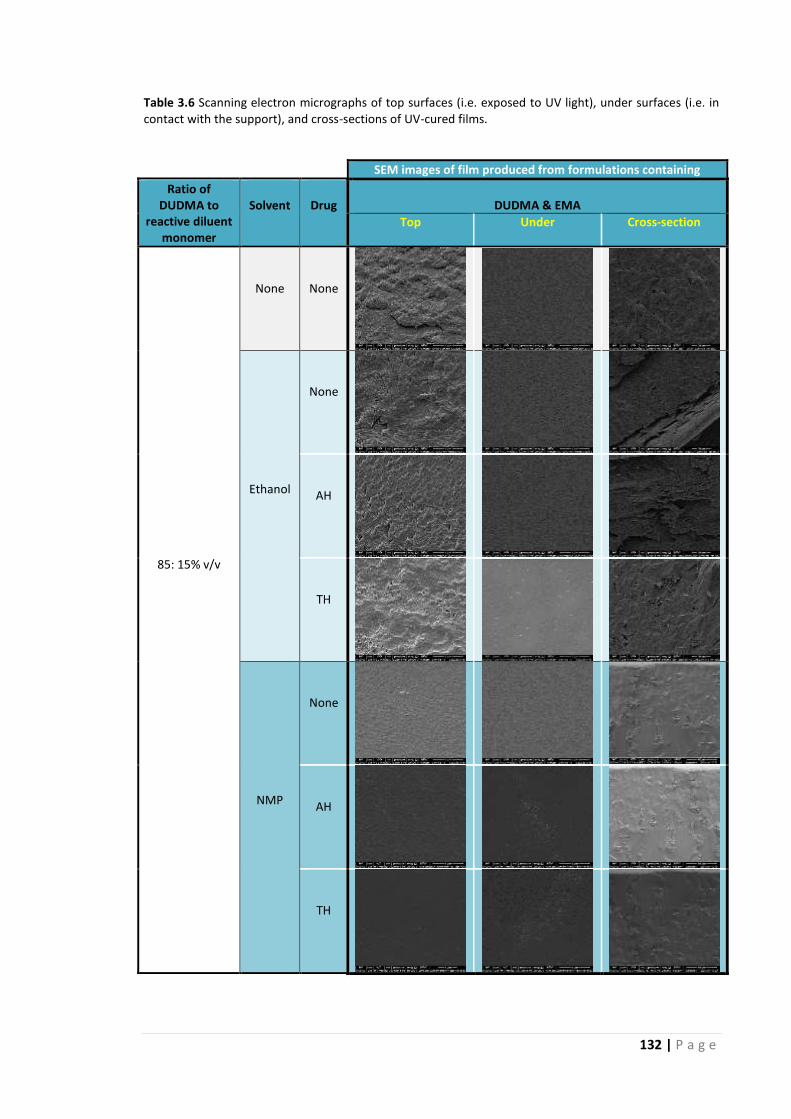
132 | P a g e
Table 3.6 Scanning electron micrographs of top surfaces (i.e. exposed to UV light), under surfaces (i.e. in contact with the support), and cross-sections of UV-cured films.
SEM images of film produced from formulations containing
Ratio of DUDMA to
reactive diluent monomer
Solvent
Drug
DUDMA & EMA
Top Under Cross-section
85: 15% v/v
None
None
Ethanol
None
AH
TH
NMP
None
AH
TH

133 | P a g e
Table 3.6 Continued
SEM images of film produced from formulations containing
Ratio of DUDMA to
reactive diluent monomer
Solvent
Drug
DUDMA & IBOMA
Top Under Cross-section
85: 15% v/v
None
None
Ethanol
None
AH
TH

134 | P a g e
Table 3.6 Continued
Abbreviations: AH, amorolfine HCl; TH, terbinafine HCl; NF, not formulated.
SEM images of film produced from formulations containing
Ratio of DUDMA to
reactive diluent monomer
Solvent
Drug
DUDMA & HEMA
Top Under Cross-section
85: 15 % v/v
None
None
Ethanol
None
AH
TH
75: 25% v/v
None
None
AH
TH

135 | P a g e
3.5.2 Drug-load in UV-cured film and drug-polymer interactions
3.5.2.1 Drug-load
The antifungals amorolfine HCl and terbinafine HCl are both crystalline powders as can
be seen from their polarised light microscopy images and XRD patterns which reveal
characteristic diffraction peaks (Fig. 3.9). DUDMA and EMA, IBOMA or HEMA copolymer
films, on the other hand, are amorphous as can be seen in Fig. 3.10. As mentioned
previously (in Section 3.4), the amount of drug incorporated in the gels formulated was
the maximum that could be dissolved in the gel mixture (Table 2.9, Chapter 2). Upon UV-
curing, the drug remained in the dissolved state with no crystals observed by polarised
light microscopy, and this was confirmed by XRD which showed no change in the
amorphicity of the polymer films (Table 3.7).
0 10 20 30 40 50 60
0
5000
10000
15000
20000
25000
30000
35000
Inte
nsit
y (
co
un
ts)
2 Theta (deg.)
0 10 20 30 40 50 60
0
5000
10000
15000
20000
25000
30000
35000
40000
45000
Inte
nsit
y (
co
un
ts)
2 Theta (deg.)
Fig. 3.9 Polarised light micrograph & corresponding XRD pattern of amorolfine HCl (left) & terbinafine HCl (right).
0 10 20 30 40 50 60
0
2000
4000
6000
8000
10000
12000
Inte
nsit
y (
co
un
ts)
2 Theta (deg.)
DUDMA & EMA copolymer
DUDMA & IBOMA copolymer
DUDMA & HEMA copolymer
Fig. 3.10 XRD patterns of DUDMA & EMA, IBOMA or HEMA copolymer films (drug-free and solvent-free with a DUDMA: diluent monomer ratio of 85:15% v/v).

136 | P a g e
Table 3.7 Polarised light micrographs and corresponding XRD patterns of drug-loaded UV-cured films.
Polarised light micrograph and corresponding XRD pattern of film produced from formulations containing
Amorolfine HCl Terbinafine HCl
DUDMA 85%: EMA 15% v/v and 25% v/v ethanol
3% w/v
0 10 20 30 40 50 60
0
2000
4000
6000
8000
10000
12000
14000
16000
Inte
nsit
y (
co
un
ts)
2 Theta (deg.)
4% w/v
0 10 20 30 40 50 60
0
2000
4000
6000
8000
10000
12000
Inte
nsit
y (
co
un
ts)
2 Theta (deg.)
DUDMA 85%: EMA 15% v/v and 25% v/v NMP
1% w/v
0 10 20 30 40 50 60
0
2000
4000
6000
8000
10000
12000
Inte
nsit
y (
co
un
ts)
2 Theta (deg.)
1% w/v
0 10 20 30 40 50 60
0
2000
4000
6000
8000
10000
12000
14000
16000
18000
20000
22000
Inte
nsit
y (
co
un
ts)
2 Theta (deg.)
DUDMA 85%: IBOMA 15% v/v and 25% v/v ethanol
3% w/v
0 10 20 30 40 50 60
0
2000
4000
6000
8000
10000
Inte
nsit
y (
co
un
ts)
2 Theta (deg.)
4% w/v
0 10 20 30 40 50 60
0
2000
4000
6000
8000
10000
12000
14000
16000
18000
Inte
nsit
y (
co
un
ts)
2 Theta (deg.)
DUDMA 85%: HEMA 15% v/v and 25% v/v ethanol
4% w/v
0 10 20 30 40 50 60
0
2000
4000
6000
8000
10000
12000
14000
16000
18000
Inte
nsit
y (
co
un
ts)
2 Theta (deg.)
6% w/v
0 10 20 30 40 50 60
0
2000
4000
6000
8000
10000
12000
14000
16000
Inte
nsit
y (
co
un
ts)
2 Theta (deg.)
DUDMA 75%: HEMA 25% v/v
2% w/v
0 10 20 30 40 50 60
0
2000
4000
6000
8000
10000
12000
Inte
nsit
y (
co
un
ts)
2 Theta (deg.)
2% w/v
0 10 20 30 40 50 60
0
2000
4000
6000
8000
10000
Inte
nsit
y (
co
un
ts)
2 Theta (deg.)

137 | P a g e
Table 3.8 shows the drug-load in the UV-cured films (following the removal of the
oxygen-inhibition layer). The drug concentrations in all the films examined are similar to
the drug concentration in their uncured gel formulation (p>0.05). The fact that the drug
concentration did not increase or decrease in the film following the removal of the
oxygen inhibition layer indicates that there was no drug migration to the film surface or
interior during UV curing. This finding reveals an interesting difference between UV-
curable gels and nail lacquers. In the latter, the drug concentration in the film formed is
higher than that in the initial formulation as the solvent evaporates off upon application
of a nail lacquer. For example, the pharmaceutical nail lacquer Curanail® contains
amorolfine HCl at 5% w/v; however, upon application, a polymer film containing drug at
a concentration of 31.5% ± 0.8% w/w is formed (measured by dissolving the Curanail®
film in ethanol and quantifying by HPLC as per Section 2.4.6 [Chapter 2]). The lower
solvent content of UV gel formulations and possible retention of some solvent in the UV-
cured film may explain the similar drug concentrations in the gel and UV-cured film.
Table 3.8 Antifungal drug concentration in the gel formulations prior to curing and in UV-cured film following the removal of the oxygen inhibition layer.
Backbone
and photoinitiator
Diluent
monomer
Solvent
(25% v/v)
Antifungal
AH or TH
Concentration in gel
(% w/v)
Concentration in film
(% w/w)
DUDMA and
2-hydroxy-2-methylpropiophenone
EMA Ethanol AH 3.0 ± 0.27 3.3 ± 0.16
TH 4.0 ± 0.39 3.9 ± 0.11
NMP AH 1.0 ± 0.14 1.0 ± 0.04
TH 1.0 ± 0.12 1.0 ± 0.01
IBOMA Ethanol AH 3.0 ± 0.18 3.3 ± 0.20
TH 4.0 ± 0.26 3.9 ± 0.06
HEMA Ethanol AH 4.0 ± 0.49 4.4 ± 0.04
TH 6.0 ± 0.22 5.9 ± 0.07
None AH 2.0 ± 0.37 2.2 ± 0.07
TH 2.0 ± 0.12 2.0 ± 0.01
Abbreviations: AH, amorolfine HCl; TH, terbinafine HCl

138 | P a g e
3.5.2.2. Any drug-polymer interactions?
The FT-IR spectra of the DUDMA & EMA, IBOMA or HEMA UV-cured polymer films (drug-
free and solvent-free) (Fig. 3.11) all show characteristic absorption bands at around 3360
cm-1 (N-H), 2957 cm-1 (C-H) and 1702 cm-1 (COO-R, R-O-CONH-R). The bands between
1300 – 1050 cm-1 relate to C-O stretches. These absorbances relate primarily to the
functional groups of the DUDMA backbone and appear to overlap the absorption bands
relating to IBOMA’s cyclic group and HEMA’s hydroxyl functional groups.
4000 3500 3000 2500 2000 1500 1000 500
Ab
so
rban
ce
Wavenumber (cm-1)
DUDMA & HEMA copolymer
DUDMA & IBOMA copolymer
DUDMA & EMA copolymer
Fig. 3.11 FT-IR spectra of DUDMA & EMA, IBOMA or HEMA copolymer films (drug-free and solvent-free with a DUDMA: diluent monomer ratio of 85:15% v/v).
The FT-IR spectra of the drug-free polymer films produced from ethanol and EMA,
IBOMA or HEMA containing gels (Table 3.9) are comparable to their solvent-free control
counterparts. Interestingly, the FT-IR spectra of the film produced from the NMP
containing formulation (Table 3.9) shows an absorption band between 1670 and 1710
cm-1 relating to the C=O stretch of NMP’s five ring lactam, suggesting that NMP does
remain in the polymer film upon curing of the gel formulation as hypothesised in earlier
sections (Section 2.5.7 [Chapter 2] and 3.5.1.2).
The FT-IR spectra of the drug-free and drug-loaded polymer films show no obvious shifts
in characteristic bands (Table 3.9), indicating the absence of chemical drug-polymer
interactions in the film. This bodes well for drug release from film and ungual drug
permeation.

139 | P a g e
Table 3.9 FT-IR spectra of UV-cured films. The spectra in black, red and blue are for drug-free, amorolfine HCl- and terbinafine HCl- loaded films respectively.
FT-IR spectra of film produced from formulations containing
DUDMA 85%: EMA 15% v/v and 25% v/v ethanol
4000 3500 3000 2500 2000 1500 1000 500
Ab
so
rban
ce
Wavenumber (cm-1)
DUDMA 85%: EMA 15% v/v and 25% v/v NMP
4000 3500 3000 2500 2000 1500 1000 500
Ab
so
rban
ce
Wavenumber (cm-1)
DUDMA 85%: IBOMA 15% v/v and 25% v/v ethanol
4000 3500 3000 2500 2000 1500 1000 500
Ab
so
rban
ce
Wavenumber (cm-1)
DUDMA 85%: HEMA 15% v/v and 25% v/v ethanol
4000 3500 3000 2500 2000 1500 1000 500
Ab
so
rban
ce
Wavenumber (cm-1)
DUDMA 75%: HEMA 25% v/v
4000 3500 3000 2500 2000 1500 1000 500
Ab
so
rban
ce
Wavenumber (cm-1)

140 | P a g e
3.5.3 Thermal properties of UV-cured films
3.5.3.1 Polymer degradation
The thermal properties of the UV-cured films were determined using TGA, and the
influence (if any) of monomer choice, monomer ratio, solvent and drug incorporation on
the film’s degradation were evaluated. The TGA curves for the drug-free polymer films
(with and without solvent) are shown in Fig. 3.12. The profiles seem to consist of four
phases, a–d, and is consistent with the profiles reported for UV-cured coatings based on
urethane acrylate oligomers elsewhere (Kunwong et al., 2011, Alshuiref et al., 2013).
The first phase relates to the loss of volatile materials from the film as the latter is
heated. Films produced from solvent-containing formulations showed a greater loss in
mass than the control solvent-free film during phase a (p<0.05). This indicates that some
solvent has remained in the film upon UV-curing and was being lost as the film was
heated. NMP-containing films showed a greater mass loss than ethanol-containing ones
and a transition at around 200⁰C. This is linked to the greater amount of residual solvent
in these films and to NMP’s boiling point of 202⁰C. Phases b–d in the profiles are related
to polymer decomposition via the degradation of polymer side-chains, followed by
scission and depolymerisation, and finally complete de-crosslinking and thermal
degradation, as suggested for similar films by other researchers (Chattopadhyay and
Webster, 2009).
It can be seen from Fig. 3.12 that the films produced from the solvent-free DUDMA &
EMA, IBOMA or HEMA gel blends with a DUDMA to diluent monomer ratio of 85:15%
v/v have comparable thermal degradation profiles. It can also be seen that the ratio of
DUDMA: diluent methacrylate monomer (i.e. HEMA) used in the gel does not affect the
resulting polymers’ thermal degradation. This could be as the higher HEMA
concentration in the gel is not sufficiently different enough to significantly affect the
resulting copolymers degradation profile. The presence of amorolfine HCl or terbinafine
HCl in the formulation also did not alter the film’s thermal stability (Appendix A8),
possibly due to their low concentrations. The thermal stability of the drug-loaded UV-
cured polymers at room temperatures therefore makes them acceptable for
consideration as a nail drug delivery vehicle.

141 | P a g e
0 100 200 300 400 500
0
20
40
60
80
100
Weig
ht
(%)
Temperature (°C)
DUDMA 85: EMA 15 v/v (solvent-free) gel
DUDMA 85: EMA 15 v/v and ethanol gel
DUDMA 85: EMA 15 v/v and NMP gel
DUDMA 85: IBOMA 15 v/v (solvent-free) gel
DUDMA 85: IBOMA 15 v/v and ethanol gel
DUDMA 85: HEMA 15 v/v (solvent-free) gel
DUDMA 85: HEMA 15% v/v and ethanol gel
DUDMA 75: HEMA 25% v/v (solvent-free) gel
a
b
c
d
Fig. 3.12 TGA profiles of films produced from drug-free UV-curable gel formulations containing EMA, IBOMA and HEMA represented by black, red and blue respectively.

142 | P a g e
3.5.3.2 Glass transition temperatures of UV-cured films and the influence of
film components
The UV-cured polymers’ glass transition temperatures (Tg) were calculated as the tan δ
peak from the DMA curves. Interestingly, the DMA curves for the film produced from
the DUDMA & EMA containing formulation absent of solvent and drug showed two
transitions as shown in Fig. 3.13. The first glass transition relates to the polymer film
produced following UVA irradiation of the gel formulation, and the second glass
transition could be in response to an additional thermal cure of the UV-cured polymer
film owing to the high temperature range used in the DMA experiment (Jong Keun et al.,
2003). A more detailed examination of the log [storage modulus] vs. temperature plot
(Fig. 3.13) shows that the polymer film starts off glassy, and as the temperature is
increased the first glass transition is evident. As the temperature further increases in this
region there is a likely increase in the mobility of macromolecular chains of the film, and
a possible additional cure with further crosslinking reaction induced by thermal energy.
Once the second cure is completed, a second glass transition of the polymer film is
evident, and a new material can be considered to have been created. This phenomenon
is feasible given that the degree of conversion from monomer to polymer was not 100%
for this formulation (Chapter 2, Table 2.12), the lifetime of residual free radicals at room
temperature could reach several days or months (Zhu et al., 1990, Tian et al., 1992), and
the reaction of an irradiated unsaturated monomer has previously been observed for
several days in the dark (Marino et al., 1995). Furthermore, upon observation of the DSC
thermogram for this polymer film (Fig. 3.13), an exothermic peak is evident (at a
temperature beyond the first glass transition temperature) in response to the additional
thermally induced polymerisation reaction. This trend is also evident with all the
solvent-free polymer films containing IBOMA and HEMA, which also display two glass
transition temperatures, the values for which are in Table 3.10. As the lower glass
transition temperature corresponds to the polymer film produced following UV-
irradiation of the gel formulation without a subsequent thermal reaction, as would be
the case in practice, it was used when comparing the different formulations.
It can be seen from Table 3.10, that the choice of diluent monomer in a gel formulation
absent of solvent and drug (and with a DUDMA to diluent reactive monomer ratio of 85:

143 | P a g e
15% v/v) significantly alters the resulting polymers’ Tg (p<0.05), where DUDMA & EMA
copolymer’s Tg is greater than that of the DUDMA & HEMA copolymer, which is in turn
greater than that of the DUDMA & IBOMA copolymer. This trend can be explained by
the viscosity of the formulations, which have values of 366.5, 598.1 and 672.4 mPas
respectively (Chapter 2, Table 2.10), and it can be seen that the lower the viscosity of
the formulation, the higher the glass transition of its UV-cured polymer film. A
formulation with a lower viscosity can increase the mobility of the reactants and
therefore reactivity during polymerisation, this in turn can produce a highly cross-linked
film with decreased chain mobility, hence the increased Tg observed. The amount of
diluent monomer used in a gel formulation also affects the resulting polymers’ Tg. The
film produced from the HEMA formulation with a DUDMA to HEMA ratio of 75:25% v/v
has a higher Tg compared to the film produced from the formulations with a DUDMA to
HEMA ratio of 85:15% v/v, also due to its lower viscosity (Table 2.10).
Inclusion of ethanol or NMP in the gels resulted in films with only one Tg – which was
significantly higher than the first glass transition temperature seen for the
corresponding solvent-free films (Fig. 3.14, Table 3.10). It was mentioned earlier (in
Chapter 2, Section 2.5.7.2) that inclusion of a solvent in a UV-curable gel considerably
decreases its viscosity and hence significantly increases its polymerisation extent, which
as mentioned above can increase Tg. The increased polymerisation can also decrease
the likelihood of an additional thermal cure of the UV-cured polymer film, and this
explains why the DSC thermogram for the films produced from the DUDMA & EMA gel
formulations with solvent (ethanol or NMP), but without drug, displays no appreciable
exotherm (Fig. 3.14), which in turn explains the absence of a second glass transition.
It can also be seen from Table 3.10, that the choice of diluent monomer in an ethanol-
containing gel formulation significantly alters the resulting polymers’ Tg (p<0.05), where
DUDMA & IBOMA copolymer’s Tg is greater than that of the DUDMA & EMA copolymer,
which is in turn greater than that of the DUDMA & HEMA copolymer. As the viscosities
of the solvent-containing gel formulations are similar (Chapter 2, Table 2.10), gel
viscosity does not explain the observations. Instead this trend can be explained by the Tg
of IBOMA, EMA and HEMA’s homopolymers, which have values of 110⁰C, 65⁰ and 57⁰C

144 | P a g e
respectively. IBOMA’s bulky cyclic group is responsible for the much higher Tg displayed
by IBOMA containing polymers due to its potential to hinder chain mobility.
Interestingly, films formed from EMA and NMP-containing gels have lower Tg values
than EMA and ethanol-containing ones (p<0.05). The greater amount of residual solvent
in the NMP-based film (as evidenced in Fig. 3.12) seems to result in a plasticising effect
on the UV-cured polymer film, by spacing polymer chains apart and enabling chain
mobility, such that Tg is lowered.
The presence of the drugs in the solvent-containing films’ does not cause any significant
changes in the films’ Tg (p>0.05), while the presence of drugs in the solvent-free DUDMA
& HEMA gel formulations with a DUDMA to HEMA ratio of 75:25% v/v, slightly increases
the film’s Tg (p<0.05). Both amorolfine HCl and terbinafine HCl have a molecular weight
greater than 300 Daltons (Table 2.6), and when incorporated in a UV-cured film, both
may restrict the mobility of polymer chain and increase Tg. In the presence of ethanol,
this restriction is less evident; indicating that the residual ethanol in the film masks the
effect of drug incorporation when considering the resulting film’s Tg.

145 | P a g e
20 40 60 80 100 120 140 160 180 200
1.2
1.4
1.6
1.8
2.0
2.2
2.4
2.6
2.8
3.0
Termperature (°C)
Lo
g [
sto
rag
e m
od
ulu
s]
(MP
a)
I
0.00
0.05
0.10
0.15
0.20
0.25
0.30
0.35
Tan
delt
a
II
III
IV
V
20 40 60 80 100 120 140 160 180 200
0.8
1.0
1.2
1.4
1.6
1.8
2.0
2.2
2.4
2.6
2.8
3.0
ETOH
NMP
ETOH
NMP
Temperature (°C)
Lo
g [
sto
rag
e m
od
ulu
s]
(MP
a)
0.0
0.1
0.2
0.3
0.4
0.5
Tan
delt
a
0 50 100 150 200 250
-0.35
-0.30
-0.25
-0.20
-0.15
-0.10
-0.05
He
at
flo
w (
W g
-1)
Temperature (°C) Fig. 3.13 Log [storage modulus] and tan δ from DMA and heat flow from DSC vs. temperature for UV-cured films produced from DUDMA & EMA gel formulations without solvent and without drug. The log [storage modulus] vs. temperature plot can be considered to show five different regions: I = glassy, II = glass transition, III = thermal reaction, IV = new glass transition, and V = rubbery (Jong Keun et al., 2003).
0 50 100 150 200 250
-0.6
-0.5
-0.4
-0.3
-0.2
-0.1
0.0
0.1
0.2
He
at
flo
w (
W g
-1)
Temperature (°C)
ETOH
NMP
Fig. 3.14 Log [storage modulus] and tan δ from DMA and heat flow from DSC vs. temperature for UV-cured films produced from the DUDMA & EMA gel formulations with solvent (ethanol or NMP), but without drug. Abbreviations: ETOH, ethanol.

146 | P a g e
Table 3.10 Tg values of UV-cured films. Means ± standard deviations are shown, n=3.
Tg values of UV-cured films produced from formulations containing
Excipients DUDMA Solvent Drug EMA IBOMA HEMA
Ethanol NMP
Formulations
DUDMA 85 % v/v : diluent monomer 15 % v/v
None None None 85.7 ± 1.2 and 146.0 ± 2.5 67.2 ± 2.4 and 164.0 ± 0.2 74.4 ± 2.5 and 147.2 ± 0.3
25% v/v
None
None 112.5 ± 1.8 139.2 ± 9.0 98.1 ± 2.3
AH 109.2 ± 1.1 137.2 ± 0.02 99.2 ± 1.8
TH 109.6 ± 0.8 133.4 ± 3.6 94.7 ± 4.8
None
25% v/v
None 95.1 ± 1.5 NF NF
AH 94.8 ± 2.0 NF NF
TH 95.0 ± 1.8 NF NF
DUDMA 75 % v/v : diluent monomer 25 v/v
None
None
None NF NF 81.8 ± 0.5 and 142.6 ± 0.4
AH NF NF 92.0 ± 0.9 and 138.9 ± 0.5
TH NF NF 92.4 ± 0.4 and 133.2 ± 0.2
Tg of homopolymers
(⁰C)
-
-
-
-
65
110
57
Abbreviations: AH, amorolfine HCl; TH, terbinafine HCl; NF, not formulated.

147 | P a g e
3.5.4 Occlusivity of UV-cured polymer films
UV gels produce an occlusive film on the nail plate surface following UV-curing and
removal of the oxygen inhibition layer. Fig. 3.15 shows just how occlusive the polymeric
film formed is by quantifying the suppression of TOWL following its formation on the
nail plate surface. It can be seen that a film formed from a drug-free and solvent-free
EMA-containing gel formulation reduces TOWL by as much as 48%. The choice of diluent
monomer (i.e. EMA, IBOMA or HEMA) in the gel formulation does not significantly alter
this % reduction (p>0.05). The presence of solvent with and without drug in the EMA
containing gel formulations also does not significantly alter this % reduction (p>0.05). As
seen from Table 3.3, formulations containing solvents produced films which are
significantly thinner (by approximately 20%) than their solvent-free counterparts.
However, the thickness of the UV-cured film does not appear to correlate with
occlusivity as can be seen from Fig. 3.16. Therefore it appears that the choice of the
polymer backbone, i.e. DUDMA, is primarily responsible for the reduction in TOWL.
When compared to the commercially available pharmaceutical nail lacquer – Curanail® –
the UV-curable gel formulations reduce TOWL % to a greater extent (p<0.05). In addition
to containing the active amorolfine hydrochloride, Curanail® contains ammonio
methacrylate copolymer A, triacetin, butyl acetate, ethyl acetate and ethanol. The
Curanail® film produced has a thickness of 13.3 µm ± 2.9 µm, which is 10 times thinner
than the drug-loaded DUDMA & EMA UV-cured films; therefore in this instance film
thickness may indeed be responsible for the lower TOWL % reduction. The DUDMA
containing copolymers may also have a lower permeability to water vapour compared to
ammonio methacrylate copolymer A, a factor which can also contribute to TOWL
reduction.

148 | P a g e
0 10 20 30 40 50 60
Reduction in TOWL %
Curanail nail lacquer
kDUDMA & EMA gel containing ETOH & 3% w/v AH
kDUDMA & EMA gel containing ETOH & 4% w/v TH
kDUDMA & EMA gel containing ETOH (drug-free)
kDUDMA & EMA gel (drug-free, solvent-free)
kDUDMA & IBOMA gel (drug-free, solvent-free)
kDUDMA & HEMA gel (drug-free, solvent-free)
Fo
rmu
lati
on
Fig. 3.15 Reduction in TOWL following application of a formulation on the nail plate surface. All gel formulations tested have a DUDMA to diluent monomer ratio of 85:15% v/v. Means and standard deviations are shown, n=5. Abbreviations: AH, amorolfine HCl; TH, terbinafine HCl; ETOH, ethanol.
160 170 180 190 200 210 220
34
36
38
40
42
44
46
48
50
Re
du
cti
on
in
TO
WL
%
Thickness of UV-cured polymer film (m)
R-squared = 0.231 (p = 0.334)
Fig. 3.16 Influence of film thickness on film occlusivity. Greater reduction in TOWL % = greater occlusivity.
As previously mentioned, a reduction in TOWL is likely to hyper-hydrate the upper nail
layers. This can aid drug permeation into and through the nail plate (Spruit, 1971, Marty,
1995), given that in vitro nail hydration has been found to increase diffusivity of water
and of the antifungal ketoconazole in the nail (Gunt et al., 2007, Gunt and Kasting,
2007). Nail hydration can also benefit in the treatment of onychomycosis by inducing the
germination of drug-resistant fungal spore into drug-susceptible hyphae, which would
enable fungi eradication (Flagothier et al., 2005). Therefore, it could be possible that
since the drug-loaded UV-curable gel formulations reduce TOWL to a greater extent
than Curanail®, they could cause a greater increase in nail plate hydration and thereby a
greater enhancement of drug permeation into the nail to tackle the onychomycosis-
causing pathogen.

149 | P a g e
It should be noted however, that these TOWL reduction readings were obtained using
healthy fingernails, and TOWL reduction in practice, i.e. on a diseased nail, following the
application of the UV-curable gel formulation could be significantly different due to
possible differences in initial TOWL readings. For example, it has been found that
onychomycotic nails show a significantly lower TOWL than healthy nails, as the diseased
nail develops a granular layer (Krönauer et al., 2001). This is based on the speculation
that the structure of a dystrophic nail is more similar to that of skin. To add to this,
onychomycosis predominately affects toenails which are thicker than fingernails, and
TOWL has also been found to be influenced by nail plate thickness (where it is lower for
thicker nails) (Murdan et al., 2008). On the other hand, a more recent study found that
TOWL measurements are similar for healthy and thicker diseased nails (McAuley et al.,
2016). Whether the application of a UV-curable gel formulation reduces TOWL of
diseased nails to the same extent as healthy nails could therefore be considered for
further work.
3.5.5 UV-cured polymer film’s adhesivity
Adhesion of a topical formulation to the nail plate surface is essential in order to allow
the drug to leave the formulation and penetrate the nail. The adhesivity of UV-cured
polymer films was measured both manually (cross-cut test) and by using a texture
analyser (pull-off test).
3.5.5.1 Cross-cut test
The mean cross-cut score for the UV-cured polymer films is shown in Fig. 3.17. A high
score means lower adhesion (or lower resistance to separation from a substrate). The
greatest adhesion was shown by the films produced from the solvent-free formulations,
with the choice of diluent monomer not altering adhesion (p>0.05). The DUDMA
backbone is primarily responsible for the resistance to separation displayed as it
inherently provides excellent adhesion and abrasion resistance (Botero et al., 2014).
However as the DUDMA to diluent monomer ratio used (i.e. 85: 15% v/v or 75:25% v/v
for HEMA) does not significantly alter the films’ adhesion (p>0.05), it appears that the
diluent monomer, in this case HEMA, also contributes to the copolymers’ adhesive
property. Presence of ethanol (with and without antifungal drugs) in the formulations

150 | P a g e
caused no statistically significant change in film adhesion (p>0.05). In contrast, presence
of NMP (with and without antifungal drugs) in the formulation significantly reduced film
adhesivity (p<0.05). As discussed earlier, more NMP (than ethanol) remains in the film
upon gel curing. The presence of a greater amount of solvent seems to cause a larger
change in the properties of the NMP-containing films, resulting in a significant decrease
in film adhesion to the substrate. The incorporation of either amorolfine HCl or
terbinafine HCl in the formulation did not affect the films’ cross-cut score (p>0.05);
possibly due to the small drug content in the film.
0 1 2 3 4 5
EMA gel (drug-free, solvent-free)
EMA gel containing ETOH
EMA gel containing ETOH & AH
EMA gel containing ETOH & TH
EMA gel containing NMP
EMA gel containing NMP & AH
EMA gel containing NMP & TH
IBOMA gel (drug-free, solvent-free)
IBOMA gel containing ETOH
IBOMA gel containing ETOH & AH
IBOMA gel containing ETOH & TH
HEMA gel (drug-free, solvent-free)
HEMA gel containing ETOH
HEMA gel containing ETOH & AH
HEMA gel containing ETOH & TH
HEMA: DUDMA (25:75%) gel
HEMA: DUDMA (25:75%) & AH gel
HEMA: DUDMA (25:75%) & TH gel
Cross-cut mean score
Fig. 3.17 Cross-cut scores for films produced from gels containing DUDMA & EMA, IBOMA or HEMA (± solvent and ± drug). Means and standard deviations are shown, n=5. High cross-cut scores mean lower adhesivity. Abbreviations: AH, amorolfine HCl; TH, terbinafine HCl; ETOH, ethanol.
3.5.5.2 Pull-off test
The pull-off test was used to determine the maximum force and work needed to
separate two surfaces in intimate contact, i.e. the substrate and UV-cured film. The peak
adhesive force (N) and work of adhesion (mJ) readings obtained for the UV-cured films
examined are shown in Fig. 3.18. It can be seen that the choice and ratio of diluent
monomer, and the incorporation of ethanol with and without amorolfine HCl or
terbinafine HCl in DUDMA containing gel formulations does not significantly affect the
resulting films’ peak adhesive force or work of adhesion (p>0.05). In contrast, the
presence of NMP (with and without antifungal drugs) reduces both parameters (p<0.05)

151 | P a g e
and thereby the film’s adhesive properties. These results are comparable to the cross-
cut test results shown in Fig. 3.17, and confirm that NMP negatively affects the films’
adhesive properties. It also suggests that the drug-loaded UV-curable gel formulations
(without NMP) have good adhesive properties when compared to the pharmaceutical
nail lacquer Curanail®, (which has peak adhesive force and work of adhesion values of
48.4 ± 8.9 N and 2.3 ± 1.1 mJ respectively). This “better” adhesivity should aid ungual
drug permeation.
0 10 20 30 40 50 60 70 80 90 100 110 120 130 140
EMA gel (drug-free, solvent-free)
EMA gel containing ETOH
EMA gel containing ETOH & AH
EMA gel containing ETOH & TH
EMA gel containing NMP
EMA gel containing NMP & AH
EMA gel containing NMP & TH
IBOMA gel (drug-free, solvent-free)
IBOMA gel containing ETOH
IBOMA gel containing ETOH & AH
IBOMA gel containing ETOH & TH
HEMA gel (drug-free, solvent-free)
HEMA gel containing ETOH
HEMA gel containing ETOH & AH
HEMA gel containing ETOH & TH
HEMA: DUDMA (25:75%) gel
HEMA: DUDMA (25:75%) & AH gel
HEMA: DUDMA (25:75%) & TH gel
Peak adhesive force (N)
0 10 20 30 40 50
EMA gel (drug-free, solvent-free)
EMA gel containing ETOH
EMA gel containing ETOH & AH
EMA gel containing ETOH & TH
EMA gel containing NMP
EMA gel containing NMP & AH
EMA gel containing NMP & TH
IBOMA gel (drug-free, solvent-free)
IBOMA gel containing ETOH
IBOMA gel containing ETOH & AH
IBOMA gel containing ETOH & TH
HEMA gel (drug-free, solvent-free)
HEMA gel containing ETOH
HEMA gel containing ETOH & AH
HEMA gel containing ETOH & TH
HEMA: DUDMA (25:75%) gel
HEMA: DUDMA (25:75%) & AH gel
HEMA: DUDMA (25:75%) & TH gel
Work of Adhesion (mJ)
Fig. 3.18 Peak adhesive force (A) and work of adhesion (B) readings obtained for the UV-cured films produced from gels containing DUDMA & EMA, IBOMA or HEMA (± solvent and ± drug). Means and standard deviations are shown, n=3.
3.5.6 UV-cured polymer film’s sensitivity to water
The water sensitivity scores of the UV-cured films are shown in Figs 3.19 - 3.22. Fig. 3.19
shows the water sensitivity score of UV-cured films produced from DUDMA & EMA gels
A
B

152 | P a g e
(± solvent and ± drug), and shows that films produced by the NMP-containing
formulations are extremely water sensitive. By five hours, the films had detached from
the substrate. NMP is miscible in water (Jouyban et al., 2010), and as it is present at a
considerable level in the film, contact with water causes the film to blister and
subsequently detach from the substrate. In contrast, solvent-free and drug-free films
had the lowest water sensitivity. This was expected as urethane methacrylate based
films are known for water resistance. Inclusion of ethanol caused no significant change
in water sensitivity (p<0.05). Like NMP, ethanol is miscible in water and therefore one
could have expected the water sensitivity of ethanol- and NMP- containing films to be
similar. A possible explanation for the greater water sensitivity displayed by NMP-
containing films is that the large planar non-polar region of NMP intercalates among
polymer chains forming hydrophobic interactions with the polymer, whereas more of
the ethanol evaporates off during and after curing due to its lower boiling point. The
incorporation of either amorolfine HCl or terbinafine HCl in the formulation did not
affect the films’ water sensitivity, possibly due to the very low drug-load.
Films produced from the DUDMA & IBOMA or HEMA containing gels (± solvent and ±
drug) show a similar trend to the EMA containing gels (Fig. 3.19 – Fig. 3.21), where the
presence of ethanol (with and without drug) in the gel does not cause a significant
change in the resulting film’s water sensitivity (p>0.05). This could be as the ethanol
concentration incorporated in all the gel formulations was the same and also as the
drug-load in these formulations was low. The incorporation of drug in DUDMA & HEMA
gels with a DUDMA: HEMA ratio of 75: 25% v/v also did not affect the film’s water
sensitivity (p>0.05) (Fig. 3.22), also probably due to its very low level in the formulation.
From Fig. 3.23, it can be seen that the choice of diluent monomer in the drug-free and
solvent-free gel formulation does not affect the resulting films’ water sensitivity score
(p>0.05). This comes as a surprise given that a HEMA polymer is expected to swell in
water due to the molecules’ hydrophilic pendant group (Chapter 2, Table 2.4). A likely
explanation for the similarities is therefore that the concentration of the diluent
monomers present in the gel formulation is not high enough to affect the resulting films’
water sensitivity.

153 | P a g e
0 10 20 30 40 50
0
1
2
3
4
5
Wa
ter
se
ns
itiv
ity s
co
re
Time (hours)
EMA gel (drug-free, solvent-free)
EMA gel containing ETOH
EMA gel containing ETOH & AH
EMA gel containing ETOH & TH
EMA gel containing NMP
EMA gel containing NMP & AH
EMA gel containing NMP & TH
Fig. 3.19 Water sensitivity score of UV-cured films produced from DUDMA & EMA gels (± solvent and ± drug). Means and standard deviations are shown, n=3.
0 10 20 30 40 50
0.0
0.2
0.4
0.6
0.8
1.0
1.2
1.4
Wate
r sen
sit
ivit
y s
co
re
Time (hours)
IBOMA gel (drug-free, solvent-free)
IBOMA gel containing ETOH
IBOMA gel containing ETOH & AH
IBOMA gel containing ETOH & TH
Fig. 3.20 Water sensitivity score of UV-cured films produced from DUDMA & IBOMA gels (± solvent and ± drug). Means and standard deviations are shown, n=3.
0 10 20 30 40 50
0.0
0.5
1.0
1.5
2.0
Wa
ter
se
ns
itiv
ity s
co
re
Time (hours)
HEMA gel (drug-free, solvent-free)
HEMA gel containing ETOH
HEMA gel containing ETOH & AH
HEMA gel containing ETOH & TH
Fig. 3.21 Water sensitivity score of UV-cured films produced from DUDMA & HEMA gels (± solvent and ± drug). Means and standard deviations are shown, n=3.
0 10 20 30 40 50
0.0
0.2
0.4
0.6
0.8
1.0
1.2
1.4
Wate
r sen
sit
ivty
sco
re
Time (hours)
HEMA: DUDMA (25:75%) gel
HEMA: DUDMA (25:75%) & AH gel
HEMA: DUDMA (25:75%) & TH gel
Fig. 3.22 Water sensitivity score of UV-cured films produced from DUDMA & HEMA gels (± drug) with a DUDMA: HEMA ratio of 75:25 % v/v. Means and standard deviations are shown, n=3.
0 20 40 60 80 100 120 140 160 180 200
EMA gel (drug-free, solvent-free)
EMA gel containing ETOH
EMA gel containing ETOH & AH
EMA gel containing ETOH & TH
EMA gel containing NMP
EMA gel containing NMP & AH
EMA gel containing NMP & TH
IBOMA gel (drug-free, solvent-free)
IBOMA gel containing ETOH
IBOMA gel containing ETOH & AH
IBOMA gel containing ETOH & TH
HEMA gel (drug-free, solvent-free)
HEMA gel containing ETOH
HEMA gel containing ETOH & AH
HEMA gel containing ETOH & TH
HEMA: DUDMA (25:75%) gel
HEMA: DUDMA (25:75%) & AH gel
HEMA: DUDMA (25:75%) & TH gel
Mean water sensitivity score (AUC )
Fig. 3.23 Area under the curve values calculated from the curves in Fig. 3.19 – Fig. 3.22 for the UV-cured films produced from DUDMA & EMA, IBOMA or HEMA containing gels (± solvent and ± drug). Means and standard deviations are shown, n=3. AUC was determined using OriginPro 9.0.
154 | P a g e
However, increasing the concentration of HEMA in the gel, (so that the DUDMA to
HEMA ratio is 75: 25% v/v as oppose to 85: 15% v/v), does not significantly alter the
resulting films’ water sensitivity either (p>0.05). This therefore indicates that a HEMA
concentration of 25% v/v or less can be incorporated in a gel formulation without having
a significant impact on DUDMA’s ability to produce a water-resistant film.
The low water sensitivity of films produced from ethanol- and drug- containing DUDMA
& EMA, IBOMA or HEMA gels indicates that these films could be used as long-term
topical nail medicines, and the next section explores just how long the film does reside
on the nail in vivo. The high water sensitivity of NMP-containing films means that these
films would not remain on the nail in vivo for prolonged periods (Murdan et al., 2015).
These films were therefore not included in the work described in the following Sections
and Chapters.
3.5.7 In vivo fingernail residence profiles of UV-cured polymer films
The mean in vivo residence of DUDMA & EMA containing gels (± drug and ± ethanol) and
the Curanail® nail lacquer on fingernails over a 28 day period is shown in Fig. 3.24. A
large variability in the residence of the same formulation in different individuals was
seen as shown by the error bars. This is as expected, given that a formulations’ residence
on the nail is governed by the individual’s activities such as hand washing, dish washing,
swimming etc. Nonetheless, it was found that a significantly greater % of the film
produced from the solvent-free gel formulation remained on the fingernail over the test
period when compared to the gel containing ethanol (p<0.05). The TGA profile of films
produced from gels containing ethanol confirmed that some ethanol does reside in the
polymer (Fig. 3.12). As previously mentioned, ethanol is miscible with water, and while
this factor did not affect the in vitro water sensitivity of the films produced from
ethanol-containing gels (Fig. 3.19), it did affect the film’s in vivo residence on the nail
plate, possibly as the formulations’ in vivo fingernail residence profile was tested over a
longer period of time compared to the water sensitivity test (28 days vs 2 days).
Furthermore, nail plates rapidly absorb water when exposed to it, and the HDPE
substrate used for the water immersion test is known for its low water absorption. The
use of HDPE sheet as a nail substitute may therefore limit how well the water sensitivity
test predicts in vivo nail residence of ungual formulations. The incorporation of

155 | P a g e
amorolfine HCl or terbinafine HCl in the gel formulation did not significantly affect the
resulting films’ fingernail residence (p>0.05), possibly due to the very low drug-load.
Fig. 3.24 also shows that UV-cured polymers resides on fingernails for significantly
longer than Curanail® (p<0.05). This could be explained by the more durable nature of
the DUDMA backbone in the gel formulations in comparison to Curanail’s polymer
backbone, i.e. ammonio methacrylate copolymer A, and explains Curanail’s 1 – 2 times
weekly application regimen as detailed in the British National Formulary (BNF) 71
(March 2016). This result highlights durability as an advantage for considering a UV-
curable gel over a lacquer as a nail drug delivery vehicle. In fact by day 14, greater than
50% of the films produced from the drug-loaded UV-curable gel still remains on the
fingernail plate, indicating a possible fortnightly application regimen for fingernails. The
residence profiles of films on the feet are generally longer than on hands (Murdan et al.,
2015), due to the feet’s lower exposure to abrasion during daily activities and lower
washing frequency when compared to hands, therefore the % of film remaining on the
toenail is expected to be much higher than that for fingernails on day 14.
As previously mentioned, commercially available cosmetic UV gels can be worn for up to
3 weeks without developing any visible defects (Schoon, 2010, Jefferson and Rich, 2012,
Schoon and Baran, 2012). Fig. 3.24 reveals that this is not the case for the UV-curable gel
formulations developed. The reason behind this is that prior to gel application, the nail is
lightly filed with a low git abrasive file (180 – 240 grit) to increase the surface area,
wiped with a nail wipe coated with isopropyl alcohol to remove surface moisture and
residual oils, and coated with a layer of primer (containing mixtures of hydroxylated
monomers or oligomers) to aid adhesion by increasing the surface compatibility
between the natural nail and product (Schoon, 2010, Schoon and Baran, 2012).
Nevertheless as the durability of the pharmaceutical UV-curable gel formulations was
still long, it was considered acceptable. It was assumed that the findings would be
applicable to formulations containing IBOMA and HEMA due to their identical
occlusivity, adhesivity and water resistance. The next section looks at how the UV-cured
films’ mass changes over 28 days and whether the drug in the film is stable during this
time period.

156 | P a g e
0 2 4 6 8 10 12 14 16 18 20 22 24 26 28
0
20
40
60
80
100%
of
film
rem
ain
ing
on
fin
gern
ail i
n v
ivo
Time (days)
EMA gel (drug-free, solvent-free)
EMA gel containing ETOH
EMA gel containing ETOH & AH
EMA gel containing ETOH & TH
Curanail nail lacquer
Fig. 3.24 In vivo residence profile of UV-cured films (± solvent and ± drug) and a commercially available nail lacquer on the ten fingernails in six volunteers. Means and standard deviations are shown, n=60. Abbreviations: AH, amorolfine HCl; TH, terbinafine HCl; ETOH, ethanol.
3.5.8 Stability of UV-cured polymer films - mass change of film with time and
drug stability in film
3.5.8.1 Mass change
The UV-cured films were stored at 30°C ± 2°C and 50% RH ± 5% RH over the 28 days, and
Fig. 3.25 – Fig. 3.27 follows the mass change of the films produced from gels containing
DUDMA & EMA, IBOMA or HEMA (± solvent and ± drug) with time. It can be seen that
while the films produced from solvent-free gel formulations display almost no mass
change, the films produced from solvent-containing gel formulations lose approximately
3% of their mass, with no differences seen amongst formulations containing EMA,
IBOMA or HEMA (p>0.05). This mass loss is thought to be the loss of the ethanol that
remains in the film following a UV-cure. The mass loss is rapid over the first three days,
most probably due to the evaporation of ethanol trapped closest to the film’s surface.
After three days, there is a gradual mass loss probably as the remaining ethanol is

157 | P a g e
trapped deep within the film. The TGA curves of the films produced from the ethanol-
containing gel formulations shown in Fig 3.12 suggest that approximately 40% of the
ethanol incorporated in the gels remains in the film. This indicates that after 28 days,
there is likely to be ethanol still trapped in the films. The presence of either amorolfine
HCl or terbinafine HCl in the gel formulation does not significantly influence this mass
loss either (p>0.05), possibly as the drug-load in the film is too low to influence its
properties significantly.

158 | P a g e
-3
-2
-1
0
0 7 14 21 28
Time (days)
% M
ass
ch
an
ge
EMA gel (drug-free, solvent-free)
EMA gel containing ETOH
EMA gel containing ETOH & AH
EMA gel containing ETOH & TH
Fig. 3.25 % Mass change of UV-cured films produced from DUDMA & EMA gels (± solvent and ± drug). Means and standard deviations are shown, n=3.
-3.5
-3.0
-2.5
-2.0
-1.5
-1.0
-0.5
0.0
0 7 14 21 28
Time (days)
% M
ass c
han
ge
IBOMA gel (drug-free, solvent-free)
IBOMA gel containing ETOH
IBOMA gel containing ETOH & AH
IBOMA gel containing ETOH & TH
Fig. 3.26 % Mass change of UV-cured films produced from DUDMA & IBOMA gels (± solvent and ± drug). Means and standard deviations are shown, n=3.
-4.0
-3.5
-3.0
-2.5
-2.0
-1.5
-1.0
-0.5
0.0
0 7 14 21 28
Time (days)
% M
as
s c
han
ge
HEMA gel (drug-free, solvent-free)
HEMA gel containing ETOH
HEMA gel containing ETOH & AH
HEMA gel containing ETOH & TH
HEMA: DUDMA (25:75%) gel
HEMA: DUDMA (25:75%) & AH gel
HEMA: DUDMA (25:75%) & TH gel
Fig. 3.27 % Mass change of UV-cured films produced from DUDMA & HEMA gels (± solvent and ± drug). Means and standard deviations are shown, n=3.

159 | P a g e
3.5.8.2 Drug stability in film
The stored UV-cured films were also assessed for any changes in their appearance and
drug concentration over time. Over the 28 day period, the state of the drug in the film
changed, with drug crystals being visible from week 3 (Table 3.11). This drug
precipitation in the films could be a direct response to the loss of residual ethanol from
the polymer film with time, as shown in Figs 3.25 – 3.27. However, the drug did not
show signs of degradation with time as the films showed no significant changes in drug
concentration (p>0.05) (Figs 3.28 – 3.29). The UV-curable gel formulations were
therefore considered to be stable over the time period that they would likely reside on
the nail (2 weeks) before reapplication, and are therefore suitable for further
consideration.
Table 3.11 Polarised light microscopy images of drug-loaded (amorolfine HCl or terbinafine HCl) UV-cured polymer films with time.
Formulation PLM of polymer films with time
Day 0 Day 7 Day 14 Day 21 Day 28
DUDMA & EMA gel containing ethanol and 3% w/w AH
DUDMA & IBOMA gel containing ethanol and 3% w/w AH
DUDMA & HEMA gel containing ethanol and 4% w/w AH
DUDMA & HEMA gel containing 2% w/w AH
DUDMA & EMA gel containing Ethanol and 4% w/w TH
DUDMA & IBOMA gel containing ethanol and 4% w/w TH
DUDMA & HEMA gel containing ethanol and 6% w/w TH
DUDMA & HEMA gel containing 2% w/w TH
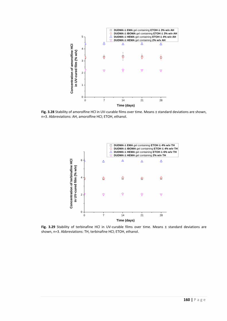
160 | P a g e
0 7 14 21 28
0
1
2
3
4
5
DUDMA & EMA gel containing ETOH & 3% w/v AH
DUDMA & IBOMA gel containing ETOH & 3% w/v AH
DUDMA & HEMA gel containing ETOH & 4% w/v AH
DUDMA & HEMA gel containing 2% w/v AH
Co
nc
en
tra
tio
n o
f a
mo
rolf
ine
HC
l
in
UV
-cu
red
fil
m (
% w
/v)
Time (days)
Fig. 3.28 Stability of amorolfine HCl in UV-curable films over time. Means ± standard deviations are shown, n=3. Abbreviations: AH, amorolfine HCl; ETOH, ethanol.
0 7 14 21 28
0
2
4
6
DUDMA & EMA gel containing ETOH & 4% w/v TH
DUDMA & IBOMA gel containing ETOH & 4% w/v TH
DUDMA & HEMA gel containing ETOH & 6% w/v TH
DUDMA & HEMA gel containing 2% w/v TH
Co
nc
en
tra
tio
n o
f te
rbin
afi
ne
HC
l
in U
V-c
ure
d f
ilm
(%
w/v
)
Time (days)
Fig. 3.29 Stability of terbinafine HCl in UV-curable films over time. Means ± standard deviations are shown, n=3. Abbreviations: TH, terbinafine HCl; ETOH, ethanol.

161 | P a g e
3.6 Conclusions
The DUMDA and EMA, IBOMA or HEMA UV-curable gels formulated (± drug and ±
solvent) produced films which were visually smooth, transparent and thin (with an
excellent uniformity of thickness).
These films were highly cross-linked as indicated by their low swelling in toluene, and
the extent of cross-linking was related to the films’ degree of conversion from monomer
gel to polymer film. The film’s interior was dense and did not vary with the choice of
diluent monomer or presence of ethanol and drug in the gel; however, the addition of
the solvent NMP or the use of a higher diluent monomer concentration in the gel
reduced this compact microstructure.
The concentration of drug that could be incorporated into the gel, as determined in
Chapter 2, was the maximum drug-load, and upon UV-curing, the drug remained in the
dissolved state and did not appear to interact with the polymer. The solvents
incorporated in the UV-curable gels resided in the cured film, although the amount of
ethanol residing was much less than NMP due to ethanol’s volatile nature.
The films produced from solvent-free UV-cured films displayed two glass transitions,
with the higher glass transition a possible consequence of a thermal reaction to
complete a previous “incomplete” cure. Films produced from formulations containing a
solvent showed one glass transition which was in the range of 95 – 140⁰C, and which
was influenced by the choice of diluent methacrylate monomer in the formulation and
solvent, but not the presence of drug.
All UV-cured films, with the exception of those containing NMP, showed good adhesivity
and water resistance in vitro. In vivo, the films also reduced TOWL and showed fairly
long residence times (during which period the drug remained stable in the film). The
adhesivity, TOWL reduction and nail residence displayed by UV-curable gels were
greater than that displayed by a pharmaceutical nail lacquer, and thus, a UV-curable gel
appears to be a promising drug delivery vehicle.

162 | P a g e
Chapter 4: UV-cured film’s drug release, ungual drug permeation
and efficacy against Trichophyton rubrum
4.1 Introduction
Drug release from an acrylic-based resin is feasible, via mechanisms such as (i) diffusion
through the acrylic resin, and/or (ii) surface release combined with drug diffusion via
cracks and channels in the resin formed by the incorporation of drug (Brook and van
Noort, 1985). Given that drug release from a topical nail medicine is an essential step
prior to drug permeation into and through the nail plate, the different UV-cured polymer
films’ drug release profiles are determined in this Chapter. Furthermore, the influence of
the film’s properties such as its structure, drug-load, glass transition and water
sensitivity (as determined in previous Chapters) on drug release is also determined to
understand the drug release mechanism from UV-cured films.
In addition to demonstrating drug release from the UV-cured film, the evaluation of
ungual and trans-ungual drug penetration is fundamental, as the success of the UV-
curable gel formulation as a topical nail medicine ultimately depends on its ability to
deliver the drug into and across the nail plate. Such an evaluation typically involves the
use of diffusion cells, (Franz or modified Franz diffusion cells), which consist of a donor
compartment, receptor compartment, and a nail plate in between the two chambers.
The donor compartment holds the drug-loaded formulation while the receptor
compartment is generally filled with a suitable solvent system to provide sink conditions,
i.e. where the concentration of the dissolved drug remains less than 10% of its
saturation solubility (Kenneth et al., 1981, Kobayashi et al., 1998, Kierstan et al., 2001,
Malhotra and Zatz, 2002a, Donnelly et al., 2005, Gunt and Kasting, 2007, Hui et al.,
2002). Due to the difficulty in obtaining intact human nail for such studies, several in
vitro models such as hoof membranes (cow, pig, sheep and horse) (Mertin and Lippold,
1997a, Mertin and Lippold, 1997b, Mertin and Lippold, 1997c, Monti et al., 2005, Monti
et al., 2010, Pittrof et al., 1992, Kim et al., 2001, Yang et al., 1997, Khengar et al., 2007),
keratin films (Lusiana et al., 2011), excised cadaver nail plates (Malhotra and Zatz,
2002a, Malhotra and Zatz, 2002b, Gunt et al., 2007, Gunt and Kasting, 2007, Nair et al.,
2011) and nail clippings (Chouhan and Saini, 2014, Hossin, 2015) have been used. While
163 | P a g e
human cadaver nail presents the closest simulation to human nail, nail clippings
obtained from volunteers can be considered an easily accessible and inexpensive
alternative. In this Chapter, the ungual drug permeation profiles of the different UV-
cured polymer films are determined using human nail clippings and modified Franz
diffusion cells designed in-house, and the relationship between drug release from the
film and drug permeation into and through the nail is explored.
While the in vitro ungual drug permeation study can provide insight into the feasibility of
in vivo drug delivery and can predict whether the amount of drug delivered will be at
therapeutic concentration against the fungal pathogen in question, a test to confirm the
antifungal efficacy of the formulation adds value to the findings. Experimental models
for evaluating the efficacy of formulations are available (Nair et al., 2013). An example is
the TurChub® model, which uses a modified Franz cell and where sections of human nail
are placed over an agar-filled receptor chamber (previously inoculated with
dermatophytes). In this model, the test formulation is placed over the nail plate and
incubated for a set duration at a set temperature. Subsequently, the extent of drug
permeation is approximated by measuring the zone of fungal growth inhibition in the
agar medium in the receptor compartment. Despite the availability of such experimental
models, there is currently no standard in vitro method to assess the anti-fungal efficacy
of anti-onychomycotic formulations. Therefore in this Chapter, the disc diffusion (Kirby-
Bauer) method of antibiotic susceptibility testing is adapted and investigated as a
method to test and compare the efficacies of the different antifungal-loaded UV-cured
polymer films (when applied on human nail clippings) against T. rubrum, which as
mentioned in Chapter 1, is the single most common fungus to cause onychomycosis
(Sigurgeirsson and Baran, 2014). The UV-cured polymer film’s in vitro efficacy against T.
rubrum, alongside its drug release and ungual drug permeation profiles, is also
compared against a commercially available pharmaceutical nail lacquer – Curanail® – to
further assess the UV gel’s potential.

164 | P a g e
4.2 Aims
Determine the UV-cured films’:
(i) drug release
(ii) ungual drug permeation
(iii) in vitro antifungal efficacy against T. rubrum
and the influence of drug and monomer compositions on the above properties.
4.3 Materials
The monomers, photoinitiator, antifungal agents, solvents, acids, UVA nail lamp and nail
wipes were obtained as detailed in Chapter 2 (Section 2.3). Curanail® (5% w/v
amorolfine hydrochloride) nail lacquer was purchased as detailed in Chapter 3 (Section
3.3). Sodium phosphate monobasic dihydrate, sodium phosphate dibasic heptahydrate
and a dialysis tubing cellulose membrane (MW 10281) were purchased from Sigma–
Aldrich (Dorset, UK). Human nail clippings (fingernails) were obtained from healthy
volunteers aged between 18 and 65 years. Sabouraud dextrose agar was purchased
from Oxoid Ltd (Basingstoke, UK).
4.4 Isolate
One dermatophyte strain, Trichophyton rubrum (CBS 118892), which was obtained from
Birkbeck, University of London was used.
4.5 Methods
The UV-cured films produced from formulations containing DUDMA and a diluent
reactive monomer (EMA, IBOMA or HEMA) at a ratio of 85:15% v/v (with ethanol and
with and without amorolfine HCl or terbinafine HCl), as well as the formulations
containing DUDMA and HEMA at a ratio of 75:25% v/v (with and without amorolfine HCl
or terbinafine HCl) were prepared as described in earlier Chapters (Chapter 3, Section
3.4 and Chapter 2, Section 2.4.4). These films’ drug release profiles, ungual drug
permeation profiles and efficacies against T. rubrum were determined as detailed below.
Unless otherwise stated, each test was conducted in triplicate.

165 | P a g e
4.5.1 Drug release from UV-cured polymer films
The drug release experiments were carried out using Franz diffusion cells. The receptor
fluid was a 0.1 M pH 5 phosphate buffer solution, as both amorolfine HCl and terbinafine
HCl are stable at this pH (Hossin, 2015). Furthermore, the saturation solubilities of
amorolfine HCl and terbinafine HCl in the pH 5 phosphate buffer solution are 9.27 ± 0.67
mg/ml and 7.91 ± 0.67 mg/ml respectively, and its use ensures that sink conditions were
maintained throughout the duration of the study. Finally, it should be noted that both
amorolfine HCl (pKa 6.6) and terbinafine HCl (pKa 7.1) are weak acids, and both are
expected to be unionised in an aqueous environment at pH 5. For these experiments,
the film produced from the Curanail® nail lacquer was used as a control.
To set up the Franz cell, a UV-cured film (circular with an area of 1.8 cm2) was placed
between the donor and receptor compartments of a Franz diffusion cell, with the
bottom surface facing the receptor side, and the compartments were clamped together.
For the Curanail® nail lacquer, a semi-permeable membrane was needed to support the
film. A cellulose membrane was therefore cut into a circle (with an area of 4.909 cm2)
and the lacquer was applied onto the surface covering a circular area of 0.9503 cm2. This
was allowed to dry and the cellulose support with the air-dried Curanail film was then
placed between the donor and receptor compartments of a Franz diffusion cell, with the
test films facing the donor side, and the compartments were clamped together. Upon
the assembly of the Franz diffusion cell, 4 ml of receptor fluid was added to the receptor
compartment while ensuring that no air bubbles were introduced. A schematic of this
set up can be seen in Fig. 4.1. The diffusion cells were immediately left to stir on a
magnetic stirrer placed in a water bath maintained at 32⁰C. Samples of the receptor fluid
were collected at pre-determined time intervals (i.e. every 20 minutes for the first 1
hour, then every 30 minutes for the second hour, then every hour for the following four
hours, then daily till day 12 and finally every three days till day 30). At each sampling
time, 0.5 ml of the receptor phase was collected via the receptor arm and replaced with
0.5 ml of fresh buffer. The samples were analysed by HPLC (as per Section 2.4.6 [Chapter
2]) to determine the amount of drug released, and the cumulative % drug release over
time was plotted.

166 | P a g e
Fig. 4.1 Franz diffusion cell set up for release studies. The cellulose membrane was only used to support the Curanail® film. UV-cured films did not need any support.
4.5.1.1 Mass & polarised light microscopy examination of polymer films
All the films were weighed and observed by polarised light microscopy (as per Section
3.4.5.1 [Chapter 3]) prior to the release study and at day 30 (end of release study) to
determine whether there was any film swelling or drug crystallisation during the release
study.
4.5.1.2 Mathematical modelling of drug release
In order to describe drug release kinetics, three commonly used mathematical models
for the characterisation of release from thin films were considered:
(i) Zero order:
Where drug release takes place at a constant rate, independent of its
concentration. Modelling of the drugs’ release using zero-order kinetics was
carried out by plotting cumulative amount of drug released over time, and then
calculating the linear regression coefficient value (R²). The data was considered
to fit a zero order profile if the R² value was equal to 1.

167 | P a g e
(ii) First order:
Where the drug release rate is dependent on drug concentration. Modelling of
the drugs’ release using first order kinetics was carried out by plotting log
(cumulative percentage of drug remaining) over time. The data was considered
to fit first order kinetics if the R² value was equal to 1.
(iii) Higuchi model:
Where the release rate through the film matrix is diffusion-controlled. The
release profile of the drug from the polymer film was considered to fit this model
if plotting cumulative % drug release against √𝑡𝑖𝑚𝑒 yielded a R² value equal to 1.
4.5.2 Determination of ungual drug permeation
The ungual drug permeation experiments were carried out using modified Franz
diffusion cells, as depicted in Fig. 4.2, and with the Curanail® nail lacquer used as a
control. Human fingernail clippings were used as the nail model as opposed to toenails,
as volunteers were more willing to grow and provide fingernails longer than 3 mm than
their toenails. These fingernail clippings were washed with water and soaked in distilled
water for one hour prior to use. They were then cut to size (circular, with a diameter of
0.3 cm) and measured for thickness using a micrometre. A layer of the formulation
(using 2 µl) was applied on the nail surface with a pipette tip (while ensuring that a 0.2
mm formulation-free perimeter was left near the edge of the nail surface). In the case of
Curanail®, the nail lacquer was applied and allowed to dry. For the UV-curable gel
formulations, the formulation was applied on the nail surface and cured under an UVA
lamp for 2 minutes. The surface of the UV-cured film was then wiped with propan-2-ol
using a nail wipe. The nail was placed in the donor compartment and fixed into place,
such that when assembled with the receptor compartment, the under-surface of the nail
alone was exposed to the receptor fluid. The area exposed was 0.025 cm2. Subsequently,
900 µl of receptor fluid was added to the receptor compartment. The receptor fluid was
a 0.1 M pH 5 phosphate buffer solution. The diffusion cell was assembled, the receptor
arm opening was covered with parafilm, and left to stir on a magnetic stirrer placed in a
water bath at 32⁰C. Samples of the receptor fluid were collected every three days, up to
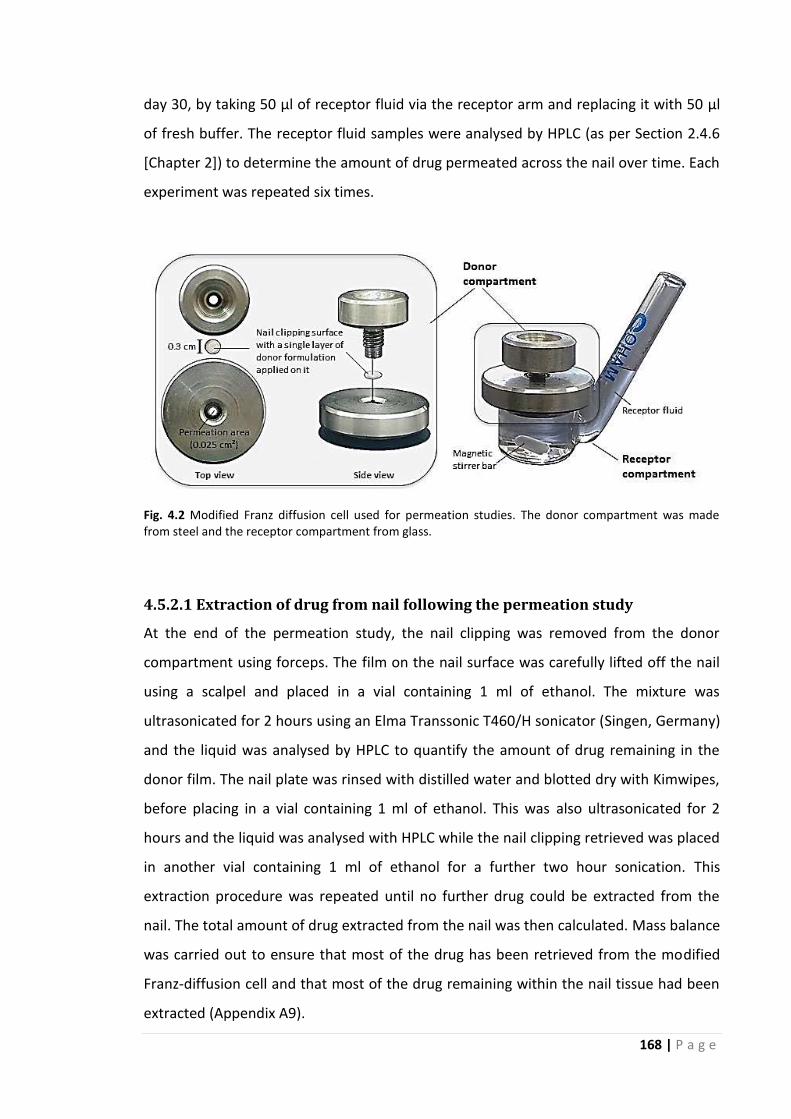
168 | P a g e
day 30, by taking 50 µl of receptor fluid via the receptor arm and replacing it with 50 µl
of fresh buffer. The receptor fluid samples were analysed by HPLC (as per Section 2.4.6
[Chapter 2]) to determine the amount of drug permeated across the nail over time. Each
experiment was repeated six times.
Fig. 4.2 Modified Franz diffusion cell used for permeation studies. The donor compartment was made from steel and the receptor compartment from glass.
4.5.2.1 Extraction of drug from nail following the permeation study
At the end of the permeation study, the nail clipping was removed from the donor
compartment using forceps. The film on the nail surface was carefully lifted off the nail
using a scalpel and placed in a vial containing 1 ml of ethanol. The mixture was
ultrasonicated for 2 hours using an Elma Transsonic T460/H sonicator (Singen, Germany)
and the liquid was analysed by HPLC to quantify the amount of drug remaining in the
donor film. The nail plate was rinsed with distilled water and blotted dry with Kimwipes,
before placing in a vial containing 1 ml of ethanol. This was also ultrasonicated for 2
hours and the liquid was analysed with HPLC while the nail clipping retrieved was placed
in another vial containing 1 ml of ethanol for a further two hour sonication. This
extraction procedure was repeated until no further drug could be extracted from the
nail. The total amount of drug extracted from the nail was then calculated. Mass balance
was carried out to ensure that most of the drug has been retrieved from the modified
Franz-diffusion cell and that most of the drug remaining within the nail tissue had been
extracted (Appendix A9).

169 | P a g e
4.5.2.2 Calculating the steady-state flux, permeability coefficient, lag time &
diffusion coefficient
The steady-state flux (J) was calculated from the slope of the linear portion of the plot of
the cumulative amount of antifungal drug permeated across the nail (µg/cm2) (Q)
against time (t):
J = ΔQ/Δt
The apparent permeability coefficient (P) which is defined as the flux divided by the
concentration of the permeant in the donor compartment (Cd) was calculated using:
P = J/Cd
The transport lag time (tL) was estimated by extrapolating the linear portion of the Q
versus t plot to the x-axis, (i.e. the x-intercept), and the effective diffusion coefficient
(Deff) was obtained from the transport lag time by the relationship:
tL= h2/6Deff
where h represents the thickness of the nail plate.
4.5.3 Determination of the antifungal efficacy of UV-curable films against T.
rubrum
The disc diffusion method of antibiotic susceptibility testing was adapted by replacing
the ‘disc’ with a circular piece of nail clipping onto which the formulation was applied,
such that the drug would have to be permeate into and through the nail plate to reach
the agar gel.
4.5.3.1 Preparation of media
Sixty five grams of Sabouraud Dextrose Agar (SDA) was dissolved in 1000 ml of distilled
water and sterilised by autoclaving at 1210C for 15 minutes. Subsequently approximately
25 ml of agar was poured into 90 mm petri dishes and allowed to solidify overnight.
4.5.3.2 Preparation of test plates
A culture plate of T. rubrum was used to inoculate the test plates. Firstly, a blank SDA
plate was labelled with the name of the organism and the date. A cork borer with an
internal diameter of 8 mm was then dipped in ethanol, flamed to sterility, and punched

170 | P a g e
into the centre of the SDA plate to punch out a plug of agar. This agar plug was
discarded. The cork borer was dipped once again in ethanol, flamed to sterility, and used
to punch a T. rubrum plug from the culture plate. This T. rubrum plug was inserted into
the slot created from the removal of the agar plug in the blank SDA plate. Twenty five
SDA plates were set up in this way and incubated at 32⁰C for three days to ensure that
the fungal culture was able to grow sufficiently and to identify defective plates. During
this period, the T. rubrum culture grew to 10 mm in diameter.
4.5.3.3 Preparation of nails
Human nail clippings (fingernails) were washed with water prior to use. They were then
cut to size (circular, with a diameter of 3 mm) and autoclaved at 1200C for 20 minutes in
order to sterilise them. This was possible as it was found that the process of autoclaving
did not affect the solvent (water & ethyl acetate) uptake properties of the nail clippings
(Appendix A10). The formulations tested were:
(i) Curanail® nail lacquer (containing 5% w/v amorolfine HCl)
(ii) DUDMA and EMA gel containing ethanol and 3% w/v amorolfine HCl
(iii) DUDMA and IBOMA gel containing ethanol and 3% w/v amorolfine HCl
(iv) DUDMA and HEMA gel containing ethanol and 4% w/v amorolfine HCl
(v) DUDMA and HEMA gel containing 2% w/v amorolfine HCl
(vi) DUDMA and HEMA gel containing ethanol and 6% w/v terbinafine HCl.
The drug-free counterparts of the above formulations (except for the Curanail® nail
lacquer) were also tested to examine the influence, in any, of the UV-cured polymer
vehicle against T. rubrum.
To prepare the nail clippings for placement on the SDA plates containing the fungal plug,
a layer of the formulation (using 2 µl) was applied on the nail surface in the same
manner as that for the ungual drug permeation studies described above in Section 4.5.2
(while ensuring that a 0.2 mm formulation-free perimeter was left near the edge of the
nail surface).

171 | P a g e
4.5.3.4 Testing of formulations
In order to test the UV-curable gel formulations’ efficacy against T. rubrum, the SDA
plates containing the fungal plugs were set up in one of three ways on day 3 as depicted
in Fig 4.3.
The first set up (A) was a formulation-free negative control to monitor the growth of T.
rubrum under the same experimental conditions and compare it with the plates
containing test formulations (a total of two plates were set up in this way).
The second set up (B) was a control containing nail clippings with drug-free polymer
films cured on the surface. The plates were set up by firstly labelling it with the
formulation to be tested, marking it into three segments, and marking a distance of 20 –
25 mm from the centre of the plate in the three segments for the nail clippings to be
placed. This distance was chosen to allow sufficient time for the permeant to penetrate
into and through the nail, a judgement based on the results obtained from the ungual
drug permeation studies (Section 4.6.2) and T. rubrum growth rates from preliminary
studies (Appendix A11). A sterile tweezer was used to pick up the prepared nail
clippings, which were carefully placed onto the agar surface (with the under surface of
the nail in contact with the agar). Each plate contained a triplicate set of the same
formulation.
The third set up (C) contained the samples (i.e. three nail clippings with the drug-loaded
polymer films cured on the surface) and one nail clipping with the corresponding drug-
free polymer film cured on the surface. To set these plates up, the plates were labelled
with the formulation to be tested and marked into four segments, one for the drug-free
formulation (control) and the remaining three for a triplicate set of the same drug-
loaded formulation. The nail clippings were placed onto the agar surface as described
above.
These plates were incubated at 32⁰C for as long as it took the T. rubrum culture to grow
over the entire SDA plate’s surface (or when no further growth was detected). During
this period, the plates were observed daily to monitor the growth of T. rubrum and to
monitor the formation of a zone of inhibition, if any, around the samples.

172 | P a g e
Fig. 4.3 Photographic images of T. rubrum inoculated test plates set up on day 0 and day 3. The formulation in this case is the DUDMA & EMA gel formulations containing ethanol (with and without 3% w/v amorolfine HCl). Abbreviations: AH, amorolfine HCl; C, control; ETOH, ethanol.

173 | P a g e
4.5.4 Statistical analyses
Statistical calculations were conducted using IBM SPSS 22. The Student’s t-test was
carried out for testing differences, if any, between two data sets and the one-way
ANOVA followed by post hoc Tukey were carried out for all statistical analyses involving
more than two data sets. Repeated measures ANOVA was conducted to determine
whether there were differences in drug release profiles and ungual drug permeation
profiles of the formulations tested over the experimental duration.

174 | P a g e
4.6 Results and discussion
4.6.1 Drug release from UV-cured polymer films and from the control
Curanail®
The drug release profiles of the amorolfine HCl–loaded and terbinafine HCl–loaded films
are shown in Fig. 4.4 and Fig. 4.5 respectively. The polarised light microscopy images of
the films pre- and post-release and the amount of drug released by day 30 are also
shown alongside the drug release profiles.
4.6.1.1 Drug release profile of Curanail®
From Curanail’s drug release profile (Fig. 4.4), a burst of drug release from the film over
the first 24 hours is evident due to the release of drug found at the film surface. After
this, the film gradually releases almost all of the incorporated drug. The polarised light
micrograph of the Curanail® film pre-release shows that it contained precipitated
amorolfine HCl crystals, (most probably as a result of its high drug-load of 31.5 ± 0.8%
w/w following solvent evaporation and film formation), and it appears that these may
have gradually dissolved and released, as no crystals were seen in the “tacky” film
remains at the end of the study. The fact that an intact film was not retrieved at the end
of the release study suggests that Curanail’s polymer backbone, (i.e. ammonio
methacrylate copolymer A), may have gradually swelled and partially disintegrated
during the 30 day study, thereby enabling the gradual and almost complete drug release
observed.
4.6.1.2 Drug release profiles of amorolfine HCl–loaded UV-cured polymer films
The amorolfine HCl–loaded UV-cured films’ release profiles (Fig. 4.4) also displayed a
burst of drug release over the first 24 hours due to the release of drug found at the
surface of the film. However, after this point, the drug release slowed and eventually
plateaued. Only part of the incorporated drug (42 – 48 %) had been released by the end
of the study, with no significant differences amongst the different UV-curable gel
formulations (p>0.05). Visually, the films remained intact after the release study, and the
negligible weight change of the films during the release experiment (Fig. 4.6) confirmed
the absence of film swelling and dissolution. In addition, polarised light micrographs of
the films (pre- and post-release) showed no obvious changes, i.e. any signs of drug
precipitation following the release study (Fig. 4.4). This enabled the evaluation of

175 | P a g e
amorolfine HCl’s release kinetics from the UV-cured films using zero order, first order
and Higuchi models. From Table 4.1, it can be seen that the Higuchi model best
describes amorolfine HCl release from the four amorolfine HCl-loaded UV-cured films, as
it yielded the highest R2 value. Therefore it appears that the drug released through the
film matrix following the initial burst phase is diffusion-controlled. However, this release
was minimal and eventually halted most probably due to the films’ highly cross-linked
nature and dense interior (Chapter 3, Section 3.5.1.2), both of which can limit the
diffusion of drug molecules through the film.
4.6.1.3 Drug release profiles of terbinafine HCl–loaded UV-cured polymer films
In a similar manner to the amorolfine HCl–loaded UV-cured polymer films, the
terbinafine HCl–loaded films release profiles (Fig. 4.5) showed a burst phase over the
first 24 hours, which then slowed and eventually plateaued. Once again, no significant
differences were seen amongst the different gel formulations (p>0.05). However, the
percentage of drug released by the end of the study from all four formulations was
significantly lower (p<0.05) than the same vehicle containing amorolfine HCl. Like the
amorolfine HCl–loaded films, the terbinafine HCl–loaded films remained intact, showed
negligible weight change (Fig. 4.6) and showed no signs of drug precipitation post-
release (Fig. 4.5), which did not offer any positive clues to explain the lower terbinafine
HCl release. Table 4.1 reveals that the Higuchi model best describes terbinafine HCl
release from all four UV-cured films, and therefore it appears that the drug released
through the film matrix following the initial burst release is also diffusion-controlled and
eventually limited by the films cross-linked nature and dense film interior. Both
amorolfine HCl and terbinafine HCl have similar molecular weights (353.98 and 327.90
Da respectively), similar logP values (5.8 and 5.3 respectively), and the DUDMA & EMA,
IBOMA or HEMA copolymer film’s structure (Chapter 3, Section 3.5.1.2), glass transition
temperature (Chapter 3, Table 3.10) and water sensitivity (Chapter 3, Fig. 3.23) was
unaffected by the incorporation of amorolfine HCl or terbinafine HCl; hence these drug
and film characteristics do not explain why terbinafine release is almost half that of
amorolfine. A low drug release could occur if terbinafine was incorporated in the
polymer structure upon UV-curing. Terbinafine does have the potential to take part in
the polymerisation process due to its alkene group as mentioned in the previous
176 | P a g e
Chapter (Section 3.5.1.2). However as most of the drug included in the gel mixture could
be extracted from the UV-cured film (just as amorolfine HCl could be) (Chapter 3, Table
3.8), this is an unlikely occurrence. It is possible that terbinafine may have therefore
bound to the polymer films, which would retard its release.
While FT-IR revealed the absence of drug-polymer interactions (Chapter 3, Table 3.9), an
alternative experiment to investigate terbinafine binding or not would shed light on
whether terbinafine does have a higher affinity for the UV-cured film compared to
amorolfine. Therefore for future work, the affinity of the two antifungal drugs to the UV-
cured films could be tested by adapting the experimental procedure used for
determining the affinities of these drugs to keratin (Tatsumi et al., 2002, Sugiura et al.,
2014). In this instance a drug-free UV-cured polymer film could be immersed in a drug
solution and left shaking. Subsequently, at intervals, the drug concentration in this
mixture could be determined to calculate the rate of drug–polymer film binding. When
no further drug-polymer film binding is evident, the UV-cured polymer film with bound
drug could be washed with a solvent and the drug concentration in the solvent could be
measured. This washing procedure could be repeated several times using fresh solvent
each time to determine the rate of release of drug from the UV-cured film, and confirm
if the drug does have a high affinity for the polymer or not.
4.6.1.4 Influence of film components on drug release
The similarities in the drug release profiles of the UV-cured films produced from DUDMA
& EMA, IBOMA or HEMA containing gels (with the DUDMA to reactive diluent monomer
ratio set at 85:15% v/v) could be due to their similar degree of conversion from
monomer gel to polymer film (Chapter 2, Table 2.12), similar extents of cross-linking
(Chapter 3, Section 3.5.1.2), comparable microstructures (Chapter 3, Table 3.6) and
comparable water sensitivities (Chapter 3, Fig. 3.23). In addition, the film produced from
the DUDMA & HEMA gel (with a DUDMA to HEMA ratio of 75:25% v/v) appears to show
a slightly higher percentage of drug release compared to the other formulations,
possibly because of its lower degree of conversion and hence lower extent of cross-
linking. In fact, linear regression analysis revealed a strong correlation between degree
of conversion from monomer gel to polymer film and drug release (R2 = 0.920, p<0.05)

177 | P a g e
for amorolfine HCl and (R2= 0.938, p<0.05) for terbinafine HCl. On the other hand, the
differences in the glass transition temperatures of the different UV-cured polymer films
(Chapter 3, Table 3.10) was not found to influence drug release from the films (p>0.05).
While a higher glass transition temperature of a film can indicate a more rigid structure
and can decrease the rate of drug diffusion (Jenquin et al., 1992), in this instance it
appears that the differences in glass transition temperatures did not play a role in drug
release.
4.6.1.5 Amount of drug released by day 30
When considering the amount of drug released by the end of the study, it can be seen
that the Curanail® film releases the highest amount of drug due to its greatest
percentage of drug release and highest initial drug-load. Meanwhile, for the UV-cured
polymer films (which contained the maximum amount of drug that could be loaded in
the dissolved state in each film), the DUDMA & HEMA gel containing ethanol and 4%
w/v amorolfine HCl has the greatest amount of drug released (p<0.05) (due to its higher
drug-load) compared to the other gel formulations, which show similar amounts of drug
release (p>0.05). Similarly, the DUDMA & HEMA gel containing ethanol and 6% w/v
terbinafine HCl has the greatest amount of drug released (p<0.05) (also due to its higher
drug-load) compared to the other terbinafine HCl–loaded gel formulations. The drug
concentration in the film (which is influenced by the nature of the reactive diluent
monomer used and the inclusion of solvent) is therefore an important parameter
governing the extent of drug release. Whether the percentage of drug and/or amount of
drug released influences the permeation of the drug across the nail plate is explored in
the next section.
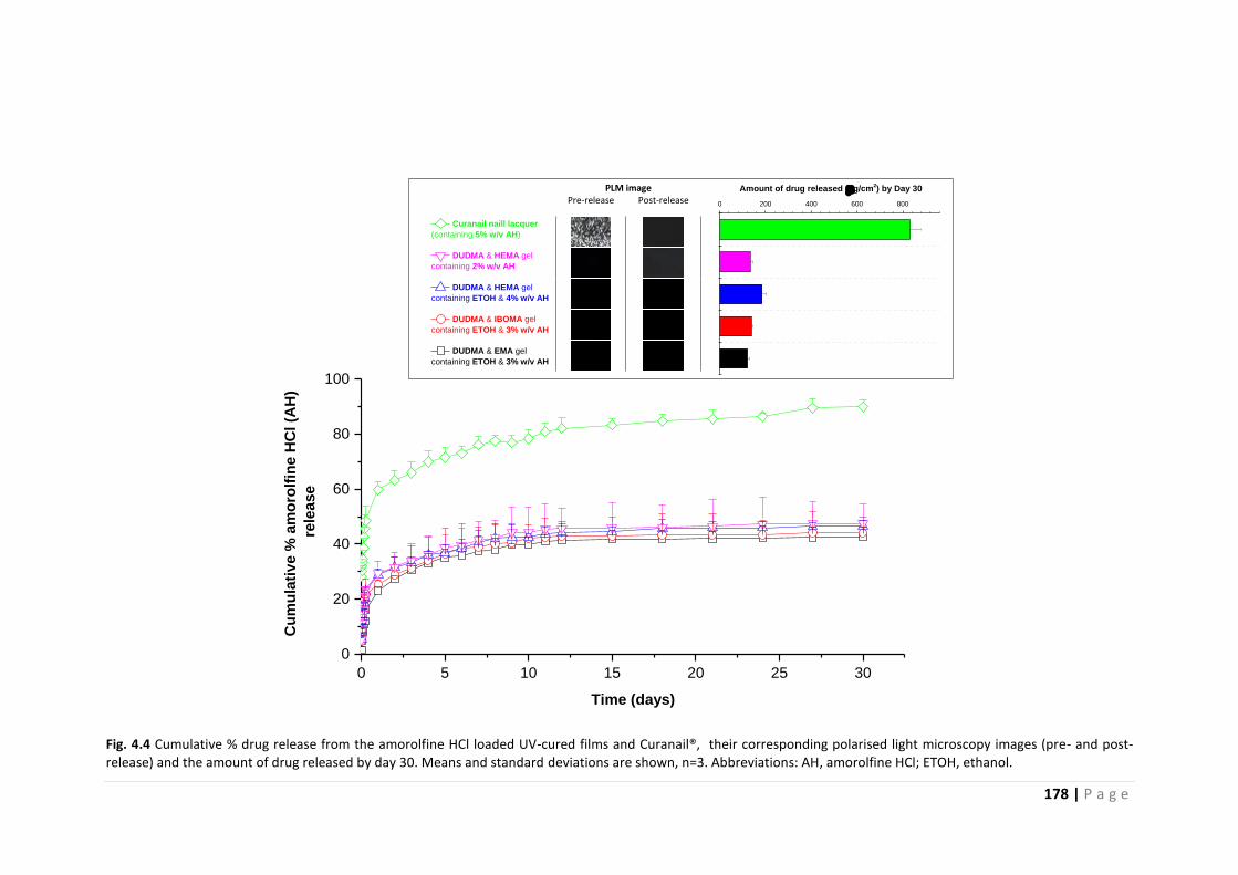
178 | P a g e
0 5 10 15 20 25 30
0
20
40
60
80
100
PLM image Pre-release Post-release
Curanail naill lacquer
(containing 5% w/v AH)
DUDMA & HEMA gel
containing 2% w/v AH
DUDMA & HEMA gel
containing ETOH & 4% w/v AH
DUDMA & IBOMA gel
containing ETOH & 3% w/v AH
DUDMA & EMA gel
containing ETOH & 3% w/v AH
Cu
mu
lati
ve %
am
oro
lfin
e H
Cl (A
H)
rele
ase
Time (days)
0 200 400 600 800
Amount of drug released (g/cm2) by Day 30
Fig. 4.4 Cumulative % drug release from the amorolfine HCl loaded UV-cured films and Curanail®, their corresponding polarised light microscopy images (pre- and post-release) and the amount of drug released by day 30. Means and standard deviations are shown, n=3. Abbreviations: AH, amorolfine HCl; ETOH, ethanol.

179 | P a g e
0 5 10 15 20 25 30
0
5
10
15
20
25
30
35
PLM image Pre-release Post-release
0 20 40 60 80 100 120 140 160
Amount of drug released (g/cm2) by Day 30
Cu
mu
lati
ve %
te
rbin
afi
ne
HC
l (T
H)
rele
as
e
Time (days)
DUDMA & HEMA gel
containing 2% w/v TH
DUDMA & HEMA gel
containing ETOH & 6% w/v TH
DUDMA & IBOMA gel
containing ETOH & 4% w/v TH
DUDMA & EMA gel
containg ETOH & 4% w/v TH
Fig. 4.5 Cumulative % drug release from the terbinafine HCl loaded UV-cured films, their corresponding polarised light microscopy images (pre- and post-release) and the amount of drug released by day 30. Means and standard deviations are shown, n=3. Abbreviations: TH, terbinafine HCl; ETOH, ethanol.

180 | P a g e
-3.0 -2.5 -2.0 -1.5 -1.0 -0.5 0.0 0.5 1.0 1.5 2.0 2.5 3.0
% weight change
DUDMA & EMA gel containing ETOH & 3% w/v AH
DUDMA & EMA gel containing ETOH & 4% w/v TH
DUDMA & IBOMA gel containing ETOH & 3% w/v AH
DUDMA & IBOMA gel containing ETOH & 4% w/v TH
DUDMA & HEMA gel containing ETOH & 4% w/v AH
DUDMA & HEMA gel containing ETOH & 6% w/v TH
DUDMA & HEMA gel containing 2% w/v AH
DUDMA & HEMA gel containing 2% w/v TH
Fig. 4.6 Weight change (%) for the drug-loaded UV-cured films produced from DUDMA & EMA, IBOMA or
HEMA containing gels following drug-release studies. Means and standard deviations are shown, n=3.
Abbreviations: AH, amorolfine HCl; TH, terbinafine HCl; ETOH, ethanol.
Table 4.1 R² values obtained for drug release modelling using zero order, first order and Higuchi models.
Formulation R2 value obtained for
Zero order First order Higuchi model
DUDMA & EMA gel containing ETOH & 3% w/v AH 0.85 0.88 0.96
DUDMA & EMA gel containing ETOH & 4% w/v TH 0.89 0.90 0.98
DUDMA & IBOMA gel containing ETOH & 3% w/v AH 0.76 0.81 0.89
DUDMA & IBOMA gel containing ETOH & 4% w/v TH 0.90 0.91 0.98
DUDMA & HEMA gel containing ETOH & 4% w/v AH 0.75 0.79 0.89
DUDMA & HEMA gel containing ETOH & 6% w/v TH 0.90 0.92 0.98
DUDMA & HEMA gel containing 2% w/v AH 0.75 0.79 0.88
DUDMA & HEMA gel containing 2% w/v TH 0.86 0.87 0.97

181 | P a g e
4.6.2 Ungual drug permeation profiles of UV-cured polymer films and
Curanail®
The ungual drug permeation profiles are shown in Fig. 4.7 and Fig. 4.8 for amorolfine
HCl– and terbinafine HCl– loaded films respectively. The % of drug permeated across the
nail and remaining in the nail at day 30 is also shown alongside the permeation profiles,
while the calculated lag times, study-state flux, apparent permeability coefficients,
effective diffusion coefficients and amount of drug in nail clippings at the end of the
experiment are shown in Table 4.2.
4.6.2.1 Amorolfine HCl–loaded UV-cured polymer films
From Fig. 4.7, when considering the UV-curable gel formulations, it can be seen that the
film produced from the DUDMA & HEMA gel containing ethanol and 4% w/v amorolfine
HCl displays greater ungual drug permeation (p<0.05) compared to the films produced
from the DUDMA & EMA or IBOMA gels containing ethanol and 3% w/v amorolfine HCl,
which were similar (p>0.05), and that these in turn display greater ungual drug
permeation profiles (p<0.05) compared to the film produced from the DUDMA & HEMA
gel containing 2% w/v amorolfine HCl. This indicates that the drug-load in the gel (which
as mentioned earlier is influenced by the nature of the reactive diluent monomer used
and the inclusion of solvent) is directly related to the extent of ungual drug permeation.
A more detailed examination of the profiles reveals fairly lengthy lag times, which is
typical of nail permeation studies (McAuley et al., 2016). A lag time of approximately 10
days was obtained for all the films apart from the DUDMA & HEMA film (containing 4%
w/w amorolfine HCl), which had a shorter time of approximately 7 days, possibly due to
its higher drug-load and hence greater concentration gradient between the donor and
the receptor phases. The lag period was followed by a linear increase in the cumulative
amount of drug permeated. The ungual drug flux in this region, i.e. steady-state flux,
was similar for all films (2.2 – 2.4 µg/cm2/day), apart from the DUDMA & HEMA film
containing 2% w/w amorolfine HCl, for which it was significantly lower (p<0.05). This
could be due to its lower drug-load compared to the other formulations and hence a
lower drug concentration gradient between the donor and the receptor phases.
Towards the end of the permeation study, the ungual drug permeation appears to slow
down. In all cases less than 10% of the applied dose had penetrated into and through
182 | P a g e
the nail suggesting infinite dosing kinetics, and therefore a slowing down in permeation
was not expected as this would suggest violation of sink conditions and/or the presence
of an air bubble under the membrane (Williams, 2003), in this case the nail plate.
However, sink conditions were maintained and no air bubbles were evident throughout
the study, therefore these factors do not explain the observation. This suggests that
perhaps amorolfine HCl’s high affinity to nail keratin (Tatsumi et al., 2002) eventually
leads to an increased amount of drug bound to keratin, thus retarding further
permeation.
4.6.2.2 Terbinafine HCl–loaded UV-cured polymer films
In a similar manner to the amorolfine HCl–loaded formulations, the drug-load in the UV
gel governs the ungual drug permeation patterns of the terbinafine HCl–loaded
formulations (Fig. 4.8). Thus, the film produced from the DUDMA & HEMA gel containing
ethanol and 6% w/v terbinafine HCl displayed greater ungual drug permeation (p<0.05)
compared to the films produced from the DUDMA & EMA or IBOMA gels containing
ethanol and 4% w/v terbinafine HCl, which were similar (p>0.05), and these in turn
displayed greater ungual drug permeation profiles (p<0.05) compared to the film
produced from the DUDMA & HEMA gel containing 2% w/v terbinafine HCl. Once again
the DUDMA & HEMA film (containing the higher drug-load, i.e. 6% w/w terbinafine HCl)
has a shorter lag time, and the steady-state flux was similar for all films (1.4 – 1.6
µg/cm2/day) apart from the DUDMA & HEMA film (containing 2% w/w terbinafine HCl),
for which it was significantly lower (p<0.05), again due to its lower drug-load and hence
a lower drug concentration gradient between the donor and the receptor phases.
Towards the end of the permeation study, the ungual drug permeation also appears to
slow down, this time in response to possible terbinafine–nail keratin binding, a well-
known phenomenon (Tatsumi et al., 2002). For the UV-cured films, it appears that most
of the drug remains in the film and a very small proportion enters the nail plate and
eventually the receptor medium. This very low ungual permeation reflects previous
reports in vitro and in man. For example, van Hoogdalem et al. (1997) showed that less
than 0.2% of the applied dose permeated fingernails after twice daily application for 6
weeks in a human volunteer study.

183 | P a g e
4.6.2.3 Amorolfine HCl–loaded vs. terbinafine HCl–loaded UV-cured polymer
films
Compared to the amorolfine HCl–loaded gel formulations, the terbinafine formulations
show comparable lag times and diffusion coefficients (p>0.05). This could be attributed
to the small difference in the molecular weights of the two drugs being insignificant for
altering their passage through the nail plate. However, the amorolfine HCl–loaded gel
formulations resulted in a significantly higher (p<0.05) steady-state flux, permeability
coefficient and percentage of drug permeated across the nail at day 30. This may be
linked to the greater amount of drug released from the amorolfine HCl–loaded films and
thereby available for ungual permeation compared to the terbinafine HCl–loaded films.
Nonetheless, the amount of drug extracted from the nail plate and the percentage of
drug found in the nail plate were similar for both drugs (p>0.05). Terbinafine has
previously been shown to bind more strongly to keratin compared to amorolfine
(Tatsumi et al., 2002), and this may explain terbinafine HCl–loaded films’ lower drug–in–
receptor phase compared to drug–in–nail. It also provides an alternative explanation for
the terbinafine HCl–loaded films’ lower steady-state flux, permeability coefficient, and
drug permeation percentage compared to amorolfine. Greater nail keratin binding by a
drug does not mean that it is less superior however. While drug-nail keratin binding has
been correlated to a reduction in a drug’s antifungal potency (Tatsumi et al., 2002), it
could also result in the creation of a drug reservoir in the nail, which could be beneficial
in the long run as the nail keratin can act as a drug depot releasing drug over time to (i)
kill the fungus and any newly-germinating fungal spore residing in the nail plate, and (ii)
permeate into, and therefore exert antifungal activity in, the nail bed.
4.6.2.4 UV-curable gels vs. Curanail® nail lacquer
In order to examine how the UV-curable gel formulations compared to a commercially
available nail medicine as a drug delivery vehicle, the Curanail® nail lacquer (which
contains 5% w/v amorolfine HCl) was compared with the DUDMA & HEMA gel
containing 4% w/v amorolfine HCl (the best of the amorolfine HCl-loaded gel
formulations). While the two vehicles displayed similar ungual drug permeation profiles
(p>0.05) and displayed similar permeability coefficients, diffusion coefficients and
amounts of drug in the nail clipping at day 30 (p>0.05), the Curanail® nail lacquer film

184 | P a g e
showed a slightly greater steady-state flux (p<0.05) probably due to its higher drug-load
(32% w/w vs. 4% w/w). However, the UV-cured film displayed a shorter lag time possibly
due to it greater occlusivity compared to the lacquer film (Chapter 3, Fig. 3.15), which is
expected to increase nail hydration and reduce the time taken for the drug to reach a
uniform concentration in the membrane, given that nail hydration is known to increase
drug diffusivity in the nail (Gunt and Kasting, 2007). Furthermore, despite the slightly
slower drug flux by the UV-cured film, a greater percentage of the drug permeated
across the nail and remained in the nail at day 30. This finding along with the lengthy lag
times and low ungual drug permeation obtained shows the critical role played by the
nail plate barrier to ungual drug permeation and the comparatively lower significance of
the film’s drug release. In fact, as the amount of drug released from the film into an
aqueous sink is greater than the amount permeated across the nail, correlations
between the two studies could not be made (Guy and Hadgraft, 1992). The Curanail®
nail lacquer’s lag time of approximately 10 days suggests that the lacquer must reside on
the nail plate surface during this period to allow sufficient drug penetration; however, its
in vivo nail residence profile (Chapter 3, Fig. 3.24) shows that by day 2 more than a
quarter of the lacquer film dislodges from the nail plate surface. Therefore in order to
show effect, its twice weekly application process will need to be followed rigorously. The
UV-curable gel formulations override the need for frequent applications, given their
longer in vivo residence (Chapter 3, Fig. 3.24); and as such appears to be a more
favourable form of topical nail medicine. However, whether the amount of drug
permeated is therapeutically effective against an onychomycosis-causing dermatophyte
or not still requires investigation, and this is explored in the next section.

185 | P a g e
0 5 10 15 20 25 30
0
5
10
15
20
25
30
35
40
45
50
0 1 2 3 4
% of AH permeated and in nail
% permeated
% in nail
Cu
mu
lati
ve a
mo
un
t o
f am
oro
lfin
e H
Cl (A
H)
perm
eate
d a
cro
ss t
he n
ail (
g/c
m2)
Time (days)
Curanail naill lacquer
(containing 5% w/v AH)
DUDMA & HEMA gel
containing ETOH & 4% w/v AH
DUDMA & IBOMA gel
containing ETOH & 3% w/v AH
DUDMA & EMA gel
containing ETOH & 3% w/v AH
DUDMA & HEMA gel
containing 2% w/v AH
Fig. 4.7 Cumulative amount of amorolfine HCl permeated across the nail with time from the UV-cured and Curanail® films, and the % of amorolfine HCl permeated across the nail and remaining in the nail at day 30. Means and standard deviations are shown, n=6. Abbreviations: AH, amorolfine HCl; ETOH, ethanol.

186 | P a g e
0 5 10 15 20 25 30
0
5
10
15
20
25
30
35
Cu
mu
lati
ve
am
ou
nt
of
terb
ina
fin
e H
Cl
(TH
)
pe
rme
ate
d a
cro
ss
th
e n
ail
(
g/c
m2)
Time (days)
DUDMA & HEMA gel
containing ETOH & 6% w/v TH
DUDMA & IBOMA gel
containing ETOH & 4% w/v TH
DUDMA & EMA gel
containg ETOH & 4% w/v TH
DUDMA & HEMA gel
containing 2% w/v TH
0.0 0.5 1.0 1.5 2.0 2.5 3.0 3.5 4.0
% permeated
% in nail
% of TH permeated and in nail
Fig. 4.8 Cumulative amount of terbinafine HCl permeated across the nail with time from the UV-cured films, and the % of terbinafine HCl permeated across the nail and remaining in the nail at day 30. Means and standard deviations are shown, n=6. Abbreviations: TH, terbinafine HCl; ETOH, ethanol.

187 | P a g e
Table 4.2 Lag time, steady-state flux, apparent permeability coefficient, effective diffusion coefficient and amount of drug in nail clippings. Means and standard deviations are shown, n=6. Abbreviations: AH, amorolfine HCl; TH, terbinafine HCl; ETOH, ethanol.
Amorolfine HCl (AH) Terbinafine HCl (TH)
% in formula-
tion (% w/v)
Lag time
(day)
Steady-state flux
(µg/cm2/day)
Permeability coefficient
x 10-5 (cm/day)
Diffusion coefficient
x 10-5 (cm2/day)
Drug in nail
clipping (mg/cm3)
% in formula-
tion (% w/v)
Lag time
(day)
Steady-state flux
(µg/cm2/day)
Permeability coefficient
x 10-5 (cm/day)
Diffusion coefficient
x 10-5 (cm2/day)
Drug in nail
clipping (mg/cm3)
Curanail® nail lacquer 5 10.4 ± 1.3 2.9 ± 0.2 5.8 ± 0.3 2.5 ± 0.5 0.6 ± 0.2 NA NA NA NA NA NA
DUDMA & EMA gel containing
ETOH & AH or TH
3 10.4 ± 0.9 2.4 ± 0.2 7.9 ± 0.7 2.7 ± 0.8 0.3 ± 0.2 4 9.5 ± 1.2 1.5 ± 0.1 3.7 ± 0.3 1.6 ± 0.4 0.9 ± 0.2
DUDMA & IBOMA gel containing
ETOH & AH or TH
3 10.5 ± 0.6 2.3 ± 0.3 7.8 ± 1.0 2.2 ± 0.4 0.4 ± 0.1 4 8.3 ± 0.8 1.4 ± 0.3 3.4 ± 0.8 3.1 ± 0.9 0.4 ± 0.1
DUDMA & HEMA gel containing
ETOH & AH or TH
4 7.1 ± 1.7 2.2 ± 0.3 5.5 ± 0.7 3.5 ± 1.4 0.6 ± 0.1 6 7.1 ± 1.0 1.6 ± 0.2 2.6 ± 0.4 3.0 ± 0.8 0.7 ± 0.2
DUDMA & HEMA gel containing
AH or TH
2 10.0 ± 1.9 1.4 ± 0.1 7.0 ± 0.7 3.2 ± 0.4 0.3 ± 0.2 2 8.9 ± 1.0 0.7 ± 0.1 3.5 ± 0.7 2.6 ± 0.9 0.3 ± 0.1

188 | P a g e
4.6.3 Antifungal efficacy of formulations against T. rubrum
The results of the adapted disc diffusion method developed to test the efficacies of the
different topical nail formulations (when applied on human fingernail clippings) against
T. rubrum are shown in Table 4.3.
As expected, the T. rubrum inoculated SDA plates used as a negative control (i.e.
formulation-free), allowed the T. rubrum culture to grow over the entire plate’s surface.
Similarly, T. rubrum inoculated SDA plates containing nail clippings with only drug-free
formulations cured on the surface (n=3) also showed fungal growth over the entire
plate’s surface. This shows that the UV-cured film vehicle is not antifungal itself. In
contrast, the T. rubrum inoculated SDA plates containing nail clippings with drug-loaded
formulations cured on the surface (n=3) showed no fungal growth for the duration of
the experiment following the introduction of the nail clippings. This suggests that the
low percentage of applied drug which permeated into and through the nail plate (Fig.
4.7 – 4.8), is therapeutically effective against the single most common fungus to cause
onychomycosis. It also suggests that the drug permeated through the nail plate may
have diffused into the agar, thus preventing the growth of the culture towards the nail
clippings containing the drug-free formulation on the same plate.
The MIC of amorolfine and terbinafine against T. rubrum has been reported to be 0.12-
0.5 µg/ml and 0.004–0.06 µg/ml respectively (Tamura et al., 2014), and the drug levels
achieved in the nail plate by the amorolfine HCl– and terbinafine HCl– loaded
formulations were considerably greater (Table 4.2); the demonstrated inhibition of
fungal growth by the formulations is therefore consistent with what would be expected
theoretically. While it can be argued that the MIC values determined in vitro in broth are
not expected to be directly applicable to MICs in the nail plate, and that the extent of
drug permeation and drug-load is expected to be significantly lower for toenails (which
are predominately affected) compared to fingernails (Nair et al., 2011), the drug levels
achieved in the nail are more than sufficient to cause fungal kill. The UV-cured gels are
therefore promising topical carriers for anti-onychomycotic drugs.
While the method developed was capable of yielding insightful qualitative data, due to
the absence of a zone of inhibition, it could not be used to deduce which of the

189 | P a g e
amorolfine HCl–loaded UV gel formulations tested was the ‘best’, i.e. facilitated better
ungual drug permeation, and whether this in turn correlated with the nail permeation
studies conducted. Similarly the amorolfine HCl–loaded DUDMA & HEMA gel (containing
ethanol) could not be compared with the terbinafine HCl–loaded DUDMA & HEMA gel
(containing ethanol) or with the Curanail® nail lacquer. A future study containing one
sample per T. rubrum inoculated SDA plate, (with the T. rubrum plug inserted at one end
of the petri dish and the sample placed at the other end after allowing the plug to grow
greater than 10 mm), may enable discrimination among the formulations or confirm
anti-fungal equivalence despite different ungual permeations.

190 | P a g e
Table 4.3 Photographic images (at day 30) of T. rubrum inoculated SDA plates containing nail clippings with drug-free and drug-loaded formulations cured on the surface. The corresponding drug-free formulation for the Curanail® nail lacquer is a formulation-free nail clipping (as a drug-free formulation could not be produced in the lab due to lack of knowledge regarding the quantity of excipients).
Formulation applied on
nail clipping surface
T. rubrum inoculated SDA plates containing nail clippings with drug-loaded formulations cured
on the surface (n=3) and the corresponding drug-free formulation cured on the surface (C)
T. rubrum inoculated SDA plates containing nail clippings with drug-free formulations cured on
the surface (n=3)
Top surface Under surface Top surface Under surface
None (nail clipping-
free & formulation-free control)
Curanail® nail lacquer
(containing 5% w/v AH)
DUDMA & EMA
gel containing ETOH ±
3% w/v AH
DUDMA & IBOMA
gel containing ETOH ±
3% w/v AH
DUDMA & HEMA
gel containing ETOH ±
4% w/v AH
DUDMA & HEMA
gel ± 2% w/v AH
DUDMA & HEMA
gel containing ETOH ±
6% w/v TH

191 | P a g e
4.7 Conclusions
The drug release profiles were similar for all the UV gels formulated. However, the
release of drug was limited (to less than 50%) possibly by the films’ cross-linked and
highly dense nature, and the release of terbinafine from the films was almost half that of
amorolfine due to possible terbinafine-polymer binding.
Permeation studies revealed that all formulations were capable of enabling drug
permeation into and through the nail in vitro. The drug-loaded DUDMA & HEMA gels
containing ethanol displayed the greater ungual drug permeation profile due to their
greater drug-load, and the amorolfine HCl–loaded DUDMA & HEMA gel containing
ethanol was in fact found to be comparable to the commercially available
pharmaceutical nail lacquer Curanail®. For the terbinafine HCl-loaded films, a lower
percentage of drug permeated across the nail compared to amorolfine HCl-loaded films,
most probably as terbinafine binds more strongly to keratin compared to amorolfine.
The amount of drug that permeated across the nail was sufficient to arrest the growth of
the fungus T. rubrum in vitro. However, differences among the formulations regarding
anti-fungal efficacy could not be discerned despite differences in ungual drug
permeations.
The UV-cured films are thus promising candidates for the topical treatment of
onychomycosis, and in the next Chapter, the incorporation of penetration enhancers are
considered for optimising the more superior of the UV-curable gels, i.e. DUDMA &
HEMA gels containing ethanol and either 4% w/v amorolfine HCl or 6% w/v terbinafine
HCl.

192 | P a g e
Chapter 5: Optimising UV-curable gel formulations with the use of
penetration enhancers
5.1 Introduction
The UV-curable gel formulations developed thus far contained the maximum amount of
drug that could be loaded in the dissolved state in each film. The gel’s maximum drug-
load depended on the drug’s solubility in ethanol and in the reactive diluent
methacrylate-based monomer. Thus the gels containing HEMA and ethanol dissolved
the most drug due to the greater drug solvency of HEMA (Chapter 2, Table 2.7). In the
previous Chapter, the films’ drug release and ungual drug permeation was the greatest
from the film with the highest drug-load, and hence the DUDMA & HEMA gels (with a
DUDMA to HEMA ratio of 85:15% v/v) containing ethanol and either 4% w/v amorolfine
HCl or 6% w/v terbinafine HCl were the superior of the formulations developed.
However the percentage of drug in the formulation that permeated across the nail and
remained in the nail was less than 2.7% and 3.6% respectively, following the 30 day
ungual drug permeation study using fingernail clippings as the nail model (Chapter 4,
Figs 4.7 – 4.8). Despite this, the amount of amorolfine and terbinafine that had
permeated across the fingernail clippings was sufficient enough to inhibit the growth of
T. rubrum, the most common onychomycosis-causing pathogen (Chapter 4, Table 4.3).
Nonetheless, as also mentioned in the previous Chapter (Section 4.6.3), the extent of
drug permeation is expected to be significantly lower for toenails (which are
predominated infected) compared to fingernails. These formulations are thus worth
considering for optimisation to overcome the poor permeability percentage and
guarantee treatment success.
In this Chapter, the incorporation of an ungual penetration enhancer in the
pharmaceutical UV gel is therefore considered for boosting nail permeability. A chemical
approach to enhance ungual drug permeation was considered as it does not add to the
formulation’s application process when the chemical is incorporated into the UV gel, and
in turn will not compromise patient compliance. The penetration enhancers considered
were 2-mepcaptoethanol (MPE), water, NMP and poly(ethylene glycol) 200 (PEG 200),
the chemical structures and molecular weights for which are shown in Table 5.1.
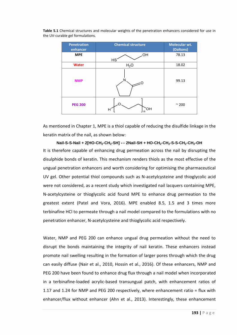
193 | P a g e
Table 5.1 Chemical structures and molecular weights of the penetration enhancers considered for use in the UV-curable gel formulations.
Penetration enhancer
Chemical structure Molecular wt. (Daltons)
MPE
78.13
Water
18.02
NMP
99.13
PEG 200
~ 200
As mentioned in Chapter 1, MPE is a thiol capable of reducing the disulfide linkage in the
keratin matrix of the nail, as shown below:
Nail-S-S-Nail + 2[HO-CH2-CH2-SH] ↔ 2Nail-SH + HO-CH2-CH2-S-S-CH2-CH2-OH
It is therefore capable of enhancing drug permeation across the nail by disrupting the
disulphide bonds of keratin. This mechanism renders thiols as the most effective of the
ungual penetration enhancers and worth considering for optimising the pharmaceutical
UV gel. Other potential thiol compounds such as N-acetylcysteine and thioglycolic acid
were not considered, as a recent study which investigated nail lacquers containing MPE,
N-acetylcysteine or thioglycolic acid found MPE to enhance drug permeation to the
greatest extent (Patel and Vora, 2016). MPE enabled 8.5, 1.5 and 3 times more
terbinafine HCl to permeate through a nail model compared to the formulations with no
penetration enhancer, N-acetylcysteine and thioglycolic acid respectively.
Water, NMP and PEG 200 can enhance ungual drug permeation without the need to
disrupt the bonds maintaining the integrity of nail keratin. These enhancers instead
promote nail swelling resulting in the formation of larger pores through which the drug
can easily diffuse (Nair et al., 2010, Hossin et al., 2016). Of these enhancers, NMP and
PEG 200 have been found to enhance drug flux through a nail model when incorporated
in a terbinafine-loaded acrylic-based transungual patch, with enhancement ratios of
1.17 and 1.24 for NMP and PEG 200 respectively, where enhancement ratio = flux with
enhancer/flux without enhancer (Ahn et al., 2013). Interestingly, these enhancement

194 | P a g e
ratios are higher than that achieved by MPE (which had an enhancement ratio of 1.03)
when incorporated in the transungual patch, making NMP and PEG 200 also worth
considering for optimising the pharmaceutical UV gel. Water was considered as its role
as an ungual penetration enhancer when incorporated into a topical nail medicine has
not been evaluated to date, despite its ability to facilitate the transport of both water-
soluble and poorly water-soluble permeants across the nail plate (Walters and Flynn,
1983, Gunt and Kasting, 2007).
In this Chapter, either water, MPE, NMP or PEG 200 is incorporated in the DUDMA &
HEMA gel formulations containing 4% w/v amorolfine HCl or 6% w/v terbinafine HCl,
and their effects on the formulations’ polymerisation process and the resulting films’
chemical and physical structure, thickness, drug-load, thermal properties, water
sensitivity, drug release profiles and ungual drug permeation profiles are investigated.
This is in turn to deduce whether the selected enhancers are indeed capable of
optimising nail drug delivery when incorporated in a UV-curable vehicle, and whether
the enhancer and gel combination itself positively or negatively influences the other
formulation properties investigated.
5.2 Aims
To incorporate a penetration enhancer in a drug-loaded UV-curable gel and determine
its influence, if any, on the resulting formulation’s polymerisation process, i.e. mass yield
and degree of conversion from monomer gel to polymer film, and also its influence, if
any, on the resulting film’s:
(i) residual monomers content
(ii) physical and chemical structure, microstructure and thickness
(iii) drug-load
(iv) thermal properties, i.e. degradation and viscoelasticity
(v) water sensitivity
(vi) drug release and ungual drug permeation profiles.
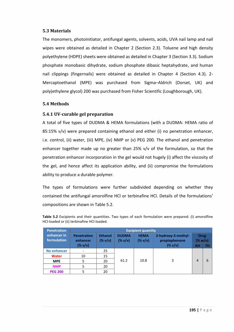
195 | P a g e
5.3 Materials
The monomers, photoinitiator, antifungal agents, solvents, acids, UVA nail lamp and nail
wipes were obtained as detailed in Chapter 2 (Section 2.3). Toluene and high density
polyethylene (HDPE) sheets were obtained as detailed in Chapter 3 (Section 3.3). Sodium
phosphate monobasic dihydrate, sodium phosphate dibasic heptahydrate, and human
nail clippings (fingernails) were obtained as detailed in Chapter 4 (Section 4.3). 2-
Mercaptoethanol (MPE) was purchased from Sigma–Aldrich (Dorset, UK) and
poly(ethylene glycol) 200 was purchased from Fisher Scientific (Loughborough, UK).
5.4 Methods
5.4.1 UV-curable gel preparation
A total of five types of DUDMA & HEMA formulations (with a DUDMA: HEMA ratio of
85:15% v/v) were prepared containing ethanol and either (i) no penetration enhancer,
i.e. control, (ii) water, (iii) MPE, (iv) NMP or (v) PEG 200. The ethanol and penetration
enhancer together made up no greater than 25% v/v of the formulation, so that the
penetration enhancer incorporation in the gel would not hugely (i) affect the viscosity of
the gel, and hence affect its application ability, and (ii) compromise the formulations
ability to produce a durable polymer.
The types of formulations were further subdivided depending on whether they
contained the antifungal amorolfine HCl or terbinafine HCl. Details of the formulations’
compositions are shown in Table 5.2.
Table 5.2 Excipients and their quantities. Two types of each formulation were prepared: (i) amorolfine HCl-loaded or (ii) terbinafine HCl-loaded.
Penetration enhancer in formulation
Excipient quantity
Penetration enhancer
(% v/v)
Ethanol (% v/v)
DUDMA (% v/v)
HEMA (% v/v)
2-hydroxy-2-methyl-propiophenone
(% v/v)
Drug (% w/v)
AH TH
No enhancer - 25
61.2
10.8
3
4
6 Water 10 15
MPE 5 20
NMP 5 20
PEG 200 5 20

196 | P a g e
The formulations (excluding the MPE–containing formulations) were prepared by first
producing an ethanol and penetration enhancer mixture, and dissolving the drug in this
mixture, and then adding the required excipients to the drug solution and leaving the
mixture to stir overnight to produce a clear homogenous solution. Each formulation was
prepared in triplicate.
The MPE–containing formulations were prepared in the same manner as the enhancer-
free formulation; however the MPE was incorporated into the gel immediately prior to
UV-curing to overcome the observed premature polymerisation of the monomer
mixture during storage. It appears that the heat generated when mixing the gel
components may have led to the formation of a thiyl radical, which in turn may have
facilitated the premature polymerisation observed.
The penetration enhancer concentration in the gel was the maximum that could be
incorporated to produce drug-loaded films with the drug remaining in the dissolved
state. A maximal concentration was sought to achieve maximal enhancement, given that
it has been found that as the concentration of penetration enhancer increases, the
permeation of drug through the nail plate increases (Patel and Vora, 2016). To
determine this maximum concentration, formulations containing DUDMA, HEMA, a
photoinitiator, the antifungal drug and penetration enhancers at concentrations of 2.5,
5, 7.5, 10 and 12.5 % v/v with corresponding ethanol concentrations of 22.5, 20, 17.5, 15
and 12.5 % v/v were prepared. These formulations were visually observed for drug
crystals and subsequently exposed to UV light for polymerisation. The resulting films
were observed under a polarised light microscope (as described in Chapter 3, Section
3.4.5.1) and examined using XRD (as described in Chapter 3, Section 3.4.5.2) to confirm
the absence or presence of drug crystals. The polarised light micrographs of the films
and their corresponding XRD patterns can be found in Appendix A12.
5.4.2 UV-curing of formulations and UV-cured film characteristics
The formulations were cured to produce polymer films as per Section 2.4.4 (Chapter 2).
The formulations’ mass yield and degree of conversion from monomer to cured polymer
film were determined as detailed in Sections 2.4.5.1 and 2.4.5.2 respectively, and the
polymer films’ thickness, residual monomer content, chemical structure, microstructure,

197 | P a g e
drug-load, thermal properties, water sensitivity, drug release profiles and ungual drug
permeation profiles were determined as detailed in Sections 3.4.1, 2.4.5.3, 2.4.5.2, 3.4.4,
3.4.6, 3.4.8, 3.4.11, 4.5.1 and 4.5.2 respectively.
The polymer films were also imaged using a Nikon Microphot-FXA microscope (Tokyo,
Japan) to assess the UV-cured polymer film’s structure. Images were taken using a
Lumenera Infinity 2 digital camera (Ottawa, Canada) attached to the microscope.
5.4.3 Statistical analyses
Statistical calculations were conducted using IBM SPSS 22. The data was tested to
determine whether they were normally distributed or not using the Shapiro-Wilk test
and then analysed using either a t-test or Mann-Whiney U test for parametric and non-
parametric data respectively. For multiple comparisons, ANOVA followed by post hoc
Tukey or the Kruskal Wallis test with post hoc analysis performed with Nemenyi’s test
was used for parametric and non-parametric data respectively. Repeated measures
ANOVA was conducted to determine whether there were differences in drug release
profiles and ungual drug permeation profiles of the formulations tested over the
experimental duration.

198 | P a g e
5.5 Results and discussion
5.5.1 Assessment of the polymerisation process
5.5.1.1 Mass yield from monomer gel to polymer film and thickness of the
resulting polymer film
The incorporation of water, MPE, NMP or PEG 200 in a drug-loaded UV-curable gel
formulation did not affect the gels’ ability to produce a polymeric film. Similarly to
formulations without any penetration enhancer, an oxygen inhibition layer was formed
on the films’ surface, and removal of this layer reduced the % mass yield from monomer
mixture to polymer film as shown in Table 5.3.
From Table 5.3, it can be seen that both the amorolfine HCl– and terbinafine HCl–
loaded formulations containing MPE have a significantly higher mass yield percentage
(p<0.05) compared to the other formulations, indicating the formation (and subsequent
removal) of a thinner oxygen inhibition layer. As mentioned in Chapter 2, the oxygen
inhibition layer is formed when O2 reacts with the primary initiating or propagating
radicals to form peroxyl radicals (as shown in Scheme 1) which are unreactive towards
the (meth)acrylate C=C bond. These peroxyl radicals therefore terminate polymerisation
through radical-radical recombination (Odian, 1991, Rabek, 1993). MPE is a thiol, and
thiols are known to reduce the inhibitory effects of O2 on resin formulations (Hoyle and
Bowman, 2010). In the presence of initiating radicals, hydrogen transfer by the thiol
provides a thiyl radical to propagate the polymerisation reaction. While a carbon-based
radical can still react with O2, the peroxyl radical formed can also abstract hydrogen
from thiol, and as a result reinitiation occurs by a thiyl radical (Scheme 2) (Ligon et al.,
2014). This phenomenon explains the observed reduction in the thickness of the oxygen
inhibition layer.
The amorolfine HCl– and terbinafine HCl– loaded formulations containing water have a
significantly higher % mass yield (p<0.05) compared to the formulations containing no
penetration enhancer, NMP or PEG 200. This is most likely due to the formulations’
lower concentration of the volatile component ethanol (15% vs. 20% or 25 %). As
previously mentioned, it is possible that the heat generated by a free radical
polymerisation reaction, coupled with the heat provided by the UVA lamp during the

199 | P a g e
curing process (Appendix A5), may cause some of the volatile components to evaporate
off, thus reducing the mass yield from monomer mixture to polymer film.
The amorolfine HCl– and terbinafine HCl– loaded formulations containing no
penetration enhancer, NMP or PEG 200 showed similar % mass yields from monomer
mixture to polymer film (p>0.05). NMP and PEG 200 are both non-volatile compounds
and thus do not evaporate off during curing, therefore it appears that at a concentration
of 5% v/v, they do not significantly interfere with the polymerisation process.
The choice of drug in the formulation did not alter the % mass yield either (p>0.05), and
this is consistent with the findings in Chapter 2 (Section 2.5.7.1). Furthermore as
expected, linear regression analysis revealed that an increased % mass yield from
monomer mixture to polymer film is correlated with a thicker film (R2 = 0.982, p<0.01)
(Table 5.3).
Therefore, it is seen that the incorporation of a penetration enhancer in a drug-loaded
DUDMA & HEMA UV-curable gel formulation does not negatively affect its mass yield
from monomer mixture to cured polymer film, which remains considerable and hence
acceptable.
Table 5.3 Mass yield of AH- or TH- loaded DUDMA & HEMA gel formulations containing ethanol and different penetration enhancers (after UV-curing and removal of oxygen inhibition layer), and the thickness of the resulting film. Means ± standard deviations are shown, n=3 for mass yield % and n=9 for film thickness.
Mass yield (%) of DUDMA & HEMA
formulations containing
Film thickness (µm) of films produced from DUDMA & HEMA formulations
containing
4% w/v AH 6% w/v TH 4% w/v AH 6% w/v TH
ETOH (25% v/v) & No PE
80.8 ± 0.5 80.7 ± 1.1 163.3 ± 7.1 164.4 ± 5.3
ETOH (15% v/v) & Water (10% v/v)
85.4 ± 1.5 86.6 ± 2.0 172.2 ± 6.7 174.4 ± 6.3
ETOH (20% v/v) & MPE (5% v/v)
94.0 ± 1.4 93.2 ± 0.9 181.7 ± 6.1 180.6 ± 6.8
ETOH (20% v/v) & NMP (5% v/v)
79.7 ± 1.4 79.2 ± 1.5 161.7 ± 7.1 162.2 ± 7.9
ETOH (20% v/v) & PEG 200 (5% v/v)
77.5 ± 1.9 77.4 ± 0.5 158.3 ± 5.0 158.9 ± 6.0
Abbreviations: AH, amorolfine HCl; TH, terbinafine HCl; ETOH, ethanol; PE, penetration enhancer.

200 | P a g e
Scheme 1 Oxygen inhibition in a radical-type photoinitiated polymerisation reaction. Abbreviations: PI, photoinitiator; M, monomer. (Adapted from Studer et al., Progress in Organic Coatings 48, 2003).
Scheme 2 Thiols in a radical-type photoinitiated polymerisation reaction. (Adapted from Ligon et al.,
Chemical Reviews, 114, 1, 2014).

201 | P a g e
5.5.1.2 Degree of conversion (DC) and amount of residual monomers in cured
polymer film
In order to determine the influence of a penetration enhancer on the extent of
polymerisation within the bulk of the film, the formulations’ DC from monomer gel to
polymer film and the films’ residual monomer contents were measured, the results for
which are in Table 5.4 and Fig. 5.1 respectively. The formulations containing the
penetration enhancers obtained a DC % between 58 – 67% and extremely low levels of
residual monomers (≤1.04% DUDMA and ≤0.007% HEMA) in the polymer films. This was
similar to the formulations with no penetration enhancer present and indicates the
presence of unreacted groups within the polymer, as opposed to unreacted monomers,
as previously discussed in Chapter 2 (Section 2.5.7.2).
When considering the amorolfine HCl-loaded formulations with and without the
different penetration enhancers, it can be seen that the formulation containing water
has the highest DC % and the lowest amount of unreacted DUDMA and HEMA
monomers in the film produced. It appears that water’s low viscosity, (0.89 mPas at 25⁰C
compared to 1.08, 1.67, 3.42 and 50 mPas for ethanol, NMP, MPE and PEG 200
respectively) coupled with its higher concentration in the gel, may have produced a
formulation with a lower viscosity than the other formulations, given that the overall
viscosity of the gel formulation is influenced by the viscosity of its components (Chapter
2, Section 2.5.5.2). The formulation’s lower viscosity may have in turn facilitated greater
mobility of the methacrylate side chains during curing and hence it’s greater
polymerisation. However, the other formulations’ higher ethanol content (20% or 25%
vs. 15%) may also be responsible for the observations. As mentioned, ethanol can
evaporate during the curing process. This evaporation can somewhat limit the mobility
of the methacrylate side chains, which already start to restrict due to the increasing
viscosity of the formulation as the polymerisation reaction progresses, thus limiting the
extent of polymerisation.
The formulations containing no penetration enhancer or NMP have similar DC
percentages (p>0.05) and similar amounts of residual DUDMA and HEMA monomers in
their films (p>0.05). This could be down to the similar viscosities of ethanol and NMP,

202 | P a g e
and therefore the similar viscosities assumed for the two formulations, with viscosity
influencing extent of polymerisation as discussed above.
The formulations containing PEG 200 or MPE have the lowest DC percentages (p<0.05)
and highest amounts of unreacted DUDMA and HEMA monomers in the films produced,
due to the higher viscosities of PEG 200 and MPE. Interestingly, the formulation
containing MPE has a slightly (but not significantly, p>0.05) lower DC % and slightly
higher (but not significant, p>0.05) amounts of DUDMA and HEMA monomers remaining
in its film, despite MPE’s considerably lower viscosity than PEG 200. A possible
explanation for this observation could be that during the polymerisation process, a large
fraction of the UVA radiation is absorbed in the upper few micrometres of the MPE-
loaded UV gel, that minimal radiation reaches the lower layers, resulting in an improper
cure.
The terbinafine HCl-loaded formulations have similar DC percentages (p>0.05) and
similar amounts of DUDMA and HEMA monomers remaining in their films (p>0.05) when
compared to their corresponding amorolfine HCl-loaded formulations. This is as
expected given that drug-loading was not found to affect the films DC % (Chapter 2,
Table 2.12) or residual monomer content (Chapter 2, Table 2.13).
Overall, as the amounts of DUDMA & HEMA monomers remaining in the films produced
from the drug-loaded UV-curable gel formulations containing water, MPE, NMP or PEG
200 were negligible, the extent of polymerisation was considered acceptable, and all the
formulations were therefore characterised further.

203 | P a g e
Table 5.4 Percentage DC from monomer gel to polymer film for AH- or TH- loaded DUDMA & HEMA gel formulations containing ethanol and different penetration enhancers. Means ± standard deviations are shown, n=3.
Degree of conversion (%) of DUDMA & HEMA formulations containing
4% w/v AH 6% w/v TH
ETOH (25% v/v) & No PE 64.7 ± 1.6 63.0 ± 3.2
ETOH (15% v/v) & Water (10% v/v) 66.6 ± 0.8 66.2 ± 4.5
ETOH (20% v/v) & MPE (5% v/v) 58.4 ± 1.4 58.2 ± 1.2
ETOH (20% v/v) & NMP (5% v/v) 63.9 ± 0.6 62.8 ± 2.7
ETOH (20% v/v) & PEG 200 (5% v/v) 59.5 ± 2.0 59.3 ± 1.3
Abbreviations: AH, amorolfine HCl; TH, terbinafine HCl; ETOH, ethanol; PE, penetration enhancer.
0.2
0.3
0.4
0.5
0.6
0.7
0.8
0.9
1.0
1.1
1.2
AH-loaded
TH-loaded
AH-loaded
TH-loaded
Co
nc
en
tra
tio
n o
f re
sid
ua
l D
UD
MA
in U
V-c
ure
d p
oly
me
r fi
lms
(%
w/w
)
E
TO
H (
25%
v/v
) &
No
PE
ET
OH
(15%
v/v
) &
Wate
r (1
0%
v/v
)
E
TO
H (
20%
v/v
) &
MP
E (
5%
v/v
)
ET
OH
(20%
v/v
) &
NM
P (
5%
v/v
)
ET
OH
(20%
v/v
) &
PE
G 2
00 (
5%
v/v
)
0.002
0.003
0.004
0.005
0.006
0.007
0.008
0.009
0.010
Co
nc
en
tra
tio
n o
f re
sid
ua
l H
EM
A
in U
V-c
ure
d p
oly
me
r fi
lms
(%
w/w
)
Fig. 5.1 Influence of penetration enhancer incorporation on concentration of residual monomers in AH- or TH- loaded UV-cured DUDMA & HEMA copolymer films. Means and standard deviations are shown, n=3.
204 | P a g e
5.5.2 Structure & microstructure of UV-cured polymer film
Water, MPE, NMP and PEG 200 have boiling points of 100⁰C, 157⁰C, 202⁰C and >250⁰C
respectively. As such, they are not expected to evaporate off during curing, and instead
are expected to remain in the film, perhaps influencing the structure of the polymer film
formed. However, the incorporation of water, MPE, NMP or PEG 200 in a drug-loaded
UV-curable DUDMA & HEMA gel formulation did not alter the visual appearance of the
film produced, which remained smooth and transparent (Appendix A13), and therefore
aesthetically acceptable. In addition, the incorporation of the penetration enhancers in
the formulations did not produce any changes in the resulting films’ FT-IR spectra when
compared to the FT-IR spectra of the formulations containing no penetration enhancer
(Appendix A14). Thus any chemical structural change in the film imposed by the
presence of a penetration enhancer in the gel was not obvious by FT-IR. The films were
therefore examined for visual changes in their microstructure using an optical
microscope (Table 5.5) and a scanning electron microscope (Fig. 5.2).
From Table 5.5, it can be seen that the incorporation of MPE, NMP or PEG 200 in
amorolfine HCl– or terbinafine HCl– loaded DUDMA and HEMA gels does not cause any
noticeable changes in the microstructure of the films produced. However, the
incorporation of water appears to produce films which contain trapped water droplets
or air bubbles. The scanning electron micrograph of the cross-section of the film
produced from the amorolfine HCl–loaded gel formulation containing water did not
appear to show the presence of water droplets or air bubbles however (Fig. 5.2). In fact,
the scanning electron micrographs of the cross-sections of the films produced from the
amorolfine HCl–loaded formulations containing water, MPE, NMP or PEG 200 revealed
dense film interiors, like the film produced from the formulation containing no
penetration enhancer.
Scanning electron micrographs of the films produced from terbinafine HCl–loaded
formulations containing penetration enhancers were not taken, as the choice of drug
was not expected to affect the film’s microstructure, given that in Table 3.6 (Chapter 3),
the drug in the formulation showed no visible changes in the resulting film’s
microstructure.

205 | P a g e
Table 5.5 Optical microscopy images of films produced from amorolfine HCl- or terbinafine HCl- loaded DUDMA & HEMA gel formulations containing ethanol (ETOH) and different penetration enhancers (PE).
Light microscopy images of DUDMA & HEMA UV-cured films produced from formulations containing
4% w/v amorolfine HCl 6% w/v terbinafine HCl
ETOH (25% v/v) &
No PE
ETOH (15% v/v) &
Water (10% v/v)
ETOH (20% v/v) &
MPE (5% v/v)
ETOH (20% v/v) &
NMP (5% v/v)
ETOH (20% v/v) &
PEG 200 (5% v/v)

206 | P a g e
Fig. 5.2 Scanning electron micrographs of the cross-sectional surfaces of UV-cured films produced from amorolfine HCl-loaded DUDMA & HEMA gels containing ethanol and (A) no enhancer, (B) water, (C) MPE, (D) NMP and (E) PEG 200.
A
B C
D E

207 | P a g e
5.5.3 Drug-load in UV-cured polymer film
The drug-load in the UV-cured films (following the removal of the oxygen-inhibition
layer) is shown in Table 5.6. In a similar manner to the drug-loaded DUDMA and HEMA
films containing no penetration enhancer, the drug concentrations in the films
containing water, MPE, NMP or PEG 200 are similar to that of the uncured gel
formulation (p>0.05). The fact that the drug concentration did not increase or decrease
in the film following the removal of the oxygen inhibition layer, indicates that even in
the presence of the penetration enhancer, there was no drug migration to the film
surface or interior during UV-curing.
Table 5.6 Antifungal drug concentration in the gel formulations prior to curing and in UV-cured films following the removal of the oxygen inhibition layer.
DUDMA & HEMA formulation
containing
Antifungal
AH or TH Concentration in gel (% w/v)
Concentration in film (% w/w)
ETOH (25% v/v) & No PE
AH 4.0 ± 0.49 4.4 ± 0.04
TH 6.0 ± 0.22 5.9 ± 0.07
ETOH (15% v/v) & Water (10% v/v)
AH 4.0 ± 0.02 4.2 ± 0.45
TH 6.1 ± 0.08 5.8 ± 0.23
ETOH (20% v/v) & MPE (5% v/v)
AH 4.1 ± 0.28 4.3 ± 0.31
TH 6.0 ± 0.22 5.8 ± 0.32
ETOH (20% v/v) & NMP (5% v/v)
AH 4.0 ± 0.21 4.4 ± 0.56
TH 6.0 ± 0.13 5.9 ± 0.17
ETOH (20% v/v) & PEG 200 (5% v/v)
AH 4.0 ± 0.36 4.4 ± 0.34
TH 6.0 ± 0.24 5.9 ± 0.19
Abbreviations: AH, amorolfine HCl; TH, terbinafine HCl; ETOH, ethanol; PE, penetration enhancer.
5.5.4 Thermal properties of UV-cured polymer films
5.5.4.1 Polymer degradation
The TGA profiles obtained for films produced from formulations containing the
penetration enhancers were comparable to the films produced from the formulations
containing no penetration enhancer (Appendix A15). The incorporation of water, MPE,
NMP or PEG 200 in a drug-loaded UV-curable gel formulation therefore did not affect
the gel’s ability to produce a polymeric film that was thermally stable at room
temperature.

208 | P a g e
5.5.4.2 Glass transition
The incorporation of water, MPE, NMP or PEG 200 in a drug-loaded UV-curable gel
formulation did however cause noticeable changes in the glass transition temperature of
the resulting polymeric film as shown in Table 5.7. The Tg’s of the films produced from
the formulations containing MPE were significantly lower (p<0.05) compared to the Tg’s
of the films produced from the formulations containing no penetration enhancer, water,
NMP or PEG 200, which were similar (p>0.05). The lower Tg’s for the films produced
from the formulations containing MPE could be due to the thiyl radicals formed during
the UV-curing of the formulation. It is possible that the thiyl radicals formed interfere
with DUDMA’s cross-linking ability, and thus increase the flexibility of the polymer
chains, which in turn can decrease the Tg of the film.
A lower degree of conversion can also lead to a decrease in polymer cross-linking extent
(Chapter 3, Section 3.5.1.2), and hence a decrease in the Tg of a film by increasing the
flexibility of the polymer chains. In fact, a positive correlation between degree of cure
and Tg has been reported previously (Keenan, 1987, Urbaniak, 2011). Thus, the lower
Tg’s for the films produced from formulations containing MPE could be also related to
their lower degree of conversions from monomer gel to polymer film when compared to
the films produced from formulations containing no penetration enhancer, water and
NMP (Table 5.4). However, degree of cure does not explain why the Tg’s of the films
produced from the formulations containing PEG 200 is significantly higher than the Tg’s
of the films produced from the formulations containing MPE (p<0.05), as both
formulations have similar degree of conversions from monomer gel to polymer film
(Table 5.4). In this instance, it appears that the incorporation of PEG 200 in the
formulation, decreases the free volume available for polymer chain mobility due its
higher molecular weight (Table 5.1), thus resulting in a higher Tg.
The choice of drug in the formulation did not alter the Tg values obtained (p>0.05), and
this is consistent with the findings in Chapter 3 which suggest that the drug-load is
insufficient to significantly alter Tg in the presence of ethanol (Section 3.5.3.2).

209 | P a g e
Table 5.7 Tg values of amorolfine HCl- or terbinafine HCl- loaded DUDMA & HEMA gel formulations containing ethanol (ETOH) and different penetration enhancers (PE). Means ± standard deviations are shown, n=3.
Tg values of DUDMA & HEMA UV-cured films produced from formulations containing
4% w/v amorolfine HCl 6% w/v terbinafine HCl
ETOH (25% v/v) & No PE 99.2 ± 1.8 94.7 ± 4.8
ETOH (15% v/v) & Water (10% v/v) 103.8 ± 7.9 98.2 ± 7.5
ETOH (20% v/v) & MPE (5% v/v) 65.8 ± 2.1 64.6 ± 0.2
ETOH (20% v/v) & NMP (5% v/v) 98.8 ± 0.3 97.0 ± 2.3
ETOH (20% v/v) & PEG 200 (5% v/v) 106.1 ± 0.3 103.9 ± 3.1
5.5.5 UV-cured polymer films’ sensitivity to water
The water sensitivity scores of the UV-cured films produced from amorolfine HCl– or
terbinafine HCl– loaded DUDMA & HEMA gel formulations containing no penetration
enhancer, water, MPE, NMP or PEG 200 are shown in Figs 5.3 and 5.4, while Fig. 5.5
shows the AUC values calculated from the curves in Figs 5.3 and 5.4.
It can be seen that the films produced from water-containing formulations were the
most sensitive to water (p>0.05); they had detached from the substrate within a couple
of hours. A possible reasoning is that the trapped water droplets or air bubbles within
the film itself (as observed in Table 5.5) limited the film’s adhesion to the substrate in
the first place, thus facilitating detachment following blister formation. The films
produced from formulations containing NMP or PEG 200 had similar water sensitivities
(p>0.05), and both were also more water sensitive compared the films containing no
penetration enhancer (p<0.05). NMP and PEG 200 are both miscible in water, and as
both are incorporated into the UV-cured films, contact with water causes the films to
blister. In contrast, the films produced from MPE-containing formulations were as
water-resistant as those containing no penetration enhancer (p>0.05), despite the fact
that MPE is also miscible in water. The incorporation of MPE in the drug-loaded gel
formulations may therefore produce films which adhere to the HDPE substrate to a
greater extent than the other penetration enhancer-loaded films, thus minimising the
extent to which it blisters when immersed in water. Once again, in a similar manner to
previous findings (Chapter 3, Section 3.5.6), the choice of drug in the formulation did not
alter the resulting films’ water sensitivity (p>0.05).

210 | P a g e
While the incorporation of water, NMP and PEG 200 in a gel formulation affected the
films’ ability to remain on the HDPE substrate in vitro, the extent to which the films’
residence on the nail in vivo is affected can only be confirmed by determining the in vivo
residence of the UV-cured films on fingernails and toenails. Therefore all the
formulations were considered for the drug release and ungual drug permeation studies
discussed in the following sections.
0 10 20 30 40 50
0.0
0.5
1.0
1.5
2.0
2.5
3.0
3.5
4.0
4.5
Wa
ter
se
ns
itiv
ity s
co
re
Time (hours)
ETOH (25% v/v) & No PE
ETOH (15% v/v) & Water (10% v/v)
ETOH (20% v/v) & MPE (5% v/v)
ETOH (20% v/v) & NMP (5% v/v)
ETOH (20% v/v) & PEG 200 (5% v/v)
0 10 20 30 40 50
0.0
0.5
1.0
1.5
2.0
2.5
3.0
3.5
4.0
4.5
Wate
r sen
sit
ivit
y s
co
re
Time (hours)
ETOH (25% v/v) & No PE
ETOH (15% v/v) & Water (10% v/v)
ETOH (20% v/v) & MPE (5% v/v)
ETOH (20% v/v) & NMP (5% v/v)
ETOH (20% v/v) & PEG 200 (5% v/v)
Fig. 5.3 Water sensitivity scores for amorolfine HCl-loaded UV-cured films produced from DUDMA & HEMA gels containing ethanol (ETOH) and different penetration enhancers (PE). Means and standard deviations are shown, n=3.
Fig. 5.4 Water sensitivity scores for terbinafine HCl-loaded UV-cured films produced from DUDMA & HEMA gels containing ethanol (ETOH) and different penetration enhancers (PE). Means and standard deviations are shown, n=3.
0 20 40 60 80 100 120 140 160 180 200
AH with ETOH (25% v/v) & No PE
TH with ETOH (25% v/v) & No PE
AH with ETOH (15% v/v) & Water (10% v/v)
TH with ETOH (15% v/v) & Water (10% v/v)
AH with ETOH (20% v/v) & MPE (5% v/v)
TH with ETOH (20% v/v) & MPE (5% v/v)
AH with ETOH (20% v/v) & NMP (5% v/v)
TH with ETOH (20% v/v) & NMP (5% v/v)
AH with ETOH (20% v/v) & PEG 200 (5% v/v)
TH with ETOH (20% v/v) & PEG 200 (5% v/v)
Mean water sensitivity score (AUC)
Fig. 5.5 Area under the curve values calculated from the curves in Fig. 5.3 & Fig. 5.4 for the UV-cured films produced from the drug-loaded DUDMA & HEMA gels containing ethanol (ETOH) and different penetration enhancers (PE). Means and standard deviations are shown, n=3. AUC was determined using OriginPro 9.0.

211 | P a g e
5.5.6 Drug release profiles of UV-cured polymer films
The drug release profiles of the amorolfine HCl-loaded DUDMA & HEMA films containing
no penetration enhancer, water, MPE, NMP or PEG 200 are shown in Fig. 5.6, and the
corresponding drug release profiles of the terbinafine HCl-loaded DUDMA & HEMA films
are shown in Fig. 5.7. The polarised light micrographs of the films pre- and post-release
experiment and the total amount of drug released by day 30 are also shown alongside
the drug release profiles.
5.5.6.1 Drug release profiles of amorolfine HCl–loaded UV-cured polymer films
In a similar manner to the drug release profile of the amorolfine HCl–loaded formulation
containing no penetration enhancer, the drug release profiles of the amorolfine HCl–
loaded formulations containing water, MPE, NMP or PEG 200 display a burst of drug
release over the first 24 hours due to the release of drug found at the surface of the film.
After this point drug release slows, and by day 30 a greater percentage, i.e. between 50
– 75%, of the incorporated drug had been released (Fig. 5.6). Following the release
study, all the films remained intact, showed negligible weight change (Fig. 5.8), and
showed no signs of drug precipitation (polarised light micrographs in Fig. 5.6). This
enabled the evaluation of the drug release kinetics from these films using zero order,
first order and Higuchi models. Like the film containing no penetration enhancer, the
Higuchi model was found to best describe amorolfine HCl release from the penetration
enhancer-loaded films (Table 5.8), and therefore the drug released through the film
matrix following the initial burst release was also diffusion-controlled.
When the influence of the different penetration enhancers on drug release was
examined, it was found that drug release was the greatest from the formulation
containing MPE, followed by the formulations containing PEG 200, NMP, water and no
penetration enhancer. However, statistically only the formulations containing MPE, PEG
200 or NMP had greater drug release profiles than the formulation containing no
penetration enhancer (p<0.05). These penetration enhancers increased drug release by
reducing the formulations’ degree of conversions from monomer gel to polymer film
(Table 5.4). In fact, linear regression analysis confirmed that lower degree of conversions
from monomer gel to polymer film was correlated with the higher release of amorolfine
HCl (R2 = 0.824, p<0.05) (Fig. 5.9). As mentioned previously (Chapter 3, Section 3.5.1.2),

212 | P a g e
a decrease in the degree of conversion from monomer gel to polymer film produces a
film with decreased polymer cross-linking. Thus the penetration enhancers increased
release by producing polymeric films with lower cross-link densities and hence a less
rigid structure. Interestingly, the differences in the glass transition temperatures of the
films did not significantly correlate with drug release (p<0.05), most probably as they
were outweighed by other formulation properties, such as degree of conversion. Finally,
in a similar manner to the film containing no penetration enhancer, the diffusion-
controlled release through the penetration enhancer-loaded films’ matrix was limited (in
that % drug release did not reach 100%), most possibly due to the films’ dense interior
(Fig. 5.2).
5.5.6.2 Drug release profiles of terbinafine HCl–loaded UV-cured polymer films
In a similar manner to the amorolfine HCl–loaded formulations, the terbinafine HCl–
loaded formulations’ release profiles showed a burst of drug release over the first 24
hours, which then gradually slowed down (Fig. 5.7). Furthermore, like the amorolfine
HCl–loaded films, the terbinafine HCl–loaded films remained intact, showed negligible
weight change (Fig.5.8) and showed no signs of drug precipitation post-release (Fig. 5.7).
Table 5.8 revealed that the Higuchi model best described terbinafine HCl release from
the penetration enhancer-loaded films, and therefore the drug released through the film
matrix following the initial burst release (due to the release of drug found at the surface
of the film) was also diffusion-controlled.
The pattern of drug release obtained by the terbinafine HCl-loaded formulations
containing no penetration enhancer, water, MPE, NMP or PEG 200 was similar to the
corresponding amorolfine HCl–loaded formulations. The drug release was the greatest
from the formulation containing MPE, followed by the formulations containing PEG 200,
NMP, water and no penetration enhancer, and statistically only the formulations
containing MPE, PEG 200 or NMP had greater drug release profiles than the formulation
containing no penetration enhancer (p<0.05). As the choice of drug in the UV-curable gel
formulation was not found to or expected to influenced the formulation’s degree of
conversion (Table 5.4), or the film’s microstructure (Section 5.5.2), glass transition
temperature (Table 5.7) and water sensitivity (Fig. 5.5), the mechanism by which

213 | P a g e
enhancement of drug release occurred is the same as that for the amorolfine HCl–
loaded formulations, i.e. the penetration enhancers affected drug released by producing
polymeric films with differing cross-link densities. However, once again the percentage
of drug released by day 30 was significantly lower from the terbinafine HCl-loaded films
compared to the amorolfine HCl-loaded films (p<0.05) due to possible terbinafine-
polymer binding, as suggested in Section 4.6.1.3 (Chapter 4).
The incorporation of water, MPE, NMP or PEG 200 in an amorolfine HCl– or terbinafine
HCl– loaded UV-curable gel formulation therefore positively influenced the
formulation’s ability to release drug, and in the next section, whether the incorporation
of water, MPE, NMP or PEG 200 in the drug-loaded UV-curable gel formulation enhances
its ungual drug permeation is explored.

214 | P a g e
0 5 10 15 20 25 30
0
10
20
30
40
50
60
70
80 PLM image
Pre-release
Post-release
0 50 100 150 200 250 300
Amount of drug released (g/cm2) by day 30
ETOH (20%) & MPE (5%)
ETOH (20%) & PEG 200 (5%)
ETOH (20%) & NMP (5%)
ETOH (15%) & Water (10%)
ETOH (25%) & No PE
Cu
mu
lati
ve
% a
mo
rolf
ine
HC
l re
lea
se
Time (days)
Fig. 5.6 Cumulative % drug release from the 4% w/v amorolfine HCl-loaded DUDMA & HEMA UV-cured films containing ethanol (ETOH) and different penetration enhancers (PE), their corresponding polarised light microscopy images (pre- and post-release) and the amount of drug released by day 30. Means and standard deviations are shown, n=3. Repeated measures ANOVA showed that the formulation containing MPE displays a similar drug release profile to the formulation containing PEG 200 (p>0.05) and greater drug release profile than the formulations containing NMP, water and no penetration enhancer (p<0.05). The formulation containing PEG 200 also has a similar drug release profile to the formulation containing NMP (p>0.05), and both have greater drug release profiles than the formulations containing water and no penetration enhancer (p<0.05), which in turn have similar drug release profiles (p>0.05).

215 | P a g e
0 5 10 15 20 25 30
0
5
10
15
20
25
30
35
40
0 50 100 150 200
Amount of drug released (g/cm2) by day 30
ETOH (20%) & MPE (5%)
ETOH (20%) & PEG 200 (5%)
ETOH (20%) & NMP (5%)
ETOH (15%) & Water (10%)
ETOH (25%) & No PE
Cu
mu
lati
ve
% t
erb
ina
fin
e H
Cl
rele
as
e
Time (days)
PLM image
Pre-release
Post-release
Fig. 5.7 Cumulative % drug release from the 6% w/v terbinafine HCl-loaded DUDMA & HEMA UV-cured films containing ethanol (ETOH) and different penetration enhancers (PE), their corresponding polarised light microscopy images (pre- and post-release) and the amount of drug released by day 30. Means and standard deviations are shown, n=3. Repeated measures ANOVA showed that the formulation containing MPE displays a similar drug release profile to the formulation containing PEG 200 (p>0.05) and greater drug release profile than the formulations containing NMP, water and no penetration enhancer (p<0.05). The formulation containing PEG 200 also has a similar drug release profile to the formulation containing NMP (p>0.05), and both have greater drug release profiles than the formulations containing water and no penetration enhancer (p<0.05), which in turn have similar drug release profiles (p>0.05).

216 | P a g e
-1.5 -1.0 -0.5 0.0 0.5
% weight change
AH with ETOH (25% v/v) & No PE
TH with ETOH (25% v/v) & No PE
AH with ETOH (15% v/v) & Water (10% v/v)
TH with ETOH (15% v/v) & Water (10% v/v)
AH with ETOH (20% v/v) & MPE (5% v/v)
TH with ETOH (20% v/v) & MPE (5% v/v)
AH with ETOH (20% v/v) & NMP (5% v/v)
TH with ETOH (20% v/v) & NMP (5% v/v)
AH with ETOH (20% v/v) & PEG 200 (5% v/v)
TH with ETOH (20% v/v) & PEG 200 (5% v/v)
Fig. 5.8 Weight change (%) for UV-cured films produced from drug-loaded DUDMA & HEMA gels containing ethanol (ETOH) and different penetration enhancers (PE) following drug-release studies. Means and standard deviations are shown, n=3. Abbreviations: AH, amorolfine HCl; TH, terbinafine HCl.
Table 5.8 R² values obtained for drug release modelling using zero order, first order and Higuchi models.
Formulation R2 value obtained for
Zero order First order Higuchi model
4% w/v AH with ETOH (25% v/v) & No PE 0.75 0.79 0.89
6% w/v TH with ETOH (25% v/v) & No PE 0.90 0.92 0.98
4% w/v AH with ETOH (15% v/v) & water (10% v/v) 0.82 0.85 0.93
6% w/v TH with ETOH (15% v/v) & water (10% v/v) 0.91 0.92 0.98
4% w/v AH with ETOH (20% v/v) & MPE (5% v/v) 0.81 0.86 0.93
6% w/v TH with ETOH (20% v/v) & MPE (5% v/v) 0.85 0.87 0.95
4% w/v AH with ETOH (20% v/v) & NMP (5% v/v) 0.77 0.81 0.89
6% w/v TH with ETOH (20% v/v) & NMP (5% v/v) 0.85 0.86 0.95
4% w/v AH with ETOH (20% v/v) & PEG 200 (5% v/v) 0.77 0.82 0.90
6% w/v TH with ETOH (20% v/v) & PEG 200 (5% v/v) 0.85 0.87 0.95
58 60 62 64 66 68
45
50
55
60
65
70
75
Cu
mu
lati
ve
% a
mo
rolf
ine
HC
l re
leas
e
Degree of conversion (%)
R-squared = 0.824
(p=0.033)
Fig. 5.9 Influence of degree of conversion from monomer gel to polymer film on amorolfine HCl release from the polymer film.

217 | P a g e
5.5.7 Ungual drug permeation profiles of UV-cured polymer films
The ungual drug permeation profiles of the films containing no penetration enhancer,
water, MPE, NMP or PEG 200 are shown in Figs 5.10 & 5.11 for amorolfine HCl and
terbinafine HCl respectively. The % of drug that permeated across the nail and that
remained in the nail at day 30 is also shown alongside the permeation profiles, while the
calculated lag times, study-state flux, apparent permeability coefficients, effective
diffusion coefficients and amount of drug in nail clippings are shown in Table 5.9.
5.5.7.1 Amorolfine HCl–loaded UV-cured polymer films
In a similar manner to the ungual drug permeation profile of the amorolfine HCl-loaded
DUDMA & HEMA formulation containing no penetration enhancer, the ungual drug
permeation profiles of the formulations containing water, MPE, NMP or PEG 200 show a
lengthy lag period prior to a linear increase in the cumulative amount of drug permeated
across the nail (Fig. 5.10). However, the film containing MPE allows greater ungual drug
permeation (p<0.05) compared to the films containing NMP, PEG 200 and water, which
have similar ungual drug permeation profiles (p>0.05), and all the films apart from the
film containing water display greater ungual drug permeation profiles compared to the
film containing no penetration enhancer (p<0.05).
Table 5.9 shows that the MPE-loaded film obtained a shorter lag time (p<0.05) and an
increased steady-state flux (p<0.05), permeability coefficient (p<0.05) and diffusion
coefficient (p<0.05) compared to the film containing no penetration enhancer. It
additionally obtained a higher percentage of drug permeated across the nail (p<0.05)
and percentage of drug remaining in the nail at the end of the permeation experiment
(p<0.05) (Fig 5.10). It therefore appears that despite MPE’s possible role in facilitating
the polymerisation of the gel formulation upon UVA-irradiation (as discussed in Section
5.5.1.1), some MPE molecules are still available intact to permeate into the nail, disrupt
the disulphide bonds of nail keratin, and thus reduce the time taken for the drug to
reach a uniform concentration in the membrane and increase ungual drug permeation.
In comparison, the incorporation of NMP, PEG 200 or water in a UV-cured film did not
significantly influence the films’ lag time, steady-state flux, permeability coefficient,
diffusion coefficient and percentage of drug permeated across the nail at the end of the
218 | P a g e
permeation experiment (p>0.05) when compared to the film containing no penetration
enhancer. These enhancers did however increase the percentage of drug remaining in
the nail at the end of the permeation experiment (p<0.05) to similar extents. While
water, NMP and PEG 200 enhance ungual drug permeation by promoting nail swelling
(Nair et al., 2010, Hossin et al., 2016), and thus form larger pores through which drug
can easily diffuse; in the permeation experiment set up used, the under-surface of the
nail was in direct contact with the receptor phase which would also have promoted nail
swelling to an extent. Therefore, NMP– and PEG 200– loaded films may have increased
the drug-in-nail percentage by promoting greater water uptake (from the receptor
phase) and hence further swelling of the nail, or the NMP–, PEG 200– and water–
loaded films may have increased drug-in-nail percentage by swelling the surface of the
nail in contact with the film.
5.5.7.2 Terbinafine HCl–loaded UV-cured polymer films
The terbinafine HCl–loaded formulations’ permeation profiles also show fairly lengthy
lag times followed by linear increases in the cumulative amount of drug permeated (Fig.
5.11). However, in a similar manner to the amorolfine HCl–loaded films, the film
containing MPE allows greater ungual drug permeation (p<0.05) compared to the films
containing NMP, PEG 200, and water, and all the films apart from the film containing
water display greater ungual drug permeation profiles compared to the film containing
no penetration enhancer (p<0.05).
Table 5.9 shows that once again the MPE-loaded film obtained a shorter lag time
(p<0.05) and an increased diffusion coefficient (p<0.05) compared to the film containing
no penetration enhancer. It additionally obtained a higher percentage of drug
permeated across the nail (p<0.05) and percentage of drug remaining in the nail at the
end of the permeation experiment (p<0.05) (Fig. 5.11). The reason behind this is as
explained in the previous section. Furthermore, the water–, NMP– and PEG 200– loaded
films, all increased the percentage of drug remaining in the nail at the end of the
permeation experiment (p<0.05) to similar extents, compared to the film containing no
penetration enhancer. The reason for which is also as explained in the previous section.

219 | P a g e
Interestingly, Fig. 5.11 also shows that while the films containing NMP and PEG 200 have
similar ungual drug permeation profiles (p>0.05), the film containing NMP has a greater
ungual drug permeation profile than the film containing water (p<0.05), whereas the
film containing PEG 200 does not (p>0.05). Therefore of the terbinafine HCl-loaded
formulations containing NMP and water, the formulation containing NMP appears to be
superior in terms of enhancing ungual drug permeation. It appears that despite water
being able to promote nail swelling to a greater extent when compared to NMP (Hossin
et al., 2016), the possible entrapment of water in the film (Table 5.5) limits its ability to
facilitate the transport of drug across the nail plate. This also explains why amorolfine
HCl– or terbinafine HCl– loaded films containing water have comparable ungual drug
permeation profiles to the films containing no penetration enhancer (p<0.05). In
addition, the similarity in the ungual drug permeation profiles of the NMP– and PEG
200– loaded films suggests that both NMP and PEG 200 promote similar degrees of nail
swelling.
Overall, it therefore appears that the incorporation of MPE, NMP or PEG 200 in an
amorolfine HCl– or terbinafine HCl– loaded DUDMA & HEMA gel formulation enhances
ungual drug permeation, with MPE as the strongest enhancer. Thus it seems that
disrupting the disulphide linkages in the keratin matrix of the nail is more effective than
promoting nail swelling, (with NMP or PEG 200 incorporation), for enhancing the ungual
drug permeation of pharmaceutical UV-curable gel formulations.
5.5.7.3 Amorolfine HCl–loaded vs. terbinafine HCl–loaded UV-cured polymer
films
When compared to the amorolfine HCl-loaded gel formulations, the terbinafine
formulations obtain comparable lag times and diffusion coefficients (p>0.05), due to the
small difference in the molecular weights of the two drugs.
However, the amorolfine HCl-loaded gel formulations obtained a significantly higher
steady-state flux, permeability coefficient and percentage of drug permeated across the
nail at day 30 (p<0.05) when compared to the terbinafine HCl-loaded formulations, due
to amorolfine’s weaker nail keratin-binding ability when compared to terbinafine. These

220 | P a g e
findings are consistent with the findings in Chapter 4 (Section 4.6.2.3), and confirm the
significant impact of drug–nail keratin binding on ungual drug permeation. Nonetheless
as also mentioned in Section 4.6.2.3, drug–nail keratin binding can be beneficial in the
long run.

221 | P a g e
0 5 10 15 20 25 30
0
20
40
60
80
100
120
ETOH (20%) & MPE (5%)
J ETOH (20%) & NMP (5%)
J ETOH (20%) & PEG 200 (5%)
J ETOH (15%) & Water (10%)
J ETOH (25%) & No PE
Cu
mu
lati
ve a
mo
un
t o
f am
oro
lfin
e H
Cl (A
H)
perm
eate
d a
cro
ss t
he
na
il (
g/c
m2)
Time (days)
0 2 4 6 8
% of AH permeated and in nail
% permeated
% in nail
Fig. 5.10 Cumulative amount of amorolfine HCl permeated across the nail with time from the 4% w/v amorolfine HCl-loaded DUDMA & HEMA UV-cured films containing ethanol (ETOH) and different penetration enhancers (PE), and the % of amorolfine HCl permeated across the nail and remaining in the nail at day 30. Means and standard deviations are shown, n=6. A mass balance had been carried out for this experiment to ensure that most of the drug had been retrieved from the modified Franz diffusion cell, and that most of the drug remaining within the nail tissue had been extracted (Appendix A16).

222 | P a g e
0 5 10 15 20 25 30
0
10
20
30
40
50
60
70
ETOH (20%) & MPE (5%)
J ETOH (20%) & NMP (5%)
J ETOH (20%) & PEG 200 (5%)
J ETOH (15%) & Water (10%)
J ETOH (25%) & No PE
Cu
mu
lati
ve a
mo
un
t o
f te
rbin
afi
ne
HC
l (T
H)
perm
eate
d a
cro
ss t
he
na
il (
g/c
m2)
Time (days)
0 2 4 6 8 10
% of TH permeated and in nail
% permeated
% in nail
Fig. 5.11 Cumulative amount of terbinafine HCl permeated across the nail with time from the 6% w/v terbinafine HCl-loaded DUDMA & HEMA UV-cured films containing ethanol (ETOH) and different penetration enhancers (PE), and the % of terbinafine HCl permeated across the nail and remaining in the nail at day 30. Means and standard deviations are shown, n=6. A mass balance had been carried out for this experiment to ensure that most of the drug had been retrieved from the modified Franz diffusion cell, and that most of the drug remaining within the nail tissue had been extracted (Appendix A16).

223 | P a g e
Table 5.9 Lag time, steady-state flux, apparent permeability coefficient, effective diffusion coefficient and amount of drug in nail clippings. Means and standard deviations are shown, n=6.
Abbreviations: ETOH, ethanol; PE, penetration enhancer.
DUDMA & HEMA
formulation containing
4% w/v Amorolfine HCl 6% w/v Terbinafine HCl
Lag time
(day)
Steady-state flux
(µg/cm2/day)
Permeability coefficient
x 10-5
(cm/day)
Diffusion coefficient
x 10-5
(cm
2/day)
Drug in nail
clipping (mg/cm
3)
Lag time
(day)
Steady-state flux
(µg/cm2/day)
Permeability coefficient
x 10-5
(cm/day)
Diffusion coefficient
x 10-5
(cm
2/day)
Drug in nail
clipping (mg/cm
3)
Ethanol (20% v/v) &
MPE (5% v/v)
1.4 ± 1.1
3.4 ± 0.5
8.5 ± 1.1
24.0 ± 16.4
1.2 ± 0.3
3.2 ± 2.3
2.0 ± 0.4
3.3 ± 0.7
12.4 ± 8.1
1.6 ± 0.2
Ethanol (20% v/v) &
NMP (5% v/v)
3.2 ± 2.8
2.4 ± 0.6
6.0 ± 1.6
10.3 ± 8.8
1.4 ± 0.3
3.2 ± 0.9
1.6 ± 0.2
2.7 ± 0.3
6.6 ± 2.3
2.1 ± 0.5
Ethanol (20% v/v) &
PEG 200 (5% v/v)
3.4 ± 3.0
2.4 ± 0.7
6.0 ± 1.8
12.8 ± 12.7
1.3 ± 0.3
5.4 ± 1.8
1.7 ± 0.3
2.8 ± 0.5
2.8 ± 1.0
2.4 ± 0.4
Ethanol (15% v/v) &
Water (10% v/v)
4.4 ± 2.2
2.0 ± 0.4
4.9 ± 0.9
7.8 ± 5.2
1.1 ± 0.2
6.6 ± 0.8
1.6 ± 0.2
2.7 ± 0.3
4.5 ± 1.3
1.5 ± 0.4
Ethanol (25% v/v) &
No PE
7.1 ± 1.7
2.2 ± 0.3
5.5 ± 0.7
3.5 ± 1.4
0.6 ± 0.1
7.1 ± 1.0
1.6 ± 0.2
2.6 ± 0.4
3.0 ± 0.8
0.7 ± 0.2

224 | P a g e
5.6 Conclusions
It is possible to incorporate either 10% v/v of water, 5% v/v of MPE, 5% v/v of NMP or
5% v/v of PEG 200 in a 4% w/v amorolfine HCl– or 6% w/v terbinafine HCl– loaded
DUDMA & HEMA UV-curable gel formulation without negatively affecting the
formulation’s mass yield from monomer gel to polymer film, and the film’s visual
appearance, drug-load, thermal degradation, glass transition, and drug release and
ungual drug permeation.
When considering the influence of these agents on the polymerisation process, the
incorporation of MPE and PEG 200 decreased the degree of conversion from monomer
gel to polymer film and increased the levels of residual monomers in the polymer film.
However despite this, the amounts of residual monomers remaining in the films were
negligible and therefore considered acceptable. In terms of the films’ microstructure,
the formulations containing water, MPE, NMP or PEG 200 produced films with a dense
interior, and the incorporation of water in the gel formulation appeared to produce films
which contained trapped water droplets or air bubbles. Finally, when considering the
films’ glass transition and water sensitivity, MPE was the only agent which reduced the
glass transition of the film produced following its incorporation in the gel, and the only
one not to negatively affect the film’s water sensitivity.
The drug release patterns obtained for both the amorolfine HCl– and terbinafine HCl–
loaded formulations containing water, MPE, NMP or PEG 200 were found to be related
to the formulations’ degree of conversion from monomer gel to polymer film (and hence
cross-linking). Once again, the release of terbinafine from the films was less than that of
amorolfine, due to possible terbinafine-polymer binding.
Ungual permeation studies revealed that MPE, NMP and PEG 200 were capable of
improving ungual amorolfine and terbinafine permeation when present in a UV-curable
gel vehicle. Overall, MPE was found to be the superior of the penetration enhancers, not
only because of the significant enhancement of ungual drug permeation, but for being
the least likely to affect the UV-cured film’s residence on the nail.

225 | P a g e
Chapter 6: Conclusions and future work
6.1 Introduction
The human nail acts as a fairly impervious barrier, presenting a challenge to the
treatment of common nail diseases such as onychomycosis and nail psoriasis. In the
past, treatment relied mainly on systemic therapy, which is associated with a number of
serious side effects, drug interactions, contraindications and high recurrence rates.
However, the recognition that drug penetration into the nail, although difficult, is
achievable, sparked a race towards the development of topical therapies. Various topical
nail drug delivery vehicles have been investigated to date, including solutions, lacquers,
gels, ointments, creams, films, patches and colloidal carriers, and considerable effort has
been made to enhance ungual drug delivery using physical, chemical, or a combination
of physical and chemical means. Unfortunately, despite these efforts, currently available
topical therapies for nail diseases such as onychomycosis and nail psoriasis remain
unsatisfactory. Treatment failure could be a result of the limited penetration of the
therapeutic agent through the nail plates’ firmly bound layers of dead keratinous cells
and compact dorsal structure. However, the formulations’ inability to maintain
therapeutic drug levels at the desired site and poor patient compliance can also
contribute to the poor treatment response, especially when considering that topical
therapies require regular application until all the affected nail tissue has grown out, a
process which can take up to several months. There appears to be a special need for a
topical nail medicine capable of overcoming these limitations. In this thesis,
pharmaceutical UV gels were proposed as such a nail medicine, as UV gels are known to
have a long residence on the nail plate and can therefore address the issues highlighted,
i.e. the need for frequent applications and failure to maintain therapeutic drug levels at
the desired site. The pharmaceutical potential of the UV gels was determined by
formulating and characterising the gel and film as depicted in Fig. 6.1, while using
antifungal drugs as model drugs, and the summary of the key findings and
recommendations for future research are discussed in this Chapter.

226 | P a g e
Fig. 6.1 Schematic diagram highlighting the investigations to assess the pharmaceutical potential of UV-curable gel formulations.
6.2 Summary of key findings
It is possible to formulate a pharmaceutical UV-curable gel formulation containing an
antifungal (amorolfine HCl or terbinafine HCl) when incorporated within DUDMA-based
UV gels containing the photoinitiator 2-hydroxy-2-methylpropiophenone, a solvent such
as ethanol and one of three different reactive diluent methacrylate-based monomers
(EMA, IBOMA or HEMA). The solvent, at a concentration of 25% v/v, was found to be
necessary for drug incorporation into the gel, and for such formulations, a DUDMA to
reactive diluent monomer ratio of 85:15 % v/v and a photoinitiator concentration of 3%
v/v was found to be optimal. Formulations containing HEMA, with a DUDMA to reactive
diluent monomer ratio of 75:25% v/v and no solvent, also enabled drug incorporation
due to the greater drug solvency of HEMA. Penetration enhancers such as water (10%
v/v), MPE (5% v/v), NMP (5% v/v) or PEG 200 (5% v/v) could also be incorporated into
drug-loaded DUDMA & HEMA UV-curable gel formulations containing ethanol.
Actives and excipients
•Choice and quantity
UV-curable gel properties
•Gel viscosity
•Drug stability
UV-curing conditions
•UVA device
•Cure time
UV-cured film
characteristics
•Mass yield
•DC and cross-link extent
•Residual monomer content
•Morphology, thickness, microsturcture and porosity
•Drug-load
•Drug-polymer interactions
•Thermal stability and glass transition temperature
•Occlusivity, adhesivity and water sensitivity
•In vivo nail residence
•Film stability and drug in film stability
•Drug release and ungual drug permeation
•In vitro efficacy against T. rubrum
UV-curable gel optimisation using penetration enhancers
227 | P a g e
These formulations, when applied on the human nail and cured for 2 minutes under a
UVA nail lamp, were capable of producing visually smooth and transparent films, all of
which were capable of enabling sufficient ungual drug permeation in vitro to inhibit the
growth of the most common onychomycosis-causing pathogen – T. rubrum. However
there were differences amongst the UV-cured films regarding other properties, based on
monomer choice, monomer ratio, presence or absence of ethanol, and nature of drug
and of penetration enhancer, as summarised below. How UV gels compare to a
commercially available pharmaceutical nail lacquer as a form of topical nail medicine is
also summarised below.
6.2.1 Influence of reactive diluent monomer choice on UV-cured film
properties
The nature of the reactive diluent methacrylate-based monomer used in the gel was
found to influence the final viscosity of the gel formulation, which in turn affected the
glass transition temperature of the resulting polymer film. However, the differences in
the glass transition temperatures of the different films did not influence drug release
from the films. Of the reactive diluent methacrylate-based monomers used, HEMA had a
higher solvency for the antifungal drugs as mentioned above. As a result, in the presence
of ethanol, the maximum drug-load in the DUDMA & HEMA gel was much higher (4%
w/v amorolfine HCl or 6% w/v terbinafine HCl) compared to the drug-load in the
DUDMA & EMA or DUDMA & IBOMA gels (3% w/v amorolfine HCl or 4% w/v terbinafine
HCl). The films’ drug release and ungual drug permeation were found to increase as the
drug concentration in the film was increased. The nature of the diluent monomer
therefore influenced drug release and ungual drug permeation via its influence on drug
concentration in the film.
6.2.2 Influence of monomer ratio on UV-cured film properties
As mentioned earlier, increasing the proportion of the HEMA monomer in a UV-curable
gel formulation to produce a formulation with the DUDMA: HEMA ratio of 75:25% v/v,
(as opposed to 85:15% v/v) made it possible to incorporate antifungals in the gel
without the need for ethanol. However, the total drug content in the film was fairly low
(2% w/v for both amorolfine HCl and terbinafine HCl compared to the 4% w/v
amorolfine HCl and 6% w/v terbinafine HCl with ethanol). Nonetheless, the higher HEMA

228 | P a g e
concentration in the gel did reduce the formulation’s viscosity, which in turn increased
its polymerisation, and subsequently its glass transition temperature.
6.2.3 Influence of ethanol incorporation on UV-cured film properties
Ethanol was incorporated in the gel for its ability to promote and increase drug loading.
Its inclusion reduced the formulations’ viscosity, which in turn increased its
polymerisation, and subsequently the resulting film’s glass transition temperature.
However, its inclusion also negatively affected the film’s in vivo residence on the nail,
which would mean more frequent medicine application. Nonetheless, the greater drug
loading enabled by ethanol resulted in higher drug release and ungual flux.
6.2.4 Influence of drug nature and presence on UV-cured film properties
The presence of the drugs – amorolfine HCl and terbinafine HCl – had negligible
influence on the films’ properties such as its morphology, structure, glass transition
temperature, adhesion, water sensitivity and in vivo nail residence. In contrast, the drug
nature did have a considerable influence on drug release and ungual flux. Amorolfine
release from the film was much higher compared to that of terbinafine, possibly due to
terbinafine-polymer binding. Similarly, amorolfine’s ungual flux was higher compared to
terbinafine’s due to the latter’s higher affinity for nail keratin. Nonetheless, as
terbinafine HCl has a smaller MIC against the most common onychomycosis-causing
pathogen, and presence of drug in the nail as well as drug movement out of the nail and
into the nail bed is important, both drugs show potential when loaded in a UV-curable
gel formulation for the treatment of onychomycosis.
6.2.5 Influence of penetration enhancer on UV-cured film properties
The incorporation of water, MPE, NMP or PEG 200 in 4% w/v amorolfine HCl– or 6% w/v
terbinafine HCl– loaded DUDMA & HEMA UV-curable gel formulations containing
ethanol did not negatively affect the formulations’ mass yield from monomer gel to
polymer film, and the films’ visual appearance, drug-load, thermal degradation, drug
release and ungual drug permeation. However, following its incorporation in the gel,
MPE was the only agent not to negatively affect the film’s water-resistance.
Furthermore, MPE incorporation in the UV gel enhanced ungual drug permeation to the
greatest extent compared to the other penetration enhancer-loaded formulations and

229 | P a g e
the penetration enhancer-free control. Disrupting the disulphide linkages in the keratin
matrix of the nail is therefore more effective than promoting nail swelling, (with water,
NMP or PEG 200 incorporation), for enhancing the ungual drug permeation of
pharmaceutical UV-curable gel formulations.
6.2.6 UV-curable gels vs. nail lacquers
Comparisons between the Curanail® nail lacquer (which contains 5% w/v amorolfine
HCl) with the best UV gel formulation (penetration enhancer-free) showed that drug
release from the UV-cured polymer film was significantly lower than that from the nail
lacquer film, possibly due to the UV-cured film’s cross-linked and highly dense nature.
Nevertheless, both lacquer and UV gel films displayed similar ungual drug permeation
profiles, and this in turn confirmed the critical role played by the nail plate barrier to
ungual drug permeation and the lesser role of drug release from the film. In comparison
to the Curanail nail lacquer®, the UV-curable gel formulations reduced TOWL to a
greater extent and resided on fingernails for considerably longer (even when containing
ethanol); they therefore appear to be a more favourable form of topical nail medicine.
6.3 Considerations for future work
In the previous Chapters, the following was suggested for future work:
(i) measuring the cross-link densities of the UV-cured films using inverse gas
chromatography (Chapter 3, Section 3.5.1.2),
(ii) determining whether the application of a UV-curable gel formulation reduces
TOWL of diseased nails to the same extent as healthy nails (Chapter 3, Section
3.5.4),
(iii) determining the affinity of amorolfine and terbinafine to the UV-cured films
(Chapter 4, Section 4.6.1.3), and
(iv) optimising the adapted disc diffusion method developed for testing the
efficacies of topical nail formulations against T. rubrum (Chapter 4, Section 4.6.3).
Details as to why the above were suggested for future work and specifics about the
experiments considered can be found in the relevant sections.

230 | P a g e
Other considerations for future work include looking at ways to further improve the
pharmaceutical UV-curable gel formulation and thus guarantee treatment success, these
are discussed below.
6.3.1 Physical approach to enhance the ungual drug delivery of
pharmaceutical UV-curable gels
While the incorporation of a penetration enhancer (in particular MPE) in the
pharmaceutical UV-curable gel enhanced ungual drug permeation, it could be further
improved by also using a physical means to enhance ungual permeation. In Chapter 1,
the dorsal layer of the nail plate was identified as the main barrier to permeant
transport through the nail plate and into the nail bed, and methods of disrupting this
layer by physical means to enhance ungual permeation were reviewed. One such
technique which has shown success was nail filing using an abrasive (Pittrof et al., 1992,
Lauharanta, 1992), which enhances permeation by decreasing the thickness of the
dorsal layer. Furthermore, it increases the surface roughness and hence surface area of
the nail as shown in Table 6.1, which in turn provides a greater opportunity for drug-
loaded films to adhere to the nail plate and thereby facilitate drug permeation.
Table 6.1 Scanning electron micrographs of the dorsal surface and the cross-section of a human nail plate clipping, with and without filing the dorsal surface with the abrasive stick provided with the Curanail® nail lacquer.
‘Control’ nail clipping ‘Filed’ nail clipping
Nail plate clipping dorsal
surface
Nail plate clipping cross-
section

231 | P a g e
Coincidently, prior to the application of commercially available cosmetic UV gels, the nail
is lightly filed with a low grit abrasive file to increase its surface area and ensure good
adhesion. Therefore, disrupting the dorsal layer of the nail plate with an abrasive file
prior to the application of a drug-loaded topical formulation to improve film adhesion
and enhance ungual drug permeation further, seems fitting for a pharmaceutical UV gel.
Hence as part of future work, the influence of nail filing on a UV-cured film’s adhesion
and ungual drug permeation can be investigated.
6.3.2 3D printed pens as a delivery device for UV-curable gel formulations to
improve formulation application procedure
When considering the application process of a UV-curable gel formulation, the
procedure is not so straight forward. Not only is there a need for a UVA nail lamp to
facilitate the polymerisation of the gel formulation, the film that subsequently forms
contains an oxygen inhibition layer which requires removal with a nail wipe soaked in
alcohol. One way of addressing the issues highlighted is by investigating hand-held 3D
printing pens that emit UV (such as CreoPop) as a formulation delivery device, where the
ink inside the pen is replaced with the pharmaceutical UV-curable gel formulation. The
application of the UV-curable gel formulation on the nail plate via a CreoPop-like device,
would eliminate the need for a UVA nail lamp, prevent the formation of an oxygen
inhibition layer and cut down total cure time, all of which could further tackle the issue
of poor patient compliance.
6.4 Conclusions
It has been demonstrated that UV-curable gel formulations can be considered as an
alternative vehicle for the delivery of topical nail medicines. Their use addresses current
topical formulation issues such as
(i) the need for frequent applications and
(ii) failure to maintain therapeutic drug levels at the desired site
by residing on the nail longer than other nail formulations, like nail lacquers for example.

232 | P a g e
References
Achten, G., Andre, J. and Laporte, M. (1991) 'Nails in Light and Electron Microscopy.', Seminars in Dermatology, 10(1), pp. 54-64.
Ahn, T., Lee, J.-P., Kim, J., Oh, S., Chun, M.-K. and Choi, H.-K. (2013) 'Effect of Pressure Sensitive Adhesive and Vehicles on Permeation of Terbinafine Across Porcine Hoof Membrane', Archives of Pharmaceutical Research, 36(11), pp. 1403-1409.
Alshuiref, A., Ibrahim, H., Abduallah, A. and Edali, M. (2013) 'Thermal and Mechanical Analysis of Urethane Acrylate Graft Copolymer Part A: Urethane Macromonomer Base on TDI and EG', International Journal of Chemistry, 5(1), pp. 58-79.
Ameen, M., Lear, J. T., Madan, V., Mohd Mustapa, M. F. and Richardson, M. (2014) 'British Association of Dermatologists' Guidelines for the Management of Onychomycosis 2014', British Journal of Dermatology, 171(5), pp. 937-958.
Andre, J., Scheers, C. and Baran, R. (2014) Normal Nail and Use of Nail Cosmetics and Treatments. Handbook of Cosmetic Science and Technology Fourth edn. Boca Raton, Florida: CRC Press. Taylor & Francis Group., p. 597-608.
Arca, E., Taştan, H. B., Akar, A., Kurumlu, Z. and Gür, A. R. (2002) 'An Open, Randomized, Comparative Study of Oral Fluconazole, Itraconazole and Terbinafine Therapy in Onychomycosis', The Journal of dermatological treatment, 13(1), pp. 3-9.
ASTM-D870 (2015) Standard Practice for Testing Water Resistance of Coatings Using Water Immersion: Available at: https://www.astm.org/Standards/D870.htm (Accessed: 2 June 2016).
Avner, S., Nir, N. and Henri, T. (2005) 'Combination of Oral Terbinafine and Topical Ciclopirox Compared to Oral Terbinafine for the Treatment of Onychomycosis', The Journal of Dermatological Treatment, 16(5-6), pp. 327-330.
Badali, H., Mohammadi, R., Mashedi, O., Hoog, G. S. and Meis, J. F. (2015) 'In Vitro Susceptibility Patterns of Clinically Important Trichophyton and Epidermophyton Species Against Nine Antifungal Drugs', Mycoses, 58(5), pp. 303-307.
Baden, H. P. (1994) 'Treatment of Distal Onychomycosis with Avulsion and Topical Antifungal Agents under Occlusion', Arch Dermatol, 130(5), pp. 558-9.
Baden, H. P., Goldsmith, L. A. and Fleming, B. (1973) 'A Comparative Study of the Physicochemical Properties of Human Keratinised Tissues.', Biochimica et Biophysica Acta, 322, pp. 269-278.
Baran, R. (2010) 'The Burden of Nail Psoriasis: An Introduction', Dermatology, 221, pp. 1-5. Baran, R., Feuilhade, M., Datry, A., Goettmann, S., Pietrini, P., Viguie, C., Badillet, G., Larnier, C. and
Czernielewski, J. (2000) 'A Randomized Trial of Amorolfine 5% Solution Nail Lacquer Combined with Oral Terbinafine Compared with Terbinafine Alone in the Treatment of Dermatophytic Toenail Onychomycoses Affecting the Matrix Region', British Journal of Dermatology, 142(6), pp. 1177-1183.
Baran, R., Hay, R. J. and Garduno, J. I. (2008a) 'Review of Antifungal Therapy and the Severity Index for Assessing Onychomycosis: Part I', The Journal of Dermatological Treatment, 19(2), pp. 72-81.
Baran, R., Hay, R. J. and Garduno, J. I. (2008b) 'Review of Antifungal Therapy, Part II: Treatment Rationale, including Specific Patient Populations', The Journal of Dermatological Treatment, 19(3), pp. 168-75.
Baran, R., Sigurgeirsson, B., de Berker, D., Kaufmann, R., Lecha, M., Faergemann, J., Kerrouche, N. and Sidou, F. (2007) 'A Multicentre, Randomized, Controlled Study of the Efficacy, Safety and Cost-Effectiveness of a Combination Therapy with Amorolfine Nail Lacquer and Oral Terbinafine Compared with Oral Terbinafine Alone for the Treatment of Onychomycosis with Matrix Involvement', British Journal of Dermatology, 157(1), pp. 149-57.
Belfiore, L. A. (2010) 'Glass Transitions in Amorphous Polymers: Basic Concepts', in Belfiore, L.A. (ed.) Physical Properties of Macromolecules. New Jersey: John Wiley & Sons, pp. 3-36.
Bianchi, L., Soda, R., Diluvio, L. and Chimenti, S. (2003) 'Tazarotene 0.1% Gel for Psoriasis of the Fingernails and Toenails: An Open, Prospective Study', The British Journal of Dermatology, 149(1), pp. 207-209.
Bohn, M. and Kraemer, K. T. (2000) 'Dermatopharmacology of Ciclopirox Nail Lacquer Topical Solution 8% in the Treatment of Onychomycosis', Journal of the American Academy of Dermatology, 43(4), pp. S57-S69.

233 | P a g e
Botero, A. R., Halal, J., Kilgore, M., McConnell, J., McCormick, J., Peters, V., Schoon, D. and Spear, J. (2014) 'UV and LED Gels', in Alisha R. Botero, John Halal, Mary Ann Kilgore, Jim McConnell, Janet McCormick, Vicki Peters, Douglas Schoon & Spear, J. (eds.) Milady Standard Nail Technology. Boston: Cengage Learning, pp. 356-364.
Brook, I. M. and van Noort, R. (1985) 'Drug Release from Acrylic Polymers Via Channels and Cracks: In Vitro Studies with Hydrocortisone', Biomaterials, 6(4), pp. 281-285.
Brown, M. B., Khengar, R. H., Turner, R. B., Forbes, B., Traynor, M. J., Evans, C. R. G. and Jones, S. A. (2009) 'Overcoming the Nail Barrier: A Systematic Investigation of Ungual Chemical Penetration Enhancement', International Journal of Pharmaceutics, 370(1), pp. 61-67.
Bueno, J. G., Martinez, C., Zapata, B., Sanclemente, G., Gallego, M. and Mesa, A. C. (2010) 'In Vitro Activity of Fluconazole, Itraconazole, Voriconazole and Terbinafine Against Fungi Causing Onychomycosis', Clinical and Experimental Dermatology, 35(6), pp. 658-663.
Burnett, D. J., Thielmann, F. and Nichols, M. E. (2007) 'Correlating Degree of Crosslink for an Automotive Topcoat to Solubility Parameters', pp. 1-7.
Cannavò, S. P., Guarneri, F., Vaccaro, M., Borgia, F. and Guarneri, B. (2003) 'Treatment of Psoriatic Nails with Topical Cyclosporin: A Prospective, Randomized Placebo-Controlled Study', Dermatology, 206(2), pp. 153-156.
Caputo, R., De Boulle, K., Del Rosso, J. and Nowicki, R. (2001) 'Prevalence of Superficial Fungal Infections among Sports‐Active Individuals: Results from the Achilles Survey, A Review of the Literature.', Journal of the European Academy of Dermatology and Venereology, 15(4), pp. 312-316.
Chartoff, R. P. (1997) 'Thermoplastic Polymers', in Turi, E.A. (ed.) Thermal Characterisation of Polymeric Materials. New York: Academic Press, pp. 483-743.
Chattopadhyay, D. K. and Webster, D. C. (2009) 'Thermal Stability and Flame Retardancy of Polyurethanes', Progress in Polymer Science, 34(10), pp. 1068-1133.
Chouhan, P. and Saini, T. R. (2012) 'Hydration of Nail Plate: A Novel Screening Model for Transungual Drug Permeation Enhancers', International Journal of Pharmaceutics, 436(1-2), pp. 179-182.
Chouhan, P. and Saini, T. R. (2014) 'Hydroxypropyl-β-cyclodextrin: A Novel Transungual Permeation Enhancer for Development of Topical Drug Delivery System for Onychomycosis', Journal of Drug Delivery, 2014, pp. 1-7.
Clemmensen, S. (1984) 'Cross-Reaction Patterns in Guinea Pigs Sensitised to Acrylic Monomers', 7(6), pp. 527-540.
CND ([No date]): Available at: http://www.cnd.com/ (Accessed: 25 January 2016). Conde‐salazar, L., Guimaraens, D., Romero, L. V., González, M. A. and Alomar, A. (1986) 'Occupational
Allergic Contact Dermatitis to Artificial Nails', Contact Dermatitis, 15(4), pp. 242-242. Cravo, M., Cardoso, J. C., Gonçalo, M. and Figueiredo, A. (2008) 'Allergic Contact Dermatitis from
Photobonded Acrylic Gel Nails: A Review of Four Cases', Contact Dermatitis, 59(4), pp. 250-251. Crowley, J. J., Weinberg, J. M., Wu, J. J. and Robertson, A. D. (2015) 'Treatment of Nail Psoriasis. Best
Practice Recommendations From the Medical Board of the National Psoriasis Foundation', Journal of the American Medical Association, 151(1), pp. 87-94.
Cruz, M. J., Baudrier, T., Cunha, A. P., Ferreira, O. and Azevedo, F. (2011) 'Severe Onychodystrophy Caused by Allergic Contact Dermatitis to Acrylates in Artificial Nails', Cutaneous and Ocular Toxicology, 30(4), pp. 323-324.
Daniel, R. C. (2013) 'Onychomycosis: Burden of Disease and the Role of Topical Antifungal Treatment.', Drugs in Dermatology, 12(11), pp. 1263-1266.
Daniels, C. A. (1989) 'Tensile Properties', in Daniels, C.A. (ed.) Polymers: Structure and Properties. Lancaster, Pennsylvania: Technomic Publishing Company, Inc., pp. 43-55.
Davies, K. J. (2006) 'Study to Determine the Efficacy of Clotrimazole 1% Cream for the Treatment of Onychomycosis in Association with the Mechanical Reduction of the Nail Plate', The Foot, 16(1), pp. 19-22.
Davies, R. R., Everall, J. D. and Hamilton, E. (1967) 'Mycological and Clinical Evaluation of Griseofulvin for Chronic Onychomycosis', British Medical Journal, 3(5563), pp. 464-8.
De Angelis, A. (2013) Dermatophytic Onychomycosis Market to Reach $3.4 Billion by 2017.: Companies And Markets - http://www.companiesandmarkets.com/News/Healthcare-and-Medical/Dermatophytic-onychomycosis-market-to-reach-3-4-billion-by-2017/NI6990 (Accessed: 26 July 2015).
de Berker, D. (2009) 'Fungal Nail Disease', The New England Journal of Medicine, 360(20), pp. 2108-2116.
234 | P a g e
de Jong, E. M., Seegers, B. A., Gulinck, M. K., Boezeman, J. B. and van de Kerkhof, P. C. (1996) 'Psoriasis of the Nails Associated with Disability in a Large Number of Patients: Results of a Recent Interview with 1,728 Patients.', Dermatology, 193(4), pp. 300-3.
Decker, C. (1989) 'Effect of UV Radiation on Polymers', in Cheremisinof, N.P. (ed.) Handbook of Polymer Science and Technology. New York: Marcel Dekker, Inc., pp. 541-608.
Deen, W. M. (1987) 'Hindered Transport of Large Molecules in Liquid‐Filled Pores', AIChE Journal, 33(9), pp. 1409-1425.
Delgado-Charro, M. B. (2012) 'Iontophoretic Drug Delivery Across the Nail', Expert opinion on drug delivery, 9(1), pp. 91-103.
Dominicus, R., Weidner, C., Tate, H. and Kroon, H. a. (2012) 'Open‐Label Study of the Efficacy and Safety of Topical Treatment with TDT 067 (Terbinafine in Transfersome ®) in Patients with Onychomycosis', British Journal of Dermatology, 166(6), pp. 1360-1362.
Donnelly, R. F., McCarron, P. A., Lightowler, J. M. and Woolfson, A. D. (2005) 'Bioadhesive Patch-Based Delivery of 5-Aminolevulinic Acid to the Nail for Photodynamic Therapy of Onychomycosis', Journal of Controlled Release, 103(2), pp. 381-392.
Dowdy, J. C. and Sayre, R. M. (2013) 'Photobiological Safety Evaluation of UV Nail Lamps', Photochemistry and Photobiology, 89(4), pp. 961-967.
Drake, L. A., Patrick, D. L., Fleckman, P., André, J., Baran, R., Haneke, E., Sapède, C. and Tosti, A. (1999) 'The Impact of Onychomycosis on Quality of Life: Development of an International Onychomycosis-Specific Questionnaire to Measure Patient Quality of Life', Journal of the American Academy of Dermatology, 41(2), pp. 189-196.
Drake, L. A., Scher, R. K., Smith, E. B., Faich, G. A., Smith, S. L., Hong, J. J. and Stiller, M. J. (1998) 'Effect of Onychomycosis on Quality of Life', Journal of the American Academy of Dermatology, 38(5), pp. 702-704.
Drake, L. A., Shear, N. H., Arlette, J. P., Cloutier, R., Danby, F. W., Elewski, B. E., Garnis-Jones, S., Giroux, J.-M., Gratton, D., Gulliver, W., Hull, P., Jones, H. E., Journet, M., Krol, A. L., Leyden, J. J., Maddin, S. C., Ross, J. B., Savin, R. C., Scher, R. K., Sibbald, G. R., Tawfik, N. H., Zaias, N., Tolpin, M., Evans, S., Marsolais, C., Chin, T., Lin, T.-H., Maher, T. and Birnbaum, J. E. (1997) 'Oral Terbinafine in the Treatment of Toenail Onychomycosis: North American Multicenter Trial', Journal of the American Academy of Dermatology, 37(5), pp. 740-745.
Elewski, B. E. (1997) 'The Effect of Toenail Onychomycosis on Patient Quality of Life', International Journal of Dermatology, 36(10), pp. 754-756.
Elewski, B. E., Aly, R., Baldwin, S. L., González Soto, R. F., Rich, P., Weisfeld, M., Wiltz, H., Zane, L. T. and Pollak, R. (2015) 'Efficacy and Safety of Tavaborole Topical Solution, 5%, A Novel Boron-Based Antifungal Agent, for the Treatment of Toenail Onychomycosis: Results from 2 Randomized Phase-III Studies', Journal of the American Academy of Dermatology, 73(1), pp. 62-69.
Elewski, B. E., Rich, P., Pollak, R., Pariser, D. M., Watanabe, S., Senda, H., Ieda, C., Smith, K., Pillai, R., Ramakrishna, T. and Olin, J. T. (2012) 'Efinaconazole 10% Solution in the Treatment of Toenail Onychomycosis: Two Phase III Multicenter, Randomized, Double-Blind Studies', Journal of the American Academy of Dermatology.
Elsayed, M. M. A. (2015) 'Development of Topical Therapeutics for Management of Onychomycosis and Other Nail Disorders: A Pharmaceutical Perspective', Journal of Controlled Release, 199, pp. 132-144.
Erdmann, S. M., Sachs, B. and Merk, H. F. (2001) 'Adverse Reactions to Sculptured Nails', Allergy, 56(6), pp. 581-582.
Escobar, A. and Yamarik, T. A. (2005) 'Final Report of the Safety Assessment of Methacrylate Ester Monomers Used in Nail Enhancement Products', International Journal of Toxicology, 24(5), pp. 53-100.
Faergemann, J., Anderson, C., Hersle, K., Hradil, E., Nordin, P., Kaaman, T., Molin, L. and Pettersson, A. (1995) 'Double-Blind, Parallel-Group Comparison of Terbinafine and Griseofulvin in the Treatment of Toenail Onychomycosis', Journal of the American Academy of Dermatology, 32(5), pp. 750-753.
Fisher, A. A. 1990. Adverse Nail Reactions and Paresthesia from "Photobonded Acrylate 'Sculptured' Nails". Cutis.
Fitzgerald, D. A., Bhaggoe, R. and English, J. S. C. (1995) 'Contact Sensitivity to Cyanoacrylate Nail‐Adhesive with Dermatitis at Remote Sites', Contact Dermatitis, 32(3), pp. 175-176.
Fitzgerald, D. A. and English, J. S. C. (1994) 'Widespread Contact Dermatitis from Sculptured Nails', Contact Dermatitis, 30(2), pp. 118-118.

235 | P a g e
Flagothier, C., Piérard‐franchimont, C. and Piérard, G. E. (2005) 'New Insights into the Effect of Amorolfine Nail Lacquer', Mycoses, 48(2), pp. 91-94.
Fleckman, P. (1997) 'Basic Science of the Nail Unit', in Scher, R.K. & Daniel, C.R. (eds.) Nails: Therapy, Diagnosis, Surgery. 2nd Edition ed. Philadelphia: WB Saunders, pp. 37-54.
Fleckman, P. (2005) Structure and Function of the Nail Unit. Nails: Diagnosis, Therapy, Surgery Philadelphia: Elsevier Saunders.
Flory, P. J. (1947) 'Effects of Cross-Linking and Branching on the Molecular Constitution of Diene Polymers', Journal of the American Chemical Society, 69(11), pp. 2893-2899.
Flory, P. J. and Rehner Jr., J. (1943) 'Statistical Theory of Chain Configuration and Physical Properties of High Polymers', Annals of the New York Academy of Sciences, 44, pp. 419-429.
Forslind, B. (1970) 'Biophysical Studies of the Normal Nail.', Acta Dermato-Venereologica, 50(3), pp. 161-8. Franz, T. J. (1992) 'Absorption of Amorolfine Through Human Nail', Dermatology, 184, pp. 18-20. Frederiksson, T. (1974) 'Topically Applied Fluorouracil in the Treatment of Psoriatic Nails', Archives of
Dermatology, 110(5), pp. 735-6. Freeman, S., Lee, M. s. and Gudmundsen, K. (1995) 'Adverse Contact Reactions to Sculptured Acrylic Nails:
4 Case Reports and a Literature Review', Contact Dermatitis, 33(6), pp. 381-385. Garson, J. C., Baltenneck, F., Leroy, F., Riekel, C. and Muller, M. (2000) 'Histological Structure of Human
Nail as Studied by Synchrotron X-ray Microdiffraction.', Cellular and Molecular Biology, 46(6), pp. 1025-34.
Ghannoum, M., Isham, N., Henry, W., Kroon, H. A. and Yurdakul, S. (2012) 'Evaluation of the Morphological Effects of TDT 067 (Terbinafine in Transfersome) and Conventional Terbinafine on Dermatophyte Hyphae In Vitro and In Vivo', Antimicrobial Agents and Chemotherapy, 56(5), pp. 2530-2534.
Ghannoum, M., Isham, N., Herbert, J., Henry, W. and Yurdakul, S. (2011) 'Activity of TDT 067 (Terbinafine in Transfersome) against Agents of Onychomycosis, as Determined by Minimum Inhibitory and Fungicidal Concentrations', Journal of Clinical Microbiology, 49(5), pp. 1716-1720.
Gilbert, J. L., Hasenwinkel, J. M., Wixson, R. L. and Lautenschlager, E. P. (2000) 'A Theoretical and Experimental Analysis of Polymerization Shrinkage of Bone Cement: A Potential Major Source of Porosity', Journal of Biomedical Materials Research, 52(1), pp. 210-8.
Gober, M. D. and Gaspari, A. A. 2008. Allergic Contact Dermatitis. Current Directions in Autoimmunity. Gonzalez-Serva, A. (1997) Structure and Function. Nails: Therapy, Diagnosis, Surgery Philadelphia: WB
Saunders. Goodfield, M. J. D. (1992) 'Short‐Duration Therapy with Terbinafine for Dermatophyte Onychomycosis: A
Multicentre Trial', British Journal of Dermatology, 126, pp. 33-35. Guin, J. D., Baas, K. and Nelson‐adesokan, P. (1998) 'Contact Sensitization to Cyanoacrylate Adhesive as a
Cause of Severe Onychodystrophy', International Journal of Dermatology, 37(1), pp. 31-36. Gumowski, P., Lech, B., Chaves, I. and Girard, J. P. (1987) 'Chronic Asthma and Rhinitis due to Candida
albicans, Epidermophyton, and Trichophyton.', Annals of Allergy, Asthma & Immunology, 59(1), pp. 48-51.
Gunt, H. B. and Kasting, G. B. (2007) 'Effect of Hydration on the Permeation of Ketoconazole Through Human Nail Plate In Vitro', European Journal of Pharmaceutical Sciences, 32(4), pp. 254-260.
Gunt, H. B., Miller, M. A. and Kasting, G. B. (2007) 'Water Diffusivity in Human Nail Plate', Journal of Pharmaceutical Sciences, 96(12), pp. 3352-3362.
Gupta, A. 2012. Examination of Cure and Relapse of Dermatophyte Toenail Onychomycosis During Long-Term Follow-Up After Oral Therapy. Journal of the American Academy of Dermatology.
Gupta, A., De Doncker, P. and Haneke, E. (2001) 'Itraconazole Pulse Therapy for the Treatment of Candida Onychomycosis', Journal of the European Academy of Dermatology and Venereology, 15(2), pp. 112-115.
Gupta, A., Gover, M. and Lynde, C. (2006) 'Pulse Itraconazole vs. Continuous Terbinafine for the Treatment of Dermatophyte Toenail Onychomycosis in Patients with Diabetes Mellitus', Journal of the European Academy of Dermatology and Venereology, 20(10), pp. 1188-1193.
Gupta, A. K., Cooper, E. A., Paquet, M. and Simpson, F. (2014) Onychomycosis. Evidence-based Dermatology Oxford, UK: John Wiley & Sons, Ltd.
Gupta, A. K., Fleckman, P. and Baran, R. (2000a) 'Ciclopirox Nail Lacquer Topical Solution 8% in the Treatment of Toenail Onychomycosis', Journal of the American Academy of Dermatology, 43(4), pp. S70-S80.

236 | P a g e
Gupta, A. K., Gupta, M. A., Summerbell, R. C., Cooper, E. A., Konnikov, N., Albreski, D., Macdonald, P. and Harris, K. A. (2000b) 'The Epidemiology of Onychomycosis: Possible Role of Smoking and Peripheral Arterial Disease', Journal of the European Academy of Dermatology and Venereology, 14(6), pp. 466-469.
Gupta, A. K., Konnikov, N., MacDonald, P., Rich, P., Rodger, N. W., Edmonds, M. W., McManus, R. and Summerbell, R. C. (1998) 'Prevalence and Epidemiology of Toenail Onychomycosis in Diabetic Subjects: A Multicentre Survey.', British Journal of Dermatology, 139(4), pp. 665-671.
Guy, R. H. and Hadgraft, J. (1992) 'Rate Control in Transdermal Drug Delivery?', International Journal of Pharmaceutics, 82(3), pp. R1-R6.
Hafeez, F., Hui, X., Chiang, A., Hornby, S. and Maibach, H. (2013) 'Transungual Delivery of Ketoconazole using Novel Lacquer Formulation', International Journal of Pharmaceutics, 456(2), pp. 357-361.
Halvorson, R. H., Erickson, R. L. and Davidson, C. L. (2003) 'The Effect of Filler and Silane Content on Conversion of Resin-Based Composite', Dental Materials, 19(4), pp. 327-333.
Hamilton, J. B., Terada, H. and Mestler, G. E. (1955) 'Studies of Growth throughout the Lifespan in Japanese: Growth and Size of Nails and Their Relationship to Age, Sex, Heredity, and Other Factors', Journal of gerontology, 10(4), pp. 401-15.
Haneke, E., Tausch, I., Bräutigam, M., Weidinger, G. and Welzel, D. (1995) 'Short-Duration Treatment of Fingernail Dermatophytosis: A Randomized, Double-Blind Study with Terbinafine and Griseofulvin', Journal of the American Academy of Dermatology, 32(1), pp. 72-77.
Hao, J. and Li, S. K. (2008) 'Transungual Iontophoretic Transport of Polar Neutral and Positively Charged Model Permeants: Effects of Electrophoresis and Electroosmosis', Journal of Pharmaceutical Sciences, 97(2), pp. 893-905.
Hao, J., Smith, K. A. and Li, S. K. (2008) 'Chemical Method to Enhance Transungual Transport and Iontophoresis Efficiency', International Journal of Pharmaceutics, 357(1), pp. 61-69.
Hao, J., Smith, K. A. and Li, S. K. (2009) 'Iontophoretically Enhanced Ciclopirox Delivery into and across Human Nail Plate', Journal of Pharmaceutical Sciences, 98(10), pp. 3608-3616.
Hardjoko, F. S., Widyanto, S., Singgih, I. and Susilo, J. (1990) 'Treatment of Onychomycosis with a Bifonazole-Urea Combination', Mycoses, 33(4), pp. 167-71.
Hay, R. J. and Baran, R. 2011. Onychomycosis: A Proposed Revision of the Clinical Classification. Hay, R. J., Mackie, R. M. and Clayton, Y. M. (1985) 'Tioconazole Nail Solution—An Open Study of its
Efficacy in Onychomycosis', Clinical and Experimental Dermatology, 10(2), pp. 111-115. Helmdach, M., Thielitz, A., Röpke, E. M. and Gollnick, H. (2000) 'Age and Sex Variation in Lipid Composition
of Human Fingernail Plates', Skin Pharmacology and Applied Skin Physiology, 13(2), pp. 111-119. Hemmer, W., Focke, M., Wantke, F., Götz, M. and Jarisch, R. (1996) 'Allergic Contact Dermatitis to Artificial
Fingernails Prepared from UV Light-Cured Acrylates', Journal of the American Academy of Dermatology, 35(3), pp. 377-380.
Hicks, J. H. (1977) 'Erythema Nodosum in Patients with Tinea Pedis and Onychomycosis.', Southern Medical Journal, 70(1), pp. 27.
Hirschl, C., Biebl–Rydlo, M., DeBiasio, M., Mühleisen, W., Neumaier, L., Scherf, W., Oreski, G., Eder, G., Chernev, B., Schwab, W. and Kraft, M. (2013) 'Determining the Degree of Crosslinking of Ethylene Vinyl Acetate Photovoltaic Module Encapsulants—A Comparative Study', Solar Energy Materials and Solar Cells, 116, pp. 203-218.
Holzberg, M. and Baran, R. (2012) The Nail in Dermatological Disease. Baran & Dawber's Disease of the Nails and their Management Forth edn. Oxford, UK: Oxford, UK: Blackwell Publishing Ltd., p. 257-314.
Hossin, B. (2015) The Use of Hansen Solubility Parameter Concept for the Rational Design of an Anti-fungal Nail Lacquer. PhD, UCL School of Pharmacy.
Hossin, B., Rizi, K. and Murdan, S. (2016) 'Application of Hansen Solubility Parameters to Predict Drug-Nail Interactions, which can Assist the Design of Nail Medicines', European Journal of Pharmaceutics and Biopharmaceutics, 102, pp. 32-40.
Hoyle, C. E. and Bowman, C. N. (2010) 'Thiol-ene Click Chemistry', Angewandte Chemie International Edition in English, 49(9), pp. 1540-73.
Hui, X., Shainhouse, Z., Tanojo, H., Anigbogu, A., Markus, G. E., Maibach, H. I. and Wester, R. C. (2002) 'Enhanced Human Nail Drug Delivery: Nail Inner Drug Content Assayed by New Unique Method', Journal of Pharmaceutical Sciences, 91(1), pp. 189-195.

237 | P a g e
Hui, X., Wester, R. C., Barbadillo, S., Lee, C., Patel, B., Wortzmman, M., Gans, E. H. and Maibach, H. I. (2004) 'Ciclopirox Delivery into the Human Nail Plate', Journal of Pharmaceutical Sciences, 93(10), pp. 2545-2548.
Hui, X. Y., Chan, T. C. K., Barbadillo, S., Lee, C., Maibach, H. I. and Wester, R. C. (2003) 'Enhanced Econazole Penetration into Human Nail by 2-N-Nonyl-1,3-dioxolane', Journal of Pharmaceutical Sciences, 92(1), pp. 142-148.
Ijzerman, M., Baker Jr, J., Flack, M. and Robinson, P. 2010. Efficacy of Topical Nanoemulsion (NB-002) for the Treatment of Distal Subungual Onychomycosis: A Randomized, Double-Blind, Vehicle-Controlled Trial. Journal of the American Academy of Dermatology.
Ishii, M., Hamada, T. and Asai, Y. (1983) 'Treatment of Onychomycosis by ODT Therapy with 20% Urea Ointment and 2% Tolnaftate Ointment', Dermatologica, 167(5), pp. 273-9.
ISO-2409 (2013) Paints and Varnishes - Cross-cut Test: Available at: https://www.iso.org/obp/ui/#iso:std:51923:en (Accessed: 2 June 2016).
Jaiswal, A., Sharma, R. and Garg, A. (2007) 'An Open Randomized Comparative Study to Test the Efficacy and Safety of Oral Terbinafine Pulse as a Monotherapy and in Combination with Topical Ciclopirox Olamine 8% or Topical Amorolfine Hydrochloride 5% in the Treatment of Onychomycosis', Indian Journal of Dermatology, Venereology, and Leprology, 73(6), pp. 393-396.
Jefferson, J. and Rich, P. (2012) 'Update on Nail Cosmetics', Dermatologic Therapy, 25(6), pp. 481-490. Jenquin, M. R., Sarabia, R. E., Liebowitz, S. M. and McGinity, J. W. (1992) 'Relationship of Film Properties to
Drug Release from Monolithic Films containing Adjuvants', Journal of Pharmaceutical Sciences, 81(10), pp. 983-9.
JESSICA ([No date]): Available at: http://jessicacosmetics.com/ (Accessed: 25 January 2016). Jevons, S., Gymer, G. E., Brammer, K. W., Cox, D. A. and Leeming, M. R. G. (1979) 'Antifungal Activity of
Tioconazole ( UK- 20,349), a New Imidazole Derivative', Antimicrobial Agents and Chemotherapy, 15(4), pp. 597-602.
Jiaravuthisan, M. M., Sasseville, D., Vender, R. B., Murphy, F. and Muhn, C. Y. (2007) 'Psoriasis of the Nail: Anatomy, Pathology, Clinical Presentation, and a Review of the Literature on Therapy', Journal of the American Academy of Dermatology, 57(1), pp. 1-27.
Jo Siu, W. J., Tatsumi, Y., Senda, H., Pillai, R., Nakamura, T., Sone, D. and Fothergill, A. (2013) 'Comparison of In Vitro Antifungal Activities of Efinaconazole and Currently Available Antifungal Agents against a Variety of Pathogenic Fungi Associated with Onychomycosis', Antimicrobial Agents and Chemotherapy, 57(4), pp. 1610-1616.
Jong Keun, L., Ji Young, K. and Bum Soon, L. (2003) 'Dynamic Mechanical Properties of a Visible Light Curable Urethane Dimethacrylate Based Dental Resin', Polymer Journal, 35(11), pp. 890-895.
Joseph, W. S. (2002) Fungal Infections: Handbook of Lower Extremity Infections. Second edn. New York: Churchill Livingstone.
Jouyban, A., Fakhree, M. A. A. and Shayanfar, A. (2010) 'Review of Pharmaceutical Applications of N-methyl-2-pyrrolidone', Journal of Pharmacy and Pharmaceutical Sciences, 13(4), pp. 524-35.
Jäckel, A., Schmelzer, C. E. H., Wartewig, S. and Neubert, R. H. H. (2006) 'Sublimation of Antimycotic Agents as Proved by Various Analytical Methods', Die Pharmazie, 61(12), pp. 1045-1047.
Kanerva, L. (2001) 'Cross-Reactions of Multifunctional Methacrylates and Acrylates', Acta odontologica Scandinavica, 59(5), pp. 320-329.
Kanerva, L., Lauerma, A., Estlander, T., Alanko, K., Henriks-Eckerman, M.-L. and Jolanki, R. (1996) 'Occupational Allergic Contact Dermatitis Caused by Photobonded Sculptured Nails and a Review of (Meth)Acrylates in Nail Cosmetics', American Journal of Contact Dermatitis, 7(2), pp. 109-115.
Keenan, M. R. (1987) 'Autocatalytic Cure Kinetics from DSC Measurements: Zero Initial Cure Rate', Journal of Applied Polymer Science, 33(5), pp. 1725-1734.
Kenneth, A. W., Gordon, L. F. and John, R. M. (1981) 'Physiocochemical Characterization of the Human Nail: I. Pressure Sealed Apparatus for Measuring Nail Plate Permeabilities', Journal of Investigative Dermatology, 76(2), pp. 76-9.
Khengar, R., Jones, S., Turner, R., Forbes, B. and Brown, M. (2007) 'Nail Swelling as a Pre-Formulation Screen for the Selection and Optimisation of Ungual Penetration Enhancers', An Official Journal of the American Association of Pharmaceutical Scientists, 24(12), pp. 2207-2212.
Kierstan, K. T. E., Beezer, A. E., Mitchell, J. C., Hadgraft, J., Raghavan, S. L. and Davis, A. F. (2001) 'UV-Spectrophotometry Study of Membrane Transport Processes with a Novel Diffusion Cell', International Journal of Pharmaceutics, 229(1), pp. 87-94.
238 | P a g e
Kim, J.-H., Lee, C. and Choi, H.-K. (2001) 'A Method to Measure the Amount of Drug Penetrated Across the Nail Plate', Pharmaceutical Research, 18(10), pp. 1468-1471.
Kinetics - Professional Nail Systems ([No date]): Available at: https://www.kineticsnails.com/en/home/ (Accessed: 25 January 2016).
Kobayashi, Y., Komatsu, T., Sumi, M., Numajiri, S., Miyamoto, M., Kobayashi, D., Sugibayashi, K. and Morimoto, Y. (2004) 'In Vitro Permeation of Several Drugs through the Human Nail Plate: Relationship between Physicochemical Properties and Nail Permeability of Drugs', European Journal of Pharmaceutical Sciences, 21(4), pp. 471-477.
Kobayashi, Y., Miyamoto, M., Sugibayashi, K. and Morimoto, Y. (1998) 'Enhancing effect of N-Acetyl-L-Cysteine or 2-Mercaptoethanol on the In Vitro Permeation of 5-Fluorouracil or Tolnaftate through the Human Nail Plate', Chemical & Pharmaceutical Bulletin, 46(11), pp. 1797-1802.
Kobayashi, Y., Miyamoto, M., Sugibayashi, K. and Morimoto, Y. (1999) 'Drug Permeation Through the Three Layers of the Human Nail Plate', Journal of Pharmacy and Pharmacology, 51(3), pp. 271-278.
Korting, H. C., Schafer-Korting, M., Zienicke, H., Georgii, A. and Ollert, M. W. (1993) 'Treatment of Tinea Unguium with Medium and High Doses of Ultramicrosize Griseofulvin Compared with that with Itraconazole', Antimicrobial Agents and Chemotherapy, 37(10), pp. 2064-8.
Krönauer, C., Gfesser, M., Ring, J. and Abeck, D. (2001) 'Transonychial Water Loss in Healthy and Diseased Nails', Acta Dermato-Venereologica, 81(3), pp. 175-177.
Kunwong, D., Sumanochitraporn, N. and Kaewpirom, S. (2011) 'Curing Behavior of a UV-Curable Coating Based on Urethane Acrylate Oligomer: The Influence of Reactive Monomers', Songklanakarin Journal of Science and Technology, 33(2), pp. 201-207.
Lauharanta, J. (1992) 'Comparative Efficacy and Safety of Amorolfine Nail Lacquer 2% Versus 5% Once Weekly', Clinical and Experimental Dermatology, 17, pp. 41-43.
Lazarov, A. (2007) 'Sensitization to Acrylates is a Common Adverse Reaction to Artificial Fingernails', Journal of the European Academy of Dermatology and Venereology, 21(2), pp. 169-174.
Lecha, M. (2001) 'Amorolfine and Itraconazole Combination for Severe Toenail Onychomycosis; Results of an Open Randomized Trial in Spain', British Journal of Dermatology, 145(S60), pp. 21-26.
Li, C., Takanohashi, T. and Saito, I. (2002) 'The Behavior of Free Radicals in Coal at Temperatures up to 300°C in Various Organic Solvents, Using in Situ EPR Spectroscopy', Energy & Fuels, 16, pp. 1116-1120.
Ligon, S. C., Husár, B., Wutzel, H., Holman, R. and Liska, R. (2014) 'Strategies to Reduce Oxygen Inhibition in Photoinduced Polymerization', Chemical Reviews, 114(1), pp. 557-589.
Lubeck, D., Patrick, D., McNulty, P., Fifer, S. and Birnbaum, J. (1993) 'Quality of Life of Persons with Onychomycosis', An International Journal of Quality of Life Aspects of Treatment, Care and Rehabilitation - Official Journal of the International Society of Quality of Life Res, 2(5), pp. 341-348.
Lusiana, S., Reichl, C. C. and Müller-Goymann, C. C. (2011) 'Keratin Film made of Human Hair as a Nail Plate Model for Studying Drug Permeation', European Journal of Pharmaceutics and Biopharmaceutics, 78(3), pp. 432-440.
Lynch, M. H., Guin, W. M., Hardy, C., Mak, L. and Sun, T.-T. (1986) 'Acidic and Basic Hair/ Nail (" Hard") Keratins: Their Colocalization in Upper Cortical and Cuticle Cells of the Human Hair Follicle and Their Relationship to "Soft" Keratins', The Journal of Cell Biology, 103(6), pp. 2593-2606.
MacFarlane, D. F. and Alonso, C. A. (2009) 'Occurrence of Nonmelanoma Skin Cancers on the Hands After UV Nail Light Exposure', Archives of dermatology, 145(4), pp. 447-449.
Malhotra, G. G. and Zatz, J. L. (2002a) 'Characterization of the Physical Factors Affecting Nail Permeation using Water as a Probe', Journal of Cosmetic Science, 51, pp. 367-77.
Malhotra, G. G. and Zatz, J. L. (2002b) 'Investigation of Nail Permeation Enhancement by Chemical Modification using Water as a Probe', Journal of Pharmaceutical Sciences, 91(2), pp. 312-323.
Marino, T., Long, O. Y. and Neckers, D. C. (1995) 'Postcure Effects on Photopolymerization using Visible-Light Initiators', Industrial & Engineering Chemistry Research, 34(12), pp. 4549-4552.
Markham, A. (2014) 'Tavaborole: First Global Approval', Drugs, 74(13), pp. 1555-1558. Markova, A. and Weinstock, M. A. (2013) 'Risk of Skin Cancer Associated with the Use of UV Nail Lamp',
Journal of Investigative Dermatology, 133(4), pp. 1097-1099. Marty, J.-P. L. (1995) 'Amorolfine Nail Lacquer: A Novel Formulation', Journal of the European Academy of
Dermatology and Venereology, 4, pp. S17-S21.

239 | P a g e
McAuley, W. J., Jones, S. A., Traynor, M. J., Guesne, S., Murdan, S. and Brown, M. B. (2016) 'An Investigation of how Fungal Infection influences Drug Penetration through Onychomycosis Patient's Nail Plates', European Journal of Pharmaceutics and Biopharmaceutics, 102, pp. 178-84.
Mertin, D. and Lippold, B. C. (1997a) 'In-Vitro Permeability of the Human Nail and of a Keratin Membrane from Bovine Hooves: Penetration of Chloramphenicol from Lipophilic Vehicles and a Nail Lacquer', Journal of Pharmacy and Pharmacology, 49(3), pp. 241-245.
Mertin, D. and Lippold, B. C. (1997b) 'In‐vitro Permeability of the Human Nail and of a Keratin Membrane from Bovine Hooves: Influence of the Partition Coefficient Octanol/Water and the Water Solubility of Drugs on their Permeability and Maximum Flux', Journal of Pharmacy and Pharmacology, 49(1), pp. 30-34.
Mertin, D. and Lippold, B. C. (1997c) 'In‐vitro Permeability of the Human Nail and of a Keratin Membrane from Bovine Hooves: Prediction of the Penetration Rate of Antimycotics through the Nail Plate and their Efficacy', Journal of Pharmacy and Pharmacology, 49(9), pp. 866-872.
Midgley, G., Moore, M. K., Cook, J. C. and Phan, Q. G. (1994) 'Mycology of Nail Disorders', Journal of the American Academy of Dermatology, 31(3), pp. S68-S74.
Mididoddi, P. K., Prodduturi, S. and Repka, M. A. (2006) 'Influence of Tartaric Acid on the Bioadhesion and Mechanical Properties of Hot-Melt Extruded Hydroxypropyl Cellulose Films for the Human Nail', Drug Development Industrial Pharmacy, 32(9), pp. 1059-66.
Mididoddi, P. K. and Repka, M. A. (2007) 'Characterization of Hot-Melt Extruded Drug Delivery Systems for Onychomycosis', European Journal of Pharmaceutics and Biopharmaceutics, 66(1), pp. 95-105.
Mohorcic, M., Torkar, A., Friedrich, J., Kristl, J. and Murdan, S. (2007) 'An Investigation into Keratinolytic Enzymes to Enhance Ungual Drug Delivery', International Journal of Pharmaceutics, 332(1-2), pp. 196-201.
Monti, D., Saccomani, L., Chetoni, P., Burgalassi, S., Saettone, M. F. and Mailland, F. (2005) 'In Vitro Transungual Permeation of Ciclopirox from a Hydroxypropyl Chitosan-Based, Water-Soluble Nail Lacquer', Drug Development and Industrial Pharmacy, 31(1), pp. 11-17.
Monti, D., Saccomani, L., Chetoni, P., Burgalassi, S., Senesi, S., Ghelardi, E. and Mailland, F. (2010) 'Hydrosoluble Medicated Nail Lacquers: In Vitro Drug Permeation and Corresponding Antimycotic Activity', British Journal of Dermatology, 162(2), pp. 311-317.
Murdan, S. (2008) 'Enhancing the Nail Permeability of Topically Applied Drugs', Expert Opinion on Drug Delivery, 5(11), pp. 1267-1282.
Murdan, S., Equizi, G. and Goriparthi, G. S. (2011) 'An Assessment of the Human Nail Plate pH', Skin Pharmacology and Physiology, 24(4), pp. 175-181.
Murdan, S., Hinsu, D. and Guimier, M. (2008) 'A Few Aspects of Transonychial Water Loss ( TOWL): Inter-Individual, and Intra-Individual Inter-Finger, Inter-Hand and Inter-Day Variabilities, and the Influence of Nail Plate Hydration, Filing and Varnish', European Journal of Pharmaceutics and Biopharmaceutics, 70(2), pp. 684-689.
Murdan, S., Kerai, L. and Hossin, B. (2015) 'To What Extent do In Vitro Tests Correctly Predict the In Vivo Residence of Nail Lacquers on the Nail Plate?', Journal of Drug Delivery Science and Technology, 25, pp. 23-28.
Murdan, S., Poojary, C., Patel, D. R., Fernandes, J., Haman, A. and Saundh, P. S. e. a. (2012) 'In Vivo Measurement of the Surface Energy of Human Fingernail Plates', Journal of Adhesion Science and Technology, 1993(7), pp. 63-75.
Myoung, Y. and Choi, H.-K. (2003) 'Permeation of Ciclopirox Across Porcine Hoof Membrane: Effect of Pressure Sensitive Adhesives and Vehicles', European Journal of Pharmaceutical Sciences, 20(3), pp. 319-325.
Márquez Balbás, G., Sánchez Regaña, M. and Umbert Millet, P. (2009) 'Tacalcitol Ointment for the Treatment of Nail Psoriasis', The Journal of dermatological treatment, 20(5), pp. 308-310.
Nair, A. B., Chakraborty, B. and Murthy, S. N. (2010) 'Effect of Polyethylene Glycols on the Trans-ungual Delivery of Terbinafine', Current Drug Delivery, 7(5), pp. 407-14.
Nair, A. B., Hui, X., Lane, M. E. and Murthy, S. N. (2013) 'In Vitro and In Vivo Models to Evaluate Topical Nail Formulations', in Murthy, S.N. & Maibach, H.I. (eds.) Topical Nail Products and Ungual Drug Delivery.
Nair, A. B., Sammeta, S. M., Kim, H. D., Chakraborty, B., Friden, P. M. and Murthy, S. N. (2009a) 'Alteration of the Diffusional Barrier Property of the Nail Leads to Greater Terbinafine Drug Loading and Permeation', International Journal of Pharmaceutics, 375(1), pp. 22-27.
240 | P a g e
Nair, A. B., Sammeta, S. M., Vaka, S. R. K. and Murthy, S. N. (2009b) 'A Study on the Effect of Inorganic Salts in Transungual Drug Delivery of Terbinafine', Journal of Pharmacy and Pharmacology, 61(4), pp. 431-437.
Nair, A. B., Vaka, S. R. K. and Murthy, S. N. (2011) 'Transungual Delivery of Terbinafine by Iontophoresis in Onychomycotic Nails', Drug Development and Industrial Pharmacy, 37(10), pp. 1253-8.
Nair, A. B., Vaka, S. R. K., Sammeta, S. M., Kim, H. D., Friden, P. M., Chakraborty, B. and Murthy, S. N. (2009c) 'Trans‐Ungual Iontophoretic Delivery of Terbinafine', Journal of Pharmaceutical Sciences, 98(5), pp. 1788-1796.
Nanda, S. and Grover, C. (2014) 'Utility of Gel Nails in Improving the Appearance of Cosmetically Disfigured Nails: Experience with 25 Cases', Journal of Cutaneous and Aesthetic Surgery, 7(1), pp. 26-31.
Narasimha Murthy, S., Wiskirchen, D. E. and Paul Bowers, C. (2007) 'Iontophoretic Drug Delivery across Human Nail', Journal of Pharmaceutical Sciences, 96(2), pp. 305-311.
Naumann, S., Meyer, J.-P., Kiesow, A., Mrestani, Y., Wohlrab, J. and Neubert, R. H. H. (2014) 'Controlled Nail Delivery of a Novel Lipophilic Antifungal Agent using Various Modern Drug Carrier Systems as well as In Vitro and Ex Vivo Model Systems', Journal of Controlled Release, 180, pp. 60-70.
Newman, M. (2005a) 'Essential Chemistry of Artificial Nails', in Newman, M. (ed.) The Complete Nail Technician. Second ed. London: Thomson Learning, pp. 152-170.
Newman, M. (2005b) Essential Chemistry of Artificial Nails. The Complete Nail Technician Second edn. London: Thomson Learning, p. 159-165.
Nogueiras-Nieto, L., Gómez-Amoza, J. L., Delgado-Charro, M. B. and Otero-Espinar, F. J. (2011) 'Hydration and N-acetyl-l-cysteine alter the Microstructure of Human Nail and Bovine Hoof: Implications for Drug Delivery', Journal of Controlled Release, 156(3), pp. 337-344.
NSI ([No date]): Available at: https://www.nsinails.com/ (Accessed: 25 January 2016). Odian, G. (1991) Principles of Polymerisation. New York: Wiley & Sons, p. 211. Parrinello, J. F., Japour, C. J. and Dykyi, D. (1995) 'Incurvated Nail. Does the Phalanx Determine Nail Plate
Shape?', Journal of the American Podiatric Medical Association, 85(11), pp. 696-8. Patel, M. and Vora, Z. (2016) 'Formulation Development and Optimization of Transungual Drug Delivery
System of Terbinafine Hydrochloride for the Treatment of Onychomycosis', Drug Delivery and Translational Research, 6(3), pp. 263-275.
Paula, M., Rodrigo, C., Cristina, A., Raquel, S. and Jorge, C. (2012) 'Allergic Contact Dermatitis from Sculptured Acrylic Nails: Special Presentation with an Airborne Pattern', Dermatology Reports, 4, pp. 20-21.
Pierard, G. E., Pierard-Franchimont, C. and Arrese, J. E. (2000) 'The Boosted Antifungal Topical Treatment (BATT) for Onychomycosis', Medical Mycology, 38(5), pp. 391-392.
Pittrof, F., Gerhards, J., Erni, W. and Klecak, G. (1992) 'Loceryl Nail Lacquer - Realization of a New Galenical Approach to Onychomycosis Therapy.', Clinical and Experimental Dermatology, 17, pp. 26-28.
Piérard, G. (2001) 'Onychomycosis and Other Superficial Fungal Infections of the Foot in the Elderly: A Pan-European Survey', Dermatology, 202(3), pp. 220-224.
Polak, A., Jäckel, A., Noack, A. and Kappe, R. (2004) 'Agar Sublimation Test for the In Vitro Determination of the Antifungal Activity of Morpholine Derivatives', Mycoses, 47(5‐6), pp. 184-192.
Polysciences (2011) Monomers Product Guide. Available at: http://www.polysciences.com/skin/frontend/default/polysciences/pdf/PRINT_Monomers%20Guide.pdf: Polysciences, Inc. (Accessed: 2 February 2016).
Price, D. M. (2002) 'Thermomechanical, Dynamic Mechanical and Dielectric Methods', in Haines, P.J. (ed.) Principles of Thermal Analysis and Calorimetry. Cambridge: Royal Society of Chemistry, pp. 94-127.
Prins, A. M. A., Vos, K. and Franssen, E. J. F. (2007) 'Instability of Topical Ciclosporin Emulsion for Nail Psoriasis', Dermatology, 215(4), pp. 362-363.
Puttipipatkhachorn, S., Nunthanid, J., Yamamoto, K. and Peck, G. E. (2001) 'Drug Physical State and Drug-Polymer Interaction on Drug Release from Chitosan Matrix Films', Journal of Control Release, 75(1-2), pp. 143-53.
Quintanar-Guerrero, D., Ganem-Quintanar, A., Tapia-Olguín, P., Kalia, Y. N. and Buri, P. (1998) 'The Effect of Keratolytic Agents on the Permeability of Three Imidazole Antimycotic Drugs through the Human Nail', Drug Development and Industrial Pharmacy, 24(7), pp. 685-690.
Rabek, J. F. (1993) 'Experimental and Analytical Methods for the Investigation of Radiation Curing', in Fouassier, J.P. & Rabek, J.F. (eds.) Radiation Curing in Polymer Science and Technology. London: Elsevier, pp. 329.

241 | P a g e
Repka, M. A., Haver, J., See, C. H., Gutta, K. and Munjal, M. (2002) 'Nail Morphology Studies as Assessments for Onychomycosis Treatment Modalities', International Journal of Pharmaceutics, 245(1), pp. 25-36.
Repka, M. A., Mididoddi, P. K. and Stodghill, S. P. (2004) 'Influence of Human Nail Etching for the Assessment of Topical Onychomycosis Therapies', International Journal of Pharmaceutics, 282(1-2), pp. 95-106.
Rich, P. and Scher, R. K. (2003) An Atlas of Diseases of the Nail, The Encyclopedia of Visual Medicines Series. London: The Parthenon Publishing Group.
Rietschel, R. L., Fowler, J. F. and Fisher, A. A. (2008) 'Medical Devices, Implants, and Equipment', in Rietschel, R.L., Fowler, J.F. & Fisher, A.A. (eds.) Fisher's Contact Dermatitis 6. Ontario: BC Decker Inc., pp. 370-392.
Rigopoulos, D., Gregoriou, S., Daniel, I. C. R., Belyayeva, H., Larios, G., Verra, P., Stamou, C., Kontochristopoulos, G., Avgerinou, G. and Katsambas, A. (2009) 'Treatment of Nail Psoriasis with a Two-Compound Formulation of Calcipotriol plus Betamethasone Dipropionate Ointment', Dermatology, 218(4), pp. 338-341.
Rigopoulos, D., Ioannides, D., Prastitis, N. and Katsambas, A. (2002) 'Nail Psoriasis: A Combined Treatment using Calcipotriol Cream and Clobetasol Propionate Cream', Acta Dermato-Venereologica, 82(2), pp. 140.
Rigopoulos, D., Katoulis, A. C., Ioannides, D., Georgala, S., Kalogeromitros, D., Bolbasis, I., Karistinou, A., Christofidou, E., Polydorou, D., Balkou, P., Fragouli, E. and Katsambas, A. D. (2003) 'A Randomized Trial of Amorolfine 5% Solution Nail Lacquer in Association with Itraconazole Pulse Therapy Compared with Itraconazole Alone in the Treatment of Candida Fingernail Onychomycosis', British Journal of Dermatology, 149(1), pp. 151-156.
Roberts and Evans (1998) 'Subungual dermatophytoma complicating dermatophyte onychomycosis', British Journal of Dermatology, 138(1), pp. 189-190.
Roberts, D. T. (1999) 'Onychomycosis: Current Treatment and Future Challenges', British Journal of Dermatology, 141, pp. 1-4.
Roobol, A., Gull, K. and Pogson, C. I. (1977) 'Griseofulvin- Induced Aggregation of Microtubule Protein', The Biochemical Journal, 167(1), pp. 39.
Rosa, P., Deborah, P. M. S., Christopher, E. M. G. and Darren, M. A. (2012) 'Global Epidemiology of Psoriasis: A Systematic Review of Incidence and Prevalence', Journal of Investigative Dermatology, 133(2), pp. 377-385.
Ryder, N. S. and Favre, B. (1997) 'Antifungal Activity and Mechanism of Action of Terbinafine.', Reviews in Contemporary Pharmacotherapy, 8, pp. 275-87.
Scher, R. K. (1996) 'Onychomycosis: A Significant Medical Disorder', Journal of the American Academy of Dermatology, 35(3), pp. S2-S5.
Scher, R. K., Breneman, D., Rich, P., Savin, R. C., Feingold, D. S., Konnikov, N., Shupack, J. L., Pinnell, S., Levine, N., Lowe, N. J., Aly, R., Odom, R. B., Greer, D. L., Morman, M. R., Bucko, A. D., Tschen, E. H., Elewski, B. E. and Smith, E. B. (1998) 'Once-Weekly Fluconazole ( 150, 300, or 450 mg) in the Treatment of Distal Subungual Onychomycosis of the Toenail', Journal of the American Academy of Dermatology, 38(6), pp. S77-S86.
Scher, R. K., Stiller, M. and Zhu, Y. I. (2001) 'Tazarotene 0.1% Gel in the Treatment of Fingernail Psoriasis: A Double-Blind, Randomized, Vehicle-Controlled Study', Cutis, 68(5), pp. 355-8.
Schlossman, M., L. (1980) 'Modern Nail Enamel Technology', Journal of the Society of Cosmetic Chemists, 31, pp. 29-36.
Schoon, D. (2010) Cosmetic Prostheses as Artificial Nail Enhancements. Cosmetic Dermatology: Products and Procedures Oxford: Blackwell Publishing Ltd., p. 206-221.
Schoon, D. and Baran, R. (2012) Cosmetics: The Care and Adornment of the Nail. Baran and Dawber's Diseases of the Nails and their Management Forth edn. Chichester, West Sussex, UK: John Wiley & Sons, p. 471-483.
Schoon, D., Bryson, P. and McConnell, J. (2010) Do UV Nail Lamps Emit Unsafe Levels of Ultraviolet Light?: http://www.schoonscientific.com/downloads/UV-Nail-Lamp-Facts.pdf (Accessed: 19 November 2014).
Schoon, D. D. (2005) 'UV Gel and Wrap Chemistry', in Schoon, D.D. (ed.) Nail Structure and Product Chemistry. Second ed. USA: Milady, a Part of Cengage Learning, pp. 153-168.
Schwalm, R. (2007) 'Raw Materials', in Schwalm, R. (ed.) UV Coatings: Basics, Recent Developments and New Applications. First ed. Oxford: Elsevier, pp. 95-140.
242 | P a g e
Schwartz, H. J. and Ward, G. W. (1995) 'Onychomycosis, Trichophyton Allergy and Asthma - A Causal Relationship?', Annals of Allergy, Asthma & Immunology, 74(6), pp. 523-4.
Segurola, J., Allen, N., Edge, M. and Roberts, I. (1999) 'Photochemistry and Photoinduced Chemical Crosslinking Activity of Acrylated Prepolymers by Several Commercial Type I Far UV Photoinitiators', Polymer Degradation and Stability, 65(1), pp. 153-160.
Shivakumar, H. N., Repka, M. A., Murdan, S. and Murthy, S. N. (2013) 'Topical Nail Formulations', in Murthy, S.N. & Maibach, H.I. (eds.) Topical Nail Products and Ungual Drug Delivery. London: CRC Press Taylor & Francis Group, pp. 61 - 86.
Shivakumar, H. N., Vaka, S. R. K., Madhav, N. V. S., Chandra, H. and Murthy, S. N. (2010) 'Bilayered Nail Lacquer of Terbinafine Hydrochloride for Treatment of Onychomycosis', Journal of Pharmaceutical Sciences, 99(10), pp. 4267-4276.
Sigurgeirsson, B. and Baran, R. (2014) 'The Prevalence of Onychomycosis in the Global Population – A Literature Study', Journal of the European Academy of Dermatology and Venereology, 28(11), pp. 1480-1491.
Sigurgeirsson, B. and Ghannoum, M. (2012) 'Therapeutic Potential of TDT 067 (Terbinafine in Transfersome): A Carrier-Based Dosage Form of Terbinafine for Onychomycosis', Expert opinion on investigational drugs, 21(10), pp. 1549-1562.
Sing, K. S. W., Everett, D. H. H., R.A.W., Moscou, L., Pierotti, R.A., and Rouquerol, J., and Siemieniewska, T., (1985) 'Reporting Physisorpton Data for Gas/Solid Systems with Special Reference to the Determination of Surface Area and Porosity', Pure and Applied Chemistry, 57(4), pp. 603-619.
Slodownik, D., Williams, J. D. and Tate, B. J. (2007) 'Prolonged Paresthesia due to Sculptured Acrylic Nails', Contact Dermatitis, 56(5), pp. 298-299.
Spruit, D. (1971) 'Measurement of Water Vapor Loss Through Human Nail In Vivo', Journal of Investigative Dermatology, 56(5), pp. 359-361.
Stüttgen, G. and Bauer, E. (1982) 'Bioavailability, Skin- and Nail-Penetration of Topically Applied Antimycotics', Mycoses, 25(2), pp. 74-80.
Sugiura, K., Sugimoto, N., Hosaka, S., Katafuchi-Nagashima, M., Arakawa, Y., Tatsumi, Y., Jo Siu, W. and Pillai, R. (2014) 'The Low Keratin Affinity of Efinaconazole Contributes to its Nail Penetration and Fungicidal Activity in Topical Onychomycosis Treatment', Antimicrobial Agents and Chemotherapy, 58(7), pp. 3837-3842.
Susilo, R., Korting, H., Greb, W. and Strauss, U. (2006) 'Nail Penetration of Sertaconazole with a Sertaconazole-Containing Nail Patch Formulation', American Journal of Clinical Dermatology, 7(4), pp. 259-262.
Sá, D., Lamas, A. and Tosti, A. (2014) 'Oral Therapy for Onychomycosis: An Evidence-Based Review', American Journal of Clinical Dermatology, 15(1), pp. 17-36.
Taki, K. and Nakamura, T. (2011) 'Effects of Curing Conditions and Formulations on Residual Monomer Contents and Temperature Increase of a Model UV Gel Nail Formulation', Journal of Cosmetics, Dermatological Sciences and Applications, 1(4), pp. 111-118.
Tamura, T., Asahara, M., Yamamoto, M., Yamaura, M., Matsumura, M., Goto, K., Rezaei‐matehkolaei, A., Mirhendi, H., Makimura, M. and Makimura, K. (2014) 'In Vitro Susceptibility of Dermatomycoses Agents to Six Antifungal Drugs and Evaluation by Fractional Inhibitory Concentration Index of Combined Effects of Amorolfine and Itraconazole in Dermatophytes', Microbiology and Immunology, 58(1), pp. 1-8.
Tatsumi, Y., Nagashima, M., Shibanushi, T., Iwata, A., Kangawa, Y., Inui, F., Jo Siu, W. J., Pillai, R. and Nishiyama, Y. (2013) 'Mechanism of Action of Efinaconazole, a Novel Triazole Antifungal Agent', Antimicrobial Agents and Chemotherapy, 57(5), pp. 2405-2409.
Tatsumi, Y., Yokoo, M., Senda, H. and Kakehi, K. (2002) 'Therapeutic Efficacy of Topically Applied KP-103 against Experimental Tinea Unguium in Guinea Pigs in Comparison with Amorolfine and Terbinafine', Antimicrobial Agents and Chemotherapy, 46(12), pp. 3797-801.
Tian, Y., Zhu, S., Hamielec, A. E., Fulton, D. B. and Eaton, D. R. (1992) 'Conformation, Environment and Reactivity of Radicals in Copolymerization of Methyl Methacrylate/ Ethylene Glycol Dimethacrylate', Polymer, 33(2), pp. 384-390.
Tosti, Piraccini, Cameli, Kokely, Plozzer, Cannata and Benelli (1998) 'Calcipotriol Ointment in Nail Psoriasis: A Controlled Double‐Blind Comparison with Betamethasone Dipropionate and Salicylic Acid', British Journal of Dermatology, 139(4), pp. 655-659.
Tripuraneni, R. and Kerdel, F. (2014) Comorbidities of Nail Psoriasis: From A to Z. Nail Psoriasis. From A to Z. Switzerland: Springer International.

243 | P a g e
Turner, R. and Testa, M. (2000) 'Measuring the Impact of Onychomycosis on Patient Quality of Life', An International Journal of Quality of Life Aspects of Treatment, Care and Rehabilitation - Official Journal of the International Society of Quality of Life Res, 9(1), pp. 39-53.
Urbaniak, M. (2011) 'A Relationship Between the Glass Transition Temperature and the Conversion Degree in the Curing Reaction of the EPY Epoxy System', Polimery, 56(3), pp. 240-243.
van der Velden, H. M. J., Klaassen, K. M. G., van de Kerkhof, P. C. M. and Pasch, M. C. (2014) 'The Impact of Fingernail Psoriasis on Patients' Health-Related and Disease-Specific Quality of Life', Dermatology, 229(2), pp. 76-82.
Vanden Bossche, H., Marichal, P., Gorrens, J., Coene, M. C., Willemsens, G., Bellens, D., Roels, I., Moereels, H. and Janssen, P. A. (1989) 'Biochemical Approaches to Selective Antifungal Activity. Focus on Azole Antifungals', Mycoses, 32 Suppl 1, pp. 35-52.
vanHoogdalem, E. J., vandenHoven, W. E., Terpstra, I. J., vanZijtveld, J., Verschoor, J. S. C. and Visser, J. N. (1997) 'Nail Penetration of the Antifungal Agent Oxiconazole After Repeated Topical Application in Healthy Volunteers, and the Effect of Acetylcysteine', European Journal of Pharmaceutical Sciences, 5(3), pp. 119-127.
Vejnovic, I., Huonder, C. and Betz, G. (2010a) 'Permeation Studies of Novel Terbinafine Formulations Containing Hydrophobins through Human Nails In Vitro', International Journal of Pharmaceutics, 397(1), pp. 67-76.
Vejnovic, I., Simmler, L. and Betz, G. (2010b) 'Investigation of Different Formulations for Drug Delivery Through the Nail Plate', International Journal of Pharmaceutics, 386(1), pp. 185-194.
Venkatraman, S. and Gale, R. (1998) 'Skin Adhesives and Skin Adhesion. 1. Transdermal Drug Delivery Systems', Biomaterials, 19(13), pp. 1119-1136.
Vázquez-Osorio, I., Espasandín-Arias, M., García-Gavín, J. and Fernández-Redondo, V. 2013. Allergic Contact Dermatitis due to Acrylates in Acrylic Gel Nails: A Report of 3 Cases.
Walters, K. A. and Flynn, G. L. (1983) 'Permeability Characteristics of the Human Nail Plate', International Journal of Cosmetic Science, 5(6), pp. 231-246.
Walters, K. A., Flynn, G. L. and Marvel, J. R. (1983) 'Physicochemical Characterization of the Human Nail: Permeation Pattern for Water and the Homologous Alcohols and Differences with Respect to the Stratum Corneum', Journal of Pharmacy and Pharmacology, 35(1), pp. 28-33.
Walters, K. A., Flynn, G. L. and Marvel, J. R. (1985a) 'Penetration of the Human Nail Plate: The Effects of Vehicle pH on the Permeation of Miconazole', Journal of Pharmacy and Pharmacology, 37(7), pp. 498-499.
Walters, K. A., Flynn, G. L. and Marvel, J. R. (1985b) 'Physicochemical Characterization of the Human Nail: Solvent Effects on the Permeation of Homologous Alcohols', Journal of Pharmacy and Pharmacology, 37(11), pp. 771-775.
Ward, G., Rose, G., Karlsson, G. and Platts-Mills, T. E. (1989) 'Trichophyton Asthma: Sensitisation of Bronchi and Upper Airways to Dermatophyte Antigen', The Lancet, 333(8643), pp. 859-862.
Weary, P. E. and Guerrant, J. L. (1967) 'Chronic Urticaria in Association with Dermatophytosis. Response to the Administration of Griseofulvin.', Archives of Dermatology, 95(4), pp. 400-401.
Whittam, L. R. and Hay, R. J. (1997) 'The Impact of Onychomycosis on Quality of Life', Clinical and Experimental Dermatology, 22(2), pp. 87-89.
Wicks, Z. W., Jones, F. N., Pappas, S. P. and Wicks, D. A. (2007) 'Radiation Cure Coatings', in Zeno W. Wicks, J., Jones, F.N., Pappas, S.P. & Wicks, D.A. (eds.) Organic Coatings: Science and Technology. Third ed. New Jersey: John Wiley & Sons, Inc., pp. 574-594.
Williams, A. (2003) Transdermal and Topical Drug Delivery: From Theory to Clinical Practice. London: Pharmaceutical Press, p. 40-48.
Williams, T. I., Rose, R. and Chisholm, S. (2007) 'What is the Function of Nail Biting: An Analog Assessment Study', Behaviour Research and Therapy, 45(5), pp. 989-995.
Wilson, B. B., Deuell, B. and Mills, T. A. (1993) 'Atopic Dermatitis Associated with Dermatophyte Infection and Trichophyton Hypersensitivity.', Cutis, 51(3), pp. 191-192.
Wozel, G. (2008) 'Psoriasis Treatment in Difficult Locations: Scalp, Nails, and Intertriginous Areas', Clinics in Dermatology, 26(5), pp. 448-459.
Wu, T. P., Morrison, B. W. and Tosti, A. (2015) 'Worn Down Nails After Acrylic Nail Removal', Dermatology Online Journal, 21(1).
Yamamoto, T., Katayama, I. and Nishioka, K. (1998) 'Topical Anthralin Therapy for Refractory Nail Psoriasis.', The Journal of Dermatology, 25(4), pp. 231-3.

244 | P a g e
Yang, D., Chaumont, J. P., Makki, S. and Millet-Clerc, J. (1997) 'A New Simulation Model for Studying In Vitro Topical Penetration of Antifungal Drugs into Hard Keratin', Journal de Mycologie Medicale, 7, pp. 195-8.
Yaoguang, Y., Xeushu, L. and Yufen, Z. (1993) 'Surface Modification of High-density Polyethylene by Plasma Treatment', Journal of Adhesion Science and Technology, 7(1), pp. 63-75.
Yoshii, E. (1997) 'Cytotoxic Effects of Acrylates and Methacrylates: Relationships of Monomer Structures and Cytotoxicity', Journal of Biomedical Materials Research, 37(4), pp. 517-524.
Zaias, N. and Alvarez, J. (1968) 'The Formation of the Primate Nail Plate. An Autoradiographic Study in Squirrel Monkey.', The Journal of Investigative Dermatology, 51(2), pp. 120-36.
Zaug, M. (1995) 'Amorolfine Nail Lacquer: Clinical Experience in Onychomycosis.', Journal of the European Academy of Dermatology and Venereology, 4, pp. S23-S30.
Zhou, Z. L., Zhang, J. P., Wang, X. M., Shao, Q., Hu, J. Y. and Han, Q. D. (2011) 'Compliance of the Patients and Related Influential Factors on the Topical Antifungal Treatment of Onychomycosis.' Zhonghua Liu Xing Bing Xue Za Zhi, 32(7), pp. 720-3.
Zhu, S., Tian, Y., Hamielec, A. E. and Eaton, D. R. (1990) 'Radical Trapping and Termination in Free-Radical Polymerization of Methyl Methacrylate', Macromolecules, 23(4), pp. 1144-1150.