An interaction between a7 nicotinic receptors and a G-protein
12
Journal of Cell Science An interaction between a7 nicotinic receptors and a G-protein pathway complex regulates neurite growth in neural cells Jacob C. Nordman and Nadine Kabbani* Department of Molecular Neuroscience, Krasnow Institute for Advanced Study, George Mason University, Fairfax, Virginia 22030, USA *Author for correspondence ([email protected]) Accepted 6 August 2012 Journal of Cell Science 125, 5502–5513 ß 2012. Published by The Company of Biologists Ltd doi: 10.1242/jcs.110379 Summary The a7 acetylcholine nicotinic receptor (a7) is an important mediator of cholinergic transmission during brain development. Here we present an intracellular signaling mechanism for the a7 receptor. Proteomic analysis of immunoprecipitated a7 subunits reveals an interaction with a G protein pathway complex (GPC) comprising Ga i/o , GAP-43 and G protein regulated inducer of neurite outgrowth 1 (Gprin1) in differentiating cells. Morphological studies indicate that a7 receptors regulate neurite length and complexity via a Gprin1- dependent mechanism that directs the expression of a7 to the cell surface. a7–GPC interactions were confirmed in embryonic cortical neurons and were found to modulate the growth of axons. Taken together, these findings reveal a novel intracellular pathway of signaling for a7 within neurons, and suggest a role for its interactions with the GPC in brain development. Key words: Gprin, Axon growth, Proteomics, GAP-43, Cytoskeleton, Nicotine, Neurite, Bungarotoxin, Growth cone Introduction Neuronal development is marked by important phases in growth involving structural remodeling of the soma and an outgrowth of neurites culminating in the formation of functional axons and dendrites (Mortimer et al., 2008; Petros et al., 2008). Various neurotransmitter receptors are known to contribute to neuronal development (van Kesteren and Spencer, 2003; Mattson, 2008). Receptor mediated signal transduction appears crucial in regulating the assembly, disassembly, and reorganization of the cytoskeleton during growth (Stiess and Bradke, 2011). Signaling via the second messenger family of heterotrimeric GTP binding proteins (G proteins) is one mechanism for structural remodeling during neuronal development (Flavell and Greenberg, 2008). Inhibitory G proteins such as Ga i/o are enriched in developmental structures such as axonal growth cones during neurite navigation (Bromberg et al., 2008). In addition to being activated by membrane spanning G protein coupled receptors (GPCRs), Ga i/o can be activated by intracellular calcium (Strittmatter et al., 1991; Yang et al., 2009). Calcium signaling is important during cellular development where it has been shown to play a role in the growth and navigation of newly formed neurites (Henley et al., 2004). Calcium-conducting ligand-gated ion channels, such as the nicotinic acetylcholine receptor (nAChR), are abundant during nervous system development and have been found to modulate the growth of neurons in the hippocampus and cortex (Zheng et al., 1994; Aramakis and Metherate, 1998). In differentiated neurons, nAChRs contribute to structural remodeling within presynaptic terminals and dendritic spines (Berg and Conroy, 2002). To date, 11 different nAChR subunits have been found in the mammalian brain (a2–a9 and b2–b4) (Albuquerque et al., 2009; Changeux, 2010). The a-bungarotoxin (Bgtx) sensitive a7 receptor is homopentameric and conducts mainly calcium upon activation (Dani and Bertrand, 2007). Previous studies indicate a role for a7 receptors in the growth of neurons and the formation of synapses (Coronas et al., 2000; Berg et al., 2006). Deactivation of a7 by ligands such as Bgtx as well as endogenous transmitters such as kynurenic acid (KYNA) has been found to impact neuronal growth (Stone and Darlington, 2002; Ru ¨diger and Bolz, 2008). To investigate the mechanisms by which a7 regulates neuronal development, we have determined the signaling properties of a7 receptors within differentiating neural cells. We present evidence on the existence of a G protein pathway complex (GPC), consisting of Ga i/o , growth associated protein 43 (GAP-43), and G protein regulated inducer of neurite growth 1 (Gprin1), that directly associates with a7 in pheochromocytoma line 12 (PC12) cells and cortical neurons. a7–GPC interactions appear to modulate neurite and axonal development. Results a7 receptors mediate neurite growth a7 receptors have been shown to affect neurite growth and synaptic development in neurons (Chan and Quik, 1993; Ru ¨diger and Bolz, 2008). To determine the role of a7 in neurite development we morphologically analyzed cells for growth using the Neuromantic software. In this study, cells with at least one process longer than the cell body were analyzed. We chronically treated PC12 cells with the a7 specific antagonist Bgtx, the nAChR agonist nicotine (Nic), the a7 specific agonist PNU282987 (PNU), or a combination of Bgtx and Nic or PNU during nerve growth factor (NGF) differentiation. Since all differentiation experiments were performed in the presence of 5502 Research Article
Transcript of An interaction between a7 nicotinic receptors and a G-protein

Journ
alof
Cell
Scie
nce
An interaction between a7 nicotinic receptors and aG-protein pathway complex regulates neurite growthin neural cells
Jacob C. Nordman and Nadine Kabbani*Department of Molecular Neuroscience, Krasnow Institute for Advanced Study, George Mason University, Fairfax, Virginia 22030, USA
*Author for correspondence ([email protected])
Accepted 6 August 2012Journal of Cell Science 125, 5502–5513� 2012. Published by The Company of Biologists Ltddoi: 10.1242/jcs.110379
SummaryThe a7 acetylcholine nicotinic receptor (a7) is an important mediator of cholinergic transmission during brain development. Here wepresent an intracellular signaling mechanism for the a7 receptor. Proteomic analysis of immunoprecipitated a7 subunits reveals an
interaction with a G protein pathway complex (GPC) comprising Gai/o, GAP-43 and G protein regulated inducer of neurite outgrowth 1(Gprin1) in differentiating cells. Morphological studies indicate that a7 receptors regulate neurite length and complexity via a Gprin1-dependent mechanism that directs the expression of a7 to the cell surface. a7–GPC interactions were confirmed in embryonic corticalneurons and were found to modulate the growth of axons. Taken together, these findings reveal a novel intracellular pathway of
signaling for a7 within neurons, and suggest a role for its interactions with the GPC in brain development.
Key words: Gprin, Axon growth, Proteomics, GAP-43, Cytoskeleton, Nicotine, Neurite, Bungarotoxin, Growth cone
Introduction
Neuronal development is marked by important phases in growth
involving structural remodeling of the soma and an outgrowth of
neurites culminating in the formation of functional axons and
dendrites (Mortimer et al., 2008; Petros et al., 2008). Various
neurotransmitter receptors are known to contribute to neuronal
development (van Kesteren and Spencer, 2003; Mattson, 2008).
Receptor mediated signal transduction appears crucial in
regulating the assembly, disassembly, and reorganization of the
cytoskeleton during growth (Stiess and Bradke, 2011). Signaling
via the second messenger family of heterotrimeric GTP binding
proteins (G proteins) is one mechanism for structural remodeling
during neuronal development (Flavell and Greenberg, 2008).
Inhibitory G proteins such as Gai/o are enriched in developmental
structures such as axonal growth cones during neurite navigation
(Bromberg et al., 2008). In addition to being activated by
membrane spanning G protein coupled receptors (GPCRs), Gai/o
can be activated by intracellular calcium (Strittmatter et al., 1991;
Yang et al., 2009).
Calcium signaling is important during cellular development
where it has been shown to play a role in the growth and
navigation of newly formed neurites (Henley et al., 2004).
Calcium-conducting ligand-gated ion channels, such as the
nicotinic acetylcholine receptor (nAChR), are abundant during
nervous system development and have been found to modulate
the growth of neurons in the hippocampus and cortex (Zheng
et al., 1994; Aramakis and Metherate, 1998). In differentiated
neurons, nAChRs contribute to structural remodeling within
presynaptic terminals and dendritic spines (Berg and Conroy,
2002). To date, 11 different nAChR subunits have been found in
the mammalian brain (a2–a9 and b2–b4) (Albuquerque et al.,
2009; Changeux, 2010). The a-bungarotoxin (Bgtx) sensitive a7
receptor is homopentameric and conducts mainly calcium upon
activation (Dani and Bertrand, 2007). Previous studies indicate a
role for a7 receptors in the growth of neurons and the formation
of synapses (Coronas et al., 2000; Berg et al., 2006). Deactivation
of a7 by ligands such as Bgtx as well as endogenous transmitters
such as kynurenic acid (KYNA) has been found to impact
neuronal growth (Stone and Darlington, 2002; Rudiger and Bolz,
2008). To investigate the mechanisms by which a7 regulates
neuronal development, we have determined the signaling
properties of a7 receptors within differentiating neural cells.
We present evidence on the existence of a G protein pathway
complex (GPC), consisting of Gai/o, growth associated protein 43
(GAP-43), and G protein regulated inducer of neurite growth 1
(Gprin1), that directly associates with a7 in pheochromocytoma
line 12 (PC12) cells and cortical neurons. a7–GPC interactions
appear to modulate neurite and axonal development.
Resultsa7 receptors mediate neurite growth
a7 receptors have been shown to affect neurite growth and
synaptic development in neurons (Chan and Quik, 1993; Rudiger
and Bolz, 2008). To determine the role of a7 in neurite
development we morphologically analyzed cells for growth
using the Neuromantic software. In this study, cells with at least
one process longer than the cell body were analyzed. We
chronically treated PC12 cells with the a7 specific antagonist
Bgtx, the nAChR agonist nicotine (Nic), the a7 specific agonist
PNU282987 (PNU), or a combination of Bgtx and Nic or PNU
during nerve growth factor (NGF) differentiation. Since all
differentiation experiments were performed in the presence of
5502 Research Article

Journ
alof
Cell
Scie
nce
NGF, unless otherwise noted, the term differentiation is fromhere on synonymous with NGF-differentiation.
PNU, Nic, and Bgtx occupy a common binding site on the a7receptor (Pankratov et al., 2002; Grabe et al., 2012b; Spitzer et al.,
2012). As shown in Fig. 1A, chronic treatment with Bgtx resultedin a statistically significant increase in neurite surface area (SA)
(P,0.001), while Nic and PNU treatments were found tosignificantly decrease neurite SA (P,0.05) compared to
controls. A coapplication of Bgtx with Nic or Bgtx with PNUwas found to result in a negligible morphological effect when
compared to control cells (P.0.05) suggesting that the two drugs
act via a common receptor. Group significance was foundbetween control, Bgtx, Nic and PNU [F(3,172)54.00, P,0.001].
Combination of PNU and Nic did not produce a greater effect onneurite SA than either drug alone (data not shown) consistent
with the idea that PNU and Nic act via the same binding site(Pankratov et al., 2002). We next examined the effects of the
endogenous transmitters acetylcholine (ACh) and KYNA, whichare known to respectively activate or deactivate a7. As shown in
Fig. 1A, chronic treatment with KYNA or ACh was found tosignificantly increase (P,0.001) or decrease (P,0.05) neurite
SA, respectively [F(2,184)54.00, P,0.001]. These effects ofKYNA and ACh on neurite growth appeared comparable to the
effects of Bgtx and Nic in the same cell line (Fig. 1A) andsuggest that prolonged pharmacological perturbations of the a7
receptor mediates neurite growth within differentiating cells.
NGF induced differentiation of PC12 cells is known to
promote RNA and protein synthesis, neurite growth, a decreasein cell division, electrical excitability, and render them more
responsive to ACh (Csillik et al., 2004). We find that NGFpromotes at least a twofold increase in neurite growth and
branching complexity within PC12 cells (Fig. 1B). We assessed
the role of nAChRs in PC12 cells in the absence of NGF. Incomparison to NGF differentiated cells, PC12 cells grown in theabsence of NGF displayed little or no neurite extension (Fig. 1B).
Chronic treatment with ACh, Nic, or PNU was found to havelittle effect on neurite SA in the absence of NGF (data notshown). In contrast, chronic treatment with Bgtx was found to
promote an increase in neurite growth even in the absence ofNGF. As shown in Fig. 1B, Bgtx was found to increase neuriteSA (compared to controls) in both differentiated and non-
differentiated cells suggesting that the modulatory effects of Bgtxare not limited to NGF differentiation.
The cytoskeletal proteins actin and tubulin drive neuriteextension and retraction during cellular growth (Zhou et al.,
2002; Lowery and Van Vactor, 2009). We quantified the levels ofa-tubulin and b-actin from lysates of cells chronically treatedwith Bgtx and Nic. As shown in Fig. 1C, a-tubulin expression
was significantly increased in response to Bgtx (P,0.05) whilesignificantly decreased after Nic treatment (P,0.01) incomparison to control cells. In these experiments, the levels oftotal b-actin in the cell did not appear to be significantly altered
between Bgtx (P50.76) and Nic (P50.55) drug treatmentconditions suggesting a role for these drugs on tubulin proteinsynthesis or stability.
Isolation of a7 interactomes
a7 has been implicated in the growth of neurites (Berg andConroy, 2002) but the mechanism remains unknown. We tested
antibodies (Abs) for their ability to detect the a7 subunit withinPC12 cells. As shown in Fig. 2A two Abs directed again the a7protein (mAb 306 and C-20) reacted to a ,57 kDa band within
PC12 cell lysate (Fig. 2A, lane 1). In these experiments,Neuroblastoma 2a (N2a) cells were used as a negative control
Fig. 1. a7 regulates neurite growth in PC12
cells. (A,B) Average neurite surface area (SA) and
representative images of treated cells. Cells were
stained with anti-a-Tubulin Abs. (A) Differentiated
cells chronically treated with ACh and KYNA or
Bgtx, Nic and PNU. (B) A comparison of neurite
growth between NGF differentiated (+NGF) and
non-differentiated (2NGF) cells. Bgtx was found
to enhance neurite SA in +/2NGF cells.
(C) Western blot detection of a-tubulin, b-actin,
and GAPDH as a loading control within
differentiating cells. Optical density (OD) analysis
of a-tubulin levels in chronically treated cells.
a7-nAChR–GPC in neurite growth 5503
Journ
alof
Cell
Scie
nce
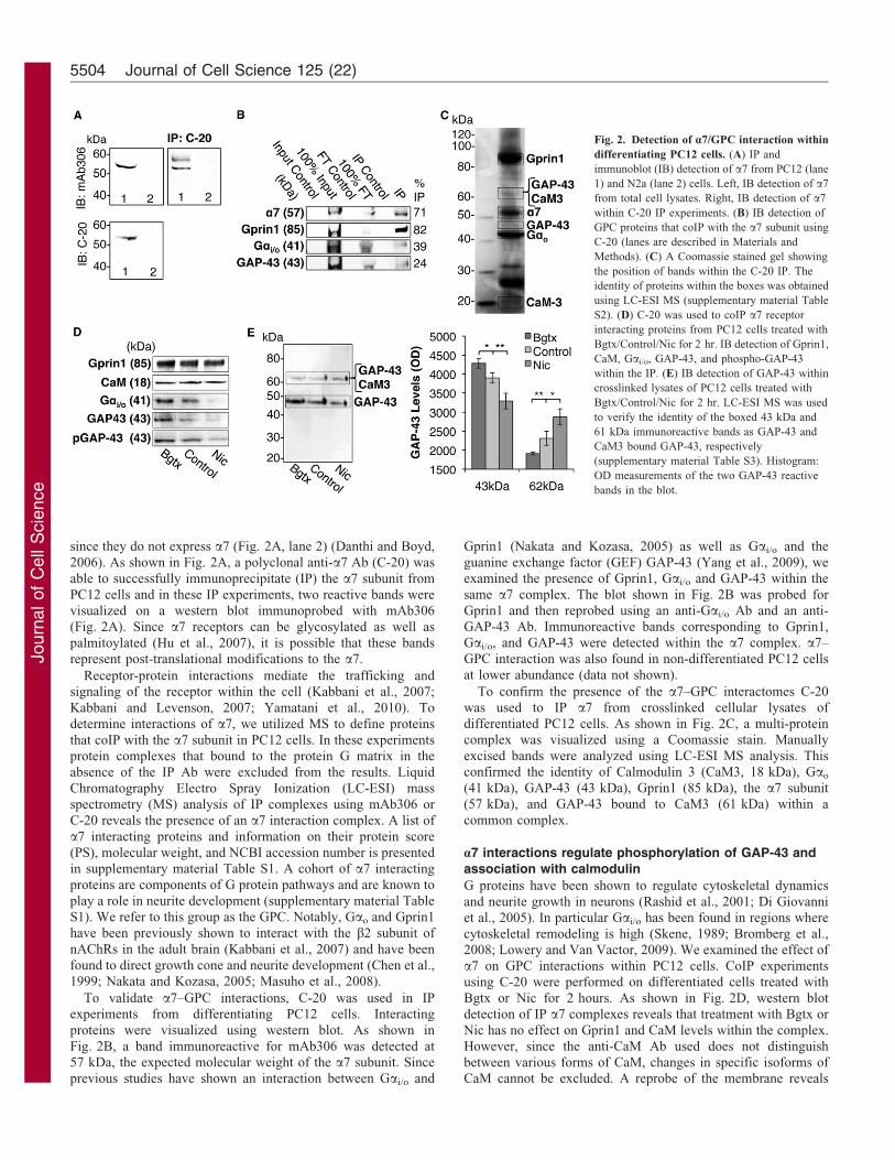
since they do not express a7 (Fig. 2A, lane 2) (Danthi and Boyd,
2006). As shown in Fig. 2A, a polyclonal anti-a7 Ab (C-20) was
able to successfully immunoprecipitate (IP) the a7 subunit from
PC12 cells and in these IP experiments, two reactive bands werevisualized on a western blot immunoprobed with mAb306
(Fig. 2A). Since a7 receptors can be glycosylated as well as
palmitoylated (Hu et al., 2007), it is possible that these bandsrepresent post-translational modifications to the a7.
Receptor-protein interactions mediate the trafficking andsignaling of the receptor within the cell (Kabbani et al., 2007;
Kabbani and Levenson, 2007; Yamatani et al., 2010). To
determine interactions of a7, we utilized MS to define proteins
that coIP with the a7 subunit in PC12 cells. In these experimentsprotein complexes that bound to the protein G matrix in the
absence of the IP Ab were excluded from the results. Liquid
Chromatography Electro Spray Ionization (LC-ESI) massspectrometry (MS) analysis of IP complexes using mAb306 or
C-20 reveals the presence of an a7 interaction complex. A list of
a7 interacting proteins and information on their protein score
(PS), molecular weight, and NCBI accession number is presentedin supplementary material Table S1. A cohort of a7 interacting
proteins are components of G protein pathways and are known to
play a role in neurite development (supplementary material TableS1). We refer to this group as the GPC. Notably, Gao and Gprin1
have been previously shown to interact with the b2 subunit of
nAChRs in the adult brain (Kabbani et al., 2007) and have been
found to direct growth cone and neurite development (Chen et al.,1999; Nakata and Kozasa, 2005; Masuho et al., 2008).
To validate a7–GPC interactions, C-20 was used in IPexperiments from differentiating PC12 cells. Interacting
proteins were visualized using western blot. As shown in
Fig. 2B, a band immunoreactive for mAb306 was detected at57 kDa, the expected molecular weight of the a7 subunit. Since
previous studies have shown an interaction between Gai/o and
Gprin1 (Nakata and Kozasa, 2005) as well as Gai/o and the
guanine exchange factor (GEF) GAP-43 (Yang et al., 2009), weexamined the presence of Gprin1, Gai/o and GAP-43 within the
same a7 complex. The blot shown in Fig. 2B was probed forGprin1 and then reprobed using an anti-Gai/o Ab and an anti-
GAP-43 Ab. Immunoreactive bands corresponding to Gprin1,Gai/o, and GAP-43 were detected within the a7 complex. a7–
GPC interaction was also found in non-differentiated PC12 cellsat lower abundance (data not shown).
To confirm the presence of the a7–GPC interactomes C-20was used to IP a7 from crosslinked cellular lysates of
differentiated PC12 cells. As shown in Fig. 2C, a multi-proteincomplex was visualized using a Coomassie stain. Manually
excised bands were analyzed using LC-ESI MS analysis. Thisconfirmed the identity of Calmodulin 3 (CaM3, 18 kDa), Gao
(41 kDa), GAP-43 (43 kDa), Gprin1 (85 kDa), the a7 subunit(57 kDa), and GAP-43 bound to CaM3 (61 kDa) within a
common complex.
a7 interactions regulate phosphorylation of GAP-43 andassociation with calmodulin
G proteins have been shown to regulate cytoskeletal dynamics
and neurite growth in neurons (Rashid et al., 2001; Di Giovanniet al., 2005). In particular Gai/o has been found in regions where
cytoskeletal remodeling is high (Skene, 1989; Bromberg et al.,2008; Lowery and Van Vactor, 2009). We examined the effect of
a7 on GPC interactions within PC12 cells. CoIP experimentsusing C-20 were performed on differentiated cells treated with
Bgtx or Nic for 2 hours. As shown in Fig. 2D, western blotdetection of IP a7 complexes reveals that treatment with Bgtx or
Nic has no effect on Gprin1 and CaM levels within the complex.However, since the anti-CaM Ab used does not distinguish
between various forms of CaM, changes in specific isoforms ofCaM cannot be excluded. A reprobe of the membrane reveals
Fig. 2. Detection of a7/GPC interaction within
differentiating PC12 cells. (A) IP and
immunoblot (IB) detection of a7 from PC12 (lane
1) and N2a (lane 2) cells. Left, IB detection of a7
from total cell lysates. Right, IB detection of a7
within C-20 IP experiments. (B) IB detection of
GPC proteins that coIP with the a7 subunit using
C-20 (lanes are described in Materials and
Methods). (C) A Coomassie stained gel showing
the position of bands within the C-20 IP. The
identity of proteins within the boxes was obtained
using LC-ESI MS (supplementary material Table
S2). (D) C-20 was used to coIP a7 receptor
interacting proteins from PC12 cells treated with
Bgtx/Control/Nic for 2 hr. IB detection of Gprin1,
CaM, Gai/o, GAP-43, and phospho-GAP-43
within the IP. (E) IB detection of GAP-43 within
crosslinked lysates of PC12 cells treated with
Bgtx/Control/Nic for 2 hr. LC-ESI MS was used
to verify the identity of the boxed 43 kDa and
61 kDa immunoreactive bands as GAP-43 and
CaM3 bound GAP-43, respectively
(supplementary material Table S3). Histogram:
OD measurements of the two GAP-43 reactive
bands in the blot.
Journal of Cell Science 125 (22)5504

Journ
alof
Cell
Scie
nce
that Bgtx increases while Nic decreases the presence of Gai/o
and GAP-43 (Fig. 2D) within the a7 complex indicating that
pharmacological activation of the receptor impacts its interactionwith select GPC proteins.
The phosphorylation of GAP-43 by protein kinase C (PKC)(Esdar et al., 1999) and CaMKII (Denny, 2006; Leu et al., 2010)
or dephosphorylation by calcineurin (PP2B) (Liu and Storm,1989; Madsen et al., 1998) has been shown to regulate CaM
association and neurite growth. In its unphosphorylated form,GAP-43 binds CaM leading to an inhibition in neurite
growth (Skene, 1990; Slemmon et al., 1996). In contrast,phosphorylation of GAP-43 promotes CaM dissociation leading
to an increase in neurite growth (Strittmatter et al., 1994b). An
anti-phospho-GAP-43 Ab (selective for CaMKII phosphorylationsite Serine 41) (Denny, 2006) was used to determine the effects
of Bgtx and Nic on GAP-43. As shown in Fig. 2D, an increase inanti-phospho-GAP-43 immunoreactivity was observed within the
a7 complex following Bgtx treatment. Similarly, Nic wasfound to decrease anti-phospho-GAP-43 immunoreactivity
within the a7 complex. To determine if change in GAP-43phosphorylation correlated with CaM binding, we utilized the
bis(sulfosuccinimidyl) substrate (BS3) to test the effects of Bgtxand Nic on GAP-43/CaM interaction. This method has been used
in the detection of GAP-43 in the CaM bound/free states wherethe GAP-43 band runs at ,18 kDa (the approximate size of
CaM) heavier on an SDS-PAGE gel when bound to CaM (Gambyet al., 1996). In these experiments, cell fractions were crosslinked
following a 2-hour treatment with Bgtx, Nic, or a vehicle control.An anti-GAP-43 Ab was used to visualize two distinct bands
within the sample and MS was used to confirm their identity asGAP-43 [bound to CaM3 (61 kDa) or alone (43 kDa)] (Fig. 2E).
In control cells, the 43 kDa band presented a stronger signal thanthe 61 kDa band suggesting that CaM free GAP-43 is a main
form of GAP-43 within PC12 cells. Densitometric analysis
showed that the band at 43 kDa increased during Bgtx treatmentand decreased during Nic treatment. In contrast, the band at
61 kDa increased during Nic treatment and decreased followingBgtx (Fig. 2E). In light of the differential associations betweena7 and GAP-43 under these conditions, and the observed increasein phospho-GAP-43 reactivity with the a7 complex in the same
cells (Fig. 2D), it is possible that Bgtx promotes interactionbetween a7 and non-CaM bound GAP-43 during growth.
a7 Receptors direct growth through Gai/o and calmodulin
Gai/o has been shown to regulate neurite growth (Strittmatteret al., 1994a; Swarzenski et al., 1996). To examine the role of
Gai/o in a7 signaling, we applied the Gai/o inhibitor Pertussistoxin (Ptx) or the Gao activator Mastoparan (MSP) during NGFdifferentiation (Fig. 3A). We tested the involvement of Gai/o inBgtx mediated neurite growth by exposing cells to both Ptx and
Bgtx during the 4 days of NGF differentiation. As shown inFig. 3B, coapplication of Ptx and Bgtx was found tosignificantly reduce neurite SA when compared to Bgtx
treatment alone (P,0.01). When applied alone Ptx was foundto decrease neurite SA to an average level below control cells(Fig. 3B). Since Nic was found to attenuate neurite growth
during differentiation, we examined the ability of MSP toenhance neurite growth. A 4-day treatment with MSP was foundto significantly increase neurite SA (P,0.01) (Fig. 3B) whencompared to controls confirming the role of Gai/o in neurite
development. In the presence of Nic, MSP was found to onlymoderately promote neurite growth when compared to controls(P50.56) (Fig. 3B) suggesting that MSP and Nic function
antagonistically within the cell. Group significance was foundbetween control, Ptx and MSP treatments [F(2,135)54.03,P,0.001]. These studies underscore the role for Gai/o in neurite
development and suggest a functional interaction between Gai/o
and a7 in growth.
Fig. 3. Elucidating a GPC pathway
mechanism in neurite growth. (A) A schematic
diagram of Gai/o function within the a7 pathway.
Drugs and their targets are shown in bold italics.
(B) Average neurite SA for cells chronically
treated with Bgtx, Nic, Ptx, MSP, a combination
of Ptx and Bgtx or MSP and Nic. (C) A
schematic diagram of CaM, CaMKII, and PP2B
within the a7 pathway. Drugs and their targets
are shown in bold italics. (D) Average neurite SA
for cells chronically treated with Bgtx, CMZ, or a
combination of CMZ and Bgtx. (E) Average
neurite SA for cells treated with Bgtx, Nic,
FK506, KN-93, or a combination of FK506 and
Nic, FK506 and Bgtx, KN-93 and Bgtx, or
FK506 and KN-93.
a7-nAChR–GPC in neurite growth 5505

Journ
alof
Cell
Scie
nce
The ubiquitous calcium sensor protein CaM (Liu and Berg,
1999; D’Alcantara et al., 2003) and a number of CaM regulated
phosphoproteins including PP2B and CaMKII were identified as
components of the a7 interaction network (supplementary
material Table S1). We utilized drugs that block CaM, PP2B,
and CaMKII to test the role of these proteins in a7 mediated
neurite growth (Fig. 3C). As shown in Fig. 3D, we exposed cells
to the CaM inhibitor calmidazolium (CMZ) during NGF
differentiation. CMZ has also been shown to have some CaM
independent effects including activation of phospholipase 2A and
regulation of intracellular Ca2+ stores (Duffy et al., 2011). We
find that CMZ produced a significant decrease in neurite
SA when compared to controls (P,0.01), confirming that
attenuation of CaM activity reduces neurite growth (Jian et al.,
1994). To confirm the effects of CMZ on a7 signaling, we
coapplied Bgtx and CMZ during differentiation. As shown in
Fig. 3D, this combination was found to block the effects of Bgtx
on neurite growth suggesting an important role for CaM in a7
signaling.
To investigate the role of PP2B and CaMKII, we probed the
morphological effects of FK506 and KN-93, which respectively
target these two proteins in cells (Liu and Berg, 1999;
Jouvenceau and Dutar, 2006). As shown in Fig. 3E, application
of FK506 was found to significantly increase neurite SA
(P,0.001). This effect of FK506 was enhanced in the presence
of Bgtx (P,0.001) and diminished in the presence of Nic
(P50.87), suggesting that Nic and PP2B operate synergistically
to inhibit growth (Fig. 3E). Similarly, application of KN-93 was
found to significantly diminish neurite SA when compared to
control cells (P,0.001), suggesting that CaMKII activity is
required for growth (Fig. 3E). Group significance was found
between control, FK506, KN-93, and CMZ [F(3,179)53.36,
P,0.001]. When combined with Bgtx, KN-93 was found to
block the effects of Bgtx on neurite SA (Fig. 3E) (P.0.05).
These findings suggest that CaMKII operates downstream of a7
receptors in PC12 cells and is required for Bgtx mediated neurite
growth. Finally, to confirm that PP2B and CaMKII operate
within a common pathway (Fig. 3C), we coapplied KN-93 and
FK506. This combination did not produce a significant change in
neurite SA when compared to controls (Fig. 3E), suggesting that
PP2B and CaMKII act antagonistically.
Gprin1 promotes a7 nAChR signaling and neurite growth
The Gai/o interactor Gprin1 has been shown to regulate neurite
growth in cells (Masuho et al., 2008; Ge et al., 2009). We
examined interactions between a7 and Gprin1 in differentiating
PC12 cells. Using a western blot we confirmed the ability of an
anti-Gprin1 Ab to IP Gprin1 as well as a7 from cells (Fig. 4A).
a7/Gprin1 interactions were further validated by heterologous
protein expression studies in N2a cells, which do not express a7
(Danthi and Boyd, 2006). In these experiments we transiently
transfected 1–10 mg of a7 cDNA (per 10 cm Petri dish of N2a
cells) and tested for interaction with endogenous Gprin1 24 hrs
later. As shown in Fig. 4B, a7 proteins were successfully
detected in N2a cells after transfection. An IP of the a7 subunit
from N2a cells reveal its association with endogenous Gprin1. A
reverse IP of Gprin1 from N2a cells was also found to maintain
an association with transfected a7 subunits (Fig. 4B).
We used a genetic knockdown strategy to test if Gprin1
mediates a7/GPC interaction. In these experiments,
differentiating PC12 cells were transfected with plasmids
encoding Gprin1 short interfering RNA (siRNA) (Gprin1siRNA)
(Ge et al., 2009). Western blot detection of Gprin1 within
transfected cells reveals that Gprin1siRNA reduces Gprin1 levels
Fig. 4. a7/Gprin1 association within
differentiating cells. (A) IB detection of a7 and
Gprin1 within an IP of PC12 cells using an anti-
Gprin1 Ab (lanes are described in Materials and
Methods). (B) N2a cells were transiently
transfected with 0, 1 and 10 mg of a7 cDNA. IB
detection of a7 and Gprin1 from (1) total cell
lysates, (2) C-20 IP, (3) Gprin1 IP of transfected
cells. (C) IB detection of Gprin1, Gai/o, and
GAP-43 within a C-20 IP. PC12 cells were
transfected with Gprin1siRNA or an empty vector
(Control). (D) A Coomassie stained gel showing
the location of proteins that coIP with Gprin1.
LC-ESI MS was used to identify proteins within
boxed bands (supplementary material Table S4).
(E,F) Confocal images of PC12 differentiated
with NGF for 2 (E) and 4 (F) days. Cells were
labeled with fBgtx (green), an anti-Gprin1 Ab
(red), and an anti-2g13 Ab (blue). Merge panel
shows colocalization of the three proteins within
the cell. Insets: magnifications of soma (1),
neurite (2), and a neurite terminal (3).
Journal of Cell Science 125 (22)5506
Journ
alof
Cell
Scie
nce
by 85% within cells (Fig. 6A). An IP of a7 was performed using
C-20. As shown in Fig. 4C, in cells transfected with Gprin1siRNA, a7 interactions with Gprin1, Gai/o and GAP-43 appeared
lower than control cells transfected with the empty vector. These
findings suggest that Gprin1 expression plays a role in GPC
interaction with a7. To test this, LC-ESI MS was used to analyzeproteins that coIP with Gprin1 from PC12 cells. As shown in
Fig. 4D, at least six discrete bands were visualized by a
Coomassie stain of an SDS-PAGE gel loaded with proteins
eluted from an anti-Gprin1 IP experiment. Based on MSidentification, Gprin1, a7, CaM3, Gai/o and GAP-43 were
successfully detected within the Gprin1 IP experiment from PC12
cells, confirming Gprin1 interactions with a7 and GPC molecules.
We used fluorescein-conjugated Bgtx (fBgtx) and anti-Gprin1
Abs to colocalize endogenous a7 and Gprin1 within
differentiating cells, respectively. As shown in Fig. 4E,F, a7and Gprin1 proteins were found to colocalize within soma and
growing neurites of differentiating PC12 cells. At 2 days of
differentiation, Gprin1/a7 colocalization appeared prominent in
the cytoplasm and near the cell surface and was seen uniformlydistributed along the growing neurite and within the neurite
terminal, which also stained positive for the growth cone marker
2G13 (Fig. 4E). At 4 days of differentiation, Gprin1/a7
colocalization was seen near the plasma membrane of the somaand in the growing neurites. However at this later stage of
development, colocalization along the growing neurites appeared
more punctuate and localized (Fig. 4F, boxes 2, 3).
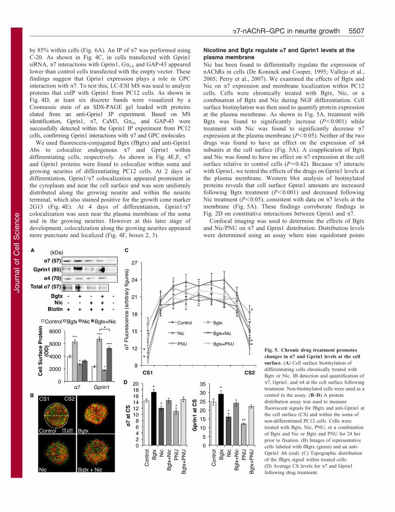
Nicotine and Bgtx regulate a7 and Gprin1 levels at theplasma membrane
Nic has been found to differentially regulate the expression ofnAChRs in cells (De Koninck and Cooper, 1995; Vallejo et al.,
2005; Perry et al., 2007). We examined the effects of Bgtx andNic on a7 expression and membrane localization within PC12
cells. Cells were chronically treated with Bgtx, Nic, or acombination of Bgtx and Nic during NGF differentiation. Cell
surface biotinylation was then used to quantify protein expressionat the plasma membrane. As shown in Fig. 5A, treatment withBgtx was found to significantly increase (P,0.001) while
treatment with Nic was found to significantly decrease a7expression at the plasma membrane (P,0.05). Neither of the two
drugs was found to have an effect on the expression of a4subunits at the cell surface (Fig. 5A). A coapplication of Bgtx
and Nic was found to have no effect on a7 expression at the cellsurface relative to control cells (P50.42). Because a7 interacts
with Gprin1, we tested the effects of the drugs on Gprin1 levels atthe plasma membrane. Western blot analysis of biotinylated
proteins reveals that cell surface Gprin1 amounts are increasedfollowing Bgtx treatment (P,0.001) and decreased following
Nic treatment (P,0.05), consistent with data on a7 levels at themembrane (Fig. 5A). These findings corroborate findings inFig. 2D on constitutive interactions between Gprin1 and a7.
Confocal imaging was used to determine the effects of Bgtxand Nic/PNU on a7 and Gprin1 distribution. Distribution levels
were determined using an assay where nine equidistant points
Fig. 5. Chronic drug treatment promotes
changes in a7 and Gprin1 levels at the cell
surface. (A) Cell surface biotinylation of
differentiating cells chronically treated with
Bgtx or Nic. IB detection and quantification of
a7, Gprin1, and a4 at the cell surface following
treatment. Non-biotinylated cells were used as a
control in the assay. (B–D) A protein
distribution assay was used to measure
fluorescent signals for fBgtx and anti-Gprin1 at
the cell surface (CS) and within the soma of
non-differentiated PC12 cells. Cells were
treated with Bgtx, Nic, PNU, or a combination
of Bgtx and Nic or Bgtx and PNU for 24 hrs
prior to fixation. (B) Images of representative
cells labeled with fBgtx (green) and an anti-
Gprin1 Ab (red). (C) Topographic distribution
of the fBgtx signal within treated cells.
(D) Average CS levels for a7 and Gprin1
following drug treatment.
a7-nAChR–GPC in neurite growth 5507

Journ
alof
Cell
Scie
nce
were measured along the axis of a 2D image of a PC12 cell
stained for fBgtx and anti-Gprin1 (de Lucas-Cerrillo et al., 2011)
(Fig. 5B). Individual fluorescence signals for Gprin1 and fBgtx
were significantly increased at the cell surface in cells chronically
treated with Bgtx relative to controls. In contrast, fluorescence
signals appeared weaker at the cell surface of cells chronically
treated with Nic/PNU relative to controls (Fig. 5B–D; data not
shown). Statistical significance was established between control,
Bgtx, and Nic/PNU treatment groups: a7 [F(3,312)53.32,
P,0.001] and Gprin1 [F(3,320)53.32, P,0.001]. In cells that
received a combination of Bgtx and Nic/PNU, the signal for
fBgtx and Gprin1 did not significantly differ from controls
(Fig. 5B–D), confirming the opposing effect of these drugs on
a7/Gprin1 levels at the plasma membrane.
Gprin1 expression regulates neurite growth
We utilized a genetic strategy to examine the role of Gprin1 on
neurite growth in PC12 cells. In these experiments, cells were
transiently transfected with plasmids encoding full length Gprin1
in pcDNA3.1 (Gprin1pcDNA) or Gprin1siRNA (Ge et al., 2009)
prior to differentiation. Transfected cells were then differentiated
using NGF in the presence of Bgtx or Nic. Western blot detection
of Gprin1 within transfected cells reveals that Gprin1pcDNA
increases total Gprin1 expression by 98% whereas Gprin1siRNA
decreases Gprin1 levels by 85% within cells (Fig. 6A). Total a7
levels were also increased in cells transfected with Gprin1pcDNA
and modestly decreased in cells transfected with Gprin1siRNA
(Fig. 6A). We did not observe a significant difference in Gprin1
protein expression between cells transfected with Gprin1siRNA1
and Gprin1siRNA3 (Fig. 6A). Cell surface biotinylation was used to
determine the effects of Gprin1pcDNA and Gprin1siRNA on a7 and
Gprin1 levels at the plasma membrane. As shown in Fig. 6B,
Gprin1pcDNA was found to significantly increase while Gprin1siRNA1/3
was found to significantly decrease a7 and Gprin1 levels at the cell
surface. In these experiments, transfection of the empty vector did not
alter the expression or the localization of a7 and Gprin1 within the
cell (Fig. 6, control lanes).
Cellular imaging and morphometric analysis of cells expressing
Gprin1pcDNA and Gprin1siRNA3 reveals a dominant role for Gprin1 on
a7 mediated neurite growth. As shown in Fig. 6C,D, transfection with
Gprin1pcDNA was found to significantly increase neurite SA
(P,0.001) while transfection with Gprin1siRNA3 was found to
significantly decrease neurite SA (P,0.05) in differentiated PC12
cells relative to controls. To examine the effects of Gprin1 on a7
mediated growth, we chronically treated cells with Bgtx, Nic, or PNU
and compared their effect between cells transfected with Gprin1pcDNA,
Gprin1siRNA3, or an empty vector control. As shown in Fig. 6C,D,
Gprin1siRNA3 was found to abolish the effect of Bgtx while
Gprin1pcDNA was found to abolish the effect of Nic/PNU on neurite
growth suggesting that Gprin1 expression at the cell surface is a main
determinant of neurite growth and drug effect within these cells.
a7 Receptors interact with GPC and modulate axonal
growth in cortical neurons
We tested the properties of Nic and Bgtx as well as ACh and
KYNA on neurite development in primary cortical neurons.
Using anti-MAP-2 and anti-Tau-1 Abs we analyzed the growth of
dendrites and axons, respectively. Chronic treatment with nAChR
Fig. 6. Gprin1 regulates a7 levels at the cell
surface and directs neurite growth in
differentiating cells. PC12 were transiently
transfected with plasmids for Gprin1cDNA,
Gprin1siRNA, or an empty vector (Control).
(A) Total protein levels of Gprin1 and a7 in
transfected cells based on IB detection. (B) a7
and Gprin1 bands within biotinylated cells
showing changes in protein levels at the cell
surface. Non-biotinylated cells were used as a
background control in the assay.
(C,D) Transfected cells were chronically
treated with Bgtx, Nic, or PNU. (C) Images of
representative cells stained with anti-a-Tubulin
Abs. (D) Histogram showing the effect of
chronic drug treatment on neurite SA in
Gprin1pcDNA or Gprin1siRNA transfected cells.
Journal of Cell Science 125 (22)5508

Journ
alof
Cell
Scie
nce
agonists (Nic/PNU/ACh) was found to significantly decrease
axonal SA (P,0.05) while chronic treatment with a7 antagonists
(Bgtx/KYNA) was found to significantly increase axonal growth
and complexity of axons (P,0.001) (Fig. 7A,B). Group
significance was found between control, Bgtx, Nic, PNU,
KYNA, and ACh [F(5,162)52.77, P,0.001]. Combining
agonists and antagonists did not alter axonal growth relative to
controls neurons (Bgtx and Nic, P50.40; Bgtx and PNU,
P50.23; KYNA and ACh, P50.79), and none of the drugs
appeared to have a statistically significant effect on dendritic
growth (data not shown).
Association between a7 and GPC proteins was confirmed in
cortical neurons. As shown in Fig. 7C, a C-20 IP of the a7 subunit
from cortical cells reveals the presence of Gprin1, Gai/o, and GAP-
43 in association with a7 nAChRs. Interaction with Gprin1 was
further validated using an anti-Gprin1 Ab that was able to coIP
Gprin1 as well as a7 from the same neuronal lysates (Fig. 7C).
Interaction between a7 and Gprin1 is likely to play an important
role in axonal development as suggested by triple labeling
fluorescence using fBgtx, anti-Gprin1, and phalloidin. As shown
in Fig. 7D, colocalization of a7 and Gprin1 was seen in growing
axons and within growth cones of cortical neurons suggesting a
role for these proteins in axonal growth and guidance.
DiscussionTo delineate mechanisms of a7 receptor function during
development, we have used proteomics to define intracellular
interactions of the a7 receptor within growing cells. PC12 cells
have proven useful in studies of neuronal development since
they endogenously express many neurotransmitter receptors
(Connolly et al., 1979; Sarma et al., 2003). Once differentiated
with NGF, PC12 cells extend elaborate neurites with the
properties of growing axons (Drubin et al., 1985; Lee et al.,
1998). We find that Bgtx and KYNA promote neurite growth in
PC12 cells while enhancing axonal length and complexity in
primary cortical neurons. ACh/Nic/PNU in contrast are found to
attenuate neurite and axonal growth in PC12 cells and cortical
neurons, respectively. It is noteworthy, that none of the drug
treatment conditions appeared to significantly alter the
percentage of cells with at least one process longer than the
cell body (an inclusion criteria in the analysis) thus confirming
that a7 receptor activity is associated with changes in neurite
growth within differentiating cells. Our findings are consistent
with earlier observations on the inhibitory effects of ACh on
neurite development (Hieber et al., 1992; Lauder and Schambra,
1999) and suggest a role for a7 in structural and synaptic
development (Corriveau et al., 1995; Romano et al., 1997).
However, since nicotine and ACh activate other cholinergic
receptors, the contribution of non-a7 receptors to neurite growth
cannot be excluded. Likewise, the observed effects of KYNA on
neurite and axonal growth (Figs 1, 7) complement data obtained
from Bgtx treatment, and suggest that a7 inactivation plays a role
in cellular function. Since KYNA is found to inactivate a7 and
NMDA receptors (Pereira et al., 2002; Stone and Darlington,
Fig. 7. Interactions of a7 and GPC in
cortical neurons modulate axonal growth.
Cortical neurons were cultured for 4 days in the
presence of drugs. (A) An analysis of axonal
SA in cortical neurons following chronic drug
treatment. (B) Black and white contrast images
of drug treated cells stained with anti-Tau-1
Abs in order to visualize growing axons. Soma
indicated by a blue dot. (C) IB detection of a7
and GPC proteins within cortical neurons.
Lysates from cortical neurons were IP using C-
20 or anti-Gprin1 Abs. (D) A representative
neuron colabeled with fBgtx and an anti-Gprin1
Ab. Single panels show the localization of a7
and Gprin1 within the soma and a growing
axon. Merge panel shows colocalization of a7,
Gprin1, and phalloidin within the cell. Insets:
magnification of the axon (1) and the growth
cone (2).
a7-nAChR–GPC in neurite growth 5509

Journ
alof
Cell
Scie
nce
2002; Alkondon et al., 2007), it is possible KYNA contributes to
synaptic development via its actions on these synaptic receptors(Lin et al., 2010).
Long-term cellular adaptations in neurotransmission includechanges in receptor synthesis, turnover, and trafficking to and
from the plasma membrane (Spitzer, 2012). To date little isknown about the processes that direct nAChR synthesis andtrafficking within growing cells. Studies have shown an effect of
chronic Nic on the expression and composition of severalnAChRs in various cell types (De Koninck and Cooper, 1995;Perry et al., 2007). Our data shows that chronic Nic decreases a7
while chronic Bgtx increases a7 expression at the cell surface indifferentiating PC12 cells. Whether such drug-induced changesin receptor expression at the membrane correlate with changes inreceptor activity such as desensitization or structural conformation is
not clear. However, changes in a7 receptor levels at the membrane arefound to shift downstream signaling via a Gprin1/GPC pathwayproducing important effects on neurite growth.
Gprin1 scaffolds a7/GPC signaling during growth
The Gai/o interactor Gprin1 has been shown to localize tosubcellular domains where cytoskeletal remodeling demands are
high (Chen et al., 1999; Ge et al., 2009). Gprin1 association withthe plasma membrane appears dependent on palmitoylation ofcysteine residues within the G protein-binding site (Nakata and
Kozasa, 2005). This along with its established role as a mediatorof cytoskeletal regulating G proteins such as Cdc42 and Rac(Nakata and Kozasa, 2005; Masuho et al., 2008) suggests that
Gprin1 is vital in neurite growth. We demonstrate an interactionbetween a7 and Gprin1 in growing cells and propose that thisinteraction links a7 to a GPC signaling apparatus as supported bythe following key observations: (i) the ability of an anti-Gprin1
Ab to coIP a7 and GPC proteins from neurons and differentiatingcells; (ii) colocalization of a7 and Gprin1 within growingneurites, axons, and growth cones; (iii) the ability of Gprin1
siRNA to decrease a7 expression at the cell surface or within thecell, reverse the effect of Bgtx on neurite growth, and attenuateinteraction between a7 and GPC proteins. These findings confirm
an important role for Gprin1 in scaffolding a7 receptors at thecell surface as shown for opioid receptors (Ge et al., 2009) andsuggest that Gprin1 interactions facilitate the effects of a7 ongrowth. In addition, it is interesting to consider that Gprin1
interacts with b2 nAChRs (Kabbani et al., 2007) also present inPC12 cells, which may explain why changes in a7 expression atthe cell surface are not entirely consistent with changes in Gprin1
levels at the cell surface following nicotine treatment (Fig. 5).
The existence of an a7–GPC network supports findings onassociations between nAChRs and G protein signaling in cells(Kabbani et al., 2007; Paulo et al., 2009). Based on the current
study we present a model for a7 signaling via the GPC duringneurite development (Fig. 8). In this model we propose thatligand-induced changes in a7 levels (or conformation) at the cell
surface direct GPC signaling towards inhibiting or promotingneurite growth. As shown in Fig. 8, drug-induced changes inreceptor levels at the cell surface appear to accompany an overall
effect on protein synthesis associated with growth. In onescenario, a decrease in a7 at the plasma membrane is found topromote inhibition of GPC function via the calcium sensitive
phosphatase PP2B that is activated under conditions of lowcalcium levels (Stefan et al., 2008). As depicted in Pathway A,PP2B dephosphorylation of its target protein GAP-43 functions
as an inhibitory switch for Gai/o within the GPC pathway
(Strittmatter et al., 1994b) and mediates interaction with CaM
(Skene, 1990; Slemmon et al., 1996). This pathway is consistent
with the finding that chronic Nic promotes enhanced GAP-43/
CaM association and that a PP2B inhibitor (FK506) effectively
blocks Nic induced neurite changes in PC12 cells.
In a second scenario, an increase in a7 levels at the plasma
membrane appears to promote the GPC via CaMKII, which is
activated under cellular conditions of higher calcium than PP2B
(Wen et al., 2004). The contributions of CaMKII to this pathway,
however, are supported by the ability of a CaMKII inhibitor to
block Bgtx mediated neurite growth, because other calcium
sensitive kinases such as PKC, also identified in our proteomic
screen, can contribute to neurite growth in this scenario. As
illustrated in Pathway B, CaMKII phosphorylation of its target
protein GAP-43 functions as an activator of Gai/o signaling
within the cell. CaMKII phosphorylation of GAP-43 has also
been shown to promote its dissociation from CaM (Denny, 2006;
Leu et al., 2010), a finding consistent with our crosslinking
experiments showing an effect of Bgtx on GAP-43/CaM
dissociation within the cell (Fig. 2). This pathway is consistent
with the effect of chronic Bgtx treatment on GAP-43
phosphorylation, or possibly the affinity of the a7 nAChR
complex for phospho-GAP-43 interaction, as well as proteomic
evidence showing that Bgtx mediates dissociation of GAP-43 and
CaM within differentiating cells.
The proposed model explores the function of a novel GPC
signaling pathway downstream of a7 nAChRs and alludes to the
contributions of localized calcium signaling on the a7/GPC
network. As suggested in Fig. 8, changes in calcium levels, from
various sources including the a7 receptor may provide a trigger
for the activity of high or low affinity calcium sensors such as
PP2B or CaMKII, respectively. However calcium sources and
Fig. 8. A model for a7–GPC interactions in neurite growth. Exposure to
agonist or antagonist is associated with changes in a7/Gprin1 levels at the
plasma membrane. Pathway A: A decrease in a7/Gprin1 levels at the cell
surface results in GPC inhibition via PP2B and a reduction in cytoskeletal
mediated neurite growth. Pathway B: Increased a7/Gprin1 levels at the cell
surface result in GPC activation via CaMKII and an increase in cytoskeletal
mediated neurite growth. Both pathways are likely impacted by Ca2+ entry
from external and internal sources.
Journal of Cell Science 125 (22)5510

Journ
alof
Cell
Scie
nce
targets within the pathway are not yet determined. Data presentedin Fig. 3 for example, does not entirely demonstrate that PP2Band CaMKII function exclusively downstream of the a7 receptor
in differentiating PC12 cells. Pharmacological suppression ofthese molecules appears slightly incomplete or additive in someexperiments suggesting a function of two (or more) possible
pathways in parallel within differentiating PC12 cells.
Materials and MethodsCell culture
Pheochromocytoma line 12 (PC12) cells were grown on a rat collagen (50 mg/mL,Gibco) matrix in dMEM containing 10% horse serum, 5% fetal bovine serum(FBS), and 1% penicillin-streptomycin (Pen-strep) antibiotic. Cells weredifferentiated with 10 nM 2.5S nerve growth factor (NGF) for 4 days unlessotherwise noted (Prince Laboratories). Primary neuronal cultures were obtainedfrom embryonic day 18 Sprague-Dawley rats similar to what is described (Linet al., 2001). Primary cultures were grown in Neurobasal media with B27supplement and 1% Pen-strep. Human a7 in pcDNA1neo was provided by Dr NeilMillar (University College London) and has been previously described (Grabeet al., 2012a). Mammalian expression vectors encoding Gprin1 in pcDNA3.1 andGprin1 siRNA in pRNAT H1.1 were provided by Dr Law (Univ. Minnesota) (Geet al., 2009). These vectors express green fluorescent protein (GFP) under the samepromoter (Ge et al., 2009). PC12 cells and Neuroblastoma 2a (N2a) cells weretransfected using Lipofectamine 2000 as described by the manufacturer(Invitrogen). Cells were transfected with 1–10 mg/cm2 a7 cDNA, 0.5 mg/cm2
GFP-Gprin1 cDNA, or 20 pmol/cm2 GFP-Gprin1 siRNA for 16 hours prior tomedia change. Transfected cells were identified using GFP fluorescence. Anempty plasmid was used as a transfection control.
Drug treatment
Drug concentrations were determined based on published studies (Chan and Quik,1993; Jian et al., 1994; Strittmatter et al., 1994a; Owen and Bird, 1995; Swarzenskiet al., 1996; Hilmas et al., 2001; Kano et al., 2002; Hruska and Nishi, 2007; Lopeset al., 2007; Takemura et al., 2009): a-Bungarotoxin (Bgtx) (10 nM, Sigma),nicotine (Nic) (50 mM, Sigma), PNU282987 (PNU) (50 mM, Sigma), pertussistoxin (Ptx) (1 mM, Calbiochem), mastoparan (30 mM, Tocris), FK506 (25 nM,Tocris), KN-93 (1 mM, Tocris), calmidazolium (CMZ) (1 mM, Enzo),acetylcholine (ACh) (100 mM, Sigma) and KYNA (1:100 mM, Sigma). Theduration of the chronic drug treatment described in this study is 4 days unlessotherwise noted. All experiments were performed in triplicate and the datapresented represents the average for each condition. At the end of each experimentcell viability was determined using Trypan Blue (EMD).
Immunocytochemistry
Cells were fixed using a 4% paraformaldehyde solution (250 mM sucrose, 25 mMHEPES HCO3 free, 2.5 mM KCl; pH 7.2). Cells were permeabilized at roomtemperature using 0.1% Triton X-100 then immunostained overnight at 4 C usingone or several of the following primary antibodies (Abs): anti-a-Tubulinpolyclonal (Sigma); anti-Gprin1 polyclonal (Abcam), anti-2g13 monoclonal(Serotec), anti-Tau-1 monoclonal (Millipore), anti-MAP-2 polyclonal (Abcam).Secondary Abs: carbocyanine (Cy) 2/3; Dylight 488; Dylight 560; and AlexaFluor647 were purchased from Jackson-ImmunoResearch. a7 receptors were alsovisualized using a fluorescein-conjugated Bgtx (fBgtx) (Molecular Probes).Rhodamine Phalloidin was purchased from Cytoskeleton. All immunostainingswere visualized using a Nikon Eclipse 80i confocal microscope fitted with a NikonC1 CCD camera and images captured using AxioVision and EZ-C1 software.
Morphological and statistical analysis
Morphological reconstruction was performed using Neuromantic software (Myattand Nasuto, 2008). Surface area (SA) was measured from cellular compartments(soma and neurites) to assess size and complexity under various conditions. 20–30cells were reconstructed per condition and group averages were derived from threeseparate experiments. An image assessment of cells using differential interferencecontrast (DIC) microscopy confirmed an overlap of the tubulin immunosignal andthe cellular contour. Statistical analysis was conducted on raw data of imagetracing using Neuromantic. Statistical values have been obtained using a Student’st test or one-way ANOVA. Asterisks indicate statistical significance in a pairedStudent’s t test, two tailed P value, * ,0.05; ** ,0.01; *** ,0.001. Error barsindicate standard error of the mean (SEM).
Protein isolation and detection
Cell lysates were obtained from proteins solubilized in a non-denaturing lysisbuffer (1% Triton X-100, 137 mM NaCl, 2 mM EDTA, and 20 mM Tris-HClpH 8) with protease inhibitor cocktail (Complete) at 4 C for 1 hour. Membranefractions and immunoprecipitation of receptor complexes were conducted
similarly to earlier described experiments (Kabbani et al., 2007).Immunoprecipitated (IP) complexes were obtained using an anti-a7 subunitmonoclonal Ab (mAb306) (Lindstrom et al., 1990), an anti-a7 polyclonal Ab (C-20) [Santa Cruz Biotechnology (SC)], or an anti-Gprin1 rabbit polyclonal Ab(Abcam). Lysates incubated with the bead matrix, without an Ab, were used as IPcontrols. Membranes were blocked with 5% nonfat milk/BSA (forbis(sulfosuccinimidyl) substrate (BS3, Pierce) crosslinking or biotinylationexperiments) then probed with primary Abs overnight at 4 C. Gprin1 (Abcam);Gai/o (SC); GAP-43 (Abcam); pGAP-43 (Abcam); a7 (SC); calmodulin (CaM,Millipore); a-Tubulin (Sigma); Actin (Sigma). Species-specific peroxidaseconjugated secondary Abs were purchased from (Jackson-ImmunoResearch).Signals were detected using a SuperSignal West Pico Chemiluminescent Substrate(Thermo Scientific). SeeBlue and MagicMark (Invitrogen) were used as molecularweight standards. Blots were imaged using a Gel Doc Imaging system (Bio-Rad).Band density analysis in western blots was measured using NIH ImageJ. Fig. 2B,Fig. 4A, Fig. 7C: Input Control, no lysate; 100% Input, total lysate as used in theIP; FT Control, flow-through supernatant of Ab/bead conjugation; 100% FT; flow-through supernatant of IP; IP Control, no Ab; % IP5IP/Input6100. Western blotvalues are based on averages from three separate experiments.
Protein crosslinking and cell surface biotinylation
For substrate bound analysis of protein complexes, the irreversible crosslinker BS3
(Pierce) was incubated with cell lysates at a concentration of 2.5 mM BS3 for30 min at room temp. The reaction was quenched using 50 mM Tris-HCl pH 7.5then loaded onto Ab conjugated Protein G Dynabeads for IP of the crosslinkedsamples. The quantification of proteins at the cell surface was conducted using acell surface biotinylation labeling protocol (Hannan et al., 2008). In theseexperiments 300 mg/mL EZ-Link Sulfo-NHS-LC-Biotin (Pierce) was used to labelcell surface proteins in live cells for 30 min at 4 C. The biotin reaction wasquenched using Tris-Buffered Saline (TBS) and membrane fractions were isolatedfor pull-down with a Neutravidin agarose bead matrix (Pierce) over a 30 mm poresize Snap Cap spin column (Pierce). Cell surface proteins were analyzed usingwestern blot. Experiments were performed in triplicate.
Mass spectrometry
Protein analysis was conducted using Liquid Chromatography ElectrosprayIonization (LC-ESI) mass spectrometry (MS). Protein complexes were preparedas described (Kaiser et al., 2008) and mass spectrometry was carried out using apublished protocol (Gutierrez et al., 2007). Tandem mass spectra collected byXcalibur (version 2.0.2) were searched against the NCBI rat protein database usingSEQUEST (Bioworks software from ThermoFisher, version 3.3.1). The SEQUESTsearch results were filtered using the following criteria: minimum X correlation(XC) of 1.9, 2.2, and 3.5 for 1+, 2+, and 3+ ions, respectively, and DCn .0.1. TheProtein Score (PS) represents the XC where scores ,0.1 were excluded from theanalysis and does not reflect the quantity of a protein in the sample.
AcknowledgementsWe thank Sarah Clark and Dylan van Kampen for assistance with thestudy, and the Center for Applied Proteomics and Molecular Medicine atGeorge Mason University for the generous use of instrumentation.
FundingThis study was supported by a Jeffress Memorial Research GrantAward [grant number J-953 to N.K.].
Supplementary material available online at
http://jcs.biologists.org/lookup/suppl/doi:10.1242/jcs.110379/-/DC1
ReferencesAlbuquerque, E. X., Pereira, E. F., Alkondon, M. and Rogers, S. W. (2009). Mammalian
nicotinic acetylcholine receptors: from structure to function. Physiol. Rev. 89, 73-120.
Alkondon, M., Pereira, E. F., Potter, M. C., Kauffman, F. C., Schwarcz, R. and
Albuquerque, E. X. (2007). Strain-specific nicotinic modulation of glutamatergictransmission in the CA1 field of the rat hippocampus: August Copenhagen Irishversus Sprague-Dawley. J. Neurophysiol. 97, 1163-1170.
Aramakis, V. B. and Metherate, R. (1998). Nicotine selectively enhances NMDAreceptor-mediated synaptic transmission during postnatal development in sensoryneocortex. J. Neurosci. 18, 8485-8495.
Berg, D. K. and Conroy, W. G. (2002). Nicotinic a7 receptors: synaptic options anddownstream signaling in neurons. J. Neurobiol. 53, 512-523.
Berg, D. K., Conroy, W. G., Liu, Z. and Zago, W. M. (2006). Nicotinic signaltransduction machinery. J. Mol. Neurosci. 30, 149-152.
Bromberg, K. D., Iyengar, R. and He, J. C. (2008). Regulation of neurite outgrowth byGi/o signaling pathways. Front. Biosci. 13, 4544-4557.
Chan, J. and Quik, M. (1993). A role for the nicotinic a-bungarotoxin receptor inneurite outgrowth in PC12 cells. Neuroscience 56, 441-451.
a7-nAChR–GPC in neurite growth 5511
Journ
alof
Cell
Scie
nce
Changeux, J. P. (2010). Nicotine addiction and nicotinic receptors: lessons fromgenetically modified mice. Nat. Rev. Neurosci. 11, 389-401.
Chen, L. T., Gilman, A. G. and Kozasa, T. (1999). A candidate target for G proteinaction in brain. J. Biol. Chem. 274, 26931-26938.
Connolly, J. L., Greene, L. A., Viscarello, R. R. and Riley, W. D. (1979). Rapid,sequential changes in surface morphology of PC12 pheochromocytoma cells inresponse to nerve growth factor. J. Cell Biol. 82, 820-827.
Coronas, V., Durand, M., Chabot, J. G., Jourdan, F. and Quirion, R. (2000).Acetylcholine induces neuritic outgrowth in rat primary olfactory bulb cultures.Neuroscience 98, 213-219.
Corriveau, R. A., Romano, S. J., Conroy, W. G., Oliva, L. and Berg, D. K. (1995).Expression of neuronal acetylcholine receptor genes in vertebrate skeletal muscleduring development. J. Neurosci. 15, 1372-1383.
Csillik, B., Mihaly, A. and Knyihar-Csillik, E. (2004). Cytochemical correlates of thesleep-wake interface: concerted expression of brain-derived nitric oxide synthase(bNOS) and the nicotinic acetylcholine receptor (nAChR) in a columnoidorganization of the primate prefrontal cortex. Ann. Anat. 186, 217-221.
D’Alcantara, P., Schiffrann, S. N. and Swillens, S. (2003). Bidirectional synapticplasticity as a consequence of interdependent calcium controlled phosphorylation anddephosphorylation pathways. Eur. J. Neurosci. 17, 2521-2528.
Dani, J. A. and Bertrand, D. (2007). Nicotinic acetylcholine receptors and nicotiniccholinergic mechanisms of the central nervous system. Annu. Rev. Pharmacol.
Toxicol. 47, 699-729.
Danthi, S. and Boyd, R. T. (2006). Cell specificity of a rat neuronal nicotinicacetylcholine receptor a7 subunit gene promoter. Neurosci. Lett. 400, 63-68.
De Koninck, P. and Cooper, E. (1995). Differential regulation of neuronal nicotinicACh receptor subunit genes in cultured neonatal rat sympathetic neurons: specificinduction of a7 by membrane depolarization through a Ca2+/calmodulin-dependentkinase pathway. J. Neurosci. 15, 7966-7978.
de Lucas-Cerrillo, A. M., Maldifassi, M. C., Arnalich, F., Renart, J., Atienza, G.,
Serantes, R., Cruces, J., Sanchez-Pacheco, A., Andres-Mateos, E. and Montiel, C.
(2011). Function of partially duplicated human a7 nicotinic receptor subunitCHRFAM7A gene: potential implications for the cholinergic anti-inflammatoryresponse. J. Biol. Chem. 286, 594-606.
Denny, J. B. (2006). Molecular mechanisms, biological actions, and neuropharmacologyof the growth-associated protein GAP-43. Curr. Neuropharmacol. 4, 293-304.
Di Giovanni, S., De Biase, A., Yakovlev, A., Finn, T., Beers, J., Hoffman, E. P. andFaden, A. I. (2005). In vivo and in vitro characterization of novel neuronal plasticityfactors identified following spinal cord injury. J. Biol. Chem. 280, 2084-2091.
Drubin, D. G., Feinstein, S. C., Shooter, E. M. and Kirschner, M. W. (1985). Nerve growthfactor-induced neurite outgrowth in PC12 cells involves the coordinate induction ofmicrotubule assembly and assembly-promoting factors. J. Cell Biol. 101, 1799-1807.
Duffy, A. M., Fitzgerald, M. L., Chan, J., Robinson, D. C., Milner, T. A., Mackie, K.
and Pickel, V. M. (2011). Acetylcholine a7 nicotinic and dopamine D2 receptors aretargeted to many of the same postsynaptic dendrites and astrocytes in the rodentprefrontal cortex. Synapse 65, 1350-1367.
Esdar, C., Oehrlein, S. A., Reinhardt, S., Maelicke, A. and Herget, T. (1999). Theprotein kinase C (PKC) substrate GAP-43 is already expressed in neural precursorcells, colocalizes with PKCg and binds calmodulin. Eur. J. Neurosci. 11, 503-516.
Flavell, S. W. and Greenberg, M. E. (2008). Signaling mechanisms linking neuronalactivity to gene expression and plasticity of the nervous system. Annu. Rev. Neurosci.
31, 563-590.
Gamby, C., Waage, M. C., Allen, R. G. and Baizer, L. (1996). Analysis of the role ofcalmodulin binding and sequestration in neuromodulin (GAP-43) function. J. Biol.
Chem. 271, 26698-26705.
Ge, X., Qiu, Y., Loh, H. H. and Law, P. Y. (2009). GRIN1 regulates m-opioid receptoractivities by tethering the receptor and G protein in the lipid raft. J. Biol. Chem. 284,36521-36534.
Grabe, H. J., Schwahn, C., Mahler, J., Schulz, A., Spitzer, C., Fenske, K., Appel, K.,Barnow, S., Nauck, M., Schomerus, G. et al. (2012a). Moderation of adultdepression by the serotonin transporter promoter variant (5-HTTLPR), childhoodabuse and adult traumatic events in a general population sample. Am. J. Med. Genet.
B. Neuropsychiatr. Genet. 159B, 298-309.
Grabe, H. J., Schwahn, C., Mahler, J., Appel, K., Schulz, A., Spitzer, C., Fenske, K.,
Barnow, S., Freyberger, H. J., Teumer, A. et al. (2012b). Genetic epistasis betweenthe brain-derived neurotrophic factor Val66Met polymorphism and the 5-HTTpromoter polymorphism moderates the susceptibility to depressive disorders afterchildhood abuse. Prog. Neuropsychopharmacol. Biol. Psychiatry 36, 264-270.
Gutierrez, G., Mendoza, C., Zapata, E., Montiel, A., Reyes, E., Montano, L. F. andLopez-Marure, R. (2007). Dehydroepiandrosterone inhibits the TNF-alpha-inducedinflammatory response in human umbilical vein endothelial cells. Atherosclerosis
190, 90-99.
Hannan, M. A., Kabbani, N., Paspalas, C. D. and Levenson, R. (2008). Interactionwith dopamine D2 receptor enhances expression of transient receptor potentialchannel 1 at the cell surface. Biochim. Biophys. Acta 1778, 974-982.
Henley, J. R., Huang, K. H., Wang, D. and Poo, M. M. (2004). Calcium mediatesbidirectional growth cone turning induced by myelin-associated glycoprotein. Neuron
44, 909-916.
Hieber, V., Agranoff, B. W. and Goldman, D. (1992). Target-dependent regulation ofretinal nicotinic acetylcholine receptor and tubulin RNAs during optic nerveregeneration in goldfish. J. Neurochem. 58, 1009-1015.
Hilmas, C., Pereira, E. F., Alkondon, M., Rassoulpour, A., Schwarcz, R. and
Albuquerque, E. X. (2001). The brain metabolite kynurenic acid inhibits a7 nicotinic
receptor activity and increases non-a7 nicotinic receptor expression: physiopathologicalimplications. J. Neurosci. 21, 7463-7473.
Hruska, M. and Nishi, R. (2007). Cell-autonomous inhibition of a7-containingnicotinic acetylcholine receptors prevents death of parasympathetic neurons duringdevelopment. J. Neurosci. 27, 11501-11509.
Hu, M., Schurdak, M. E., Puttfarcken, P. S., El Kouhen, R., Gopalakrishnan, M.and Li, J. (2007). High content screen microscopy analysis of Ab1-42-induced neuriteoutgrowth reduction in rat primary cortical neurons: neuroprotective effects of a7neuronal nicotinic acetylcholine receptor ligands. Brain Res. 1151, 227-235.
Jian, X., Hidaka, H. and Schmidt, J. T. (1994). Kinase requirement for retinal growthcone motility. J. Neurobiol. 25, 1310-1328.
Jouvenceau, A. and Dutar, P. (2006). A role for the protein phosphatase 2B in alteredhippocampal synaptic plasticity in the aged rat. J. Physiol. Paris 99, 154-161.
Kabbani, N. and Levenson, R. (2007). A proteomic approach to receptor signaling:molecular mechanisms and therapeutic implications derived from discovery of thedopamine D2 receptor signalplex. Eur. J. Pharmacol. 572, 83-93.
Kabbani, N., Woll, M. P., Levenson, R., Lindstrom, J. M. and Changeux, J.-P. (2007).Intracellular complexes of the b2 subunit of the nicotinic acetylcholine receptor in brainidentified by proteomics. Proc. Natl. Acad. Sci. USA 104, 20570-20575.
Kaiser, P., Akerboom, T., Molnar, P. and Reinauer, H. (2008). Modified HPLC-electrospray ionization/mass spectrometry method for HbA1c based on IFCCreference measurement procedure. Clin. Chem. 54, 1018-1022.
Kano, Y., Hiragami, F., Kawamura, K., Kimata, Y., Nakagiri, S., Poffenberger,
C. K., Akiyama, J., Okishima, K., Koike, Y. and Gomita, Y. (2002).Immunosuppressant FK506 induces sustained activation of MAP kinase and promotesneurite outgrowth in PC12 mutant cells incapable of differentiating. Cell Struct. Funct. 27,393-398.
Lauder, J. M. and Schambra, U. B. (1999). Morphogenetic roles of acetylcholine.Environ. Health Perspect. 107 Suppl. 1, 65-69.
Lee, G., Newman, S. T., Gard, D. L., Band, H. and Panchamoorthy, G. (1998). Tauinteracts with src-family non-receptor tyrosine kinases. J. Cell Sci. 111, 3167-3177.
Leu, B., Koch, E. and Schmidt, J. T. (2010). GAP43 phosphorylation is critical forgrowth and branching of retinotectal arbors in zebrafish. Dev. Neurobiol. 70, 897-911.
Lin, R., Karpa, K., Kabbani, N., Goldman-Rakic, P. and Levenson, R. (2001).Dopamine D2 and D3 receptors are linked to the actin cytoskeleton via interactionwith filamin A. Proc. Natl. Acad. Sci. USA 98, 5258-5263.
Lin, H., Vicini, S., Hsu, F. C., Doshi, S., Takano, H., Coulter, D. A. and Lynch, D. R.
(2010). Axonal a7 nicotinic ACh receptors modulate presynaptic NMDA receptorexpression and structural plasticity of glutamatergic presynaptic boutons. Proc. Natl.
Acad. Sci. USA 107, 16661-16666.
Lindstrom, J., Schoepfer, R., Conroy, W. G. and Whiting, P. (1990). Structural andfunctional heterogeneity of nicotinic receptors. Ciba Found. Symp. 152, 23-42,discussion 43-52.
Liu, Q. and Berg, D. K. (1999). Actin filaments and the opposing actions of CaMkinase II and calcineurin in regulating a7-containing nicotinic receptors on chickciliary ganglion neurons. J. Neurosci. 19, 10280-10288.
Liu, Y. C. and Storm, D. R. (1989). Dephosphorylation of neuromodulin bycalcineurin. J. Biol. Chem. 264, 12800-12804.
Lopes, C., Pereira, E. F., Wu, H. Q., Purushottamachar, P., Njar, V., Schwarcz, R.
and Albuquerque, E. X. (2007). Competitive antagonism between the nicotinicallosteric potentiating ligand galantamine and kynurenic acid at a7* nicotinicreceptors. J. Pharmacol. Exp. Ther. 322, 48-58.
Lowery, L. A. and Van Vactor, D. (2009). The trip of the tip: understanding the growthcone machinery. Nat. Rev. Mol. Cell Biol. 10, 332-343.
Madsen, J. R., MacDonald, P., Irwin, N., Goldberg, D. E., Yao, G. L., Meiri, K. F.,
Rimm, I. J., Stieg, P. E. and Benowitz, L. I. (1998). Tacrolimus (FK506) increasesneuronal expression of GAP-43 and improves functional recovery after spinal cordinjury in rats. Exp. Neurol. 154, 673-683.
Masuho, I., Mototani, Y., Sahara, Y., Asami, J., Nakamura, S., Kozasa, T. and
Inoue, T. (2008). Dynamic expression patterns of G protein-regulated inducer ofneurite outgrowth 1 (GRIN1) and its colocalization with Gao implicate significantroles of Gao-GRIN1 signaling in nervous system. Dev. Dyn. 237, 2415-2429.
Mattson, M. P. (2008). Glutamate and neurotrophic factors in neuronal plasticity anddisease. Ann. N. Y. Acad. Sci. 1144, 97-112.
Mortimer, D., Fothergill, T., Pujic, Z., Richards, L. J. and Goodhill, G. J. (2008).Growth cone chemotaxis. Trends Neurosci. 31, 90-98.
Myatt, D. R. and Nasuto, S. J. (2008). Improved automatic midline tracing of neuriteswith Neuromantic. BMC Neurosci. 9 Suppl. 1, P81.
Nakata, H. and Kozasa, T. (2005). Functional characterization of Gao signaling throughG protein-regulated inducer of neurite outgrowth 1. Mol. Pharmacol. 67, 695-702.
Owen, A. and Bird, M. (1995). Acetylcholine as a regulator of neurite outgrowth andmotility in cultured embryonic mouse spinal cord. Neuroreport 6, 2269-2272.
Pankratov, Y., Lalo, U., Krishtal, O. and Verkhratsky, A. (2002). Ionotropic P2Xpurinoreceptors mediate synaptic transmission in rat pyramidal neurones of layer II/III of somato-sensory cortex. J. Physiol. 542, 529-536.
Paulo, J. A., Brucker, W. J. and Hawrot, E. (2009). Proteomic analysis of an a7nicotinic acetylcholine receptor interactome. J. Proteome Res. 8, 1849-1858.
Pereira, E. F., Hilmas, C., Santos, M. D., Alkondon, M., Maelicke, A. and
Albuquerque, E. X. (2002). Unconventional ligands and modulators of nicotinicreceptors. J. Neurobiol. 53, 479-500.
Perry, D. C., Mao, D., Gold, A. B., McIntosh, J. M., Pezzullo, J. C. and Kellar, K. J.
(2007). Chronic nicotine differentially regulates a6- and b3-containing nicotiniccholinergic receptors in rat brain. J. Pharmacol. Exp. Ther. 322, 306-315.
Journal of Cell Science 125 (22)5512
Journ
alof
Cell
Scie
nce
Petros, T. J., Rebsam, A. and Mason, C. A. (2008). Retinal axon growth at the opticchiasm: to cross or not to cross. Annu. Rev. Neurosci. 31, 295-315.
Rashid, T., Banerjee, M. and Nikolic, M. (2001). Phosphorylation of Pak1 by the p35/Cdk5 kinase affects neuronal morphology. J. Biol. Chem. 276, 49043-49052.
Romano, S. J., Corriveau, R. A., Schwarz, R. I. and Berg, D. K. (1997). Expression ofthe nicotinic receptor a7 gene in tendon and periosteum during early development.J. Neurochem. 68, 640-648.
Rudiger, T. and Bolz, J. (2008). Acetylcholine influences growth cone motility andmorphology of developing thalamic axons. Cell Adh. Migr. 2, 30-37.
Sarma, T., Voyno-Yasenetskaya, T., Hope, T. J. and Rasenick, M. M. (2003).Heterotrimeric G-proteins associate with microtubules during differentiation in PC12pheochromocytoma cells. FASEB J. 17, 848-859.
Skene, J. H. (1989). Axonal growth-associated proteins. Annu. Rev. Neurosci. 12, 127-156.Skene, J. H. (1990). GAP-43 as a ‘calmodulin sponge’ and some implications for
calcium signalling in axon terminals. Neurosci. Res. 13 Suppl, S112-S125.Slemmon, J. R., Morgan, J. I., Fullerton, S. M., Danho, W., Hilbush, B. S. and
Wengenack, T. M. (1996). Camstatins are peptide antagonists of calmodulin basedupon a conserved structural motif in PEP-19, neurogranin, and neuromodulin. J. Biol.
Chem. 271, 15911-15917.Spitzer, N. C. (2012). Activity-dependent neurotransmitter respecification. Nat. Rev.
Neurosci. 13, 94-106.Spitzer, T. L., Rojas, A., Zelenko, Z., Aghajanova, L., Erikson, D. W., Barragan, F.,
Meyer, M., Tamaresis, J. S., Hamilton, A. E., Irwin, J. C. et al. (2012).Perivascular human endometrial mesenchymal stem cells express pathways relevantto self-renewal, lineage specification, and functional phenotype. Biol. Reprod. 86, 58.
Stefan, M. I., Edelstein, S. J. and Le Novere, N. (2008). An allosteric model ofcalmodulin explains differential activation of PP2B and CaMKII. Proc. Natl. Acad.
Sci. USA 105, 10768-10773.Stiess, M. and Bradke, F. (2011). Neuronal polarization: the cytoskeleton leads the
way. Dev. Neurobiol. 71, 430-444.Stone, T. W. and Darlington, L. G. (2002). Endogenous kynurenines as targets for drug
discovery and development. Nat. Rev. Drug Discov. 1, 609-620.
Strittmatter, S. M., Valenzuela, D., Vartanian, T., Sudo, Y., Zuber, M. X. andFishman, M. C. (1991). Growth cone transduction: Go and GAP-43. J. Cell Sci. 15
Suppl, 27-33.Strittmatter, S. M., Fishman, M. C. and Zhu, X. P. (1994a). Activated mutants of the
a subunit of Go promote an increased number of neurites per cell. J. Neurosci. 14,2327-2338.
Strittmatter, S. M., Igarashi, M. and Fishman, M. C. (1994b). GAP-43 aminoterminal peptides modulate growth cone morphology and neurite outgrowth.J. Neurosci. 14, 5503-5513.
Swarzenski, B. C., O’Malley, K. L. and Todd, R. D. (1996). PTX-sensitive regulationof neurite outgrowth by the dopamine D3 receptor. Neuroreport 7, 573-576.
Takemura, M., Mishima, T., Wang, Y., Kasahara, J., Fukunaga, K., Ohashi, K. and
Mizuno, K. (2009). Ca2+/calmodulin-dependent protein kinase IV-mediated LIMkinase activation is critical for calcium signal-induced neurite outgrowth. J. Biol.
Chem. 284, 28554-28562.Vallejo, Y. F., Buisson, B., Bertrand, D. and Green, W. N. (2005). Chronic nicotine
exposure upregulates nicotinic receptors by a novel mechanism. J. Neurosci. 25, 5563-5572.
van Kesteren, R. E. and Spencer, G. E. (2003). The role of neurotransmitters in neuriteoutgrowth and synapse formation. Rev. Neurosci. 14, 217-231.
Wen, Z., Guirland, C., Ming, G. L. and Zheng, J. Q. (2004). A CaMKII/calcineurin switchcontrols the direction of Ca2+-dependent growth cone guidance. Neuron 43, 835-846.
Yamatani, H., Kawasaki, T., Mita, S., Inagaki, N. and Hirata, T. (2010). Proteomicsanalysis of the temporal changes in axonal proteins during maturation. Dev.
Neurobiol. 70, 523-537.Yang, H., Wan, L., Song, F., Wang, M. and Huang, Y. (2009). Palmitoylation
modification of Gao depresses its susceptibility to GAP-43 activation. Int. J. Biochem.
Cell Biol. 41, 1495-1501.Zheng, J. Q., Felder, M., Connor, J. A. and Poo, M. M. (1994). Turning of nerve
growth cones induced by neurotransmitters. Nature 368, 140-144.Zhou, F. Q., Waterman-Storer, C. M. and Cohan, C. S. (2002). Focal loss of actin bundles
causes microtubule redistribution and growth cone turning. J. Cell Biol. 157, 839-849.
a7-nAChR–GPC in neurite growth 5513


![Human a4b2 Nicotinic Acetylcholine Receptor as a Novel ......nicotine through the activation of nicotinic acetylcholine receptors (nAChRs) [22,23,24,25]. Previous studies indicate](https://static.fdocuments.in/doc/165x107/5f0f0a627e708231d442317c/human-a4b2-nicotinic-acetylcholine-receptor-as-a-novel-nicotine-through.jpg)