
An Intelligent Iris Based Chronic Kidney Identification System
14
symmetry S S Article An Intelligent Iris Based Chronic Kidney Identification System Sohail Muzamil 1,2, * , Tassadaq Hussain 1,3, * , Amna Haider 3 , Umber Waraich 3,4 , Umair Ashiq 2 and Eduard Ayguadé 5 1 Department of Electrical Engineering, Riphah International University, Islamabad 46000, Pakistan 2 Department of Electrical and Computer Engineering, Abbottabad Campus, COMSATS University Islamabad, Khyber Pakhtunkhwa, Abbottabad 22060, Pakistan; [email protected] 3 UCERD Pvt Ltd. Islamabad, Islamabad 44000, Pakistan; [email protected] (A.H.); [email protected] (U.W.) 4 Department of Biomedical Engineering, Narowal Campus, University of Engineering and Technology Lahore, Punjab, Narowal 54890, Pakistan 5 Barcelona Supercomputing Center(BSC-CNS), E08034 Barcelona, Spain; [email protected] * Correspondence: [email protected] (S.M.); [email protected] (T.H.) Received: 20 November 2020; Accepted: 8 December 2020; Published: 12 December 2020 Abstract: In recent years, the demand for alternative medical diagnostics of the human kidney or renal is growing, and some of the reasons behind this relate to its non-invasive, early, real-time, and pain-free mechanism. The chronic kidney problem is one of the major kidney problems, which require an early-stage diagnosis. Therefore, in this work, we have proposed and developed an Intelligent Iris-based Chronic Kidney Identification System (ICKIS). The ICKIS takes an image of human iris as input and on the basis of iridology a deep neural network model on a GPU-based supercomputing machine is applied. The deep neural network models are trained while using 2000 subjects that have healthy and chronic kidney problems. While testing the proposed ICKIS on 2000 separate subjects (1000 healthy and 1000 chronic kidney problems), the system achieves iris-based chronic kidney assessment with an accuracy of 96.8%. In the future, we will work to improve our AI algorithm and try data-set cleaning, so that accuracy can be increased by more efficiently learning the features. Keywords: iridology; health-care; embedded computer vision; artificial intelligence 1. Introduction The prime function of the human urinary system [1] is to eliminate waste materials and products from the body. In order to maintain homeostasis, the kidney plays an important role in the urinary system. The waste materials; mineral [2], vitamin [3], nitrogenous waste [4], ammonia [5], and creatinine [6] are converted into urine, which is taken out of the body through ureters, bladder, and urethra [1]. Some other functions of the urinary system include: the regulation of plasma ionic composition [7], plasma volume [8], secretion of hormones, plasma hydrogen ion concentration (pH), and plasma osmolarity [9]. The kidney disorder leads to: (a) diabetes, (b) hypertension, (c) hereditary (inheritance), (d) urological, (e) acute renal failure, (f) infection of the urinary tract, kidney (g) glomerular disease, and (h) vascular renal [10–13]. Aforesaid problems increase the risk of major fatal problems, such as: cardiovascular issues [14], human immunodeficiency virus “HIV” [15], and malaria [16]. The death rate due to kidney failure from 2005 to 2013 is increased by 32% [17]. In 2010, 2.3 to 7.1 million people lost their lives because of non-accessibility to kidney dialysis [18]. Approximately Symmetry 2020, 12, 2066; doi:10.3390/sym12122066 www.mdpi.com/journal/symmetry
Transcript of An Intelligent Iris Based Chronic Kidney Identification System
symmetryS S
Article
An Intelligent Iris Based Chronic KidneyIdentification System
Sohail Muzamil 1,2,* , Tassadaq Hussain 1,3,* , Amna Haider 3 , Umber Waraich 3,4,Umair Ashiq 2 and Eduard Ayguadé 5
1 Department of Electrical Engineering, Riphah International University, Islamabad 46000, Pakistan2 Department of Electrical and Computer Engineering, Abbottabad Campus,
COMSATS University Islamabad, Khyber Pakhtunkhwa, Abbottabad 22060, Pakistan;[email protected]
3 UCERD Pvt Ltd. Islamabad, Islamabad 44000, Pakistan; [email protected] (A.H.);[email protected] (U.W.)
4 Department of Biomedical Engineering, Narowal Campus, University of Engineering and TechnologyLahore, Punjab, Narowal 54890, Pakistan
5 Barcelona Supercomputing Center(BSC-CNS), E08034 Barcelona, Spain; [email protected]* Correspondence: [email protected] (S.M.); [email protected] (T.H.)
Received: 20 November 2020; Accepted: 8 December 2020; Published: 12 December 2020 �����������������
Abstract: In recent years, the demand for alternative medical diagnostics of the human kidney orrenal is growing, and some of the reasons behind this relate to its non-invasive, early, real-time,and pain-free mechanism. The chronic kidney problem is one of the major kidney problems,which require an early-stage diagnosis. Therefore, in this work, we have proposed and developedan Intelligent Iris-based Chronic Kidney Identification System (ICKIS). The ICKIS takes an imageof human iris as input and on the basis of iridology a deep neural network model on a GPU-basedsupercomputing machine is applied. The deep neural network models are trained while using2000 subjects that have healthy and chronic kidney problems. While testing the proposed ICKISon 2000 separate subjects (1000 healthy and 1000 chronic kidney problems), the system achievesiris-based chronic kidney assessment with an accuracy of 96.8%. In the future, we will work toimprove our AI algorithm and try data-set cleaning, so that accuracy can be increased by moreefficiently learning the features.
Keywords: iridology; health-care; embedded computer vision; artificial intelligence
1. Introduction
The prime function of the human urinary system [1] is to eliminate waste materials andproducts from the body. In order to maintain homeostasis, the kidney plays an important role in theurinary system. The waste materials; mineral [2], vitamin [3], nitrogenous waste [4], ammonia [5],and creatinine [6] are converted into urine, which is taken out of the body through ureters, bladder,and urethra [1]. Some other functions of the urinary system include: the regulation of plasma ioniccomposition [7], plasma volume [8], secretion of hormones, plasma hydrogen ion concentration (pH),and plasma osmolarity [9].
The kidney disorder leads to: (a) diabetes, (b) hypertension, (c) hereditary (inheritance),(d) urological, (e) acute renal failure, (f) infection of the urinary tract, kidney (g) glomerular disease,and (h) vascular renal [10–13]. Aforesaid problems increase the risk of major fatal problems, such as:cardiovascular issues [14], human immunodeficiency virus “HIV” [15], and malaria [16].
The death rate due to kidney failure from 2005 to 2013 is increased by 32% [17]. In 2010, 2.3 to7.1 million people lost their lives because of non-accessibility to kidney dialysis [18]. Approximately
Symmetry 2020, 12, 2066; doi:10.3390/sym12122066 www.mdpi.com/journal/symmetry
Symmetry 2020, 12, 2066 2 of 14
1.7 million people died annually only due to acute kidney injuries [19]. The World Health Organization(WHO) report published in 2018 revealed that overall 5–10 million people lost their lives due to urinarydisorders [20]. These statistics show that kidney related diseases are increasing day-by-day.
Chronic kidney disease is one of the major problems of the urinary system. Other kidney-relatedproblems are: (1) Autosomal Dominant Polycystic Kidney Disease (ADPKD) [21], which is a geneticdisorder that is caused by the growth of numerous cysts in the kidneys; (2) the long term inflammationand scarring of the glomeruli known as chronic glomerulonephritis (CGN) [22]; (3) chronic interstitialnephritis (CIN) [23], in which swelling occurs in between the kidney tubules; and, (4) renovasculardisease (RVD) [24], which is related to the arteries to the kidneys.
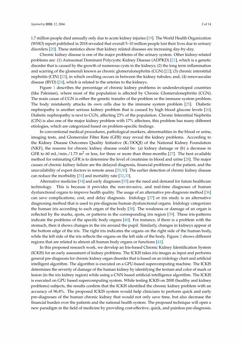
Figure 1 describes the percentage of chronic kidney problems in underdeveloped countries(like Pakistan), where most of the population is affected by Chronic Glomerulonephritis (CGN).The main cause of CGN is either the genetic transfer of the problem or the immune system problem.The body mistakenly attacks its own cells due to the immune system problem [25]. Diabeticnephropathy is another serious kidney problem that is caused by high blood glucose levels [26].Diabetic nephropathy is next to CGN, affecting 23% of the population. Chronic Interstitial Nephritis(CIN) is also one of the major kidney problem with 17% affectees, this problem has many differentetiologies, which are categorized based on problem-specific findings.
In conventional medical procedures, pathological markers, abnormalities in the blood or urine,imaging tests, and Glomerular Filter Rate (GFR) may reveal the kidney problems. According tothe Kidney Disease Outcomes Quality Initiative (K/DOQI) of the National Kidney Foundation(NKF), the reasons for chronic kidney disease could be: (a) kidney damage or (b) a decrease inGFR to 60 mL/min./1.73 m2 or less, for three or more than three-months [27]. The best availablemethod for estimating GFR is to determine the level of creatinine in blood and urine [28]. The majorcauses of chronic kidney failure are the delayed diagnosis, financial problems of the patient, and theunavailability of expert doctors in remote areas [29,30]. The earlier detection of chronic kidney diseasecan reduce the morbidity [31] and mortality rate [32,33].
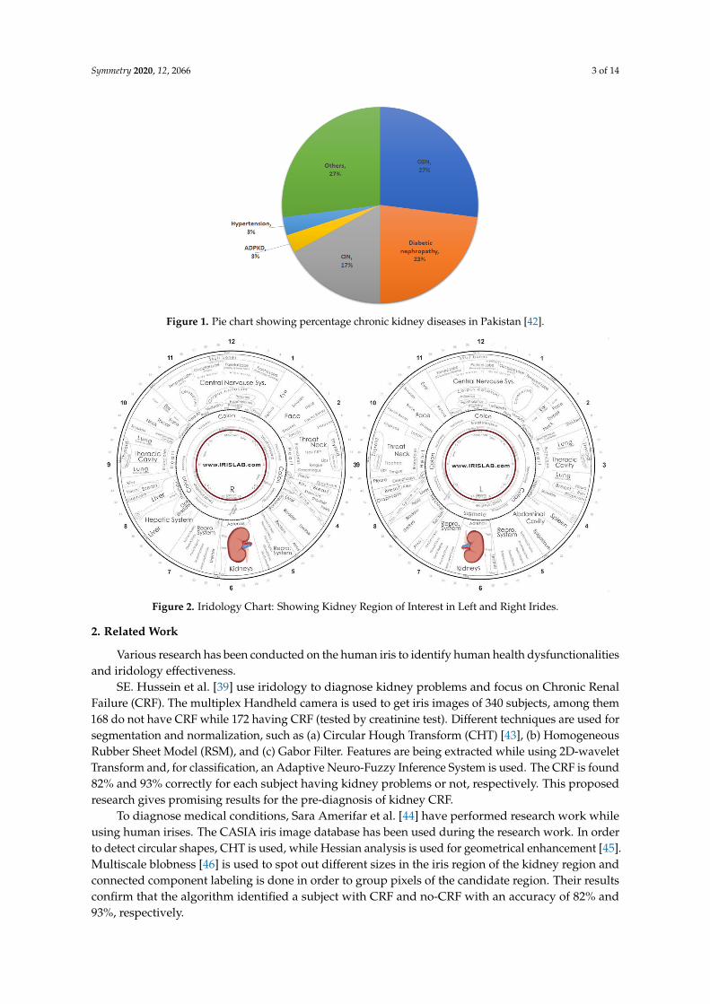
Alternative medicine [34] and early diagnoses [35] are the need and demand for future healthcaretechnology. This is because it provides the non-invasive, and real-time diagnoses of humandysfunctional organs to improve health quality. The usage of an alternative pre-diagnosis method [36]can save complications, cost, and delay diagnosis. Iridology [37] or iris study is an alternativediagnosing method that is used to pre-diagnose human dysfunctional organs. Iridology categorizesthe human iris according to each organ of the body [38]. The weakness or damage of an organ isreflected by the marks, spots, or patterns in the corresponding iris region [39]. These iris-patternsindicate the problems of the specific body organs [40]. For instance, if there is a problem with thestomach, then it shows changes in the iris around the pupil. Similarly, changes in kidneys appear atthe bottom edge of the iris. The right iris indicates the organs on the right side of the human body,while the left side of the iris reflects the organs on the left side of the body. Figure 2 shows differentregions that are related to almost all human body organs or functions [41].
In this proposed research work, we develop an Iris-based Chronic Kidney Identification System(ICKIS) for an early assessment of kidney problems. The ICKIS takes iris images as input and performsgeneral pre-diagnosis for chronic kidney organ disorder that is based on an iridology chart and artificialintelligent algorithm. The algorithm is executed on a GPU-based supercomputing machine. The ICKISdetermines the severity of damage of the human kidney by identifying the texture and color of mark orlesion (in the iris kidney region) while using a CNN based artificial intelligence algorithm. The ICKISis executed on GPU based supercomputing system. While testing ICKIS on 2000 (healthy and kidneyproblems) subjects, the results confirm that the ICKIS identified the chronic kidney problem with anaccuracy of 96.8%. The proposed ICKIS system would help clinicians to perform quick and earlypre-diagnoses of the human chronic kidney that would not only save time, but also decrease thefinancial burden over the patients and the national health system. The proposed technique will open anew paradigm in the field of medicine by providing cost-effective, quick, and painless pre-diagnosis.
Symmetry 2020, 12, 2066 3 of 14
Figure 1. Pie chart showing percentage chronic kidney diseases in Pakistan [42].
Figure 2. Iridology Chart: Showing Kidney Region of Interest in Left and Right Irides.
2. Related Work
Various research has been conducted on the human iris to identify human health dysfunctionalitiesand iridology effectiveness.
SE. Hussein et al. [39] use iridology to diagnose kidney problems and focus on Chronic RenalFailure (CRF). The multiplex Handheld camera is used to get iris images of 340 subjects, among them168 do not have CRF while 172 having CRF (tested by creatinine test). Different techniques are used forsegmentation and normalization, such as (a) Circular Hough Transform (CHT) [43], (b) HomogeneousRubber Sheet Model (RSM), and (c) Gabor Filter. Features are being extracted while using 2D-waveletTransform and, for classification, an Adaptive Neuro-Fuzzy Inference System is used. The CRF is found82% and 93% correctly for each subject having kidney problems or not, respectively. This proposedresearch gives promising results for the pre-diagnosis of kidney CRF.
To diagnose medical conditions, Sara Amerifar et al. [44] have performed research work whileusing human irises. The CASIA iris image database has been used during the research work. In orderto detect circular shapes, CHT is used, while Hessian analysis is used for geometrical enhancement [45].Multiscale blobness [46] is used to spot out different sizes in the iris region of the kidney region andconnected component labeling is done in order to group pixels of the candidate region. Their resultsconfirm that the algorithm identified a subject with CRF and no-CRF with an accuracy of 82% and93%, respectively.
Symmetry 2020, 12, 2066 4 of 14
Agus Prayitno et al. [47] undertake their studies on kidney complications that are caused bydiabetes. The iris region of interest (ROI) is used according to the iridology chart. To analyze the ROI,color constancy [48] and independent component analysis [49] are used. They use Dino-Lite Ver. 2.0for capturing iris images. The results show that 76% of patients are diabetic with kidney complications.The results of the proposed system are verified by the patient medical creatinine level.
Adhi et al. [50] classify the last stage of kidney CRF using the iridology chart. They capture irisimages of 82 subjects using the Dino-Lite Digital iris scope AMH-RUT camera system having 1.3 Mpixels resolution and 30 frame rate. The watershed method for feature segmentation is used in order toexamine the broken tissues in the iris. The author uses the mean of the gradient, mean of the watershed,mean of watershed binary, and size of broken tissue as features, which are inputted to Support VectorMachine (SVM) for machine learning-based classification. During the research, 61 hemodialyses and 21healthy patients are used for analysis and the results show that the algorithm identified hemodialysispatients with an accuracy of 87.5%.
Samant and Agarwal [51] use different machine learning techniques, including (a) Binary Treemodel, (b) SVM, (c) Adaptive Boosting model, (d) Neural Network, and (e) Random Forest modelfor classification and diagnosis of diabetes. Aforesaid techniques use Iris-SCAN-2 with cross-matchtechnology to obtain gray infrared images for the investigation of (a) statistical, (b) texture, and (c)discrete wavelet-based features. The authors perform modified CHT to extract the aforementionedfeatures. Rubber-sheet normalization is used to map iris features on fixed rectangular representation.They divided 338 patients into two groups, one having a sign of diabetes include 180 subjects, and theother with no sign have 158. The results confirm that the model identified diabetes with an accuracyof 89.63%.
Rossi Erwin et al., in [52], use iridology to develop a database for colon disorder. AutonomicNervous Wreath (ANW) of the iris is focused on digestive problems. Hough transform and RSM areused for feature extraction. Sixty subjects are used, in which 25 are controlled while 35 are provencolon disorder. The results show an 8% error. This research shows progressive results and concludesthat, to obtain better and reliable results, a large data-set is required.
Jamal et al. [53] work to develop a tool for detecting diabetes through the corresponding regionof the pancreas organ, while using color coding and visual inspection. Segmentation is done byDaugman’s approach and normalization is done while using RSM; adaptive histogram equalizationof the spatial domain method, which directly operates on pixels, is used for image enhancement.Features of 20 × 20 ROI are extracted while using Principal Component Analysis (PCA). Feedforward-back propagation based, Artificial Neural Network is applied for classification with theLevengurg–Marquardt back-propagation for weight and bias of network as training function. Out of10 subjects, eight subjects are identified as diabetic subjects. Later on, all of these eight are verified asdiabetic type II using an insulin normality test. 100 % accuracy is achieved for the pancreas organ.
UCERD Private Limited [54], Iridology Research Group [55], has developed imaging systemarchitecture [56] and algorithms [57] for iridology based dysfunctional organs identification. The groupproposed intelligent algorithms for health-care. It uses high-performance architecture [58,59] thatmanages an enormous volume of patient images [60,61] information, and effectively uses them tomake a decision.
3. Methodology: An Intelligent Iris Based Chronic Kidney Identification System
This section explains the methodology of the Iris-based Chronic Kidney Identification System(ICKIS). The section is subdivided into four main sections: the Kidney Pathology, the Iris-AcquisitionSystem, the Processing System, and the Artificial Intelligence (AI) Algorithm.
3.1. Kidney Pathology
The section explains the pathology of the kidney against chronic kidney disease while usingconventional methods and iridology techniques.
Symmetry 2020, 12, 2066 5 of 14
3.1.1. Conventional Method
Conventional methods use Glomerular Filtration Rate (GFR), in order to determine the stage ofchronic kidney disease. In GFR, samples of the patient’s blood and urine are compared. A test forcreatinine using the blood sample is conducted in order to measure GFR (if creatinine value increases,the GFR decreases).
GFR measures milliliters of waste that is filtered by kidneys in a minute. The kidneys of ahealthy person filter over 90 mL of waste per minute [27]. The following are the stages of chronickidney disease concerning the GFR described by the Kidney Disease Outcome Quality Initiative(K/DOQI) [62]:
Stage1: (GFR > 90 mL/min./1.73 m2) kidney functioning is normal up to 90%, but with minordamage (protein in urine).
Stage 2: (GFR 60–89 mL/min./1.73 m2) Acute Kidney Disease, mild loss in kidney functioning,and it is working up to 60 to 89%.
Stage 3: (GFR 30–59 mL/min./1.73 m2) Chronic Kidney Disease, moderate loss of kidneyfunctioning, and it is decreased to 45 to 59%, but it may lead to a moderate to severe loss of kidneyfunctioning, up to 30 to 44%.
Stage 4: (GFR 15–29 mL/min./1.73 m2) Severe Chronic Disease, severe loss of kidney functioningfrom 15 to 29%.
Stage 5: (GFR < 15 mL/min./1.73 m2) End-Stage Renal Disease (ESRD) i.e., kidney failure stage.At this stage, there is a total loss or less than 15% functioning.
The GFR level keeps on changes with time for different age groups. In these early stages,GFR alone does not secure the finding that’s why other kidney inspection tests like (a) blood, (b) urine,and (c) kidney scan can be used in order to determine the kidney anomalies. In stage 4 and stage 5,the severe symptoms are being felt by the patients, which leads him/her to chronic kidney failure.In this research work, patients who are grouped under stages 4 and 5 are focused, as indicated by theirGFR, and the emphasis is on the presence or absence of chronic kidney disease of stage 4 and stage 5.
3.1.2. Iridology
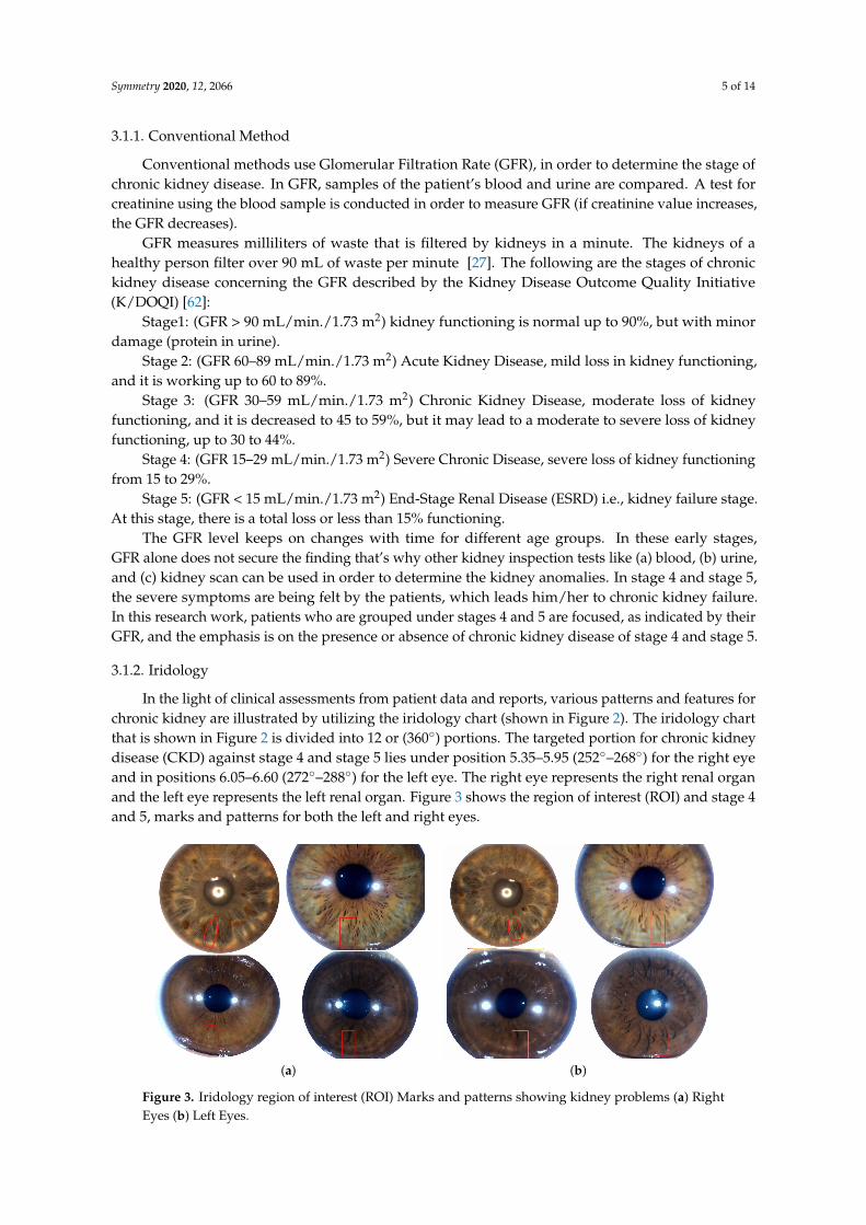
In the light of clinical assessments from patient data and reports, various patterns and features forchronic kidney are illustrated by utilizing the iridology chart (shown in Figure 2). The iridology chartthat is shown in Figure 2 is divided into 12 or (360◦) portions. The targeted portion for chronic kidneydisease (CKD) against stage 4 and stage 5 lies under position 5.35–5.95 (252◦–268◦) for the right eyeand in positions 6.05–6.60 (272◦–288◦) for the left eye. The right eye represents the right renal organand the left eye represents the left renal organ. Figure 3 shows the region of interest (ROI) and stage 4and 5, marks and patterns for both the left and right eyes.
(a) (b)
Figure 3. Iridology region of interest (ROI) Marks and patterns showing kidney problems (a) RightEyes (b) Left Eyes.
Symmetry 2020, 12, 2066 6 of 14
Iris images of subjects having chronic kidney problem contained the following patterns andmarks: (a) A long solid black line. (b) One or more, large or small open lesion. (c) Change in colorof the iris tissue. As kidney problems get worse, the GFR level decreases, and, similarly, the marks,lesions, and crypts appear in the iris in the corresponding region. Furthermore, in Figure 3 it can beclearly seen the variations in the iris pattern or colour of right and left eye irises. Change in the marksand patterns on the irises reflect the after-effects of chronic kidney disease.
3.2. Iris-Acquisition System (IAS)
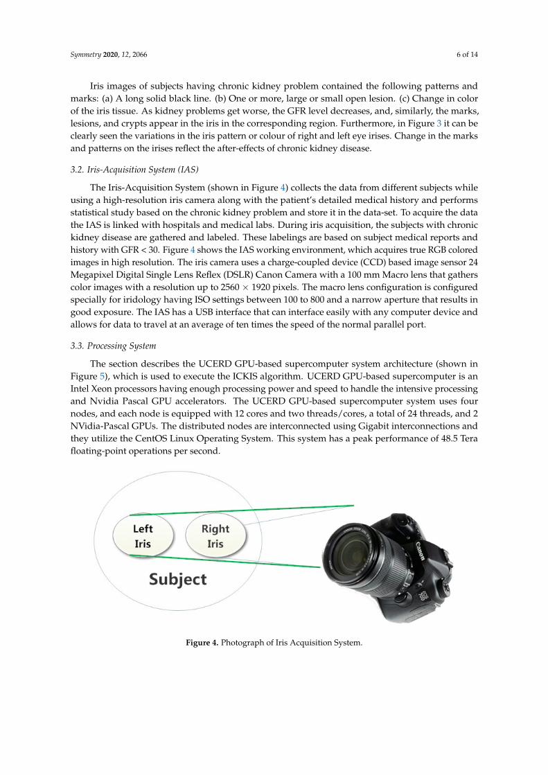
The Iris-Acquisition System (shown in Figure 4) collects the data from different subjects whileusing a high-resolution iris camera along with the patient’s detailed medical history and performsstatistical study based on the chronic kidney problem and store it in the data-set. To acquire the datathe IAS is linked with hospitals and medical labs. During iris acquisition, the subjects with chronickidney disease are gathered and labeled. These labelings are based on subject medical reports andhistory with GFR < 30. Figure 4 shows the IAS working environment, which acquires true RGB coloredimages in high resolution. The iris camera uses a charge-coupled device (CCD) based image sensor 24Megapixel Digital Single Lens Reflex (DSLR) Canon Camera with a 100 mm Macro lens that gatherscolor images with a resolution up to 2560 × 1920 pixels. The macro lens configuration is configuredspecially for iridology having ISO settings between 100 to 800 and a narrow aperture that results ingood exposure. The IAS has a USB interface that can interface easily with any computer device andallows for data to travel at an average of ten times the speed of the normal parallel port.
3.3. Processing System
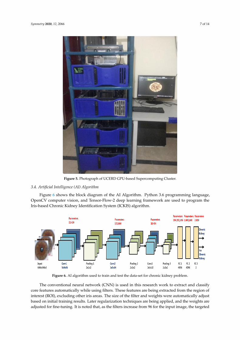
The section describes the UCERD GPU-based supercomputer system architecture (shown inFigure 5), which is used to execute the ICKIS algorithm. UCERD GPU-based supercomputer is anIntel Xeon processors having enough processing power and speed to handle the intensive processingand Nvidia Pascal GPU accelerators. The UCERD GPU-based supercomputer system uses fournodes, and each node is equipped with 12 cores and two threads/cores, a total of 24 threads, and 2NVidia-Pascal GPUs. The distributed nodes are interconnected using Gigabit interconnections andthey utilize the CentOS Linux Operating System. This system has a peak performance of 48.5 Terafloating-point operations per second.
Figure 4. Photograph of Iris Acquisition System.
Symmetry 2020, 12, 2066 7 of 14
Figure 5. Photograph of UCERD GPU-based Supercomputing Cluster.
3.4. Artificial Intelligence (AI) Algorithm
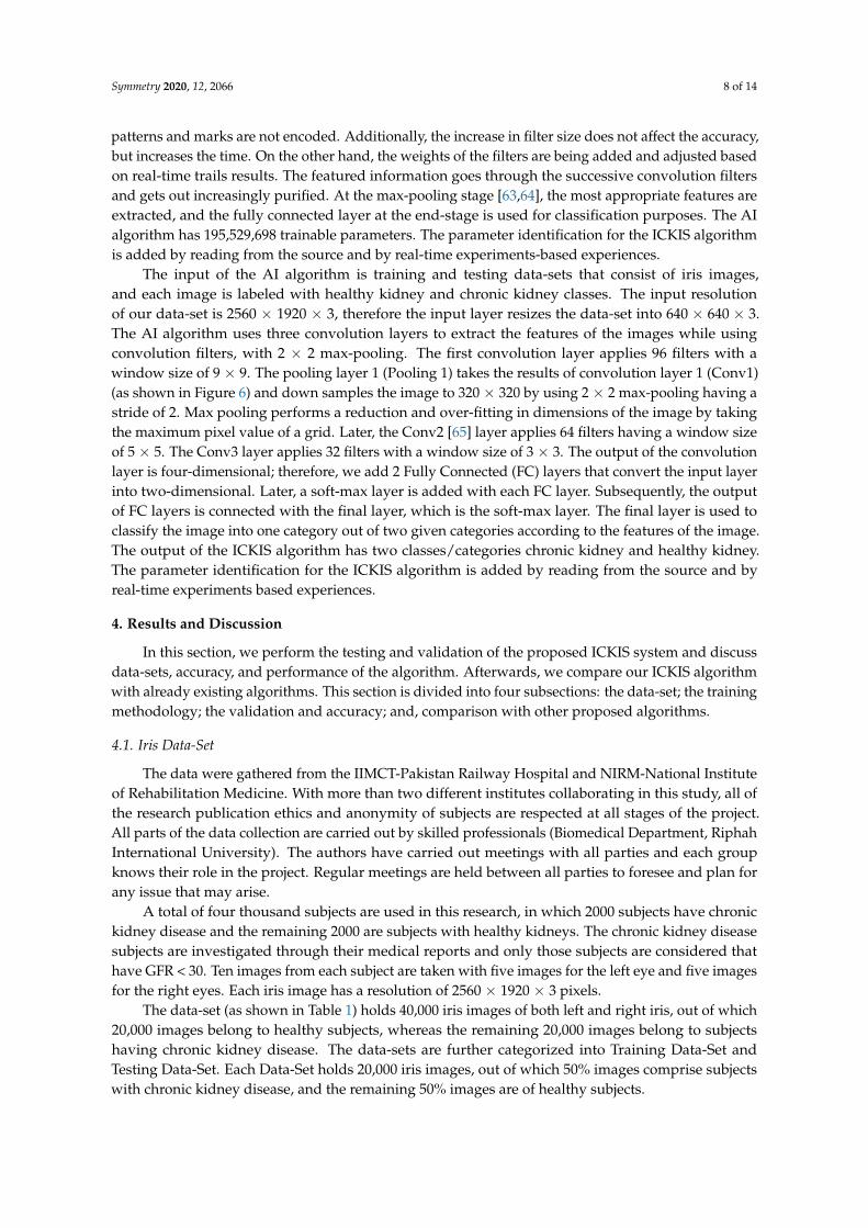
Figure 6 shows the block diagram of the AI Algorithm. Python 3.6 programming language,OpenCV computer vision, and Tensor-Flow-2 deep learning framework are used to program theIris-based Chronic Kidney Identification System (ICKIS) algorithm.
Figure 6. AI algorithm used to train and test the data-set for chronic kidney problem.
The conventional neural network (CNN) is used in this research work to extract and classifycore features automatically while using filters. These features are being extracted from the region ofinterest (ROI), excluding other iris areas. The size of the filter and weights were automatically adjustbased on initial training results. Later regularization techniques are being applied, and the weights areadjusted for fine-tuning. It is noted that, as the filters increase from 96 for the input image, the targeted
Symmetry 2020, 12, 2066 8 of 14
patterns and marks are not encoded. Additionally, the increase in filter size does not affect the accuracy,but increases the time. On the other hand, the weights of the filters are being added and adjusted basedon real-time trails results. The featured information goes through the successive convolution filtersand gets out increasingly purified. At the max-pooling stage [63,64], the most appropriate features areextracted, and the fully connected layer at the end-stage is used for classification purposes. The AIalgorithm has 195,529,698 trainable parameters. The parameter identification for the ICKIS algorithmis added by reading from the source and by real-time experiments-based experiences.
The input of the AI algorithm is training and testing data-sets that consist of iris images,and each image is labeled with healthy kidney and chronic kidney classes. The input resolutionof our data-set is 2560 × 1920 × 3, therefore the input layer resizes the data-set into 640 × 640 × 3.The AI algorithm uses three convolution layers to extract the features of the images while usingconvolution filters, with 2 × 2 max-pooling. The first convolution layer applies 96 filters with awindow size of 9 × 9. The pooling layer 1 (Pooling 1) takes the results of convolution layer 1 (Conv1)(as shown in Figure 6) and down samples the image to 320 × 320 by using 2 × 2 max-pooling having astride of 2. Max pooling performs a reduction and over-fitting in dimensions of the image by takingthe maximum pixel value of a grid. Later, the Conv2 [65] layer applies 64 filters having a window sizeof 5 × 5. The Conv3 layer applies 32 filters with a window size of 3 × 3. The output of the convolutionlayer is four-dimensional; therefore, we add 2 Fully Connected (FC) layers that convert the input layerinto two-dimensional. Later, a soft-max layer is added with each FC layer. Subsequently, the outputof FC layers is connected with the final layer, which is the soft-max layer. The final layer is used toclassify the image into one category out of two given categories according to the features of the image.The output of the ICKIS algorithm has two classes/categories chronic kidney and healthy kidney.The parameter identification for the ICKIS algorithm is added by reading from the source and byreal-time experiments based experiences.
4. Results and Discussion
In this section, we perform the testing and validation of the proposed ICKIS system and discussdata-sets, accuracy, and performance of the algorithm. Afterwards, we compare our ICKIS algorithmwith already existing algorithms. This section is divided into four subsections: the data-set; the trainingmethodology; the validation and accuracy; and, comparison with other proposed algorithms.
4.1. Iris Data-Set
The data were gathered from the IIMCT-Pakistan Railway Hospital and NIRM-National Instituteof Rehabilitation Medicine. With more than two different institutes collaborating in this study, all ofthe research publication ethics and anonymity of subjects are respected at all stages of the project.All parts of the data collection are carried out by skilled professionals (Biomedical Department, RiphahInternational University). The authors have carried out meetings with all parties and each groupknows their role in the project. Regular meetings are held between all parties to foresee and plan forany issue that may arise.
A total of four thousand subjects are used in this research, in which 2000 subjects have chronickidney disease and the remaining 2000 are subjects with healthy kidneys. The chronic kidney diseasesubjects are investigated through their medical reports and only those subjects are considered thathave GFR < 30. Ten images from each subject are taken with five images for the left eye and five imagesfor the right eyes. Each iris image has a resolution of 2560 × 1920 × 3 pixels.
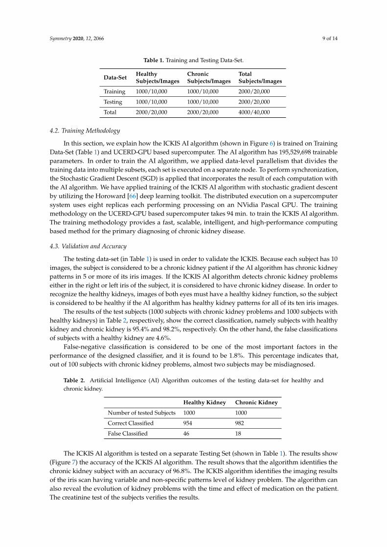
The data-set (as shown in Table 1) holds 40,000 iris images of both left and right iris, out of which20,000 images belong to healthy subjects, whereas the remaining 20,000 images belong to subjectshaving chronic kidney disease. The data-sets are further categorized into Training Data-Set andTesting Data-Set. Each Data-Set holds 20,000 iris images, out of which 50% images comprise subjectswith chronic kidney disease, and the remaining 50% images are of healthy subjects.
Symmetry 2020, 12, 2066 9 of 14
Table 1. Training and Testing Data-Set.
Data-Set Healthy Chronic TotalSubjects/Images Subjects/Images Subjects/Images
Training 1000/10,000 1000/10,000 2000/20,000
Testing 1000/10,000 1000/10,000 2000/20,000
Total 2000/20,000 2000/20,000 4000/40,000
4.2. Training Methodology
In this section, we explain how the ICKIS AI algorithm (shown in Figure 6) is trained on TrainingData-Set (Table 1) and UCERD-GPU based supercomputer. The AI algorithm has 195,529,698 trainableparameters. In order to train the AI algorithm, we applied data-level parallelism that divides thetraining data into multiple subsets, each set is executed on a separate node. To perform synchronization,the Stochastic Gradient Descent (SGD) is applied that incorporates the result of each computation withthe AI algorithm. We have applied training of the ICKIS AI algorithm with stochastic gradient descentby utilizing the Horoward [66] deep learning toolkit. The distributed execution on a supercomputersystem uses eight replicas each performing processing on an NVidia Pascal GPU. The trainingmethodology on the UCERD-GPU based supercomputer takes 94 min. to train the ICKIS AI algorithm.The training methodology provides a fast, scalable, intelligent, and high-performance computingbased method for the primary diagnosing of chronic kidney disease.
4.3. Validation and Accuracy
The testing data-set (in Table 1) is used in order to validate the ICKIS. Because each subject has 10images, the subject is considered to be a chronic kidney patient if the AI algorithm has chronic kidneypatterns in 5 or more of its iris images. If the ICKIS AI algorithm detects chronic kidney problemseither in the right or left iris of the subject, it is considered to have chronic kidney disease. In order torecognize the healthy kidneys, images of both eyes must have a healthy kidney function, so the subjectis considered to be healthy if the AI algorithm has healthy kidney patterns for all of its ten iris images.
The results of the test subjects (1000 subjects with chronic kidney problems and 1000 subjects withhealthy kidneys) in Table 2, respectively, show the correct classification, namely subjects with healthykidney and chronic kidney is 95.4% and 98.2%, respectively. On the other hand, the false classificationsof subjects with a healthy kidney are 4.6%.
False-negative classification is considered to be one of the most important factors in theperformance of the designed classifier, and it is found to be 1.8%. This percentage indicates that,out of 100 subjects with chronic kidney problems, almost two subjects may be misdiagnosed.
Table 2. Artificial Intelligence (AI) Algorithm outcomes of the testing data-set for healthy andchronic kidney.
Healthy Kidney Chronic Kidney
Number of tested Subjects 1000 1000
Correct Classified 954 982
False Classified 46 18
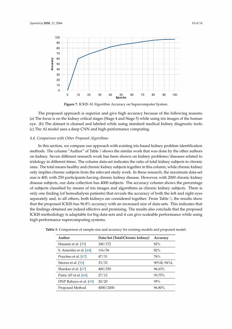
The ICKIS AI algorithm is tested on a separate Testing Set (shown in Table 1). The results show(Figure 7) the accuracy of the ICKIS AI algorithm. The result shows that the algorithm identifies thechronic kidney subject with an accuracy of 96.8%. The ICKIS algorithm identifies the imaging resultsof the iris scan having variable and non-specific patterns level of kidney problem. The algorithm canalso reveal the evolution of kidney problems with the time and effect of medication on the patient.The creatinine test of the subjects verifies the results.
Symmetry 2020, 12, 2066 10 of 14
Figure 7. ICKIS AI Algorithm Accuracy on Supercomputer System.
The proposed approach is superior and give high accuracy because of the following reasons:(a) The focus is on the kidney critical stages (Stage 4 and Stage 5) while using iris images of the humaneye. (b) The dataset is cleaned and labeled while using standard medical kidney diagnostic tools.(c) The AI model uses a deep CNN and high-performance computing.
4.4. Comparison with Other Proposed Algorithms
In this section, we compare our approach with existing iris-based kidney problem identificationmethods. The column “Author” of Table 3 shows the similar work that was done by the other authorson kidney. Seven different research work has been shown on kidney problems/diseases related toiridology in different times. The column data-set indicates the ratio of total kidney subjects to chronicones. The total means healthy and chronic kidney subjects together in this column, while chronic kidneyonly implies chronic subjects from the relevant study work. In these research, the maximum data-setsize is 400, with 250 participants having chronic kidney disease. However, with 2000 chronic kidneydisease subjects, our data collection has 4000 subjects. The accuracy column shows the percentageof subjects classified by means of iris images and algorithms as chronic kidney subjects. There isonly one finding (of hemodialysis patients) that reveals the accuracy of both the left and right eyesseparately and, to all others, both kidneys are considered together. From Table 3, the results showthat the proposed ICKIS has 96.8% accuracy with an increased size of data-sets. This indicates thatthe findings obtained are indeed effective and promising. The results also conclude that the proposedICKIS methodology is adaptable for big data-sets and it can give scaleable performance while usinghigh-performance supercomputing systems.
Table 3. Comparison of sample size and accuracy for existing models and proposed model.
Author Data-Set [Total/Chronic kidney] Accuracy
Hussein et al. [39] 340/172 82%
S. Amerifar er al. [44] 116/56 82%
Prayitno et al. [47] 47/31 76%
Sitorus et al. [50] 33/33 90%R, 94%L
Shankar et al. [67] 400/250 96.63%
Putra AP et al. [68] 27/12 93.75%
DNP Rahayu et al. [69] 20/20 95%
Proposed Method 4000/2000 96.80%
Symmetry 2020, 12, 2066 11 of 14
5. Conclusions and Future Work
In this work, an alternative medical diagnostic method, called the Intelligent Iris-based ChronicKidney Identification System (ICKIS), is proposed. ICKIS detects chronic kidney disorders at an earlystage while using an artificial intelligence algorithm and iridology. The proposed ICKIS uses humaniris as input and a deep neural network-based algorithm, in order to determine whether or not thesubject has a chronic kidney problem. ICKIS has tested over 2000 subjects and the findings concludethat the device categorized subjects as healthy kidney and chronic kidney with an accuracy of 96.8%.The ICKIS algorithm is trained and tested by a GPU based distributed computing machine.
In the future, we shall continue to detect more renal problems with our AI algorithm. We aimto find a method for assessing the problem of the chronic kidney for both kidneys independently.This provides a pre-diagnosis method and a better treatment of early stage kidney disease. We arenow working to build and make our iris data-sets accessible to the research community for advancedhealth-care applications.
Author Contributions: Conceptualization, T.H. and A.H.; Data curation, A.H.; Formal analysis, A.H.; Fundingacquisition, T.H.; Investigation, S.M. and A.H.; Methodology, S.M. and T.H.; Project administration, T.H.;Resources, T.H. and E.A.; Software, A.H. and U.W.; Supervision, T.H.; Validation, S.M. and U.W.; Visualization,U.W.; Writing—original draft, S.M.; Writing—review & editing, S.M., T.H., A.H., U.W. and U.A. All authors haveread and agreed to the published version of the manuscript.
Funding: This work is supported by the Higher Education Commission (HEC) of Pakistan under the TechnologyDevelopment Fund with project Number TDF03-097 and National Research Program for Universities (NRPU)with project grant no. 8153/Federal/NRPU/R&D/HEC/2017.
Acknowledgments: The authors would like to thank the Unal Color of Education Research and Development(UCERD), Private Limited Islamabad, and Barcelona Supercomputing Center Spain for the support.
Conflicts of Interest: The authors declare no conflict of interest.
References
1. Jones, T.C.; Hard, G.C.; Mohr, U. Urinary System; Springer: Berlin/Heisenberg, Germany, 2013.2. Kurtz, I.; Maher, T.; Hulter, H.N.; Schambelan, M.; Sebastian, A. Effect of diet on plasma acid-base
composition in normal humans. Kidney Int. 1983, 24, 670–680. [CrossRef] [PubMed]3. Baranowska, I.; Płonka, J. Determination of biogenic amines and vitamins in urine samples with HPLC.
J. Liq. Chromatogr. Relat. Technol. 2008, 31, 2974–2987. [CrossRef]4. Kirchmann, H.; Pettersson, S. Human urine-chemical composition and fertilizer use efficiency. Fertil. Res.
1994, 40, 149–154. [CrossRef]5. Veeralingam, S.; Sahatiya, P.; Badhulika, S. Low cost, flexible and disposable SnSe2 based photoresponsive
ammonia sensor for detection of ammonia in urine samples. Sens. Actuators B Chem. 2019, 297, 126725.[CrossRef]
6. Wyss, M.; Kaddurah-Daouk, R. Creatine and creatinine metabolism. Physiol. Rev. 2000, 80, 107–213.7. Yaroshenko, I.; Kirsanov, D.; Kartsova, L.; Sidorova, A.; Borisova, I.; Legin, A. Determination of urine ionic
composition with potentiometric multisensor system. Talanta 2015, 131, 556–561. [CrossRef]8. Abbrecht, P.H.; Malvin, R.L. Effects of GFR and renal plasma flow on urine osmolarity. Am. J. Physiol.
Leg. Content 1961, 201, 754–758. [CrossRef]9. Griggers, S.; Paccamonti, D.; Thompson, R.; Eilts, B. The effects of pH, osmolarity and urine contamination
on equine spermatozoal motility. Theriogenology 2001, 56, 613–622. [CrossRef]10. Schrier, R.W. Diseases of the Kidney and Urinary Tract; Lippincott Williams & Wilkins: Philadelphia, PA, USA,
2007; Volume 1.11. Romagnani, P.; Remuzzi, G.; Glassock, R.; Levin, A.; Jager, K.J.; Tonelli, M.; Massy, Z.; Wanner, C.; Anders, H.J.
Chronic kidney disease. Nat. Rev. Dis. Prim. 2017, 3, 17088. [CrossRef]12. Levin, A.; Tonelli, M.; Bonventre, J.; Coresh, J.; Donner, J.A.; Fogo, A.B.; Fox, C.S.; Gansevoort, R.T.;
Heerspink, H.J.; Jardine, M. Global kidney health 2017 and beyond: A roadmap for closing gaps in care,research, and policy. Lancet 2017, 390, 1888–1917. [CrossRef]
Symmetry 2020, 12, 2066 12 of 14
13. Levin, A. The clinical epidemiology of cardiovascular diseases in chronic kidney disease: Clinicalepidemiology of cardiovascular disease in chronic kidney disease prior to dialysis. In Seminars in Dialysis;Wiley Online Library; Blackwell Science Inc.: Oxford, UK, 2003; Volume 16, pp. 101–105.
14. Wilson, B.J.; Watson, M.S.; Prescott, G.J.; Sunderland, S.; Campbell, D.M.; Hannaford, P.; Smith, W.C.S.Hypertensive diseases of pregnancy and risk of hypertension and stroke in later life: Results from cohortstudy. BMJ 2003, 326, 845. [CrossRef] [PubMed]
15. Fauci, A.S. The human immunodeficiency virus: Infectivity and mechanisms of pathogenesis. Science 1988,239, 617–622. [CrossRef] [PubMed]
16. World Health Organization. World Malaria Report 2015; World Health Organization: Geneva,Switzerland, 2016.
17. Naghavi, M.; Wang, H.; Lozano, R.; Davis, A.; Liang, X.; Zhou, M. GBD 2013 Mortality and Causes of DeathCollaborators. Global, regional, and national age-sex specific all-cause and cause-specific mortality for 240causes of death, 1990–2013: A systematic analysis for the Global Burden of Disease Study 2013. Lancet 2015,385, 117–171.
18. Liyanage, T.; Ninomiya, T.; Jha, V.; Neal, B.; Patrice, H.M.; Okpechi, I.; Zhao, M.h.; Lv, J.; Garg, A.X.;Knight, J. Worldwide access to treatment for end-stage kidney disease: A systematic review. Lancet 2015,385, 1975–1982. [CrossRef]
19. Mehta, R.L.; Cerdá, J.; Burdmann, E.A.; Tonelli, M.; García-García, G.; Jha, V.; Susantitaphong, P.; Rocco, M.;Vanholder, R.; Sever, M.S. International Society of Nephrology’s 0by25 initiative for acute kidney injury (zeropreventable deaths by 2025): A human rights case for nephrology. Lancet 2015, 385, 2616–2643. [CrossRef]
20. Luyckx, V.A.; Tonelli, M.; Stanifer, J.W. The global burden of kidney disease and the sustainable developmentgoals. Bull. World Health Organ. 2018, 96, 414. [CrossRef]
21. Mischak, H. Autosomal-Dominant Polycystic Kidney Disease (ADPKD). US Patent App. 13/140,106,6 July 2011.
22. Cattran, D.C.; Feehally, J.; Cook, H.T.; Liu, Z.H.; Fervenza, F.C.; Mezzano, S.A.; Floege, J.; Nachman, P.H.;Gipson, D.S.; Praga, M. Kidney disease: Improving global outcomes (KDIGO) glomerulonephritis workgroup. KDIGO clinical practice guideline for glomerulonephritis. Kidney Int. Suppl. 2012, 2, 139–274.
23. Murray, T.; Goldberg, M. Chronic interstitial nephritis: Etiologic factors. Ann. Intern. Med. 1975, 82, 453–459.[CrossRef]
24. Foley, R.N.; Murray, A.M.; Li, S.; Herzog, C.A.; McBean, A.M.; Eggers, P.W.; Collins, A.J. Chronic kidneydisease and the risk for cardiovascular disease, renal replacement, and death in the United States Medicarepopulation, 1998 to 1999. J. Am. Soc. Nephrol. 2005, 16, 489–495. [CrossRef]
25. Chadban, S.J.; Atkins, R.C. Glomerulonephritis. Lancet 2005, 365, 1797–1806. [CrossRef]26. Gross, J.L.; De Azevedo, M.J.; Silveiro, S.P.; Canani, L.H.; Caramori, M.L.; Zelmanovitz, T. Diabetic
nephropathy: Diagnosis, prevention, and treatment. Diabetes Care 2005, 28, 164–176. [CrossRef] [PubMed]27. Tim Newman. Symptoms, Causes, and Treatment of Chronic Kidney Disease. 2017. Available online:
https://www.medicalnewstoday.com/articles/172179 (accessed on 3 October 2020).28. Stevens, L.A.; Levey, A.S. Measured GFR as a confirmatory test for estimated GFR. J. Am. Soc. Nephrol. 2009,
20, 2305–2313. [CrossRef] [PubMed]29. Avorn, J.; Winkelmayer, W.C.; Bohn, R.L.; Levin, R.; Glynn, R.J.; Levy, E.; Owen, W., Jr. Delayed nephrologist
referral and inadequate vascular access in patients with advanced chronic kidney failure. J. Clin. Epidemiol.2002, 55, 711–716. [CrossRef]
30. Levin, A.; Stevens, P.E. Early detection of CKD: The benefits, limitations and effects on prognosis.Nat. Rev. Nephrol. 2011, 7, 446–457. [CrossRef] [PubMed]
31. Jungers, P.; Zingraff, J.; Albouze, G.; Chauveau, P.; Page, B.; Hannedouche, T.; Man, N. Late referral tomaintenance dialysis: Detrimental consequences. Nephrol. Dial. Transplant. 1993, 8, 1089–1093. [PubMed]
32. Perazella, M.A.; Khan, S. Increased mortality in chronic kidney disease: A call to action. Am. J. Med. Sci.2006, 331, 150–153. [CrossRef]
33. Roubicek, C.; Brunet, P.; Huiart, L.; Thirion, X.; Leonetti, F.; Dussol, B.; Jaber, K.; Andrieu, D.;Ramananarivo, P.; Berland, Y. Timing of nephrology referral: Influence on mortality and morbidity. Am. J.Kidney Dis. 2000, 36, 35–41. [CrossRef]
34. Jonas, W.B. Alternative medicine—Learning from the past, examining the present, advancing to the future.JAMA 1998, 280, 1616–1618. [CrossRef]

Symmetry 2020, 12, 2066 13 of 14
35. Shon, H.S.; Batbaatar, E.; Kim, K.O.; Cha, E.J.; Kim, K.A. Classification of Kidney Cancer Data UsingCost-Sensitive Hybrid Deep Learning Approach. Symmetry 2020, 12, 154. [CrossRef]
36. MacLennan, A.H.; Wilson, D.H.; Taylor, A.W. Prevalence and cost of alternative medicine in Australia.Lancet 1996, 347, 569–573. [CrossRef]
37. Jensen, B. Iridology Simplified, 5th ed.; Book Pub Co.: Summertown, TN, USA, 2012.38. Lodin, A.; Demea, S. Design of an iris-based medical diagnosis system. In Proceedings of the 2009
International Symposium on Signals, Circuits and Systems, Iasi, Romania, 9–10 July 2009; pp. 1–4.39. Hussein, S.E.; Hassan, O.A.; Granat, M.H. Assessment of the potential iridology for diagnosing kidney
disease using wavelet analysis and neural networks. Biomed. Signal Process. Control 2013, 8, 534–541.[CrossRef]
40. Hussain, T.; Haider, A.; Muhammad, A.M.; Agha, A.; Khan, B.; Rashid, F.; Raza, M.S.; Din, M.; Khan, M.;Ullah, S. An Iris based Lungs Pre-diagnostic System. In Proceedings of the 2019 2nd International Conferenceon Computing, Mathematics and Engineering Technologies (iCoMET), Sukkur, Pakistan, 30–31 January 2019;pp. 1–5. [CrossRef]
41. Li, Y.H.; Aslam, M.S.; Yang, K.L.; Kao, C.A.; Teng, S.Y. Classification of Body Constitution Based on TCMPhilosophy and Deep Learning. Symmetry 2020, 12, 803. [CrossRef]
42. Jha, V. Current status of end-stage renal disease care in India and Pakistan. Kidney Int. Suppl. 2013, 3, 157–160.[CrossRef]
43. Pedersen, S.J.K. Circular Hough Transform; Aalb. Univ. Vision, Graph. Interact. Syst.; University in Aalborg:Aalborg, Denmark, 2007; Volume 123.
44. Amerifar, S.; Targhi, A.T.; Dehshibi, M.M. Iris the picture of health: Towards medical diagnosis of diseasesbased on iris pattern. In Proceedings of the 2015 Tenth International Conference on Digital InformationManagement (ICDIM), Jeju, Korea, 21–23 October 2015; pp. 120–123.
45. You, J.; Chen, B. New approach to retinal image enhancement based on Hessian matrix. Jisuanji Yingyong/ J.Comput. Appl. 2011, 31, 1560–1562. [CrossRef]
46. Frangi, A.F.; Niessen, W.J.; Vincken, K.L.; Viergever, M.A. Multiscale vessel enhancement filtering.In Proceedings of the International Conference on Medical Image Computing and Computer-AssistedIntervention, Cambridge, MA, USA, 11–13 October 1998; pp. 130–137.
47. Prayitno, A.; Wibawa, A.D.; Purnomo, M.H. Early detection study of Kidney Organ Complication causedby Diabetes Mellitus using iris image color constancy. In Proceedings of the 2016 International Conferenceon Information & Communication Technology and Systems (ICTS), Surabaya, Indonesia, 12 October 2016;pp. 146–149.
48. Ebner, M. Color Constancy; John Wiley & Sons: Hoboken, NJ, USA, 2007; Volume 7.49. Comon, P. Independent component analysis, a new concept? Signal Process. 1994, 36, 287–314. [CrossRef]50. Sitorus, M.A.; Purnomo, M.H.; Wibawa, A.D. Iris image analysis of patient Chronic Renal Failure (CRF)
using watershed algorithm. In Proceedings of the 2015 4th International Conference on Instrumentation,Communications, Information Technology, and Biomedical Engineering (ICICI-BME), Bandung, Indonesia,2–3 November 2015; pp. 54–58.
51. Samant, P.; Agarwal, R. Machine learning techniques for medical diagnosis of diabetes using iris images.Comput. Methods Programs Biomed. 2018, 157, 121–128. [CrossRef] [PubMed]
52. Passarella, R.; Erwin, E.; Fachrurrozi, M.; Sutarno, S. Development of iridology system database for colondisorders identification using Image processing. Indian J. Bioinform. Biotechnol. 2013, 2, 100–103.
53. Banzi, J.F.; Xue, Z. An automated tool for non-contact, real time early detection of diabetes by computervision. Int. J. Mach. Learn. Comput. 2015, 5, 225. [CrossRef]
54. About: UCERD Private Limited Islamabad. 2018. Available online: http://ucerd.com/my_uploads/pdfs/info/UCERD.pdf (accessed on 17 September 2020).
55. Iridology Research Group (IRG). 2018. Available online: http://ucerd.com/Iridology_Research_Group.php(accessed on 22 September 2020).
56. Hussain, T.; Haider, A.; Taleb-Ahmed, A. A Heterogeneous Multi-Core Based Biomedical ApplicationProcessing System and Programming Toolkit. J. Signal Process. Syst. 2019, 91, 963–978. [CrossRef]
57. Haider, A.; Hussain, T.; Agha, A.; Khan, B.; Rashid, F.; Muzamil, S.; Ahmed, A.T.; Alharbi, S.A.; Ayguade, E.An Iris based Smart System for Stress Identification. In Proceedings of the 2019 International Conference onElectrical, Communication, and Computer Engineering (ICECCE), Swat, Pakistan, 24–25 July 2019; pp. 1–5.
Symmetry 2020, 12, 2066 14 of 14
58. Hussain, T.; Palomar, O.; Cristal, A.; Ayguade, E.; Amna, H. ViPS: Visual Processing System for MedicalImaging. In Proceedings of the 2015 8th International Congress on Image and Signal Processing (CISP2015) and the 2015 8th International Conference on BioMedical Engineering and Informatics (BMEI 2015),Shenyang, China, 14–16 October 2015.
59. Hussain, T.; Haider, A. PGC: A pattern-based graphics controller. Int. J. Circuits Archit. Des. 2014, 1, 117–140.60. Hussain, T. ViPS: A novel visual processing system architecture for medical imaging. Biomed. Signal
Process. Control 2017, 38, 293–301. [CrossRef]61. Hussain, T.; Haider, A.; Cristal, A.; Ayguadé, E. EMVS: Embedded Multi Vector-core System. J. Syst. Archit.
2018, 87, 12–22. [CrossRef]62. Levey, A.S.; Eckardt, K.U.; Tsukamoto, Y.; Levin, A.; Coresh, J.; Rossert, J.; Zeeuw, D.D.; Hostetter, T.H.;
Lameire, N.; Eknoyan, G. Definition and classification of chronic kidney disease: A position statementfrom Kidney Disease: Improving Global Outcomes (KDIGO). Kidney Int. 2005, 67, 2089–2100. [CrossRef][PubMed]
63. Krizhevsky, A.; Sutskever, I.; Hinton, G.E. Imagenet classification with deep convolutional neural networks.Commun. ACM 2017, 60, 84–90. [CrossRef]
64. Oh, S.L.; Vicnesh, J.; Ciaccio, E.J.; Yuvaraj, R.; Acharya, U.R. Deep convolutional neural network model forautomated diagnosis of schizophrenia using EEG signals. Appl. Sci. 2019, 9, 2870. [CrossRef]
65. Jaderberg, M.; Vedaldi, A.; Zisserman, A. Speeding up convolutional neural networks with low rankexpansions. arXiv 2014, arXiv:1405.3866.
66. Sergeev, A.; Del Balso, M. Horovod: Fast and easy distributed deep learning in TensorFlow. arXiv 2018,arXiv:1802.05799.
67. Shankar, K.; Manickam, P.; Devika, G.; Ilayaraja, M. Optimal Feature Selection for Chronic Kidney DiseaseClassification using Deep Learning Classifier. In Proceedings of the 2018 IEEE International Conferenceon Computational Intelligence and Computing Research (ICCIC), Madurai, India, 13–15 December 2018;pp. 1–5.
68. Putra, A.P.; Sutojo, T. Identifikasi Penurunan Kondisi Fungsi Organ Ginjal Melalui Iris Mata MenggunakanMetode Jaringan Syaraf Tiruan Learning Vector Quantization. Fakultas Ilmu Komputer UniversitasDian Nuswantoro 2014, 13, 45–52.
69. Rahayu, D.N.P.; Isnanto, R.R.; Hidayatno, A. Aplikasi Pendiagnosis Gangguan Ginjal Melalui Citra IrisMata Menggunakan Metode Segmentasi Berdasar Deteksi Tepi. Transient J. Ilm. Tek. Elektro 2013, 2, 283–288.
Publisher’s Note: MDPI stays neutral with regard to jurisdictional claims in published maps and institutionalaffiliations.
c© 2020 by the authors. Licensee MDPI, Basel, Switzerland. This article is an open accessarticle distributed under the terms and conditions of the Creative Commons Attribution(CC BY) license (http://creativecommons.org/licenses/by/4.0/).