Conductance modulation of charged lipid bilayer using electrolyte ...

Plant Physiol. (1992) 98, 198-2050032-0889/92/98/01 98/08/$01 .00/0
Received for publication May 15, 1991Accepted August 17, 1991
An Improved Method for Using Electrolyte Leakage toAssess Membrane Competence in Plant Tissues1
Thomas H. Whitlow*, Nina L. Bassuk, Thomas G. Ranney, and Deborah L. ReichertUrban Horticulture Institute, Cornell University, Ithaca, New York 14853 (T.H. W., N.L.B.); North Carolina StateUniversity Mountain Horticultural Crops Research Station, Fletcher, North Carolina 28732-9628 (T.G.R.); and
Department of Plant Breeding and Biometry, Cornell University, Ithaca, New York 14853 (D.L.R.)
ABSTRACT
A new expression for ion leakage from plant tissue, the tissueionic conductance (gT), is compared with electrical conductivity(EC) and a commonly used damage index (Id) to test the abilityof each expression to correctly describe leakiness in two modelsystems representing examples of physiological processes withwell-known effects on membrane permeability. In experiments inwhich drought-acclimated leaves were compared with nonaccli-mated leaves and senescing leaves were compared with nonse-nescing leaves, Id contradicted our expectation that acclimatedtissue would be less leaky than nonacclimated tissue, and gT1andEC confirmed this expectation. In a comparison of senescing andnonsenescing tissue, Id again contradicted our expectation thatsenescing tissue would be more leaky than nonsenescing, andEC and gT, were confirming. Using a diffusion analysis approach,we show that Id fails to account for variation in the concentrationgradient between the tissue and the bathing solution and variationin the surface area through which efflux occurs. Furthermore,because Id is a parameter that relates treatment performance tocontrol performance as a percentage value, it distorts the actualdifferences among treatments. The resulting artifacts lead to apresentation of membrane integrity which is probably incorrect.EC is a more direct measurement of net ion efflux and appearsto be less vulnerable to artifact. However, because gTi is the onlyexpression that explicitly includes chemical driving force andtissue surface area, it is the most reliable of the three expres-sions.
Measuring solute leakage from plant tissue is a long-stand-ing method for estimating membrane permeability in relationto environmental stresses, growth and development, and gen-otypic variation. Early published accounts used total electro-lyte leakage, expressed as specific conductance (EC2) of theaqueous bathing solution in which the tissue was immersed,to indicate degree ofdamage resulting from chilling injury (3,4). Recognizing that electrolyte content could vary amongsamples, Stuart (23) recommended expressing electrolyte leak-age as an index percentage of total electrolyte present in the
Supported by an R.P. White research grant from the HorticulturalResearch Foundation and Hatch projects NE-27 and NY-430 toT.H.W.
2 Abbreviations: EC, electrical conductivity; RWC, relative watercontent.
tissue. Flint et al. (6) reduced Stuart's calculation to thefollowing form:
Id= 100 (R, - Ro)/(l -R,) (1)where, according to the terminology of Flint et al. (6), Id isthe index of injury, R, = ECin,,itiaI/ECio,aIfor stressed tissues, R0= ECiniIiai/ECIoIai for nonstressed tissues, and ECini,ial is theconductivity of the bathing solution following a given periodof leakage, EC,otai is the conductivity of the bathing solutionfollowing heat killing to release all ions from the tissue. Thiscalculation of Id adjusts all values to a scale of 0 to 100, withhigher percentages indicating greater damage.To generate data for calculating Id, the standard procedure
is to remove appropriate samples of plant tissue, bring thesamples to full hydration, rinse off surface adhering electro-lytes and contents of cells damaged by excision, and thenimpose an environmental stress such as dehydration (1, 5, 7,12, 19), high temperature (8, 13, 18, 21, 25), or low temper-ature (2-4, 9, 23, 27, 28). The assumption is that it is necessaryto stress tissue in vitro to elicit differences among samples.Leaf discs cut with a cork borer are common sample units,although stem or root segments have also been used. Toobtain data for calculation of R,, a common procedure is toplace samples in standardized volumes of glass-distilled waterand allow them to leak solutes into the bathing solution for12 to 24 h, at which time ECinitialfor the solutions is measuredusing a solute bridge. Following measurement, samples arekilled by autoclaving or rapid freezing in liquid N2 andallowed to leak for an additional period, typically 24 h. Theconductivity is then remeasured to obtain EC,0o1. Ro is calcu-lated from measurements obtained from nonstressed controlsamples that have been monitored along with the stressedsamples. Many recent applications ofthe technique have usedsimilar procedures and either the above formula or one thatyields the same value (for example, see ref. 13).
This technique has enduring appeal because it is simple,uses readily available and inexpensive equipment, and issuited to analyzing large numbers of samples. Despite theseadvantages, Id is flawed in ways that make interpretationdifficult and potentially misleading. The lack of adequatetheoretical derivation makes it unclear exactly what Id meas-ures in relation to membrane competence. Id is a scaled valuebetween 0 and 100 measured at a moment in time followingan arbitrary and lengthy incubation period. Although studiesusing Id frequently claim to assess "membrane stability" or
198
www.plantphysiol.orgon July 22, 2018 - Published by Downloaded from Copyright © 1992 American Society of Plant Biologists. All rights reserved.
EVALUATION OF ELECTROLYTE LEAKAGE DETERMINATION OF INJURY
"membrane permeability," the Id calculation includes noneof the variables typically used to describe ion movementacross a membrane. Id cannot be interpreted as a flux anddoes not take into account activity or electrical gradients thatcould be related to membrane permeability in a defined,quantitative fashion. This suggests that Id cannot discriminatebetween membrane characteristics and other confoundingvariables that affect flux. Our preliminary attempts to use Idto rank genotypes and acclimation treatments in relation toleakiness indicated that both leafthickness (hence surface areaof the cut disc margin) and RWC varied along with theindependent treatment variables under study. Failure to ac-count for variation in leaf thickness ignores the fact that fluxof any solute varies with the surface area through which fluxoccurs. Failure to account for variation in RWC neglects theinfluence of solvent volume on ion concentration inside theleaves, which in turn determines the electrochemical gradientdriving ion efflux. As we will show, neglecting these variablescauses artifacts that lead to erroneous conclusions about theleakiness of the tissues being compared.These limitations prompted us to modify the traditional Id
protocol to permit calculation of an empirical coefficient,which we call gTi, which takes into account variation in tissuethickness and electrolyte concentration and provides a dy-namic picture of recovery following stress. Our goal was toretain the convenience of traditional protocols while deliber-ately factoring out sources of variation not directly related tomembrane characteristics. In this paper, we present a methodfor deriving gTi and use two prototype systems to comparegTi, EC, and Id methods for evaluating tissue leakiness.
METHODS
Derivation of gTi
In the simple case of an uncharged solute, Fick's first lawof diffusion states that flux through a planar surface is afunction of driving force and the resistance of the mediumthrough which the flux occurs. In a membrane-bound system,driving force is determined by the concentration gradientacross the membrane and resistance is determined by thesurface area and permeability of the membrane for the solute.In the case of a charged solute such as an inorganic ion, fluxis affected by the gradient in electrical potential across themembrane in addition to the gradient in chemical potential.Ideally, any method purporting to quantify membrane im-pairment in plant tissue would include direct measurementsor estimates of all variables governing flux. With experimentsinvolving many comparisons, as in germplasm screening, itis not practical to measure the electrical gradient and thesurface area of the plasma membrane. However, estimates ofthe average concentration gradient of an ion and the area ofthe cut surface through which flux occurs are easily obtainablewithin the framework of familiar protocols. These estimatesserve as the basis for the gTi calculation, which effectivelyremoves them as confounding variables.Nobel (16) derived the following expression from Fick's
first law which shows explicitly how area, concentration gra-dient, and a conductance coefficient influence flux:
J = PjA(cj - ci') (2)where J, = dsJ/dt, the flux of solute species j per unit time, P,is a permeability coefficient for solute speciesj, A is the surfacearea through which diffusion occurs, and (cj - cji') is thedifference between outside (or higher) concentration and theinside (or lower) concentration. Drawing on the formalism ofEq. 2 and substituting gTi for P, and area of cut surface for A,we solve for gTi, obtaining:
gTi = J,A(c - ci) (3)
This empirical expression ignores the contribution ofmem-brane potential to the electrochemical gradient but adjusts fortissue characteristics that might otherwise be confused withbehavior of membranes. We make no claim that gTi is a directestimate of membrane permeability and discourage othersfrom making similar claims. We justify the liberty of ignoringthe electrical component on the grounds that it is preferableto remove known sources of variation that are estimable eventhough other, essentially nonestimable, sources still remain.We have borrowed the symbol g, for conductance, from leafgas exchange terminology. We propose the term gTi insteadof Pj not to proliferate new terminology but to emphasize theimportant departures from the definition of the permeabilitycoefficient. First, gTi is based on net flux for bulk tissue, notindependent measurements of influx and efflux from anisolated cell. As such, gTi represents average concentrationgradient and leakiness of all cells in the sample. Heterogeneityin concentration among cells and poor mixing in the apo-plasm will affect the behavior of individual cells that cannotbe resolved. Second, gTi is not based on surface area of theplasma membrane but rather the planar area of the cut discmargin. The true membrane surface area is much larger, andconcentration gradients for internal cells that are not directlyexposed to the bathing solution are likely to be less steep.Additionally, treating the cut surface as a smooth cylinder isitself an approximation that underestimates total surface areaat the disc margin. In reality, the surface is three dimensionaland consists of cell wall and cell lumina with adhering frag-ments ofplasmalemma. (Because the cut perimeter ofthe discis typically exposed to glass-distilled water, any unboundprotoplasts would burst.) Third, Pj is usually interpreted asdescribing the physical properties ofthe membrane independ-ently of the membrane potential.
General Experimental Protocol
Our protocol followed standard Id procedures except thatwe measured fresh and dry weights and measured EC repeat-edly during a 24-h experiment. Leaves were cut before dawn,and petioles were recut under glass-distilled water and allowedto hydrate in a cool (20 ± 2°C) dark laboratory. After 3 h,five 9-mm diameter discs were punched from each leaf witha cork borer and placed in beakers containing 50 mL glass-distilled water to remove the contents of cells lysed by theborer. Punches avoided major veins. Beakers were agitatedperiodically by hand. A sample consisted of five leaf discs(one disc from each leaf), with eight to 10 replicate samplesper treatment. After 1 h, leaf discs were removed from the
199
www.plantphysiol.orgon July 22, 2018 - Published by Downloaded from Copyright © 1992 American Society of Plant Biologists. All rights reserved.

Plant Physiol. Vol. 98, 1992
water, placed on laboratory tissues (KimWipes) previouslysaturated with glass-distilled water, and sealed in small plasticbags (WhirlPacs). Half of the samples were randomly selectedand removed from the bags, blotted dry, and weighed toobtain fully hydrated weights. These samples were then al-lowed to dry on the laboratory bench until they reached 80%of their fresh weight. This level of stress had been previouslydetermined to be the operational limit for discs to regain fullhydration following stress. Following the secondary dehydra-tion stress, discs were quickly placed in small cylindricalstainless steel screen cages mounted on stainless steel rods andsuspended inside test tubes over 7 mL glass-distilled waterequilibrated to 25°C in a water bath (model 25, Precision,Chicago, IL) oscillating at 1 cycle s-'. The remaining non-stressed discs were removed from the plastic bags, quicklyblotted, weighed, and treated identically with the stressedsamples. At time 0, all samples were plunged into the glass-distilled water. At predetermined intervals, the samples wereremoved from the bathing solutions and the conductivity ofthe solutions was measured with a conductivity meter (model32 equipped with a model 3417 electrode, Yellow SpringsInstruments, Yellow Springs, OH). The cage facilitated rapidsubmersion and removal from the distilled water, ensuredthat all discs were fully submerged, and provided a means forholding the leaf discs in the humid headspace of the tubeduring those periods when EC was being measured. After theinitial 120 min, during which time measurements were fre-quent, the tubes were covered with Parafilm to retard evapo-ration. EC of the bathing solution was measured at predeter-mined intervals for 24 h, at which time all samples in theircages were plunged into liquid N2 to kill the tissue. Sampleswere then replaced in their respective tubes for an additional24 h, at which time final conductivity was measured. Dryweight of each sample was measured after 24 h of drying at70°C in a convection oven.
Calculations
Several quantities must be measured or derived to solveEq. 3. These include the following.
Internal Aqueous Volume
Internal aqueous volume was determined gravimetrically.The weight of the leaf discs was measured with an electronicbalance with 0.1-mg resolution (model AE 163, Mettler In-strument Corp., Hightstown, NJ). Initial solvent volume ofthe plant tissue was determined by subtracting oven-driedtissue weight from fresh weight. For nonstressed samples, thisvalue was assumed to be the total internal solvent volumeand was constant for the duration of the experiment. Stressedleaf discs that had been dried to 80% of their initial freshweight experienced a rapid increase in internal water contentduring rehydration, substantially affecting internal ion con-centration and hence the concentration gradient between thediscs and the external solution. To account for this, leaf discswere subsampled from the same leaves used in the effluxexperiments, dried to 80% fresh weight, and weighed at 1-min intervals until they regained full hydration. Curves fromthese rehydration experiments were used in conjunction with
fresh and dry weights to estimate internal water volume atintermediate times coinciding with EC measurement in themain experiment.
External Volume
Glass-distilled water was dispensed with a bottle top dis-penser that had been calibrated to deliver precisely 7 mL.We assumed that volume remained constant during theexperiment.
Ionic Concentration
At dilute concentrations, EC is a positive, nearly linearfunction of the concentration of all ions in solution. Thisrelationship is widely applied in routine soil and water analy-sis. Instead of calculating total ion concentration from ECvalues using empirical coefficients developed by others, weselected potassium as a representative ion because it is themajor inorganic cation present in glycophytes (12, 17). Itshould be noted that using [K+] in lieu of EC is not essentialbut is used in our demonstration because it provided a con-venient way to check the accuracy of our estimates of internalconcentrations against published values. Palta et al (17) pro-vide independent verification of the strong relationship be-tween EC and [K+] in aqueous solutions containing effusatefrom plant tissue. In our study, empirical regression equationsrelating EC to [K+] were developed for each plant species andleaf type. Bulk subsamples of leaves similar to those used inefflux experiments were washed in glass-distilled water, blot-ted dry, inserted into 30-mL plastic syringes, and frozen at-20°C. After thawing, leaf sap was squeezed from the leaves,and aqueous serial dilutions were made over a range thatbracketed the EC values measured in the external solutionsin the efflux experiments. Ion concentration ofthese solutionswas measured using an inductively coupled argon plasmaatomic emission spectrometer (Jarrell-Ash model 975).
Because the external volume was a known constant in allexperiments, concentrations in the external solution wereeasily convertible to absolute amounts, and net efflux couldbe calculated for each time interval. Internal concentrationswere calculated by freezing the tissue in liquid N2 at the endof the experiment and permitting all ions to leak from thetissue. The total concentration in the bathing solution wasderived from EC using our regressions equations. Because theratio of internal to external solvent volumes and the amountof K+ leaving the leaf discs both could be estimated, internalconcentrations could be calculated for any appropriate timeinterval. The concentration gradient was calculated as thedifference between internal and external concentrations.
Area
There are two parallel pathways for ion efflux: one throughthe intact upper and lower epidermis and another throughthe cut perimeter of the leaf discs. The resistance of the cutsurface is much lower than the resistance of the intact epider-mis and cuticle; therefore, the flux through the ab- and adaxialsurfaces can be considered negligible (22). Disc thickness wasdetermined by water displacement from a 30-mL pycnometerbottle. Assuming a cylindrical shape,
200 WHITLOW ET AL.
www.plantphysiol.orgon July 22, 2018 - Published by Downloaded from Copyright © 1992 American Society of Plant Biologists. All rights reserved.
EVALUATION OF ELECTROLYTE LEAKAGE DETERMINATION OF INJURY
Tav = (V/Ad)/n (4)
where Tav is average disc thickness, Vis total displaced volumeby the sample, Ad is the circular area of the disc, and n is thenumber of discs in the sample. Discs were subsampled fromeach leaf used in the efflux experiment for determination ofleaf thickness. Batched samples of 20 to 40 discs were suffi-cient for accurate determination of Tav. Discs were submergedindividually to avoid trapping bubbles between them.
Experimental Design and Plant Material
Two model systems were selected to represent differentphysiological phenomena that have been shown in otherstudies to be accompanied by changes in electrolyte leakage.These phenomena were stress acclimation and senescence.
Stress acclimation has been shown to make membranes lessleaky (12), and senescence and aging are associated withincreased leakiness to ions (24). These systems allowed us topose a priori hypotheses about experimental outcomes thatcould be used to test the ability ofeach computational method(i.e. EC, Id, and gTi) to correctly describe the behavior of planttissues. By using two independent examples of processes andspecies, we intended to establish a general approach withbroad application. The specific systems used were droughtacclimation in crab apple and senescence in red oak.
Experiment 1: Drought Acclimation in Crab Apple
Hypothesis: Leaves acclimated to slowly imposed, sublethaldrought stress will be less leaky to ions than nonacclimatedcontrol leaves.
Prairie crab apple (Malus ioensis 'Klehm's Improved'scions grafted on Malus baccata rootstocks) was selected as a
woody species in which drought acclimation was known tooccur (20). Thirty bare root 1-m tall whips were trimmedback to three basal buds and planted in a 1:1:1 peat:soil:perlitemix in 130-L plastic trash cans. Plants were kept outdoors,receiving tap water as needed and weekly application ofcomplete nutrient solution (20 N-10 P05-20 K20 applied ata rate equivalent to 200 ppm N). After 14 weeks, all plantswere placed in a greenhouse (day 21C/night 1 6°C) to permitsupplemental illumination with incandescent lights between5 and 11 PM to delay bud set. Half ofthe trees were designatedas controls and were watered regularly to maintain predawnleaf water potentials of approximately -0.33 MPa. The re-
maining trees were subjected to drought stress which consistedof withholding all water until predawn leaf water potentials(measured with a SoilMoisture water status console, SantaBarbara, CA) reached approximately -2.0 MPa. This required27 days. At this time, leaves from five control and stress treeswere selected and handled as discussed above in the generalprotocol.
Experiment 2: Senescence Effects in Red Oak Leaves
Hypothesis: Ion leakage will be greater from tissue at a
more advanced stage of autumnal senescence than tissue inwhich senescence is less advanced.
Individual leaves of many deciduous trees develop autumn
color in patches or zones, presumably indicating small-scalevariation in onset of senescence in tissue that had previouslyappeared quite uniform. In red oak, Quercus rubra, colorationchanges from green to yellow as Chl breaks down and ulti-mately turns brown just before abscission. On November 3,mottled leaves showing patches ofyellow and green colorationwere cut from a 69-cm diameter breast height red oak growingon the Cornell campus. The tree was growing in the openunder ambient conditions and had shown no previous symp-toms ofabnormal growing conditions. Five leaf punches werecut from either yellow or green patches on each of eight leavesto yield eight, five-disc batch samples of each color per leaf.This sampling procedure ensured that yellow and green sam-ples were as similar as possible in all respects except degree ofsenescence. Discs were handled according to the general pro-tocol previously described, with four batches assigned to eachtreatment.
RESULTS
Experiment 1: Drought Acclimation
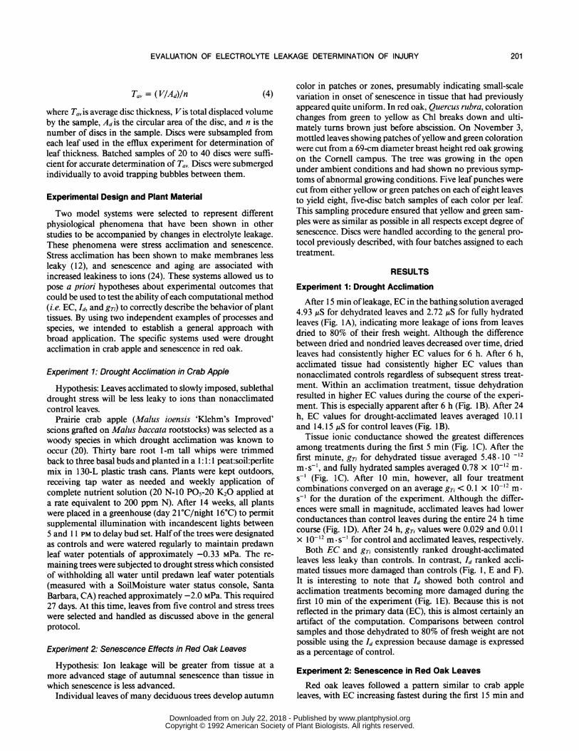
After 15 min ofleakage, EC in the bathing solution averaged4.93 ,iS for dehydrated leaves and 2.72 ,uS for fully hydratedleaves (Fig. 1A), indicating more leakage of ions from leavesdried to 80% of their fresh weight. Although the differencebetween dried and nondried leaves decreased over time, driedleaves had consistently higher EC values for 6 h. After 6 h,acclimated tissue had consistently higher EC values thannonacclimated controls regardless of subsequent stress treat-ment. Within an acclimation treatment, tissue dehydrationresulted in higher EC values during the course of the experi-ment. This is especially apparent after 6 h (Fig. 1 B). After 24h, EC values for drought-acclimated leaves averaged 10.11and 14.15 ,uS for control leaves (Fig. 1 B).
Tissue ionic conductance showed the greatest differencesamong treatments during the first 5 min (Fig. IC). After thefirst minute, gTi for dehydrated tissue averaged 5.48. 10 -12m*s_1, and fully hydrated samples averaged 0.78 x 10⁻¹² m.s-' (Fig. IC). After 10 min, however, all four treatmentcombinations converged on an average gTi < 0.1 x 10⁻¹² m.s-' for the duration of the experiment. Although the differ-ences were small in magnitude, acclimated leaves had lowerconductances than control leaves during the entire 24 h timecourse (Fig. 1D). After 24 h, gTi values were 0.029 and 0.01 1X 10-12 m.s-' for control and acclimated leaves, respectively.Both EC and gT, consistently ranked drought-acclimated
leaves less leaky than controls. In contrast, Id ranked accli-mated tissues more damaged than controls (Fig. 1, E and F).It is interesting to note that Id showed both control andacclimation treatments becoming more damaged during thefirst 10 min of the experiment (Fig. IE). Because this is notreflected in the primary data (EC), this is almost certainly anartifact of the computation. Comparisons between controlsamples and those dehydrated to 80% of fresh weight are notpossible using the Id expression because damage is expressedas a percentage of control.
Experiment 2: Senescence in Red Oak Leaves
Red oak leaves followed a pattern similar to crab appleleaves, with EC increasing fastest during the first 15 min and
201
www.plantphysiol.orgon July 22, 2018 - Published by Downloaded from Copyright © 1992 American Society of Plant Biologists. All rights reserved.

WHITLOW ET AL. Plant Physiol. Vol. 98, 1992
A.-d- Cont 100-o- AeelIm 100d Cont 60
---- Acol1m 60
2 40 2'0 4'0 00 60 100 12,07
Coot 00 c.---I Aceem 100
5 Cont 00
AAelIm 80
4
2
10 20 40 60 so 100 120
E.
0.4Acclimated
02 Control
0.0 *-0 20 40 00 0 100 120
16-
14-
12.
10I
6.-
4.
2 -
Bt
Con, 100---Aelm 100
- Cont 80Acelim 80
0200 400 600 600 1000 1200 1400
0.12 -
0.10 Cont 100
Acclim 100
0.08 ; Cont 80
0.00
0.06-
0.02.
0.00
200 400 600 600 1000 1200 1400
1.0
1.4 -,--Acclimated F1.4- Control
12 ~_
1.0
0.6
0.4
0.2
0.0200 400 600 600 1000 1200 1400
Time (min)
Figure 1. Comparison of apparent ion efflux from drought-acclimated(Acclim) and nonacclimated (Cont) crab apple leaf discs during 24 h.100 and 80, discs that were either fully hydrated or dried to 80% offresh weight, respectively. Points, averages of five replicates. Errorbars, ± 1 SD; omitted from C for clarity and could not be computedfor E and F because Id uses average values for its calculation (seetext). EC and gri rank acclimated leaves less leaky than nonaccli-mated, whereas Id consistently ranks acclimated leaves as leakier.
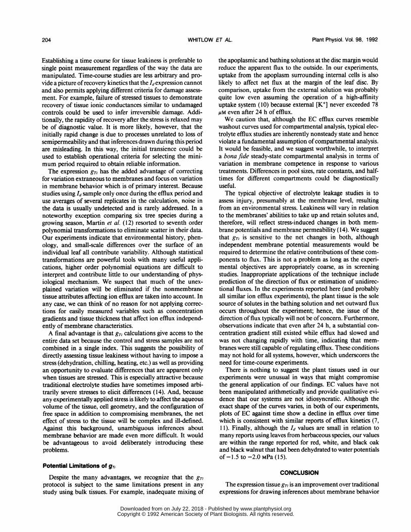
then increasing more slowly during the remainder of theexperiment (Fig. 2, A and B). Initially, there was a nearlytwofold difference in solution EC between control samplesand those that had been subjected to a dehydration stress (Fig.2A). Dried leaves averaged 4.26 ,S and nondried averaged2.42 uS after 15 min. After 4 h, tissue senescence had a
stronger influence than the dehydration treatment, with yel-low leaves leaking more than green leaves (Fig. 2B).During the first 15 min, gTi declined from an initial range
of 3.71 to 7.58 x 10⁻¹² m.st to a range of 2.32 to 7.47 x
10-3 m s-' (Fig. 2C). Tissues that had been dried to 80% offresh weight were consistently leakier than controls, andwithin a drying treatment senescing leaves were leakier thangreen leaves (Fig. 2, C and D). Between 6 and 18 h, yellowleaves became increasingly leaky, and green leaves showedlittle change (Fig. 2D). After 24 h, gTi of yellow leaves haddecreased to levels similar to green leaves.Whereas EC and gTi rank yellow leaf samples leakier than
green samples, Id reverses this ranking consistently during thecourse of the experiment. The Id increased slightly between 1
and 5 min and then remained relatively constant between 3and 5% for 12 h (Fig. 2, E and F). Yellow leaf discs were
more damaged than green for the first 2 h but less leaky
thereafter. After 12 h, Id showed that green leaves becameslightly more damaged and yellow leaves became less dam-aged. Id values for yellow leaves eventually became negativebetween 18 and 24 h. As in experiment 1, it was not possibleto evaluate the effect of the dehydration treatment independ-ently of senescence effects because the Id calculation expressesdehydrated values as a percentage of control.
DISCUSSION
Results of the experiments reported here indicate that theId commonly used to express electrolyte efflux data contra-dicts our expectations about the relative leakiness of accli-mated and senescing leaves and their respective controls.Failure to correctly rank leaf tissues by leakiness indicatesthat Id is not merely unreliable, as reported by Murray et aL(15), it is fundamentally wrong. To accept the inferencesdrawn from the Id calculations in our experiments, we wouldhave to conclude that acclimated tissue was leakier thannonacclimated and that healthy tissue was leakier than se-nescing tissue. We submit that these conclusions are improb-able and that Id is in error. Although EC achieves rankings
A.
6)--,0..
21 *0G1002Y1O-YY 0
0 2Y0 00l
C.
o- 0100
6 0--
-0-- Y60
0)
02
06
el
5-
2-
1-
oron E.,] Yallow
O I0 20 40 60 60 100 120
30 -
20
10
_... G100 B.--O 80
-0-*- VI000-6- Y100
O ,200 400 600 600 000 1200 400
1.0.
0.0-
0.6-
0.4-
0.2-
00.
_ G1000Y100 .
b0 400 600 600 1000 1200 400
Time (min.)
Figure 2. Comparison of apparent ion efflux from red oak leaf discscut from either green, competent tissue (G) or yellow, senescingtissue (Y) during 24 h. 100 and 80, leaf discs that were either fullyhydrated or dried to 80% of fresh weight, respectively. Error bars, ±1 SD; omitted from C for clarity and could not be computed for E andF because Id uses average values for its calculation (see text). ECand gTi show that green tissue is less leaky, whereas Id shows yellowtissue as being less leaky.
202
16.
14.
12
10*
8
Ul44o0
20
6
70
!06x
1.6.
1A.
12.
1.0O
0.68
0O6.
4
3
www.plantphysiol.orgon July 22, 2018 - Published by Downloaded from Copyright © 1992 American Society of Plant Biologists. All rights reserved.

EVALUATION OF ELECTROLYTE LEAKAGE DETERMINATION OF INJURY
consistent with our expectations, gTi is the only expressionthat explicitly corrects for variation in surface area and con-centration gradient. Accordingly, it is more likely to yield arealistic picture of membrane competence than either Id orEC.The importance of including surface area in presentations
of flux data was elegantly demonstrated by Smith and Epstein(22). Using leaf discs of constant thickness but varying di-ameter, they showed that Rb uptake appeared to decreasewhen expressed as a function of tissue fresh weight. When thesame data were plotted against disc circumference, there wasno trend. In this special case, leaf thickness was constant, andtherefore, circumference was adequate for resolving fluxesfrom different samples into equivalent units so that reliablecomparisons could be made. If leaf thickness had varied,however, expressing flux in relation to circumference wouldnot have adjusted uptake data so that valid comparisons couldbe made. Surface area, rather than length, of the cut marginis the more appropriate measurement. Extending this argu-ment to efflux experiments, it is clear that area of the cutsurface will affect net efflux. Disc diameter was constant inour experiments, so the major uncontrolled variable affectingcut surface area was leaf thickness. All else being equal, thesurface area through which flux occurs will be rate limiting,and unless it is included as a variable, apparent variation inleakage cannot be attributed to alteration of membrane prop-erties. To dramatize the magnitude of variation even amongsimilar samples, consider that, in our study, drought accli-mation caused crab apple leaves to increase in average thick-ness from 0.0206 to 0.0258 cm, corresponding to a 25%increase in cut surface area. Neglecting this difference wouldtend to make acclimated leaves appear leakier than they reallyare. This is illustrated by comparing the spread in the data inFigure 1, A and B. Treatment differences suggested by theraw EC of the bathing solution are substantially greater thanthe differences shown by gTi, in part because variation insurface area is not included in the EC term. If leaf thicknessshows this much phenotypic plasticity within a single vegeta-tive apple clone, it is reasonable to expect similar variationamong cultivars and ecotypes of other species and even greatervariation among plants from more distantly related taxa.
Concentration gradients are recognized as a major compo-nent of the electrochemical driving force for ion movement.Furthermore, it is well-known that both leaf water contentand K+ content, which together determine internal concen-tration, vary with age, stress history, and other factors (26). Idconfuses ion content with ion concentration and fails tocompensate for differences in chemical driving force. Theeffect of concentration gradient on tissue conductance isshown graphically by comparing the initial behavior of sam-ples that had been dried to 80% of their fresh weight withnondried controls. The apparent rapid decline in gTi in de-hydrated leaves during the first 20 min after submersionreflects the sensitivity of the calculation to rehydration (Figs.1 C and 2C). As water re-enters the discs, the internal solutionvolume increases, resulting in a decrease in the average inter-nal ion concentration and concomitantly decreasing the theconcentration gradient between the leaf disc and the externalsolution. Of the three expressions, gTi is most sensitive to thischange. It is unlikely that this is a computational artifact
because EC, a measure of efflux not subjected to arithmeticmanipulation, reflects the same phenomenon by increasingmore slowly over time as the discs recover full hydration.
Limitations of Traditional Approaches
In addition to their uncertain relationship to ion transporttheory and failure to arrive at a true determination of tissueleakiness, the traditional protocols, EC and Id have otherproblems. The fallibility of taking only one measurement isillustrated by the variation in ranking over time in the senes-cence experiment. EC showed that dehydrated leaves leakedmore than fully hydrated leaves for the first 120 min, whereasgTi consistently showed that yellow leaves were leakier thangreen regardless of drying treatment. Although it could beargued that leaf color is not a reliable indicator of senescenceor that senescing tissue might not always be leakier, it isunlikely that leaf tissue damaged by dehydration would infact be less leaky than nondamaged control samples. Both ECand gTi ranked yellow leaves more leaky than green leavesafter 24 h, although the absolute differences were small. Again,Id was contradictory, ranking green leaves leakier than yellowafter 24 h and yielding negative values for yellow leaves. Thispoints to a second problem specific to the Id. It is apparentfrom Eq. 1 that negative values will result whenever Ro > R,.Although negative Id values could be interpreted in terms ofthe relative performance of stressed and nonstressed tissues,this exercise is needlessly convoluted. We suggest that express-ing the behavior of stressed tissue as a percentage of non-stressed should be avoided.Our results show that using EC measurements as a direct
estimate of net flux can achieve the correct relative rankingof treatments, although the magnitude of differences may beeither exaggerated or diminished. Recently, Murray et al. (15)criticized traditional EC methods and advocated calculatingrates of change from EC time-course data. As shown earlier,flux is proportional to concentration gradient and surfacearea. Flux values derived from EC will lead to correct inter-pretations if, and only if, both variables are constant amongsamples. The same caveats apply to expressing EC as a per-centage of the total in the same sample. When accounting forvariation in concentration gradient, one must recognize thatconcentration is a function of both ion content and theinternal aqueous volume.We emphasize that it is not the use of the specific conduct-
ance of the external bathing solution per se that causes prob-lems. Although we have converted EC to [K+] to permitcomparison of our estimates with published values for internalconcentrations, this is largely unnecessary if a simple rankingof samples is all that is desired. EC could be substituted forion concentration in the calculation ofgTi with minimal effect,and the units still cancel to meters per second. It should benoted, however, that the relationship between EC and [K+] isslightly curvilinear even at dilute concentrations, so using ECin the gTi calculation introduces a small error.
Advantages of a Revised Protocol
The protocol described in this paper has several attributesthat make it superior to the alternatives currently available.
203
www.plantphysiol.orgon July 22, 2018 - Published by Downloaded from Copyright © 1992 American Society of Plant Biologists. All rights reserved.
Plant Physiol. Vol. 98, 1992
Establishing a time course for tissue leakiness is preferable tosingle point measurement regardless of the way the data aremanipulated. Time-course studies are less arbitrary and pro-vide a picture ofrecovery kinetics that the Id expression cannotand also permits applying different criteria for damage assess-ment. For example, failure of stressed tissues to demonstraterecovery of tissue ionic conductances similar to undamagedcontrols could be used to infer irreversible damage. Addi-tionally, the rapidity of recovery after the stress is relaxed maybe of diagnostic value. It is more likely, however, that theinitially rapid change is due to processes unrelated to loss ofsemipermeability and that inferences drawn during this periodare misleading. In this way, the initial transience could beused to establish operational criteria for selecting the mini-mum period required to obtain reliable information.The expression gTi has the added advantage of correcting
for variation extraneous to membranes and focus on variationin membrane behavior which is of primary interest. Becausestudies using Id sample only once during the efflux period anduse averages of several replicates in the calculation, noise inthe data is usually undetected and is rarely addressed. In anoteworthy exception comparing six tree species during agrowing season, Martin et al. (12) resorted to seventh orderpolynomial transformations to eliminate scatter in their data.Our experiments indicate that environmental history, phen-ology, and small-scale differences over the surface of anindividual leaf all contribute variability. Although statisticaltransformations are powerful tools with many useful appli-cations, higher order polynomial equations are difficult tointerpret and contribute little to our understanding of phys-iological mechanism. We suspect that much of the unex-plained variation will be eliminated if the nonmembranetissue attributes affecting ion efflux are taken into account. Inany case, we can think of no reason for not applying correc-tions for easily measured variables such as concentrationgradients and tissue thickness that affect ion efflux independ-ently of membrane characteristics.A final advantage is that gTi calculations give access to the
entire data set because the control and stress samples are notcombined in a single index. This suggests the possibility ofdirectly assessing tissue leakiness without having to impose astress (dehydration, chilling, heating, etc.) as well as providingan opportunity to evaluate differences that are apparent onlywhen tissues are stressed. This is especially attractive becausetraditional electrolyte studies have sometimes imposed arbi-trarily severe stresses to elicit differences (14). And, becauseany experimentally applied stress is likely to affect the aqueousvolume of the tissue, cell geometry, and the configuration offree space in addition to compromising membranes, the neteffect of stress to the tissue will be complex and ill-defined.Against this background, unambiguous inferences aboutmembrane behavior are made even more difficult. It wouldbe advantageous to avoid deliberately introducing theseproblems.
Potential Limitations of gTi
Despite the many advantages, we recognize that the gTiprotocol is subject to the same limitations present in anystudy using bulk tissues. For example, inadequate mixing of
the apoplasmic and bathing solutions at the disc margin wouldreduce the apparent flux to the outside. In our experiments,uptake from the apoplasm surrounding internal cells is alsolikely to affect net flux at the margin of the leaf disc. Bycomparison, uptake from the external solution was probablyquite low even assuming the operation of a high-affinityuptake system (10) because external [K+] never exceeded 78AM even after 24 h of efflux.We caution that, although the EC efflux curves resemble
washout curves used for compartmental analysis, typical elec-trolyte efflux studies are inherently nonsteady state and henceviolate a fundamental assumption ofcompartmental analysis.It would be feasible, and we suggest worthwhile, to interpreta bona fide steady-state compartmental analysis in terms ofvariation in membrane competence in response to varioustreatments. Differences in pool sizes, rate constants, and half-times for different compartments could be diagnosticallyuseful.The typical objective of electrolyte leakage studies is to
assess injury, presumably at the membrane level, resultingfrom an environmental stress. Leakiness will vary in relationto the membranes' abilities to take up and retain solutes and,therefore, will reflect stress-induced changes in both mem-brane potentials and membrane permeability (14). We suggestthat gTi is sensitive to the net changes in both, althoughindependent membrane potential measurements would berequired to determine the relative contributions of these com-ponents to flux. This is not a problem as long as the experi-mental objectives are appropriately coarse, as in screeningstudies. Inappropriate applications of the technique includeprediction of the direction of flux or estimation of unidirec-tional fluxes. In the experiments reported here (and probablyall similar ion efflux experiments), the plant tissue is the solesource of solutes in the bathing solution and net outward fluxoccurs throughout the experiment; hence, the issue of thedirection of flux typically will not be ofconcern. Furthermore,observations indicate that even after 24 h, a substantial con-centration gradient still existed while efflux had slowed andwas not changing rapidly with time, indicating that mem-branes were still capable of regulating efflux. These conditionsmay not hold for all systems, however, which underscores theneed for time-course experiments.There is nothing to suggest the plant tissues used in our
experiments were unusual in ways that might compromisethe general application of our findings. EC values have notbeen manipulated arithmetically and provide qualitative evi-dence that our systems are not idiosyncratic. Although theexact shape of the curves varies, in both of our experiments,plots of EC against time show a decline in efflux over timewhich is consistent with similar reports of efflux kinetics (7,1 1). Finally, although the Id values are small in relation tomany reports using leaves from herbaceous species, our valuesare within the range reported for red, white, and black oakand black walnut that had been dehydrated to water potentialsof-1.5 to -2.0MPa (15).
CONCLUSION
The expression tissue gTi is an improvement over traditionalexpressions for drawing inferences about membrane behavior
WHITLOW ET AL.204
www.plantphysiol.orgon July 22, 2018 - Published by Downloaded from Copyright © 1992 American Society of Plant Biologists. All rights reserved.

EVALUATION OF ELECTROLYTE LEAKAGE DETERMINATION OF INJURY
from observations of electrolyte leakage. In contrast, Id ishighly susceptible to artifacts and should be abandoned. Thereliability of conclusions drawn from the numerous studies inwhich Id has been used to evaluate membrane injury is thusin question. It would be prudent to reevaluate any studiesthat may have served as the basis for breeding programs andother long-term research. Raw EC of the bathing solution andEC expressed as a percentage of the total for a given samplemay be useful for determining relative tissue leakiness ifinternal ion concentration (not content) and cut surface area
are constant among samples.
ACKNOWLEDGMENTS
We have appreciated the stimulating discussions with Drs. ArnoldBloom, Jaleh Daie, Jay Jacobson, Carl Leopold, Richard Lovelace,Jack Paul, Roger Spanswick, Robert Turgeon, and Anne Wronaduring the course of this study. We offer special thanks to Drs. LeonKochian, Peter Marks, Kenneth Mudge, and David Wolfe for theirconscientious reviews of the manuscript and the comments of threeanonymous reviewers.
LITERATURE CITED
1. Blum A, Ebercon A (1981) Cell membrane stability as a measureof drought and heat tolerance in wheat. Crop Sci 21: 43-47
2. Burr KE, Tinus RW, Wallner SJ, King RM (1990) Comparisonof three cold hardiness tests for conifer seedlings. Tree Physiol6: 351-369
3. Dexter ST, Tottingham WE, Graber LF (1930) Preliminaryresults in measuring the hardiness of plants. Plant Physiol 5:215-223
4. Dexter ST, Tottingham WE, Graber LF (1932) Investigations ofthe hardiness of plants by measurement of electrical conduc-tivity. Plant Physiol 7: 63-78
5. Dlugokecka E, Kacperska-Palacz A (1978) Re-examination ofelectrical conductivity method for estimation of drought inju-ries. Biol Plant 20: 262-267
6. Flint Hl, Boyce BR, Beattie DJ (1966) Index of injury-a usefulexpression of freezing injury to plant tissues as determined bythe electrolytic method. Can J Plant Sci 47: 229-230
7. Gupta RK (1977) A study ofphotosynthesis and leakage ofsolutesin relation to the desiccation effects in bryophytes. Can J Bot55: 1185-1194
8. Ingram DL, Buchanan D (1981) Measurement of directheat injury of roots of three woody plants. HortScience 16:768-771
9. King MM, Ludford PM (1983) Chilling injury and electrolyteleakage in fruit of different tomato cultivars. J Am Soc HortSci 108: 74-77
10. Kochian LV, ShaffJE, Lucas WJ (1989) High affinity K+ uptakein maize roots. Plant Physiol 91: 1202-1211
1 1. Leopold AC, Musgrave ME, Williams KM (1981) Solute leakageresulting from leaf desiccation. Plant Physiol 68: 1222-1225
12. Martin U, Pallardy SG, Bahari ZA (1987) Dehydration toleranceof leaf tissues of six woody angiosperm species. Physiol Plant69: 182-186
13. Martineau JR, Spect JE, Williams JH, Sullivan CY (1979)Temperature tolerance in soybeans. I. Evaluation of a tech-nique for assessing cellular membrane thermostability. CropSci 19: 75-78
14. Minorsky PV (1985) An heuristic hypothesis of chilling injuryin plants: a role for calcium as the primary physiologicaltransducer of injury. Plant Cell Environ 8: 75-94
15. Murray MB, Cape JN, Fowler D (1989) Quantification of frostdamage in plant tissues by rate of electrolyte leakage. NewPhytol 113: 307-311
16. Nobel P (1974) Introduction to Biophysical Plant Physiology. W.H. Freeman, San Francisco
17. Palta J, Levitt J, Stadelmann EJ (1977) Freezing injury in onionbulb cells. I. Evaluation ofthe conductivity method and analy-sis of ion and sugar efflux. Plant Physiol 60: 393-397
18. Peck KM, Wallner SJ (1982) Ecotypic differences in heat resist-ance of aspen leaves. HortScience 17: 52-53
19. Premachandra GS, Saneoka H, Ogata S (1989) Nutrio-physio-logical evaluation of the polyethylene glycol test of cell mem-brane stability in maize Crop Sci 29: 1287-1292
20. Ranney TG (1991) Comparative drought resistance among flow-ering crabapple (Malus) scions (abstract No. 468). HortScience26:131
21. Shanahan JF, Edwards IB, Quick JS, Fenwick JR (1990) Mem-brane thermostability and heat tolerance of spring wheat. CropSci 30: 247-251
22. Smith RC, Epstein E (1964) Ion absorption by shoot tissue:technique and first findings with excised leaf tissue in corn.Plant Physiol 39: 338-341
23. Stuart NW (1939) Comparative cold hardiness of scion rootsfrom fifty apple varieties. Proc Am Soc Hortic Sci 37: 330-334
24. Thompson JE (1988) The molecular basis for membrane deteri-oration during senescence. In LD Nooden, AC Leopold, eds,Senescence and Aging in Plants. Academic Press, San Diego,CA, pp 51-81
25. Wallner SJ, Becwar MR, Butler JD (1982) Measurementof turfgrass heat tolerance in vitro. J Am Soc Hortic Sci 107:608-613
26. Wyn Jones RG, Brady CJ, Speirs J (1979) Recent Advances inthe Biochemistry of Cereals. Academic Press, New York
27. Yadava UL, Doud SL, Weaver DJ (1978) Evaluation of differentmethods to assess cold hardiness of peach trees. J Am SocHortic Sci 103: 318-321
28. Yelenosky G (1990) Survival of young cold-hardened 'Hamlin'orange trees at -6.7'C. HortScience 25: 98-99
205
www.plantphysiol.orgon July 22, 2018 - Published by Downloaded from Copyright © 1992 American Society of Plant Biologists. All rights reserved.