An Implantable Extracardiac Soft Robotic Device for the ... · PDF fileAn Implantable...
10
ORIGINAL ARTICLE An Implantable Extracardiac Soft Robotic Device for the Failing Heart: Mechanical Coupling and Synchronization Christopher J. Payne, 1,2, * Isaac Wamala, 3, * Colette Abah, 1,2 Thomas Thalhofer, 1,2,4 Mossab Saeed, 3 Daniel Bautista-Salinas, 3 Markus A. Horvath, 1,2,5 Nikolay V. Vasilyev, 3 Ellen T. Roche, 1,2,6 Frank A. Pigula, 3,7,{ and Conor J. Walsh 1,2 Abstract Soft robotic devices have significant potential for medical device applications that warrant safe synergistic interaction with humans. This article describes the optimization of an implantable soft robotic system for heart failure whereby soft actuators wrapped around the ventricles are programmed to contract and relax in synchrony with the beating heart. Elastic elements integrated into the soft actuators provide recoiling function so as to aid refilling during the diastolic phase of the cardiac cycle. Improved synchronization with the biological system is achieved by incorporating the native ventricular pressure into the control system to trigger assistance and synchronize the device with the heart. A three-state electro-pneumatic valve configuration allows the actuators to contract at different rates to vary contraction patterns. An in vivo study was performed to test three hy- potheses relating to mechanical coupling and temporal synchronization of the actuators and heart. First, that adhesion of the actuators to the ventricles improves cardiac output. Second, that there is a contraction– relaxation ratio of the actuators which generates optimal cardiac output. Third, that the rate of actuator contraction is a factor in cardiac output. Keywords: soft actuation, direct cardiac compression, ventricular assist device, heart failure, robotic implant, artificial muscle Introduction S oft-bodied robots made from low modulus materials are an emerging technology gaining interest in the robotics community. 1,2 These devices are inherently compliant and conformable, giving them many advantages over traditional rigid robots. Devices made from compliant materials can be designed for safe operation in environments with humans. For example, researchers have developed soft surgical instru- mentation 3–5 and compliant robotic catheters that are intrin- sically atraumatic. 6,7 A compelling application of this robotic technology is in assistive medical devices that warrant syner- gistic interaction with humans. Examples of this approach include soft exosuits 8,9 and rehabilitative devices. 10,11 The majority of soft assistive devices have focused on assisting limbs in wearable devices outside of the body. Soft robotic devices have been proposed for cardiac assistance, 12–14 and recently, we proposed a soft robotic implant that can resuscitate and augment cardiac function 15 based on a bio- inspired soft actuated material. 13 Such an approach may prove to be a compelling alternative to current therapies for treating heart failure (HF). 1 John A. Paulson Harvard School of Engineering and Applied Science, Harvard University, Cambridge, Massachusetts. 2 Wyss Institute for Biologically Inspired Engineering, Harvard University, Cambridge, Massachusetts. 3 Boston Children’s Hospital, Harvard Medical School, Boston, Massachusetts. 4 Department of Mechanical Engineering, Technical University of Munich, Munich, Germany. 5 Harvard-MIT Health Sciences and Technology, Massachusetts Institute of Technology, Cambridge, Massachusetts. 6 Discipline of Biomedical Engineering, College of Engineering and Informatics, National University of Ireland, Galway, Ireland. 7 Department of Cardiovascular and Thoracic Surgery, University of Louisville School of Medicine, Louisville, Kentucky. *Joint first authors. { Current affiliation: Jewish Hospital, Rudd Heart and Lung Center, Louisville, Kentucky. SOFT ROBOTICS Volume 4, Number 3, 2017 ª Mary Ann Liebert, Inc. DOI: 10.1089/soro.2016.0076 241 Soft Robotics 2017.4:241-250. Downloaded from online.liebertpub.com by 172.56.23.242 on 09/22/17. For personal use only.
-
Upload
nguyennhan -
Category
Documents
-
view
223 -
download
3
Transcript of An Implantable Extracardiac Soft Robotic Device for the ... · PDF fileAn Implantable...
ORIGINAL ARTICLE
An Implantable Extracardiac Soft RoboticDevice for the Failing Heart:Mechanical Coupling and Synchronization
Christopher J. Payne,1,2,* Isaac Wamala,3,* Colette Abah,1,2 Thomas Thalhofer,1,2,4 Mossab Saeed,3
Daniel Bautista-Salinas,3 Markus A. Horvath,1,2,5 Nikolay V. Vasilyev,3 Ellen T. Roche,1,2,6
Frank A. Pigula,3,7,{ and Conor J. Walsh1,2
Abstract
Soft robotic devices have significant potential for medical device applications that warrant safe synergisticinteraction with humans. This article describes the optimization of an implantable soft robotic system for heartfailure whereby soft actuators wrapped around the ventricles are programmed to contract and relax in synchronywith the beating heart. Elastic elements integrated into the soft actuators provide recoiling function so as to aidrefilling during the diastolic phase of the cardiac cycle. Improved synchronization with the biological system isachieved by incorporating the native ventricular pressure into the control system to trigger assistance andsynchronize the device with the heart. A three-state electro-pneumatic valve configuration allows the actuatorsto contract at different rates to vary contraction patterns. An in vivo study was performed to test three hy-potheses relating to mechanical coupling and temporal synchronization of the actuators and heart. First, thatadhesion of the actuators to the ventricles improves cardiac output. Second, that there is a contraction–relaxation ratio of the actuators which generates optimal cardiac output. Third, that the rate of actuatorcontraction is a factor in cardiac output.
Keywords: soft actuation, direct cardiac compression, ventricular assist device, heart failure, robotic implant,artificial muscle
Introduction
Soft-bodied robots made from low modulus materialsare an emerging technology gaining interest in the robotics
community.1,2 These devices are inherently compliant andconformable, giving them many advantages over traditionalrigid robots. Devices made from compliant materials can bedesigned for safe operation in environments with humans. Forexample, researchers have developed soft surgical instru-mentation3–5 and compliant robotic catheters that are intrin-sically atraumatic.6,7 A compelling application of this robotic
technology is in assistive medical devices that warrant syner-gistic interaction with humans. Examples of this approachinclude soft exosuits8,9 and rehabilitative devices.10,11
The majority of soft assistive devices have focused onassisting limbs in wearable devices outside of the body. Softrobotic devices have been proposed for cardiac assistance,12–14
and recently, we proposed a soft robotic implant that canresuscitate and augment cardiac function15 based on a bio-inspired soft actuated material.13 Such an approach mayprove to be a compelling alternative to current therapies fortreating heart failure (HF).
1John A. Paulson Harvard School of Engineering and Applied Science, Harvard University, Cambridge, Massachusetts.2Wyss Institute for Biologically Inspired Engineering, Harvard University, Cambridge, Massachusetts.3Boston Children’s Hospital, Harvard Medical School, Boston, Massachusetts.4Department of Mechanical Engineering, Technical University of Munich, Munich, Germany.5Harvard-MIT Health Sciences and Technology, Massachusetts Institute of Technology, Cambridge, Massachusetts.6Discipline of Biomedical Engineering, College of Engineering and Informatics, National University of Ireland, Galway, Ireland.7Department of Cardiovascular and Thoracic Surgery, University of Louisville School of Medicine, Louisville, Kentucky.*Joint first authors.{Current affiliation: Jewish Hospital, Rudd Heart and Lung Center, Louisville, Kentucky.
SOFT ROBOTICSVolume 4, Number 3, 2017ª Mary Ann Liebert, Inc.DOI: 10.1089/soro.2016.0076
241
Soft
Rob
otic
s 20
17.4
:241
-250
.D
ownl
oade
d fr
om o
nlin
e.lie
bert
pub.
com
by
172.
56.2
3.24
2 on
09/
22/1
7. F
or p
erso
nal u
se o
nly.

In HF, the heart cannot pump a sufficient blood flow tomeet the metabolic demands of the body. HF prevalence inthe United States is around 5.7 million people and around halfof those diagnosed will die within 5 years of diagnosis.16 Thetotal financial cost of HF in the United States is estimated at$30.7 billion per year.16 For patients with advanced HF,transplantation is widely accepted as an effective treatment,but limited donor availability means that many patients willdie waiting for a donor heart.
Ventricular assist devices (VADs) provide a means ofunloading the heart by supplementing pumping function forpatients with advanced HF. The VADs in current clinicalpractice work by extracting blood from the ventricles or greatvessels before pumping the blood back in to the aorta orpulmonary artery so as to assist left or right ventricle func-tion, respectively. VADs can be utilized either as a bridge totransplantation or for permanent implantation in some cases.
The design of VADs has evolved considerably since theirvery initial use in the 1960s. Nonetheless, clinically approvedVADs have largely been based on conventional mechanicalpump designs, with the most recent generation utilizing asuspended impeller to provide continuous blood flow.17–19
Despite clinical uptake, VADs are associated with compli-cations relating to the flow of blood over nonbiologic surfaceswhich can cause coagulation of the blood and lead to stroke.20
To mitigate this problem, blood-thinning anticoagulants arecommonly prescribed in conjunction with VADs. However,high dosages of anticoagulants can lead to internal bleedingwhere the VAD has been installed.20
Direct cardiac compression devices (DCCs) are an alter-native VAD design for augmenting blood flow. These de-vices assist cardiac function by externally massaging the heart.Since they do not directly contact the blood, the need forblood-thinning agents agents can potentially be avoided alto-gether along with the associated risks of impeller-based VADs.
A number of DCC designs have previously been investi-gated.20–25 Such designs are based on inflatable pneumaticcuffs, which can invert the heart during actuation, and do notprovide biomimetic motion to match the native musclefunction. This motivated the development of a soft roboticsleeve with integrated soft actuators that are oriented so as tomimic the natural dynamics of the heart.12,15 The actuatorscould be programmed to actuate in different modes to stim-ulate different sides of the heart, provide peristaltic-likesequential contraction, and to provide combinations of con-tracting and twisting motions.
While DCC devices have demonstrated good ability tocompress the heart in the systolic contraction phase of thecardiac cycle, less attention has been given to the diastolicrefilling phase. The ability of the ventricles to refill withblood between each cycle is important for sustained cardiacoutput. Diastole is an active relaxation process;26 the cardiacmuscles store elastic energy during the systolic contractionphase and uncoil in the diastolic phase to cause rapid refillingof the ventricles.27
While some DCC devices have been designed to conformto the heart21 they have not been mechanically coupled in amanner that allows traction forces to be applied during thediastolic cardiac cycle phase to aid ventricle refilling. Vac-uum is often used for adhering DCC devices to the heart atthe apical region;22,23 however, the vacuum is not applieddirectly to the ventricle surfaces to aid diastolic function.
Furthermore, previous research has not addressed the effectof mechanical coupling between DCC devices and the heartfor either systolic or diastolic function.
Previous DCC devices have aimed to synchronize with theheart using a pacemaker15 or by detecting the electrocardio-gram signal on the heart.20,21,24,25 The former approach pre-vents the heart from adjusting to the metabolic requirements ofthe body and can impede the heart function itself. The elec-trocardiogram signal triggering method can potentially sufferfrom temporal variability depending on the condition of themyocardium.28,29 Finally, irrespective of the triggering meth-odologies used, previous studies have not considered the opti-mal synchronization between DCC devices and the heart.
Here we present a modular soft robotic implant for HF thatincorporates a series of features to promote diastolic func-tion and temporal synchronization with the native heart. Wepropose actuators with recoiling ability and a method bywhich they can be coupled to the heart. This enables en-hanced blood refilling during the diastolic phase of the car-diac cycle. Sensing of the real-time hemodynamics using apressure catheter is used to control and synchronize the de-vice with the natural heart rhythm, forgoing the need fora pacemaker or electrocardiogram triggering methods.
We investigated the intrinsic performance of the soft roboticimplant in terms of contraction ability and temporal response.An in vivo study was performed to test three hypotheses re-lating to mechanical coupling and temporal synchronization ofthe actuators and heart. First, that adhesion of the actuators tothe ventricles augments cardiac output. Second, that there is acontraction–relaxation ratio of the soft actuators which gen-erates optimal cardiac output. Third, that the rate of actuatorcontraction is a factor in determining cardiac output.
Implantable Soft Actuators and Control System
We adopt individual McKibben-based actuators to assistthe native heart muscle. McKibben actuators are composed ofan inflatable bladder placed within a mesh: when the bladderis pressurized, the mesh contracts linearly and expands ra-dially30 (Fig. 1).
Our previous work12,13,15 incorporated multiple actuatorsin to a sleeve that is wrapped around the heart. In this study,we use individual McKibben actuators that are wrappedaround the heart ventricles (shown in Fig. 1). This approachallows the surgeon to systematically position and orient theactuator on the ventricle which allows control over the deviceplacement in vivo. The surgeon can choose how many actu-ators are placed and the size of each can be specified toaccount for variability between hearts. This also enables anactuator to be placed away from important structures such asthe coronary vessels or in regions that might impair functionof the valves. The overall size and weight of the implant isalso minimized which may allow improved refilling duringthe diastolic phase.
The McKibben-based actuator design incorporates a pliablewire within the bladder. The ductility of the wire allows thesurgeon to individually bend each actuator to fit the shape ofthe ventricle surface (Fig. 1). Matching the actuator profile tothat of the ventricle surface forgoes the need for overtighteningwhich could constrict the ventricle and impede refilling.
Conventional McKibben actuators can generate signifi-cant loads in contraction as a result of the bladder pressure.
242 PAYNE ET AL.
Soft
Rob
otic
s 20
17.4
:241
-250
.D
ownl
oade
d fr
om o
nlin
e.lie
bert
pub.
com
by
172.
56.2
3.24
2 on
09/
22/1
7. F
or p
erso
nal u
se o
nly.

However in relaxation, the force generated is much less andlargely determined by elastic energy stored in the actuatormesh structure. In the diastolic relaxation phase of the cardiaccycle, the heart muscles recoil so as to cause rapid refilling.27
To mimic and enhance this effect in an actuator, we incorporatean elastic sleeve that is placed over the mesh so as to cir-
cumferentially stretch during contraction. When depressurized,the elastic energy stored in the stretched sleeve is transferredback to the actuator allowing it to recoil to a fully elongatedstate (Fig. 2A).
The actuator inner bladders are fabricated from a ther-moplastic elastomer (Stretchlon 200; Airtech International).
FIG. 1. (A) Front view of heart and placement of the actuators. (B) Section view of the heart showing the locations ofcoupling bands that adhere to the ventricle surfaces (three per actuator and two near the septal region). (C) Photographshowing a soft actuator configured in to a profile and its shape retained by the integrated ductile wire. (D) Shows a sectionview of the contracted actuator with elastic sleeve and pliable wire within the bladder. Color images available online atwww.liebertpub.com/soro
FIG. 2. (A) McKibben actuators with elastic sleeve in both relaxed and contracted states. (B) Underside view of thecontrol box plate with mounted hardware. (C) Schematic of the three-state valve system that allows pressure to be heldinside the actuators. (D) Pressure triggering control scheme for soft robotic implant. P, pressure; V, vacuum. Color imagesavailable online at www.liebertpub.com/soro
SOFT ROBOTIC DEVICE FOR THE FAILING HEART 243
Soft
Rob
otic
s 20
17.4
:241
-250
.D
ownl
oade
d fr
om o
nlin
e.lie
bert
pub.
com
by
172.
56.2
3.24
2 on
09/
22/1
7. F
or p
erso
nal u
se o
nly.
This is a 38 lm thick material that can be thermally bonded into a bladder using a heat press. The inner pliable wire isadhered to the airline and is made to be less than 60% of thelength of the bladder so as not to cause rupture of the innerbladder when contracted.
The elastic sleeve is manufactured from dipped rubber of*250 lm in thickness. In addition to the elastic sleeve, weadhere each actuator to the heart surface using elasticatedrubber bands (vessel loops, Medi-Loop�; Medline) of1.3 mm diameter that are tightly sutured to a point on theventricle. Like the elastic sleeve, the elastic coupling bandscan also stretch in systole and transmit stored elastic energyback to the heart in diastole.
Triggering and control system
Achieving temporal synchronization between the actuatorsand ventricle is essential for ensuring good augmentation ofcardiac function. We previously controlled this synchroni-zation in an open loop manner, using a pacemaker to contractthe heart and provide an input to our control system.15 Adrawback to this approach is that use of the pacemaker caninterfere with the native cardiac synchronization and furtherdiminish contractility in the heart.
In this study, we propose a pressure catheter (Scisense;Transonics, Inc.) inside the left ventricle (LV) to sense thesystolic phase of the cardiac cycle, allowing the controlsystem to trigger the actuators in sync with the native phys-iology. The signal from the pressure catheter is acquired bythe control system and a thresholding function is used totrigger the actuators when the pressure rises 2 mmHg abovethe diastolic baseline pressure.
A control box incorporates the electro-pneumatic hard-ware required to contract and relax the actuators (Fig. 2B).We use a field programmable gate array integrated witha real-time controller (cRIO 9030; National Instruments)for the computational control of the actuators. Field effect
transistor-based amplifiers are used to actuate a series orpneumatic valves (NVKF333-5G-01T; SMC Corporation)according to the controller output. The valves are arranged sothat the actuators can be subject to three conditions; inflation;pressure hold; and deflation (Fig. 2C).
This three-state system allows for modulation of the con-traction response time by altering the input flow rate andvalve opening time. A regulator is used to maintain the outputpressure at a preset, which is also controlled by the real-timecontroller. A host PC can be connected to the control box, sothat variables (such as set-point pressure and actuation peri-od) can be issued to the real-time controller. Similarly, dataare passed back to the host to provide a graphical user in-terface of the real-time data stream during experimentation.
Device Characterization
Pressure–contraction relationship
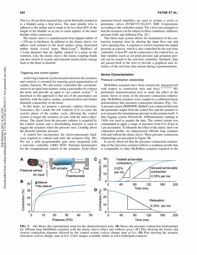
McKibben actuators have been extensively characterizedwith respect to contraction ratio and force.12,13,15,31 Weperformed characterization tests to study the effect of theelastic sleeve in terms of the pressure–contraction relation-ship. McKibben actuators were coupled to a calibrated linearpotentiometer that measures contraction distance (Fig. 3A).A pressure sensor (BSP000W; Balluff) was connected betweenthe pneumatic output from the control box and the actuator soas to measure the instantaneous pressure in the actuator itself. Adata logging system (PowerLab; ADInstruments) running at1 kHz was used to acquire the data. The control system wascommanded to apply a range of pressures from 0 to 45 psi in1 psi increments. To illustrate the effect of the elastic sleeve oncontraction profile, we characterized 100 mm long actuatorswith and without the elastic sleeve. These pressure–contractionrelationships are presented in Figure 3B.
It can be observed that the pressure–contraction relation-ship of the sleeveless actuator follows a nonlinear profile thatis comparable to other McKibben actuators reported in the
FIG. 3. (A) Shows the experimental setup for the characterization tests. (B) Shows the pressure–contraction relationshipsfor 100 mm long McKibben actuators with the elastic sleeve (blue) and without (gray). (C) Plot showing the fastest andslowest contraction response allowed by the control system (valves change state at 0 s). (D) Plot showing the actuatorrelaxation (valves change state at 0 s). Color images available online at www.liebertpub.com/soro
244 PAYNE ET AL.
Soft
Rob
otic
s 20
17.4
:241
-250
.D
ownl
oade
d fr
om o
nlin
e.lie
bert
pub.
com
by
172.
56.2
3.24
2 on
09/
22/1
7. F
or p
erso
nal u
se o
nly.
literature.13,31 The total contraction of the sleeveless designat 45 psi was 29%. Greater contraction was observed with thesleeved actuator (38% at 45 psi) since it is contracted froma fully elongated state.
For the sleeved actuator, Figure 3 highlights that a pressurethreshold is required before significant contraction is observedin the actuator. This is due to the nonlinear rubber modulus andthe nonlinear loading condition of the rubber. A greater pres-sure is required to achieve the same contraction as thesleeveless actuator due to the additional energy that is requiredto stretch the sleeve during pressurization. The improvedcontraction function is significant for the proposed cardiacapplication as it allows the ventricle wall to fully relax in thediastolic phase and contract by a greater distance in systole.
Contraction rate
We performed an experiment to quantify the slowest andfastest response times to achieve full contraction using asleeved 100 mm actuator (n = 6 runs). The actuator wascoupled to the calibrated linear potentiometer so that thecontraction length could be measured over time (setup shownin Fig. 3). A second signal which indicated when the valvesystem changed state was acquired simultaneously. To de-termine the slowest time to full contraction, the regulator wasconfigured to a pressure set point of 10 psi and the valve wasopened to enable pressurization. To determine the maximumcontraction time, we use the three-state valve system tocontrol the volume of air that is injected in to the actuator.
The regulator was configured to provide a maximum flowrate (pressure set point of 45 psi), and the valve was openedfor a set time period of 45 ms (empirically determined) beforebeing shut off to achieve a pressurization of 10 psi. Finally,when the actuator was pressurized at 10 psi, a vacuum of-11.6 psi was applied to the actuator to quantify the relaxa-tion time. The results are shown in Figure 3C, D.
The contraction-time profiles demonstrate that the con-traction response time can be varied significantly throughdifferent control methodologies. There is a latent contractiontime period in both cases due to the valve response time andthe minimum pressure threshold required to initiate con-traction. In the linear region of the fastest actuator contractionprofile, a rate of 0.62% per ms (SD –0.040% per ms) wasattained, in the slowest profile the peak rate was 0.30% per ms(SD –0.003% per ms). The overall response times to achievefull contraction for the fastest and slowest profile were120.3 ms (SD –2.1 ms) and 179.5 ms (SD –2.4 ms), respec-tively. In relaxation, the actuator returns to its fully elongatedstate in 156.0 ms (SD –1.4 ms) and achieved a peak relaxa-tion rate of 0.25% per ms (SD –0.001% per ms).
In Vivo Experiments
We carried out an in vivo porcine study (n = 1) to test threehypotheses.
� Hypothesis 1: mechanical coupling between the actu-ators and ventricle augments cardiac output.
� Hypothesis 2: there is an optimal systolic actuation timeperiod for the device for maximizing cardiac output anddiastolic function.
� Hypothesis 3: the rate of actuator contraction is a factorin cardiac output.
In vivo study setup
We followed the 1996 guide for the care and use of labo-ratory animals as recommended by the US National Instituteof Health. The study was performed at the Boston Children’sHospital. Ethical approval for the experimental protocol wasgranted through the Institutional Animal Care and UseCommittee. A general anesthetic was induced and mechan-ical ventilation used to support the swine throughout thesurgery. The swine (70 kg) was instrumented with pressuremonitoring transducers (SurgiVet; Smiths Medical PM,Inc.) that were located in the left atrium, aorta, pulmonaryartery, and right ventricle. A pressure sensor catheter (Sci-sense; Transonics, Inc.) was placed in the LV, and flow probes(TS420; Transonics, Inc.) were placed on the pulmonary arteryand aorta to acquire the blood flow rate through these vessels.
A multichannel data measurement system (PowerLab; ADInstruments) acquired these transducer signals at 1 kHz. Thebaseline heart rate before implantation was 83 bpm. Twopairs of 100 mm actuators were sutured to the ventricles withelasticated bands (according to the arrangement shown inFig. 1). To simulate HF we used the drug Esmolol, a short-acting, cardioselective beta blocker which reduces heart rateand contractility. After a steady HF baseline was achieved(heart rate of 72 bpm), we actuated the soft robotic implant atsystolic actuation periods of 25%, 30%, 35%, and 40% of thecardiac cycle. The LV pressure signal was used to trigger therobotic implant to contract at the beginning of systole. Theheart was allowed to return to the HF baseline levels in be-tween each actuation period. The three elastic coupling bandson each actuator were then removed so that the device wasuncoupled from the ventricles.
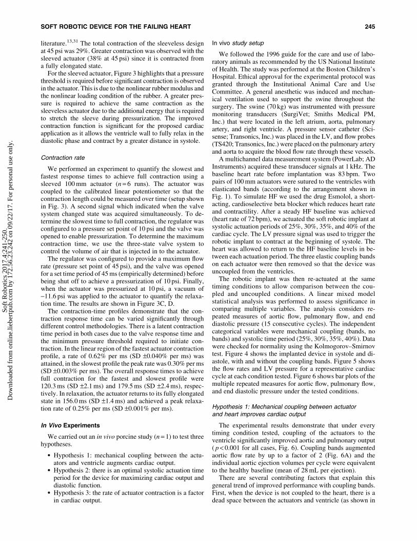
The robotic implant was then re-actuated at the sametiming conditions to allow comparison between the cou-pled and uncoupled conditions. A linear mixed modelstatistical analysis was performed to assess significance incomparing multiple variables. The analysis considers re-peated measures of aortic flow, pulmonary flow, and enddiastolic pressure (15 consecutive cycles). The independentcategorical variables were mechanical coupling (bands, nobands) and systolic time period (25%, 30%, 35%, 40%). Datawere checked for normality using the Kolmogorov–Smirnovtest. Figure 4 shows the implanted device in systole and di-astole, with and without the coupling bands. Figure 5 showsthe flow rates and LV pressure for a representative cardiaccycle at each condition tested. Figure 6 shows bar plots of themultiple repeated measures for aortic flow, pulmonary flow,and end diastolic pressure under the tested conditions.
Hypothesis 1: Mechanical coupling between actuatorand heart improves cardiac output
The experimental results demonstrate that under everytiming condition tested, coupling of the actuators to theventricle significantly improved aortic and pulmonary output( p < 0.001 for all cases, Fig. 6). Coupling bands augmentedaortic flow rate by up to a factor of 2 (Fig. 6A) and theindividual aortic ejection volumes per cycle were equivalentto the healthy baseline (mean of 28 mL per ejection).
There are several contributing factors that explain thisgeneral trend of improved performance with coupling bands.First, when the device is not coupled to the heart, there is adead space between the actuators and ventricle (as shown in
SOFT ROBOTIC DEVICE FOR THE FAILING HEART 245
Soft
Rob
otic
s 20
17.4
:241
-250
.D
ownl
oade
d fr
om o
nlin
e.lie
bert
pub.
com
by
172.
56.2
3.24
2 on
09/
22/1
7. F
or p
erso
nal u
se o
nly.

FIG. 4. Images from the in vivo study: (A)LV and soft robotic device with couplingbands in systole, (B) LV and soft actuatorswith coupling bands in diastole, (C) close-upof a soft actuator and coupling band in di-astole, and (D) close-up of a soft actuatorwith coupling band released and arrow in-dicating dead zone. Color images availableonline at www.liebertpub.com/soro
FIG. 5. Plots showing (A) aortic flow rate, (B) pulmonary flow rate, and (C) LV pressure under different systolic actuationtiming and mechanical coupling conditions, showing single representative cardiac cycles. Color images available online atwww.liebertpub.com/soro
246
Soft
Rob
otic
s 20
17.4
:241
-250
.D
ownl
oade
d fr
om o
nlin
e.lie
bert
pub.
com
by
172.
56.2
3.24
2 on
09/
22/1
7. F
or p
erso
nal u
se o
nly.
Fig. 4) which reduces the extent of contraction and recoil thatcan be transmitted to the heart. Second, in the uncoupledcondition, the dead space introduces a time delay before thecontracting actuator makes contact with the ventricle insystole, causing an interference with actuator-ventricle syn-chronization. Third, coupling also constrains the actuators toapply normal forces on to the ventricle and prevents tan-gential slippage over the ventricle surface.
Hypothesis 2: A longer systolic actuation periodimproves cardiac function with coupling
The mean aortic and pulmonary flow rates both positivelycorrelate with longer systolic actuation time conditions whenthe soft robotic device is coupled to the heart. Without cou-pling, increasing the systolic time period beyond 30% of thecardiac cycle does not yield significant augmentation for eitheraortic or pulmonary flow rate (Fig. 6). When coupled, a systolicperiod of 40% was demonstrated to provide the greatest aortic
flow rate (2 L/min); this same condition also yielded thegreatest pulmonary flow rate (1.8 L/min).
All conditions of device actuation led to a statisticallysignificant increase in cardiac output relative to the HFbaseline levels for aortic flow and pulmonary flow ( p < 0.001for all cases, Fig. 6). The effect of coupling is most prominentat the 40% actuation period compared to other times. At thislonger actuation period, the actuators relax after the hearthas entered the diastolic phase and assist with relaxation.At shorter actuation periods, the device recoils before theheart has completed systole and impedes the native con-traction, causing lower cardiac output. Furthermore, me-chanical coupling may also be more effective at the longeractuation because the time for refilling is reduced.
An encouraging observation was a significant reduction inend diastolic pressure with device operation from HF base-line ( p < 0.001 for 30–40% systolic period, with couplingbands, Fig. 6C). These results implied improved refillingfunction of the heart during diastole. A greater drop in end
FIG. 6. Bar plots showing (A) aortic flow,(B) pulmonary flow, and (C) end diastolicpressure. ***p < 0.001; **p < 0.01. Error barsare standard deviations. N.S., not signifi-cant. Color images available online at www.liebertpub.com/soro
SOFT ROBOTIC DEVICE FOR THE FAILING HEART 247
Soft
Rob
otic
s 20
17.4
:241
-250
.D
ownl
oade
d fr
om o
nlin
e.lie
bert
pub.
com
by
172.
56.2
3.24
2 on
09/
22/1
7. F
or p
erso
nal u
se o
nly.
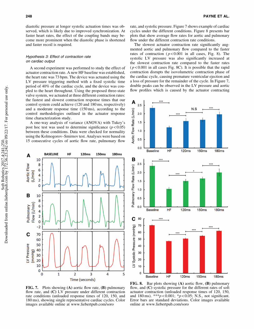
diastolic pressure at longer systolic actuation times was ob-served, which is likely due to improved synchronization. Atfaster heart rates, the effect of the coupling bands may be-come more prominent when the diastolic phase is shortenedand faster recoil is required.
Hypothesis 3: Effect of contraction rateon cardiac output
A second experiment was performed to study the effect ofactuator contraction rate. A new HF baseline was established,the heart rate was 73 bpm. The device was actuated using theLV pressure triggering method with a fixed systolic timeperiod of 40% of the cardiac cycle, and the device was cou-pled to the heart throughout. Using the proposed three-statevalve system, we actuated at three different contraction rates:the fastest and slowest contraction response times that ourcontrol system could achieve (120 and 180 ms, respectively)and a moderate response time (150 ms), according to thecontrol methodologies outlined in the actuator responsetime characterization study.
A one-way analysis of variance (ANOVA) with Tukey’spost hoc test was used to determine significance (p < 0.05)between these conditions. Data were checked for normalityusing the Kolmogorov–Smirnov test. Analyses were based on15 consecutive cycles of aortic flow rate, pulmonary flow
rate, and systolic pressure. Figure 7 shows example of cardiaccycles under the different conditions. Figure 8 presents barplots that show average flow rates for aortic and pulmonaryflow under the different contraction rate conditions.
The slowest actuator contraction rate significantly aug-mented aortic and pulmonary flow compared to the fasterrates of contraction ( p < 0.001 in all cases, Fig. 8). Thesystolic LV pressure was also significantly increased atthe slowest contraction rate compared to the faster rates( p < 0.001 in all cases Fig. 8C). It is possible that the rapidcontraction disrupts the isovolumetric contraction phase ofthe cardiac cycle, causing premature ventricular ejection anda loss of pressure for the remainder of the cycle. In Figure 7,double peaks can be observed in the LV pressure and aorticflow profiles which is caused by the actuator contracting
FIG. 7. Plots showing (A) aortic flow rate, (B) pulmonaryflow rate, and (C) LV pressure under different contractionrate conditions (unloaded response times of 120, 150, and180 ms), showing single representative cardiac cycles. Colorimages available online at www.liebertpub.com/soro
FIG. 8. Bar plots showing (A) aortic flow, (B) pulmonaryflow, and (C) systolic pressure for the different rates of softactuator contraction (unloaded response times of 120, 150,and 180 ms). ***p < 0.001; *p < 0.05; N.S., not significant.Error bars are standard deviations. Color images availableonline at www.liebertpub.com/soro
248 PAYNE ET AL.
Soft
Rob
otic
s 20
17.4
:241
-250
.D
ownl
oade
d fr
om o
nlin
e.lie
bert
pub.
com
by
172.
56.2
3.24
2 on
09/
22/1
7. F
or p
erso
nal u
se o
nly.
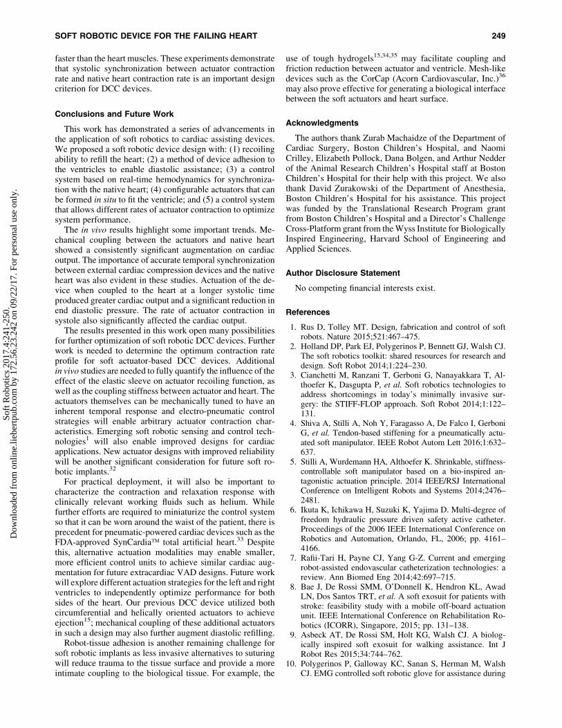
faster than the heart muscles. These experiments demonstratethat systolic synchronization between actuator contractionrate and native heart contraction rate is an important designcriterion for DCC devices.
Conclusions and Future Work
This work has demonstrated a series of advancements inthe application of soft robotics to cardiac assisting devices.We proposed a soft robotic device design with: (1) recoilingability to refill the heart; (2) a method of device adhesion tothe ventricles to enable diastolic assistance; (3) a controlsystem based on real-time hemodynamics for synchroniza-tion with the native heart; (4) configurable actuators that canbe formed in situ to fit the ventricle; and (5) a control systemthat allows different rates of actuator contraction to optimizesystem performance.
The in vivo results highlight some important trends. Me-chanical coupling between the actuators and native heartshowed a consistently significant augmentation on cardiacoutput. The importance of accurate temporal synchronizationbetween external cardiac compression devices and the nativeheart was also evident in these studies. Actuation of the de-vice when coupled to the heart at a longer systolic timeproduced greater cardiac output and a significant reduction inend diastolic pressure. The rate of actuator contraction insystole also significantly affected the cardiac output.
The results presented in this work open many possibilitiesfor further optimization of soft robotic DCC devices. Furtherwork is needed to determine the optimum contraction rateprofile for soft actuator-based DCC devices. Additionalin vivo studies are needed to fully quantify the influence of theeffect of the elastic sleeve on actuator recoiling function, aswell as the coupling stiffness between actuator and heart. Theactuators themselves can be mechanically tuned to have aninherent temporal response and electro-pneumatic controlstrategies will enable arbitrary actuator contraction char-acteristics. Emerging soft robotic sensing and control tech-nologies1 will also enable improved designs for cardiacapplications. New actuator designs with improved reliabilitywill be another significant consideration for future soft ro-botic implants.32
For practical deployment, it will also be important tocharacterize the contraction and relaxation response withclinically relevant working fluids such as helium. Whilefurther efforts are required to miniaturize the control systemso that it can be worn around the waist of the patient, there isprecedent for pneumatic-powered cardiac devices such as theFDA-approved SynCardia� total artificial heart.33 Despitethis, alternative actuation modalities may enable smaller,more efficient control units to achieve similar cardiac aug-mentation for future extracardiac VAD designs. Future workwill explore different actuation strategies for the left and rightventricles to independently optimize performance for bothsides of the heart. Our previous DCC device utilized bothcircumferential and helically oriented actuators to achieveejection15; mechanical coupling of these additional actuatorsin such a design may also further augment diastolic refilling.
Robot-tissue adhesion is another remaining challenge forsoft robotic implants as less invasive alternatives to suturingwill reduce trauma to the tissue surface and provide a moreintimate coupling to the biological tissue. For example, the
use of tough hydrogels15,34,35 may facilitate coupling andfriction reduction between actuator and ventricle. Mesh-likedevices such as the CorCap (Acorn Cardiovascular, Inc.)36
may also prove effective for generating a biological interfacebetween the soft actuators and heart surface.
Acknowledgments
The authors thank Zurab Machaidze of the Department ofCardiac Surgery, Boston Children’s Hospital, and NaomiCrilley, Elizabeth Pollock, Dana Bolgen, and Arthur Nedderof the Animal Research Children’s Hospital staff at BostonChildren’s Hospital for their help with this project. We alsothank David Zurakowski of the Department of Anesthesia,Boston Children’s Hospital for his assistance. This projectwas funded by the Translational Research Program grantfrom Boston Children’s Hospital and a Director’s ChallengeCross-Platform grant from the Wyss Institute for BiologicallyInspired Engineering, Harvard School of Engineering andApplied Sciences.
Author Disclosure Statement
No competing financial interests exist.
References
1. Rus D, Tolley MT. Design, fabrication and control of softrobots. Nature 2015;521:467–475.
2. Holland DP, Park EJ, Polygerinos P, Bennett GJ, Walsh CJ.The soft robotics toolkit: shared resources for research anddesign. Soft Robot 2014;1:224–230.
3. Cianchetti M, Ranzani T, Gerboni G, Nanayakkara T, Al-thoefer K, Dasgupta P, et al. Soft robotics technologies toaddress shortcomings in today’s minimally invasive sur-gery: the STIFF-FLOP approach. Soft Robot 2014;1:122–131.
4. Shiva A, Stilli A, Noh Y, Faragasso A, De Falco I, GerboniG, et al. Tendon-based stiffening for a pneumatically actu-ated soft manipulator. IEEE Robot Autom Lett 2016;1:632–637.
5. Stilli A, Wurdemann HA, Althoefer K. Shrinkable, stiffness-controllable soft manipulator based on a bio-inspired an-tagonistic actuation principle. 2014 IEEE/RSJ InternationalConference on Intelligent Robots and Systems 2014;2476–2481.
6. Ikuta K, Ichikawa H, Suzuki K, Yajima D. Multi-degree offreedom hydraulic pressure driven safety active catheter.Proceedings of the 2006 IEEE International Conference onRobotics and Automation, Orlando, FL, 2006; pp. 4161–4166.
7. Rafii-Tari H, Payne CJ, Yang G-Z. Current and emergingrobot-assisted endovascular catheterization technologies: areview. Ann Biomed Eng 2014;42:697–715.
8. Bae J, De Rossi SMM, O’Donnell K, Hendron KL, AwadLN, Dos Santos TRT, et al. A soft exosuit for patients withstroke: feasibility study with a mobile off-board actuationunit. IEEE International Conference on Rehabilitation Ro-botics (ICORR), Singapore, 2015; pp. 131–138.
9. Asbeck AT, De Rossi SM, Holt KG, Walsh CJ. A biolog-ically inspired soft exosuit for walking assistance. Int JRobot Res 2015;34:744–762.
10. Polygerinos P, Galloway KC, Sanan S, Herman M, WalshCJ. EMG controlled soft robotic glove for assistance during
SOFT ROBOTIC DEVICE FOR THE FAILING HEART 249
Soft
Rob
otic
s 20
17.4
:241
-250
.D
ownl
oade
d fr
om o
nlin
e.lie
bert
pub.
com
by
172.
56.2
3.24
2 on
09/
22/1
7. F
or p
erso
nal u
se o
nly.
activities of daily living. IEEE International Conference onRehabilitation Robotics (ICORR), Singapore, 2015; pp. 55–60.
11. Cezar CA, Roche ET, Vandenburgh HH, Duda GN, WalshCJ, Mooney DJ. Biologic-free mechanically induced mus-cle regeneration. Proc Natl Acad Sci 2016;113:1534–1539.
12. Obiajulu SC, Roche ET, Pigula FA, Walsh CJ. Soft pneu-matic artificial muscles with low threshold pressures for acardiac compression device. ASME 2013 International De-sign Engineering Technical Conferences and Computers andInformation in Engineering Conference, Portland, OR.American Society of Mechanical Engineers, Vol. 6A: 37thMechanisms and Robotics Conference, pp. V06AT07A009.
13. Roche ET, Wohlfarth R, Overvelde JTB, Vasilyev NV,Pigula FA, Mooney DJ, et al. A bioinspired soft actuatedmaterial. Adv Mater 2014;26:1200–1206.
14. Mac Murray BC, An X, Robinson SS, van Meerbeek IM,O’Brien KW, Zhao H, et al. Poroelastic foams for simplefabrication of complex soft robots. Adv Mater 2015;27:6334–6340.
15. Roche ET, Horvath MA, Wamala I, Alazmani A, Song S-E,Whyte W, et al. Soft robotic sleeve supports heart function.Sci Transl Med 2017;9:eaaf3925.
16. Mozaffarian D, Benjamin E, Go A, Arnett D, Blaha M,Cushman M, et al. Heart disease and stroke statistics-2016update: a report from the American Heart Association.Circulation 2016;133:e38–e360.
17. Givertz MM. Ventricular assist devices: important infor-mation for patients and families. Circulation 2011;124:305–312.
18. Rodriguez LE, Suarez EE, Loebe M, Bruckner BA. Ven-tricular assist devices (VAD) therapy: new technology, newhope? Methodist DeBakey Cardiovasc J 2013;9:32–37.
19. Rogers JG, Pagani FD, Tatooles AJ, Bhat G, Slaughter MS,Birks EJ, et al. Intrapericardial left ventricular assist devicefor advanced heart failure. N Engl J Med 2017;376:451–460.
20. Oz MC, Artrip JH, Burkhoff D. Direct cardiac compressiondevices. J Heart Lung Transplant 2002;21:1049–1055.
21. Moreno MR, Biswas S, Harrison LD, Pernelle G, MillerMW, Fossum TW, et al. Assessment of minimally invasivedevice that provides simultaneous adjustable cardiac sup-port and active synchronous assist in an acute heart failuremodel. J Med Devices 2011;5:041008.
22. Anstadt GL. Heart massage apparatus. United States patentUS5119804. 1992.
23. Lowe JE, Hughes GC, Biswas SS. Non–Blood-ContactingBiventricular Support: Direct Mechanical Ventricular Ac-tuation. Oper Tech Thorac Cardiovasc Surg 1999;4:345–351.
24. Kavarana MN, Loree HM 2nd, Stewart RB, Milbocker MT,Hannan RL, Pantalos GM, et al. Pediatric mechanicalsupport with an external cardiac compression device. JCardiovasc Dis Diagns 2013;1:1000105.
25. Artrip JH, Yi G-H, Levin HR, Burkhoff D, Wang J. Phy-siological and hemodynamic evaluation of nonuniformdirect cardiac compression. Circulation1999;100:II236–II243.
26. Pouleur H. Diastolic dysfunction and myocardial energet-ics. Eur Heart J 1990;11:30–34.
27. Rademakers FE, Buchalter MB, Rogers WJ, Zerhouni EA,Weisfeldt ML, Weiss JL, et al. Dissociation between leftventricular untwisting and filling. Accentuation by cate-cholamines. Circulation 1992;85:1572–1581.
28. Pueyo E, Sornmo L, Laguna P. QRS slopes for detectionand characterization of myocardial ischemia. IEEE TransBiomed Eng 2008;55:468–477.
29. Kashani A, Barold SS. Significance of QRS complex du-ration in patients with heart failure. J Am Coll Cardiol2005;46:2183–2192.
30. Schulte HF. The Characteristics of the McKibben ArtificialMuscle. The Application of Extemal Power in Prostheticsand Orthotics. Washington, DC: National Academy ofSciences and National Research Council, 1961.
31. Chou C-PCC-P, Hannaford B. Static and dynamic charac-teristics of McKibben pneumatic artificial muscles. ProcIEEE Int Conf Robot Autom 1994;281–286.
32. Robertson MA, Sadeghi H, Florez JM, Paik J. Soft Pneu-matic Actuator Fascicles for High Force and Reliability.Soft Robot 2017;4:23–32.
33. Slepian MJ, Smith RG, Copeland JG. The SynCardiaCardioWest� total artificial heart. Fundam Clin Cardiol2006;56:473.
34. Sun J-Y, Zhao X, Illeperuma WR, Chaudhuri O, Oh KH,Mooney DJ, et al. Highly stretchable and tough hydrogels.Nature 2012;489:133–136.
35. Darnell MC, Sun J-Y, Mehta M, Johnson C, Arany PR, SuoZ, et al. Performance and biocompatibility of extremelytough alginate/polyacrylamide hydrogels. Biomaterials2013;34:8042–8048.
36. Oz MC, Konertz WF, Kleber FX, Mohr FW, Gummert JF,Ostermeyer J, et al. Global surgical experience with theAcorn cardiac support device. J Thorac Cardiovasc Surg2003;126:983–991.
Address correspondence to:Frank A. PigulaJewish Hospital
Rudd Heart and Lung Center201 Abraham Flexner Way
Suite 1200Louisville, KY 40202
E-mail: [email protected]
Conor J. WalshJohn A. Paulson Harvard School
of Engineering and Applied ScienceHarvard University
60 Oxford StreetCambridge, MA 02138
E-mail: [email protected]
250 PAYNE ET AL.
Soft
Rob
otic
s 20
17.4
:241
-250
.D
ownl
oade
d fr
om o
nlin
e.lie
bert
pub.
com
by
172.
56.2
3.24
2 on
09/
22/1
7. F
or p
erso
nal u
se o
nly.