AN EXQUISITE APPROACH FOR IMAGE COMPRESSION …airccse.com/ijcsitce/papers/2115ijcsitce01.pdf · an...
13
International Journal of Computational Science, Information Technology and Control Engineering (IJCSITCE) Vol.2, No.1/2, April 2015 1 AN EXQUISITE APPROACH FOR IMAGE COMPRESSION TECHNIQUE USING LOSSLESS COMPRESSION ALGORITHM FOR ROI & NON-ROI REGIONS Shivaputra a , H.S.Sheshadri b , V.Lokesha c a Department of ECE, Assistant Professor, Dr Ambedkar Institute of Technology, India and Research scholar @ Jain University, India. [email protected] b Department of ECE, Professor and Research Dean, PES College of Engineering, India c Department of Mathematics, Associate Professor, Vijayanagara Sri Krishnadevaraya University, India ABSTRACT The imminent evolution in the field of medical imaging, telehealth and teleradiology services has been on a significant rise with a dire need for a proficient structure for the compression of a DICOM (Digital Imaging and Communications in Medicine) standard medical image obtained through various modalities, with clinical relevance and digitized clinical data, and various other diagnostic phenomena and the progressive transmission of such a medical image over varying bandwidths. The data loss redundancy during the process of compression is to be maintained below the alarming level, meaning it is to be under scanner without the loss of data/information. In this paper we present an efficient time bound algorithm that utilizes a process flow wherein multiple ROI sectors as well as the Non-ROI sector of the DICOM image are considered in the algorithmic machine and the compression is done based upon a hybrid compression algorithm by LZW & SPIHT encoder & decoder machines. The paper provides a magnitude of the overall compression ratio involved in thus compressing the DICOM standard image. It also provides a brief description about the PSNR values obtained after suitably compressing the image. We analyze the various encoder scenarios and have projected a suitable hybrid lossless compression algorithm that helps in the retrieval of the data/information related to the image. This paper also discusses around the Integer Wavelet Transforms used in order to encode the image and thus parameterize various metrics related to the transform function provided by IWT. It is seen that we could achieve a compression ratio value specific to 92% thereby swaying with a large deviation in the considered metric. KEYWORDS Wavelet Transform, Hybrid Encoder, IWT (Integer Wavelet Transforms), ROI (Region of Interest) & Non- ROI (Non-Region Of Interest), PSNR, Compression Ratio 1. INTRODUCTION The image compression has evolved into one of the imminent and necessary dynamic field in today’s research and the needful areas of medical imaging, telehealth, and telemedicine services. Thus care needs to be taken while the data/information pertaining to a DICOM standard image is being compressed for transmission across a suitable medium between various health inspectors or
-
Upload
truongtruc -
Category
Documents
-
view
220 -
download
2
Transcript of AN EXQUISITE APPROACH FOR IMAGE COMPRESSION …airccse.com/ijcsitce/papers/2115ijcsitce01.pdf · an...

International Journal of Computational Science, Information Technology and Control Engineering (IJCSITCE) Vol.2, No.1/2, April 2015
1
AN EXQUISITE APPROACH FOR IMAGE
COMPRESSION TECHNIQUE USING LOSSLESS
COMPRESSION ALGORITHM FOR ROI & NON-ROI
REGIONS
Shivaputraa, H.S.Sheshadri b, V.Lokesha c
aDepartment of ECE, Assistant Professor, Dr Ambedkar Institute of Technology, India
and Research scholar @ Jain University, India.
[email protected] bDepartment of ECE, Professor and Research Dean, PES College of Engineering, India cDepartment of Mathematics, Associate Professor, Vijayanagara Sri Krishnadevaraya
University, India
ABSTRACT The imminent evolution in the field of medical imaging, telehealth and teleradiology services has been on a
significant rise with a dire need for a proficient structure for the compression of a DICOM (Digital
Imaging and Communications in Medicine) standard medical image obtained through various modalities,
with clinical relevance and digitized clinical data, and various other diagnostic phenomena and the
progressive transmission of such a medical image over varying bandwidths. The data loss redundancy
during the process of compression is to be maintained below the alarming level, meaning it is to be under
scanner without the loss of data/information. In this paper we present an efficient time bound algorithm
that utilizes a process flow wherein multiple ROI sectors as well as the Non-ROI sector of the DICOM
image are considered in the algorithmic machine and the compression is done based upon a hybrid
compression algorithm by LZW & SPIHT encoder & decoder machines. The paper provides a magnitude of
the overall compression ratio involved in thus compressing the DICOM standard image. It also provides a
brief description about the PSNR values obtained after suitably compressing the image. We analyze the
various encoder scenarios and have projected a suitable hybrid lossless compression algorithm that helps
in the retrieval of the data/information related to the image.
This paper also discusses around the Integer Wavelet Transforms used in order to encode the image and
thus parameterize various metrics related to the transform function provided by IWT. It is seen that we
could achieve a compression ratio value specific to 92% thereby swaying with a large deviation in the
considered metric.
KEYWORDS Wavelet Transform, Hybrid Encoder, IWT (Integer Wavelet Transforms), ROI (Region of Interest) & Non-
ROI (Non-Region Of Interest), PSNR, Compression Ratio
1. INTRODUCTION
The image compression has evolved into one of the imminent and necessary dynamic field in
today’s research and the needful areas of medical imaging, telehealth, and telemedicine services.
Thus care needs to be taken while the data/information pertaining to a DICOM standard image is
being compressed for transmission across a suitable medium between various health inspectors or

International Journal of Computational Science, Information Technology and Control Engineering (IJCSITCE) Vol.2, No.1/2, April 2015
2
health institutions for various diagnostic purposes. Lossless image compression has proven to be
a well established approach in reducing the image data without compromising the quality of the
same. Medical Imaging is been in use for various diagnostic as well as surgical processes. It
sometimes may need a long-term storage for profiling the patient’s data as well as the
transmission of the same over to long distances. One of the most proven and valuable tools
supporting the Picture Archiving and Communication System (PACS), a key successor
facilitating the exchange of the structured medical imaging data to PACS is, Digital Imaging and
Communications in Medicine (DICOM) standard. This DICOM file consists of a header,
containing the information about the patient’s name, type of scan, position & dimension of the
scan, and other image data. While all the image information will be contained in the image data.
The very motivating aim of image compression is to maintain a very squat but rate representation
with respect to the image data of a patient. Reduction in the storage and potency with its
limitations as such, without any loss in the entropy of an image and the transmission of the raw
information pertaining to the image reducing the ubiquitous redundancy is the major aspects that
needs to be addressed during an image compression and by its underlying algorithm. With such
an approach we have two different types of compression techniques;
Lossy Compression Techniques – some of the data pertaining to the image data may be
lost or some identified changes or loss may be incurred, in the reconstructed image.
Lossless Compression Technique – the reconstructed image will be exactly the same as
the original image, without loss of data in the image.
The proven task is to obtain better compression ratios during multimedia and image compression,
without any as such compromise in the data of the multimedia file or an image file, during the
loss in the communication field or during compression.
A well constructed as such approach is discussed and highlighted in this paper, which focuses on
the potential gains which are achieved in archiving and compressing a medical image like a
DICOM image using a lossless compression technique which is an hybrid with the dictionary
based approach of LZW lossless image compression algorithm & SPIHT, with the amount of data
being minimal for the transmission of data with a high quality of the image and IWT (Integer
Wavelet Transforms) algorithms across a region of importance termed as the Region Of Interest
(ROI) and the Non-ROI (Non Region Of Interest) sections of the medical image and applying
suitable compression algorithms and differentiating a metric among various lossless compression
algorithms and their deviations from each other, in terms of the compression ratios and the data
redundancy.
2. IMAGE COMPRESSION
The compression of digital medical images is of great demand nowadays. The compression of
such images may involve a single image or a sequence of images. With the days the medical
community has been reluctant on the compression techniques by adopting to lossy over lossless
compression techniques in clinical practices. With a geometric increase in the diagnostic data and
images produced by the medical organizations, a suitable compression technique is needed to
compress large amounts of data resulting in greater data reductions and transmission speeds over
a bandwidth. The preference to lossy compression techniques is provided in these cases for the
perseverance of the diagnostic information. Lossy compression techniques involve deliberate
discarding of information for the image that is not visually or diagnostically important. Modest
compression metrics can be achieved in lossy compression techniques with a significant loss of
data. But on a contrary note greater compression rates are achievable in lossy compression
techniques with an acceptance of visible loss of data for the clinical task. For particular

International Journal of Computational Science, Information Technology and Control Engineering (IJCSITCE) Vol.2, No.1/2, April 2015
3
applications, there is still a controversial role with lossy compression techniques. This due course
of compression techniques can be overcome by an appropriate lossless compression technique
and the performance of traditional and state-of-art metrics involved in the compression techniques
are evaluated.
The lossless compression techniques involve some of the crude classifiers such as;
Statistical Modelling involving predictive schemes, with the differences between the
underlying and the neighbouring pixels of an image are computed before the coding of
compression algorithm with Context Modelling.
Each pixel or a cluster of pixels being transformed into frequency or wavelet domain
prior to modelling and coding, as in Transform Based Coding.
Ad-Hoc Schemes (as such in Run-Length Encoding)
Replacing strings of symbols with code snippets, as in Dictionary Based Schemes.
Dictionary Based Schemes are widely used for text data compression. Advantage of data
redundancy occurs in image compression techniques. Each one of these redundancies will be
exploited by each of the compression methodologies. The various redundancies are;
Spatial
Temporal
Spectral
The underlying idea behind most of the compression algorithms is to transform an image suitable
for storage and transmission and also to represent the image in the form of an orthogonal function
so as to distribute the energy signal components among the various de-correlated components.
3. RELATED WORKS
Compression of DICOM images for different entities on SPIHT (Set Partitioning in Hierarchical
Trees) with progressive transmission qualities for telemedical applications, wherein the header of
the DICOM file is transmitted first followed by the image data in various stages. It works on the
principle of spatial relationship among the various wavelet co-efficient at different levels and
frequency sub-bands in the pyramid like structure of the wavelet decomposition, producing
streams of embedded bits for an image. This work was proposed as a methodology by
Ramakrishnan et al. (2006) for the compression of wavelets and splints for telemedicine
applications. Various compression metrics were evaluated with other standard lossless image
compression techniques and were concluded that they produce the best results with less
consumption of bandwidth and lesser execution time as well as transmission time. In another
methodology proposed by Kumar et al. (2008), they considered DCT (Discrete Cosine
Transforms), SVD and BTC (Block Truncation Code), SD (Standard Deviation) depending upon
the properties of the image and the requirements. The evaluation of such a kind of a methodology
yielded quite reliable results with respect to the performance metrics in terms of PSNR (Peak
Signal to Noise Ratio) and MSE (Mean Square Error). Limitations with respect to the SPIHT
techniques as proposed by Ramakrishnan et al. (2006), involving the non specification of the
types of wavelets for image compression lead to the evolution of a new methodology proposed
and worked around by Bairagi et al., wherein they made as selection of wavelets for medical
image compression. Analysis over all the ‘db’ wavelet series and the comparison of the results
over several metrics such as PSNR, MSE, NAE (Normalized Absolute Error), Structural Contents
etc, showed up an improvement with their proposed methodology. A revival over various
implemented image compression techniques and algorithms involved in this compression

International Journal of Computational Science, Information Technology and Control Engineering (IJCSITCE) Vol.2, No.1/2, April 2015
4
techniques were discussed by Dhawan et al. (2011). They also proposed a low bit compression
and fractal approach utilizing resolution free decoding property for a low bit compression.
Verification approach by correlation using the wavelet compression of pre-processing digital
images was presented by Morales et al. (2012). A biometric system was implemented that used a
recognition pattern for digital finger-print which satisfies the characteristics of uniqueness,
universally, performance and collectability. They used a specific Fourier Transform and wavelet
transform to perform the function of correlation, compression, compressed filter implementation,
transformation and GUI implementation. An advanced approach using DCT in DCM to improve
the performance of the image compression and its underlying metrics such as performace ,
minimum data loss, and reconstructed image clarity with HAAR process, using orthogonal
combination on unit interval was proposed by Alka Sharma et al. (2013). There was also a
proposal of such a work involving the multiple ROI sections over a suitable medical image and an
efficient lossless compression algorithm to compress the ROI section of the medical image while
the Non-ROI section being done with a lossy compression algorithm was subdued with an
ideology proposed by Shivaputra et al. (2014). They proposed a workflow that rather utilizes an
efficient lossless image compression algorithm for both ROI and Non-ROI section of a DICOM
standard medical image.
Before the discussion of the actual methodology involved in this proposed work we considered
the very basic workflow or the idea of implementing the lossless image compression algorithm
for both ROI and Non-ROI sections and evaluation of the various performance metrics such as
PSNR, MSE, NAE, and energy distribution within the compressed image and the need for a
suitable lossless image compression algorithm for a suitable transmission over a bandwidth.
4. PROPOSED METHODOLOGY
In this paper we have proposed an efficient approach for the lossless image compression using the
IWT transformations for both ROI and the Non-ROI sections of the DICOM standard medical
image. This approach has provided an improvement in the performance, redundant loss in the
image data and reconstructed image. The process involved in this approach is as given below;

International Journal of Computational Science, Information Technology and Control Engineering (IJCSITCE) Vol.2, No.1/2, April 2015
5
Figure 1. Proposed Methodology Flow Diagram
A suitable medical image obtained from the varsity of the modalities is obtained in the prescribed
DICOM standard, which can be in any of the acceptable formats such as bitmap (.bmp), tiff or
jpeg. Here for the justification of our methodology we have used a Nose Fracture X-ray image of
the format .bmp and the resolution 256X256. Thou image is subjected to the Image Segmentation
(Global Thresholding) to segment the complex image file into simpler structure to be input to the
compression algorithm. The values specific in this proposed work is considered as, 0.44 as the
Global Threshold (Graythresh) and a threshold value of 0.55. We also implement the Canny
operator to segment the complex DICOM standard medical image. The below figure justifies the
segmented image and its histogram analysis of the Graythresh value.
Figure 2. Original Nose Fracture Image
International Journal of Computational Science, Information Technology and Control Engineering (IJCSITCE) Vol.2, No.1/2, April 2015
6
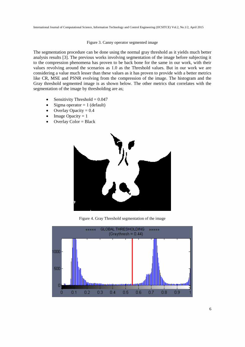
Figure 3. Canny operator segmented image
The segmentation procedure can be done using the normal gray threshold as it yields much better
analysis results [3]. The previous works involving segmentation of the image before subjecting it
to the compression phenomena has proven to be back bone for the same in our work, with their
values revolving around the scenarios as 1.0 as the Threshold values. But in our work we are
considering a value much lesser than these values as it has proven to provide with a better metrics
like CR, MSE and PSNR evolving from the compression of the image. The histogram and the
Gray threshold segmented image is as shown below. The other metrics that correlates with the
segmentation of the image by thresholding are as;
Sensitivity Threshold = 0.047
Sigma operator = 1 (default)
Overlay Opacity = 0.4
Image Opacity = 1
Overlay Color = Black
Figure 4. Gray Threshold segmentation of the image

International Journal of Computational Science, Information Technology and Control Engineering (IJCSITCE) Vol.2, No.1/2, April 2015
7
Figure 5. Global Gray Threshold Value
The considered image if not in a DICOM standard then a mechanism for the conversion of the
same to a standard DICOM medical image format has to be done (usually a grayscale image
format). The image is input to the ROI tool to classify the ROI and the Non-ROI sections. Figure
5 & Figure 6 provides a substantial proof for the classification.
Figure 6. Classification of the ROI and the Non-ROI from the segmented image
A selection is made with respect to the multiple ROI sections in a DICOM standard medical
image. We analyze various parameters such as Statistics, Histogram with reference to the ROI
section selected and the Data related to the ROI selected, which is imported to the Workspace of
the MATLAB environment. Multiple ROI sections may be recorded or classified from a DICOM
standard medical image. We have considered 2 ROI sections within this image considered for our
work.

International Journal of Computational Science, Information Technology and Control Engineering (IJCSITCE) Vol.2, No.1/2, April 2015
8
Figure 7. Multiple ROI selections within the segmented image
The Histogram feature set of the ROI section 1 being selected from the DICOM standard medical
image provides details about the data features of the ROI section placed in the Workspace
environment of the MATLAB tool. The given data is in the form of a structure 1X1, with the
metrics such as Mean, Standard Deviation, Mask, Minimum and the Maximum pixel values of
the image. The selected ROI will be expressed in the double format while the mask is also in the
double format to classify between the ROI and the Non ROI sections in an image.
Figure 8. ROI Data in the workspace environment
The image is compressed using the hybrid compression algorithm involving both LZW & SPIHT,
and the image is obtained after the algorithm is run with a maximum loop structure for about 16
iterations. The minimum number of loops encountered will be 12 as the variation in the
compression ratios in the order 3:1 is achievable only from the loop value ranging from 12. The
Bit-Per-Pixel (bpp), MSE (Mean Square Error) and the (Peak Signal-to-Noise Ratio) PSNR is
recorded for the same and the deviation with the metrics are evaluated.

International Journal of Computational Science, Information Technology and Control Engineering (IJCSITCE) Vol.2, No.1/2, April 2015
9
Thus obtained image with the classification of ROI and NON-ROI sections is input to the
transformation algorithm machine, based on IWT (Integer Wavelet Transforms). High fidelity
images like the medical images require an efficient lossless encoding procedure for its
preservation.
The set of transforms are governed by the following equations in IWT;
+
The IWT engine takes in the DICOM standard medical image and the effectiveness of this
transformation algorithm is done by substantially measuring the first order entropy relate to the
medical image. The magnitude of these entropies with respect to various quadrants of the ROI
sections and the Non-ROPI sections varies relatively, while their mean is calculated. The wavelet
filter is attached to the coding algorithm in order to obtain the higher entropies to compute the
actual bit rate. Higher order filters can be used in order to produce a better compressed image. We
could achieve a comprehensive compression ratio rate of about 11.039 with the S-filter and the
efficiency was improved with higher order filters like (4,2) and (2,2+2). Bit Rates in the range
3.56, 4.89, 6.81, 4.77, and 3.14 were obtained with S-random, SPB, SPC, 2/6, and 2/10 random
filters. Among the different flavours of approaches we calculate with the spatial domain approach
using the equation;
Table 1. Bit Rates with respect to various filters
Filters Bit Rate
S-transform 3.42
SPB 4.89
SPC 6.81
2/6 4.77
2/10 3.14
Now the ROI and Non-ROI classified image is compressed using the hybrid compression
algorithm which has a dictionary based approach as in LZW Lossless Image Compression
algorithm as well as the pyramid like structure approach with the 4 different filters used in the
SPIHT (Set Partitioning in Hierarchial Trees). The procedure of the proposed work / algorithm is
as follows;
Step1: Select the segmented image X;
Step2: Initialize the vectors in the dictionary table for the hybrid algorithm with uint16 & double
format;
Step3: The grayscale pixel values are imposed onto the segmented pixels;
International Journal of Computational Science, Information Technology and Control Engineering (IJCSITCE) Vol.2, No.1/2, April 2015
10
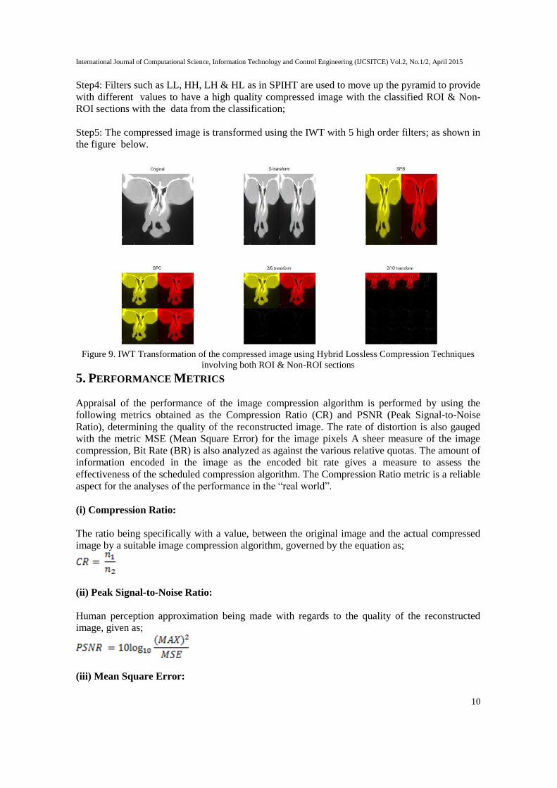
Step4: Filters such as LL, HH, LH & HL as in SPIHT are used to move up the pyramid to provide
with different values to have a high quality compressed image with the classified ROI & Non-
ROI sections with the data from the classification;
Step5: The compressed image is transformed using the IWT with 5 high order filters; as shown in
the figure below.
Figure 9. IWT Transformation of the compressed image using Hybrid Lossless Compression Techniques
involving both ROI & Non-ROI sections
5. PERFORMANCE METRICS
Appraisal of the performance of the image compression algorithm is performed by using the
following metrics obtained as the Compression Ratio (CR) and PSNR (Peak Signal-to-Noise
Ratio), determining the quality of the reconstructed image. The rate of distortion is also gauged
with the metric MSE (Mean Square Error) for the image pixels A sheer measure of the image
compression, Bit Rate (BR) is also analyzed as against the various relative quotas. The amount of
information encoded in the image as the encoded bit rate gives a measure to assess the
effectiveness of the scheduled compression algorithm. The Compression Ratio metric is a reliable
aspect for the analyses of the performance in the “real world”.
(i) Compression Ratio:
The ratio being specifically with a value, between the original image and the actual compressed
image by a suitable image compression algorithm, governed by the equation as;
(ii) Peak Signal-to-Noise Ratio:
Human perception approximation being made with regards to the quality of the reconstructed
image, given as;
(iii) Mean Square Error:

International Journal of Computational Science, Information Technology and Control Engineering (IJCSITCE) Vol.2, No.1/2, April 2015
11
Quota of the scale of distortion in a reconstructed image obtained from a decoder is MSE,
monitored by the mathematical formulae;
Higher the deterministic values output with respect to PSNR and higher CR values, yield a high
quality of the reconstructed image.
The values for the above mentioned metrics are recorded after the compression of the DICOM
standard medical image. The table given below gives the details about the Maximum loops the
iterations have entered for the compression of the image, CR, BPP, MSE and PSNR.
Table 2. Table representing various metrics with respect to the maximum number of loops of iterations
Maximum Loop CR BPP MSE PSNR (dB)
12 3.4261 0.8223 6.9835 39.6901
13 5.7938 1.3905 3.4397 42.7656
14 9.9747 2.3939 1.9047 45.3324
15 16.4083 3.9380 1.3057 46.9722
16 25.1424 6.0342 1.1157 47.6552
6. CONCLUSION
In this work, we have worked around the compression techniques using the lossless compression
algorithms with IWT. DICOM standard medical images from various medical diagnosis
phenomena are obtained and are used for telemedical applications and diagnostic purposes. The
recommended format of medical images providing the information about the patients in the
header of the DICOM file while the information about the image in the image data file. Here we
have considered the metrics such as PSNR, MSE and CR between various transforms using
various high order filters. Thus obtained results are compared with the various images and their
compression techniques and were found that the proposed algorithm depicts that the value of
PSNR, CR, MSE for the DICOM standard medical image, proving to be more reliable and
efficient. The performance of our proposed method of IWT using Lossless Image Compression
techniques (Hybrid Compression technique) is measured over the DICOM medical image and
observed the results for decomposition levels. The proposed algorithm scheme has given superior
image compression results for DICOM images.
ACKNOWLEDGEMENTS
The authors would like to thank the Management Panchajanya Vidya Peetha Welfare Trust
(Regd), Dr. C. Nanjundaswamy our beloved Principal, Dr. G V Jayaramaiah, Dr. R.Murali, Dr M
V Mandi and Dr S Ramesh of Dr. Ambedkar Institute of Technology, Bengaluru for their
assistance, suggestions, insight and valuable discussion over the course of this research work.
REFERENCES

International Journal of Computational Science, Information Technology and Control Engineering (IJCSITCE) Vol.2, No.1/2, April 2015
12
[1] Shivaputra, H.S. Sheshadri, V. Lokesha “An Efficient Lossless Medical Image Compression
Technique for Telemedicine Applications”, Computer Applications: An International Journal (CAIJ),
Vol.2, No.1, February 2015, pp-63-69
[2] K. Vidhya and S. Shenbagadevi “Medical Image Compression using Hybrid Coder with Fuzzy Edge
Detection”, ICTACT Journal on Image and Video Processing, February, 2011, vol. 01, Issue. 03.
[3] David A Clunie “ Lossless Compression of Grayscale Medical Images – Effectiveness of Traditional
and State of the Art Approaches”
[4] A.R. Calderbank, Ingrid Daubechies, Wim Sweldens, Bon Lock Yeo, “Lossless Image Compression
using Integer Wavelet Transforms” ICIP, 1997
[5] G. UmaVetri Selvi, R. Nandarajan , “DICOM Image Compression using Bilinear Interpolation”
IEEE, 978-1-4244-6561-3/10, 2010.
[6] Alka Sharma, Gagangeet Singh Aujla, Jagbir Singh Gill, “A Comprehensive Lossless Modified
Compression in Medical Application on DICOM CT images”, IOSR Journal of Computer
Engineering (IOSR-JCE), e-ISSN: 2278-0661, p-ISSN: 2278-8727, Vol. 15, Issue 3, Dec 2013, pp
01-07
[7] K. Vidhya and S. Shenbagadevi “A Two Component Medical Image Compression Technique”,
IJRTE, Vol. 1, No. 1, May 2009.
[8] S. Vijayadheeswarreddy, I.V.G. Manohar, “Compressing DICOM Images using SPIHT with Huffman
Encoding”, IJERT, ISSN: 2278-0181, Vol.1, Issue. 5, July 2012.
[9] Nazeeh Aranki, Didier Keymeulen, Alireza Bakshi and Matthew Klimesh, “Hardware
Implementation of Lossless Adaptive and Scalable Hyperspectral Data Compression for Space”,
NASA/ESA, Conference on Adaptive Hardware and Systems, 2009
[10] Said A, Pearlman W, "Reversible image compression via multi-resolution representation and
predictive coding", SPIE VCIP Symposium, Cambridge, MA,1993.
[11] Randers-Pehrson G, et al, PNG (Portable Network Graphics) specification version 1.1. PNG
Development Group. February 1999.
[12] Deutsch P, Gailly JL, ZLIB Compressed data format specification version 3.3. Internet RFC 1950.
Aladdin Enterprises. May 1996.
[13] Rice RF, Some practical universal noiseless coding techniques, Technical Report JPL-79-22.Pasadena
CA 1979.
[14] Golomb SW, "Run-length encodings", IEEE Transactions on Information Theory, 1966 pp 140-149.
[15] Kivijärvi J, et al, "A comparison of lossless compression methods for medical images", Computerized
Medical Imaging and Graphics, 22, pp 323-339, 1998.
[16] Denecker K, Van Overloop J, Lemahieu I, "An experimental comparison of several lossless image
coders for medical images", Proc. 1997 IEEE Data Compression Conference, Storer J, Cohn M eds.,
IEEE Computer Society Press, Los Alamitos CA, 1997.
[17] Young SS, Whiting BR, Foos DH, "Statistically lossless image compression for CR and DR",
Proceedings of SPIE: Medical Imaging 1999: Image Display, 3658, SPIE Press, Bellingham WA,
1999.
[18] Munteanu A, Cornelis J, Cristea P, "Wavelet-based lossless compression of coronary angiographic
images", IEEE Transactions on Medical Imaging, 18, pp 272-281, 1999.
AUTHORS
Shivaputra is an PhD Scholar majoring in Electronics Engineering at the Jain University,
Bengaluru. He received the B.E. Degree in Electronics & Communication Engineering
from the University of Visvesvaraya College of Engineering, Bengaluru, Bangalore
University in 2002, and M.Tech. Degree in VLSI Design & Embedded System from the
BMS College Engineering, Visvesvaraya Technological University in 2009. He is a life
member of ISTE, SIE, IAENG, IASCIT and has participated in various national
and international conferences and seminars in India and abroad.
Dr. H.S. Sheshadri Obtained his B.E. from University of Mysore during 1979 in E & C
Engg, M.E (Applied Electronics) from Bharathiar University, Coimbatore during 1989

International Journal of Computational Science, Information Technology and Control Engineering (IJCSITCE) Vol.2, No.1/2, April 2015
13
and PhD from Anna University Madras during 2008.He is serving presently as a Professor and
Dean(Research) in PES College of Engineering, Mandya. His field of interest is Medical Image
processing and embedded systems. He has published 12 papers in national journals and 21 papers in
International journals. Also guiding 6 research candidates for PhD programme under university of
Mysore, Two candidates under VTU, Belgaum. He is a life member of IE (India), IETE, ISTE,
SSI, and has participated in various national and international conferences and seminars in India and
abroad.
Dr. V. Lokesha, has awarded a PhD from Mysore University, Mysore and is currently
Special Officer and Deputy Registrar in Vijayanagara Sri Krishnadevaraya University,
Bellary. He is presently Chairman of Department of Computer Science and Mathematics.
He served as a Professor at Acharya Institute of Technology, Bangalore. He was invited
in Turkey, France, South Korea, and Iran for International conferences. He is a reviewer of
American Mathematical reviews, USA and several Journal of international repute. He
delivered several series of lectures under the V.T. U EDUSAT - LIVE telecast lecture
series programme. He has published more than 100 research papers in the journals of international repute.
Under his Guidance 8 PhD and 28 MPhil degrees are awarded and working as editorial member of several
international and National Journals.