An enzyme immunoassay for the measurement of ... · An enzyme immunoassay for the measurement of...
23
000681-120409 mm dd yy KR/CD 1 EL-ANA Profiles TM An enzyme immunoassay for the measurement of autoantibodies directed against the following antigens: ssDNA, dsDNA, Sm HR , RNP/Sm, SSA (Ro) HR , SSB (La) HR , Histones, Scl-70, Jo-1, Ribosomal Protein P, Centromere HR , and Chromatin For professional use only Instruction Manual Catalog Nos: 101-005 (EL-ANA/6™, 5 plates kit), 103-005 (EL-ANA/8™, 5 plates kit), 102-005 (EL-ANA/7 TM , 5 plates kit), 104-005 (EL-ANA/9 TM , 5 plates kit), 104-101 (EL-ssDNA™ kit), 104-201 (EL-dsDNA™ kit), 104-301 (EL-SSA™ kit), 104-401 (EL-SSB™ kit), 104-501 (EL-Sm™ kit), 104-601 (EL-RNP/Sm™ kit), 104-701 (EL-Histone™ kit), 104-801 (EL-Scl-70™ kit), 104-901 (EL-Jo-1™ kit), 104-110 (EL-Ribo-P™ kit), 104-111 (EL-Centromere™ kit), 104-113 (EL-Chromatin™ kit) TheraTest Laboratories, Inc. 1111 North Main Street Lombard, IL 60148 USA Tel: 1-800-441-0771 1-630-627-6069 Fax: 1-630-627-4231 e-mail: [email protected] www.TheraTest.com
Transcript of An enzyme immunoassay for the measurement of ... · An enzyme immunoassay for the measurement of...

000681-120409 mm dd yy
KR/CD 1
EL-ANA ProfilesTM
An enzyme immunoassay for the measurement of autoantibodies directed against the following antigens: ssDNA, dsDNA, SmHR,
RNP/Sm, SSA (Ro)HR, SSB (La)HR, Histones, Scl-70, Jo-1, Ribosomal Protein P, CentromereHR, and
Chromatin
For professional use only
Instruction Manual
Catalog Nos: 101-005 (EL-ANA/6™, 5 plates kit), 103-005 (EL-ANA/8™, 5 plates kit), 102-005 (EL-ANA/7TM, 5 plates kit), 104-005 (EL-ANA/9TM, 5 plates kit), 104-101 (EL-ssDNA™ kit), 104-201 (EL-dsDNA™ kit), 104-301 (EL-SSA™ kit), 104-401 (EL-SSB™ kit), 104-501 (EL-Sm™ kit), 104-601 (EL-RNP/Sm™ kit), 104-701 (EL-Histone™ kit), 104-801 (EL-Scl-70™ kit), 104-901 (EL-Jo-1™ kit), 104-110 (EL-Ribo-P™ kit), 104-111 (EL-Centromere™ kit), 104-113 (EL-Chromatin™ kit)
TheraTest Laboratories, Inc. 1111 North Main Street
Lombard, IL 60148 USA Tel: 1-800-441-0771 1-630-627-6069 Fax: 1-630-627-4231
e-mail: [email protected] www.TheraTest.com
000681-120409 mm dd yy
KR/CD
TABLE OF CONTENTS
Page INTRODUCTION ................................................................... 1
REAGENTS ............................................................................. 2
WARNINGS AND PRECAUTIONS ..................................... 4
STORAGE AND HANDLING…….………………………...5
SPECIMEN REQUIREMENTS ............................................ 5
PROCEDURE.......................................................................... 6
QUALITY CONTROL ......................................................... 10
RESULTS ............................................................................... 10
EXPECTED VALUES .......................................................... 11
GUIDE TO INTERPRETATION........................................ 12
PERFORMANCE DATA ..................................................... 15
TROUBLESHOOTING ....................................................... 17
REFERENCES ...................................................................... 18
ABBREVIATED TEST PROCEDURE……….…………. 20

000681-120409 mm dd yy
KR/CD 1
INTRODUCTION Name: TheraTest EL-ANA Profiles™
Intended Use: “The EL-ANA ProfilesTM” is an in vitro diagnostic test for the detection and measurement of autoantibodies directed against the following autoantigens: single-stranded DNA (ssDNA), double-stranded DNA (dsDNA), Sm, RNP/Sm, SSA (Ro), SSB (La), Histones, Scl-70, Jo-1, Ribosomal Protein P, Centromere, and Chromatin (Nucleosomes). This test system is intended as an aid in diagnosis of systemic lupus erythematosus, Sjögren’s syndrome, progressive systemic sclerosis (scleroderma), drug-induced lupus and polymyositis. Summary and Explanation of the Test
Autoantibodies against nuclear constituents are used for the diagnosis of rheumatic diseases. Antibodies against native or dsDNA occur predominantly in Systemic Lupus Erythematosus (SLE) and are included in the diagnostic criteria for SLE.1 Anti-ssDNA antibody, when present at high levels, is an indicator of active SLE.2 However, lower levels are observed in chronic inflammatory and infectious diseases.16 The presence of autoantibodies to Sm is considered a marker for SLE and is included in the diagnostic criteria for SLE.1 Anti-RNP antibody, when present in serum alone at high levels, is diagnostic for Mixed Connective Tissue Disease (MCTD).3 Anti-RNP, when detected at lower levels, particularly in conjunction with other autoantibodies, is observed in the serum of patients with Progressive Systemic Sclerosis, Sjögren’s Syndrome, and Rheumatoid Arthritis. In SLE patients, autoantibodies to RNP are associated with a lower incidence of renal dysfunction and with relatively benign disease, while patients with anti-Sm have a higher incidence of renal and central nervous system disease.4 Auto-antibodies to both SSA and SSB are observed in the sera of patients with SLE,5 and in Sjögren’s Syndrome.6 Anti-SSA is frequently detected in ANA-negative patients (i.e., subacute cutaneous LE).7 Some of these ANA-negative patients have antibodies to ssDNA.8 Furthermore, anti-SSA is present in a subset of patients with nephritis who lack anti-dsDNA.9 Anti-Scl-70 is frequently associated with Scleroderma, but is also present in a minority of patients with active SLE.9,12 Anti-Histones, although frequently observed in idiopathic SLE, are also observed in patients with drug-induced LE.9 The presence of only anti-ssDNA and anti-Histones is suggestive of drug-induced LE. Anti-Jo-1 antibodies, directed toward histidyl-tRNA synthetase, are found in a subset of patients with connective tissue disease that have either polymyositis, dermatomyositis or myositis with another rheumatic disease.17,18 Anti-Ribosomal Protein P antibodies (anti-Ribo P) are highly specific serologic markers for SLE since they are not usually detected in other autoimmune, infectious or neoplastic diseases.19 Anti-centromere antibodies are detected mainly in a subset of patients with progressive systemic sclerosis (scleroderma).20 Anti-chromatin antibodies are detected with high frequency in SLE and drug-induced LE, but with relatively low frequency in other autoimmune diseases and are reported to be valuable indicators of disease activity.21,22 For drug-induced lupus, the sensitivity reported in some studies is as high as 100%.21 Autoantibodies may be present with a great deal of variability in patients with SLE and in other connective tissue diseases. Therefore, it has become clinically relevant to determine the panel of autoantibodies present in the serum of each patient.12
Most autoimmune diagnostic kits detect a single type of autoantibody; a distinction between the presence of IgG and IgM autoantibodies generally is not possible. Although a number of attributes contribute to pathogenicity of autoantibodies (e.g., class, avidity, charge, complement fixation), IgG
000681-033109 mm dd yy
KR/CD 2
and not IgM autoantibodies are more clearly linked to pathogenesis in these connective tissue disorders.10 Therefore IgG antibodies warrant measurement. The EL-ANA ProfilesTM test system provides an IgG autoantibody profile by simultaneously testing for as many as nine of the most clinically relevant autoantibodies in various combinations (anti–ssDNA, -dsDNA, -Sm, -RNP/Sm, -SSA, -SSB, -Histones, -Chromatin, -Scl-70, and -Centromere) identified in Rheumatology today. The system can be further expanded to test for two additional autoantibodies (anti–Jo-1, and –Ribosomal Protein P,) by using single antigen kits.
Method Description EL-ANA ProfilesTM kits are enzyme immunoassays with a 96 well plate format. Antigen precoated
microplate wells are incubated with Calibrator, Controls, and serum Specimens. The antigens are from thymus (calf or rabbit): ssDNA, RNP, Histone, Scl-70, ribosomal protein P and chromatin; thymus plus human recombinant: SSA 60 + 52kDa and Sm + BB’; human recombinant: Centromere, Jo-1 and SSB. The presence of human recombinant alone or in combination with a native fraction is marked on the box label and plate label with HR superscript. The dsDNA is from bacteriophage lambda. During the incubation, antibody present in the test sample binds to the coated wells. The wells are washed to remove unbound antibodies and horseradish peroxidase labeled goat anti-human IgG is incubated in the wells. The wells are washed again to remove unbound conjugate. As a final step, Chromogen is added and autoantibodies are measured using a spectrophotometric plate reader.
REAGENTS
For specific kit reagents, refer to Materials and Reagents Supplied with Each Kit under the PROCEDURE section.
Each EL-ANA kit consists of Autoantibody Specific Reagents (antigen-coated wells, Controls, Calibrators, and IgG Enzyme Conjugate) plus General Immunoassay Reagents, and a resealable pouch. General Reagents 1. Specimen Diluent
Phosphate buffer containing stabilizer, preservative and yellow dye.
2. Wash Buffer (10X) 10X concentrated buffer containing preservative.
3. Chromogen A ready to use solution containing both the peroxide substrate of Horseradish Peroxidase (HRP) and 3,3’,5,5’-tetramethylbenzidine (TMB) as chromogenic indicator. Protect from light.
4. Stop Reagent Phosphoric Acid (2 mol / L).
Conjugate 1. IgG Enzyme Conjugate (Tracer)
Anti-human IgG (Fcγ specific) conjugated with horseradish peroxidase, containing preservative and green dye.

000681-120409 mm dd yy
KR/CD 3
Controls 1. Positive Control*
a. ANA/6, ANA/7, ANA/8 and ANA/9 kits - Human sera containing antibodies to the following antigens: ssDNA, dsDNA, Sm, RNP/Sm, SSA, SSB, Histones, Chromatin, Scl-70, and Centromere and preservative.
b. Single antigen kits – Kit specific human sera containing antibodies to the nominal antigen: ssDNA, dsDNA, Sm, RNP/Sm, SSA, SSB, Histones, Scl-70, Jo-1, Centromere, ribosomal protein P, or Chromatin, and preservative.
2. Negative Control* Normal human serum, with preservative.
3. Calibrators* ANA/6, ANA/7, ANA/8, ANA/9 and all of the following Single Antigen Kits: ssDNA, dsDNA, Sm, RNP/Sm, SSA, SSB, Histones, Scl-70, Chromatin, Jo-1, Ribosomal Protein P, and Centromere contain Antigen-specific Calibrators (ready-to-use) containing prediluted human serum with preservative. * Reagents containing sodium azide.
Coated Wells All wells are for single-use only.
Wells are printed to indicate the coated antigen present: ssDNA, dsDNA, Sm, RNP/Sm (RNP), SSA, SSB, Histones (His), Scl-70, Jo-1, RPP, Centromere (Cm), and Chromatin (Chr).
Blank BLssDNA ssDNAdsDNA dsDNASm SmRNP/Sm RNPSSA SSASSB SSBHistone His Scl-70 Scl 70Jo-1 Jo1Ribosomal Protein P RPPCentromere Cm Chromatin Chr
1. EL-ANA/6 Coated Wells Each metallized pouch contains 12 columns of wells in a frame and a desiccant. Each column contains two blank wells (Specimen Blank and Chromogen Blank) and one each of the following antigen coated wells: ssDNA, dsDNA, Sm, RNP/Sm, SSA and SSB.
2. EL-ANA/7 Coated Wells Each metallized pouch contains 12 columns of wells in a frame and a desiccant. Each column contains one blank well (Specimen Blank) and one each of the following antigen coated wells: ssDNA, dsDNA, Sm, RNP/Sm, SSA, SSB, and Chromatin.

000681-033109 mm dd yy
KR/CD 4
3. EL-ANA/8 Coated Wells Each metallized pouch contains 8 rows of wells in a frame and a desiccant. Each row contains one blank well (Specimen Blank) and one of each of the following antigen-coated wells: ssDNA, dsDNA, Sm, RNP/Sm, SSA, SSB, Histones, and Scl-70.
4. EL-ANA/9 Coated Wells Each metallized pouch contains 8 rows of wells in a frame and a desiccant. Each row contains one blank well (Specimen Blank) and one of each of the following antigen-coated wells: ssDNA, dsDNA, Sm, RNP/Sm, SSA, SSB, Chromatin, Scl-70, and Centromere.
5. Single Antigen Coated Wells Single Antigen Kits – Each metallized pouch contains wells coated with one of the following antigens: ssDNA, dsDNA, Sm, RNP/Sm, SSA, SSB, Histones, Scl-70, Jo-1, Ribosomal Protein P, Centromere, or Chromatin and a desiccant.
6. Blank Wells Single Antigen Kits – Each metallized pouch contains blank wells and a desiccant.
WARNINGS AND PRECAUTIONS
Reagents Containing Human Source Material Treat as potentially infectious.
Each serum/plasma donor unit used in the preparation of this product has been tested by an FDA approved method and found non-reactive for the presence of HBsAg, antibody to HCV and antibody to HIV-1 / HIV-2. While these methods are highly accurate, they do not guarantee that all infected units
will be detected. This product may also contain other human source material for which there is no approved test. Because no known test method can offer complete assurance that hepatitis B virus, hepatitis C virus (HCV), Human Immunodeficiency Virus (HIV) or other infectious agents are absent, all products containing human source material should be handled in accordance with good laboratory practices using appropriate precautions as described in the Centers for Disease Control and Prevention/National Institutes of Health Manual, “Biosafety in Microbiological and Biomedical Laboratories,” 4th Edition, 1999. U.S. GPO. Web address: http://bmbl.od.nih.gov/
Stop Reagent .(Phosphoric Acid, 2 mol/L) Categorized as Corrosive!
May cause severe burns on contact with skin. Do not get in eyes, on skin, or on clothing. Do not ingest or inhale fumes. On contact, flush with copious amounts of water for at least 15 minutes.
Hazardous Substance Risk & Safety Phrases R34 - Causes burns. S26 – In case of contact with eyes, rinse immediately with plenty of water and seek medical advice. S36/37/39 – Wear suitable protective clothing, gloves and eye/face protection. S45 – In case of accident or if you feel unwell, seek medical advice immediately (show label where possible).
Chromogen Categorized as an Irritant
This product contains 3,3’,5,5’-tetramethylbenzidine (TMB) (≤0.05%), a peroxidase chromogenic co-substrate which has not shown mutagenic or carcinogenic effects in laboratory experiments.23
Hazardous Substance Risk & Safety Phrases R36/37/38 – Irritating to eyes, respiratory system, and skin. Avoid inhalation and direct contact. S24/25 – Avoid contact with skin or eyes. S26 – In case of contact with eyes, rinse immediately with plenty of water and seek medical advice. S36 – Wear suitable protective clothing. S51 – Use only in well-ventilated areas.
000681-120409 mm dd yy
KR/CD 5
Reagents Containing Sodium Azide
Calibrators and Controls contain sodium azide which can react with lead and copper plumbing to form highly explosive metal azides. On disposal, flush drain with large quantities of water to prevent azide build-up.
Hazardous Substance Risk & Safety Phrases: R22 - Harmful if swallowed. R36/37/38 - Irritating to eyes, respiratory system, and skin. Avoid inhalation and direct contact. S26 - In case of contact with eyes, rinse immediately with plenty of water and seek medical advice. S28 - After contact with skin, wash immediately with plenty of water. S36/37/39 - Wear suitable protective clothing, gloves and eye/face protection S46 - If swallowed, seek medical advice immediately and show this container label.
General Precautions and Information 1. Specimen Diluent, Wash Buffer, Chromogen, and Stop Reagent are interchangeable among the
EL-ANA kits. All other reagents are kit and lot specific and therefore not interchangeable. 2. The Stop Reagent can irritate eyes and mucous membranes. 3. Do not allow Chromogen to come in contact with metal or oxidizing agents. 4. Handle patient sera and kit reagents with appropriate precautions. Do not pipette by mouth. 5. Work in a well ventilated area when using kit reagents. 6. Do not use test components beyond expiration date. 7. Avoid microbial contamination of the reagents. If solutions become turbid, they should not be
used. 8. Avoid exposure of reagents to excessive heat or light during storage. 9. Use disposable labware or wash all glass and plastic thoroughly according to standard laboratory
practice. 10. Dispose of containers and unused kit reagents in accordance with local regulatory requirements.
STORAGE AND HANDLING
1. Store all reagents at 2o – 8oC when received. Avoid freezing reagents. 2. All reagents must be brought to room temperature (18o – 25oC) for 30 minutes prior to use. 3. Avoid direct sunlight. 4. When stored at 2o - 8oC, the 1X Wash buffer is stable for 8 weeks and the 10X Wash
concentrate is stable until date of kit expiration.
SPECIMEN REQUIREMENTS Collection and Storage of Serum
A whole blood Specimen should be obtained using acceptable medical techniques to avoid hemolysis. Hemolysis does not affect the final results when Specimen Blanks are used. However, when Diluent Blanks are used normal values may become equivocal (See EXPECTED VALUES-Autoantibody Units.) The blood should be allowed to clot and the serum separated by centrifugation (~2000 rpm / 500xg). Unseparated blood can be stored at 18-25°C for 24 hours before the separation of serum. The test serum should be clear, i.e., without cells. Serum samples may be stored at 2-8°C for up to 14 days prior to testing. Allow serum samples to equilibrate to room temperature (18-25°C) prior to testing. Freeze-thawing of sera may result in variable loss of autoantibody activity. Testing of heat-inactivated serum is not recommended.

000681-033109 mm dd yy
KR/CD 6
PROCEDURE Before starting the assay, read the product insert carefully. Instructions should be followed exactly as they appear in this kit insert to ensure valid results.
Materials and Reagents Supplied with Each Kit EL-ANA/6 kits 5 Plate Number of tests 60 x 6 (antigens) Specimen Diluent 100 mL Wash Buffer (10X) 2 x 100 mL IgG Enzyme Conjugate 2 x 27 mL Chromogen 2 x 27 mL Stop Reagent 2 x 27 mL Positive Control 0.15 mL Negative Control 0.35 mL Calibrators 7 x 1.5 mL
EL-ANA/8 kits 5 Plate Number of tests 60 x 8 (antigens) Specimen Diluent 100 mL Wash Buffer (10X) 2 x 100 mL IgG Enzyme Conjugate 2 x 27 mL Chromogen 2 x 27 mL Stop Reagent 2 x 27 mL Positive Control 0.15 mL Negative Control 0.35 mL Calibrators 9 x 1.5 mL
Single Antigen Kit Number of tests 96 Wells with antigen 1 x 96 Blank wells 1 x 96 Specimen Diluent 100 mL Wash Buffer (10X) 100 mL IgG Enzyme Conjugate 27 mL Chromogen 27 mL Stop Reagent 27 mL Positive Control 0.35 mL Negative Control 0.35 mL Calibrators 2 x 1.5 mL
Note: A resealable pouch is included in each kit.
Materials Required but not Provided In addition to the reagents supplied, the following materials are required: 1. Calibrated micropipettors (with disposable plastic tips) that deliver 10 μL and 100 μL; 2. Calibrated adjustable multichannel pipettors, 50-300 μL (8- or 12-channel); 3. Pipette tips (disposable); 4. Deionized or distilled water; 5. Test tubes (12 x 75 mm) or 1.2 mL minitubes;
EL-ANA/7 kits 5 Plate Number of tests 40 x 7 (antigens) Specimen Diluent 100 mL Wash Buffer (10X) 2 x 100 mL IgG Enzyme Conjugate 2 x 27 mL Chromogen 2 x 27 mL Stop Reagent 2 x 27 mL Positive Control 0.15 mL Negative Control 0.35 mL Calibrators 8 x 1.5 mL
EL-ANA/9 kits 5 Plate Number of tests 40 x 9 (antigens) Specimen Diluent 100 mL Wash Buffer (10X) 2 x 100 mL IgG Enzyme Conjugate 2 x 27 mL Chromogen 2 x 27 mL Stop Reagent 2 x 27 mL Positive Control 0.15 mL Negative Control 0.35 mL Calibrators 10 x 1.5 mL
000681-120409 mm dd yy
KR/CD 7
6. Timer; 7. Pipettes (1 mL, 5 mL, and 10 mL); 8. Pipette reagent reservoirs; 9. Single (450 nm) or dual (450 nm test, 620-690 nm reference) wavelength spectrophotometer
(ELISA reader) for 96 well microtiter plates; 10. Clean wash bottle and automated plate washer (optional). 11. Absorbent paper towels for blotting washed plates Reagent Preparation 1. EL-ANA/6 Coated Wells
The frame is comprised of 12 columns (labeled 1 through 12; see enclosed Data Sheet for plate setup). Each column has eight wells positioned A through H. Wells from rows A and B are interchangeable as needed. The wells in row A are designated as Chromogen Blanks. The wells in row B are designated as Specimen Blanks. The wells in rows C through H are antigen-coated wells (as labeled). Designate specific columns for Calibrators, Controls, and Specimens (use the Data Sheet as a guide).
2. EL-ANA/7 Coated Wells The frame is comprised of 12 columns (labeled 1 through 12; see enclosed Data Sheet for plate setup). Each column has eight wells positioned A through H. The wells in row A are designated as specimen Blanks. The wells in rows B through H are antigen-coated wells (as labeled). Designate specific columns for Calibrators, Controls, and Specimens (use the Data Sheet as a guide).
3. EL-ANA/8 Coated Wells The frame is comprised of 8 rows (labeled A through H; see enclosed Data Sheet for plate setup). Each row has nine wells positioned 1 through 9. The wells in column 1 are designated as Specimen Blanks. The wells in columns 2 through 9 are antigen-coated wells (as labeled). Designate specific rows for Calibrators, Controls, and Specimens (use the Data Sheet as a guide).
4. EL-ANA/9 Coated Wells The frame is comprised of 8 rows (labeled A through H; see enclosed Data Sheet for plate setup). Each row has ten wells positioned 1 through 10. The wells in column 1 are designated as Specimen Blanks. The wells in columns 2 through 10 are antigen-coated wells (as labeled). Designate specific rows for Calibrators, Controls, and Specimens (use the Data Sheet as a guide).
5. Single Antigen Kits Coated Wells The entire strip of break-apart wells may be employed or individual wells may be used as desired. An example for distributing the antigen-coated wells and blank wells is shown on the Data Sheet. For each Specimen tested, place one antigen-coated well and one blank well in the frame. If the Specimen Diluent Blank method is used, please follow the layout C on the Data Sheet included in the kit.
6. 1X Wash Buffer Dilute the 10X Wash Buffer 1:10 prior to use, in CAP Type 1 water or equivalent, in a graduated cylinder; mix solution thoroughly. Prepare 1X Wash Buffer when needed and store remaining at 2-8°C. The diluted Wash Buffer is stable for 8 weeks at 2-8°C. Note: Check the 10X Wash Buffer for crystals prior to use; if crystals are present, warm bottle at 37°C until crystals dissolve.
7. IgG Enzyme Conjugate and the Chromogen are ready for use.
000681-033109 mm dd yy
KR/CD 8
8. Calibrators a. ANA/6, ANA/7, ANA/8, ANA/9, Single Antigen Kits (ssDNA, dsDNA, Sm, RNP/Sm, SSA,
SSB, Histone, Scl-70, Chromatin, Jo-1, Ribo-P, and Centromere) – All Calibrators are prediluted and ready for use. Do not dilute further.
Note: The Units for the Calibrator(s) are given on the Data Sheet, are kit lot number specific, and must be used in calculating the Unit values for the Controls and test Specimens. The acceptable absorbance ranges for each Calibrator are reported on the enclosed Data Sheet.
Assay Procedure (see Procedural Comments below) 1. Allow all reagents and patient sera to equilibrate to room temperature (18-25°C). Upon removal
from 2-8°C storage, the plate should equilibrate to room temperature in its sealed metallized pouch to protect wells from condensation.
2. Prepare 1X Wash Buffer as described in Reagent Preparation section. 3. Using dilution tubes, dilute controls and patient Specimens 1:10 and mix without foaming until
homogeneous; e.g. add 10 μL of serum to 1 mL of specimen diluent, or add 11 μL of serum to 1.1 mL of specimen diluent for ANA/9.
4. Select the appropriate plate format, cut open the metallized pouch, and remove the appropriate number of strips. Return any remaining strips to the pouch with the desiccant and then place into the resealable pouch. Carefully reseal pouch and refrigerate.
Note: Panel assays (e.g., ANA/6, ANA/7, ANA/8 or ANA/9) are always run with Specimen Blanks, which are built into the preformatted plate. Single antigen assays (e.g., anti-dsDNA, anti-Sm, anti-SSA, etc.) can be performed in two different modes, as shown in the layout on the Data Sheet. Mode 1 for high precision: Blank wells are provided for each Specimen (Specimen Blank). For this method, heavy hemolysis does not affect the results. Mode 2: The Blank is generated by adding Specimen Diluent to an antigen-coated well (Specimen Diluent Blank). For this method, the Ribosomal Protein P and Chromatin tests may be affected by heavy hemolysis. The absorbance value for the Specimen Diluent Blank is relatively consistent (e.g., 0.05 – 0.08) whereas the values for the Specimen Blanks are individual and may vary (0.04 to 0.3, rarely higher). Such variance may affect the values close to the cut-off for normals.
5. Loading the plate. ANA/6, ANA/7, ANA/8, ANA/9, Single Antigen Kits (ssDNA, dsDNA, Sm, RNP/Sm, SSA, SSB, Histone, Scl-70, Chromatin, Jo-1, Ribo-P, and Centromere).
Mode 1 with Specimen Blanks. Pipette 100 μL of Calibrator, diluted Positive Control, diluted Negative Control, and diluted test Specimens into the appropriate blank wells and antigen-coated wells. (See Reagent Preparation section for dilutions.) No addition is made to the Chromogen Blank wells in ANA/6 at this time. Mode 2 with Diluent Blank. Pipette 100 μL of the Specimen Diluent, Calibrator, diluted Positive Control, diluted Negative Control, and diluted test Specimens into the antigen-coated wells. (Use the layout C shown on the Data Sheet accompanying the kit).
Note: For each plate, all Specimens must be pipetted within 5 minutes. 6. Incubate the wells for 30-35 minutes at room temperature (18-25°C). 7. Discard well contents by aspirating the fluid or “flicking” the plate over a sink designated for
disposal of biohazardous waste. 8. Wash wells 3 times. Fill all wells with 1X Wash Buffer (~300 μL per well). Empty wells by
aspiration or by “flicking” the plate over a sink. Repeat wash cycle two more times for a total of three washes. A semi-automated plate washer may be used in place of a manual wash. In
000681-120409 mm dd yy
KR/CD 9
either case, after the last wash remove all residual liquid from the wells by inverting and blotting the plate on absorbent paper.
9. Immediately pipette 100 μL of anti-IgG Enzyme Conjugate (Tracer) into all wells except Chromogen Blank wells (when used).
10. Incubate plate for 30-35 minutes at room temperature (18-25°C). 11. Discard Enzyme Conjugate (Tracer) from wells and wash plate as described in step 7. 12. Immediately pipette 100 μL of Chromogen into all wells. Incubate at room temperature (18-
25°C) for 15±1 minutes. 13. Pipette 100 μL of Stop Reagent into all wells and mix by gently tapping the plate. The color
will change from blue to yellow. 14. Measure the absorbance of each well at 450 nm. Absorbance values should be measured within
30 minutes of completing the assay. If a dual wavelength ELISA reader is used, set the test wavelength at 450 nm with the reference between 620 and 690 nm.
Procedural Comments 1. Storage
The wells that are not used during the assay should be promptly placed in the metallized pouch with desiccant, then sealed into the resealable pouch and stored at 2-8°C.
2. Washing Each column of wells may be washed using a multichannel pipettor or a repetitive pipettor. The fluid in the wells may be removed by aspiration or by “flicking” the plate over a disposal sink. Alternatively, commercial semi-automated washing systems may be used. When using either washing technique, the wells should be blotted thoroughly on absorbent paper after the last wash. Attention: Incomplete washing of wells may lead to decreased precision.
3. Pipetting To avoid cross-contamination and sample carryover, the Positive Control, Negative Control, Calibrators and test Specimens MUST be pipetted with separate pipette tips. When testing multiple Specimens, a calibrated multichannel pipettor should be used to pipette the anti-IgG Enzyme Conjugate, Wash Buffer, Chromogen and Stop Reagent. Use of plugged tips (also called filter tips or barrier tips) will help prevent false positive results.
4. Measurement of Absorbance Values Absorbance values should be measured within 30 minutes after completion of assay. If an absorbance value exceeds the limit of detection for the instrument, an approximate value may be obtained by one of two methods:
a. Dilute the developed well with deionized or distilled water to bring the absorbance value within the capacity of the reader. Multiply the measured value by the dilution factor. LIMITATION: Patient antibody levels may exceed the available antigenic sites of the well.
b. Repeat the assay, testing the Specimen at a 1:1010 dilution (or greater dilution) in Specimen Diluent. Multiply the measured Unit value by the additional dilution factor beyond 1:101 (i.e., Unit value x 10 for 1:1010 dilution). LIMITATION: Approximation of measurement is subject to the intrinsic nonlinearity of (diluted) serum antibodies.
000681-033109 mm dd yy
KR/CD 10
QUALITY CONTROL
1. Analysis of Values for Chromogen Blank Running the Chromogen Blanks in ANA/6 is optional. The absorbance value of the Chromogen Blank should be less than 0.15. If the absorbance value exceeds 0.15, the Chromogen in use is likely to be contaminated and should be replaced.
2. Analysis of Values for Specimen Blank or Specimen Diluent Blank If the Specimen Blank absorbance values for Calibrator(s), Positive Control(s), Negative Control(s) and for multiple test Specimens exceed 0.4, the assay should be repeated. If the absorbance value of the Specimen Diluent Blank, when it is used, exceeds 0.2, the assay should be repeated.
If the Specimen Blank absorbance value obtained for any given patient Specimen exceeds 0.4, the given Specimen should be retested. If upon repeat testing, the Specimen Blank absorbance value obtained is again above 0.4, the patient Specimen may be abnormal with respect to IgG (e.g., hypergammaglobulinemia ). If a more definitive explanation is needed, please contact the manufacturer. Inappropriately stored Specimens may have relatively high blanks.
3. Analysis of Positive and Negative Controls A Positive and a Negative Control should be run for each analyte being tested in every assay. The Positive and Negative Control Unit values should fall within the ranges provided on the enclosed Data Sheet. If values are not in agreement with the Data Sheet the assay is not valid and the results should not be reported.
RESULTS Calculation of Results
1. Determination of net absorbance (net Abs) values The net Abs for each antigen well is calculated by subtracting the absorbance (Abs) value of the paired Specimen Blank well (or that of the Specimen Diluent Blank well) from the raw absorbance value of the antigen-coated well.
EXAMPLE: A) Use of Specimen Blanks
Abs450 for paired Specimen Blank well (Specimen #1) = 0.250 Abs450 for antigen-coated well (Specimen #1) = 1.250 Net Abs450 for Specimen #1 incubated in antigen-coated well = 1.250 - 0.250 = 1.000
B) Use of Specimen Diluent Blank Abs450 for Specimen Diluent Blank well=0.050 Abs450 for Specimen (Specimen #1) = 1.100
Net Abs450 for Specimen #1= 1.100-0.050=1.050
2. Calculation of autoantibody Unit activity (i.e., U/mL) Autoantibody activity is calculated as follows:
Conversion Factor = (Units/mL of Calibrator) / (Net Abs of Calibrator)
Units/mL of Specimen = (Conversion Factor) x (Net Abs of Specimen)
3. The autoantibody Unit value is unique for each Calibrator; the autoantibody Units for the Calibrator are reported on the enclosed Data Sheet. The Conversion Factor for the Calibrator
000681-120409 mm dd yy
KR/CD 11
must be calculated each time the Calibrator is used for testing. Calculation of data can be facilitated by the use of computer software; check first that manual and computer calculations yield the same results. NOTE: Absorbance values >2.9 may exceed the acceptable linearity of the test and should be reported as >n Units/ml.
Limitations of the Procedure 1. Anti-ssDNA: Sera with abnormal levels of anti-dsDNA antibody almost invariably have
abnormal levels of anti-ssDNA antibody. Thus, when anti-ssDNA activity is reported alone, without any information regarding the presence of anti-dsDNA, it should be considered as total anti-DNA antibody activity (i.e., anti-ssDNA plus anti-dsDNA). (See Interpretation of Results.)
2. The Positive Control and the Calibrator for a specific autoantibody may contain other autoantibodies, i.e., these reagents are not monospecific.
3. The EL-ANA ProfilesTM tests should not be performed on microbially contaminated samples. These assays have been tested with serum samples only. The performance using other types of Specimens has not been determined.
4. Diagnosis should not be made solely on basis of a positive test result. The data provided by the patient history and other clinical information available to the physician are essential in the diagnosis of autoimmune disease.
EXPECTED VALUES
Autoantibody Units 100 normal blood bank donors were tested. Values from a representative lot are shown below. The normal limits change very rarely and are expected to remain unchanged from lot to lot. However, see the Data Sheet for normal values for the lot being used. Values exceeding these normal limits should be considered abnormal in clinical context. See also Guide to Interpretation below. NOTE: The absorbance values generated by the Specimen Diluent Blanks (residual signal) are generally lower than those generated by Specimen Blanks. This is due to the fact that in the Diluent Blank, the residual signal is generated by the presence of residual Enzyme Conjugate; whereas in the Specimen Blank, the residual signal is due to the residual Enzyme Conjugate plus the residual human immunoglobulins. The latter is variable from patient to patient, particularly in patients with autoimmune diseases. Obviously, only relatively low autoantibody levels are affected by this difference in blanks. However, as a result of this difference, the cut-off values for normals for anti-dsDNA and for anti-Centromere are different, depending on which type of blank is used; anti-Ribosomal Protein P may have an equivocal zone with a Diluent Blank. (See Table below). (See the enclosed Data Sheet for normal values for the lot being used.)
EXAMPLE

000681-033109 mm dd yy
KR/CD 12
Antibody
Normal Values** (Units/mL)
Equivocal zone (Units/mL)
anti-ssDNA < 99 100-200 U/mL anti-dsDNA *anti-dsDNA
< 40 IU < 85 IU*
41-80 IU 86-120 IU
anti-Sm < 89 anti-RNP/Sm < 83 anti-SSA < 91 anti-SSB < 73 anti-Histones < 96 anti-Scl-70 < 32 anti-Jo-1 < 90 anti-Ribosomal Protein P < 83 84-125 U/mL anti-Centromere *anti-Centromere
<100 <150*
anti-Chromatin <100 •
GUIDE TO INTERPRETATION
General 1. The EL-ANA ProfilesTM tests are assays that measure IgG autoantibodies at a standard dilution of
1:101. 2. The fluorescent ANA titer or other ANA screening methods do not predict the levels or
specificity of antibodies detected by the EL-ANA Profiles tests. 3. The sensitivity of the EL-ANA Profiles assays is greater than the sensitivity of double diffusion
assays used to measure these antibodies.11 4. The upper limit of normal values has been defined experimentally (based on groups of 100
random blood bank donors) to include approximately the following percentiles of normals: 95% for ssDNA, RNP/Sm, SSA, and Histone, 97% for Centromere, 98% for SSB and 99% for dsDNA, Sm, Scl-70, Jo-1 and Ribosomal Protein P. Results from individual laboratories may vary with the demographics. For some of these autoantibodies a retrospective study was performed21 on 123 SLE patients. Out of these 123, 25 were new, untreated patients. (TABLE 1).
5. The presence of multiple autoantibodies may reflect an active underlying disease process even during clinical remission.
6. The antibody levels may vary significantly with change in either disease activity or therapy. Repeating the test in the same patients during the course of the disease is suggested, even during clinical remission.
7. The disease sensitivity calculated from data in Table 1 for the first 6 antibodies used as a panel (ANA/6) was 87-100% (our SLE profiles type I+II+III) and the specificity 98%-100% (TABLE 1).23
TABLE 1. Percentage of patients with antinuclear antibodies and SLE profiles in four populations: new (untreated) SLE, all SLE patients in study, other collagen vascular diseases (CVD) and random blood bank donors (BBD).
*Values for tests using Diluent Blank
**Interpret with caution. Values of 40 to 80 IU for anti-dsDNA (tests run with Specimen Blank), 85 to 120 IU for anti-dsDNA (tests run with Diluent Blank), 99 to 200 U for anti-ssDNA (tests run with Specimen Blank or Diluent Blank), and 84 to125 U for anti-Ribosomal Protein P (tests run with Diluent Blank), should be interpreted with caution. Such values are in an “equivocal zone” or “gray area” of abnormal values.

000681-120409 mm dd yy
KR/CD 13
Antibody or Profile Type
New SLE N=25
All SLE in Study* N=123
Other CVD N=100***
BBD N=100
ssDNA 100 87 3 5
dsDNA 64 54 1 1
Sm 44 42**** 0 1
RNP/Sm 44 59 6 2
SSA 56 48 14 2
SSB 44 33 8 1
Histone 54 53 29 1
Scl-70 37 35 11 1
Type I SLE 64 54 0 0
Type II SLE ** 16 15 0 0
Type III SLE ** 20 18 2 0
Type I+II+III** 100 87 2 0
*Based on the highest value recorded during the observation of each patient. **Types of SLE profiles: Type I: at least anti-ssDNA and anti-dsDNA; type II: at least
anti-ssDNA and anti-Sm; and type III: at least anti-ssDNA and anti-SSA or anti-dsDNA and anti-RNP/Sm.
***Patients with other collagen vascular diseases (CVD) included: 24 rheumatoid arthritis, 52 scleroderma, 17 Sjögren’s, and 7 others.
****This number is estimated to be as high as 52% with the use of human recombinant Sm (BB’)
Individual autoantibodies. 1. Anti-ssDNA and anti-dsDNA: Specimens that contain only anti-ssDNA will react only with
denatured DNA and Specimens that contain only anti-dsDNA will react with both denatured and bacteriophage (native, double stranded) DNA. Abnormal levels of anti-dsDNA with normal levels of anti-ssDNA is a rare occurrence, and in such cases anti-dsDNA is found only at low abnormal levels (<100 IU). When this inverted relationship occurs, the result should be considered to be an artifact and the test should be repeated and/or interpreted with caution. Most SLE patients (>95%) have higher values for anti-ssDNA than for anti-dsDNA, an indication that both antibody specificities are present. The difference between the two Unit values may be taken as a rough estimate of the levels of anti-ssDNA in a Specimen. An anti-ssDNA result considered separately indicates total anti-DNA antibodies. Thus, the anti-ssDNA test may be used to screen for total anti-DNA in patient sera; Specimens found to be normal are unlikely to contain anti-dsDNA antibodies. For correct interpretation, it is recommended that all Specimens with abnormal levels of anti-ssDNA be also tested for anti-dsDNA. The test for anti-dsDNA may be done simultaneously with anti-ssDNA, or may follow an abnormal anti-ssDNA. Out of 1552 Specimens tested in a laboratory specialized in Rheumatology, 410 (26%) had anti-DNA antibodies; of these 410 patients, 67% were positive for anti-ssDNA% but negative for anti-dsDNA, 33 % were positive for both anti-ssDNA and anti–dsDNA, and 0% were positive for anti-dsDNA alone. These results
000681-033109 mm dd yy
KR/CD 14
also show that anti-ssDNA may be used as a screening test for anti-DNA in general (ssDNA and dsDNA).
2. Presence of abnormal levels of anti-ssDNA without anti-dsDNA may result from a chronic inflammatory process.11 Abnormal levels of anti-ssDNA together with abnormal levels of anti-SSA or anti-RNP/Sm strongly suggest SLE.23 The levels of anti-ssDNA are much higher in patients with active SLE than in those with inactive SLE.11 Although less specific for SLE than anti-dsDNA, anti-ssDNA appears to be a good indicator of response to therapy.2
3. A subset of patients may be positive by EL-dsDNATM, but negative by Crithidia luciliae (CL) immunofluorescence.11 This may be due to the presence of antigenic determinants in dsDNA that are not well exposed in C. luciliae kinetoplast.
4. Testing for anti-Sm and anti-RNP/Sm for diagnostic purpose should be done simultaneously. Abnormal levels of both anti-Sm and anti-RNP/Sm indicate that both antibody specificities are likely present. In this situation, almost invariably the anti-RNP/Sm levels are higher than the levels of anti-Sm, which is suggestive of SLE (antibodies to Sm are very unlikely to be detected in isolation). When anti-RNP/Sm is abnormal and anti-Sm is normal, the antibody is likely to be directed only against RNP. The numerical difference in Units between positive results in both anti-RNP/Sm and anti-Sm may be used as a gross estimate of the levels of anti-RNP. The presence of high levels of anti-RNP/Sm in the absence of other autoantibodies suggests MCTD. If anti-Sm is present without anti-RNP/Sm, the test should be repeated and the result should be interpreted with caution and in clinical context.
5. Anti-SSA and anti-SSB: Anti-SSA may be found in some patients with a low titer or negative fluorescent ANA test.13 Anti-SSA and anti-SSB are found in Sjögren’s syndrome and SLE. Both 60 kDa and 52 kDa SSA components are detected. Abnormal levels of anti-SSB with normal levels of anti-SSA are rare findings. The test should be repeated and the results should be interpreted with caution and in clinical context.
6. Anti-Scl-70 antibody is found in patients with Scleroderma and at relatively low levels in about 35% of patients with active SLE (TABLE 1).23
7. Anti-Histone IgG antibody - IgG anti-Histone antibody levels are generally lower in idiopathic SLE than in drug-induced lupus. The absence of other autoantibodies such as anti-dsDNA, anti-Sm, etc. may differentiate an idiopathic SLE profile from a drug-induced SLE profile.14 The presence of antibodies to ssDNA and Histone without other antibodies (anti-dsDNA, -Sm, -RNP, -SSA and -SSB), increases the likelihood of drug-induced lupus erythematosus.15
8. Anti-Jo-1, directed toward histidyl-tRNA synthetase, is found in a subset of patients with polymyositis, dermatomyositis, or myositis with another rheumatic disease. The presence of this antibody is associated with interstitial lung disease.18
9. Anti-Ribosomal Protein P showed >99% specificity for SLE based on 100 random blood bank donors. Published data suggest that anti-RPP is found more frequently in patients with active disease, often with psychiatric manifestations, and decreases with effective treatment.19 However, we have not verified these observations with our studies.
10. Anti-Centromere antibodies by ELISA with CENP B as antigen showed 97% specificity based on 100 blood bank donors. This autoantibody was found in 2-6 % of patients with rheumatoid arthritis (N=50) and in 4-8% of SLE patients (N=50)(data on file), mostly at slightly above normal levels20, and when a Specimen Diluent Blank was used (data on file). These antibodies were found in 48-50% of patients with limited scleroderma and in 5-9% of patients with diffuse scleroderma20 (data on file).
11. Anti-Chromatin antibodies are detected with high frequency in SLE and in drug-induced LE, but with relatively low frequency in other autoimmune diseases. Anti-Chromatin antibodies are reported to be valuable indicators of disease activity21,22. For drug-induced lupus, the sensitivity reported in some studies is as high as 100%.21 In a random sampling of SLE and drug-induced
000681-120409 mm dd yy
KR/CD 15
lupus specimens (active and inactive patients), we found 68% and 48% positive, respectively, for anti-Chromatin antibodies. None of the 100 random blood bank donors were positive for anti-Chromatin antibodies and only 6% of specimens from patients with rheumatoid arthritis were positive (data on file).
12. One or more of these autoantibodies may be present in some healthy donors at the time of testing. Relatives of patients with autoimmune disease have also been found to have such autoantibodies. However, the presence of anti-dsDNA in healthy donors is unlikely and the test should be repeated or the patient carefully investigated.
PERFORMANCE DATA
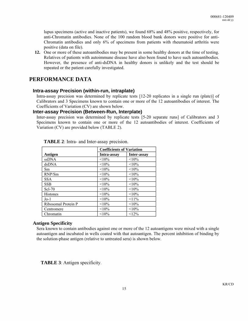
Intra-assay Precision (within-run, intraplate) Intra-assay precision was determined by replicate tests [12-20 replicates in a single run (plate)] of Calibrators and 3 Specimens known to contain one or more of the 12 autoantibodies of interest. The Coefficients of Variation (CV) are shown below.
Inter-assay Precision (Between-Run, Interplate) Inter-assay precision was determined by replicate tests [5-20 separate runs] of Calibrators and 3 Specimens known to contain one or more of the 12 autoantibodies of interest. Coefficients of Variation (CV) are provided below (TABLE 2).
TABLE 2: Intra- and Inter-assay precision. Coefficients of Variation Antigen Intra-assay Inter-assay ssDNA <10% <10% dsDNA <10% <10% Sm <10% <10% RNP/Sm <10% <10% SSA <10% <10% SSB <10% <10% Scl-70 <10% <10% Histones <10% <10% Jo-1 <10% <11% Ribosomal Protein P <10% <10% Centromere <10% <10% Chromatin <10% <12%
Antigen Specificity
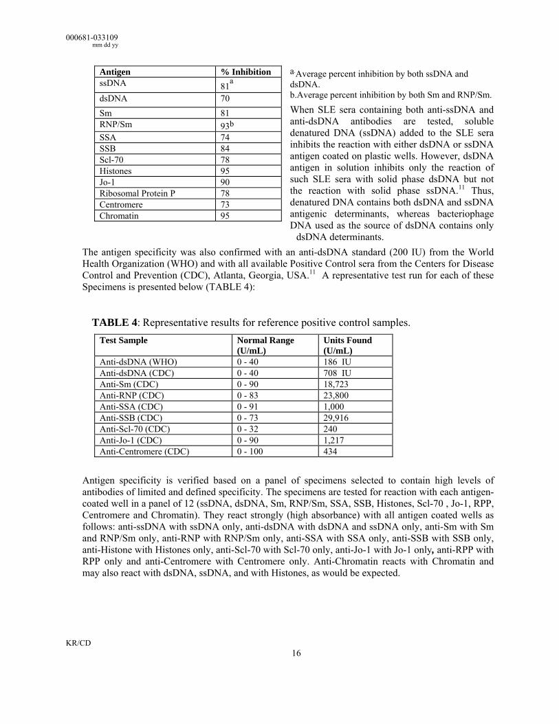
Sera known to contain antibodies against one or more of the 12 autoantigens were mixed with a single autoantigen and incubated in wells coated with that autoantigen. The percent inhibition of binding by the solution-phase antigen (relative to untreated sera) is shown below.
TABLE 3: Antigen specificity.
000681-033109 mm dd yy
KR/CD 16
a.Average percent inhibition by both ssDNA and dsDNA. b.Average percent inhibition by both Sm and RNP/Sm. When SLE sera containing both anti-ssDNA and anti-dsDNA antibodies are tested, soluble denatured DNA (ssDNA) added to the SLE sera inhibits the reaction with either dsDNA or ssDNA antigen coated on plastic wells. However, dsDNA antigen in solution inhibits only the reaction of such SLE sera with solid phase dsDNA but not the reaction with solid phase ssDNA.11 Thus, denatured DNA contains both dsDNA and ssDNA antigenic determinants, whereas bacteriophage DNA used as the source of dsDNA contains only
dsDNA determinants. The antigen specificity was also confirmed with an anti-dsDNA standard (200 IU) from the World Health Organization (WHO) and with all available Positive Control sera from the Centers for Disease Control and Prevention (CDC), Atlanta, Georgia, USA.11 A representative test run for each of these Specimens is presented below (TABLE 4):
TABLE 4: Representative results for reference positive control samples.
Test Sample Normal Range (U/mL)
Units Found (U/mL)
Anti-dsDNA (WHO) 0 - 40 186 IU Anti-dsDNA (CDC) 0 - 40 708 IU Anti-Sm (CDC) 0 - 90 18,723 Anti-RNP (CDC) 0 - 83 23,800 Anti-SSA (CDC) 0 - 91 1,000 Anti-SSB (CDC) 0 - 73 29,916 Anti-Scl-70 (CDC) 0 - 32 240 Anti-Jo-1 (CDC) 0 - 90 1,217 Anti-Centromere (CDC) 0 - 100 434
Antigen specificity is verified based on a panel of specimens selected to contain high levels of antibodies of limited and defined specificity. The specimens are tested for reaction with each antigen-coated well in a panel of 12 (ssDNA, dsDNA, Sm, RNP/Sm, SSA, SSB, Histones, Scl-70 , Jo-1, RPP, Centromere and Chromatin). They react strongly (high absorbance) with all antigen coated wells as follows: anti-ssDNA with ssDNA only, anti-dsDNA with dsDNA and ssDNA only, anti-Sm with Sm and RNP/Sm only, anti-RNP with RNP/Sm only, anti-SSA with SSA only, anti-SSB with SSB only, anti-Histone with Histones only, anti-Scl-70 with Scl-70 only, anti-Jo-1 with Jo-1 only, anti-RPP with RPP only and anti-Centromere with Centromere only. Anti-Chromatin reacts with Chromatin and may also react with dsDNA, ssDNA, and with Histones, as would be expected.
Antigen % Inhibition ssDNA 81a
dsDNA 70 Sm 81 RNP/Sm 93b SSA 74 SSB 84 Scl-70 78 Histones 95 Jo-1 90 Ribosomal Protein P 78 Centromere 73 Chromatin 95
000681-120409 mm dd yy
KR/CD 17
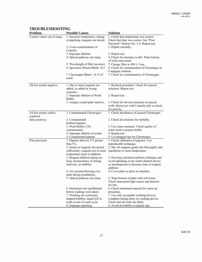
TROUBLESHOOTING Problem Possible Causes Solution Control values out of range. 1. Incorrect temperature, timing
or pipetting; reagents not mixed. 1. Check that temperature was correct. Check that time was correct. See “Poor Precision” (below) No. 2-4. Repeat test.
2. Cross-contamination of Controls.
2. Pipette carefully.
3. Improper dilution. 3. Repeat test. 4. Optical pathway not clean. 4. Check for moisture or dirt. Wipe bottom
of wells and reread. 5. Wavelength of filter incorrect. 5. Change filter to 450 ± 5 nm. 6. Specimen Diluent Blank >0.2 6. Check for contamination of Chromogen or
Conjugate solution. 7. Chromogen Blank > 0.15 (if
used)
7. Check for contamination of Chromogen.
All test results negative. 1. One or more reagents not added, or added in wrong sequence.
1. Recheck procedure. Check for unused solutions. Repeat test.
2. Improper dilution of Wash Buffer.
2. Repeat test.
3. Antigen coated plate inactive. 3. Check for obvious moisture in unused wells. Rerun test with Controls only to check for activity.
All test results yellow; scattered
1. Contaminated Chromogen. 1. Check absorbance of unused Chromogen.
false positives 2. Contaminated buffers/reagents.
2. Check all solutions for turbidity.
3. Wash Buffer (1X) contaminated.
3. Use clean container. Check quality of water used to prepare buffer.
4. Improper dilution of serum. 5. Contaminated pipette
4. Repeat test. 5. Use plugged tips for Chromogen
Poor precision. 1. Pipettor delivery CV greater than 5%.
1. Check calibration of pipettor. Use reproducible technique.
2. Serum or reagents not mixed sufficiently; reagents not at room temperature prior to addition.
2. Mix all reagents gently but thoroughly and equilibrate to room temperature.
3. Reagent addition taking too long; inconsistency in timing intervals, air bubbles.
3. Develop consistent uniform technique and avoid splashing or use multi-channel device or autodispenser to decrease time of reagent addition.
4. Air currents blowing over plate during incubations.
4. Cover plate or place in chamber.
5. Optical pathway not clean. 5. Wipe bottom of plate with soft tissue. Check instrument light source and detector for dirt.
6. Instrument not equilibrated before readings were taken.
6. Check instrument manual for warm up procedure.
7. Washing not consistent; trapped bubbles; liquid left in wells at end of wash cycle.
7. Use only acceptable washing devices. Lengthen timing delay on washing device. Check that all wells are filled.
8. Improper pipetting. 8. Avoid air bubbles in pipette tips.
000681-033109 mm dd yy
KR/CD 18
REFERENCES 1. Tan EM, AS Cohen, JF Fries, AT Masi, et al. 1982. The 1982 revised criteria for the classification of systemic
lupus erythematosus. Arthritis Rheum. 25 1271-1277. 2. Lange A. 1978. Evaluation of the simultaneous estimation of anti-dsDNA and anti-ssDNA antibodies for
clinical purposes. Clin. Exp. Immunol. 31:472-481. 3. Sharp GC, WS Irvin, E. Tan, RG Gould, et al. 1972. Mixed Connective Tissue Disease—an apparently
distinct rheumatic disease syndrome associated with a specific antibody to an extractable nuclear antigen (ENA). Am. J. Med. 52:148-159.
4. Winfield JB, CM Brunner, and D Koffler. 1978. Serologic studies in patients with systemic lupus erythematosus and central nervous system dysfunction. Arthritis Rheum. 21:289-294.
5. Maddison P, H Mogavero, TT Provost, and M Reichlin. 1979. The clinical significance of autoantibodies to soluble cytoplasmic antigen in systemic lupus erythematosus and other connective tissue diseases. J. Rheumatol. 6:189-195 (1979).
6. Alspaugh MA, N Talal, and EM Tan. 1976. Differentiation and characterization of autoantibodies and their antigens in Sjögren’s syndrome. Arthritis Rheum. 19:216-222.
7. Sontheimer RD, JR Thomas, and JN Gilliam. 1979. Subacute cutaneous lupus erythematosus. A cutaneous marker for a distinct lupus erythematosus subset. Arch. Dermatol. 115:1409-1415.
8. Wasicek CA, PJ Maddison, and M Reichlin. 1979. Occurrence of antibodies to single-stranded DNA in ANA negative patients. Clin. Exp. Immunol. 37:190-195.
9. Tan EM. 1989. Antinuclear antibodies: Diagnostic markers for autoimmune diseases and probes for cell biology. Adv. Immunol. 44:93-115.
10. Hahn BH, FM Ebling, and KC Kalunian. 1989. Characteristics of pathogenic antibodies in SLE (abstract). Proceedings of Second International Conference on Systemic Lupus Erythematosus. 172-176.
11. Froelich CJ, J Wallman, JL Skosey, and M Teodorescu. 1990. Clinical value of an integrated ELISA system for the detection of 6 autoantibodies (ssDNA, dsDNA, Sm, RNP/Sm, SSA and SSB). J. Rheumatol. 17:192-200.
12. Teodorescu M and C Froelich. 1994. Advanced Immunoassays in Rheumatology. CRC Press, Boca Raton, Ann Arbor, London, Tokyo, pp. 390.
13. Urowicz MB and DD Gladman. 1992. Antinuclear antibody-sensitive systemic lupus erythematosus. In RG Lahita (ed.) Systemic Lupus Erythematosus, 2nd edition. Ch. Livingstone, N.Y., Edinburgh, Melbourne, London, Tokyo, pp. 561-567.
14. Rubin RL and S Waga. 1987. Anti-histone antibodies in systemic lupus erythematosus. J. Rheumatol. 14(S-13):118-126.
15. Hess EV. and AB Mongey. 1991. Drug-related lupus. Bull. Rheum. Dis., 40: 1-8. 16. Reichlin M. 1993. ANAs and antibodies to DNA: their use in clinical diagnosis. Bull. Rheum. Dis. 42:3-5. 17. Arnett FC, TJ Hirsch, WB Bias, M Nishikai, and M Reichlin. 1981. The Jo-1 antibody system in myositis:
relationships to clinical features and HLA. J. Rheumatol. 8:925-930. 18. Hochberg MC, D Feldman, MB Stevens, FC Arnett, and M Reichlin. 1984. Antibody to Jo-1 in
polymyositis/dermatomyositis: association with interstitial pulmonary disease. J. Rheumatol. 11:663-665. 19. Bonfa E and KB Elkon. 1986. Clinical and serologic associations of the antiribosomal P protein antibody.
Arthritis Rheum. 29:981-985. 20. Russo K, S Hoch, C Dima, J Varga, and M Teodorescu. 2000. Circulating anticentromere CENP-A and
CENP-B antibodies in patients with diffuse and limited systemic sclerosis, systemic lupus erythematosus, and rheumatoid arthritis. J. Rheumatol. 27:142-148.
21. Rubin RL 1999. Anti-histone antibodies. in RG Lahita (ed). Systemic Lupus Erythematosus, 3rd edition, Academic Press, pp. 227-245.
22. Berden, JHM, Smeenk, RJT. 1996. Nucleosome-specific autoantibodies. In JB Peter and Y Shoenfeld (eds). Autoantibodies. Elsevier, Amsterdam, pp. 574-581.
23. Ignat GP, A-C Rat, JJ Sychra, J Vo, J Varga, and M Teodorescu. 2003. Information on diagnosis and management of systemic lupus erythematosus patients derived from the routine measurement of 8 nuclear autoantibodies. J. Rheumatol. 30:1761-1769.
000681-120409 mm dd yy
KR/CD 19
(GB)(USA)(CDN) Expiry date (D)(A)(B)(CH) Verfallsdatum (F)(B)(CH)(CDN) Date de péremption (I)(CH) Data di scadenza (E) Fecha de caducidad (P) Data de validade (NL) Uiterste gebruiksdatum (DK) Udløbsdato (S) Utgångsdatum
(GB)(USA)(CDN) Consult instructions for use (D)(A)(B)(CH) Bitte Gebrauchsanweisung einsehen (F)(B)(CH)(CDN) Consultez la notice d'utilisation (I)(CH) Consultare le istruzioni per l'uso (E) Consulte las instrucciones de utilización
(P) Consulte as instruções de utilização (NL) Raadpleeg de gebruikaanwijzing (DK) Se brugsanvisningen (S) Läs anvisningarna före användning
(GB)(USA)(CDN) In Vitro Diagnostic Medical Device (For In Vitro Diagnostic Use) (D)(A)(B)(CH) Medizinisches In-vitro-Diagnostikum (zur In-vitro-Diagnostik)
(F)(B)(CH)(CDN) Dispositif médical de diagnostic in vitro (Pour usage diagnostique in vitro) (I)(CH) Dispositivo medico per diagnostica in vitro (per uso diagnostico in vitro) (E) Dispositivo médico de diagnóstico in vitro (para uso diagnóstico in vitro) (P) Dispositivo médico para diagnóstico in vitro (Para utilização de diagnóstico "in vitro") (NL) Medisch hulpmiddel voor diagnostiek in vitro (Voor diagnostisch gebruik in vitro) (DK) Medicinsk udstyr til in vitro-diagnostik (Udelukkende til in vitro diagnostisk anvendelse) (S) Medicinteknisk produkt avsedd för in vitro-diagnostik (För in vitro-diagnostiskt bruk)
(GB)(USA)(CDN) Lot / Batch Number (D)(A)(B)(CH) Charge / Chargennummer (F)(B)(CH)(CDN) Lot / Code du lot (I)(CH) Lotto / Numero di lotto (E) Lote / Código
de lote (P) Lote / Código do lote (NL) Lot-/Partijnummer (DK) Lot / Batchkode (S) lot / Satskod
(GB)(USA)(CDN) Manufactured by (D)(A)(B)(CH) Hergestellt von (F)(B)(CH)(CDN) Fabriqué par (I)(CH) Prodotto da (E) Fabricado por (P) Fabricado por (NL) Vervaardigd door (DK) Fabrikation af (S) Tillverkad av
(GB)(USA)(CDN) Catalogue Number (D)(A)(B)(CH) Bestell-Nummer (F)(B)(CH)(CDN) Numéro de référence (I)(CH) Numero di riferimento (E) Número de
referencia (P) Número de referência (NL) Referentienummer (DK) Referencenummer (S) Katalognummer
(GB)(USA)(CDN) Store at between (D)(A)(B)(CH) Lagerung bei zwischen (F)(B)(CH)(CDN) Conserver à entre (I)(CH) Conservare a tra (E) Conservar a temp. entre (P) Armazene a entre (NL) Bewaar bij tussen (DK) Opbevares mellem (S)
Förvaras vid
(GB)(USA)(CDN) Contains sufficient for x tests (D)(A)(B)(CH) Inhalt ausreichend für x Tests (F)(B)(CH)(CDN) Contient suffisant pour x tests (I)(CH) Contenuto sufficiente per x test (E) Contiene suficiente para x pruebas (P) Contém suficiente para x testes
(NL) Bevat voldoende voor x bepalingen (DK) Indeholder tilstrækkeligt til x prøver (S) Innehàllet räcker till x analyzer
000681-033109 mm dd yy
KR/CD 20
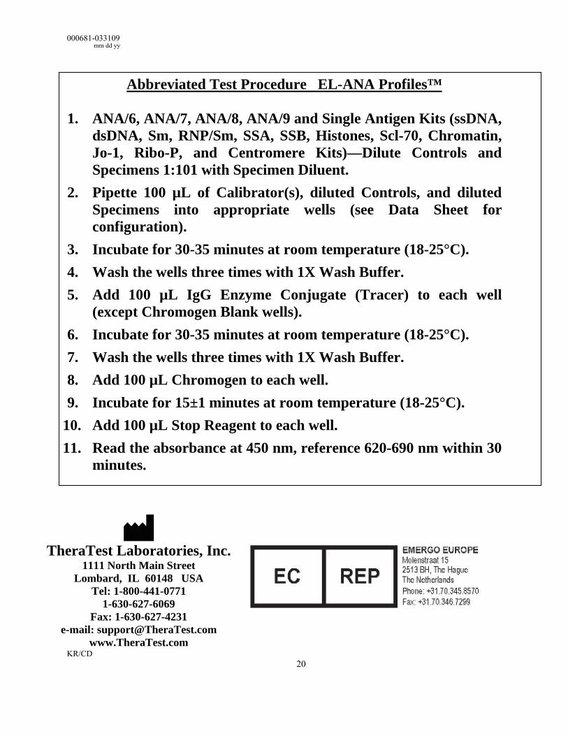
Abbreviated Test Procedure EL-ANA Profiles™
1. ANA/6, ANA/7, ANA/8, ANA/9 and Single Antigen Kits (ssDNA, dsDNA, Sm, RNP/Sm, SSA, SSB, Histones, Scl-70, Chromatin, Jo-1, Ribo-P, and Centromere Kits)—Dilute Controls and Specimens 1:101 with Specimen Diluent.
2. Pipette 100 µL of Calibrator(s), diluted Controls, and diluted Specimens into appropriate wells (see Data Sheet for configuration).
3. Incubate for 30-35 minutes at room temperature (18-25°C). 4. Wash the wells three times with 1X Wash Buffer. 5. Add 100 µL IgG Enzyme Conjugate (Tracer) to each well
(except Chromogen Blank wells). 6. Incubate for 30-35 minutes at room temperature (18-25°C). 7. Wash the wells three times with 1X Wash Buffer. 8. Add 100 µL Chromogen to each well. 9. Incubate for 15±1 minutes at room temperature (18-25°C).
10. Add 100 µL Stop Reagent to each well. 11. Read the absorbance at 450 nm, reference 620-690 nm within 30
minutes.
TheraTest Laboratories, Inc.
1111 North Main Street Lombard, IL 60148 USA
Tel: 1-800-441-0771 1-630-627-6069
Fax: 1-630-627-4231 e-mail: [email protected]
www.TheraTest.com


