An Atomic Force Microscopy Observation Of
15
An Atomic Force Microscopy Observation Of Poly(vinylidene fluoride) Banded Spherulites Akihiko Toda a)1, Takeshi Arita a, Masamichi Hikosaka a, Jamie K. Hobbsb, Mervyn J. Milesb, a Faculty of Integrated Arts and Sciences, Hiroshima University, 1-7-1 Kagamiyama, Higashi-Hiroshima 739-8521, JAPAN b H.H. Wills Physics laboratory, University of Bristol, Tyndall Avenue, Bristol BS8 1TL, UK Abstract Wehave examined the free surface of a banded spherulite of poly(vinylidene flu- oride) (PVDF) by an atomic force microscopy. The directions of the slope of mul- tilayer terraces of lamellar crystals are retained in each half of a banded spherulite; this evidence confirms the macroscopic selection of one handedness in the formation of spiral terraces in each growth direction of the sheaf at the center of a banded spherulite of PVDF. In a previous paper it was confirmed that the three-dimensional morphology of all single crystals of PVDF grown from the melt is chair-type, and hence it is most probable that the stress in the chair crystal is responsible for the formation of spiral dislocations and terraces keeping the same handedness in each growth direction. The chair-type morphology is created because of the chain tilting to the fold surface, which can introduce symmetry breaking and consequently the selection of handedness in non-chiral polymers such as PVDF. Keywords: AFM, spherulite, banding, PVDF, chair type, spiral terrace 1 Corresponding author, tel: +81-824-24-6558 , fax: +81-824-24-0757, Email: [email protected] J. Macromol. Sci. revised 19 November 2002
Transcript of An Atomic Force Microscopy Observation Of

An Atomic Force Microscopy Observation Of
Poly(vinylidene fluoride)
Banded Spherulites
Akihiko Toda a)1, Takeshi Arita a, Masamichi Hikosaka a,
Jamie K. Hobbsb, Mervyn J. Milesb,
a Faculty of Integrated Arts and Sciences, Hiroshima University,
1-7-1 Kagamiyama, Higashi-Hiroshima 739-8521, JAPAN
b H.H. Wills Physics laboratory, University of Bristol,
Tyndall Avenue, Bristol BS8 1TL, UK
Abstract
Wehave examined the free surface of a banded spherulite of poly(vinylidene flu-
oride) (PVDF) by an atomic force microscopy. The directions of the slope of mul-
tilayer terraces of lamellar crystals are retained in each half of a banded spherulite;
this evidence confirms the macroscopic selection of one handedness in the formation
of spiral terraces in each growth direction of the sheaf at the center of a banded
spherulite of PVDF. In a previous paper it was confirmed that the three-dimensional
morphology of all single crystals of PVDF grown from the melt is chair-type, and
hence it is most probable that the stress in the chair crystal is responsible for the
formation of spiral dislocations and terraces keeping the same handedness in each
growth direction. The chair-type morphology is created because of the chain tilting
to the fold surface, which can introduce symmetry breaking and consequently the
selection of handedness in non-chiral polymers such as PVDF.
Keywords: AFM, spherulite, banding, PVDF, chair type, spiral terrace
1 Corresponding author,
tel: +81-824-24-6558 , fax: +81-824-24-0757, Email: [email protected]
J. Macromol. Sci. revised 19 November 2002

1 Introduction
Spherulites are composed of small crystallites growing in the radial direction
with the formation of branches filling three-dimensional space. With some
polymers, a concentric pattern of periodic banding appears in spherulitic
crystallization [1]. The formation of banding is not limited only in polymer
spherulites. A number of organic compounds exhibit similar types of concen-
tric rings in spherulites [2], and hence this topic has a wider implication in the
self-organization process of crystals, in general.
In the banded spherulites of polymers, a twisting relationship of lamellar crys-
tallites in the radial direction has been confirmed experimentally by WAXS [3]
and is considered to be responsible for the creation of the periodic extinction
banding observed by polarizing optical microscopy.
For lamellar crystallites, twisting must choose one of the two handedness,
and the mechanism of the choice has been a challenging topic. With non-
racemic chiral polymers, strong evidences suggest that the choice is made by
the handedness of the chirality in the polymer molecules [4-8]. For non-chiral
polymers, the tilting of polymer chains to the fold surface can be the origin of
the choice of the handedness.
Twomodels of twisting relationship have been proposed, based on the chain
tilting. One is proposed by Keith and Padden [9,10] under the hypothesis that
the degree of congestion of upper and lower fold surfaces of a lamellar crystal
in the melt is different because of the crystallization of polymer chains on
the growth face which is tilted to the fold surface. Another possible mode
of twisting relationship can be caused by the stress in a chair-type crystal
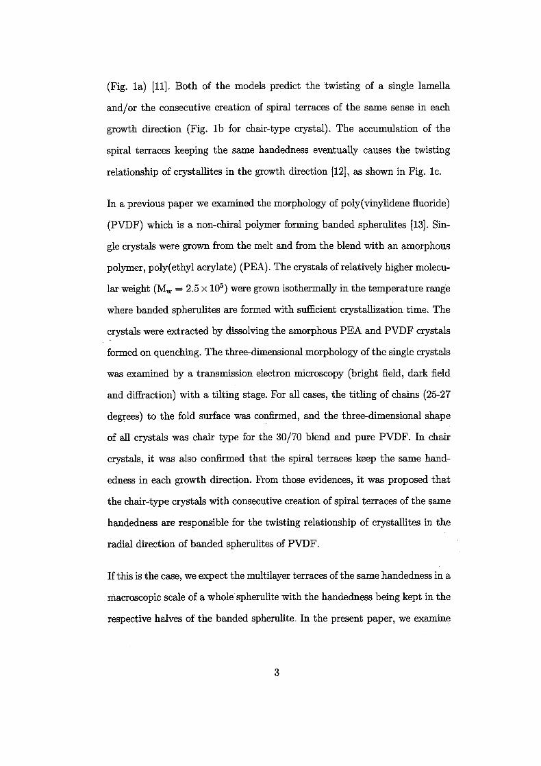
(Fig. la) [11]. Both of the models predict the twisting of a single lamella
and/or the consecutive creation of spiral terraces of the same sense in each
growth direction (Fig. lb for chair-type crystal). The accumulation of the
spiral terraces keeping the same handedness eventually causes the twisting
relationship of crystallites in the growth direction [12], as shown in Fig. lc.
In a previous paper we examined the morphology of poly(vinylidene fluoride)
(PVDF) which is a non-chiral polymer forming banded spherulites [13]. Sin-
gle crystals were grown from the melt and from the blend with an amorphous
polymer, poly(ethyl acrylate) (PEA). The crystals of relatively higher molecu-
lar weight (Mw = 2.5 x 10⁵) were grown isothermally in the temperature range
where banded spherulites are formed with sufficient crystallization time. The
crystals were extracted by dissolving the amorphous PEA and PVDF crystals
formed on quenching. The three-dimensional morphology of the single crystals
was examined by a transmission electron microscopy (bright field, dark field
and diffraction) with a tilting stage. For all cases, the titling of chains (25-27
degrees) to the fold surface was confirmed, and the three-dimensional shape
of all crystals was chair type for the 30/70 blend and pure PVDF. In chair
crystals, it was also confirmed that the spiral terraces keep the same hand-
edness in each growth direction. From those evidences, it was proposed that
the chair-type crystals with consecutive creation of spiral terraces of the same
handedness are responsible for the twisting relationship of crystallites in the
radial direction of banded spherulites of PVDF.
If this is the case, we expect the multilayer terraces of the same handedness in a
macroscopic scale of a whole spherulite with the handedness being kept in the
respective halves of the banded spherulite. In the present paper, we examine

the ordering of the handedness by means of surface observation of banded
spherulites with an atomic force microscopy (AFM). Prom the observation, we
confirm that the multilayer terraces are going down in one direction in each half
of a banded spherulite, and this evidence shows that the spiral terraces choose
one of the two possible handedness in each half of the banded spherulites of
PVDF.
2 Experimental
Weused PVDF of grade KF1000 (Mw = 2.5 x 10⁵ and Mw/Mn = 2.1, Kureha
Chemical Industries, Co., Ltd.) and PEA of Mn = 2.9 x 10⁴ and Mw/Mn
=2.4 from Scientific Polymer Products. Two weight ratios were examined:
PVDF/PEA = 30/70 and 100/0. The details of the sample preparation was
described in the previous paper [13].
The free surface of the sample on a cover-slip was examined by an atomic mi-
croscope (Digital Instruments Nanoscope III, Dimension 3100 SPM system).
Integrated silicon tips and cantilevers with a nominal spring constant of 30 N
m"1wereused. The instrument was operated in tapping mode in air. The tap-
ping mode data was recorded in constant force mode. Scanning was performed
at a frequency of 1 Hz. The cantilever was oscillated at its resonant frequency
of about 250 kHz. In order to examine a wide area of a spherulite, the im-
ages of small area (eg. 2 /mix2 fj,m) were taken successively with a parallel
displacement of the tip (shorter than the length of the area) for each image.
All images were recorded at room temperature. All images represented in the
following are of "amplitude" image which is sensitive to the change in sam-
ple height; the contrast is similar to that of shadowed image in transmission
electron microscopy and suitable for examining the morphology.

3 Results and Discussion
Figure 2 shows an AFM image of the growth tip of a PVDF multilayer crystal
grown from the 30/70 blend. Figs. 2a and b, which is an enlargement of Fig 2a,
show the arrangement of the terrace of lamellae coming downward in the figure.
This arrangement of the terrace clearly indicates that the multilayering was
not created by a single spiral terrace but made by a row of spiral terraces
keeping the same handedness, as schematically shown in Fig. 2c.
Figure 3 shows the surface profile of a banded spherulite in one period of
extinction corresponding to half a period of the rotation of the crystal axis
around the radial direction. If the arrangement merely represents the crosssec-
tion of stacked lamellae which are rotating around the growth direction, then
the arrangement must be the alternation of upward and downward terraces.
In fact, this is not the case, since the upward terrace has not been observed.
Therefore, it should be considered that the downward terrace which is visible
in the figure is what was made by a row of spiral terraces keeping the same
handedness like in the growth tip of the PVDF multilayer crystal shown in
Fig2.
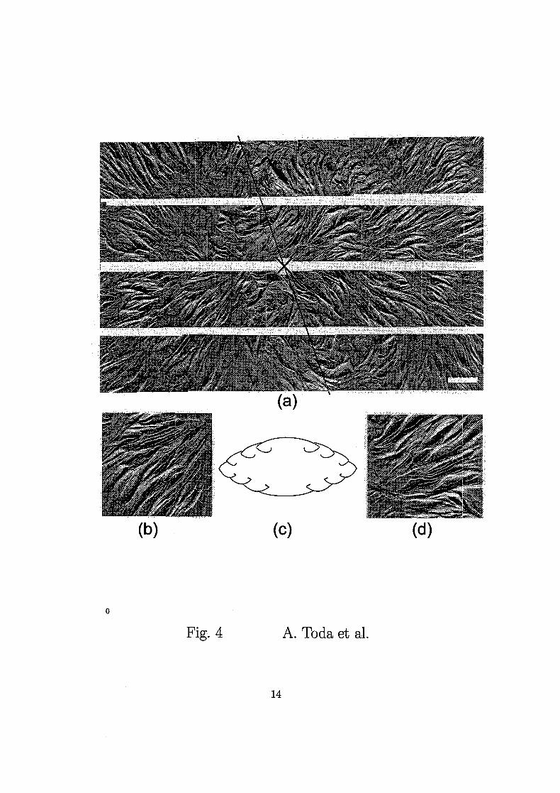
Figure 4 is the whole view of a banded spherulite and enlargements of two
portions. It is confirmed that the arrangement of the multilayer terraces in
both the right and left halves come down from the top to the bottom in
clockwise and counterclockwise manners around the center, respectively.
Those observations confirm the arrangement of spiral terraces in PVDF
banded spherulites, which we expect for the banded spherulite with rows of
spiral terraces keeping the handedness in the respective growth directions.

Figure 5 represents the possible orientations of the banded spherulite formed
by this rule and its relation with the handedness of spiral terraces and/or
lamellar twisting which will appear on the top surface of the film. As shown in
Fig. 5c, only when the seed crystal of the banded spherulite stands up, one of
the handedness is chosen on the top surface. There were few cases of standing
in this way in fact. However, in almost all cases of the present observation,
the arrangement of the terraces corresponds to the case of Figs. 5a and b.
In PVDF, we have confirmed that chair-type crystals grow into banded
spherulites in the previous paper [13]. A chair crystal suffers from the con-
secutive formation of spiral terraces with the handedness chosen to relax the
stress in the chair, as shown in Fig. lb. Therefore, it is reasonable to expect
that the consecutive creation of spiral terraces keeping handedness is respon-
sible for the twisting relationship along the growth direction of the banded
spherulite of PVDF, as shown in Fig. lc.
In terms of the possibility of lamellar twisting, the twisting direction of a
single lamellar crystal could not be clearly identified because of the multi-
layering of lamellae by the formation of spiral terraces. On the other hand,
there are independent observations of twisting lamellae of polyethylene in lit-
erature, as seen in ref. [1]. For this reason, although we have confirmed the
row of spiral terraces keeping the handedness, which will be responsible for
the twisting relationship in the banded spherulites of PVDF, we cannot come
to the conclusion that the origin of the twisting in PVDF is solely due to the
formation of this type of spiral terraces in chair crystals. The determination
of the mechanism of twisting relationship will therefore need a closer look at
the three dimensional shape of a single lamella in the banded spherulites.

Acknowledgements
This work was partly supported by a Grant-in-Aid for Scientific Research
from Japan Society for the Promotion of Science and from the Ministry of
Education, Culture, Sports, Science and Technology of Japan.

References
[1] Geil PH. "Polymer Single Crystals" , John Wiley, New York, 1963.
[2] Bernauer F. "Gedrillte Kristalle" , Borntraeger, Berlin, 1929.
[3] Fujiwara Y. The superstructure of melt-crystallized polyethylene. I. Screwlike
orientation of unit cell in polyethyene spherulites with periodic extinction rings
J. Appl. Polymer Sci., 4; 1960: 10.
[4] Singfield KL, Brown GR. Optically active polyethers. 1. Studies of the
crystallization in blends of the enantiomers and stereoblock form of
poly(epichlorohydrin) Macromolecules, 28; 1995: 1290.
[5] Singfield KL, Klass MJ, Brown GR. Optically active polyethers. 2. Atomic
force microscopy of melt-crystallized poly(epichlorohydrin) enantiomers and
their equimolar blend Macromolecules, 28; 1995: 8006.
[6] Singfield KL, Hobbs JK, Keller A. Correlation between main chain chirality and
crystal "twist" direction in polymer spherulites J. Cryst. Growth, 183; 1998:
683.
[7] Li CY, Cheng SZD, Ge JJ, Bai F, Zhang JZ, Mann IK, Harris FW, Chien LC,
Yan DY, He T, Lotz B. Double twist in helical polymer "soft" crystals Phys.
Rev. Lett., 83; 1999: 4558.
[8] Saracovan I, Keith HD, Manley RStJ, Brown GR. Banding in spherulites
of polymers having uncompensated main-chain chirality Macromolecules, 32;
1999: 8918.
[9] Keith HD, Padden FJJr. Twisting orientation and the role of transient states
in polymer crystallization Polymer, 25; 1984: 28.
[10] Keith HD, Padden FJJr. Banding in polyethylene and other spherulites
Macromolecules, 25; 1996: 7776.
[11] Toda A, Keller A. Growth of polyethylene single crystals from the melt:
Morphology Colloid Polym. Sci., 271; 1993: 328.
[12] Schultz JM, Kinloch DR. Transverse screw dislocations: A source of twist in
crystalline polymer ribbons Polymer, 10; 1969: 271.
[13] Toda, A, Arita T and Hikosaka M. Three-dimensional morphology of PVDF
single crystals forming banded spherulites Polymer, 42; 2001: 2223.

Figure Captions
Fig. 1. Schematic representation of (a) halves of a chair crystal of PVDF in a relaxed
state, (b) spiral dislocations keeping the handedness in each growth direction, the
formation of which can relax the stress in the chair crystal, and (c) the twisting re-
lationship in the radial direction by the consecutive generation of screw dislocations
keeping the handedness [12].
Fig. 2. AFM image of the growth tip of a PVDF multilayer crystal grown from 30/70
blend at 165°C for 2.5 h. In (b), a portion of (a) is enlarged. Bar line represents
1 fixa.
Fig. 3. AFM image of the free surface of a banded spherulite grown from the melt
at 165°C for 2.5 h, showing one period of extinction. Bar line represents 1 /im.
Fig. 4. AFM image of (a) the whole view of a banded spherulite grown from the
melt at 165°C for 1.5 h and (b) and (d) the enlargement of portions. In (a), the
cross represents the center of the spherulite and the thick line represents the border
between the two regions keeping the directions of the slope of the terraces. Bar line
represents 1 /im.
Fig. 5. The relationship between the orientation of the banded spherulite of PVDF
in a film and the handedness of spiral terraces and/or lamellar twisting which appear
on the top surface of the film.
10
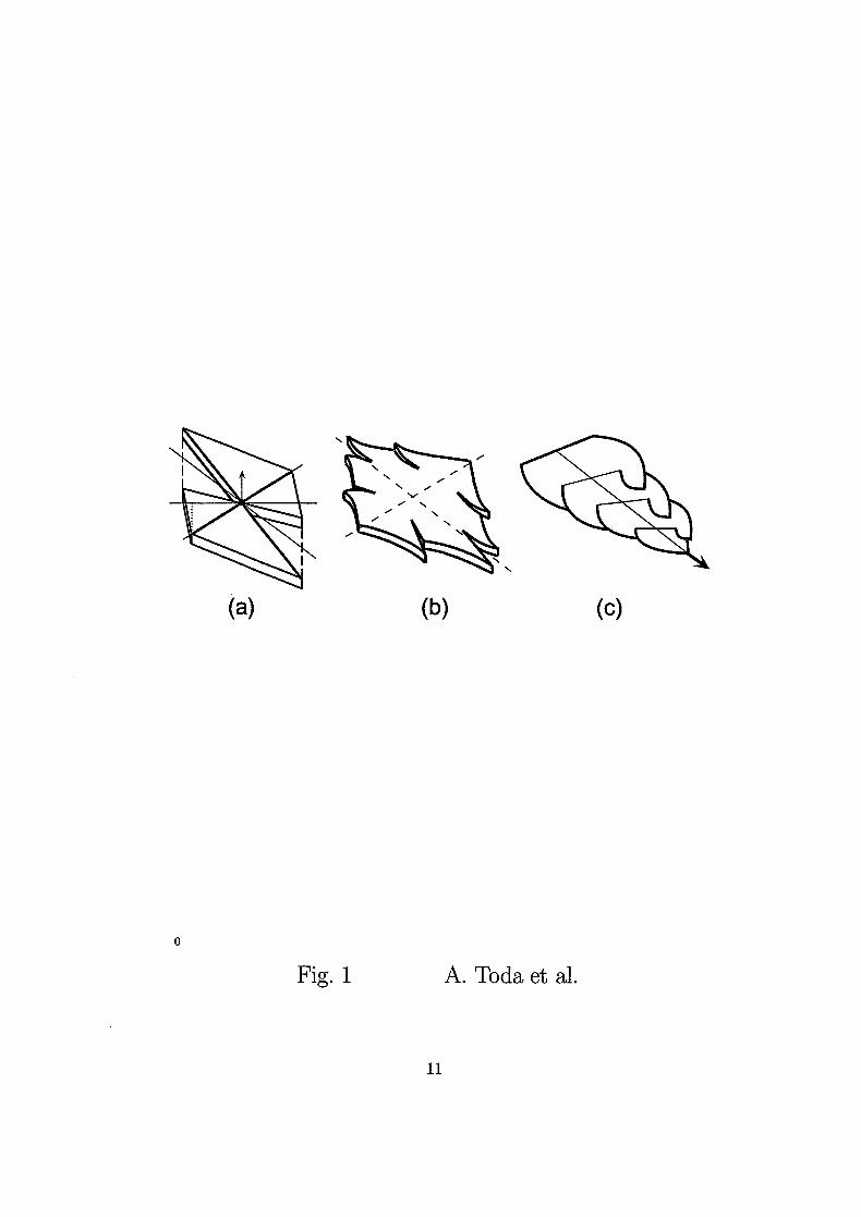
Fig. 1 A. Toda et al.
ll

HHil ;* -.-vjssitisw. *"'r* *r:?;.
(a)
Fig. 2 A. Toda et al.
12

Period of extinctiongrowth direction
Fig. 3 A. Toda et al.
13
m
(c) (d)
Fig. 4 A. Toda et al.
14

top
sides^>
(a)
u(b) (c)
F ig. 5 A. Toda et al
15