AN ABSTRACT OF THE THESIS OF HIRAM GORDON LAREW BY …
417
AN ABSTRACT OF THE THESIS OF HIRAM GORDON LAREW for the Ph.D.. in Entomalogy presented on December 17, 1981 Title: A COMPARATIVE ANATOMICAL STUDY OF GALLS CAUSED BY THE MAJOR CECIDOGENETIC GROUPS. WITH SPECIAL EMPHASIS ON THE NUTRITIVE TISSUE Abstract approved: I Peter B.. McEvoy The anatomies of 44 galls are discussed with special attention given to the development, longevity, and tannin content of the nutritive tissues. Within the main section of the thesis, representative galls from the major cecidogenetic groups, with the exception of bacterial and Australian scale galls, are studied. These include galls caused by fungi, nematodes, mites, moths, sawflys, scales, aphids, adelgids, tephritids, cecidomyiids, and cynipids. Three leaf mines are also described. Observations were taken from thin sections (plastic embedment) with a light microscope. The galls are arrayed along a continuum of increasing structural complexity, as judged by the degree
Transcript of AN ABSTRACT OF THE THESIS OF HIRAM GORDON LAREW BY …

AN ABSTRACT OF THE THESIS OF
HIRAM GORDON LAREW for the Ph.D..
in Entomalogy presented on December 17, 1981
Title: A COMPARATIVE ANATOMICAL STUDY OF GALLS CAUSED BY
THE MAJOR CECIDOGENETIC GROUPS. WITH SPECIAL EMPHASIS ON
THE NUTRITIVE TISSUE
Abstract approved: IPeter B.. McEvoy
The anatomies of 44 galls are discussed with special
attention given to the development, longevity, and tannin
content of the nutritive tissues.
Within the main section of the thesis,
representative galls from the major cecidogenetic groups,
with the exception of bacterial and Australian scale
galls, are studied. These include galls caused by fungi,
nematodes, mites, moths, sawflys, scales, aphids,
adelgids, tephritids, cecidomyiids, and cynipids. Three
leaf mines are also described. Observations were taken
from thin sections (plastic embedment) with a light
microscope.
The galls are arrayed along a continuum of
increasing structural complexity, as judged by the degree
of gall tissue differentiation. The sclerenchymatous
"protective" zone and nutritive cells are used as
indicators of gall complexity and of strength of the
gall-former's influence over host plant tissue. Starting
with the Fungal galls, then moth and sawfly galls, to the
thrips, scale, mites, nematode. cecidomyiid and cynipid
galls, one sees greater differentiation of gall tissues,
with an increasingly distinct nutritive layer. A
scierenchyma zone develops only in inidge and wasp galls.
The longevity of the nutritive tissue varies from
gall to gall. Generally, the nutritive tissue is
maintained in an enriched state for an extended period
only in galls caused by cynipids (and perhaps by
nematodes). The mites and midges show enriched nutritive
cells only in early gall development. No distinctive
nutritive tissue occurs in the aphid galls that were
studied.
Generally, nutritive tissue contains less
tanniferous material (as detected by the ferrous sulfate
stain) than do either peripheral gall tissues or cells of
the leaf. Thus, many gall-f ormers avoid tanriins by
directing the development of the cells upon which they
feed.
The epilogue includes a list of features shared by
many galls and by gall-forming organisms. Gall-f ormers
also share several characteristics with other types of

parasites.
The three appendices include 1) an anatomical study
of eight galls on shrubs from the drylands of eastern
Oregon (mite, cecidomyiid, tephritid, and moth galls), 2)
a discussion of fossil galls and leaf mines as indicators
of the age and stability of these co-evolutionary
relationships. Two galled acorns from the La Brea Tar
pits (Los Angeles, California) are described in this
section. Lastly, 3) a discussion of economically
important galls is provided. This last appendix
addresses the question of why there are relatively few
gall-forming insect pests, and includes a discussion of
the supposed benignity of insect galls.
A Comparative Anatomical Study of GallsCaused by the Major CecidoQenetic Groups.1
with Special Emphasis on the Nutritive Tissue
by
Hiram Gordon Larew
A THESIS
submitted to
Oregon State University
in partial fulfillment ofthe requirements for the
degree of
Doctor of Philosophy
CompletedDecember 17., 1981
Commencement June 1982

Approved:
6 2'iAssistant Professor à'f Entomology
in charge of major
Chairman of Department of En,.t)Smology
Dean of Graduate S,e4,ol
Date thesis is presented December 17, 1981
Typed by Hiram Larew + or Hiram Larew

DEDICATION
This dissertation is dedicated to my parents, Dr. H.
Gordon Larew and Mrs. Mary Jo Larew, and to my two
sisters, Jane Larew and Liz Larew. They listen and care.

ACKNOWLEDGEMENTS
I wish to thank Dr. Peter McEvoy for guidance at
appropriate times, and + or his emphasis on a broad view,
the large questions. Dr. Fred Rickson was especially
generous with his time and laboratory facilities. Dr.
Harry Phinney gave invaluable advice on macrophotography,
and unselfishly made camera and microscope equipment
available. Becky and Gene Fichter were kind enough to
let me use their home computer. Dr. Frank Smith lent
microscope equipment and provided office space in which
to work. Mr. Al Soeldner and Dr. Pat Buckley provided
convenient access to equipment needed for sectioning.
Mr. Kay Fernald provided help and advice on darkroom
technique. Ms. Doris Tilles assisted with inter-library
loans.
The following individuals provided specimens and/or
information about specimens; their help has made the
study possible: Dr. W. Akersten (Los Angelos County
Museum), Dr. A. Menke (United States National Museum),
Dr. W. Pulaski (California Academy of Sciences), Dr. H.
Schorn (Department of Paleontology, University of
California, Berkeley), Dr. B. Tif+ney (Vale University),
Dr. D. O'Dawd (CSIRO, Canberra, Australia), Dr. R. E.
Evans (Norfolk, England), Dr. A. Raman (Loyala College,
India), and Dr. B.. Daniels, (Kansas State University).

Faculty and students at Oregon State University who
have been o-F help include: Dr. G. Krantz, Dr. A.
lloldenke, Dr. L. Moore, Dr. L. Russell, Dr N. Anderson,
Dr. F. Smith, Dr. P. Koepsell, Dr. K. Chambers, Dr. R.
Hampton, Dr. A. Boucot, Dr. 6. Guppy, Dr. R. Ha].se, Dr.
B. Krapp, A. Churchill, 1. Vanderheyden, 6. Nielsen, E.
McEvoy, 6. Baker, P. Hessburg, P. Hanson, P. Hennen, B.
Simko, D. Henneberger, T. Dudley, K. Gottwald, S.
Russell, 3. Pinkerton, and B. Fichter.
All of the above as well as other friends have
offered help, advice, and encouragement during the study.
I thank all of them.
Lastly, I wish to thank Dr. Marshall Darley
(University of Georgia) for the time and energy he so
willingly invested in my undergraduate education. I also
want to acknowledge Dr. John Lattin for encouraging my
early interest in galls.

Section through speckled oak apple gall on Quercusqarryana Dougi. caused by Besbicus inirabilis showinglarval cavity (at bottom of photograph) larval capsulewall, and radiating fibers (top of photograph). Specimencollected Vil/lO/BO. 730X.


TABLE OF CONTENTS
INTRODUCTION
LITERATURE REVIEW
Classification System Based on Host Tissue
Disruption 4
Organoid Galls 4
Histioid Galls 6
Kataplasmas 6
Prosoplasmas S
Relationship Between Kataplasmas and
Prosoplasmas. - . .9
Classification System Based on Cecidozoan/Plant
Interaction 11
Filzgalls - 11
Fold Galls 12
Pouch Galls.... . 12
Covering Galls 13
Lysenchyme Galls 14
Mark Galls 14
OtherClassiFicationSystems
Gall Anatomy and Morphogenesis . 15
Gall Anatomy .... 15
Gall Morphogenesis: The Chemical Gradient Theory...17
1

Characteristics of the Nutritive Tissue 19
Definition 19
Development of Nutritive Tissue 21
Replacement and Maintenance of Nutritive Tissue. . .22
Are Nutritive Cells Meristematic? 24
Absence of Nutritive Cells 26
How Do Gal 1-Farmers Feed? 27
Characteristics of Nutritive Cells 29
MATERIALS AND METhODS 31
RESULTS 35
Leaf Mines
Leaf Mine on Fringe Cup 35
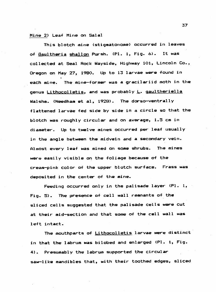
Leaf Mine on Salal 37
Leaf Mine on Black Cottonwood..... 38
Summary 39
Fungal Galls . 40
Gall on Larkspur . 40
Azalea Gall 41
Black Cottonwood Gall 43
Summary 45
Nematode Gall 46
Summary 47
Eriophyoid Galls 48

Willow Leaf Gall 48
Alder Bead Gall 50
Linden Leaf Gall 54
Erineum on Garry Oak ..57
Ash Leaflet Gall 59
Gall on Poison Oak Leaflet 61
Lea-F Gall on Trembling Aspen 62
Leaf Gall on Choke Cherry . 63
BigBud of Filbert... .............. ......-. .....s .64
Summary - 65
Classification 65
Nutritive Layer Dynamics 67
Tannins 69
LepidopteranGall
Stem Gall on Peck's Penstemon 71
Summary 73
Sawfly Galls 74
Gall on Snowberry 74
Willow Leaf Gall 75
Summary . 79
Classification 79
Nutritive Cell Dynamics..... 80
Tanni ns 80
Thrips Gall 81
Summary 82

Classification 82
Nutritive Cells 82
Tannins 83
Scale Gall (Oak Pit Gall) 84
Summary 85
Classification 85
Nutritive Cells 85
Aphid and Adelgid Galls 86
Poplar Petiole Gall 86
Poplar Marginal Gall 91
Midvein Gall on Poplar Leaves
Leaf Gall on Bearberry 97
Leaf Gall on Witch Hazel 99
Anasas Gall on Engelmann Spruce 100
Summary 102
Classification 102
Diet 102
Tannins 104
Cecidomyiid Galls 106
Hawthorn Leaf Gall 106
Leaf Roll Gall on Snowberry 109
Leaf Gall on Servi ceberry . - - - - . - - 113
Poplar Bud Gall 117
Willow Stem Beaked Gall 120
Summary 125

Classification . - 125
Nutritive Layer Dynamics and Tannins 126
Cynipid Galls 128
Stem Gall on Cat's Ear 128
Small Leaf Gall on Garry Oak 129
Oak Bullet Gall on Garry Oak 131
Rose Tip Gall 135
Spherical Stem Gall on Garry Oak 136
Mossy Rose Gall 138
Speckled OakAppleonGarryflak..........- l42
Summary 148
Classification 148
Nutritive Tissue and Its Dynamics 148
Sclerid Zone 149
Tannins 150
DISCUSSION - CONCLUSIONS 152
Degree of Reorganization 152
Nutritive Cells 157
Aphid Galls 159
Tanning 161
EPILOGUE: Characteristics of the Gall-Forming Habit 162
Host Plant Specificity 162
Morphological Modifications of the Gall-Former. 164

Gall Invaders 166
Parasitoids and Predators .147
Inquilines 169
Galls and Ant Colonies 170
Site of Attack, and Host Response 172
Diet Enhancement 174
Symbionts. 176
Galls as Metabolic Sinks 177
Unusual Life Cycles and Reproduction 179
Damage to Plant 180
How Does The Host Plant Defend Itself? 180
PLATES 184
BIBLIOGRAPHY 269
APPENDIX 1: EasternOregonGalls......
Published Accounts of Galls on Dryland Shrubs 291
Anatomical Studies 297
Galls on Artemisia tridentata 300
ART 300
ARC . . . . . . . . . . 302
ANP 304
Galls on Chrysothamnus nauseosus. -. . - -. ..... . ... .307
CRC 307

COT. 308
CPG 310
Galls on Tetradymia spp
TES 313
TET 314
Summary of Eastern Oregon Galls 317
Dryland Shrub Galls - A Discussion 318
Pattern 1 319
Pattern 2 319
Pattern 3.. 323
APPENDIX 2: Evidence for Plant-Insect Interactions
in the Fossil Record 326
Introduction 326
Fossil Plant Parts ..329
Pollen 329
Fruits and Seeds 330
Bark and Stems 335
Roots 337
Leaves 337
Fossil Galls 339
Gall Insects 344
"Hidden" Fossil Galls 345
A Search Method for Discovering Fossil Galls
in Collections 347

Fossil Leaf Mines 350
Domatia 351
Summary and Conclusions 353
APPENDIX 3: SaIls of Importance to Humans. 360
Detrimental Gall-Forming Organisms 361
Slime Molds 361
Bacteria 362
Fungi 365
Characteristics of Gall-Forming Fungi........=365
Nematodes 367
Mites 369
Insects ...... 369
Cecidomyiids 370
Other Diptera 371
Scales 371
Aphids 371
Adelgids 374
Summary of Detrimental Galls 374
Beneficial Gall Organisms 377
Galls as Biocontrol Agents 379
Case 1 380
Case 2 . 381
Case 3 381
Case 4 384

Other Cases 386
Summary a4 Beneficial Galls 387
Conclusion 390

LIST OF PLATES
Pit. Fig. Pg.
1 1-3 Fly lea-F mine on Tellimea grandiflorum 185
1 4-6 Moth leaf mine on Gaultheria shallon 185
1 7 Moth leaf mine on Papulus trichocarpa 185
1 8-10 Root-knot gall on tomato 185
2 1-2 Puccinia gall on Delphinium trolliifolium 188
3-6 Exobasidium gall on azalea 188
2 7-10 Taphrina aurea gall on P. trichocarpa 188
1-10 Eriophyaid gall on Salix sp. 191
4 1 Eriophyoid gall on Salix sp. 193
4 2-10 Eriophyoid gall on Alnus rubra 193
5 1-6 Eriophyoid gall on Alnus rubra 196
5 7-13 Eriophyoid gall on Tilia sp. 196
6 1-3 Eriophyoid gall on Tilia sp. 198
6 4-10 Eriaphyoid gall on Quercus qarryana 198
7 1-8 Eriophyoid gall on Fraxinus latifolia 200
7 9-10 Eriophyoid gall New York" Ti 1 i a 200
8 1-6 Eriophyoid gall on Rhus diversiloba 202
8 7-10 Eriophyoid gall on Populus tremuloides 202
9 1 Eriophyoid gall on Populus tremuloides 204
9 2-7 Eriophyoid gall on Prunus virginiana 204
9 8-9 Eriophyoid gall on Corylus avellana 204
10 1-5 Eriophyoid gall on Carylus avellana 206

11 1-4 Scale gall an Quercus garryana 208
11 5-9 Moth gall on Penstemon peckii 208
12 1-8 Sawfly gall on Symphoricarpos albus 210
13 1,4 Caloptilia murfeltella (Bsk) 213
13 2-3 Sawfly gall on Salix sp. 213
13 5-12 Sawfly gall on Salix sp. 213
14 1-6 Sawfly gall on Salix sp. 215
15 1-9 Aphid petiole gall on P.. trichacarpa 217
16 1-4 Aphid petiole gall an P. trichacarpa 219
16 5-8 Anasas gall on Picea engelmannii 219
17 1-B Aphid marginal gall on P. trichocarpa 221
17 9-10 Thrips gall on Memecylon sp. 221
18 1-5 Aphid marginal gall on P. trichocarpa 223
18 6-8 Aphid mi dvein gall on P.. tn chocarpa 223
19 1-9 Aphid gall an Arctostaphylos uva ursi 225
20 1 Aphid gall on A. uva ursi 227
20 2-5 Aphid gall on Hamainelis virginica 227
20 6-9 Midge gall on Crataegus dauglassii 227
21 1-5 Midge gall on C. douglassii 230
21 6-10 Midge gall on Symphoricarpos albus 230
22 1-2 Midge gall on S. albus 232
22 3-8 Midge gall on Amelanchier alnifolia 232
23 1-9 Midge gall on A.. alnifolia 234
24 1-9 Midge gall on Populus trichocarpa 236
25 1-9 Midge gall on Salix sp 238

26 1-9 fudge gall on Salix sp. 238
27 1-5 Midge gall on Salix sp. 240
27 6-10 Cynipid gall on Hypochaeris radicata 242
28 1 Cynipid gall on H. radicata 242
28 2-.5 Small cynipid leaf gall on Q. garryana 244
28 6-9 Cynipid bullet gall on Q. garryana 244
28 10 Thrips gall on Memecylon sp. 244
29 1-5,7 Cynipid bullet gall on Q. qarryana 246
29 6,8-9 Cynipid gall on Rosa nutkana 246
30 1-4 Cynipid spherical stem gall on
Q. garryana 248
30 5-9 Mossy gall on Rosa eglanteria 248
31 1-8 Mossy gall on R. eglanteria 250
32 1-2 Mossy gall on R. eglanteria 252
32 3-8 Speckled oak apple on Q. qarryana 252
33 1-8 Speckled oak apple on Q. garryana 254
34 1-4 Midge gall (ART) on Artemisia
tridentata 256
34 7-8 Midge gall (ARC) on A.. tridentata 256
35 1-3 Midge gall (ARC) on A. tridentata 258
35 4-6 Eriophyoid gall (ANP) on
tridentata 258
35 7-8 Speckled oak apple on Q. qarryana 258
36 1-5 Midge gall (CRC) on Chrysathamnus
nauseosus 260

36 6-8 Tephritid gall (COT) on C. nauseosus 260
37 1-2 Tephritid gall (COT) on C. nauseosus 262
37 3-8 Midge gall (CPG) on C. nauseosus 262
38 1-4 Moth gall (TES) on Tetradymia spinosa 264
38 5-8 Moth gall (TET) anT. labrata 264
39 1-2 Young petiole gall on P. trichocarpa 266
39 3-7 Swollen petioles of fossil poplars 266
39 B Possible lea-f mine on Q. nevadensis 266
40 1-2 Fossil acorn gall from La Brea 268
40 3-4 Galled acorns oF Q. wislizenii 268
40 5 Callirhytis milleri 268
40 6 Fossil rose stem with thorns 268
40 7 Phylloxera gall on Vitis vinifera 268

LIST OF TABLES
Table Page
Su-1 Summary Table: Reorganization of Host Plant
Tissues and Nutritive Cell Characteristics
in the Studied 6alls 151
Fo-1 Published Accounts of Fossil Galls 355
Fo-2 Genera of Fossil Plants on Which
Galls Are Likely to Be Found 356
Fo-3 Published Accounts of Fossil Mines 357
Fo-4 Plant Families Attacked by Miners 358
Fo-5 Extant Plant Families Showing Domatia 359
Hu-1 Gall-Forming Fungi 391
Hu-2 Published Accounts of Galls Used in
Biological Control Efforts Against Weeds 392

A COMPARATIVE ANATOMICAL STUDY OF GALLS
CAUSED BY THE MAJOR CECIDOGENETIC GROUPS,
WITH SPECIAL EMPHASIS ON THE NUTRITIVE TISSUE
INTRODUCT ION
This dissertation is a survey of representative
plant galls caused by most of the gall-forming groups of
organisms. I have observed and studied galls caused by
fungi, nematodes mites, aphids, scales. thrips.
cecidomyiids, saw-flies, moths, and cynipids for intra-
and intergroup patterns in gall morphology. To hicihlight
the unique characteristics a-f the gall-forming guild I
have briefly compared this guild to another
endaphytophagous group a-f insects, the leaf miners.
In addition, I have placed special emphasis on
studying the diet of the various groups of gall-f ormers.
As -first attempts, such descriptions will highlight
general trends and suggest future approaches.
The specific objectives are:
To assess the degree of tissue reorganization
and, thus, the complexity a-f the galls. In same cases
these observations are made over the period a-f gall
development.
Ta study the nutritive cells with the light

2
microscope. In some cases I record the cytological
changes that occur in these cells through time, and in
some instances I interpret these changes in light of
life-stage changes of the gall-former.
3) To determine the pattern of deposition of
tannins in many of the galls1 and then to discuss the
pattern with the view that tannins are host plant
defensive compounds that are best avoided by the gall
insects.
By following the development of several galls and
pointing to trends in both gall development and nutritive
cell characteristics, I hope to contribute to an
understanding of the dynamics of gall formation. In so
doing, I also suggest novel work of both a specific and
general nature which will further that understanding.

LITERATURE REVIEW
This review emphasizes studies that discuss the
influence of gall-f ormers on plant anatomy, with special
attention given to studies of nutritive (food) cells
within galls. In toto, few galls have been examined in
any detail (Meyer, 1969b). Of the roughly 15,000 types
of galls in the world, I estimate that between 100 to 200
have been studied in detail. Certain large floras (e.g.
the eucalypt galls) are completely unstudied. With this
small sample size in mind, we must make generalizations
cautiously.
At the same time, there is no dearth of information.
The cecidological literature is a large collection that
can trace its beginning to the ancients. It was,
however, not until the late 17th and early 18th centuries
that the study of galls became active. Maiphighi (1686),
f or example, was the first to clarify that galls were not
seeds, but were abnormal plant structures caused by
insects. For his insight, he is recognized as the father
of cecidology. Both Fockeu (1889) and Plumb (1953)
discuss the early history of cecidology.
Most of the significant early studies were made by
the Germans and French. The best English summary of
these is included in M. S. Mani monograph, "The Ecology
3

4
of Plant Galls" (1964). I have drawn on his summaries
and upon the original sources or translations of them.
Classification System Based on Host Tissue Disruption
With the increase in number of carefully studied
galls came classification schemes based on gall
morphology., The authors a-f these schemes sought to
organize the galls into manageable units, and also to
highlight the interactive process between gall-former and
plant. Because I use gall anatomy to comment upon the
interaction, I think it useful to briefly review the
schemes.
In his "Patholoqische Pflanzenanatomie" (1903) and
in his "Die Gallen der Pflanzen" (1911) Kuster divided
galls into two major classes.
1) Organoid galls are abnormal modifications of
plant organs. "The form. number, or distribution of
organs is modified in organoid galls" (Bloch, 1953).
The internal anatomy of the attacked organ is not
completely disrupted by the gall-former. Usually the
attacked organ remains recognizable. For example,
+ lowers and leaves may occur in the wrong place.
Fasciations (ribbon-like stems resulting -from fusion of

apices), chioranthy (greening of petals) and witches'
brooms (many buds developing at one spot) are typical
organoid galls..
Bacteria, fungi, mites and aphids are representative
agents that cause organoid galls. Recently a virus has
also been implicated as the disease agent causing an
organoid gall on Salix (Westphal, 1977). If the
gall-forming agent is an animal, it is generally found in
large numbers on the surface of the affected plant part.
Until recently organoid galls were not suspected of
showing a distinct enriched nutritive region. Psyllids,
for example, that cause chioranthy and brooming of Juncus
articulatus L. feed on the phloem as do non-gall-forming
psyllids (Schmidt, 1966; Schmidt and Meyer, 1966)..
Westphal (1977) has shown, however, that eriphyoid mites
that cause virescence (abnormal flowers) induce the
development of a nutritive layer similar to that seen in
highly complex cynipid galls.
Generally, organoid galls have been ignored. They
are not as eye catching as the more complex galls. They
may provide us, however, with a unique opportunity, 4 or
if cases exist in which a gall-former is feeding on
unaltered plant tissue then we can factor out the
nutritional benefits of gall living and instead, focus
solely on the other benefits of this life habit. If not

+ or an enriched diet, why form a gall?
2) Histicid qalls are characterized by complex and
sometimes navel types and arrangements c-f internal
tissues.. Like organoids, hyperplasia and hypertrophy
occur in histioid galls, but unlike organaids, some
degree of de- and re-differentiation occurs in histioids.
Kuster further subdivided the histioid galls into
two groups, the kataplasmas and prosoplasmas. What
follows is a review of the characteristics of those two
types, and a discussion of the relationship between them..
a) Kataplasmas are histioid galls that possess no
regular external form, size, volume or period of
development. Usually tissues within these galls are less
differentiated (e.g. callus-like parenchyma) than are
those in unqalled parts. Cecidogenetic agents that cause
kataplasmas (e.g.. slime molds, nematodes, mites,
bacteria, fungi, hcimopterans) are not localized in a
single spot on the plant organ, but either spread through
the tissues or wander and feed over the plant's surface.
Thus the field of stimulation and proliferation is large
and is a function of the seemingly random wandering of
the gall-former. Frequently, i-f the kataplasma is caused
by an animal, both immature and adult life stages live in
6

7
and contribute to the maintenance of the kataplasmas.
Many of the economically important galls are kataplasmas.
Although numerous distinct tissue layers are not
present in kataplasmas, these galls may possess a well
defined nutritive tissue. Root knots caused by
nematodes, for example, are mostly hypertrophied
parenchyma. Surrounding the nematode's head, however,
are multinucleate nurse cells that share many of the
characteristics a-f nutritive cells found in insect-caused
galls. In another example, the mite-caused gall of
vegetative buds on Corylus avellana is listed by Kuster
as a kataplasma. Two reports (Westphal. 1977; Larew,
1977) have described the epidermal nutritive cells from
this gall. Similarly, nutritive cells or tissues from
coccid (Parr, 1940), from aphid (Rohfritsch, 1976) and
from adelgid (Plumb, 1953) kataplasmas have been
described.
Whether other kataplasmas. such as those caused by
bacteria and fungi, contain enriched nutritive cells is
unclear. Root cells invaded by Rhizobium may show an
enrichment (Newcomb, et al, 1979). Plant cells invaded
by the fungus. Gymnosporanqium Juniperi-virqinianae,
displays a
large vacuole, a conspicuous nucleus. . .. ribosomes.

8
chloroplasts, mitochondria, dictyosomes, lipid bodies,and strands of endoplasmic reticulum...We did notdetect any morphological abnormalities in any hostorganelles. (Mims and Glidewell, 1978)
This statement and the published plates suggest that
the invaded cells possess some o-f the features of
"typical" nutritive cells.
b) The second group of histioid galls are the
prosoplasmas. These are structurally complex, and are
caused primarily by cecidomyiids and cynipids.
Prosoplasmas have a definite size, shape, and brief
period of development as well as distinctively oriented
tissue layers. Differentiation as great as, but often
different from, that in the normal host organs occurs in
prosoplasmas. Although arguments have been made that
cell-types and tissues never seen before in the host
plant arise in prosoplasmas (Mani, 1964), it is now
believed that what is novel is not the cell types but
their arrangement (J. Shorthouse, personal
communication)
The animals that cause prosoplasmas are usually
sedentary. Thus the field of stimulation and control of
gall morphogenesis is localized (Kuster, 1903). Usually
only the larval life stages of the gall-former live in
and cause prosoplasmas.

9
C) The relationship between kataplasmas and
prosoplasmas was discussed by Kuster (1903). He pointed
out that galls exist which show characteristics of both
kataplasmas and prosaplasmas. It was Wells (1921),
however, who argued that the prosoplasmic form evolved
from the kataplasmic form.. The strongest of his
arguments is stated as follows:
All prosoplasmas in their ontogeny recapitulatethe kataplasma stage.. - - kataplasmic developmentprogresses, through a process of increasing inhibitionof host characters, from the normal hostdifferentiation to complete homogeneity, upon theattainment of which prosoplasmic development maycommence the construction of new differentiations andnew forms..
Thus according to Wells, kataplasmas are galls in which
only dedifferentiation occurs. The prosoplasmas, on the
other hand, pass through de- and then re-differentiation.
Furthermore, he envisions a continuum in which, through
evolutionary time, simple kataplasmas were followed by
complex kataplasmas, which were f allowed by simple and
then complex prosoplasmas. The terms simple and complex
are subjective, but generally denote the degree to which
differentiation and tissue reorganization has occurred in
the gall..
Interestingly, neither in Wells' time nor since has
the phylogeny of a group of gall-farmers (e.g. the
10
cynipids) been compared to Well's phylogeny of the galls.
In fact, Kuster (1903) commented that the "the systematic
position of the gall animal...can determine no regular
connection between the form and structure of the galls
produced by them." This should be tested. Malyshev's
treatment of the Hymenoptera (1968) and Mamaev's work
(1968) on cecidomyiids are evolutionary in approach, but
neither correlates the complexity of the gall structure
with the evolutionary position of the cecidozoan.
Kinsey's work with the cynipids (1930) comes the closest
to providing such correlations in that he considers gross
gall morphology, but anatomy is not coupled with his
phylogenetic schemes for the wasps. Smith (1970) finds
that sawf lies that gall primitive willows form primitive
galls. His assessments of gall structure, however, are
based on naked eye study. The anatomy of the galls
should be examined.
Two additional points made by Wells should be
mentioned. He dismisses the difference between organoid
and histioid galls by saying that organoid galls are
simply very primitive kataplasmas. His argument is
strengthened by the discovery of nutritive cells in
organoids. Without doubt, a continuum exists between
organoids and histioids.
Secondly Wells uses the presence or absence of a

scierenchyma layer in the gall as a principle feature
that distinguishes prosoplasmas from kataplasmas:
The presence in all of them (psyllidprosoplasmas) of specific scierenchyma layers,together with other highly defined tissue formcharacters, makes them striking examples ofprosopi asmas.
The reasoning here is that a scierenchyma layer
represents an advanced state of differentiation. Thus
any gall showing a scierenchyma layer is more complex
than one without such a layer.
Classification System Based on Cecidozoan/Plant
Interaction
Kuster (1903) devised another system of
classification that is based on the position of the
gall-former in the gall, and on the type of covering that
develops over the cecidozoan. Because these types are
used in the literature, as well as in this dissertation,
I review them here. There are six major types:
1) Filzqalls are dense patches of hair on the
surface of plant parts (usually leaves). The gall-maker
lives on the plant's external surface and is covered only
by hairs. Filzgalls are simple kataplasmas to simple
11
12
prosoplasmas (very little to moderate differentiation),
and they are usually caused by eriophyoid mites. Many
mites (all life stages) occur in a single filzgall. The
hairs of some filzgalls contain a rich cytoplasm and, the
mites feed at the base of these nutritive hairs
(Westphal, 1977; Kant and Arya, 1971).
Fold qalls and roll qalls on leaves are caused by
uneven growth of the leaf's cell layers. Except in the
case of endophytic fungi, the gall-former occurs
externally. The attacked leaf either curls at its margin
or the leaf blade folds up at the midrib. In both cases
the gall-former is enclosed by hypertrophied leaf tissue.
According to Kuster (1903) these types of galls can be
either kataplasmas or simple prosoplasmas -- they are
prosoplasmas if they are "set off absolutely sharply from
the healthy part of the leaf" or if "all galls produced
by the same species are of the same size" and if they
"show a peculiar tissue differentiation." Fold and roll
galls can be formed by mites, thrips, aphids, psyllids,
cecidomyiids, wasps, and fungi.
Pouch or sac galls are formed when gall animals
that live on the leaf epidermis force the leaf blade or
petiole to invaginate or "out pocket." The gall-former

13
1sinks" into the newly formed pocket. A wide to narrow
ostiole usually occurs where fusion of the lips of the
gall is incomplete. Prosoplasmic sac galls are deemed
primitive because of their incomplete closure, their lack
of predictable form, and their simple internal tissue
(Kuster, 1903). Mites aphids, and cecidomyiids form
pouch and sac galls. One to several animals can occur in
such galls.
4) Covering galls are also formed by gall-f ormers
that live on the epidermis. In this case, however, the
cecidozoan induces leaf, petiole, or stem tissue to grow
up and over it (the process of "umwallung"). The tissue
usually fuses over the gall-former so that no ostiole, or
only a minute one, is observed. Mites, aphids, adelgids,
cecidomyiids, and cynipids cause these complex
kataplastnas or simple prosoplasmas.
Kuster points out that numerous galls share
characteristics of both the sac and covering types. In
these cases the gall-former is first covered by
umwallung, and then the entire gall bulges out -from the
leaf surface. Common covering galls include the
Pemphiqus galls on poplar petioles (see the Results
section)

14
Cynipids commonly cause lysenchyme galls. These
are initiated when an egg that is laid on the surface of
a plant organ lyses the plant tissue under it (Bronner,
1973). The egg sinks into its excavation. Proliferation
occurs in the area neighboring the cavity and tissue
fusion closes the cavity. Generally lysenchyme galls
include distinct scierid and nutritive zones, and are
thus counted as prosoplasmas.
Mark galls (Mani, 1964; Kuster's "cambial galls)
are those in which the egg is deposited into plant tissue
by it mother. Ovipositional fluid and/or larval saliva
induce gall growth. The galls range from simple
prosoplasmas (Pontania galls on Salix) to complex
prosoplasmas (various cecidomyiid and cynipid galls),
based on degree of sclerenchyma and nutritive tissue
development. Mark galls may erupt through their
overlying tissue in which case they are called free mark
galls. If, on the other hand, the overlying tissue
remains unruptured the gall is termed an enclosed mark
gall. One must observe the very early events in gall
formation to confidently distinguish between lysenchyme
and mark galls.
Other Classification Systems

15
Thomas (1877) distinguished between two basic types
o-F stem galls.. Acrocecidia are those formed at the
growing tip of the main axis, while pleurocecidia develop
at points an the stem other than at the apex.. Houard
(1903) further subdivided the pleurocecidia into four
groups based on the position a-f the cecidozoan on or in
the stem (external, in cortex, in vascular bundle, or
within the pith).. Largely because of Hauard's use of
this system, one still finds mention of it in the
literature.
Weidner (1961) suggested that galls be divided into
two groups -- those caused by chewing insects, and those
caused by sucking insects. This system has not been
widely accepted, but this thesis points out that the
distinction is useful.
Gall Anatomy and Morphoqenesis
In this section, a brief review of gall anatomy is
followed by a discussion of the chemical di-ffusion theory
-- a theory that has been offered to explain the pattern
a-f tissue organization that is -freqently seen in galls.
1) Gall Anatomy
16
At least two distinct tissue layers occur in most of
the advanced kataplasmas and in all of the prasaplasmas.
The layer which surrounds the gall-former, upon which the
cecidozoan feeds., and over which the cecidozoan has most
direct control is the nutritive layer. More will be said
below about this tissue.
Moving out from this layer in a kataplasma, one
finds relatively homogenous parenchyma making up the
remaining bulk of the gall. In a prosoplasma, however,
the layer just outside of the nutritive tissue is the
so-called protective layer which is composed of
thick-walled scierenchyma (Kuster, 1903).
Once formed, this layer may protect the gall-former
from attack by parasites or predators, but before it
develops, many parasitic insects attack the gall. "One
can contest its efficacy, since the lignified envelope
far from totally protects the cynipid larvae against
attack by secondary parasites" (Maresquelle and Meyer,
1965). Most authors now agree that this layer is
important for the structural support it provides. It may
also play a role in gall dehiscence. In some
prosoplasmas it may make up a large part of the gall, or
it may occur in two separate layers.
Beyond the protective layer lies the cortical
parenchyma and outer epidermis of the gall. In many

17
prosoplasmas the cortical layer makes up the bulk of the
gall.
The vascular system within a gall may range from a
disorganized arrangement of elements (in many
kataplasmas) to a well-organized system that envelops the
nutritive layer (prosoplasmas) (Bloch, 1953; Meyer,
1969a). It is connected to the vascular system of the
host plant. In at least one gall, the system ends at the
nutritive layer as phloem elements (Docters van Leeuwen
and fl. van Leeuwen-Rei jnvaan, 1909).
2) Gall Morphaqenesis: The Chemical Gradient Theory
This popular theory holds that a gradient of
stimulation diffuses out from the gall-former. The
further a plant cell is from the gall-former, the less
affected it is by the stimulant. The nature of the
stimulant is unknown; the saliva or ovipostional fluid
contains either a plant hormone analogue that directly
stimulates plant cells, or a compound that indirectly
affects cell growth by triggering hormone production.
(See Mani, 1964, for a review of attempts to determine
the identity of cecidogenetic agents.)
The existence of a chemical gradient has been most
thoroughly discussed by Garrigues (1951) He offered his
theory to explain the ordered, concentric arrangement of

18
hyperplastic and hypertrophied layers in galls. He
believed that hyperplasia, observed most dramatically in
the nutritive zone, results From high concentrations of
the stimulant. Interestingly, in some instances there is
an inhibition of growth close to the cecidozoan. In
these cases a high concentration a-F stimulant dampens
plant cell growth responses. We do not understand how
high or low concentrations of stimulants lead to very
di-Fferent plant cell responses..
As the stimulant diffuses out from the source, and
is thus diluted, one sees cellular hypertraphy.
Additionally, as the gall ages and the stimulation
becomes weaker or the plant cells less receptive, one
sees hypertrophy of the nutritive cells.
The protective layer is believed by same to
represent the zone in which the stimulant is neutralized
(Mani, 1964). This speculation, however, does not
explain the hypertrophy that commonly occurs beyond the
mechanical layer in the cortical gall tissues.
Boysen-Jénsen (1948) suggested that by injecting
compounds at scattered spats over the surface a-f an
organ, the cecidozoan could affect the design of the
gall. If this is the case, then the rambling behavior a-f
a cecidozoan may be reflected in the design of its gall.
This may be particularly true for galls caused by animals

19
that move about on the plant's surface.
Little is known, however, about the movement of
seemingly sedentary gall-larvae submerged within gall
tissues (e.g. cecidomyiids, sawf lies, and cynipids). The
extent of movement of the enclosed larva should be
indicated by the location of ruptured nutritive tissue,
but I am unaware of studies that have mapped this area
through time. Since gall morphology is determined by the
young instars, it is their feeding patterns that should
be followed. Their size and endophytic habit. however,
makes this difficult.
Characteristics of the Nutritive Tissue
The nutritive tissue has often been spotlighted.
This is the layer over which the gall-former has
strictest control and, thus, is a logical point of focus
for studies of the interaction between gall insect and
plant tissue. In this section reports which describe
characteristics of the nutritive cells will be summarized
in the following seven sections.
1) Definition
There is some confusion in the literature about the
definition of nutritive tissue. The trouble stems from

20
our fondness for the complex prosoplasma. K:uster (1903)
offers the broadest definition:
Those gall tissues which are devoured by theirinhabitants, or the contents of which at least are ofbenefit to them may be termed nutritive tissue.
He goes on to say that "no gall is without nutritive
tissue." Thus., according to Kuster, any gall by virtue
of the fact that it provides food, contains nutritive
tissue.. The same author, however, never discusses
nutritive tissue in kataplasmas. Instead, he states that
"the histology of kataplasmas needs no detailed
description.
In contrast., he goes to some length to describe the
nutritive tissue in prosoplasmas. He begins his
discussion with the following comment.
In prosoplasmas, the division of labor among galltissues produces definite zones. ...Especially in thehighly organized cynipid and diptera galls, the layersof the nutritive tissue are extraordinarily sharplyset of f....the cells of which serve exclusively + orthe storage of carbo-hydrates or of food stuffscontaining nitrogen.
The cytology of nutritive cells in complex
prosoplasmas is known from several examples. Nutritive
tissues in the kataplasmas and lower prosoplasmas,
however, are so poorly known (with the exception of
Westphal's study of eriophyid galls; 1977) that their
features are rarely listed or compared to those of
21
nutritive tissues in higher prosoplasmas. The following
review retains the bias. For a thorough understanding of
galls we must study all types of nutritive tissues, not
just the most densely cytoplasmic.
2) Development of Nutritive Tissue
Nutritive tissue may develop as a single-layered
nutritive epidermis, as a patch of nutritive epidermal
hairs, or as many cell layers (a nutritive parenchyma)
(Kuster, 1903). Meyer (1952a), in his description of
nutritive tissue development, coined the term
"metaplasia. " During metaplasy, meristematic and
slightly differentiated cells cease to differentiate and
remain in or return to a meristematic state. They then
differentiate to become nutritive cells. Presumably
cells of a certain maturity can no longer undergo
metaplasy, but we know little about this "point of no
return." The point has practical implications: Are
there periods of maximum host plant susceptibility to
gall formation, and if so, how long are these periods?
According to Thomas (1872) "gall formation is only
possible while the affected plant is still in the
developmental stage." Yet, according to Maresquelle and
Meyer (1965), "tissue differentiation does not prevent
cecidogenetic hyperplasia. Differentiated palisade

22
tissue can show hyperplasia when under the influence of
certain aphids.'1 It may be that the amount of control
and reorganization exerted by the gall-former is simply a
function of the age of attacked tissue. The younger the
tissue, the greater the control.
This question of the differentiation state of the
attacked host plant cell is also of critical importance
to the individual who is interested in characterizing the
diet of gall-formers. Important questions are: How much
feeding occurs before the nutritive tissue develops,
where does that feeding occur, and what is the
nutritional compostion of that early food? Does the gall
insect contend with host plant defensive compounds in
differentiated cells before development of the nutritive
tissue?
In at least one example, the answer to such
questions have been provided. In the lysenchyme gall
caused by Rhodites rosae L. on Rosa sp., cellular
hypertrophy and metaplasia occur around the unhatched egg
-- a "cytologie nourriciere" develops in the cells
surrounding the egg. Thus, before the larva begins to
feed, a nutritive tissue develops (tiaresquelle and Meyer,
1965).
3) Replacement and Maintenance of Nutritive Tissue

23
In the Few observed cases, nutritive tissue replaced
as it is eaten. In the Rhodites rosae gall, a secondary
nutritive tissue develops from a generative layer
("assise generatrice") that lies just outside the
nutritive tissue. It proliferates towards the larval
cavity (Maresquelle and Meyer, 1965). More frequently,
however, there is no such generative layer in galls.
Instead, the nutritive cells themselves divide rather
regularly and thus give rise to new nutritive tissue.
The cDntinual presence of the gall--former is
required for the maintenance of the nutritive tissue.
Experiments have shown that when the cecidozoan is
artifically removed, the nutritive tissue becomes
enlarged and vacuolate. Under such conditions, "the
nutritive tissue takes on the cytological aspects of a
normal parenchymatous cell" (Rohfritsch, 1975).
Natural ablation occurs when the gall-former is
killed by a parasite. Shorthouse (1975), for example,
showed that when parasitic members of the genus
Periclistus killed gall-forming Diplolepis sp., the
nutritive tissue became "parenchymatous in appearance.
Interestingly, however, when the larvae of the parasites
began to feed in the gall, new nutritive cells appeared.
Meyer (1952a) observed that the fundatrix female of
Eriophves macrorhynchus Nal. formed nutritive epidermis

24
on the leaves of Acer psuedoplatanus L. when she fed.
Between the time the female died and eggs hatched,
however, the nutritive tissue lost its typical appearance
and eventually died. Once the eggs hatched and the
progeny began to feed, the cell layers under the former
nutritive epidermis assumed the characteristics of a
nutritive layer. "...Couplirigs exists between the life
cycle of the cecidozoan and the reactions of its host,
which are clearly seen in the development of the
nutritive tissue."
4) Are Nutritive Cells Meristematic?
There is some confusion about this. Meyer (1969),
for example, states that 0nutritive tissue, at the light
microscope level, resembles to a certain extent a
meristem with an intense physiological activity.'1 The
nutritive cells have been called "pseudomeristematic"
(Bronner and Meyer, 1976).
In other papers, however, the differences between
meristematic and nutritive tissues are stressed. The
nutritive tissue "is different from a meristem, both by
its cellular, nuclear, and nucleolar hypertrophy and by
the excessive richness of the mitochondria" (Maresquelle
and Meyer, 1965).
The studies that address this question most

25
thoroughly are by Jauf fret (1972, 1973), Jauf fret and
Westphal (1974) and Jauffret et al (1970). In these
papers, acrocecidia are examined in which the nutritive
tissue is directly derived from apical meristem cells.
Based on ultrastructural and functional differences
between these two cell types, the authors conclude that
nutritive cells are differentiated, specialized cells.
The fact that many nutritive cells store lipids and
starch distinguish them from meristematic cells
(Jauffret, 1972)..
The transformation of the apex to nutritivetissue with a definite loss of meristematic activityshows that these cells have acquired a new state ofdifferentiation. The cytological similarities(cytopisamic density, richness of RNA, nuclearappearance) which exists between the nutritive tissueand meristematic tissue are only characteristicscommon to young or hyperactive tissues. (Jauf fret, atal, 1970)
From another article: "Nutritive cells, far from
conserving the totipotentiality of meristematic cells,
are on a course which rapidly leads to degenerance and
death once they fulfill their role.." (Jauf-f ret, 1973)..
Bronner and Meyer (1976) note the cytological
similarities between nutritive tissue, secretory tissue
(in nectar glands), storage cells in grain cotyledons,
and companion cells. All of these cells show a strong
metabolic activity, and are non-meristematic. The

26
observation, however, that the nutritive cells in some
galls divide throughout gall development argues for the
meristematic nature of these cells.
5) Absence of Nutritive Cells
The literature contains descriptions of interesting
galls that do not have nutritive tissues. For example,
many members of the cecidomyiid tribes Oligotrophini and
Cecidomyiini form galls with larval cavities that are
lined with a fungal growth. The midge larvae feed on the
inycelial material rather than on plant tissue. The galls
show no nutritive cells (Meyer, 1952b). Mamaev (1968)
believes that these galls represent the transitional
stage between fungivorous and phytophaqous midges.
Additionally, the absence of nutritive cells in
galls of three cecidomyiid species was correlated with
the close proximity of vascular bundles to the larval
cavity wall (Bronner and Meyer, 1972). It is not
impossible that the larva takes its nourishment directly
from the sap conveyed by the conducting elements that
abut the cavity.0 Fockeu (1897) observed the absence of
nutritive tissue in leaf galls on Populus euohratica
caused by a homopteran or midge, but does not suggest
what the insects are feeding on. Seed galls should be
checked for the presence of nutritive cells. The

richness of this host tissue may obviate the need for
further enrichment. The only anatomical study of seed
galls (Shorthouse, 1977a,b), however, has shown that
enriched nutritive cells occur around the larvae.
6) How Do Gall-Farmers Feed?
The details of the feeding process "are still poorly
known" (Maresquelle and Meyer, 1965). What is known is
based on very few observations. Gall-forming cynipids
and cecidomyiids take a liquid diet. Neither defecate in
the gall until pupation. Cecidomyiid larvae are thought
to strike the nutritive cells, rupture them, and feed on
the leaking cell sap (see Summary of cecidomyiid galls).
Cynipid larvae tear the nutritive cells with their
mandibles and suck up the cell sap. Larval gall sawf lies
use their strong mandibles to tear through the plant
tissue, and they feed on both solid and liquid plant
tissue. These larvae produce fecal matter while in the
gall. Presumably larvae o-F gall-forming Coleoptera and
Lepidoptera feed like sawf lies.
Questions remain about the diet of stylet-bearing
gall-forming organisms (e.g. aphids, coccids, adelgids,
thrips, eriophycids). Rohfritsch (1976) showed that the
fundatrix a-f Chermes (=Adelqes) abietis L. and .
strobilobius Kalt produced a stylet sheath that could be
27

28
traced into the nutritive cells at the base of a modified
bud. She does not indicate what food is taken by the
progeny. Saigo (1968) observed that twig-galling Adelqes
piceae Ratz (undetermined lifestage) cause giant cell
formation in the host plant, and these are probably the
cells upon which same of the adelgids feed.
Sterling (1952) observed that Phylloxera leaf galls
on grape develop an enriched nutritive zone that enlarges
as the gall grows. Presumably the enlargement reflects a
change in feeding depth Maillet (1957) and Buchner
(1965) suggested that the reason Phvlloxera contains no
gut symbionts is that unlike other phloem sap-feeding
aphids, they feed on a more complete diet of cell
cytoplasm. Whether root-galling Phvlloxera feed on
nutritive cells is not clear (Cornu, 1878).
The gal 1-forming coccid, Asterolecanium variolosum.
feeds "while the stylets are being inserted and as they
pass through each cell" of a young twig (Parr, 1940).
Once the caccid has settled, the plant cells at the end
of the stylets develop a rather heavy cytaplasmic
content. Unlike other coccids, this species presumably
feeds on nutritive cells rather than on phloem sap. The
only gall caused by an Australian coccid that has been
studied in this regard shows a platform, a modified
meristem at which the scale feeds (Gullan, 1978).

29
Patches of epidermal nutritive cells have been
observed in a Few thrips galls (A.. Raman, personal
communication). Presumably these insects use their
stylets to suck the contents of the cells.
Eriophyoid mites puncture individual epidermal cells
with their stylets and suck the cellular contents.
Interestingly, however, the fed-upon cell is not emptied,
nor does it die immediately. Instead, the wound site is
plugged with callose (Westphal, 1977).
Gall-forming root knot nematodes (Meloidogyne sp.)
use their stylets to puncture and feed upon giant cells
in the root galls (Bird, 1961).
7) Cvtolociical Characteristics of Nutritive Cells
(Bronner and Meyer, 1976; Bronner, 1976)
-cytoplasmic richness and a vacuolar fragmentation
-nuclear and nuclealar hypertrophy
-richness in ribosames, often grouped in polysomes
-weak differentiation of the plastids
-strong development of dictyasomes
-presence of autophagic vacuoles
-accumulation of nuclear and ribosomal RNA
-strong concentration of soluble proteins that are
continually replenished
-strong hydrolase activity (acid phosphatase,
amino-peptidase, i nvertase)
-absence o+ starch in the nutritive cells nearest
the gall insects
30

MATERIALS AND METHODS
Unless otherwise stated I collected gall specimens
from spring to fall, 1980 in MacDonald Forest, Benton
County, Oregon (5 miles northwest of Corvallis).
Starting at the Oak Creek entrance and walking north
along Oak Creek I was able to find an abundance and
variety of galls on trees and shrubs.
Rather than collect individual galls or galled
leaves, I clipped entire branches and immediately
enclosed the material in a plastic bag. This harvesting
method minimized wilt. In the lab I separated the gall
material, wrapped it in moist tissue paper, returned it
to plastic bags and refrigerated it until it was
processed. I fixed all material for microscopy within 24
hours of collection, and most was processed within 3
hours. Processing included photographing the material
and preparing it for sectioning. Considering the amount
of material that I handled, the above procedure assured
the quickest processing with no wilt.
Photography -- Photographs (both macro and micro)
were taken both with black and white film (ASA 32) and
color slide film (ASA 40, color-corrected for tungsten
light source). I took macrophotographs using 1) a
31

32
reversed 55 or 35 mm lens on a 35 mm single lens reflex
camera, 2) bellows extension with the above lenses
(unreversed) and camera, or 3) Bausch and Lomb - Zeiss
tessar microscope objectives (32 and 48 mm microaplanats
with iris diaphragm in the objective). The last set-up
is well suited for work that requires dissecting scope
magnification, high resolution and controlled depth of
field. The tessars were used on a classroom microscope
with a lox eyepiece (without the eyepiece, a light ring
appeared in each exposure).
Sample preparation for sectioning -- All tissue that
was embedded was parasite-free. The only possible
exception to this was cynipid gall material in which
larval parasites were difficult to identify. I prepared
all tissue for embedding in paraffin and in methacrylate
plastic. (I also prepared material -f or electron
microscopy, and this material awaits study.)
Paraffin embedment and staining followed Johansen's
(1940) procedure. The material to be embedded was vacuum
infiltrated and fixed with either FAA or CRAF. It was
dehydrated in tertiary butyl alchol and embedded in
paraffin. I sectioned with a cold steel knife on a
rotary microtome at thicknesses betwen 4-10 microns, and
I stained paraffin-embedded sections with fast green and
safranin.

33
I embedded the majority of specimens with plastic
and followed the methods c-f Ruddell (1967) and Feder and
O'Brien (1968).. I sliced the fresh material so that no
block of tissue exceeded 3 mm in any dimension. When
possible, the slicing was done under fixative. I then
vacuum-infiltrated and fixed the tissue block with fresh,
cold Karnovsky's (1965), a fixative used routinely in
electron microscopy.
After fixation the tissue was dehydrated in passes
through methyl cellusolve, pure ethanol, isopropanol and
n-butanol. I introduced the plastic to the tissue in
increasing concentrations of n-butanol.. Before being
used, the plastic was charcoal-cleaned -- a procedure I
used only late in the study, and which minimizes
background staining.
After hardening the plastic by catalysis, I glued
the blocks of plastic with embedded tissue to pegs for
sectioning. I used a manual ultramicrotome to cut the
sections on a dry glass knife that had been aged. (I
noticed that freshly broken glass knives chipped more
quickly than did those allowed to age a week before use).
Sections were cut on a dry knife at 2-4 microns, and were
then floated out on a drops of water on a glass slide,
dried on a hot plate, and stained in a 0.057.. solution c-f
Toluidirie Blue buffered at pH 4.4.

34
Stain for tannin's -- I used Johansen's (1940)
procedure for this stain. Tissue was vacuum-in-filtrated
and fixed in a solution of 27. ferrous sulfate in 107.
formalin solution. After 24 hours of fixation the
material was dehydrated as in the paraffin embedding
procedure. I embedded the tissue in paraffin and
sectioned it at 7-12 microns. Two passes through xylene
were used to de-paraffinize the sections which were then
mounted in 'Permount" for observation under the
microscope.
Reeve (1951) noted that in addition to staining
polyphenols dark (black, brown), this stain also formed
dark precipitates with pectin's in the middle lamella.
Thus a positive reaction associated with the outer cells
wall cannot be interpreted as indicating the presence of
tannin's.
The advantage o-f the Johansen's tannin test is that
it allows -for preservation and for thin section
observation of the material. Reeve's nitroso technique
for tannin identification requires fresh sectioned
material, and the stain is not permanent. I used the
nitroso reaction -f or a 'short time, but settled on the
Johansen stain primarily because it allowed f or bulk
processing, and at-leisure observation.
RESULTS
I have grouped the galls according to the type of
cecidozoan. At the end of each group (the aphid galls,
+ or example), I summarize the results from that group.
Comparisons between groups are made in the Discus5ian.
Each account of a gall includes comments on its
external morphology, its developmental anatomy (if the
gall was collected more than once), nutritive tissue
characteristics and, finally, the pattern of tannin
deposition.
LEAF MINES
Three leaf mines were studied both to focus on some
a-f the characteristics of this guild a-f insects, and to
open a brief discussion on the comparison between this
guild and the gall-f ormers.
Mine 1) Leaf Mine on Fringe Cup
The small, early (January 19) leaves of the host,
Tellima qrandiflarum (Pursh) Douqi, were the most
-freqently attacked. The plant was found along creek
banks.. It leafed out beginning in December and flowered
35
36
in mid-May.. By mid-spring mined leaves frequently were
matted beneath the plant and were decomposing.
The mine was a simple, meandering ophionome (linear
mine) (Hering, 1951). It was caused by an undescribed
species of the agromyzid genus, Phytomyza. The adult fly
is shown in Plate 1, Figure 3.. and the diagnostic
atrophied ventral branch of the larva's tined pharyngeal
skeleton is seen in Plate 1, Figure 2. The mine did not
resemble any known mine on saxifraqes (Frost, 1924;
Hering, 1951).
During early instars the larva mined the one-cell
thick palisade layer. Pupation, however, occurred in the
spongy mesophyll (P1. 1, Fig.. 1), and some late instar
feeding perhaps also occurred in this tissue. The larva
used the pharyngeal skeleton as a sickle to slice through
cells, and the contents were taken up by the larva.
Frass was deposited along the edge of the mine.
In order to characterize the diet of this miner one
would need to determine if and when the larva stopped
feeding on the palisade and began in the spongy
mesophyll. In many plant species the palisade is
cytoplasmically richer and less vacuolate than the spongy
layers. It may provide the richer diet for young
instars.
37
Mine 2) Lea-f Mine on Salal
This blotch mine (stigmatanome) occurred in leaves
of Gaultheria shallon Pursh. (P1. 1, Fig. 6). It was
collected at Seal Rock Wayside, Highway 101, Lincoln Co.,
Oregon on May 27, 1990. Up to 13 larvae were found in
each mine. The mine-former was a gracilariid moth in the
genus Lithocalletis, and was probably j. aaultheriella
Walshm. (Needham et al, 1928). The dorso-ventrally
flattened larvae fed side by side in a circle so that the
blotch was roughly circular and on average, 1.5 cm in
diameter. Up to twelve mines occurred per leaf usually
in the angle between the midvein and a secondary vein.
Almost every leaf was mined on some shrubs.. The mines
were easily visible on the foliage because a-f the
cream-pink color of the upper blotch surface. Frass was
deposited in the center of the mine.
Feeding occurred only in the palisade layer (P1. 1,
Fig. 5). The presence of cell wall remnants of the
sliced cells suggested that the palisade cells were cut
at their mid-section and that some of the cell wall was
left intact.
The mouthparts of Lithacolletis larvae were distinct
in that the labrum was bibbed and enlarged (P1. 1, Fig.
4). Presumably the labrum supported the circular
saw-like mandibles that, with their toothed edges, sliced

38
through the cell walls (See Needham et al, 1928, Fig.
23-E, for diagram of mandibles). According to some
sources, early instar larvae of this genus usually are
sap--feeders, meaning that only fluid cellular contents
are taken, while later instars feed on cellular contents
and cell walls (Hering, 1951)
Mine 3) Leaf Mine on Black Cottonwood
This linear tortuous mine in the leaves of Populus
trichocarpa T. and 6. looked like a slime trail over the
surface of the leaf. Only the upper epidermis was mined
(P1. 1, Fig. 7), and the separated epidermis gave a
silvery sheen to the mine. The mine began on the petiole
and then proceeded along the major veins or lea-f edge.
The epidermal cells in many plant genera, including
Populus, have a large central vacuole that is often
tannin-rich. Presumably then, the quality of this
miner's diet was poor.
The mining larva was of the gracilariid (or
lyonetid; Needham et al, 1928) genus Phvllocnistis and
perhaps was the species P. populiella, a common member of
the genus (Needham et al, 1928). Species of
Phyllocnistis "represent the only miners which throughout
their life live as sap-feeders solely in the relatively
flat cells of the epidermis" (Hering, 1951). These

39
miners also have an enlarged labrum which covers the
serrated mandibles and presumably prevents the mandibles
from accident].y rupturing the epidermis -- an accident
that, according to Hering, would be fatal.
Summary of Leaf Mines
The major feature which distinguished these leaf
miners from gall-formers was that the miners moved
through the tissue and fed on the contents of unaltered
plant cells.. They controlled the quality of their diet
by selecting the cells upon which they fed.
Gall-f ormers, as will be seen, induce the formation of
their food.

GALLS
Fungal Galls
Fungal Gall 1) Gall on Larkspur
This gall was collected on Delphinium trolliifolium
Gray. Infected plants were found only in April. The
galls occurred on only a few plants in a dense patch of
larkspur. Leaves and stems were galled, and were easily
found because of the pigmented uredospores and uredia
that ruptured the host plant epidermis. From a distance,
the galled organ looked as if it was covered with a
bright orange powder.
The causative fungus was a species of the rust,
Puccinia (Basidiomycetae: Uredinales), and was E-
delphinii, P. recondita, or P. rubigo-vera (Pirone et al,
1960). It is a simple basidiomycete -- it has no
basi di ocarp -
The fungus caused considerable hypertrophy. A
transverse section of an infected stem (P1. 2, Fig. 1, 2)
showed that cortical cells in the infected region were
three to four times as large as comparable cells in the
uninfected cortex. The enlarged host plant cells did not
40

41
possess a dense, rich cytoplasm. Even those cells
directly beneath the uredium that might service spore
development did not show a dense cytoplasm.
The distinction between the palisade and spongy
mesophyll layers was lost in infected leaves. Instead,
the mesophyll of an infected leaf was composed of large,
isodiametric cells with little intercellular space.
Epidermal cells of the infected area also were
hypertrophied.
Vascular bundles were larger on the infected side of
the stem. More phloem and xylem cells occurred in these
enlarged bundles than in normal bundles, and the xylem
cells, particularly the metaxylem, were about twice the
diameter of those found in the normal vascular bundle.
It was not clear from the collected material how and
where this intercellular funqus derived its food.
Perhaps the collected galls were old enough that host
cells that had previously been parasitized via haustorial
invasion were now empty. Harvesting tissue earlier,
bef ore maturation of the funqal fruiting structures.
might provide a better idea of host cell reaction to
haustorial invasion.
Fungal Gall 2) Azalea Gall
Specimens of this gall were collected on the Oregon

42
State University campus. It was a common leaf and flower
bud gall on azaleas (Rhododendron sub-genus Azalea). It
was caused by the simple basidiomycete Exobasidium
discoideum (Basidiomycetae: Exobasidiales). The leaves
became thick and fleshy, and turned pale green or whitish
(P1. 2, Fig. 3). As the fungus matured (May 21) the free
basidia occurred in a dense accumulation (hymenium) over
both surfaces of the leaf so that to the naked eye the
leaf had a white powdery bloom..
A galled leaf from April 1 (P1. 2, Fig. 5) was at
least twice as thick as an ungalled leaf (P1. 2. Fig. 4)
due to the excessive hypertrophy of the mesophyll cells.
The distinction between palisade and spongy layers was
lost. Instead, the mesophyll was filled with large,
tightly packed, vacuolate isodiametric cells that did not
possess a dense cytoplasm.. The lower epidermal cells
retained much of their size, shape, and orientation, but
the upper epidermal cells became enlarged. Before and
during sporulation, both epidermises became filled with a
deeply stained deposit that was not common in the
epidermal cells of ungalled leaves. Stomates were absent
in the lower epidermis of the galled leaf. The fungus
appeared to travel intercellularly (P1. 2, Fiq. 6)..
The vascular strands in galled leaves showed both an
increase in number and size of elements, and the

Fungal Gall 3) Black Cottonwood Gall
43
arrangement a-F elements also was disrupted. This
disruption was most apparent in the midvein. In galled
leaves, the xylem cells of the midvein radiated out in
all directions in irregular rows, and were surrounded by
a ring oF phloem. In the healthy leaf, however, phloem
elements occurred in the lower side of the bundle and the
xylem cells formed rows on the adaxial side of the
bundle.
As in the Puccinia gall, it was not clear where the
fungus derived its food. Graaf land (1960) stated that
infection by E. .japonicum on Azalea was first observed in
the youngest leaves of the unfolding buds, and suspected
that leaves were probably susceptible to attack until
they were about 1 cm long.
Thus it might be that very early in the infection
process, the young cells contained a richer, denser
cytoplasm which served as food for the fungus. If such a
food supply was not replenished by the host plant but
instead was exhausted by the fungus, then one might
expect to see what I observed: namely, that attacked host
plant cells in older galls were devoid of dense
cytoplasm. The early stages of infection should be
studied further.

44
This gall was formed on the leaves of Populus
trichocarpa T. and 6. by Taahrina aurea (Pers. ) Fr.
(Ascomycetae: Exoascales), a simple ascomycete (i.e.. one
that produces no ascocarp).. The lower surface of the
leaf showed oval depressions (P1. 2, Fig. 7) that were
1-2 cm long, 1 cm wide, and 0.5 cm deep. The
undersurface of the depression remained light green until
sporulation, at which time it took on a yellow, powdery
appearance. The gall was expressed as an oval chlorotic
bulge on the upper leaf surface.
In transverse section, the leaf tissue that formed
the depression was thicker than was the adjacent unqalled
tissue (P1. 2, Figs. 8,9). Hypertrophy occurred
particularly in the epidermal and spongy mesaphyll cells.
This, coupled with hyperplasia, resulted in a dense
spongy mesophyll.
The palisade parenchyma in the healthy leaf was 2
cell layers thick and was characterized by a large
quantity of darkly stained substance in almost all of the
cells (probably tannins; see P1. 18, Fig. 1). The same
tissue was recognizable in the gall, but it was
disorganized - the rows of cells were not as neatly
aligned in palisade fashion as in the healthy leaf. The
darkly stained material did not occur as regularly in the
palisade cells of the gall as in the healthy palisade.

45
Vascular bundles in the galled tissue were generally
larger (primarily due to the increased number of xylem
cells), but this difference was not especially marked.
Asci occurred on the lower surface of the leaf (P1.
2, Fig. 10) and their hyphal bases occurred
intercellularly in the epidermis and subepidermal layers.
Other than in this region, it was difficult to Find
hyphal strands. Graaf land (1960) states that the fungus
moves intercellularly. It is not clear which host cells
the fungus parasitized.
Summary of Fungal Galls
There was a continuum in the amount of disruption of
host tissue caused by these gall-forming fungi. For
example, Taphrina-attacked poplar leaves retained some of
the characteristics of the two mesophyll layers, while
Exobasidium-attacked leaves underwent complete
homogenization of the mesophyll. Such a continuum
reflected the variation in ability of fungi to control
host tissue organization.
All three fungal galls showed cellular hypertrophy
as well as a lack of distinct tissue layers -- they were
true kataplasmas. They also showed augmented vascular
systems -- a fact that suggested that the galls were
acting as sinks.
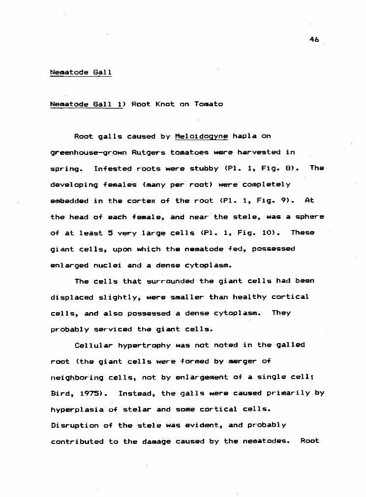
Nematode Gall
Nematode Gall 1) Root Knot on Tomato
Root galls caused by Meloidoqyne hapla on
greenhouse-grown Rutgers tomatoes were harvested in
spring. Infested roots were stubby (P1. 1, Fig. 8). The
developing females (many per root) were completely
embedded in the cortex of the root (P1. 1, Fig. 9). At
the head of each female, and near the stele. was a sphere
of at least 5 very large cells (P1. 1, Fig. 10). These
giant cells, upon which the nematode fed, possessed
enlarged nuclei and a dense cytoplasm.
The cells that surrounded the giant cells had been
displaced slightly, were smaller than healthy cortical
cells, and also possessed a dense cytoplasm. They
probably serviced the giant cells.
Cellular hypertrophy was not noted in the galled
root (the giant cells were formed by merger of
neighboring cells, not by enlargement of a single cell;
Bird, 1975). Instead, the galls were caused primarily by
hyperplasia of stelar and some cortical cells.
Disruption of the stele was evident, and probably
contributed to the damage caused by the nematodes. Root
46

47
anatomy was most disrupted near the nematode's head. The
cortex and epidermis, and the stele slightly above and
below the giant cells were undisturbed. No sclerid zone
was formed. Healthy and galled roots were free of
tanni ns.
Summary of Nematode Gall
In this example, a sedentary gall-forming nematode
manipulated the development of host plant cells that
surrounded its head so that the cells became
cytoplasmically enriched.. The gall was a kataplasma with
a well-defined nutritive tissue.

Eriophyoid Mite Galls
Eriophycid Gall 1) Willow Leaf Gall
The gall occurred an Salix sp. (perhaps S. piperi
Bebb) and was caused by an eriophyid (P1. 3. Fig 3). It
was found on only a few leaves on an isolated group of
meter high saplings. Felt (1965) says that "a number of
undescribed capsule or pocket galls are produced (on
Salix sp.) by undescribed species of Eriophves." He
suggests that same of these mites should tentatively be
assigned to Eriophyes oeniama Walsh.
On May 20 the galls (P1. 3, Fig. 1) were small
sub-spherical blisters (2-3 mm in diameter) that were red
on the upper leaf surface (surrounded by a chloratic
area) and were covered with a white tomentum on the lower
leaf surface. They occurred in no regular arrangement
over the leaf surface.
When the upper surface of the gall was cut away (P1.
3, Fig. 2) one could see that the internal gall cavity
was incompletely partitioned by 2-3 enations that arose
from the gall walls. A few hairs also grew out from the
gall wall, particularly from the tips of the enatians.
An average of 12 mites occurred per gall by early June.
48

49
The gall completely disrupted the cellular
arrangement of the mesophyl]. in healthy leaves (P1. 3.
Fig. 4, 5), but derivation of gall wall tissues was easy
to trace through the transition zone.. The upper wall of
the gall was evenly thick, contained large vascular
strands, and contained cells that in their staining
properties resembled both mesophyll and palisade cells.
These observations suggested that this wall was derived
-from minimally proliferated leaf tissue. The lower wall,
however, was derived from '1umwallung" a-f the spongy
mesophyll region. The shape a-f the mature gall and its
lack a-f an aperture suggested that it was a covering
gall. The gall was a complex kataplasma; distinct,
cytoplasmically rich nutritive cells were present, but
the mature gall wall was otherwise parenchymataus in
composition..
Nutritive cells were present in young galls (May 27)
(P1. 3, FIgs. 5, 6, 7). They lined the gall cavity in
layers two to -four cells thick, and showed a rich
cytoplasm and a prominent nucleus and nucleolus.
Nutritive cells on June 5 continued to show a cytoplasmic
richness. As the gall aged, however, the nutritive
tissues became thicker in some spots (5-10 cells thick)
(P1. 3, Fig 8)) and thinner in others (one cell thick)
(P1. 3, Fig. 9). The increase in thickness was due to

50
cell division, and the decrease to incorporation of
tannins into the outer layers. There was a deposit of
unknown material lining the cavity (P1. 3, Fig. 8). This
was perhaps ruptured nutritive cell contents, or the
ruptured contents of mites.
In a dying gall that contained mites (August 19),
the nutritive cells had little cytoplasm, either because
they had been heavily fed upon, and/or because they were
no longer being serviced from the surrounding tissue (P1.
3, Fig. 10). On this same day I observed new galls on
young leaves.
The nutritive cells stained lightly for the presence
of tannins (P1. 4, Fig. 1) in galls collected on June 5,
1980. These cells contained less tanning than did most
of the cells forming the gall wall, and less than many of
the cells in healthy leaves. In fact, there was a
gradient of decreasing amounts of tannins from the outer
gall wall to the nutritive cells.
Eriophyoid Gall 2) Alder Bead Gall
This gall occurred on Alnus rubra Bong. - Russo
(1979) described a gall on A.. rubra caused by the
sierraphytoptid, Phytopus laevis Nal., that resembled the
mite gall in MacDonald Forest. He stated that the mites
induced "round, yellow to green bead galls an the upper

51
surfaces of the leaves.... There is an opening on the
underside.." Shevtchenko (1957) studied the life history
of this mite.
The galls that I collected were yellow or red when
young, were roughly spherical (about 2 mm in diameter)
(P1. 4, Fig. 5), and occurred toward the leaf edge (P1.
4, Fig. 2). The gall projected both above and below the
leaf surface.
Until the middle of June, only a single orange mite,
probably the deutogyne (=overwintering female), occurred
in the gall (P1. 4, Fig. 3) and was responsible for gall
formation. Her eggs were laid in the galls beginning in
the middle of May, but eclosion apparently did not occur
immediately because the first white nymphs were not
observed until June 25. By mid-August many mites
occurred in each gall. At this time, orange mites were
present along with white forms (P1. 4, Fig. 4) (males and
protogynes (summer females)).
When one cut a mature gall longitudinally (P1. 4,
Fig. 5), one saw that there was usually a large
unpartitioned central cavity. Additionally, the upper
surface of the gall was flat while the lower surface was
more V-shaped. The internal cavity was occasionally
partitioned (P1. 4, Fig. 6).
A longitudinal thin section of a young (April 22)
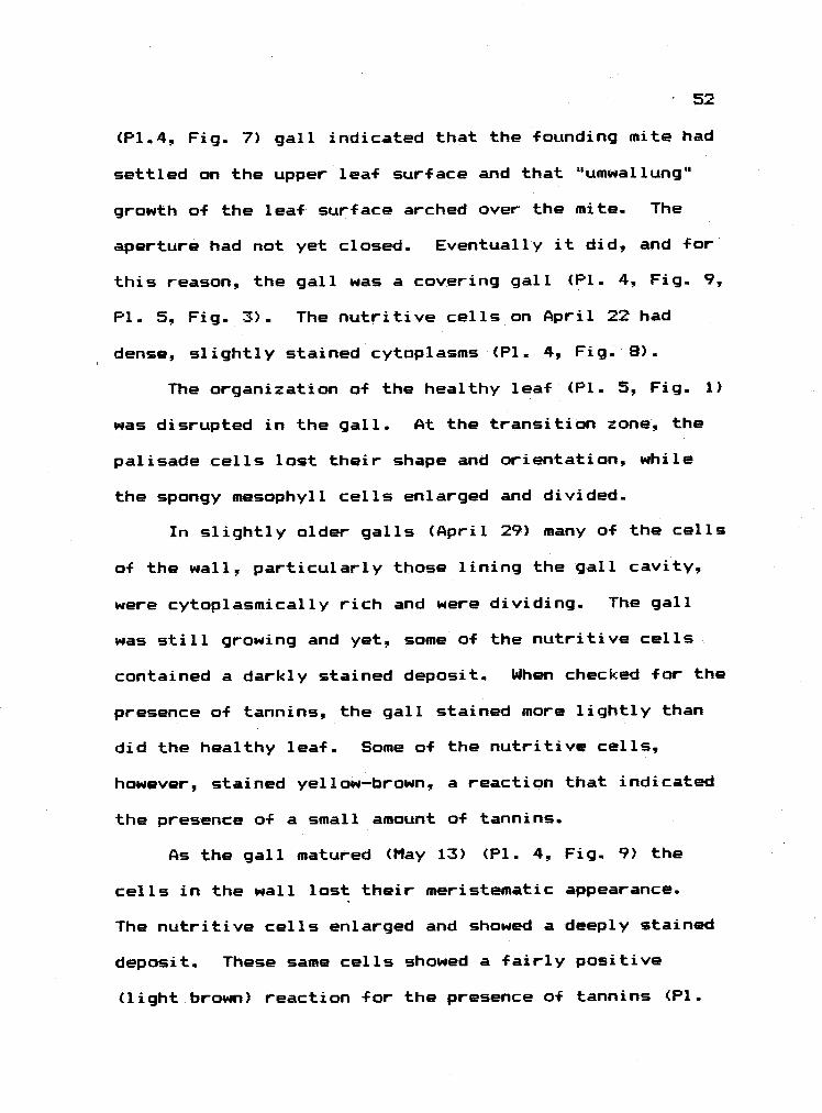
52
(P1.4, Fig. 7) gall indicated that the founding mite had
settled on the upper leaf surface and that "umwallung'
growth of the leaf surface arched over the mite. The
aperture had not yet closed. Eventually it did, and for
this reason, the gall was a covering gall (P1. 4, Fig. 9,
P1. 5., Fig. 3). The nutritive cells on April 22 had
dense, slightly stained cytoplasms (P1. 4, Fig. 8).
The organization of the healthy leaf (P1. 5, Fig. 1)
was disrupted in the gall. At the transition zone, the
palisade cells lost their shape and orientation, while
the spongy mesophyll cells enlarged and divided.
In slightly older galls (April 29) many of the cells
of the wall, particularly those lining the gall cavity,
were cytoplasmically rich and were dividing. The gall
was still growing and yet, same of the nutritive cells
contained a darkly stained deposit. When checked for the
presence of tannins, the gall stained more lightly than
did the healthy leaf. Some a-F the nutritive cells,
however, stained yellow-brown, a reaction that indicated
the presence a-f a small amount of tannins.
As the gall matured (May 13) (P1. 4, Fig. 9) the
cells in the wall lost their meristematic appearance.
The nutritive cells enlarged and showed a deeply stained
deposit. These same cells showed a fairly positive
(light.brown) reaction f or the presence of tannins (P1.

53
5, Fig. 5). Up to this point, only the founding mite was
present in the gall. Plate 4, Figure 10 shows the
extremely enlarged nutritive cells that were present as
eggs began to appear in the gail. These cells were
multinucleate and possessed a very dense cytoplasm with
many small vacuoles. Many dark deposits occurred in the
lightly stained granular cytoplasm of these cells. The
cells that underlaid the nutritive cells contained
similar deposits.
By July 16 (P1. 5, Fig. 3) several mites were found
in each mature gall. According to Jeppsan et al (1975)
up to 400 mites may occur per gail. Many of the
nutritive cells were flat and no longer bulged into the
gall cavity. Some were empty while others showed
cytoplasms that, with the exception of many small
non-staining vacuoles, stained deeply (P1. 5, Fig. 4).
The same stained positively for the presence of tannins
(deep yellow-brown) (P1. 5, Fig. 6). By September 21,
many of the galls had bequn to die (P1. 5, Fig. 2).
Thus, fully developed nutritive cells occurred in
these complex kataplasmas before they were heavily
populated. With the exception of very young galls, the
nutritive cells contained compounds that stained
positively for the presence of tannins (yellow-brown),
although not to the same extent as did the palisade cells

of ungalled leaves (black).
Eriophyoid Gall 3) Linden Lea-F Gall
This is commonly called the cerateon gall on Tilia
spp. According to Felt (1965) it is "presumably" incited
by the eriophyid Eriophyes abnarmis Garm.. The galls
were collected from T. europaea, or some cultivar of this
species, near the Oregon State University campus in
Corvallis. They sometimes occurred singly or a few per
lea-f, but most often many occurred per lea-F in no
apparent arrangement.
The very young gall appeared as a small blister on
the upper surface of the leaf. It grew and elongated so
that when mature (May 22). it was cylindrical, tapered,
and sometimes curved at the tip. It was widest at
slightly lower than its midpoint, and tapered at bath
ends (P1. 5, Fig. 7). The lower leaf surface showed few
i-f any signs of the gall.
I have no photograph a-f the mites that caused this
gall. Instead, the mite that is shown in Plate 5, Figure
B was taken from a linden leaf gall brought from New York
state (P1. 7, Fig. 9) that, according to Felt, is also
caused by E. abnormis.
Until the middle a-F May mast galls held only a
single mite. Fully grown galls (May 20) contained eggs
54

55
and a few mites. A single founding mite was undoubtedly
responsible far forming the gall. Only when the gall was
nearly mature did progeny appear.
A young gall (late April) in longitudinal section
(P1. 5, Fig. 10) showed that it was a pouch gall that was
formed when the founding mite attacked the lower leaf
surface. The leaf surface bulged up, and a lipped portal
that was plugged with hairs remained on the qalVs
undersurface. The region of transition between leaf and
gall wall was sharp (P1. 5, Fig. 13). The palisade and
mesophyll layers lost distinction in the thick gall wall.
The nutritive cells in these young galls (P1. 5,
Figs. 11,12) were in a palisade-like arrangement, had
dense, granular cytoplasms, and prominent nuclei. They
thus formed a distinct layer that lined the cavity. Many
of the portal hairs also had a dense cytoplasm.
As the gall matured (May 6) the nutritive layer
became even more distinctly set off from underlying
cells; the nutritive cells retained their dense cytoplasm
while underlying cells became vacuolate. The nutritive
cells retained their palisade arrangement in the lower
part of the cavity, but were more randomly arranged in
the upper part.
By May 20, when egg and progeny began to appear in
the gall, the nutritive cells in the upper half of the

56
cavity were vacuolate (P1. 6, Fig. 2). Cytoplasmically
rich, palisade nutritive cells were most common in
enations (=surf ace outgrowths) along the cavity wall (see
P1.6, Fig. 3). At the portal, the nutritive cells showed
no particular arrangement and many were without dense
cytoplasms (P1.. 6, Fig. 1).
By July 24 almost all of the cells lining the gall
cavity were vacuolated (P1. 6, Fig. 3) and some had
collapsed. Occasionally, a multinucleate,
cytoplasmically rich cell was seen in the enations. Most
of the portal hairs had developed thick walls, and showed
little if any cytoplasm.
The mature gall wall contained large canals or ducts
(P1. 6, Fig. 3) that were filled with an amorphous darkly
stained material (not tannins). The derivation of the
ducts was not clear in that they resembled nothing seen
in the healthy leaf (P1. 5, Fig. 9).
Tannins were present in only a few scattered
palisade cells in the ungalled leaf. In walls of mature
(July 24) galls, the outer epidermis and subepidermal
layers stained positively f or tanriins. A few cells
scattered throughout the gall wall also contained
tannins, but the nutritive cells did not.
The E. abnormis pouch gall collected in New York
state (by G. Nielsen) on May 26, 1980 was similar in
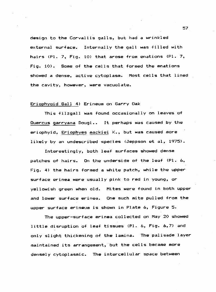
57
design to the Corvallis galls, but had a wrinkled
external surface. Internally the gall was filled with
hairs (P1. 7, Fig. 10) that arose from enations (P1. 7,
Fig. 10). Some of the cells that formed the enations
showed a dense, active cytoplasm. Most cells that lined
the cavity however, were vacuolate.
Eriophyoid Gall 4) Erineum on Garry Oak
This filzgall was found occasionally on leaves of
Quercus garryana Doug 1 . . It perhaps was caused by the
eriophyid, Eriophyes mackiei K., but was caused more
likely by an undescribed species (Jeppson et al, 1975).
Interestingly, both leaf surfaces showed dense
patches of hairs. On the underside of the leaf (P1. 6,
Fig. 4) the hairs formed a white patch, while the upper
surface erinea were usually pink to red in young, or
yellowish green when old. Mites were found in both upper
and lower surface erinea. One such mite pulled from the
upper surface erineum is shown in Plate 6, Figure 5.
The upper-surface erinea collected on May 20 showed
little disruption of leaf tissues (P1. 6, Fig. 6,7) and
only slight thickening of the lamina. The palisade layer
maintained its arrangement, but the cells became more
densely cytoplasmic. The intercellular space between

58
many spongy mesaphyll cells was decreased. Cells of both
epidermises were vacuolate, and remained much as they
were in ungalled areas of the leaf.
The unicellular erineal hairs appeared to be of
epidermal or subepidermal origin; their bases were placed
deeply in the epidermis. The hairs were thick-walled and
vacuol ate.
A stain for tannins at this time (P1. 6, Figs, 9,
10) indicated that the epidermal and sub-epidermal layers
of the erineum contained rich deposits of tannins (black
stain) as did the bundle sheath cells. The palisade
layer, bath in the erineum and healthy leaf, stained
positively. The spongy mesophyll region of the erineum
stained poorly, and the erineal hairs stained lightly
brown.
Older galls collected on July 24 (P1 6, Fig. 8)
showed a leaf mesaphyll with palisade cells of shorter,
broader shape than normal and a dense spongy mesophyll.
Most of the mesophyll cells stained very lightly if at
all, and were vacuolate. The cells of the lower
epidermis stained very deeply with a deposit that was
most likely tanniferous.
The mites probably fed on the epidermal cells and
not on the thick walled hairs, but this was not
established. If they did feed on epidermal cells, then
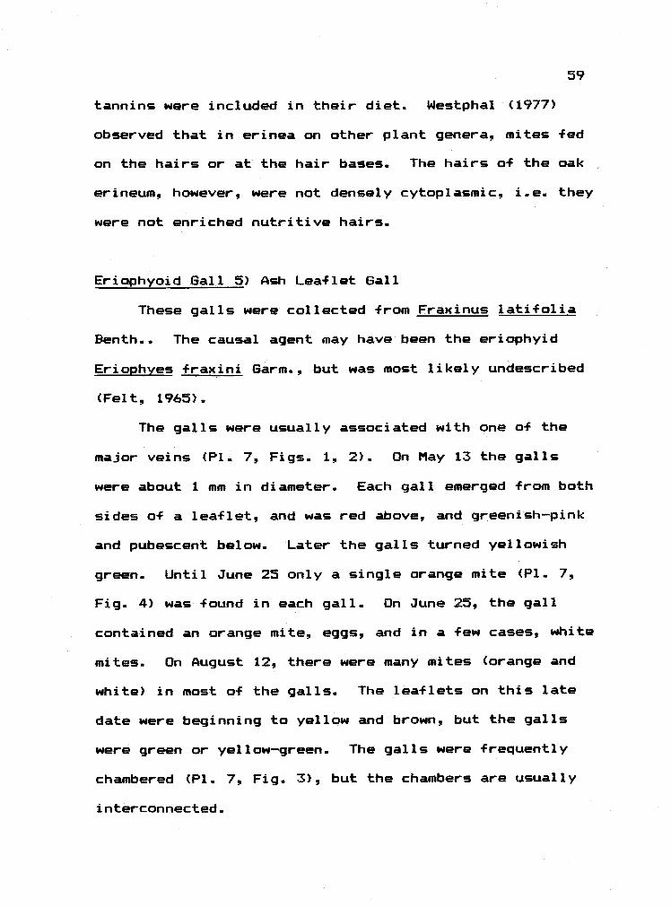
59
tannins were included in their diet. Westphal (1977)
observed that in erinea on other plant genera, mites -Fed
on the hairs or at the hair bases. The hairs of the oak
erineum, however, were not densely cytoplasmic, i.e. they
were not enriched nutritive hairs.
Eriophyoid Gall 5) Ash Leaflet Gall
These galls were collected from Fraxinus latifolia
Benth The causal agent may have been the eriophyid
Eriophyes fraxini Garm., but was most likely undescribed
(Felt. 1965).
The galls were usually associated with one a-f the
major veins (P1. 7, Figs.. 1., 2). On May 13 the galls
were about 1 mm in diameter. Each gall emerged -from both
sides of a leaflet, and was red above, and greenish-pink
and pubescent below. Later the galls turned yellowish
green. Until June 25 only a single orange mite (P1. 7,
Fig. 4) was found in each gall. On June 25, the gall
contained an orange mite, eggs, and in a few cases, white
mites. On August 12, there were many mites (orange and
white) in most a-F the galls. The leaflets on this late
date were beginning to yellow and brown, but the galls
were green or yellow-green. The galls were frequently
chambered (P1. 7, Fig. 3), but the chambers are usually
interconnected.
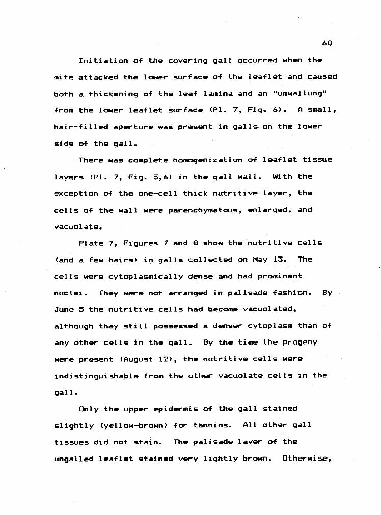
60
Initiation of the covering gall occurred when the
mite attacked the lower surface of the leaflet and caused
both a thickening of the leaf lamina and an "umwallunq
from the lower leaflet surface (P1. 7, Fig. 6). A small,
hair-filled aperture was present in galls on the lower
side of the gall.
There was complete homogenization of leaflet tissue
layers (P1. 7, Fig. 5,6) in the gall wall.. With the
exception of the one-cell thick nutritive layer, the
cells of the wall were parenchymatous, enlarged, and
vacuol ate.
Plate 7, Figures 7 and 8 show the nutritive cells
(and a few hairs) in galls collected on May 13. The
cells were cytoplasmically dense and had prominent
nuclei. They were not arranged in palisade fashion. By
June 5 the nutritive cells had become vacuolated,
although they still possessed a denser cytoplasm than of
any other cells in the gall. By the time the progeny
were present (August 12), the nutritive cells were
indistinguishable from the other vacuolate cells in the
gall.
Only the upper epidermis of the gall stained
slightly (yellow-brown) for tannins. All other gall
tissues did not stain.. The palisade layer of the
ungalled leaflet stained very lightly brown. Otherwise,

the leaflet was tannin-poor.
Eriophyoid Gall 6) Poison Oak Leaflet Gall
This gall was collected in Finley Wildlife Refuge (5
miles south of Corvallis) on May 31, 1980. It also
occurred in MacDonald Forest. It was found on the
leaflets (P1. 8, Fig. 1) of Rhus diversiloba 1. and 3.,
and it was caused by the eriophyid Aculops toxicophagus
Ewing (P1. 8, Fig. 3). The mite caused "raised,
pubescent, bead galls on the upper and lower surfaces of
the leaves in late spring" (Russo, 1979). The galls
occurred singly or in clusters and were usually red.
Transverse sections of this leaflet pouch gall (P1.
8, Figs. 4, 6) showed that it was primarily caused by
hypertrophy of the lower epidermal cells. All other leaf
tissues layers remained almost unaffected (see section of
unqalled leaf; P1. 8, Fig. 5). The transition between
normal epidermal and gall epidermal (=nutritive) cells
took place over about ten cells. The normal epidermis
was vacuol.ated with only peripheral, granular cytoplasm
while the nutritive cells contained a homogenous granular
cytoplasm with few if any vacuoles. Nuclei in the
nutritive cells, interestingly, were not prominent.
Epidermal hairs arose from the gall epidermis and were
thick walled and void of cytoplasm. The gall was not
61

62
stained for tannins.
I also found poison oak flowers that had been galled
by eriophyoids (perhaps A. toxicophagus) (P1. 8, Fig. 2).
The galls were pubescent convolutions a-f the flower
spray. Flowers were completely disrupted and
non-functional. These mites have not been used in poison
oak control programs.
Eriophyoid Gal]. 7) Leaf Gall on Trembling Aspen
This filzgall was collected from Populus tremulaides
Michx. on June 1, 1980 one mile west of Sisters, Oregon
(Deschutes Co.) on Highway 20 in the Pilot Butte State
Park. It was caused by an undescribed species of
eriophyoid mite (P1. 8, Fig. 8). According to Felt
(1965) there are many erinea produced by unnamed species
of mites on poplars.
The gall occurred on the lower leaf surface as a
circular to oblong spot (4-5 mm in diameter) (P1. 8, Fig.
7). Many fleshy, finger-like enations (P1. 8. Fig. 9)
arose from the leaf surface and the mites lived on,
between1 and at the base of the enations. The upper leaf
surface at the gall was yellow and bulged slightly.
The upper epidermis and palisade layers of the leaf
retained their orientation in the gall (compared to
ungalled leaf, P1. 8, Fig. 10), but the spongy mesophyll

63
became densely packed with cells. The cells in the
enatians resembled those in the spongy layer. The lower
epidermis covered the enations, and its cells were
enlarged and vacuolate. Thus, the gall showed no tissue
that resembled an enriched nutritive layer. The enatians
superficially resembled those seen in the filbert bud
gall (P1. 9, Fig. 9). As will be seen, however, the lack
of an enriched palisade-like nutritive enatial epidermis
in this poplar leaf gall distinguished it from the bud
gall.
It may be that the gall was collected too late to
observe enriched nutritive cells. The mites, however,
were present, active, and presumably feeding in the
collected specimens. The gall was not stained for
tannins.
Eriophyoid Gall 8) Leaf Gall on Choke Cherry
This gall was collected on Prunus virqiniana L. on
June 15, 1980 in Iranside, Oregon (Malheur Ca.) at the
southern foot of the Blue Mountains. It is most likely
the plum finger gall caused by the eriophyid Phytoptus
emarginatae (K.) (which may be synamymous with f. path;
Jeppson et al, 1975).
The gall was visible on the upper surface of the
leaf as a short, yellow, finger-like pouch (P1. 9, Figs.

64
2, 5). Like the linden gall, it was a tubular
out-pocketing of the lea-f surface (P1. 9, Fig. 4). Most
of the galls contained a single orange mite (P1. 9, Fig.
3), but a few contained eggs and white mites as well.
The cells of the leaf's upper epidermis became flat,
elongate and filled with deeply stained deposits as they
formed the gall's outer epidermis. The leaf mesophyll
(P1. 9, Fig. 6) layers underwent complete homogenization
as the gall was approached. Compared to the cells of the
palisade and spongy layers, the cells in the gall wall
were enlarged and elongated with the long axis parallel
to the long axis of the gall. Most of the cells of the
gall wall were vacuolate, and droplets in the thin
peripheral cytoplasm stained deeply. In some cases the
cells were filled with this deposit (P1. 9, Fig. 7).
The nutritive cells (P1. 9, Fig. 7) were large,
occasionally bulging into the cavity. Some contained a
lightly stained cytoplasm with a prominent nucleus and
nucleolus, and many small vacuoles. Others contained
deeply stained droplets around the nucleus. This gall
was not stained for the presence c-f tannins.
Eriophyoid Gall 9) Big Bud of Filbert
This gall, caused by the sierraphytoptid Phytoptus
avellanae Nal. (P1. 10, Fig. 5) was collected in

65
Corvallis, Oregon from Corylus avellana (Cultivar,
Daviana') throughout 1976. It was discussed in detail
elsewhere (Larew, 1977; Westphal, 1977). The galls ware
swollen stunted buds (P1 9., Fig. 8). The enlargment of
bud parts primarily was due to the presence of many
multicellular enations which covered the surface of all
bud parts (P1. 9, Fig. 9).. The mites lived on, between,
and at the bases of the enations. The surface of an
enation was covered with a nutritive layer that was
generally one cell thick (P1. 10, Fig. 1) and was
palisade-like in arrangement.
Electron micrographs of the nutritive cells (P1. 10,
Fig. 2) showed that the cytoplasmic density observed with
the light microscope primarily was due to an accumulation
of rough endoplasmic reticula and of mitochondria (P1 10,
Fig. 3) The large nucleus and nucleolus also were
apparent in these micrographs.
When tested for tannins, the nutritive cells stained
poorly, while the cells immediately beneath them stained
positively (P1. 10, Fig. 4). Epidermal cells of ungalled
filbert bud scales and leaves also stained positively.
Summary of Eriophyoid Galls
1) Classification. Those galls which possessed no
distinctive nutritive layer (oak erineum, trembling aspen

66
gall) and/ar those that retained much of the organization
of the healthy leaf (oak erineum, poison oak gall and
trembling aspen gall) were simple kataplasmas. The
anatomical evidence suggested that the mites in these
cases caused relatively little disruption. Whether
amount of disruption indicated amount of damage to the
host plant was unclear.
The remaining eriophyoid galls possessed a
distinctive nutritive layer at least during the early
stages of gall development. Additionally, the disruption
(homogenization) of the mesophyll layer was seen in all
of them. These two characteristics indicated that the
mites influenced the host plant tissue to a significant
extent, and thus their galls were categorized as complex
kataplasmas.. None of the mite galls was a prosoplasma
primarily because they lacked a scierid zone. Instead,
the gall wall was usually composed a-F hypertrophied,
hamogenous parenchyma.
Within the group of complex kataplasmas one might
sub-categorize the galls based on features such as
thickness a-f nutritive layer at gall maturity, or
alignment of nutritive cells and general type of
nutritive cell. For example, the mature willow leaf gall
had a nutritive layer that was 2-5 cells thick while all
the other galls generally possessed a one-cell thick

67
nutritive layer. If amount of nutritive tissue was
indicative of damage to the host plant, such anatomical
information might be useful in planning biological
control programs (Shorthouse, 1977a,b).
A palisade arrangement of nutritive cells was seen
in the willow gall, and the filbert bud gall. A more
random arrangement of globular nutritive cells was seen
in the alder bead gall and the choke cherry gall. The
ash leaflet gall had a few nutritive cells arranged as a
palisade, but most were small and randomly aligned. The
linden gall was an interesting case, for when the gall
was mature there were both cytoplasmically rich
palisade-like nutritive cells and vacuolate, randomly
arranged cells lining the gall cavity. I know of no
discussion of the significance of different shapes and
arrangements of nutritive cells in mite (or any other)
galls.
2) Nutritive layer dynamics. The mite galls on
willow, alder, linden, and ash showed an enriched
nutritive layer before they were inhabited by a large
population of mites. By the time the progeny were
present, most of the nutritive cells in the linden and
ash galls were vacuolate, and in the alder gall, these
cells were collapsed and stained heavily for the presence

68
of tannins. In the willow gall, nutritive cell collapse
was coincident with an increase in the number of mites.
In at least one known example (Meyer, 1952a), a
nutritive layer reappeared once the progeny began
feeding. This was not observed in the alder and ash
galls.. Instead, the results indicated that the deutogyne
elicited and enjoyed a rich diet while poorer food cells
were available to her progeny. The filbert bud gall, on
the other hand, showed active nutritive cells when the
bud was filled with mites. Short of gall death, there
was no period during infestation that this gall did not
show rich nutritive tissues in at least some portion of
the gall. The willow gall provided a good example of
nutritive tissue that was enriched and available for the
progeny. Once used by the progeny, it was not
rejuvenated. Both the choke cherry and poison oak galls
showed enriched nutritive tissues in young galls. The
aspen gall, however, showed no enriched nutritive tissue
even though mites were present.
The phenomenon of a single founding mother preparing
the gall for her progeny was also seen in some of the
gall aphids and sawf lies. See the discussions of these
galls for comparisons of the quality of the diet that was
passed on to the' progeny..
According to Jeppson et al (1975), "urgency to

69
abandon (mite) galls only comes when the plant prepares
to shed its leaves at the end of the season." Before
this study, I would have disagreed, and suspected that
deterioration of the nutritive tissue (an event that
often occurred well before abscission) was the primary
factor forcing mites to abandon their galls. Apparently
this was not the case in that mites were often found in
galls with collapsed nutritive cells. Eriophyoids have
stylets of 15-35 urn in length. As the nutritive cells
deteriorated and collapsed, the mites perhaps tapped
underlying unenriched cells, and waited almost until
abscission before leaving the galls.
3) Tannins. Those mite galls that were stained + or
the presence of tannins gave varying results. For
example, gall and leaflet tissues of ash stained poorly
for tanning. Regardless of where a mite chose to feed on
an ash leaflet, in or out of a gall, it would avoid
tannins.
The galls on willow and filbert showed many
tanniferous cells in their walls, but the nutritive
layers stained poorly for tannins. In these two cases
the mites probably avoided tannins that accumulated
nearby.
As the alder gall matured its nutritive tissue
accumulated tanning. Early inhabitants probably

70
contended with fewer dietary tannins than did later
inhabitants. Such observations emphasized the dynamic
nature of the nutritive tissue
The possibility that mites in mature oak erinea were
feeding on slightly transformed, tannin-rich cells is
worth additional study. This gall may be an interesting
exception to the proposal that gall-f ormers avoid tanning
throughout gall development.

Lepidopteran Gall
71
Lepidopteran Gall 1) Stem Gall on Peck's Penstemon
This gall was collected by Dr. Andrew tioldenke at
Camp Sherman (Jefferson County) Oregon near Sisters. It
occurs on Penstemon peckii Pennell and is caused by the
microlepidopterous gracillariid, Calotilia murt+eltella
(Bsk.) (identified by Dr. Don Davis, Smithsonian
Institution) (P1. 13, Fig. 1). (In the summer of 1981,
Dr.. Moldenke collected a similar stem gall caused by a
lepidopteran from P. procerus Dougi. on Lookout Mountain
(6200 ft elevation) in the Andrews Forest, Lane Co..,
Oregon.) Young instar larvae were found in the galls on
June 6. By July 31, old instars and pupae were found in
the galls, and adults emerged in the lab on August 13
(from July 31 material). The larva chewed a window in
the gall wall (outer epidermis was left intact) (P1. 11,
Fig. 5) before pupating in a silken cocoon on the gall
wall just beneath the window. The adult broke through
the window.
The spindle-shaped gall was formed directly beneath
the apex a-f this herbaceous perennial (P1. 11, Fig. 5)
and was thus an acrocecidium (Houard, 1904). The single
larva found in each gall (P1. ii, Fig. 6) uses strongly

72
dentate mandibles (P1. 13, Fig. 4) to cut through the
nutritive tissue.
In tranverse section. the open pith of an ungalled
stem was partially filled with isodiametric
parenchymatous cells that increased in diameter toward
the center. A ring of radially arranged xylem elements
surrounded the pith that was in turn surrounded by a
compact ring of phloem. The cortical cells were similar
in appearance to the pith cells, but there were smaller
intercellular spaces in the cortex (P1. 11, Fig. 7).
On June 6 cortical cells were almost twice as large
in diameter in the gall wall as they were in the ungalled
stem, and in the gall, air spaces between these cells
were prevalent. The vascular system was disrupted;
instead of solid rings, the tissues occurred
intermittently around the stem in large to small bundles.
The xylem cells lost their radial alignment and, on the
average, were smaller in diameter than those in the
ungalled stem.
Only remnants of pith cells remained in the gall.
This suggested that most of the pith was eaten by the
larva soon after the stem was attacked. The first instar
larva probably bored from the stem apex into the pith of
the young stem. As the pith (and occasionally, xylem)
was consumed, new, smaller parenchymatous cells arose

73
near the vascular cambium (P1. 11, Fig. 8). Thus, the
pith cells and the derivatives a-f the vascular cambium
were the nutritive cells in this gall. Both of these
were unenriched and vacuolate with only a minimal
peripheral cytoplasm and an unenlarged nucleus (P1. 11,
Fig. 9). Staining indicated that only the epidermal
cells of the healthy stem and of the gall contained
tannin deposits.
Summary of Lepidopteran Gall
This gall was a simple kataplasma. A scierid zone
was not formed. The stem's cortex, although
hypertrophied in the gall, was recognizable, and the
nutritive tissue was of a law level of differentiation
and was unenriched.

Sawfly Galls
74
Sawfly Gall 1) Gall on Snowberry
This gall was collected from Symphoricarpos albus
var. laeviqatus Fern.. The gall was caused by an
undescribed tenthredinid (Felt, 1965). Each gall
contained a single larva. Because the earliest collected
galls (April 29) contained larvae, it was not known if a
colleterial (=ovipositional) fluid was responsible for
early gall development as it was in other sawfly galls.
The larvae used sclerotized mandibles to chew through the
nutritive tissue (P1.. 12, Fig. 2).
The galls were small capsule-like structures (P1.
12, Fig. 1) at the twig terminal. These were probably
bud galls, but their derivation was unclear. Each of the
two terminal buds was replaced by a gall, and often the
pairs shared a common wall. The gall walls were thick
(2-3 mm) , and deep green.
A transverse section of a young (April 29) gall wall
(P1. 12, Fig. 3) showed that the outer epidermis and the
5-6 subepidermal layers were composed c-f vacuolate cells.
The bulk of the wall, however, was composed o-f several
layers of cells that contained a fairly rich cytoplasm
with prominent nuclei and many plastids. This was the
nutritive tissue. It was produced as very broad, stubby

enations from the gall wall. The nutritive cells were
not arranged in palisade -fashion.
As the gall matured (May 6) (P1. 12, Figs. 4 and 5)
the nutritive cells became vacuolate. This occurred from
the outer surface of the gall inward. Some of the
nutritive cells lining the gall cavity were ruptured,
probably from having been fed upon.
Vacuoles continued to enlarge in older nutritve
cells (May 13) (P1 12, Fig. 8), so that by June 5 (P1.
Fig. 7) only a peripheral cytoplasm with plastids was
seen. By July 25 (P1. 12, Fig. 8) the nutritive cells
showed no cytoplasm. Many had been ruptured. Frass was
abundant in the gall. At this time, however, the larvae
had not eaten all of the gall wall material (i.e. the
wall was still thick).
The cells of the gall wall stained lightly
yellow-brown when stained for tannins, but there were no
tannin-rich cells in the gall.
Sawfly Gall 2) Willow Leaf Gall
This gall occurred on Salix sp.. I generally -found
it on creek bank clumps (clones?) of saplings that showed
few of the other types of willow galls. The gall (P1.
Fig. 6) was caused by the tenthredinid, Pontariia sp.
(P1. 13, Fig. 3). The species was presumed to be f.
75

76
pacifica (but see Smith, 1970) because lenticels occurred
as brown spots on the surface of the galls - a
distinctive characteristic of galls caused by this
species (Russo, 1979).
This was an unusual gall when compared to most
others in that it was initiated by the adult female as
she oviposited into the young willow leaf. Thus it was a
mark gall.. Compounds in the colleterial fluid were
responsible for a great deal of gall development (Smith,
1970). Although I collected galls that contained eggs as
early as April 26, it was not until May 13 that I
collected galls that contained larvae.
The youngest galls that were collected (April 26)
(P1.. 13, Fig. 8) were small pubescent bulges on the
underside of the leaf (P1. 13, Fig. 5) that coresponded
to shiny reddish spot on the upper surface (P1. 13, Fig.
2). Galls were frequently adjacent to the midvein. Some
of these very young galls showed a small slit on the
upper surface of the gall, the tissue on either side of
which had turned necrotic. This perhaps was the slit cut
by the female sawfly to deposit the egg.
In longitudinal section, the young gall was seen to
be produced primarily on the lower leaf surface, and the
transition between ungalled leaf and gall was sharp. The
leaf midvein occurred on one edge of the gall, and it was

77
partially split by the gall cavity (P1. 13, Fig. 11). In
fact, much of the gall wall tissue arose from
merjstematjc tissues associated with the differentiating
midvein.. Although much of the gall at this time was
composed of actively dividing, cytoplasmically rich
cells, these were mast prevalent in the center of the
upper (P1. 13, Fig. 9) and lower (P1.. 13, Fig. 10) gall
wall.
Young nutritive cells were slightly larger in
diameter than most of the cells in the gall, and they
were vacuolate with a thin strip of peripheral cytoplasm.
Some of these cells were ruptured. They were probably
cut by the ovipositor, and/ar lysed by the colleterial
fluid.
Throughout the young gall, one finds scattered
groups of cells that, although densely cytoplasmic, also
contain a deeply stained deposit. This material stains
dark reddish brown with ferrous sulfate, i.e. it is
tanniferous. Many of the gall epidermal cells, scattered
cells throughout the midvein, and the palisade cells in
the young ungalled leaf (P1.. 14, Fig.. 4) also stained
positively (dark brown).
The most noticeable change that occurred in slightly
older galls (April 29, and May 6) was that many of the
cells of the gall wall lost their meristemati.c

78
appearance, and became vacuolate and filled with tannins.
The nutritive cells lining the larval cavity remained
free of these deposits and, although mostly vacualate,
began to show a dense cytoplasm.
By the time that larvae began to appear and feed in
the gall (May 13, and 22; P1. 13 Fig.. 7), bath upper and
lower gall walls had doubled in thickness since the first
collection date. A band o-f tannin-rich cells encircled
the nutritive cells, and was derived from the scattered
groups of tannin cells that were seen in younger galls.
At the same time, the nutritive tissue had increased
in depth, and its cells, in cytoplasmic density (Pl 14,
Figs. 2, 3). The cells were not arranged in palisade
fashion, and small enations grew into the larval cavity.
Feeding damage was still not yet extensive (P1. 13, Fig.
6).
Older galls collected on June 5 showed vacuolate
nutritive cells that contained no tannins (P1. 14, Fig.
6), while many of the cells in the gall wall stained
positively f or tannins (P1. 14, Fig. 5). The lower gall
wall continued to grow in thickness such that by August
12 it was 2-3 times as thick as in the May specimens.
Much of the nutritive tissue had been fed upon and in
spots had been eaten to the tannin-rich band a-f cells..
The nutritive cells that had not been fed upon were

79
vacuolate with only a thin strip of peripheral cytoplasm.
Frass was abundant in these galls.
The last galls that were sectioned (September 22)
were about 1 cm in diameter. They were yellow with brown
lenticels. Those galls that had not been invaded by
weevils (probably Anthonomus sp.; see Caltaqirone, 1964)
and that still contained a sawfly larva displayed a band
of vacuolate nutritive cells that was 10-12 cells thick
(P1. 14, Fig. 1). Galls collected on October 16, 1981
were on completely dead leaves that had fallen to the
ground. Many of the galls, however, were still succulent
and contained a 1 cm long white or purplish sawfly larva.
Caltagirone (1964) mentioned that feeding by late
instar larvae of P. pacificus might completely hollow the
gall, or might merely tunnel it, depending on the
thickness of the gall wall. My own observations
indicated that the younger larvae fed on the tannin-poor,
cytoplasmically richer nutritive cells, while the older
instars consumed bath tannin-rich and tannin-poor cells.
Summary of Sawfly Galls
1) Classification. Both sawfly galls were simple
kataplasmas. Neither contained sclerid zones, and both
showed callus-like nutritive tissue. In complexity, they
resembled the gracilariid gall an Peck's penstemon.
80
Nutritive cell dynamics. Both galls showed their
most cytoplamically dense cells early in the season. As
the season progressed, the proportion o-f vacuolate to
cytoplasmically dense cells increased. The older the
instar, the less nutritious was the appearance of its
diet.
Tannins.. In the case of the snowberry gall, the
larva fed on tissue that stain relatively poorly + or
tannins. As the larva in the willow gall matured, on the
other hand, it began to feed on tissue containing
tannin-charged cells. Thus in the willow gall, the older
instars contended with a diet that was probably less
nutritious both because of the vacuolation of many cells,
and because of the inclusion of tannins in many of those
vacuoles. It should be stressed, however, that in the
willow gall, the larva was presented with a diet poor in
tannins when compared to willow lea-f tissues (compare P1.
14, Figs. 4, 5 and 6).

Thrips Gall
Thrips Gall 1) Memecylon Leaf Roll Gall
A preserved leaf gall on Memecylon sp. Roxb.
(Melastomaceae) caused by Crotonothrips danahasta
Ramakrishna was shipped to me from India by Dr. A. Raman.
The specimen was collected on July 7, 1980 on the campus
of Madras Christian College, Chengalpat District, Tamil
Nadu, India.
The gall, like many thrips galls, was a leaf edge
roll gall. Both edges curled over the leaf's upper
surface.. The galled leaves were green when picked and
were, on average, 2.5 cm in length. No thrips were found
in the preserved galls, but a few were found at the
bottom a-f the specimen vial.
The leaves had a speckled appearance because of
black circular, thickened areas, 1-1.5 mm in diameter
that occurred between the secondary veins. There were
approximately 90 of these areas per leaf. Most were
discrete, but a few were fused.
The galled leaf showed an abnormal mesophyll layer
that was thick and contained enlarged, densely arranged
isodiametric cells.. In patches the upper epidermis
changed from vacuolate, small rectangular cells that had
their long axes parallel to the lea-f surface, to much
81

82
enlarged palisade-like cells with cytoplasms that
contained deeply stained deposits (P1. 17 Fig. 10; P1.
28, Fig. 10). These last were the nutritive cells, and
the nutritive patches in which they occurred corresponded
to the black patches on the leaf surface that were seen
with the naked eye. They were underlaid by radial rows
of small cells. Some of which were empty (dead) while
others contained large vacuoles, the contents of which
stained lightly and homogenously. The gall was not
stained for tannins.
Summary of Thrips Gall
Classification.. This gall had characteristics of
a complex kataplasma in that it showed a fairly distinct
nutritive epidermis. It was simple in design and
contained no protective layer. As with the eriophyoid
galls, I classified this as a complex kataplasma.
Nutritive Cells. Thrips feed with stylets (P1.
17, Fig. 9). According to Ananthakrishnan (1978) in
the majority of thrips galls, feeding is oftenlocally restricted to the surface layer of aparticular portion of the infected organ, where theepidermal cells are densely filled with cytoplasmiccontents and are prominently nucleated. These cellsand some of the cells of the mesophyll withinconstitute the feeding zone.
The last statement is supported by measurements I

83
made of the stylets of mature thrips. The stylets of C.
danahasta were on average. 0. 15 mm long. The nutritive
cells were 0.07 mm deep. Thus this thrips was able to
penetrate beyond the surface nutritive cells. Most
interesting is the fact that the nutritive cells in the
Memecylon gall occurred in patches. This indicated a
limited manipulative ability and a sedentary feeding
behavior.
3) Tannins. The presence of dark deposits in the
nutritive cells suggested that tannins were present in
the cells. In response to an inquiry about this, A.
Raman wrote that "tannin deposition is not unusual in
mature galls induced by thrips especially in the
nutritive cells." The important word here was "mature."
Most likely, nutritive cells in thrips galls develop
deposits o-f tannins over time. I infer from this and
from the absence of thrips in my specimens that the galls
I studied were mature. Young galls should be studied.

Scale Gall
Scale Gall 1) Oak Pit Gall
Although common, this gall was collected only once
from Quercus garryana Dougls. in Corvallis, Oregon. The
gall was caused by a species a-f Asterolecanium (probably
A. minus Lindinger - a common scale an west coast
deciduous oaks; Koehler, 1964). The galls that were
collected (April 2) had been formed the previous spring.
Thus the scale insect was either old, dead, or missing.
The pit scales, as they are commonly called, formed
galls on the current or previous year's twigs. The galls
were unusual in at least one respect - they did not cover
or enclose the insect, but instead surrounded the sides
a-F the insect with an incomplete "umwallung" (P1. ii,
Fig. 1). The insect was cradled in the gall (P1. Ii.
Fig. 3).
The gall was caused by a proliferation of the
phellogen (=bark meristem) which gave rise to the
umwallung' The periphery a-f the gall wall was composed
of elongate, thin-walled cells arranged in radial rows
(P1. 11, Fig. 3). Toward the center a-F the pit,
underlying and couching the insect, were radial rows of
elongate sclerids. Under these were small isodiametric
cells (probably phelloderm). Cortical cells were under
84

85
these cells. There was a slight enlargement of the
cortical cells, and former cellular enrichment was
indicated by prominent nuclei (P1. 11, Fig.. 4) and
granular cytoplasm in a few of the cells. Most, however,
were vacuolate. The stem's vascular tissue and pith
appeared undisrupted by the gall (P1. 11, Fig. 2). The
gall was not stained for tannins.
Summary of Scale Gall
Classification. The presence of a sclerid zone
that couched the gall suggested that it was prosoplasmic..
The layer, however, did not surround the insect. The
facts that the gall did not enclose the insect and that
the vascular and pith tissues remained undisturbed by the
scale indicated that the gall was a complex kataplasma.
Nutritive cells. The collected specimens were
too old to allow for a study of the nutritive cells.
Other workers have observed enriched cells in this and
another scale gall (Parr, 1940, and Houard, 1903,
respectively), but more work with younger galls is needed
to further characterized these cells.

Aphid and Adelgid Galls
Aphid Gall 1) Poplar Petiole Gall
This gall was collected from saplings and mature
Populus trichocarpa T. and S. It was caused by an
undescribed species of Pemphigus (Aphididae: Subfamily
Eriosomatinae) . Stem mother measurements did not match
any of those given by Palmer (1952) for species that
occurred in the Rocky Mountains. The gall involved both
a thickening of the petiole (P1.. 15, Figs.. 2 and 4) and
an overgrowth of the lamina at the petiole-leaf junction.
It was a covering gall.
One of the earliest signs of galling (April 15; P1.
15, Fig. 1) was a sligtly swollen and bent petiole, and a
twisted leaf lamina at the leaf base. By April 26 the
gall had formed a curled and swollen structure at the
leaf base (P1. 15, Figs. 2 and 4). When the gall was
forcibly opened, one saw a gall chamber formed by the
flared petiole and enlarged leaf base (P1. 15, Fig. 5).
The aphids lived in this chamber on the modified lower
leaf surface. The gall continued to grow until it
reached full size in late June at which time it resembles
a subspherical pouch (P1. 15, Fig. 3).
Until May 20, the galls contained only a single stem
86

87
mother that malted at least twice in the gall before
producing progeny. Thus this female incited gall
development and prepared a large gall cavity for her
progeny. As a young (April 15) gall was approached. the
mesaphyll underwent complete homogenization. Different
amounts of cell division in the mesophyll layers caused
the lamina portion of the wall to arch up and curl
partially around the petiole.
At the gall, the lower surface of the petiole
thickened due to proliferation of the vacuolate,
parenchymatous cells underlying the vascular bundle. The
petiole lost its circular shape and became oblong in
transverse section with the long edge forming part of the
gall wall (P1. 15,Fig.. 5). Comparison with ungalled
petioles indicated that there was little disruption in
the organization of the vascular bundle in the galled
petiole.
At the tap a-f the cavity and on the petiole side a-f
the gall, the cavity epiderrnal cells (these were probably
not nutritive cells; see below) were palisade-like and,
although partially vacuolate, showed a fairly dense
cytoplasm and prominent nucleus.
By April 29, most of the cavity epidermal cells were
vacuolate (P1. 15, Fig. 6). There was. however, a small
area of cytaplasmically dense epidermal cells in the

88
arched gall wall (P1. 15, Fig. 7). These cells were not
arranged in a palisade-like fashion, and a few formed
unicellular hairs.
The meristematic cells of the vascular bundles were
cytoplasmically rich and were 8-15 cell layers in from
the gall cavity. Frequently there was a large
intercellular cavity in the center of the bundles. At
the same time, the outer surface of the leafy gall wall
was uneven in thickness due to ridges on the outer gall
surface (P1. 15, Fig. 8).
Specimens collected May 6 still showed large groups
of densely cytoplasmic cells in the vascular bundles of
the leafy gall wall. The bundles were larger and more
widely separated than previously.
Specimens from May 13 showed a cavity epidermis
composed of cells that were either densely cytoplasmic
(on petiole gall wall) or vacuolate (on leafy gall wall).
The leafy gall wall continued to contain bundles of
cytoplasmically dense cells (P1. 15, Fig. 9) that were
5-10 cells beneath the gall cavity's epidermis.
The June 5 specimens contained progeny and were
larger than earlier, mainly due to growth of the leaf
gall wall (P1. 15, Fig. 5). The epidermal cells lining
the gall cavity at this time were generally vacuolate but
a few along the leafy gall wall possessed a rich

89
cytoplasm. The band of densely cytoplasmic elongate
vascular cells were still present in the gall wail (P1.
16, Fig. 1), and in some spots, passed within 4 cells of
the cavity's epidermis. Frequently xylem cells on the
periphery of the band showed spiral secondary wall
thickening.
On August 12 many of the galls were dying. The
specimens contained no living aphids. The cells of the
cavity epidermis were vacuolate and the cells of the
densely cytoplasmic vascular band had either vacuolated
and taken on deeply stained deposits or had
differentiated into vascular elements (P1. 16, Fig. 2).
The cells of the leafy gall wall had begun to collapse
and die (P1. 16, Fig. 3).
Some of the galls collected May 6 were stained for
tannins. Cells scattered on either side of the vascular
strands contained deposits of tannins (light to dark
brown) as did randomly scattered cells throughout the
gall wall. The cavity epidermis stained very lightly
brown, while the subepidermal layer, in some cases
stained dark brown (P1. 16, Fig. 4).
The most important unanswered question concerning
the poplar petiole gall was: Where do the stem mother
and progeny feed? A gall collected on May 13 showed
20-30 collapsed, neighboring cavity epidermal and

90
subepidermal cells. Same of the cells beneath these
showed dense cytoplasms. This may have been a feeding
site, but no stylet tracks were seen in the area.
A few cells a-F the cavity epidermis retained
cytoplasmic density from April through June, and one
wonders i-F these were the nutritive cells. The stylets,
however, undoubtedly penetrated beyond this layer. On
May 20, + or example, the stem mother's rostrum on the
average measured 0.5-0.6 mm in length. (Rostral length
should slightly underestimate stylet length.) Young
progeny had rostrums 0.45 mm long. The gall wall on the
same day was 0.95 mm thick. Thus the aphids, regardless
of life stage, were capable of feeding well beyond the
epidermal layer.
Although they did not resemble in shape or position
the nutritive cells -Found in mite, inidqe, and wasp galls,
the densely cytoplasmic cells of the vascular bundles may
have been a primary food f or the stem mother and her
progeny. Their candidacy as a possible food source was
strengthened by the fact that they were present in
relatively large numbers, in fairly close proximity to
the aphids (0.37 mm under the cavity epidermis), and in
an enriched state f or a long period a-F time.
Another possibility is that the aphids fed on
vascular sap. Suggestive observations included the

91
occurrence of liquid balls in galls beginning on May 20.
Aphid Gall 2) Poplar Marginal Leaf Gall
This gall was collected from Populus trichocarpa T.
and G. , and was caused by Thecabius papuli monilis
(Riley) (Aphididae: Subfamily Eriosomatinae). Palmer
(1952) described the galls in abbreviated farm as
fol laws:
The fundatrix gall contains a solitary fundatrix,consists of a pocket-like gall on upperside of a leafat the base of the new growth., gall opening onunderside, her young migrating soon after birth andlocating separately near the margins on the undersideof new leaves, forming a row of pocket-like orbead-like galls. each containing a solitary aphidapterous or alate.
Her description fits well with what I observed. The
stem mother (P1. 17, Fig. 4) and her pouch gall were
first seen on young leaves that were not fully expanded.
She attacked the lower leaf surface and caused an
autpocketing of the lamina along a straight line f or at
least 1 cm (P1. 17, Figs. 1 and 2). The gall usually
occurred slightly in from the leaf's edge with the long
axis parallel to that of the leaf. It was red when
young.
Up until May 20 only the stem mother was found in
the gall.. On this collecion date, however, progeny were

92
present in the gall.. On June 25, all o-f the galls were
empty and many of the new poplar leaves (a late flush)
bore rows of new pouch galls (1 cm long and 1 cm in
diameter at widest point) formed by the progeny (=the
fundatrigeniae) (P1.. 17, Fig.. 3).. The rows ran the
length of the leaf and there were usually 2-3 rows per
leaf. Each row contains 5-B bead-like galls arranged end
to end.. The rows resemble linked sausage.. These galls,
like the stem mother galls, were produced when the aphids
attacked the underside of the leaf and caused
outpacketing.. Each cavity was separated by a septum
formed of pinched lamina.
An apterae fundatrigeniae is seen in Plate 17,
Figure 5. Only a single aphid occurred per
fundatriqeniae gall on June 25, but on July 15, some
contained 3-4 aphids--most apterae, a few alatae. Liquid
droplets also were common in these galls. On the last
collection date, August 12, the galls were empty, but
many were still green and succulent..
In transverse section the young (April 15) stem
mother gall appeared as in Plate 17, Figure 6. The
mesophyll layers (P1. 17, Fig. 7) lost their
distinctiveness as the gall was approached so that its
apex, the gall wall was composed of tightly packed
isodiametric cells (P1. 17, Fig. 8). The cells in the

93
periphery of the gall wall were larger than those near
the gall cavity - a difference that caused the curved
outpocketing.
The cavity epidermis in this young stem mother gall
was made up of vacuolate cells. Densely cytoplasmic
cells made up much of the center of the gall wall.
By April 29 the gall was closed and the transition
zone between ungalled leaf and gall wall was sharp. The
cavity was nearly circular in transverse section. The
cavity epidermis cells were small and vacuolate. The
majority of cells in the gall wall were also vacuolate.
Several vascular bundles ran the length of the gall.
They contained many cytoplasmically dense xylem and
phloem parenchyma cells that were both small and
dividing. The bundles were 3-4 cells in from the cavity
epidermis (P1. 18, Fig. 2) and were partially surrounded
by vascular bundle cells that contained deeply stained
deposits.
On May 13, the stem mother was still alone in the
galls. The cavity epidermal cells in some areas were
collapsed, and in other areas were uncollapsed and
vacuolate (P1. 18. Fig. 4). Vascular cells on the inside
of the bundle (phloem parenchyma) were cytoplasmically
dense. The healthy leaf by this time has matured, and
many a-f its palisade parenchyma cells contain deeply
94
stained deposits (P1. 18, Fig. 1).
By May 20 the gall had reached full size. The
cavity was about 3 mm in diameter in transverse section.
The cavity epidermis contained groups of collapsed and
uncollapsed cells. The vascular bundles, still with
densely cytoplasmic cells, contained more elements and
were broader under the collapsed cells.
By June 5, progeny were present in the stem mother
galls. As before, the cavity epidermis was collapsed in
spots and those spots were underlaid by 4-5 layers of
cells filled with deeply stained deposits (P1. 18, Fig.
3). Cells in the vascular bundles were now vacuolate and
many had collapsed. Short segments of stylet sheath
tracks (stained purple with toluidine blue) extended out
to the external epidermis.
On August 16 empty fundatrigeniae galls were larger
than the mature stem mother galls, and the gall cavity
was 3-4 mm in diameter in transverse section. With
respect to anatomy, however, these galls were like the
stem mother gall. The cavity epidermal and subepidermal
layers in many areas were collapsed. Numerous vascular
bundles ran the length of the gall wall and these
contained a few cytoplasmically dense, small cells in the
phloem of the bundle. The bulk of the gall wall was made
up of enlarged vacuolate parerichyma. Scattered cells

95
throughout the gall wall contained deeply stained
deposits, and a few contained stellate crystals. Stylet
tracks were not seen in sections of these galls.
When a young (April 29) stem mother gall was stained
for tannins, scattered cells throughout the wall stained
positively (light to dark brown). The cavity epidermis
and the vascular tissue were free of tannins (P1. 18,
Fig. 5).
As with the previous aphid gall, the location of
feeding sites in both the stem mother and fundatrigeniae
galls was not clear. The densely cytoplasmic vascular
cells may be the primary feeding site, rather than the
vacuolate epidermal cells that line the cavity. The
occurrence of stylet sheaths deep within the gall wall,
however, suggested that the older stem mothers fed in the
wall parenchyma.
The wall of a mature stem mother gall was on average
between 0.6 and 0.9 mm thick. The vascular bundles were
0.2-0.3 mm beneath the cavity epidermis. A mature stem
mother's rostrum was about 0.6 mm long. Her young's
rostrums were 0.3-0.6 mm long. Thus in a stem mother or
fundatrigeniae gall, even considering a winding
intercellular stylet path, the aphids could penetrate
into the gall wall beyond the vascular tissue.

96
Aphid Gall 3) Flidvein Gall on Poplar Leaves
This gall (P1. 18, Fig. 6) was collected on Populus
trichocarpa T. and 6. There was some question about the
identity of the gall-former (Palmer, 1952; Grigarick and
Lange, 1962). It was most likely either Pemphiaus
balsamiferae or P. populivenam.
According to Palmer (1952), both of these species
overwinter on poplar and the stem mothers form galls on
the young leaves in spring. The stem mothers produce
alate progeny (-Fundatrigeniae) in the gall and, in
mid-spring to mid-summer, the alates leave the gall and
settle on the crowns of herbs such as sugar beets. Their
progeny in turn attack but do not gall the roots of these
herbs. Alates returns to poplars.
The gall cavity was formed by the out-pocketing of
the leaf lamina. The midvein., which was turned on its
side by the galling, formed a portion of the gall wall
(P1. 18, Fig 7). The transition between ungalled leaf
and gall wall was sharp and, as in many other galls,
occurred at a vascular bundle.. On the ungalled side o-f
the bundle the two distinct mesophyll layers were seen
while on the galled side there was an homogenization of
the two layers.
The wall of a young gall (May 6) and its cavity
epidermis were primarily made up of vacuolate, enlarged
97
parenchyma cells. Many of the cavity epidermal cells
elongated and divided to form short hairs (2-3 cell).
Small cells rich in cytoplasm occurred 2-5 cell layers
under the cavity epidermis in the vascular strand. Some
of these had differentiated into metaxylem with spiralled
secondary wall thickenings.
One week later (May 13) the stem mother was still
the only gall inhabitant. All of the cells in the gall
wall other than those in the vascular bundle showed very
thin peripheral strips of cytoplasm Many of the
elongate cells of the vascular strand still contained a
dense cytoplasm although vacuoles were present (P1. 18,
Fig. 8).
The galls on May 20 looked much like those of the
week before. These galls, however, contained the stem
mother and her progeny. As in previous aphid galls, the
identity of the food tissue was unknown, but enriched
vascular bundle cells were likely candidates.
Aphid Sail 4) Leaf Gall on Bearberry
This gall was collected from Arctostaphvlos uva ursi
CL.) Spreng. in Corvallis, Oregon (Oregon State
University Campus) and from . qiutinosa in Tuolumne
County, California (P1. 19, Fig. 1). The causative aphid
was Tamalia coweni (Cockerell) (Aphididae: Subfamily

98
Aphinae). The galls were leaf rolls. The margin of the
leaf swelled and curled under the lower leaf surface.
Curling formed the gall cavity. On May 8 most of the
galls contained a single stem mother and two caste skins,
but a few contained 2 aphids. All galls that were
checked on May 20 contained a single stem mother. By
August 20, progeny were present in the gall (P1. 19, Fig..
2).
When the young (May 8) gall was cut transversely
(P1. 19, Fig. 4) one saw that the transition zone between
ungalled and galled lamina was either long (P1. 19; Fig.
5), or abrupt (P1.. 19., Fig. 7). The palisade cell
derivatives ware easily identified in the gall wall
because of their staining characteristics (dark, granular
material) and their alignment (P1. 19, Fig.. 6). Some of
the intercellular space in the leaf mesaphyl 1 layer was
retained in the wall and, the gall mesophyll cells were
vacuolate much like spongy mesophyll cells (compare P1.
19, Figs. 5 and 6).
The cavity epidermis and subepidermis were made of
small, deeply stained cells with long aces tangent to the
cavity surface. Cells with granular contents made up the
bulk of the wall and showed a peripheral nucleus. In
addition to the midvein, there were about 10 vascular
bundles that ran the length of the gall.. Each was 2-3

99
times as large in transverse section as its counterpart
in the ungalled side o-F the leaf. The bundles were 4-5
cell layers under the cavity epidermis, and all contained
several small cytoplasmically dense cells.
The galls collected May 20 showed further thickening
of the gall. The vascular bundles contained fewer
meristematic cells, and the cavity epidermis cells
remained flat and darkly stained (P1. 19, Fig 8).
With the exception of the epidermal. vascular, and a
few spongy mesophyll cells (P1. 20, Fig. 1) in an
ungalled leaf collected on May 20, cytoplasms stain
positively (black) for the presence of tannins. In the
gall, the contents of the cavity epidermal cells and the
granular contents of many of the gall wall cells stained
positively (black) (P1. 19, Fig 9).
The stylets of the young stem mothers in the gall on
May 8 were about 0.2 mm long (P1. 19, Fig. 3). The gall
wall at this time was 0.5 mm wide. The aphids could
penetrate beyond the vascular tissue.
No nutritive tissue such as found in cynipid galls
was found in this gall. If the aphids fed upon the
contents of the large, parenchymatous wall cells, then
they encountered tannins.
Aphid Gall 5) Leaf Gall on Witch Hazel

100
This gall was collected on Hamamelis virciinica L. in
New York State by Gary Nielsen on May 26, 1980. It was
caused by the aphid Hormaphis hamamelidis Fitch. Each
gall contained a single stem mother. The life cycle of
this aphid has been studied by Pergande (1901) and by
Morgan and Shull (1910).
The galls were cone-shaped outpocketings of the
lamina and were produced on the upper leaf surface. They
had a lipped aperture on the lower leaf surface (P1. 20,
Fig. 2). The transition zone between ungalled leaf and
gall wall was sharp (P1. 20, Fig. 3). The cells of the
cavity epidermis and the subepidermal layer were
vacuolate (P1. 20, Figs. 4,5) and were of various sizes,
shapes and alignments.
The vascular bundles that ran up the sides of the
gall wall contained many cytoplasmically enriched cells.
The bundles were 3-4 cells layers (0.15 mm) beneath the
cavity epidermis. Although my observations did not
suggest a feeding site, Pergande (1901) observed liquid
balls in older (mid-May) galls in which stem mother and
progeny occurred -- an observation that suggested that
vascular saps were tapped at this time.
Adelgid Gall 1) Anasas Gall on Engelmann Spruce
The gall was collected in Oakridge, Oregon (Lane

101
Co..) on July 5., 1980. It was taken from a 1 m sapling of
Picea enqelmannii Parry and was caused by Adelges cooleyi
(Gillette) (AdelgidaeChermidae). The gall resembled a
small pineapple (P1. 16, Fig. 5) and was formed by
swollen needle bases.
According to Plumb (1953) the stem mother of another
adelgid, A. abieis "preconditions" vegetative buds of f.abies for her progeny by causing them to swell. Both
mother and progeny are required f or gall formation, but
only the gallicolae (the progeny) are found in the gall
chambers. The same scenario is assumed to be true for
the collected gall. Four to eight small wax-covered
adelgids (P1. 16, Fig.7) were seen in each gall cavity at
the time of collection (P1. 16, Fig. 6).
A longitudinal section through a gall cavity showed
that the cavity epidermis was made of large, unaligned,
isodiametric cells that were either vacuolate or filled
with a granular material (P1. 16, Fig. 8). These cells
rested upon 3-4 rows of cells that were also filled with
granular material. Under these were cells filled with
large refractive grains (probably starch). These last
cells formed a band about 10 cells thick. The vascular
tissue was under this band.
The stylet length of the collected gallicolae was
about 0.36 mm. The granular layer surrounding the gall

102
cavity was, on average, 0.44 mm thick, while the
presumptive starch layer beyond was 0.88 mm thick. Thus,
the gallicolae were most likely tapping the cellular
contents of the granular layer. No stylet tracks were
found in these sections. Increased stylet length might
allow growing gallicalae to tap the deeper starch layers
and vascular system. The lack of liquid balls in the
collected galls suggested that the vascular tissue was
not being tapped at the time of collection. Perhaps the
stem mother fed on nutritive cells much like those in
cynipid galls (Rohfritsch, 1976) while the gallicolae fed
on a starch storage tissue.
Summary of Aphid and Adelgid Galls
Classification. My observations did not allow
f or identification or characterization of the nutritive
tissue in aphid and adelgid galls.. Characters other than
those of the nutritive tissue, however, indicate that
these were simple kataplasmas in which no protective
layer occurred. The leaf mesophyll was disrupted in the
gall wall but the derivation of the cells could b
quickly discerned. The galls were relatively simple in
design. An aperture, although often pinched closed,
occurred in all of the galls.
Diet. These homopterans possessed stylets of
103
lengths that allowed for penetration beyond the cavity
epidermis. Thus, unlike most other gall insects (with
the possible exception of the gall-forming coccids), the
aphids and adelgids probably tapped the contents of cells
that were layers beneath the cavity epidermis. The
epidermal cells perhaps were fed upon in passing, but
most feeding undoubtedly occurred deep in the gall wall.
The fact that the cavity epidermal cells became
vacuolate early in gall development (such as the poplar
petiole gall), the fact that they were generally some of
the smallest in the gall, and the fact that they
generally showed no ordered alignment suggested that the
epidermal cells nor the subepidermal cells were the food
tissues. In all but the adelgid gall, the vascular
bundles were within reach a-f all but the shortest
(youngest) stylets. The bundles were abundant in the
galls and contained elements that remained
cytoplasmically rich longer than any other cells in the
gall. Whether the cytoplasms of these constituted a
nutritive material was not clear. The presence of liquid
balls in some of the galls suggested that excess fluids
were taken up by the aphid, as would occur if phloem sap
was tapped.
In the poplar midvein gall stylet paths were found
well beyond the vascular tissue. In this case, the

enlarged vacuolate, unenriched cells that made up the
bulk a-f the gall wall were possible food cells..
All of the aphid and adelgid galls described were
formed or initiated by a single stem mother. In the
poplar petiole poplar midvein, and bearberry galls the
progeny developed in the stem mother gall. There was,
however, no rejuvenation of wall tissue with the
appearance a-f the progeny. On the contrary, the cells of
the gall wall continued to deteriorate through the
season. To the human eye, the dietary quality of
cytoplasm appeared to decrease with time.
The fact that Thecabius populi manilis
-F undatrigeniae left the stem mother galls and hazarded
dangers of migration to form new marginal leaf galls on
poplar suggested that the stem mother gall provided an
inadequate diet. Rather than rejuvenating tissues in an
old gall, the aphids moved to new tissue.. In fact, it
was interesting that not more gall-forming poplar aphids
used this late season resource. Instead, many moved to a
completely different host plant species.
From Rohfritich's (1976) work we know that the stem
mother adelgids -fed on cytoplasmically enriched nutritive
cells. My own observations suggested that the progeny
fed an a much different storage-like tissue.
3) Tannins. Without knowing where feeding occurred
104

103
it was not possible for me to say if tannins were
ingested. If, however, cell contents were taken randomly
throughout any of the galls, then tannins would be
ingested. This would be particularly true in the
bearberry gall where the wall was composed of
tannin-charged cells. At the same time, the proportion
of tannin-rich to tannin-poor cells in poplar galls was
less than in leaves. Regardless of their feeding site,
these aphids suppress the development of the normal
tannin content.
Cecidomyiid Galls
Cecidomyiid Gall 1) Hawthorn Leaf Gall
This gall was caused by an undescribed cecidomyiid
on Crataequs douqlasii var. suksdorf ii Sarg.,. Larvae
caused a leaf fold along the length of the midvein (P1.
20, Figs. 6q7). The lamina adjacent to and on both sides
of the midvein swelled and formed the thick walls of the
gall. Where the two sides met over the cavity, they
remained unf used, but pinched together. There were about
10 larvae in the single long, tube-like larval cavity.
Ungalled leaves on May 13 showed a spongy mesophyll
with large intercellular spaces (P1. 21, Fig. 2). The
palisade cells were irregular in alignment, shape, and
spacing.
Galls collected on May 13 were already fairly
mature. In transverse section (P1. 20, Fig. B) the gall
walls were 3-4 times as thick as the ungalled lamina.
The transition zone between leaf and gall wall occurred
abruptly at a vascular bundle (P1. 21, Fig. 1). The gall
wall was made up o-f enlarged vacuolate cells with very
little intercellular space (P1. 21, Fig. 4).
The nutritive cells lined the cavity wall and were
the smallest cells in the gall. They were usually
sub-circular to elongate, and some showed a nucleus and
106

prominent nucleolus. Those near the neck of the gall
were vacualate. Those in the neck of the gall produced
cytoplasmically rich, single-celled hairs. Many
nutritive cells at the bottom of the cavity also had rich
cytopl asms
The bulk of this young gall wall was made up of
about 14 layers of elongate cells with their long axes
perpendicular to the cavity's surface. These large cells
were vacuojate. A few showed a nucleus and some were
filled with dark deposits.
The leaf midvein at the base of the gall contained
metaxylem elements and phloem and xylem parenchyma that
were still densely cytoplasmic. In addition to the
midvein there were 7 enlarged bundles that were several
cell layers under the nutritive cells.
By June 5, nutritive cells at the bottom of the gall
still contained more cytoplasm than did those at the
neck, but vacuoles were more prominent than previously.
A few groups of collapsed nutritive cells on the side of
the cavity wall were seen at this time. Nuclei and
nucleoli were still present in these cells, and the
neighboring and underlying cells were not affected.
On August 12, the larvae were 0.84 mm long and show
no sternal spatula (=a T-shaped scierite on the anterior
ventral surface of older instar cecidomyiid larvae). The
107

108
nutritive cells both at the bottom and neck of the gall
were vacuolate by August and many contained dark
droplets. The same was true of the cells in the 3-4
underlying layers. As in earlier material, groups of
neighboring nutritive cells on the side wall and bottom
of the cavity were collapsed. The collapsed cells
contained cytoplasmic remnants and some showed nuclei and
nucleoli.
The very large cells that made up the gall wall
remained vacuolate (some with deeply stained droplets),
and showed nuclei. Their cell walls at this time were
very thick and perforated. Perforations were seen in
earlier (May 13) material, but had become more evident in
the August 12 material. The walls of the cells directly
beneath the nutritive cells also showed these pits.
Galls collected on September 22 had a gall cavity
only slightly larger in diameter (h5 mm) than the same
on May 13 (1.0 mm). Similarily, the gall wall was only
slightly thicker. The larvae had grown (Ph 20, Fig. 9)
so that they were 5.2 mm in length and filled the gall
cavity. Each showed a prominent sternal spatula.
Extensive areas of the nutritive layer were
collapsed and necrotic on September 22. Where
uncollapsed these cells contained dark droplets and many
were thick walled (P1. 21, Fig. 5). A few of the

109
nutritive cells near the neck region continued to have a
fairly rich cytoplasm. The large, pitted, thick-walled
cells of the gall wall were filled with unstained
crystalline deposits (perhaps starch). As of September
22 no extensive sclerid layer had developed in this gall.
A thin band o-f scierids occurred under the midvein, but
did not extend up the sides of the gall.
When stained for tannins, a young ungalled leaf (May
13) showed deposits (dark brown to black) in the palisade
parenchyma cells and in the spongy mesaphyll. The same
was true for older leaves (June 5 and September 22).
From May 13 to September 22, cells associated with the
vascular bundles stained positively (P1. 21, Fig. 3) as
did several cells scattered through the gall wall.
Through the season the cells of the nutritive layer
and 3-4 subepidermal layers stained light to dark brown
(droplets). Collapsed nutritive cells (September 22)
stained more deeply for tannins (darker brown).
Cecidomyiid Gall 2) Leaf Roll Gall an Snowberry
This gall occurred on Symphoricarpos albus var.
laevigatus Fern.. It was frequently collected on the
same plants as were the previously described sawfly
snowberry galls. The gall was caused by an undescribed
cecidomyiid (Felt, 1914).

110
The gall was formed when the leaf edge became thick
and rolled up across the leaf's upper surface to the
midrib. When both edges were galled, as often happens,
the lamina looked like a scroll. The gall usually
occurred on leaves at the tip of the branch. Many larvae
occurred in the gall cavity at the center of the leaf
curl (P1. 21, Figs. 6,7).
On May 6 the larvae were 0.56 mm long and showed no
sternal spatula. The young ungalled leaf had well
differentiated palisade and spongy mesophyll layers (P1.
21, Fig. 8). The midrib was about twice as large in
diameter on a galled leaf as on an ungalled lea-f. The
transition between galled and urigalled lamina often
occurred at the enlarged midvein. For a short distance
on the galled side of the midvein, the lamina was only
slightly thickened (P1. 21, Fig. 9), but showed no
distinct mesaphyll layer.
In the thick gall wall, there were 12-14 irregular
cell layers as compared to 6-7 in the ungalled leaf. The
vacuolate cells in the gall wall were 3-4 times the
diameter a-f the cells in the adjacent lamina and
contained numerous (starch-storing?) plasti ds.
In this young gall the nutritive cells were
transformed epidermal cells of the upper leaf surface
(P1. 21, Fig. 10). They showed no special alignment and

111
were half the diameter of the enlarged gall wall cells.
Most were vacuolate with a peripheral nucleus, nucleolus
and some cytoplasm.
In transverse section, the gall cavity was
teardrop-shaped and was 0.45 mm long and 0.21 mm wide at
its widest point. The cavity spiralled out as a thin
space between rolled leaf surfaces. Thus, in May the
gall was not completely closed.
There were about as many vascular bundles in the
gall wall as there were in the ungalled lamina, but those
in the wall were 2-4 times larger and were about 8 cell
layers beneath the cavity epidermis.
On May 20 the curled leaf surfaces were pressed
against one another, i.e. the cavity was closed. In
transverse section the cavity was 0.53 mm in length and
0.34 mm at its widest point. The gall cavity was
partially surrounded by a strip of thick-walled scierids.
Nothing similar to these was seen in the ungalled leaf,
or in the outer curls of the gall wall.
Many nutritive cells in late May were partially
collapsed and rich in cytoplasm with a prominent nucleus
and nucleolus. Some had pitted walls that were shared
with the underlying cells.
By June 5 a larva was 1.33 mm long and had a
shallowly forked sternal spatula. Jr-i transverse section

112
the gall cavity was 0.84 mm long and 0.52 mm wide at the
widest point. The band of sclerids (3-4 cell layers
thick) formed an overlapping spiral around the gall
cavity. Most nutritive cells were partially collapsed.
Some still contained a nucleus and dense cytoplasm, but
many were vacuolate and anucleate.
On July 24 an average larva remained 1.33 mm long.
The gall cavity was almost circular and was 1.6 mm in
diameter. The band of sclerids had increased in width (6
cell layers) so that most cells that were beyond the
vascular tissue were sclerids.
Many nutritive cells at this time were uncollapsed,
elongate and vacuolate with a peripheral cytoplasm (P1.
22q Fig.. 1). Nuclei were rarely present.. The wall pits
were still present.
By October 9 a larva was 3.92 mm long and the
sternal spatula was deeply cleft. On October 16, galled
leaves were the only ones remaining on the plant. The
dry, brown, and slightly uncurled galls contained larvae.
When a young gall (May 6) was stained for tanning,
scattered cells throughout the gall wall stained brown
(P1. 22, Fig. 2). Most gall cells, however, did not
stain positively. The same held for galls collected on
June 5 and July 24. Only the palisade cells of a young
(May 6) ungalled leaf stained slightly (light brown) for

tannins.
Cecidomyiid Gall 3) Lea-f Gall on Serviceberry.
This gall was collected on Amelanchier alnifolia
Nutt.. It was caused by the cecidomyiid Trishormomvia
canadensis Felt or a closely related species. One larva
occurred in each gall.
The gall was a pouch gall of complex design. It
usually occurred to the side a-F the midrib (P1. 22, Fig.
6) and was often found in clusters. The gall's ostiole
protruded slightly above the lea-f's upper surface and was
at the upper end of a neck. The central chamber was at
the bottom of the neck. This chamber, for much of the
season, was distinct and separated from the larval
cavity.
Below the leaf surface, one saw the gall's tomentose
U-shaped pouch with two broad, flat sides (P1. 22 Fig.
3). When cut longitudinally through its narrow width
(P1. 22, Fig. 4), the gall showed a tunnel that ran along
its bottom. This tube-like cavity ran half way up both
sides of the pouch where it tapered to an end. This was
the larval cavity. Wall outgrowths separated it -from the
open central chamber mentioned above.
On April 15 the ungalled leaf lamina (P1. 23. Fig.
1) was actively growing. Although distinct, the palisade
113
114
and spongy layers were meristematic and relatively rich
in cytoplasm. At this time the transition from healthy
to galled lamina was abrupt (P1. 23, Fig. 2). The galVs
upper neck region was built primarily of enlarged
vacuolate palisade derivatives. The lower neck, however,
and much of the wall at the bottom of the gall were
derived -From the spongy mesophyll layer. Vascular
bundles with meristematic undifferentiated elements ran
down the middle of the gall wall.
In this young gall the central cavity and larval
cavity were interconnected (P1. 22, Fig. 6) and both were
lined with cells derived from upper epidermal cells of
the leaf. Along the wall of the central cavity these
cells were vacuolate. At the bottom of the larval
cavity, however, several layers were actively
meristematic, with cytoplasmically rich cells (P1. 22,
Figs. 7 and 8). The galls at this stage were 1.4 mm
long, and 0.9 mm wide (on edge). The larval cavity was
about 0.52 mm long, and 0.21 mm wide.
On April 22 an orange larva from a gall was 0.59 mm
long. By May 6 the healthy leaf showed a
non-meristematic palisade and spongy layer (P1. 23, Fig.
3). Close to the gall, the palisade cells were filled
with plastids. In early May the central cavity was
closed off from the larval cavity.. The larval cavity was

lined by vacuolate, anucleate nutritive cells that
contained dark deposits (P1. 23, Fig. 4). The cells of
the 2-3 layers under the nutritive layer resembled the
nutritive cells.
Many small vascular bundles occurred 6-8 cell layers
beneath the nutritive layer. Several layers of scierids
occurred beneath the bundles and around the bottom of the
gall.
By May 20 the gall was difficult to slice through
because of sclerid development. It is 4.9 mm long. The
nutritive cells were vacuolate with peripheral cytoplasm
and dark deposits.
On June 5 the gall was 5.0 mm long. The nutritive
cells were vacuolate and thick-walled. A few were
collapsed. The cell walls between nutritive and
subepidermal cells were pitted.
On June 25 a larva was 0.98 mm long and was without
a distinct sternal spatula.. On August 12 the larva was
1.68 mm long and the sternal spatula was present (P1. 22,
Fig. 5). On August 12 the band of scierids had developed
around the entire periphery of the gall, and a band also
lined the central empty gall cavity. Thus the gall wall
and the gall's central core were re-inforced by these
cells. In transverse section (P1. 23, Fig. 5), there was
a striking difference between the scierids lining the
115

116
central cavity and the slightly larger, vacuolate
nutritive cells (P1. 23, Fig. 6). The nutritive cells
showed numerous large pit connections with the underlying
cells. Several of the underlying cells had fairly rich
cytopl asms..
On September 20 a larva was 4.48 mm long. Thus,
most larval growth occurred between August 12 and
September 20. The gall was 4-5 mm long. The nutritive
cells contained dark deposits (P1. 23, Fig. 7), as did
most a-f the cells of the gall wall. There was no
indication of collapse or rupturing of the nutritive
cells, but many were covered on their exposed surface
with material that resembled ruptured cell contents (P1.
23, Fig. 7).
By October 16, galls on the few leaves remaining on
shrubs were dry and the two chambers in the gall were no
longer separated by constrictions. The larva, however,
remained in the bottom a-f the gall.
The stain for tannins indicated that the cells of
the upper epidermis of a young (May 20) ungalled leaf
were filled with tannins (brown stain), and that the
palisade cells contained dark brown droplets.. Vascular
bundles also were surrounded by cells with large dark
droplets. At this time the gall was mostly free of
tannins. The nutritive cells, however, contained

117
numerous small brown droplets in the cytoplasm (P1. 23,
Fig. 9).
On June 5 the tannin deposits in the palisade layer
af the ungalled leaf had became larger (P1. 23, Fig. 8).
Some o-f the nutritive cells contained small brawn
droplets.
On September 20 the ungalled leaf had palisade cells
filled with tannins (dark brawn). Most of the gall wall
was tannin--free. The nutritivecells and subepidermal
cells contained dark brown droplets. The debris on the
surface of the nutritive cells contained numerous small
dark brown droplets.
Cecidomyiid Gall 4) Poplar Bud Gall
The gall was collected from Populus trichocarpa 1.
and G.. It was an abnormally enlarged terminal or side
bud (P1. 24, Fig. 1), and was common on mature trees.
Several gall midge larvae lived between the bud scales
(P1. 24, Fig. 2), and were immersed in a yellaw resin
that bathed the bud parts. The gall was caused by an
undescribed cecidomyiid (Felt, 1965).
The earliest (May 6) indications of galling were
slightly swollen buds that had exuded a yellow resin from
between the bud scales. The brawn necrotic, swollen buds
from last year's infestation also occurred on the same

118
tree or same branch as the young galls.
When cut transversely a galled bud collected an May
13 showed the whorled arrangement of bud parts (P1. 24,
Fig. 4). All of the inner bud parts at this time were
composed almost entirely of meristematic, densely
cytoplasmic cells with prominent nuclei and nucleoli.
The inidge larvae occurred between the surfaces of
adjacent bud parts (P1. 24, Fig. 4). Thus, both the
outer and inner epidermises of bud parts were potential
nutritive cell layers.
In May these epidermal cells were arranged in
palisade fashion and were cytoplasmically rich (P1. 24,
Fig. 5). All showed a prominent nucleus and nucleolus.
By August 12 the galled bud was 1.7 cm long and 1.0
cm in diameter at its widest point while an unqalled bud
was 1.1 cm long and 0.3 cm in diameter. The galled bud
was dark green externally while the internal tissues were
yellow to white. At this time an outer scale from a
galled bud was 0.91 mm thick (approximately 47 cell
layers thick) (P1. 24, Fig. 6). The outer (abaxial),
palisade-like nutritive layer of the scale had a thick
cuticle. Most of the nutritive cells were vacuolate and
contained dark deposits. The bulk of the caie was
composed of isodiametric, vacuolate cells. Vascular
bundles were about 13 cell layers under the inner

119
(adaxial) nutritive layer. The inner nutritive layer was
heterogenous. In extended patches (roughly 200 cells),
the cells were palisade-like and were completely filled
with dark deposits. Plate 24, Figure 8 shows the area of
transition between these cells and the neighboring
nutritive cells that had rich cytoplasms and no dark
deposits. In still other patches, lightly stained,
vacuolate, round cells were seen (P1. 24, Fig. 7).
Occasionally a few small (10 cells in section) enations
arose from the scale's inner surface and were covered
with the palisade-like nutritive cells.
On September 22 (P1. 24, Fig. 9) a scale from an
ungalled bud was 0.30 mm wide (19 cell layers thick).
Its abaxial epidermis was covered with a thick cuticle
and the small bead-like cells of this surface contained
dark deposits. The bulk of the ungalled scale was made
up of lightly stained, richly cytoplasmic, isodiametric
cells. Each vascular bundle contained 2-6 vessels. The
cells of the adaxial surface were palisade-like in
arrangement toward the middle of the scale, and bead-like
on either end. Dark deposits occurred in these cells.
A scale from a galled bud collected an the same day
was 0.98 mm wide (46 cell layers thick). The scale
resembled the specimen from August 12 with two
exceptions. The September scale showed an inner

120
epidermis with all cells filled with dark deposits.
Secondly, through the middle of the scale, one saw
circular groups of 7-12 sclerids. Whether the groups
eventually formed a continuous band was unknown.
On October 16 the galls still contained several
white larvae most of which were at the bud's center.
Resin was abundant but had dried to a gum. Many of the
bud scales showed brown, dead areas near the midges.
December 20, many large larvae were found under the
outermost scales.
When stained for tannins on June 5. the nutritive
cells frOm the adaxial surface of a galled bud stained
positively (brown). The further from the center of the
bud, i.e. the older the scale, the darker the brown. The
lumen of the abaxial surface cells stained brown.
On May 6 a larva was 0.77 mm long with no sternal
spatula. On September 22 a larva was 3.5 mm long and
showed a sternal spatula (not seen in P1. 24, Fig. 3).
Cecidomyiid Gall 5) Willow Stem Beaked Gall
This gall was collected from Salix sp.. It was
caused by Phytophaga riqidae 0. 6. (P1. 26, Fig. 1).
This stem tip gall (acrocecidia) killed the terminal bud.
Side shoots from the base or on the side of the gall
frequently developed as new terminals.
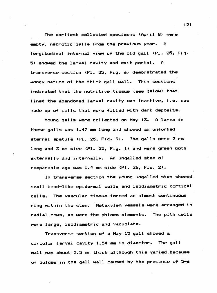
121
The earliest collected specimens (April 8) were
empty, necrotic galls from the previous year. A
longitudinal internal view of the old gall (P1. 25. Fig.
5) showed the larval cavity and exit portal. A
transverse section (P1. 25, Fig. 6) demonstrated the
woody nature a-f the thick gall wall. Thin sections
indicated that the nutritive tissue (see below) that
lined the abandoned larval cavity was inactive, i.e. was
made up of cells that were filled with dark deposits.
Young galls were collected on May 13. A larva in
these galls was 1.47 mm long and showed an un-forked
sternal spatula (P1. 25, Fig. 9). The galls were 2 cm
long and 3 mm wide (P1. 25, Fig. 1) and were green both
externally and internally. An ungalled stem of
comparable age was 1.4 mm wide (P1. 26, Fig. 2).
In transverse section the young ungalled stem showed
small bead-like epidermal cells and isodiametric cortical
cells. The vascular tissue formed an almost continuous
ring within the stem.. Metaxylem vessels were arranged in
radial rows, as were the phloem elements. The pith cells
were large, isodiametric and vacuolate.
Transverse section of a May 13 gall showed a
circular larval cavity 1.54 mm in diameter. The gall
wall was about 0.5 mm thick although this varied because
a-f bulges in the gall wall caused by the presence of 5-6

122
large vascular bundles in the cortex. These bundles were
lea-f traces, a fact made clear by the presence of smaller
vascular bundles nearer the cavity (P1.. 26, Fig. 3).
The cuticle, epidermis, and cortex in the gall wall
were similar to their counterparts in ungalled stems.
The vascular tissue, however, was dispersed as small
circular bundles.. The pith was hollow in the gall. The
cells lining the larval cavity were undamaged. This
suggested that the cavity was not the result of
burrowing. The nutritive cells were pith cell
derivatives and contained dark deposits (P1. 26, Fig.4).
There were, however, lightly stained vacuolate nutritive
cells in small patches (P1. 26, Fig. 5).
On June 5 the gall was 2.5 cm long and 4.5 mm wide..
The vascular bundles had increased in width such that the
interfasicular spaces (i.e. spaces between bundles) were
only 4-12 cells wide. The bundles were no longer
circular, but were strips or very shallow semicircles
with radial rows of elements. The bundles were about 23
cell layers (0.7 mm) in from the cavity epidermis..
Fifteen layers of cortical-like cells that were large,
vacuolate, and lightly stained occurred between the
bundles and the cavity.
The nutritive cells resembled those cells in the 4-5
underlying layers. All were vacuolate with peripheral

123
cytoplasms, a large nucleus and nucleolus. Many of these
cells contained dark deposits. There were patches,
however, of deposit-free nutritive cells (P1 26, Fig.
6).
By July 15 the gall was 3.5 cm long and 0.7 cm wide
(P1. 25, Fig. 2). It had become woody. A gall from July
24, when cut transversely, showed a larval cavity that
was 2.3 mm in diameter. The gall wall was 1.82 mm wide.
A larva at this time was 1.89 mm long and had an unforked
sternal spatula.
On July 24 half of the gall cortical cells were
filled with dark deposits. The vascular ring was
continous around the gall (P1. 26, Fig. 7), and radial
files of secondary xylem contributed 0.3 mm to the wall
width. Inside the ring of vascular tissue were 12-16
layers of sclerids. To the inside of the sclerids were
3-4 layers of cells that contained dark deposits.
Resting on these cells were the fairly large, lightly
stained nutritive cells (P1. 26, Fig. 8), each of which
showed a nucleus, nucleolus and many small vacuoles in a
granular cytoplasm. The lightly stained nutritive layer
was continuous around the cavity -- it was not found in
patches as previously observed.
On August 12 a larva was 2. 1 mm long and showed an
un-Forked sternal spatula. The gall wall was 2.94 mm wide

124
of which about 1.26 mm was secondary xylem (P1. 26, Fig.
9). Air pockets had developed in the cortex and most all
of the cortical cells contained dark deposits. The cells
of the 4-5 layers encircling the larval cavity had become
unusual in appearance (P1. 27, Fig. 2). Many were
partially filled with a lightly stained amorphous
(non-granular) material. This material frequently
accumulated on the larval cavity side of the cell. The
nutritive cells resembled those underlying them. A few
patches a-f 4-5 collapsed nutritive cells were seen.
On August 19 the gall's "beak" (distal end) had
begun to turn yellow (P1. 25, Fig. 3), and the larva was
noticeably larger (P1. 25, Figs. 7 and 8).
By September 21 a larva was 5.6 mm long and had a
forked sternal spatula. By September 21 the gall's beak
had turned brown (P1. 25, Fig. 4). The nutritive cells
were either empty, contained dark deposits (P1. 27, Fig.
1), or in some spots, contained a small amount of
granular cytoplasm and a nucleus and nucleolus.
Many galls were green on October 16 and contained a
3 mm long larva. The tissue lining the larval cavity was
yel low-green -
When an ungalled stem from May 13 was stained for
tannins (P1. 27, Fig. 3) one saw dark brown deposits in
the epdiermal cells, in some cortical cells, in some of

125
the vascular elements, and in scattered pith cells.
Tannins in galls (May 13 and June 5) occurred in
scattered cortical cells. Some nutritive cells also
stained positively, but there were patches that stained
negatively (corresponding to the patches of lighter cells
in Toluidine Blue stained material) (P1. 27, Fig. 4).
Most nutritive cells in late July had brown
deposits. On September 21 the amorphous deposit material
in the nutritive cells did not stain as tannins, but
several nutritive cells did contain tannins (P1. 27, Fig.
5).
Summary of Cecidomyiid Galls
1) Classification. The midge galls were of
different complexities. The poplar bud gall and hawthorn
fold gall showed abbreviated sclerid zones. Because both
showed enriched nutritive cells for at least a short
period, I classified them as simple prosoplasmas. The
other midge galls were complex prasoplasmas primarily
because of their extensive scierid zones.
The leaf gall on serviceberry with its false central
chamber caused more reorganization and disruption of host
plant tissues, and showed a more complex design than did
any of the other midge galls. In this respect it was
comparable to cynipid galls (see next section).

126
2) Nutritive layer dynamics and tannins. With the
exception of the willow gall see below), the nutritive
cells in these galls were cytoplasmically richest when
the galls were very young. By the time the larva was
half to fully grown, the cells upon which it Fed were
vacuolate and contained tannins. Generally, however, the
tannins in gall tissues were not as prevalent as in the
cells of the ungalled leaf. If cytoplasmic richness was
positively correlated with nutritional quality, then I
conclude that the best diet was available to the early
instars. The larvae did not maintain the cytoplasmically
rich diet through time.
The number of collapsed nutritive cells tended to
increase in the midge galls as the season progressed.
This was most likely due to damage by the larvae. Never,
however, was there evidence a-f torn cells. Apparently
the larvae crushed but did not tear or cut through the
tissue as they fed. On the other hand, the following
observations suggested that the larvae subsisted
partially on nutritive cell secretions: The presence of
debris on the surface of nutritive cells in older
serviceberry galls, the pitted walls of the nutritive
cells in mature hawthorn leaf galls and snowberry leaf
roll galls, the abundance of resins in the poplar bud
gall, and the accumulation toward the cavity of amorphous

127
material in the willow stem beaked gall nutritive cells.
These observations suggested that nutritive cells
produced and secreted materials, or simply facilitated
the passage of materials through the wall to the larvae.
The willow gall was interesting in that as the gall
matured, the patches of lightly stained nutritive cells
increased in size. Galls in late summer (August 21) also
showed nutritive tissues with large amorphous deposits in
many of the cells. The deposited material was not
tanniferous. It perhaps was nutritious (i.e.
carbohydrates, lipids). Given these observations one
would suggest that the nutritive cells in the willow gall
improved as the season progressed.
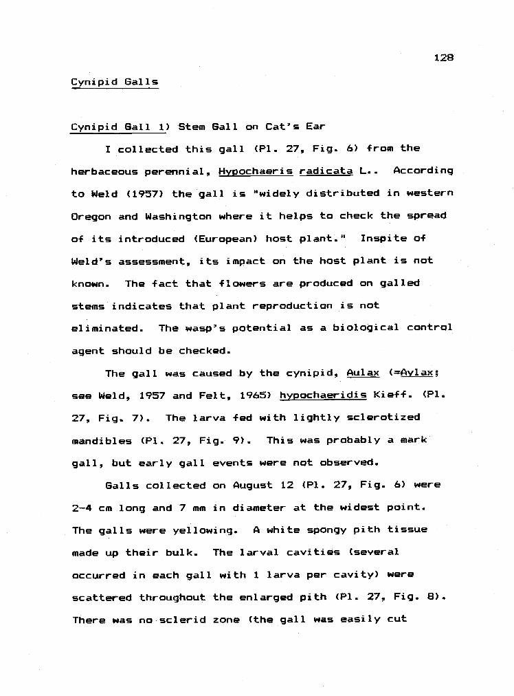
Cynipid Galls
Cynipid Gall 1) Stem Gall on Cat's Ear
I collected this gall (P1. 27, Fig. 6) from the
herbaceous perennial, Hypochaeris radicata L.. According
to Weld (1957) the gall is "widely distributed in western
Oregon and Washington where it helps to check the spread
of its introduced (European) host plant." Inspite of
Weld's assessment, its impact on the host plant is not
known. The fact that flowers are produced on galled
stems indicates that plant reproduction is not
eliminated. The wasp's potential as a biological control
agent should be checked.
The gall was caused by the cynipid, Aulax (Avlax;
see Weld, 1957 and Felt, 1965) hypochaeridis Kieff. (P1.
27, Fig.. 7). The larva fed with lightly sclerotized
mandibles (P1. 27, Fig. 9). This was probably a mark
gall, but early gall events were not observed.
Galls collected on August 12 (P1. 27, Fig. 6) were
2-4 cm long and 7 mm in diameter at the widest point.
The galls were yellowing. A white spongy pith tissue
made up their bulk. The larval cavities (several
occurred in each gall with 1 larva per cavity) were
scattered throughout the enlarged pith (P1. 27, Fig. 8).
There was no sclerid zone (the gall was easily cut
128
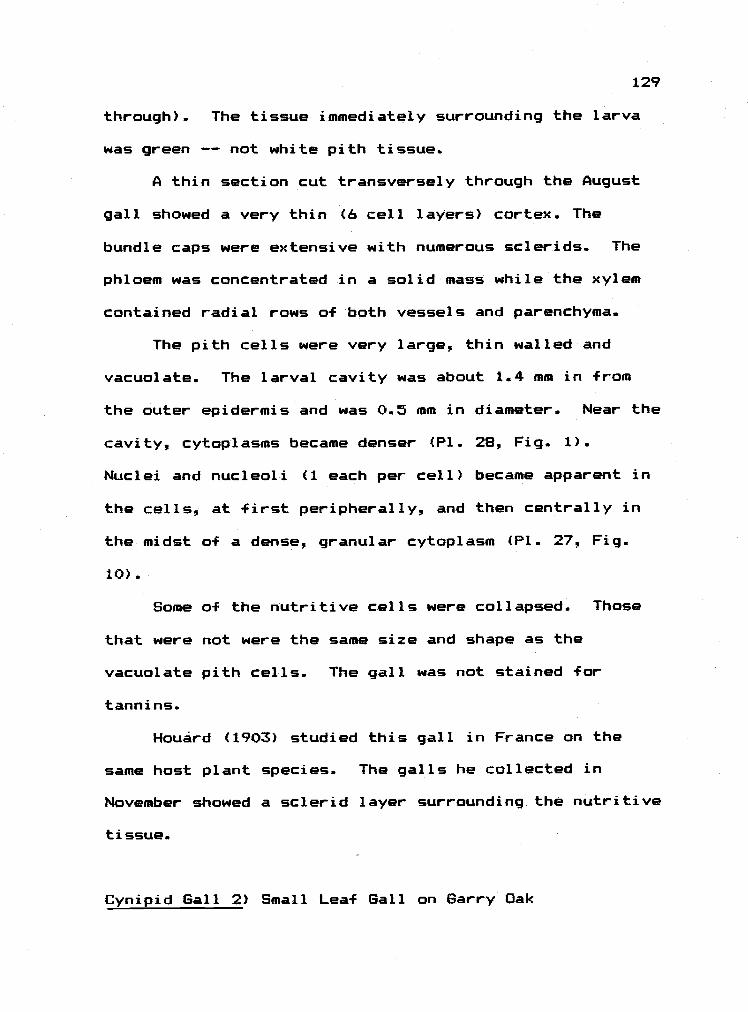
129
through). The tissue immediately surrounding the larva
was green -- not white pith tissue.
A thin section cut transversely through the August
gall showed a very thin (6 cell layers) cortex. The
bundle caps were extensive with numerous scierids. The
phloem was concentrated in a solid mass while the xylem
contained radial rows of both vessels and parenchyma.
The pith cells were very large, thin walled and
vacuolate. The larval cavity was about 1.4 mm in from
the outer epidermis and was 0.5 mm in diameter. Near the
cavity, cytoplasms became denser (P1. 28, Fig. 1).
Nuclei and nucleoli (1 each per cell) became apparent in
the cells, at first peripherally, and then centrally in
the midst of a dense, granular cytoplasm (P1. 27. Fig.
10)
Some of the nutritive cells were collapsed. Those
that were not were the same size and shape as the
vacuolate pith cells. The gall was not stained f or
tanni ns.
Houárd (1903) studied this gall in France on the
same host plant species. The galls he collected in
November showed a sclerid layer surrounding the nutritive
tissue.
Cynipid Gall 2) Small Leaf Gall on Barry Oak

The gall (P1. 28, fig. 2) was collected on young
leaves of Quercus qarryana Dougl. It was found only once
(May 6). The gall occurred on the upper surface of very
young leaves. It was red and spherical with a nipple at
its top. It was not associated with a major vein, and
was 2.38 mm in diameter. The gall was caused by an
undescribed cynipid.
The oak leaf that bore the gall showed a
meristematic mesaphyll. In longitudinal section the gall
was set deeply in the young spongy layer, but the lower
leaf surface remained undisturbed by the gall. The leaf
veins ran from the ungalled lamina to the base of the
gall, but no vascular strands were seen in the gall wall.
The wall was 0.63 mm wide and anatomically was divided
into an outer and inner gall wall. The outer wall was
0.21 mm wide (7 cell layers) and was made up of thin
walled, tangentially elongate, vacuolate cells primarily
derived from palisade parenchyma. The outer and inner
gall walls were separated at the base of the gall by a
single cell layer of pitted, densely cytoplasmic cells.
The inner wall was made up of 7 cell layers (P1. 28,
Fig. 3). The most peripheral were relatively small, half
vacuolate, half densely cytoplasmic. Each contained a
small nucleus and nucleolus and many crystalline
granules. Nearer the cavity, the cells and their nuclei
130

131
and nucleoli became larger (the cells were uninucleate
and the nuclei were uninucleolate) (P1. 28. Fig. 4).. The
cytoplasms become avacuolate, and densely granular (P1.
28, Fig. 5).
The nucleolus in the nutritive cells that lined the
cavity were either very large or dispersed, and the
nuclei showed many finger-like projections.. The long
axes of the nutritive cells were perpendicular to the
gall surface, but the cells were not regularly
palisade-like in alignment. Collapsed or vacuolate
nutritive cells occurred in patches. The larval cavity
was 1.26 mm in diameter. The gall was not stained for
tannins.
Cynipid Gall 3) Bullet Gall on Garry Oak
This gall was collected from Quercus oarryana Dougl.
and was caused by Andricus sponqiolus Sill. (according to
Houard (1940) the wasp was . quercus-californicus var.
sponqiolus Sill.) (P1. 28, Fig. 9). Felt (1965)
described the gall as "globose, clustered, many-cells,
buff colored, diameter 1 to 3 inches on Oregon oak, gall
wasp." Houard (1940) gave a more complete description a-f
the external and internal appearance of the gall (see his
page 121).
This is one of the largest insect-caused galls in

132
North America. Although diameters as small as 1.5 cm
were common these small galls were frequently in
clusters with galls that were 9 cm long and 6.5 cm wide.
Regardless of size, the galls resembled small to large
green apples. They were yellowish green when young and
turned brown to black with age. When sliced through,
young galls showed a white, pulpy internal tissue. The
galls were detachable stem galls, and the stem swelled at
the point of attachment (P1. 28, Fig. 6).
Free-hand sections suggested that the gall arose
from the cambial region of the stem and ruptured the
cortex and epidermis of the stem. Thus, this was most
likely a mark gall. Several larval cavities occurred in
each gall (1 larva per cavity) (P1. 28, Fig. 7) and the
cavities were grouped into what became a hardened mass at
the center of the gall (P1. 28, Fig. 8).
On May 6 the gall was small (1.7 cm long, and 1 cm
in diameter at the widest point). The circular larval
cavity was 0.38 mm in diameter. A larva was 0.40 mm in
diameter -
The cells that made up the white pulp in the gall
were large, vacuolate and parenchymatous. They contained
a prominent nucleus and nucleolus in a peripheral band of
granular cytoplasm. The pulpy tissue was traversed by
provascular strands that showed small, elongate, densely

133
cytoplasmic, undifferentiated cells.
Near the larval cavity the pulp cells became
smaller, and had fewer large vacuoles. The enriched
nutritive tissue was several cells thick. These cells
were filled with a lightly stained cytoplasm and had a
greatly enlarged nucleolus (P1. 29, Fig. 3). Many were
elongate with their long axis perpendicular to the cavity
(P1. 29, Fig. 2). The cells that actually lined the
larval cavity showed a similar cytoplasm but had no
nucleus or nucleolus.
A week later (May 13,) the larval cavity was 0.91 mm
in diameter. The pulp cells were larger than earlier.
The further from the larval cavity, the larger were the
cells (some were 0.15 mm long). The amount of peripheral
cytoplasm had decreased, and the central vacuole had
enlarged. The vascular strands remained mostly
undifferentiated.
In July, most all of the cells in the nutritive
region, including those that lined the cavity, had
developed vacuoles (P1. 29, Fig.. 4). The cytoplasm in
the nutritive cells stained lightly, was of fine-grain
consistency, and held large nuclei (1 per cell, sometimes
lobed) with very large nucleoli. The nucleoli frequently
contained small to large vacuole-like spaces in their
centers. Occasionally very small "satellit&' nucleoli

134
occurred in the nuclei.
A layer o-F lightly stained fine-grained material
covered the surface of the nutritive cells that lined the
cavity. The presence of recognizable cell parts in this
material (e.g. an intact nucleolus) indicates that it was
in part the ruptured contents of the nutritive cells.
At this time, the midgut of a larva was filled with
a very lightly stained material (more lightly stained
than nutritive cell cytoplasm) with dark droplets
scattered throughout. No solid cell material (wall,
nucleoli) was seen in the gut.
By June 16 a larval cavity was 2.7 mm in diameter.
Larvae were about 2.5 mm in diameter. Hand dissections
of the gall indicated that the larval cavities were
embedded in woody tissue that was very difficult to slice
through. The pulp of the gall showed macroscopic air
spaces throughout. By July 15 the galls were yellowish
green and were 4-6 cm in diameter, 4-9 cm long (P1. 28,
Figs 6). The fibers that ran from the stem to the
hardened mass of capsules were seen in an older gall that
was collected October 9 (P1. 28, Fig. B).
A small gall collected on July 24 that contained
larval cavities with diameters of 0.5-0.7 mm was stained
for tannins. The cells in the nutritive region (May 6)
stained poorly, while those of the pulp contained

135
peripheral contents and centrally placed droplets that
stained dark brown or black (P1. 29 Fig. 5). The
ruptured cell contents on the surface of the nutritive
cells stained darkly (P1. 29, Fig. 7). A transverse
section of the larval gut also showed dark droplets in an
unstained matrix.
Cynipid Gall 4) Rose Tip Gall
This acrocecidium was collected by Dr. Andrew
Moldenke in Corvallis, Oregon from Rosa nutkana Presl..
Of the galls on R. nutkana, those of Diplolepis
oreqonensis (Beut.) (holotype from Corvallis) most
closely resembled the collected specimens (Weld, 1957).
This was a bud gall (Felt, 1965), and was probably formed
as a mark gall.
On August 12 the gall was mature (2 cm long, 1.4 cm
in diameter at widest point). The gall was
spindle-shaped with its tapered point at the distal end
of the gall (P1. 29, Fig. 6). The gall surface showed 2
to 4 overlapping expanded stipular bases with an axillary
bud at the apex a-f each.
The gall contained several larvae, each in a
separate cavity. Some of the galls showed browning of
the tissue surrounding the larval cavities. A cavity was
0.9 mm in diameter (P1. 29, Fig. 8). Most of the cells
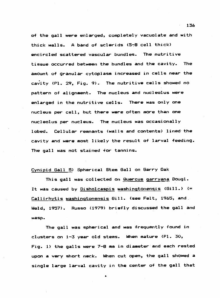
136
a-f the gall were enlarged, completely vacuolate and with
thick walls. A band of sclerids (5-8 cell thick)
encircled scattered vascular bundles. The nutritive
tissue occurred between the bundles and the cavity. The
amount of granular cytoplasm increased in cells near the
cavity (PL. 29, Fig. 9). The nutritive cells showed no
pattern o-f alignment. The nucleus and nucleolus were
enlarged in the nutritive cells.. There was only one
nucleus per cell, but there were often more than one
nucleolus per nucleus. The nucleus was occasionally
lobed. Cellular remnants (walls and contents) lined the
cavity and were most likely the result of larval feeding.
The gall was not stained for tannins.
Cynipid Gall 5) Spherical Stem Gall on Barry Oak
This gall was collected on Quercus qarryana Daugl.
It was caused by Disholcaspis washinqtonensis (Gill.) (
Callirhytis washintonensis Gill. (see Felt, 1965, and
Weld, 1957). Russo (1979) briefly discussed the gall and
wasp -
The gall was spherical and was frequently found in
clusters on 1-3 year old stems. When mature (P1. 30,
Fig. 1) the galls were 7-B mm in diameter and each rested
upon a very short neck. When cut open, the gall showed a
single large larval cavity in the center of the gall that
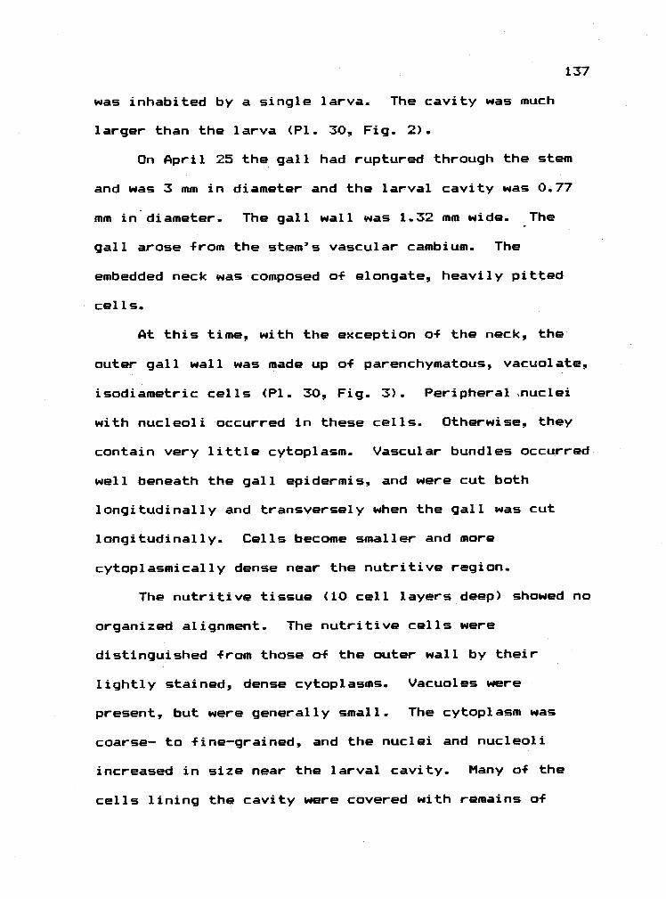
137
was inhabited by a single larva. The cavity was much
larger than the larva (P1. 30, Fig. 2).
On April 25 the gall had ruptured through the stem
and was 3 mm in diameter and the larval cavity was 0.77
mm in diameter. The gall wall was 1.32 mm wide. The
gall arose from the stem's vascular cambium. The
embedded neck was composed of elongate, heavily pitted
cells.
At this time, with the exception of the neck, the
outer gall wall was made up of parenchymatous, vacuolate,
isodiametric cells (P1. 30, Fig. 3). Peripheral nuc1ei
with nucleoli occurred in these cells. Otherwise they
contain very little cytoplasm. Vascular bundles occurred
well beneath the gall epidermis, and were cut both
longitudinally and transversely when the gall was cut
longitudinally. Cells become smaller and more
cytoplasmically dense near the nutritive region.
The nutritive tissue (10 cell layers deep) showed no
organized alignment. The nutritive cells were
distinguished from those of the outer wall by their
lightly stained, dense cytoplasms. Vacuoles were
present, but were generally small. The cytoplasm was
coarse- to fine-grained, and the nuclei and nucleoli
increased in size near the larval cavity. Many of the
cells lining the cavity were covered with remains of

138
ruptured nutritive cells.
On June 5 the larval cavity was 2.8 mm in diameter,
and the gall wall was 1.4 mm thick (P1. 30, Fig. 4). The
cells immediately outside the nutritive region had
developed porous, thick secondary walls.
The cells of the nutritive region were vacuolated
with a strip of cytoplasm peripherally. Nuclei with
nucleoli were present in some of the cells, but were not
as prevalent as earlier. The nutritive cells lining the
cavity were covered with a thin layer of amorphous
material.
On May 20 the nutritive cells did not contain
tannins. Most of the cells of the outer gall wall had
peripheral tannin deposits (black stain). The same
description held for material from June 5 with the
exception that the ruptured contents of the nutritive
cells stained positively.
Cynipid Gall 6) Mossy Rose Gall
This gall was collected from the introduced Rosa
eglanteria L. My observations indicated that females
oviposited in buds that had opened only slightly in
mid-spring. The galls generally arose from a twig, but
small galls were observed on sepals, petals, and leaflet
midveins and petioles. Apparently most organs in a young
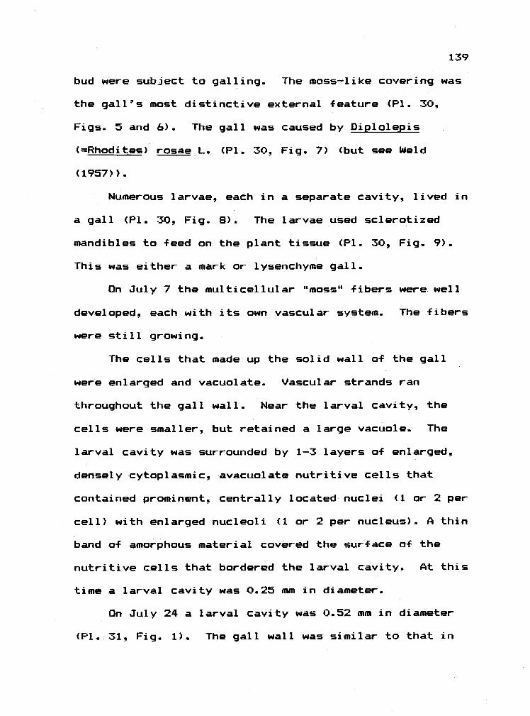
139
bud were subject to galling. The moss-like covering was
the gall's most distinctive external feature (P1. 30,
Figs. 5 and 6). The gall was caused by Diplolepis
(Rhodites) rosae L. (P1. 30.. Fig. 7) (but see Weld
(1957)).
Numerous larvae, each in a separate cavity, lived in
a gall (P1. 30, Fig. 8). The larvae used scierotized
mandibles to feed on the plant tissue (P1. 30, Fig. 9).
This was either a mark or lysenchyme gall.
On July 7 the multicellular "moss" fibers were well
developed, each with its own vascular system. The fibers
were still growing.
The cells that made up the solid wall of the gall
were enlarged and vacuolate. Vascular strands ran
throughout the gall wall. Near the larval cavity, the
cells were smaller, but retained a large vacuole. The
larval cavity was surrounded by 1-3 layers of enlarged,
densely cytoplasmic, avacuolate nutritive cells that
contained prominent, centrally located nuclei (1 or 2 per
cell) with enlarged nucleoli (1 or 2 per nucleus). A thin
band of amorphous material covered the surface of the
nutritive cells that bordered the larval cavity. At this
time a larval cavity was 0.25 mm in diameter.
On July 24 a larval cavity was 0.52 mm in diameter
(P1.31, Fig. 1). The gall wall was similar to that in

144)
July 7 material (P1. 31, Fig. 2). The cells were mostly
vacuolate, but as the nutritive cells were approached a
rich cytoplasm took the place of a large central vacuole,
and the cells began to enlarge and bulge toward the
cavity (P1. 31, Fig. 3).
At this time the nutritive cells were avacuolate or
contained only small vacuoles. The nucleus was lobed and
the nucleoli were enlarged.
By August 12 the larval cavity was 1.33 mm in
diameter. The galls themselves were 2-4 cm in diameter.
The mossu fibers were mature. Near the nutritive tissue
(P1. 31, Fig. 4), cells showed a thick band of peripheral
cytoplasm. The lobed nucleus with enlarged nucleolus
also increased in size near the larval cavity. The cells
bordering the cavity were cytoplasmically dense (P1. 31,
Fig. 5) and contained few vacuoles. The nucleus in these
cells was dispersed and frequently had no nucleolus. The
nutritive cells show no particular alignment or shape.
On September 21 the 1t055fl was brownish green. On
average the galls were 2-3 cm in diameter. A larval
cavity was 1.96 mm in diameter. A band of sclerids (4-5
cells thick) occurred 5 cell layers in from the solid
surface. The band encircled the larval cavity and ran
through, or on either side of the vasuclar bundles.
As in younger galls, the cytoplasmic density

141
increased near the larval cavity (P1. 31, Fig. 6). The
nutritive tissue was made up of enlarged, elongate cells
that contained numerous small vacuoles in an otherwise
lightly stained granular cytoplasm (P1. 31, Fig. 7).
Nuclei were present, and nucleoli (frequently 2 per
nucleus) were prominent, but were not as enlarged as
earlier. Some of the cells bordering the cavity
contained no nucleus. There was a thick film of ruptured
contents over the surface of the nutritive cells that
lined the cavity.
On October 9 the galls were difficult to slice
through. Large white larvae were still present in the
cavities. The wall of the larval cavity was woody and
cream-colored. Galls collected on December 20 still
contained larvae.
Galls from July 7 that were stained for tannins
showed that many o-f the cells in the "moss" fibers were
tannin-rich (black cytoplasmic stain), as were many cells
in the outer gall wall. The cells near the nutritive
tissue were free of tanning. The nutritive cells
contained some tannins (stain light brown).
A similar pattern occurred in galls from July 24
(P1. 31, Fig. 8), August 12 (P1. 32, Fig. 1) and
September 21 (P1. 32, Fig. 2). The older the gall, the
more common were tannin-rich cells in the outer gall wall

142
and mossy -fibers, and the less darkly stained were the
nutritive cells.
A larva on July 7 was 0.77 mm long. By August 12 a
larva was 4.34 mm long. Many of the September larvae
(lactophenol-cleared, and mounted in Hoyer's) show dark
granules in the mid gut that were not seen in larvae
collected earlier in the season.
Cynipid Gall 7) Speckled Oak Apple on Garry Oak
This gall (P1. 32, Fig. 4) was collected from
Quercus qarryana Dougi.. It was caused by agamic
(=asexual) females of Besbicus (=Cynips) mirabilis var.
mirabilis (Kinsey) Weld (P1. 32, Fig. 7). According to
Kinsey (1930), pupae occur in mid-September in the gal1s
and adults emerge in October - November. Kinsey (1930)
also described the external and internal macroscopic
appearance of the gall.
This was a mark gall that ruptured the cortex and
epidermis of one of the major veins (most frequently the
midvein) on the underside of the leaf (P1. 32, Fig. 3).
Each gall contained a single centrally placed larval
cavity that held a single larva.. The cavity was within a
capsule (see below). The spherical gall showed a short
neck at the leaf - gall junction. Very young galls were
covered with a dense pile of white hairs. As the gall

143
grew this pile was lost and was replaced by fuzz that in
turn fell off so that mature galls were glabrous and
shiny.
The young gall was solid, but as the gall matured
the outer gall wall became separated from the
centrally-placed larval capsule. The outer wall or shell
of a mature gall was thin (the flexibility of stiff wax
paper -- if the wall was very fragile, the gall had
probably been invaded by an inquiline). The central
capsule was suspended in the center of the gall by fibers
that radiated from it to the outer gall wall. There was
a concentrated group of these fibers that rans from the
capsule to the point of gall attachment on the leaf (P1.
32, Fig. 5). The larva lived within the central capsules
(P1.. 32, Fig. 6) and fed with lightly scierotized
mandibles (P1. 32, Fig. 8).
On May 31 the young gall was 1.54 mm in diameter.
The gall wall was solid and was 0.56 mm thick. The
larval cavity was 0.36 mm in diameter. The neck of the
gall was deeply embedded within the midvein (P1. 33, Fig.
1). The gall arose from elements near the vascular
cambium (P1.. 33, Fig. 3). Vascular strands occurred
along both sides of the neck and up through the outer
gall wall, and most cells within these strands were
densely cytopl asmi c.

144
At this time, the outer gall wall, was made up of
vacuolate cells.. Nuclei were visible in these cells.
Toward the center of the gall was a band of thick walled
cells (6-7 cells thick) that encircled the nutritive
tissue and constituted an early "protective" zone..
Most of the nutritive cells were arranged in radial
rows around the cavity.. The cells were recent products
of cell division. Only those lining the cavity were
enlarged and densely cytoplasmic. These showed a
prominent nucleus and nucleolus, and a dense granular
cytoplasm (no vacuoles). The nutritive cells that lined
the cavity were covered with amorphous material.
On June 3 and 11 the galls were still pubescent and
red to pink. They were solid, with a light brown capsule
at their center. Green succulent tissue surrounded the
capsule.
By June 17 the pink gall was h96 mm in diameter,
the larval cavity was 0.31 mm in diameter, and the gall
wall was 0.87 mm thick. Many of the cells of the outer
gall wall were elongate and radiated out from the central
gall capsule.. These were the forerunners of the fibers
that suspended the capsule in a mature gall.. As yet,
they had not separated..
The radiating cells abutted the "protective" zone
(P1. 33, Fig. 4). The nutritive tissue was inside this

145
zone and the transition between the two was abrupt. The
nutritive cells closest to the "protective" zone
(furthest from the larva) were small, flat, and densely
cytoplasmic with few vacuoles (P1. 33 Fig. 5). Near the
cavity the cells swelled, and showed larger nuclei and
nucleoli (usually one of each per cell).
By June 25 the gall was 3.72 mm in diameter. It was
covered with a white pubescence. The larval cavity was
0.49 mm in diameter, and the gall wall was 1.40 mm thick.
The internal fibers had begun to separate so that the
gall wall had a spongy consistency. The outer gall wall
(the shell) was 9-10 cell layers thick.
On July 10 the galls were about 1 cm in diameter.
The central capsule was 1 mm in diameter. The larval
cavity was 0.30 mm in diameter (Frontispiece). The
fibers were long and separated. Interestingly., lightly
stained cytoplasm (the same color as in the nutritive
cells) occurred between some of these "protective" cells.
Apparently there was an invasive growth of
protoplast-like nutritive cells into the "protective"
zone -
With the exception of some of the large nutritive
cells bordering the cavity, all showed a prominent
nucleus (usually unlobed) and nucleolus (larger nearer
the cavity). Many of the largest cells contained

146
numerous small vacuoles.
By August 12 the galls were yellowish green and were
2-3 cm in diameter. The outer shell was firm, but still
fleshy. The larval cavity was 0.30 mm in diameter. The
long fibers had reached their full length (P1. 33, Fig.
6). The cells of the "protective" zone were being
replaced by thin-walled densely cytoplasmic cells that
contained numerous small vacuoles, but otherwise
resembled the nutritive cells. Cell wall remnants of the
early protective zone were lodged between these new
metabolically active cells. Because of this invasive
growth, the nutritive tissue had almost doubled in
thickness since the previous collection.
Nutritive cells nearer the cavity showed lobed,
enlarged nuclei and enlarged nucleoli. Many bordering
the cavity showed a dispersed nucleus with no nucleolus
or nucleus (P1. 33, Fig. 7)..
On September 22 some of the largest galls were
brown. The central capsule was suspended on brown
fibers, and could be separated easily from the gall and
fibers. The larval cavity was 3.78 mm in diameter. A
band of thick walled cells (3 cell layers) occurred
between the nutritive region and constituted the sclerid
zone (not the same as the early "protective" zone).
The nutritive cells in older galls showed numerous

147
vacuoles and a granular cytoplasm. Remnants of thick
walls from the old "protective" zone pin-pointed the new
nutritive cells derived from the invasive growth. These
cells were as large but more vacuolate than the older
nutritive cells.
The nuclei in the older nutritive cells were very
large, dispersed, and granular (P1. 35, Fig. 7), while
most nucleoli had broken down completely (P1 33, Fig. 8).
Many of the cells neighboring the cavity had been torn,
and it was probably their contents that formed a film on
the surface of intact cells.
On June 3, June 17, June 25, and August 25 the
deposition of tannins was the same: The radiating fibers
were filled with a deep reddish brown deposit. Most gall
wall cells stained lightly, and the nutritive cells did
not stain. The material on their surfaces (P1. 35., Fig.
8) stained slighly.
On September 12, 1980 I collected mature leaves with
small spherical galls in clusters on the leaf's underside
at the midvein (P1. 33, Fig. 2). They were 3-5 mm in
diameter and were succulent within. A single larva was
in a single central cavity in each gall. According to
Kinsey (1930) these were stunted B. mirabilis galls, but
he states that "I have no explanation f or the stunted
development of so many of the growths." Instead of being

hollow with radiating fibers, the gall was solid.
Summary of Cynipid Galls
Classification. The cynipid galls I studied were
complex prosoplasmas because they, possessed sclerid zones
(usually late in gall development) , because they were
closed, and because they showed distinctly enriched and
multiple layers of nutritive cells. Most were complex in
design.
Nutritive tissue and its dynamics. As mentioned
earlier, when we think of nutritive cells in galls, the
tendency is to consider only cells in cynipid galls.
Although this bias has led to confusion, the association
is understandable; cytological characteristics of these
nutritive cells immediately set them apart (and some
would say, above) all others in any gall. Their
abundance is noteworthy. For example, at least half of
the substance of the small leaf gall on Garry oak was
nutritive tissue. The nuclear and nucleolar hypertrophy
in the nutritive cells of all 7 cynipid gall were
striking and most likely indicated an active metabolism.
From the herbaceous Cat's Ear, to the shrubby wild roses,
to the arborescent oaks -- regardless of genus or growth
form, the host plants produced very similar nutritive
cells around the larva.
148

149
Was the cytoplasmic richness of the nutritive cells
maintained through the season? In the two examples where
this was followed (mossy rose gall, and speckled oak
gall) the nutritive cells contained rich cytoplasm
through the last sampling. Vacuoles appeared in older
cells, but they did not completely replace the granular
cytoplasm.
Most interesting was the occurrence of the invasive
nutritive tissue in the spotted oak gall that replaced
the early "protective' zone. Although the larval cavity
remained about the same size (0.3-0.4 mm) throughout the
summer, in mid-summer the nutritive cell doubled in
thickness. The larval cavity remained about the same
size (0.3-0.4 mm) throughout the summer. Whether the
larva fed on the new nutritive tissue later in the season
was unknown.
3) Scierid zone. The sclerid zone usually developed
late in gall development. The speckled oak gall provided
an interesting variation. In this gall 2 layers o-f thick
walled cells developed. One developed after the other
had broken down. The nature of the early "protective"
layer should be examined more closely; even when
encircled by thick walled cells through most of its
development, the gall-former was heavily parasitized (Ken
West, personal communication).

150
4) Tannins The nutritive tissue in the galls that
were appropriately stained was usually tannin-poor.
Peripheral gall tissues were tannin-rich. Young
nutritive cells in the mossy rose gall stained slightly
positively for tannins but as they matured, they stained
more lightly.
The material that covered the surface of the
nutritive cells in many of the cynipid galls stained
fairly positively for tannins. This material may have
been ruptured cell contents and/or nutritive cell
secretions. Whether it was larval food was unknown. The
observation that larval midguts (oak bullet gall)
contained tannin droplets suggested that some tannins
were ingested.

Moth
Sawf lies
Nematode
Aphids
Thrips
TABLE Su-1
Sunnary Table: Reorganization of Host Plant Tissues,
and Nutritive Cell Characteristics in the Studied Galls
Gal 1-Former Reorganization Nutritive Cells
(Leaf Miners) (No reorganization of tissues.) (Nutritive cells do notdevelop; but see Hering,1951.)
Fungi Parenchymatous. No distinct tissue No long-lived, enrichedlayers. Derivation of cells easy to cells; but see Erlichtrace. and Erlich, 1971.
Simple proliferation of parenchymainto mined pith. Peripheral stem tissuesare mostly unaffected.
Galls with simple(parenchymatous) walls. Reorganizationmoderate. Derivation of cells in wallnot quickly traced.
Structurally simple root swelling.Little reorganization of root otherthan in immediate vicinity of giantcells.
Simple leaf curls or pouch galls withparenchymatous walls. Derivation ofcells in wall easy to trace.
Simple Darenchymatous leaf curl.
Much reorganization of needletissue. Gall wall parenchymatous.
Leaf epidermal cells strongly re-organized. Mesophyll cells muchless so. Parenchymatous wall.
Moderate to considerable reorganiza-tion and structural complexity.Sclerid zone in mature galls.
Considerable reorganization of leafor stem tissue. Derivation of cellsdifficult to trace. Sclerid zone inmature galls.
Callus-like. Not enriched.
Callus-like enations.Some short-termenri chment.
Enriched synctial nut-ritive cells. Long-termenrichment (Bird, 1975).
No obvious, enrichednutritive tissue.
Patches of palisade-likeenriched cells.
Storage-like nutritivecells.
Palisade-like or enlargedepidermal nutritive cells.Enriched when young.
Usually briefly enrichedwhen young. Apparentrapid deterioration,collapse. Sometimesheavily pitted.
Enriched through muchof gall development.Usually multi-layered.
151
Scale Moderate reorganization. Distinct No obvious, enrichedlayers in gall. Incomplete sclerid nutritive cells, butzone. see Parr; 1940.
Adelgid
En ophyoi ds
Cecidomylids
Cynipids

DISCUSSION - CONCLUSIONS
In this section I discuss four patterns that have
emerged from the results.. The first two patterns are
summarized in Table Su-1.
PATTERNS
1) Degree of Reorganization. There is a continuum
in the degree of disruption and the amount of
reorganization of host plant tissues caused by
gall-f ormers. My assessment o-f reorganization is based
upon three types of observations.. First, a completely
parenchymatous, layer-less gall wall indicates little
reorganization. If a distinct nutritive or scierid zone
is present, then the gall-former has caused noticeable
reorganization.. Second., the more difficult it is to
trace the derivation of cells in the gall, the greater
the reorganization. Third, the more complex the disign
of the gall, the greater the reorganization.
In contrast to the gall-f ormers, most leaf miners
cause no reorganization as they wander and -feed through
the leaf. There are, however, mines in which callus-like
enlarged cells partially fill the tunnel (Hering, 1951).
Pith flecks in wood caused by cambial miners provide an
excellent example of this phenomenon (Greene, 1914). In
152
153
some instances the miners return to -Feed on these cells
and thus, by definition, these could be called (slightly
enriched) nutritive cells. In some mines, then, we see a
generalized host wound response to insect attack that is
initiated by, but is not -Further influenced by, the
insect. The wound response, while only infrequently used
by miners, is capitalized on by many gall-farmers.
The fungal galls I studied show minor
reorganization. All internal cell layers are made up of
enlarged mesophyll cells. Na differentiation occurs
beyond the parenchymatous state. Mechanical disruption
is minimal and instead, the fungus relies on chemical
disruption to elicit host cell responses. Invaded host
cell cytoplasm may be enriched in some cases (Erlich and
Erlich, 1971).
Galls caused by moths are glorified mines. A moth
larva bores from the apex (site of oviposition) into the
stem, and feeds on pith as it goes. The larva, however,
limits the length of the mine. The attacked host plant
produces callus-like tissue from the vascular cambium --
a response probably elicited both by mechanical damage
and salivary constituents. The older instar larva feeds
on this new nutritive tissue, and continues to elicit the
host's callus response. Thus the gall-forming moth larva
shows a dependence on and control over, callus

154
development. Interestingly, the gracilariids and
gelechiids, two families that contain most of the
gall-forming moths, also contain numerous leaf-mining
members. Wangberg (1976) has observed tephritid flies
that are both stem miners and gall-f ormers.
The sawfly galls are not merely swellings of healthy
organs, but instead, are detachable structures that show
same reorganization of host tissues. The disruption may
be great enough that the gall's derivation is not clear
(snawberry gall). The gall wall is homogenously
parenchymatous.. No scierid zone is produced. Sawfly
galls show nutritive cells that are more cytoplasmically
enriched than are those in moth galls -- an observation
that indicates that sawfly larvae influence the plant's
response to a slightly greater extent than do gall moths.
Nutritive tissue in sawfly galls consists of callus-like
enati ons.
The root-knot nematode (as well as the cyst
nematode, Heterodera sp.) behaves like a miner as it
tunnels into a root, but instead of wandering, it settles
and causes dramatic enrichment of the cells surrounding
its head. Thus the nematode strongly influences cells in
closest proximity to its mouth. The degree of cellular
alteration decreases greatly outside of the giant cells
zone. There is some hypertrophy and hyperplasia in a few

I 55
cell layers surrounding the giant cells, but there is
little differentiation beyond the parenchymatous state.
The disruption of the stele is primarily of a mechanical
nature (i.e. elements crushed or pushed aside by giant
cells) - Thus, the arena of strict control in a nematade
gall appears to be fairly restricted.
Bird (1975) has stated that the giant cells "undergo
a cycle of growth that is directly related to the
physiological age of the nematode." The metabolic
activity a-f the syncytium peaks "around the time the
nematade has started to lay eggs" and then decline sets
in. This is the best indication we have that maintenance
of the nutritive cells is tightly linked to the
physiology of the gall-former.
Cecidogenetic stylet-bearing insects (scales.
thrips, aphids, adelgids) possess the power to disrupt
host tissue, but the disruptions are simple
parenchymatous swellings, leaf curls, or out-pocketings.
In only the scale gall is there differentiation of a
scerid zone, and it is incomplete. The thrips gall is
the only one that shows an enriched, but discontinuous,
nutritive epidermis. The adelgid gall show considerable
reorganization a-f needle tissue and a storage-like
nutritive zone. The aphid galls show no readily
distinguishable nutritive zone. No sc]erid zone is

156
produced. These organisms do not shape host tissue
responses to the same extent as do the midges and wasps.
The mite galls also show only a parenchymatous cell
wall. These galls, however, have a continuous layer of
enriched epidermal nutritive cells for at least a short
time. Thus, somewhat like the nematode galls, the mites
cause marked metabolic disruption in the cells with which
they are in intimate contact, but the impact and
influence drops off quickly with distance from the mites.
Cecidomyiid galls have a scierid zone in the wall
that encircles the larva(e) -- a zone that is not found
in the ungalled leaf. With the possible exception of the
cecid galls on dryland shrubs (Appendix 1), however, the
nutritive cells in the galls do nat remain enriched
through larval maturation. Additionally some of these
galls cause only moderate disruptions of the host organ
(leaf curl, swollen bud, leaf fold), while others (e.g.
dryland shrub cecid galls and the service berry gall)
show considerable tissue realignment in the leaf and
stem. All in all, the influence exerted by midge larvae
(as indicated by the depth and degree a-f disruption) is
second only to that exerted by cynipids. The tephritid
gall on rabbitbrush (Appendix 1) is similar in complexity
to the midge galls.
The complex structure of most cynipid galls is

157
undoubtedly the product of strong, continuous influence
by the larva. All show a definite scierid zone, and the
nutritive tissue is always heavily enriched through much
of the life of the gall.
It should be stressed that a gall-farmer's ability
to produce a complex or simple gall has little bearing on
the success of the gall-former. Gall-forming nematodes
and crowngall bacteria, for example, occur globally and
show numerous races, i.e. are successful, but cause
structurally simple galls. We also know little about
the relationship between complexity of the structure and
intimacy a-f the interactants. The relationship between
the crown gall bacterium and its host cell, + or example,
appears to be very intimate (a matter of gene splicing),
and yet, as already said, this is a structurally simple
gall.
2) Nutritive Cells. In many instances the diet of
gall-forming arthropads apparently deteriorates as
indicated by vacuolization) before or during periods of
greatest consumption. There is no rejuvenation of the
feeding cells in these galls. In sawfly and mite galls
the nutritive tissue is formed by the mother before
progeny begin to feed. The deutogyne (mites) or young
instar (sawf lies) enjoys a better diet than do the mid-

158
to late-season gal]. inhabitants. There are exceptions to
this pattern.. The big bud gall of filbert, for example,
shows cytoplasmically rich nutritive cells for some time
after progeny are produced. More exceptions may come to
light as more types of gall are studied.
Most midge galls also display a deterioration of the
nutritive region through the season. The early instars
enjoy the cytoplasmically richest diet. The beaked
willow gall may provide an exception in that changes in
the nutritive tissue fairly late in the season suggest an
enri chmènt.
The cynipid galls that were followed through much of
the season also showed a deterioration of the nutritive
tissue. The process however appeared slower than in
other types of galls. Even the last samples taken showed
nutritive cells with considerable granular cytoplasm.
The results indicate that cynipids can maintain their
diet in an active state for longer than most other
gal 1-forming arthropods.
The degree of enrichment and the longevity of the
nutritive cells in various galls are summarized in Table
Su-1.. Generally there is a positive correlation between
the amount of reorganization and the enrichment and
longevity of nutritive tissues. The nematode and some
cecidomyiid galls. however, run against the trend.

159
Nematode galls show little reorganization but have
enriched, long-live nutritive cells, while some
cecidomyiid galls (e.g. the hawthorn, snowberry poplar,
and service berry galls) show only briefly enriched
nutritive cells but a distinct sclerid zone.
The discussion of diet highlights an important
point: cynipids are riot the representative gall-f ormers
they are freqently assumed to be. To be sure, they are,
to the human eye, consummate cecidozoans, and their
abilities distinguish them from most other groups. In
fact, however, no single gall-forming group encompasses
all cecidogenetic methods and results. There are common
features of many galls and gall-f ormers (Epilogue), but
the uniqueness of each should also be appreciated.
Lastly, I suggest that the deterioration of diet
seen in many galls as they mature may in part be
compensated for by improved protection. As many galls
mature their walls thicken and, in many instances, are
reinforced. This undoubtedly provides protection.
3) The Aphid Galls.. The aphids and their galls are
problematic. In no instances was I able to determine
precisely in which cells the stem mother or her progeny
feed. In no instance did I find cells in the gall wall
that were clearly nutritive. The fact that aphid stylets
16')
allow for sub-epidermal foraging, coupled with the fact
that the depth of foraging most likely increases through
instars1 indicates that a wide variety of cell types
found throughout the gall wall are potential food. The
potential feeding area is further increased by the
aphid's movement within the gall.
Based on these possibilities, if an enriched
nutritive area were to occur in an aphid gall, one would
expect it to encircle the gall chamber, to lie well
within the gall wall, and to consist of numerous cell
layers. Nothing exactly like this is seen in the galls.
The closest match to the prediction is the vascular
tissue which lies within the gall wall, and which retains
cytaplasmically dense members throughout the season. Its
candidacy as a food tissue is unconfirmed because stylet
tracks to it were never observed. Additionally, stylet
length was often well in excess of that needed to
penetrate the vascular tissue.
Interestingly, the known examples of nutritive cells
in homopteran galls occur in instances in which the
gall-former is sedentary (e.g. adelgid stem mother and
oak pit scale). Meandering may shape the gall (sensu
Soysen-Jensen, 1948), but at the same time it may prevent
(or obviate the need for) establishment and maintenance
of enriched nutritive cells.

161
This dissertation points out the uniqueness of aphid
and scale galls. They deserve the thorough study that
midge and wasp galls have received.
4) Tannins. The galls that were appropriately
stained showed generally low levels of tannins in the
nutritive cells. Younger galls usually contained lower
amounts of tannins. In some cases, particularly in the
cynipid galls, even older galls showed very low levels of
tannins in the nutritive cells.
Even when tannins occur in the food tissue, they
appear to occur in lower quantities than Found in the
ungalled leaf.. Thus the development of a nutritive
layer, regardless of degree of enrichment or longevity,
assures the gall-forming arthropods of a diet that, in
terms of tannin content, is superior to a leafy diet.
Frequently the tannin-poor nutritive tissue is
surrounded by a tannin-rich gall wall. This is
particularly true of the cynipid galls. D. H. Janzen
(personal communication) proposes that tannins in the
gall's periphery protect the cecidozoan and the nutritive
layer from exploiters. Such an interpretation, however,
fails to explain the high rates of invasion experienced
by so many galls -- particularly by the cynipid galls.

EPILOGUE: CHARACTERISTICS OF THE GALL-FORMING HABIT
Gall--foriners are internal plant parasites. Viewed
in this way they can be compared to other internal plant
parasites (e.g. lea-f miners) and, interestingly enough,
to internal parasites of animals. A thorough comparison
will not be made here, but as gall-forming -features are
discussed, occasional comparison to other life habits
will be made. Not far behind any discussion of
parasitism come questions about host defense. These will
be considered briefly in closing.
As internal plant parasites, gall-f ormers show the
following features:
1) Host Plant Specificity.. The physiological
tracking of host plant response and biochemistry required
for formation of a gall undoubtedly forces a strict
monophagous habit upon the gall-former, i.e. the
gall-former attacks a single host plant genus, or
frequently, a single species. In fact in some instances
I suspect that denies of gall-f ormers become restricted to
individual long-lived host plants, much like scale
insects on coni-fer needles (Edmunds and Aistad., 1978).
I take as indicative c-f specificity the occurrence
162
163
of numerous species or strains a-f a gall-former. In many
cases strains have been detected, but their specificity
is unknown. For example, the taxonomy of crown gall
bacteria is "confused" and numerous strains are known to
exist (Lippincott and Lippincott. 1975). The degree to
which such strains are host specific, however, is not
clear. The study of host specificity within the genus
Rhizobium (a genus very closely related to Aqrobacterium)
has also focused on bacterial strains and
cross-inoculation groups of plants. In this case each
nodulating strain is considered host specific
particularly "when one takes account of effectiveness of
the association" (Vincent, 1974).
According to Brian (1976), "Plasmadiophora
brassicae....and most of the rusts, powdery mildews, and
downy mildews" (all with gall-forming members) show the
strictest host specificity among the plant pathogenic
-Fungi. Later he states, "...the pathogen species. ..may
consist of physiologic races each restricted to a few
genotypes of a host species, as, for instance, in the
well studied case of Puccinia praminis f. sp. tritici on
wheat.
Nematodes in the genus tleloidogvne also show races,
each with a restricted host range. In fact, host
susceptibility has historically been used to determine

164
identity of the root-knot nematode (Jenkins and Taylor,
1967) -
Arthropod gall-f ormers are generally very host
specific, and in those instances in which a gall-former
is suspected of being oligophagous or polyphagous. the
possibility of cecidozoic races should be considered. As
biochemical coupling or intimacy between host and
parasite increases, so should specificity.
Thus, the gall-f ormers resemble other monophagous
groups all of which are parasitic. IZA very large
proportion of miners are monophaqous" (Hering, 1951).
The parasitic insects (fleas, lice, wasps) include some
highly host specific members (Price, 1980) as do the
zooparasitic helminths (Cameron. 1964; Whitfield, 1979),
and the entomoparasitic fungi (e.g. the Laboulbeniales;
Madelin, 1966).
2) Morphological Modifications of the Gall-Former.
From the fungi (naked fruiting bodies in some
gall-forming groups), to the cecidogenetic mites
(reduction in appendages), to the gall-forming insects
(reduction in appendages in cecidomyiid larvae (Mamaev,
1975) and cynipid larvae), there is a pattern of
reduction in parts. Do root-knot nematodes, crown gall
bacteria, and gall-forming homopterans show a similar

165
reduction?
The reductions seem to occur as the gall-former
becomes more sedentary and the gall more confining. Only
Mamaev (1975) has traced the reductions through a group
of gall-f ormers, and he states that they are
specializations (advancements). Interestingly., it is
often implied that the gall-forming fungi are primitive.
The leaf miners have also shown a general reduction
in appendages (Hering, 1951) as have the parasitic fleas
and lice (Rothschild and Clay, 1952). Many of the
ectoparasitic insects and zooparasitic helminths,
however, have developed elaborate mechanisms for holding
on to the moving hosts.
There are also internal modifications in many gall
organisms. For example, the midgut of cynipids and
root-knot nematodes is blind. According to Mamaev (1975)
the gut of gall-forming cecids is open, but simplified
(few if any blind side sacs), and in some cases excretion
only occurs before pupation. The eriophyoids, although
presumably equipped with an open digestive tract, do not
produce visible quantities o-F feces, and the suggestion
has been made that excreta is stored within the hemolymph
or used in eggshell construction (Jeppson et al, 1975).
The "biological harmfulness a-f contamination of the
gall by excretion" (Mani, 1964) is given as the possible

166
reason far closed gut characteristics. In this regard it
is of interest to note that gall aphids actively push
liquid droplets out of galls, and that saw-flies cut holes
in their galls through which they eject feces. Despite
these efforts, however, most aphid, math, sawfly, and
beetle galls frequently contain solid or fluid
excretions.
Most leaf miners leave a fecal trail behind them.
Many parasitic helminths are gut-less and absorb
nutrients through the body wall. I have never seen this
mode of nutrient uptake suggested f or the metazoan
gall-farmers, but Mamaev remarked upon a thin cuticle in
some gall-forming midge larvae. I do not know when
excretion occurs in helminths.
Apparently then, storage of excreta occurs
frequently in sedentary or confined parasites. Mamaev
points out that the phenomenon of stored excreta may
simply indicate that very little excreta is produced,
i.e. that assimilation efficiency and diet quality are
very high. This suggests that gall-forming moths,
sawflys, and aphids, as well as most miners are poor
assimilators and/or take a low quality diet much of which
is unassimilable.
3) Gall Invaders.

167
A) Parasitoids and predators. Most all gall-forming
insects are attacked by parasitoids and/or predators
(Mani, 1964; Caltagirone, 1964). A possible exception:
according to A. F. Bird (personal communication) "the
gall probably does protect the nernatode." One wonders if
this protection occurs under uncultivated conditions?
The below-ground habit of mast nematodes may explain the
lack of insect parasitoids (do above-ground gall-forming
nematodes show parasites?), but subterraneanism should
not prevent below-ground parasites (helminths. bacteria,
fungi) from attacking the nematodes.
Predators are common in or on the periphery of some
eriphyoid galls (Jeppson et al, 1975). I have never read
of bacterial or fungal diseases (parasites) of
gall-forming arthrapcds, but one suspects that numerous
individuals, frequently in a single chamber, would
provide excellent opportunities for epidemics. I do not
know the extent to which gall-forming bacteria, slime
molds, and fungi are protected from parasitization (by
phages or other bacteria) or from predators.
Like gall-f ormers, leaf miners are heavily
parasitized (Hering, 1951; Nèedham, et al, 1928). I do
not know if by being in their host, parasites of animals
gain protection from hyperparasites, although if galls
and mines are again considered, hyperparasitoids

168
(parasitoids of the parasitoids that attack the
gall-f ormers) are common. In this case then, being
within the body of a host animal that is within a gall
does not afford complete protection. To what extent are
fleas and lice parasitized? It would be interesting to
compare rates of (hyper)parasitism on endo- and
ectoparasi tes.
Sall insects, like all other organisms, are attacked
because they are an available resource. But why are they
attacked so heavily? I suggest that in addition to
providing food, the gall offers another important
attraction. The microclimate of a gall is most likely
conducive to growth. Thus a parasite (or predator)
attacking and living in a gall feeds on a high protein
diet in an ameliorated climate. The same reasoning could
be extended one more trophic level to explain the high
rates of hyperparasitism in galls.
There are other locations on the plant that combine
these features of microclimate and diet. These include
the flowers, seeds, or seed heads. It would be
interesting to compare rates of invasion in fruits vs.
galls.
Based on the above, I would predict that parasitic
helmiriths and ectoparasitic insects would experience
parasitization because they are a resource, but not at

169
the intensity experienced by gall insects. These
creatures do not create a habitat with an improved
microclimate. Just as importantly, they provide no
richer a source of protein than does their animal host.
Thus instead of attacking the helminth or flea,
(hyper)parasite would do as well to attack the primary
animal host.
B) Inquilines. Inquilines are frequently found in
arthropod galls. These are organisms that invade the
gall while it is inhabited by the gall-former, but dci not
directly kill the gall-former. Some inquilines, however,
may kill the gall-former indirectly. Thrips, mite, and
cynipid galls provide good examples of gall types with a
rich inquiline -fauna. I am unaware of reports a-f
inquilines in homopteran galls.. Only a few inquilines
are known from leaf mines, but this may reflect lack of
observation (Hering, 1951).
Like parasites, inquilines are most likely attracted
by the gall microclimate, but unlike parasites, they are
attracted to the plant tissue or insect feces as a food
source rather than to the gall-former. Based an their
location in the gall, inquilines can be divided into 3
groups: 1) those that live in the gall or larval cavity
and feed on the nutritive tissue (or rarely, perhaps an

170
the gall-former's excreta), 2) those that feed in the
gall wall, and 3) those that feed in all tissues of the
gall. The last 2 groups, in many galls, feed in
tannin-rich tissues. Apparently the advantages of the
microclimate more than compensate for efforts expended to
contend with tannins
The only other instance in the insect literature in
which inquilinity is mentioned is in discussions of ant
nest inhabitants (myrmecophiles). "The number and
diversity of myrmecophilous arthropods are almost
incredible" (Wheeler, 1910). From the same reference,
The existence of this great number ofmyrmecophiles can be accounted for only on thesuppositon that ant nests have a strong attraction + orterrestrial arthropods. It is not difficult tounderstand how this can be the case since, in thefirst place, the nests are usually permanent abodesinhabited for months or years by successive broods ofants. Second, these nests have at all seasons aslightly higher temperature than the surrounding soil.Third, there is usually more or less refuse, food, oroffal, pupal exuviae and dead ants, at least in thesuperficial chambers. Fourth, the living larvae andpupae represent an abundant and highly nutritious foodsupply for any insect that can elude the watchfulnessof the ants.
The extended quote is provided to highlight the
similarities between galls and ant colonies. I do not
believe that the high incidence of parasitism and
inquilinity in these two particular life habits is
fortuitous. I suggest that the prerequisites for a high

171
incidence of invasion is an accumulation of energy and
protein sources in an ameliorated microclimate (elevated
or buffered temperature, high humidity). The higher rate
or incidence a-f invasion in ant nests (vs. galls) is
undoubtedly due to their longevity and to the ants' group
defense (the inquiline is also defended), and to the
philoprogenetive behaviors that may be deceived and
exploited by the myrmecophile -- characteristics not
exhibited by galls and gall-f ormers. Additionally the
social insects show greater active control of the
microclimate than do the gall insects.
The proposed pattern, however, is immediately called
to question by Wheeler's (1910) observation that some
colonial insects have fewer inquilines than ants.
Colonial wasps, bees, and termites "have their nest mates
and parasites. These, however, are far less numerous
than the myrmecophiles." Wilson (1971) amends Wheeler's
statement by pointing out that the higher termites have a
large and complex symbiont (inquiline) fauna. Wilson
further explains that colonies c-f bees and wasps do not
host as many guests because only those insects with
specializations "for arboreal life and a preference for
dark, tight spaces, perhaps together with a tolerance for
higher temperatures and lower humidity" become successful
guests. He suggests that "opportunities for invasion"

172
are lower in these above ground colonies. Using his
suggestion one would predict higher invasion rates in
below-ground galls than above-ground -- a prediction that
probably does not hold.. I suggest that climate control
may not be as great in an arboreal colony and, in part,
for this reason, one may find fewer inquilines here than
in terrestrial colonies.
Comparisons between invasion of an insect colony and
a gall should continue. This is a first attempt. It
will require more information on inquilinity in galls,
and an appreciation for the difference in definitions
(Wilson's inquilinity does not equal Mani's), as well as
systems. The chance for a more accurate
conceptualization and modelling of inquilinity will
result..
4) Site of Attack, and Host Response. Gall-farmers
attack meristematic regions of the host plant or tissues
that have undergone only slightly differentiation. The
tissue must be responsive. It must be capable of
dedifferentiation to a meristematic state.
As far as I can determine very few other parasites
have this requirement or cause similar host reactions of
hypertrophy and hyperplasia. Leaf miners frequently mine
mature leaves. Callus development, if it occurs in

173
mines, results from the activity of either meristematic
tissue associated with the vascular tissue or from
dedifferentiation of "fully developed tissue" (Hering,
1951).
The only example of a hyperplastic reaction in an
animal host to a parasite is provided by Dawes (1963).
He observed that "minute cytoplasmic blebs appear on the
free surfaces of the epithelial cells of the bile ducts
over extensive areas" early in the infection of mice,
rats, sheep, and other hosts by the liver fluke, Fasciola
hepatica. The reaction occurs in the tissues immediately
surrounding the parasite. He goes on to say that,
mitotic figures indicate that (the affected) cells aremultiplying.. This becomes more evident with theproduction of minute invaqinations of the epithelium,possibly the earliest indications of enormousoverdevelopment and folding which ensues.
The epithelium in later stages becomes "folded with
numerous crypt-like formations with crowded nuclei." As
a result of the inflammation the flukes are provided with
a "pasture" of tissue on which to feed. The hyperplastic
state lasts from 3-11 yearS.
The resemblance of the verbal and microphotographic
descriptions of this host reaction to that of a host
plant attacked by a gall insect is striking.. Differences
certainly exist between the 2 systems, but the

similarities invite further comparisons. For example,
how do the hyperplastic animal cells compare
ultrastructurally to nutritive cells?
5) Diet Enhancement. Most gall--Forming organisms
induce a cytological enrichment a-F the cells upon which
they -Feed. As seen in this study, the nutritive cells
remain enriched f or different lengths of time, depending
on the gall-former.
Bird (1961) used protein stains and cytophotometry
to observe that the giant cell cytoplasm in root knots
gave 4 times more extinction (i.e. a greater amount of
protein) than did "normal cells". According to Rapp and
Kirst (1974) "there is no distinct difference in the
content o-f carbohydrates based on dry weights" between
unga]led leaves, galled leaves, and galls of the
cecidomyiid, Mikiola fagi Htg. on Faaus silvatica L..
a fresh weight basis "the protein content of the gall
decreases compared with the quantity of protein in the
leaves. - - (and) two thirds of the soluble proteins are
located in the nutritive tissue of the gall."
Rapp and Kirst also showed that a gall collected on
July 12 contained nutritive tissue with 1.3 mg soluble
protein per gram -Fresh weight. An unga].led leaf at this
time contained 1.4 mg soluble protein per gram fresh
174

175
weiqht. Thus, although there is a concentration of
soluble proteins in the nutritive tissue, the larva, in
mid summer, was feeding on no better a diet than if it
had fed on an ungalled leaf. Perhaps earlier in the
season the nutritive tissue was of a higher soluble
protein content. How would the soluble protein contents
of nutritive tissue in a cynipid, thrips or eriophyoid
gall compare to the same in this cecid gall?
Rapp and Kirst also found that, on a dry weight
basis, whole galls between mid-June and early July
averaged 9.3 mg total protein per gram of tissue.
Leaves averaged 18.7 mg/g. Thus galls were 0.937.
protein on dry weight basis and leaves were 1.87%.
These last figures are at least an order of magnitude
lower than that given by Southwood (1973) as an average
percent protein (22%) on a dry weight basis a-f leaves of
eight different (mostly herbaceous) crop species.
Insect tissues on average are 50% protein an dry weight
basis. Thus if the figures of Rapp and Kirst are
correct, there is a very large disparity (a large
hurdle, sensu Southwaod) between total protein in the
gall and total protein in the cecid larva.
In spite of the figures, 1 predict that the young
nutritive tissues of mast galls, and the young-to-old
nutritive tissues of cynipid galls provide the gall

176
larva with a diet as rich in protein as any other organ
or tissue on or in the host plant. I also predict that
nutritive cells will be found comparable in protein
content to animal tissues. In this regard it is
interesting that there are eurytomid and torymid
parasites in cynipid galls that switch from feeding on
the host larva to nutritive tissue (Shorthouse. 1975).
Apparently nutritive tissue satisfies same of the
dietary requirements of these hyperparasites.
As far as I can determine, parasites a-f animals
(with the possible exception a-f Fasicola hepatica; see
above) cause no host response that can be interpreted as
an enrichment of food cell cytoplasm- Most animal
parasites either -feed as they wander through the
tissues, settle and -Feed on gut contents, or feed on
blood cells in the vascular system. Enrichment, if it
occurs at all, would most likely be seen where a
parasite settles to feed in a protein-poor region a-f the
host -- i-f such regions exist in an animal host.
6) Symbiants. Gut symbionts assist their host in
obtaining a full complement of nutrients -from a nutrient
poor diet. Buchner (1965) has the following to say
about the apiomorphine coccids, gall--farmers on
eucalypts in Australia:

177
Apparently the absence of symbionts is connectedwith the Fact that the apiomarphines are the onlycoccids which live in galls, and it is certainly noaccident that in the gall-forming Hymenoptera andDiptera or in mast of the insects which mine inleaves and stems. symbionts are always lacking.
Likewise gut symbionts "are not associated with the
genus Meloidogyne" (A. F. Bird personal communication) -
Mamaev (1975) predicts that intracellular sytnbionts may
occur in gall midge larvae. The evidence at the moment
suggests that both gall-4ormers and leaf miners require
few if any symbionts. The diet alone satisfies the
nutritional needs of larvae. Blood-feeding parasites
(lice, fleas, and certain mites) frequently require
symbiants -F or vitamin production even though the diet is
protein- and carbohydrate-rich.
7) Galls as Metabolic Sinks. Kirst and Rapp (1974)
showed that galls of Mikiola -Faqi on leaves of Faqus
silvatica L. began to accumulate 14C metabalites "3-5
minutes after 14CO2 application to the leaf tissue."
Translocation of metabolites to the gall occurred when
the host leaf or adjacent leaves were labelled.
Jankiewicz et al (1969) studied the sink properties
o-f oak apple galls on Quercus robur L. caused by Cynips
quercus-folii L.. Results were variable, but when 14CO2
was introduced to certain spots on the underside of a
178
galled lea-f, the gall, regardless of its position with
respect to the point o-F label application, was labelled
at 36 hours. Generally the major vein servicing the
gall showed heavy labelling. Additionally, the longer
the labelled assimilates were allowed to be transported
in the lea-F (up to 6 days), the greater was the
concentration of label in the larva and nutritive
tissue.
Both of the above reports used detached leaves or
branches. Thus they do not give an indication a-F how
galls would compete for metabolites against natural
sinks such as the roots.
Bird and Loveys (unpublished results discussed in
Bird, 1975), on the other hand, used whole tomato plants
with root knots.
In-f ected plants were allowed to photosynthesizein an atmosphere a-f 14CO2 and were le-ft in theglasshouse -for periods of time which varied -from twohours to five days be-Fore being harvested. Theuptake a-F the label into the nematode is slow and notsignificant over periods o-F two and four hours.However, over periods a-f from one to five days, therewas a much greater accumulation a-F translocatedphotosynthates in the galls than in adjacent tissues.After -five days exposure to 14CO2 f or instance, gallswith egg masses contained about six times as much 14Cas did adjacent roots and about half a-f this hadbecome incorporated into the eggs.
This report gives the clearest indication of the
sink properties a-F a gall and gall--Former that is

179
available.. Similar work with insect galls (on whole,
perhaps herbaceous plants) should be done.
I have never seen the suggestion that mines are
sinks. Other sedentary phytophagous insects, however,
establish sinks. For example, various authors have
shown that aphids, when feeding in groups on a plant,
can influence nutrient flow (Way, 1968; Kennedy and
Fosbrooke, 1973).
I do not know if animal parasites are considered
sinks to which blood and nutrients are shunted
preferentially. Again, sedentary parasites would be the
most likely ones to show this phenomenon.
8) Unusual Life Cycles and Reproduction. The
cynipids are the only Hymenoptera that show alternation
0+ generations. Both generations usually occur on the
same host plant species..
Many gall-forming aphids, like many other aphids,
show complex life cycles in which at least two species
(or genera) of host plants are attacked by different
generations.
Some of the gall-forming eriophyoid mites show
summer and winter forms, but these do not switch hosts,
nor do they form different galls. Parthenogenesis
occurs in the nematode genus Meloidogyne, but no host or

180
gall switching is known. Parthenogenesis is also known
in a few gall-forming cecidomyiids.
Parasites of animals frequently show very complex
life cycles that involve host switching and
parthenogenesis (Whitfield, 1979). The unusual
reproductive biologies of parasites are usually
interpreted as means of producing numerous offspring
that saturate the environment. In this regard, it is
unfortunate that there is no comprehensive review of
fecundity of gall--farmers.
9) Damage to Plant. See Appendix 3 + or a
discussion of this point.
HOW DOES THE PLANT DEFEND ITSELF AGAINST
GALL PARASITES?
Historically same cecidologists have considered the
gall itself as a form of plant protection in that it
walls off, contains, localizes the damage caused by the
insect. Otherwise, the reasoning goes, the insect would
cause considerable damage by moving through plant
tissues. The anatomy of a plant gall, however,
indicates both that the insect controls gall development
and that the gall is a drain on the plant. In fact,
others have suggested that galls represent a host
181
defensive response (callusing) that has been usurped by
the gall-former.. If a gall is viewed as detrimental to
the host plant then one should consider possible
defensive plant strategies.
Interestingly, there are no observations available
on the percentage of ovipositions or attempted
establishment by gall arthropods that fail. Evelyn
Westphal has studied host plant resistance to eriophyoid
attack,, but her results are not yet available. It may
well be that plant defense reactions early in attempted
gall establishment are an important deterrent to
successful attack. In another system, hypersensitive
reactions have been observed. The reactions prevent
root knot nematodes from parasitizing the host, and
occur early in the infection process (Paulson and
Webster, 1972)..
Gall formation requires meristematic or only
partially differentiated cells for successful
initiation. Thus a plant might defend itself by making
these tissues less available in time, in space, in
amounts, or in quality. Whitham (1979) has suggested
that prime galling sites on a host are in limited
numbers.
Gall formation requires a tight biochemical and
physiological coupling between host plant and
182
gall-former. The host plant species might realize some
short term protection by being able to vary the
biochemical pathways targeted and usurped by the
gall-former from plant to plant. (The crown gall system
has the greatest chance of elucidating such a
mechanism.) One would expect,, however, that variation
in the host plant would eventually be tracked by the
gal 1-former.
Lastly, we should consider the possibility that
rather than chemical control, host plants may rely on
biological control of gall-forming pests. The high
incidence of predation and parasitism undoubtedly
contributes a great deal to the control of gall-f ormers.
According to Stinner and Abrahamson (1979), parasitism
may also reduce the damage that a gall causes its host
plant. To be sure, a gall-former, parasitized or not.,
is a drain on the plant. Damage may be minimized.
however, if parasitoids are in some way actively
encouraged to attack the gall. In this regard, it is
interesting that some cynipid galls on oaks secrete a
nectar that attracks ants. It may be that the
predaceous ants protect the galls from parasitoids (Tom
Seibert, personal communication). (The development of
secretory cells in the gall tissues would be interesting
to follow.) Thus, by in some way directing the gall

183
tissues to secrete nectar, the gall-farmer may have
exploited a strategy that compromises any system used by
the plant to attrack parasitoids.

PLATE I
Figure 1. Trans. section through the agromyzid mine inthe leaf of Tellima grandiflorum (Pursh)Dougi... Pupa (arrow) rests in the mine cavity(MC). The cavity has been excavated throughthe spongy mesophyll.. (III/21/80).* lO5X.
Figure 2.. The pharyngeal skeleton of the agromyzid leafminer with mouth hooks (upper arrow) and theatrophied ventral branch (lower arrow) -(111/21/80). 85X.
Figure 3. The adult agromyzid that attacks T. cirandi-florum.. (IV/8/80). lOX.
Figure 4. Head capsule of Lithocolletis sp. that minesleaves of Gaultheria shallon Pursh.. Notebibbed labrum (arrow). (V/27/80). 55X.
Figure 5. Trans. section through leaf mine on j. shal-ion. The mine cavity (MC) has been excavatedthrough the palisade (arrow).. The larva (L)is seen in trans. section. (V/27/80). 70X.
Figure 6. Blotch leaf mine (arrow) on 6. shallon.(V/27/80). 2.5X.
Figure 7. Trans. section through mine in leaves a-fPopulus trichocarpa T. and 3. caused byPhyliocriistis sp.. The mine cavity (tIC) isexcavated through the upper epidermis(arrow). The larva (L) is seen in trans.section. (V/13/80).. 85X.
Figure 8. The stubby root knot gall (arrow) ontomato caused by Meloidocivne hacila Chitw.(Grhse. culture). 6X.
Figure 9. Long. section through galled tomato rootshowing numerous groups of nurse cells(arrow) near the center of the root. 12X.
Figure 10. A group a-F nurse cells (left arrow) thatare being fed upon by a female root-knotnematode (right arrow) in tomato roots. 290X.
* Date specimen was collected.
184

185

PLATE 2
Figure 1. Trans. section of Delphinium troiliifoliumGray stem galled by Puccinia rust. Theuredia, (left arrow) occur on galledside of stem, as does fungal mycelium inthe stem's hollow center (right arrow).Note enlargement of vascular bundles ongalled side of stem. (IV/8/80). 15X
Figure 2. An enlargement of the galled region oflarkspur stem. Uredia with urediospores(arrow) are apparent. Enlargedvascular bundle (V) is surrounded byenlarged, vacuolate stem cells. (IV/8/80).55X..
Figure 3. A galled leaf (arrow) of azalea attacked byExobasidium discoideum. Note chioroticcondition. (V/l/8O). 2X.
Figure 4. Trans. section through an unqalled azalealeaf. Palisade parenchyma (arrow) andspongy mesophyll layers are distinct.(V/l/80). 200X.
Figure 5. Trans. section through a galled leaf ofazalea. Fruiting structures seen on bothsurfaces (arrows). (V/l/8O). l3OX.
Figure 6. The free basidia (upper arrow) on thesurface c-f a galled azalea leaf. Lowerarrow points to intercellular mycelium.(V/I/SO). 320X.
Figure 7. Leaves of Populus trichocarpa galledby Taohrina aurea on upper (left arrow)and lower (right arrow) leaf surface.(VIII/12/8O). Cm scale.
Figure 8. Trans. section through an ungalled poplarleaf. Palisade (arrow) and spongy meso-phyll layers are distinct. (VIII/l2/80). 100X.
Figure 9. Trans. section through a galled poplar leaf.Palisade layer (arrow) is recognizable.(VIII/l2/8O). bOX.
186

PLATE 2 (Cont)
Figure lO. Lower leaf epidermis in poplar gall.. Asci(arrow) with spores are seen. (VIII/l2/8O)275X.
187

2
188
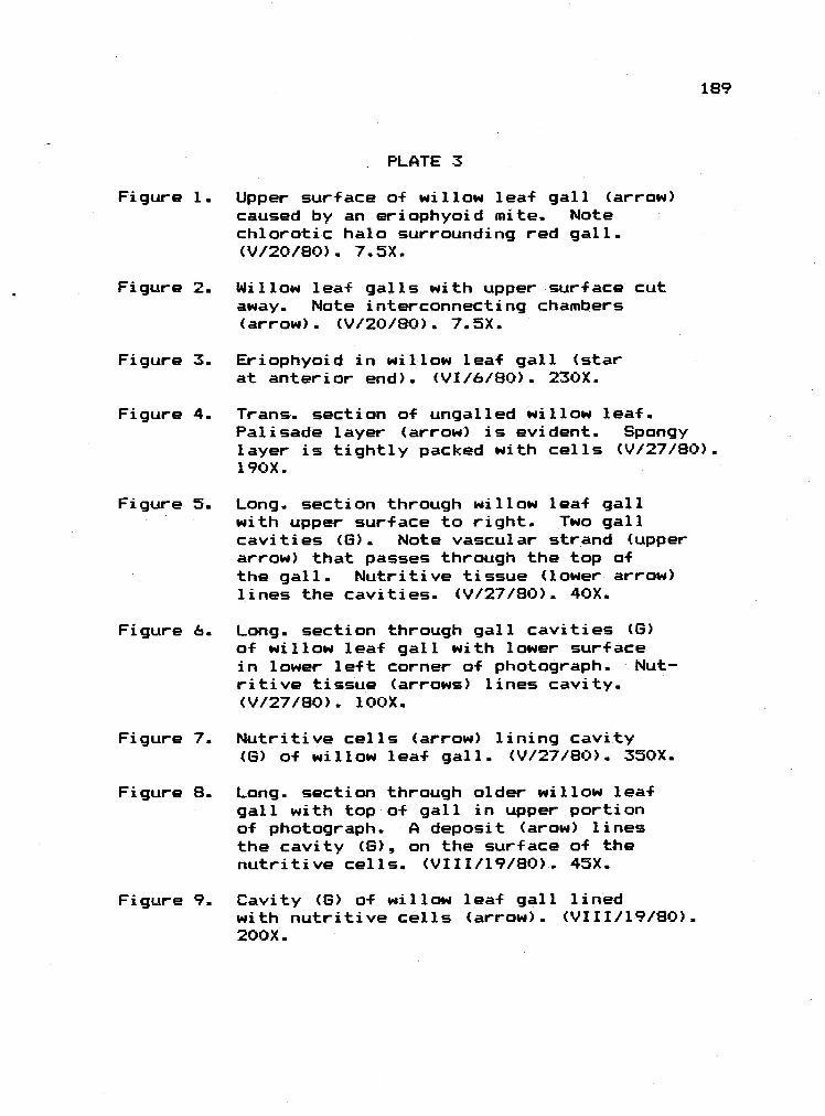
PLATE 3
Figure 1. Upper surface of willow leaf gall (arrow)caused by an eriophyoid mite. Notechiorotic halo surrounding red gall.(V/20/80). 7.SX.
Figure 2. Willow leaf galls with upper surface cutaway. Nate interconnecting chambers(arrow). (V/20/80). 7.5X.
Figure 3. Eriaphyoid in willow leaf gall (starat anterior end). (VI/6/80L. 230X.
Figure 4. Trans. section of ungalled willow leaf.Palisade layer (arrow) is evident. Spongylayer is tightly packed with cells (V/27/80).190X.
Figure . Long. section through willow leaf gallwith upper surface to right. Two gallcavities (6). Note vascular strand (upperarrow) that passes through the top ofthe gall. Nutritive tissue (lower arrow)lines the cavities. (V/27/8O). 40X..
Figure 6. Long. section through gall cavities (G)of willow leaf gall with lower surfacein lower left corner of photograph. Nut-ritive tissue (arrows) lines cavity.(V/27/8O). ]OOX.
Figure 7. Nutritive cells (arrow) lining cavity(6) of willow leaf gall. (V/27/8O). 350X.
Figure 8. Long. section through older willow leafgall with top of gall in upper portionof photograph. A deposit (arow) linesthe cavity (6), on the surface of thenutritive cells. (VIII/l9/8O). 45X.
Figure 9. Cavity (6) of willow leaf gall linedwith nutritive cells (arrow). (VIII/l9/8O).200X.
189
PLATE 3 (Cont.)
Figure 10. Cavity (G) of necrotic willow leaf gallwith vacuolate nutritive cells (arrow).(VIII/19/80). 185X.
190

3
191

PLATE 4
Figure 1. Wall of willow leaf gall stained fortannins. The gall cavity (G) is linedby lightly stained nutritive cells (arrow)..(VI/5/80). 250X.
Figure 2. Upper surface of leaf of Alnus rubra Bong.galled by Phytoptus laevis Nal. The arrowpoints to a single bead gall (VII/l5/80).Scale in mm.
Figure 3. Deutoqyne taken from alder bead gall.Star at anterior end. (V/6/80). 270X..
Figure 4. Mite taken from alder bead gall. Starat anterior end. (VIII/12/9O). 270X.
Figure 5. Long. cut-away view of alder bead gallwith upper surface to the left. Large,single gall cavity is seen (arrow)..(VIII/12/8O). 30X.
Figure 6. Trans.. cut-away view of alder bead gallwith cavity at arrow. Note cavity partitionin upper gall. (V/20/BO). 15X.
Figure 7. Long. section of young alder bead gallwith central gall cavity (8). (IV/22/80). 60X.
Figure 8.. Nutritive cells (arrow) of young alderbead gall. (IV/22/8O). 265X.
Figure 9. Cavity of alder bead gall with large,prominent nutritive cells (arrow) -(V/l3/80). lOOX..
Figure 10. Nutritive cells (arrows) that line thecavity a-f the alder bead gall. (VhS/GO).l7SX.
192

£61

PLATE 5
Figure 1. Trans. section through an ungalled alderleaf.. Palisade (arrow) and spongy layersare distinct.. (IV/22/80). 250X.
Figure 2. Older alder leaf with necrotic beadgalls (arrow). (IX/2l/80). Cm scale.
Figure 3. Long. section of mature alder bead gallwith large central cavity. Nutritivecells (some collapsed) line the cavity(arrow). (VII/16/8O). 25X.
Figure 4. Wall of alder bead gall showing partiallycollapsed nutritive cells (arrow) -(VII/l6/8O).. l4OX.
Figure 5. Wall of alder bead gall stained fortannins. Nutritive cells (arrow) stainfairly positively. (V/20/8O). 160X.
Figure 6.. Wall of alder bead gall stained fortannins. Nutritive cells (arrow) stainvery positively. (VII/24/80). 150X.
Figure 7. Leaf gall on Tilia caused by aneriophycid. (V/20/8O). 4.5X.
194

PLATE 5 (Cont..)
Figure 8. Eriophyoid mite from New York Tilialeaf gall. Star at anterior end.(V/26/80). 250X.
Figure 9. Trans. section of ungalled Tilia leaf.Palisade (arrow) and spongy mesophylilayers are distinct. (IV/30/80). 325X.
Figure 10. Long. section through young Tilia gallshowing central cavity (8) and portal hairs(arrow). (IV/7/79). 40X.
Figure 11. Wall of Tilia gall with nutritive cells(arrow) lining gall cavity. (IV/30/80). 250X.
Figure 12. Close-up of nutritive cells in very youngTilia gall. (IV/7/79). 550X.
Figure 13. Transition zone between ungalled Tilialeaf (to lower left o-f lower arrow) andgall wall (to upper right of lower arrow).Palisade layer is indicated by upper arrow.(IV/30/80). 200X.
195

S196

PLATE 6
Figure 1. Portal region of Tilia gall with numeroushairs (arrow).. The gall cavity is tothe right, outside of the photograph.(V/20/80). 75X.
Figure 2.. Wall of Tilia gall. Arrow points towardscavity and at vacuolate nutritive cells.(V/20/80). l3OX.
Figure 3. Wall of Tilia gall. Upper arrow lieswithin cavity and points to base o-Fcavity hairs. Large dark-filled ductsare seen (lower arrow). (VII/24/8O). l3OX.
Figure 4. Erineum gall (arrow) on the undersideof Garry oak leaf. (V/20/8O). 25X.
Figure 5.
Figure 6.
Eriophyoid mite taken from oak erineum..Star at anterior end. (VII/24/8O). 260X.
Trans. section of young oak leaf showingpalisade (arrow) and spongy mesophyll.(V/20/8O). l7SX.
Figure 7. Transition zone of oak erineum. Atleft of bundle, the leaf is ungalled,while to the right (arrow) of the bundle,the spongy inesophyll is denser, andhairs develop from both epidermises.(V/20/8O). l4OX.
Figure 8. Trans. sect through oak erineum showingdarkly stained lower epiderma]. cells(arrow) and hairs arising from lower epi-dermis. (VII/24/80). l25X.
Figure 9. Transition zone of oak erineum stainedfor tannins. Epidermal layers to theright of the vascular bundle (arrow)stain deeply. The hairs do not.(V/20/8O). lOX.
Figure 10. Tannin stain of oak erineum.. Upperepidermal cells (arrow) stain verydeeply. (V/20/80). 285X.
197

198

PLATE 7
Figure 1. Upper surface of eriophyoid gall (arrow)on leaflets of Fraxinus latifolia Benth..Several galls occur on each leaflet.(VIII/12/80). O.6X.
Figure 2. View of ash leaflet galls (arrow) from under-surface of leaflet.. (V/13/80). Scale in mm.
Figure 3. Long section through ash leaflet gall thathas been built over a large vein (arrow).Gall cavity (G) is seen. (VIII/l2/80). 20X.
Figure 4. Deutogyne from ash leaflet gall.Star at anterior end. (V/20/GO).. 260X.
Figure 5. Trans. section of ungalled ash leaflet.Palisade (arrow) and spongy layers aredistinct. (V/13/80). 220X.
Figure 6. Long. section through ash gall showingtransition zone (lower arrow), gall.cavity (8), and nutritive cells liningcavity (upper arrow). (V/13/80). 70X.
Figure 7. Section through gall cavity of ash gallshowing nutritive cells (arrow) liningthe gall cavity. (V/l3/80). 190X.
Figure 8. Section through older cavity of ash gall(lower right corner) showing nutritivecells (arrow) lining the gall cavity.(V/13/80). 295X.
Figure 9. Eriophyoid gall on Tilia sp from NewYork. Cut away to show hairy interior.Leaf surface at arrow. (V/26/8O). l3X.
Figure 10. Long. section of New York Tilia gallshowing cavity (star) and enation (arrow) -(V/26/80). liOX.
199

7
200

PLATE 8
Figure 1.. Eriophycid gall on Rhus diversiloba T.and 6.. A group a-F galls (arrow) isseen on the upper leaflet surface.(V/31/80). 4X.
Figure 2..
Figure 3.
Figure 4.
Eriophyoid gall (arrow) on poison oakflowers. (VI/25/80). 4X.
Eriophyoid mite taken from poison oak gallon leaflets. (V/30/80). 250X.
Section through poison oak leaflet gallshowing transition between ungalled leaflet(bottom arrow) and gall (upper arrow).Most change occurs at large vein (middlearrow). (V/31/80L. 13X.
Figure 5.. Trans. section through ungalled poison oakleaflet. Palisade (arrow) and spongy mesa-phyll layers are distinct. (V/31/80). 150X.
Figure 6.. Trans.. section. of poison oak leaflet gallshowing enlarged nutritive cells on lowerleaflet surface (upper arrow), and recogniz-able palisade (lower arrow) and spongy mesa-phyll layers.. (V/3l/80). 350X.
Figure 7. Eriophyoid leaf gall o-f Populus tremuloidesMichx. on underside of the leaf (right arrow).(VI/l5/80). 0.4X.
Figure 8. Eriaphyoid mite taken from the leaf gallon trembling aspen Star at anterior end.(VI/15/80). 260X.
Figure 9. Close up of trembling aspen leaf gall. Gallis the circular, enated area (arrow) onunderside of leaf. (VI/l5/80). 15X.
Figure 10. Trans. section of unqalled tremblingaspen leaf. Palisade (arrow) and spongy meso-phyll layers are distinct. (VI/15/80). 200X.
201

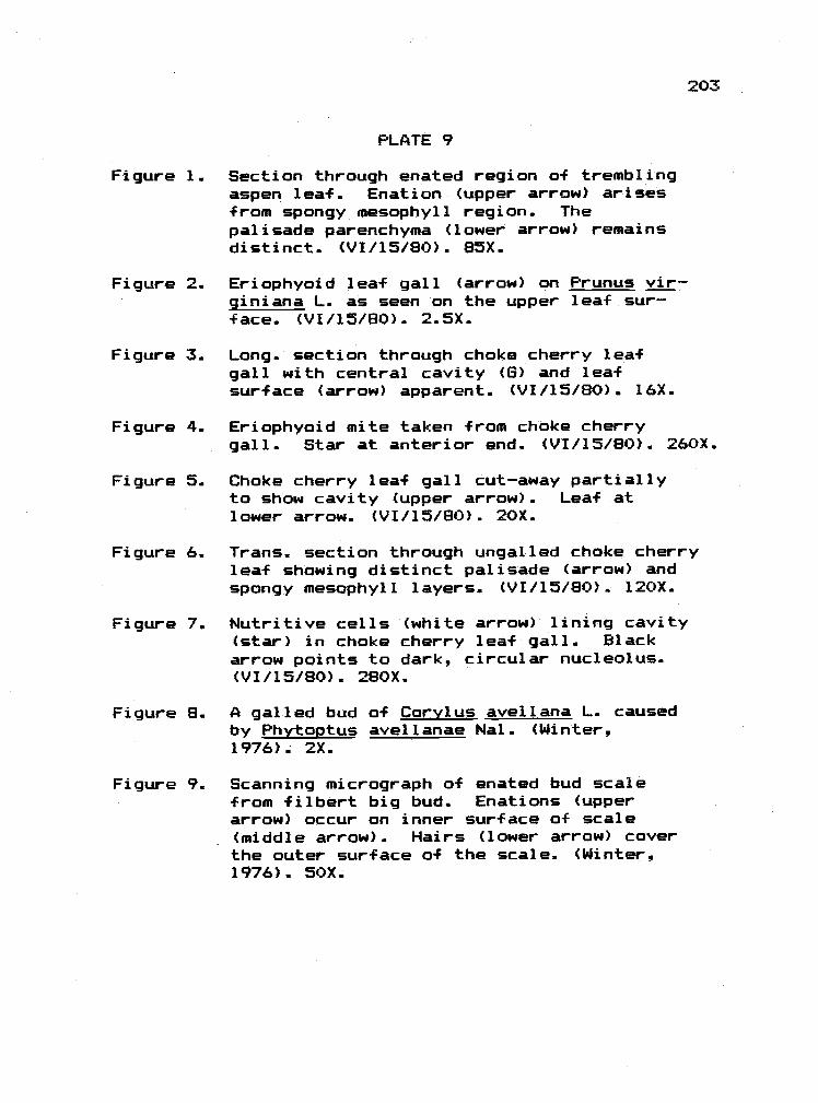
PLATE 9
Figure 1. Section through enated region of tremblingaspen leaf. Enation (upper arrow) arisesFrom spongy mesophyll region. Thepalisade parenchyma (lower arrow) remainsdistinct. (VI/15/80). 85X.
Figure 2. Eriophyoid leaf gall (arrow) on Prunus vir-giniana L. as seen on the upper leaf sur-f ace. (VI/15/8O). 2.5X.
Figure 3. Long. section through choke cherry leafgall with central cavity (5) and leafsurface (arrow) apparent. (VI/l5/80). 16X..
Figure 4. Eriophyoid mite taken from choke cherrygall. Star at anterior end. (VI/l5/80). 260X.
Figure 5. Choke cherry leaf gall cut-away partiallyto show cavity (upper arrow). Leaf atlower arrow. (VI/15/BO). 20X.
Figure 6. Trans. section through ungalled choke cherryleaf showing distinct palisade (arrow) andspongy mesophyll layers. (VI/15/BO). l2OX.
Figure 7.. Nutritive cells (white arrow) lining cavity(star) in choke cherry leaf gall. Blackarrow points to dark, circular nucleolus.(VI/15/80). 280X.
Figure 8. A galled bud of Corylus avellana L. causedby Phytoptus avellanae Nal. (Winter,1976). 2X.
Figure 9. Scanning micrograph of enated bud scalefrom filbert big bud. Enations (upperarrow) occur on inner surface of scale(middle arrow). Hairs (lower arrow) coverthe outer surface of the scale. (Winter,1976). SOX.
203

9204

PLATE 10
Figure 1. Trans. section through enated big bud scale.Base of enation is seen at Far left (leftarrow). Nutritive cells (left, middlearrows) cover surface of enation. At farright (right arrow), epidermal cells arenot nutritive, but are undergoing trans-ition to nutritive state. Mite (m) lieson scale surface. (Spring, 1976). l75X.
Figure 2. Nutritive cell from filbert big bud withprominent nucleus (Nc) and nucleolus (Nu).(Spring, 1976). 2000X.
Figure 3. Cytoplasm of nutritive cell from filbertbig bud showing vacuoles (Va), rough endo-plasmic reticulum (rER), mitochondria(arrows) and cell wall (CW). (Spring, 1976).8700X.
Figure 4.. Enation in filbert big bud stained f ortannins. Nutritive layer (arrow) stainspoorly while subsurface layers (TA) stainpositively. (Spring, 1976). l6OX.
Figure 5. Phytoptus avellana Nal.. Star atanterior end. (Spring, 1976). 260X.
205

206

PLATE 1].
Figure 1. Galls (arrows) on Quercus qarryana causedby Asterolecanium sp.. (IV/2/8O). 3X.
Figure 2. Trans. section of oak stem with pit scale.Proliferation of gall tissue is at arrow.(IV/2/80).. 20X.
Figure 3. Trans. section through oak pit gall, andthrough scale insect (black arrow). Thesclerenchyma tissue that underlies theinsect is seen at the white arrow.(IV/2/9O). 55X
Figure 4. Trans. section through oak pit scale gallshowing insect (left arrow) , sclerenchyma(right arrow), and the cortical tissue under-lying the insect. (IV/2/8O). 85X.
Figure 5. Stem gall on Penstemon peckii Pennellcaused by Caloptilia murt+eltella (Bsk.)Note the exit window (arrow). (VIII/28/SO).Scale in mm.
Figure 6. C. murtfeltella larva. (VI/6/80). 6X.
Figure 7. Trans. section of ungalled stem of Peck'spenstemon showing pith (P), vasculartissue (V). and cortex (C). (VI/6/8O). lOOX.
Figure 8. Trans. section through a galled stem ofPeck's penstemon showing nutritive tissue(arrow) that lines the larval cavity (star).Vascular tissue (V) is also seen. (VI/6/80).75X.
Figure 9. Trans section through nutritive tissueof Peck's penstemon (lower arrow) that linesthe larval cavity (star). Vascular tissue(upper arrow) is also seen. (VI/6/80). 140X.
207
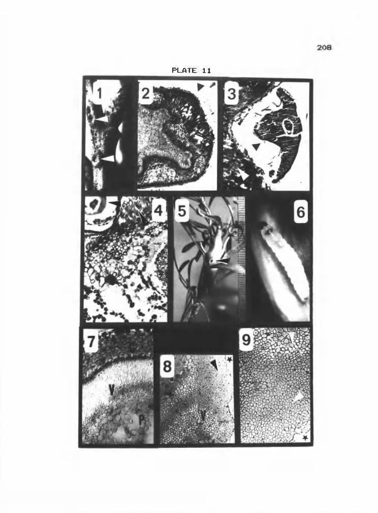
wi,
ti

PLATE 12
Figure 1. Sawfly gall (arrow) on Symphoricarposalbus var. laevigatus Fern.. (V/6/8O). l.5X.
Figure 2. Head capsule of sawfly larva taken fromsnowberry gall. Sclerotized mandibles(arrow) are seen. (IV/25/80). 60X.
Figure 3. Trans. section through wall of snowberrygall showing enatial-like development ofnutritive tissue (arrow) that lines thelarval cavity (G). (IV/29/80). BOX.
Figure 4. Close up of nutritive tissue of snawberrygall. (V/6/80).. 300X.
Figure 5. Trans. section throughgall showing nutritivebeen fed upon (arrow),the larval cavity (6).
Figure 6. Trans. section throughgall showing nutritivecavity (G). (V/l3/BO).
wall of snowberrytissue that hasand that borders(V/6/80). 60X.
wall of snawberrytissue (arrow) andbOX.
Figure 7. Close up of nutritive tissue (arrow) thatlines the gall cavity (6) in thesnowber-ry gall. WI/S/SO). 200X.
Figure 8. Trans. section through old snowberrygall showing nutritive tissue (leftarrow), gall cavity (6), and frass pellet(right arrow). (VI/25/8O). 50X.
209

12
210

Figure 3.
Figure 4.
Figure 5.
PLATE 13
Figure 1. Adult Caloptilia murfeltella (Bsk.).(VII/3l/80). 4.5X.
Figure 2. Very young willow leaf gall (arrow) causedby Pontania sp.. Upper leaf surface.(IV/26/80). Scale in mm.
Adult Pontariia sp. (From OSU insectCollection) -
Manidibles of C.. murfeltella larva.(VII/3l/80). Scale in mm.
Very young willow leaf gall (arrow) causedby Pontania sp.. Lower leaf surface.(IV/26/8O). 200X.
Figure 6. Mature willow leaf gall (arrow) on under-side of leaf (VII/lO/80). Scale in mm.
211

PLATE 13 (Cont.)
Figure 7. Cut-away view of willow leaf gall showinglarval cavity (6) and larva (arrow)(V/22/B0). 8X.
Figure 8. Section through young willow leaf gallshowing leaf (star), upper gall wall(upper arrow), lower gall wall (lowerarrow), and gall cavity (6) (IV/26/80).25X.
Figure 9. Upper wall of young willow leaf gallshowing nutritive tissue (arrow) thatlines the gall cavity (6). (IV/26/80).l5OX.
Figure 10. Lower wall a-f young willow leaf gallshowing nutritive tissue (arrow) thatlines the gall cavity (6). (IV/26/80).bOX.
Figure 11. Section showing the junction betweena willow leaf (star) and the willow leafgall (middle arrow). The leaf midvein(upper arrow) and gall cavity(lower arrow) are also seen. (IV/26/80).50X.
Figure 12. Section trough willow leaf gall showinggall cavity (13) and nutritive tissue(star). The lower surface of the gall isindicated by the arrow. (IX/22/80). lOX.
212


PLATE 14
Figure 1. Section through wall of willow leafgall showing gall cavity (6), nutritivetissue (upper arrow), outer wall cells(middle arrow) and outer epidermis(lower arrow). (IX/22/8O). 175X.
Figure 2. Section through willow leaf gall wallshowing enatial-like growth of nutritivetissue (right arrow), darkly stainedunderlying cells (left arrow), and gallcavity (star). (V/13/80). 85X.
Figure 3. Nutritive cells (arrow) in willow leafgall that line gall cavity (star). (V/l3/80).215X.
Figure 4. Trans. section of willow leaf stained fortannins showing positively stainedpalisade layer (arrow). (IV/26/SO). 70X.
Figure 5. Willow leaf gall stained for tanninsshowing cells in outer wall that containpositively stained material (arrow).(VhS/SO). 65X.
Figure 6. Willow leaf gall nutritive tissue (arrow)stained for tannins. Negative results.Star in gall cavity. (VhS/GO. l5OX.
214

14215

Figure 3..
Figure 4.
Figure 5.
PLATE 15
Figure 1. Young petiole gall (arrow) on Populustrichocarpa T. and 6. caused byPemphiqus sp. as viewed from upper leafsurface. (IV/l5/80). 3X. (For view fromunderside see P1. 39, Fig. 1).
Figure 2. Popular petiole gall (arrow). Notecurled petiole. (IV/30/80). 2.5X. (Foranother view of same aged gall see P1.39, Fig. 2.). Scale in mm.
Mature poplar petiole galls (arrows).(VIII/l2/80). Scale in mm.
Poplar petiole gall viewed fromundersurface a-f leaf. (IV/30/80). 2.SX.
Section through poplar petiole gallshowing gall cavity (6), and curled leaflamina (left arrow). (VhS/GO). 15X.
Figure 6. Section through poplar petiole gallwall showing cavity (6) and groups ofcytoplasmically dense vascular cells(arrow). (IV/29/80). 50X.
Figure 7. Sections through poplar petiole gallshowing cavity (6) and cells lining cavity(arrow). (IV/29/80). 215X.
Figure 8. Section through poplar petiole gall wallshowing ridges (arrow) in outer gall wall,and cavity (3). (V/20/80). 53X.
Figure 9. Section through poplar petiole gall wall.showing gall cavity (6) and cytoplasmicallydense vascular tissue (arrow). (V/13/80).270X.
216


PLATE 16
Figure 1. Section through poplar petiole gall wallshowing cavity (G) and strand of cyto-plasmically dense vascular cells (arrow).(VI/5/80). bOX.
Figure 2.. Section through poplar petiole gall wallshowing gall cavity (G) and vascularstrand (arrow). (VIII/12/80). 85X.
Figure 3. Section through poplar petiole gallwall showing gall cavity (6) andcollapsed, necrotic region (arrow).(VII/12/8O).. 55X..
Figure 4. Section through poplar petiole gall wallstained F or tannins showing cavity (6).(V/6/80). 85X.
Figure 5. Anasas gall (arrow) on Picea enqelmanniiParry caused by Adelges coo1e'i (Gill.).(VII/5/80). Scale in mm.
Figure 6. Cut-away view of anasas gall showingadelgids (arrows) in gall cavity.(Vu/S/GO). 12X.
Figure 7. Adeiqes cooleyi. Arrow at stylet. (VII/5/8O).GSX.
Figure 8. Section through gall cavity (6) ofanasas gall. Arrow at cells liningcavity. (VII/5/8O). 70X.
218

-4N
-4

PLATE 17
Figure 1.. Marginal lea-f gall (arrow) on Populustrichocara T. and 8.. caused by Thecabiuspopuli monilis (Riley) as seen fromupper leaf surface. (IV/l5/80). 4X.
Figure 2.. Marginal leaf gall (arrow) as seen fromlower lear surface (IV/l5/8O).. 4X..
Figure 3. Secondary galls (arrow) on P.. trichocarpacaused by fundatrigeniae of 1. populimonilis. (VIII/l2/BO). Scale in mm.
Figure 4. I. populi monilis stem mother..(V/22/80). 20X..
Figure 5. I. populi monilis apterous fundatrigeniae..(VIII/12/80). 65X.
Figure 6. Section through marginal leaf gall causedby stem mother. Cavity (arrow) isseen. (IV/l5/80). 20X.
Figure 7. Trans. section through ungalled poplarleaf.. Palisade (arrow) and spongymesophyll layers are distinct..(IV/lS/80).. 170X..
Figure 8. Transition zone from ungalled leaf tomarginal leaf gall caused by stem mother.Spongy mesophyll (left arrow) of ungalledportion becomes more densely packed(right arrow) beyond area of transition(middle arrow). (IV/l5/80). l3OX.
Figure 9. Head of Crotonothrips danahasta Ram.that causes a leaf roll gall on Memecylonsp.. Arrow points to stylet.. (VII/7/80).200X.
Figure 10. Section through thrips leaf roll gallshowing gall cavity (star) and patchof darkly stained nutritive cells (arrow).(VII/7/80). bOX.
220
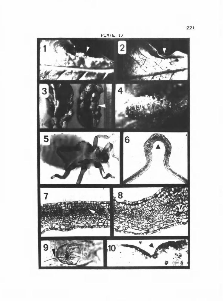
221.

PLATE 18
Figure 1. Trans. section of matures ungalled poplarleaf. Palisade (arrow) and spongylayers are distinct. (V/8/80). bOX.
Figure 2. Trans. section through wall of marginalleaf gall caused by stem mother. Gallcavity (6) and cytoplasmically densevascular cells (arrow) are seen. (IV/29/8O).llOX.
Figure 3. Trans. section through wall of stem mothermarginal leaf gall, showing gall cavity (8)and vascular bundles (arrow). (VI/5/80). 75X.
Figure 4. Section through wall of stem mothermarginal leaf gall, showing gall cavity(6) and vascular bundle (arrow). (V/13/80).200X.
Figure 5. Section through wall of stem mothermarginal leaf gall stained for tannins.Only cells in outer region of wall stainpositively. Those near gall cavity (8)do not. (IV/29/8O). 1OSX.
Figure 6. Midvein gall (arrow) on leaves ofPopulus trichocarpa T. and G. caused byPemphiqus sp. as viewed from undersideof leaf. (IV/22/80). 4X.
Figure 7. Section through poplar inidvein gallshowing midvein (arrow) and gall cavity(star). (V/6/80). lox.
Figure 8. Section through wall of poplar midveingall showing gall cavity (G) and strandof cytoplasmicaily dense vascular cells(arrow). (V/13/8O). 250X.
222

223

Figure 2.
Figure 3.
Figure 4.
Figure 8.
Figure 9.
PLATE 19
Figure 1. Leaf gall (arrow) on Arctostaphylosqlutinosa caused by Tamalia coweni(Cock.). (X/6/BO). Scale in mm.
Cut-away view of leaf gall on A. uva-ursi L. showing aphids (arrow) insidegall cavity. (VII/20/80). 8X.
Anterior end of I. coweni showing stylet(arrow). (V/8/80). 170X.
Trans. section through galled bearberry leafshowing ungalled portion of leaf (arrow) andgalled leaf curled around gall cavity(6). (V/B/GO). laX.
Section of bearberry gall wall showinggall cavity (6) and cells lining cavity.(V/20/8O). 200X..
Wall of bearberry gall stained f ortannins with gall cavity (6) indicated.(V/20/80). 40X.
224
Figure 5. Ungalled bearberry leaf showing distinctpalisade (arrow) and spongy mesophylllayer. (V/8/8O). l4OX.
Figure 6. Galled bearberry leaf showing recognizablepalisade (arrow) and dense spongy mesophylllayer. Gall cavity indicated by star.(V/B/GO). BSX.
Figure 7. Transition zone between ungalled bearberryleaf (lower arrow) and galled leaf(upper arrow). (V/20/SO). 55X.


PLATE 20
Figure 1. Trans sectiofl through ungalled bearberryleaf stained for tannins, showingdistinct palisade (arrow) and spongymesophyll layers. (V/20/BO). 55X..
Figure 2. Long. section of Hormaphis hamamelidisFitch on Hamamelis virqinica L. showingthe large gall cavity at the base ofwhich is a portal (arrow). Ungalled leafsurface is indicated by stars. (V/26J80L.8.5X.
Figure 3. Ungalled witch hazel leaf showing distinctpalisade (arrow) and spongy mesophyllcells. (V/26/80). BOX.
Figure 4. Long. section through wall of witch hazelleaf gall4 showing the gall cavity (6)and the cells that line the cavity(arrow). (V/26/80). 80X.
Figure 5.. Section through wall of witch hazel leafgall showing gall cavity (immediatelyto the left of the 6) and the cellslining the cavity (arrow). (V/26/8O). 290X.
Figure 6. The leaf gall (arrows) on Crataeausdouqlasii var. suksdorf ii Sarg.caused by a cecidomyiid. Ungalled leafis in lower right corner. Hand cut trans.sections through gall are seen in upperright corner. (VIII/12/80). Scale in mm.
Figure 7. Cut-away hawthorn leaf gall showingyoung cecid larva (arrow). (V/22/80). 75X.
Figure 8. Trans. section through hawthorn leafgall showing cavity (6), portal with hairs(arrow) and ungalled leaf (arrow). (V/l3/80).15X..
Figure 9.. Older instar cecid larvae in hawthornleaf gall. Forked sternal spatula (arrow)is visible in one larva. (IX/22/B0). 20X.
226

..i.u,s.o.,&.iiiiiiittiIiIIuIIIIIIiIIIIIIIIIUIlIIIIIflhllIjIiIIIIIUIt IltIllIllIltull 1
227

PLATE 21
Figure 1. Transition region of hawthorn lea-f gall. Atright the already altered leaf becomes thickerand mare densely packed with cells as the gallis approached (to the left). The remnants ofthe palisade layer (arrow) are stillapparent. (V/13/8O). 130X.
Figure 2. Trans. section of ungalled hawthorn leafshowing distinct palisade (arrow) and spongymesophyll layer. (V/13/8O). 150X.
Figure 3. Wall of hawthorn leaf gall stained f ortannins. Cells lining the larval cavity(arrow) contain some positively stained mat-erial. (V/l3/80). l5OX.
Figure 4 Section through wall of hawthorn lea-f gallshowing larval cavity (6) and cells lining thecavity (arrow). (V/13/80). 1SOX.
Figure 5. Section through wall of hawthorn leaf gallshowing larval cavity (star) and cellslining the cavity (arrow). (IX/22/80). l9OX.
Figure 6. Cut-away view of leaf roll gall on Symphor-icarpos albus var laevigatus Fern. causedby a cecidomyiid. Larvae are at upper arrow,larval cavity at lower arrow. (V/20/80). 7X.
Figure 7. Anterior end of cecidomyiid larva taken fromleaf roll gall on snowberry. Head capsule atarrow. (X/20/8O). ilOX.
Figure 8. Trans. section through ungalled snowberry leafshowing palisade (arrow) and spongy meso-phyll layers. (V/6/80). 55X.
Figure 9. Transition zone from ungalled snowberry leaf(lower arrow) to slightly swollen laminashowing no palisade or spongy layers (upperarrow). Edge of swollen gall wall is seenat left (middle arrow). (V/6/8O). 55X.
228
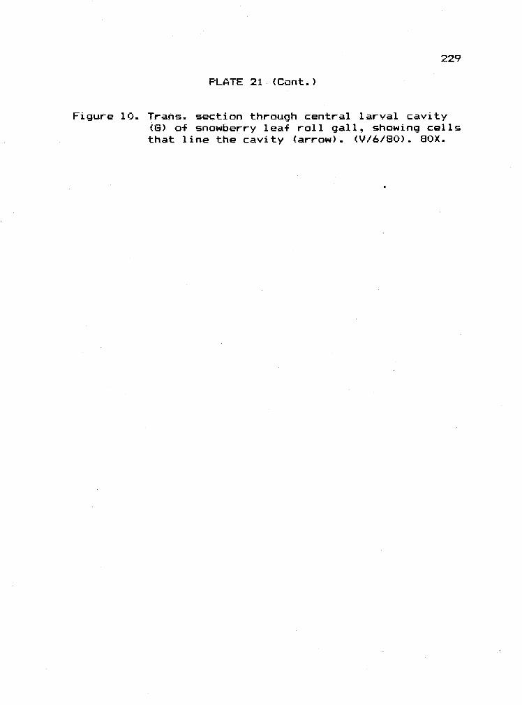
PLATE 21 (Cont.)
Figure 10. Trans. section through central larval cavity(6) a-F snowberry leaf roll gall, showing cellsthat line the cavity (arrow). (V/6/80). 80X.
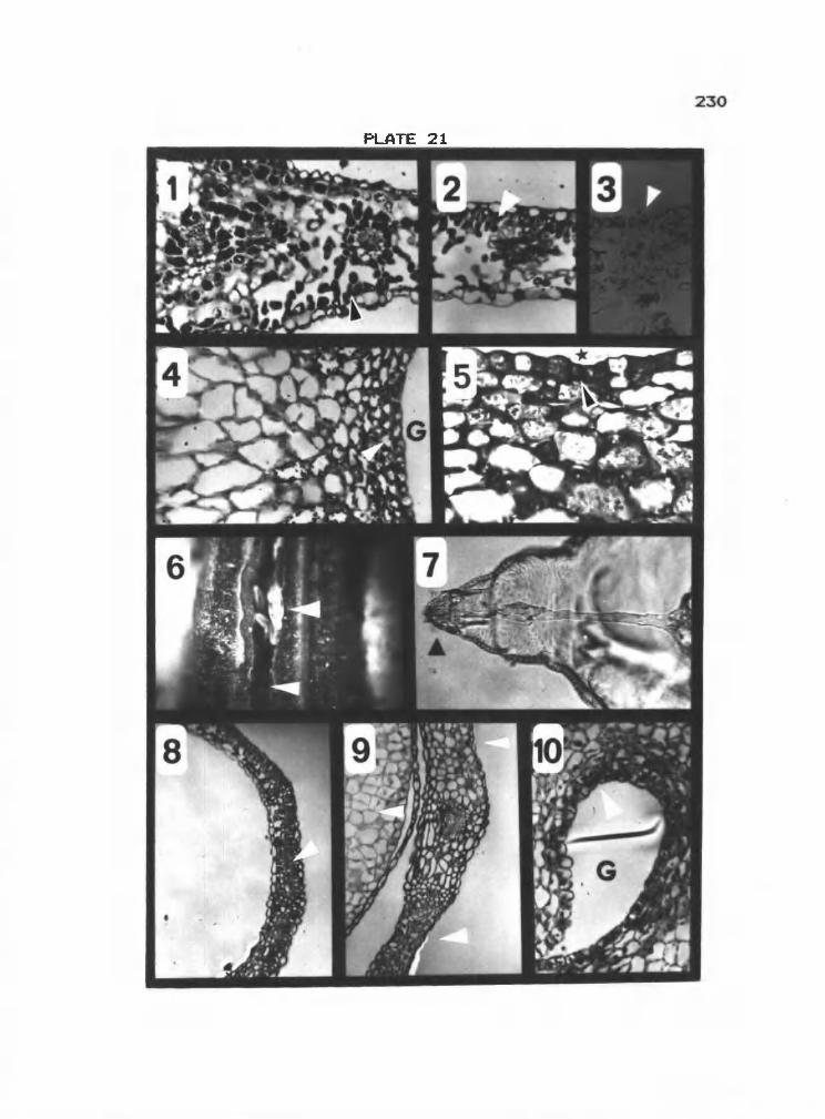
230

PLATE 22
Figure 1. Trans. section through wall of snowberry leafroll gall showing larval cavity (6) andcells lining the cavity (arrow). (VII/24/80).130X.
Figure 2. Trans. section through snowberry leaf rollgall stained for tanniris, showingcentral larval cavity (6). (V/6/80).ox.
Figure 3. Leaf gall on Amelanchier alnifolia. Nutt.caused by a cecidomyiid, as seen from theupper leaf surface (left arrow) and the lowerleaf surface (right arrow). Two galls thathave been pulled from the leaf are shownbroadside in the upper right corner. (VII/12/80). Scale in mm.
Figure 4. Cut-away view of ser-viceberry leaf gallshowing larval cavity (right white arrow),outgrowth that separates larval cavity fromcentral cavity (left white arrow), and ostiole(black arrow). (V/20/80). 3.5X.
Figure 5. Anterior end of cecidomyiid larva taken fromserviceberry gall. Head capsule indicated byright arrow, and out-of-focus sternalbreastbone by left arrow. (VIII/12/80). 150X.
Figure 6. Long. section through young leaf gall onserviceberry with midvein (black arrow), gallcavity (white arrow), and ungalled leaf (star)indicated. (IV/l5/80). l3X.
Figure 7. Long. section through larval cavity (6)of serviceberry leaf gall with larva (arrow).(IV/l5/80). bOX.
Figure 8. Section through wall a-f leaf gall on service-berry with larval cavity (6) and cells liningthe cavity (arrow). (IV/l5/80). 330X.
231


PLATE 23
Figure i. Trans. section through ungalled serviceberryleaf showing palisade (arrow) and spongy meso-phyll layers. dy/iS/GO).. 160X.
Figure 2. Trans. section through transition zone showingungalied serviceberry leaf at right and thick-ened lamina left. Altered palisade cells areseen at arrow. dIV/IS/SO). 140X.
Figure 3. Trans. section through ungalled serviceberryleaf showing palisade (arrow) and spongymesophyll layers (V/6/8O). 130X.
Figure 4. Section through larval cavity (6) 0+ service-berry leaf gall showing cells lining thecavity (arrow). (V/6/80). 290X.
Figure 5. Trans. section through serviceberry leaf gallshowing larval cavity (6), central cavity(star) and scierid layer (5). (VIII/12/80).14X.
Figure 6. Trans. section through serviceberry gallshowing cells (arrow) lining the larvalcavity (B), and scierids that linethe central cavity (star).. (VIII/12/80). 55x.
Figure 7. Trans.. section through larval cavity (B) ofserviceberry gall showing cells lining cavity(arrow). (IX/20/8O). 240X.
Figure 8. Trans. section through ungalled serviceberryleaf stained for tannins showing lightlystained palisade layer (arrow). (VI/5/8O).190X.
Figure 9. Long. section through serviceberry gallstained for tannins showing the larvalcavity (6), cells lining the larval cavity(arrow), and the central cavity (star).(V/20/80). 65X.
233

234

PLATE 24
Figure 1. Bud gall of Populus trichocarpa T. and 8. (farleft arrow) caused by a cecidomyiid. Ungalledbud of same age shown to right (middleleft arrow) of galled bud. Hand cut trans.section through galled (far right arrow) andungalled (middle right arrow) bud areshown. (VIII/12/80). l.2X.
Figure 2. Trans.. cut-away view of poplar bud gall.Larvae (arrow) present between budscales but are difficult to see.(VIII/19/80). l2X.
Figure 3. Anterior end of cecidomyiid larva taken frompoplar bud gall.. Head capsule is indicated byarrow. (IX/22/8O). 280X.
Figure 4. Trans. section through galled poplar budshowing overlapping bud scales. Centerof bud is indicated by star, and midgelarva by arrow. (V/l3/80). l2X.
Figure 5. Trans. section through galled poplar budshowing cells on surface of bud scale (rightarrow), and larva (left arrow). (V/l3/80).400X.
Figure 6. Trans. section through bud scale from poplarbud gall showing inner (adaxial) surface(right arrow) and outer (abaxial) surface(left arrow). (VIII/l2/8O). 90X.
Figure 7. Trans. section through scale from poplarbud gall showing inner surface (arrow).(VIII/12/80). 350X..
Figure 8. Trans.. section through bud scale from poplarbud gall showing inner surface with changein surface cells at arrow. (VIII/12/8O).350X.
Figure 9. Trans. section through bud scale from pop-lar bud gall showing inner (upper arrow) andouter (lower arrow) surfaces. (IX/22/8O).1 80X.
235

F')
N
N

PLATE 25
Figure 1. Stem beaked gall (arrow) on Salix sp. causedby Phytophaga rigidae 0. 8. (V/13/80).Cm scale.
Figure 2. Willow stem gall (arrow). (VII/15/80).Cm scale.
Figure 3. Willow stem gall showing yellow "beak"(arrow). (VlII/19/80). Scale in mm.
Figure 4. Willow stem galls showing necrotic "beak"(arrow). (IX/2l/8OL Cm scale.
Figure 5. Long. cut-away view a-f willow stem gallshowing larval cavity (le-Ft arrow) andexit pOrtal through beak (right arrow).(IV/8/80). 3X.
Figure 6. Trans. cut-away view a-f willow stem gallshowing larval cavity (arrow). (IV/8/80). 3X.
Figure 7. Long. cut-away view of willow stem gallshowing larva (arrow) in larval cavity.(VIII/l9/80). 8X.
Figure 8. Large cecidomyiid larva in willow stemgall. (VIIl/l9/8O). l2X.
Figure 9. Anterior end of larva -from willow stemgall showing head capsule (right arrow) andsternal spatula (left arrow). (V/l3/80).1 70X.
237
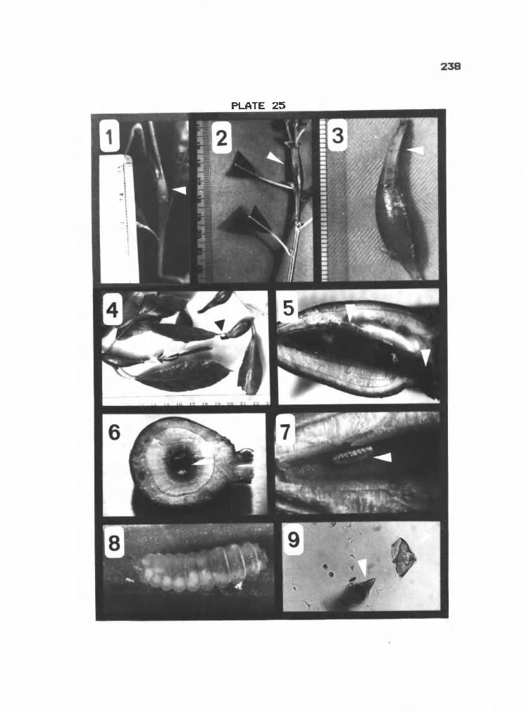
C1
191111iiflfl
(T)1111111H
111111111li1111111U1111

PLATE 26
Figure 1. Phytophaga riqidae adult.. (OSU InsectCollection). Scale in mm.
Figure 2. Trans. section c-F ungalled willow stemshowing pith (P), vascular tissue (V)1 andcortex (C). (V/13/80). 35X.
Figure 3. Trans. section through young willow stem gallshowing larval cavity (6) and inner and outervascular bundles (upper and lower arrows,respectively). (V/l3/8O). l3X.
Figure 4. Trans. section through young willow stemgall showing larval cavity (6) and cellslining the cavity (arrow). (V/13/8O). 50X.
Figure 5. Trans. section through young willow stem gallshowing larval cavity (6) and cells liningthe cavity (arrow). (V/13/BO). l4OX.
Figure 6. Trans. section through willow stem gallshowing larval cavity (6) and cells liningthe cavity (arrow). (VhS/GO). 240X.
Figure 7. Trans. section through wall of willow stemgall showing larval cavity (6), sceleridzone (5), and secondary xylem (V).(Vhh/24/80). 30X.
Figure 8. Trans. section through wall of willow stemgall showing larval cavity (6). cells liningcavity (arrow) and scierenchyma zone (S).(Vhh/24/8O). 200X.
Figure 9. Trans.. section through wall of willow stemgall showing gall cavity (6) and secondaryxylem CV). (VhII/l2/80). 17X.
239

240

PLATE 27
Figure 1. Trans. section through wall of willow stemgalls showing larval cavity (6), cells liningthe cavity (arrow) and sclerenchyma zone (9).(IX/21/80). 200X.
Figure 2.. Trans. through wall of willow stem gallshowing larval cavity (6) and cellslining cavity (arrow). (VIII/l2/8O). 200X.
Figure 3. Trans. section through ungalled willow stemstained for tannins, showing cortical cellsand epidermal cells (upper lower arrow,respectively). (V/13/80). 40X.
Figure 4. Trans. section through willow stem gallstained -for tannins showing larvalcavity (star) and cells lining the cavity(arrow). (Vl/5/80). 100X.
Figure 5. Trans. section through willow stem gallstained + or tannins showing larvalcavity (star) and cells lining thecavity (arrow). (IX/21/8O). lOOX.
Figure 6. Stem gall (arrow) on Hypachaeris radicata L.caused by Aulax hvpochaeridis kieff.(VIII/12/BO). Cm scale.
Figure 7. Aulax hyopchaeridis adult. (OSU InsectCollection). Scale in mm.
Figure 8. Wasp larva (arrow) in larval cavity of Cat'sEar stem gall. (VIII/12/80). 35X.
Figure 9. Mandible a-f wasp larva taken from Cat's Earstem gall. (VI/ll/80). 170X.
Figure 10. Section through larval cavity (6) 0-f Cat'sEar stem gall. Cells lining the cavity(arrow) are indicated. (VIII/12/80). l2OX.
241

242

PLATE 28
Figure 1. Trans. section through larval cavity (star)a-f stem gall on Cat's Ear. Area of nutritivecells is indicated (arrow). (VIII/12/80). l7X.
Figure 2. Small gall (arrow) on young lea-f a-f Quercusgarryana flougi. caused by a cynipid. (V/6/80).4X -
Figure 3. Section through wall of small lea-f gall onBarry oak showing larval cavity (6),nutritive region (left arrow), and outerregion of gall wall (right arrow).(V/6/8O). BOX.
Figure 4. Section through nutritive region of small leafgall an Garry oak showing larval cavity(star) and nutritive cells with prominentnucleoli (arrow). (V/6/80). 180X.
Figure 5. Section through nutritive cells of small leafgall on Garry oak showing nucleus (Nc) andnucleolus (Nu). (V/6/80). 300X.
Figure 6. Oak bullet gall on Quercus garryana Dougl.caused by Andricus sponqiolus Gill.. Noteswollen stem at point o-f attachment (arrow).(VII/15/80). Cm scale.
Figure 7. Cut-away view of oak bullet gall showingopened larval cavity (arrow) and larva (star).
(VIII/l8/80L. lOX.
Figure 8. Cut-away view o-f old oak bullet gall showingpoint a-F attachment (upper arrow) , connect-ive strands (middle arrow), and hardenedsphere o-f larval capsules (lower arrow).(X/9/80). Cm scale.
Figure 9. Adult Andricus spongiolus. (OSU InsectCollection). Scale in mm.
Figure 10. Section through thrips leaf roll gall showinggall cavity (left arrow) and nutritiveregion (right arrow). (VII/7/80). 200X.
A '


PLATE 29
Figure 1. Section through oak bullet gall showinglarval cavity (arrow). (V/6/80). 20X..
Figure 2. Section through larval cavity (6) of oakbullet gall showing nutritive cells thatline the cavity. (V/6/8O). l5OX.
Figure 3. Nutritive cells of oak bullet gall showingprominent nucleoli (right arrow). Larvalcavity (6) is to the upper left(arrow). (V/6180). 280X.
Figure 4. Section through larval cavity (6) of oakbullet gall showing nutritive cellsthat line the cavity (arrow). (VII/l5/80).75x.
Figure 5. Section through oak bullet gall stained fortannins. Cells lining larval cavity (rightarrow) stain poorly, while those in surround-ing area (left arrow) stain positively.(V/6/8O). 16X.
Figure 6. Tip gall (arrow) on Rosa nutkana Presi.caused by cynipids. (VIII/l2/BO). Scalein mm.
Figure 7.. Section through oak bullet larval cavity(6) stained for tannins showing poorlystained nutritive cells (left arrow) anddeeply stained material that coversnutritive cells (right arrow). (VII/24/8O).BOX.
Figure 8. Section through rose tip gall showing larvalcavity (6) and nutritive region lining thecavity (arrow).. (VIII/12/80). 20X.
Figure 9. Section through nutritive region of rose tipgall showing nutritive cells with prominentnucleolus (lower arrow) Larval cavity(G) is toward upper right corner (upperarrow). (VIII/l2/80). l6OX.
245

29
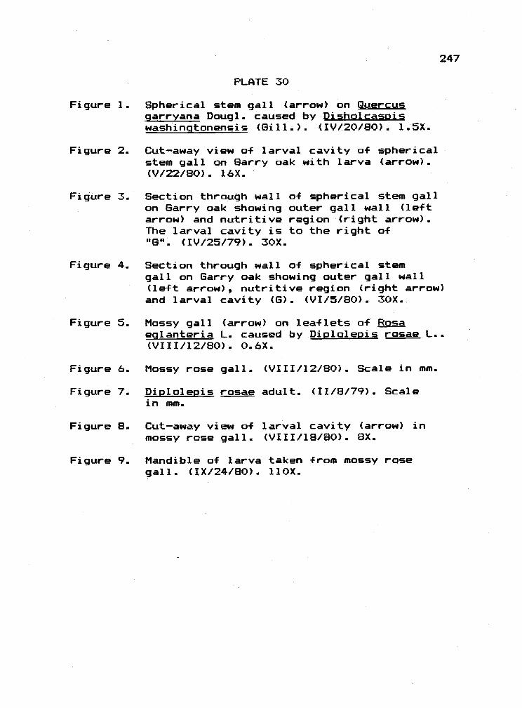
PLATE 30
Figure 1. Spherical stem gall (arrow) on Quercusgarryana Dougi. caused by Dishalcasaiswashingtonensis (Gill..).. (IV/20/80). l.5X.
Figure 2. Cut-away view of larval cavity a-F sphericalstem gall on Barry oak with larva (arrow)..(V/22/80). 16X.
Figure 3. Section through wall a-f spherical stem gallon Barry oak showing outer gall wall (leftarrow) and nutritive region (right arrow).The larval cavity is to the right of(IG* (IV/25/79). 30X.
Figure 4. Section through wall of spherical stemgall on Barry oak showing outer gall wall(left arrow), nutritive region (right arrow)and larval cavity (B).. (VI/5/80).. 30X.
Figure 5. Mossy gall (arrow) on leaflets of Rosaeglanteria L. caused by Diplolepis rosae L..(VIII/l2/80). 0.6X..
Mossy rose gall.. (VIII/12/80). Scale in mm.
Diplolepis rosae adult. (11/8/79). Scalein mm
Cut-away view of larval cavity (arrow) inmossy rose gall. (VI11/lG/80). 8X.
Mandible of larva taken from mossy rosegall. (IX/24/B0). liOX.
247
Figure 6.
Figure 7.
Figure 8..
Figure 9.

0
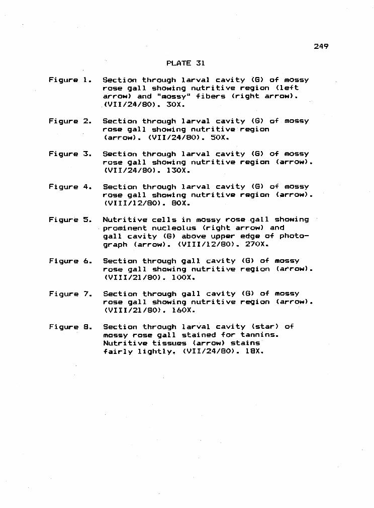
PLATE 31
Figure 1. Section through larval cavity (6) of mossyrose gall showing nutritive region (leftarrow) and "mossy" fibers (right arrow).(VII/24/80). 30X.
Figure 2. Section through larval cavity (6) of mossyrose gall showing nutritive region(arrow). (VII/24/8O). 50X.
Figure 3. Section through larval cavity (6) of mossyrose gall showing nutritive region (arrow).(VII/24/8O). 130X.
Figure 4.. Section through larval cavity (6) of mossyrose gall showing nutritive region (arrow).(VIII/12/80). 80X.
Figure 5. Nutritive cells in mossy rose gall showingprominent nucleolus (right arrow) andgall cavity (6) above upper edge of photo-graph (arrow). (VIII/l2/8O). 270X.
Figure 6. Section through gall cavityrose gall showing nutritive(VIII/2l/80). 100X.
Figure 7. Section through gall cavityrose gall showing nutritive(VIII/2l/80). 16OX.
(6) of mossyregion (arrow)
(6) of mossyregion (arrow).
Figure 8. Section through larval cavity (star) ofmossy rose gall stained for tannins.Nutritive tissues (arrow) stainsfairly lightly. (VII/24/80). 1BX.
249


Figure 1.
Figure 2.
Figure 4.
Figure 5.
PLATE 32
Section through larval cavity (G) of mossyrose gall stained for tannins showingnutritive region (right arrow) andunderlying, darkly stained region(left arrow). (VIIl/12/80L 60X.
Section through larval cavity (G) of mossyrose gall stained for tannins, showingnutritive region (right arrow) and "moss"fibers (left arrow). (IX/21/80). 1BOX.
Speckled oak apple on Garry oak. (IX/21/80).Scale in mm.
Cut-away view of speckled oak apple onGarry oak, showing outer gall wall (lowerarrow), point a-f attachment (upper arrow),and fibers suspending central larvalcapsule (star is just above capsule).(IX/23/80). Cm scale.
Figure 6. Cut-away view a-f larval capsule (arrow)a-f speckled oak apple on Garry oak.(VIII/lS/8O). 12X.
Figure 7. Adult Besbicus mirabilis. (OSU InsectCollection). 9X.
Figure 8. Mandibles of larva pulled from speckledoak apple an Garry oak. (VIII/l9/8O). 300X.
251
Figure 3. Young speckled oak apple (arrow) on Quercusqarryana Dougl. caused by Besbicus mira-bilis (Kinsey). Note that gall is emergingfrom split midvein. (V/31/80). Scale in mmrulings.

c..1
U)
N
PLATE 33
Figure 1. Section through midveiri (V) and youngspeckled oak apple on Barry oak, showingthe larval cavity (G at arrow). (V/31/BO).1 5X.
Figure 2. Small galls (arrow) on underside of Barry oakleaf. (IX/22/80). Scale in mm.
Figure 3. Junction of inidvein (right arrow) andspeckled oak apple on Barry oak showinggall cavity (G) and stalk of gall (leftarrow). (V/31/8O). 30X.
Figure 4. Section through speckled oak apple onBarry oak showing base of fibers (whitearrow) at capsule wall (black arrow).Larval cavity is out of photograph aboveupper edge. (VI/17/80). l2OX.
Figure 5. Section through speckled oak apple onBarry oak showing nutritive region (whitearrow), larval cavity (star) , and capsulewall (black arrow). (VI/17/80). 290X.
Figure 6. Section through speckled oak apple on Barryoak showing fibers (arrow). Larval cavity(G) is in direction of arrow, out of photo-graph. (VIII/l.2/80). 50X.
Figure 7. Section through speckled oak apple on Barryoak showing nutritive cells with prominentnuclei (Nc) and nucleoli (Nu) and remnantsof early capsule wall (below star). Larvalcavity (B) is out of photograph at upperleft hand corner. (VlII/l2/80). 300X.
Figure 8. Section through speckled oak apple on Barryoak showing larval cavity (B) andbordering nutritive tissue (arrow).(IX/22/80L. 30X.
253
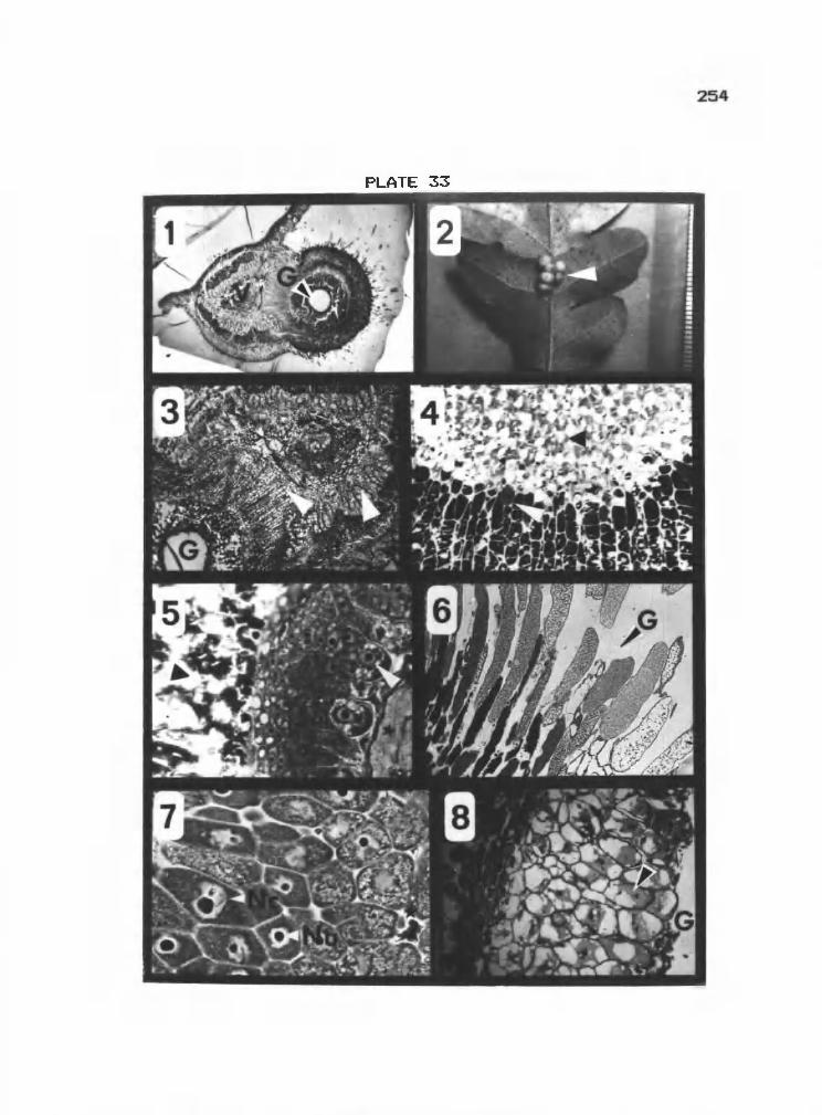
N

Figure 1..
Figure 2.
Figure 3.
PLATE 34
Cecidomyiid gall (arrow) on Artemisia trident-ata Mutt. (ART) (IX/li/BO). Scale in mm.
ART, showing midge larva (arrow) in cavity.(IX/ll/80). 12X
Trans.. section through ART showing 4 adjacentlarval ca,ities (6) each with larva (arrow).(X/18/80). 17X.
Figure 4. Trans. section through ART showing spongygall wall. Vascular bundles (left arrow)and larval cavity (right arrow) are seen.(IX/ll/8O). 40X.
Figure 5. Trans. section through ART showing nutritivetissue (arrow) bordering larval cavity (G).(IX/19/8O). 73X.
Figure 6.
Figure 7.
Figure 8.
Head capsule of midge found in ART.(IX/ll/8O). 250X..
Cecidomyiid galls (arrow) on Artemisiatridentata. (ARC) (IX/ll/80). Scale in mm.
Cut-away view of ARC showing larval cavities(upper arrow) that rest on dome-like platform(lower arrow). (IX/ll/8O). 9X..


PLATE 35
Figure 1 Trans. section through ARC at base of 3larval cavities (6 at arrow) Nutritivetissue at base o-F cavities seen ascircles of tissue in micrograph.(IX/19/8O). 23X.
Figure 2.. Trans. section through ARC at larvalcavity (6) showing nutritive tissue borderingcavity (arrow). (IX/l9/80). 70X.
Figure 3. Head capsule of larva found in ARC.(IX/19/80). 260X.
Figure 4. Section through eriophyoid gall onArtemisia tridentata, ANP, showing leaf(arrow) and gall cavity (star). (IX/19/80)
Figure 5. Section through ANP showing gall cavity (6)with mites (m) and leaf (star). (IX/19/8O).85X -
Figure 6.. Section through ANP showing nutritive cells(arrow) lining gall cavity., and mites (rn).(IX/l9/80). l9OX.
Figure 7. Nutritive cells of speckled oak appleon Sarry oak showing dispersed nuclear area(star at arrow). (IX/22/80). l3OX.
Figure 8. Section through speckled oak apple on Garryoak stained for tannins with larval cavity(6) and lightly stained nutritive tissue(arrow). (VI/25/80). lOOX.

258

PLATE 36
Figure 1. Cecidomyiid gall (arrow) on Chrysothamnusnauseosus (Pall.) Britt (CRC) (IX/lo/GO).Cm scale.
Figure 2. Lang. cut-away through CRC showing larvalcapsules (upper arrow) resting on top ofdome-like structure (lower arrow).(IX/lO/80). 6X.
Figure 3.. View looking down into CRC from above withsome hairs removed. Tops of larval capsulesare seen (arrow). (IX/lO/GO). 8X.
Figure 4. Long. section of CRC through larval cavity(G) containing a larva, and bordered bynutritive tissue (arrow). (IX/lO/BO). 50X.
Figure 5. Wall of larval capsule of CRC with cavity(6) and larva (arrow). (IX/lO/80). 1BOX.
Figure 6. Tephritid galls on C. nauseosus. (COT)(VI/14/80). Scale in mm.
Figure 7. Trans. section of COT showing larval cavity(arrow). (IX/12/80). 7.5X.
Figure 8. Section of COT showing larval cavity (6).(IX/12/BO). 20X.
259

0N
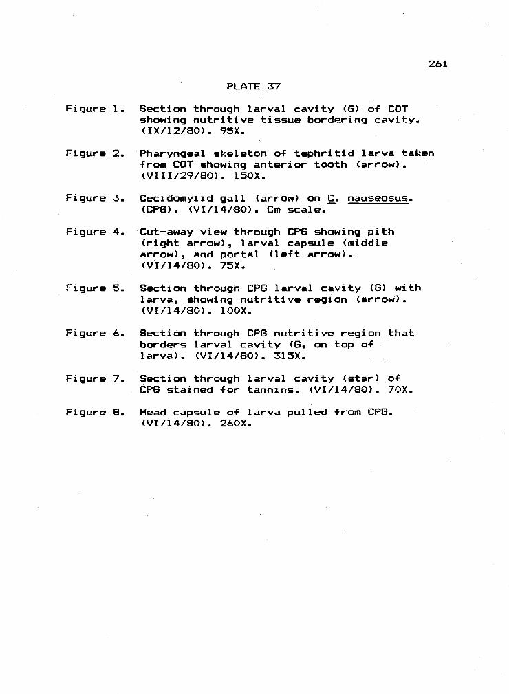
PLATE 37
Figure 1. Section through larval cavity (6) of COTshowing nutritive tissue bordering cavity.(IX/l2/80).. 95X.
Figure 2. Pharyngeal skeleton of tephritid larva takenfrom COT showing anterior tooth (arrow).(VIII/29/8O). 150X_
Figure 3. Cecidomyiid gall (arrow) on C. nauseosus..(CR3). (VI/14/80L. Cm scale.
Figure 4. Cut-away view through CR6 showing pith(right arrow), larval capsule (middlearrow), and portal (left arrow)(VI/14/80). 75X.
Figure 5. Section through CPS larval cavity (6) withlarva, showing nutritive region (arrow).(VI/l4/8O). bOX.
Figure 6. Section through CR13 nutritive region thatborders larval cavity (6, on top oflarva). (VI/14/8O). 315X.
Figure 7. Section through larval cavity (star) ofCPG stained for tannins.. (VI/b4/80). 70X.
Figure 8. Head capsule of larva pulled from CR6.(VI/14/80). 260X.
261

o.

PLATE 38
Figure 1. Stem gall (arrow) on Tetradymia spinosa H.and A. caused by Gnorimoschema tetradvmi-ella Busck. (TES) (VI/14/80). Cm scale.
Figure 2. Trans. section through ungalled stem of j.spinosa, showing pith (P), vascular tissueCV), and cortex (C). (VI/l4/8O). 120X.
Figure 3. Trans. section showing outer portion of TESwall. Arrow points towards gall cavity,and rests near a vascular bundle. (VI/14/80).75X.
Figure 4. Section through gall cavity (6) of TESshowing nutritive region (arrow). (VI/l4/80).120X.
Figure 5. Gall on T. qlabrata caused by 6. tetra-dymiella (?) (TET) (VI/14/8O). Cm scale.
Figure 6. Head capsule of larva pulled from TET.Note mandibles (arrow). (VI/14/80). l6OX.
Figure 7. Trans. section through lET showing larvalcavity (right arrow) and gall wall Cleftarrow). (VI/14/80). 30X.
Figure 8. Nutritive cells bordering larval cavity(in direction of arrow) of TET. CVI/l4/80).75X.
263

N

PLATE 39
Figure 1. Very early stage of petiole gall (arrow)caused by Pemphigus populi-caulis Fitchon Populus trichocarpa T. and S..Viewed from underside. (IV/l5/80). 2X.
Figure 2. Early stage of poplar petiole gall (arrow).Upper surface view (IV/30/8O). 2X.
Figure 3. Petiole and major veins of Populus plio-tremuloides Axel. from Alvord Creek, OregonLower Pliocene). Nate twisted petiole(arrow). (Axeirod, 1944; U. C. Berkeley,Paleon. Call.). Scale in mm.
Figure 4. Close up of P1. 39, Fig.. 3, showing twistedpetiale (arrow). lOX.
Figure 5.. Swollen petiole (arrow) of Populus eotretn-uloides kn. from Miocene Trapper creekflora of southern Idaho (Axelrod, 1964;U. C. Berkeley Paleon. Coil.). Scale in mm.
Figure 6. Close up of P1. 39. Fig. 5, showing swollenpetiole (arrow). 12X.
Figure 7. Twisted petiole (arrow) of Papuluspayettensis (Kn) Axel. from theMio-Pliocene flaras of west-centralNevada (Axelrod, 1956; U.C. BerkeleyPaleon. Coil.). Scale in mm.
Figure 8. Possible mine (arrow) in leaf of Quercusnevadensis (Lesq.) from the La Porteflora of California (Potbury, 1935; U.C.Berkeley Paleon. Call.), aX.
265

266

PLATE 40
Figure 1. Galled acorn of Quercus agrifolia Neefrom the La Brea Tar Pits. Plesiotype No.PB 1481, Page Museum, Los Angeles. Notetwo opened larval chambers (arrows) Scalein mm.
Figure 2. Galled acorn of undetermined speciesfrom the La Brea Tar Pits. SpecimenNo. 14189, Pit 3 (2001/495), Page Museum.Los Angeles. Note prominent, opened larvalchamber (arrow) - Scale in mm.
Figure 3. Acorn of Q.. wislizenii A. de Cand. galled byCallirhytis milleri Weld. Cut-away view ofchambers (arrow). (Cal. Acad. Sci - specimen).Scale in mm.
Figure 4. Woody mass of cotyledons from acorn ofa. wislizenii galled by . milleri. Remnantof chamber wall (arrow) is similar to chamberwall in La Brea material. (Cal. Acad. Sci.specimen). Scale in mm.
Figure 5. Adult C. milleri (Hymenoptera: Cynipidae).Antennae are missing. USNM specimen. Scalein mm.
Figure 6. Stem with thorns (arrow) of Rosa hilliaeLesq. from Crooked River Basin, Or.(Eocene-Ol igocene) (Chaney, 1927; u_C.Berkeley Paleon. Call.). Scale in mm.
Figure 7. Leaves and roots of Vitis vinif era L.galled (arrow) by Phylloxera vastatrixPlanchon. (Houard Call, Musee Entomol.,Paris). 0.25X.
267

268

BIBLIOGRAPHY
Agrios, 6. N. 1973. Plant Pathology. Academic Press. NewYork. 629p.
Ambrus, B. and L. Hably. 1979. Eriophyes daphnoqene sp.n., a fossil gall from the Upper Oligocenein Hungary. Ann. Historico-Naturales MuseiNationalis Hungarici. 71:55-56.
Ananthakrishnan, T. N. 1978. Thrips galls and gallthrips. Zool. Surv. India, Tech. Monog.1:1-69.
Andersonq H. W. 1956. Diseases of Fruit Crops.McGraw-Hill. New York. 429p.
Axe1rod D. I. 1956. Mio-Pliocene floras from westcentral Nevada. U. of Cal. Pub. Geol.33:322.
Axelrod, D. I. 1964. The Miocene Trapper Creek flora ofsouthern Idaho. U. of Cal. Pub. Geol. Sci. 51:148.
Barnes, H. F. 1946a. Gall Midges of Economic Importance.Vol. 1: Gall Midges of Root and Vegetable Crops.Crosby Lockwood and Son.. London. lO4p.
Barnes, H. F. 1946b. Gall Midges of Economic Importance.Vol. 2: Gall Midges of Fodder Crops. Crosby Lock-wood and Son, London. l6C)p.
Barnes, H. F. 1948. Gall Midges of Economic Importance.Vol. 3: Gall Midges of Fruit. Crosby Lockwoodand Son, London. lG4p.
Barnes, H. F. 1956. Gall Midges of Economic Importance.Vol. 7: Gall Midges of Cereal Craps. Crosby Lock-wood and Son, London. 26lp.
BCGRG (Belgian Crown Gall Research Group, J. Schell,Director). 1979. Crown gall: Transfer ofbacterial DNA to plants via the Ti-plasmid.In: Nucleic Acids in Plants. (T. C. Hall and 3.W. Davies, Eds. ) CRC Press, Boca Raton, Florida.
Beck, E. G. 1953. The nature of the stimulus inthe "SolidaQo" gall induced by the larva ofGnorimoschema aal laesol idaainis. Brookhaven
269

Symp. Bid. 6:235-251.
Berger, W. 1949. Lebensspuren schmarotzender Insektenan Jungtertiaren Laubblatern. Sitz.-Ber. as-terr. Akad. Wiss., Vienna, Abt I. 158:789-792.
Berry, E. W. 1916. The Lower Eocene floras of South-eastern North America. U. S. Geal. Surv.Prof. Paper 91.
Berry, E. W. 1925. Pleistocene plants from NorthCarolina. U. S. Geal. Surv. Prof. Paper140:97-120.
Beutenmuller, W. 1918. New species of Rhodites fromOregon. Can. Ent. 50:305.
Bird, A. F. 1961. The ultrastructure and histochemistryof a nematode induced giant cell. 3. Biophys. &Biochem. Cytol. 11:701-715.
Bird, A. F. 1974. Plant response to root-knot nematade.Ann. Rev. Phytopath. 12:69-85.
Bird, A. F. 1975. Symbiotic relationships betweennematodes and plants. Symp. Soc.. exp. Biol.29:351-371.
Bird, A. F. and B. R. Loveys. 1975. The incorporationof photosynthates by Meloidoqyne javanica. 3.
Nematology. 7:111-113.
Black, C. C. 1971. Ecological implications of dividingplants into groups with distinct photosyntheticproduction capacities. Adv. Ecol. Res. 7:87-114.
Blakely, W. F. 1955. A Key to the Eucalypts. For.Timb. Bureau, Canberra. 2nd Ed. 359p.
Bloch, R. 1953. Abnormal plant growth. BrookhavenSymp. in Biology. 6:41-54.
Bones. T. J. 1979. Atlas of fossil fruits and seeds fromNorth Central Oregon. Oregon Museum of Science andIndustry, Occasional Papers in Natural Science. 1:1-23.
270
Borror, D. 3. and D. W. Delong. 1971. An Introduction tothe Study of Insects. 3rd edition. Halt, Rinehart,

and Winston. New York. 812p.
Boysen-Jensen, P. 1948. Formation of galls of Mikiolafagi. Physiol. Plantar. 1:95-108.
Brayshaw, 1. C. 1976. Catkin bearing plants ofBritish Columbia. B. C. Provin. Mus., 0cc.Paper Ser., Victoria. 18:1-176.
Brian, P. W. 1976. The phenomenon of specicityin plant disease. Pages 15-26 In: Specificityin Plant Disease. (R. K. S.. Wood and A. Graniti,Eds.) Plenum, New York. 354p.
Bronner, R. 1973. Proprietes lytiques des oeufs deBiorhiza pallida 0l. C. R. Bar. D. 276:189-192.
Bronner, R. 1976. Observations histochimiques concernantlas tissue nourriciers des zoocecidies. Actes97e Cong. Nat. Soc. Savantes. Nantes, 1972.Sec. 4:463-470.
Bronner R. and 3. Meyer. 1972. Observations histo-cytologiques at histachimiques comparees surquartre galles "rudimentaires" in pustulescausees par des cecidomyiides. Marcellia37(5) :71-83.
Bronner, R. and 3. Meyer. 1976. Cecidogenese ataspects ultrastructuraux da la galle deDasyneura affinis Kief far sur Viola odarata L.Marcel ha 39:149-154.
Brooks, H. K. 1955.. Healed wounds and galls an fossilleaves from the Wilcox deposits (Eacene) of West-ern Tennessee. Psyche 62 (1) : 1-9.
Brues, C. T. 1910. The parasitic Hymenoptera of theTertiary of Florissant, Colorado. Bull.. Mus. Camp.Zool. 54(1) :1-125.
Buchner, P. 1965. Endosymbiosis of Animals with PlantMicroorganisms. Intersci. Pub., New York.909p.
Buhr, H. 1964/65. Bestimmungstabellen der Gallen(Zoo- und Phytocecidien) an Pflanzen Mittle-und Nordeuropas. 2 vol. 6. Fischer, Jena.
271

Caltagirone, L. E.. 1964. Notes on the biology ofparasites and inquilines of Pontania paci-Fica(Hymenoptera: Teathredinidae) a leaf gallincitant on Salix lasiolepis. Annals. Ent.Soc. Am. 57(3):279-291..
Cameron.1 T.. W. H. 1964. Host specificity and theevolution a-F helminthic parasites. In: Adv..
Paras. (B. Dawes, Ed. ) Academic Press, NewYork. 2:1-34.
Casweil, H. and F. C. Reed. 1975. Indigestibility a-f C4bundle sheath cells by the grasshopper, Melan-oplus confusus. Ann. Ent. Soc. Am. 48(4):684-688.
Chaney,R. W. 1920. The flora a-f the Eagle CreekFormation. Univ.. of Chicago Walker MuseumContribution 2t5) :5-181.
Chaney. R. W. 1927. Geology and Palaeontologyof the Croaked River Basin, with specialreference to the Bridge Creek Flora. In:Additions to the palaeontalogy a-F thePacific Coast and Great Basin regions ofNorth America. (R. Kellogg, et al, Eds.) Cam.Inst. Wash. 346:123.
Chaney. R.. W.. 1944. Pliocene floras of California andOregon. Cam.. Inst. Wash.. No. 553.
Cobb, N.., et al. 1981.. An ecological study a-f theAivord Basin Dunes, southeastern Oregon. FinalTech. Report. Oregon State Univ., Corvallis..(NSF Grant No. SP-7905328). l42p..
Cocker-eli, T.. D. A. 1908. Fossil insects from Florissant,Colorado. Bull.. Am. Mus.. Nat.. Hist. 24(3):59-69.
Cockerell, T. D. A. 1927. In: Geology and palaentologya-f the Crooked River Basin with specialreference to the Bridge Creek Flora. Cam..Inst. Wash.. 346:45-138.
Cooper-Smith, J. 1974.. Cynipid galls as indicators ofhybridization between two California oaks. Unpub-lished undergraduate thesis. Pomona College, CA.1 49p.
£ I £
Cornu, M. 1878. Etudes sur Phylloxera vastatrix. Mem.Acad. Sci. Inst. France. 26:1-356.
Crane, P. R. and E. A. Jarzembowski. 1980. Insectleaf mines from the Palaeocene a-F SouthernEngland. 3. Nat. Hist. 14:629-636.
Crepet, W. L. 1979. Some aspects a-f the pollinationbiology a-f Middle Eocene angiosperms. Rev. Palea-bat. and Palynol. 27:213-238.
CSIRO (Commonwealth Scientific and Industrial ResearchOrganization). 1970. Insects of Australia. Mel-bourne University Press, Melbourne.
Curtis, K. M. 1921. The life history and cytology ofSynchytrium endobioticum (Schilb.) Perc.. thecause of wart disease in potato. Phil.Trans. Roy. Soc. Land. 210:4C)9.
Dart, P. 3. and F. V. Mercer. 1964. Fine structurechanges in the development of the nodules of Th-folium subterraneum L. and Medicaqo tribuloidesDesr.. Arch. fur Mikrobiol. 49:209-235.
Dawes, B. 1963. Hyperplasia a-F the bile duct in fascio-liasis and its relation to the problem o-f nut-rition in the liver-fluke, Fasciola hepatica L.Parasitology 53:123-133.
Dickson. 3. 6. 1947. Diseases of Field Crops. Mc6raw-Hill. New York. 429p.
Dilrher, D L. 1979. Early angiosperm reproduction: Anintroductory report. Rev. Paleobot. and Palynol.27:291-328.
Docters van Leeuwen, W. and 3. D. v. Leeuwen-Reiinvaan.1909. Kleinere zezidologische Mitteilungen.I. Eine von der Sesiide Aeqeria uniformis Snellenan Commelina communis L. verursachte Stengel-galle. Ber. dtsch. bot. Ges.- 27:572-581.
Dodd, A. P. 1961. Biological control of Eupatorium adeno-phorurn in Queensland. Austral. 3. Sci. 23(11):356-365.
273
Dunn, 3. A. 1959. The biology of the lettuce root aphid.Ann. Appl. Biol. 47(3):475-49l.

Edmunds, 6. F. and D. N. Aistad. 1978. Coevolutianin insect herbivores and conifers. Science.199(3) :941-945.
Erlich, ti. A. and H. 6. Erlich. 1971. Fine structureof the host-parasite interfaces in mycoparas-itim. Ann. Rev. Phytopath. 9:155-184.
Esau! K. 1977. Anatomy of Seed Plants. Second edition.John Wiley and Sons. New York. 550p.
Fagan, M. 1918. The uses of insect galls. Amer. Nat.52:155-176.
Feder., W. and T. P. O'Brien. 1968. Plant micro-technique: Same principles and new methods. Am.J. Bat. 55(l):123-142.
Feeny, P. 1970. Seasonal changes in oak leaf tanninsand nutrients as a cause of spring feeding bywinter math caterpillars. Ecology 51(4):565-58l.
Felt, E. P. 1911. Hosts and galls of americangall inidges.. 3. Econ. Entomol. 4:451-475.
Felt, E. P. 1914. Description of gall midges.N. V. Entomol. Soc. 3. 22:124-134.
Felt, E. P. 1916. New western gall midges. 3. N. V.Entamol. Soc. 24:174-196.
Felt, E. P. 1965. Plant Galls and Gall Makers. HafnerPress. New York. 364p.
Fockeu, H. 1889. Contribution a l'histaire des galles.Camille Robbe, Lille. ll0p.
Fockeu, H. 1897. Etudes sur quelques galles. Soc.d'Editians Scientifiques. Paris. 38p.
Freeman, T. N. 1965. A lepidopterous leaf-mine fromthe Tertiary period. Can. Entom. 97:1069.
Frank, W. D., A. A. Beetle, and D. G. Fullerton. 1964.Dipterous galls on the Artemisia tridentatacomplex and insects associated with them. Ann.Ent. Soc. Am. 57:575-577.
274

Frost, S. W. 1924. A study of the leaf-miningdiptera of North America. Cornell Univ.Ag. Exp. Stat. Mem. 78:1-228.
Furniss, M. W. and W. F. Barr. 1975. Insects affectingimportant native shrubs of the NorthwesternUnited States. USDA For. Serv., Sen. Tech.Rept. INT - 19, (Intermountain Forest andRange Experiment Station), 64 pp.
Furniss, R. L. and V. M. Carolin. 1977. WesternForest Insects. USDA, For. Serv. Misc.Pub. 1339:1-654.
Sarrigues, R. 1951. Recherches sur les cecidies,le cancer, et V actions des carburescancerigenes sur les vegetaux. Thesis, Doct.es Sciences. Lyon, France. 398p.
Giannasi.. D. E. and K. 3. Niklas. 1981. Comparativepaleobiochemistry of some fossil and extantFagaceae. Am. 3. Bot. 68 (6):762-770.
Goeden R. D. 1978. Biological control of weeds. In:Introduced parasites and predators of arthropodpests and weeds: A world review. Published by Ag.Res. Service, USDA. Ag. Handbook No. 480. Washing-ton, D. C. 545p.
Graaf land, W. 1960. The parasitism of Exobasidiumjaponicum Skir. on Azalea. Acta. bat. Neerl.9:347-379.
Greene, C. T. 1914. The cambium miner in riverbirch. 3. Agric. Res. 1(6):471-474.
Grigarick, A. A and W. H. Lange. 1962. Hostrelationships of the sugar-beet root aphidin California., 3. Econ. Ent. 55(5):740-764.
Gullan, P. 3. 1978. Male insects and galls of thegenus Cylindrococcus Maskell (Homoptera:Coccidoidea). 3. Aust ent. Sac. 17:53-61.
Hall, H. M and 1. H. Goodspeed. 1919. A RubberPlant Survey a-f Western North America.U. C. Press, Berkeley. 278p.
Harris, P. l977a. Biological control of knapweed with
276
Uroohora affinis and . auadri+asciata: A summary..In: Proc. Knapweed Symposium. Sponsored by the B..C.. Plant Science Lead Committee. p265-270.
Harris, P. 1977b. The energetics of gall formation byU. a-Ffinis and U. civadrifasciata, and a-F seedproduction in diffuse and spotted knapweed.In: Proc. Knapweed Symposium. Sponsored bythe B.. C. Plant Science Lead Committee.p255-264.
Harnett, D. C. and W.. B.. Abrahamson. 1979. The effectsof stem gall insects on life history patternsin Solidacio canadensis L. Ecology 60(5):910-917.
Hawker, L. E. 1950. Physiology of Fungi. Universityof London Press, London. 360p.
Heer, 0. 1855. Die tertiare Flora der Schweiz. Fl.tert. Helvetiae I., 1. Wurster and Co.,Wi nterthur.
Hering, E. M. 1951. Biology of the Leaf Miners. DrW. Junk, 's-Gravenhage. Berlin.. 420p.
Hickey., L.. 3.. and R. W. Hodges. 1975. Lepidoptera lea-Fmine from the early Eocene Wind River formation ofnorthwestern Wyoming. Science 189:718-720.
Hidaka, T., P. Vungsilabutr. and S. Raiamani. 1977. Geo-graphical differentiation of the rice gall midge,Orseol ia orvzae (Wood-Mason) (Diptera: Cecida-myiidae).. Appi. Ent. Zool. l2(l):4-8.
Hitchcock, C. L., A. Cronquist, M. Owithey, and3. W. Thompson. 1955. Vascular Plants ofthe Pacific Northwest. Univ.. of Wash. Press,Seattle.. Part 5. l28p.
Hoffman, A. 1932.. Miocene insect gall impressions. Bat.Gaz. 93:340-342..
Holloway, J. K. and C. B.. Huf faker. 1953. Establishmentof a root borer and a gall fly for controla-f Klamath weed. 3. Econ. Ent.. 46:65-67.
Houard, C. 1903. Recherches anatomiques sur lesqalles de tiges: pleurocecidies. Bull. Sci.

France et Belgique. 38:140-421.
Houard, C. 1904. Recherches anatamique sur les gallesdes tiges: acrocecidies. Ann. Soc. Nat.,20(8) :289-384.
Houard, C. 1922 Les Zoocecidies des Plantes d'Afriqueet d'Asie et d'Oceanie. Librairie ScientifiqueJules Hermann. Paris. Vol. I andII. 1056p.
Houard, C. 1933. Les Zoocecidies des Plantes de1'Amerique Sud et de l'Amerique Centrale.Librairie Scientifique Jules Hermann. Paris.51 9p.
Hauard. C. 1940. Les Zoocecidies des Plantes del'Amerique du Nord: Galles des Chenes.Librairie Scientifique Jules Hermann, Paris.SSOp.
Hughes4 N. F. and J. Smart. 1967. Plant-insect relat-ionships in Paleozoic and later time. Pages 107-117, In: The Fossil Record. (W. B. Harland etal, Eds. ) Geol. Soc. London.
Hutchinson. 3.. 1967. The Genera of FloweringPlants. Vol. II. Clarendon Press, Oxford. 659p.
Ivanova., T. S. 1966. Biological control of mountain bluet(Acroptilon picris C.A.M.) (In Russian) Izv. Acad.Nauk. tadzhik. SSSR (Otdel. biol.. Nauk..) 2:51-63.(Translation - Translation Bureau, Can. Dept. Sec-retary of State, No. 3793..)
Jankiewicz. L. S.., H. Plich, and R. Antoskewski. 1969.Preliminary studies on the translocation of 14C-labelled assimilates and 32PO4 towards the gallevoked by Cynips (Diplolepis) ciuercus-follii L. onoak leaves. Marcellia 36:143-174.
Jauff ret, F. 1972. Transformations ultrastructuralesdu meristeme apical du Taxus baccata L. entissu nourricier, sous Faction de Taxomyiataxi Inch. C. R. 274:226-229.
Jauff ret, F. 1973. Etude ultrastructurale de latransformation du meristeme apical del'Euphorbia cyparissias L. en tissu nourriciersous Faction du Bayeria caoitiaena (Bre.).
277

C. R. 276:1177-1180.
Jauf fret, F. and E. Westphal. 1974. Etude ultra-structurale de la transformation du meristemeapical du Taxus baccata L. sous l'action dunAcarien, le Cecidophyses psilasois Nal. C. R.,271:1769-1772.
Jau-Ffret, F, O Rohfritsch, and C. Wasser. 1970.Observations sur l'actioa cecidogene duTaxomyia taxi Inch. sur le bourgean duTaxus baccata L. et la transformation dumeristeme apical en tissu nourricier. C. R.271:1767-1770.
Jenkins, W. R. and D.. P. Taylor. 1967. PlantNematology. Reinhold, New York. 270p.
Jeppson, L. ft., H. H. Keifer, and E. W. Baker. 1975.Mites Injurious to Economic Plants. Univ.Calif. Press, Berkeley. 614p.
Johansen, D. A. 1940. Plant Microtechnique. McGraw-Hill. New York. 523p.
Jones, F R. and C. Drechsler. 1920. Crown wart ofalfalfa caused by Urophlyctis alfalfae. J. Ag.Res. 20:295.
Jones, R. 8. 1971. Ecology of Rhopalomyia and Diarthr-onomyia gall midges (Diptera: Cecidomyiidae) onsage brush, Artemisia spp., in Idaho. PhD Dis-sertation. Univ. of Idaho, USA. 121p.
Jones, T. H. 1918. Life history of Pemphiciuspopuli-transversus. J. Ag. Res. 14(13) :577-594.
Kant, U. and, H. C. Arya 1971. Anatomy of the leafgall on Salvadora persica L. , induced byEriophyes. Marcellia 37:47-57.
Karnovsky., M. J. 1965. A formaldehyde-glutaridehydefixative of high osmolarity for use in electronmicroscopy. J. Cell Biol. 27:137.
Kellogg, R 1927. Additions to the palaeontologyof the Pacific Coast and Great Basin regionsof North America. Cam. Inst. Wash. 348:123.
279

Kennedy. 3. S. and I. H. M. Fosbrooke. 1973.The plant in the life of an aphid. In: Insect!Plant Relationships, H. F. van Emden, Ed.Blackwell Sci. Pubi., Oxford. 215p.
Kevan, R. S., W. C. Chaloner, and D. B. 0. Savile. 1975.interrelationships of early terrestrial arthropodsand plants. Palaentol. 18:391-417.
Kingsbury, 3. M. 1944. Poisonous Plants of theUnited States and Canada. Prentice- Hall,Inc., Englewood Cliffs, New Jersey, 626p.
Kinney, C. R., T. W. Jackson, L. E. Detlitt, andA. H. Harris. 1941. Oil of Artemisiatridentata (American sagebrush).. 3. Org.Chem. 6:612-625.
Kinsey, A. C. 1919. Fossil Cynipidae. Psyche 26:44-49.
Kinsey, A. C. 1930. The gall wasp genus Cynips:A study in the origin of species.. IndianaUniv. Studies 16(84,85.86):l-577.
Knowlton, F. H. 1898. A catalogue of the Tertiary andCretaceous plants a-F North America. U. S.Seal. Surv. Bull. 152:1-247.
Knowlton, F. H. 1925. Flora of the Latah formation ofSpokane, Washington, and Coeur D'Alene, Idaho. U.S. Seal. Surv. Prof. Paper. 140:17-92.
Koehier, C. 5. 1964. Control of Asterolecanium scalesand cynipid leaf galls on oak in northernCalifornia. J. Econ. Ent. 57(4):577-581.
Kaehler, c. s. 1977. Pit scales on oak. Univ.o-fCalif.., Div. a-F Agric. Sci. Leaflet 2543.
Kuster, E. 1903. Pathologische P-Flanzenanatomie.Gustav Fischer, Jena. 3l2p. (Englishtranslation by F. Dorrance. 1913, Univ.of Wisconsin.)
Kuster, E. 1911. Die Gallen der P+lanzen. Leipzig.437p.
Kuster, E. l9lla.. In: Rubsaamen, E. H. Die Zoocecidien,
279

durch Tiere erzeugte P-f lanzengallen Deutsch-lands und ihre Bewohner. Zoolagica 61:159-160.
Lacaze-Duthiers, H. 1853. Recherches pour servira l'histoire des qalles. Ann. Sci. nat.Bat., l9(3):273.
Larew, H. 6. 1977.. 11Big bud" disease of Filbert: A des-criptive cytological study of the affectedplant tissues. MS Thesis. Oregon State Univer-sity. 79p.
Larsean, S. 6. 1978. Baltic Amber - A paleobiologicalstudy. Scandinavian Science Press Ltd.Klampenborg, Denmark. (Entomograph Vol. 1)1 92p.
Leppik, E. E. 1960. Early evolution of flower types.Lloydia. 23:72-92.
Lewis, S. E. 1969. Lepidopterous larval-mining of anoak(?) leaf -from the Latah Formation (Miocene) a-fEastern Washington. Ann. Ent. Soc. Am. 62:1210.
Lippincott, 3. A. and B. B.. Lippincott. 1975.The genus Agrobacterium and plant tumarigenesis.Ann. Rev. Microbiol. 29:377-405.
Littlefield, L. 3. and M.. C. Heath. 1979. Ultrastruct-ure a-f rust fungi. Academic Press, New York.277p.
Laveys, B. R. and A. F. Bird. 1973. The influence onphotosynthesis in tomato plants. Physial. PlantPath. 3:525-529.
MacGinitie, H. D. 1969. The Eocene Green River flora ofNorthwestern Colorado and Northeastern Utah. U. C.Pub. Geal. Sci. 83:93.
Madelin, M. F. 1966. Fungal parasites of insects.Ann. Rev. Entom.. 15:423-448.
Madler, A. K. 1936. Eine Blattgalle an einemvorweltlichen Papel-Blatt. Natur und Folk,66:271-274.
Mail].et, P. 1957. Contribution a l'etude de labiologie du Phyllaxera de la vigne. Ann.
280

Sc. Nat., Zool. 11:283-409.
Malphighi, M. 1686. Opera Omnia. De gallis. London.
Malyshev, S. I. 1968. Genesis of the Hymenoptera.Translated from Russian by Nat. Lending Libraryfor Science and Tech. (0. W. Richards),Methuen & Co., Ltd.., London. 305p.
Mamaev, B. M. 1948. Evolution of Gall-FormingInsects - Gall Midges. Translated (1975) by A.Crozy.. The British Library (Lending Div..),West Yorkshire. 3l7p.
Mani, M. S. 1964. Ecology of Plant Galls. Dr W Junk.The Hague. (Monogr. Biol. 12) 434p.
Maresquelle, H. J. and J. Meyer. 1965. Physiologieet morphogenese des galles d'origine animale(zoocecidies). Encyclo.. Plant Phys. 15(2):280-329.
Marty, P. 1894. De l'anciennete de la "Cecidomyiafagi.." Feuilles jeune Naturalistes 24:173.
McArthur, E. fl. C. F. Tiernan and B. L. Welch. 1979.Subspecies specificity of gall forms on Chryso-thamnus nauseosus. Great Basin Naturalist 39:81-87.
Meschinelli, L. 1898. Fungorum -F ossilium omniumhucusque cognitorum. Iconographia Vicenza.. 144p.
Metcalf, C. L., W. P. Flint, and R. L. Metcalf. 1942.Destructive and useful insects. McGraw-Hill, New York. l087p.
Metcalf e, C. R. and L Chalk. 1950. Anatomy a-f theDicotyledons, Vol II, Clarendon Press,Oxford. lSOp.
Meyer, 3. l952a. Rapparts entre l'evolutioncytalogique du tissue nourricier D'Eriophyesmacrorhynchus Nal. et la biologie de l'Acarien.C. R. 235:1545-1547.
Meyer, 3. 1952b. Cecidogenese de la galle deLasioptera rubi. Heeger et role nourricierd'un mycelium symbiotique. C. R. 235:1545-1547.

Meyer, 3. 1969a. Irrigation vasculaire dans les.galles. Sac. bat. Fr. Mem. 75-97.
Meyers 3. 1969b. Problemes actuels de cecidalogie.Bull. Soc. bat. Fr. 116:445-481.
Miller, S. 1978. A fossil a-f Scaphinotus interruptusfrom the Pleistocene Carpinteria asphaltdeposit, Santa Barbara County, California(Coleoptera, Carabidae). Pan-Pac.. Entamol. 54(1):74-75.
Mims, C. W. and D. C. Glidewell. 1978. Some ultrastruct-ural observations on the host-pathogenrelationship within the telial gall a-f therust fungus Gymnosporanqium juniperi-virqin--ianae. Bat. Gaz. 139(l):ll-17.
Mitchell, D. T. and K. A. Rice. 1979. Translocationof l4C-labelled assimilates in cabbage duringclub root development. Ann. appl. Biol. 92:143-152.
Mohn, E. 1960. Eine neue Galimucke aus derniederrheinischen Braunkohle Seciuoiomyiakrauseli n. g., n. sp. (Diptera, Itonididae),Senckenberg. Lethaea 41:513-522.
Morgan T. H. and A. F. Shull. 1910. The life cyclea-f Hormaphis hamamelidis. Ann. Ent. Sac.Am. 3:144-146.
Nachtigall, W. 1974. Biological mechanisms ofattachment: The comparative morphology andbia-engineering of organs for linkage, suction,and adhesion. Springer Verlag, Berlin.
Nagy, 3. G. 1966. PhD Dissertation. ColoradoState Univ. Fart Collins.
Nagy, 1. 6. and R. P. Tengerdy. 1967.. Antibacterialaction a-f essential oils of Artemisia as anecological factor. Appi. Microbial. l5(4):8l9-82l.
Nagy, 3. G. and R. P. Tengerdy. 1968.. Appi.Microbial. 16(3):441.
Needham, 3. 6., S. W. Frost, and B. H. Tothill.1928. Leaf-Mining Insects. The Williams &
282

Wilkens Co... Baltimore. 351p.
Newcomb, W... D. Sippel. and R. L. Peterson. 1979.The early morphogenesis of Glycine max andPisum sativum root nodules. Can. J. Bot. 57(23):2603-2616.
Niklas, K. 3. 1981. Ultrastructural states of pres-ervation in fossil angiosperm leaf tissues.(Abstract). Bot. Soc. Am. Misc. Pub. Ser. 160:1-83.
Oechssler, G. von. 1962. Studien uber die Saug-schaden mi ttel -europai scher Tanneni auseim Gewebe em heimischer und auslandischerTannen. Z. fur Angew. Entamol. 50:408-454.
Opler, P. A. 1973. Fossil lepidopterous leaf minesdemonstrate the age of some insect-plant relation-ships. Science. 179:1321-1322.
Owens. R. 6. and H. M. Novotny. 1960. Physiological andbiochemical studies on nematodes galls. Phytopath.50:650 (Abstract).
Palmer, M. A. 1952. Aphids of the Rocky MountainRegion. Thomas Say Foundation, Vol 5. Hirsch-feld Press, Denver, 452p.
Parr, T. 1940. Asterolecanium variolosum Ratzeburg,a gall-forming coccid, and its effect uponthe host trees. Yale For. Sch. Bull. 46:1-49.
Paulson, R. E. and 3. M. Webster. 1972. Ultra-structure of the hypersensitive reaction inroots of tomato, Lycoperscion esculentum L.to infection by the root-knot nematode,Meloidogyne incognita. Physiol. Plant Path..'-. .t_.,_/ ._._?
Penzig, 0. and C. Chiabrera. 1903. Malpighia. 17:429-487.
Pergande. T. 1901. The life history of two speciesof plant lice inhabiting both the witch hazeland birch. USDA, Div. Entomol.. Tech. Ser.9:1-44.
Pirone, P. P., 8. 0. Dodge, and H. W. Rickett. 1960.Diseases and Pests of Ornamental Plants. RonaldPress, New York. 755p.
283

Pirozynski., K. A. 1976. Fossil fungi. Ann. Rev.Phytopath. 14: 237-308.
Plumb, S. H. l953. The formation and development ofthe Norway spruce gall caused by Adeiqesabietis L.., Conn. Agr. Exp. Bull. 566:1-77.
Price., P. W. 1980. Evolutionary Biology ofParasites. Princeton U. Press, Princeton,New Jersey. 237p.
Potbury, S. S. 1935. The La Porte flora of Plumas Co.,California. Cam. Inst. Wash. 465:29-81.
Rapp, H. And 5. 0. Kirst. 1974. Zur Physiologie derSalle von Mikiola faqi Htg. auf Blattern vonFagus silvatica L. I. Vergleichende untersuch-ungen einiger Inhaltsstoffe der Salle und desBlatts.. Biochem. Physiol. Pflanzen. 165:437-444.
Raven, P. H., R. F. Evert, and H. Curtis. 1976.Biology of Plants. Worth Pub.. New York. 685p.
Reeve, R. M. 1951. Histochemical tests for polyphenolsin plant tissues. Stain Tech. 26(2):9l-96.
Rohfritsch, 0. 1975. Etude comparative de cellulesdu tissu nourricier de la jeune galle de l'Aulaxqlechomae L. sur le Glechoma haderaceum L. et decellules du tissue nourricier abandonne par leparasite. Marcellia 38:185-196.
Rohfritsch, 0. 1976. Traces de succion de deuxChermesidae: Chermes abietis L. et Chermes.strobilobius Kalt. dans les bourgeans dePicea excelsa L.. Marcellia 39:49-84.
Ross. H. H. 1937. A generic classification of thenearctic sawf lies (Hymenoptera. Symphyta). Illi-nois Biol. Monog. 35(2):1-l73.
Rothschild, M. and T. Clay. 1952. Fleas, Flukes,and Cuckoos. Philosophical Library, NewYork. 304p.
Ruddell, C. L. 1967. Embedding media for 1-2 micronsectioning. 2-Hydroxyethyl methacrylate
284

combined with 2-butoxyethanol. Stain Tech. 42:253-255.
Russo, R. A. 1979. Plant Galls of the California Region.Boxwood Press. Pacific Grove, California. 2O3p.
Saigo, R. H. 1968. Anatomical changes in the secondaryphloem of grand fir (Abies grandis (Dougl.)Lindi.), induced by the balsam wooly aphid(Adelges piceae Ratz.). PhD Dissertation.Oregon State Univ.., Corvallis.. 9lp.
Schmidt, E. 1966. Cycle biologique et phases cecidogenesde Livia juncorurn Latr. Marcellia 33(4):223-235.
Schmidt. E. and 3. Meyer. 1966. Observationssur la structure des galles de Livia JuncorumLatr. sur Juncus articulatus L.. Marcellia33(4) :237-253.
Scott, F. M. 1972. Stellate epidermal hairs, some10,000 years old. Madrona 21:458..
Scudder, S.. H. 1886. Bull. U. S. Geol. Bury. 31:98.
Shevtchenka, V. 6. 1957. The life-history of aldergall mite, Eriophyes laevis (Nalepa). Revued'entomol. de 1'URSS. 36:558-61.
Shorthouse, 3. D. 1975.. The roles of insectinhabitants in six Diplolepis (Cynipidae,Hymenaptera) rose leaf galls of westernCanada. PhD Dissertation.. Univ. ofSaskatchewan, Saskatoon 293p.
Shorthouse, 3. D. 1977a. Developmental morphology ofUrophora affinis galls. In: Proc. Knapweed Sym-posium. Sponsored by the B. C. Plant ScienceLead Committee. p188-195.
Shorthouse, 3.. D. 1977b. Developmental morphology ofUrophora quadrifasciata galls. In: Proc. KnapweedSymposium. Sponsored by the B. C. Plant ScienceLead Committee. p196-201.
Shorthouse, 3.. D. and A. K. Watson. 1976.. Plant gallsand the biological control of weeds. InsectWorld Digest 3(6):8-ll.
285

286
Smart.1 J. and N. F. Hughes. 1972. The insect andplant: Progressive palaeoecological integration.In: Insect/plant relationships (H. F. van Emden.Ed.) p. 142-155. Symp. Royal Entom. Soc. London,6.
Smith, E. L. 1970. Biosystematics and morphology a-fSymphyta. II. Biology of gall-making nematinesaw-flies in the California region. Ann. Ent. Soc.Am. 63(l):36-5l.
Smith, H. 1932. The fossil flora a-f Rockville, Oregon.M. S. Thesis. U.. of Oregon (Eugene). 44p.
Sobhian R. and L. A. Andres. 1978. The response a-fthe skeleton weed gall midge. Cystiphora schmidti(Diptera: Cecidomyiidae). and gall mite, Aceriachondrillae (Eriophyidae) to North Americanstrains a-f rush skeleton weed (Chondrillajuncea). Environ. Entomol. 7:506-508.
Southcott, R. V.. and R. T. Lange, 1971. Acarine andother micro-fossils from Maslin Eocene. SouthAustralia. Rec. S. Aust. Mus. 16(7):l-2l.
Southwood, T. R. E. 197:3. The insect/plantRelationship - an evolutionary perspective.In: Insect/Plant relationships. (H. F. vanEmden, Ed.) p.3-20. Blackwell Sci. Pub.,Oxford. 215p.
Sterling, C. 1952. Ontogeny of the phylloxera galla-F grape leaf. Am. 3. Bat. 39:6-15.
Stevens, F. L. 1925. Plant Disease Fungi. MacMillan.New York. 469p.
Stewart, A. 1915. An anatomical study of 6ymnospor-anqium galls. Am. 3. Bat. 2:402-417.
Stinner, B. R. and 14. 8. Abrahamsan. 1979. Energeticsof Solidago canadensis stem gall insect - para-sitoid guild interaction. Ecology 40(5):9l8.
Straus, A. 1977. Gallen, Minen und andere Frass-spureflim Pliokan von Willershausen am Harz. Verh.Bat. Ver. Pray. Brandenburg. 113:43-80.
Strobel, 8. A. and D. E. Mathre. 1970. Outlines of

Plant Pathology. Van Nostrand Reinhold Co., NewYork. 463p.
Suss, H. 1979. Durch Pratophytobia cupressorumgen. nov., sp. nov. (Agromyzidae, Diptera)verursachte Markflecke in einem Holz vonJuniperoxylon aus dem Tertiar von Sud-Limburg(Niederlande) und der Nachweis von Mark-f leckenin einer rezenten Callitris-Art.). FeddesRepertorium 90(3) :163-172.
Swain, 1.. 1976. The ef-f act a-f plant secondary productson insect-plant co-evolution. Proc. 15th Intern.Cong. Entomol., Washington, D.C.. 249-256.
Swenson. K. 1969. Biology, control studied -for pearroot aphid. Oregon 0mm. Nurs. Digest,Oregon State University 13(l):l.5-6.
Szafer, W. 1946. Flora Pliocenska z kroscienko NIDunajcem. Poiska Akadernia Umiejetnosci..Rozprawy Wydz i al u matematyczno-Przyrod-aiczego. Tam 72. Dzial B (Seria III, Torn32) nr. 2.
Templeton., B.. C. 1964. The fruits and seeds of theRancho La Brea Pleistocene deposits. PhD Dis-sertation. Oregon State Univ.., Corvallis. 224p.
Thomas, F. 1872. Zur Entstehung de Milbengallen undverwandten P-f lanzen auswuchse. Bat. Ztg.30: 284.
Thomas, F. 1877.. Uber Einteilung der Phytoptocecidien(Milbengallea). Verh. bat.. ver. Pray.Brandenburg. 19:76.
Ti-f-f nay. B.. H.. 1980. Fruits and seeds of the Brandonlignite V. Rutaceae. 1. Arnold Arboretum 61:1-40.
Trelease, W. 1924. The American oaks. National Academya-f Sciences, Memoirs, Vol. 20.
Trotter, A. 1903. Studi Cecidologici. III. La galleed i cecidozoi fossili. Rivista Italiana diPaleontologia 9(I-II):12-21.
Townsend, C. H. J. 1893. A trypetid bred -fromgalls on Bigeloviae. Can. Entomol. 25:48-52.
287
Vincentq 3. Fl.. 1974. Root-nodule symbioses withRhizobium. In: The Biology of NitrogenFixation.. A. Quispel. Ed.. American Elsevier,New York. p245-341.
Wangberg, 3. K. 1976. Biology of the tephritid gall-farmers (Diptera: Tephritidae) an rabbit-brush, Chrysothamnus spp., in Idaho.PhD Dissertation. Univ. of Idaho. 240p.
Wangberg. 3. K. 1977. A new Tetrastichus parasitizingtephritid gall-f ormers on Chrysothamnus inIdaho (Hymenoptera: Eu]ophidae). Pan. Pac.Entomol. 53:237-240.
Watson, A. K. 1975. The potential of a nematode,Paranguina picridis. Kirianova and Ivanova.as a biological control agent 0+ Russianknapweed (Acroptilcin repens (1., DC.). PhDDissertation. University of Saskatchewan.122p.
Watson, A. K. and 3. D. Shorthouse. 1979. Gall form-ation on Cirsium arvense by Ditylenchus dipsaci.3. Nematol. ll(1):18-22.
Way1 Fl. 3. 1968. Intraspecific mechanisms withspecial reference to aphid populations. In:
Insect Abundance (1. R. E. Southwaod, Ed.)Symp. R. ent. Soc. Land. 4:18-36.
Weaver, 1. W. and D. Kiarich. 1977. Alleopathic effectson volatile substances from Artemisiatridentata Nutt.. Am. Midi. Nat. 97(2):508-512.
Weld, L. H. 1922. Notes on American gall flies ofthe family Cynipidae producing galls on acorns.with descriptions of new species. Proc. U. S.Nat. Mus. No. 2440, 61 (19) :1-32.
Weld, L. H. 1957. Cynipid Galls of the PacificSlope. Privately printed. Ann Arbor,Michigan. 64p.
Wells, B. W. 1921. Evolution of zoocecidia. Bat.Gaz. 71:358-377.
288

289
Weidner, H. 1961. Gallen aus Indien und neue Grundsatzefur eine Einteilung der Gallen. Abh. Verh.naturw. Verein Hamburg N. F. 5:19-67.
Westphal, E. 1977. Marphogenese, ultrastructure,et etialogie de quelques galles cI'eriophyides(Acariens) - Marcellia 39:193-375.
Wheeler4 W. Pt. 1910. Ants, Their Structure, Develop-ment and Behavior. Columbia Univ. Press4New York. 663p.
Whit-Field, P. 3. 1979. The Biology of Parasitism.University Park Press, Baltimore. 277p.
Whitham, T. 6. 1979. Territorial behavior ofPemphigus gall aphids. Nature 279(5711):324-325.
Williams, P. H. 1966. A cytochemical study of hyper-trophy in clubroot of cabbage. Phytopathology56: 521-524.
Wilson, B. F. 1970. The Growing Tree. Univ. of Mass.Press, Amherst. 152p.
Wilson, C. L. 1969. Use of plant pathogens in weedcontrol. Ann. Rev. Phytopath. 7:411-434.
Wilson, E. 0. 1971. The Insect Societies. BelknapPress, Cambridge. Mass. 548p.

APPENDICES

APPENDIX 1: EASTERN OREGON GALLS
The diversity of galls that occur on dryland shrubs
has been noted in the literature- Jones (1971), for
example detected 35 species a-f gall midges on sagebrush
(mostly on Artemisia tridentata), all with
morpholagically distinct galls. He states that Hit 5
doubtful that all midge species associated with Artemisia
spp. in Idaho have been found." Wangberg (1976)
described 12 tephritid gall-farmers on four species of
rabbitbrush (Chrvsothamnus) in Idaho. In a
non-systematic scanning of galls in eastern Oregon
(primarily -From tlalheur National Wildlife Refuge, and the
shores a-f Lake Abert) I have collected at least 25 types
of galls from the following host plants:
Family: Cupressaceae
Juniperus occideatali Hook.
Family: Sal icaceae
Salix spp.. L.
Fami 1 y: Chenopodi aceae
Atriplex con-fertifolia (Torr. and Frem)
Wats..
Sarcobatus vermiculatus (Hack..) Torr.
Family: Compositae
Artemisia tridentata Nutt.
290
291
Chrysothamnus nauseosus (Pall.) Britt.
C. viscidiflorus (Hook.) Nutt.
Tetradymia spinosa H. and A.
1. qlabrata Gray.
Of these host plants A tridentata, C. nauseasus,
and C. viscidiflorus bear the majority of gall types
observed, with the remaining species hosting one to three
types of galls.
Not only is the diversity high, but the incidence of
galls per plant is also remarkable. Observations
indicate that the majority o-f plants are galled, and
frequently each branch bears more than one gall. In this
regard, a careful sampling of the vegetation to determine
the density of galls would be instructive.
The purpose of this appendix is to review the
published accounts of dryland shrub galls, to provide the
results of a first attempt to describe features of the
internal structure of certain of these galls, and
finally, to offer speculative explanations f or the
diversity of gall insects on these shrubs.
Published Accounts of Galls on Dryland Shrubs. The
earliest published notice of galls on dryland shrubs was
by Townsend (1893). This report included a description
of a tephritid that galled Chrysothamnus in New Mexico.

292
Townsend observed numerous hymenopterous parasites in the
galls.
Until recently the mast detailed list a-F dryland
shrub gall-f ormers was provided by Felt (1911, 1916,
1945). In his book (1965) he listed seven different
types of galls on Artemisia tridentata (six cecidamyiids,
one tephritid), 14 types on Bigelovia graveolens
(-Chrysothamnus nauseosus; Hitchcock et al, 1955) (1.2
cecidomyiids, one tephritid, one cynipid). two types on
Tetradymia spinosa (one math, one cecidomyiid) seven
types on southwestern Atriplex spp. (five cecidomyiid. 2
scales), and nine types on Juniperus spp. (six
cecidomyiids, one cynipid, one mite, one -Fungus).
Although his descriptions are helpful, they by no
means are a definitive treatment. Felt never collected
dryland shrub galls himself so that biological
information and thorough mapping a-f the geographical
occurrence a-f the gall was not included in his
descriptions.
Frank et al (1964) in a short article listed the
insects, both gall-farmers and others, that they reared
from tephritid and cecidomyiid galls on Artemisia
tridentata in Wyoming. The authors did not discuss the
biology of the insects or galls, but did say that
'1sagebrush, Artemisia tridentata Nutt., is one of the

293
most conspicuous plants in western North Ameriaca. Galls
are often found on it, but they seem to have been little
studied.
The next account a-f gall insects on dryland shrubs
was given by Jones (1971). In his dissertation he
described 34 species c-f cecidomyiids (all but one are in
the genus Rhopalomyia) that form galls on the persistent
leaves, ephemeral leaves, branch tips or nodes of .
tridentata (four subspecies), A. nova, A. arbuscula, A.
lonqiloba, A. cana, and A. tripartita. Most of his
collections were made in Idaho, but a few were made in
Montana, Nevada, Utah, and Washington.
For each midge species he described the seasonal
occurrence of all life stages, host plant preference,
geographical distribution, and external gall appearance.
He also began a checklist of parasites and inquilines
that were reared from the galls.
The life histories a-f all the midge species that he
studied were similar. Adult mating flight activity was
usually at sunrise in late summer for a few days only.
Eggs were laid immediately after mating and young galls
were first observed in early autumn through late winter.
Eggs were laid on the plant surface near the plant part
that had been galled. (He did not observe eclasion or
entry into the plant.) Larval and pupal development was

completed within the gall. Early larval instars were
thought to overwinter-.
A particularly interesting observation was that
Rhopalomyia ampullaria had two generations per year, and,
unlike any other cecidomyiid, the two generations formed
different types of galls.
In describing the impact of the galls on the host
plant, Jones stated that most of the galls had no
apparent detrimental effect on the plant. In one case
(Rhopalornyia sp. 13) he suspected that tip galls were
responsible for branch death. In another case
(Diarthronomyia artemisiae) he counted 1185 leaf galls on
a plant, yet "no plant or branch mortality was..the
direct result of this gall formation."
Wangberg (1974) studied 12 species of tephritids on
four species of Chrysothamnus (seven species on ..
nauseosus and five species on C. viscidiflorus). In six
of the species, the eggs overwintered, and the galls were
generally leafy. The egg overwinterers hatched in spring
and their galls became visible in late spring. Adults
emerged in the summer and the eggs were laid in axillary
buds at this time. The egg stage lasted for eight to ten
months.
The other six species had larvae that overwiritered
as third instars in the stem galls that they incited as

295
first instars in the fall. Interestingly, as first and
second instars the larvae (at least of Aciurina
biqeloviae) -fed only occasionally in the young gall.
Instead, most feeding occurred in a tunnel through the
vascular tissue that the larva carved in the stem just
beneath the gall. As winter approached, more feeding
occurred in the gall. The gall continued to grow at
least into late fall if not through winter, and third
instars continued to feed in spring. Wangberg made the
point that in those cases in which the larvae
overwintered, the galls were actively growing after the
host plant had finished its active seasonal growth.
The adult tephritids lived for two to three weeks
and were active from mid-morning to late afternoon. Host
plant specificity was strong. Bath field observations
and caging experiments indicated "that those species
associated with C. nauseosus never form galls on C.
viscidiflorus," and vice versa. Additionally, he showed
that morphological differences between galls on four
subspecies of C. nauseosus caused by Aciurina bigeloviae
were due not to differences in the flies but to
differences between subspecies of the host plant.
Wangberg also noticed that the general appearance of a
type of gall varied with geographical location.
Two of the galls (those caused by ciurina
296
bigeloviae and Procecidochares sp. A) were studied by
Wangberg f or the effect thay had on the host plant. In
both cases the galled stems were significantly shorter
than ungalled stems, and the number of new stems or
leaves that developed from galled stems was significantly
less than the number from unattacked stems. The observed
effects were "due to the incorporation of (leaf and stem)
tissues into gall tissue." The number of galls per plant
was not recorded, but infestation frequently appeared
heavy. The plants, however, apparently survived heavy
attacks. "It seems probable that the vitality o-f such
heavily infested plants would be less than non-affected
plants but what effect this has on mortality rate is
unknown" (Wangberg, 1976). No mention was made of the
effect of infestation on host plant fitness.
In his conclusion, Wangberg listed those parameters
which serve to separate the niches occupied by the
tephritids. He speculated that host plant specificity,
area on the plant attacked, organ attacked, the seasonal
occurrence of the gall-farmers, and adult gall fly
behavior act in combination to minimize interspecific
competition for host space.
The most recent publication an the galls of dryland
shrubs was by McArthur et al (1979). Their findings
showed that tephritid galls were host specific on four

297
sub-species of Chrysothamnus nauseosus in Utah.
"callus" gall was found only on ssp. albicaulis, while a
"cotton" gall occurred on ssp. consimilis and raveolens.
"Mace" galls occurred on all four ssp, but were the only
type of gall found on ssp. salicifalius. Host plant
specificity was not as tight in Idaho (Wangberg. 1974),
and McArthur et al (1979) suggested that specificity
broke down at the margins a-f the host plant's range --
fewer host plants at the edge a-f the range necessitated a
broader host selection by the fly.
Anatomical Studies. The studies of dryland shrub
galls have provided valuable information particularly
with respect to the pattern of specificity and the
biology of the gall-farmers. No report, however, is
available on the anatomy o-f these galls. I know a-f only
two descriptions a-f the internal structure of the galls.
One is provided by Wangberg (1976) in his discussion of
the galls of Aciurina biqelovias on Chrysothamnus
nauseosus:
Regardless of the outer appearance of the galls,all are similar in general anatomy. They arecomprised entirely a-f thickened tissue....The gallsare monothalamaus and are made up o-f three distincttissue layers. The tissue layer immediatelysurrounding the central cavity is firm and quite hardat maturity. The next layer is of similar thicknessbut soft. The third layer is an outer, thinepidermis, making the galls sticky to the touch.

The second description is mentioned by Kuster (1903)
where he cites Kustenmacher's findings that the
cecidomyiid galls on Artemisia campestris contain oil
pares that "act completely passively."
A study of the internal anatomy of the dryland galls
should provide a first indication of how gall insects
flourish an host plants such as Artemisia tridentata. a
species that contains foliar terpenes that are
alleopathic (Weaver and Klauch, 1977) and antibacterial
(Nagy and Tengerdy, 1967). Leaves and green twigs of A.
tridentata contain one to five percent volatile terpenes
(a mixture of aipha-pinene, cineole, eucalyptole.
1-camphere methacrolein, camphene. beta-pinene, artho].e.
1,8-cineole, and p-cymene) (Kinney et al, 1941; Nagy and
Tenegerdy, 1968).
The secondary chemistry of Chrysothamnus is poorly
studied. Like most composites. however, members of the
genus probably contain sesquiterpene lactones (Hall and
Gaodspeed, 1919). Sarcobatus vermiculatus, a chenopod
that is heavily galled in eastern Oregon, is known + or
its foliar soluble axalates (10-227.) (Kingsbury, 1964)
Whether these compounds occur in gladular trichomes
(as they do in same composites) is not known. If terpene
synthesis and storage is found only in trichomes, then
any insect feeding within a leaf or stem (e.g. miners or

299
gall-f ormers) would avoid these compounds. If., however,
the compounds are found in the leaf mesophyll, then
internal lea-f feeders would have to avoid or contend with
these compounds.
Some gall--forming insects avoid host plant tannins
by directing the development of a nutritive layer that
contains low amounts of these compounds. The primary
purpose of this anatomical survey was to determine the
extent a-f nutritive layer development in the galls of
dryland shrubs.
I studied the internal structure of eight galls on
four species of dryland shrubs. During the late spring
and summer o-f 1980 I either collected or received galls
from eastern Oregon. All material was processed for
microtomy, and sections were stained for light microscopy
(see Material and Methods). Because most of the galls
were collected only once, a study of changes through time
in the structure and in feeding tissue characteristics
was not possible.
Identification of the cecidazoan was taken at least
to family and further if possible. Attempts to rear
adults usually failed so that determination to genus was
possible only i-f the gall had been previously described.
The type of feeding damage caused by the gall insects
should be suggested by the type of mouthparts they

300
possess.. For this reason I have included photographs of
the head capsules. The galls will be treated by host
plant. For convenience, each gall is given an acronym..
GALLS ON ARTEtIISIA TRIDENTATA
1) This gall (P1. 34, Fig.1) was abbreviated ART.
It was caused by cecidomyiid larvae. The galls were
collected by Dick Halse on September 11, 1980 in Bend
Oregon (Deschutes Co.). I presumed from the collected
specimens that larvae overwintered in galls.
The gall did not resemble any of the midge galls
described by Jones (1971). It was found at the tips of
short side branches and appeared as a sub-spherical
swollen and stunted branch tip. Leaves arose
particularly from the distal surface of the gall (the
proximal end of the gall was not obscured by leaves or
leaf bases). Rather than pointing in all directions, the
leaves on the gall painted up over the (distal) top of
the gall. The gall resembled a minature kohlrabi. Its
surface bore a felt-like tomentum. The gall was easily
crushable. and after handling it, one suspected a spongy
internal matrix. When collected, the galls were 0.5-1.0
cm long and were 0.3-0.5 cm in diameter at the widest
point.
Internally the bulk of the gall was composed of very
301
light green spongy tissue (P1. 34, Fig.. 2). At the
center of the gall, one found one to six bright orange
cecidomyiid larvae each within a cell of succulent. dark
green tissue (nutritive tissue). The occurrence of
chlorophyllic tissues in galls is apparently unusual
(Kuster, 1903), but its significance is unknown. Larval
cells abutted (P1.. 34, Fig. 3).
When sectioned, the spongy tissue resembled an
aerenchymatous tissue (P1. 34, Figs. 3 and 4); cells were
separated by large air spaces so that contact between
adjacent cells was minimal. The contents of these cells
stained lightly and were coarsely granular. Nuclei were
not prominent. Vascular bundles ran the length of the
gall but it was not clear how the nutritive tissue was
serviced by the vascular system.
As one moved from the spongy tissue toward the
larval cell, the most noticeable change was the increase
in cell wall contact. The cells of the nutritive tissue
(P1. 34, Fig. ) showed no intercellular spaces. Along
with this change one observed a general increase in the
cytoplasmic density of the cells. The cells closest to
the larva showed no prominent vacuoles, had a granular
cytoplasm and a fairly prominent nucleus and nucleolus.
The larval head capsule (P1. 34, Fig. 6) was small
and was recessed in body folds. Mouth parts were very
small and were concentrated in the most heavily
scierotized anterior region of the head. For these
reasons4 the type of mandibulation possessed by this
midge was not clear.
2) The second gall was abbreviated ARC. It was
another cecidomyiid gall. It was collected by Dick Ha].se
on September ii, 1980 in Bend.. Oregon (Deschutes Co.).
Apparently, the larvae overwintered.
ARC (P1. 34, Fig. 7) resembled the gall caused by
Jones's Rhopalomyia sp. 19 which he collected in Idaho on
A. tridentata tridentata and A. t.. vasevana.. His
observations indicated that the galls began to form in
the field in mid-July and that adults emerged in late
April. He described the gall as a bud gall. The gall,
like ART, was located at the tips of short side brances.
Unlike ART, however, this gall had no solid subspherical
structure in which larval cells were embedded. Instead,
the tip of the branch flared out to form a dome-like
platform upon which the larval cells were borne (P1. 34,
Fig. 8). The arrangement of gall parts was analogous to
that seen in composite flower heads.. The platform in the
gall resembled the compound receptacle and the larval
cells were arranged on it much like 'composite flowers or
seeds on the receptacle. Short leaves and many long
302

303
white hairs were also borne on the platform. The hairs
arose from both the platform and From the outer surface
of the larval cells. The larval cells occurred between
and at the base of these leaves so that, externally, one
could not see the cells.. Ten to twenty larval cells
occurred per gall with one larva per cell.
The external appearance of the gall resembled a very
small artichoke or a mature composite seed head with the
dense whorls of stunted leaves looking like floral bracts
and the intermingled hairs looking like achene pappi.
The galls were 0.7-1.5 cm long and 0.7-1.0 cm in diameter
at their widest point.
In all of the collected specimens much of the
tissues that supported the platform were dead or
beginning to die (P1. 34, Fig. 8) so that cavities at the
base of the platform were apparent. The tissue upon
which the larval cells rested however was succulent as
were the larval cells walls..
When the platform was sectioned transversely at the
level at which larval cells arose (P1. 35, Fig. 1), it
was seen that the cells making up the bulk of the
platform possessed large vacuoles that either did or did
not contain darkly staining material. The nature of this
material was not known, but it may have been phenolic or
terpenic.

304
As one moved through the platform tissue to the base
of a larval cell, a zone or halo approximately ten cells
thick that encircled the nutritive tissue was recognized
by its lightly stained cells. The nutritive tissue, on
the other hand, was composed of evenly stained cells that
show no large vacuoles Instead these cells were endowed
with a rich cytoplasm and prominent nuclei. As one moved
From the outer cells layers of the nutritive tissue
toward the larva, the cells' nuclei and nucleoli became
larger (P1. 35, Fig. 2).
The nutritive tissue at the bottom of the larval
cell was approximately 20-25 cell layers thick. The
inner surface of the larval cell was coated with contents
of ruptured (fed upon) cells or, more likely, of a
ruptured larva.
The larval head capsule was very lightly
scierotized, and the mouthparts were very small (P1. 35.
Fig. 3).
3) This gall was abbreviated ANP. It is the first
mite-caused gall to be described on any of the dryland
shrubs of western North America. Houard (1922) recorded
six different eriophyid galls on various species of
Artemisia in the Old World ANP was collected by Dick
Halse on September 19, 1980 in Bend, Oregon (Deschutes

Co.).
The ANP gall was accidentally studied. Originally I
preserved and sectioned the gall thinking that it was the
very early stage of a nipple-like cecidomyiid gall caused
by Rhopalomyia ampullaria. It was only after observing
sections a-f the galls and seeing that the gall cavity was
filled with eriophyids that I realized the nature of the
true cecidozoan. No mites were mounted for
identification.
The galls occurred singly or in fused groups a-f two
to five on the leaves. They were sub-spherical, and were
0.1 to 0.2 cm in diameter. Because of their small size,
they could easily be overlooked. The galls were visible
from both surfaces a-f the leaf (P1. 35, Fig. 4).
As one moved from the mesaphyll toward the gall
cavity there was a marked change in staining properties
of the cell contents. Most of the cells that made up the
wall of the gall cavity possessed large vacuoles. the
contents of which stained poorly (P1. 35, Fig. 5).
Prominent nucleoli were -found in these cells. The cells
in closest proximity to the mites were empty, and in many
cases were ruptured (P1. 35, Fig. 6). The large number
of mites in the cavity along with the disruption of the
cells that lined the cavity (most likely due to feeding)
suggested that the galls were relatively old. If this
305

306
was so then it would be instructive to study younger
galls to characterize young nutritive cells.
It appeared likely, however, from the available
material, that the cells upon which the mites fed
contained low amounts of the deeply stained compounds
that were found in abundance in mesophyll cells (see
below). The diet of the gall mites was different
(probably more nutritious) from what it would be if they
fed on unaltered mesophyll cells. The same could be said
of the diet of the cecidomyiids that formed the two
previously described galls.
The means by which mites formed and eventually left
the gall was not clear. Mature galls showed no sign of
an aperture, and most likely the mites escaped as the
gall wall dried and ruptured.
In studying this gall I also examined cross sections
a-F sagebrush leaves. According to Metcalf and Chalk
(1950) the mesophyll of composite leaves was very
variable. In the case of A. tridentata, I found that a
clearly discernable palisade and spongy mesophyll were
not present. Instead, the mesaphyll was composed of
densely packed, isodiametric cells that possessed
cellular contents which stained very deeply (and thus, to
a degree, obscured internal leaf structure). The nature
of the deeply stained material within the mesophyll cells

307
was not known, but I suspected that it was either
terpenic or phenolic. If such suspicions were correct,
then any phytophage feeding on unaltered sagebrush leaves
would have to contend with these defensive compounds.
GALLS ON CHRYSOTHAMNUS NAUSEOSUS
1) This gall was abbreviated CRC. The material that
was examined for this discussion came from specimens that
were collected September 10, 1980 in Bend, Oregon
(Deschutes Co.) by Dick Halse. I had previously
collected the gall at the edge of Lake Abert (Lake Co.)
on April 21, 1979.
CRC was an undescribed cecidomyiid-caused gall. It
was borne on the tips of young stems and resembled a
composite seed head (much like ARC). The gall was 1.0 to
1.3 cm in length and about 1.0 cm in diameter (P1. 36,
Fig. 1). A whorl of two to five brown bract-like leaves
with broad bases formed the perimeter wall of the gall --
they formed a cup-like structure. All that could be seen
when looking down into the gall from above was a very
dense accumulation of white hairs (not visible in P1. 36,
Fig. 1, but seen in P1. 36, Fig. 2)..
When the gall was cut longitudinally (P1. 36, Fig.
2) the resemblance to a composite seed head became
sharper. The stem tip, + or example, was flared into a

308
circular thickened platform that formed the base of the
gall, much like a compound receptacle formed the base of
a composite flower head. The enlarged leaves encircled
the platform and arose from its side. The platform, in
its center, bore five to ten cylindrical larval cells
(much like the seeds on a receptacle). Each cell
contained a single 1arva The base o-f each cell was
embedded in the platform's tissues. White hairs that
arose from the upper surface of the platform, as well as
from the surface of the larval cells, surrounded and
covered the cells, so that only when the hairs were
removed did the cells become visible (P1. .36, Fig. 3).
Thin sections through a larval cell indicated that
the cell cavity was lined by cells with dense cytoplasms
(P1. 36, Fig. 4). The larva was found at the lower
(proximal) end of the cavity where the plant cells (10-12
cell layers deep) showed a very dense cytoplasm (P1. 36,
Fig. 5) . Thus the larva was surrounded by an enriched
nutritive tissue.
2) This gall was abbreviated COT. Specimens of this
gall were collected on August 29, 1980, and an September
12q 1980 in Bend, Oregon (Deschutes Cc.) by Dick Halse.
It was also collected on June 14 at Maiheur Field Station
(Harney, Co.). Each gall was caused by a single

309
tephritid larva. A single gall was collected on December
28, 1980 in eastern Oregon by Jim Mclver. An adult
emerged from the gall that showed a wing coloration
pattern similar to that of Aciurina bigeloviae (see Fig.
102, Wangberg 1976). According to Wangberg (1976) there
was variation in the wing coloration pattern in this
species. The gall, however, did not fit the description
of any of the five types caused by A. bigeloviae
(Wangberg, 1976). I concluded that COT was caused by an
undescribed tephritid species. The September 12 material
was used for the following description.
The galls were located on the stem at nodes (P1. 36,
Fig. 6). The galls probably arose from axillary buds,
but developmental work would be required to establish
this. The galls were spherical and measured 0.9-1.3 cm
in diameter. They were covered by a thick, nonsticky
white tomentum. Four to ten stunted leaves emerged
singly from the tomentum. The short pedicel that
connected the gall to the stem was roughly circular and
was 0.3cm in diameter. The attachment point covered
only a portion of the circumference of the stem, so that
the side of the stem apposite the gall was visible, and
other than being slightly flared, appeared normal.
Internally the gall was a spherical mass of
succulent green tissue. The single larval cell was in
310
the center of the gall and was completely embedded within
the gall wall tissue (P1. 36, Fig. 7) (i.e. it was not
free standing as in CRC). The cells of the gall wall
stained lightly for the presence of cytoplasm (P1. 36.
Fig. 8). They contained large vacuoles (P1. 37, Fig. 1).
As one approached the larval cell, however, the cytoplasm
of the nutritive cells became denser so that the cells
that lined the larval cavity were rich with cytoplasm
(P1. 37, Fig. 1).
The tephritid larva possessed a heavily sclerotized
cephalopharyngeal skeleton (P1. 37, Fig. 2), the front
scythe-like edge of which was used to slice through
cells. Ruptured cells that lined the larval cavity were
visible in Plate 37, Figure 1.
3) This gall was abbreviated CPG. It was one of the
most common galls on Chrysothamnus nauseosus in eastern
Oregon. It was a stem gall caused by cecidomylids. Felt
(1965) described a gall caused by the midge, Rhopalomyia
chrysothamni Felt on Chrysothamnus that resembled CPG:
"Conical stem or oval bud gall, with a variable amount of
cottony fibers protruding, dimension, 1/4 to 1/2 inch,
inidge, summer." I collected the gall from the shore of
Abert Lake (Lake Co.) and from the Maiheur National
Wildlife Refuge (Harney Co., OR). I also received

311
specimens of CPG from Bend, Oregon (Deschutes Co.).
Material from the Refuge was collected June 14, 1980, and
was the material from which this discussion was drawn.
The gall was green and was covered with a very light
tomentum (like that seen on the normal stem). The gall
was a stem swelling that was associated with an axillary
bud (P1. 37, Fig. 3). The stem swelling involved only
1/2 to 3/4 a-f the circumference of the stem, and the side
o-f the stem opposite the swelling remained nearly to
completely unaffected by the gall. When viewed from the
side or straight on, the gall looked very much like a
drop of wax that had run down and cooled an the side of
the stem. Its shape was roughly that a-f a tear drop.
Frequently the galls were found bunched along the stem,
and in such cases, some coalescing of peripheral gall
tissues occurred. Even if the galls were bunched along
the stem, however, each gall could have been visually
separated from its neighbor by the mast prominent feature
of the gall; I called this gall the portal gall because
of the circular aperture located at the center or
slightly lower than the center of the gall's outer
surface. Numerous white hairs plugged the aperture.
The bulk of the gall involved space normally
occupied by the stem cortex (P1. 37, Fig. 4). The pith
was unaffected. A single cone-shaped larval cell
312
(occasionally more than one per gall) had its long axis
perpendicular to the stem's axis directly under the
portal. The broad base of the cell's cone was embedded
in the stem in or near the cambial zone. The side walls
and tip of the larval cell., on the other hand, were not
embedded in gall tissue but were surrounded by long
hairs. The hairs arose from the surface of the tissue at
the base of the larval cell, and they passed along the
cell wall to emerge from, and plug the portal.
If the hairs were removed from the gall the
arrangement of tissues became clear. The outer gall wall
rose dome-like over the larval cell, and was only
incomplete at the circular portal directly over the
distal tip (or point) of the cell. The base of the outer
wall, around its entire circumference, was thickened
(cells resembling those in the stem cortex were
responsible for the thickening). The wall tapered such
that at the portal, this outer gall wall was thin. The
space between the outer gall wall and the larval cavity
wall was occupied by the long, prominent hairs.
A longitudinal thin section 0+ the larval cavity
showed a nutritive zone at the base and lower sides of
the cavity wall (P1. 37, Fig. 5). The larva was located
in this proximal end of the cavity.. Cytoplasmic density
of the plant cells surrounding the larva was marked, as

313
was the prominence a-f nuclear and riucleolar regions in
these cells (P1.. 37. Fig.. 6). Thus the nutritive layer
resembled that seen in other types of galls.
When stained for tannins the tissue forming the
larval cavity stained slightly positively (P1. 37. Fig.
7), a result that suggested that the gall larva contended
with law levels of tannins in the diet.. How this
compared with amounts of taniuns found in other plant
parts was not known..
The mouthparts of the midqe larva were very small
and sclerotized (P1. 37, Fig. 8). Because of their size
and compact arrangement they did not easily lend
themselves to interpretation.
GALLS ON TETRADYMIA SPP.
Stems galls were collected an two species of
Tetradymia. 1) The first, abbreviated TES, was collected
from T.. spinosa on June 14, 1980 on the north shore of
Harney Lake (Harney Co., OR) (P1. 38, Fig.. 1). Each gall
contained a single lepidopteran larva. The gall was
probably caused by Gnorimoschema tetradymiella Bueck.
(Family: GelechiidaeL. Felt (1965) described the gall as
follows: "fusif arm, wooly, stem enlargement, length 1 1/2
inches, on T. spinosa, caterpillar, summer."
Most of the galls occurred at stem tips and were

314
covered by a white tomentum that was of a similar density
to that which occurred on ungalled stems. The recurved
spines that occurred over the surface of the ungalled
stems were also seen on the surface of the gall. The
diameter of the gall at its widest point was 0.6-0.9 cm
while the diameter of an ungalled stem was 0.3 cm.
When cut longitudinally the gall was seen to contain
a long narrow larval cavity that ran the length of the
gall. One white larva (with brown, scierotized head
capsule) lived in the cavity. The cavity was in the stem
space normally occupied by the pith.
Transverse sections of the gall showed that, unlike
the ungalled stem (P1. 38, Fig. 2) which had clearly
distinguishable pith and cortex, the gall wall was fairly
homogenous (P1. 38, Fig. 3). Cellular hypertrophy
occurred throughout the gall wall, and vascular bundles,
instead of being arranged side by side like in the
ungalled stem, were widely separated. Vascular bundle
cap cells were also not as common in the galled stem as
in the ungalled stem.
A large central vacuole in most of the cells that
formed the gall wall pushed the thin strip of cytoplasm
with nucleus to the periphery of the cell. As one moved
inward through the gall wall just past the vascular
bundles toward the larval cavity, the plant cells became

315
oval in transverse section, and arranged in irregular
radial rows.
The plant cells that bordered the larval cavity, On
the other hand, were irregular in shape and orientation.
They were generally small and a -Few stained deeply for
the presence of a cytoplasm.. A band of these cells that
was five to eight layers thick lined the larval cavity
(P1. 38, Fig. 4).
The general impression that one was left with was
that the cells of the gall wall were all fairly similar.
Distinctive tissue layers were not clearly apparent as
they were in the cecidomyiid galls. All cells in the
gall stained poorly for the presence of tannins, all had
prominent vacuoles, and most all showed very little
cytoplasm. The plant cells that lined the larval cavity
were sometimes richer in cytoplasm than were cells
elsewhere in the gall, and were completely free of
tannins.
2) The second gall on Tetradymia was found on
glabrata (P1. 38, Fig. 5). It was abbreviated TET. It
was collected on June 15, 1980 at the Maiheur National
Wildlife Refuge Field Station (South Coyote 8utte). TET
was usually found at the tip of a young stem. A thick
white tomentum covered the gall's surface, and leaves

emerged through the tomentum. The gall was ovoid or
broadly fusif arm with a broad proximal end and a tapered
distal end.. It was usually h0-1..3 cm long, and 0.8-1.0
cm wide at its widest point.
This gall, as TES, was caused by a lepidopteran.
The insect was unidentified, but may have been a species
closely related to, or identical to Gnorimoschema
tetradymiel la.
The gall wall (P1. 38, Fig. 7) maintained many of
the feature of a healthy stem. The most noticeable
features of the gall wall were the presence of prominent
ducts that occurred throughout the cortex, as well as the
unremarkable nature of the cells that farmed the
nutritive band 10-12 layers deep around the larval cavity
(P1. 38, Fig. 8). These food cells showed a prominent
nucleus, but otherwise, appeared completely vacuolated
and resembled the nutritive cells in TES
Many of the cells that bordered the cavity were
ruptured (most likely from being fed upon). The food
cells were apparently scraped by the sclerotized, toothed
mandibles of the larva (P1. 38, Fig. 6).
The galls on Tetradymia were similar to other moth
galls. For example, Beck (1953) studied the stem gall on
Solidago caused by Gnorimoschema qallaesolidaainis Riley.
Initially the larva invaded the plant by boring down
316

317
through the stem tip and fed on young pith, rays, and
vascular tissue. Once gall growth began the larva fed on
"small proliferating cells" derived 'From the ray and
xylem parenchyma. Beck believed that stimuli for tissue
proliferation included mechanical wounding of the tissue
(caused by the scierotized inanidibles of the larvae) as
well as active compounds in the silk secreted by the
larva. I observed no mass of silk in the Tetradymia
galls. Entry into the Tetradymia sterns was most likely
as described by Beck.
Summary of Eastern Oregon 6alls
The observations made on the galls of eastern Oregon
dryland shrubs indicated that in most cases a well
defined nutritive layer was present in the galls. The
two galls caused by moth larvae, like most moth-caused
galls (Kuster, 1903) showed a simple structure with a
feeding region that was composed of cells that were
reminiscent of undifferentiated callus. This was in
contrast to cytoplasm-charged nutritive cells seen in
many cecidomyiid and cynipid galls.
I suspect that the gall-forming insects that
occurred on dryland shrubs were able to avoid many host
plant secondary compounds such as terpenes by feeding on
plant cells that contained relatively low amounts of

318
these compounds. Additionally1 the nutritive tissue most
likely provided a diet relatively rich in proteins, oils,
and carbohydrates.
A convergence in gall structure was observed. A
cecidomyiid gall on A. tridentata (ARC) closely resembled
the nUdge gall on C. nauseosus (CRC). It was possible
that the two galls were formed by the same species of
midge, but this was unlikely given the strict monophagy
exhibited by most dryland cecidomyiids. It was also
intriguing that both of the galls resembled composite
flower heads.. Perhaps the gall insect in some way called
up the floral developmental pattern of the hdst plant
(see also, Wangberg, 1976).
The significance o-f this trend was not clear.
Apparently certain gall forms (particularly the artichoke
type) had distinct advantages. A convergence in the
structure of oak galls was noted (Meyer, 1969a). Whether
a similar phenomenon has been seen in other plant genera
that host a rich gall flora (e.g. Eucalyptus, Ficus,
Salix4 Populus) is not known.
DRYLAND SHRUB GALLS - A DISCUSSION
Three patterns emerged when the dryland gall flora
was considered. Each pattern was worthy o-f brief
discussion.

319
Pattern 1. The gall-forming fauna on the dryland
shrubs was noteworthy for its preponderance of fly-caused
galls (either cecidomyiid or tephritid) and for the
almost complete lack of wasp-caused galls. This pattern
seemed to hold on a worldwide basis. Of the 49 galls
Houard (1922) described on Artemisia from the Old World
30 were caused by cecidomyiids, six by eriophyids, five
by muscoid flies, and three by lepidopterans. No
hymenopterous gall-f ormers were reported. In fact, in
the Old World relatively few wasps attacked any
composites. Of the 180 galls he reported on composites,
16 were caused by wasps. Of the 164 galls on 22 genera
of composites that he reported from South America
(Houard, 1933) there were no wasp-caused galls. Instead
in both areas a-f the world the cecidomyiids, muscoids
(tephritids) and eriophyoids were the dominant
cecidozoans an composites. Why this pattern exists is
not clear.
Pattern 2. The impression one was left with when
returning from a visit to the drylands of eastern Oregon
was that the gall-farmers constituted a large portion of
those insects that fed on the shrubs One would even be
led to believe that the gall-farmers were favored in such

320
a setting. Some of the literature, scanty though it is,
supported this impression. Furniss and Barr (1975)
listed nine species of insects that attacked but did not
gall Artemisia spp in the Pacific Northwest.. Similarly
they listed -Five species of non-gall-forming phytaphagous
insects on Chry'sothamnus. These -Figures were in contrast
to reports that in Idaho alone, 35 species of
cecidomyiids galled Artamisia (Jones, 1971), and that 12
tephritid gall flys on Chrysothamnus (Wangberg, 1976)
occurred in the same region. Cobb et al (1981) provided
a checklist of insects that occurred in the Alvord Basin
sand dunes of southeastern Oregon. Several orders (e.g..
Orthoptera. Homoptera, Co] eoptera. and Lepidoptera) were
well represented by plant-feeding species. The
gall-forming Diptera, however, were not intensively
collected and thus, comparisons between guilds were not
possible.
Undoubtedly additional species in all guilds of
phytophagous insects remain to be detected and described
from the dryland shrubs. The literature and my
observations suggested, however, that regardless of the
length a-f future checklists. gall-f ormers will constitute
a large percentage a-F the total number of insect species
(and perhaps of the total number of insects) that feed on
dryland shrubs.

321
An explanation for the success of this type of
feeding in the drylands of course is speculative.
Perhaps features o-f gall living were particulary suited
f or existence under semi-arid conditions. For example,
the temperature within a gall may have fluctuated less,
or may have fluctuated more slowly than the temperature
outside a gall. In this regard, it was interesting that
many of the galls on dryland shrubs were covered with a
dense white tomentum or were packed with hairs, and that
many had walls that were thick and spongy. These
features perhaps increased the insulatory properties c-F
the gall.
The relative humidity within the gall was perhaps
also maintained at a more constant and higher level than
externally. Buffering a-f temperature and humidity would
be advantageous to a developing insect in an environment
such as the Great Basin's in which ambient conditions
flucuate greatly on both a diurnal and seasonal basis.
The -Fact that many of the gall insects on dryland shrubs
overwintered in the gall (Jones, 1971; Wangberg, 1976)
suggested that the gall provided some insulatory
protection.
The gall perhaps also protected the insect from
precipitation, from UV light, and from ants (general
predators) that patrolled the stems. Also, a

gall-former, unlike an external feeder, did not contend
with the heavy tomentum or stout thorns that frequently
covered many healthy parts of a dryland shrub. Instead,
it fed on succulent tissue.
Interestingly, most all of the above advantages
could be shared by any endophytic insect. No mention,
however, was made in the literature of leaf mining or
stem boring insects on the dryland shrubs. Neil Cobb
(personal communication), however, observed a leaf miner
on sage.
Furthermore, many a-f the benefits of gall-living
outlined above could be enjoyed by any insect that
invaded the gall. To what extent parasitism affected the
success of gall-farmers in the drylands was not known,
but we know that parasitism occurred. The studies by
Jones (1971) and Wangberg (1976, 1977), and my own casual
observations indicated that galls caused by dipterans on
these shrubs were frequently parasitized by chalcidoids.
Inquiliries and predators were detected as well (Wangberg.
1976).
I did not observe parasites in the galls caused by
Gnorimoschema but predict that parasites and other gall
inhabitants will be found when these galls are studied
further. Eriophyid galls usually do not host parasites,
but predators and inquilines do occur in mite galls.

Whether the eriophyid galls on Artemisia were attacked
was not known.
Pattern 3. Like the gall-forming insects on oaks
(and, I suspect., on eucalypts) the gall-forming insects
on the dryland shrubs (particularly those on sagebrush)
probably avoided host plant defensive compounds by
directing the development of a nutritive tissue. The
important point, however, is that in these three groups
of host plants, there is an abundance c-f foliar defensive
compounds (phenols and/cr terpenes) as well as a rich
gall flora. Relative to other phytophagous guilds,
gall-farmers may succeed on these types of host plants
because of their unique ability to control or lower the
amounts of these detrimental compounds in their diet.
In addition, oaks, eucalypts. figs, willows,
poplars, and some of the dryland shrubs (e.g. Artemisia..
Chrysothamnus) are taxonomical ly difficult groups
(Hutchinson, 1967; Blakely, 1955; Brayshaw., 1976; Hall
and Goodspeed, 1919), a fact that is symptomatic o-f the
apparent frequency and ease with which species within
these genera hybridize. Perhaps there is something in
the abundance o-F species and hybrid intermediates, in the
diversity a-f host-plant substrates within these genera,
that facilitates radiative speciation of the
323

324
gall -farmers.
Not only are the oaks,, eucalypts and dryland shrubs
rich in species and sub-species, but they also are
lang-lived dominants in their respective communities, and
they occur over relatively wide geographic areas. In
other words, they are predictable bath in time and space.
and thus may allow close host-plant tracking by the gall
insect. Such tracking could lead to specialization on a
host plant (Edmunds and Aistad, 1978). which in turn
could lead to development a-f races, sub-species, or
species of gall insects
In addition, Patrice Morrow (personal communication)
has pointed out that in Australia, gall insects have few
host plant options other than eucalypts. The same can be
said of the gall insects on dryland shrubs. The
abundance of long-lived clominants in habitats where few
other host plant species are found may encourage and
force gall-insects to radiate on those host plants.
Rich gall faunas such as -found on the dryland shrubs
occur unevenly throughout the plant kingdom. On a
worldwide basis, certain plant genera are more prone to
attack by gall insects than others. Attempts to explain
the host plant preferences of insect guilds have not been
made before.. Any first attempt with its dependence on
data sets not tailored to its questions, must be
tentative.. I nonetheless suggest that when acting in
combination, all a-F the host plant characteristics
described above lead to and maintain the preferences a-F
gall insects for dryland shrubs.

APPENDIX 2: EVIDENCE FOR PLANT-INSECT INTERACTIONS
IN THE FOSSIL RECORD
Recently there has been increasing speculation about
the pace of evolution. Baucot (in preparation) has
provided numerous examples which suggest that
evolutionary and ca-evolutionary behavorial radiations
occur rapidly and are followed by long periods of minimal
change in characteristics.. He draws some of his support
for this view from published examples that describe
palea-interactions between insects and plants. It is the
primary objective of this appendix to compile those and
additional examples, and to indicate the potential f or
finding others..
The attempts to integrate what is known about the
evolution of plants with that of insects have used two
basic approaches. The first has focused on the evolution
of secondary plant compounds -- compounds thought to
defend the plant against herbivores. Swain (1978)
studied both evidence From the fossil record and analysis
of relictual plants to sketch the origins of secondary
plant chemicals, and to suggest reasons for their
subsequent proliferation. He estimated the points in
time that insects and other herbivores influenced the
secondary chemistry of plants. Perhaps his most
328
327
important point, and one that needs to be kept in mind
for both chemical and structural plant defenses, is that
what is now viewed as a defense may have developed under
environments in which herbivores did not occur or had
little impact. He suggested that defense systems arose
as offshoots of more basic physiological systems, and
perhaps initially served in capacities other than that of
defense. This suggestion leads one to suspect that
antecendents of defense systems (i.e. those systems seen
in the fossil record) might appear more generalized than
do present day systems.
The second approach has lifted morphological
information from the fossil record that suggests
interactions. The angiosperm flower has attracted the
most attention. Its structure is thought to fairly
precisely reflect a particular mode of pollination.
Leppik (1960), Crepet (1979), and Dilcher (1979) have
described finely preserved angiosperm flowers, the
structures of which suggest insect pollination. For
example, based on fossil floral morphology, Crepet
believes that beetle, fly, bee, and butterfly pollination
systems were established by the Middle Eacene (Claiborne
formation).
Features of flowers that suggest interaction, and
that might be found in well preserved specimens, include

328
the arrangement, color. and amount o-F fusion of sepals
and/or petals (for visual cues, landing platforms, and
development of nectar tubes), the presence of floral
nectaries, the placement of pistils and stamens (the
well-studied pin and thrum arrangement in some extant
species presumably requires entomophyily) , and the
presence of food tissue in the flower (such as in
Calycanthus - a beetle-pollinated primitive flower).
Because of their short visits pollinators would not be
expected to be preserved in the flower (except in those
unusual cases, such as in some figs and in Yucca where
the pollinator lives in the flower), but other insects
(such as thrips) that live in flowers might be detected
in excellently and quickly preserved material such as in
amber. S. Chitaley recently presented information on
well preserved (petrified) flowers from India (American
Institute of Biological Sciences meeting, Bloomington,
Indiana, 1981) I hope to contact her about checking
material for thrips.
As Crepet and Dilcher have suggested, the study of
the evolution of specialized pollinator mouthparts that
are matched to flower morphology should yield some of the
best evidence for reciprocal changes that occur in
co-evolutionary partners. This appendix is also
concerned with structures of plant fossils that suggest

329
insect- or herbivore-plant interactions. Unlike Dilcher
and Crepet, however, I -Focus not on flowers, but on the
seeds and vegetative structures. This is a -First attempt
at scanning the paleo-botanical literature -For mention a-f
those normal vegetative plant structures that are
suspected of imparting some protection to the plant
against herbivory (e.g. thorns). Additionally, I
summarize the incidence of fossilized abnormal plant
structures that arose when the plant was attacked (e.g.
galls, mines), or a-F structures that indicate a symbiotic
relationship with arthropods (e.g. domatia).
Fossil Plant Parts
The scanning of an angiosperm -F or structures other
than the flower that may indicate interactions, and which
might be found in fossil material, would best be
approached by dividing the plant into its parts.
1) Pollen. The science a-F palynology has as a
primary purpose the description of the -Floral composition
(and thus climate) a-f prehistoric regions. I suggest,
however, that it might also be used to study the
integration between pollen structure and pollen carrier.
For example, the size, shape, and surface sculpturing of
pollen grains indicate the general mode of dispersal

330
(wind vs. insect pollination).
I am unaware of studies that correlate present day
pollen grain morphology (exine sculpturing), or packaging
(such as in pollinia) to type of insect pollination. If
such correlations exist today, they should also be
checked for in fossil pollens. Possible fossil
pollinators should also be scanned. Pollen baskets in
Hymenoptera, or pollen scattered over the surface of
amber-trapped insects, for example, would indicate that
pollen gathering had occurred.
2) Fruits and Seeds. The nut beds of Eastern Oregon
may provide evidence, if they already have not, of fruit
structures that indicate animal dispersal of seed. Such
structures include fleshy coverings around the seeds, or
barbs on the fruits. Thick husks, or hard seed coats.
such as seen in walnuts of John Day (Bones, 1979), may in
part develop to protect the embryo from damage by seed
predators or dispersers.
Damaged fruits, hollowed out fruits, tunnelled,
chewed or punctured fruits would indicate the presence of
seed predators. Tif-fney (1980) has described middle to
late Oligocene seeds of Zanthoxylum (Rutaceae) with "bug
holes."
I thank Dr. Harry Phinney for calling my attention
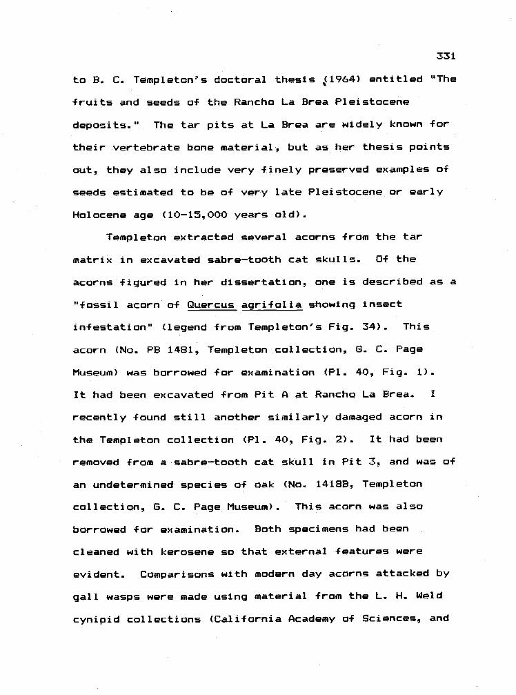
331
to B. C. Templeton's doctoral thesis ,ç1964) entitled "The
fruits and seeds of the Rancho La Brea Pleistocene
deposits." The tar pits at La Brea are widely known for
their vertebrate bone material, but as her thesis points
out, they also include very finely preserved examples of
seeds estimated to be of very late Pleistocene or early
Holocene age (10-15,000 years old).
Templeton extracted several acorns from the tar
matrix in excavated sabre-tooth cat skulls. Of the
acorns figured in her dissertation, one is described as a
"fossil acorn of Quercus agrifolia showing insect
infestation" (legend from Templeton's Fig. 34). This
acorn (No. PB 1481. Templeton collection, S. C. Page
Museum) was borrowed for examination (P1. 40, Fig. 1).
It had been excavated from Pit A at Rancho La Brea. I
recently found still another similarly damaged acorn in
the Templeton collection (P1. 40, Fig. 2). It had been
removed -from a sabre-tooth cat skull in Pit 3, and was of
an undetermined species of oak (No. 1418B. Templeton
collection, S. C. Page Museum). This acorn was also
borrowed for examination. Both specimens had been
cleaned with kerosene so that external features were
evident. Comparisons with modern day acorns attacked by
gall wasps were made using material from the L. H. Weld
cynipid collections (California Academy of Sciences, and
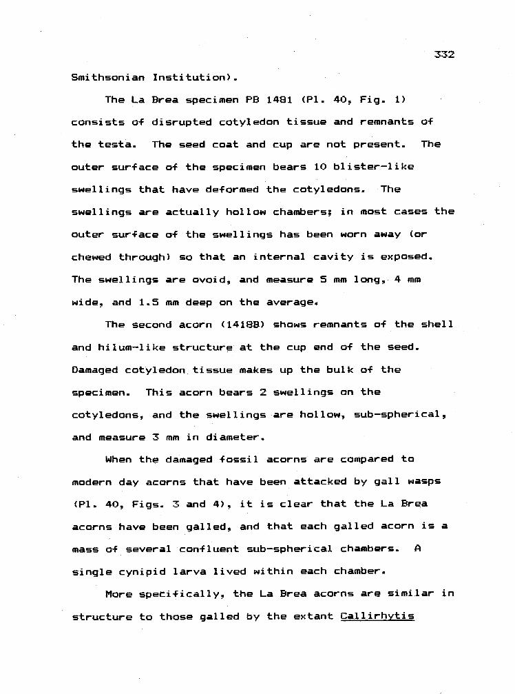
332
Smithsonian Institution) -
The La Brea specimen PB 1481 (P1. 40, Fig.. 1)
consists of disrupted cotyledon tissue and remnants of
the testa. The seed coat and cup are not present. The
outer surface of the specimen bears 10 blister-like
swellings that have deformed the cotyledons. The
swellings are actually hollow chambers; in most cases the
outer surface of the swellings has been worn away (or
chewed through) so that an internal cavity is exposed.
The swellings are ovoid, and measure 5 mm long, 4 mm
wide, and 1.5 mm deep on the average.
The second acorn (1418B) shows remnants of the shell
and hilum-like structure at the cup end of the seed.
Damaged cotyledon tissue makes up the bulk of the
specimen. This acorn bears 2 swellings on the
cotyledons, and the swellings are hollow, sub-spherical,
and measure 3 mm in diameter.
When the damaged fossil acorns are compared to
modern day acorns that have been attacked by gall wasps
(P1. 40, Figs. 3 and 4), it is clear that the La Brea
acorns have been galled, and that each galled acorn is a
mass of several confluent sub-spherical chambers. A
single cynipid larva lived within each chamber.
More specifically, the La Brea acorns are similar in
structure to those galled by the extant Callirhytis
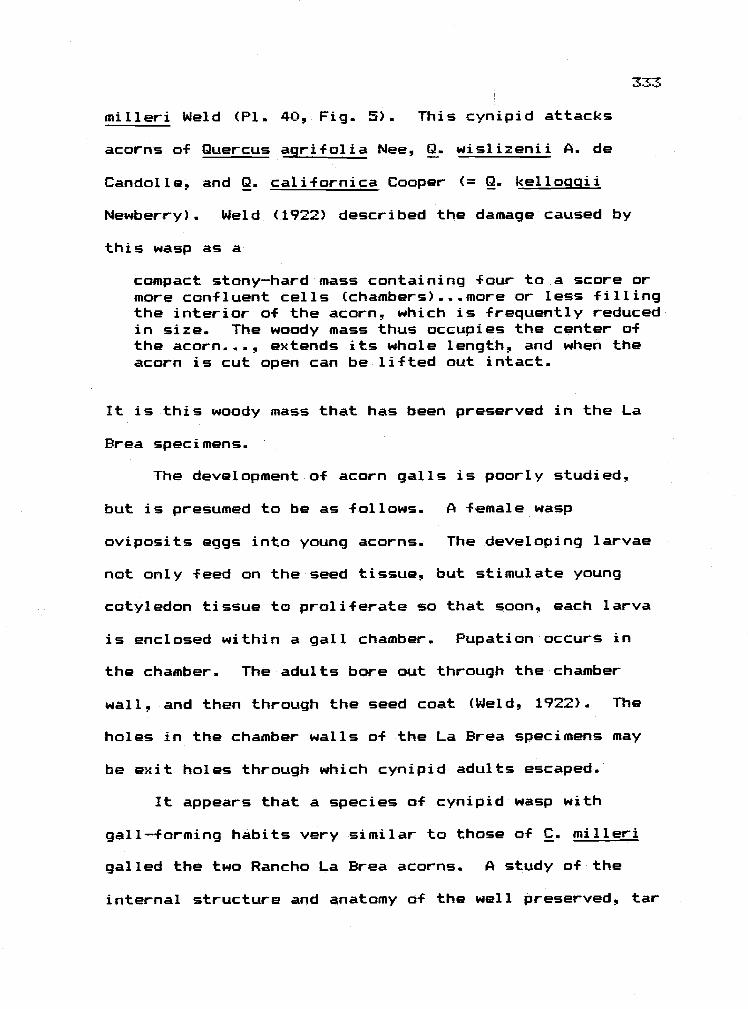
milleri Weld (P1. 40, Fig. 5). This cynipid attacks
acorns of Quercus agrifolia Nee, Q. wislizenii A.. de
Candolle, and Q. californica Cooper (= Q. kellociqii
Newberry). Weld (1922) described the damage caused by
this wasp as a
compact stony-hard mass containing F our to a score ormore confluent cells (chambers).,...more or less fillingthe interior of the acorn, which is frequently reducedin size. The woody mass thus occupies the center ofthe acorn. - , extends its whole length, and when theacorn is cut open can be lifted out intact.
It is this woody mass that has been preserved in the La
Brea specimens.
The development of acorn galls is poorly studied,
but is presumed to be as follows. A female wasp
oviposits eggs into young acorns. The developing larvae
not only feed on the seed tissue, but stimulate young
cotyledon tissue to proliferate so that soon, each larva
is enclosed within a gall chamber. Pupation occurs in
the chamber. The adults bore out through the chamber
wall, and then through the seed coat (Weld, 1922). The
hales in the chamber walls of the La Brea specimens may
be exit holes through which cynipid adults escaped.
It appears that a species of cynipid wasp with
gall-forming habits very similar to those of C. milleri
galled the two Rancho La Brea acorns. A study of the
internal structure and anatomy of the well preserved, tar

334
impregnated galls may be possible, but must await
discovery of additional paratypic specimens.
Additional points about Rancho La Brea should be
mentioned. First, all types of plant material have been
excavated from the pits. For example, Templeton provides
photographs of three tree trunks with portions of intact
root systems that have been pulled from the pits. At
least one trunk was identified as Cupressus arizonica
var. Hancockii n. var.. Templeton frequently mentions
"masses of plant material" or "quantites of plant
material" or considerable brush and roots" that are mixed
with bones. Leaves ("mostly parts"), twigs, wood and
seeds (flowers?) have been observed. This abundance of
plant material has, for the most part, been ignored.
Secondly, Templeton mentions that in processing
material, she sorted out insect parts. Dr. William
Akersten, Curator of the La Brea fossil collection, has
indicated that most of the insect material is unstudied
(but see Miller, 1978). Thus, there is a very good
chance that much "interaction" material occurs in the
pits and is awaiting study.
Thirdly, the plant material from the tar pits may
provide plant tissue with a preserved internal structure.
Scott (1972) describes sections of leaves taken from the
pits that show cell protopla5m. As will be discussed,

the chance to section through fossil leaf tissue that has
a preserved internal structure is one that should not be
ignored..
Well preserved specimens of galled Sequoia seeds in
cones from Miocene? deposits in Germany have also been
reported (Mohn, 1960). The galls contained cecidomyiid
larvae and pupae. The damage resembles that caused by
members of the genus Contarinia to the seeds of Douglas
fir in the Pacific Northwest (Furniss and Carolin, 1977)..
3) Bark and Stems.. Present day bark and/or phloem
is frequently tunnelled by a variety of beetles.. With
this in mind, the tree trunks taken at La Brea should be
scanned for beetle damage.. Suss (1979) described pith
flecks in Miocene conifers (e.g. Pruninium, Callitris)
caused by cambium tunnelling agromyzids (Phytobia). The
tunnels are filled with altered tracheids that preserve
well.. These filled mines (pith flecks) strongly resemble
those caused by Phytobia in modern day hosts.
Interestingly, however, the fossil specimens are
gymnosperms, while extant Phytobia mine only angiosperm
woods.
Stems are also frequently galled, but no example of
a fossil galled stem is available.. Stems may display
extra-floral nectarys or spines. One clear example of

336
fossil stem spines on Crataegus is shown in an article by
Berry (1925) on the Pleistocene plants of North Carolina.
Axeirod (1956) describes a thorn from Crataequs
middleqatei Axeirad. Berry (1925) refers to Pleistocene
prickly twigs that resemble Rubus stems Kellogg (1927)
describes an imprint of a lower Eacene to lower Oligocene
stem of Rosa hjlliae that shows thorns (P1. 40, Fig. 6).
Spines from the Pliocene have also been described by
Szafer (1946). All of these thorn specimens indicate
that as early as the Eacene-Oligacene, plants had
developed recognizable structural defenses.
The presence of intercalary meristems in grasses, if
possible to detect in fossil grass stems, would suggest
the presence of grazers. Knowlton provides plates of
what he believes are grass stems and rhizomes in his 1925
paper. Templeton (1964) mentions that "grass or grass
roots similar to Bermuda grass" have been found at La
Brea. Additionally, Heer (1855) figures several Tertiary
grass stems from Europe. many of which have attached
crowns and raat. Thus, the material is available -- it
merely needs to be studied.
(K. B. Leslie and C. H. Driver, from the University
of Washington, have recently described a paleopathogen on
grass stems taken from pack rat middens in Nevada (1981,
paper presentation at Northwest Scientific Association.
337
Corvallis! OR). The material is estimated at 3500 years.
Pircnzynski (1976) reviewed the literature on fossil
fungi. Fossil evidence for the association between fungi
and plants dates from the Devonian. See Heer (1855) and
Meschinelli (1898) for several figures of fossil plant
parasitic fungi.)
Roots. Although many must exist I have only
Templeton's (1964) reference to preserved roots. She
states that roots are very frequently encountered in the
tar pits of La Brea. It would be interesting to know if
these roots are galled (many extant oak bear cynipid root
galls), or are damaged from chewing or tunnelling.
Leaves. With the exception of pollen and
possibly wood, more fossil leaves have been collected,
described and photographed than any plant part. Both
internal and external features of leaves may suggest
interactions with herbivores. One might wish for fossil
material in which the internal cellular structure of the
leaf had been preserved. Such material would be very
valuable for at least two reasons. The presence of
silica crystals (in grasses), calcium oxalate deposits
(many plant families) or calcium carbonate crystals
(Ficus) within the leaves might be observed (Esau, 1977).

338
The function of lea-F crystals is unclear but they may in
part serve to deter herbivores.. Silica crystals. for
example, are thought to have influenced the dentition of
grazing herbivores. In the same vein, the foliar anatomy
of C4 plants (a type of photosynthetic pathway) has been
suggested recently to inte-fere with herbivory (Caswell
and Reed, 1975).. Presumably, the foliar proteins in C4
leaves are bound in thick walled (easily preserved?)
bundle sheath cells that may not be easily digested by an
herbivore.. This -Feature would be one that could only be
detected in -Fossil material with intact internal leaf
structure. Plant physiologists should have an interest
in the search f or such paleo-anatamical -Features.
Giannasi and Niklas (1981) have reported using
biochemical techniques to identify several secondary
plant compounds from Miocene leaf material. Niklas
(1981) has also exhibited electron micrographs that show
the ultrastructure a-f 20 million year old angiosperm
leaves. This work should add a new dimension to
paleobotanical and paleoecological studies.
External leaf features that suggest interactions
include marginal spines, examples of which have been
observed in fossil material -From the Pleistocene (figured
in Berry, 1925; Axelrod, 1964). Foliar spines
undoubtedly occur on leaves a-f same of the 28 species of

339
hex that have been described -from -fossil material
(Knowlton. 1898). Additionally, -F oliar investiture
and/ar glandular trichomes may deter herbivores.. Their
presence on the surface of fossil leaves (Scott, 1972)
would be suggestive of deterrence but as of yet,
pertinent observations have not been checkhisted..
Other external lea-f features that paint to
interactions with insects include galls, mines, and
domati a.
FOSSIL GALLS
Kinsey (1919) comments that it is almost pointless
to attempt identification a-f the cecidozoan from
preserved galls.. He relys more on the promise of
preserved gall insects. After scanning a leaf imprint
collection, I would agree that difficulty exists in
first, determining if a lea-f blemish is a gall, and then,
suggesting a causative agent.. The more galls that are
available to study, however, the easier it may be to
identify the causal agent. Kinsey's remark begins to
pale when specimens such as Mahn's (1960) are discovered.
Reports of galls in the North America and Europe
follow.. In mast cases, fossil galls are compared to
similar extant types. The reports are treated
chronologically by publishing date, and are checklisted

340
in Table Fo-1. Trotter (1903) and Kuster (1911a)
compiled lists of accounts of Tertiary galls. As of yet,
I have been unable to track all of their references, but
those that I have found are included.
Scudder (1886) states that "two or three" cynipid
galls have been obtained from the Florissant. Kinsey
states, however that these galls are not caused by
cynipids (1919).
Marty (1894) figured a cecidomyiid gall on Faqus
from the Pliocene deposits of Pas-de-la--Mougudo.
Cockerell (1908) described a cecidomyiid gall
(Cecidomyia (?) pontaniiformis sp nov.) on the leaf of
Myrica drymeja (Lx) from the Florissant Formation that
"very closely resembles those formed by the species of
Pontania (a genus of sawf lies) on Salix. The gall is
about 8 mm lang, 5 mm broad, being contiguous to the
midrib. Several examples were found." In the same paper
he described galls on what is possibly a Salix leaf taken
from the Fiorissant that he believed were caused by the
eriaphyid mite, Eriophyes (?) beutenmulleri sp. nov..
These were described as being "small subtriangular galls
about 2 mm in diameter, at the angles formed by the
junction of the principal lateral veins of the leaf with
the midrib; four galls on the leaf found, 2 to 5 mm
apart." Extant genera of Salix are heavily galled by

341
eriaphyoids and, it is possible that Cockerell's leaves
were galled by mites. Both of these specimens should be
confirmed, in as much as the published photographs are
somewhat unc 1 ear.
4) Brues (1910) described a galled leaf on Myrica
obscura Lx. or M. drymeja (Lx.) from the Florissant that
he suggested was caused by a cynipid (Andricus myricae
sp. nov.). He mentioned that Lesquereux also figured
galls that resembled this gall in early papers. Once
again, the specimen should be recon-firmed and
photographed. Houard (1922) describes an eriophyid leaf
gall on Myrica fava that does not fit Brues' description.
This is the only described gall on extant Myrica. If
both Cockerell's and Brues specimens of Myrica are indeed
galled, then one might suspect that the incidence of
galling o-f this genus has changed through time.
) Berry (1916) figures a gall on the petiole of
Cedrela puryearensis (Family: Meliaceae) , and galls on
Rhamnus leaves from the Wilcox formation of Tennessee.
He suggested that the Cedrela gall was caused by
cynipid or hemipterans. The Cedrela specimen resembles
none of the described galls on extant species of Cedrela
(Houard, 1922). Berry suggested that the Rhamnus galls
were either cecidomyiid or aphid caused. Houard (1922)
describes a cecidomyiid gall on 2 extant species of

342
Rhamnus one of which resembles the fossil gall: "Small
projecting growths on the upper surface of the leaf, very
numerous, measuring from 0. to 3.0 mm in height; tinged
green." Bath fossil specimens should be studied further..
Cackerell (1927) mentioned the galls of
Cecidomyia chaneyi on the leaves of Acer osmanti, and
eriaphycid galls on alder leaves from the Bridge Creek
Flora.
Hoffman (1932) published photographs of a
preserved leaf of Quercus coanatus from the late Miocene
shales of Douglas County, Washington. The leaf shows "25
gall-like impressions, circular in outline, ranged in
diameter from 0.3 cm to 0.4 cm, and were characterized by
a dotlike protuberance at their centers." He likened the
galls to cynipid or cecidomyiid galls an extant oaks.
Many species of cynipids are known to form spherical
galls (frequently in large numbers) on the underside of
oak leaves. The "dotlike protuberance" may be the
imprint of the hard wall larval capsule.
Madler (1936) provided a photograph of a Tertiary
petiole gall on Populus latior. The gall was probably
caused by Pemphiqus aphids.
Berger (1949) described a cynipid gall on
Pliocene oak leaf material. He suggested that the gall
was caused by Neuroterus.

343
10) Brooks (1955) described the preserved leaf galls
on Cupanites formosus from Puryear, Tennessee (Wilcox
Formation: Eccene) that resemble "recent simple pouch
galls like those produced...by gall mites (Eriophyidae),
aphids (Aphidae), jumping plant lice
(Chermidae=Psyllidae) and several other types of lowly
arthropods" Cupanities is an extinct genus that
resembles the extant genus Cupania or Cupanioosis
(Sapindaceae), both of which have leaf pocket galls
recorded from New Calcedania that are caused by
eriophyaids and insects (Houard, 1922).
Brooks also provided plates of a gall on a leaf of
Nectandra pseudocoriacea (Lauraceae) from the same
formation that "bears eleven well preserved cone galls.
The structures are mammillif arm and seem to be separated
from the leaf by a constriction at the point of
attachment. " He goes on to say that "immediately
surrounding the galls, the leaves are darkened which
probably reflect sclerotized leaf tissue in the vicinity
of the galls as the coloration is due to a greater amount
of lignified leaf residue." He compares these galls to
mite. psyllid, gall midge and gall wasp galls. Houard
(1933) lists galls on Nectandra from Central and South
America, many of which are an the leaves, are stalked.
and are caused by cecidomyiids.

Straus (1977) provided photographs o-f 34 leaf
galls (the richest gall flora yet described) from the
Miocene clay pits of Willerhausen. Germany. Included are
many presumed mite, aphid, midge, and wasp galls.
Ambrus and Hably (1979) provided a photograph of
a presumed eriaphyoid gall on a Daphogene leaf from a
Hungarian upper Oligocene deposit.
In summary, to my mind the clearest example of a
fossil gall is that provided by Mohn (1960) from the
Miocene There are older specimens (Eocene), but many of
these are equivocal. One can certainly say, however,
that recognizable galls occurred in the mid-Tertiary.
GALL INSECTS
Brooks (1955) provides a list of those gall-forming
arthropods that have been found in fossil material.
Larsson (1978) briefly discusses the cynipid and
cecidomyiid material that has been collected in baltic
amber -
Southcott and Lange (1971) discovered an eriophyoid
mite (a rust mite, I believe) in Australia (Eocene).
Preserved remains of insects that belong to a taxon
that today contains gall-f ormers is suggestive, but does
not definitely indicate that the fossil insects formed
galls; all of the extant taxa containing gall-f ormers
344

345
also contain insects o-f non-gall--forming habits..
Malyshev (1968),, -for example, suggested that the early
members of the gall--forming lineage a-F hymenopterans were
free-i iving.
"HIDDEN" FOSSIL GALLS
There is reason to believe that more fossil galls
will be described. Collections continue to be made in
deposits containing beautifully preserved leaves (such as
those in Eastern Oregon) . What is even more assuring is
that undescribed examples a-F galled leaves are to be
found in museum collections a-f fossils. For example, R.
W. Chaney in 1920 reported that "leaf galls are present
on a number of specimens" a-f Quercus psuedo-lyrata taken
-From the Eagle Creek Formation (Oligocene) of Northeast
Oregon (collection housed at the Field Museum Chicago).
When one considers that Chaney reports collecting 1600
leaves a-f this species his "a number a-f" becomes
significant, and yet, this statement has not been
pursued. Chaney published no photographs a-f these galled
leaves, and did not speculate as to the causative
agent (s).
Another example suggestive a-f unnoticed gall
material is provided by plates in Knowlton (1925). The
petiales of three a-F these leaves of Populus heteromorpha

346
leaves -From the Miocene of Spokane and Coeur D'Alene
appear to have either been bent or thickened -- much like
the early stages o-f petiole galls on poplars that are
caused by Pemphiqus aphids (Plate 39, Figs. 1 and 2) or
Ectoedemia moths today.
In quickly scanning the collection of lea-f imprints
at the University of California at Berkeley
Paleobotanical collection., I found six leaf imprints of
various species o-f Populus that showed unusual petiole
features (bent,, curved, or thickened). Representatives
are shown in Plate 39. Figures 3-7. All resemble early
stages o-F aphid or moth petiole galls. Pertinent fossil
specimens are available -- and await appreciation.
With the exception o-F Madler's work, larger petiole
galls have not been observed, but work by Smith (1932) is
suggestive. In her Master's thesis (University of
Oregon) she studied the fossil flora of Rockville, Oregon
(Malheur County). When describing the leaves of Populus
Lindqreni Kn. she says,
The base is either rounded or cordate. petiale notpreserved. The specimens give the unique impressiona-f having the upper part of the petiole lost in thesubstance of the leaf as in no instance is the midribdistinguishable in the lowest portion a-F the bladealthough it is very prominent above.
Such a comment should be pursued, -for any abnormality
observed in poplar petiales suggests the presence of

galls Unfortunately her photographs are not clear
enough to see this character a-f the leaves.
A SEARCH METHOD FOR DISCOVERING FOSSI GALLS IN
COLLECT I ONS
If one compares a list of fossil plant genera with a
list a-f extant plant genera that are known to have galls.
one should derive a list of fossil plant genera that
could be expected to show plant galls (or mines, or
domatia). I have compiled such a list. I used
Knowlton's (1898) "A catalogue a-f the Cretaceous and
Tertiary plants a-f North America," and various volumes by
C. Houard and E.. P. Felt that describe galls o-f the
war 1 d.
Limitations of such a survey need to be mentioned.
First, I assume that the -floral distribution a-f galls was
similar in the Cretaceous and Tertiary to that seen
today. Also, Knowlton's list is old. More recent
catalogues af this type are available but, at the generic
level, Knowlton's is fairly up to date. His list does
not distinguish between plants known only -from pollen and
those known from vegetative material. Also, his list
includes only North American material. I do not know how
such a list would change i-f world wide paleo-floras were
included. In same cases I have been unable to determine
347

348
if a plant genus in Knowlton is extinct or extant. In
terms o-f gall -floras, Houard's volumes are as thorough as
we have. Undoubtedly more galls exist than he described,
but his list is a representative one.
0-F the 384 fossil genera listed by Knowlton, 280
have no recorded galls on extant members, while 104 do.
I do not know how many of the 280 genera that have no
galls are extinct.
There are 26 genera that have both many fossil
species tmost likely, many fossil specimens) and a rich
gall flora (Table Fo-2). Based on the number of fossil
species and the richness of the gall flora, five of those
genera are particularly apt to provide examples o-f galls.
Both in the field and in museum collections, fossilized
vegetative material of Salix, Eucalyptus, Ficusq Populus
and Quercus should be thoroughly studied for galls. I-F I
had one day in an extensive fossil leaf collection I
would scan the oaks.
Knowing the type o-f cecidozoan responsible for galls
on extant genera, one might more easily predict the type
of gall that should be found in preserved material.
Similarly, given the size, shape, and location of a
fossil gall, one should have some idea a-f the
gall-former. For this reason, I briefly list the
cecidozoans, and the features of their galls.

349
- Nematode galls are predominantly found an roots -
a plant part that is infrequently preserved.
- Eriaphyid galls may be difficult to detect in
fossil material because the galls are usually only a
swollen bud, or a dense patch of hairs on the leaves,
particularly at vein axes or along veins.. If the mites
-Form pocket or spherical galls an the leaves, the galls
are frequently small and numerous..
- Aphids frequently cause leaf rolls or leaf
crinkling. The spiral petiole galls caused by aphids an
paplars should be fairly distinct.
- Thrips galls are primarily leaf rolls or small
pimples on the surface o-f leaves, usually found many per
leaf. These might be confused with mite galls.
- Psyllid galls are usually pit or pouch galls on
leaves.. They might be confused with midge galls.
- Scale galls are usually tubular swellings the
galls of males) on the leaves a-F Eucalyptus.. The La Brea
woody stem material should be checked -For the presence o-f
the common pit scale gall that occurs on many oaks.
- Beetles and moths generally form stem galls that
should preserve nicely.
- Sawfly leaf galls are usually sub-spherical.
contain frass, and are frequently found on Salix. Stem
swelling on willows are often caused by a sawfly.

350
- Cynipid galls are morphalogically diverse. Any
gall on oak should -first be suspected of being a cynipid
gall (but see Kinsey, 1919).
- Cecidomyiid galls are variable in shape. They are
usually foliar, and can involve everything -from a leaf
roll to a blister to a fairly large chamber. They occur
an many host species.
FOSSiL LEAF MINES
Evidence is strong that by Upper Miocene the leaf
mining habit was well established. New evidence from
Poland pushes the habit back to the Cretaceous (Skalski,
personal communication). Table Fo-3 compiles the
published accounts of fossil lea-f mines.. Table Fo-4
indicates those extant plant -families that host 3 or 4
orders o-f insect miners (-from Hering. 1951). I-F miners
preferences -for certain host plant -families were similar
in the past to those exhibited today, then those plant
families listed in Table Fo-4 should contain examples of
-fossil mines. Thus, one would predict that at the
generic level, Populus, Quercus. and Salix would likely
contain mined paleo-specimens. (Fossil Eucalyptus leaves
will also perhaps be -found to contain mines -- see CSIRO.
1970.)
Using such predictions, I scanned the Berkeley

351
collection -for mines and -found the specimen shown in
Plate 39, Figure 8. In Paul Oplers opinion (personal
communication> this is a leaf mine (perhaps two) of
Ectoedemia (Lepidoptera: Nepticulidae). It occurs on a
lea-f o-F Quercus navadensis (Lesq.) -from the La Porte
flora of California (Potbury, 1935). The tunnel is
linear and meanders near the edge o-F the lea-f. The
tunnel is raised above the level of the lea-f imprint --
an unusual -feature, in that all other -fossil leaf mines
that I have seen photographs of show little relief. The
tunnel shows no frass (suggesting a microlepidopteran
mine), and it crosses numerous lea-f veins.
According to Hering (1951):
We are -familiar with mines in a fossilizedcondition only from the Tertiary. Pre-Tertiaryspecimens described as mines cannot be considered assuch; they are in many cases other types o-f feedingpatterns, mostly -furrows in the leaves caused by wormslying at the bottom of lakes.
Un-fortunately, he does not detail how worm damage can be
distinguished -from that a-f miners. In this regard, Crane
and Jarzembowski (1980) provide useful criteria -for
recognizing fossil mines.
DOMATIA
These are structures on leaves that are used by
mites as domiciles. They are not caused by the mites,

but once formed by the plant., are used by the mites.
There is considerable interest in why plants should form
domatia and undoubtedly with additional work the
association between the domatia and their inhabitants
should become clearer. Hereq howeverq I ignore the
controversy and simply assume that domatia are leaf
structures which are indicative o-f a plant-arthropod
association
Although many forms have been described, there are
two basic types of domatia. One is a pocket that is
formed by the leaf lamina at vein axils.. A dense patch
o-f hairs (erineum) at the vein axils constitutes the
second type. Both types are found on the underside of
the leaf.. I have compiled a list of plant families
(taken from Penzig, 1903) that have 10 or more species
with domatia (Table Fo-5).
After scanning this table one would be tempted to
look at rubiaceous fossil material (particularly. Coffea)
f or the presence a-f domatia. Knowlton (1898), however,
lists none of the rubiaceous genera that Penzig tallies.
In other words, rubiaceous material from North America,
up to 1898, was unavailable, and is most likely still
very scarce. Comments by paleobotantists at the 1981
AIBS meetings in Bloomington. Indiana, however, indicate
that a few specimens a-f Rubiaceous fossil leaves exist.

Imprints of Aesculus (Family: Aesculaceae) or Tilia
(Family: Tiliaceae) might provide -First examples of
erineal domatia. Straus (1977) describes what he
suspects is a -fossil erineal domatium on Tilia-like
leaves (Pliocene). This is the only reference to fossil
domatia.
SUMMARY AND CONCLUSIONS
I have attempted here to point out plant structures
that suggest an interaction and that might fossilize.
The task now is one of thorough examination of the
fossils -for these features.
In terms o-f host plant genera, Quercus seems to be a
genus in which more work with fossil material will prove
productive. "Few plants are so variously and so
characteristically deformed by gall insects as are the
oaks..." (Trelease 1924). One could also add mines and
domatia to the unusual features found on oaks. The
abundance of Quercus leaves in North American fossil
collections has been mentioned frequently (see Trelease.
1924, for a list of fossil species with collection site
data).
Published evidence of paleo-herbivory is neither
abundant nor compiled. Such evidence should, on the one
hand, come from morphological features (e.g. mouthparts)

354
of the palso-herbivores (only rarely mentioned in this
review) and on the other hand come from features of
paleo-plants that suggest or demonstrate that herbivory
occurred.
The relatively small number o-f published accounts o-f
-fossil specimens and the uncertainty surrounding many of
these specimens make detection o-F reciprocal changes
through time in plant and herbivore difficult.
Nonetherless the recent increase in palea-examples of
mines and galls in the U.S. and in Europe indicates that
these habits were well established in the Tertiary.
It is the degree of subsequent changes that is
di-fficult to discern. Two examples suggest that shifts
in host plant preference have occurred through time.
Suss's stem-mining agramyzid that has presumably moved
from gymnosperms to angiosperms is one such example.
Likewise no extant seed gall midges are recorded from
Sequoia in the western U.S. while several occur in cone
o-F Pinus and Psuedotsugae. Thusq Mohn's fossil Sequoia
cone midge suggests a host shift..

TABLE Fo-1
PUBLISHED ACCOUNTS OF FOSSIL GALLS*
* See Trotter (1903) and Kuster (1977a) for additional oxamples.
355
Source Plant Cecidozoan Age
Scudder, 1886 Cynipid OligoceneMiocene
Marty, 1894 Fequs Cecidomyia faqi Pliocene
Cockerell, 1908 Myrica drynieja Cecidomyiid Oliocene-Miocene
Salix Eriophyoid
Brues, 1910 ?lyrica obscura Cynipid Olicocene-Miocene
Berry, 1916 Cedrela Cynipid or hersipteran Eocene
Rhamnus Cecid or aphid Eocene
Chaney, 1920 Quercus oseudo-lyrata ? Oligocene
Cockereli , 1927 Acer osmonti Cecidomyla chaneyi Upper Miocene
Alnus carpinoides Eriopbyes leevis
Hoffman, 1932 Quercus cognatus Cynipid or cecid Miocene
Madler, 1936 Populus latior Pemphiqus Upper iocene
Berger, 1949 Quercus Neuroterus Pliocene
Brooks, 1955 Cupanites formosus Stylet-bearer Eocene
Nectandra ? Eocene
Mohn, 1960 Sequoia lanqsdorfii Sequoiomyia krauseli Miocene
Straus, 1977 Several Several Pliocene
Ambrus and Rably, 1979 Daohnogene bilinica Erioohyes daohonooane Upper Oligocene

TABLE Fo-2
GENERA OF FOSSIL PLANTS ON WHICH
GALLS ARE LIKELY TO BE FOUND
*Richest fossil remains, and extant menbers most commonly galled.
Acacia Celtis *Fjcus Pistachia *Saljx
Acer Cinnamomum Grewia *populus Spirea
Amelañchier Crataegus Juniperus Prunus Tilia
Betula *Eucalyptus Pinus *Quercus Ulmus
Carex Eugenia Piper Rhus Viburnum
Vitis

TABLE Fo-3
PUBLISHED ACCOUNTS OF FOSSIL MINES
Source Plant Miner Formation
Berry. 1916 Several Lepid. Tineidae S. F. North America
Berger, 1949 Cinnaniomum ? Agromyzidee Sarajevo
Brooks, 1955 Proteoldes wilcoxensis Lepid. Nepticulidac Wilcox
Freeman, 1965 7 Lepid. Lithocolletis 7 White Lake, B. C.
Opler, 1973 Several (Quercus) Lepidoptera Western U.S.
Hickey and Hodges,1975 Quercus 7 Nepticulidae Latah
Cedrela Lepid. Phyllocnistis Wind River
Straus, 1977 Several Several orders Willershausen
Crane and Jarzembowski, 7 Nepticula, Phytomyza,1980 Bucculatrix, Stigmella, Woolrich and Reading

TABLE Fo-4
PLANT FAMILIES ATTACKED BY MINERS
(From Flering, 1951)
Those plant families mined by four orders of insects:
Juncaceae Ranunculaceae
Sal i caceae Rosaceae
Betulaceae Geraniceae
Ulmaceae
Those plant families mined by three orders of insects:
Graminae Capparidaceae Campanulaceae Labiatae
Orchidaceae Cruciferae Aceraceae Scrophulariaceae
Fagaceae Crassulaceae Tiliaceae Plantaginaceae
Polygonaceae Legumfnosae Malvaceae Caprifoliaceae
Chenopodi aceae Tropaeol aceae Primul aceae Di psacaceae
Caryophyllaceae Oleaceae Gentianaceae Compositae
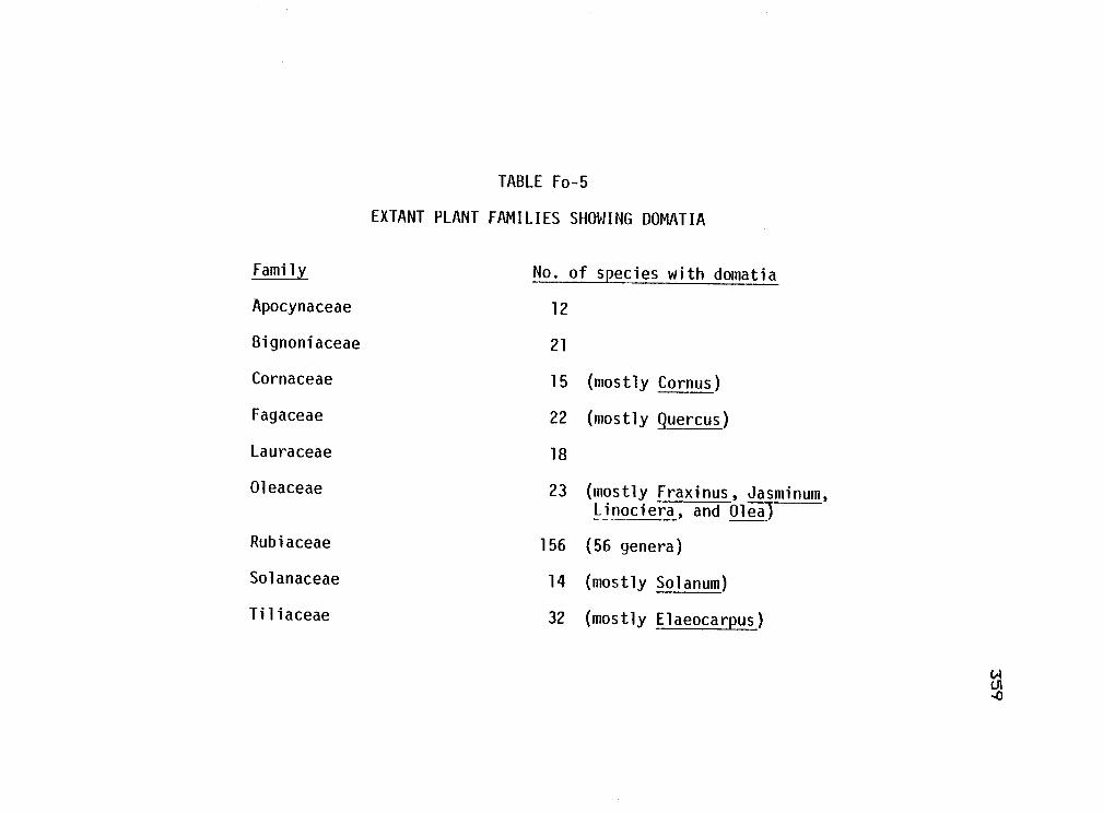
TABLE Fo-5
EXTANT PLANT FAMILIES SHOWING DOMATIA
Family No. of species with domatia
Apocynaceae 12
Bignoniaceae 21
Cornaceae 15 (mostly Cornus)
Fagaceae 22 (mostly Quercus)
Lauraceae 18
Oleaceae 23 (mostly Fraxinus, Jasminum,Linociera, and Olea)
Rubiaceae 156 (56 genera)
Solanaceae 14 (mostly Solanum)
Tiliaceae 32 (mostly Elaeocarpus)

APPENDIX 3: GALLS OF IMPORTANCE TO HUMANS
When considered in sum, gall--Forming organisms are
economically and scientifically important.. When one
realizes that such a group encompasses everything from
nitrogen--Fixing bacteria to -Fig pollinating wasps a
diversity o-f activities and impacts should be expected.
This appendix has a two-fold purpose.. First, it
contains a checklist of gall organisms that are important
to humans.. Although long., the list is undoubtedly not
complete. Perhaps most disturbing are the omissions that
result from our North American - European bias.. Although
briefly covered here., the Oriental and Tropical galls
will undoubtedly contribute more to such a list in -future
years..
Secondly,, during the course of reviewing the
important galls, I detected patterns that suggest reasons
for the destructive or non-destructive nature of plant
galls. I also noticed a convergence in anatomical
features of the host-parasite inter-Face. A discussion of
these patterns will be integrated with the descriptions
of the galls.. In the summary and conclusions I will
discuss what these patterns indicate about the usefulness
a-F galls.
360
361
Galls are significant either because of the damage
they cause to crops., or because of their potential
benefit to humans. I have organized the appendix with
these two types of impact in mind.
DETRIMENTAL GALL-FORMING ORGANISMS
The most familiar and studied gall-forming organisms
are those that cause damage to crop plants. Most crop
damaging gall-f ormers are either pathogens or nematodes -
there are -few gall-forming mites or insects that are
considered crop pests..
The purpose of this section is to review the
detrimental gall-forming pathogens., nematodes., and
insects. Possible reasons f or the difference in
agricultural importance between gall-forming pathogens
and arthropods will be also discussed.
1) Slime Molds.. The most important slime mold plant
pathogen is the clubroot organism. Plasmodiophora
brassicae. As is indicated by the common name, this
pathogen causes an abnormal swelling of the roots.
Crucifers are the most susceptible plants, and when a
field becomes infested not only is the current years
crop lost, but the field can never be replanted with
cruci-Fers. Resistant plant varieties have only been
362
marginally useful (Agrios1 1973).
The slime mold invades the root system via wounds or
root hairs. Moving intracellularly within the host root,
the plasmodium spreads -From the cortex to the cambium and
then into the xylem. The invaded root cells are
-Frequently stimulated by the pathogen to a hypertrophic
and hyperplastic state such that their overall size is
-five times that a-f the normal cell. The nuclei and
nucleoli a-f the in-f ected cell are also greatly enlarged,
and the cytoplasm of these cells is abnormally dense and
-Fine-grained (Stevens, 1925; Williams, 1966).
Meristematic host tissue, such as the cambium, is
mare susceptible to invasion and alteration by the
pathogen than is non-meristematic tissue. Damage to the
host plant is caused by the diversion a-f -foodstu-F-fs to
the diseased tissues (Mitchell and Rice, 1979) as well as
by the disruption a-F the transport system through the
roots. Wilting and stunting are common above-ground
symptoms.
2) Bacteria. Pseudomonas savastanoi causes tumors
on the stems a-F Oleander spp.. The bacterium produces
indole acetic acid which presumably a-f-f ects plant cells
that neighbor the bacterial colony such that they are
enlarged and multinucleate. The enlarged cells

363
eventually undergo cytokinesis such that many cells are
formed from each enlarged cell. The new cells are
meristematic, and have a denser cytoplasm. and more
prominent nuclei and nucleoli than do normal cells
(Strobel and Mathre, 1970)..
Of all detrimental gall-forming organisms, the most
thoroughly studied is crown gall.. This is a disease
primarily of fruit trees caused by the bacterium,
Agrobacterium tuma-faciens. Diseased plants generally
grow poorly, show reduced yield, and if severely
infected, may die. Galls are usually located at the
stem-root junction of the plant..
Crown gall bacteria enter the host through wounds
and move within the crown tissue intercellularly. Host
cells near the bacteria become hyperplastic, and
multinucleate. Rapid division by these cells results in
a fleshy mass a-F undifferentiated parenchymatous tissue
with an unorganized vascular system. The growth of the
gall crushes adjacent normal tissue.. Vascular transport
from the roots to the stem and leaves is frequently
reduced one half to one fifth the normal flow
(Agrios, 1973) -
We now know that the hyperplasia. or oncogenecity o-F
host plant cells caused by the bacteria is incited or
induced by genes on a plasmid which are transferred -From

364
a bacterium to the plant cells. The mechanism of plasmid
transfer between parasite and host, and between host
cells, however, is not clear (Lippincott and Lippincott.
1975).
Besides becoming hyperplastic, the multinucleate
invaded host cells also produce relatively large amounts
of basic amino acid analogs (e.g. nopaline, octopine>.
The role o-F these compounds is unclear, but the
intriguing suggestion has been made that these compounds,
produced by plant cells, are used by the bacteria as
food. According to this scenario, genes carried on the
bacterial plasmid that invade the host cell nucleus
direct that cells to produce compounds which the bacteria
can use, but which the plant cell cannot use, or can only
poorly use (BCGRGI 1979).
The study of crown gall has contributed
substantially to our understanding of the transformation
process <normal to hyperplastic) in a gall. We do not
know, however, the extent of similarities between crown
gall and other types o-f galls. For example. plasmid
tran-fer is not suspected in insect galls, but at the same
time, we do not understand how insect saliva induces
rather specific changes in host plant cells.
Additionally, we know relatively little about the
products of the increased metabolism in nutritive cells,

365
and one wonders if these cells synthesize products
tailored to the needs o-f the cecidozoan.
There is no reason to suspect that a single
gall--forming process is shared by all gall-forming
organisms.. The point is, however, that for the first
time the process is described well enough in one type of
gall that we can begin a comparative inspection o-f other
galls. The detrimental impact o-f crown gall disease
pales slightly as its usefulness as a laboratory organism
increases.
3) Fungi.. Table Hu-1 lists those genera or species
o-f fungi that cause a hyperplastic and/or hypertrciphic
response in the infected host tissues.. Undoubtedly other
gall-forming species exist, but the list should at least
be representative.
I studied descriptions of each a-f these galls and
noted the -following shared characteristics.. Sources I
used include Anderson (1956), Curtis (1921), Dickson
(1947), Jones and Dreschler (1920), Stevens (1925), and
Stewart (1915)..
A) The pathogen interferes with the plant hormone
system.. Usually i-f hormone balance has been studied, an
increase in the amount a-f these compounds (e.g. auxins,
gibberrelins, or cytokinins) at or near the infected
366
tissue is observed (qrios 1973). The increase may be
caused in various ways, but in well studied examples the
pathogen both produces the hormones itself, and induces
the host tissue to produce the hormone (e.g. Ustilaga
maydis Gymnosporangium juniperi-virqinianae and
Gibberella fujikuroi) (Agrios., 1973).
Many o-f the more serious gall diseases are those
that attack the subterranean plant parts or crown (e.g.
potato wart crown wart a-F al-fal-faL, or the reproductive
structures (e.g. plum pockets., ergot smuts). Galling a-f
the roots or crown disrupts translocation while galling
a-F either the roots or fruits may destroy the harvestable
portion a-f the plant.
Frequently the infection site is a meristematic
tissue (e.g. Ustilaqo, Urophlyctis., Taphrina). Whether
this is coincidental or an actual requirement -for gall
initiation in these plant pathogens is not clear.
) The host plant cells found in close proximity to
the pathogen -frequently become hypertrophied and
hyperplastic. The nuclei and nucleoli a-f the plant cells
may be -found mare than one per cell, and/or are
frequently enlarged (Strobel and Mathre, 1970; Mims and
Glidewell, 1978).
In at least one example, that a-F the cruci-fer white
rust (Albuqo candida), infected plant cells have a
367
cytoplasm richer in ribosomes than do normal cells
(Agrios, 1973). Ultrastructural studies a-f plant cells
invaded by haustoria of rust fungi (e.g. Puccinia.,
Melampsora. Gymnosporangium) are instructive..
Micrographs indicate that a rich band a-F cytoplasm
encircles the haustorium. "Virtually every study
0-f.. .haustoria shows cisternae of ER lying parallel to
the extrahaustorial membrane" (Little-field and Heath.
1979). Usually ribosomes are associated with these ER.
Additionally., the haustorium is usually closely
associated with the host nucleus.. Future studies of the
response a-f invaded host plant cells should consider when
and where enrichment occurs.
Lastly, Dennis Gray (personal communication, North
Carolina State University) observed isolated, highly
enriched cortical cells in galls caused by
Gymnosporangium. In summary, a statement made by Strobel
and Mathre (1970) should be pursued:
It is not too unusual to expect that cytological-Features of tumorous tissues would in manyways resemble normal meristematic tissue. -An increase in the size a-f the nucleus andthe amount of cytoplasm per cell are add-itional features that may be associated withcells in tumors.
4) Nematodes. Two genera a-f plant parasitic
nematodes are in-famous -F or the root galls that they
368
cause. Members of the genus Heterodera are commonly
called cyst nematodes, while those of Meloidogyne are
known as root-knot nematodes. Damage caused by these
nematodes is frequently very heavy (yield reductions of
30-75h). They both cause stunting, chiorosis. and
frequent wilting o-f the above-ground parts as well as
reduced photosynthetic rates (Loveys and Bird, 1973).
The root system is also stunted and bears many individual
spherical cysts (dead, egg-filled bodies) in the case of
cyst nematodes, or show a general swelling due to the
coalescence of numerous irregularly shaped root-knot
galls.
In both cases when the larvae settle to feed in the
root, the head of the nematode is usually near to. or in
the pericycle (sometimes the cortex). Within a -few days
after the larva settles, the cells that surround its head
begin to enlarge. Nuclear divisions in these cells
occurs without cell wall divisions. Existing cell walls
may also break down, so that by the time that the feeding
site is completely established, 3-6 syncytia. or giant
cells, encircle the head of the nematode (Agrios. 1973;
Bird, 1974). The giant cells usually possess enlarged
nuclei and nucleoli, as well as a cytoplasm that is rich
in proteins, -free amino acids, and nucleic acids (Owens
and Novotny. 1960; Bird, 1961).

369
The nematode functions as a metabolic sink in the
root (Bird and Loveys, 1975). Loveys and Bird (1973)
showed that infested tomatoes had depressed
photosynthetic rates soon after infestation when compared
to uninfested plants. Additionally., the giant cells may
disrupt or crush existing xylem elements. Tissues
surrounding the feeding site also exhibit hypertrophy and
hyperplasia, and a disrupted vascular system frequently
results. Translocation o-f root-produced hormones may be
disrupted (Bird, 1974). It would be interesting to know
what portion of the damage caused by root-knot is caused
by the nutrient sink and, what by the vascular
disruption.. The thoroughness with which root-knot has
been studied increases the chance that this will be the
first eucaryote-caused gall that will be thoroughly
explained.
Mites. Jeppson et al k1975) mention many
injurious eriophyoid mites, but most are rust mites, not
gall mites. These authors do mention, however, that
galled plants are frequently stunted.. Metcalf, et al
(1962) list no gall-forming eriophyoids as economically
important.
Insects.

370
A) Cecidomyiids. In four volumes. Barnes
(1946a,b,1948,1956) discusses the impact o-f about 450
species o-F cecidomyiid midges on various crop plants.. Of
those 450 species approximately 45 cause an economically
important amount of damage to their host plants. Of
those 45, only 17 species cause damage by actually
galling the plant. Most o-F the midges that damage plants
feed on1 but do not gall, the seeds of plants (Barnes1
1956). Also1 midge leaf galls rarely cause significant
damage unless found, as with Procontarinia mattiana on
mango, in very high numbers.
The most important cecidomyiids that form galls are
the rice gall midges (Pachydiplosis oryzeae, and Orseolia
oryzae) (Hidaka, et al, 1977) in tropical Asia. The
midges attack cultivated rice, and use wild rice as a
secondary host. Up to 847. destruction of tillers has
been recorded. No description o-f the galls are given by
Hidaka, but his photographs suggest that the stems are
swollen by the midges.
A variety of cecidomyiids gall either the needles or
twigs of conifers. According to Furniss and Carolin
(1977), however, "none is a major forest pest." Some may
be serious pests o-f ornamental conifers, and in some
years Contaririia orecionensis Foot, by galling the coat of
Douglas fir seeds, may be "the most destructive species"

371
of seed insects on this plant (Furniss and Carolin,
1977).
The Hessian 'fly is the most well known destructive
cecidomyiid. Its larvae, however, do not 'form galls, but
instead suck the saps from stein tissues, and thus cause a
dwarfing of the plant.
Other Diptera.. Stinner and Abrahamson (1979)
showed that 'a gall insect (either a tephritid -fly or a
gelechiid moth larva) represents a loss to a goldenrod
plant o-F over 6,000 calories or approximately 4 percent
o-f "production."" Harnett and Abrahamson (1979) found a
32-43% decrease in reproductive effort of the galled
goldenrod ramet. Furthermore, parasitism 0-f the
gall-former decresed the impact a-f the gall by about 2000
calories. Although goldenrod is not a cultivated crop, I
mention the above work because o-F the careful
measurements o-f damage to the host plant.
Scales. Oak pit scale, a general term -for galls
on the twigs of deciduous and evergreen oaks, caused by
members o-f the homopteran 'family. Asterolecaniidae, can
cause die-back o-F twigs and death of a tree i-f
infestation occurs year after year (Koehier, 1977).
Aphids. In the late part of the nineteenth
century, the grape phylloxera (Phylloxera vitifoliae
(Fitch) was accidentally introduced into Europe on grape

372
roots shipped from the U.S... The phylloxera quickly
became a pest in vineyards on the continent; the leaves
and roots of European vines were heavily galled by the
various life stages of the aphid-like homopteran (P1. 40,
Fig. 7). Mortality of vines became so extensive that the
wine producing industry was threatened. It was only
after resistant rootstocks from North America were
grafted onto European scions that the industy recovered.
In the U.S. the insect usually galls the foliage and
causes minimal damage (Sterling, 1952; Borror and Belong.
1971).
In his study a-f the anatomy a-f Phylloxera leaf
galls, Sterling (1952) noted that the upper surface of
young leaves was attacked most frequently. The insect's
stylet was usually inserted three cell layers deep into
the leaf, and once inserted, it remained in the same spot
for the life o-f the insect. (The li-fe stage is not
indicated by Sterling -- it is most likely the stem
mother.
Hyperplasia was most intense 15 cells away -from the
puncture site, and there was actually a depression of
mitotic activity near the stylet. The cells in layers I
and II, at about 15 cells away from the stylet, developed
enlarged nuclei and nucleoli, and small vacuoles. Starch
grains appeared in these cells. Small vascular bundles

373
were seen in the nutritive zone. Sterling likened this
zone to that seen in other types of insect galls.
Other gall-forming aphids that cause economically
important damage include species of Pemphigus (Aphididae:
Eriosomatinae). The winter host of this genus is
Populus. In the summer the aphids move from the leaf
galls on poplars to the roots of either crucifers,
chenopods, or composites. P. bursarius L... -For example,
attacks lettuce roots (Dunn, 1959), while summer
generations a-F P. populi-transversus Riley move to roots
of numerous cruci-fers (e.g. cabbage, broccoli, brussels
sprouts, cauliflower, and turnips) (Jones, 1919). f.populivenae Fitch moves from poplar to roots of sugar
beets (Grigarick and Lange. 1962). Members of these
species do not produce galls on the summer host, but
instead, remain exposed on the surface a-F the roots, and
suck plant saps. One o-f the earliest symptoms of
infestation is wilting a-F above ground parts. Damage to
cultivated crops may be "considerable1' (Grigarick and
Lange. 1962).
A closely related genus., Eriosama (Aphididae:
Eriasomatinae), contains species that gall the leaves a-F
elms in the spring and early summer. In the summer,
aphids leave the galls and migrate to pomaceous fruit
trees. Members a-F E. laniqerum (Hausemann) gall apple

374
roots and crowns. Twigs may also be attacked but
generally are not galled. Damage to the roots "sometimes
causes the death a-f the tree, stunting1 or serious
retardation of growth" (Metcal-F, et al, 1962). E.
pyricola Baker and Davidson causes a cockcamb lea-F gall
on elm. In the summer the aphids migrate to pear roots.
Damage is caused by removal a-f plant sap, but no root
galls are formed. The aphid can become a serious pest on
European rootstocks (Swenson, 1969).
E) Adelgids. The adelgids attack only conifers.
The sexual stage -form pineapple-like galls on the twigs
of various species o-f Picea. On secondary hosts (e.g.
Pinus, Pseudotsuqa, Abies) the asexuals occur on bark in
large cottony masses. Adelges piceae (Ratz.), a native
a-F Europe, is a major forest pest. This insect may cause
damage to, or death a-f Pacific silvr fir, Subalpine -Fir,
or Grand -Fir in the Pacific Northwest. In addition to
the pineapple gall, these aphids gout twigs, and
-Frequently cause compression-like wood at attack sites on
the bole. This last -Form a-f damage can cause tree death
i-F infestation occurs over several years (Furniss and
Carolin, 1977).
Summary o-F Detrimental Galls
There are relatively -Few recorded gall--farming

375
insects that are pests of crop plants. I suggest that
there are three reasons why this is so First, the gal1s
formed by seed-dwelling insects are sometimes difficult
to detect. Many of the gall-midges. for example, that
feed in seed heads may actually gall the ovary without
this damage being noted. Such omissions in the
literature would bias perceived trends.
Secondly, the majority of insect galls occur on
leaves (Mani. 1964), while very few attack roots. The
pattern observed with bacterial. fungal. and nematode
galls shows, however, that galling of the roots or crown
causes the most extensive damage. Phylloxera is a case
in point. This is one o-F the few gall insects that
attacks roots. When roots are attacked, the host plant
usually dies, but if leaves are attacked, the host plant
often survives. Why damage to roots should be o-f more
detriment to the plant is not clear.
Perhaps the important -Factor, however, is not so
much the tissue attacked, but the extent o-F attack. Most
bacterial and funqal galls arise during the systemic
infection of the host, so that large amounts of host root
tissue are involved. Insect galls, on the other hand,
are generally more localized. Whole leaves are rarely
galled, much less whole branches. Localized galling may
lead to less direct destruction of host plant tissues.

376
Thirdly.! relatively few gall--forming insects attack
crop plants. Instead, for unknown reasons, a very large
percentage of them attack non-crop perennials, such as
Quercus and Salix. (I do not know i-f the Australian
timber industry considers scale galls on eucalypts as
economically important pests.)
Additionally, most gall-forming insects are poor
dispersers and have no spore stage that allows -for
extended periods o-f dormancy. Annual and biennial crops
may escape most gall--forming insects in time.
Sall-farming slime molds, bacteria, fungi and nematodes,
on the other hand, saturate the soil with dormant
propagules so that even annuals are attacked.
The above speculations may not be supported by
patterns that emerge from future studies, say, of
tropical agricultural systems. In the -face of seeming
convergence a-f traits in the gall--forming guild, however,
it becomes interesting and important to perceive and
understand any basic differences between groups o-f
gal 1-forming organisms.
Lastly, Hering (1951) describes lea-F miners that
cause economically significant damage on sugar beet and
cruci-fers. Metcalf et al (1962) mention only miners on
cabbage and Chrysanthemum as being of economic
importance. Both sources mention that mines may serve as
portals for disease entry. It would be interesting to
scan this group a-f endo-phytophagous insects for
behavioral or life-history characteristics that may be
correlated with pest status.
BENEFICIAL GALL ORGANISMS
Historically galls have been used as a source a-f
tannin in the production of h:Ldes, inks, and dyes. In a
-Few instance galls were used as -food, or in medicinal
salves (Fagan, 1918). The commercial production of figs
still depends upon an agaonid wasp (Blastophaga psenes)
+ or pollination a-F the crop. The wasp lives in the fig
flowers, galls some a-f the ovules, and with adult
dispersal, pollinates the flowers (Borrar and Delong.
1971).
The most beneficial of all gall-forming organisms
are members of the nitrogen-f:ixing bacterial genus,
Rhizobium. Estimates of amounts of nitrogen fixed by
these nodule-inhabiting bacteria are given as 450
kilograms per hectare per season (Raven. Evert arid
Curtis, 1976).
The nitrogen fixing nodule is an unusual root gall
in that unlike all other galls, the relationship between
the bacterium and the host plant is mutualistic, not
377

379
parasitic. The bacterium receives reduced carbon
compounds -from the host plant, while the host plant
receives fixed nitrogen produced by the bacterium.
The intimate association between bacteria and host
plant cell has been described by Dart and tlercer (1944).
The bacteria invade meristematic root cells that possess
relatively large nuclei in a young nodule.. Once invaded4
a number of cytoplasmic changes ensue. The number of
plate-like endoplasmic reticulLim increases, as does the
number of proplastids., miotochondria4 and Golgi bodies.
There is also an increase in the number of ribosomes
following invasion. Starch is actively synthesized.
Yacuolation stops. Cell expansion occurs. The nucleus
becomes granular which suggests that activity which is
usually restricted to the nucleolus (ribosome synthesis.,
for example) is distributed throughout the nucleus.
There are numerous large gaps in the nuclear membrane
suggesting that rapid exchange between cytoplasm and
nucleus is occurring. The invaded cells are undoubtedly
actively producing ribonucleic acids and proteins.
Eventually as the transformed bacteria, the bacteroids,
divide, they occupy most of the cytoplasmic space in the
cell. The increased metabolism and cytoplasmic
characterstics o-f the invaded nodule cell are similar to
those seen in nutritive cells in many types a-f galls.

Galls as Bio-control Agents
379
Ever since the dramatically successful use of
imported insects to control species of the cactus,
Opuntia, in Australia biological control has been an
often used option in the war against weeds. Organisms
that have been used in control programs include -fungi
(Wilson, 1969), nematodes (Watson and Shorthouse, 1979).
and insects (Goeden 1978).
Lea-f-chewing beetles and lea-f-chewing lepidoptera
have been the most frequently used insects in biological
control (Goeden, 1978). Apparently this mode of feeding
causes considerable damage to the host weed. There is no
question but what defoliation is a very visible type o-F
damage, so that both the farmer and scientist can
immediately see results a-f such bia-control efforts.
In a -few instances, however, insects other than leaf
chewers have been used in bio-control programs. Table
Hu-2 lists published accounts of bIo-control efforts in
which a gall--forming organism has been used, or in which
a gall--former has been suggested -f or use.
The purpose of this section is to describe the cases
in which gall insects have been used in weed control
efforts, and then to speculate upon how characteristics
of the gall--forming syndrome influence the effectiveness

of gall-f ormers as control agents.
Case 1. The most successful use of a gall insect has
been with a tephritid fly, Procecidochares utilis Stone.,
on the composite Eupatorium adenophorum Sprengel.
Crof ton weed, as the plant is called commonly, is a
native of Mexico and was imported as an ornamental into
Hawaii. Australia, and New Zealand. Soon after
introduction it escaped cultivation to become a serious
pasture weed. The gall -Fly was introduced into Hawaii in
1945, into Australia in 1952, and into New Zealand in
1958. In all three cases, substantial to moderate
control c-F the weed by the fly has been observed. This
is inspite of the fact that native parasites may cause
75% mortality in the flies in Australia.
The gall fly is highly host specific. It attacks
only E. adenophorum in Mexico where -five other species of
Eupatorium occur. Damage to the host plant is believed
to be due to disruption of the vascular system caused by
the numerous galls per stem. Stem death, and a reduction
in growth in both above- and below-ground plant parts
result from attack.
Interestingly, the flies are also suspected of
vectoring the fungus, Cercospora eupatorii, that attacks,
and in some cases, devastates stands c-F Cro-F ton weed in
380

Australia. This is one of the few instances in which a
gall insect has been implicated in vectory of a plant
pathogen.
Case 2. St. Jahnswort (Hypericum perforatum L. ) was
one of the first weeds of arable fields targeted for
biocontrol. The weed is not only aggressive but also
contains a compound that in small doses may cause skin
irritation in livestock and in large doses, death. In
those countries where the introduced weed became a
problem (Australia, United States, New Zealand, Canada
and Chile) the leaf chewing chrysomelid beetles,
Chrysolina hyperici and C. quadrigemina, provided good
control in open areas.
A less successful bio-control agent, the gall-midge
Zeuxidiplosis giardi (Kie-ff.) has been imported into the
affected countries -From Europe. In most instances the
midges failed to survive transit (the adults are fragile,
and short lived) or introduction. If they survived, they
performed poorly. The midges cause galls on the leaf
buds, and according to some reports, i-f the attack is
heavy, the midges can markedly reduce the vigor of the
attacked host plant (Holloway and Huff aker. 1953).
381
Case 3. Since their introduction into North America

in the early 19Os , the knapweeds (Centaurea spp. and
Acroptilon spp. ) have become pests in North America.. A
symposium held in 1977 in Kamloops, British Columbia
assessed the effectiveness of bia-control programs
directed against the weeds. Two species of tephritid
flies Uraphora affinis Frfld. and U. quadrifaciata
(Meigen).1 were introduced into British Colulmbia in the
early 1970's as possible control agents. Bath species
cause galls in the seed heads of C. diffusa (diffuse
knapweed) and C. maculosa (spotted knapweed).
On the average. 3.5 Urophora galls occur per flower
head of spotted knapweed and 1.6 occur per head of
diffuse knapweed. This high incidence of attack is
attributed to the lack of parasites and diseases on
Urophora in North America (Harris., 1977a).
Harris (1977a) estimated that the gall flies were
responsible -F or a 75/. reduction in seed production in
both species.. The spread of the weed is thought to have
been slowed by the gall fly attack.
In interesting contributions to the symposium.
Harris (1977b) and Shorthouse (1977a,b) stress that galls
are metabolic sinks i.e from inception they draw upon
nutritional sources other than those a-F the attacked
organ (Jankiewicz, et al, 1969). Thus the damage caused
by gall--farmers should include not only the disruption or
destruction a-F attacked organs, but also the drain on
plant resources that lasts -For as long as the gall-former
feeds.
The effects a-F this drain are observed in -Figures
provided by Harris (1977b). Each larva a-F U.. a-F-f mis
accounted for the direct destruction a-f 1.1 seeds (about
10 calories) in a spotted knapweed head. Each mature
larva and gall in a knapweed head together represented
33.2 calories.. Using typical conversion efficiency
figures, Harris estimated that -f or each gall larva to
have accumulated the calories that it did it must have
fed on 114 calories of energy provided by the plant.
Thus, the energy required by the developing gall and
larva was great enough that sources outside a-f the seed
head must have been tapped. Based an this observation
Harris estimated that each U. af-Finis actually causes the
destruction of 4.4 diffuse knapweed seeds, and 8-9
spatted knapweed seeds. The difference in amount of
destruction on the two host plant species is believed to
be due to differences in flowering phenology. These
figures provide the first suggestion that the damage
caused by galls may be more extensive than what initially
meets the eye..
Based on work done during his doctoral thesis,
Watson (1975) recommended the use of the nematade,
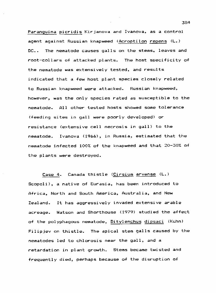
384
Paranguina picridis Kirianova and Ivanc3va. as a control
agent against Russian knapweed (Acroptilon repens (L..)
DC.. The nematode causes galls on the stems, leaves and
root-collars of attacked plants. The host specificity 0-f
the nematode was extensively tested, and results
indicated that a -few host plant species closely related
to Russian knapweed were attacked. Russian knapw.eed,
however, was the only species rated as susceptible to the
nematode All other tested hosts showed some tolerance
(feeding sites in gall were poorly developed) or
resistance (extensive cell necrosis in gall) to the
nematode. Ivanova (1966). in Russia. estimated that the
nematode infected 1007. of the knapweed and that 20-307. o-F
the plants were destroyed.
Case 4. Canada thistle (Cirsium arvense (L..)
Scopoli), a native of Eurasia, has been introduced to
Africa, North and South America. Australia, and New
Zealand. It has aggressively invaded extensive arable
acreage. Watson and Shorthouse (1979) studied the affect
of the polyphagous nematode, Ditylenchus dipsaci (Kuhn)
Filipjev on thistle. The apical stem galls caused by the
nematodes led to chlorosis near the gall, and a
retardation in plant growth. Stems became twisted and
frequently died, perhaps because o-f the disruption of

3B
cortical and vascular systems. The internal structure o-f
the nematode gall was also studied and was seen to
include a layer a-F enriched nutritive cells.
In a number of reports (Shorthouse., 1977a,b;
Shorthouse and Watson. 1976; Watson and Shorthause,
1979) the suggestion has been made that a-F the tissues
in a gall, the nutritive tissue requires the heaviest
metabolic investment by the host plant, i.e. the
nutritive tissue is a strong metabolic sink. I-f the
strength o-F such a sink is positively correlated with
damage to the host plant, then based upon the amount a-F
nutritive tissue seen in the gall, the amount a-F damage
that a gall-former inflicts upon a plant should be
predictable. The greater the amount a-F nutritive tissue,
the greater the host plant damage. As support f or this
idea Shorthause and Watson (1976) mention that two
species of tephritids that attack Canadian thistle incite
galls with no nutritive tissue (this observation should
be pursued>, and cause less damage to the host plant than
do tephritids that -Form galls with nutritive tissue.
D. Berube (personal communication) an the other
hand, suggests that a better predictor of host plant
damage caused by a gall-former is the amount a-f
tanni-ferous tissue in the galls. Such tissues are
frequently found in the peripheral zones of a gall.

386
Tannins, he argues, are more abundant than, and more
energetically expensive to produce and store than are the
compounds found in nutritive cells.
Another physical indication o-f sink strength (damage
caused) may be the amount of vascular tissue servicing
the gall. To my knowledge this possibility has not been
considered. The possiblilty that morphological evidence
can be used to increase the efficiency with which control
agents are selected is strong enough that it should be
pursued.
Other Cases. Shorthouse and Watson (1976) briefly
mention that the gall wasp. Gilletea taraxaci, has
potential use as a biocontrol agent of dandelion. This
is the only mention in the bio-control literature of a
cynipid gall wasp.
The qelechiid moth, Crasimorpha sp. was introduced
into Hawaii from Brazil in the mid-1950's to control
Christmas berry (Schinus terebinthifolius Raddi). The
moth causes stem galls. It was not, however,
successfully established in Hawaii after repeated
introductions (Soeden, 1978).
A species of gall-forming eriophyid mite (Aceria
chondrillae) and a gall midge (Cystiphora schmidti S.
Canestrini) have been tested as control agents against

387
skeleton weed (Chondrilla juncea) an important weed in
Australia, and a spreading problem in the United States..
The midge shows promise in trials., and further testing of
the mite has been encouraged (Sobhian and Andres, 1978)..
Summary of Beneficial Galls
There are three characteristics that affect the
suitability of gall-forming organisms as bio-control
agents.
First., it is usually true that gall-f ormers are host
specific (strictly monophagous). A single species or a
small group of related species of host plants are
attacked.. For example, gall insects have been used as
taxonomic characteristics to separate sub-species of host
plants (McArthur et al, 1979)., or to detect hybridization
between closely related species of host plants
(Cooper-Smith1 1974). Even in the case of a presumably
polyphagous species such as the gall-forming nematode.
Ditylenchus dipsaci. suspicions are that the species is
made up of biological races that are monophagous (Watson
and Shorthouse. 1979)..
The specificity exhibited by gall-f ormers fulfills
one of the criteria required of bio-control agents, and
in fact, may be the one attribute of gall-f ormers that
makes them most attractive as possible bia-control

388
agents. Non-target plants are not attacked.
Speci'ficity however, may be correlated with
features that decrease the gall--Former's weed-controlling
potential. Conditions that allow for successful attack
by gall--Farmers may be so rigidly set that., when
introduced into a new environment, the gall insect
mis-cues., or fails to detect cues. Hating flights of
adult gall midges that attack sagebrush occur, for
example, at only certain temperatures and 'F or only brief
periods during the day, as does oviposition (Jones,
1971). Dispersal ability of the adults is usually poor.
Perhaps most importantly, however, successful gall
induction requires that host plant tissue in the proper
developmental state (meristematic) be attacked. The
insects must track the plant's phenology. Because of
their strict temporal, behavioral, and physiological
requirments. gall insects may be prone to failure when
introduced into environments that require shifts in
responses.
Secondly, gall insects are frequently heavily
parasitized. To what extent this is true of other
gall-forming organisms is not clear. Perhaps because o-f
the nature a-F the resource (a rich source of food that is
sheltered -From the elements), a gall and its former

389
attract parasitoids and inquilines. Special precautions
against the introdution a-f parasites should be taken
whenever a gall-former is used in a bia-control program.
Harris's concern about the chance a-f introducing Urophora
parasites into Canada i-F consignments from Europe are
continued is well -founded. He suspects that the
effectiveness a-f Urophora in Canada is in part explained
by the fact that when introduced, the -Fly escaped many a-f
it parasites.
Thirdly, perhaps because a-f the seemingly tight
co-evolution between gall--Formers and host plants the
amount a-f damage caused by same gall-formers may be
minimal. As Shorthause (1977a) states "it is a commonly
held opinion that plant galls do little damage to their
hosts." Such a view runs counter to the goals a-f
bia-control programs where damage to the host plant is
the goal.. Shorthouse (1977a) was the first to challenge
the view that gall farmers are benign. As already
discussed, the damage caused by a galL, although perhaps
not a dramatic necrosis, nonetheless occurs through time
as a drain on plant resources (Jankiewicz, et al, 1969;
Harris, 1977b; Stinner and Abrahamson, 1979). What is
unclear, however, in the vast majority a-F examples is the
amount of damage. One can predict that -f or greatest
damage to host plants. bia-controllers should use

gall-f ormers that attack roots, stems and seeds.
Conclusion
Some gall--forming organisms are clearly either of
detriment or bene-fit to humans. This statement rings
true particularly when the non-arthropod gall-f oriners are
considered. I-F a species a-f bacterium causes a plant
gall usually it ha an impact (positive or negative) on
humans.
Interestingly, however, the majority a-f arthropod
gall-formers are neither directly beneficial nor
detrimental to human welfare. Gall--forming insects are
perceived as benign curiosities. We must move to the
laboratory to envision how such curiosities may assume an
important role in agriculture and medicine in the near
future. The questions a-f how galls are formed, and of
how the gall--former directs host plant cell metabolism
become increasingly important as we attempt to manipulate
genetic expression in plant and animal cells.
390

TABLE ku-I
GALL-FORMING FUNGI
Fungus Disease
Class: Phycomycetae
Order: Chytridales
Synchytrium endobioticurn (Schilb.) Perc. Potato wart
Synchytrium vaccinii Thomas Cranberry wart
Urophlyctis alfalfae (Lagerh.) P. Nag. Crown wart of alfalfa
Physoderma zeae-maydis Shaw Brown spot of corn
Order: Peronosporales
Albuqo candida (Pars.) Roussel White rust of crucifers
Class: Ascomycetae
Subclass: Pyrenomycetes
Order: F1ypocreal es
Claviceps purpurea (Fr.) Tel. Ergot
Nectria galliqena Pores. Apple stem canker
Gibberella fujikuroi Bakanae of rice
Subcl ass: Di scomycetae
Order: Tapnrinales
Taphrina deformans (Ed.) Tul. Peach curl
Taphrina j_ (Fc.) Tul. Plum pockets
Class Basidomycetae
Subclass: Teliosporae
Order: Ustilaginales
Ustilaqo maydis (DC.) Cda. Corn smut
Order: Uredinales
Cronartium spp. Fries Pine blister rusts
Gymnosporanqiuni spp. Hedwig Cedar apple rusts
Subclass: lymenomycetae
Order: Agaricales
Exobasidium spp. Azalea/Rhodedendron leaf gall
391

INSECT
Procecidochares utilis Stone
Zeuxdiplosis jiardi (Kieff.)
Urophora affinis Frfld.
Urophora quadrifasciata (Meigen)
Paraquina picridis Kir. and Ivan.
Ditylenchus dipsaci (Kuhn) Fiuipjev
Crasiuniorpha sp.
Aceria chondrilIne (Canes.)
TABLE Hu-2
PUBLISHED ACCOUNTS OF GALLSUSED IN BIOLOGICAL CONTROL EFFORTS AGAINST WEEDS
TARGET WEED
Eupa tori urn adenophorurn
ypericuni perforaturn
Ceutaurea niaculosa
Centaurea di ffusa
Acroptiliori repens
Cirsiuni arvense
Schinus terebinthifolius
Chondriula schmidti
REFERENCE
Dodd, 1961; Goeden, 1978
holloway and Huffaker, 1952
Harris, 1977a
Harris, 1971a
Watson, 1975
Watson and Shorthouse, 1979
Goeden, 1978
Sobhian and Andres, 1978