AN ABSTRACT OF THE THESIS OF E. Leyla Arsan for the degree of
184
AN ABSTRACT OF THE THESIS OF E. Leyla Arsan for the degree of Master of Science in Fisheries Science presented on December 15, 2006 . Title: Potential for Dispersal of the Non-native Parasite Myxobolus cerebralis: Qualitative Risk Assessments for the State of Alaska and the Willamette River Basin, Oregon. Abstract approved: ______________________________________________ Jerri L. Bartholomew First introduced to the USA in 1958, Myxobolus cerebralis, the parasite responsible for whirling disease in salmonids, has since spread across the country causing severe declines in wild trout populations in the intermountain west. Recent development of risk assessment models used to assess the likelihood and consequences of exotic parasite introduction, have strengthened the process of science-based decision-making in aquatic animal health. In the case of M. cerebralis, it is necessary to use a risk assessment model with two unique segments that clearly address the distinct life stages and respective hosts of the parasite separately. The studies described examine the probability of M. cerebralis introduction and establishment for two regions: the state of Alaska, and the Willamette River basin, Oregon. The Alaska risk assessment was based on the assumption that the parasite did not already occur in the state. However, in the process of validating this assumption, we documented the first polymerase chain reaction (PCR) detection of the parasite in the state. The pathogen was identified in hatchery rainbow trout (Oncorhynchus mykiss) from the Anchorage area. Although this is the first detection of the parasite in Alaska, clinical whirling disease has never been documented in the state. To qualitatively assess the risk of further spread of M. cerebralis in Alaska, four potential routes of dissemination were examined: movement of fish by humans, natural dispersal (via migratory birds and stray anadromous salmon), recreational
Transcript of AN ABSTRACT OF THE THESIS OF E. Leyla Arsan for the degree of

AN ABSTRACT OF THE THESIS OF E. Leyla Arsan for the degree of Master of Science in Fisheries Science presented on December 15, 2006. Title: Potential for Dispersal of the Non-native Parasite Myxobolus cerebralis: Qualitative Risk Assessments for the State of Alaska and the Willamette River Basin, Oregon. Abstract approved: ______________________________________________ Jerri L. Bartholomew First introduced to the USA in 1958, Myxobolus cerebralis, the parasite
responsible for whirling disease in salmonids, has since spread across the country
causing severe declines in wild trout populations in the intermountain west.
Recent development of risk assessment models used to assess the likelihood and
consequences of exotic parasite introduction, have strengthened the process of
science-based decision-making in aquatic animal health. In the case of M.
cerebralis, it is necessary to use a risk assessment model with two unique
segments that clearly address the distinct life stages and respective hosts of the
parasite separately. The studies described examine the probability of M. cerebralis
introduction and establishment for two regions: the state of Alaska, and the
Willamette River basin, Oregon.
The Alaska risk assessment was based on the assumption that the parasite did not
already occur in the state. However, in the process of validating this assumption,
we documented the first polymerase chain reaction (PCR) detection of the parasite
in the state. The pathogen was identified in hatchery rainbow trout (Oncorhynchus
mykiss) from the Anchorage area. Although this is the first detection of the
parasite in Alaska, clinical whirling disease has never been documented in the
state.
To qualitatively assess the risk of further spread of M. cerebralis in Alaska, four
potential routes of dissemination were examined: movement of fish by humans,
natural dispersal (via migratory birds and stray anadromous salmon), recreational

activities, and commercial seafood processing. This research indicates the most
likely pathway for M. cerebralis transport in Alaska is human movement of fish.
In the Willamette River basin, Oregon, introduction of M. cerebralis has already
occurred, though establishment appears limited to a single private hatchery.
Introduction in this region was considered the most likely to occur as a result of
human movements of fish. Straying anadromous salmonids were also assessed
and were present in higher numbers than predicted. However, they were not
infected with the parasite, and thus the probability for introduction by this route is
low. The probability of introduction of the parasite varies throughout the
Willamette River basin. Areas with the highest probability for M. cerebralis
introduction were identified as the Clackamas and Santiam River subbasins. The
Clackamas River has already experienced an introduction of the parasite, has the
largest concentration of hatcheries (state, federal, and private), has a popular sport
fishery, and is the closest major tributary to the enormous piscivorous bird-
populations in the Columbia River estuary. The Santiam subbasin has a popular
sport fishery, received the highest number of stray fish in the Willamette River
basin, and has the second largest concentration of hatcheries in the Willamette
River basin.
Unique from introduction, establishment of the parasite is dependent upon several
environmental and biological factors including: water temperatures,
spatial/temporal overlap of hosts, and the distribution and genetic composition of
the parasite’s invertebrate host, Tubifex tubifex. The distribution, genetic
composition and susceptibility of T. tubifex, were considered the most important
factor in the ability of M. cerebralis to establish in both systems. Surveys of
oligochaete populations were conducted in both study regions.
In Alaska, T. tubifex was not detected from the southeast region and the apparent
lack of appropriate tubificid hosts may prevent establishment in that part of the
state. However, 4 lineages (I, III, IV, and VI) of the species were identified from

southcentral Alaska. Lineage IV has not been previously been described in North
America and its susceptibility to M. cerebralis was unknown. When lineage IV T.
tubifex and 3 mixed-lineage (I, III, IV and VI) groups were exposed to M.
cerebralis, only lineage III became infected under our experimental conditions.
Thus, if the parasite were dispersed, conditions are appropriate for establishment
and propagation of the parasite life cycle in southcentral Alaska, although
detrimental effects on fish populations may be reduced as a result of the presence
of non-susceptible lineages of T. tubifex. The probability of further establishment
in this area is greatest in Ship Creek, where the abundance of susceptible T.
tubifex, the presence of susceptible rainbow trout (Oncorhynchus mykiss), and the
proximity to the known area of infection make conditions particularly appropriate.
Similar to findings in Alaska, the Willamette River basin, Oregon also supports
populations of susceptible T. tubifex. If the pathogen were introduced, probability
of establishment is high in certain areas of the basin as all conditions are
appropriate for propagation of the parasite life cycle. Tributaries to the mainstem
Willamette River have the highest probability of establishment as these areas have
the greatest numbers of susceptible T. tubifex. However, the abundance of
resistant strains of T. tubifex could mitigate the effects of M. cerebralis if
introduced.
Management recommendations to reduce the likelihood of parasite dissemination
are similar for Oregon and Alaska since human movement of fish and angler
activities were considered the most likely routes of introduction for both regions.
Based on this research, steps should also be taken to limit human movement of
fish, whether by restricting carcass planting for stream enrichment in Oregon, or
by prohibiting use of fish heads as bait in southcentral Alaska. The states should
also allot resources to angler education and awareness of the effects of angler
activity and recreation on dispersal of M. cerebralis. This could be done using a
combination of brochures and signage at boat ramps describing how to prevent
spread of aquatic nuisance species.

©Copyright by E. Leyla Arsan
December 15, 2006
All Rights Reserved
Potential for Dispersal of the Non-native Parasite Myxobolus cerebralis: Qualitative Risk Assessments for the State of Alaska and the Willamette River
Basin, Oregon.
by
E. Leyla Arsan
A THESIS
submitted to
Oregon State University
in partial fulfillment of the requirements for the
degree of
Master of Science
Presented December 15, 2006 Commencement June 2007

Master of Science thesis of E. Leyla Arsan presented on December 15, 2006. APPROVED: _______________________________________________________________ Major Professor, representing Fisheries Science ________________________________________________________________ Chair of the Department of Fisheries and Wildlife ________________________________________________________________ Dean of the Graduate School I understand that my thesis will become part of the permanent collection of Oregon State University libraries. My signature below authorizes release of my thesis to any reader upon request. ________________________________________________________________
E. Leyla Arsan, Author

ACKNOWLEDGEMENTS I express my sincere gratitude to my major professor, Dr. Jerri L. Bartholomew,
for the opportunity to pursue a graduate degree, and for her patience and guidance
during my education. Special thanks to the ODFW pathology group and those at
the Center for Fish Disease Research, Salmon Disease Lab for their assistance,
guidance, and humor throughout my research. I would like to thank my friends
and family for their encouragement and support. Lastly, I offer my tremendous
appreciation to Matthew Giorgio for his unwavering belief in me.

CONTRIBUTION OF AUTHORS Dr. Jerri L. Bartholomew was involved in the design, analysis, and writing of all
phases of this project. Sascha L. Hallett and Stephen Atkinson assisted with
experimental design and laboratory analysis and consultation. Dr. Theodore
Meyers also provided consultation on experimental design and management
recommendations for Alaska.

TABLE OF CONTENTS Page INTRODUCTION……………………………………………….………….. 1
Myxobolus cerebralis description and lifecycle………………………. 1 Whirling Disease: distribution and impacts…………………………... 2 Risk Assessment description and use in aquatic animal health……….. 4 Research Goals………………………………………………………… 6
TUBIFEX TUBIFEX FROM ALASKA AND THEIR SUSCEPTIBILITY TO MYXOBOLUS CEREBRALIS………………………………………… 7 Abstract……………………………………………………………….. 8 Introduction…………………………………………………………… 9 Methods………………………………………………………………. 11 Results………………………………………………………………… 23 Discussion…………………………………………………………….. 28 Acknowledgements…………………………………………………… 37 References…………………………………………………………….. 39 Tables…………………………………………………………………. 45 Figures………………………………………………………………… 49 EXPANDED GEOGRAPHICAL DISTRIBUTION OF MYXOBOLUS CEREBRALIS: FIRST POLYMERASE CHAIN REACTION (PCR) DETECTIONS FROM ALASKA………………………………………….. 54 Abstract……………………………………………………………….. 55 Introduction…………………………………………………………… 56 Methods…………………………………………………………….…. 57 Results………………………………………………………………… 61 Discussion…………………………………………………………….. 62 Acknowledgements…………………………………………………… 69 References…………………………………………………………….. 70 Tables…………………………………………………………………. 75 Figures………………………………………………………………… 76 A QUALITATIVE ANALYSIS OF RISK FOR THE INTRODUCTION AND ESTABLISHMENT OF MYXOBOLUS CEREBRALIS INTO THE STATE OF ALASKA, USA…………………………………………………………….. 79 Abstract……………………………………………………………….. 80 Introduction…………………………………………………………… 81 The Parasite Hazard…………………………………………………... 82 Risk Analysis…………………………………………………………. 84 Release Assessment…………………………………………………... 86 Exposure Assessment………………………………………………… 95 Conclusions and Risk Management………………………………….. 101

TABLE OF CONTENTS (Continued) Page Acknowledgements…………………………………………………… 104 References…………………………………………………………….. 106 Tables…………………………………………………………………. 116 Figures………………………………………………………………… 118 POTENTIAL DISPERSAL OF THE NON-NATIVE PARASITE MYXOBOLUS CEREBRALIS: A QUALITATIVE ANALYSIS OF RISK FOR THE WILLAMETTE RIVER BASIN, OREGON……………………………….. 122 Abstract………………………………………………………………. 123 Introduction…………………………………………………………… 124 Methods………………………………………………………………. 126 Release Assessment………………………………………………….. 128 Exposure Assessment ……………………………………………….. 139 Conclusions and Risk Management………………………………….. 145 Acknowledgements…………………………………………………... 148 References……………………………………………………………. 149 Tables…………………………………………………………………. 157 Figures………………………………………………………………… 159 SUMMARY………………………………………………………………… 164 BIBLIOGRAPHY…………………………………………………………... 167

LIST OF FIGURES Figure Page 1.1 Life cycle of Myxobolus cerebralis…………………………………. 1 1.2 Map of the Columbia River basin with the Myxobolus cerebralis
enzootic area circled and the Willamette River basin marked .…….. 3 2.1 Map of study locations, sampling sites for Tubifex tubifex (black
dots), and rainbow trout (Oncorhynchus mykiss) hatcheries (triangles) in southcentral and southeast Alaska ……………………. 50
2.2 Tubifex tubifex lineages from Fort Richardson Hatchery, Alaska.
Agarose gel electrophoresis of PCR-amplified genomic DNA using mt 16S rDNA lineage-specific primers. Two lineages were present: I (196 bp) and IV (2 bands at 320 bp and 147 bp). A negative control (water) and a positive control (lineage III, 147 bp) were included. A 50 bp DNA Ladder is shown on both sides of the gel (2% agarose) …………………………………………………………. 51
2.3 First experimental exposures- Susceptibility of lineage IV Tubifex
tubifex to Myxobolus cerebralis: triactinomyxon release per 100-worm replicate (3 Alaskan replicates are pooled as 1) …………. 52
2.4a Second experimental exposures- Comparative triactinomyxon
release from Alaskan Tubifex tubifex. Number of triactinomyxons noted as absolute numbers 0-7000; greater values noted as >7000 triactinomyxons and not as an absolute number …………………….. 53
2.4b Comparative triactinomyxon release from Alaskan Tubifex tubifex
groups A, B, and C (enlargement of Figure 4a) …………………….. 54 3.1 Agarose gel electrophoresis of PCR-amplified genomic DNA from 4
Alaska rainbow trout (sample numbers shown), using mt 18S rDNA specific primers. A 1 kb+ DNA Ladder is shown on both sides of the gel (1.5% agarose). A blank (B) well precedes each of the two positive (+) controls and a negative (-) control (water) is provided in the last well ………………………………………………………… 77
3.2 QPCR cycle threshold values of Alaska hatchery rainbow trout plotted
against reference samples spiked with known numbers of Myxobolus cerebralis myxospores (100, 1000). ………………………………….. 78
4.1 Map areas of highest risk for Myxobolus cerebralis dissemination in
southeast and southcentral Alaska. Rainbow trout hatcheries
LIST OF FIGURES (Continued) Figure Page
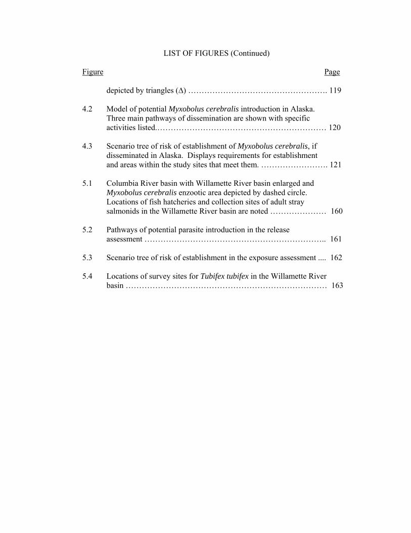
depicted by triangles (∆) ……………………………………………. 119 4.2 Model of potential Myxobolus cerebralis introduction in Alaska.
Three main pathways of dissemination are shown with specific activities listed.……………………………………………………… 120
4.3 Scenario tree of risk of establishment of Myxobolus cerebralis, if
disseminated in Alaska. Displays requirements for establishment and areas within the study sites that meet them. ……………………. 121
5.1 Columbia River basin with Willamette River basin enlarged and
Myxobolus cerebralis enzootic area depicted by dashed circle. Locations of fish hatcheries and collection sites of adult stray salmonids in the Willamette River basin are noted ………………… 160
5.2 Pathways of potential parasite introduction in the release
assessment ………………………………………………………….. 161 5.3 Scenario tree of risk of establishment in the exposure assessment .... 162 5.4 Locations of survey sites for Tubifex tubifex in the Willamette River
basin ………………………………………………………………… 163

LIST OF TABLES Table Page 2.1 Survey sites (excluding hatcheries) for Tubifex tubifex and their
corresponding abundance and mitochondrial lineage ………………. 45 2.2 Tubifex tubifex collected from Alaskan rainbow trout hatcheries ….. 46 2.3 Prevalence and level of Myxobolus cerebralis infection in different
mitochondrial lineages of Tubifex tubifex used in laboratory exposure experiments ……………………………………………….. 47
2.4 Numbers of Tubifex tubifex exposed, infected and surviving
experimental exposure to Myxobolus cerebralis ……………………. 48 3.1 Variable loci in the 18S SSU rRNA gene from different isolates of
Myxobolus cerebralis. Loci are measured relative to 5` position of universal primer ERIB1; heterogeneous bases: R=A+G, K=T+G, Y=C+T.……………………………………………………………… 75
4.1 Definitions of probability levels used in the risk analysis ………….. 116 4.2 Summary of the probability of introduction and within-state
dissemination of Myxobolus cerebralis in Alaska ………………….. 117 5.1 Summary of adult summer steelhead strays found at Oregon
Department of Fish and Wildlife adult collection facilities in the Willamette River Watershed in 2004 and 2005 …………………….. 157
5.2 Survey sites and corresponding abundance and lineages of Tubifex
tubifex ……………………………………………………………… 158

DEDICATION
This thesis is dedicated to my aunt, Katherine Martin, who passed away during my
research for this degree. She was always supportive of me and is deeply missed.

Potential for Dispersal of the Non-native Parasite Myxobolus cerebralis: A
Qualitative Risk Assessments for the State of Alaska and the Willamette River
Basin, Oregon.
CHAPTER 1: INTRODUCTION
Myxobolus cerebralis description and lifecycle
Myxobolus cerebralis, a metazoan fish parasite exotic to North America, was first
detected in the USA in 1958 (Hoffman 1962). There are some 1,350 known
species of myxozoans, most of which are parasitic to fish at some point in their life
cycle (Tops et al. 2004). The vast majority of myxozoans are relatively non-
pathogenic. In contrast, M. cerebralis is one of the most pathogenic myxozoans
known for fish (Hedrick et al., 1998).
Figure 1.1. Life cycle of Myxobolus cerebralis.
M. cerebralis life cycle
Salmonid HostMyxospore
Oligochaete Host
Triactinomyxon (TAM)

2
Life cycles are only known for 1% of the described myxosporeans (Kerans and
Zale, 2002). The life cycle of M. cerebralis requires two obligate hosts: a
salmonid and the aquatic oligochaete worm, Tubifex tubifex (Fig 1.1). There are
also two distinct spore stages of the parasite (one particular to each of the hosts),
the myxospore and the actinospore.
Myxospores develop in the fish host and are released upon death of the fish.
These round and durable spores are characterized by two hardened valves
surrounding two polar capsules with coiled filaments (Hedrick and El-Matbouli,
2002). Myxospores are ingested by T. tubifex as the worms consume sediment and
bacteria.
Once inside the worm host, the parasite undergoes reproduction and structural
transformation and is released in its actinospore stage as a triactinomyxon (TAM),
morphologically different but genetically identical to a myxospore. The TAM
attaches to its fish host in the water column, where the parasite sporoplasm is
injected through the epidermis of the fish and migrates through the nervous system
to the cartilage (El-Matbouli et al., 1995). The parasite consumes cartilage,
causing lesions on the gills, cranium, and vertebral column of the fish
(MacConnell and Vincent, 2002). Each spore stage requires approximately three
months to develop within and be released from the respective host. Due to the
myxospore’s durability (El-Matbouli and Hoffmann, 1991), this parasite stage is
more likely to survive transport and be a risk for dissemination than the more
fragile TAM stage.
Whirling Disease: distribution and impacts
Thought to have originated from a shipment of frozen infected brown trout (Salmo
trutta) from Europe, M. cerebralis is now reported in a total of 24 US states in the
contiguous U.S, (Bartholomew and Reno, 2002; Vermont Department of Fish and

3
Wildlife, 2002; Stromberg, 2006) and 26 different countries (Hoffman, 1962). The
pathogen has been at the forefront of fish health research due to its potential
impacts on rainbow trout culture and its implication in rapid dramatic declines of
wild rainbow trout populations in Colorado and Montana (Walker and Nehring,
1995; Vincent, 1996; Nehring et al., 1998). The ecologic and economic impacts of
M. cerebralis, in addition to its rapid spread and establishment across the globe,
are indicative of the need for research to assist fishery managers in halting the
spread or effects of the pathogen.
Myxobolus cerebralis was first detected in Oregon in 1986 (Lorz et al. 1989), and
is now enzootic in certain tributaries of the upper Columbia River basin. Since the
1980’s, the parasite has been detected elsewhere in the basin from stray adult
salmonids originating from these tributaries (Engelking 2002); however, there is
no evidence of establishment of the parasite life cycle outside of the enzootic
region. The parasite was detected further west in the state in 2001, at a private
facility in the lower Willamette River Basin (Clear Creek). Infection at the facility
was contained and there is no apparent establishment of the parasite in the wild
(Bartholomew et al., 2007). Currently, M. cerebralis is not known to be
established in the Willamette River basin outside of the private facility on Clear
Creek. The closest enzootic area is the upper Columbia River basin (Figure 1.2).
Oregon
Washington

4
Figure 1.2. Map of the Columbia River basin with the Myxobolus cerebralis enzootic area circled and the Willamette River basin marked. Risk Assessment description and use in aquatic animal health
Risk analysis is a tool used to assess the likelihood and consequences of exotic
parasite introduction. It addresses the following questions (MacDiarmid, 2000):
• What can go wrong?
• How likely is it to go wrong?
• What would be the consequences of its going wrong?
• What can be done to reduce either the likelihood or the consequences of its
going wrong?
The International Aquatic Animal Health Code subdivides a risk analysis into: a) a
release assessment, b) an exposure assessment and c) a consequence assessment
(Office International des Epizooties [OIE], 2002). Risk assessments provide
decision-makers with tools to assess management implications and to eliminate
non-issues by using logical scientific arguments. Such assessments give managers
a better understanding of where to allocate resources to help prevent introduction
or spread of pathogens. In this way, risk analysis can be used as a type of map
(MacDiarmid, 2000) to navigate through possible pathways or scenarios leading to
an investigated risk. It is an essential first step in understanding potential
outcomes and associated risks of management decisions.
Several risk assessment models have been created in the fields of animal health,
biosecurity, and invasive species management (EPA 1992; Agriculture and Agri-
Food Canada, 1994; Orr, 1995; OIE, 2002; Hayes, 2003). Typically, such
qualitative risk assessments are initiated to either assess the risk of a newly
proposed activity or pathway (i.e. importation) or to identify the route of
introduction of a targeted species.
5
Most invasive species or biosecurity risk assessment models integrate the risk of
introduction, the risk of survival during transport and risk of survival in the new
environment, as the risk of establishment (EPA 1992; Agriculture and Agri-Food
Canada, 1994; Orr, 1995). The establishment of M. cerebralis depends not only
on the organism’s survival during transport and the environmental conditions of its
new location, but also on spatial overlap with its definitive host, T. tubifex. The
two distinct life stages and hosts of M. cerebralis necessitate the need for a risk
assessment model with two distinct segments that clearly address each spore stage
(and the respective host) separately. The model used in this research addresses the
risk of introduction (myxospore stage) and the risk of establishment (actinospore
stage) independently. The framework for this model (Bartholomew et al. 2005)
was created in efforts to tailor such assessments to whirling disease. It is a
combination of models employed by Orr (1993), OIE (2002) and MacDiarmid
(2000), and is unique in that numerous potential pathways are assessed in one risk
assessment model and that the targeted organism, M. cerebralis, has a complex
two-host lifecycle that requires distinct assessments of each spore stage.
Research Goals
The goals of this research were to qualitatively assess the risk of introduction and
establishment of M. cerebralis into the Willamette River basin, Oregon and the
state of Alaska. Introduction in the Willamette River basin appears limited to a
private hatchery (Batholomew et al. 2007) and the parasite was not known to be
present in Alaska. The purpose of this study is to better understand the future risk
of and routes of introduction, establishment, and further spread of the parasite into
both areas. Specific study objectives are outlined below:
Objective 1) Assess the probability of introduction of M. cerebralis into the state
of Alaska? Approach: Determine the validity of the current assumption that M.
6
cerebralis has not been established in Alaska. Determine and evaluate potential
pathways for pathogen introduction and identify current critical gaps in data.
Objective 2) Assess the probability of establishment of M. cerebralis in the state
of Alaska? Approach: Evaluate conditions that must be met to allow
establishment in Alaska, including T. tubifex populations and lineages, and water
temperatures.
Objective 3) Assess the probability of introduction of M. cerebralis into the
Willamette River, Oregon? Approach: Determine the validity of the current
assumption that M. cerebralis has not been established in the Willamette River
basin. Determine and evaluate potential pathways for pathogen introduction and
identify current critical gaps in data.
Objective 4) Assess the probability of establishment of M. cerebralis in the
Willamette River, Oregon? Approach: Evaluate conditions that must be met to
allow establishment in Willamette River basin, including T. tubifex populations
and lineages, and water temperatures.

7
CHAPTER 2
TUBIFEX TUBIFEX FROM ALASKA AND THEIR SUSCEPTIBILITY TO MYXOBOLUS CEREBRALIS.
E. Leyla Arsan1, Sascha L. Hallett2, and Jerri L. Bartholomew2 1Department of Fisheries and Wildlife, Center for Fish Disease Research, Oregon State University, 220 Nash Hall, Corvallis, Oregon 97331 2 Department of Microbiology, Center for Fish Disease Research, Oregon State University, 220 Nash Hall, Corvallis, Oregon 97331

8
ABSTRACT
Although widespread throughout the continental United States, Myxobolus
cerebralis, the myxozoan parasite that causes whirling disease in salmonids, has
not been reported from the state of Alaska. As part of a risk assessment for the
introduction and establishment of M. cerebralis into Alaska, the distribution of the
invertebrate host, Tubifex tubifex, was surveyed and its genetic composition and
susceptibility to the parasite determined. Many oligochaetes, but no T. tubifex,
were collected from southeast Alaska; however, 4 lineages of T. tubifex (I, III, IV,
and VI) were identified from southcentral Alaska. Lineage IV had not been
previously described in North America and its susceptibility to M. cerebralis was
unknown. When lineage IV T. tubifex and 3 mixed-lineage (I, III, IV and VI)
groups were exposed to M. cerebralis, only lineage III became infected under our
experimental conditions. Infection occurred in this lineage even when it
comprised just 3% of the population. Implications of the presence of non-
susceptible lineages of T. tubifex on Alaskan salmonids would be significant in
areas where these lineages dominate Tubifex populations.
9
INTRODUCTION
Myxobolus cerebralis, the myxosporean parasite responsible for whirling
disease in salmonids, has caused severe declines in wild trout populations in the
intermountain west of North America (Walker and Nehring, 1995; Vincent, 1996;
Nehring et al., 1998). The parasite’s definitive host, the oligochaete worm Tubifex
tubifex (Wolf and Markiw, 1986), is a hearty and cosmopolitan species capable of
withstanding extreme and variable environmental conditions. Such qualities allow
the worm to inhabit areas where other species cannot compete, and thus to span
across ecosystems as a widespread aquatic invertebrate.
Tubifex tubifex are commonly found in environments with abundant
organic matter, fine sediments, and low flow (Brinkhurst and Gelder, 1991;
Brinkhurst, 1996). The species is tolerant of low oxygen, desiccation, and variable
temperature regimes (Reynoldson, 1987; Brinkhurst, 1996). Populations have
consequently been found in environments extending from very unproductive high
latitude lakes to eutrophic nutrient-rich lakes (Milbrink, 1994). Tubifex tubifex are
important members of the aquatic ecosystem and play a vital role in breaking
down organic matter and ingesting sediments (Brinkhurst and Gelder, 1991).
Though T. tubifex is thought to be cosmopolitan, it is not usually a
common species (Brinkhurst, 1996) and its identification can be complicated.
Positive identification is based on morphology of the reproductive structures such
as the penis sheath, vas deferens, and atrium (Brinkhurst, 1996). Not only must
worms be sexually mature to be confidently identified, but reproductive organs are

10
reabsorbed after breeding (Poddubnaya, 1984), a further hindrance to
identification. Additionally, number of chaete, another important diagnostic trait,
may vary depending on environmental conditions (Chapman and Brinkhurst,
1987). Non-mature T. tubifex can be difficult to distinguish phenotypically from
other oligochaetes with similar physical characteristics, such as Ilyodrilus
templetoni and Rhyacodrilus spp. Genetic assays have been developed for such
differentiation among species (Beauchamp et al., 2001; Hallett et al., 2005).
In addition to complexities with identification of the species, recent studies
have revealed the presence of 6 cryptic mitochondrial (mt) lineages of T. tubifex
(lineages I - VI) (Sturmbauer et al., 1999; Beauchamp et al., 2001). Lineages
cannot be distinguished morphologically, and therefore must be differentiated
genetically. Four lineages (I, III, V, and VI) have been reported from North
America (Beauchamp et al., 2001) and 5 are known from Europe (I-V)
(Sturmbauer et al., 1999). Lineages II and IV have been described only from
Europe, and lineage VI only from North America. Lineages commonly cohabitate,
with more than a single given lineage found at a site, though populations can be
mono-lineal (Beauchamp et al., 2006). Lineages of T. tubifex differ in
susceptibility to M. cerebralis from highly susceptible to completely resistant.
Populations of lineages I and III have been shown to propagate the parasite
whereas lineages V and VI do not (Beauchamp et al. 2002, 2005, & 2006; DuBey
et al., 2005). Although mt 16S rDNA does not directly confer resistance to M.
cerebralis, the relationship can be useful in assessing general susceptibility.

11
This study was conducted as part of a risk assessment for the introduction
and establishment of M. cerebralis into the state of Alaska. Myxobolus cerebralis
has never been reported in Alaska, though monitoring for the parasite in the state is
limited. The nearest known enzootic area is the Snake River basin in northeast
Oregon and Idaho. The risk assessment included a T. tubifex survey to determine
the presence, relative abundance, and lineages of T. tubifex from sites sampled in
southeast and southcentral Alaska. Subsequent laboratory parasite-exposure
experiments were conducted to determine the susceptibility of these lineages to M.
cerebralis, with particular focus on a lineage previously unknown in North
America. This paper describes results from both the Tubifex survey and lineage
susceptibility experiments.
MATERIALS AND METHODS
Sampling design
A qualitative survey was used to determine presence/absence and relative
abundance by order of magnitude of T. tubifex. Areas most likely to contain the
organisms were targeted for sampling, i.e., those with low flow, fine sediments,
and accumulations of organic material. Due to the magnitude in geographic size
of Alaska [the state is approximately the size of the continent of Europe (Pagano
2000; Atlas A-Z 2001)], this study focused on the areas of the state where we
considered the probability for introduction and establishment of M. cerebralis to

12
be highest: southeast and southcentral Alaska (the Cook Inlet basin) (Figure 2.1).
These sites have physical attributes that are well- suited for parasite introduction
and proliferation, such as, temperatures appropriate for parasite propagation,
populations of salmonids susceptible to the parasite, a large commercial or sport
fishery, and accessibility by boat or car.
Site descriptions
Oligochaetes were sampled from waterways in southeast and southcentral
Alaska during August and September 2005 (Figure 2.1). Additionally, 2 fish
hatcheries were sampled, representing the only locations in the state where
rainbow trout [the species most susceptible to M. cerebralis, (MacConnell and
Vincent, 2002; Sollid et al., 2002)] are reared. These samples were collected in
November 2004 and May 2005 at Fort Richardson (FTR) and Elmendorf
hatcheries in southcentral Alaska.
In southeast Alaska, 2 creeks near Juneau were sampled: Peterson Creek
and Montana Creek. Southeast Alaska has a maritime climate and is within the
Pacific Northwest temperate rainforest ecosystem. The area has cool winters and
wet summers with stream temperatures generally warmer than those in the interior
of the state. Peterson Creek annual water temperatures ranged from 0-16 C during
2002-2004 [R. Harding, Alaska Department of Fish and Game (ADFG), personal
communication]. The natural hydrographs of these creeks are influenced by spring
snowmelt and autumn rainfall (Chaloner et al., 2004; Montgomery et al., 1996;

13
Milner et al., 1997). Peterson and Montana Creeks have small basins, typical of
coastal southeast Alaska, where mountains and icefields rise sharply from sea level
and create relatively short watersheds that flow into the Pacific Ocean. Both
creeks originate in the Taku Mountain Range and flow less than 15 km (Alaska
Atlas and Gazetteer, 1998) to the protected waters of the inside passage off the
Gulf of Alaska. The creeks support various salmonid populations including,
steelhead trout (Oncorhynchus mykiss), pink (O. gorbuscha), chum (O. keta), and
coho (O. kisutch) salmon, Dolly Vardon (Salvelinus malma), and coastal cutthroat
trout (O. clarkii) (Harding and Jones, 1992; Chaloner et al., 2004). There is a
barrier to fish migration approximately 2 km from the Peterson creek mouth;
sampling was conducted below this barrier and before the short tidal area at the
creek mouth.
In southcentral Alaska, three river systems within the Cook Inlet basin
were sampled: Ship Creek, Campbell Creek, and the Kenai River. The Cook Inlet
basin is home to over half the state’s human population. The area has a
transitional climate (National Climate Center, 1982) and is the ecotone between
the Pacific Northwest rainforest and the northern boreal forest. Annual water
temperatures in these streams range from 0-16.5 C [U.S. Geological Survey
(USGS), 2006a and 2006b]. Hydrographs in the basin are highly predictable and
influenced by snow and glacier melt in the summer; typical freshwater inflow into
Cook Inlet is 15 times higher in July than in February (Dorava and Milner, 2000).
14
Ship and Campbell Creeks, which originate in the Chugach Mountains, run
west through the city of Anchorage and are impacted by urban development. The
Kenai River flows approximately 132 unregulated km across the width of the
Kenai Peninsula. The river has several distinct sections. The upper section (Kenai
Lake to Skilak Lake) is included in the Kenai National Wildlife Refuge and has a
more pristine character; the middle and lower sections of the river (Skilak Lake to
Cook Inlet) contain more private property, industry, urbanization, and motorized
vessels.
The Kenai supports populations of rainbow trout, Chinook, coho, pink, and
sockeye (O. nerka) salmon, and Dolly Vardon and is the largest freshwater sport
fishery in Alaska (Hammarstrom, 1988). Ship and Campbell Creeks support
populations of these species as well as chum salmon (Miller and Bosch, 2004).
Ship Creek is the most popular sport fishery in the Anchorage area and sustains the
only 2 hatcheries, FTR and Elmendorf, in the state that rear rainbow trout. Ship
and Campbell Creeks and the lower Kenai River have numerous sources of
potential organic loading due to their urban proximity, commercial and industrial
activity, streambank degradation due to recreational traffic, and large pulses of
organic material from spawning salmon runs. Sampling sites in the Cook Inlet
were chosen to represent water bodies with and without fish hatcheries, urban
development, and glacial sources.

15
Tubifex tubifex collection and isolation
Oligochaete samples from hatchery settling ponds at FTR and Elmendorf
(both on Ship Creek) were obtained as initial pilot samples in autumn 2004 before
the complete T. tubifex survey was conducted. Sediments were collected with a
shovel and shipped in fresh water and on ice to the Salmon Disease Laboratory
(SDL), Corvallis, Oregon.
All other sediment samples were obtained via 5-gal bucket and 500 µm
sieve or by 500 µm mesh kicknet. A bucket was used to scrape the substrate of the
stream and collect sediment. Sediment was swirled with water in the bucket to
dislodge worms and allow heavy particulates to sink to the bottom. Water with
suspended material was poured through a sieve and washed with water to remove
fines. In areas too deep or too rocky for a bucket, a kicknet was used. The
remaining sample was deposited in a bag with clean water, placed on ice and sent
to the SDL.
Once in the laboratory, the water in which samples were shipped was
passed through a 20 µm mesh filter and inspected microscopically to determine if
myxozoan actinospores were present. The sediment was then rinsed through a 246
µm screen to flush out fines. Bulk organic matter was retained and placed in a
24.8 x 14 cm shallow container and covered with water. The sample was swirled
to evenly distribute sediments and organisms and a 24-cell grid was placed over
the top of the container. Three subsamples were taken by simultaneously inserting

16
plexiglass squares (roughly 4 x 3.6 cm) into 3 randomly assigned cells in the grid.
The material within each square was removed (approximately 30 ml) and sorted on
a white tray. Oligochaetes were collected and inspected under a microscope to
determine presence of hair chaete. In subsamples with greater than 500 worms,
only 7.5 – 15 ml was sorted and numbers extrapolated to total 30 ml subsample
volume. This was only necessary in subsamples from 3 sites: Centennial and
Eagle Rock Boat Landings on the lower Kenai River, and from above Amalga
Lake on Peterson Creek in Southeast Alaska.
Tubifex tubifex characterization
A subsample of 10 worms with both hair and pectinate chaete was mounted
on slides for positive identification (Brinkhurst, 1986). Another 10 worms with
both hair and pectinate chaete were assayed using a species-specific PCR (Hallett
et al., 2005) to determine percentage of worms with morphological T. tubifex
characteristics that were actually T. tubifex. All 10 worms suspected to be T.
tubifex morphologically were determined to be so by genetic tests, thus hair and
pectinate chaete were used as the definitive identification method for the
remainder of the sorting process. Rinsed samples were maintained in culture at
12.8 C in an incubator with an air stone or on flow-through well water.

17
Oligochaete genetic analyses
In addition to the 10 worms subsampled for species characterization, 20
more T. tubifex per sample site were genetically assayed to determine lineage. In
samples with fewer than 20 total T. tubifex, all T. tubifex found were assayed.
Worms were digested with 95 µL ATL buffer (QIAGEN, Valencia, California)
and 5 µL Proteinase K at 55 C, boiled for 5 minutes, and diluted 1:101 with buffer
AE (QIAGEN) then stored frozen. A T. tubifex species-specific PCR assay was
conducted following Hallett et al. (2005). Lineages were determined based on the
PCR assays described by Beauchamp et al. (2001) and Sturmbauer et al. (1999).
PCR products were visualized by gel electrophoresis (2% agarose) and UV
illumination (Figure 2.2).
Worms morphologically identified as T. tubifex but which did not visibly amplify
in the lineage assay had their mt 16S rDNA sequenced to determine whether they were
a novel lineage or a different species. Two other morphologically similar tubifids,
Rhyacodrilus sodalis and Ilyodrilus templetoni, whose sequences were not available
on GenBank, were also sequenced. The gene was amplified with either the primers
Tub16SF and Tub16SR (Beauchamp et al., 2001) or 16sar and 16sbr (Sturmbauer et
al., 1999). Products were purified using a QIAquick PCR purification kit (QIAGEN).
DNA concentration was measured using a NanoDrop ND-1000 Spectrophotometer
(NanoDrop Technologies, Wilmington, Delaware). Samples were sequenced in 1
direction with Tub16SF or 16sar and ABI Big Dye Terminator chemistry on an
Applied Biosystems Capillary 3100 Genetic Analyzer (Foster City, California) at the

18
OSU sequencing facility (Center for Gene Research and Biotechnology, Central
Service Laboratory). Sequences were aligned in BioEdit (Hall, 1999) with other T.
tubifex and selected oligochaetes from GenBank. A standard nucleotide-nucleotide
BLAST (blastn) search was also conducted (Altschul et al., 1997).
Myxobolus cerebralis susceptibility experiments
Two exposure experiments were conducted: the first concentrated on lineage IV
T. tubifex whereas the second included T. tubifex of all identified lineages from
Alaska. The initial focus of these experiments was on lineage IV, as the susceptibility
of this novel group was unknown. However, after the start of this study, other studies
have changed our understanding of what lineages are susceptible, finding that only
lineage III is susceptible to the parasite (authors unpublished data; B. Nehring,
Colorado Division of Wildlife, personal communication). Thus, a second exposure
experiment was conducted to compare susceptibility of all lineages found in Alaska
with those from elsewhere in North America.
First exposure- Susceptibility of lineage IV T. tubifex to M. cerebralis:
FTR (Fort Richardson) T. tubifex were chosen for this study as the
population was shown to have a high concentration of lineage IV worms. The
FTR T. tubifex collected in 2004 were 81% lineage IV, and the remainder lineage
I. Samples (in original sediment) from FTR were allowed to acclimatize to 12.8 C
water for approximately 30 days before the start of the experiment. A known M.
cerebralis-susceptible strain of T. tubifex (lineage III) was collected for a reference

19
group, from Wizard Falls Fish Hatchery (WF) on the Metolius River, Oregon.
Triplicate groups of 100 worms from both FTR and WF and a single unexposed
FTR control group were used for the parasite challenge (total of 7 groups). Worms
were held in 38.5 cm2 plastic containers at a density of 2597 worms/m2 with 1-2
cm of coarse and fine sand and slow-flowing 12.8 C well water.
Myxobolus cerebralis myxospores were collected from rainbow trout
infected and held at the SDL. Heads of infected fish were removed and processed
in a blender. Spores from the resulting suspension were enumerated using a
hemocytometer. Worms were exposed to 500 myxospores each. Running water
was diverted for the first 48 hours to allow myxospores to settle to the bottom of
containers. Spores were left in the worm containers after exposure to imitate a
natural source of infection that would likely not be removed after 24-48 hr.
Worms were fed once a week (after screening) with ALGAMAC-2000 (Biomarine
Inc., Hawthorne, CA).
Samples were filtered for triactinomyxon (TAM) spores through a 20 µm
mesh screen twice a week beginning at 60 days post-exposure (PE). This time
period was chosen as the earliest date of initial TAM release from T. tubifex has
occurred at 74 days PE at 15 C (Gilbert and Granath, 2001), the latest at 182 days
PE (Kerans et al., 2005). Water inflow was diverted for 24 hr prior to screening.
Site replicates were pooled for screening until TAMs were detected. Two 50 µl
aliquots of the retained screened material were placed on a glass microscope slide,
air-dried and examined with a compound microscope at 100x magnification with
20
phase contrast. TAMs were extrapolated to number/ml.
At peak TAM release in the WF reference group, subsamples of 20 worms
per replicate were separated into 24-well plates and monitored for 48 hr for
individual worm TAM release. After 48 hr, the worms were frozen for infection
verification via QPCR. Since no TAMs had been detected in the FTR group, 20
worms per replicate were separated at the same as the WF group, with individual
worms directly frozen for genetic analysis. A M. cerebralis QPCR assay (Kelley
et al., 2004, see below) was performed to ascertain if worms were infected but not
releasing TAMs. The lineage of all worms was also determined.
Biweekly filtering was continued until TAM numbers significantly
declined or were near zero, 191 days PE. Worms were held until 280 days to
assess post infection survival and reproduction. At this point, remaining
individual worms were counted and assessed as adults or juvenile progeny from
original worms placed in containers at day 0. Juveniles were frozen for lineage
analysis to assess changes in lineage structure of the population.
Second exposures- Comparative susceptibility of Alaskan T. tubifex to M.
cerebralis:
A second round of experimental exposures to M. cerebralis was conducted
to test the susceptibility of all the lineages found in Alaska: I, III, IV, and VI. Six
groups of T. tubifex were used: 3 mixed lineage groups, an FTR group from the
first experimental exposure, a WF reference group, and a WF unexposed control
group. Group A (roughly 71% I, 21% III, and 10% IV) was chosen to represent

21
lineage III as it had the largest proportion of that lineage. Group B, representing
lineage VI, was roughly 90% VI and 10% III, and group C, representing lineage I,
was approximately 84% I, 8% III, 3% IV, and 5% VI. These lineage proportions
are representative of samples collected in Alaska.
Lineage IV worms from the first experimental exposures were re-exposed
to assess if their susceptibility is dependant on maturity or size. At the time of
previous exposure they were small in size in comparison with other worms we
have sampled from the Pacific Northwest. As these are the first data on Alaskan T.
tubifex, it was unknown if these specimens were simply smaller due to the climatic
and environmental pressures of their high latitude environment, and if given the
opportunity, they would grow larger. Indeed, after being held at 12.8 C, being fed
every week, and having an absence of predators, the worms grew much larger,
matured and reproduced and thus were re-exposed as known adults.
In the second exposure challenge, the first round methods were modified to
ensure similar methods, and thus comparison, with research in other laboratories.
Two hundred worms per container were used (density 5194 worms/m2), with 1 cm
clean coarse and fine sand. Filtering was conducted every 7-10 days starting at
100 days PE, a date by which other studies have documented TAM release to
begin (Gilbert and Granath, 2001; Blazer et al., 2003; Kerans et al., 2004;
Beauchamp et al., 2006). Numbers of TAMs released from the WF reference
group were counted until numbers exceeded 7000 TAMs per 100 µl of filtrate.

22
Numbers above this value were noted as “greater than 7000”. Filtering ended at
160 days PE, as TAM release in the Alaskan groups had begun to decline.
At 120 days, 50 worms from each group that was releasing TAMs, were
separated into 24-well plates and monitored for 48 hours for individual worm
TAM release. This number was selected to detect an infection prevalence of at
least 5% (with 95% confidence) (USFWS and AFS-FHS 2003). At the end of the
experiment, all worms were counted and adults frozen for genetic analyses
described in the first experimental exposures.
Myxobolus cerebralis QPCR
To detect Myxobolus cerebralis infected worms, the QPCR assay described
by Kelley et al. (2004) was followed except that 4 µl of extracted DNA was used
in a final reaction volume of 20 µl. Reactions were performed in an ABI PRISM
7000 Sequence Detection System in either ABI PRISMTM optical tubes or
MicroAmp® optical 96-well reaction plates. A negative (water) and positive
(spores or infected fish) control were included in each run. In the second
experimental exposures, individual extracted samples were pooled into groups of 4
worms per reaction, with 1 µL from each worm used. This method was tested
with worms with known levels of infection to ensure that pooling samples would
be sensitive enough to detect 1 worm with a low infection (authors unpublished
data). If infection was detected, worms in that pool were assayed individually.
The pooled samples were run singly and rerun if the amplification plot was

23
dubious. An individual sample presenting a cycle threshold (Ct) value of 38 or
less was considered infected (Hallett and Bartholomew, 2006).
RESULTS
Tubifex tubifex survey
Over 2,700 oligochaetes were collected from southeast Alaskan sample
sites (Table 2.1); however, none of the worms assayed were T. tubifex. Many of
the non-mature worms from this area were identified as T. tubifex
morphologically, until contrary genetic identification; sequencing indicated these
specimens to be most similar to Rhyacodrilus sodalis, an oligochaete with
comparable morphological traits to T. tubifex, such as hair chaete. Because worms
from other sample sites in southcentral Alaska that had hair cheate were in fact T.
tubifex, this trait was used as a determining factor while processing samples. This
method was also successful in other studies (Zendt and Bergerson, 2000; Allen and
Bergerson, 2002) but proved unsuccessful for the samples from southeast Alaska.
Tubifex tubifex were abundant at certain locations in southcentral Alaska,
with the highest numbers on the lower Kenai River, particularly at Centennial
(~RM 20.4) and Eagle Rock (~RM 11.3) Boat Landings (Table 2.1). Tubifex
tubifex were also identified from Ship and Campbell Creeks in the Anchorage
subbasin, though not in the Upper or Middle Kenai River on the Kenai Peninsula.
Furthermore, settling ponds at the 2 fish hatcheries provided many T. tubifex
24
(Table 2.2). Other oligochaetes morphologically identified in the survey include:
Lumbriculus variegatus, Spirosperma nikolski, Kincaidiana hexatheca, and
members from the Naididae.
The T. tubifex populations in southcentral Alaska were a mixture of 2 or 3
mt lineages (Table 2.1). Four mt lineages were identified in the state: I, III, IV,
and VI. Lineage I dominated (71-86%) sites on Ship Creek and Campbell Creek,
whereas Lineage VI dominated (69%) sites on the Lower Kenai River. Lineage III
was present in low numbers (7-21%) throughout the Cook Inlet basin at 7 out of
the 9 sites where T. tubifex was found. However, it was not identified from any of
the hatchery sites.
The FTR hatchery settling pond on Ship Creek had the greatest abundance
of lineage IV worms. This lineage was also found in Ship Creek near both FTR
and Elmendorf hatcheries, but not elsewhere in Alaska. Four of the lineage IV
worms were sequenced to confirm the results of the lineage assay; a BLAST
search revealed them to be most similar to a lineage IV T. tubifex from Europe
(Sturmbauer et al., 1999; AJ225910). There were 4 base differences (1.2%) over
the aligned 325 bases, which is consistent with the intra-lineage variation
determined by Sturmbauer et al. (1999).
Mt 16S rDNA from a subset of worms that did not amplify in the lineage
PCR but was amplified in the T. tubifex species-specific PCR was sequenced.
Sequences were genetically identical to each other and to a lineage I T. tubifex
from Europe (Sturmbauer et al., 1999; AJ225904). However, they differed (in 2-4

25
base positions over 300bp) to 2 other European lineage I worms (AJ225903 and
AJ225906).
Actinospore screening from survey sites
No M. cerebralis TAMs were observed in filtrates from any of the sites
sampled in the T. tubifex survey. However, actinospores of several other
myxozoans were found in both southeast and southcentral Alaska; these were
genetically identified as Sphaerospora oncorhynchi, Myxobilatus gasterostei, and
the CKX organism triactinomyxon [unknown coho kidney parasite (Jones et al.,
2004)] (S. Atkinson, Center for Fish Disease Research, personal communication).
Other unidentified triactinomyxon and raabeia type actinospores were also found.
This validates that our methodology was sufficient for detecting M. cerebralis
TAMs.
Susceptibility of lineage IV T. tubifex to M. cerebralis
No TAMs were detected from any of the 3 replicates of lineage IV worms
from FTR (Table 2.3, Figure 2.3). Typical signs of infection (decreased growth,
slowing or absence of reproduction) were not observed; to the contrary, the FTR
worms continued to reproduce and grow in size. QPCR verified no M. cerebralis
infection.
TAM release in the susceptible WF reference group of T. tubifex began at
98 days PE (Table 2.3, Figure 2.3). This group exhibited peak TAM release (>270

26
spores per worm per day) between 112 and 143 days PE. The experiment ran for
191 days, at which time no WF replicates were releasing TAMs. When individual
worms were monitored for TAM release at peak infection, 90% were releasing
TAMs; at least 2151 TAMs per worm were detected in a 48-hr period. Infection
was verified by QPCR in 85% of WF worms.
All WF worms assayed at peak TAM release (n = 60) were lineage III.
FTR worms assayed at the same time were comprised of 68% lineage IV, 30%
lineage I, and 2% unknown lineage. For the purposes of this study, a lineage is
considered unknown when the specimen amplifies as T. tubifex in a species-
specific PCR but does not amplify in a lineage PCR, or vice versa. This will be
explained further in the discussion of this paper.
Worms were held after filtering ended at 191 days PE to assess post-
infection survival and reproduction. At 280 days PE, 38-50% of adult Alaskan
worms remained among replicates, with 42–131 additional juvenile worms (Table
2.4). In contrast, only 7% of the WF worms survived in 1 lone replicate. All other
WF worms did not survive to 280 days PE. Juvenile Alaskan worms from the end
of the experiment (progeny of original worms placed in experiment) were
approximately 74% lineage IV, 23% lineage I, and 3% unknown lineage.
Comparative susceptibility of Alaskan T. tubifex to M. cerebralis
Table 2.3 and Figure 2.4 show TAM release from all groups in our second
experimental exposures. TAMs were first detected in Alaskan worms at 108 days
27
PE, with numbers peaking at only 188-280 TAMs per 7 days. In contrast, the
reference WF group exhibited heavy infection with detection of TAMs by 100
days PE (day of first filter) and numbers peaking at >7000 TAMs per 7 days.
Once again, the FTR group did not become infected. Only group C worms were
individually separated into 24-well plates and monitored for TAM release. Results
from this separation indicated this method is ineffective in detecting TAM release
from groups with low infection prevalence.
Group A began releasing TAMs at 120 days PE. Of the 90 total surviving
worms at 160 days PE, 3 (3%) were lineage III. These were the only worms from
group A that were infected as determined by QPCR.
In group B, a single TAM was detected at 132 days PE and then another
again at 153 days. Extrapolated to total volume of filtrate, this is the equivalent of
11 TAMs on day 132 and 9 TAMs at 153 days. The number of TAMs detected
per Alaskan group was not proportional to infection prevalence as determined by
QPCR. Group B had the highest infection prevalence (10%) and the lowest peak
number of TAMs detected (11) (Table 2.3) whereas group C had the highest peak
number of TAMs detected (280) and the lowest infection prevalence (0.5%).
No TAMs were detected in worms from group C that were sampled at 120
days PE, and QPCR confirmed none of these worms were infected. This indicates
that infection prevalence in this group was less than 5%. Indeed, QPCR analysis
at the end of the experiment confirmed that only 1 worm out of the 158 surviving

28
(0.6% of the group) was infected. The single infected worm was lineage III. The
6 other surviving lineage III in this group were not infected.
Worm survival was similar to that of the first experimental exposure (Table
2.4). The WF reference group had poor adult survival of only 17.5%, with only
9.5% of the original population replenished by juvenile worms. In contrast, the
population of the FTR group increased with an adult survival rate of 100% and an
additional 98% of the original population reproduced as juvenile worms. Groups
A-C had adult survival rates similar or better than that of the unexposed WF
control group.
DISCUSSION
Myxobolus cerebralis has not been described from Alaska, although it is
recognized as an economically and ecologically important pathogen of salmonid
fishes elsewhere in North America. To assess the potential for the parasite to
become established in that state, we investigated the distribution of the
invertebrate host, T. tubifex, which M. cerebralis requires to propagate. Prior to
our study, there was no record of the distribution of T. tubifex in Alaskan
freshwaters and no data on whether lineages present in the region were susceptible
to M. cerebralis should the pathogen be inadvertently introduced.
Tubifex tubifex survey

29
Tubifex tubifex were identified in southcentral Alaska, but not from the
sites sampled in southeast Alaska. Sites sampled in southeast Alaska were
approximately 900 km from sites sampled in southcentral Alaska. The sites with
highest abundance of T. tubifex were Centennial and Eagle Rock Boat Landings on
the lower Kenai River, areas with high sedimentation and organic loading
(primarily decaying salmon carcasses) and heavy recreational use. The second
highest abundance was at FTR hatchery settling pond. Excluding hatcheries, the
area with the second highest T. tubifex abundance was near the inflow for
Elmendorf fish hatchery on Ship Creek.
Tubifex tubifex are commonly associated with fish hatcheries, likely due to
the large amount of organic matter available and thus the provisions of both a
seemingly endless food source and a habitat of accumulated sediment. In
reference to the dense populations of worms frequently found where organic
enrichment is high, the term sludge worms is often used to describe T. tubifex.
Researchers have found the worms in hatchery settling ponds, but not in the
surrounding stream sediments (Allen and Bergersen, 2002; Bartholomew et al.,
2007). Fish hatchery worms were tallied separately in this study (Table 2.2)
because settling ponds are independent from the main creek channel and are not
representative of T. tubifex abundance in the wild.
Our data support Brinkhurst’s (1996) statement that, contrary to
expectation, T. tubifex is not a common species. The taxon was present at only 10
of the 37 sites (27%) that we sampled (outside of hatcheries) in Alaska. Where it

30
was present, it was usually also abundant, comprising at least half the total worms
found at 5 out of the 10 sites.
Greater numbers of T. tubifex in the lower Kenai River could be due to the
influence of Skilak Lake (9945 ha, from approximately RM 50-65) on both
biological processes and the attenuation of glacial runoff. Lakes have been shown
to dampen peak flows in rivers, sustain high flows in summer, supplement low
flows in winter, increase river temperatures, and provide settling for suspended
sediment (Oswood et al., 1995). Macroinvertebrate densities and diversity are
greater below Skilak Lake than above, and higher in the Kenai River in general
than in other glacial-fed rivers in the Cook Inlet basin (Dorava and Milner, 2000).
Milner and Petts (1994) suggest this is because of increased temperature and
channel stability due to lake regulation. The USGS’s NAWQA (National Water
Quality Assessment) data shows the lower Kenai River as one of the sites with the
highest numbers of worms (species not specified) in the Cook Inlet basin (Glass et
al., 2004). The area was one of only two sites in the basin where aquatic worms
made up nearly one-third of all macroinvertebrates sampled.
While water temperatures affect M. cerebralis proliferation within T.
tubifex (Hedrick and El-Matbouli, 2002; Blazer et al., 2003), they are not known to
affect worm presence or absence. Tubifex tubifex is tolerant of variable
temperature and oxygen regimes (Reynoldson, 1987; Brinkhurst, 1996), and
populations have been found in environments extending from unproductive high
latitude lakes to eutrophic nutrient-rich lakes (Milbrink, 1994).

31
Additionally, the lower Kenai River may provide more suitable substrates
for T. tubifex. The lower river is approximately 43% silt/sand as opposed to the
upper and middle sections of the river that are only 2% silt/sand and 32-41%
cobbles (12.7-25.4 cm) (Bendock and Bingham, 1988).
The discovery of non- M. cerebralis actinospores in our water filtrates
confirms that other myxozoan lifecycles have established in Alaska. Some of the
Alaskan myxozoans were found in the oligochaetes Ilyodrilus templetoni and
Limnodrilus hoffmeisteri (S. Atkinson, Center for Fish Disease Research, personal
communication), species commonly associated with T. tubifex. Several of these
specimens were collected from southeast Alaska.
While T. tubifex were not found in southeast Alaska, it is difficult to
determine whether the absence is true or simply due to a lack of detection or small
sample size. As our methods of detection have proven effective in other parts of
Alaska (southcentral) as well as in other studies (Bartholomew et al., 2007; authors
unpublished data), we believe this study accurately represents the relative
abundance of T. tubifex in the areas sampled. It is possible that more extensive
surveys may detect T. tubifex in southeast Alaska. This study did not sample
glacial rivers in these areas as they did not meet our sampling criteria of being at
highest risk for M. cerebralis introduction (i.e., supporting populations of rainbow
or steelhead trout and being close to a road system). Glacial Rivers in southeast
Alaska are generally longer than non-glacial streams, as they follow a path that has
gouged through mountains by a receding glacier. In contrast, non-glacial streams

32
in this area of the state are typically short, low order, and steep, a reflection of the
topography of the Alaskan Coast Range that rises sharply from the Pacific Ocean.
This may limit appropriate habitat for T. tubifex. However, the large numbers of
other oligochaetes found in southeast Alaska, shows that there is habitat to support
some species of this class of annelids commonly associated with T. tubifex.
Additionally, the more pristine character of some southeast Alaskan streams may
allow proliferation of oligochaete species that could out-compete T. tubifex.
Three of the four North American T. tubifex lineages were found in Alaska
(I, III, and VI). These lineages are documented from other states such as
Colorado, Montana, and Utah (Beauchamp et al., 2002), as well as New Mexico
(Dubey and Caldwell, 2004), Pennsylvania (Kaeser et al., 2006), and Oregon
(authors’ unpublished data). In addition, we discovered a fifth mt lineage, lineage
IV, which has previously been documented only in Europe (Sturmbauer et al.,
1999).
While determining presence and lineages of T. tubifex, we encountered
some discrepancies in our genetic analyses. Several worms did not amplify in the
lineage PCR but were confirmed as T. tubifex in the species-specific PCR. The mt
16S rDNA of these worms was sequenced to investigate the possibility of the
presence of a lineage not targeted by the original lineage assay. The worms were
verified as lineage I as they were identical to a lineage I worm in GenBank (from
Europe; Sturmbauer et al., 1999), but they had a single base difference in the
lineage I primer annealing location. Two of the 3 lineage I worms in GenBank also

33
had this base difference (GenBank accession numbers AJ225904, AJ225906); the
third worm (AJ225903) did not. Thus, if only a lineage PCR is performed in lieu
of a species-specific PCR, false negatives could be reported.
Another problem encountered in the T. tubifex molecular assays was
inhibition in samples taken directly from the field. Inhibition was eliminated by
dilution of extracted DNA to 1:100. Although the exact source of inhibition is
unknown, humic substances have been known to cause inhibition in PCR by
forming complexes with metal ions (De Boer et al., 1995). Peterson Creek in
southeast Alaska has dark humic-stained water and a high level of dissolved
organic carbon (Chaloner et al., 2004). We observed similar dark-colored water at
other sampling sites with non-glacial sources in both southeast and southcentral
Alaska. Samples held in the laboratory in clean sediment and water did not exhibit
inhibition of the PCR assays.
Susceptibility of Alaskan T. tubifex to M. cerebralis
Of the 4 mt lineages (I, III, IV, and VI) of T. tubifex that were exposed to
M. cerebralis myxospores, only lineage III worms became infected and released
actinospores. Even when comprising a low proportion of the population (Table
2.3), the lineage became infected and, in 2 of the 3 Alaskan groups, released a
steady if meager supply of TAMs. This lineage has been consistently susceptible
to M. cerebralis; all exposed populations from North America propagate the
parasite, including California and Colorado (Beauchamp et al., 2002, 2006),

34
Montana (Stevens et al., 2001), New Mexico (DuBey et al., 2005), and Oregon
(authors’ unpublished data).
We found the severity of infection (as determined by QPCR) and numbers
of TAMs produced in Alaskan lineage III T. tubifex to differ from lineage III from
other geographical locations. A 10-100 fold greater number of TAMs have been
documented from lineage III from: Mt. Whitney Fish Hatchery, California
(Beauchamp et al., 2002, 2006), the Deschutes and Metolius Rivers, Oregon
(authors’ unpublished data) and the Madison River, Montana (Stevens et al.,
2001). Upwards of 23,000 TAMs were produced in 3 days in lineage III T. tubifex
from Mt. Whitney (Beauchamp et al., 2006) and Metolius River lineage III (WF
reference group for this study) produced over 11,000 TAMs in a 4 day period
(Figure 2.3). In contrast, TAM production in Alaskan lineage III T. tubifex peaked
at a meager 188-280 TAMs in 7 days (Figure 2.4). Given the low abundance of
lineage III T. tubifex found in Alaska and the small number of TAMs released
from the group, salmonids in Alaskan streams would likely experience a very low
exposure to M. cerebralis (if the parasite were introduced).
Our study is the first to examine the susceptibility of lineage IV to M.
cerebralis. We found the lineage not to be susceptible to the parasite when
exposed to 500 myxospores per worm. This dosage and exposure appears to be
appropriate to cause infection, as our reference control group (WF) became
heavily infected and released TAMs. Water temperatures during the experiment
were similar to those tubifex populations would encounter during an Alaskan
35
summer. However temperatures would be lower in the spring and fall causing
delayed parasite propagation and prolonged TAM release (El-Matbouli et al.,
1999; Blazer et al., 2003).
Worm populations collected from Alaska and used in our experiments were
a mixture of lineage IV (65%) and lineage I (31%). Lineage I T. tubifex have been
shown elsewhere to be susceptible to the parasite in mixed lineage populations,
though virulence can vary among different geographical strains of T. tubifex
(Beauchamp et al., 2002; Kerans et al., 2004).
Myxospores are typically ingested as worms burrow through sediment to
consume particles and bacteria (Brinkhurst, 1996). Kerans and Lemmon (in
Kerans and Zale, 2002) found that the negative effects of myxospores on the
biomass of T. tubifex were reduced if the species was grown in culture with L.
hoffmeisteri, presumably because myxospores were consumed by L. hoffmeisteri
and were thus unavailable to T. tubifex. Other studies have shown a 70%
reduction in parasite production in mixed (susceptible and resistant) cultures of T.
tubifex compared to pure cultures of susceptible worms (Beauchamp et al., 2006).
It is possible that because lineage IV worms were not susceptible and were also
dominant in the population used in the first experimental exposures, that they
ingested spores, inactivating or making them unavailable to lineage I worms.
However, 30% lineage I seems to be a sufficient presence in a population to
become infected if susceptible; populations of approximately the same proportion
of susceptible to resistant T. tubifex have been used in similar parasite exposure

36
studies (Beauchamp et al., 2006) and became infected with M. cerebralis.
Similarly, in the second exposures, Group A and C were 80-89% lineage I, yet
only lineage III worms became infected (Table 2.3).
Other studies have shown that even resistant worms have adverse effects
from the parasite, such as impaired reproduction, even though spores were not
released (Steinbach Elwell et al., 2006). Our data show the control unexposed
FTR worms had 29-69% higher reproductive success than their exposed
counterparts. Thus it appears that both lineage IV and lineage I T. tubifex
experience some adverse effects from parasite exposure, though infection and
TAM release were never detected. However, considering the relatively large
proportion of Alaskan worms alive at 280 days PE (compared to the reference
group), it is clear that adult survival and reproduction in the Alaskan lineages
exposed to the parasite is much greater than that of the susceptible WF worms.
The 4 lone worms that remained in the WF group after the same amount of time is
an indication of poor adult survival and reproductive success in infected lineage III
worms.
Conclusions
Positive implications of the resistance of lineages I, IV, and VI T. tubifex
from Alaska toward the potential for establishment of M. cerebralis could be
significant in areas where the lineages dominate Tubifex populations. Other
researchers have found lineage susceptibility to correspond to severity of infection

37
in fish (Beauchamp et al., 2006; B. Nehring, Colorado Division of Wildlife,
personal communication). This suggests that when resistant lineages are in high
abundance, the effects of M. cerebralis may be diminished. Lineage III represents
the highest risk to Alaskan salmonids. However, given the low abundance of
lineage III T. tubifex found in Alaska and the low level of parasite production from
the group, negative impacts on Alaskan salmonids are likely to be reduced if the
parasite were introduced.
ACKNOWLEDGMENTS
This work was supported in part by the National Partnership for the
Management of Wild and Coldwater Species, the U.S. Fish and Wildlife Service,
and the U.S. Geological Survey. We are grateful to: Tammy Burton of the Alaska
Department of Fish and Game (ADFG) for providing live worm samples and field
support; Stephen Atkinson, Harriet Lorz, and Don Stevens at the Center for Fish
Disease Research, for assistance with worm identification and for efforts in
maintaining oligochaete cultures; Jenny Dubanoski and Genny Cobarrubias at
Oregon State University for assistance with genetic assays; and Paul Reno and
Carl Schreck at Oregon State University, Department of Fisheries and Wildlife for
providing critical comments on this manuscript. We acknowledge Ted Meyers
and Tim McKinley of the ADFG for their help in the field; Ted Meyers also
provided essential consultation on study design and background. We are thankful
to Alaska Wildland Adventures for providing equipment and field support on the

38
upper Kenai River.We appreciate the identified non-T. tubifex DNA provided by
Charlotte Rasmussen of the USGS Western Fisheries Research Center and Billie
Kerans of Montana State University.

39
REFERENCES Alaska Atlas and Gazetteer. 1998. DeLorme, Yarmouth, ME. Allen, M. B., and E. P. Bergersen. 2002. Factors influencing the distribution of
Myxobolus cerebralis, the causative agent of whirling disease, in the Cache la Poudre River, Colorado. Diseases of Aquatic Organisms 49: 51-60.
Altschul, S. F., T. L. Madden, A. A. Schäffer, J. Zhang, Z. Zhang, W. Miller, and
D. J. Lipman. 1997. Gapped BLAST and PSI-BLAST: a new generation of protein database search programs. Nucleic Acids Research 25: 3389-3402.
Atlas A-Z. 2001. Dorling Kindersley Publishing Inc., New York, NY. Bartholomew J. L., H. V. Lorz, S. D. Atkinson, S. L. Hallett, D. G. Stevens, R. A.
Holt, K. Lujan, and A. Amandi. 2007. Evaluation of a Management Plan to Control the Spread of Myxobolus cerebralis in a Lower Columbia River Tributary. North American Journal of Fisheries Management. Submitted May 2006.
Bendock, T., and A. Bingham. 1988. Juvenile salmon seasonal abundance and
habitat preference in selected reaches of the Kenai River, Alaska, 1987-1988. Alaska Department of Fish and Game, Division of Sport Fish, Fisheries Data Series No. 70, Juneau, Alaska.
Beuchamp, K. A., R. D. Kathman, T. S. McDowell, and R. P. Hedrick. 2001.
Molecular phylogeny of tubificid oligochaetes with special emphasis on Tubifex tubifex (Tubificidae). Molecular Phylogenetics and Evolution 19: 216-224.
Beuchamp, K. A., M. Gay, G. O. Kelley, M. El-Matbouli, R. D. Kathman, R. B. Nehring, and R. P. Hedrick. 2002. Prevalence and susceptibility of infection to Myxobolus cerebralis, and genetic differences among populations of Tubifex tubifex. Diseases of Aquatic Organisms 51: 113-121.
Beuchamp, K. A., G. O. Kelley, R. B. Nehring, and R. P. Hedrick. 2005. The
severity of whirling disease among wild trout corresponds to the differences in genetic composition of Tubifex tubifex populations in central Colorado. Journal of Parasitology 91: 53-60.
Beuchamp, K. A., M. El-Matbouli, M. Gay, M. P. Georgiadis, R. B. Nehring, and
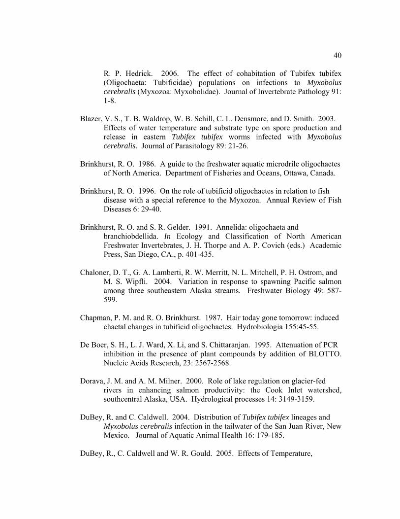
40
R. P. Hedrick. 2006. The effect of cohabitation of Tubifex tubifex (Oligochaeta: Tubificidae) populations on infections to Myxobolus cerebralis (Myxozoa: Myxobolidae). Journal of Invertebrate Pathology 91: 1-8.
Blazer, V. S., T. B. Waldrop, W. B. Schill, C. L. Densmore, and D. Smith. 2003.
Effects of water temperature and substrate type on spore production and release in eastern Tubifex tubifex worms infected with Myxobolus cerebralis. Journal of Parasitology 89: 21-26.
Brinkhurst, R. O. 1986. A guide to the freshwater aquatic microdrile oligochaetes
of North America. Department of Fisheries and Oceans, Ottawa, Canada. Brinkhurst, R. O. 1996. On the role of tubificid oligochaetes in relation to fish
disease with a special reference to the Myxozoa. Annual Review of Fish Diseases 6: 29-40.
Brinkhurst, R. O. and S. R. Gelder. 1991. Annelida: oligochaeta and
branchiobdellida. In Ecology and Classification of North American Freshwater Invertebrates, J. H. Thorpe and A. P. Covich (eds.) Academic Press, San Diego, CA., p. 401-435.
Chaloner, D. T., G. A. Lamberti, R. W. Merritt, N. L. Mitchell, P. H. Ostrom, and
M. S. Wipfli. 2004. Variation in response to spawning Pacific salmon among three southeastern Alaska streams. Freshwater Biology 49: 587-599.
Chapman, P. M. and R. O. Brinkhurst. 1987. Hair today gone tomorrow: induced
chaetal changes in tubificid oligochaetes. Hydrobiologia 155:45-55. De Boer, S. H., L. J. Ward, X. Li, and S. Chittaranjan. 1995. Attenuation of PCR
inhibition in the presence of plant compounds by addition of BLOTTO. Nucleic Acids Research, 23: 2567-2568.
Dorava, J. M. and A. M. Milner. 2000. Role of lake regulation on glacier-fed
rivers in enhancing salmon productivity: the Cook Inlet watershed, southcentral Alaska, USA. Hydrological processes 14: 3149-3159.
DuBey, R. and C. Caldwell. 2004. Distribution of Tubifex tubifex lineages and
Myxobolus cerebralis infection in the tailwater of the San Juan River, New Mexico. Journal of Aquatic Animal Health 16: 179-185.
DuBey, R., C. Caldwell and W. R. Gould. 2005. Effects of Temperature,

41
Photoperiod, and Myxobolus cerebralis infection on growth, reproduction and survival of Tubifex tubifex lineages. Journal of Aquatic Animal Health 17: 338-344.
El-Matbouli, M., T. S. McDowell, D. B. Antonio, K. B. Andree, and Hedrick, R.P.
1999. Effect of water temperature on the development, release and survival of the triactinomyxon stage of Myxobolus cerebralis in its oligochaete host. International Journal for Parasitology 29: 627-641.
Gilbert, M. A., and W. O. Granath Jr. 2001. Persistent infection of Myxobolus
cerebralis, the causative agent of salmonid whirling disease, in Tubifex tubifex. Journal of Parasitology 87 (1): 101-107.
Glass, R. L., T. P. Brabets, S. A. Frenzel, M. S. Whitman, and R. T. Ourso. 2004.
Water quality in the Cook Inlet basin Alaska, 1998-2001. U. S. Department of Interior, U. S. Geological Survey, circular 1240.
Hall, T.A. 1999. BioEdit: a user-friendly biological sequence alignment editor
and analysis program for Windows 95/98/NT. Nucleic Acids Symposium Series 41:95-98.
Hallett, S. L., S. D. Atkinson and J. L. Bartholomew. 2005. Countering
morphological ambiguities: development of a PCR assay to assist in the identification of Tubifex tubifex oligochaetes. Hydrobiologia 543: 305-309.
Hallett, S. L. and J. L. Bartholomew 2006. Effects of water flow on the infection
dynamics of Myxobolus cerebralis. Page 71 in Abstracts from the Fifth International Symposium on Aquatic Animal Health, San Francisco, California, USA.
Hammarstrom, S. L. 1988. Angler effort and harvest of Chinook salmon
Oncorhynchus tshawytscha and coho salmon O. kisutch by the recreational fisheries in the lower Kenai River, 1987. Alaska Department of Fish and Game, Division of Sport Fish, Fisheries Data Series No. 50, Juneau, Alaska.
Harding, R and D. Jones. 2002. Peterson Creek and lake system steelhead
evaluation, 1991. Alaska Department of Fish and Game, Division of Sport Fish, Fishery Data Series N0. 92-46, Anchorage, Alaska.
Jones, S., G. Preoperi-Porta, S. Dawe, J. Blackbourn, K. Taylor, G. Lowe, and A.

42
Osborn. 2004. Proliferative renal myxosporidiosis in spawning coho salmon (Oncorhynchus kisutch) in British Columbia and Washington. Folia Parasitologica 51: 221-227.
Kaeser, A. J., C. Rasmussen, and W. E. Sharpe. 2006. An examination of
environmental factors associated with Myxobolus cerebralis infection of wild trout in Pennsylvania. Journal of Aquatic Animal Health 18: 90-100.
Kelley, G. O., F. J. Zagmutt-Vergara, C. M. Leutenegger, K. A. Myklebust, M. A.
Adkison, T. S. McDowell, G. D. Marty, A. L. Kahler, A. L. Bush, I. A. Gardner, R. P. Hedrick. 2004. Evaluation of five diagnostic methods for the detection and quantification of Myxobolus cerebralis. Journal of Veterinary Diagnostic Investigation 16: 195-204.
Kerans, B. L., and A. V. Zale. 2002. The ecology of Myxobolus cerebralis.
Pages 145-166 in J. L. Bartholomew and J. C. Wilson, editors. Whirling disease: reviews and current topics. American Fisheries Society Symposium 29, Bethesda, Maryland.
Kerans, B. L., C. Rasmussen, R. Stevens, A. E. L. Colwell, and J. R. Winton. 2004. Differential propagation of the metazoan parasite Myxobolus cerebralis by Limnodrilus hoffmeisteri, Ilyodrilus templetoni, and genetically distinct strains of Tubifex tubifex. Journal of Parasitology 90: 1366-1373.
Kerans, B. L., R. I. Stevens, and J. C. Lemmon. 2005. Temperature affects a host-
parasite interaction: Tubifex tubifex and Myxobolus cerebralis. Journal of Aquatic Animal Health 17: 216-221.
MacConnell, E. and E. R. Vincent. 2002. The effects of Myxobolus cerebralis on
the salmonid host. Pages 95-107 in J. L. Bartholomew and J. C. Wilson, editors. Whirling disease: reviews and current topics. American Fisheries Society Symposium 29, Bethesda, Maryland.
Milbrink, G. 1994. Oligochaetes and water pollution in two deep Norwegian
lakes. Hydrobiologia 278: 213-222. Miller, M. G. and D. Bosch. 2004. Area management report for the recreational
fisheries of Anchorage, 2003. Alaska Department of Fish and Game, Fisheries Management Series No. 04-07, Anchorage.
Milner, A. M. and G. E. Petts. 1994. Glacial rivers: physical habitat and ecology.
Freshwater Biology 32: 295-308.

43
Milner, A. M., J. G. Irons III and M. W. Oswood. 1997. The Alaskan landscape: an introduction for limnologists. In Freshwaters of Alaska, A. M. Milner and M. W. Oswood (eds.). Springer, New York, NY., p. 1-44.
Montgomery, D. R., J. M. Buffington, N. P. Peterson, D. Schuett-Hames, and T. P.
Quinn. 1996. Stream-bed scour, egg burial depths, and the influence of salmonid spawning on bed surface mobility and embryo survival. Canadian Journal of Fisheries and Aquatic Science 53: 1061-1070.
National Climate Center. 1982. Climate of Alaska. NOAA Environmental Data
Service, Ashville, NC. Nehring, R. B., K. G. Thompson, and S. Hebein. 1998. Impacts of whirling
disease on wild trout populations in Colorado. Pages 82-94 in K. G. Wadsworth, editor. Transactions of the 63rd North American Wildlife and Natural Resources Conference. Wildlife Management Institute, Washington, D. C.
Oswood, M. W., J. G. III Irons and A. M. Milner. 1995. River and Stream
Ecosystems of Alaska. Pages 9-32 in C. E. Cushing, K. W. Cummins, and G. W. Minshall, editors. River and Stream Ecosystems: Ecosystems of the World, vol.22. Elsevier, Amsterdam, Netherlands.
Pagano, R. 2000. The Alaska almanac. Alaska Northwest Books, Portland, OR. Poddubnaya, T. L. 1984. Parthenogenesis in tubificidae. Hydrobiologia 115: 97-
99. Reynoldson, T. B. 1987. The role of environmental factors in the ecology of
Tubificid oligochaetes- an experiemental study. Holarctic Ecology 10: 241-248.
Sollid, S. A., H. V. Lorz, D. G. Stevens, and J. L. Bartholomew. 2002. Relative
susceptibility of selected Deschutes River, Oregon, salmonid species to experimentally induced infection by Myxobolus cerebralis. Pages 117-124 in J. L. Bartholomew and J. C. Wilson, editors. Whirling disease: reviews and current topics. American Fisheries Society Symposium 29, Bethesda, Maryland.
Steinbach Elwell, L. C., B. L. Kerans, C. Rasmussen, and J. R. Winton. 2006.
Interactions among two strains of Tubifex tubifex (Oligochaeta: Tubificidae) and Myxobolus cerebralis (Myxozoa). Diseases of Aquatic Organisms 68: 131-139.

44
Stevens, R., B. L. Kerans, J. C. Lemmon, and C. Rasmussan. 2001. The effects of Myxobolus cerebralis myxospore dose on triactinomyxon production and biology of Tubifex tubifex from two geographic regions. Journal of Parasitology 87: 315–321.
Sturmbauer, C., G. B. Opadiya, H. Niederstatter, A. Riedmann, and R. Dallinger.
1999. Mitochondrial DNA reveals cryptic oligochaete species differing in cadmium resistance. Molecular Biology and Evolution 16: 967-974.
USFWS and AFS-FHS. 2003. Standard procedures for aquatic animal health
inspections. In: Suggested procedures for the detection and identification of certain finfish and shellfish pathogens. Blue Book 5th edition, Ch VI: pp 1-7. Fish Health Section, American Fisheries Society, Bethesda, MD.
U.S. Geological Survey (USGS). 2006a. Cook Inlet Basin, Alaska, NAWQA
News Number 5, Status Report, Winter-Spring 2002. National Water Quality Assessment Program, Anchorage, Alaska. http://ak.water.usgs.gov/Projects/Nawqa/data.htm.
U.S. Geological Survey (USGS). 2006b. National water information system.
http://water.usgs.gov/data.html. Vincent, E. R. 1996. Whirling disease and wild trout: the Montana experience.
Fisheries 21:32-33. Walker, P. G. and R. B. Nehring. 1995. An investigation to determine the
cause(s) of the disappearance of young wild rainbow trout in the upper Colorado River, in Middle Park, Colorado. Colorado Division of Wildlife Report, Denver.
Wolf, K., and M. E. Markiw. 1986. Biology contravenes taxonomy in the
Myxozoa: new discoveries show alteration of the invertebrate and vertebrate hosts. Science 225: 1449-1452.
Zendt, J. S., and E. P. Bergersen. 2000. Distribution and abundance of the aquatic
oligochaete host Tubifex tubifex for the salmonid whirling disease parasite Myxobolus cerebralis in the upper Colorado River basin. North American Journal of Fisheries Management 20: 502-512.

45
Table 2.1. Abundance and mitochondrial lineage of Tubifex tubifex from survey sites (excluding hatcheries).
* Total worms; 3 subsamples of 30 ml each per sample processed. † Total Tubifex tubifex in 3 subsamples of 30 ml each from each site. Total is adjusted: total worms morphologically identified as T.
tubifex minus percent identified as false positive (determined to be another species by PCR). ‡Assayed by both T. tubifex species-specific and lineage PCRs.
River Sites sampled
Sites with Tubifex tubifex
Total worms*
Tubifex tubifex†
Worms assayed
‡
Worms sequenced
Lineages (% of sampled
population) Southeast Alaska Peterson Creek 4 0 2,741 0 61 2 n/a Montana Creek 4 0 54 0 42 1 n/a Total southeast 8 0 2,795 0 103 3 Southcentral Alaska
Ship Creek 6 5 1,545 251 71 3 I (71%), III (21%), IV (7%)
Campbell Creek 6 2 415 135 49 2 I (86%), III (14%) Upper Kenai River 6 0 117 0 7 0 n/a Mid Kenai River 6 0 55 0 13 0 n/a Lower Kenai River (before brackish area)
4 3 6,578 4,805 61 2 I (24%), III (7%), VI (69%)
Total southcentral 28 10 8,710 5,191 199 7 Total 36 10 11,505 5,191 302 10
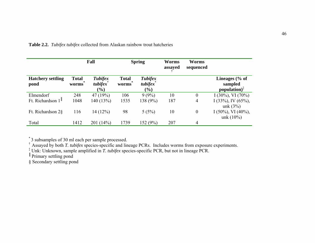
46
Table 2.2. Tubifex tubifex collected from Alaskan rainbow trout hatcheries Fall Spring Worms
assayed†
Worms sequenced
Hatchery settling pond
Total worms*
Tubifex tubifex*
(%)
Total worms*
Tubifex tubifex*
(%)
Lineages (% of sampled
population)‡
Elmendorf 248 47 (19%) 106 9 (9%) 10 0 I (30%), VI (70%) Ft. Richardson 1║ 1048 140 (13%) 1535 138 (9%) 187 4 I (33%), IV (65%),
unk (3%) Ft. Richardson 2§ 116 14 (12%) 98 5 (5%) 10 0 I (50%), VI (40%),
unk (10%) Total 1412 201 (14%) 1739 152 (9%) 207 4
* 3 subsamples of 30 ml each per sample processed. † Assayed by both T. tubifex species-specific and lineage PCRs. Includes worms from exposure experiments. ‡ Unk: Unknown, sample amplified in T. tubifex species-specific PCR, but not in lineage PCR. ║ Primary settling pond § Secondary settling pond

47
Table 2.3. Prevalence and level of Myxobolus cerebralis infection in different mitochondrial lineages of Tubifex tubifex used in laboratory exposure experiments Group Lineage (% of
sampled population)*
TAM Release period
(days PE)†
Maximum TAMs
detected per week
Infection prevalence (by QPCR)
Lineages infected
First Exposure Ft. Richardson hatchery‡ I (30%), IV (68%),
Unknown (2%) n/a 0 0 None
Wizard Falls (reference)‡ III (100%) 98- 182 14,967 85% III Second Exposure A I (89%), III (3%),
IV (5%), VI (2%), Unknown (1%)
120-160 188 3% III
B III (12%), VI (88%) 132-153 11 10% III C I (80%), III (4%),
VI (14%), Unknown (2%)
108-160 280 0.6% III
Ft. Richardson hatchery I (65%), IV (35%) n/a 0 0 None Wizard Falls (reference) III (100%) 100-160 ≥7000 100% III
* Unknown: sample amplified in Tubifex tubifex species-specific PCR, but not in lineage PCR. † Second experiment ended at 160 days post-exposure (PE), triactinomyxon (TAM) release not monitored after this date. ‡ Total of three replicates.

48
Table 2.4. Numbers of Tubifex tubifex exposed, infected and surviving experimental exposure to Myxobolus cerebralis
* In three replicates of 100. † Not individually monitored as no TAMs were detected. ‡ Total worms remaining at end of experiment; does not include worms removed for monitoring during experiment.
First Exposure Second Exposure
Groups: Ft. Richardson
hatchery
Wizard Falls
(reference)
Control (Ft.
Richardson hatchery)
A B C Ft. Richardson
hatchery
Wizard Falls
(reference)
Control (Wizard
Falls)
Exposed 300* 300* 0 (100 unexposed)
200 200 200 100 200 0 (200 unexposed)
Monitored at peak infection (number infected)
60 (0)
60 (54)
20 (0)
0† 0† 50 (0)
0† 20 (20)
0†
Surviving Adults‡ (number infected)
127 4 29 90 (3)
137 (13)
108 (1)
100 (0/20)
15
97
Progeny 205 0 131 28 41 50 98 19 57

49
Figure Captions Figure 2.1. Map of study locations, sampling sites for Tubifex tubifex (black dots), and rainbow trout (Oncorhynchus mykiss) hatcheries (triangles) in southcentral and southeast Alaska. Figure 2.2. Tubifex tubifex lineages from Fort Richardson Hatchery, Alaska. Agarose gel electrophoresis of PCR-amplified genomic DNA using mt 16S rDNA lineage-specific primers. Two lineages were present: I (196 bp) and IV (2 bands at 320 bp and 147 bp). A negative control (water) and a positive control (lineage III, 147 bp) were included. A 50 bp DNA Ladder is shown on both sides of the gel (2% agarose). Figure 2.3. First experimental exposures- Susceptibility of lineage IV Tubifex tubifex to Myxobolus cerebralis: triactinomyxon release per 100-worm replicate (3 Alaskan replicates are pooled as 1). Figure 2.4a. Second experimental exposures- Comparative triactinomyxon release from Alaskan Tubifex tubifex. Number of triactinomyxons noted as absolute numbers 0-7000; greater values noted as >7000 triactinomyxons and not as an absolute number. Figure 2.4b. Comparative triactinomyxon release from Alaskan Tubifex tubifex groups A, B, and C (enlargement of Figure 2.4a).

50
Figure 2.1
Alaska
N
Ship Creek
Kenai River
Cook Inlet
Campbell Creek
10 km
Skilak Lake
Kenai Lake
Anchorage
5 km
Montana CreekPeterson Creek
Juneau
Mendenhall River
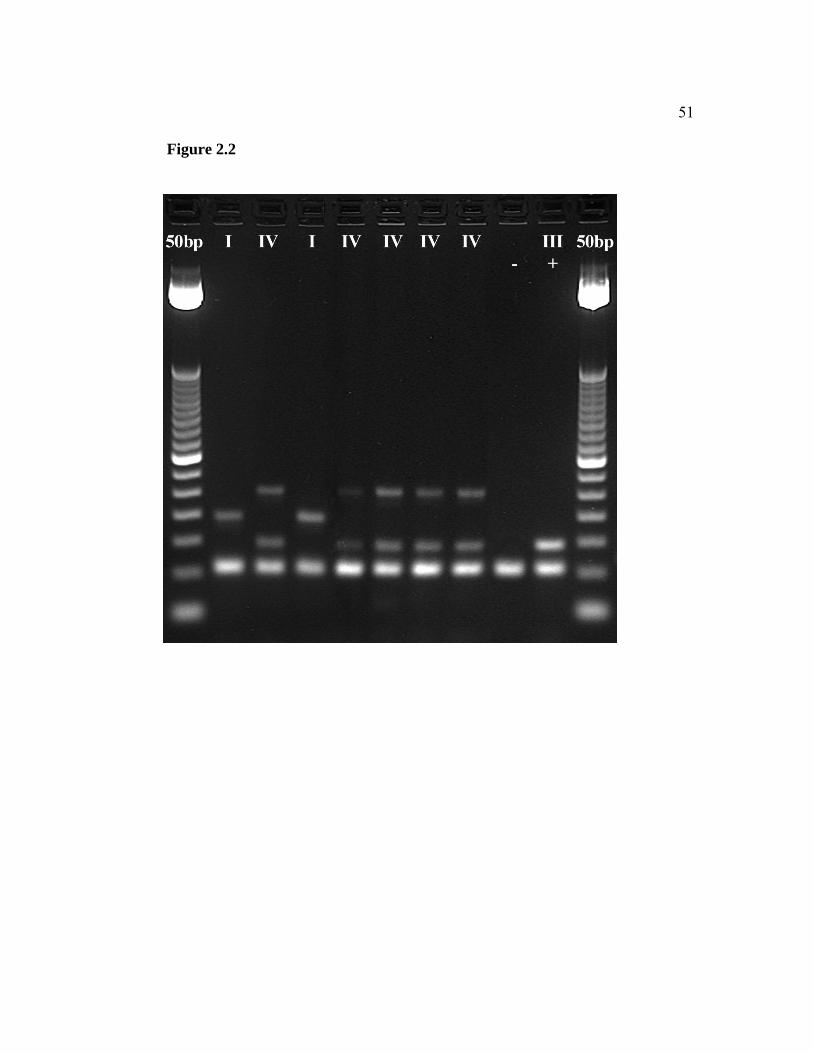
51
Figure 2.2

52
Figure 2.3
0
2000
4000
6000
8000
10000
12000
98 101 105 108 112 115 119 122 126 129 132 136 140 143 150 154 161 168 182 191
Days post-exposure
Tria
ctin
omyx
ons
Alaskan wormsWF 1WF 2WF 3

53
Figure 2.4a
0
1000
2000
3000
4000
5000
6000
7000
8000
100 108 120 126 132 139 147 153 160
Days post-exposure
Tria
ctin
omyx
ons
Group AGroup CWF
Figure 2.4b
0
50
100
150
200
250
300
100 108 120 126 132 139 147 153 160
Days post-exposure
Tria
ctin
omyx
ons
Group AGroup BGroup C

54
CHAPTER 3
EXPANDED GEOGRAPHICAL DISTRIBUTION OF MYXOBOLUS CEREBRALIS: FIRST DETECTIONS FROM ALASKA.
E. Leyla Arsan1,2, Stephen D. Atkinson2, 3, Sascha L. Hallett2, Theodore Meyers4, and Jerri L. Bartholomew2*
1 Department of Fisheries and Wildlife, Center for Fish Disease Research, Oregon State University, Corvallis, Oregon, USA
2 Department of Microbiology, Center for Fish Disease Research, Oregon State University, Corvallis, Oregon, USA
3 School of Molecular and Microbial Sciences, University of Queensland, Brisbane Queensland, Australia
4Alaska Department of Fish and Game, Juneau Fish Pathology Laboratory, Juneau, Alaska , USA

55
Abstract
Introduced to the USA in 1958, Myxobolus cerebralis, the parasite responsible for
salmonid whirling disease, has since spread across the country causing severe
declines in wild trout populations. While assessing the risk of introduction of this
pathogen into the state of Alaska, an area where clinical whirling disease has never
been documented, we detected the parasite using a species-specific polymerase
chain reaction (PCR). This is the first report of M. cerebralis in Alaska. Testing
of 180 hatchery rainbow trout (Oncorhynchus mykiss) by pepsin-trypsin digest and
quantitative PCR (QPCR) revealed fourteen positive samples. Infection in two of
these was confirmed by sequencing the parasite 18S rRNA gene and by a nested
PCR assay based on the same gene. We were unable to visually identify
myxospores, indicating that either infection was light or mature spores had not
formed. Sequence comparison of M. cerebralis from several locations
demonstrated the Alaska isolates were genetically distinct and therefore not false
positives arising from contamination during processing. This paper presents DNA
sequence data from the Alaska M. cerebralis isolates, provides a brief history of
the fish and facility of origin, and discusses implications of different testing
methods on asymptomatic fish populations.

56
Introduction
Myxobolus cerebralis, a freshwater metazoan parasite exotic to North
America, was first detected in the USA in 1958 (Hoffman, 1962). Thought to have
originated from a shipment of infected brown trout (Salmo trutta) from Europe, it
is now reported in a total of 24 states in the USA and 26 countries worldwide
(Bartholomew and Reno, 2002; Vermont Department of Fish and Wildlife, 2002;
Stromberg, 2006). The pathogen has been at the forefront of fish health research in
the USA due to its implication in dramatic rapid declines of wild rainbow trout
(Oncorhynchus mykiss) populations in the North American intermountain west,
primarily in Colorado and Montana (Vincent, 1996; Nehring et al., 1998).
Myxobolus cerebralis is not known to occur in Alaska, though little
monitoring for the pathogen has been conducted. In response to concerns
regarding allocation of resources for monitoring programs, research was initiated
to assess the risk of introduction and establishment of the parasite. In the process
of validating the initial assumption that the parasite did not occur in Alaska, we
detected the pathogen by PCR in samples from a state hatchery.
The closest M. cerebralis enzootic area to Alaska is the Snake River basin
in northeast Oregon, southeast Washington, and Idaho. The parasite has also been
reported in wild and cultured salmonids from the Sakhalin Islands off the east
coast of Russia (Bogdanova, 1960, 1968). However, the distance from the
Sakhalin Islands to southcentral Alaska is approximately double that from the
Snake River basin.
The assumption that M. cerebralis was not present in Alaska was based on
a lack of observed clinical disease and on data from previous limited monitoring
for the parasite in wild fish stocks by the US Fish and Wildlife Service (USFWS)
as part of the National Wild Fish Health Survey (NWFHS). The USFWS uses
pepsin trypsin digest (PTD) for parasite detection and PCR as confirmation of
infection (USFWS and AFS-FHS, 2003). Examination of 2,049 fish from 26

57
locations did not result in detection of the parasite [USFWS, 2006; T. Burton,
Alaska Department of Fish and Game (ADFG), personal communication]. Thus,
PCR has never been conducted. However, the NWFHS tests for a number of fish
pathogens and does not target areas at high-risk for M. cerebralis. As a result,
only 18% of fish tested by USFWS (361 fish from seven locations) were from
southcentral Alaska. Hatchery fish in Alaska are not monitored for the parasite.
To provide further information on the status of infection in fish in Alaska, we
tested rainbow trout from state hatcheries, using PCR as the initial screening
method.
Methods
Origin of fish tested
Rainbow trout, the salmonid most susceptible to the parasite (MacConnell
and Vincent, 2002; Sollid et al., 2002), were obtained from the only two hatcheries
in Alaska that routinely rear the species: Fort Richardson (FTR) and Elmendorf
(ELM). Both hatcheries are located on Ship Creek in the Cook Inlet basin in
southcentral Alaska. In 2005, 60 fish each from FTR and ELM were tested for the
parasite. In 2006, another group of 60 fish from ELM (of the same broodyear as
those tested in 2005) was assayed. Rainbow trout at ELM are hatched at FTR and
transferred to ELM after 4-8 months.
Detection of Myxobolus cerebralis
In 2005, the 60 fish sampled from each hatchery were randomly segregated
into five-fish pools and cartilage from whole heads was digested by pepsin trypsin
digest (PTD) following USFWS and AFS-FHS (2003) procedures with minor
modifications noted below. After trypsin was added, the pH was adjusted to 8.0
[not 8.5, as stated in USFWS and AFS-FHS (2003)] with 1 N NaOH. The PTD
pellet was suspended in 1 mL phosphate buffered saline (PBS) and frozen for later
analysis. DNA was extracted from the entire thawed sample using a QIAGEN
58
DNeasy Tissue Kit and eluted with 50 µL buffer AE. This was then tested for M.
cerebralis by QPCR (Kelley et al., 2004) with 4 µL of extracted DNA used in a 20
µL reaction volume. Duplicate reactions were performed in an ABI PRISM 7300
or 7500 Sequence Detection System. In 2006, fish were tested individually (not
pooled). Testing methodology was the same as 2005, except that only 200 µL of
fully homogenized (vortexed) PTD product suspended in PBS was used for DNA
extraction. The remainder of the PTD product was retained for visual inspection
for myxospores. As the blue book procedures do not specify the volume of sample
to use for DNA extraction, we consulted with the Oregon Department of Fish and
Wildlife (ODFW), Fish Pathology Division (J. Kaufman, pers. comm.) who have
found 200 μL of PTD product to be sufficient volume for parasite confirmation in
Oregon.
Samples positive for M. cerebralis by QPCR in 2006 were visually
inspected for myxospores under 200 and 400X magnification (USFWS and AFS-
FHS, 2003). However, due to the difficulty of visual confirmation of low-grade
infections, a nested PCR assay that targeted the 18S rRNA gene (Andree et al.,
1998) was also used on selected samples to provide further evidence that the
organism amplified in QPCR was actually M. cerebralis. As entire 2005 PTD
samples were processed for DNA extraction (by microwaving), any spores would
have been destroyed and therefore were not discernible by a visual inspection.
A positive control and negative control (water) were used in each
molecular assay. The positive control was an adult Chinook salmon cranial core
(Lostine River, Oregon M. cerebralis enzootic area) that had been previously
digested and determined positive for M. cerebralis by PTD detection and PCR
confirmation. Based on our reference samples, a fish sample presenting any cycle
threshold (Ct) value was considered infected. Samples presenting a Ct in one of
their two duplicate wells were rerun. Further discussion on Ct values and their
reflection of parasite load appears below.

59
Verification of testing methods and determination of QPCR threshold
To verify QPCR and PTD results and determine the relationship between
Ct value and myxospore number, we created a reference set of samples spiked
with known numbers of M. cerebralis myxospores. Six half-heads of uninfected
juvenile rainbow trout originally obtained as eggs from Trout Lodge, WA and held
at the Salmon Disease Laboratory, Corvallis, Oregon on well water were digested
by PTD then spiked with either 100 or 1000 myxospores (three half-heads each per
dose). Myxospores used to spike samples were obtained from three fish exposed
as fry to M. cerebralis actinospores at the Salmon Disease Laboratory and were
added after the PTD to maximize recovery. Each half-head sample was processed
for DNA extraction and QPCR exactly as described for the 2006 samples.
Samples and serial 10-fold dilutions were run in triplicate to determine the limit of
detection in the QPCR assay. We deemed (in consultation with ABI QPCR
representatives) that if one of three triplicates of a sample fluoresced within our
limit of detection, then the sample was positive. The limit of detection was defined
by the sample set in which fewer than all (three) wells tested, fluoresced.
Amplification plots (curves) and delta Rn values were evaluated to confirm the
efficiency and threshold of the QPCR reaction.
The remainder of the PTD product was also visually inspected using both
methods (hemocytometer and wet mount preparation) in the Blue Book (USFWS
and AFS-FHS, 2003). In wet mount preparation, 380 fields at 200x magnification
were examined per 20 and 40 µl of sample for the 1000-myxospore references and
20 µl were examined for the 100-myxospore samples.
DNA sequencing
For two samples (one from each year) positive by the QPCR assay, we
amplified parasite 18S rDNA to confirm the presence of M. cerebralis. We also
sequenced the QPCR positive control (Lostine River adult fish) and our M.

60
cerebralis laboratory isolate, to examine the possibility of positive assay results
due to DNA contamination.
The M. cerebralis SSU 18S rRNA gene was amplified by two-round PCR:
amplification of the entire gene by universal primers ERIB1 and ERIB10 (Barta et
al., 1997) followed by semi-nested reactions using primer pairs ERIB1-ACT1r and
MYXGEN4f-ERIB10 (Hallett & Diamant, 2001, Kent et al., 2000). Reactions
were carried out in 20 µL volumes, which comprised: 2-4 µL template DNA, 0.5
µL each primer (10 µM stock), 0.4 µL dNTPs (10 mM), 0.5 µL BSA (Bovine
Serum Albumin, 10 mg/mL), 1 µL Rediload loading dye (Invitrogen), 1.2 µL
MgCl2 (25 mM), 4 µL Colorless GoTaq Flexi buffer (5X) (Promega), 0.25 µL
GoTaq Flexi (5 U/µL) (Promega) and water. This was scaled up to 50 µL to
generate products for sequencing. PCRs were run on a MJ Research PT100
thermacycler, using the following program: block preheated to 95°C, then initial
denaturation @ 95 °C for 2 min, 35 cycles of 94 °C for 30 s, 55 °C for 30 s, 72 °C
for 60 s, terminal extension of 72 °C for 10 min, then rest at 4 °C. DNA was either
purified directly from PCR products, or electrophoresed on an agarose gel and the
appropriate band excised and purified using Qiagen kits and standard protocols.
DNA concentrations were measured using a NanoDrop ND-1000
Spectrophotometer (NanoDrop Technologies, Wilmington, Delaware). Fragments
were sequenced using the same primers as in the amplifications, with ABI Big Dye
Terminator chemistry on an Applied Biosystems Capillary 3100 Genetic Analyzer
(Foster City, California) at the Oregon State University sequencing facility (Center
for Gene Research and Biotechnology, Central Service Laboratory). Sequences
were aligned and edited in BioEdit (Hall, 1999) with other M. cerebralis isolates
from Genbank (see Table 3.1 for accession numbers).

61
Results
PCR detection of Myxobolus cerebralis
In 2005, assays of 12 five-fish pools from ELM revealed two positive
pools. QPCR indicated low target DNA quantities (mean Ct values 33.6 and
38.7). Potentially positive samples as well as several negative samples were blind-
tested three different times by two different researchers and results were
consistent. None of the fish from FTR were positive.
Testing in 2006 of individual fish of the same cohort but reared at ELM for
an additional year, revealed 12 out of 60 fish to be positive (mean Ct range 32.2–
39.1). Two more fish fluoresced in the QPCR assay, but presented Ct values
which were above the limit of detection of our reference samples and thus were
not included in the total. The nested PCR assay amplified DNA from 2 of 4 fish
that were determined to have the greatest quantity of parasite DNA by QPCR
(Figure 3.1).
Comparison of QPCR Ct values of ELM samples with the reference
samples containing known numbers of spores indicated that in 2005 one pool (5
fish) had approximately 1000 myxospores and the other 100 myxospores (Figure
3.2). In 2006, two samples (1 fish each) had 1000 or more myxospores, three had
approximately 100 myxospores and the remainder had less than 100 myxospores
(approximately 10) (Figure 3.2).
Visual detection of Myxobolus cerebralis
Myxospores were not detected by visual inspection of the PTD product of
QPCR-positive samples in either 2005 or 2006.

62
Verification of testing methods
Myxobolus cerebralis was not visually detected in samples of uninfected
rainbow trout tissue spiked with 100 myxospores, and only one disrupted spore
was observed in samples spiked with 1000 myxospores, when recommended
procedures were followed (USFWS and AFS-FHS, 2003). In contrast, all of the
spiked samples were positive by QPCR. Samples spiked with 1000 myxospores
produced Ct values ranging from 33.3-35.4, with a mean Ct value of 34.4 (Figure
3.2). Fish spiked with 100 myxospores had QPCR Ct values ranging from 35.3-
39.4, with a mean Ct value of 37.3. The 10-fold dilution of a 1000 myxospore
sample had a Ct of 37.1-38.5 (mean 37.6), consistent with a 100 myxospore
sample. The limit of detection was the equivalent of 10 myxospores (either a 10-
fold dilution of a 100 myxospore sample or a 100-fold dilution of a 1000
myxospore sample) since not all triplicate wells of these samples fluoresced (0 to 2
wells per sample).
18S rDNA sequence analysis
Myxobolus cerebralis DNA was amplified and sequenced from two
Alaskan fish that tested positive by QPCR, as well as our laboratory isolate and the
positive control (Lostine River). The SSU 18S rRNA gene was determined to be
2120 bp in length (including terminal primers ERIB1 and ERIB10). Twenty
variable loci, including seven sites with heterogeneous bases, were identified from
alignment of our novel M. cerebralis isolates with sequences in GenBank (Table
3.1). The two Alaska isolates differed from both the laboratory isolate (6-8 loci)
and the positive control (3-5 loci), and from each other (6 loci).
Discussion
The molecular detection of M. cerebralis from hatchery rainbow trout is
the first record of the parasite in Alaska. The rainbow trout that tested positive for

63
M. cerebralis were hatched at FTR and transferred to ELM after 4-8 months. FTR
is supplied by well water and no fish examined from that facility were positive for
the parasite. ELM is supplied by surface water from Ship Creek and the fish tested
were reared at this facility for 11 months (2005 samples) and 23 months (2006
samples).
Because M. cerebralis had not previously been detected in Alaska and the
fish tested had no clinical signs of disease, we used the most sensitive methods
(PCR, QPCR) for diagnostic screening. However, we were unable to visually
detect myxospores from samples that tested positive by these assays. Given the
low DNA quantity indicated by QPCR, we suspected that visual detection of a low
number of spores in a sample would be highly improbable and designed reference
samples to test this. In our reference samples that had been spiked with 1000
myxospores after PTD (then frozen before examination) we were able to identify
only one presumptive spore. This observation is in accordance with Markiw and
Wolf (1974), who documented that in fish spiked with 1000 myxospores, less than
0.2% of spores were recovered by the PTD process. Thus, visual confirmation in
asymptomatic fish with low or early infections may be unattainable.
For regulatory inspection purposes, PCR testing is offered along with
histology as a confirmatory test after presumptive identification of M. cerebralis
by visual inspection of myxospores in enzymatic tissue digests (USFWS and AFS-
FHS, 2003). Molecular tests in general are not approved diagnostic screening
techniques because of the difficulties in verifying positive results using a second,
often less sensitive, test. For bacterial and viral pathogens that can be amplified by
culture techniques, there is generally not a significant difference in sensitivity
between culture and molecular detection. However, detection of pathogens that
cannot be amplified by culture is more problematic and likely to result in false
negatives when pathogen numbers are low, even when using concentration
procedures like PTD. This is compounded when identification relies on detection
of a single life stage, like the myxospore. In studies that have compared these

64
testing methods (Schisler et al., 2001; Kelley et al., 2004), PCR was found to be an
effective diagnostic tool and is approximately 10-fold more sensitive than PTD
(Andree et al. 2002). Kelley et al. (2004) determined that any of the five
diagnostic approaches they evaluated was effective in identifying moderately to
heavily infected fish. Schisler et al. (2001) screened free-ranging and hatchery-
reared fish with various levels of infection and documented that PCR identified M.
cerebralis more often than PTD, and that the heightened sensitivity of PCR
resulted in detection of the parasite in areas previously thought to be free of the
pathogen.
Another advantage of genetic tests as detection tools is their capability to
detect not only the different stages of parasite development, but also mature spores
that have lost their morphological integrity under harsh extraction conditions. In
contrast, PTD is capable of detecting only mature final stages of parasite
development that are visually distinguishable.
Estimates of infection levels in the Alaskan hatchery fish were inferred by
comparison of their QPCR Ct values with those of the reference samples that
contained known numbers of myxospores and were processed identically. All six
reference samples spiked with either 100 or 1000 myxospores each were positive
by QPCR as were 10-fold dilutions of a 1000 spore sample. Dilutions equivalent to
10 myxospores of the 100 and 1000 spore samples fluoresced in zero to two of
their three replicate wells, thus defining the limit of detection as a Ct value less
than 40 (Figure 3.2). Infections in hatchery fish ranged from 10 myxospores to
over 1000 (mean Ct range 32.2–39.1).
For infection levels in the range of our estimates for the Alaska fish (1000
myxospores or less), we found that neither of the two visual detection methods
described in the Blue Book is a valid method for enumeration of spores following
PTD digest (USFWS and AFS-FHS, 2003). Firstly, for hemocytometer data to be
accurate, 15-50 spores per 1 mm2 grid (with four grids per hemocytometer) must
be observed (Cascade Biologics, Inc., 2002). This suggests that a minimum of
65
150,000 spores per mL is required for an accurate enumeration by hemocytometer
assessment. In samples with low infections such as 1000 myxospores, an average
of only 1 spore per 10 grids would be present. Secondly, if the other visualization
method stipulated in the Blue Book (examination of all myxospores in 150 fields
under 200X magnification) is used, we should have seen 20 myxospores in 20 µL
of the 1000-spore sample. We saw only one disrupted spore in a single sample,
which suggests the PTD method jeopardized the integrity of the spore.
As we were unable to visually detect M. cerebralis spores, we used a
second PCR assay and DNA sequencing of the second round product as an
alternate method of confirmation of parasite presence. The two fish from Alaska
from which M. cerebralis DNA was sequenced contained approximately 1000
myxospores. Another two samples that were positive by QPCR, but could not be
amplified in the nested PCR, likely had infections on the order of 100 myxospores.
Schisler et al. (2001) observed an exponential increase in myxospore numbers with
increasing PCR band strength, and that “weak positives” had a mean spore count
of 958 myxospores. These researchers also found that only 2 out of 40 “weak
positive” samples produced enough DNA to be sequenced. Thus, it is not
surprising that only two of the positive samples from Alaska produced sufficient
DNA for sequencing.
The Alaska isolates differed in at least five loci from the positive control
and our lab isolates, which indicates cross-contamination was unlikely. Overall
variation between M. cerebralis isolates was very low: 20 loci in 2120 (<1%), and
the maximum variation between isolates was only 12 loci (0.56%). This low level
of intraspecific variation in the 18S is consistent with data from ITS sequences
(Whipps et al., 2004). Some variations were only present in the older GenBank
sequences and may be sequence noise or misreads, especially the insertion at base
position 1387, which was not present in any of our isolates. The presence of
heterogeneous bases at loci in several isolates indicates that source fish may either
be infected with multiple ‘genotypes’ of M. cerebralis, or that some isolates may

66
have multiple alleles of the 18S rRNA gene present – a feature we have observed
in another myxozoan parasite of salmonids, Parvicapsula minibicornis (Atkinson
et al., 2006). Interestingly, the Alaska isolates differed between themselves at 6
loci, which suggests different sources of the parasite may be involved. Although,
given the degree of anthropogenic mixing and translocation of both host and
parasite populations in North America, it is difficult to draw conclusions on
finescale phylogeny of M. cerebralis.
The source of infection for the hatchery fish remains unknown. ELM is
supplied by surface water from Ship Creek, with incoming surface water screened
for debris but not treated for potential biota. Thus, M. cerebralis myxospores,
actinospores, or even Tubifex tubifex [the alternate oligochaete host of the parasite
(Wolf and Markiw, 1984)] could potentially enter the hatchery if the parasite were
present in the Creek. Also, until 2004, water sources for both hatcheries were
heated to ambient temperatures as high as 14oC, either by wastewater (ELM) or by
heat exchanger from power plants nearby. Heated water temperatures could have
contributed to parasite propagation, as temperatures from 10-15°C accelerate the
parasite life cycle and are optimal for parasite development (El-Matbouli et al.,
1999). Effluent from both ELM and FTR is routed through earthen settling ponds
[that contain populations of T. tubifex (Arsan et al., chapter 2 of this volume)] and
flows back into Ship Creek untreated. Thus, fish in Ship Creek could also be
exposed to parasites in hatchery effluent.
While the origin of M. cerebralis in Alaska remains unknown, the state of
Alaska has made significant efforts to limit movements of fish and hence sources
of pathogen introduction, and the origin of M. cerebralis in the state remains
unknown. Anecdotal evidence suggests that rainbow trout were introduced as
eggs in southcentral Alaska in the 1950’s from broodstock at Winthrop National
Fish Hatchery, Washington. Interior Alaska received transplants (eggs) from
Idaho and Montana in 1952 (Marvich and McRea, 1953; Morrow, 1980). As these
imports were eggs and also were made before M. cerebralis was known to have

67
been introduced into the USA (Hoffman, 1962; Bartholomew and Reno, 2002), it
is unlikely that they resulted in parasite transfer. Additionally, since the mid
1970s, Alaska has adopted strict regulations that prohibit the import of live non-
ornamental fish into the state (Alaska Administrative Code [AAC] 5: 41.070).
Though it is unknown how long the parasite has been present in Alaska, it
is likely that previous monitoring would have missed a low prevalence or low level
of infection due to the limited sensitivity of testing methods. The use of PTD as
the sole screening method could miss low infections that would be identified by
molecular tests such as PCR, which are approximately 10-fold more sensitive
(Andree et al., 2002). However, for management purposes, it may only be
necessary to detect infections that cause negative impacts on fish populations or
are considered “significant” infections. Each state has its own stipulations as to
what constitutes such an infection and what the implications of detection are. For
example, the state of Colorado transfers only fish that are PCR negative for M.
cerebralis to areas where the parasite is not known to occur. The 2004 broodyear
rainbow trout from ELM were transplanted throughout southcentral and interior
Alaska prior to parasite detection. Many of these stocked locations were land-
locked lakes with no outlets, which would limit the spread of the parasite, but
some fish were also stocked in open stream systems where susceptible lineages of
T. tubifex have been documented (Arsan et al., chapter 2 of this volume). Thus,
some degree of spread may already have occurred. However, the low prevalence
and severity of infection in the fish populations tested and the low abundance of
suitable oligochaete hosts (Arsan et al., chapter 2 of this volume) may limit further
spread of the parasite.
The first detection of M. cerebralis in Alaska is not without consequence.
Though the risk of parasite dissemination by ELM fish may be low, it is
nevertheless unacceptable by current ADFG fish disease policies. An interim
management strategy will include restricting all fish transplants from ELM to land-
locked lakes having no native susceptible resident salmonid species or to seawater
68
netpens for terminal commercial and sport fisheries. Release of Chinook salmon
smolts into Ship Creek will continue in order to maintain returning broodstock.
This species develops resistance to M. cerebralis more rapidly than rainbow trout
(Sollid et al. 2003) and thus presents a lower risk of parasite dissemination. Future
plans include replacement of the current Elmendorf Hatchery with a new facility
constructed on a nearby site with water supplied from wells rather than Ship
Creek. The FTR will be discontinued entirely once the new ELM facility is
completed. The use of well water should facilitate breaking the parasite life cycle,
and will be corroborated by additional QPCR testing of hatchery fish stocks in the
new facility. Negative results may allow future unrestricted fish transplants.
Placement of juvenile sentinel rainbow trout in cages within Ship Creek would
also determine if the parasite has become established within the basin, an approach
that has been used in similar situations after isolated parasite introduction
(Bartholomew et al., 2007).
While this is the first detection of M. cerebralis in Alaska, clinical whirling
disease has never been reported. Because hatchery rainbow trout in Alaska
originated from the contiguous USA (Marvich and McRea, 1953; Morrow, 1980),
their susceptibility to the pathogen is likely similar to other resident rainbow trout
strains. We found rainbow trout from ELM to have very low infections (as
determined by QPCR), which probably reflects exposure to a low number of
actinospores or exposure when fish are older and have already gained some degree
of resistance to the parasite. In laboratory susceptibility challenges with T. tubifex
from different locations in Alaska, infection prevalence in populations fed 500
myxospores/ worm ranged from 0.5 – 10 % depending on the lineage composition
of the population, and each group of 200 exposed worms produced up to several
hundred actinospores (Arsan et al., chapter 2 of this volume). While this number
is relatively low, each triactinomyxon that reaches its fish host is capable of
proliferating 1000-fold (Kerans and Zale, 2002), though environmental conditions
and water temperatures may slow development. Thus, even introduction of fish

69
with low infections of M. cerebralis can be significant in areas where physical or
environmental conditions are optimal for parasite proliferation.
Current physical and environmental conditions in Alaska likely contribute
to keeping infection rates low and disease from becoming apparent. Abundance of
resistant lineages of T. tubifex (Arsan et al., chapter 2 of this volume), as well as
cooler water temperatures, may slow the proliferation and life cycle of M.
cerebralis and cumulatively prevent the parasite from severely impacting salmonid
populations in this region. In such cases, disease may never become apparent.
A complete analysis of risk of further spread and of potential routes of
introduction of M. cerebralis into Alaska is forthcoming.
Acknowledgements
This work was supported in part by the National Partnership for the
Management of Wild and Coldwater Species, the USFWS, the U.S. Geological
Survey, and the ADFG. We are grateful to: Tammy Burton of the ADFG and to
FTR and ELM hatchery personnel for providing fish samples; John Kaufman and
Nadine Hurtado of the ODFW for providing positive control samples as well as
assistance and advice regarding parasite assays; Harriet Lorz at the Center for Fish
Disease Research for her tremendous help with the pepsin trypsin digest and with
reference samples; and Jenny Dubanoski and Kyle Thames at Oregon State
University for preparing reference samples and for assistance in the laboratory.

70
References
Andree, K. B., R. P. Hedrick, and E. MacConnell. 1998. A nested polymerase
chain reaction for the detection of genomic DNA of Myxobolus cerebralis in rainbow trout (Oncorhynchus mykiss). Diseases of Aquatic Organisms 34: 145-154.
Andree, K. B., R. P. Hedrick, and E. MacConnell. 2002. Review of the
approaches to detect Myxobolus cerebralis, the cause of salmonid whirling disease. American Fisheries Society Symposium 29: 197-211.
Arsan, L., S. Hallett, and J. Bartholomew. 2007. Tubifex tubifex from Alaska: distribution and susceptibility to Myxobolus cerebralis. Chatper 2 in Potential for the dispersal of the non-native parasite Myxobolus Cerebralis: qualitative risk-assessments for the state of Alaska and the Willamette River basin, Oregon (Master of Science thesis, Oregon State University, 2007).
Atkinson, S. D., S. R. M. Jones, and J. L. Bartholomew. 2006. Parvicapsula minibicornis: a myxozoan species complex?. Page 64 in Abstracts from the Fifth International Symposium on Aquatic Animal Health, San Francisco, California, USA.
Barta, J.R., D.S. Martin, P.A. Liberator, M. Dashkevicz, J.W. Anderson, S.D.
Feighner, A. Elbrecht, A. Perkins-Barrow, M.C. Jenkins, H.D. Danforth, M.D. Ruff, and H. Profous-Juchelka. 1997. Phylogenetic relationships among eight Eimeria species infecting domestic fowl inferred using complete small subunit ribosomal DNA sequences. Journal of Parasitology 83: 262-71.
Bartholomew, J. L. and P. W. Reno. 2002. The history and dissemination of
whirling disease. Pages 3-24 in J. L. Bartholomew and J. C. Wilson, editors. Whirling disease: reviews and current topics. American Fisheries Society Symposium 29, Bethesda, Maryland.
Bartholomew, J. L., H. V. Lorz, S. D. Atkinson, S. L. Hallett, D. G. Stevens, R. A.

71
Holt, K. Lujan, and A. Amandi. 2007. Evaluation of a management plan to control the spread of Myxobolus cerebralis in a lower Columbia River tributary. North American Journal of Fisheries Management, in press.
Bogdanova, E. A. 1960. Natural habitat of the myxosporidian (Myxosoma
cerebralis, whirling disease) at Sakhalin (S. E. Russia). Doklady Akademii Nuak SSSR 134: 1501-1503.
Bogdanova, E. A. 1968. Modern data on the distribution and biology of
Myxosoma cerebralis (Protozoa, Cnidosporidia) as an agent of whirling disease of salmonids. Troisiéme Symposium de la Commission de l’Office International des Épizooties pour l’Étude des Maladies des Poissons, Stockholm.
Cascade Biologics, Inc. 2002. Using a Hemocytometer. Document 107.
http://www.cascadebio.com/pdfdocs/Cell%20Documents/hemat.pdf
El-Matbouli, M., T. S. McDowell, D. B. Antonio, K. B. Andree, and R. P.
Hedrick. 1999. Effect of water temperature on the development, release, and survival of the triactinomyxon stage of Myxobolus cerebralis in its oligochaete host. International Journal for Parasitology 29: 627-641.
Hall, T. A. 1999. BioEdit: a user-friendly biological sequence alignment editor
and analysis program for Windows 95/98/NT. Nucleic Acids Symposium Series 41:95-98.
Hallett, S. L. and A. Diamant. 2001. Ultrastructure and small-subunit ribosomal
DNA sequence of Henneguya lesteri n.sp. (Myxosporea), a parasite of sand whiting Sillago analis (Sillaginidae) from the coast of Queensland, Australia. Diseases of Aquatic Organisms 46: 197-212.
Hoffman, G. L. 1962. Whirling Disease of Trout. United States Department of
Interior. Fishery Leaflet 508. Fish and Wildlife Service.

72
Kelley, G. O., F. J. Zagmutt-Vergara, C. M. Leutenegger, K. A. Myklebust, M. A.
Adkison, T. S. McDowell, G. D. Marty, A. L. Kahler, A. L. Bush, I. A. Gardner, R. P. Hedrick. 2004. Evaluation of five diagnostic methods for the detection and quantification of Myxobolus cerebralis. Journal of Veterinary Diagnostic Investigation 16: 195-204.
Kent, M. L., J. Khattra, R. P. Hedrick, and R. H. Devlin. 2000. Tetracapsula renicola n. sp. (Myxozoa: Saccosporidae); The PKX myxozoan—the cause of proliferative kidney disease of salmonid fishes. Journal of Parasitology 86: 103–111.
Kerans, B. L., and A. V. Zale. 2002. The ecology of Myxobolus cerebralis. Pages 145-166 in J. L. Bartholomew and J. C. Wilson, editors. Whirling disease: reviews and current topics. American Fisheries Society Symposium 29, Bethesda, Maryland.
MacConnell, E. and E. R. Vincent. 2002. The effects of Myxobolus cerebralis on
the salmonid host. Pages 95-107 in J. L. Bartholomew and J. C. Wilson, editors. Whirling disease: reviews and current topics. American Fisheries Society Symposium 29, Bethesda, Maryland.
Markiw, M. E. and K. Wolf. 1974. Myxosoma cerebralis: isolation and
concentration from fish skeletal elements- sequential enzymatic digestions and purifications by differential centrifugation. Journal of the Fisheries Research Board of Canada 31: 15-20.
Marvich, E. S. and A. H. McRea. 1953. Sport fish annual report 1952. Alaska
Fish Board and Alaska Department of Fisheries 4: 45-54.
Morrow, J. E. 1980. The freshwater fishes of Alaska. Alaska Northwest
Publishing Co., Anchorage, Alaska.
Nehring, R. B., K. G. Thompson, and S. Hebein. 1998. Impacts of whirling
disease on wild trout populations in Colorado. Pages 82-94 in K. G. Wadsworth, editor. Transactions of the 63rd North American Wildlife and

73
Natural Resources Conference. Wildlife Management Institute, Washington, D. C.
Schisler, G. J., E. P. Bergersen, P. G. Walker, J. Wood, and J. K. Epp. 2001.
Comparison of a single-round polymerase chain reaction (PCR) and pepsin-trypsin digest (PTD) methods for detection of Myxobolus cerebralis. Diseases of Aquatic Organisms 45: 109-114.
Sollid, S. A., H. V. Lorz, D. G. Stevens, and J. L. Bartholomew. 2002. Relative
susceptibility of selected Deschutes River, Oregon, salmonid species to experimentally induced infection by Myxobolus cerebralis. Pages 117-124 in J. L. Bartholomew and J. C. Wilson, editors. Whirling disease: reviews and current topics. American Fisheries Society Symposium 29, Bethesda, Maryland.
Sollid, S. A., H. V. Lorz, D. G. Stevens, and J. L. Bartholomew. 2003. Age- dependant susceptibility of Chinook salmon to Myxobolus cerebralis and effects of sustained parasite challenges. Journal of Aquatic Animal Health 15: 136-146.
Stromberg, K. 2006. Nebraska confirmed as 24th state to detect parasite. The
Whirling Disease Initiative Newsletter, Volume 2, October 2006.
Thompson, J.D., Gibson, T.J., Plewniak, F., Jeanmougin, F. and Higgins, D.G. 1997.
The ClustalX windows interface: flexible strategies for multiple sequence alignment aided by quality analysis tools. Nucleic Acids Research 24:4876-4882.
USFWS (United States Fish and Wildlife Service). 2006. Wild fish health survey.
<http://www.esg.montana.edu/nfhdb/>. Accessed: 25 Aug 2006.
USFWS and AFS-FHS. 2003. Standard procedures for aquatic animal health
inspections. In: Suggested procedures for the detection and identification of certain finfish and shellfish pathogens. Blue Book 5th edition, Ch VI: pp 1-7. Fish Health Section, American Fisheries Society, Bethesda, MD.
74
Vermont Department of Fish and Game. 2002. Whirling Disease Found in the
Batten Kill. Batten Kill News, Vol. 3, issue 2, Summer/Fall 2002. Springfield, VT.
Vincent, E. R. 1996. Whirling disease and wild trout: the Montana experience.
Fisheries 21: 32-33.
Whipps, C. M., M. El-Matbouli, R. P. Hedrick, V. Blazer, and M. L. Kent. 2003.
Myxobolus cerebralis internal transcribed spacer 1 (ITS-1) sequences support recent spread of the parasite to North America and within Europe. Diseases of Aquatic Organisms 60: 105-108.
Wolf, K. and M. E. Markiw. 1984. Biology contravenes taxonomy in the
Myxozoa: new discoveries show alternation of invertebrate and vertebrate hosts. Science 225: 1449-1452.
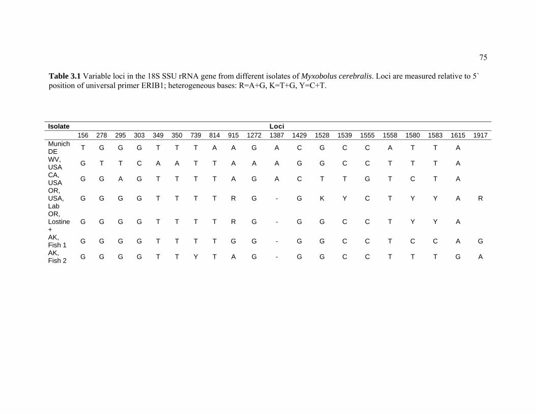
75
Table 3.1 Variable loci in the 18S SSU rRNA gene from different isolates of Myxobolus cerebralis. Loci are measured relative to 5` position of universal primer ERIB1; heterogeneous bases: R=A+G, K=T+G, Y=C+T.
Isolate Loci 156 278 295 303 349 350 739 814 915 1272 1387 1429 1528 1539 1555 1558 1580 1583 1615 1917 Munich DE T G G G T T T A A G A C G C C A T T A
WV, USA G T T C A A T T A A A G G C C T T T A
CA, USA G G A G T T T T A G A C T T G T C T A
OR, USA, Lab
G G G G T T T T R G - G K Y C T Y Y A R
OR, Lostine +
G G G G T T T T R G - G G C C T Y Y A
AK, Fish 1 G G G G T T T T G G - G G C C T C C A G
AK, Fish 2 G G G G T T Y T A G - G G C C T T T G A

76
Figure Legend
Figure 3.1 Agarose gel of PCR-amplified genomic DNA from four rainbow trout from Elmendorf Hatchery, Alaska (sample numbers shown), using mt 18S rDNA specific primers. A 1 kb+ DNA Ladder is shown on both sides of the gel (1.5% agarose). A blank (B) well precedes each of the two positive (+) controls and a negative (-) control (water) is provided in the last well.
Figure 3.2 QPCR cycle threshold values of Alaska hatchery rainbow trout plotted against reference samples spiked with known numbers of Myxobolus cerebralis myxospores (100, 1000). Data are represented in log scale. Values for fish samples are an average of duplicates, all other values are individual.
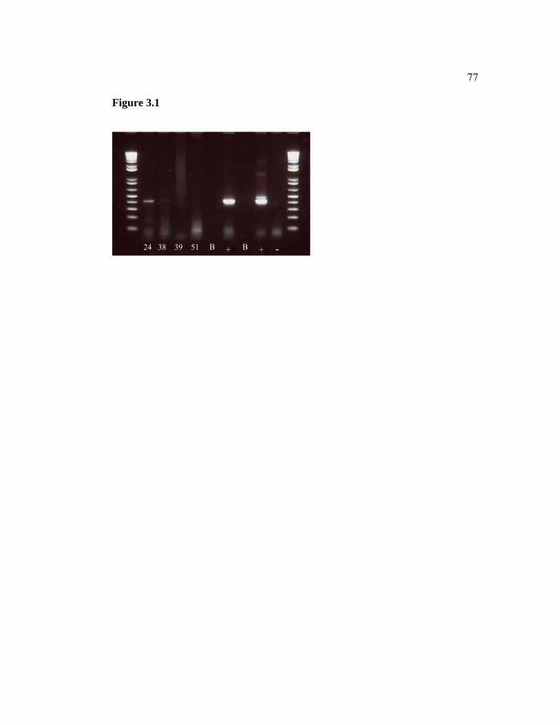
77
Figure 3.1
++ ++24 38 39 51 -+BB

78
Figure 3.2

79
CHAPTER 4
A QUALITATIVE ANALYSIS OF RISK FOR THE DISSEMINATION OF MYXOBOLUS CEREBRALIS IN THE STATE
OF ALASKA.
E. Leyla Arsan1,2 and Jerri L. Bartholomew2*
1Department of Fisheries and Wildlife, Center for Fish Disease Research, Oregon State University, 220 Nash Hall, Corvallis, OR 97331, USA
2Department of Microbiology, Center for Fish Disease Research, Oregon State University, 220 Nash Hall, Corvallis, OR 97331, USA

80
Abstract
Myxobolus cerebralis, the myxosporean parasite responsible for whirling disease
in salmonids, was first introduced to the United States in 1958. It has since spread
across the country causing severe declines in wild trout populations in the
intermountain west. The recent detection of the parasite in Alaska is further
evidence of the species’ capability to invade and colonize new habitat, though it is
unknown when colonization occurred. The goal of this study was to qualitatively
assess the risk of further spread and establishment in the state by examining
potential routes of dissemination. This paper explores potential routes of
introduction of M. cerebralis in Alaska by assessing four main elements of
dispersal: movement of fish by humans, natural dispersal (via migratory birds and
stray anadromous salmon), recreational activities, and commercial seafood
processing. Establishment of the parasite is dependent upon several environmental
and biological factors including: water temperatures, spatial/temporal overlap of
hosts, and the distribution and genetic composition of the oligochaete host, Tubifex
tubifex. The most likely pathway of M. cerebralis transport in Alaska is human
movement of fish by stocking. While the extent of M. cerebralis infection in
Alaskan salmonid populations is unknown, if the parasite were dispersed,
conditions are appropriate for establishment and propagation of the parasite life
cycle in some areas of southcentral Alaska. The probability of further
establishment is greatest in Ship Creek where the abundance of susceptible T.
tubifex, the presence of susceptible rainbow trout (Oncorhynchus mykiss), and the
proximity to the known area of infection, make conditions particularly suitable.
The apparent lack of appropriate tubificid hosts in southeast Alaska may prevent
establishment in that part of the state.

81
INTRODUCTION
Myxobolus cerebralis, a metazoan parasite that causes salmonid whirling disease,
is exotic to North America and was first detected in the USA in 1958 (Hoffman,
1962). Thought to have originated from a shipment of frozen infected brown trout
(Salmo trutta) from Europe, the parasite is now reported in 25 US states and 26
different countries (Bartholomew and Reno, 2002; Vermont Department of Fish
and Wildlife, 2002; Stromberg, 2006; Arsan et al., chapter 3 of this volume), and
its range continues to expand. Though the pathogen appears to have little impact
on fish populations in the eastern and coastal western states (Modin, 1998), it has
caused dramatic rapid declines of wild rainbow trout (Oncorhynchus mykiss)
populations in the intermountain west, primarily in Colorado and Montana
(Walker and Nehring, 1995; Vincent, 1996; Nehring et al., 1998). The ecologic
and economic impacts of M. cerebralis, in addition to its rapid spread and
establishment across the globe, are indicative of the need for specific
recommendations to assist fishery managers in halting the spread or effects of the
pathogen.
Myxobolus cerebralis was first detected in Alaska in 2006 (Arsan et al.,
chapter 3 of this volume) in rainbow trout from a hatchery on Ship Creek in
Anchorage in southcentral Alaska. Prior to this detection, the closest enzootic area
was the upper Columbia River basin (CRB) in northeast Oregon, southeast
Washington, and Idaho. The parasite has also been reported in wild and cultured
salmonids from the Sakhalin Islands off the east coast of Russia (Bogdanova,
1960, 1968). However, the distance from the Sakhalin Islands to southcentral
Alaska is approximately double that from the upper CRB.
The parasite was detected and confirmed in Alaska using molecular
techniques as the low level of infection precluded detection by less sensitive
methods (Arsan et al., chapter 3 of this volume). The prevalence of infection was
low and clinical whirling disease has never been reported from the hatchery or

82
other locations in the state. Cultured salmonids in Alaska are not monitored for
the parasite, and limited monitoring occurs in wild salmonids [US Fish and
Wildlife Service (USFWS), 2006]. As salmonids are inextricably linked to the
culture and economy of Alaska [Kenai River salmon runs alone generate annual
revenues of $70 million (Glass et al., 2004)], the potential impacts of M. cerebralis
could be catastrophic both ecologically and economically.
This paper uses risk analysis as a tool to assess the likelihood and
consequences of exotic parasite introduction. The framework for this type of risk
assessment (Bartholomew et al., 2005) was created in efforts to tailor such
assessments to whirling disease. We use risk analysis as a type of map
(MacDiarmid, 2000) to navigate through possible pathways or scenarios leading to
parasite introduction and establishment, and to assess where to allocate resources
to help prevent such an occurrence. This is an essential first step in understanding
potential outcomes and associated risks of management decisions. Our aim is to
provide decision-makers with tools to assess management implications and to
eliminate non-issues by using logical scientific arguments. This paper assesses the
risk for future spread of M. cerebralis within southcentral Alaska and to southeast
Alaska, and explores the potential for new introductions to the state.
THE PARASITE HAZARD
Tracking the epidemiology of parasites requires investigation of an
organism’s life cycle, biophysical properties, and hosts. The life cycle of M.
cerebralis requires two obligate hosts: a salmonid and the aquatic oligochaete
worm, Tubifex tubifex. The parasite maintains two distinct spore stages, one
unique to each of the hosts, the myxospore and the actinospore. Each spore stage
requires approximately three months to develop within and be released from the
respective host. Myxospores develop in the fish host and are released upon death
of the fish. These round and durable spores are ingested by T. tubifex as they
burrow through sediment (Brinkhurst, 1996). The parasite then undergoes

83
reproduction and structural transformation and is released as a triactinomyxon
(TAM). When the waterborne TAM attaches to its fish host, the parasite
sporoplasm is injected through the epidermis of the fish and migrates through the
nervous system to the cartilage (El-Matbouli et al., 1995). The parasite consumes
cartilage, causing lesions in the gills, cranium, and vertebral column of the fish
(MacConnell and Vincent, 2002).
The biophysical properties of M. cerebralis are also a factor in its potential
dissemination, with introduction most likely occurring by the myxospore stage.
Myxospores are far more resilient than TAMs and are capable of withstanding
severe environmental variables (El-Matbouli & Hoffmann, 1991) that might occur
during transport and dissemination. Of the two obligate hosts of M. cerebralis, the
fish host is more mobile; thus, myxospores are more likely to be distributed over a
broader area than TAMs. Indeed other researchers have speculated that myxozoan
colonization on a landscape would most likely occur via myxospores (Cone et al.,
2006).
Susceptibility of hosts is another contributing factor to parasite dispersal
and establishment. Salmonid hosts of M. cerebralis differ in their susceptibility to
infection and subsequent disease caused by the parasite. Rainbow trout have
repeatedly shown the highest susceptibility in laboratory and field experiments
(O’Grodnick, 1979; Hedrick et al., 1999a, 1999b; MacConnell and Vincent, 2002).
Most other salmonids, except lake trout (Salvelinus namaycush) and arctic
grayling (Thymallus arcticus), are also susceptible to the parasite (MacConnell and
Vincent, 2002), but infections result in varying degrees of clinical disease.
Whereas many species of salmonids are susceptible to the parasite, only
one species of oligochaete, T. tubifex, is capable of propagating the pathogen.
Tubifex tubifex is a hearty and cosmopolitan species that proliferates in areas with
high organic content. The worm’s ability to withstand extreme and variable
environmental conditions allow it to inhabit areas where other species cannot
compete, and thus to span across ecosystems as a widespread aquatic invertebrate.
These qualities also have made control efforts of this host unpractical or

84
unsuccessful (Wagner, 2002). However, susceptibility of T. tubifex to M.
cerebralis varies greatly, and has been linked to the worm’s lineage. There are six
cryptic mitochondrial lineages of T. tubifex. Five lineages (I, III, IV, V, and VI)
have been reported from North America (Beauchamp et al., 2001; Arsan et al.,
chapter 2 of this volume) and five are known from Europe (I-V) (Sturmbauer et
al., 1999). Susceptibility of these different lineages varies from highly susceptible
to the parasite to completely resistant. While lineage does not confer
susceptibility, researchers have found the genetic composition of tubifex
populations to correspond to severity of infection in fish (Beauchamp et al., 2006).
RISK ANALYSIS
To begin the risk assessment process, we narrowed the magnitude of
geographic size of Alaska [the state is approximately the size of the continent of
Europe (Pagano, 2000; Atlas A-Z, 2001)], by focusing on areas of the state where
we considered the probability for new introduction, or further within-state spread,
of M. cerebralis to be highest: southeast and southcentral Alaska (the Cook Inlet
basin) (Figure 4.1). These areas have high concentrations of susceptible fish hosts,
high angler traffic and/or a large commercial fishery, the highest concentration of
human populations in the state, and high potential organic loading. The areas are
also close to the road system and ports, and in the migration path of fish from
enzootic areas. Definitions of risk levels used in this analysis are displayed in
Table 4.1.
Study Sites
Southeast Alaska
Southeast Alaska has a maritime climate and is within the Pacific
Northwest temperate rainforest ecosystem. The area has cool winters and wet

85
summers with stream temperatures generally warmer than those in the interior of
the state. Typical hydrographs for Southeast Alaska creeks are influenced by
spring snowmelt and autumn rainfall (Chaloner et al., 2004; Montgomery et al.,
1996; Milner et al., 1997). River basins of coastal southeast Alaska are generally
small, due to mountains and icefields that rise sharply from sea level and create
relatively short watersheds that flow into the Pacific Ocean. Two streams were
selected based on the criteria described above: Peterson and Montana Creeks near
Juneau.
Southcentral Alaska
The Cook Inlet, in southcentral Alaska, is home to over half the state’s
human population. The area has a transitional climate (National Climate Center,
1982) and is the ecotone between the Pacific Northwest rainforest and the northern
boreal forest. Hydrographs in the basin are highly predictable and influenced by
snow and glacier melt in the summer; typical freshwater inflow into Cook Inlet is
15 times higher in July than in February (Dorava and Milner, 2000). This study
focuses on Ship and Campbell Creeks in the Anchorage area and on the Kenai
River on the Kenai Peninsula. All three streams are popular sport fisheries due to
abundant fish runs, proximity to major population centers, and accessibility by
roads. They also have numerous sources of potential organic loading due to their
urban proximity, commercial and industrial activity, streambank degradation due
to recreational traffic, and large pulses of organic material from spawning salmon
runs.
The Kenai River supports populations of rainbow trout, Chinook, coho,
pink, and sockeye (O. nerka) salmon, and Dolly Varden and is the largest
freshwater sport fishery in Alaska (Hammarstrom, 1988). Ship and Campbell
Creeks support populations of these species as well as chum salmon (Miller and
Bosch, 2004). Ship Creek is the most popular sport fishery in the Anchorage area
and sustains the only two hatcheries in the state that rear rainbow trout: Ft.
Richardson (FTR) and Elmendorf (ELM). Detections of M. cerebralis in Alaska
were in rainbow trout from ELM (Arsan et al., chapter 3 of this volume). This

86
hatchery uses surface water that, until 2004, was heated to 14°C [an optimal
temperature for parasite development (El-Matbouli et al., 1999)] by wastewater
from a power plant near the facility. The incoming surface water at ELM is
screened for debris but is not treated for potential biota. Thus, M. cerebralis
spores or even T. tubifex could potentially enter the hatchery through Ship Creek
water. Effluent from both ELM and FTR is routed through earthen settling ponds
and flows back into Ship Creek untreated. Thus, fish in Ship Creek could also be
exposed to parasites in hatchery effluent. Futhermore, T. tubifex were identified
from settling-ponds at both hatcheries (Arsan et al., chapter 2 of this volume).
RELEASE ASSESSMENT
The release assessment explores potential pathways of pathogen introduction and
is focused on the myxospore stage of the parasite. As the parasite has been
detected in southcentral Alaska, the release assessment provides insight into how
introduction may have occurred and by what route further dissemination is most
likely. Four main pathways of introduction of M. cerebralis were identified:
movement of fish by humans, natural dispersal, recreational activities, and
commercial seafood processing (Figure 4.2). These potential introduction routes
could include: legal and illegal movement of fish by humans, importation of frozen
food fish, dispersal of parasite via migratory birds, angler activities, dissemination
by stray anadromous fish from enzootic areas, and effluent discharge from seafood
processing plants.
Human Movement of Fish
The primary spread of many invasive species has been by human activity.
In the case of M. cerebralis, the pathogen is thought to have spread throughout the
USA as a result of shipments of infected fish, both commercially and through state
and federal stocking programs (Hoffman, 1990; Bartholomew and Reno, 2002).
Since the mid 1970’s, the state of Alaska has adopted strict laws prohibiting the

87
import of live non-ornamental fish (Alaska Administrative Code [AAC] 5:
41.070). However, frozen food fish are regularly imported in the state. Imported
frozen rainbow trout (with heads still on) are common supermarket items in
Alaska [T. Meyers, Alaska Department of Fish and Game (ADFG), personal
communication]. Given that the majority of rainbow trout produced in the USA
originate in the Hagerman Valley, Idaho (in the M. cerebralis enzootic area), it is
likely these fish are sold in Alaska. Because M. cerebralis myxospores survive
freezing (El-Matbouli and Hoffmann, 1991), frozen fish still pose a risk for
parasite dissemination and are thought to be the original pathway of introduction
of M. cerebralis to the USA (Hoffman, 1962). However, the resulting whirling
disease epizootics that ensued from the parasite’s initial introduction to the USA
likely stemmed from the use of the frozen imports as food for hatchery fish.
Hatchery practices have changed considerably since the 1950’s, and it is unlikely
that frozen fish imported for human consumption would end up in Alaskan waters.
Though the potential for pathogen dissemination by frozen fish is low, the risk can
be further reduced by requiring that imported fish have heads removed.
Little information is known regarding the number and distribution of
private fish-ponds in the state or possible illegal importation of fish. However,
instances of illegal importation from out-of-state are thought to be low considering
general lack of accessibility and proximity to ports in addition to legal
complications and climatic logistics of private fishpond operation in Alaska.
State regulations (AAC 5: 41.005) also prohibit within-state transport,
possession, export, or release of any live fish or fish eggs without a Fish Transport
Permit. However, illegal transfer (stocking) of fish has occurred and is thought to
be the primary reason for the presence of northern pike (Esox lucius) in the upper
Cook Inlet basin and Kenai Peninsula (ADFG, Southcentral Alaska northern pike
brochure).
Another factor influencing potential within-state dissemination is the use of
game fish (fresh or frozen) as bait. Heads, tails, fins and viscera of legally caught
game fish may be used as bait (AAC 5: 75.026), though live fish may not. Fish

88
heads, where parasite concentrations would be highest, may thus be dispersed in
rivers throughout the state. To add to this potential pathway of dissemination, the
parasite survives passage through the alimentary canal of predatory fish (El-
Matbouli and Hoffmann, 1991), and thus may be further distributed beyond an
initial introduction as bait.
Assessment of risk
Although human movement of fish presents a low risk for new
introductions of M. cerebralis to Alaska, it is the most likely pathway for parasite
dissemination within the state. Prior to parasite detection, potentially infected fish
were transplanted throughout southcentral and interior Alaska. Many of these
stocked locations were land-locked lakes with no outlets, which would limit the
spread of the parasite, but some fish were also stocked in open stream systems.
Thus, some degree of spread may already have occurred. As the estimated
prevalence of infection in production rainbow trout from ELM is 23% (Arsan et
al., chapter 3 of this volume), if 10,000 fish are stocked, 2,300 potentially have
some degree of infection. Typically, 2,000-10,000 fish are transplanted per site in
southcentral Alaska (ADFG, 2006b). This would readily disperse the parasite
throughout the stocking area. Thus, without management action, the probability
for M. cerebralis spread by human movement of fish is high. In contrast, if
stocking of ELM fish is limited to no-outlet lakes where susceptible native resident
fish are absent or to seawater netpens for terminal commercial and sport fisheries,
and anglers are prohibited from using heads of fish as bait, the probability of
parasite dissemination by human movement of fish decreases (Table 4.2) to a low
level. Detailed management recommendations are discussed in the conclusions of
this paper.
Commercial Seafood Processing
Fish from M. cerebralis enzootic areas, such as the upper Columbia River
basin, are regularly caught in commercial fisheries in Alaska [ADFG, 2006a;
Regional Marking Information Systems (RMIS), 2006]. If effluent from seafood

89
processors were discharged into freshwater where T. tubifex were present, M.
cerebralis could potentially be introduced. Fish solids released into marine waters
utilized by migratory birds or scavengers, such as gulls, could also result in
dispersal from marine environments inland to riverine systems via excrement and
egested pellets. (See Birds Pathway.)
Authorized seafood processors in Alaska individually discharge 30,000 to
over 10 million pounds of waste solids annually (USEPA, Fact Sheet a). Shore-
based fish processors in Alaska are required (Clean Water Act, section 301(a), 33
U.S.C.) to grind solid effluent waste to 0.5 inch (1.27 cm) or less prior to
discharge. These smaller pieces, including cartilage where myxospores would be
concentrated, would be less attractive to scavengers that may transport the parasite
in freshwater ecosystems. Processors are allowed to discharge into freshwater
rivers with a waiver. However, very few of these waivers are granted. Seafood
processing solid wastes (i.e.: fish heads where myxospores would be concentrated)
are taken to by-product reduction facilities for reduction to fish meal or other
secondary products (USEPA, Fact Sheet b). Though the majority of Alaska fish
meal is comprised of whitefish (Alaska Office of Economic Development, By-
Product Fact Sheet), all fish meal is brought to 100-600°C during processing
(USEPA, 1995), temperatures that would destroy M. cerebralis myxospores.
There are 218 fish processing plants with National Pollutant Discharge
Elimination System (NPDES) permits in the state of Alaska (USEPA, 2006).
None of the 28 permits in the Cook Inlet Basin or the 22 permits in southeast
Alaska have a waiver to discharge effluent into freshwater. As the majority of
processors are near the coast, discharges into freshwater would likely be near a
river mouth and hence would be tidal. Tidal areas are not suitable habitat for T.
tubifex (Seys et al., 1999; Rundle et al., 1998), thus discharging into these areas
would present little risk for introduction of M. cerebralis.
Seafood processors discharging less than 1,000 pounds of seafood waste
per day and less than 15 tons per calendar year are not required to have a permit.
This limit was imposed to allow subsistence processing and direct market

90
processors (processors receiving mostly processed seafood requiring minimal
further processing) to discharge without a permit. These processors are not
included in this risk assessment, as information regarding their effluent discharges
is unavailable.
Assessment of risk
We consider the probability that commercial seafood processing practices
would result in introducing M. cerebralis into Alaskan fresh waters to be
negligible. None of the permitted processing plants discharge into freshwater, and
discharges into marine waters are made less attractive to scavengers by grinding
effluent to a small size. Scavengers are also more likely to be attracted to tissue
than cartilage where parasite loads would be greatest.
Dispersal via Recreation
Alaska has a world-class sport fishery that attracts anglers from across the
country and the globe; many of these anglers use equipment in Alaska that has
been used in other river systems. River systems most likely to experience
introduction of the parasite via recreational activity would be those with popular
sport fisheries and accessibility by road. The Kenai River is the largest freshwater
sport fishery in Alaska (Hammarstrom, 1988). Ship Creek is the most popular
sport fishery in the Anchorage area (accounting for 69% of the 2002 Anchorage
angling effort) (Miller and Bosch, 2004). Peterson Creek is the only recreational
steelhead (Oncorhynchus mykiss) fishery on the Juneau road system (Harding and
Jones, 1992).
Anglers in these popular areas could introduce M. cerebralis by
inadvertently transporting the parasite on the soles of their waders. Myxospores
trapped in the moisture of a felt-sole would likely remain wet and thus viable far
longer than in a less absorbent material. Anecdotal data collected prior to the
determination of the life cycle of M. cerebralis (Schäuperclaus, 1931; Hoffman
and O’Grodnick, 1977) suggest myxospores remain viable after drying. Yet, more
current studies indicate myxospores do not remain viable after drying for 24 hr (R.

91
Hedrick, University of California, Davis, personal communication). A recent
laboratory study demonstrated removable felt soles were able to transport
myxospores and TAMs (P. Reno, Oregon State University, personal
communication). However, spores became less viable after drying the soles for 8-
24 h, and infectious parasites were no longer transmitted after drying for 7 d.
Because of their fragility, individual TAM stages are less likely to be transported
by this route. However, infected T. tubifex adhered to a felt sole provide a suitable
environment for TAMs to remain viable. More research is needed to determine
the duration of drying required to disinfect entire waders. However, the length of
time required to dry and thus disinfect waders will vary by environmental
conditions.
While the probability of transporting M. cerebralis in a single event (i.e.: a
single angler or single angling day) by recreational activities may be low, when all
angler days in a year are considered, the probability increases. In 2001, the state
saw 421,000 anglers (US Department of Interior, 2001); 239,000 were not Alaska
residents and may have used gear that was used outside of Alaska. The sheer
number of anglers from within and outside the state indicates the potential for
parasite transport is not only high, but continues to grow, with a 36% increase in
number of anglers from 1991 to 2001 (U.S. Department of Interior, 2001).
Recreational activities can also influence the risk of potential M. cerebralis
introduction in indirect ways by enhancing T. tubifex habitat. Tubifex tubifex are
commonly found in environments with abundant organic matter, fine sediments,
and low flow (Brinkhurst and Gelder, 1991; Brinkhurst, 1996). Recreational foot
traffic (such as angling from the bank) can damage vegetation and increase
streambank erosion causing more sediment to enter surrounding waters.
Motorized boats also create wakes that can increase streambank erosion. Previous
studies have documented these activities and their effects on the Kenai River
(Liepitz, 1994; Weiner, 1998).
Assessment of risk

92
The risk of recreational activities as a pathway of parasite introduction is
difficult to assess, as the lack of information regarding potential spore transfer via
felt-soled waders represents a significant data gap. A conservative estimate of the
probability of new introductions of M. cerebralis by recreational activity is
moderate, but the probability for within-state transfer of the parasite is low, if
prevalence and severity of infection remain low (Table 4.2), and if management
actions are taken to limit stocking and thus dissemination of infected sportfish.
However, the cumulative and long-term effects of angler and recreational activities
in heavy-use areas could be much greater than the likelihood of introduction (or
further spread) in the short term or by a single event.
Natural dispersal: birds
The role of piscivorous birds in fish pathogen dissemination has been
examined in several studies (Taylor and Lott, 1978; El-Matbouli and Hoffmann,
1991). Because M. cerebralis survives passage through the guts of birds, long
food retention times would lengthen the distance the pathogen could be dispersed.
If all food is voided within a short period, the parasite could be contained locally.
Other omnivorous fish-consumers such as bears (Ursus americanus, Ursus arctos)
and river otters (Lutra canadensis) do not present a risk for M. cerebralis
dispersal, as the parasite does not survive passage through the guts of mammals
(El-Matbouli et al., 2005).
Alaska is a migratory destination for thousands of birds worldwide.
Although the probability of a bird releasing viable myxospores over a waterbody
remains unknown, deposition near (and not in) water may be sufficient for
transport of the parasite under certain environmental conditions. It is unknown
how long myxospores deposited in bird feces would remain moist, and thus viable.
Time to inactivation of spores is likely to vary by environmental conditions. Thus,
if spores are deposited near a waterbody, and are rapidly washed into the river by
high water or precipitation, dissemination of the parasite could occur.

93
Since M. cerebralis manifests in cartilage of fish, it is likely that the
parasite would be egested (regurgitated) in pellets of birds. Piscivores that
swallow their prey whole (e.g. non- raptors) present the biggest risk of M.
cerebralis dispersal. Double-crested cormorants (Phalacrocorax auritus) have a
simple gut structure and egest bones, pieces of fish, and solid markers 1-2 days
following ingestion (Brugger, 1993). In contrast, the passage time of rainbow
trout through bald eagles (Haliaeetus leucocephalus), which have a more complex
gut morphology, is approximately 62 hours (F. Barrows, USFWS, personal
communication). Thus spores could be excreted 2-3 d after eating an infected fish.
Raptors and large waterbirds (waders, seabirds, etc) have some of the
fastest known migration speeds among birds; bald eagles travel 201 km/day in
migration (Kerlinger, 1995), and ospreys (Pandion haliaetus) 108-431 km/day
(Alerstam, 2003; Hake et al., 2001). The nearest M. cerebralis enzootic area
outside Alaska [upper Columbia River Basin (CRB)] is approximately 1800 km
from southeast Alaska and 2750 km from southcentral Alaska. An osprey would
have to retain food material for 4.2 to 16.7 days to transport spores to Juneau and
for 6.4 to 25.5 days to transport spores to Anchorage.
Assessment of risk
Whereas the probability of new parasite introduction via bird transport
from the CRB is negligible, the probability of within-state transfer by bird
transport is greater, but still low. Because of the low prevalence and severity of
infection in the state, and the fact that numerous events must align in order for
dissemination to occur, the probability of parasite transport by birds is likely to
remain low unless prevalence or severity of infection increases.
Natural Dispersal: Stray anadromous salmon
Anadromous salmonids may stray into non-natal streams during their
return migration to spawning grounds. Pathogen dissemination is an often-
overlooked effect of straying. For example, introduction of M. cerebralis as a
result of straying salmonids has been documented in the Deschutes River, Oregon

94
(mid CRB) (Engelking, 2002). Some anadromous salmonids from the CRB
migrate north to waters off of Alaska to feed for 3-5 years before typically
returning south to spawn (Groot and Margolis, 2003; RMIS, 2006).
Fish that have strayed into non-natal waters are usually identified by
examination of fin clips or by evidence of a coded-wire tag (CWT). As any single
fin clip pattern is commonly used by more than one hatchery in the Pacific
Northwest in a given year, fin clips reveal only the “stray” status of a fish and a
rough list of potential origins. A CWT or fin clip unique to a certain hatchery
would be required for identification of exact hatchery of origin.
Though CRB fish are commonly harvested in commercial marine fisheries
off the coast of Alaska, little data are available on salmon straying into the fresh
waters of Alaska. There is only one such record: in 2001, a Chinook salmon from
Marion Forks Hatchery, North Santiam River, Oregon (lower CRB) was recovered
in the Copper River, Southcentral Alaska (RMIS, 2006; ADFG, 2006a). Because
stray wild fish cannot be visually identified, no data (current or historical) are
available on the stray rates of wild CRB salmon in Alaska, yet these fish are
potential carriers of M. cerebralis. This represents a data gap in this risk analysis.
Assessment of risk
Based on the available data, the probability of M. cerebralis dispersal by
straying anadromous hatchery salmon from the CRB is negligible. However,
presence of the parasite at ELM on Ship Creek provides a local source of
dissemination. While it is unknown if M. cerebralis is established in Ship Creek,
if naturally reproducing fish were infected, this would increase the potential for
parasite dispersal. As rainbow trout are typically resident species and do not make
long migrations (Morrow, 1980; Behnke, 1992), spread of the parasite by natural
migration of this species is likely to be local. Parasite dispersal by straying of
anadromous fish from Ship Creek is more likely, though M. cerebralis has not
been documented in anadromous fish species in Alaska.

95
Summary of the release assessment
The probability of re-introduction and within state dissemination of M.
cerebralis in Alaska is summarized in Table 4.2. While the overall probability of
new introductions from out-of –state is low; the likelihood of further transport
within the state is high if no management actions are taken. The pathway with the
greatest likelihood for within-state parasite transfer is stocking of infected rainbow
trout, since this would repeatedly introduce large numbers of potentially infected
fish. However, if stocking is limited to no-outlet lakes where native susceptible
resident salmonid species are absent, or to seawater netpens for terminal
commercial and sport fisheries, the probability of further spread of M. cerebralis is
low. Illegal stocking of fish remains a risk for dissemination of the pathogen. The
importation of whole frozen food fish also presents a risk for parasite introduction.
The risk of parasite transfer posed by the use of fish heads as bait could also be
reduced if this practice were prohibited with fish from ELM-stocked areas.
Because of the low prevalence of infection in the state, the likelihood of parasite
transport by pathways other than human movement of fish is likely to remain low
unless prevalence or severity of infection increases.
EXPOSURE ASSESSMENT
The exposure assessment, which explores the risk of parasite
establishment, is focused on the TAM stage of M. cerebralis and its oligochaete
host. Establishment of M. cerebralis has already occurred in southcentral Alaska
at ELM on Ship Creek. Whether establishment has also occurred outside the
hatchery, remains unknown.
Establishment of M. cerebralis is dependent upon several environmental
and biological factors including: the distribution and genetic composition of T.
tubifex populations, water temperatures, and spatial temporal overlap of hosts.
Each of these factors determines the outcome of the exposure assessment, as
depicted by the scenario tree in Figure 4.3.

96
Tubifex tubifex habitat and populations
Habitat for T. tubifex is defined as areas with fine sediment, low flow, and
organic matter (Brinkhurst and Gelder, 1991). These areas were found throughout
the study area.
A T. tubifex survey was conducted to ascertain relative abundance of T.
tubifex in selected streams in the study area (Arsan et al., chapter 2 of this
volume). Over 2,700 oligochaetes were collected from southeast Alaskan sample
sites; however, morphological and genetic analysis demonstrated that none of the
worms examined were T. tubifex. In contract, T. tubifex were identified from
southcentral Alaska, with the highest numbers on the lower Kenai River,
particularly at Centennial and Eagle Rock Boat Landings, areas with high
sedimentation and organic loading (primarily decaying salmon carcasses) and
heavy recreational use. As many as 1,768 T. tubifex out of 3,024 total worms were
detected in 30ml of sediment at Centennial Boat Landing. The species was also
identified from Ship and Campbell Creeks, though not from the upper or middle
Kenai River.
Oligochaetes gathered in the T. tubifex survey were held in water that was
screened for M. cerebralis actinospores. No M. cerebralis TAMs were observed
from any of the sites sampled in the T. tubifex survey. However, actinospores of
several other myxozoans were detected (Arsan et al., chapter 2 of this volume),
confirming that other myxozoan life cycles have established in both southeast and
southcentral Alaska.
Tubifex tubifex lineages
Though T. tubifex is the only species of oligochaete capable of harboring
M. cerebralis, susceptibility to the parasite varies dramatically within the species
and has been correlated to the taxa’s mitochondrial lineage. Populations of
lineages I and III have been shown to propagate the parasite whereas lineages IV,
V, and VI do not (Arsan et al., chapter 2 of this volume; Beauchamp et al., 2002,

97
2005, & 2006; DuBey et al., 2005). Four lineages were identified in the state: I,
III, IV, and VI (Arsan et al., chapter 2 of this volume). Lineage IV had previously
been reported only from Europe.
Parasite exposure experiments using T. tubifex collected from our study
sites demonstrated that lineages I, IV, and VI from Alaska do not support the
parasite life cycle (Arsan et al., chapter 2 of this volume). Lineage III was the only
lineage from the state to propagate the parasite. This lineage, though susceptible
to M. cerebralis, was a low parasite producer, releasing a maximum of 280 TAMs
in 7 d, as compared to the control lineage III population from Oregon that released
over 7000 TAMs during the same time period when exposed to the same number
of myxospores.
Tubifex tubifex populations in southcentral Alaska were a mixture of two or
three lineages. Lineage I dominated (71-86%) at sites on Ship Creek and
Campbell Creek, whereas Lineage VI dominated (69%) at sites on the Lower
Kenai River. Lineage III was present in low numbers (7-21%) throughout the
Cook Inlet basin at 7 out of the 9 sites where T. tubifex were collected, however, it
was not identified from any of the hatchery sites.
The presence of resistant lineages I, IV, and VI T. tubifex could have
significant beneficial implications for Alaskan salmonids in areas where these
lineages dominate Tubifex populations. Other researchers have found lineage
susceptibility to correspond to severity of infection in fish (Beauchamp et al.,
2006). This suggests that when lineages I, IV, and VI are in high abundance, the
effects of M. cerebralis may be less severe.
Populations of lineage III T. tubifex from other geographic regions are
highly susceptible to M. cerebralis (DuBey et al., 2005; Beauchamp et al., 2006;
Hallett and Bartholomew, 2005) and detection of the lineage in Alaska is cause for
concern. Though this lineage composed approximately 14-21% of worm
populations in Ship and Campbell Creeks and only 7% on the lower Kenai River,
it can become infected with M. cerebralis and release TAMs when in proportions
as low 0.6% of the total population (Arsan et al., chapter 2 of this volume). Hallett

98
et al. (2007) documented similar lineage III infection dynamics when the lineage
was in proportions as low as 5% of the total T. tubifex population. Researchers
have suggested that only a few T. tubifex and TAMs are required to produce
infection and population loss in fish in cold, oligotrophic streams (Allen and
Bergersen, 2002).
Water temperature seasonally reaching 10-15 °C
Water temperature influences M. cerebralis development in each of its
hosts (Hedrick and El-Matbouli, 2002; Blazer et al., 2003). Both severity of
infection in fish and development and release of TAMs from oligochaetes are
highest when average daily water temperatures are between 11-15°C (El-Matbouli
et al., 1999; Baldwin et al., 2000; Granath and Gilbert, 2002). Temperatures
below 10°C delay the development of TAMs and those above 20°C hinder parasite
development and can be lethal to the TAM stage of M. cerebralis.
Lower water temperatures retard the spore formation process in both fish
and oligochaetes, and could delay TAM development and release at high latitudes.
However, M. cerebralis has been found to persist and cause reduced juvenile trout
recruitment even in cold, oligotrophic, sediment poor, high-gradient streams
(Allen and Bergersen, 2002) and at elevations as high as 3,300m (10,800 ft) in
Colorado (Nehring and Thompson, 2002). Temperatures in such areas are
comparable, if slightly warmer, to those in our study sites in Alaska. Since snow
covers the ground for more than 6 months of the year, streams in southcentral
Alaska are generally cooler than those in the contiguous US, typically with a total
of 1780 annual degree days (Oswood, 1997). In comparison, the Madison River in
Montana (enzootic for M. cerebralis) has roughly 2650 annual degree days
(USGS, 2005).
Mean summer (June- August) water temperatures in the lower Kenai River
ranged from 10.7- 11.2°C between 1999-2001 (USGS, 2005), with an average of
79 days/year continuously greater than 10°C, and 1801 degree days annually.
Temperatures in Campbell Creek ranged from 9.5-10.5°C, with an average of only
99
50 days above 10°C each year from 2000-2001 (USGS, 2002). Lineage III T.
tubifex from Alaska required 1382-1536 degree days for M. cerebralis to develop.
Therefore, water temperatures in southcentral Alaska are sufficient for parasite
development and propagation, though complete life cycle duration may be longer
than in warmer climates. The lower Kenai River has water temperatures most
conducive to propagation of the M. cerebralis life cycle.
Future climate trends could also influence parasite development; USGS
reports water temperatures within the Cook Inlet basin are likely to increase 3°C in
the next 10 years (Kyle and Brabets, 2001). If such an increase occurs, water
temperatures in southcentral Alaska would be similar to that of the Madison River,
Montana, an area where M. cerebralis has caused population loss in rainbow trout
(Krueger et al., 2006).
Spatial/ temporal overlap of hosts
For the parasite to establish after introduction of myxospores, obligatory
spatial overlap must occur between parasite and host. The complex life cycle of
M. cerebralis requires spatial overlap to occur twice: once between myxospores
and T. tubifex, and subsequently between salmonids and TAMs.
Researchers have documented varied patterns of seasonality associated
with TAM release in different geographic locations. In Montana, TAM numbers
peak in the spring warming and fall cooling periods (Gilbert and Granath, 2003);
in high altitude waters in Colorado, TAM release peaks in the fall through the
winter (Nehring and Thompson, 2002). Yet, other areas of Colorado tend to
exhibit peak TAM numbers in the summer and early fall, with the lowest number
of TAMs occurring in the spring (Thompson and Nehring, 2000; Allen and
Bergersen, 2002). Seasonality of TAMs is believed to relate to water temperature
and the availability of myxospores as influenced by fish stocking schedules. We
speculate that high latitude Alaskan water temperatures would be similar to high
altitude water temperatures in Colorado and that seasonal TAM release in Alaska
would be comparable to these areas.

100
Worm infection and TAM release are dependant on myxospore
availability. Myxospores are released upon the death of the fish host and
subsequent degradation of the cartilage. As breakdown of this tissue is likely to
occur gradually in slow moving or cold waters, release may occur over a long
period (Hallett and Bartholomew, 2006). Once infected, worms can remain
persistently infected throughout their lifespan (Gilbert and Granath, 2001) with
TAM release occurring when water temperatures are appropriate. The majority of
young salmonids (both wild and hatchery) in southcentral Alaska hatch December
through August (ADFG, 2003; Quinn, 2005) and would be most susceptible to
infection during the first weeks post-hatch. Rainbow trout (and to a lesser extent
steelhead) are most susceptible to the parasite from 0-9 weeks post-hatch (up to
756 degree days at 12°C) (Ryce et al., 2004). However, rainbow trout hatched in
June would exhibit resistance to the parasite by September. On this schedule,
young rainbow trout may avoid peak TAM release from oligochaetes if peak
release begins in September. Thus, it may be possible for the parasite to
proliferate at cold temperatures and yet have little impact on rainbow trout
populations (Kerans et al., 2005).
Salmonid species composition will also affect the outcome of introduction.
Generally, areas with the highest T. tubifex abundance, like the lower Kenai River,
would have a higher likelihood of spatial-temporal overlap of hosts and parasite
and thus of M. cerebralis establishment. However, the juvenile salmonid
assemblage in this area is 88% Chinook salmon, with rainbow trout making up
only 0.1% of the total assemblage (Bendock and Bingham, 1988). While Chinook
salmon are susceptible to M. cerebralis, they acquire resistance to the parasite
more quickly and are less susceptible than rainbow trout (MacConnell and
Vincent, 2002; Sollid et al., 2003). Thus, the likelihood of spatial overlap of
susceptible hosts in the lower Kenai River is low. Juvenile salmonid assemblages
in Ship Creek are speculated to be greater than 80% Chinook and coho salmon (D.
Bosch, ADFG Sport Fish Division, personal communication), with proportions of
rainbow trout unknown.

101
Summary of the exposure assessment
The probability of further establishment of M. cerebralis in southcentral
Alaska is variable between locations (Figure 4.3). Distribution of susceptible T.
tubifex, water temperatures, and the juvenile salmonid species composition would
be determining factors for establishment. Thus the probability of establishment in
the upper Kenai River is low, due to a lack of oligochaete hosts. While the lower
Kenai River maintains a high abundance of T. tubifex, the lack of juvenile rainbow
trout lowers the risk of M. cerebralis establishment. Ship and Campbell Creeks
have environmental conditions representative of non-glacial streams in
southcentral Alaska, and remain the most likely areas to experience parasite
establishment (Figure 4.3). Yet while these creeks support populations of T.
tubifex, abundance and susceptibility of the worms appear to be low. Not only is
susceptibility limited to lineage III T. tubifex, but Alaska strains of this lineage are
low parasite producers (Arsan et al., chapter 2 of this volume). Thus, although
conditions are permissible in southcentral Alaska for establishment of M.
cerebralis, they are not ideal and may keep infection rates low and disease from
becoming apparent. Changes to physical or environmental conditions, such as
climate change, may alter the probability of parasite establishment in the state.
The probability of establishment in southeast Alaska is negligible, as no T.
tubifex were detected. However, oligochaete surveys have been limited (Arsan et
al., chapter 2 of this volume).
CONCLUSIONS AND RISK MANAGEMENT
Risk of new introductions
The probability of new introductions of M. cerebralis into the state of
Alaska is low, with the most likely pathway for introduction being recreation and
angler activity. Thus, areas most likely to first experience introduction are high-
use sport fisheries such as the Kenai River and Ship Creek. If introduction occurs,
102
the probability of parasite establishment is moderate, particularly in rivers like
Ship Creek where permissive temperatures, susceptible T. tubifex, and potential
rainbow trout populations overlap.
Although conditions (temperatures, T. tubifex populations, and
spatial/temporal overlap of hosts) in Ship Creek are permissive for parasite
development, they are not optimal, and may slow parasite development and lower
the risk for establishment. For example, water temperatures are acceptable for
parasite development, but are low enough to abate rapid proliferation. Susceptible
T. tubifex are present in the creek, but populations are dominated by resistant
strains, again lowering the risk of rapid parasite proliferation. Lastly, fish species
composition in southcentral Alaskan creeks may also help to lower the risk of
parasite establishment, as Chinook salmon (having lower susceptibility to the
parasite) tend to dominate areas where susceptible T. tubifex were collected. This
study focused on the first two steps of a risk analysis (release and exposure
assessments), but did not complete a consequence assessment to evaluate the
effects of parasite introduction and establishment in Alaska.
Policies that prevent importation of live salmonids into Alaska have been
the most effective tool for limiting introduction. Existing regulations discourage
establishment of private ponds, which are believed to contribute to spread of the
parasite in areas of the contiguous USA (B. Nehring, Colorado Division of
Wildlife, personal communication). We also recommend the state maintain the
policy requiring all seafood processor effluent be disposed in marine waters, and to
restrict effluent waivers in areas considered high risk (Cook Inlet Basin).
Although the potential for introduction by anglers is moderate, the risks could be
further reduced, especially in light of recent data on the vulnerability of
myxospores to desiccation. To further reduce the risk of introduction by anglers,
we urge the state to allot resources to angler education and further research on
effects of angler activity on dispersal. This could be accomplished with signage at
boat ramps, parking areas, or other access points, brochures distributed upon
purchase of fishing licenses, and by an informational web page.
103
Risk of further dissemination within Alaska
The probability of further transport of M. cerebralis within the state is high
due to the presence of the parasite at a state hatchery. The pathway presenting the
greatest risk for within-state parasite transfer is human movement of fish (e.g.
transplanting of infected trout and use of fish heads as bait) since large numbers of
infected fish could be repeatedly introduced. Because of the low prevalence of
infection in the state, the likelihood of parasite transport by other pathways is
likely to remain low unless prevalence or severity of infection increases. Although
the probability of establishment of M. cerebralis in southeast Alaska is considered
negligible due to the lack of suitable invertebrate hosts, only a few sites were
surveyed. To gain more confidence in this assessment, additional sites should be
surveyed for susceptible lineages of T. tubifex.
While it is unknown how long the parasite has occurred in Alaska, it is
likely that previous monitoring using the pepsin-trypsin digest as an initial
screening test would have missed a low infection prevalence. Not only did all
prior monitoring efforts in Alaska use the pepsin-trypsin digest, but testing was
non-targeted and did not focus on areas at risk for M. cerebralis introduction.
Molecular tests such as PCR are approximately 10-fold more sensitive (Andree et
al., 2002) and may have detected the parasite. For management purposes it may
only be necessary to detect infections that cause negative impacts on fish
populations or are considered “significant” infections, and both pepsin-trysin
digest and histology are adequate for this purpose. However, if the criterion for a
“significant” infection is presence of the parasite, then use of more sensitive assays
is necessary. Each state has its own stipulations as to what constitutes such an
infection and what the implications of detection are. For example, the state of
Colorado transfers only fish that are PCR negative for M. cerebralis to areas where
the parasite is not known to occur.
The first detection of M. cerebralis in Alaska is not without consequence.
Clearly the risk of parasite dissemination is low (if stocking fish from ELM is

104
limited), but nevertheless unacceptable by current ADFG fish disease policies.
Future plans include replacement of ELM with a new facility constructed on a
nearby site with water supplied from wells rather than Ship Creek. The FTR will
be discontinued entirely once the new ELM facility is completed. The use of well
water should facilitate breaking the parasite life cycle, and will be corroborated by
additional QPCR testing of hatchery fish stocks in the new facility. Negative
findings would suggest the possibility of future unrestricted fish transplants.
Exposure of sentinel rainbow trout fry in Ship Creek would also determine
if the parasite has become established outside the hatchery, and has been used in
similar situations after isolated parasite introduction (Bartholomew et al., 2007).
Given the low prevalence and severity of infection in ELM fish, examination of
other salmonid species or monitoring of Ship Creek water for TAMs would likely
prove ineffective. Regular monitoring for M. cerebralis in cultured salmonids
would provide baseline data if prevalence or severity of infection were to change.
Monitoring of wild salmonids should focus on areas of highest risk for parasite
introduction and establishment.
In addition to Alaska’s M. cerebralis interim management plan, we
recommend the state prohibit the use of fish heads from ELM-stocked areas as
bait. Allotment of resources toward angler education would further benefit this
action.
The risk of parasite dissemination in Alaska is an evolving process and will
vary with changes to environmental or physical conditions that affect parasite
proliferation and development, such as climate change or land use modifications.
This risk assessment provides a framework for future re-evaluation of the risk of
M. cerebralis dispersal in Alaska.
ACKNOWLEDGEMENTS
This work was supported in part by the National Partnership for the
Management of Wild and Coldwater Species, the Whirling Disease Foundation,
the United States Fish and Wildlife Service, and the United States Geological

105
Survey. We are grateful to the following members of the Oregon State University,
Center for Fish Disease Research: Sascha Hallett and Stephen Atkinson for
providing critical consultation on oligochaete sampling, identification and genetic
assays, Don Stevens also assisted with worm identification, and Harriet Lorz for
her efforts in maintaining oligochaete cultures. We also thank a number of ADFG
personnel for their assistance: Tammy Burton for providing live worm samples
and field support, Ted Meyers and Tim McKinley for their help in the field, Ted
Meyers also provided essential consultation on study design and background. We
are thankful to Alaska Wildland Adventures for providing equipment and field
support on the upper Kenai River.

106
REFERENCES ADFG (Alaska Department of Fish and Game). Southcentral Alaska northern pike
brochure, Matanuska-Susitna Valley / Kenai Peninsula / Anchorage Bowl. Recreational Fishing Series, Division of Sport Fish, Southcentral Region, Anchorage, Alaska. [http://www.sf.adfg.state.ak.us/region2/pdfpubs/02pike.pdf]
ADFG (Alaska Department of Fish and Game). 2003. Fort Richardson Hatchery
Rearing Model. Division of Sport Fish, Anchorage, Alaska, November 12, 2003. [http://www.sf.adfg.state.ak.us/Statewide/Hatchery/frfig1.cfm].
ADFG (Alaska Department of Fish and Game). 2006a. Division of Commercial
Fisheries, Coded Wire Tag Lab Database [www.taglab.org/cwt/reports]. Accessed 26 April 2006.
ADFG (Alaska Department of Fish and Game). 2006b. Statewide Stocking Plan
for Recreational Fisheries, 2006. Sport Fish Division, Anchorage, Alaska.
Alaska Office of Economic Development. By-Product Development Fact Sheet.
Office of Fisheries Development, Juneau, Alaska. [http://www.dced.state.ak.us/oed/seafood/by_products.htm]
Alerstam, T. 2003. Bird migration speed. Pages 261-265 in P. Berthold, E.
Gwinner and E. Sonnenschein, editors. Avian Migration. Springer-Verlag, Berlin Heidelberg.
Allen, M. B., and E. P. Bergersen. 2002. Factors influencing the distribution of
Myxobolus cerebralis, the causative agent of whirling disease, in the Cache la Poudre River, Colorado. Diseases of Aquatic Organisms 49: 51-60.
Andree, K. B., R. P. Hedrick, and E. MacConnell. 2002. Review of the
approaches to detect Myxobolus cerebralis, the cause of salmonid whirling disease. Pages 197-211 in J. L. Bartholomew and J. C. Wilson, editors. Whirling disease: reviews and current topics. American Fisheries Society Symposium 29, Bethesda, Maryland.
Arsan, L., S. Hallett, and J. Bartholomew. 2007. Tubifex tubifex from Alaska:
distribution and susceptibility to Myxobolus cerebralis. Chatper 2 in Potential for the dispersal of the non-native parasite Myxobolus Cerebralis: qualitative risk-assessments for the state of Alaska and the Willamette River basin, Oregon (Master of Science thesis, Oregon State University, 2007).

107
Arsan, L., S. Adkinson, S. Hallett, T. Meyers, and J. Bartholomew. 2007.
Expanded geographical distribution of Myxobolus cerebralis: first polymerase chain reaction (PCR) detections from Alaska. Chatper 3 in Potential for the dispersal of the non-native parasite Myxobolus Cerebralis: qualitative risk-assessments for the state of Alaska and the Willamette River basin, Oregon (Master of Science thesis, Oregon State University, 2007).
Atlas A-Z. 2001. Dorling Kindersley Publishing Inc., New York, NY. Baldwin, T. J., E. R. Vincent, R. M. Siflow, and D. Stanek. 2000. Myxobolus
cerebralis infection in rainbow trout (Oncorhynchus mykiss) and brown trout (Salmo trutta) exposed under natural stream conditions. Journal of Veterinary Diagnostic Investigation 12: 312-321.
Bartholomew, J. L. and P. W. Reno. 2002. The history and dissemination of
whirling disease. Pages 3-24 in J. L. Bartholomew and J. C. Wilson, editors. Whirling disease: reviews and current topics. American Fisheries Society Symposium 29, Bethesda, Maryland.
Bartholomew, J. L., B. L. Kerans, R. P. Hedrick, S. C. McDiarmid, and J. R.
Winton. 2005. A risk assessment based approach for the management of whirling disease. Reviews in Fisheries Science 13: 205-230.
Bartholomew, J. L., H. V. Lorz, S. D. Atkinson, S. L. Hallett, D. G. Stevens, R. A.
Holt, K. Lujan, and A. Amandi. 2007. Evaluation of a management plan to control the spread of Myxobolus cerebralis in a lower Columbia River tributary. North American Journal of Fisheries Management, in press.
Behnke, R. J. 1992. Native Trout of Western North America. American Fisheries
Society Monograpgh 6, Bethesda, Maryland. Bendock, T. and A. Bingham. 1988. Juvenile salmon seasonal abundance and
habitat preference in selected reaches of the Kenai River, Alaska, 1987-1988. Alaska Department of Fish and Game, Fisheries Data Series no. 70, Juneau, Alaska.
Beuchamp, K. A., R. D. Kathman, T. S. McDowell, and R. P. Hedrick. 2001.
Molecular phylogeny of tubificid oligochaetes with special emphasis on Tubifex tubifex (Tubificidae). Molecular Phylogenetics and Evolution 19: 216-224.

108
Beuchamp, K. A., M. Gay, G. O. Kelley, M. El-Matbouli, R. D. Kathman, R. B. Nehring, and R. P. Hedrick. 2002. Prevalence and susceptibility of infection to Myxobolus cerebralis, and genetic differences among populations of Tubifex tubifex. Diseases of Aquatic Organisms 51: 113-121.
Beuchamp, K. A., G. O. Kelley, R. B. Nehring, and R. P. Hedrick. 2005. The
severity of whirling disease among wild trout corresponds to the differences in genetic composition of Tubifex tubifex populations in central Colorado. Journal of Parasitology, 91: 53-60.
Beuchamp, K. A., M. El-Matbouli, M. Gay, M. P. Georgiadis, R. B. Nehring, and
R. P. Hedrick. 2006. The effect of cohabitation of Tubifex tubifex (Oligochaeta: Tubificidae) populations on infections to Myxobolus cerebralis (Myxozoa: Myxobolidae). Journal of Invertebrate Pathology 91: 1-8.
Blazer, V. S., T. B. Waldrop, W. B. Schill, C. L. Densmore, and D. Smith. 2003.
Effects of water temperature and substrate type on spore production and release in eastern Tubifex tubifex worms infected with Myxobolus cerebralis. Journal of Parasitology 89: 21-26.
Bogdanova, E. A. 1960. Natural habitat of the myxosporidian (Myxosoma
cerebralis, whirling disease) at Sakhalin (S. E. Russia). Doklady Akademii Nuak SSSR 134: 1501-1503.
Bogdanova, E. A. 1968. Modern data on the distribution and biology of
Myxosoma cerebralis (Protozoa, Cnidosporidia) as an agent of whirling disease of salmonids. Troisiéme Symposium de la Commission de l’Office International des Épizooties pour l’Étude des Maladies des Poissons, Stockholm.
Brinkhurst, R. O. 1996. On the role of tubificid oligochaetes in relation to fish disease with a special reference to the Myxozoa. Annual Review of Fish Diseases 6: 29-40.
Brinkhurst, R. O. and S. R. Gelder. 1991. Annelida: oligochaeta and
branchiobdellida. Pages 401-435 in H. Thorpe and A. P. Covich, editors. Ecology and Classification of North American Freshwater Invertebrates. Academic Press, San Diego, California.
Brugger, K. E. 1993. Digestibility of three fish species by double-crested
cormorants. The Condor 95: 25-32.
109
Chaloner, D. T., G. A. Lamberti, R. W. Merritt, N. L. Mitchell, P. H. Ostrom, and M. S. Wipfli. 2004. Variation in response to spawning Pacific salmon among three southeastern Alaska streams. Freshwater Biology 49: 587-599.
Cone, D. K., and D. J. Marcogliese, A. M. Barse, and M. D. B. Burt. 2006. The
myxozoan fauna of Fundulus diaphanous (cyprinodontidae) from freshwater localities in eastern North America: prevalence, community structure, and geographic distribution. Journal of Parasitology 92: 52-57.
Dorava, J. M. and A. M. Milner. 2000. Role of lake regulation on glacier-fed
rivers in enhancing salmon productivity: the Cook Inlet watershed, southcentral Alaska, USA. Hydrological processes 14: 3149-3159.
DuBey, R., C. Caldwell and W. R. Gould. 2005. Effects of Temperature,
Photoperiod, and Myxobolus cerebralis infection on growth, reproduction and survival of Tubifex tubifex lineages. Journal of Aquatic Animal Health 17: 338-344.
El-Matbouli, M. and R. W. Hoffmann. 1991. Effects of freezing, aging, and
passage through the alimentary canal of predatory animals on the viability of Myxobolus cerebralis spores. Journal of Aquatic Animal Health 3: 260-262.
El-Matbouli, M., T., R. W. Hoffmann and C. Mandok. 1995. Light and electron
microscopic observations on the route of the triactinomyxon-sporoplasm of Myxobolus cerebralis in its oligochaete host. Journal of Fish Biology 46: 919-935.
El-Matbouli, M., T. S. McDowell, D. B. Antonio, K. B. Andree, and R. P.
Hedrick. 1999. Effect of water temperature on the development, release, and survival of the triactinomyxon stage of Myxobolus cerebralis in its oligochaete host. International Journal for Parasitology 29: 627-641.
El-Matbouli, M., I. Sobottka, U. Schumacher, and J. Schottelius. 2005. Effect of
passage through the gastrointestinal tract of mice on the viability of Myxobolus cerebralis (Myxozoa) spores. Bulletin of the European Association of Fish Pathologists 25: 276-279.
Engelking, H. M. 2002. Potential for introduction of Myxobolus cerebralis into
the Deschutes River watershed in central Oregon from adult anadromous salmonids. Pages 25-31 in J. L. Bartholomew and J. C. Wilson, editors. Whirling disease: reviews and current topics. American Fisheries Society Symposium 29, Bethesda, Maryland.

110
Gilbert, M. A. and W. O. Granath, Jr. 2001. Persistant infection of Myxobolus
cerebralis, the causative agent of salmonid whirling disease, in Tubifex tubifex. Journal of Parasitology 87: 101-107.
Gilbert, M. A., and W. O. Granath Jr. 2003. Whirling disease of salmonid fish:
life cycle, biology, and disease. Journal of Parasitology 89: 658-667. Glass, R. L., T. P. Brabets, S. A. Frenzel, M. S. Whitman, and R. T. Ourso. 2004.
Water quality in the Cook Inlet basin Alaska, 1998-2001. U. S. Department of Interior, U. S. Geological Survey, circular 1240.
Granath, W. O. Jr., and M. A. Gilbert. 2002. The role of Tubifex tubifex
(Annelida: Oligochaeta: Tubifidae) in the transmission of Myxobolus cerebralis (Myxozoa: Myxospporea: Myxobolidae). Pages 79-85 in J. L. Bartholomew and J. C. Wilson, editors. Whirling disease: reviews and current topics. American Fisheries Society Symposium 29, Bethesda, Maryland.
Groot, C. and L. Margolis. 2003. Pacific salmon life histories. University of
British Columbia Press, Vancouver. Hake, M., N. Kjellén, and T. Alerstram. 2001. Satellite tracking of Swedish
ospreys, Pandion haliaetus: autumn migration routes and orientation. Journal of Avian Biology 32: 47-56.
Hallett, S.L., and J. L. Bartholomew. 2005. Tubifex tubifex genotypes in Oregon
and whirling disease implications. 11th Annual Whirling Disease Symposium, February 3-4, Denver, Colorado.
Hallett, S. L. and J. L. Bartholomew 2006. Effects of water flow on the infection
dynamics of Myxobolus cerebralis. Page 71 in Abstracts from the Fifth International Symposium on Aquatic Animal Health, San Francisco, California.
Hallett, S.L., E. L. Arsan, and J. L. Bartholomew. 2007. Susceptibility of
different geographical and genetic populations of Tubifex tubifex to infection by Myxobolus cerebralis in the Pacific Northwest. 13th Annual Whirling Disease Symposium, February 12- 13, Denver, Colorado.
Hammarstrom, S. L. 1988. Angler effort and harvest of Chinook salmon
Oncorhynchus tshawytscha and coho salmon O. kisutch by the recreational fisheries in the lower Kenai River, 1987. Alaska Department of Fish and Game, Fisheries Data Series no. 50, Juneau, Alaska.

111
Harding, R. and D. Jones. 1992. Peterson Creek and Lake system steelhead
evaluation, 1991. Alaska Department of Fish and Game, Fisheries Data Series No. 92-46, Juneau.
Hedrick, R. P., T. S. MacDowell, M. Gay, G. D. Marty, M. P. Georgiadis and E.
MacConnell. 1999a. Comparative susceptibility of rainbow trout Oncorhynchus mykiss and brown trout Salmon trutta to Myxobolus cerebralis, the cause of salmonid whirling disease. Diseases of Aquatic Organisims 37:173-183.
Hedrick, R. P., T. S. MacDowell, K. Mukkatira, M. P. Georgiadis and E.
MacConnell. 1999b. Susceptibility of selected inland salmonids to experimentally induced infections with Myxobolus cerebralis, the causative agent of whirling disease. Journal of Aquatic Animal Health 11:330-339.
Hedrick, R. P. and M. El-Matbouli. 2002. Recent advances with taxonomy, life
cycle, and development of Myxobolus cerebralis in the fish and oligochaete hosts. Pages 45-53 in J. L. Bartholomew and J. C. Wilson, editors. Whirling disease: reviews and current topics. American Fisheries Society Symposium 29, Bethesda, Maryland.
Hoffman, G. L. 1962. Whirling Disease of Trout. United States Department of
Interior. Fishery Leaflet 508. Fish and Wildlife Service. Hoffman, G. L. and J. J. O’Grodnick. 1977. Control of whirling disease
(Myxobolus cerebralis) effects of drying and disinfection with hydrated lime or chlorine. Journal of Fish Biology 10: 175-179.
Hoffman, G. L. 1990. Myxobolus cerebralis, a worldwide cause of salmonid whirling disease. Journal of Aquatic Animal Health 2: 30-37. Kerlinger, P. 1995. How Birds Migrate. Stackpole Books, Mechanicsburg, PA. Kerans, B.L., R. I. Stevens and J. C. Lemmon. 2005. Water temperature affects a
host-parasite interaction: Tubifex tubifex and Myxobolus cerebralis. Journal of Aquatic Animal Health 17: 216-221.
Krueger, R. C., B. L. Kerans, E. R. Vincent, and C. Rasmussen. 2006. Risk of
Myxobolus cerebralis infection to rainbow trout in the Madison River, Montana, USA. Ecological Applications, 16: 770-783.
Kyle, R.E., and T.B. Brabets. 2001. Water temperature of streams in the Cook
112
Inlet basin, Alaska, and implications of climate change. U. S. Geological Survey Water- Resources Investigations Report 01-4109. Anchorage, Alaska.
Leider, S. A. 1989. Increased straying by adult steelhead trout, Salmo gairdneri,
following the 1980 eruption of Mount St. Helens. Environmental Biology of Fishes 24: 219-229.
Liepitz, G. S. 1994. An assessment of the cumulative impacts of the development
and human uses on fish habitat in the Kenai River. Technical report 94-6. Alaska Department of Fish and Game, Habitat and Restoration Division, Anchorage, Alaska.
MacConnell, E. and E. R. Vincent. 2002. The effects of Myxobolus cerebralis on
the salmonid host. Pages 95-107 in J. L. Bartholomew and J. C. Wilson, editors. Whirling disease: reviews and current topics. American Fisheries Society Symposium 29, Bethesda, Maryland.
MacDiarmid, S. C. 2000. Risk analysis in aquatic animal health. World
Organization for Animal Health (Office International des Epizooties), Conference Proceedings.
Miller, M. G. and D. Bosch. 2004. Area management report for the recreational
fisheries of Anchorage, 2003. Alaska Department of Fish and Game, Fisheries Management Series No. 04-07, Anchorage.
Milner, A. M., J. G. Irons III and M. W. Oswood. 1997. The Alaskan landscape:
an introduction for limnologists. Pages 1-44 in A. M. Milner and M. W. Oswood, editors. Freshwaters of Alaska, ecological studies, vol. 119. Springer-Verlag, New York, New York.
Modin, J. 1998. Whirling disease in California: a review of its history,
distribution, and impacts, 1965-1997. Journal of Aquatic Animal Health 10: 132-142.
Montgomery, D. R., J. M. Buffington, N. P. Peterson, D. Schuett-Hames, and T. P.
Quinn. 1996. Stream-bed scour, egg burial depths, and the influence of salmonid spawning on bed surface mobility and embryo survival. Canadian Journal of Fisheries and Aquatic Science 53: 1061-1070.
Morrow, J. E. 1980. The Freshwater Fishes of Alaska. Alaska Northwest
Publishing Co., Anchorage. National Climate Center. 1982. Climate of Alaska. NOAA Environmental Data

113
Service, Ashville, NC. Nehring, R. B., K. G. Thompson, and S. Hebein. 1998. Impacts of whirling
disease on wild trout populations in Colorado. Pages 82-94 in K. G. Wadsworth, editor. Transactions of the 63rd North American Wildlife and Natural Resources Conference. Wildlife Management Institute, Washington, D. C.
Nehring, R. B. and K. G. Thompson. 2002. Whirling disease investigations,
federal aid project F-237-R9. Colorado Division of Wildlife, Job Progress Report. Fort Collins, Colorado, July 2002.
O’Grodnick, J. J. 1979. Susceptibility of various salmonids to whirling disease
(Myxobolus cerebralis). Transactions of the American Fisheries Society 108: 187-190.
Oswood, M. W. 1997. Streams and rivers of Alaska: a high latitude perspective
on running waters. Pages 331-356 in A. M. Milner and M. W. Oswood, editors. Freshwaters of Alaska, ecological studies, vol. 119. Springer-Verlag, New York, New York.
Pagano, R. 2000. The Alaska almanac. Alaska Northwest Books, Portland, OR. Quinn, T. P. 2005. Behavior and ecology of pacific salmon and trout. American
Fisheries Society and University of Washington Press, Seattle, Washington.
RMIS (Regional Mark Information System). 2006. Pacific States Marine
Fisheries Commission, Regional Mark Processing Center. [www.rmpc.org]. Accessed 26 April 2006.
Rundle, S.D., M. J. Attrill and A. Arshad. 1998. Seasonality in macroinvertebrate
community composition across a neglected ecological boundary, the freshwater-estuarine transition zone. Aquatic Ecology 32: 211–216.
Ryce, E. K. N., A. V. Zale and E. MacConnell. 2004. Effects of fish age and
parasite dose on the development of whirling disease in rainbow trout. Diseases of Aquatic Organisms 59: 225-233.
Schäuperclaus, W. 1931. XXI. Die drehkrankheit in der forellenzucht und ihre
bekämpfung. (Whirling disease ‘giddiness’ in trout culture and its control). Zeit. F. Fisherei 29: 521-67. (Translation No. 12996 by Owens-Illinois, Toledo, Ohio).

114
Seys, J, M. Vincx and P. Meire. 1999. Spatial distribution of oligochaetes (Clitellata) in the tidal freshwater and brackish parts of the Schelde estuary (Belgium). Hydrobiologia 406: 119–132.
Sollid, S. A., H. V. Lorz, D. G. Stevens, and J. L. Bartholomew. 2003. Age-
dependant susceptibility of Chinook salmon to Myxobolus cerebralis and effects of sustained parasite challenges. Journal of Aquatic Animal Health 15: 136-146.
State of Alaska. Department of Fish and Game. Recreational Fishing Series,
Southcentral Region, Division of Sport Fish. Stromberg, K. 2006. Nebraska confirmed as 24th state to detect parasite. The
Whirling Disease Initiative Newsletter, Volume 2, October 2006. Sturmbauer, C., G. B. Opadiya, H. Niederstatter, A. Riedmann, and R. Dallinger.
1999. Mitochondrial DNA reveals cryptic oligochaete species differing in cadmium resistance. Molecular Biology and Evolution 16: 967-974.
Taylor, R. L. and M Lott. 1978. Transmission of salmonid whirling disease by
birds fed trout infected with Myxosoma cerebralis. Journal of Protozoology 25: 105-106.
Thompson, K. G. and R. B. Nehring. 2000. A simple technique used to filter and
quantify the actinospore of Myxobolus cerebralis and determine its seasonal abundance in the Colorado River. Journal of Aquatic Animal Health 12: 316–323.
US Department of Interior, Fish and Wildlife Service and US Department of
Commerce, US Census Bureau. 2001. 2001 National survey of fishing, hunting, and wildlife-associated recreation.
USEPA (United States Environmental Protection Agency). Fact Sheet a. Fact
Sheet for General Seafood Processors in Alaska, NPDES Permit AK-G52-0000. Seattle, Washington.
USEPA (United States Environmental Protection Agency). Fact Sheet b. Fact
Sheet for Seafood Processors in Kodiak, Alaska, NPDES Permit AK-G52-8000. Seattle, Washington.
USEPA (United States Environmental Protection Agency). 1995. Food and
agriculture industries, emission factors: AP-42, CH 9.13.1: Fish Processing. [http://www.epa.gov/ttn/chief/ap42/ch09/final/c9s13-1.pdf]

115
USEPA (United States Environmental Protection Agency). 2006. Permit Compliance System (PCS) database [http://www.epa.gov/enviro/html/pcs/pcs_query_java.html]. Accessed 26 Jan 2006.
USFWS (United States Fish and Wildlife Service). 2006. Wild fish health survey.
<http://www.esg.montana.edu/nfhdb/>. USGS (U.S. Geological Survey). 2006. Cook Inlet Basin, Alaska, NAWQA
News Number 5, Status Report, Winter-Spring 2002. National Water Quality Assessment Program, Anchorage, Alaska. http://ak.water.usgs.gov/Projects/Nawqa/data.htm.
USGS (United States Geological Survey). 2005. National water information
system. <http://water.usgs.gov/data.html>. Vermont Department of Fish and Wildlife. 2002. Whirling Disease Found in the
Batten Kill. Batten Kill News 3 (2), summer/fall 2002. Vermont Department of Fish and Wildlife, Springfield, VT.
Vincent, E. R. 1996. Whirling disease and wild trout: the Montana experience.
Fisheries 21: 32-33. Wagner, E. J. 2002. Whirling disease prevention, control, and management: a
review. Pages 217-225 in J. L. Bartholomew and J. C. Wilson, editors. Whirling disease: reviews and current topics. American Fisheries Society Symposium 29, Bethesda, Maryland.
Walker, P. G. and R. B. Nehring. 1995. An investigation to determine the
cause(s) of the disappearance of young wild rainbow trout in the upper Colorado River, in Middle Park, Colorado. Colorado Division of Wildlife Report, Denver.
Weiner, A. H. 1998. Kenai River restoration and management. Fisheries 23: 6-
10.

116
Table 4.1. Definitions of probability levels used in the risk analysis Probability Definition
High The event would be expected to occur
Medium There is less than an even chance of the event occurring
Low The event is unlikely to occur
Negligible The chance of the event occurring is so small that in practical terms it can be ignored
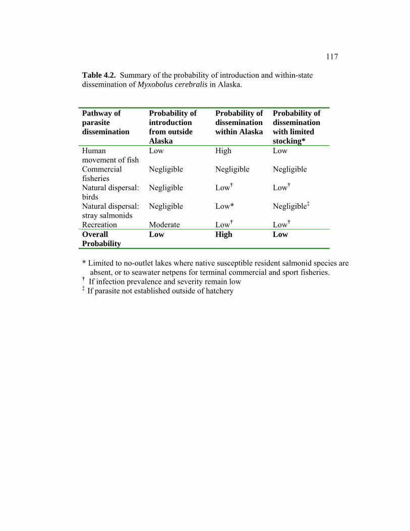
117
Table 4.2. Summary of the probability of introduction and within-state dissemination of Myxobolus cerebralis in Alaska. Pathway of parasite dissemination
Probability of introduction from outside Alaska
Probability of dissemination within Alaska
Probability of dissemination with limited stocking*
Human movement of fish
Low High Low
Commercial fisheries
Negligible Negligible Negligible
Natural dispersal: birds
Negligible Low† Low†
Natural dispersal: stray salmonids
Negligible Low* Negligible‡
Recreation Moderate Low† Low† Overall Probability
Low High Low
* Limited to no-outlet lakes where native susceptible resident salmonid species are absent, or to seawater netpens for terminal commercial and sport fisheries. † If infection prevalence and severity remain low ‡ If parasite not established outside of hatchery

118
Figure Legend Figure 4.1. Map of areas of highest risk for Myxobolus cerebralis dissemination
in southeast and southcentral Alaska. Rainbow trout hatcheries depicted by triangles (∆).
Figure 4.2. Model of potential Myxobolus cerebralis introduction in Alaska.
Three main pathways of dissemination are shown with specific activities listed.
Figure 4.3. Scenario tree of risk of establishment of Myxobolus cerebralis, if
disseminated in Alaska. Requirements for establishment and areas within the study sites that meet them are displayed.

119
Figure 4.1.
Alaska
N
5 km
Montana CreekPeterson Creek
Juneau
Mendenhall River
Ship Creek
Kenai River
Cook Inlet
Campbell Creek
10 km
Anchorage

120
Figure 4.2.
Commercial Activities
Recreational Activities Introduction
Hatchery fish transfers and
stocking
Illegal importation
of fish
Infected fish in commercial
harvests
Angler/ boating activity
Migration of piscivorous birds and mammals
Natural Dispersal
Human Movement of Fish
Stray fish from
enzootic areas

121
Figure 4.3.
Habitat for T. tubifex Throughout Alaska
No Establishment does not occur
Populations of susceptible T. tubifex Southcentral Alaska:
lower Kenai River, Ship and Campbell Creeks
Southeast Alaska
Water temperature seasonallyreaching 10-15 °C
Spatial/ temporal overlap of hosts
Where susceptible salmonid assemblages overlap with susceptible lineages of T. tubifex: Ship and Campbell Creeks
Establishment occurs In Ship and Campbell Creeks
Yes
Yes
Yes
Yes
Southcentral Alaska: lower Kenai River, Ship and Campbell Creeks

122
CHAPTER 5
A QUALITATIVE ANALYSIS OF RISK FOR DISPERSAL OF THE NON-NATIVE PARASITE MYXOBOLUS CEREBRALIS IN THE
WILLAMETTE RIVER BASIN, OREGON
E. Leyla Arsan1 and Jerri L. Bartholomew2
1Department of Fisheries and Wildlife, Center for Fish Disease Research, Oregon State University, 220 Nash Hall, Corvallis, OR 97331, USA
2 Department of Microbiology, Center for Fish Disease Research, Oregon State University, 220 Nash Hall, Corvallis, OR 97331, USA

123
Abstract
Myxobolus cerebralis, the myxosporean parasite responsible for whirling
disease in salmonids, was first detected in the United States in 1958. It has since
spread across the country causing severe declines in wild trout populations in the
intermountain west. This study qualitatively assesses the risk of introduction and
establishment of the pathogen into the Willamette River basin, Oregon by
examining potential routes of dissemination and relationships among obligate
hosts, the parasite, and the environment. The risk of M. cerebralis introduction in
the Willamette River basin is addressed as a function of three main elements of
dispersal: 1) movement of infected fish by humans, 2) natural dispersal (via
migratory birds and stray anadromous salmonids), 3) and recreational activities.
Establishment of the parasite is dependent upon several environmental and
biological factors including: water temperatures, density and spatial/temporal
overlap of hosts, and the distribution and genetic composition of the oligochaete
host, Tubifex tubifex. This study finds the probability of introduction of the parasite
to vary throughout the Willamette River basin. Areas with greater probability have
been identified as the Clackamas and Santiam River subbasins. If the pathogen
were introduced, probability of establishment is high in certain areas of the basin as
conditions are appropriate for propagation of the parasite life cycle.

124
INTRODUCTION
Myxobolus cerebralis, a metazoan fish parasite exotic to North America,
was first detected in the USA in 1958 (Hoffman, 1962). Thought to have
originated from a shipment of frozen infected brown trout (Salmo trutta) from
Europe, it is now reported from 25 US states and 26 different countries
(Bartholomew and Reno, 2002; Vermont Department of Fish and Game, 2002;
Stromberg, 2006; Arsan et al., chapter 3 of this volume). The pathogen has been at
the forefront of fish health research due to its potential impacts on rainbow trout
culture and its implication in rapid dramatic declines of wild trout populations in
Colorado and Montana (Vincent, 1996; Nehring et al., 1998). The ecologic and
economic impacts of M. cerebralis, in addition to its rapid spread and
establishment across the globe, are indicative of the need for specific
recommendations to assist fishery managers in halting the spread or effects of the
pathogen. These recommendations can be developed through the process of risk
analysis (MacDiarmid, 2000), and be tailored for a specific area. This study
assesses the risk of introduction and establishment of M. cerebralis into the
Willamette River basin, Oregon, a major tributary of the lower Columbia River.
Preventing pathogen introduction is a primary objective in fish health
management. However, once introduction occurs, parasitic infestations are often
managed by disrupting the parasite life cycle, generally by altering accessibility to
one or more of the organism’s hosts. The life cycle of M. cerebralis requires two
obligate hosts: a salmonid and the aquatic oligochaete worm, Tubifex tubifex.
There are also two distinct spore stages of the parasite (one particular to each of the
hosts). The myxospore stage develops in the fish host and is released upon death
and decomposition of the fish. These round and durable spores are ingested by T.
tubifex as the worms burrow through sediment consuming bacteria (Brinkhurst,
1996). The parasite then undergoes reproduction and structural transformation and
is released in its actinospore stage as a triactinomyxon (TAM). The TAM attaches
to its fish host in the water column, where the parasite sporoplasm is injected
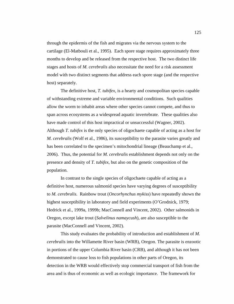
125
through the epidermis of the fish and migrates via the nervous system to the
cartilage (El-Matbouli et al., 1995). Each spore stage requires approximately three
months to develop and be released from the respective host. The two distinct life
stages and hosts of M. cerebralis also necessitate the need for a risk assessment
model with two distinct segments that address each spore stage (and the respective
host) separately.
The definitive host, T. tubifex, is a hearty and cosmopolitan species capable
of withstanding extreme and variable environmental conditions. Such qualities
allow the worm to inhabit areas where other species cannot compete, and thus to
span across ecosystems as a widespread aquatic invertebrate. These qualities also
have made control of this host impractical or unsuccessful (Wagner, 2002).
Although T. tubifex is the only species of oligochaete capable of acting as a host for
M. cerebralis (Wolf et al., 1986), its susceptibility to the parasite varies greatly and
has been correlated to the specimen’s mitochondrial lineage (Beauchamp et al.,
2006). Thus, the potential for M. cerebralis establishment depends not only on the
presence and density of T. tubifex, but also on the genetic composition of the
population.
In contrast to the single species of oligochaete capable of acting as a
definitive host, numerous salmonid species have varying degrees of susceptibility
to M. cerebralis. Rainbow trout (Oncorhynchus mykiss) have repeatedly shown the
highest susceptibility in laboratory and field experiments (O’Grodnick, 1979;
Hedrick et al., 1999a, 1999b; MacConnell and Vincent, 2002). Other salmonids in
Oregon, except lake trout (Salvelinus namaycush), are also susceptible to the
parasite (MacConnell and Vincent, 2002).
This study evaluates the probability of introduction and establishment of M.
cerebralis into the Willamette River basin (WRB), Oregon. The parasite is enzootic
in portions of the upper Columbia River basin (CRB), and although it has not been
demonstrated to cause loss to fish populations in other parts of Oregon, its
detection in the WRB would effectively stop commercial transport of fish from the
area and is thus of economic as well as ecologic importance. The framework for

126
this risk assessment (Bartholomew et al., 2005) was created in efforts to tailor such
assessments to whirling disease. This paper includes a synthesis of historical data
as well as new research to fill data gaps. Our aim is to provide decision-makers
with tools to assess management implications, to eliminate non-issues by using
logical scientific arguments, and to provide guidance on where to allocate
resources to help prevent introduction of the pathogen.
METHODS
Study Area
Myxobolus cerebralis was first detected in Oregon in 1986 (Lorz et al.,
1989), and is now enzootic in certain tributaries of the upper CRB (Figure 5.1).
Since the 1980’s, the parasite has been detected elsewhere in the basin in stray
adult salmonids originating from these tributaries (Engelking, 2002). However,
there is no evidence of establishment of the parasite life cycle outside of the
enzootic region. The parasite was detected further west in the state in 2001, at a
private facility in the lower WRB (Clear Creek). Infection at the facility was
contained and discharges are no longer made into creek (Bartholomew et al., 2007).
Currently, M. cerebralis is not known to be established in the WRB outside of the
private facility on Clear Creek.
The Willamette River originates in the Cascade Mountains (up to 10,000 ft)
and flows approximately 300 km to the lower Columbia River (10 ft). The WRB
houses 70% of the population of Oregon and is the fastest growing and most
economically and agriculturally developed region in the state (USEPA, 2006a,
2006b). The river, which is the 13th largest in terms of discharge in the contiguous
US (Urich and Wentz, 1999), flows through Oregon’s three largest cities, and
accounts for 15% of the total flow in the Columbia River (Payne and Baker, 2002).
Stream flow in the basin reflects precipitation, with the majority (60-85%) of the
runoff occurring from October to March (Urich and Wentz, 1999). The basin

127
contains the richest native fish fauna in the state (Rathert et al., 1999) and provides
a migratory corridor and spawning grounds for a variety of anadromous salmonids.
This study concentrated on four areas of the WRB that we believed to have
the highest risk for introduction and establishment of M. cerebralis: the Willamette
River mainstem, Clackamas River subbasin, McKenzie River, and North and South
Santiam Rivers (Figure 5.1). These areas have high concentrations of susceptible
fish hosts, high angler traffic, hatcheries rearing susceptible fish species, private
fish rearing ponds, and high potential organic loading leading to increased T.
tubifex habitat.
Validation of preliminary assumption
To verify the preliminary assumption that M. cerebralis is not present in
wild or hatchery salmonids in the WRB, we used data from 20 years of Oregon
Department of Fish and Wildlife (ODFW) parasite testing, representing over
12,000 wild and cultured fish (N. Hurtado, ODFW, unpublished data).
From 1986-1987, immediately following the first detection of the parasite in
Oregon, 82 fish from 6 locations in the basin were presumed positive for the
parasite. All fish were transfers from a positive private facility in the upper CRB to
other private facilities in the WRB, and the ODFW subsequently halted transfers of
fish from the enzootic area. The parasite was not detected in further monitoring of
these 6 locations.
Though more presumptive positive fish were detected in the WRB from
1988-2001, infection was never confirmed. Presumptive identification of M.
cerebralis is based on myxospore morphology, a trait that is similar in many
Myxobolus species. Until 2001, confirmation of infection was based on
histopathology, a method with high specificity but low sensitivity. Since 2001 and
the incorporation of sensitive genetic tests such as polymerase chain reaction (PCR)
into monitoring regimes, the only locations in the WRB where M. cerebralis has
been detected are in Clear Creek. Because of the low sensitivity of parasite
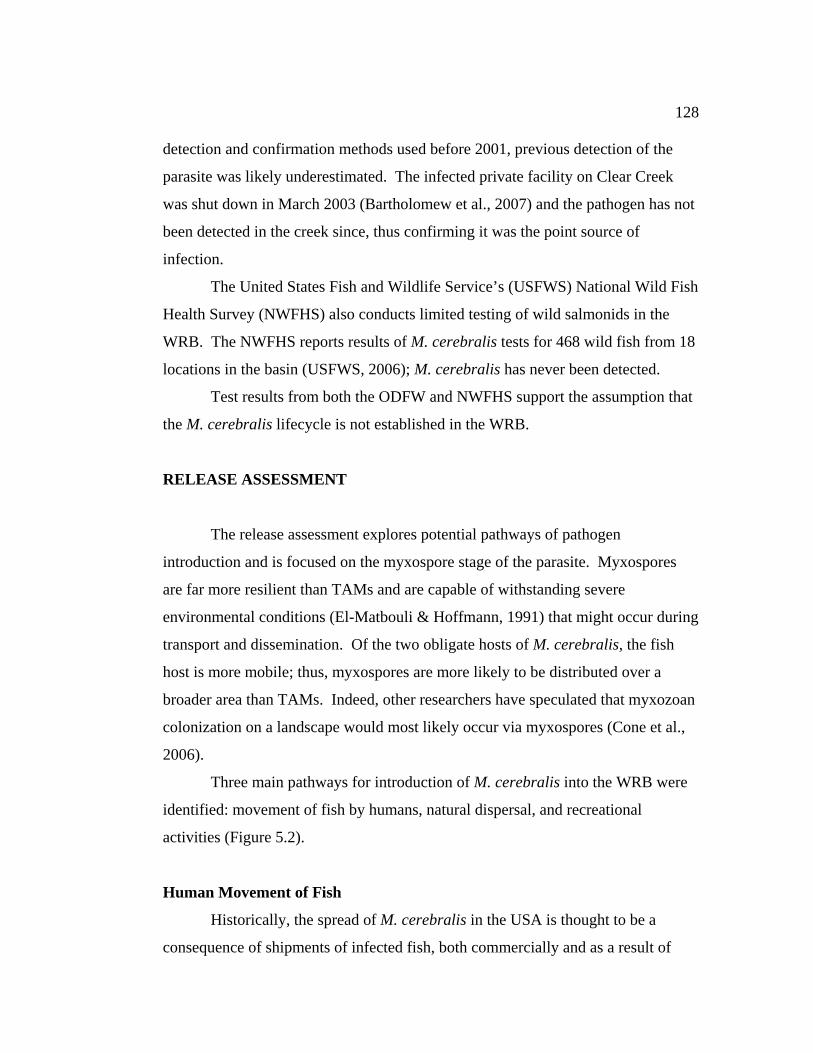
128
detection and confirmation methods used before 2001, previous detection of the
parasite was likely underestimated. The infected private facility on Clear Creek
was shut down in March 2003 (Bartholomew et al., 2007) and the pathogen has not
been detected in the creek since, thus confirming it was the point source of
infection.
The United States Fish and Wildlife Service’s (USFWS) National Wild Fish
Health Survey (NWFHS) also conducts limited testing of wild salmonids in the
WRB. The NWFHS reports results of M. cerebralis tests for 468 wild fish from 18
locations in the basin (USFWS, 2006); M. cerebralis has never been detected.
Test results from both the ODFW and NWFHS support the assumption that
the M. cerebralis lifecycle is not established in the WRB.
RELEASE ASSESSMENT
The release assessment explores potential pathways of pathogen
introduction and is focused on the myxospore stage of the parasite. Myxospores
are far more resilient than TAMs and are capable of withstanding severe
environmental conditions (El-Matbouli & Hoffmann, 1991) that might occur during
transport and dissemination. Of the two obligate hosts of M. cerebralis, the fish
host is more mobile; thus, myxospores are more likely to be distributed over a
broader area than TAMs. Indeed, other researchers have speculated that myxozoan
colonization on a landscape would most likely occur via myxospores (Cone et al.,
2006).
Three main pathways for introduction of M. cerebralis into the WRB were
identified: movement of fish by humans, natural dispersal, and recreational
activities (Figure 5.2).
Human Movement of Fish
Historically, the spread of M. cerebralis in the USA is thought to be a
consequence of shipments of infected fish, both commercially and as a result of

129
state and federal stocking programs (Bartholomew and Reno, 2002). All import,
export, or transfers of live fish in Oregon require a Fish Transport Permit (Oregon
Administrative Rules [OAR] 635-007-0600 and 635-007-0615) that entails an
annual health examination of both the broodstock and of the lot of fish that are to
be imported (OAR 635-007-0585 and 635-007-0990). Currently, permits are not
granted if the exporting facility has a history of M. cerebralis infection, even if the
importing area is enzootic for the parasite. All state (6), federal (1), and private (6)
trout rearing facilities in the WRB are monitored annually for the parasite (Figure
5.1).
When buying live fish in Oregon, it is the buyer’s responsibility to know the
rules and regulations surrounding fish transfers and required permits. Buyers are
often unaware of required permits (T. Amandi, ODFW, personal communication),
which may result in illegal (even if inadvertent) movements of fish that cannot be
traced. Because rules and regulations can be lengthy and ambiguous, and because
businesses want to ensure sales, most licensed fish sellers in Oregon acquire
ODFW fish transport permits for prospective buyers and often explain regulations
and procedures. While there are six facilities licensed to sell fish in the WRB, the
number of private ponds that contain fish is more difficult to estimate. Only 30
permits are on record to legally impound water and hold fish in the WRB (Oregon
Water Resources, 2006), yet transport records from a single private facility show
that greater than 400 sites in the WRB received fish, demonstrating that numbers of
private ponds stocked with salmonids is far higher. Hobby ponds and U-Catch
facilities are examples of private ponds where fish may be reared and resold or
transferred by owners who may be unaware of the need for fish transport permits.
Even legal transports of fish present a risk. For example, although
establishment of M. cerebralis at the private facility on Clear Creek was contained,
the facility legally transferred potentially infected fish throughout the state of
Oregon before detection of the parasite occurred. Over 760 shipments to 430
different locations in the WRB were received between 1999-2000 alone (N.
Hurtado, ODFW, personal communication). Three of the facilities in the WRB that
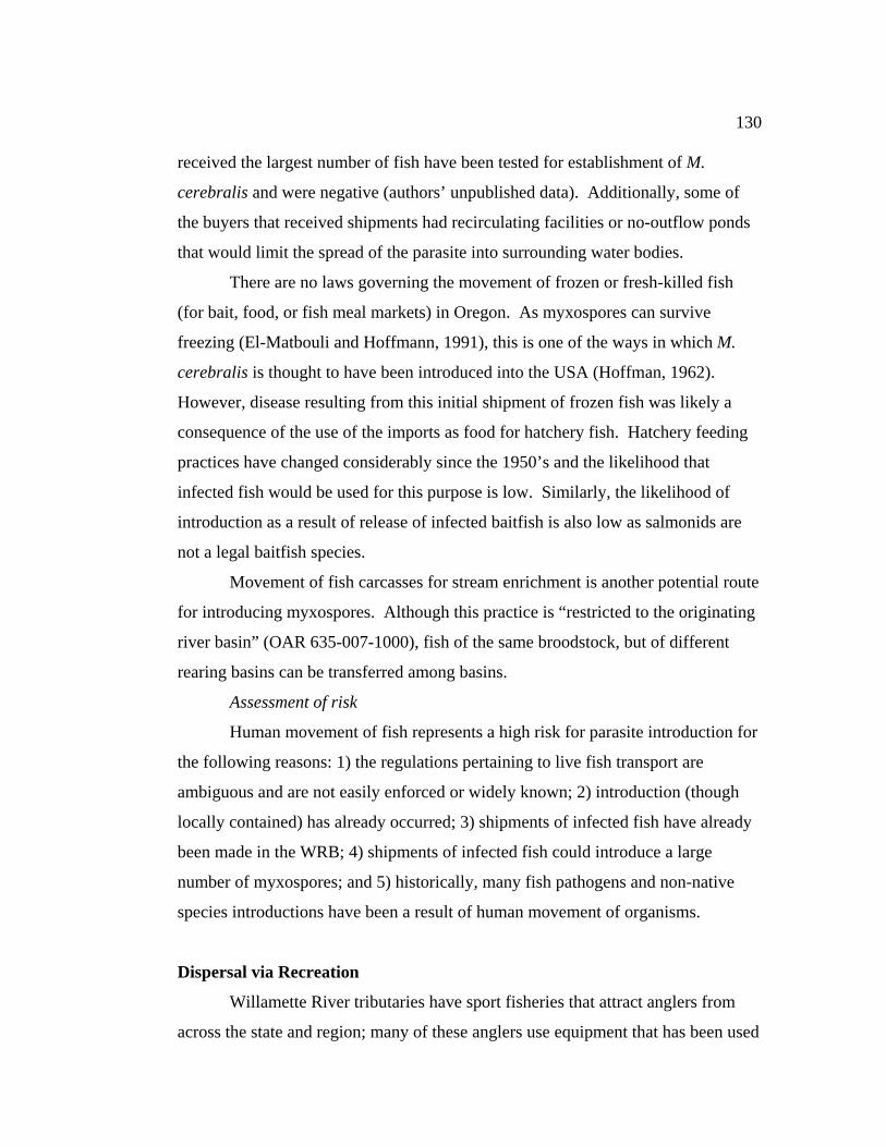
130
received the largest number of fish have been tested for establishment of M.
cerebralis and were negative (authors’ unpublished data). Additionally, some of
the buyers that received shipments had recirculating facilities or no-outflow ponds
that would limit the spread of the parasite into surrounding water bodies.
There are no laws governing the movement of frozen or fresh-killed fish
(for bait, food, or fish meal markets) in Oregon. As myxospores can survive
freezing (El-Matbouli and Hoffmann, 1991), this is one of the ways in which M.
cerebralis is thought to have been introduced into the USA (Hoffman, 1962).
However, disease resulting from this initial shipment of frozen fish was likely a
consequence of the use of the imports as food for hatchery fish. Hatchery feeding
practices have changed considerably since the 1950’s and the likelihood that
infected fish would be used for this purpose is low. Similarly, the likelihood of
introduction as a result of release of infected baitfish is also low as salmonids are
not a legal baitfish species.
Movement of fish carcasses for stream enrichment is another potential route
for introducing myxospores. Although this practice is “restricted to the originating
river basin” (OAR 635-007-1000), fish of the same broodstock, but of different
rearing basins can be transferred among basins.
Assessment of risk
Human movement of fish represents a high risk for parasite introduction for
the following reasons: 1) the regulations pertaining to live fish transport are
ambiguous and are not easily enforced or widely known; 2) introduction (though
locally contained) has already occurred; 3) shipments of infected fish have already
been made in the WRB; 4) shipments of infected fish could introduce a large
number of myxospores; and 5) historically, many fish pathogens and non-native
species introductions have been a result of human movement of organisms.
Dispersal via Recreation
Willamette River tributaries have sport fisheries that attract anglers from
across the state and region; many of these anglers use equipment that has been used

131
in other river systems. In a laboratory study, felt-soled waders were demonstrated
to transport myxospores and TAMs (P. Reno, Oregon State University, personal
communication). However, after drying waders for 8-24 h, spores became less
viable, and after drying for 7 d, spores were no longer able to cause infection in
appropriate hosts. These findings support other studies that suggest that
myxospores do not remain viable after drying for 24 hr (R. Hedrick, University of
California at Davis, personal communication). Myxospores trapped in the moisture
of a felt-sole would likely remain wet and thus viable for longer than in a less
absorbent material. Prior data on myxospore viability after drying (Schäuperclaus,
1931; Hoffman and O’Grodnick, 1977) were collected before the determination of
the M. cerebralis life cycle and are anecdotal at best. Time required to dry and thus
disinfect waders will vary with environmental conditions. Because of their
fragility, it is unlikely that TAM stages could be transported large distances by this
route. However, infected T. tubifex adhered to a felt sole provide a suitable
environment for TAMs to remain viable.
While the probability of transporting M. cerebralis in a single event (i.e.: a
single angler or single angling day) by recreational activities may be low, when all
angler days in a year are considered, the probability increases. The Middle Fork of
the Willamette River and South Santiam River saw 22,501 and 17,937 anglers
respectively in 1975 (Moring, 1985). These areas, along with the Clackamas River
(the closest major tributary and sport fishery to Portland) would be most likely to
experience introduction of the parasite via recreational activity. While the number
of anglers traveling between the enzootic area and the WRB is unknown, anglers
from Portland [the area with the greatest number of anglers (US Department of
Interior, 2001)] could recreate in NE Oregon (a renowned recreational destination)
and return home to use gear in local waterways.
Boat bilge and engine cooling water represent another route by which
recreational activities could introduce M. cerebralis. TAMs would be more likely
than myxospores to be suspended in the water column and be retained in bilge and
engine cooling water. Survival of TAM stages is temperature dependent, with

132
spores surviving for 5-7 d at 7°C and only 2 d at 20°C (Markiw, 1992b). Thus,
TAMS are unlikely to survive multiple days of transport or high summer
temperatures that would alter the condition of small amounts of bilge water.
However, water caught in the covered intake of the engine cooling system does not
experience elevated temperatures and has been shown to transport live larval zebra
mussels (Johnson et al., 2001). While the volume of spores transported per boat is
likely to be low, when considering all boats recreating in a year, the potential for
M. cerebralis transfer increases. In 2005, 3,380 boats registered in the WRB were
used in the enzootic area (Oregon State Marine Board, 2005). Likewise, 330 boats
registered in the enzootic area were used in the WRB in 2002 (Oregon State Marine
Board, 2002).
Assessment of risk
A conservative estimate of recreational activities as a pathway for transfer
of M. cerebralis to the WRB is that it represents a moderate risk. Numbers of
spores introduced in a single angling or boating event are expected to be low, but
the cumulative and long-term effects of angler and recreational activities in heavy-
use areas would be much greater. Recreational activity is also one of the few
pathways where management steps can be taken to reduce the risk of introduction.
Management considerations are discussed in the conclusions of this paper.
Natural dispersal: birds
The possibility of M. cerebralis dissemination by birds has been considered
in several studies (Taylor and Lott, 1978; El-Matbouli & Hoffmann, 1991).
However, none of these studies have aligned the numerous events that must occur
for M. cerebralis to be spread long distances via bird transport. Below, we outline
the chain of events that must occur to result in introduction.
1) Bird eats infected fish.
The CRB estuary houses the largest colony of Caspian terns (Sterna caspia)
in North America and the second largest colony of double-crested cormorants
(Phalacrocorax auritus)- 11,223 and 7,501 birds respectively in 1998 (Collis et al.,

133
2002). A smaller Caspian tern colony (990 birds in 1997) is located at Crescent
Island, just below the confluence of the Snake and Columbia Rivers and near the
enzootic area. Birds from these colonies eat juvenile salmonids as their primary
food source, with steelhead smolts being the most vulnerable to predation (Collis et
al., 2001). This species is also among the most susceptible to M. cerebralis
(MacConnell and Vincent, 2002).
While prevalence of M. cerebralis infection in juvenile salmonids in the
CRB is unknown, the proportion of infected fish is higher in the enzootic area than
in the estuary. However, the estuary houses larger piscivorous bird colonies and
timing of seasonal dispersal from these colonies coincides with peak migration of
highly susceptible juvenile salmonids. Thus, birds from both the enzootic area and
the estuary represent a risk of M. cerebralis dispersal. In addition to the Caspian
tern and double-crested cormorant colonies, numerous other piscivorous bird
species inhabit the CRB, and may also present risks for M. cerebralis dispersal.
2) Myxospores survive passage through gut of piscivorous bird.
Myxobolus cerebralis myxospores have been demonstrated to survive
passage through the guts of piscivorous birds and fish (El-Matbouli & Hoffmann,
1991).
3) Bird enters WRB.
Banding data on the dispersal of birds from particular breeding colonies are
limited, thus flight paths mentioned here are speculative based on expert opinion
(D. Roby, USGS-Oregon Cooperative Fish and Wildlife Research Unit, personal
communication). Caspian terns at Crescent Island have been banded and resighted
1-2 days later in the Columbia River estuary. Although these birds do not nest in
the WRB, they do use foraging sites in the WRB during spring and fall migration
and terns from breeding colonies in eastern Washington may use this route post-
nesting. Immature and sub-adult eagles undertake seasonal movements in search of
foraging areas and could disperse from NE Oregon to the WRB. Migratory paths
of ospreys after the breeding season could take the birds west following the
Columbia River and south through the WRB.
134
4) Bird retains infected fish in gut for flight to WRB.
As M. cerebralis manifests in cartilage of fish, it is likely that birds would
egest, or regurgitate, the parasite in pellets. Double-crested cormorants have a
simple gut structure and egest bones, pieces of fish, and solid markers 1-2 days
following ingestion (Brugger, 1993). In contrast, the passage time of rainbow trout
marked with trace minerals through the guts of bald eagles (which have a more
complex gut morphology) is approximately 62 hours (F. Barrows, USFWS,
personal communication). Thus spores could be excreted 2-3 d after eating an
infected fish.
Raptors and large waterbirds have the fastest known migration speeds
among birds (Kerlinger, 1995). Migrating bald eagles travel 201 km/day
(Kerlinger, 1995), and ospreys 108-431 km/day (Alerstam, 2003; Hake et al.,
2001). As the nearest enzootic area is approximately 450-500 km from the WRB,
an osprey would have to retain food material for 1-4.2 days and bald eagles for 2.2-
2.5 days to transport spores to the WRB. Thus transport of spores from the
enzootic area to the WRB is possible.
5) Bird releases viable spores over water body.
The probability of such an occurrence is unknown, and is likely to vary by
habits and behaviors of species. For example, waterbirds (waders, waterfowl,
gulls, etc.) characteristically spend more time over water than raptors. Although
the probability of a bird releasing viable myxospores over a waterbody remains
unknown, deposition near (and not in) water may be sufficient for transport of the
parasite to a new location under certain environmental conditions. Anecdotal
evidence suggests that myxospores can withstand drying for long periods of time
(Schäuperclaus, 1931), but more current studies indicate myxospores do not remain
viable after drying for 24 hr (R. Hedrick, University of California at Davis,
personal communication). It is unknown how long myxospores deposited in bird
feces would remain moist, and thus viable. Time required for inactivation of spores
is likely to vary by environmental conditions. Thus, if spores are deposited near a
135
waterbody, and are rapidly washed into the river by high water or precipitation,
dissemination of the parasite could occur.
Assessment of risk
Although the likelihood of parasite introduction by a single bird may be
low, if the event were to occur, a considerable number of spores could be released
because many piscivores swallow their prey whole. As many as 1.7 x 10⁶
myxospores have been reported from experimentally challenged rainbow trout,
though such numbers are variable (Markiw, 1992b; Sollid et al., 2002). Therefore,
even a single introduction could have large consequences. Still, numerous events
must align in order for birds to transport M. cerebralis long distances, thus the
probability of birds transporting the pathogen to the WRB is low. The cumulative
risks when considering the large numbers of birds traveling through the WRB in a
year would be much higher.
Data gaps identified in this part of the release assessment are the infection
rate of salmonids migrating from both the Snake River basin and the Columbia
River estuary, and time required to disinfect myxospores in guano or egested
pellets.
Natural dispersal: stray anadromous salmonids
Anadromous salmonids may stray into non-natal streams during their return
migration to spawning grounds. An often-overlooked effect of straying is the
potential for pathogen dissemination. Introduction of M. cerebralis as a result of
straying salmonids has been documented in the Deschutes River, Oregon (mid
CRB) (Engelking, 2002). Between 1983 and 2005, 32-92% of adult hatchery
steelhead collected annually during peak migration were strays (J. Seals, ODFW,
unpublished data). Examination of over 450 stray adult steelhead between 1997
and 2000 showed that at least 5% were infected with M. cerebralis (Engelking,
2002), with unconfirmed but suspected detections in an additional 15% of the fish.
Infection prevalence in stray Chinook salmon was lower (1% confirmed; 7%
presumptive).
136
Stray fish are typically documented by examination of fin clips or evidence
of a coded wire tag (CWT). As any single fin-clip pattern is commonly used by
more than one hatchery in the Pacific Northwest in a given year, fin clips reveal
only the “stray” status of a fish and a rough list of potential origins. A CWT or fin
clip unique to a certain hatchery would be required for identification of exact
hatchery of origin.
Very little data (current or historical) are available on the stray rates of
cultured or wild salmonids in the WRB. A search of recovery data for the WRB
through the Regional Mark Information System (RMIS) database, shows only 16
recoveries of stray Chinook salmon between 1977-2003 (RMIS, 2006). Four of the
hatcheries of origin of these fish, representing 81% of the historical strays, are
within the enzootic area of the Snake River basin.
As the frequency of adult salmonids straying into the WRB was identified
as a significant data gap for this risk assessment, we conducted a study to
investigate the frequency and prevalence of infection of straying salmonids in the
WRB.
Methods for investigation of frequency and infection of stray fish in the WRB
From 2004 to 2005, ODFW personnel at all adult salmonid collection
facilities in the WRB (Figure 5.1) monitored returning adults for marks or tags
which would indicate they were strays from other river basins. Traps were located
on the McKenzie, Clackamas, Middle Fork Willamette, and North and South
Santiam Rivers.
Collected fish were killed, checked for coded-wire tags, the fins or
maxillary marks recorded, and the heads removed and frozen for M. cerebralis
testing. The RMIS and ODFW databases were queried for the origin of salmonids
with marks corresponding to those of the stray fish collected. Additional efforts
were made by the National Marine Fisheries Service (NMFS, Conservation
Biology Division, DNA Laboratory, Seattle, WA) to genetically identify these fish
using microsatellite markers.

137
To determine M. cerebralis infection in strays, core samples were taken
from the cranium using an electric hand drill with a 2.5 cm diameter coring-bit.
Cores were maintained individually and processed by pepsin-trypsin digest (PTD)
(USFWS and AFS-FHS, 2003). The defleshed cores were blended with a
commercial Waring blender for 30 seconds to accelerate digestion in pepsin
solution. Because the samples were stored frozen until processing, 0.05% trypsin in
Rinaldini’s saline solution was used during the trypsin digestion phase. The final
pellet of digest was suspended in 1 ml of phosphate buffered saline or 70% ethanol
and observed microscopically for myxospores by examining wet mounts of
undiluted and diluted (1:5 and 1:10) digest at 250X and 400X magnification. In
2004, the PTD product was tested by the PCR assay using protocols developed by
Andree et al. (1998). In 2005, a quantitative version of this assay (Kelly et al.,
2004) was used to increase sensitivity.
Results and discussion of stray fish studies in the WRB
In contrast to the low number of reported stray salmonids from 1977-2003,
129 adult summer steelhead with fin clips identifying them as strays were
recovered at traps in the WRB during 2004-2005. Table 5.1 lists the locations,
number and percent strays found and the different marks observed on these fish.
Most of the strays were collected in the Santiam River subbasin (97%), possibly
because the area releases more steelhead than other WRB tributaries (ODFW,
unpublished data). If strays follow the masses of fish migrating upriver, they
would be more likely arrive in the Santiam River subbasin. Also, mis-marked fish
(i.e. not strays) would be more probable in an area where more fish are clipped. No
stray fish were obtained from traps on either the Clackamas or Middle Fork
Willamette Rivers.
Fin marks recorded from WRB strays were used by a number of hatcheries
releasing summer steelhead in the Pacific Northwest during 1999-2002 (RMIS,
2006; ODFW, Fish Liberation Database, unpublished data). Of the 19 facilities,
seven (37%) are in the M. cerebralis enzootic area (Table 5.1). Interestingly,
ADLVRV (adipose, left ventral, right ventral), one of the most prominent markings

138
(31%), was not recorded in RMIS or any of the federal, state, or regional databases
from Oregon or surrounding states. Efforts to genetically identify the WRB strays
did not reveal a hatchery of origin (Melanie Paquin, NMFS, personal
communication); however, the high level of genetic differentiation between the
WRB strays and Snake River basin hatchery summer steelhead indicate that these
fish likely originated outside the enzootic region.
Stray Chinook salmon were not encountered in this study, which could be
explained by a decreased rate of straying compared with steelhead, or by the lack of
distinctive marks. Chinook salmon rarely receive marks other than an adipose fin
clip and determination of hatchery of origin would require a CWT. However,
Chinook salmon also represent a lower risk for introduction because of their
decreased susceptibility to the parasite (Sollid et al., 2003).
Overall, numbers of strays collected in the WRB in 2004-2005, while lower
than that of mid CRB tributaries (ODFW, unpublished data), are far higher than
indicated by database records. It is likely that this increase in number can, at least
in part, be attributed to a concerted effort by ODFW to train personnel to recognize
stray fish. If similar efforts had been made in previous years a different pattern of
straying may have emerged. It is also possible that the years 2004 and 2005 were
abnormal. As there are few historical data, we cannot determine a “normal” stray
rate per year. However, stray rates of 0-20% are not uncommon for hatchery
salmonids (Quinn, 1997). Actual numbers of strays in the WRB are likely to be
higher than seen in this study as all stray wild fish as well as unmarked or single-
marked (only adipose clip) hatchery fish were not recorded as out of basin fish.
Another source of error in estimating straying frequency is that collections occurred
only at fish traps; fish spawning in the river and tributaries were not counted. It is
also possible that some stray fish were actually mis-marked fish from WRB
hatcheries and were indeed not strays.
Spores of a Myxobolus spp. were detected in 3 fish. All samples were
negative by PCR, suggesting that the spores detected were of another Myxobolus
species.

139
Assessment of risk
Although the straying frequency for steelhead was higher than expected,
none of these fish had detectable M. cerebralis infection and therefore are not likely
to have originated from the enzootic area. If we assume this stray rate and origin is
representative of strays in other years, the likelihood of introducing M. cerebralis
as a result of stray adult salmonids is low. However, if it were to occur, its
probability is higher in the Santiam River subbasin as the majority of stray fish
were recovered in this area.
Summary of the release assessment
The probabilities described in this assessment are defined as: high – the
event would be expected to occur, moderate – less than an even chance of the event
occurring, low - the event is unlikely to occur, and negligible – the chance of the
event occurring is so small that it can be ignored. The probability of introduction
of M. cerebralis into the WRB is summarized as:
• Dispersal via human movement of fish: high
• Dispersal via recreation: moderate
• Dispersal via natural dispersal (birds and stray anadromous salmonids): low
Overall probability of introduction: moderate (though long-term cumulative
probability is higher.)
Areas with the highest probability for M. cerebralis introduction are the
Clackamas and Santiam River subbasins. The Clackamas River has already
experienced an introduction of the parasite, has the largest concentration of
hatcheries (state, federal, and private), has a popular sport fishery, and is the closest
major tributary to the enormous piscivorous bird populations in the Columbia River
estuary. The Santiam subbasin has a popular sport fishery, received the highest
number of stray fish in the WRB, and has the second largest concentration of
hatcheries in the WRB.

140
EXPOSURE ASSESSMENT
The exposure assessment, which explores the risk of parasite establishment,
is focused on the TAM stage of M. cerebralis and its oligochaete host.
Establishment of M. cerebralis is dependent upon several environmental and
biological factors including: water temperatures, spatial temporal overlap of hosts,
and the distribution and genetic composition of T. tubifex populations. Each of
these factors determines the outcome of the exposure assessment, as depicted by
the scenario tree in Figure 5.3.
Habitat for T. tubifex
Habitat for T. tubifex is defined as areas with fine sediment, low flow, and
organic matter (Brinkhurst and Gelder, 1991). These areas were found throughout
the WRB.
Sustainable populations of susceptible T. tubifex
Bartholomew et al. (2007) documented only a single specimen of T. tubifex
in a survey of Clear Creek, Clackamas River subbasin. In contrast, a private
hatchery adjoining the creek had dense T. tubifex populations. A literature review
revealed little other data regarding T. tubifex populations in the WRB. The
prevalence and susceptibility of T. tubifex was identified as a significant data gap in
the risk analysis and a qualitative survey for T. tubifex was conducted with
subsequent characterization of specimens to determine mitochondrial lineage and
susceptibility.
Tubifex tubifex survey methods
A qualitative survey was used to determine relative abundance of T. tubifex
in selected streams in the study area. Areas most likely to contain the organisms
were targeted for sampling, i.e. those with: low flow, fine sediments, and
accumulations of organic material. Thirty-three sample sites with these attributes
as well as with temperatures appropriate for parasite propagation, populations of

141
salmonids susceptible to the parasite, a large sport fishery, documented stray fish,
and accessibility by road were chosen (Figure 5.4). Ten sites were located in the
mainstem Willamette River, 12 in the South Santiam River, four in the North
Santiam River and seven in the McKenzie River. The Clackamas River has
previously been surveyed (Bartholomew et al., 2007) and no further collections
were made in that system.
Oligochaetes in sediment were collected by 5 gal bucket and 500 µm sieve
or by 500 µm kicknet, maintained, sorted, and presumptively characterized as T.
tubifex following methods described in Arsan et al. (chapter 2 of this volume). A
subsample of 20 worms presumed to be T. tubifex were then genetically analyzed to
confirm identity as T. tubifex (Hallett et al., 2005) and to determine lineage
(Sturmbauer et al., 1999; Beauchamp et al., 2001). In preparation for genetic
analysis, worms were by digested with 95 µL ATL buffer (QIAGEN) and 5 µL
Proteinase K at 55°C, boiled for 5 minutes, and diluted 1:101 with buffer AE
(QIAGEN) then stored frozen.
To determine whether worms genetically identified as T. tubifex (in a
species-specific PCR) but which did not visibly amplify in the lineage assay
(lineage-specific PCR) were a novel lineage or a different species, a portion of their
mitochondrial 16S rDNA was sequenced. Mitochondrial 16S DNA was amplified
with either the primers Tub16SF and Tub16SR (Beauchamp et al., 2001) or 16sar
and 16sbr (Sturmbauer et al., 1999) using reaction conditions described by Arsan et
al. (chapter 2 of this volume).
Water in which samples were transported was retained and passed through a
20 µm mesh filter and inspected microscopically to determine if actinospores were
present. Actinospores were identified to group and species, when possible.
Results of T. tubifex survey
Over 5,300 oligochaetes were collected from 33 sites, with Tubifex tubifex
identified from 19 sites (Figure 5.4) and accounting for approximately 10% of the
total oligochaetes collected (Table 5.2). Other oligochaetes morphologically
identified from the WRB include: Tubifex ignotus, Limnodrilus hoffmeisteri,
142
Lumbriculus varigatus, Kincaidiana hexatheca, Aulodrilus pluriseta, Dero digitata,
Spirosperma nikolski, Ophidonais serpentina, Chaetogaster limnaei, and
Pristonella jenkinae.
The largest numbers of T. tubifex were observed from the South Santiam
and McKenzie Rivers, particularly at sites near Foster (RM 36.5) and Leaburg (RM
29.4) Dams, and at Hendrick Boat Landing on the McKenzie River. These sites
receive heavy recreational use and had areas with deep sediments and organic
enrichment (due to both decaying salmonid carcasses and overhanging vegetation).
While this study presents some of the few data on WRB T. tubifex
populations, our surveys were qualitative and limited by access. Accessibility to
sample sites on the North Santiam River was restricted to areas accessible by road.
As only four of these sites had appropriate T. tubifex habitat, data from that river
are limited.
Susceptibility of T. tubifex to M. cerebralis was inferred from results of the
lineage analysis. Different lineages vary in susceptibility. Populations from
lineages I and III have been shown to be susceptible to the parasite while lineages
IV, V and VI are considered resistant (Beauchamp et al., 2002, 2005, & 2006;
DuBey et al., 2005; Arsan et al., chapter 2 of this volume). All five T. tubifex
lineages reported from North America were found in the WRB (Table 5.2),
including lineage IV, which had previously been described only from Europe and
Alaska (Sturmbauer et al., 1999; Beauchamp et al., 2001; Arsan et al., chapter 2 of
this volume).
The presence of resistant lineages IV, V, and VI T. tubifex could have
significant beneficial implications for WRB salmonids in areas where these
lineages dominate Tubifex populations. Together, these lineages comprised
approximately 88% of the T. tubifex population in the mainstem Willamette River
and 24-54% in river tributaries. Other researchers have found lineage susceptibility
to correspond to severity of infection in fish (Beauchamp et al., 2006). This
suggests that when lineages IV, V, and VI are in high abundance, the effects of M.

143
cerebralis may be diminished if the parasite were introduced and established in the
WRB.
Lineage III is the most susceptible to M. cerebralis (Beauchamp et al.,
2002; DuBey et al., 2005; Hallett and Bartholomew, 2007; Arsan et al., chapter 2 of
this volume) and its detection in the WRB is cause for concern. This lineage
comprised approximately 12-56% of worm populations, with the greatest numbers
in the North Santiam River and fewest in the mainstem of the Willamette River
(Table 5.2). Even in proportions as low 2% of the total population, lineage III T.
tubifex become infected with M. cerebralis and release TAMs (Beauchamp et al.,
2006; Hallett and Bartholomew, 2007; Arsan et al., chapter 2 of this volume).
Populations of T. tubifex from the North Santiam, McKenzie and Middle Fork
Willamette Rivers that have been experimentally exposed to the parasite (Hallett
and Bartholomew, 2007) were moderate TAM producers, releasing up to 48,960 in
4 d when exposed to 500 myxospores and held at 12.8°C.
No M. cerebralis TAMs were observed in filtrates from any of the sites
sampled. However, actinospores of other myxozoans were observed and were
genetically identified as Myxobilatus gasterostei and other Myxobolus spp.
(Stephen Atkinson, Center for Fish Disease Research, unpublished data). Other
unidentified triactinomyxon, aurantiactinomyxon, siedleckiella, echinactinomyxon
and raabeia type actinospores were also found. Several of these unidentified
actinospores were released from T. tubifex hosts. The detection of other
myxozoans validates our methodology was sufficient for detecting M. cerebralis
TAMs. This also confirms that other myxozoan life cycles, some with documented
T. tubifex hosts, are established in the WRB.
Water temperature seasonally reaching 10-15 °C
Water temperature influences parasite development in both hosts (Hedrick
and El-Matbouli, 2002; Blazer et al., 2003). In the oligochaete, 10 to 15°C has
been demonstrated to be optimal for development and release of the TAM stage of
the parasite (El-Matbouli et al., 1999). Temperatures below 10°C delay the
144
development of TAMs and those above 20°C hinder parasite development and can
be lethal to the TAM stage of M. cerebralis. Highest prevalence of infection in fish
occur when average daily water temperatures are between 11-14°C (Baldwin et al.,
2000; Granath and Gilbert, 2002).
Because continuous water quality monitoring is a relatively new
phenomenon, temperature data for certain rivers are limited to only a few years.
For WRB sites where temperature data were available, average summer (May-
September) water temperatures ranged from 11.5- 16.5°C between 2001- 2005
(USGS, 2006), with an average of 142-204 d/yr continuously >10°C.
In major tributaries, temperatures are within the optimum range for parasite
development from approximately May to October. The mainstem Willamette River
has higher water temperatures and could temporally support parasite development
for over half the year. However, there are locations where temperatures may be too
warm for TAM development and survival during some parts of the summer.
Spatial/ temporal overlap of hosts
For the parasite to establish after introduction of myxospores, obligatory
spatial overlap of myxospores and T. tubifex, with subsequent spatial overlap of
salmonids and TAMs would be required.
If an introduction of myxospores occurred, the spores could remain viable
in river sediments for up to five months (El-Matbouli and Hoffmann, 1991), with
anecdotal evidence suggesting they may survive up to 30 years (Halliday, 1976).
Furthermore, worms can remain persistently infected throughout their lifespan,
which can be at least 3 yr (Gilbert and Granath, 2001, 2003) with TAM release
occurring when water temperatures are appropriate. Depending on the temperature
of the water when myxospores are introduced, this cycle could be prolonged in the
winter months or require the minimal 80-90 days in the summer or early fall
months. However, the number of TAMs to which fish would be exposed would be
diluted during the winter by seasonal high water flows typical of western Oregon

145
streams. Thus peak infections (in both fish and oligochaetes) would likely occur
seasonally in response to temperature and water flows.
The majority of young salmonids in the WRB emerge April to October,
coinciding with the period when temperatures are conducive for complete parasite
life cycle proliferation. Younger fish are more susceptible to the parasite, with
rainbow trout obtaining some resistance at 9 weeks post-hatch (Ryce et al., 2004).
Chinook salmon require less time to become resistant to the pathogen (0-3 weeks
post-hatch) (Sollid et al., 2003). Thus, the progeny of late fall spawners that
emerge once water temperatures have cooled and flows have increased could have
a lower risk of infection of M. cerebralis by avoiding peak TAM release periods
earlier in the season.
Summary of the exposure assessment
The probability of establishment of M. cerebralis in certain locations in the
WRB is high, as all of the following exist:
• Habitat for T. tubifex
• Populations of susceptible T. tubifex
• Seasonal water temperatures of 10-15ºC
• Spatial temporal overlap of hosts
If introduced, conditions are appropriate for the parasite life cycle to proliferate.
Tributaries to the mainstem Willamette River have the highest probability of
establishment as these areas also have the greatest numbers of susceptible T.
tubifex. However, the large numbers of resistant strains of T. tubifex could mitigate
the effects of M. cerebralis if introduced.
CONCLUSIONS AND RISK MANAGEMENT
Human movement of fish is the most likely route for introduction of M.
cerebralis into the WRB. The ODFW has taken steps to decrease this risk by
146
increasing the frequency of inspections of fish transferred from public and private
facilities and by working cooperatively with fish sellers regarding fish transfer
regulations and permit acquisition. However, although rules, regulations and
policies exist, they are not well known to the general public in regards to transfers
of live salmonids. These could be articulated more clearly and made more
prominent by emphasizing policies to the buyers of fish (mostly private pond
owners) in the form of a brochure or website. Additionally, efforts should be made
to restrict carcass planting for stream enrichment to streams where fish were reared,
regardless of broodstock. Alternatively, carcasses could be de-headed and frozen
individually, with outplanting occurring after fish health testing has been
conducted. This method is already in place at several hatcheries in the Pacific
Northwest (S. Gutenberger, USFWS, personal communication).
Recreation and angler activity was evaluated as the second most likely route
for introduction of M. cerebralis into the WRB. This potential pathway of M.
cerebralis introduction is another point where management steps can be taken to
reduce the risk of introduction. The state should allot resources to angler education
and awareness of the effects of recreation and angler activity on dispersal of M.
cerebralis and other invasive species. This could be done using a combination of
brochures and signage at boat ramps and other river access points, describing how
to prevent spread of aquatic nuisance species.
The role of infected anadromous fish in disseminating M. cerebralis was
difficult to evaluate because stray fish have not always been identified at fish traps,
marks are commonly used by multiple hatcheries in the CRB and databases within
the CRB are not coordinated. While a designated clip used uniquely by enzootic-
area hatcheries would allow more thorough investigations of stray fish and their
potential as pathogen dispersal agents, the practice may not be feasible. Marks are
usually used to differentiate lots within a hatchery, not a geographic area. The use
of a CWT would alleviate the need for a distinctive clip, but would require more
time and resources. Additionally, results of this study suggest that stray fish are not
routinely identified and thus historic stray rates in the WRB have been

147
underestimated. To improve detection of strays, fish trap handlers should be
instructed on how to identify fish with markings indicating they are from out of
basin, and to remove these fish from the population. Lastly, efforts should be made
to coordinate databases within the CRB, so fish release and recovery data are
compiled in one easily accessible database.
In contrast to introduction, few steps can be taken to reduce the probability of
establishment of M. cerebralis, as measures for eliminating its oligochaete host are
impractical or have proven unsuccessful (Wagner, 2002). Yet the presence of
resistant lineages of T. tubifex may help to diminish negative effects of M.
cerebralis in areas where these strains dominate, such as the mainstem of the
Willamette River. However, data on the distribution of T. tubifex is limited, and
qualitative oligochaete surveys in this study were narrow in scope.
Similarly, little can be done once parasite establishment has occurred; there
are no vaccines or effective treatments against M. cerebralis. This makes the
management of potential parasite introduction more imperative. Steps to control
the pathogen should start by focusing resources on areas where probability of
introduction and establishment are greatest: the Santiam River subbasin. This
subbasin has the second largest concentration of hatcheries (state and private) in the
WRB, a popular sport fishery, close proximity to the enormous piscivorous bird
populations in the Columbia River estuary, the highest number of documented stray
fish in the WRB, and the greatest numbers of susceptible T. tubifex. The
Clackamas River subbasin also has a high probability of introduction of the parasite
(already experienced an introduction, has the largest concentration of hatcheries in
the WRB and the closest proximity to piscivorous bird populations in the Columbia
River estuary). Yet, the probability of parasite establishment in the area appears
low (limited oligochaete surveys in the subbasin indicate few T. tubifex inhabit the
tributary where introduction previously occurred) (Bartholomew et al., 2007).
Probabilities of parasite introduction and establishment discussed in this
paper may vary with changes to physical or environmental conditions, such as
climate change or land-use modifications. Land-use practices that enhance T.

148
tubifex habitat by increasing sedimentation or organic enrichment or by decreasing
flow may increase the probability of M. cerebralis establishment.
ACKNOWLEDGEMENTS
This work was supported in part by the National Partnership for the
Management of Wild and Coldwater Species, the United States Fish and Wildlife
Service, the United States Geological Survey, and the Oregon Department of Fish
and Wildlife (ODFW). We are grateful to the following members of the Oregon
State University - Center for Fish Disease Research: Sascha Hallett and Stephen
Atkinson for critical consultation on oligochaete sampling and genetic assays, Rich
Holt for assays of stray fish, Don Stevens for assistance with worm identification,
Harriet Lorz for her efforts in sampling and maintaining oligochaete cultures,
Lindsay Osborne and Richard Stocking for help in the field. We also thank a
number of ODFW personnel for their assistance: hatchery personnel for collecting
stray fish, Ken Kenaston and crew for assistance in the field, Mark Lewis and
Nadine Hurtado for help using agency databases, and Tony Amandi for providing
essential consultation on study design and background. Genetic work on stray
steelhead was done by Paul Moran and Melanie Paquin of NMFS. We
acknowledge Paul Reno and Carl Schreck of Oregon State University, Department
of Fisheries and Wildlife for providing critical comments on this manuscript.

149
REFERENCES Alerstam, T. 2003. Bird migration speed. Pages 261-265 in P. Berthold, E.
Gwinner and E. Sonnenschein, editors. Avian Migration. Springer-Verlag, Berlin Heidelberg.
Andree, K. B., R. P. Hedrick, and E. MacConnell. 1998. A nested polymerase
chain reaction for the detection of genomic DNA of Myxobolus cerebralis in rainbow trout (Oncorhynchus mykiss). Diseases of Aquatic Organisms 34:145-154.
Arsan, E. L., S. L. Hallett, and J. L. Bartholomew. 2007. Tubifex tubifex from
Alaska: distribution and susceptibility to Myxobolus cerebralis. Chatper 2 in Potential for the dispersal of the non-native parasite Myxobolus Cerebralis: qualitative risk-assessments for the state of Alaska and the Willamette River basin, Oregon (Master of Science thesis, Oregon State University, 2007).
Arsan, E. L., S. A. Adkinson, S. L. Hallett, T. Meyers and J. L. Bartholomew.
2007. Expanded geographic distribution of Myxobolus cerebralis: first polymerase chain reaction (PCR) detections from Alaska. Chatper 3 in Potential for the dispersal of the non-native parasite Myxobolus Cerebralis: qualitative risk-assessments for the state of Alaska and the Willamette River basin, Oregon (Master of Science thesis, Oregon State University, 2007).
Baldwin, T. J., E. R. Vincent, R. M. Siflow, and D. Stanek. 2000. Myxobolus cerebralis infection in rainbow trout (Oncorhynchus mykiss) and brown trout (Salmo trutta) exposed under natural stream conditions. Journal of Veterinary Diagnostic Investigation 12: 312-321.
Bartholomew, J. L. and P. W. Reno. 2002. The history and dissemination of
whirling disease. Pages 3-24 in J. L. Bartholomew and J. C. Wilson, editors. Whirling disease: reviews and current topics. American Fisheries Society Symposium 29, Bethesda, Maryland.
Bartholomew, J. L., B. L. Kerans, R. P. Hedrick, S. C. McDiarmid, and J. R.
Winton. 2005. A risk assessment based approach for the management of whirling disease. Reviews in Fisheries Science 13: 205-230.
Bartholomew, J. L., H. V. Lorz, S. D. Atkinson, S. L. Hallett, D. G. Stevens, R. A.
Holt, K. Lujan, and A. Amandi. 2007. Evaluation of a management plan to control the spread of Myxobolus cerebralis in a lower Columbia River

150
tributary. North American Journal of Fisheries Management. Submitted May 2006.
Beuchamp, K. A., R. D. Kathman, T. S. McDowell, and R. P. Hedrick. 2001.
Molecular phylogeny of tubificid oligochaetes with special emphasis on Tubifex tubifex (Tubificidae). Molecular Phylogenetics and Evolution 19: 216-224.
Beuchamp, K. A., M. Gay, G. O. Kelley, M. El-Matbouli, R. D. Kathman, R. B. Nehring, and R. P. Hedrick. 2002. Prevalence and susceptibility of infection to Myxobolus cerebralis, and genetic differences among populations of Tubifex tubifex. Diseases of Aquatic Organisms 51: 113-121.
Beuchamp, K. A., G. O. Kelley, R. B. Nehring, and R. P. Hedrick. 2005. The
severity of whirling disease among wild trout corresponds to the differences in genetic composition of Tubifex tubifex populations in central Colorado. Journal of Parasitology, 91: 53-60.
Beuchamp, K. A., M. El-Matbouli, M. Gay, M. P. Georgiadis, R. B. Nehring, and
R. P. Hedrick. 2006. The effect of cohabitation of Tubifex tubifex (Oligochaeta: Tubificidae) populations on infections to Myxobolus cerebralis (Myxozoa: Myxobolidae). Journal of Invertebrate Pathology 91: 1-8.
Blazer, V. S., T. B. Waldrop, W. B. Schill, C. L. Densmore, and D. Smith. 2003.
Effects of water temperature and substrate type on spore production and release in eastern Tubifex tubifex worms infected with Myxobolus cerebralis. Journal of Parasitology 89: 21-26.
Brinkhurst, R. O. and S. R. Gelder. 1991. Annelida: oligochaeta and
branchiobdellida. Pages 401-416 in J. H. Thorpe and A. P. Covich, editors. Ecology and Classification of North American Freshwater Invertebrates. Academic Press, Inc., San Diego, California.
Brinkhurst, R. O. 1996. On the role of tubificid oligochaetes in relation to fish
disease with a special reference to the Myxozoa. Annual Review of Fish Diseases 6: 29-40.
Brugger, K. E. 1993. Digestibility of three fish species by double-crested
cormorants. The Condor 95: 25-32. Collis, K., D. D. Roby, D. P. Craig, B. A. Ryan, and R. D. Ledgerwood. 2001.

151
Colonial waterbird predation on juvenile salmonids tagged with integrated transponders in the Columbia River estuary: vulnerability of different salmonid species, stocks, and rearing types. Transactions of the American Fisheries Society 130: 385-396.
Collis, K., D. D. Roby, D. P. Craig, S. Adamany, J. Y. Adkins, and D. E. Lyons.
2002. Colony size and diet composition of piscivourous waterbirds on the lower Columbia River: implications for losses of juvenile salmonids to avian predation. Transactions of the American Fisheries Society 130: 385-396.
Cone, D. K., and D. J. Marcogliese, A. M. Barse, and M. D. B. Burt. 2006. The
myxozoan fauna of Fundulus diaphanous (Cyprinodontidae) from freshwater localities in eastern North America: prevalence, community structure, and geographic distribution. Journal of Parasitology 92: 52-57.
DuBey, R., C. Caldwell and W. R. Gould. 2005. Effects of Temperature,
Photoperiod, and Myxobolus cerebralis infection on growth, reproduction and survival of Tubifex tubifex lineages. Journal of Aquatic Animal Health 17: 338-344.
El-Matbouli, M. and R. W. Hoffmann. 1991. Effects of freezing, aging, and
passage through the alimentary canal of predatory animals on the viability of Myxobolus cerebralis spores. Journal of Aquatic Animal Health 4: 44-47.
El-Matbouli, M., T., R. W. Hoffmann and C. Mandok. 1995. Light and electron
microscopic observations on the route of the triactinomyxon-sporoplasm of Myxobolus cerebralis in its oligochaete host. Journal of Fish Biology 46: 919-935.
El-Matbouli, M., T. S. McDowell, D. B. Antonio, K. B. Andree, and R. P. Hedrick.
1999. Effect of water temperature on the development, release, and survival of the triactinomyxon stage of Myxobolus cerebralis in its oligochaete host. International Journal for Parasitology 29: 627-641.
Engelking, H. M. 2002. Potential for introduction of Myxobolus cerebralis into
the Deschutes River watershed in central Oregon from adult anadromous salmonids. Pages 25-31 in J. L. Bartholomew and J. C. Wilson, editors. Whirling disease: reviews and current topics. American Fisheries Society Symposium 29, Bethesda, Maryland.
Fish Passage Center. 2006. Database of adult salmonid returns.

152
http://www.fpc.org/adultsalmon/adulthistory/adultsalmonsites.html Gilbert, M. A. and W. O. Granath Jr. 2001. Persistent infection of Myxobolus
cerebralis, the causative agent of salmonid whirling disease, in Tubifex tubifex. Journal of Parasitology 87: 101-107.
Granath, W. O. Jr., and M. A. Gilbert. 2002. The role of Tubifex tubifex
(Annelida: Oligochaeta: Tubifidae) in the transmission of Myxobolus cerebralis (Myxozoa: Myxosporea: Myxobolidae). Pages 79-85 in J. L. Bartholomew and J. C. Wilson, editors. Whirling disease: reviews and current topics. American Fisheries Society Symposium 29, Bethesda, Maryland.
Hake, M., N. Kjellén, and T. Alerstram. 2001. Satellite tracking of Swedish
ospreys, Pandion haliaetus: autumn migration routes and orientation. Journal of Avian Biology 32: 47-56.
Hallett, S. L., S. D. Atkinson, and J. L. Bartholomew. 2005. Countering
morphological ambiguities: development of a PCR assay to assist in the identification of Tubifex tubifex oligochaetes. Hydrobiologia 543: 305-309.
Hallett, S.L., and J.L Bartholomew. 2007. Susceptibility of different geographical
and genetic populations of Tubifex tubifex to infection by Myxobolus cerebralis in the Pacific Northwest. 13th Annual Whirling Disease Symposium, February 12- 13, Denver, Colorado.
Halliday, M. M. 1976. The biology of Myxosoma cerebralis: the causative
organism of whirling disease. Journal of Fish Biology 9: 339-357. Hedrick, R. P., T. S. MacDowell, M. Gay, G. D. Marty, M. P. Georgiadis and E.
MacConnell. 1999a. Comparative susceptibility of rainbow trout Oncorhynchus mykiss and brown trout Salmon trutta to Myxobolus cerebralis, the cause of salmonid whirling disease. Diseases of Aquatic Organisims 37:173-183.
Hedrick, R. P., T. S. MacDowell, K. Mukkatira, M. P. Georgiadis and E.
MacConnell. 1999b. Susceptibility of selected inland salmonids to experimentally induced infections with Myxobolus cerebralis, the causative agent of whirling disease. Journal of Aquatic Animal Health 11:330-339.
Hedrick, R. P. and M. El-Matbouli. 2002. Recent advances with taxonomy, life
cycle, and development of Myxobolus cerebralis in the fish and oligochaete hosts. Pages 45-53 in J. L. Bartholomew and J. C. Wilson, editors.
153
Whirling disease: reviews and current topics. American Fisheries Society Symposium 29, Bethesda, Maryland.
Hoffman, G. L. 1962. Whirling Disease of Trout. United States Department of
Interior. Fishery Leaflet 508. Fish and Wildlife Service. Hoffman, G. L. and J. J. O’Grodnick. 1977. Control of whirling disease
(Myxobolus cerebralis) effects of drying and disinfection with hydrated lime or chlorine. Journal of Fish Biology 10: 175-179.
Johnson, L. E., A. Ricciardi, and J. T. Carlton. 2001. Overland dispersal of aquatic
invasive species: a risk assessment of transient recreational boating. Ecological Applications 11: 1789-1799.
Kelley, G. O., F. J. Zagmutt-Vergara, C. M. Leutenegger, K. A. Myklebust, M. A.
Adkison, T. S. McDowell, G. D. Marty, A. L. Kahler, A. L. Bush, I. A. Gardner, and R. P. Hedrick. 2004. Evaluation of five diagnostic methods for the detection and quantification of Myxobolus cerebralis. Journal of Veterinary Diagnostic Investigation 16: 195-204.
Kerans, B. L., C. Rasmussen, R. Stevens, A. E. L. Colwell, and J. R. Winton.
2004. Differential propagation of the metazoan parasite Myxobolus cerebralis by Limnodrilus hoffmeisteri, Ilyodrilus templetoni, and genetically distinct strains of Tubifex tubifex. Journal of Parasitology 90: 1366-1373.
Kerlinger, P. 1995. How Birds Migrate. Stackpole Books, Mechanicsburg, PA. Lorz, H. V., A. Amandi, C. R. Banner, and J. S. Rohovec. 1989. Detection of
Myxobolus (Myxosoma) cerebralis in salmonid fisheries in Oregon. Journal of Aquatic Animal Health 1: 217-221.
MacConnell, E. and E. R. Vincent. 2002. The effects of Myxobolus cerebralis on
the salmonid host. Pages 95-107 in J. L. Bartholomew and J. C. Wilson, editors. Whirling disease: reviews and current topics. American Fisheries Society Symposium 29, Bethesda, Maryland.
MacDiarmid, S. C. 2000. Risk analysis in aquatic animal health. World
Organization for Animal Health (Office International des Epizooties), Conference Proceedings.
Markiw, M. E. 1992a. Experimentally induced whirling disease I. Dose response
of fry and adults of rainbow trout exposed to the triactinomyxons stage of Myxobolus cerebralis. Journal of Aquatic Animal Health 4: 40-43.

154
Markiw, M. E. 1992b. Experimentally induced whirling disease II. Determination of longevity of the infective triactinomyxon stage of Myxobolus cerebralis by vital staining. Journal of Aquatic Animal Health 4: 44-47.
Moring, J. R. 1985. Relation of angler catch rates and effort to reductions in
numbers of trout stocked in five Oregon steams. North American Journal of Fisheries Management 5: 575-579.
Nehring, R. B., K. G. Thompson, and S. Hebein. 1998. Impacts of whirling
disease on wild trout populations in Colorado. Pages 82-94 In K. G. Wadsworth, editor. Transactions of the 63rd North American Wildlife and Natural Resources Conference. Wildlife Management Institute, Washington, D. C.
O’Grodnick, J. J. 1979. Susceptibility of various salmonids to whirling disease
(Myxobolus cerebralis). Transactions of the American Fisheries Society 108: 187-190.
Oregon State Marine Board. 2002. Boating in Oregon, triennial survey results
2002. Salem, OR, Nov. 2002. Oregon State Marine Board. 2005. Boating in Oregon, triennial survey results
2005. Salem, OR, Nov. 2005. Oregon Water Resources. 2006. Database of water rights.
http://www.wrd.state.or.us/OWRD/WR/wris.shtml. Accessed 28 Aug 2006.
Payne, S. and J. Baker. 2002. Introduction. Pages 1-3 in D. Hulse, S. Gregory,
and J. Baker, editors. Willamette River basin planning atlas: trajectories of environmental and ecological change. Oregon State University Press; Corvallis, OR.
Quinn, T. P. 1997. Homing, straying, and colonization. Pages 73-87 in W. S.
Grant, editor. Genetic effects of straying of non-native hatchery fish into natural populations: proceedings of the workshop. U.S. Dep. Commer., NOAA Tech Memo. NMFS-NWFSC-30, 130p.
Rathert, D., D. White, J. Sifneos, and R. M. Hughes. 1999. Environmental
associations of species richness in Oregon freshwater fishes. Journal of Biogeography 26: 257-73.
RMIS (Regional Mark Information System). 2006. Pacific States Marine Fisheries

155
Commission, Regional Mark Processing Center. [www.rmpc.org]. Accessed 8 August 2006.
Ryce, E. K. N., A. V. Zale, and E. MacConnell. 2004. Effects of fish age and
parasite dose on the development of whirling disease in rainbow trout. Diseases of Aquatic Organisms 59: 225-233.
Sandell, T. A., H. V. Lorz, S. A. Sollid, and J. L. Bartholomew. 2002. Effects of
Myxobolus cerebralis infection on Juvenile Spring Chinook in the Lostine River, Oregon. Pages 135-141 in J. L. Bartholomew and J. C. Wilson, editors. Whirling disease: reviews and current topics. American Fisheries Society Symposium 29, Bethesda, Maryland.
Schäuperclaus, W. 1931. XXI. Die drehkrankheit in der forellenzucht und ihre
bekämpfung. (Whirling disease ‘giddiness’ in trout culture and its control). Zeit. F. Fisherei 29: 521-67. (Translation No. 12996 by Owens-Illinois, Toledo, Ohio).
Sollid, S. A., H. V. Lorz, D. G. Stevens, and J. L. Bartholomew. 2002. Relative
susceptibility of selected Deschutes River, Oregon, salmonid species to experimentally induced infection by Myxobolus cerebralis. Pages 117-124 in J. L. Bartholomew and J. C. Wilson, editors. Whirling disease: reviews and current topics. American Fisheries Society Symposium 29, Bethesda, Maryland.
Sollid, S. A., H. V. Lorz, D. G. Stevens, and J. L. Bartholomew. 2003. Age-
dependant susceptibility of Chinook salmon to Myxobolus cerebralis and effects of sustained parasite challenges. Journal of Aquatic Animal Health 15: 136-146.
Stromberg, K. 2006. Nebraska confirmed as 24th state to detect parasite. The
Whirling Disease Initiative Newsletter, Volume 2, October 2006. Sturmbauer, C., G. B. Opadiya, H. Niederstatter, A. Riedmann, and R. Dallinger.
1999. Mitochondrial DNA reveals cryptic oligochaete species differing in cadmium resistance. Molecular Biology and Evolution 16: 967-974.
Taylor, R. L. and M. Lott. 1978. Transmission of salmonid whirling disease by
birds fed trout infected with Myxosoma cerebralis. Journal of Protozoology 25: 105-106.
Urich, M. A. and D. A. Wentz. 1999. Environmental setting of the Willamette
River basin, Oregon. Water-Resources Investigations Report 97-4023-A. U. S. Geological Survey; Portland, OR.

156
US Department of Interior, Fish and Wildlife Service and US Department of
Commerce, US Census Bureau. 2001 National survey of fishing, hunting, and wildlife-associated recreation.
USEPA (United States Environmental Protection Agency). 2006a. State of the
River Report 2003. American Heritage River Reports, Willamette River. USEPA (United States Environmental Protection Agency). 2006b. Willamette
River Fact Sheet. American Heritage River Reports <http://www.epa.gov/rivers/98rivers/fswillam.html>.
USFWS (United States Fish and Wildlife Service). 2006. Wild fish health survey.
<http://www.esg.montana.edu/nfhdb/>. Accessed: 25 Aug 2006. USFWS and AFS-FHS. 2003. Standard procedures for aquatic animal health
inspections. In: Suggested procedures for the detection and identification of certain finfish and shellfish pathogens. Blue Book 5th edition, Ch VI: pp 1-7. Fish Health Section, American Fisheries Society, Bethesda, MD.
USGS (United States Geological Survey). 2006. National water information
system. <http://water.usgs.gov/data.html>. Vermont Department of Fish and Game. 2002. Whirling Disease Found in the
Batten Kill. Batten Kill News, Vol. 3, issue 2, Summer/Fall 2002. Springfield, VT.
Vincent, E. R. 1996. Whirling disease and wild trout: the Montana experience.
Fisheries 21: 32-33. Vincent, E. R. 2002. Relative susceptibility of various salmonids to whirling
disease with emphasis on rainbow trout and cutthroat trout. Pages 109-115 in J. L. Bartholomew and J. C. Wilson, editors. Whirling disease: reviews and current topics. American Fisheries Society Symposium 29, Bethesda, Maryland.
Wagner, E. J. 2002. Whirling disease prevention, control, and management: a
review. Pages 217-225 in J. L. Bartholomew and J. C. Wilson, editors. Whirling disease: reviews and current topics. American Fisheries Society Symposium 29, Bethesda, Maryland.
Wolf, K., M. E. Markiw, and J. K. Hiltunen. 1986. Salmonid whirling disease:
Tubifex tubifex (Müller) identified as the essential oligochaete in the protozoan life cycle. Journal of Fish Diseases 9: 83-85.

157
Table 5.1. Summary of adult summer steelhead strays found at Oregon Department of Fish and Wildlife adult collection facilities in the Willamette River basin in 2004 and 2005. Observed marks* (number of fish)
Recovery river
(number of fish) Total collect-ed
Percent of total strays collected
Potential Origin
ADLM (2)
Mckenzie (2)
2 1.5% Seymour R, BC; Chehalis R, BC; Deschutes R; Umpqua R N Fk
ADRM (2)
Mckenzie (2) 2 1.5% Chehalis R, BC; Capilano R, BC ; Hood R E Fk; Deschutes R
ADLV (45)
S Santiam (36), N Santiam† (6), N Santiam M‡ (3)
45 35% Wallowa R; Umatilla R; Clearwater R; Grande Ronde R; Salmon R; Pahsimeroi R; Cowlitz R; Lewis R; Big Sheep Cr; Big Canyon Hatchery
ADRV (27)
S Santiam (23), N Santiam† (4)
27 21% Columbia R; Wilson R; Cedar Cr; Deschutes R
ADLVRV (45)
S Santiam (26), N Santiam† (1), N Santiam M‡
(18)
45 35% Unknown
Other marks/tags (8)
S Santiam (8)
8 6% Unknown
Total n/a 129 100% * ADLVRV=adipose left and right ventral clip; ADLV=adipose and left ventral
clip; ADRV=adipose and right ventral clip; ADLM= adipose and left maxillary clips; ADRM= adipose and right maxillary clips.
† NS = North Santiam River at Lower Bennett Trap ‡ NSM = North Santiam River at Minto Pond. Bold letters connote facilities in the Myxobolus cerebralis enzootic area.


158
Table 5.2. Tubifex tubifex survey sites and corresponding abundance and mitochonrial lineages.
* Total worms; 3 subsamples of 30 ml each per sample processed. † Total Tubifex tubifex; 3 subsamples of 30 m each per sample processed. Number reflects total number morphologically identified as T. tubifex subtracted by the proportion of false positives (worms genetically identified as another species).
River Sites sampled
Sites with Tubifex tubifex
No. total
worms*
No. Tubifex tubifex†
Worms assayed
Worms sequenced
Lineages
Mainstem Willamette
10 3 414 8 27 4 III (12%), VI (88%)
North Santiam 4 2 462 53 45 0 I (20%), III (56%), IV (12%), V (8%), VI
(4%) South Santiam 12 8 2532 235 132 2 I (13%), III (45%), V
(1%), VI (41%) McKenzie 7 6 1939 242 126 1 I (3%), III (43%), IV
(2%), VI (52%) Total 33 19 5347 538 330 7 I, III, IV, V, VI

159
Figure Legend Figure 5.1. Columbia River basin with Willamette River basin enlarged and Myxobolus cerebralis enzootic area depicted by dashed circle. Locations of fish hatcheries and collection sites of adult stray salmonids in the Willamette River basin are noted. Figure 5.2. Pathways of potential introduction of Myxobolus cerebralis in the Willamette River basin. Figure 5.3. Scenario tree of the risk of establishment of Myxobolus cerebralis in the Willamette River basin. Figure 5.4. Locations of survey sites for Tubifex tubifex in the Willamette River basin.

160
Figure 5.1.
State hatchery
Private hatchery
Federal hatchery Fish trap with stray fish
Fish trap without stray fish
Oregon
Washington

161
Figure 5.2.
Introduction
Human Movement of Fish
Hatchery fish transfers and
stocking
Illegal importation
of fish
Recreational Activities
Angler/ boating activity
Movement of fish carcasses
for stream enrichment
Migration of
piscivorous birds
Stray fish from
enzootic areas
Natural Dispersal
Movement of frozen
fish

162
Figure 5.3.
No Establishment does not occur
Establishment occurs
Habitat for T. tubifex
Sustainable populations of susceptible T. tubifex Yes
Water temperature seasonallyreaching 10-15 °C Yes
Spatial/ temporal overlap of hosts Yes
Yes If introduction occurs:

163
Figure 5.4.
Tubifex tubifex detected
Tubifex tubifex not detected
164SUMMARY
The risk of M. cerebralis introduction and establishment in both the Willamette
River basin, Oregon and the state of Alaska was investigated to provide
information to managers concerned with resource allocation for parasite
monitoring. Several potential pathways of introduction were identified: human
movement of fish, natural dispersal by migratory piscivorous birds, natural
dispersal by stray anadromous salmonids, recreation and angler activities, and
commercial seafood processing. In addition, factors necessary for establishment
were examined: populations and lineages of T. tubifex, water temperatures, and
spatial-temporal overlap of hosts.
Summary of research conclusions
Willamette River Basin
Introduction risks:
1. The most likely pathway for parasite dissemination is
human movement of fish.
2. Recreation (boats and angler activities) is the second most likely route
of parasite introduction to the Willamette River basin, though the
number of spores transported would be fewer than by human
movement of fish.
3. Stray salmonids in the Willamette River basin present little probability
of parasite introduction; while we documented a greater number of
strays than previously recorded, none of the strays encountered were
infected with M. cerebralis.
4. Piscivorous birds also present little probability of parasite introduction
as numerous events must align in order for parasite dissemination to
occur.
1655. Areas with greatest probability of introduction are the Clackamas and
Santiam River subbasins.
Establishment potential:
1. As susceptible T. tubifex were detected throughout the Willamette
River basin, and temperatures in the basin are conducive to parasite
propagation, the probability of parasite establishment is high.
2. Areas with greatest probability of establishment are the tributaries of
the mainstem Willamette River.
Management recommendations:
1. Steps should also be taken to limit human movement of fish by clearly
and prominently articulating fish transport policies to the buyers of fish
in the form of a brochure or website
2. Human movement of fish can also be limited by restricting carcass
planting for stream enrichment to streams where fish were reared,
regardless of broodstock.
3. The state should allot resources to angler education and awareness of
the effects of angler activity and recreation on dispersal of M.
cerebralis and other invasive species. This could be done using a
combination of brochures and signage at boat ramps describing how to
prevent spread of aquatic nuisance species.
4. To reduce the likelihood of dissemination by stray fish, fish trap
handlers should be instructed on how to identify fish with markings
indicating they are from out of basin, and to remove these fish from the
population. Efforts should also be made to coordinate databases within
the Columbia River basin, so fish release and recovery data are
compiled in one easily accessible database.
Alaska

166Introduction risks:
1. The most likely pathway for parasite dissemination is
human movement of fish and recreational activities.
2. Recreation and angler activities present a low probability of parasite
transfer, if the prevalence and severity of M. cerebralis infection in
Alaska remain low.
3. Natural dispersal by stray salmonids and piscivorous birds present little
probability of parasite dissemination if the parasite is not established
outside of Elmendorf Hatchery.
4. Seafood processing has a negligible probability of parasite introduction.
Establishment potential:
1. As susceptible T. tubifex were detected in southcentral Alaska, and
temperatures in the region are permissible (though not ideal) for
parasite propagation, the probability of parasite establishment is
moderate.
2. Areas most likely for establishment: Ship and Campbell Creeks.
Management recommendations:
1. Steps should also be taken to limit human movement of fish by limiting
transplants of fish from Elmendorf Hatchery and by prohibiting use of
fish heads as bait in southcentral Alaska.
2. The state should allot resources to angler education and awareness of
the effects of angler activity and recreation on dispersal of M.
cerebralis and other invasive species. This could be done using a
combination of brochures and signage at boat ramps describing how to
prevent spread of aquatic nuisance species.
3. Maintain the policy requiring all seafood processor effluent be disposed
in marine waters, and restrict effluent waivers in areas with high
probability of introduction and establishment (Cook Inlet Basin).
167BIBLIOGRAPHY Agriculture and Agri-Food Canada. 1994. Risk assessment models of the animal
and plant health risk assessment network. Animal and plant health directorate, Food production and inspection branch, Agriculture and Agri-food Canada, Ontario, Canada.
Bartholomew, J. L. and P. W. Reno. 2002. The history and dissemination of
whirling disease. Pages 3-24 in J. L. Bartholomew and J. C. Wilson, editors. Whirling disease: reviews and current topics. American Fisheries Society Symposium 29, Bethesda, Maryland.
Bartholomew, J. L., B. L. Kerans, R. P. Hedrick, S. C. McDiarmid, and J. R.
Winton. 2005. A risk assessment based approach for the management of whirling disease. Reviews in Fisheries Science 13: 205-230.
Bartholomew, J. L., H. V. Lorz, S. D. Atkinson, S. L. Hallett, D. G. Stevens, R. A.
Holt, K. Lujan, and A. Amandi. 2007. Evaluation of a management plan to control the spread of Myxobolus cerebralis in a lower Columbia River tributary. North American Journal of Fisheries Management. Submitted May 2006.
El-Matbouli, M. and R. W. Hoffmann. 1991. Effects of freezing, aging, and
passage through the alimentary canal of predatory animals on the viability of Myxobolus cerebralis spores. Journal of Aquatic Animal Health 4: 44-47.
El-Matbouli, M., T., R. W. Hoffmann and C. Mandok. 1995. Light and electron
microscopic observations on the route of the triactinomyxon-sporoplasm of Myxobolus cerebralis in its oligochaete host. Journal of Fish Biology 46: 919-935.
Engelking, H. M. 2002. Potential for introduction of Myxobolus cerebralis into
the Deschutes River watershed in central Oregon from adult anadromous salmonids. Pages 25-31 in J. L. Bartholomew and J. C. Wilson, editors. Whirling disease: reviews and current topics. American Fisheries Society Symposium 29, Bethesda, Maryland.
Hayes, K. R. 2003. Biosecurity and the role of risk assessment. Pages 382-414 in
G. M. Ruiz and J. T. Carlton, editors. Invasive species vectors and management strategies, Island Press, Washington, D. C.
Hedrick, R. P., M. El-Matbouli, A. Adkison, and E. MacConnell. 1998. Whirling
Disease: re-emergence among wild trout. Immunological Reviews 166: 365-376.

168 Hedrick, R. P. and M. El-Matbouli. 2002. Recent advances with taxonomy, life
cycle, and development of Myxobolus cerebralis in the fish and oligochaete hosts. Pages 45-53 in J. L. Bartholomew and J. C. Wilson, editors. Whirling disease: reviews and current topics. American Fisheries Society Symposium 29, Bethesda, Maryland.
Hoffman, G. L. 1962. Whirling Disease of Trout. United States Department of
Interior. Fishery Leaflet 508. Fish and Wildlife Service. Kerans, B. L., and A. V. Zale. 2002. The ecology of Myxobolus cerebralis.
American Fisheries Society Symposium 29: 145-166. Lorz, H. V., A. Amandi, C. R. Banner, and J. S. Rohovec. 1989. Detection of
Myxobolus (Myxosoma) cerebralis in salmonid fisheries in Oregon. Journal of Aquatic Animal Health 1: 217-221.
MacConnell, E. and E. R. Vincent. 2002. The effects of Myxobolus cerebralis on
the salmonid host. Pages 95-107 in J. L. Bartholomew and J. C. Wilson, editors. Whirling disease: reviews and current topics. American Fisheries Society Symposium 29, Bethesda, Maryland.
MacDiarmid, S. C. 2000. Risk analysis in aquatic animal health. World
Organization for Animal Health (Office International des Epizooties), Conference Proceedings.
Nehring, R. B., K. G. Thompson, and S. Hebein. 1998. Impacts of whirling
disease on wild trout populations in Colorado. Pages 82-94 in K. G. Wadsworth, editor. Transactions of the 63rd North American Wildlife and Natural Resources Conference. Wildlife Management Institute, Washington, D. C.
Orr, R. L. 1995. Generic Non-indigenous aquatic organisms risk analysis review
process. Aquatic Nuisance Species Task Force, US Department of Agriculture.
Office International des Epizooties. 2002. International Aquatic Animal Health
Code: Fish, mollusks, and crustaceans. Office International des Epizooties, Paris: 346.
Stromberg, K. 2006. Nebraska confirmed as 24th state to detect parasite. The
Whirling Disease Initiative Newsletter, Volume 2, October 2006.
Tops, S. and B. Okamura. 2003. Infection of bryozoans by Tetracapsuloides

169bryosalmonae at sites endemic for salmonid proliferative kidney disease. Diseases of Aquatic Organisms, Vol. 57: 221-226.
USEPA. 1992. Framework for ecological risk assessment. EPA/630/R-92/001.
USEPA, Office of Research and Development, Washington, D. C. Vermont Department of Fish and Game. 2002. Whirling Disease Found in the
Batten Kill. Batten Kill News, Vol. 3, issue 2, Summer/Fall 2002. Springfield, VT.
Vincent, E. R. 1996. Whirling disease and wild trout: the Montana experience.
Fisheries 21: 32-33. Walker, P. G. and R. B. Nehring. 1995. An investigation to determine the
cause(s) of the disappearance of young wild rainbow trout in the upper Colorado River, in Middle Park, Colorado. Colorado Division of Wildlife Report, Denver.