An absolute requirement for Pax7-positive satellite cells...
8
3639 DEVELOPMENT AND STEM CELLS RESEARCH ARTICLE INTRODUCTION Since the discovery of the satellite cell lying in a groove on the external surface of the myofiber and beneath the external basal lamina (Mauro, 1961), it has been the major candidate for the source of myogenic cells for muscle repair during injury-induced regeneration. This has been supported by a growing body of evidence. Pioneering single myofiber culture experiments demonstrated that they harbor mononucleated cells, presumed to be satellite cells, that can generate new myofibers in vitro (Bischoff, 1975; Konigsberg et al., 1975). Cultured myoblasts, presumed to be derived from satellite cells, give rise to new myofibers when transplanted into minced muscles (Lipton and Schultz, 1979; Snow, 1977). Additionally, transplantation into dystrophic muscle of a single muscle fiber along with its satellite cells generates plentiful new myofibers and myofiber-associated cells (Collins et al., 2005). More directly, single cells expressing Pax7, a marker for satellite cells (Seale et al., 2000), when isolated from myofibers, manifest muscle regenerative capacity upon engraftment (Sacco et al., 2008). Lineage studies have demonstrated that Pax7 expression marks adult muscle stem cells in injury-induced regenerative myogenesis in vivo, but expression of Pax7 is needed for myogenic function only within the perinatal period (Lepper et al., 2009). However, whether Pax7 + cells are the main or the only source for myofiber regeneration is still subject to debate. This question has come into focus over the past decade with reports not only of heterogeneity in gene expression and myogenic potential in the population of myofiber-associated satellite cells (Cerletti et al., 2008; Kuang et al., 2007) but also of contributions to muscle regeneration on transplantation of many different types of stem cell selected and characterized in vitro on the basis of surface markers (Peault et al., 2007). The first such ‘unorthodox’ cell type was bone marrow-derived progenitors (Ferrari et al., 1998). Subsequently, several other cell types have been demonstrated to incorporate into newly formed myofibers, including skeletal muscle side population cells (Gussoni et al., 1999), mesoangioblasts (Sampaolesi et al., 2003), pericytes (Dellavalle et al., 2007), CD133 (Prom1) + progenitors (Torrente et al., 2004) and PW1 (Peg3) + interstitial cells (Mitchell et al., 2010). Authentication of the myogenicity of these stem/progenitor cells is based either on transplantation after fluorescent activated cell sorting (FACS) or in vitro culture. Although they might provide potential sources for cell therapy of debilitating muscular dystrophies, their relevance to normal physiological muscle repair has not yet been established. We reasoned that elimination of Pax7 + satellite cells would distinguish the extent of their participation in acute injury- induced regenerative myogenesis from that of the totality of other cell populations. Two distinct scenarios can be envisaged: (1) full or partial restoration of the injured muscle, indicating that Pax7 negative (Pax7 – ) cells can substitute for satellite cells, or (2) grossly impaired or no muscle regeneration, indicating that under normal physiological conditions, satellite cells are the major or only progenitors for myofiber regeneration after severe injury. Below, we describe genetically engineered ablation of Pax7 + cells, followed by TA muscle injury and EDL muscle transplantation assays in the mouse. Our results reveal that Pax7 + cells represent a source of stem cells that is absolutely necessary for acute injury-induced muscle regeneration. This conclusion Development 138, 3639-3646 (2011) doi:10.1242/dev.067595 © 2011. Published by The Company of Biologists Ltd 1 Department of Embryology, Carnegie Institution for Science, 3520 San Martin Drive, Baltimore, MD 21218, USA. 2 Research Center for Genetic Medicine, Children’s National Medical Center, 111 Michigan Avenue NW, Washington, DC 20010, USA. *Author for correspondence ([email protected]) Accepted 27 May 2011 SUMMARY Skeletal muscle tissue provides mechanical force for locomotion of all vertebrate animals. It is prone to damage from acute physical trauma and physiological stress. To cope with this, it possesses a tremendous capacity for rapid and effective repair that is widely held to be accomplished by the satellite cells lying between the muscle fiber plasmalemma and the basement membrane. Cell transplantation and lineage-tracing studies have demonstrated that Pax7-expressing (Pax7 + ) satellite cells can repair damaged muscle tissue repeatedly after several bouts of acute injury. These findings provided evidence that Pax7 + cells are muscle stem cells. However, stem cells from a variety of other origins are also reported to contribute to myofibers upon engraftment into muscles, questioning whether satellite cells are the only stem cell source for muscle regeneration. Here, we have engineered genetic ablation of Pax7 + cells to test whether there is any significant contribution to muscle regeneration after acute injury from cells other than this source. We find that such elimination of Pax7 + cells completely blocks regenerative myogenesis either following injury to the tibialis anterior (TA) muscle or after transplantation of extensor digitorum longus (EDL) muscles into nude mice. As Pax7 is specifically expressed in satellite cells, we conclude that they are essential for acute injury-induced muscle regeneration. It remains to be established whether there is any significant role for stem cells of other origins. The implications of our results for muscle stem cell-based therapy are discussed. KEY WORDS: Muscle regeneration, Pax7, Satellite cells, Mouse An absolute requirement for Pax7-positive satellite cells in acute injury-induced skeletal muscle regeneration Christoph Lepper 1 , Terence A. Partridge 2 and Chen-Ming Fan 1, * DEVELOPMENT
Transcript of An absolute requirement for Pax7-positive satellite cells...

3639DEVELOPMENT AND STEM CELLS RESEARCH ARTICLE
INTRODUCTIONSince the discovery of the satellite cell lying in a groove on theexternal surface of the myofiber and beneath the external basallamina (Mauro, 1961), it has been the major candidate for thesource of myogenic cells for muscle repair during injury-inducedregeneration. This has been supported by a growing body ofevidence. Pioneering single myofiber culture experimentsdemonstrated that they harbor mononucleated cells, presumed tobe satellite cells, that can generate new myofibers in vitro(Bischoff, 1975; Konigsberg et al., 1975). Cultured myoblasts,presumed to be derived from satellite cells, give rise to newmyofibers when transplanted into minced muscles (Lipton andSchultz, 1979; Snow, 1977). Additionally, transplantation intodystrophic muscle of a single muscle fiber along with its satellitecells generates plentiful new myofibers and myofiber-associatedcells (Collins et al., 2005). More directly, single cells expressingPax7, a marker for satellite cells (Seale et al., 2000), when isolatedfrom myofibers, manifest muscle regenerative capacity uponengraftment (Sacco et al., 2008). Lineage studies havedemonstrated that Pax7 expression marks adult muscle stem cellsin injury-induced regenerative myogenesis in vivo, but expressionof Pax7 is needed for myogenic function only within the perinatalperiod (Lepper et al., 2009). However, whether Pax7+ cells are themain or the only source for myofiber regeneration is still subject todebate.
This question has come into focus over the past decade withreports not only of heterogeneity in gene expression and myogenicpotential in the population of myofiber-associated satellite cells(Cerletti et al., 2008; Kuang et al., 2007) but also of contributionsto muscle regeneration on transplantation of many different typesof stem cell selected and characterized in vitro on the basis ofsurface markers (Peault et al., 2007). The first such ‘unorthodox’cell type was bone marrow-derived progenitors (Ferrari et al.,1998). Subsequently, several other cell types have beendemonstrated to incorporate into newly formed myofibers,including skeletal muscle side population cells (Gussoni et al.,1999), mesoangioblasts (Sampaolesi et al., 2003), pericytes(Dellavalle et al., 2007), CD133 (Prom1)+ progenitors (Torrente etal., 2004) and PW1 (Peg3)+ interstitial cells (Mitchell et al., 2010).Authentication of the myogenicity of these stem/progenitor cells isbased either on transplantation after fluorescent activated cellsorting (FACS) or in vitro culture. Although they might providepotential sources for cell therapy of debilitating musculardystrophies, their relevance to normal physiological muscle repairhas not yet been established.
We reasoned that elimination of Pax7+ satellite cells woulddistinguish the extent of their participation in acute injury-induced regenerative myogenesis from that of the totality ofother cell populations. Two distinct scenarios can be envisaged:(1) full or partial restoration of the injured muscle, indicatingthat Pax7 negative (Pax7–) cells can substitute for satellite cells,or (2) grossly impaired or no muscle regeneration, indicating thatunder normal physiological conditions, satellite cells are themajor or only progenitors for myofiber regeneration after severeinjury. Below, we describe genetically engineered ablation ofPax7+ cells, followed by TA muscle injury and EDL muscletransplantation assays in the mouse. Our results reveal that Pax7+
cells represent a source of stem cells that is absolutely necessaryfor acute injury-induced muscle regeneration. This conclusion
Development 138, 3639-3646 (2011) doi:10.1242/dev.067595© 2011. Published by The Company of Biologists Ltd
1Department of Embryology, Carnegie Institution for Science, 3520 San MartinDrive, Baltimore, MD 21218, USA. 2Research Center for Genetic Medicine,Children’s National Medical Center, 111 Michigan Avenue NW, Washington, DC20010, USA.
*Author for correspondence ([email protected])
Accepted 27 May 2011
SUMMARYSkeletal muscle tissue provides mechanical force for locomotion of all vertebrate animals. It is prone to damage from acutephysical trauma and physiological stress. To cope with this, it possesses a tremendous capacity for rapid and effective repair that iswidely held to be accomplished by the satellite cells lying between the muscle fiber plasmalemma and the basement membrane.Cell transplantation and lineage-tracing studies have demonstrated that Pax7-expressing (Pax7+) satellite cells can repair damagedmuscle tissue repeatedly after several bouts of acute injury. These findings provided evidence that Pax7+ cells are muscle stemcells. However, stem cells from a variety of other origins are also reported to contribute to myofibers upon engraftment intomuscles, questioning whether satellite cells are the only stem cell source for muscle regeneration. Here, we have engineeredgenetic ablation of Pax7+ cells to test whether there is any significant contribution to muscle regeneration after acute injury fromcells other than this source. We find that such elimination of Pax7+ cells completely blocks regenerative myogenesis eitherfollowing injury to the tibialis anterior (TA) muscle or after transplantation of extensor digitorum longus (EDL) muscles into nudemice. As Pax7 is specifically expressed in satellite cells, we conclude that they are essential for acute injury-induced muscleregeneration. It remains to be established whether there is any significant role for stem cells of other origins. The implications ofour results for muscle stem cell-based therapy are discussed.
KEY WORDS: Muscle regeneration, Pax7, Satellite cells, Mouse
An absolute requirement for Pax7-positive satellite cells inacute injury-induced skeletal muscle regenerationChristoph Lepper1, Terence A. Partridge2 and Chen-Ming Fan1,*
DEVELO
PMENT
3640
should give a fresh perspective to the contentious issue ofmuscle stem cell identity, as well as to stem cell-based therapiesfor muscular dystrophies.
MATERIALS AND METHODSAnimalsThe Pax7CE allele has been described (Lepper et al., 2009). Both R26Rlacz
(Soriano, 1999) reporter and R26ReGFP-DTA (Ivanova et al., 2005) mice wereobtained from the Jackson Laboratory (ME, USA). NU/NU nude micewere obtained from Charles River (MA, USA). To obtainPax7+/CE;R26R+/lacZ mice, Pax7+/CE mice were mated to R26RlacZ/lacZ mice.Pax7+/CE;R26ReGFP-DTA/lacZ mice were obtained by matingPax7+/CE;R26R+/lacZ mice to R26ReGFP-DTA/eGFP-DTA mice. Mice weregenotyped by PCR using standard protocols and primers (see Table S1 inthe supplementary material). The R26RlacZ allele was used to assessefficacy of killing of Pax7+ cells after tamoxifen administration inPax7+/CE;R26ReGFP-DTA/lacZ mice. No -galactosidase (-gal; product of thelacZ gene) marked cells were found in TA muscles of suchPax7+/CE;R26ReGFP-DTA/lacZ mice (by X-galactosidase reaction for -galactivity). A total of 12 male mice (3-4 months old) were used for analysisat day 0 and day 5 (Figs 1 and 2, and see Figs S1 and S2 in thesupplementary material). Additional mice used for transplantation studiesare included in the section below. All in vivo mouse manipulations wereapproved by the Institutional Animal Care and Use Committee (IACUC)of the Carnegie Institution for Science.
PCR genotyping and RT-PCRPCR genotyping and RT-PCR were performed as described previously(Lepper et al., 2009). Primer sequences are in listed in the supplementarymaterial (see Table S1 in the supplementary material).
CreERT2 activation by tamoxifenTamoxifen (tmx; Sigma) was prepared as described previously (Lepper etal., 2009) and administered intraperitoneally to 3- to 4-month-old mice orNU/NU nude mice. For Pax7+ cell ablation in Pax7+/CE;R26ReGFP-DTA/lacZ
mice, animals received a single 10 mg dose by intraperitoneal injection. Inour hands, efficacy of satellite cell ablation via the R26ReGFP-DTA allele bythis tmx regimen is equal to or better than satellite cell labeling with theR26RlacZ reporter allele using the Pax7CE allele (Lepper et al., 2009).NU/NU nude mice with Pax7+/CE;R26ReGFP-DTA/lacZ EDL muscle graftsreceived daily 5 mg doses by intraperitoneal injection for five consecutivedays.
EdU and BrdU cell proliferation assaysEdU (5-ethyl-2�-deoxyuridine; Invitrogen) administration and detectionwere performed as described previously (Lepper et al., 2009). For theshort-term proliferation assay, EdU was injected at 0.1 mg/30 g bodyweighton days 2, 3 and 4 after injury. BrdU (5-bromo-2�-deoxyuridine; Sigma)was administered at 0.8 mg/ml in the drinking water for eight consecutivedays post-injury.
TransplantationEDL muscles were used as donor muscles because their long proximal anddistal tendons permit them to be removed and tied into host tendonswithout damaging the body of the muscle. EDL muscles, includingtendons, were isolated by surgical dissection. Recipient nude mice wereanaesthetized by intraperitoneal injection of Avertin (2,2,2-tribromoethanolfrom TCI America at 15 l/g body weight of 20 mg/ml solution). Tominimize contact with host muscles, we modified a previously publishedtransplantation method (Grounds and Partridge, 1983) by removing allmuscles from the anterior tibial compartment except the adjacent peronealmuscles whose thick epimysial covering protects against migration ofunmarked satellite cells from the host into the genetically marked graft.Such mixing between donor and host satellite cells would be difficult todetect once fused into a myofiber syncytium. The proximal EDL tendonswere sutured to the proximal host EDL tendon at the knee, and distal EDLtendons to the host’s distal EDL tendons at the ankle. A total of 36 EDLmuscles from 18 female Pax7+/CE;R26ReGFP-DTA/lacZ mice (3-4 months old)were transplanted into 36 male NU/NU recipient mice (3-4 months old;
including eight preliminary trials to develop the assays). Only transplantswith a robust, live GFP signal (33/36, 91.7% success rate) were processedfor further analysis. The remaining 3/36 (8.3%) transplants gave nodetectable GFP signal and were deemed to be technical failures, a lowincidence of which is routinely seen. Seventeen transplants were performedfor experiments shown in Figs 3 and 4, and eight transplants were done forexperiments shown in Fig. 5 and Fig. S3 in the supplementary material.
Muscle injury with cardiotoxinCardiotoxin (CTX; Sigma) was prepared in PBS (10 M) and 50 l wasinjected percutaneously into TA muscles of anesthetized mice. For injuryof EDL muscle grafts, NU/NU nude mice were anaesthetized byintraperitoneal Avertin injection, as above. A small incision was made inthe skin to expose the grafted tissue and 15 l of a 10 M CTX solution(in PBS) were injected into the graft, followed by suturing the wound inthe skin. Animals were kept under a warming lamp until recovery fromanesthesia.
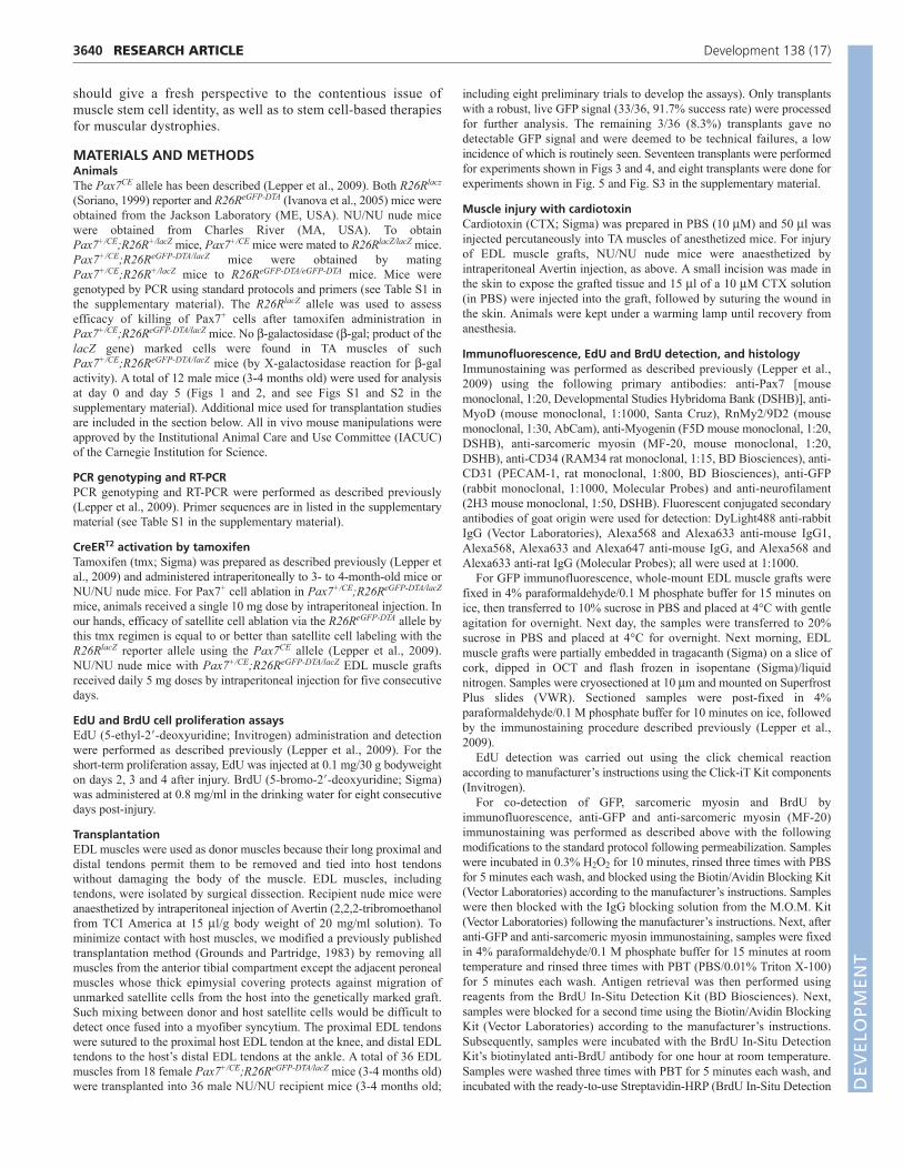
Immunofluorescence, EdU and BrdU detection, and histologyImmunostaining was performed as described previously (Lepper et al.,2009) using the following primary antibodies: anti-Pax7 [mousemonoclonal, 1:20, Developmental Studies Hybridoma Bank (DSHB)], anti-MyoD (mouse monoclonal, 1:1000, Santa Cruz), RnMy2/9D2 (mousemonoclonal, 1:30, AbCam), anti-Myogenin (F5D mouse monoclonal, 1:20,DSHB), anti-sarcomeric myosin (MF-20, mouse monoclonal, 1:20,DSHB), anti-CD34 (RAM34 rat monoclonal, 1:15, BD Biosciences), anti-CD31 (PECAM-1, rat monoclonal, 1:800, BD Biosciences), anti-GFP(rabbit monoclonal, 1:1000, Molecular Probes) and anti-neurofilament(2H3 mouse monoclonal, 1:50, DSHB). Fluorescent conjugated secondaryantibodies of goat origin were used for detection: DyLight488 anti-rabbitIgG (Vector Laboratories), Alexa568 and Alexa633 anti-mouse IgG1,Alexa568, Alexa633 and Alexa647 anti-mouse IgG, and Alexa568 andAlexa633 anti-rat IgG (Molecular Probes); all were used at 1:1000.
For GFP immunofluorescence, whole-mount EDL muscle grafts werefixed in 4% paraformaldehyde/0.1 M phosphate buffer for 15 minutes onice, then transferred to 10% sucrose in PBS and placed at 4°C with gentleagitation for overnight. Next day, the samples were transferred to 20%sucrose in PBS and placed at 4°C for overnight. Next morning, EDLmuscle grafts were partially embedded in tragacanth (Sigma) on a slice ofcork, dipped in OCT and flash frozen in isopentane (Sigma)/liquidnitrogen. Samples were cryosectioned at 10 m and mounted on SuperfrostPlus slides (VWR). Sectioned samples were post-fixed in 4%paraformaldehyde/0.1 M phosphate buffer for 10 minutes on ice, followedby the immunostaining procedure described previously (Lepper et al.,2009).
EdU detection was carried out using the click chemical reactionaccording to manufacturer’s instructions using the Click-iT Kit components(Invitrogen).
For co-detection of GFP, sarcomeric myosin and BrdU byimmunofluorescence, anti-GFP and anti-sarcomeric myosin (MF-20)immunostaining was performed as described above with the followingmodifications to the standard protocol following permeabilization. Sampleswere incubated in 0.3% H2O2 for 10 minutes, rinsed three times with PBSfor 5 minutes each wash, and blocked using the Biotin/Avidin Blocking Kit(Vector Laboratories) according to the manufacturer’s instructions. Sampleswere then blocked with the IgG blocking solution from the M.O.M. Kit(Vector Laboratories) following the manufacturer’s instructions. Next, afteranti-GFP and anti-sarcomeric myosin immunostaining, samples were fixedin 4% paraformaldehyde/0.1 M phosphate buffer for 15 minutes at roomtemperature and rinsed three times with PBT (PBS/0.01% Triton X-100)for 5 minutes each wash. Antigen retrieval was then performed usingreagents from the BrdU In-Situ Detection Kit (BD Biosciences). Next,samples were blocked for a second time using the Biotin/Avidin BlockingKit (Vector Laboratories) according to the manufacturer’s instructions.Subsequently, samples were incubated with the BrdU In-Situ DetectionKit’s biotinylated anti-BrdU antibody for one hour at room temperature.Samples were washed three times with PBT for 5 minutes each wash, andincubated with the ready-to-use Streptavidin-HRP (BrdU In-Situ Detection
RESEARCH ARTICLE Development 138 (17)
DEVELO
PMENT
Kit, BD Biosciences) for 30 minutes at room temperature. Horseradishperoxidase (HRP) detection was performed using the TSA Plus Cyanine 3System (Perkin Elmer). Samples were then washed three times in PBT for5 minutes each wash, incubated with DAPI in PBS for 10 minutes, washedtwice in PBT for 5 minutes each wash, and mounted in FluoromountGsolution (Southern Biotechnology) with a coverslip (VWR).
Histology was performed using Hematoxylin and Eosin according to themanufacturer’s instructions (Surgiopath).
a-BungarotoxinFor detection of motor end plates, Alexa594-conjugated a-bungarotoxin(Molecular Probes) was applied at a 1:400 dilution (in PBS) and incubatedfor 30 minutes at room temperature prior to incubation with DAPI.
Image acquisition, cell quantification and statisticsDigital images of random 0.154 mm2 fields were taken under an Axioscopeequipped with an AxioCam and labeled cells were counted by hand.Images were pseudocolored and superimposed using the Metamorphprogram. Excel was used for tabulation and statistical analysis. Numbersof fibers in grafts and regenerates subjected to different treatments werecompared using the two-tailed, Mann-Whitney U-test.
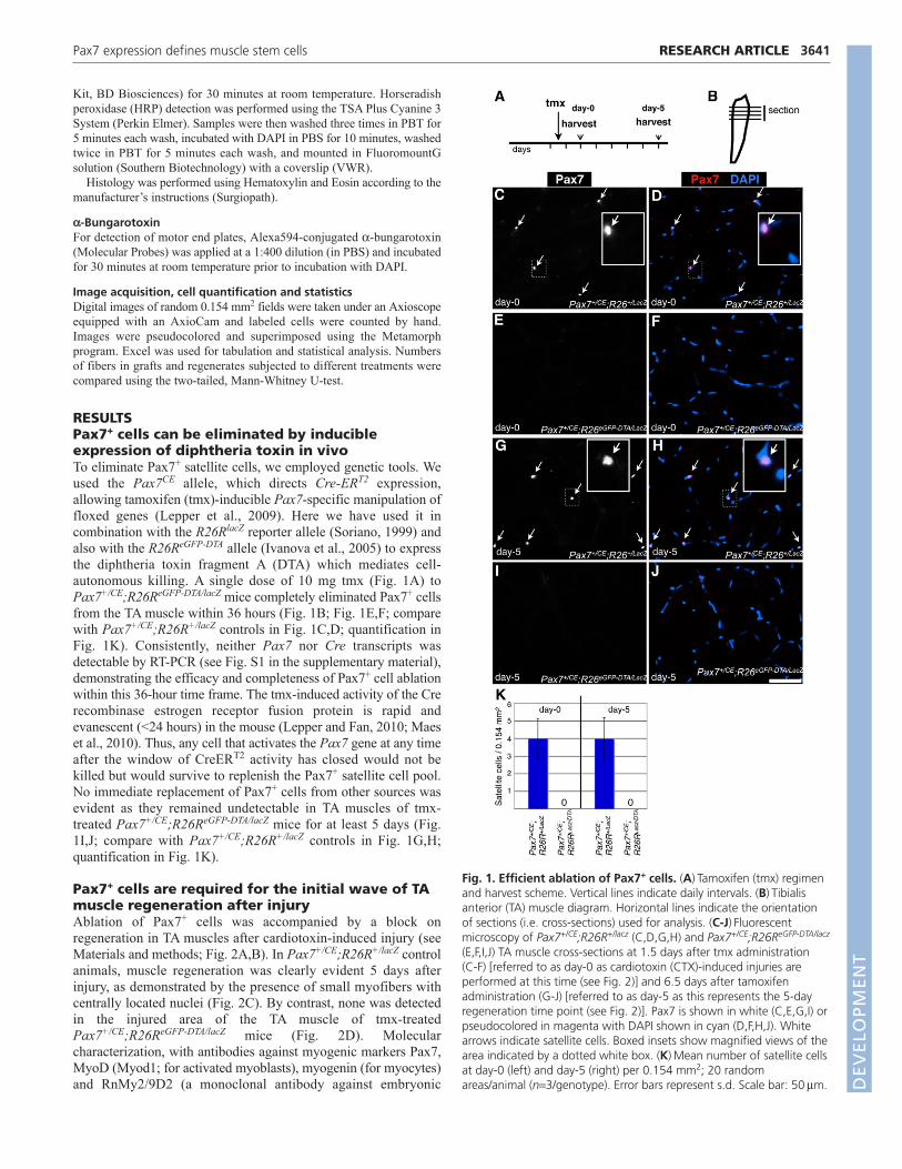
RESULTSPax7+ cells can be eliminated by inducibleexpression of diphtheria toxin in vivoTo eliminate Pax7+ satellite cells, we employed genetic tools. Weused the Pax7CE allele, which directs Cre-ERT2 expression,allowing tamoxifen (tmx)-inducible Pax7-specific manipulation offloxed genes (Lepper et al., 2009). Here we have used it incombination with the R26RlacZ reporter allele (Soriano, 1999) andalso with the R26ReGFP-DTA allele (Ivanova et al., 2005) to expressthe diphtheria toxin fragment A (DTA) which mediates cell-autonomous killing. A single dose of 10 mg tmx (Fig. 1A) toPax7+/CE;R26ReGFP-DTA/lacZ mice completely eliminated Pax7+ cellsfrom the TA muscle within 36 hours (Fig. 1B; Fig. 1E,F; comparewith Pax7+/CE;R26R+/lacZ controls in Fig. 1C,D; quantification inFig. 1K). Consistently, neither Pax7 nor Cre transcripts wasdetectable by RT-PCR (see Fig. S1 in the supplementary material),demonstrating the efficacy and completeness of Pax7+ cell ablationwithin this 36-hour time frame. The tmx-induced activity of the Crerecombinase estrogen receptor fusion protein is rapid andevanescent (<24 hours) in the mouse (Lepper and Fan, 2010; Maeset al., 2010). Thus, any cell that activates the Pax7 gene at any timeafter the window of CreERT2 activity has closed would not bekilled but would survive to replenish the Pax7+ satellite cell pool.No immediate replacement of Pax7+ cells from other sources wasevident as they remained undetectable in TA muscles of tmx-treated Pax7+/CE;R26ReGFP-DTA/lacZ mice for at least 5 days (Fig.1I,J; compare with Pax7+/CE;R26R+/lacZ controls in Fig. 1G,H;quantification in Fig. 1K).
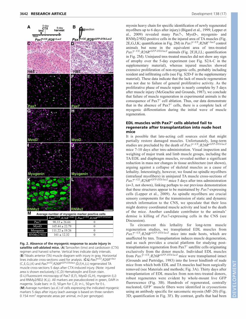
Pax7+ cells are required for the initial wave of TAmuscle regeneration after injuryAblation of Pax7+ cells was accompanied by a block onregeneration in TA muscles after cardiotoxin-induced injury (seeMaterials and methods; Fig. 2A,B). In Pax7+/CE;R26R+/lacZ controlanimals, muscle regeneration was clearly evident 5 days afterinjury, as demonstrated by the presence of small myofibers withcentrally located nuclei (Fig. 2C). By contrast, none was detectedin the injured area of the TA muscle of tmx-treatedPax7+/CE;R26ReGFP-DTA/lacZ mice (Fig. 2D). Molecularcharacterization, with antibodies against myogenic markers Pax7,MyoD (Myod1; for activated myoblasts), myogenin (for myocytes)and RnMy2/9D2 (a monoclonal antibody against embryonic
3641RESEARCH ARTICLEPax7 expression defines muscle stem cells
Fig. 1. Efficient ablation of Pax7+ cells. (A)Tamoxifen (tmx) regimenand harvest scheme. Vertical lines indicate daily intervals. (B)Tibialisanterior (TA) muscle diagram. Horizontal lines indicate the orientationof sections (i.e. cross-sections) used for analysis. (C-J)Fluorescentmicroscopy of Pax7+/CE;R26R+/lacz (C,D,G,H) and Pax7+/CE;R26ReGFP-DTA/lacz
(E,F,I,J) TA muscle cross-sections at 1.5 days after tmx administration (C-F) [referred to as day-0 as cardiotoxin (CTX)-induced injuries areperformed at this time (see Fig. 2)] and 6.5 days after tamoxifenadministration (G-J) [referred to as day-5 as this represents the 5-dayregeneration time point (see Fig. 2)]. Pax7 is shown in white (C,E,G,I) orpseudocolored in magenta with DAPI shown in cyan (D,F,H,J). Whitearrows indicate satellite cells. Boxed insets show magnified views of thearea indicated by a dotted white box. (K)Mean number of satellite cellsat day-0 (left) and day-5 (right) per 0.154 mm2; 20 randomareas/animal (n3/genotype). Error bars represent s.d. Scale bar: 50m. D
EVELO
PMENT
3642
myosin heavy chain for specific identification of newly regeneratedmyofibers up to 6 days after injury) (Bigard et al., 1999; Lepper etal., 2009) revealed many Pax7-, MyoD-, myogenin- andRNMy2/9D2-positive cells in the injured area of TA muscles (Fig.2E,G,J,K; quantification in Fig. 2M) in Pax7+/CE;R26R+/lacZ controlanimals but none in the equivalent area of tmx-treatedPax7+/CE;R26ReGFP-DTA/lacZ animals (Fig. 2F,H,J,L; quantificationin Fig. 2M). Uninjured tmx-treated muscles did not show any signof atrophy over the 5-day experiment (see Fig. S2A-C in thesupplementary material), whereas injured muscles showedextensive proliferation of non-myogenic cells, probably includingresident and infiltrating cells (see Fig. S2D-F in the supplementarymaterial). These data indicate that the lack of muscle regenerationwas not due to failure of general proliferative activity. As theproliferative phase of muscle repair is nearly complete by 5 daysafter muscle injury (McGeachie and Grounds, 1987), we concludethat failure of muscle regeneration in experimental animals is theconsequence of Pax7+ cell ablation. Thus, our data demonstrate that in the absence of Pax7+ cells, there is a complete lack ofmyogenic differentiation during the initial wave of muscleregeneration.
EDL muscles with Pax7+ cells ablated fail toregenerate after transplantation into nude hostmiceIt is possible that late-acting cell sources exist that mightpartially restore damaged muscles. Unfortunately, long-termstudies are precluded by the death of Pax7+/CE;R26ReGFP-DTA/lacZ
mice 7-10 days after tmx-administration. Visual inspection andweighing of major trunk and limb muscle groups, including theTA/EDL and diaphragm muscles, revealed neither a significantreduction in mass nor changes in tissue architecture (not shown),arguing against a collapse of skeletal muscles as a cause oflethality. Interestingly, however, we found no spindle myofibers(intrafusal myofibers) in uninjured TA muscle cross-sections ofPax7+/CE;R26ReGFP-DTA/lacZ mice 5 days after tmx-administration(n3, not shown), linking perhaps to our previous demonstrationthat these structures appear to be maintained by Pax7-expressingcells (Lepper et al., 2009). As spindle myofibers are crucialsensory components for the transmission of static and dynamicstretch information to the CNS, we speculate that their lossmight destroy coordinated muscle activity and lead to the deathof the mice. Another candidate contributor to the animals’demise is killing of Pax7-expressing cells in the CNS (seeDiscussion).
To circumvent this lethality for long-term muscle regeneration studies, we transplanted EDL muscles fromPax7+/CE;R26ReGFP-DTA/lacZ mice into nude hosts, which areunaffected by tmx. Transplantation induces muscle degeneration,and as such provides a crucial platform for studying post-transplantation regeneration from Pax7+ satellite cells originatingexclusively from the donor muscle. Individual EDL musclesfrom Pax7+/CE;R26ReGFP-DTA/lacZ mice were transplanted intact(Grounds and Partridge, 1983) into the lower hindlimb of nudemice from which both EDL and TA muscles had been surgicallyremoved (see Materials and methods; Fig. 3A). Thirty days aftertransplantation of EDL muscles from non-tmx-treated donors,robust regenerates were evident by whole-mount live GFPfluorescence (Fig. 3B). Hundreds of regenerated, centrallynucleated, GFP+ muscle fibers were identified in cryosectionsusing an antibody specific for sarcomeric myosin (MF-20) (Fig.3D; quantification in Fig. 3F). By contrast, grafts that had been
RESEARCH ARTICLE Development 138 (17)
Fig. 2. Absence of the myogenic response to acute injury insatellite cell-ablated mice. (A)Tamoxifen (tmx) and cardiotoxin (CTX)regimen and harvest scheme. Vertical lines indicate daily intervals.(B)Tibialis anterior (TA) muscle diagram with injury in gray. Horizontallines indicate cross-sections used for analysis. (C-L)Pax7+/CE;R26R+/lacz
(C,E,G,I,K) and Pax7+/CE;R26ReGFP-DTA/lacz (D,F,H,J,L) regenerated TAmuscle cross-sections 5 days after CTX-induced injury. (Note: injuredarea is shown exclusively.) (C,D) Hematoxylin and Eosin stain. (E-L)Fluorescent microscopy of Pax7 (E,F), MyoD (G,H), myogenin (I,J)and RNMy2/9D2 (K,L). All markers are pseudocolored in green, DAPI inmagenta. Scale bars: in D, 50m for C,D; in L, 50m for E-L.(M)Average numbers (±s.d.) of cells expressing the indicated myogenicmarkers 5 days after injury (each average is based on three random0.154 mm2 regenerate areas per animal, n3 per genotype). D
EVELO
PMENT
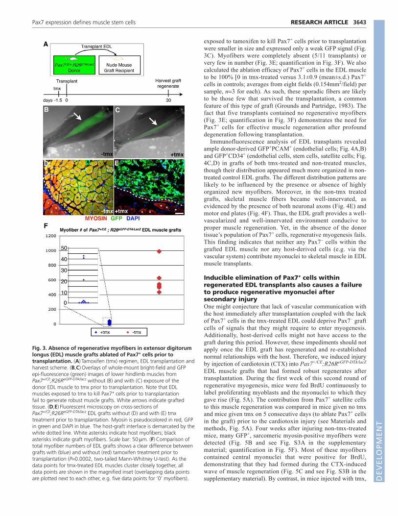
exposed to tamoxifen to kill Pax7+ cells prior to transplantationwere smaller in size and expressed only a weak GFP signal (Fig.3C). Myofibers were completely absent (5/11 transplants) orvery few in number (Fig. 3E; quantification in Fig. 3F). We alsocalculated the ablation efficacy of Pax7+ cells in the EDL muscleto be 100% [0 in tmx-treated versus 3.1±0.9 (mean±s.d.) Pax7+
cells in controls; averages from eight fields (0.154mm2/field) persample, n3 for each). As such, these sporadic fibers are likelyto be those few that survived the transplantation, a commonfeature of this type of graft (Grounds and Partridge, 1983). Thefact that five transplants contained no regenerative myofibers(Fig. 3E; quantification in Fig. 3F) demonstrates the need forPax7+ cells for effective muscle regeneration after profounddegeneration following transplantation.
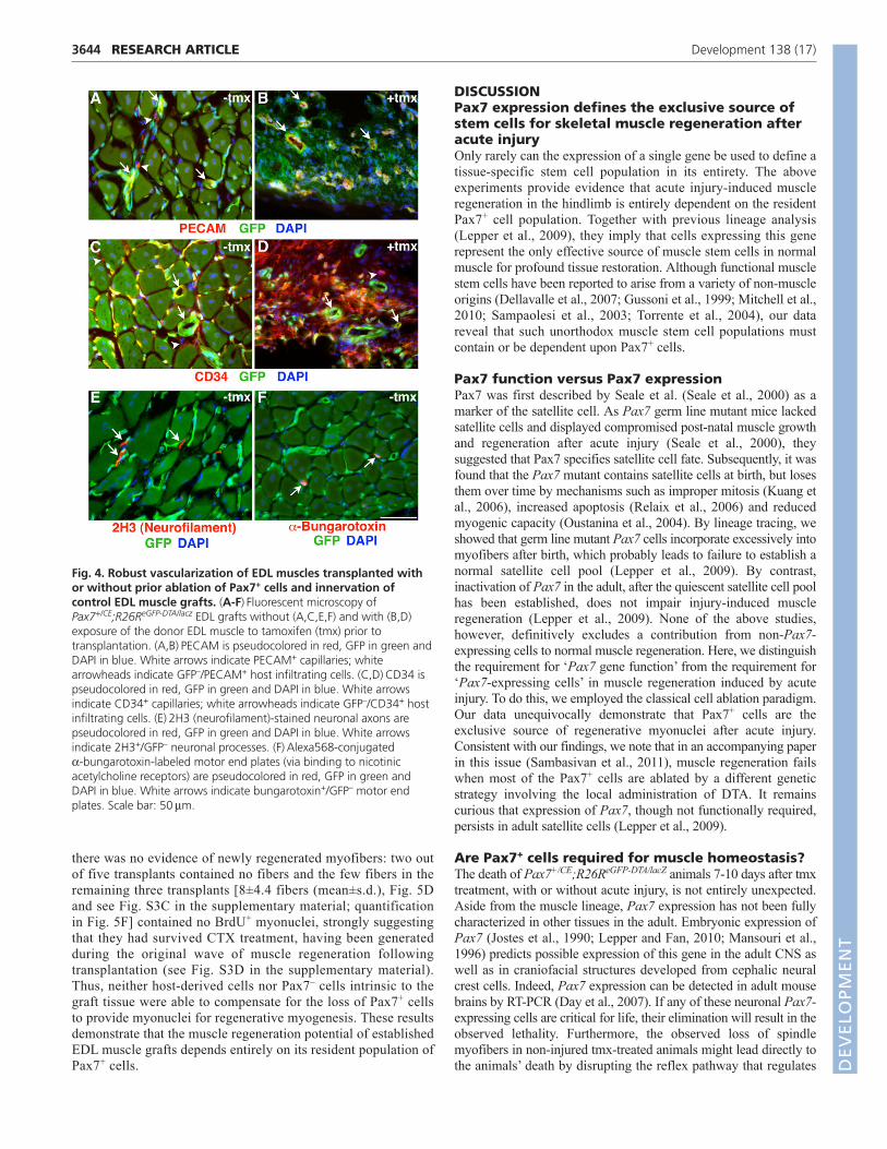
Immunofluorescence analysis of EDL transplants revealedample donor-derived GFP+PCAM+ (endothelial cells; Fig. 4A,B)and GFP+CD34+ (endothelial cells, stem cells, satellite cells; Fig.4C,D) in grafts of both tmx-treated and non-treated muscles,though their distribution appeared much more organized in non-treated control EDL grafts. The different distribution patterns arelikely to be influenced by the presence or absence of highlyorganized new myofibers. Moreover, in the non-tmx treatedgrafts, skeletal muscle fibers became well-innervated, asevidenced by the presence of both neuronal axons (Fig. 4E) andmotor end plates (Fig. 4F). Thus, the EDL graft provides a well-vascularized and well-innervated environment conducive toproper muscle regeneration. Yet, in the absence of the donortissue’s population of Pax7+ cells, regenerative myogenesis fails.This finding indicates that neither any Pax7– cells within thegrafted EDL muscle nor any host-derived cells (e.g. via thevascular system) contribute myonuclei to skeletal muscle in EDLmuscle transplants.
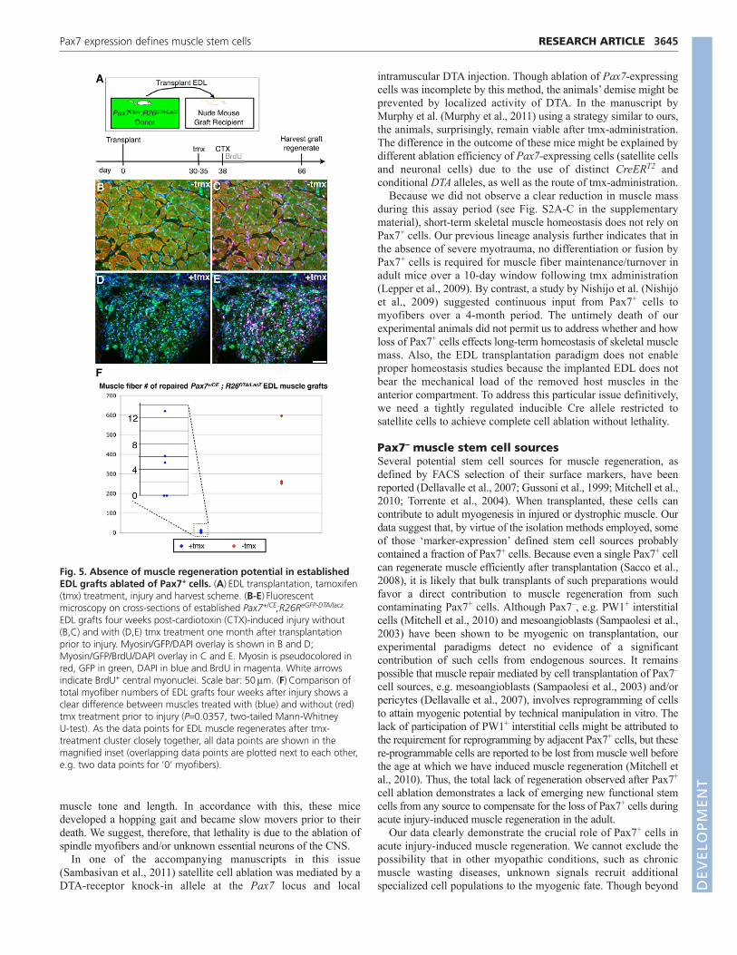
Inducible elimination of Pax7+ cells withinregenerated EDL transplants also causes a failureto produce regenerative myonuclei aftersecondary injuryOne might conjecture that lack of vascular communication withthe host immediately after transplantation coupled with the lackof Pax7+ cells in the tmx-treated EDL could deprive Pax7– graftcells of signals that they might require to enter myogenesis.Additionally, host-derived cells might not have access to thegraft during this period. However, these impediments should notapply once the EDL graft has regenerated and re-establishednormal relationships with the host. Therefore, we induced injuryby injection of cardiotoxin (CTX) into Pax7+/CE;R26ReGFP-DTA/lacZ
EDL muscle grafts that had formed robust regenerates aftertransplantation. During the first week of this second round ofregenerative myogenesis, mice were fed BrdU continuously tolabel proliferating myoblasts and the myonuclei to which theygave rise (Fig. 5A). The contribution from Pax7+ satellite cellsto this muscle regeneration was compared in mice given no tmxand mice given tmx on 5 consecutive days (to ablate Pax7+ cellsin the graft) prior to the cardiotoxin injury (see Materials andmethods, Fig. 5A). Four weeks after injuring non-tmx-treatedmice, many GFP+, sarcomeric myosin-positive myofibers weredetected (Fig. 5B and see Fig. S3A in the supplementarymaterial; quantification in Fig. 5F). Most of these myofiberscontained central myonuclei that were positive for BrdU,demonstrating that they had formed during the CTX-inducedwave of muscle regeneration (Fig. 5C and see Fig. S3B in thesupplementary material). By contrast, in mice injected with tmx,
3643RESEARCH ARTICLEPax7 expression defines muscle stem cells
Fig. 3. Absence of regenerative myofibers in extensor digitorumlongus (EDL) muscle grafts ablated of Pax7+ cells prior totransplantation. (A)Tamoxifen (tmx) regimen, EDL transplantation andharvest scheme. (B,C)Overlays of whole-mount bright-field and GFPepi-fluorescence (green) images of lower hindlimb muscles fromPax7+/CE;R26ReGFP-DTA/lacz without (B) and with (C) exposure of the donor EDL muscle to tmx prior to transplantation. Note that EDLmuscles exposed to tmx to kill Pax7+ cells prior to transplantation fail to generate robust muscle grafts. White arrows indicate graftedtissue. (D,E)Fluorescent microscopy on cross-sections of Pax7+/CE;R26ReGFP-DTA/lacz EDL grafts without (D) and with (E) tmxtreatment prior to transplantation. Myosin is pseudocolored in red, GFPin green and DAPI in blue. The host-graft interface is demarcated by thewhite dotted line. White asterisks indicate host myofibers; blackasterisks indicate graft myofibers. Scale bar: 50m. (F)Comparison oftotal myofiber numbers of EDL grafts shows a clear difference betweengrafts with (blue) and without (red) tamoxifen treatment prior totransplantation (P0.0002, two-tailed Mann-Whitney U-test). As thedata points for tmx-treated EDL muscles cluster closely together, alldata points are shown in the magnified inset (overlapping data pointsare plotted next to each other, e.g. five data points for ‘0’ myofibers). D
EVELO
PMENT
3644
there was no evidence of newly regenerated myofibers: two outof five transplants contained no fibers and the few fibers in theremaining three transplants [8±4.4 fibers (mean±s.d.), Fig. 5Dand see Fig. S3C in the supplementary material; quantificationin Fig. 5F] contained no BrdU+ myonuclei, strongly suggestingthat they had survived CTX treatment, having been generatedduring the original wave of muscle regeneration followingtransplantation (see Fig. S3D in the supplementary material).Thus, neither host-derived cells nor Pax7– cells intrinsic to thegraft tissue were able to compensate for the loss of Pax7+ cellsto provide myonuclei for regenerative myogenesis. These resultsdemonstrate that the muscle regeneration potential of establishedEDL muscle grafts depends entirely on its resident population ofPax7+ cells.
DISCUSSIONPax7 expression defines the exclusive source ofstem cells for skeletal muscle regeneration afteracute injuryOnly rarely can the expression of a single gene be used to define atissue-specific stem cell population in its entirety. The aboveexperiments provide evidence that acute injury-induced muscleregeneration in the hindlimb is entirely dependent on the residentPax7+ cell population. Together with previous lineage analysis(Lepper et al., 2009), they imply that cells expressing this generepresent the only effective source of muscle stem cells in normalmuscle for profound tissue restoration. Although functional musclestem cells have been reported to arise from a variety of non-muscleorigins (Dellavalle et al., 2007; Gussoni et al., 1999; Mitchell et al.,2010; Sampaolesi et al., 2003; Torrente et al., 2004), our datareveal that such unorthodox muscle stem cell populations mustcontain or be dependent upon Pax7+ cells.
Pax7 function versus Pax7 expressionPax7 was first described by Seale et al. (Seale et al., 2000) as amarker of the satellite cell. As Pax7 germ line mutant mice lackedsatellite cells and displayed compromised post-natal muscle growthand regeneration after acute injury (Seale et al., 2000), theysuggested that Pax7 specifies satellite cell fate. Subsequently, it wasfound that the Pax7 mutant contains satellite cells at birth, but losesthem over time by mechanisms such as improper mitosis (Kuang etal., 2006), increased apoptosis (Relaix et al., 2006) and reducedmyogenic capacity (Oustanina et al., 2004). By lineage tracing, weshowed that germ line mutant Pax7 cells incorporate excessively intomyofibers after birth, which probably leads to failure to establish anormal satellite cell pool (Lepper et al., 2009). By contrast,inactivation of Pax7 in the adult, after the quiescent satellite cell poolhas been established, does not impair injury-induced muscleregeneration (Lepper et al., 2009). None of the above studies,however, definitively excludes a contribution from non-Pax7-expressing cells to normal muscle regeneration. Here, we distinguishthe requirement for ‘Pax7 gene function’ from the requirement for‘Pax7-expressing cells’ in muscle regeneration induced by acuteinjury. To do this, we employed the classical cell ablation paradigm.Our data unequivocally demonstrate that Pax7+ cells are theexclusive source of regenerative myonuclei after acute injury.Consistent with our findings, we note that in an accompanying paperin this issue (Sambasivan et al., 2011), muscle regeneration failswhen most of the Pax7+ cells are ablated by a different geneticstrategy involving the local administration of DTA. It remainscurious that expression of Pax7, though not functionally required,persists in adult satellite cells (Lepper et al., 2009).
Are Pax7+ cells required for muscle homeostasis?The death of Pax7+/CE;R26ReGFP-DTA/lacZ animals 7-10 days after tmxtreatment, with or without acute injury, is not entirely unexpected.Aside from the muscle lineage, Pax7 expression has not been fullycharacterized in other tissues in the adult. Embryonic expression ofPax7 (Jostes et al., 1990; Lepper and Fan, 2010; Mansouri et al.,1996) predicts possible expression of this gene in the adult CNS aswell as in craniofacial structures developed from cephalic neuralcrest cells. Indeed, Pax7 expression can be detected in adult mousebrains by RT-PCR (Day et al., 2007). If any of these neuronal Pax7-expressing cells are critical for life, their elimination will result in theobserved lethality. Furthermore, the observed loss of spindlemyofibers in non-injured tmx-treated animals might lead directly tothe animals’ death by disrupting the reflex pathway that regulates
RESEARCH ARTICLE Development 138 (17)
Fig. 4. Robust vascularization of EDL muscles transplanted withor without prior ablation of Pax7+ cells and innervation ofcontrol EDL muscle grafts. (A-F)Fluorescent microscopy ofPax7+/CE;R26ReGFP-DTA/lacz EDL grafts without (A,C,E,F) and with (B,D)exposure of the donor EDL muscle to tamoxifen (tmx) prior totransplantation. (A,B)PECAM is pseudocolored in red, GFP in green andDAPI in blue. White arrows indicate PECAM+ capillaries; whitearrowheads indicate GFP–/PECAM+ host infiltrating cells. (C,D)CD34 ispseudocolored in red, GFP in green and DAPI in blue. White arrowsindicate CD34+ capillaries; white arrowheads indicate GFP–/CD34+ hostinfiltrating cells. (E)2H3 (neurofilament)-stained neuronal axons arepseudocolored in red, GFP in green and DAPI in blue. White arrowsindicate 2H3+/GFP– neuronal processes. (F)Alexa568-conjugated a-bungarotoxin-labeled motor end plates (via binding to nicotinicacetylcholine receptors) are pseudocolored in red, GFP in green andDAPI in blue. White arrows indicate bungarotoxin+/GFP– motor endplates. Scale bar: 50m.
DEVELO
PMENT
muscle tone and length. In accordance with this, these micedeveloped a hopping gait and became slow movers prior to theirdeath. We suggest, therefore, that lethality is due to the ablation ofspindle myofibers and/or unknown essential neurons of the CNS.
In one of the accompanying manuscripts in this issue(Sambasivan et al., 2011) satellite cell ablation was mediated by aDTA-receptor knock-in allele at the Pax7 locus and local
intramuscular DTA injection. Though ablation of Pax7-expressingcells was incomplete by this method, the animals’ demise might beprevented by localized activity of DTA. In the manuscript byMurphy et al. (Murphy et al., 2011) using a strategy similar to ours,the animals, surprisingly, remain viable after tmx-administration.The difference in the outcome of these mice might be explained bydifferent ablation efficiency of Pax7-expressing cells (satellite cellsand neuronal cells) due to the use of distinct CreERT2 andconditional DTA alleles, as well as the route of tmx-administration.
Because we did not observe a clear reduction in muscle massduring this assay period (see Fig. S2A-C in the supplementarymaterial), short-term skeletal muscle homeostasis does not rely onPax7+ cells. Our previous lineage analysis further indicates that inthe absence of severe myotrauma, no differentiation or fusion byPax7+ cells is required for muscle fiber maintenance/turnover inadult mice over a 10-day window following tmx administration(Lepper et al., 2009). By contrast, a study by Nishijo et al. (Nishijoet al., 2009) suggested continuous input from Pax7+ cells tomyofibers over a 4-month period. The untimely death of ourexperimental animals did not permit us to address whether and howloss of Pax7+ cells effects long-term homeostasis of skeletal musclemass. Also, the EDL transplantation paradigm does not enableproper homeostasis studies because the implanted EDL does notbear the mechanical load of the removed host muscles in theanterior compartment. To address this particular issue definitively,we need a tightly regulated inducible Cre allele restricted tosatellite cells to achieve complete cell ablation without lethality.
Pax7– muscle stem cell sourcesSeveral potential stem cell sources for muscle regeneration, asdefined by FACS selection of their surface markers, have beenreported (Dellavalle et al., 2007; Gussoni et al., 1999; Mitchell et al.,2010; Torrente et al., 2004). When transplanted, these cells cancontribute to adult myogenesis in injured or dystrophic muscle. Ourdata suggest that, by virtue of the isolation methods employed, someof those ‘marker-expression’ defined stem cell sources probablycontained a fraction of Pax7+ cells. Because even a single Pax7+ cellcan regenerate muscle efficiently after transplantation (Sacco et al.,2008), it is likely that bulk transplants of such preparations wouldfavor a direct contribution to muscle regeneration from suchcontaminating Pax7+ cells. Although Pax7–, e.g. PW1+ interstitialcells (Mitchell et al., 2010) and mesoangioblasts (Sampaolesi et al.,2003) have been shown to be myogenic on transplantation, ourexperimental paradigms detect no evidence of a significantcontribution of such cells from endogenous sources. It remainspossible that muscle repair mediated by cell transplantation of Pax7–
cell sources, e.g. mesoangioblasts (Sampaolesi et al., 2003) and/orpericytes (Dellavalle et al., 2007), involves reprogramming of cellsto attain myogenic potential by technical manipulation in vitro. Thelack of participation of PW1+ interstitial cells might be attributed tothe requirement for reprogramming by adjacent Pax7+ cells, but thesere-programmable cells are reported to be lost from muscle well beforethe age at which we have induced muscle regeneration (Mitchell etal., 2010). Thus, the total lack of regeneration observed after Pax7+
cell ablation demonstrates a lack of emerging new functional stemcells from any source to compensate for the loss of Pax7+ cells duringacute injury-induced muscle regeneration in the adult.
Our data clearly demonstrate the crucial role of Pax7+ cells inacute injury-induced muscle regeneration. We cannot exclude thepossibility that in other myopathic conditions, such as chronicmuscle wasting diseases, unknown signals recruit additionalspecialized cell populations to the myogenic fate. Though beyond
3645RESEARCH ARTICLEPax7 expression defines muscle stem cells
Fig. 5. Absence of muscle regeneration potential in establishedEDL grafts ablated of Pax7+ cells. (A)EDL transplantation, tamoxifen(tmx) treatment, injury and harvest scheme. (B-E)Fluorescentmicroscopy on cross-sections of established Pax7+/CE;R26ReGFP-DTA/lacz
EDL grafts four weeks post-cardiotoxin (CTX)-induced injury without(B,C) and with (D,E) tmx treatment one month after transplantationprior to injury. Myosin/GFP/DAPI overlay is shown in B and D;Myosin/GFP/BrdU/DAPI overlay in C and E. Myosin is pseudocolored inred, GFP in green, DAPI in blue and BrdU in magenta. White arrowsindicate BrdU+ central myonuclei. Scale bar: 50m. (F)Comparison oftotal myofiber numbers of EDL grafts four weeks after injury shows aclear difference between muscles treated with (blue) and without (red)tmx treatment prior to injury (P0.0357, two-tailed Mann-Whitney U-test). As the data points for EDL muscle regenerates after tmx-treatment cluster closely together, all data points are shown in themagnified inset (overlapping data points are plotted next to each other,e.g. two data points for ‘0’ myofibers).
DEVELO
PMENT

3646
the scope of this work, future pre-translational studies to test therelative contribution of Pax7+ cells in a murine Duchenne musculardystrophy model (i.e. the Mdx mouse) should advance ourunderstanding of the differences, if any, between basic normalbiology versus pathological biology of muscle stem cells.
Therapeutic use of satellite cell versus other stemcell sourcesWe emphasize that our data do not diminish the potential clinicaluse of alternative stem cells for therapy of muscular dystrophies,which theoretically should benefit from healthy cells regardless oforigin. In fact, the effectiveness of transplanted mesoangioblasts inameliorating muscle dystrophy in a canine model, has raisedconsiderable hope in human patients (Sampaolesi et al., 2006).Conversely, transplantation of satellite cells after culture (i.e.myoblasts) to human patients has proven ineffective, presumablyowing to poor graft cell survival (Partridge, 1991). The problem ofharnessing the regenerative capacity of such cells appears to betechnical in nature and not insuperable. Our data, therefore, supportthe view that more attention should be directed at improving thesurvival and administration of the professional muscle stem cells,i.e. the Pax7+ satellite cells, as a route to this therapeutic dream.
AcknowledgementsWe thank E. Dikovskaia and E. Siple for technical assistance and J. J. Weyers, Y.Zheng and A. Spradling for comments on the manuscript. We also thank S.Tajbakhsh and A. Galy for sharing their results and coordinating manuscriptsubmission. The Carnegie Institution and NIH (AR060042) provided funding forthis work. T.A.P. is supported by DOD/USAMRAA grants W81XWH-09-1-0599and W81XWH-09-1-021 and by The Foundation to Eradicate Duchenne(FEDS). Deposited in PMC for release after 12 months.
Author contributionsC.L., T.A.P. and C.-M.F. designed and C.L. and T.A.P. conducted the research.All three authors contributed to manuscript writing.
Competing interests statementThe authors declare no competing financial interests.
Supplementary materialSupplementary material for this article is available athttp://dev.biologists.org/lookup/suppl/doi:10.1242/dev.067595/-/DC1
ReferencesBigard, A. X., Janmot, C., Sanchez, H., Serrurier, B., Pollet, S. and d’Albis, A.
(1999). Changes in myosin heavy chain profile of mature regenerated musclewith endurance training in rat. Acta Physiol. Scand. 165, 185-192.
Bischoff, R. (1975). Regeneration of single skeletal muscle fibers in vitro. Anat.Rec. 182, 215-235.
Cerletti, M., Jurga, S., Witczak, C. A., Hirshman, M. F., Shadrach, J. L.,Goodyear, L. J. and Wagers, A. J. (2008). Highly efficient, functionalengraftment of skeletal muscle stem cells in dystrophic muscles. Cell 134, 37-47.
Collins, C. A., Olsen, I., Zammit, P. S., Heslop, L., Petrie, A., Partridge, T. A.and Morgan, J. E. (2005). Stem cell function, self-renewal, and behavioralheterogeneity of cells from the adult muscle satellite cell niche. Cell 122, 289-301.
Day, K., Shefer, G., Richardson, J. B., Enikolopov, G. and Yablonka-Reuveni,Z. (2007). Nestin-GFP reporter expression defines the quiescent state of skeletalmuscle satellite cells. Dev. Biol. 304, 246-259.
Dellavalle, A., Sampaolesi, M., Tonlorenzi, R., Tagliafico, E., Sacchetti, B.,Perani, L., Innocenzi, A., Galvez, B. G., Messina, G., Morosetti, R. et al.(2007). Pericytes of human skeletal muscle are myogenic precursors distinct fromsatellite cells. Nat. Cell Biol. 9, 255-267.
Ferrari, G., Cusella-De Angelis, G., Coletta, M., Paolucci, E., Stornaiuolo, A.,Cossu, G. and Mavilio, F. (1998). Muscle regeneration by bone marrow-derived myogenic progenitors. Science 279, 1528-1530.
Grounds, M. D. and Partridge, T. A. (1983). Isoenzyme studies of whole musclegrafts and movement of muscle precursor cells. Cell Tissue Res. 230, 677-688.
Gussoni, E., Soneoka, Y., Strickland, C. D., Buzney, E. A., Khan, M. K., Flint,A. F., Kunkel, L. M. and Mulligan, R. C. (1999). Dystrophin expression in themdx mouse restored by stem cell transplantation. Nature 401, 390-394.
Ivanova, A., Signore, M., Caro, N., Greene, N. D., Copp, A. J. and Martinez-Barbera, J. P. (2005). In vivo genetic ablation by Cre-mediated expression ofdiphtheria toxin fragment A. Genesis 43, 129-135.
Jostes, B., Walther, C. and Gruss, P. (1990). The murine paired box gene, Pax7, isexpressed specifically during the development of the nervous and muscularsystem. Mech. Dev. 33, 27-37.
Konigsberg, U. R., Lipton, B. H. and Konigsberg, I. R. (1975). The regenerativeresponse of single mature muscle fibers isolated in vitro. Dev. Biol. 45, 260-275.
Kuang, S., Charge, S. B., Seale, P., Huh, M. and Rudnicki, M. A. (2006).Distinct roles for Pax7 and Pax3 in adult regenerative myogenesis. J. Cell Biol.172, 103-113.
Kuang, S., Kuroda, K., Le Grand, F. and Rudnicki, M. A. (2007). Asymmetricself-renewal and commitment of satellite stem cells in muscle. Cell 129, 999-1010.
Lepper, C. and Fan, C. M. (2010). Inducible lineage tracing of Pax7-descendantcells reveals embryonic origin of adult satellite cells. Genesis 48, 424-436.
Lepper, C., Conway, S. J. and Fan, C. M. (2009). Adult satellite cells andembryonic muscle progenitors have distinct genetic requirements. Nature 460,627-631.
Lipton, B. H. and Schultz, E. (1979). Developmental fate of skeletal musclesatellite cells. Science 205, 1292-1294.
Maes, C., Kobayashi, T., Selig, M. K., Torrekens, S., Roth, S. I., Mackem, S.,Carmeliet, G. and Kronenberg, H. M. (2010). Osteoblast precursors, but notmature osteoblasts, move into developing and fractured bones along withinvading blood vessels. Dev. Cell 19, 329-344.
Mansouri, A., Stoykova, A., Torres, M. and Gruss, P. (1996). Dysgenesis ofcephalic neural crest derivatives in Pax7–/– mutant mice. Development 122,831-838.
Mauro, A. (1961). Satellite cell of skeletal muscle fibers. J. Biophys. Biochem.Cytol. 9, 493-495.
McGeachie, J. K. and Grounds, M. D. (1987). Initiation and duration of muscleprecursor replication after mild and severe injury to skeletal muscle of mice. Anautoradiographic study. Cell Tissue Res. 248, 125-130.
Mitchell, K. J., Pannerec, A., Cadot, B., Parlakian, A., Besson, V., Gomes, E.R., Marazzi, G. and Sassoon, D. A. (2010). Identification and characterizationof a non-satellite cell muscle resident progenitor during postnatal development.Nat. Cell Biol. 12, 257-266.
Murphy, M. M., Lawson, J. A., Mathew, S. J., Hutcheson, D. A. and Kardon,G. (2011). Satellite cells, connective tissue fibroblasts and their interactions arecrucial for muscle regeneration. Development 138, 3625-3637.
Nishijo, K., Hosoyama, T., Bjornson, C. R., Schaffer, B. S., Prajapati, S. I.,Bahadur, A. N., Hansen, M. S., Blandford, M. C., McCleish, A. T., Rubin, B.P. et al. (2009). Biomarker system for studying muscle, stem cells, and cancer invivo. FASEB J. 23, 2681-2690.
Oustanina, S., Hause, G. and Braun, T. (2004). Pax7 directs postnatal renewaland propagation of myogenic satellite cells but not their specification. EMBO J.23, 3430-3439.
Partridge, T. A. (1991). Invited review: myoblast transfer: a possible therapy forinherited myopathies? Muscle Nerve 14, 197-212.
Peault, B., Rudnicki, M., Torrente, Y., Cossu, G., Tremblay, J. P., Partridge, T.,Gussoni, E., Kunkel, L. M. and Huard, J. (2007). Stem and progenitor cells inskeletal muscle development, maintenance, and therapy. Mol. Ther. 15, 867-877.
Relaix, F., Montarras, D., Zaffran, S., Gayraud-Morel, B., Rocancourt, D.,Tajbakhsh, S., Mansouri, A., Cumano, A. and Buckingham, M. (2006). Pax3and Pax7 have distinct and overlapping functions in adult muscle progenitorcells. J. Cell Biol. 172, 91-102.
Sacco, A., Doyonnas, R., Kraft, P., Vitorovic, S. and Blau, H. M. (2008). Self-renewal and expansion of single transplanted muscle stem cells. Nature 456,502-506.
Sambasivan, R., Yao, R., Kissenpfennig, A., Van Wittenberghe, L., Paldi, A.,Gayraud-Morel, B., Guenou, H., Malissen, B., Tajbakhsh, S. and Galy, A.(2011). Pax7-expressing satellite cells are indispensable for adult skeletal muscleregeneration. Development 138, 3647-3656.
Sampaolesi, M., Torrente, Y., Innocenzi, A., Tonlorenzi, R., D’Antona, G.,Pellegrino, M. A., Barresi, R., Bresolin, N., De Angelis, M. G., Campbell, K.P. et al. (2003). Cell therapy of alpha-sarcoglycan null dystrophic mice throughintra-arterial delivery of mesoangioblasts. Science 301, 487-492.
Sampaolesi, M., Blot, S., D’Antona, G., Granger, N., Tonlorenzi, R.,Innocenzi, A., Mognol, P., Thibaud, J. L., Galvez, B. G., Barthelemy, I. et al.(2006). Mesoangioblast stem cells ameliorate muscle function in dystrophicdogs. Nature 444, 574-579.
Seale, P., Sabourin, L. A., Girgis-Gabardo, A., Mansouri, A., Gruss, P. andRudnicki, M. A. (2000). Pax7 is required for the specification of myogenicsatellite cells. Cell 102, 777-786.
Snow, M. H. (1977). Myogenic cell formation in regenerating rat skeletal muscleinjured by mincing. I. A fine structural study. Anat. Rec. 188, 181-199.
Soriano, P. (1999). Generalized lacZ expression with the ROSA26 Cre reporterstrain. Nat. Genet. 21, 70-71.
Torrente, Y., Belicchi, M., Sampaolesi, M., Pisati, F., Meregalli, M., D’Antona,G., Tonlorenzi, R., Porretti, L., Gavina, M., Mamchaoui, K. et al. (2004).Human circulating AC133(+) stem cells restore dystrophin expression andameliorate function in dystrophic skeletal muscle. J. Clin. Invest. 114, 182-195.
RESEARCH ARTICLE Development 138 (17)
DEVELO
PMENT