An 8-year prospective clinical investigation on the ...
25
A 8-year prospective clinical investigation on the survival rate of feldspathic veneers: Influence of occlusal splint in patients with bruxism Vincente Faus-Matoses a , Esther Ruiz-Bell b , Ignacio Faus-Matoses c , Mutlu Özcan d , Sauro Salvatore e* , Vincente J. Faus-Llácer f a DDS, MSc, PhD. Co-director of the Master in Restorative Dentistry and Endodontics, Department of Stomatology, Faculty of Medicine and Dentistry, University of Valencia, Spain. b DDS, Master of Restorative Dentistry and Endodontics, Department of Stomatology, Faculty of Medicine and Dentistry, University of Valencia, Spain. c DDS, MSc, PhD. Professor of the Master in Orthodontics, Department of Stomatology, Faculty of Medicine and Dentistry, University of Valencia, Spain. d DDS, MSc, PhD. University of Zurich, Dental Materials Unit, Center for Dental and Oral Medicine, Clinic for Fixed and Removable Prosthodontics and Dental Materials Science, Zurich, Switzerland. e BSc, PhD. Dental Biomaterials, Preventive and Minimally Invasive Dentistry, Department of Dentistry, School of Health Sciences, CEU-Cardenal Herrera University, Valencia, Spain. f MD, DDS, MSc, PhD. Director of the Master in Restorative Dentistry and Endodontics, Department of Stomatology, Faculty of Medicine and Dentistry, University of Valencia, Spain. Short title: Survival rate of feldspathic veneers and influence of occlusal splint in patients with bruxism *Corresponding author: Prof. Dr. Salvatore Sauro. Dental Biomaterials, Preventive and Minimally Invasive Dentistry, Department of Dentistry, School of Health Sciences, CEU-Cardenal Herrera University, Valencia, Spain. C/Del Pozo s/n, Alfara del Patriarca, 46115 - Valencia, Spain. Tel Office: (+34) 96 136 90 00; Ext. 64304. [email protected]
Transcript of An 8-year prospective clinical investigation on the ...

A 8-year prospective clinical investigation on the survival rate of feldspathic veneers: Influence
of occlusal splint in patients with bruxism
Vincente Faus-Matosesa, Esther Ruiz-Bellb, Ignacio Faus-Matosesc, Mutlu Özcand, Sauro
Salvatoree*, Vincente J. Faus-Llácerf
a DDS, MSc, PhD. Co-director of the Master in Restorative Dentistry and Endodontics, Department of
Stomatology, Faculty of Medicine and Dentistry, University of Valencia, Spain.
b DDS, Master of Restorative Dentistry and Endodontics, Department of Stomatology, Faculty of
Medicine and Dentistry, University of Valencia, Spain.
c DDS, MSc, PhD. Professor of the Master in Orthodontics, Department of Stomatology, Faculty of
Medicine and Dentistry, University of Valencia, Spain.
d DDS, MSc, PhD. University of Zurich, Dental Materials Unit, Center for Dental and Oral Medicine, Clinic
for Fixed and Removable Prosthodontics and Dental Materials Science, Zurich, Switzerland.
e BSc, PhD. Dental Biomaterials, Preventive and Minimally Invasive Dentistry, Department of Dentistry,
School of Health Sciences, CEU-Cardenal Herrera University, Valencia, Spain.
fMD, DDS, MSc, PhD. Director of the Master in Restorative Dentistry and Endodontics, Department of
Stomatology, Faculty of Medicine and Dentistry, University of Valencia, Spain.
Short title: Survival rate of feldspathic veneers and influence of occlusal splint in patients with bruxism
*Corresponding author: Prof. Dr. Salvatore Sauro. Dental Biomaterials, Preventive and Minimally Invasive
Dentistry, Department of Dentistry, School of Health Sciences, CEU-Cardenal Herrera University, Valencia,
Spain. C/Del Pozo s/n, Alfara del Patriarca, 46115 - Valencia, Spain. Tel Office: (+34) 96 136 90 00; Ext. 64304.

1
ABSTRACT
Objectives: The aim of this study was to perform a 8-year prospective clinical investigation on the survival rate
of feldspathic ceramic veneers, as well as analyse the influence of the occlusal splint in patients with
parafunctional bruxism.
Methods: Three hundred and sixty-four veneers fabricated using conventional feldspathic ceramic were
provided in 64 patients. The patient sample included 40 individuals with bruxism. During the follow-up period,
the effect of wearing the occlusal splint on the incidence of failure (fracture and/or debonding) in patients with
bruxism was also assessed. The survival rate of veneers was determined using the Kaplan-Meier estimator.
Statistical significance was set at p<0.05 with a confidence interval of 95%.
Results: The occurrence of fracture for the feldspathic veneers tested in this study was 7.7%, while only 1.9%
of the total number of veneers debonded. The overall survival rate was 93.7% after 3 years, 91% after 5 years,
and 87.1% after 8 years. Patients with bruxism using an occlusal splint showed a survival rate of 89.1% after
7 years, while the survival rate in patients with bruxism using no occlusal splint was 63.9% (p<0.05).
Conclusion: This study confirmed that feldspathic veneers may represent a suitable clinical approach for
indirect aesthetic restorations. Such a treatment may be an option also for those patients affected by bruxism,
as long as they regularly wear an occlusal splint. However, patients with bruxism using no occlusal splint may
still present a potential higher risk of failure and/or debonding.
Keywords: Bruxism; Ceramics; Feldspathic ceramic; Fracture; Occlusal Splint; Surface treatment.

2
1. INTRODUCTION
Ceramic laminate veneers represent one of the main choices to perform an highly aesthetic restoration of
buccal and proximal dental surfaces.1 indeed, due to excellent optical and biocompatibility properties,
feldspathic ceramics represent a suitable material for the fabrication of veneers to restore anterior teeth.2-4
Certainly, this type of veneers may offer an exceptional conservative treatment option to solve cases of
discoloration, fractures, incisal wear, modifications of dental size and shape, as well as for treatment of dental
misalignment.6-8 Moreover, in those cases where palatal/lingual wear and/or erosion is encountered, such
veneers can also represent a suitable option to obtain predictable outcomes in terms of aesthetic and
functional properties.9,10
Feldspathic dental ceramics can be successfully etched using hydrofluoric acid (HF) in order to create
micromechanical retentions, which will facilitate the bonding performance of luting and self-adhesive
cements.7,11,12
Retrospective studies performed to evaluate the clinical performance of indirect veneers showed that the
results may vary significantly due to differences in study design, operators, and inclusion criteria.1,13,14 Indeed,
the failure rate for feldspathic veneers after 10 years may range between 5%15 and 47%,14 while after 5 years
the range is between 2%15 and 42%16. Fractures and debonding are the most common reasons for failure in
such a scenario.17,18 Moreover, a further interesting study showed that only 11% of feldspathic veneers failed
due to fractures during a 10-year follow-up period.19
The bonding interface between dental ceramics, cement and dental substrates (e.g. enamel and dentine) is
a further important factor that may affect the clinical success of dental veneers.8 Indeed, it has been advocated
that excessive dental preparations, which causes exposure of dentine, may increase the risk of microleakage,
interface degradation, and therefore consequent debonding of ceramic veneers.13 Conversely, due to a much

3
more reliable bonding performance, veneers applied in enamel may have a greater resistance to fracture and
debonding compared to those bonded to dentine.17
Bruxism is a parafunctional habit in which individuals clench and/or grind the teeth during the day and/or
night time.2,5 Several authors have argued that bruxism may represent a contraindication for placement of
ceramic veneers, as such a clinical situation may increases the risk for failure.2,4,7,15 Generally, an occlusal
splint is desirable for such patients undergoing prosthodontic treatments, especially in those cases where
specific parafunctional habits such as bruxism have been diagnosed.5 Certainly, a high failure rate might be
reduced if the parafunctional activity is appropriately managed2,4 through the using an occlusal splints.5
However, there is still little evidence about such a clinical situation, hence the present study was proposed.
The aim of this study was to perform a 8-year prospective clinical investigation on the survival rate of veneers
created using feldspathic ceramic, as well as evaluate the influence of wearing occlusal splint in patients with
bruxism.
The first hypothesis of this study was that the use of feldspathic veneers would represent an appropriate
approach for indirect anterior and posterior (first and second premolar) aesthetic restorations. The second
hypothesis was that such a treatment for patients with bruxism would have comparable longevity to treatments
performed in patients with no bruxism, as long as they regularly wear an occlusal splint.
2. Materials and methods
2.1 Study design and pre-operative procedures
The study design was performed under approval of the institutional ethical committee (Registration Nº
H1514909920703). Sixty-four patients were included in the study and treated with a total of 364 conventional
feldspathic ceramic veneers. The veneers were placed between January 2009 and December 2014 by two

4
fully-trained operators in the field of restorative and aesthetic dentistry, and with more than 20 years of clinical
experience, who always followed the same standardised operative protocol.
Clinical and radiographic exploration was performed in each patient, along with intraoral and extraoral
photographs. Moreover, a preliminary baseline impression was taken from each patient using alginate and a
study model was casted using dental stone. Subsequently, a diagnostic wax-up and mock-up was performed
in order to assess in advance the aesthetic, phonetic, and functional parameters, as well as to plan minimally
invasive dental preparation in each tooth.
Considering the results of Manfredini et al (2013) 5, who showed that 63% of patients exhibiting bruxism would
appear to be higher than what it may be expected in a general population, we have included in the group of
patients with night bruxism all those patients presenting any of the following signs: tired/tight jaw muscles,
locked jaw that cannot open or close completely, pain of jaw, neck or headache, lesions due to chewing on
the cheeks’ mucosa and patients who reported teeth grinding during sleep along with mandibula pain in the
morning.
2.2 Inclusion and exclusion criteria
All patients selected for this study presented no sign of periodontal disease and acceptable oral hygiene; they
were assessed through the periodontal disease index of Ramfjord (PDI) and the simplified oral hygiene index
(OHI). Conversely, patients with gingivitis or periodontitis were treated beforehand and only included in this
study after they were able to show no sign of gingival inflammation, along with and acceptable oral hygiene.
Those patients with bruxism, who presented on incisor margins or buccal cusps exposed dentine and/or sign
of fracture/chipping, were excluded from this study, because it was not possible to perform the “window”
preparation for veneers (see chapter 2.3).
Inclusion criteria: anterior and posterior teeth (up to the second premolar), without extensive loss of dental
enamel, with a degree of horizontal mobility of ≤1 mm, vital pulp or stable and well-performed endodontic

5
treatment, resorption less than one third of the length of the root and correct occlusal relation. Any patients
unwilling to comply with the follow-up schedule were excluded from the study. All patients included in this
study were recalled every six months for the first year and thereafter annually (up to 8 years) for a session of
oral hygiene, where it was also evaluated the compliance of the patience via a questionnaire and through a
photographic assessment to evaluate new or complication of previous signs and symptoms of bruxism.
2.3 Dental preparation
As stated before, a diagnostic wax-up and mock-up was performed in order to assess in advance the aesthetic,
phonetic, and functional parameters, as well as to plan minimally invasive dental preparation. Dental
preparation was carried out through a conservative “window” preparation 6 using Calibrated burs (868B, 314
and 020; Komet Dental, Besigheim, Germany) and tapered, fine-grain, round point, diamond burs (881, 314,
010 and 016; Komet Dental). The window preparation is considered a conservative approach where the
veneer is carried out without involving the incisal edge. Such a specific preparation allows the clinician to
obtain an acceptable ceramic thickness of 0.4 to 0.7 mm, which can decrease the risk of experiencing
porcelain fracture and wear of opposing teeth, and will not interfere with incisal guidance or occlusion in cases
of veneers in premolars. 6 Moreover, it has been shown that it may yield to lower level of microleakage
compared to other common methods to prepare teeth for veneers.6, 8 When necessary, contact points were
broken with metal strips, and then recreated with the veneers (anterior teeth). Margins were prepared
juxtagingivally having a curved chamfer finish line.
After dental preparation, high precision impressions were taken using a silicon impression material (Aquasil
Ultra XLV, Dentsply Sirona, Konstanz, Germany). Moreover, the dental shade of the treated teeth colour was
obtained using the Vitapan 3D-Master colour guide (Vita Zahnfabrick, Bad Säckingen, Germany). Temporary
restorations were performed with acrylic resin (Integrity, Dentsply Sirona). For the adhesion of temporary

6
restorations, the buccal surface of the treated teeth were acid-etched using a 36% phosphoric acid gel
(Detrey® Conditioner 36, Dentsply Sirona) only in the middle third. This was subsequently rinsed off and an
adhesive system was applied for 3s (Prime & Bond NT, Dentsply Sirona) and immediatelly light-cured for 10
s using a LED unit with an irradiance of 1200 Mw/cm2 (SmartLite Focus Pen Style, Dentsply Sirona). Finally,
the provisional veneers were applied using a flowable composite (SDR, Dentsply Sirona) and light cured for
20 s. The definitive feldspathic porcelain veneers (Noritake Kisai Co, Japan) were then fabricated by a dental
technician using standardised operative protocols.
2.4 Veneers application: adhesive operative procedures
After removing the temporary restorations, the dental surface was polished using a silicon rubber polishing
bur (Dentsply Sirona) mounted on a slow handpiece working under continuous irrigation. The veneers were
etched using 9.6% of hydrofluoric acid for 2 minutes (Ultradent, Ultradent Products. Inc.) and subsequently
copiously rinsed with water and dried. A layer of silane coupling agent (Calibra, Dentsply Sirona) was applied
and air-dried, followed by application of the adhesive (Prime & Bond NT, Dentsply Sirona). The enamel
surface of each tooth was etched again with a 36% orthophosphoric acid gel for 20 seconds. An adhesive
system was applied as per manufacturer’s instructions (Prime & BondNT, Dentsply Sirona), but without
performing any light-curing procedure.
The resin cement (Calibra, Dentsply Sirona) was applied onto the inner surfaces of the veneers and the
enamel and the veneers were applied and light-cured for 3 seconds (SmartLite Focus Pen Style, Dentsply
Sirona) in order to be able to easily eliminate the excess of the cement at the outer interface between veneers
and the tooth. Subsequently, both the palatine/lingual and buccal surfaces were light-cured for 40 seconds
(SmartLite Focus Pen Style, Dentsply Sirona). Finally, glycerine gel was applied to the edges of each veneer
to avoid the formation of an oxygen inhibition layer during a final light-curing step of 20 s. The occlusion and

7
possible presence of pre-contacts were checked with articulating paper (AccuFilm II; Parkell Inc., Farmingdale,
NY, USA). The edges of all veneers were then polished with silicone discs (Enhance PoGo, Dentsply Sirona)
and interproximal polishing strips (Soft-Lex Finishing Strips, 3M ESPE, USA).
Finally, an impression was taken using alginate and dental stone model was casted. This model was used for
the execution of the occlusal splint with a 2-mm-thick self-curing acrylic resin. The splint was finally adjusted
in the mouth by using an occlusal paper and finally polished using a slurry of pumice. All materials used during
the construction of the occlusal splint were handled according to the manufacturer's instructions. The patients
were instructed to wear the occlusal splint overnight and every day.
2.5 Clinical evaluation
Post-treatment follow-up was scheduled at 6 and 12 months after treatment, and thereafter annually for up to
8 years. All data of the exams was entered by a single operator. The follow-up sessions included clinical
examination, dental prophylaxis, and the management of any complications such as fracture or debonding.
We also evaluated the compliance of the patience via a questionnaire and through photographic report to
evaluate new or complication in previous signs and symptoms of bruxism, as well as the state of the occlusal
splint. Moreover, patients reported information about the use of the occlusal splint and on frequency of its use,
as well as the state of the occlusal splint. In those cases where the splint was broken or damaged, a new one
was provided to the patient.
In the clinical examination, the state of each veneer was assessed taking particular care if there was any
fracture and/or debonding of the ceramic veneers. The veneers were evaluated following the method of the
California Dental Association (CDA) as described in previous studies .3 It was considered a clinical success
when no defect or intervention was required. In details, survival: restorations still in place or with necessity of
repair or rebonding; failure: veneers not present in loco or totally unusable. Longevity was defined as the

8
period starting at the moment when the veneer was placed and the period when an irreversible matter
occurred.2-4,17 Patients were also classified in three groups: 1). Non-bruxism; 2). Bruxism using an occlusal
splint; 3). Bruxism who failed to use the splint.
2.6 Statistical analysis
Data was submitted to binary logistic regression model using the ‘Generalized Estimation Equations’ (GEE).
The chi-squared test (χ2) was used to measure the degree of dependence between two variables if there were
at least five cases present in contingency tables, otherwise Fisher’s exact test was used. Accumulated rates
of survival and failure were analysed using Kaplan-Meier test. Survival curves were generated to analyse
different independent factors and the comparison was performed using the log-rank test. Statistical
significance was set at p<0.05 with an interval of confidence at 95%. Statistical analysis was performed using
SPSS software (version 15, IBM).
3. Results
The study included 364 veneers (273 anterior and 91 posterior, including first and second premolars), who
were followed up for a period of 5 years (287) and 8 years (57); mean follow-up: 5.2 ± 1.7 years. Patients
received a number of veneers between 1 and 20. However, each veneer was evaluated individually as a
single statistical element. There were 40 women and 24 men (average age: 52 years). Forty patients were
found affected by bruxism; in this group 257 veneers were bonded in patients who then used an occlusal splint
and 36 veneers in patients who admitted to not follow our recommendation to wear the splint provided.
Twenty-eight veneers (7.7%) fractured in seventeen patients. Ten veneers were fractured in three patients
with bruxism that used no occlusal splint. Five patients presented debonding of 7 veneers (1.9%); three of
these occurred in a single patient affected by bruxism, who also used no occlusal splint (Figure1).
9
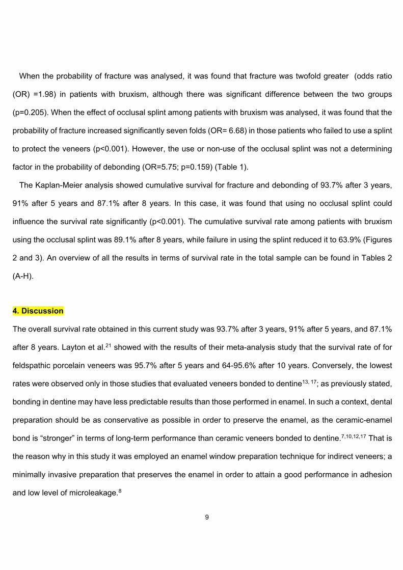
When the probability of fracture was analysed, it was found that fracture was twofold greater (odds ratio
(OR) =1.98) in patients with bruxism, although there was significant difference between the two groups
(p=0.205). When the effect of occlusal splint among patients with bruxism was analysed, it was found that the
probability of fracture increased significantly seven folds (OR= 6.68) in those patients who failed to use a splint
to protect the veneers (p<0.001). However, the use or non-use of the occlusal splint was not a determining
factor in the probability of debonding (OR=5.75; p=0.159) (Table 1).
The Kaplan-Meier analysis showed cumulative survival for fracture and debonding of 93.7% after 3 years,
91% after 5 years and 87.1% after 8 years. In this case, it was found that using no occlusal splint could
influence the survival rate significantly (p<0.001). The cumulative survival rate among patients with bruxism
using the occlusal splint was 89.1% after 8 years, while failure in using the splint reduced it to 63.9% (Figures
2 and 3). An overview of all the results in terms of survival rate in the total sample can be found in Tables 2
(A-H).
4. Discussion
The overall survival rate obtained in this current study was 93.7% after 3 years, 91% after 5 years, and 87.1%
after 8 years. Layton et al.21 showed with the results of their meta-analysis study that the survival rate of for
feldspathic porcelain veneers was 95.7% after 5 years and 64-95.6% after 10 years. Conversely, the lowest
rates were observed only in those studies that evaluated veneers bonded to dentine13, 17; as previously stated,
bonding in dentine may have less predictable results than those performed in enamel. In such a context, dental
preparation should be as conservative as possible in order to preserve the enamel, as the ceramic-enamel
bond is “stronger” in terms of long-term performance than ceramic veneers bonded to dentine.7,10,12,17 That is
the reason why in this study it was employed an enamel window preparation technique for indirect veneers; a
minimally invasive preparation that preserves the enamel in order to attain a good performance in adhesion
and low level of microleakage.8
10
Several studies have emphasised that the main cause for clinical failure in veneers made of feldspathic
ceramics is related to fracture.11,12,18,19 However, Cötert et al.13 reported that the most frequent type of failure
in such veneers was debonding, while Gurel et al.17 concluded that both fracture and debonding were the
most common causes of failure in feldspathic restorations. However, the clinical performance of veneers may
vary due to differences in terms of study design, inclusion and exclusion criteria and restorative materials
employed. For this reason, it is difficult to make an “ultimate” comparisons between studies, although some
data may be comparable to the present study.
The results of the current study may be comparable to those of Gurel et al.17 who found that veneers fractured
with an incidence of 3% and only 2 % debonded over a period of 12 years. However, the reason why they
found less incidence in fracture may be due to the use of high-resistance heat-pressed ceramic veneers (e.g
lithium disilicate-based ceramics), which have higher strength and less translucency.22 Peumans et al.19
evaluated conventional feldspathic ceramic veneers and they obtained a fracture rate of 14% and 0%
debonding over a 10-year follow-up. In this case, the fracture rate was higher than that obtained in our study,
but with comparable results in the rate of debonding compared to the present study. According to the literature,
the rate of failure for feldspathic veneers over different follow-up periods varies between 0 and 14%.11
The results of the current study are in accordance with the average survival rate (87%) of conventional
feldspathic veneers reported in a systematic review paper published by Morimoto et al.12 These latter authors
also reported that the incidence of fracture was 4% over a maximum period of 8 years. The fracture rate was
lower than that observed in the present study; it could be due to the fact that in that article both conventional
feldspathic and high-resistance non-feldspathic ceramics were included in the analysis. Moreover, the
debonding rate was 2% 12, a result quite similar to that obtained in the current study. However, some of the
articles reviewed by Morimoto et al. 12 did not specify whether patients with bruxism were included or excluded
in the study; differences in the inclusion/exclusion criteria always influence the fracture rates registered. 1,13,19

11
Unfavourable occlusion and parafunctional habits are considered an important risk factor for the failure of
ceramic veneers.10,15,17,18 Indeed, bruxism can be considered a contraindication for veneer placement.3 Beier
et al.2 found that the probability of failure was eight-time higher among patients with bruxism. Nevertheless, in
the present study, the probability of failure was only two-fold higher in such patients affected by bruxism,
although no significant difference was encountered when compared to patients with no parafunctions.
Likewise, the current study corroborate the results of Gurel et al.17 who found no significant relationship
between the presence of bruxism and restoration failure. However, the use of occlusal splints can reduce the
failure rate of ceramic veneers in patients with bruxism; satisfactory general and clinical outcomes have been
reported in previous studies.2 Indeed, Granell et al,5 who evaluated the effect on the use of occlusal splints on
the failures of veneers, found a significant higher rate of fracture in patients with bruxism who used no occlusal
splint. Nevertheless, no significant difference was encountered in terms of debonding. The results of our study
are in accordance with those presented by Granell et al. 5; the probability of fracture was seven-fold greater
than when the patients used no occlusal splint. However, the use or non-use of the occlusal splint was not a
determining factor in the probability of debonding.
5. Conclusions
Considering the limitations of this study, it may be concluded that conventional feldspathic porcelain veneers
can be considered an acceptable and predictable treatment with long-lasting performance. Moreover,
feldspathic porcelain veneers can be also considered a suitable clinical option for the treatment of patients
with bruxism, especially such patients can use regularly an occlusal splint to protect the veneers.
Conflict of interest
The authors did not have any commercial interest in any of the materials used in this study.

12
REFERENCES
1. Layton DM, Walton TR. The up to 21-year clinical outcome and survival of feldspathic porcelain veneers:
accounting for clustering. International Journal of Prosthodontics 2012;25:604-12.
2. Beier US, Kapferer I, Burtscher D, Dumfahrt H. Clinical performance of porcelain laminate veneers for up to
20years. International Journal of Prosthodontics 2012;25:79-85.
3. Nejatidanesh F, Savabi G, Amjadi M, Abbasi M, Savabi O. Five year clinical outcomes and survival of
chairside CAD/CAM ceramic laminate veneers - a retrospective study. J Prosthodont Res. 2018 Oct;62:462-
467.
4. Fradeani M, Redemagni M, Corrado M. Porcelain laminate veneers: 6- to 12-year clinical evaluation- a
retrospective study. International Journal of Periodontics and Restorative Dentistry 2005;25:9-17.
5. Manfredini D, Winocur E, Guarda-Nardini L, Paesani D, Lobbezoo F. Epidemiology of bruxism in adults: a
systematic review of the literature. Journal of Orofacial Pain. 2013;27:99-110.
6. Shetty A, Kaiwar A, Shubhashini N, Ashwini P, Naveen D, Adarsha M, Shetty M, Meena N. Survival rates of
porcelain laminate restoration based on different incisal preparation designs: An analysis. J Conserv Dent.
2011;14:10-5.
7. Layton DM, Walton T. An up to 16-year prospective study of 304 porcelain veneers. International Journal of
Prosthodontics 2007;20:389-96.
8. Faus-Matoses I, Solá-Ruiz F. Dental preparation with sonic vs high-speed finishing: analysis of microleakage
in bonded veneer restorations. Journal of Adhesive Dentistry 2014;16:29-34.
9. Furuse AY, Soares JV, Cunali RS, Gonzaga CC. Minimum intervention in restorative dentistry with V-shaped
facial and palatal ceramic veneers: A clinical report. Journal of Prosthetic Dentistry 2016;115:527-30.
10. Federizzi L, Gomes ÉA, Báratro SS, Baratto-Filho F, Bacchi A, Spazzin AO. Use of feldspathic porcelain
veneers to improve smile harmony: A 3-year follow-up. Brazilian Dental Journal 2016;27:767-74.

13
11. Rinke S, Lange K, Ziebolz D. Retrospective study of extensive heat-pressed ceramic veneers after 36
months. Journal of Esthetic and Restorative Dentistry 2013;25:42-52.
12. Morimoto S, Albanesi RB, Sesma N, Agra CM, Braga MM. Main clinical outcomes of feldspathic porcelain
and glass-ceramic laminate veneers: a systematic review and meta-analysis of survival and complication rates.
International Journal of Prosthodontics 2016;29:38-49.
13. Cötert HS, Dündar M, Oztürk B. The effect of various preparation designs on the survival of porcelain
laminate veneers. Journal of Adhesive Dentistry 2009;11:405-11.
14. Burke FJ, Lucarotti PS.Ten-year outcome of porcelain laminate veneers placed within the general dental
services in England and Wales. Journal of Dentistry 2009;37:31-8.
15. Dumfahrt H, Schäffer H. Porcelain laminate veneers. A retrospective evaluation after 1 to 10 years of
service: Part II--Clinical results. International Journal of Prosthodontics 2000;13:9-18.
16. Shaini FJ, Shortall AC, Marquis PM. Clinical performance of porcelain laminate veneers. A retrospective
evaluation over a period of 6.5 years. l Journal of Oral Rehabilitation 1997;24:553-9.
17. Gurel G, Sesma N, Calamita MA, Coachman C, Morimoto S. Influence of enamel preservation on failure
rates of porcelain laminate veneers. International Journal of Periodontics and Restorative Dentistry 2013;33:31-
9.
18. Beier US, Kapferer I, Dumfahrt H.Clinical long-term evaluation and failure characteristics of 1,335 all-
ceramic restorations. International Journal of Prosthodontics 2012;25:70-8.
19. Peumans M, De Munck J, Fieuws S, Lambrechts P, Vanherle G, Van Meerbeek B.A prospective ten-year
clinical trial of porcelain veneers. Journal of Adhesive Dentistry 2004;6:65-76.
20. Granell-Ruiz M, Fons-Font A, Labaig-Rueda C, Martínez-González A, Román-Rodríguez JL, Solá-Ruiz
MF.A clinical longitudinal study 323 porcelain laminate veneers. Period of study from 3 to 11 years. Medicina
Oral Patologia Oral y Cirugia Bucal 2010;15:531-7.

14
21. Faus-Matoses V, Faus-Matoses I, Ruiz-Bell E, Faus-Llácer VJ. Severe tetracycline dental discoloration:
Restoration with conventional feldspathic ceramic veneers. A clinical report. Journal of Clinical Experimental
Dentistry 2017;9:1379-82.
22. Layton DM, Clarke M, Walton TR. A systematic review and meta-analysis of the survival of feldspathic
porcelain veneers over 5 and 10 years. International Journal of Prosthodontics 2012;25:590-603.

15
Table 1. Association between veneer failure and bruxism patients who failed to use occlusal splints. Chi2 test
results and odds ratio (OR) and estimation odds ratio (OR) with generalized estimating equations (GEE) for
binary logistic regression model. The non-use of the occlusal splint increased the risk of fracture seven times.
Non-use of splints was not a determinant factor for debonding.
p-value OR CI 95% OR
FRACTURE <0.001**
*
6.68 2.86 – 15.6
DEBONDING 0.159 5.75 0.51 – 65.5
FAILURE 0.001** 7.51 2.17 – 26.1
*p<0.05; **p<0.01; ***p<0.001
Tables 2. A-H: Failure rates with respect to the location (Maxillary/mandible), type of the tooth
(anterior/posterior) and the association with or without bruxism and patients using or not using occlusal splints.
A.- Survival rate in the total sample: failures according to position (maxillary/mandible) and tooth type.
Total
TOOTH
Total IC IL CAN PM1 PM2 Total IC
N % N % N % N % N % N % N % N %
FRACTURE Total 364 100.0% 108 100.0% 89 100.0% 76 100.0% 54 100.0% 37 100.0% 245 100.0% 80 100.
DEBONDING Total 364 100.0% 108 100.0% 89 100.0% 76 100.0% 54 100.0% 37 100.0% 245 100.0% 80 100.
FAILURE Total 364 100.0% 108 100.0% 89 100.0% 76 100.0% 54 100.0% 37 100.0% 245 100.0% 80 100.

16
B.- Survival rate in the total sample: failures according to position (maxillary/mandible) and sector (anterior/posterior).
Total
SECTOR
Total Anterior Posterior
N % N % N %
FRACTURE Total 364 100.0% 273 100.0% 91 100.0%
DEBONDING Total 364 100.0% 273 100.0% 91 100.0%
FAILURE Total 364 100.0% 273 100.0% 91 100.0%
C.- Survival rate in the total sample: failures according to position (maxillary/mandible) and tooth type in no-bruxism patient.
Total
TOOTH
Total IC IL CAN PM1 PM2
N % N % N % N % N % N %
FRACTURE Total 71 100.0% 26 100.0% 18 100.0% 14 100.0% 11 100.0% 2 100.0%
DEBONDING Total 71 100.0% 26 100.0% 18 100.0% 14 100.0% 11 100.0% 2 100.0%
FAILURE Total 71 100.0% 26 100.0% 18 100.0% 14 100.0% 11 100.0% 2 100.0%

17
D.- Survival rate in the total sample: failures according to position (maxillary/mandible) and sector (anterior/posterior) in no-bruxism patients.
Total
SECTOR
Total Anterior Posterior Total
N % N % N % N %
FRACTURE Total 71 100.0% 58 100.0% 13 100.0% 47 100
DEBONDING Total 71 100.0% 58 100.0% 13 100.0% 47 100
FAILURE Total 71 100.0% 58 100.0% 13 100.0% 47 100
E.- Survival rate in the total sample: failures according to position (maxillary/mandible) and tooth type in patients with bruxism and with occlusal splint.
Total
TOOTH
Total IC IL CAN PM1 PM2 Total IC
N % N % N % N % N % N % N % N %
FRACTURE Total 257 100.0% 74 100.0% 63 100.0% 54 100.0% 37 100.0% 29 100.0% 172 100.0% 53 100.0
DEBONDING Total 257 100.0% 74 100.0% 63 100.0% 54 100.0% 37 100.0% 29 100.0% 172 100.0% 53 100.0
FAILURE Total 257 100.0% 74 100.0% 63 100.0% 54 100.0% 37 100.0% 29 100.0% 172 100.0% 53 100.0

18
F.- Survival rate in the total sample: failures according to position (maxillary/mandible) and sector (anterior/posterior) in patients with bruxism and with occlusal splint.
Total
SECTOR
Total Anterior Posterior Total
N % N % N % N
FRACTURE Total 257 100.0% 191 100.0% 66 100.0% 172
DEBONDING Total 257 100.0% 191 100.0% 66 100.0% 172
FAILURE Total 257 100.0% 191 100.0% 66 100.0% 172
G.- Survival rate in the total sample: failures according to position (maxillary/mandible) and tooth type in patients with bruxism and without occlusal splint.
Total
TOOTH
Total IC IL CAN PM1 PM2 Total IC
N % N % N % N % N % N % N % N %
FRACTURE Total 36 100.0% 8 100.0% 8 100.0% 8 100.0% 6 100.0% 6 100.0% 26 100.0% 6 100.0
DEBONDING Total 36 100.0% 8 100.0% 8 100.0% 8 100.0% 6 100.0% 6 100.0% 26 100.0% 6 100.0
FAILURE Total 36 100.0% 8 100.0% 8 100.0% 8 100.0% 6 100.0% 6 100.0% 26 100.0% 6 100.0

19
H.- Survival rate in the total sample: failures according to position (maxillary/mandible) and sector (anterior/posterior) in patients with bruxism and without occlusal splint.
Total
SECTOR
Total Anterior Posterior Total
N % N % N % N %
FRACTURE Total 36 100.0% 24 100.0% 12 100.0% 26 100
DEBONDING Total 36 100.0% 24 100.0% 12 100.0% 26 100
FAILURE Total 36 100.0% 24 100.0% 12 100.0% 26 100
Captions to the legends and tables:
Tables
Table 1. Association between veneer failure and bruxism patients who failed to use occlusal splints. Chi2 test
results and odds ratio (OR) and estimation odds ratio (OR) with generalized estimating equations (GEE) for
binary logistic regression model. The non-use of the occlusal splint increased the risk of fracture seven times.
Non-use of splints was not a determinant factor for debonding.
Tables 2. A-H: Failure rates with respect to the location (Maxillary/mandible), type of the tooth
(anterior/posterior) and the association with or without bruxism and patients using or not using occlusal splints.
Figures

20
Fig. 1 Distribution of incidence of fractures and debonding in patients with or without bruxism and patients
using or not using occlusal splints. The failure rate is higher among the group of patients with bruxism who
did not use the occlusal splint. Patients with bruxism who did use splints presented a similar failure rate to
non-bruxism patients No= Non-bruxism; Splint=Bruxism with splint; No splint=Bruxism no splint.
Colour: No= Light Blue; Splint= dark blue; No splint= orange.
Fig. 2 Veneer survival during a maximum follow-up period of 8 years. The survival rate shows a progressively
decreasing constant pattern, indicating a constant failure rate over time (95% interval of confidence).
Fig. 3. Veneer survival among patients with bruxism according to use or non-use of occlusal splints over a
maximum follow-up period of 8 years. Patients with bruxism who used no splint presented a higher incidence
of veneer failure with a decreasing inconstant curve, particularly during the first year. Patients with bruxism who
did use splints present a progressively decreasing constant curve over time (95% interval of confidence).

Table 1. Association between veneer failure and bruxism patients who failed to use occlusal splints. Chi2 test results and odds ratio (OR) and estimation odds ratio (OR) with generalized estimating equations (GEE) for binary logistic regression model. The non-use of the occlusal splint increased the risk of fracture
seven times. Non-use of splints was not a determinant factor for debonding.
p-value OR CI 95% OR
FRACTURE <0.001*** 6.68 2.86 – 15.6
DEBONDING 0.159 5.75 0.51 – 65.5
FAILURE 0.001** 7.51 2.17 – 26.1
*p<0.05; **p<0.01; ***p<0.001
Tables 2. A-H: Failure rates with respect to the location (Maxillary/mandible), type of the tooth (anterior/posterior) and the association with or without bruxism and patients using or not using occlusal splints.
A.- Survival rate in the total sample: failures according to position (maxillary/mandible) and tooth type.
Total Maxillary Mandible
TOOTH TOOTH TOOTH
Total IC IL CAN PM1 PM2 Total IC IL CAN PM1 PM2 Total IC IL CAN PM1 PM2
N % N % N % N % N % N % N % N % N % N % N % N % N % N % N % N % N % N %
FRACTURE Total 364 100.0% 108 100.0% 89 100.0% 76 100.0% 54 100.0% 37 100.0% 245 100.0% 80 100.0% 62 100.0% 50 100.0% 31 100.0% 22 100.0% 119 100.0% 28 100.0% 27 100.0% 26 100.0% 23 100.0% 15 100.0%
DEBONDING Total 364 100.0% 108 100.0% 89 100.0% 76 100.0% 54 100.0% 37 100.0% 245 100.0% 80 100.0% 62 100.0% 50 100.0% 31 100.0% 22 100.0% 119 100.0% 28 100.0% 27 100.0% 26 100.0% 23 100.0% 15 100.0%
FAILURE Total 364 100.0% 108 100.0% 89 100.0% 76 100.0% 54 100.0% 37 100.0% 245 100.0% 80 100.0% 62 100.0% 50 100.0% 31 100.0% 22 100.0% 119 100.0% 28 100.0% 27 100.0% 26 100.0% 23 100.0% 15 100.0%
B.- Survival rate in the total sample: failures according to position (maxillary/mandible) and sector (anterior/posterior).
Total Maxillary Mandible
SECTOR SECTOR SECTOR
Total Anterior Posterior Total Anterior Posterior Total Anterior Posterior
N % N % N % N % N % N % N % N % N %
FRACTURE Total 364 100.0% 273 100.0% 91 100.0% 245 100.0% 192 100.0% 53 100.0% 119 100.0% 81 100.0% 38 100.0%
DEBONDING Total 364 100.0% 273 100.0% 91 100.0% 245 100.0% 192 100.0% 53 100.0% 119 100.0% 81 100.0% 38 100.0%
FAILURE Total 364 100.0% 273 100.0% 91 100.0% 245 100.0% 192 100.0% 53 100.0% 119 100.0% 81 100.0% 38 100.0%