AMSER Rad Path Case of the Month: Synovial Sarcoma · AMSER Rad Path Case of the Month August 2018...
27
AMSER Rad Path Case of the Month August 2018 Synovial Sarcoma Benjamin Swanson OMS 3 Kossivi Dantey MD, Pathology Dept AHN William Peterson MD, Radiology MSK Division AHN Matthew Hartman MD, Abdominal Imaging AHN
-
Upload
truongkhanh -
Category
Documents
-
view
215 -
download
0
Transcript of AMSER Rad Path Case of the Month: Synovial Sarcoma · AMSER Rad Path Case of the Month August 2018...
AMSER Rad Path Case of the Month August 2018
Synovial Sarcoma
Benjamin Swanson OMS 3
Kossivi Dantey MD, Pathology Dept AHN
William Peterson MD, Radiology MSK Division AHN
Matthew Hartman MD, Abdominal Imaging AHN
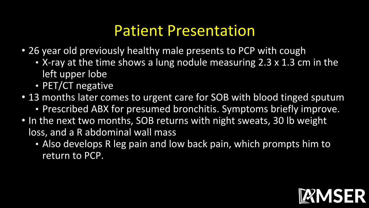
Patient Presentation • 26 year old previously healthy male presents to PCP with cough
• X-ray at the time shows a lung nodule measuring 2.3 x 1.3 cm in the left upper lobe
• PET/CT negative • 13 months later comes to urgent care for SOB with blood tinged sputum
• Prescribed ABX for presumed bronchitis. Symptoms briefly improve. • In the next two months, SOB returns with night sweats, 30 lb weight
loss, and a R abdominal wall mass • Also develops R leg pain and low back pain, which prompts him to
return to PCP.
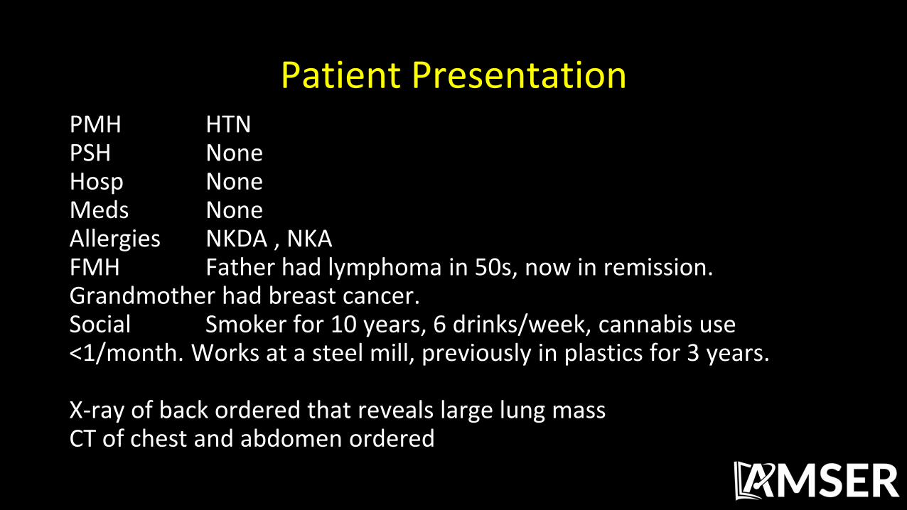
Patient Presentation PMH HTN PSH None Hosp None Meds None Allergies NKDA , NKA FMH Father had lymphoma in 50s, now in remission. Grandmother had breast cancer. Social Smoker for 10 years, 6 drinks/week, cannabis use <1/month. Works at a steel mill, previously in plastics for 3 years. X-ray of back ordered that reveals large lung mass CT of chest and abdomen ordered
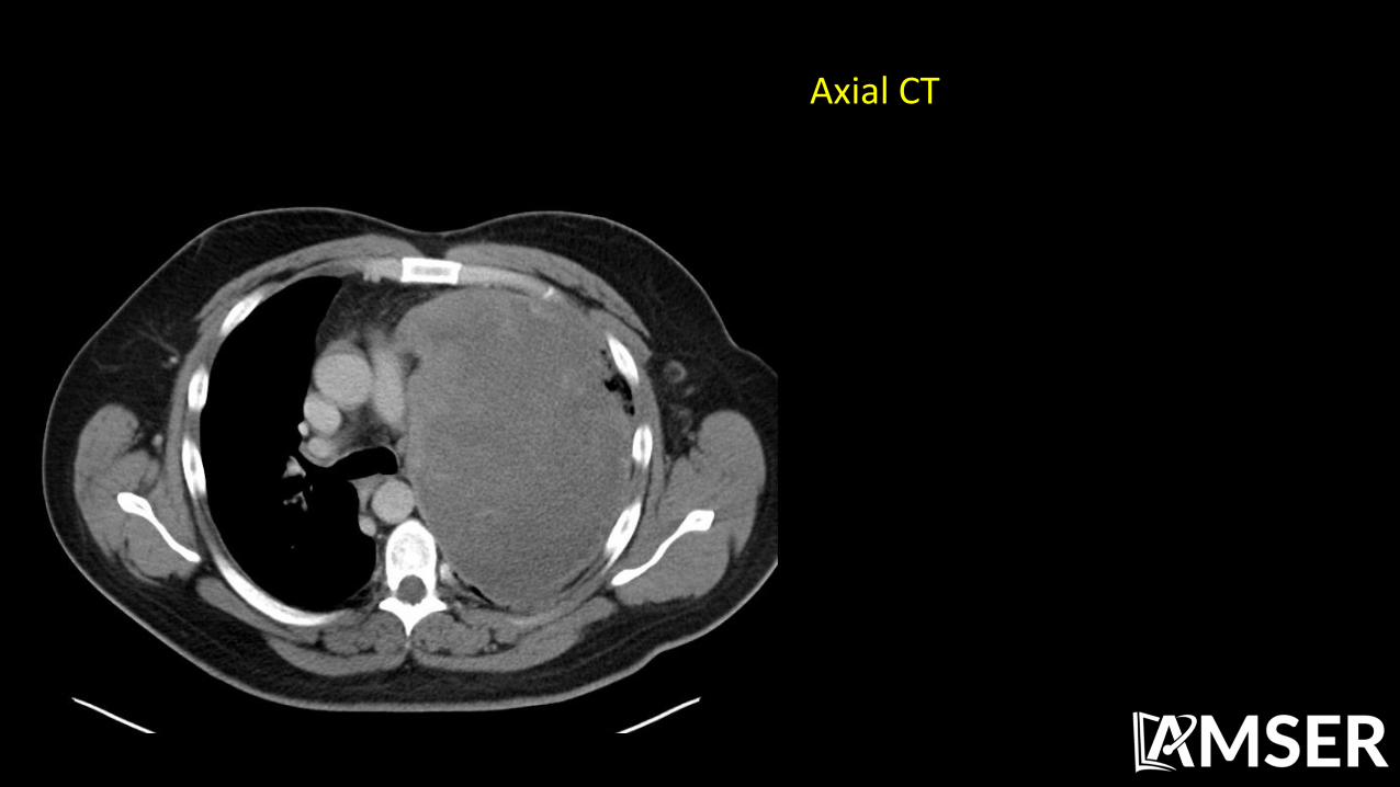
Axial CT
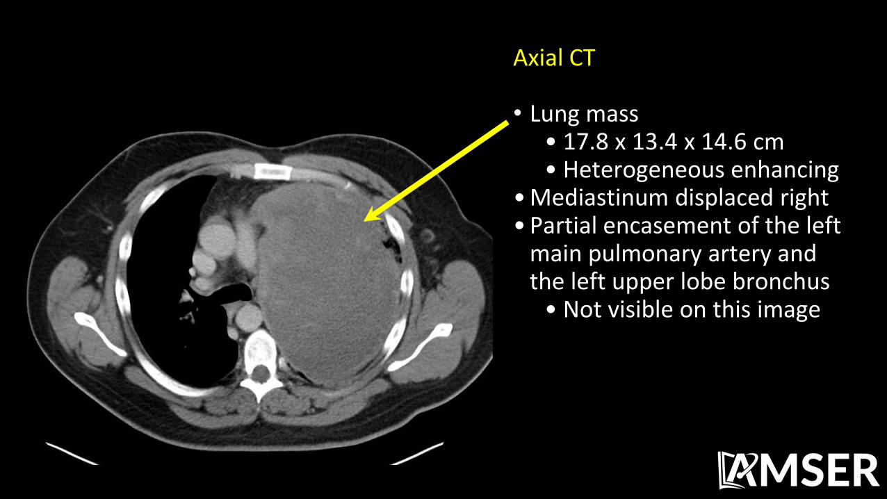
Axial CT • Lung mass
• 17.8 x 13.4 x 14.6 cm • Heterogeneous enhancing
• Mediastinum displaced right • Partial encasement of the left
main pulmonary artery and the left upper lobe bronchus
• Not visible on this image
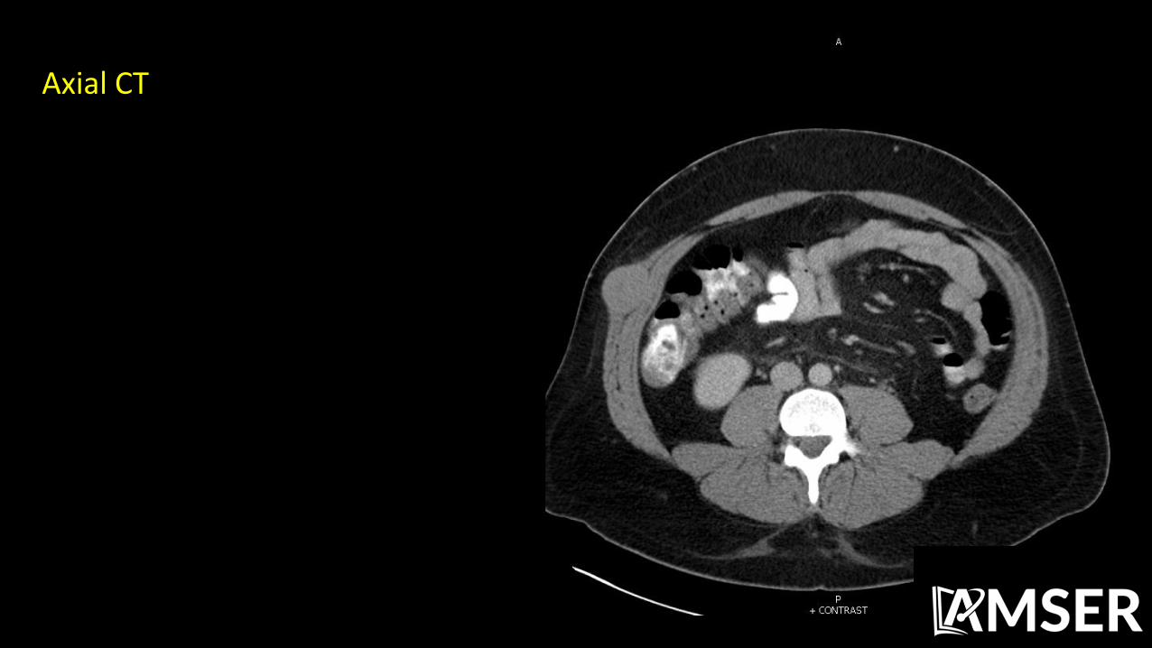
Axial CT
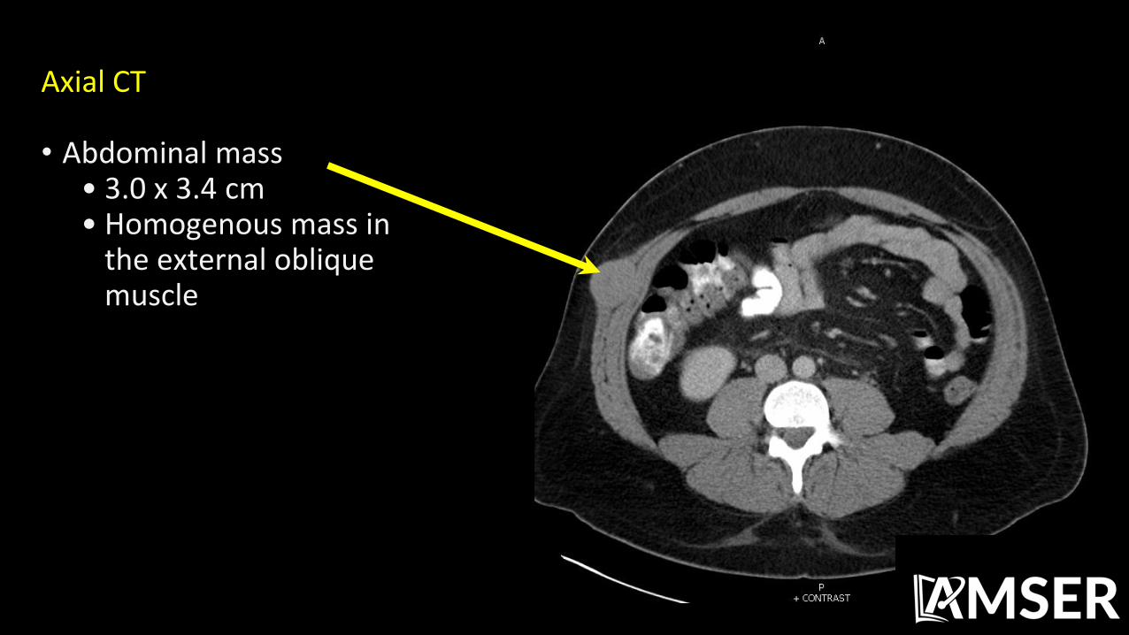
Axial CT • Abdominal mass
• 3.0 x 3.4 cm • Homogenous mass in
the external oblique muscle
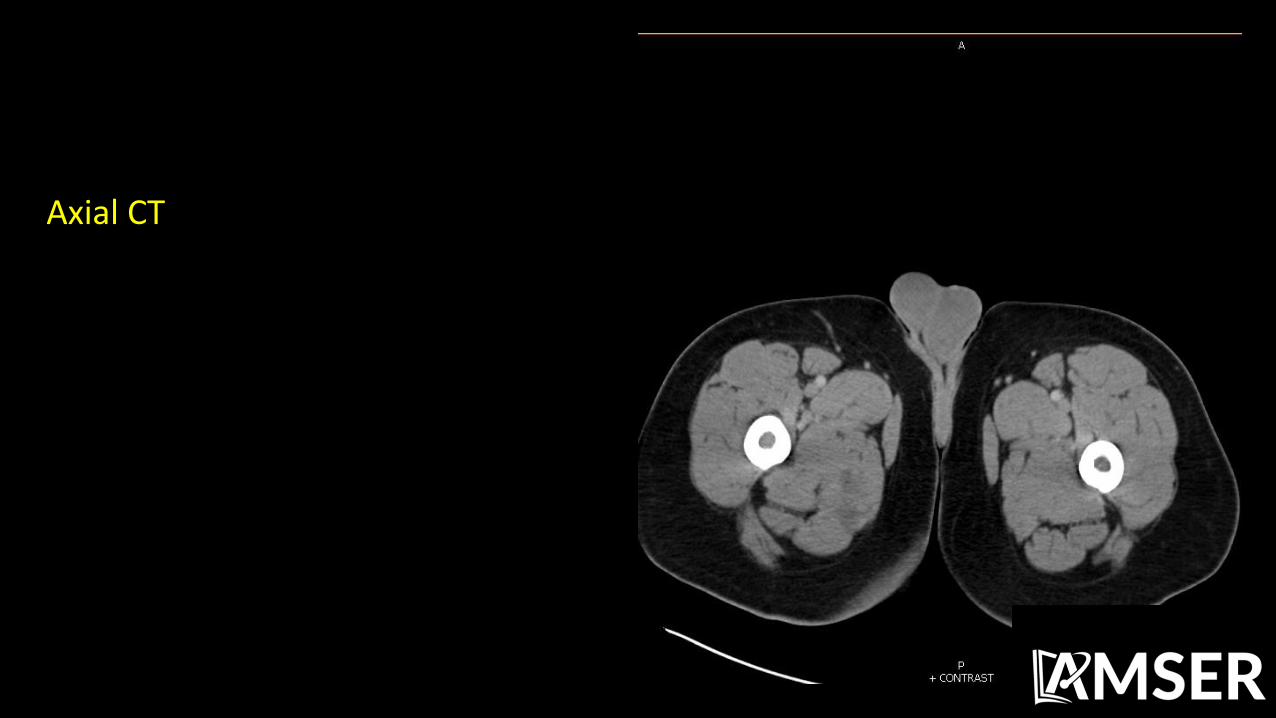
Axial CT
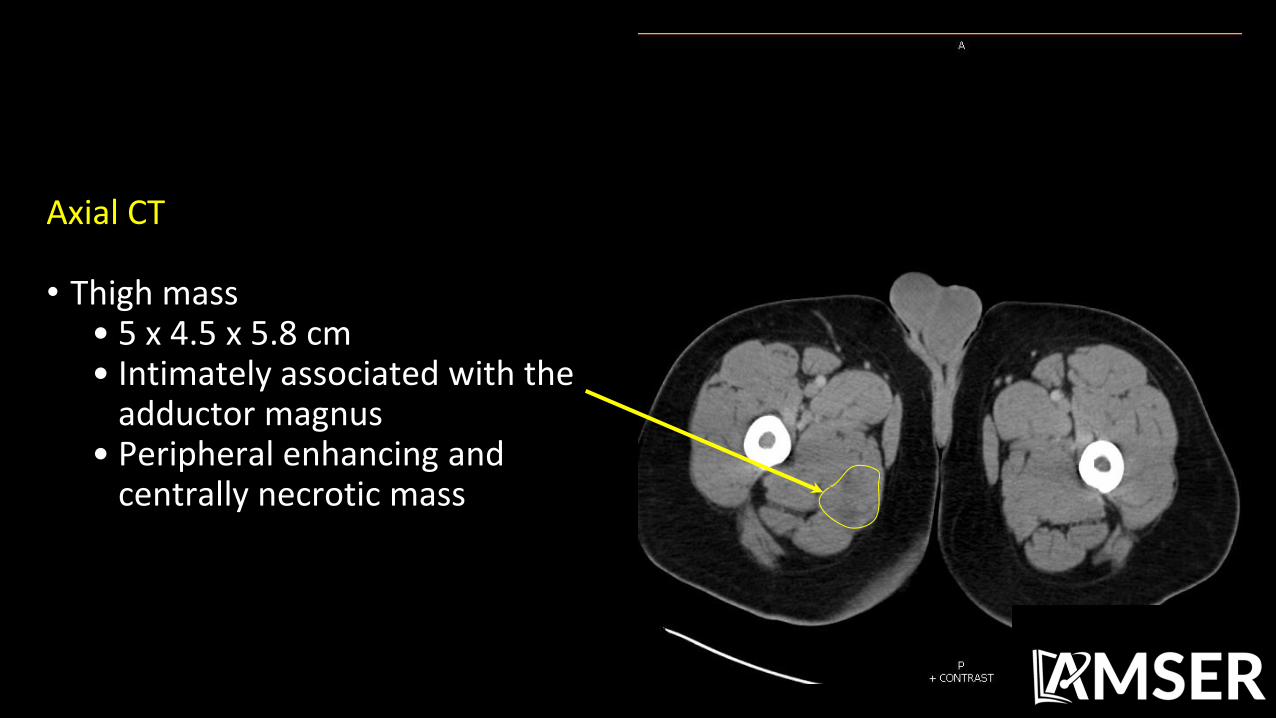
Axial CT • Thigh mass
• 5 x 4.5 x 5.8 cm • Intimately associated with the
adductor magnus • Peripheral enhancing and
centrally necrotic mass

MRI T1 Fat Sat with Contrast

MRI T1 Fat Sat with Contrast • Enhancing periosteum consistent
with periostitis • Thigh mass
• Peripheral enhancement • Intrinsic hyperintensity
suggests blood products and/or proteinaceous material

MRI T2 Fat Sat
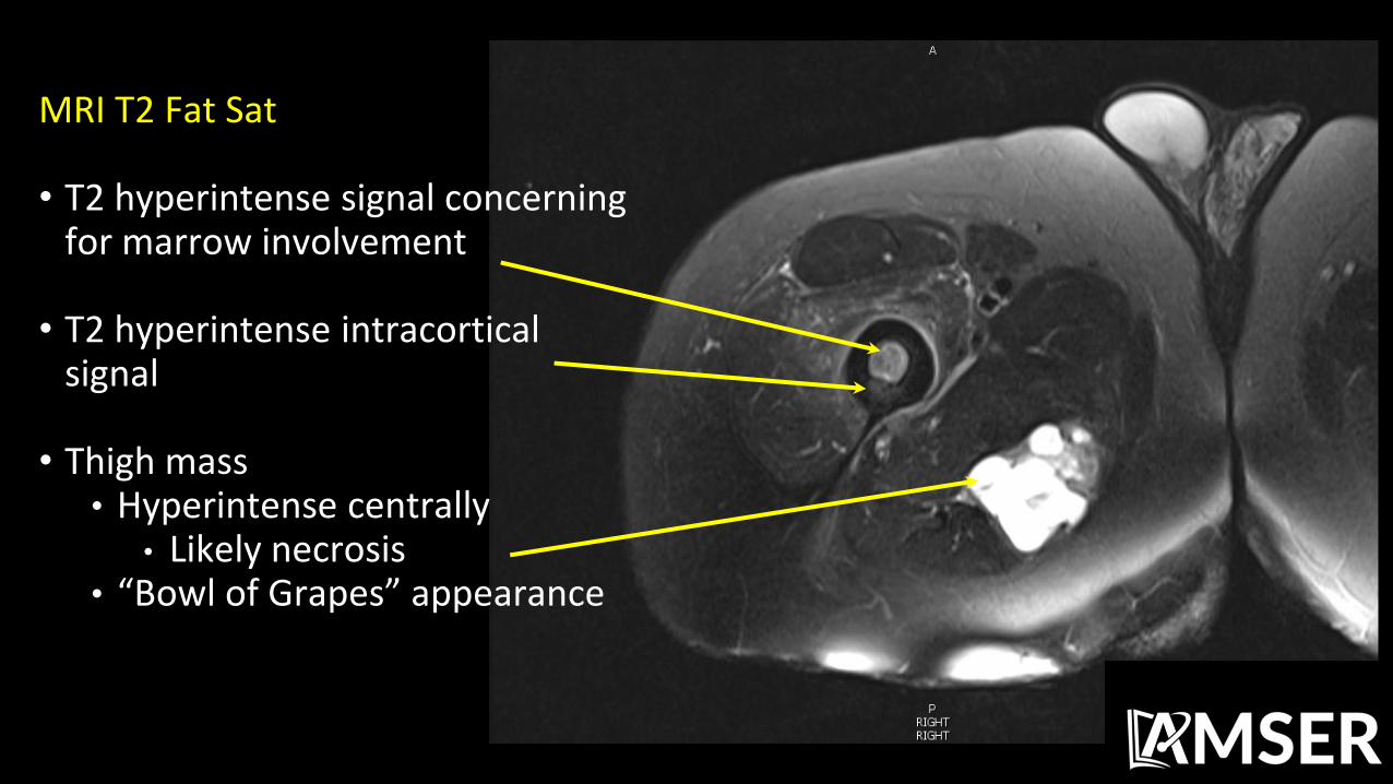
MRI T2 Fat Sat • T2 hyperintense signal concerning
for marrow involvement • T2 hyperintense intracortical
signal • Thigh mass
• Hyperintense centrally • Likely necrosis
• “Bowl of Grapes” appearance
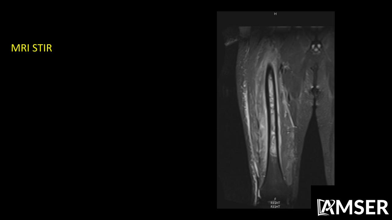
MRI STIR
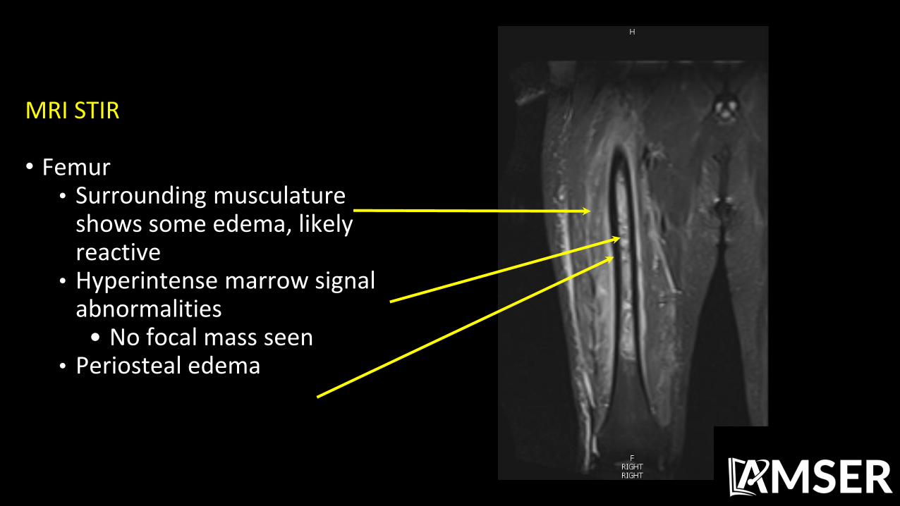
MRI STIR • Femur
• Surrounding musculature shows some edema, likely reactive
• Hyperintense marrow signal abnormalities
• No focal mass seen • Periosteal edema

Differential Diagnosis Based on Imaging
• Primary lung cancer with metastases to other organs
• Sarcoma with metastases to the lung
• Lymphoma
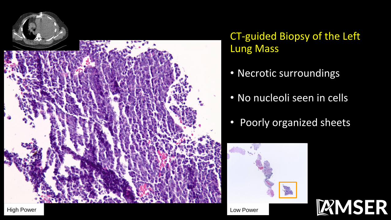
CT-guided Biopsy of the Left Lung Mass • Necrotic surroundings • No nucleoli seen in cells • Poorly organized sheets
Low Power High Power

Lung Cytology • Uniform population of small
round blue cells • “Spindle cells” seen are likely
smeared cells from plating
High Power Low Power
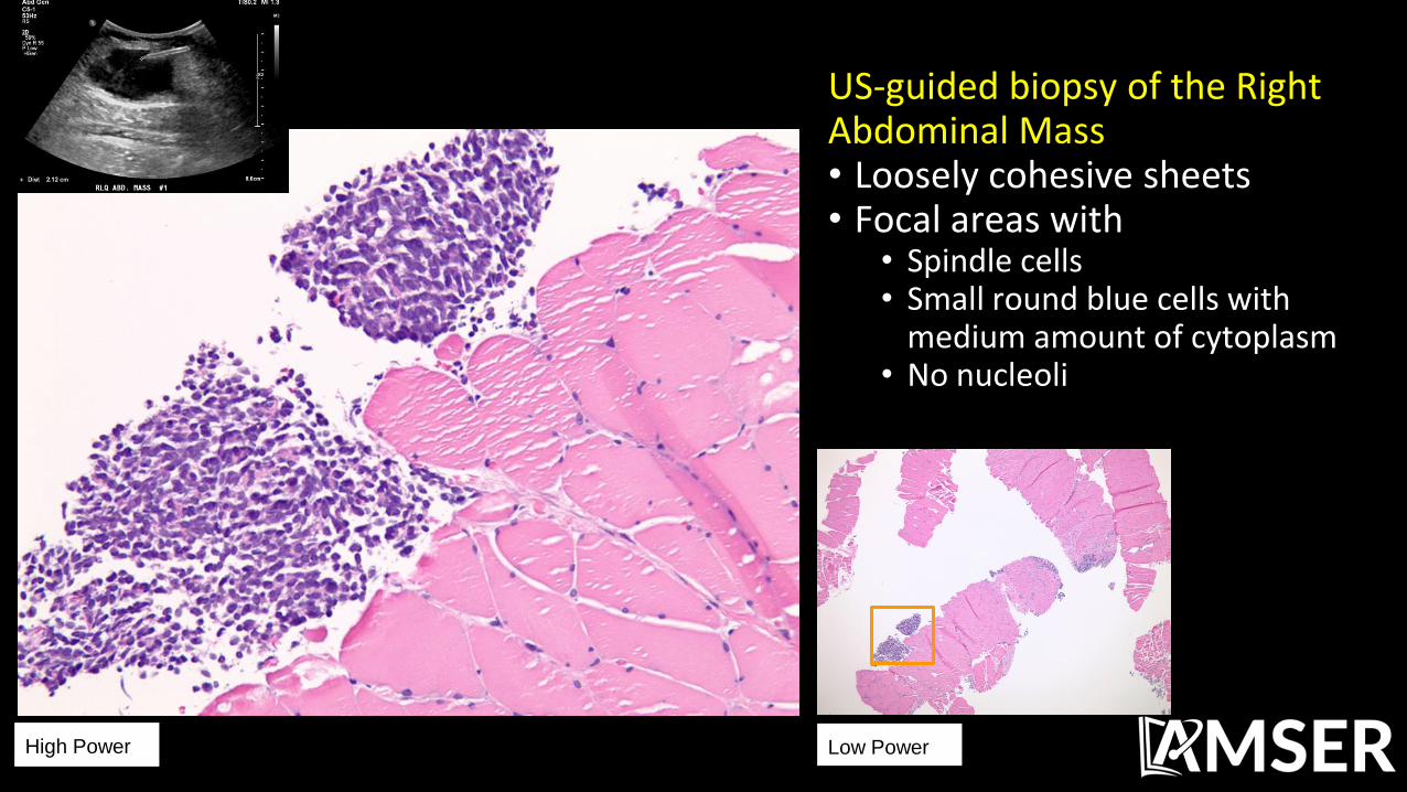
US-guided biopsy of the Right Abdominal Mass • Loosely cohesive sheets • Focal areas with
• Spindle cells • Small round blue cells with
medium amount of cytoplasm • No nucleoli
High Power Low Power

CD99 Stain • Used to distinguish Ewing’s sarcoma,
mesenchymal chondrosarcoma, solitary fibrous tumors, and synovial sarcoma
• Cytoplasm here shows strongly
positive result for CD99 • FISH for t(X;18)(p11;q11) is specific
to synovial sarcoma returned positive
Final Dx:
Synovial Sarcoma

Epidemiology • Presents in adolescents and
young adults (15-40 years of age) • Mild male predilection (1.2:1) • Uncommon, 2.5-10% of soft
tissue sarcomas
• Location • Extremities 80-95%
• Lower Limb 60-70% • Upper Limb 15-25%
• Head and neck 5% • Chest wall Rare • Viscera Rare
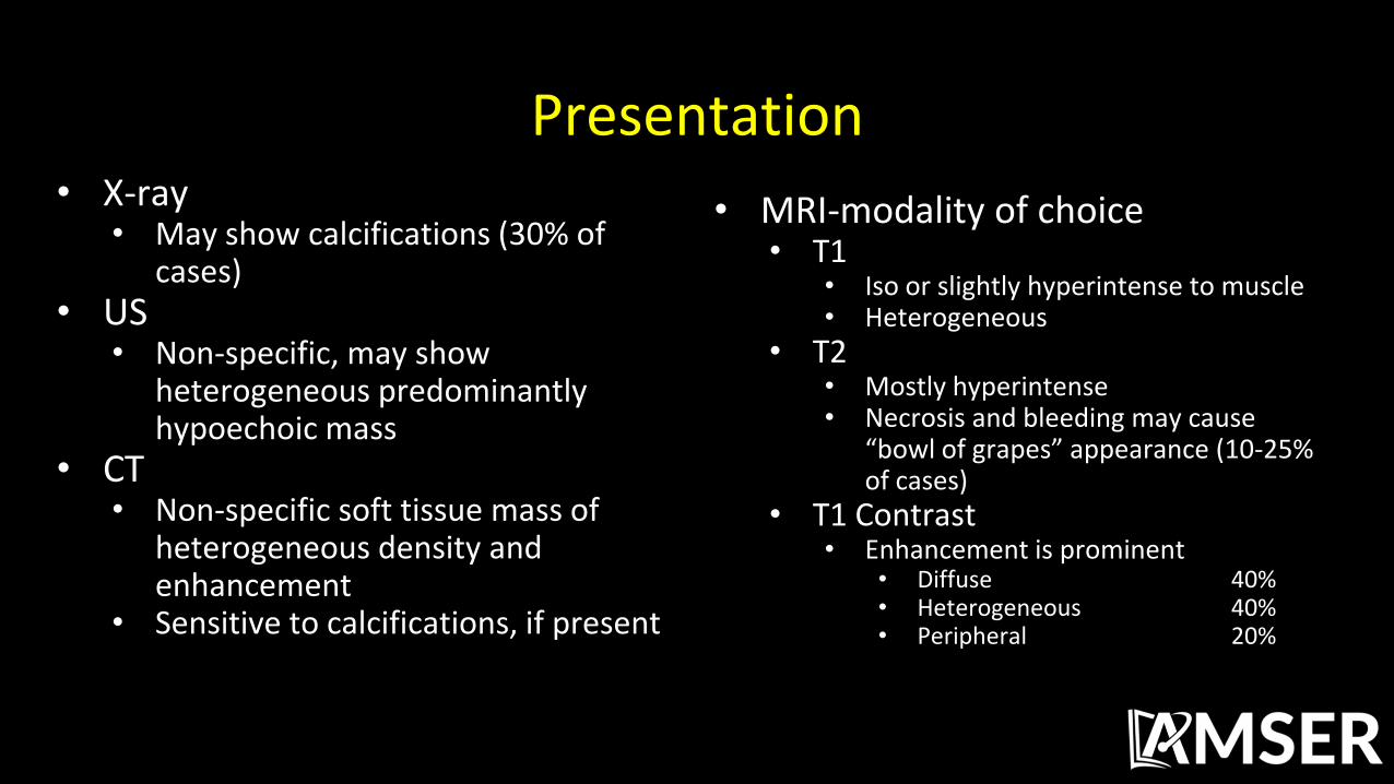
Presentation • X-ray
• May show calcifications (30% of cases)
• US • Non-specific, may show
heterogeneous predominantly hypoechoic mass
• CT • Non-specific soft tissue mass of
heterogeneous density and enhancement
• Sensitive to calcifications, if present
• MRI-modality of choice • T1
• Iso or slightly hyperintense to muscle • Heterogeneous
• T2 • Mostly hyperintense • Necrosis and bleeding may cause
“bowl of grapes” appearance (10-25% of cases)
• T1 Contrast • Enhancement is prominent
• Diffuse 40% • Heterogeneous 40% • Peripheral 20%

Synovial Sarcoma Histology • Not derived from synovium • Microscopically resembles normal synovium
• Stains for epithelial markers while true synovium does not • Usually biphasic appearance with epithelial cells and spindle cells • Can be monophasic
• Macroscopically appears as heterogeneous masses with areas of
hemorrhage and necrosis originating within soft tissues near large joints
Treatment • Surgery
• Curative in 20-70% of patients • Used on smaller tumors with no evidence of metastasis
• Chemotherapy
• Benefit of treatment still unclear • Some evidence supports use of doxorubicin/ifosfamide in advanced disease
• Radiotherapy
• Benefit is less clear than for chemotherapy • Used to reduce the chance of local recurrence
• This patient received combination chemo/radiotherapy
Prognosis • 5 year survival is 36-76% depending on the stage of disease • Local recurrence is common (30-50% overall)
• 14% recurrence over 10 years after diagnosis • Distant metastases is frequent (40-70%) within 5 years of diagnosis • Common locations of metastasis
• Lung 80% • Bones 15% • Lymph Nodes 10% • Chest wall/Abdomen 7.5%
References: • Krieg AH, Hefti F, Speth BM et-al. Synovial sarcomas usually metastasize after >5 years: a multicenter
retrospective analysis with minimum follow-up of 10 years for survivors. Ann. Oncol. 2011;22 (2): 458-67. • Krisanaprakornkit S, Chotjumlong P, Pata S, Chruewkamlow N, Reutrakul V, Kasinrerk W (January 2013).
"CD99 ligation induces intercellular cell adhesion molecule-1 expression and secretion in human gingival fibroblasts". Arch. Oral Biol. 58 (1): 82–93.
• Leong AS-Y, Cooper K, Leong FJW-M (2003). Manual of Diagnostic Cytology (2nd ed.). Greenwich Medical Media, Ltd. pp. 145–146.
• Lewis JJ, Antonescu CR, Leung DH, et al. (2000). "Synovial sarcoma: a multivariate analysis of prognostic factors in 112 patients with primary localized tumors of the extremity". J. Clin. Oncol. 18 (10): 2087–94
• Murphey MD, Gibson MS, Jennings BT et-al. From the archives of the AFIP: Imaging of synovial sarcoma with radiologic-pathologic correlation. Radiographics. 26 (5): 1543-65.
• Ren, Xiao-Hua; WU, Xiao-Min; JIN, Cheng; CUI, Yong-An (2009). "Advances in the diagnosis and treatment of synovial sarcoma". Journal of Medical Biomechanics. 15 (4): 541–542
• Spurrell EL, Fisher C, Thomas JM et-al. Prognostic factors in advanced synovial sarcoma: an analysis of 104 patients treated at the Royal Marsden Hospital. Ann. Oncol. 2005;16 (3): 437-44.
• Thway, K.; Fisher, C. (2014). "Synovial sarcoma: defining features and diagnostic evolution". Annals of Diagnostic Pathology. 18: 369–380.