Amino Acids - Philadelphia University acids.pdf · Amino Acids The study of proteins has occupied...
29
Amino Acids Presented by Dr. Mohammad Saadeh The requirements for the Pharmaceutical Biochemistry I Philadelphia University Faculty of pharmacy مينيةض احما ا
Transcript of Amino Acids - Philadelphia University acids.pdf · Amino Acids The study of proteins has occupied...

Amino Acids
Presented by
Dr. Mohammad Saadeh
The requirements for the Pharmaceutical Biochemistry I
Philadelphia University
Faculty of pharmacy
االحماض األمينية


Cell Structure

Amino Acids
The study of proteins has occupied biochemists to
understand three dimensional shapes, and chemical
activities of proteins may be key to the most scientific
challenge.
1. Thermophilic bacteria thrive at high temperatures.
What is the different about the proteins?
2. HIV-1 associated with AIDS.

The most important biological function of
proteins 1. Many proteins function as enzymes, the biochemical catalysts.
2. Proteins bind other molecules for storage and transport,
myoglobin and hemoglobin
3. Protein such as tubulin, actin, and collagen, provide support
and shape to cells.
4. Proteins can do mechanicals work, movement of flagella,
separation chromosome at mitosis, contraction of muscles.
5. Proteins play decoding information in the cell, translation,
regulating gene expression.
6. Proteins are hormones, which regulate biochemical activity in
target cells or tissues.
7. Proteins have highly specialized functions, Antibody, and
toxin.

I. General structure of Amino acids
• About 300 a.a have been found in nature but all
organisms use the same 20 a.a as building blocks for
the assembly of protein molecules. These 20 a.a are
called the common or standard a.a.
• Enormous variety of different polypeptides can be
produced by connecting the 20 standard a.a in various
combinations.

I. General structure of Amino acids
An a.a is a compound that
contains both a carboxyl
(-COOH) and amino (-NH2)
group as part of its structure.
In the 20 standard a.a that make
up proteins, the carboxyl and amino
groups are bonded to the same carbon atom,
called α -carbon. In addition, the amino acids in proteins
contain side chains that determine their role in the
proteins behavior.

I. General structure of Amino acids
Pka≈9 dipolar ions Pka≈3
Inside the cell, under normal physiological condition PH
6.8-7.4.
• The amino group are protonated (-NH3+), Pka≈9
• Carboxyl is ionize (-coo-), Pka≈3.
• So amino acid are zwitterions or dipolar ions, even
though their net charge may be zero.

I. General structure of Amino acids
• All 20 standard a.a except glycine is achiral, the α-carbon is chiral or asymmetric, since it has 4 different group bonded to it.
• Amino acid steroisomers are nonsuperimposable mirror image so it called enantiomers such as L and D isomers.
• Both L &D a.a are found in nature.
• Protein contain only L-a.a. but in bacteria D-a.a occur in bacterial cell walls and antibiotic of bacterial origin.
enantiomers

II. Structures of the common amino acids
• In fischer projection of a.a, horizontal bonds at a chiral center extend toward the viewer, and vertical bonds extend away.
• The properties of the side chains greatly influence the overall three- dimentional shape.

The RS system of configuration
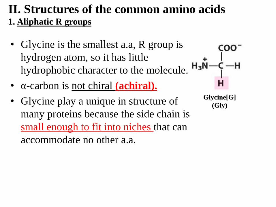
II. Structures of the common amino acids 1. Aliphatic R groups
• Glycine is the smallest a.a, R group is
hydrogen atom, so it has little
hydrophobic character to the molecule.
• α-carbon is not chiral (achiral).
• Glycine play a unique in structure of
many proteins because the side chain is
small enough to fit into niches that can
accommodate no other a.a.
Glycine[G]
(Gly)

II. Structures of the common amino acids 1. Aliphatic R groups Alanine(Ala,A), Valine(Val,V), Leucine(Leu,L) and Isoleucine(Ile,I), have saturated side chains.
• Isoleucine, two carbon α&β are chiral so it have (22=4) stereoisomers (L&D. Isoleucine and L&D. alloisoleucine).
• Alanine, Valine, Leucine, Isoleucine are important in establishing and maintaining the three-dimensional structures because their cluster away from water.
• Valine, Leucine, Isoleucine are branched side chain, All three a.a are highly hydrophobic.

• Proline(Pro,P) differ from the other 19 a.a
• Three carbon side chain is bonded to the
nitrogen of its α-amino group, the α-carbon
creating a cyclic molecule.
• The heterocyclic pyrrolidine ring of
proline restrict the geometry of
polypeptide, sometimes introducing abrupt
changes in direction of the peptide chain.
• Cyclic structure of proline makes it much
less hydrophobic than valine, leucine,
isoleucine.
II. Structures of the common amino acids 1. Aliphatic R groups

• Phenylalanine (phe,F),Tyrosine
(Tyr,Y), Tryptophan (Trp, W)
have side chains with aromatic
groups.
• Phenylalanin has a hydrophobic
benzyle side chain,Tyrosine has
phenol group, Tryptophan
contain bicyclic indol group.
• Tyrosine(Tyr,Y), Tryptophan
are not hydrophobic as
Phenylalanine because the side
chains include polar groups
II. Structures of the common amino acids 2. Aromatic R groups

• All three aromatic a.a absorbe U.V because it contain delocalized π electron.
• At neutral PH, both tryptophan and tyrosine absorbe light at 280nm.
• Phenylalanine is almost transport at 280nm and absorbed light weakly at 260nm.
• Absorbance at 280 nm is used to estimate the concentration Of proteins in solutions.
II. Structures of the common amino acids 2. Aromatic R groups
Absorbance four times of tyrosine.
Absorption of light for tryptophan and tyrosine at pH6

• Methionine (Met, M), contain non polar thioether group, it one of more hydrophobic a.a. and play an important role in synthesis of protein, it is the first a.a in polypeptide chain.
• Cysteine(Cys, C), side chain somewhat hydrophobic and highly reactive. The sulfhydryl group of Cysteine form weak H-bonds with O, N. sulfhydryl group is weak acid, its lose proton to be come negative charge thiolate ion.
II. Structures of the common amino acids 3. Sulfur containing R groups
Cysteine
Methionine

II. Structures of the common amino acids 3. Sulfur containing R groups
• Cysteine don’t have to be close
together in amino acid sequence of
the polypeptide chain they found in
different chains.
• Disulfide bond, stabilize the three-
dimentional structures of some
peptide by covalently cross linking
cysteine residues in peptide chains.
• Inside the cell protein don’t contain
disulfide bond because the
condition not fever oxidation.
However, many secreted
extracellular protein contain
disulfide bridge. Oxidation at slightly alkaline pH, because
sulfhydryl groups are ionized at low pH.
The two Cysteine adjacent in three
dimention space to form disulfide bond

II. Structures of the common amino acids 4. Side chains with alcohol groups
• Serine and threonine have uncharged polar side
chains (β-hydroxyl groups). These give
hydrophilic character to aliphatic side chains.
• The hydroxyl groups of Serine and threonine
have the weak ionization properties of primary
and secondary alcohols.
• The hydroxymethyl group of serine dose not
appreciably ionized in aqueous solution;
nevertheless, this alcohol can react with active
site of No. of enzymes as thought it were ionized.
• Threonine has two chiral carbon so it have
(22=4) stereoisomers (L&D. threonine and
L&D. allothreonine ). (L-threonine in protein)
Threonine (Thr, T)
Serine (Ser, S)

II. Structures of the common amino acids 5. Basic R groups
• Histidine (His, H), Lysine (Lys, k), and arginine (Arg,R) have
hydrophilic side chains that are nitrogenous bases and are
positively charged
at pH 7.
• The side chain of histidine contains an imidazole ring substituent. The protonated from of this ring side chain is called an imidazolium ion.
Imidazole group Charged guanidino
group
Second primary amino
group at e position

II. Structures of the common amino acids 5. Basic R groups
Charged guanidino
group
Second primary amino
group at e position
• Lysine is diamino acid, having both α and ε as
an alkylammonium ion (-CH2NH3+) at neutral
pH and confers a positive charge in proteins.
• Arginine is the most basic acid of the 20 a.a its
side chain guanidinium ion is protonated under
all conditions normally found within a cell.
(Arginine side chains also contribute positive
charges in proteins).

II. Structures of the common amino acids 6. Acidic R groups and their amide derivatives
• Aspartate(Asp, D) and glutamate
(Glu, E) [aspartic acid or glutamic
acid] are dicarboxilic and have
negative charged hydrophilic side
chain at pH 7 (ionized).
• Monosodium glutamate (MSG),
which is used in food as flavoue
enhancer.
Carboxyl side chain

II. Structures of the common amino acids 6. Acidic R groups and their amide derivatives
• Asparagine(Asn, N) and glutamine
(Gln, Q) are amides of aspartic acid
or glutamic acid
• The side chains of Asparagine and
glutamine are uncharged, highly
polar and found on the surface on
proteins, where they can interact
with the water molecules.
• The polar amide groups of
Asparagine and glutamine can form
H-bonds with side chains of other
polar a.a.
amide side chains
II. Structures of the common amino acids 7. The hydrophobicity of amino acid side chains
• The relative hydrophobicity or
hydrophilicity of each a.a is called
hydropathy
• There are measuring hydropathy
according the tendency of an amino
acid to prefer a hydrophobic to
hydrophilic environment.
• In some scales tryptophan has a
much lower hydropathy value
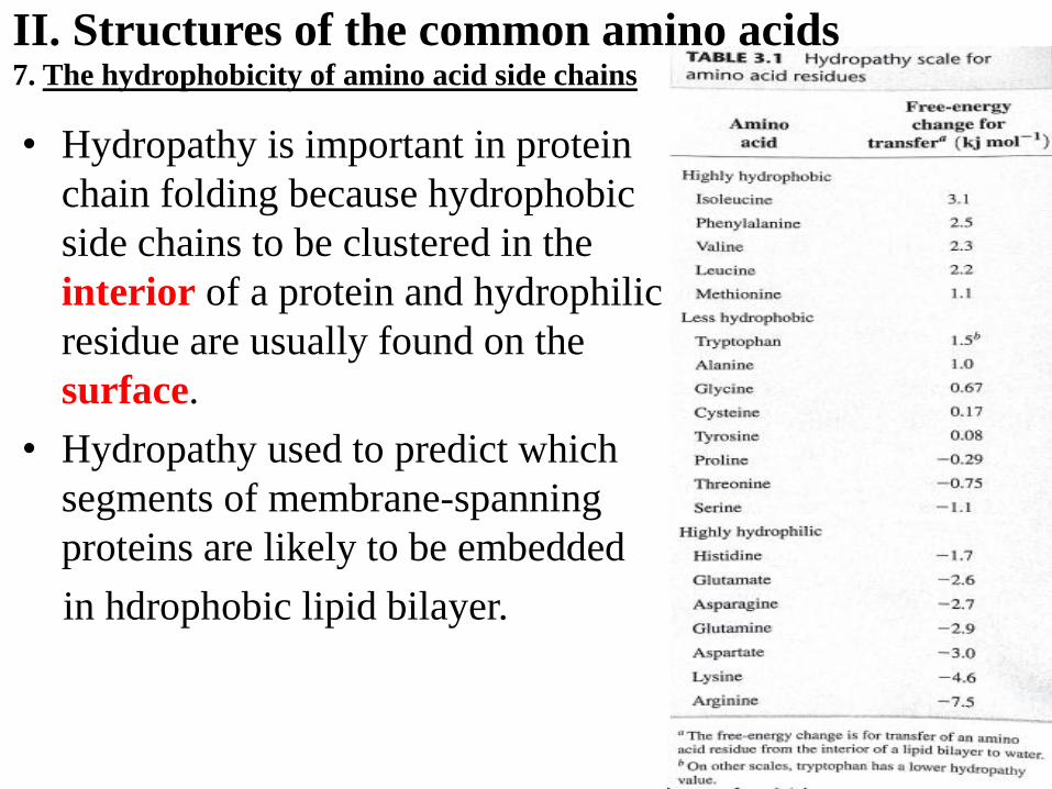
II. Structures of the common amino acids 7. The hydrophobicity of amino acid side chains
• Hydropathy is important in protein
chain folding because hydrophobic
side chains to be clustered in the
interior of a protein and hydrophilic
residue are usually found on the
surface.
• Hydropathy used to predict which
segments of membrane-spanning
proteins are likely to be embedded
in hdrophobic lipid bilayer.

III. Other amino acids and amino acid derivatives
More than 200 different a.a are found in living
organisms.
• S-Adenosylmethionine, methyl doner. Many
species of bacteria & fungai synthesized D-amino
acids that are used in cell walls & antibiotic
(actionomycin).
• In the mammalian brain, glutamate is converted to
the neurotansmitter γ-aminobutyrate (GABA).
• Histamine is synthesized mammals from histidine,
to controls the constriction of certain blood vessels
and secretion of HCL by stomach.

III. Other amino acids and amino acid derivatives
• Tyrosine is metabolized to epinephrine
(adrenaline).
• Hormons that are regulate metabolism in
mammals. Tyrosine is the precursor of the
thyroid hormones thyroxin.
• Small amount of sodium iodide prevent
goiter (hypothyroidism) caused by lack of
iodide in the diet.
• Some amino acids are chemically modified
after incorporated into polypeptides. Ex.,
proline residue in collagen are oxidized to
form hydroxyproline residue.

III. Other amino acids and amino acid derivatives
• Glycosylation process that is addition
carbohydrate to molecules such as proteins.
• Many protein are phosphorylated by the
addition of phosphoryl groups to the side
chains of, serine, threonine or tyrosine.
• the oxidation of cysteine residue to form
cystine occurs after a polypeptide synthesized.
• 21st a.a, selenocysteine is found from serine
during protein synthesis and it is incorporated
into a few proteins in a wide variety of species.
• 22nd a.a is pyrrolysine, found in some species
of archae bacteria. Pyrrolysine modified from
lysine that is synthesized after being added to a
growing peptide by the translation