AMINO ACID RADICALS IN RHENIUM-MODIFIED …thesis.library.caltech.edu/2295/1/Thesis.pdfAMINO ACID...
129
AMINO ACID RADICALS IN RHENIUM-MODIFIED COPPER PROTEINS Thesis by William Amine Wehbi In Partial Fulfillment of the Requirements for the Degree of Doctor of Philosophy California Institute of Technology Pasadena, California 2003 (Defended June 4, 2003)
-
Upload
vuongduong -
Category
Documents
-
view
223 -
download
3
Transcript of AMINO ACID RADICALS IN RHENIUM-MODIFIED …thesis.library.caltech.edu/2295/1/Thesis.pdfAMINO ACID...

AMINO ACID RADICALS IN RHENIUM-MODIFIED COPPER PROTEINS
Thesis by
William Amine Wehbi
In Partial Fulfillment of the Requirements
for the Degree of
Doctor of Philosophy
California Institute of Technology
Pasadena, California
2003
(Defended June 4, 2003)

ii
© 2003
William Amine Wehbi
All Rights Reserved
iii
Acknowledgements
There are many people at Caltech whom I would like to thank for making my
graduate experience personally and professionally enjoyable and memorable.
I first met Harry Gray in November 1995 when he came to the University of
Arizona to give a seminar on electron transfer in ruthenium-modified proteins. At that
time, I have to admit, the subject of his talk did not particularly interest me, even though I
learned something and I actually remember the talk. It's amazing how Harry has that
effect on people. Harry even frightened me when, as his host fiddled furiously through
twenty light switches, he turned to the audience, scanned them, and said with a glowing
grin: "I like to keep the lights on during my talk so that I can point out anyone who falls
asleep." Not knowing Harry's jovial personality, I took him seriously. Six and a half
years later, at a lecture in Copenhagen, as he started to present some of my results, Harry
called out to me to get my attention. I was wide-awake, although Harry may have a
different recollection. Either way, I figured that I had succeeded in the Gray group if
Harry addressed me directly during a seminar. I would like to thank Harry for his
generosity, for his support of me during my six years in graduate school, and for his
incredible enthusiasm for science. Harry introduced me to the Big Picture, a guest at
every group meeting, inorganic and organometallics seminar, Chem 153 lecture, and
sundry meetings. He gave me the freedom to figure things out for myself and to go
where my mind and my abilities would take me. This made the journey more difficult,
but also more worthwhile when I made it to the end. Thank you, Harry.
iv
I received all of my biological training in the labs of Doug Rees and Jack
Richards and I thank them for their time, patience, and insight. I would also like to thank
Mitchio Okumura and Nate Lewis (rounding out an All-Star committee) for their support
and encouragement.
I would like to thank Jay Winkler who always provided me with his critical (and
honest) assessment of the work that I did. Jay was rigorous in his demand for the details,
while never losing sight of our friend, the Big Picture.
John Enemark gave a naïve freshman a chance to do chemical research in his lab
nine years ago. I would like to thank him for getting my scientific career started and for
supporting and encouraging me ever since.
The Gray group was a great place to be a graduate student, with outstanding
colleagues and friends, whom I would like to thank: Malin Abrahamson, Gitrada Arjara,
Wendy Belliston, Jesper Bendix, Steve Contakes, Alex Dunn, Cecelia Engman, Jasmin
Faraone-Mennella, Mike Green, Corinna Hess, Kristine Jensen, Chip Kent, Judy Kim,
Liz Krider, Brian Leigh, Mike Machczynski, John Magyar, Libby Mayo, Alex Meier,
Kate Pletneva, Adrian Ponce, Jeff Rack, Karn Sorasaenee, Jason Telford, Akif Tezcan,
Randy Villahermosa, Jeremy Weaver, Oliver Wenger, Jon Wilker, and certainly my
classmates Xenia Amashukeli, Jenn Lee.
I would like to thank Pat Anderson, Sherry Feick, Kathleen Hand, Margot Hoyt,
Rick Jackson, Catherine May, Anne Penney, and Virginia Russell for many friendly
conversations and for needling me to the point of laughter, whenever I so desperately
needed a laugh. "Catherine—Where's Harry?" I'm sorry. I should know better by now.
v
I would like to thank Dian Buchness for taking care of all of the administrative
paperwork so that I could focus on the science. Dian always had a free moment (or 40 to
50 minutes) for me when I stopped by her office unannounced, and she always listened to
my frustrations and triumphs. Thank you, Dian!
Angelo Di Bilio (aka Dottore) got me started on the rhenium-azurin project.
From Dottore, I learned the practical aspects of the preparation of metal-modified
proteins; he also trained me to be a competent EPR spectroscopist. I had a valued
collaborator on the radical project, and we endured many of the anxieties and
uncertainties that any project inevitably provides. Grazie, Dottore.
Brian Crane and Cristian Gradinaru were the collaborators on the crystal structure
determination of the Re(His107) azurin. Brian was originally a postdoctoral colleague
and had a hand in getting me started on the rhenium-azurin project, one of his many
accomplishments at Caltech.
Michele McGuirl (M2) had a tremendous influence on me during her time as a
postdoctoral colleague. She patiently explained all of the biological steps that I had to go
through to get protein and kept my eyes open to problems of biological interest and
kindly gave me a tour of Yellowstone, a nice detour during a visit to her in Missoula.
Ivan Dmochowski and I spent three years as graduate student colleagues and in a
few months I will be his postdoc at Penn. I eagerly look forward to this adventure.
Katrine Museth and I collaborated on the rhenium-azurin project. Katrine made
all three of the His83 mutants starting with His107 plasmids; she and I got the cysteine
mutant experiments going together.

vi
Just over a year ago, Yen Hoang Le Nguyen joined the Gray Group and moved
into the office with me. Yen and I had many interesting discussions, which provided
necessary distractions while I was writing up my props and my thesis and she was writing
up her candidacy. The last year of graduate school is the most exhilarating and stressful,
but I could always count on Yen to put everything in the proper perspective. Thank you,
Yen!!
Julia Lyubovitsky and I shared an office for four years. Many interesting
conversations resulted, on various subject—life, science, psychology, language, and
history (to name a few)—and I found Julia's frank perspective refreshing. Towards the
end of our graduate experiences, we shared our office with a fourth person, Julia and
Russ' soon-to-be one-year-old son David, who always and easily managed to get the
attention of the third floor of Noyes and who, for some reason, always found it
fascinating. Julia liked to remind me about how much smarter David is than I, a point
that I could never argue against and that I am not likely to forget!
Kevin Hoke was a more senior graduate student when I joined the Gray Group
and helped get a first-year started in the lab on the third floor of Noyes. He was always
very generous with his time, and he patiently answered an endless list of my questions.
Jeremiah Miller was my resource for all the laser experiments whose results I
present in this thesis. In addition, Jeremiah was an outstanding collaborator to have on
the rhenium-azurin project and a great person to bounce (crazy) ideas off of, over the
ever-present cup of coffee. I discussed every aspect of my project and my thesis with
him and I appreciate his suggestions and insight, which improved both my thesis and my
vii
graduate experience. The youthful Jeremiah made PhD before I did (by a day). It was an
honor for me to work with him.
Pierre Kennepohl (aka Dr. Pierre) was worth the wait. During my entire career in
chemistry, I have not met anyone with more similar interests or approaches in science to
mine than Dr. Pierre. Even though we overlapped for just one year, I am forever in his
debt for his unwavering and infectious enthusiasm for science and for his optimistic
perspectives, both of which invariably cheered me up during the final stages of my stay in
graduate school. Dr. Pierre and I worked on the thiyl radicals together (experimentally
and theoretically); he also helped me with the theoretical calculations (and the
understanding of their results) on the rhenium-coordinated imidazole radicals, which I
present in this thesis. He reawakened my interest in ligand field theory and in the study
of the electronic structures of inorganic complexes ("The buzz word is spin-orbit
coupling. We don't know why, but it is."). He also very patiently listened to every
complaint or frustration that I had, over the bottomless cup of tea or diet-coke or, when I
was really down, 12 to 18 year old single-malt Scotch Whiskeys (for which I have
become quite the connoisseur!). Special thanks go to Jen Love, for letting me get away
with teasing Dr. Pierre endlessly. That was my way of continuously thanking him.
Lori Lee is an outstanding friend with whom I had many lengthy and memorable
conversations while indulging in fine chocolates or while running to the Rose Bowl and
throughout San Marino. From organic photochemistry to unraveling the meaning of
various mysteries in the Big Picture, I learned from Lori's logical approach, which differs
from mine, and benefited from all her points of view. Somehow, everything always
viii
became clearer after running an idea by Lori, a mystery in itself to me, but a tribute to her
for making me think from another perspective. Thank you, Lori!
Ramez El-Gammal and Sara Klamo are wonderful friends with whom I enjoyed
many extensive conversations over the Middle Eastern feasts we prepared, and over the
obligatory glasses (note the plural) of Arak and the plentiful supply of Turkish coffee. I
thank them for their encouragement and levelheadedness (and runs through San Marino!),
especially during the last few months when my life became the most stressful and hectic.
Thank you, Ramez and Sara!
Cindy Quezada is Lady Luck. For the last year, Cindy and I motivated each other
to finish our experiments and to write and defend our theses in time to walk in the 2003
commencement ceremony. We made it and we managed to have some fun along the
way, with the humor always (and rightfully) at my expense. Earlier on, whenever I got
colonies on plates of azurin mutants (and wanted to celebrate) or needed to run a gel, I
wandered over to Cindy's corner in the Simon Labs (I pondered asking Mel for bench
space!) and then proceeded to talk about what I was up to while she graciously listened.
When I struggled, Cindy always came up with the right suggestions that I had
overlooked, which always worked. I appreciate the perspectives, professional and
otherwise, that Cindy gave me along with her encouragement during the final stretch of
my graduate career. Lady Luck smiled upon me and for this I am grateful. Thank you,
Cindy. Thank you.
ix
I could never have come to this point in my life and career without the constant
support, encouragement, and patience from my family in the U.S.A. and Lebanon: Dad,
Mom, Jen, Tayta Faridi, Uncle George, Auntie Mary, Margaret, Linda, Georgia, Uncle
Samir, Auntie Amal, Rick, Sam, Uncle Nicola, Tayta Zakia, Auntie MM, Uncle Kamal,
Auntie Suzie, Uncle Maher, Auntie May, Nuha, Michel, Naji, Bassem, and Rhea. My
parents especially, who have put up with me the longest, put up with extended and
excessive stretches of time between my visits to them and with my venting phone calls at
all hours of the day and night whenever I felt that I could no longer handle the pressures
and stresses I put myself through. My sister always found clever ways to cheer me up
and then was gracious and patient with me when I was overly exuberant. Visits with my
Uncle George provided me with weekly escapes from Caltech and with the equanimity I
needed to maintain the focus on my work and the proper perspective on my
responsibilities in life. Except for me, no one in my family ever lost any sleep over the
role of tyrosyl radicals in multi-step tunneling or over how a sulfinyl radical could have
formed in a supposedly anaerobic EPR tube (to name two examples). Even when they
wondered what I was studying, how I could it find it exciting, and why it consumed six
years of my life, they always had confidence in me and in the path that I chose. The most
difficult part of graduate school was the geographical separation I had to endure from the
ones I love most. Because of this, I know how precious the moments are that I spend
with them.
Finally, the year 2003 marks the Centennial of the birth of my paternal
grandfather Amine. I never met him or my maternal grandfather Michel, yet the effects
x
of their lives and of their losses have been and are still visible on the generations who
followed them in the last half-century. My greatest inspiration has come from my great-
uncle William, who filled the role of grandfather. He was an outstanding teacher and
scholar who set for his namesake nephew the highest possible standards with his
integrity, diligence, patience, enthusiasm, and optimism.
I DEDICATE THIS THESIS TO THE MEMORY OF
UNCLE WILLIAM.
xi
Table of Contents
Acknowledgements iii
Table of Contents x
Abstract xi
Chapter 1: Introduction 1
Chapter 2: Histidine Radicals Coordinated to Rhenium 22
Chapter 3: Tyrosyl Radicals 53
Chapter 4: Cysteine-Based Radicals 75
Appendix A: Preparation of Rhenium-Modified Azurins 100
Appendix B: Photochemical and Spectroscopic Methods 112
Epilogue 115
xii
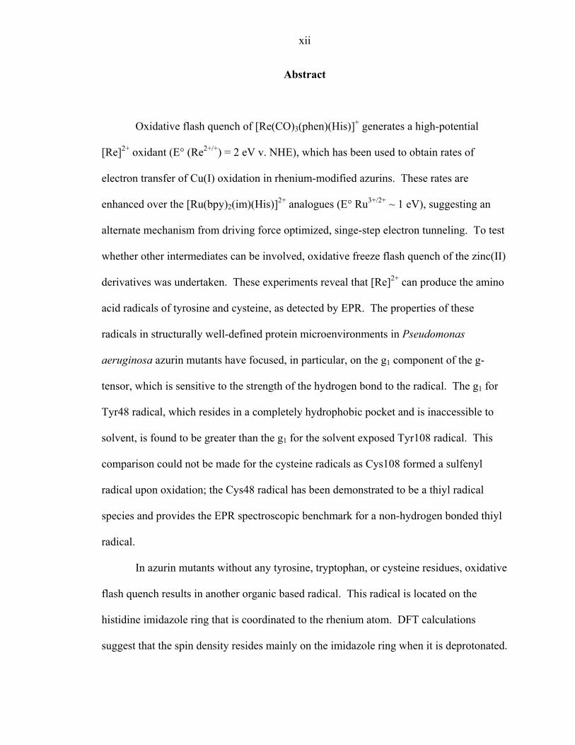
Abstract
Oxidative flash quench of [Re(CO)3(phen)(His)]+ generates a high-potential
[Re]2+ oxidant (E° (Re2+/+) = 2 eV v. NHE), which has been used to obtain rates of
electron transfer of Cu(I) oxidation in rhenium-modified azurins. These rates are
enhanced over the [Ru(bpy)2(im)(His)]2+ analogues (E° Ru3+/2+ ~ 1 eV), suggesting an
alternate mechanism from driving force optimized, singe-step electron tunneling. To test
whether other intermediates can be involved, oxidative freeze flash quench of the zinc(II)
derivatives was undertaken. These experiments reveal that [Re]2+ can produce the amino
acid radicals of tyrosine and cysteine, as detected by EPR. The properties of these
radicals in structurally well-defined protein microenvironments in Pseudomonas
aeruginosa azurin mutants have focused, in particular, on the g1 component of the g-
tensor, which is sensitive to the strength of the hydrogen bond to the radical. The g1 for
Tyr48 radical, which resides in a completely hydrophobic pocket and is inaccessible to
solvent, is found to be greater than the g1 for the solvent exposed Tyr108 radical. This
comparison could not be made for the cysteine radicals as Cys108 formed a sulfenyl
radical upon oxidation; the Cys48 radical has been demonstrated to be a thiyl radical
species and provides the EPR spectroscopic benchmark for a non-hydrogen bonded thiyl
radical.
In azurin mutants without any tyrosine, tryptophan, or cysteine residues, oxidative
flash quench results in another organic based radical. This radical is located on the
histidine imidazole ring that is coordinated to the rhenium atom. DFT calculations
suggest that the spin density resides mainly on the imidazole ring when it is deprotonated.
xiii
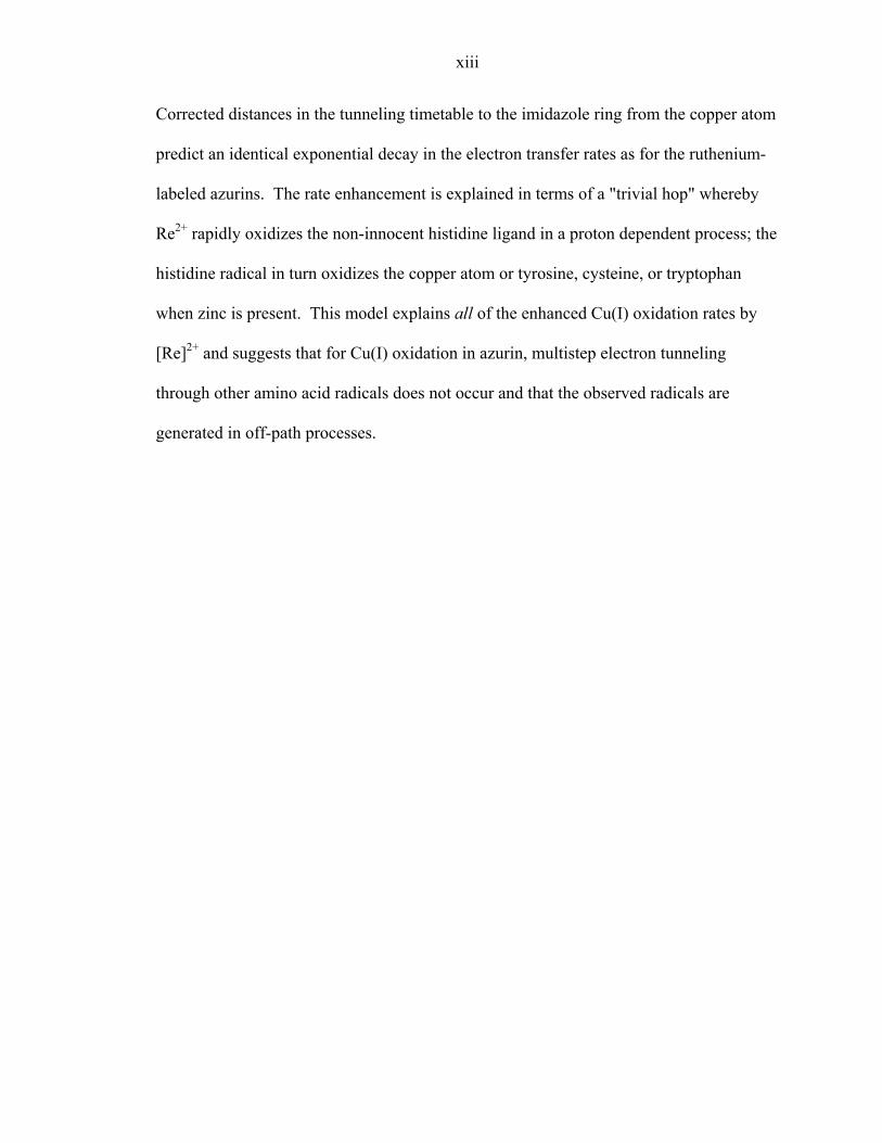
Corrected distances in the tunneling timetable to the imidazole ring from the copper atom
predict an identical exponential decay in the electron transfer rates as for the ruthenium-
labeled azurins. The rate enhancement is explained in terms of a "trivial hop" whereby
Re2+ rapidly oxidizes the non-innocent histidine ligand in a proton dependent process; the
histidine radical in turn oxidizes the copper atom or tyrosine, cysteine, or tryptophan
when zinc is present. This model explains all of the enhanced Cu(I) oxidation rates by
[Re]2+ and suggests that for Cu(I) oxidation in azurin, multistep electron tunneling
through other amino acid radicals does not occur and that the observed radicals are
generated in off-path processes.
1
Chapter 1
Introduction

2
In 1900, Gomberg reported on his attempts to prepare hexaphenylethane.1 He had
proposed that he could reach the target molecule by adding silver metal to a solution of
triphenylmethylchloride in benzene (Equation 1).
2 Ph3CCl + Ag Ph3C—CPh3 + 2 AgCl (1)
While the elemental analysis for carbon and hydrogen matched the predicted value, the
physical and chemical properties of the product did not fit the expectation that
hexaphenylethane, a saturated alkane, would be colorless and unreactive. Although
Gomberg found that the crystals of the compound were colorless, the solutions were
yellow-orange. The solution reacted with oxygen to give the triphenylmethyl peroxide
dimer, with iodine to give triphenylmethyliodide, and hydrogen (on platinum metal) to
give triphenylmethane, all of which were characterized. In addition, Gomberg found that
the mystery compound was sensitive to acid and light and that it conducted an electric
current in liquid SO2. These observations led Gomberg to suggest that he had prepared
an unsaturated species with a trivalent carbon atom, the triphenylmethyl radical, which
became the first compound known to deviate from Kekulé's quadrivalent theory of
carbon. Further preparations of various triarylmethyl radicals and other radicals along
with the studies of their properties and reactivities were carried out by Gomberg and
others, with Gomberg leading the field.2-5 The structure of the colorless solid that
Gomberg obtained was proposed in 1904 to be an unsymmetrical quinoid (Equation 2),
but was not confirmed until 1968 by NMR (the history of these developments has been
reviewed by McBride6).

3
A recent review of radical chemistry in the twentieth century provides a modern
perspective on these early studies on radicals.7
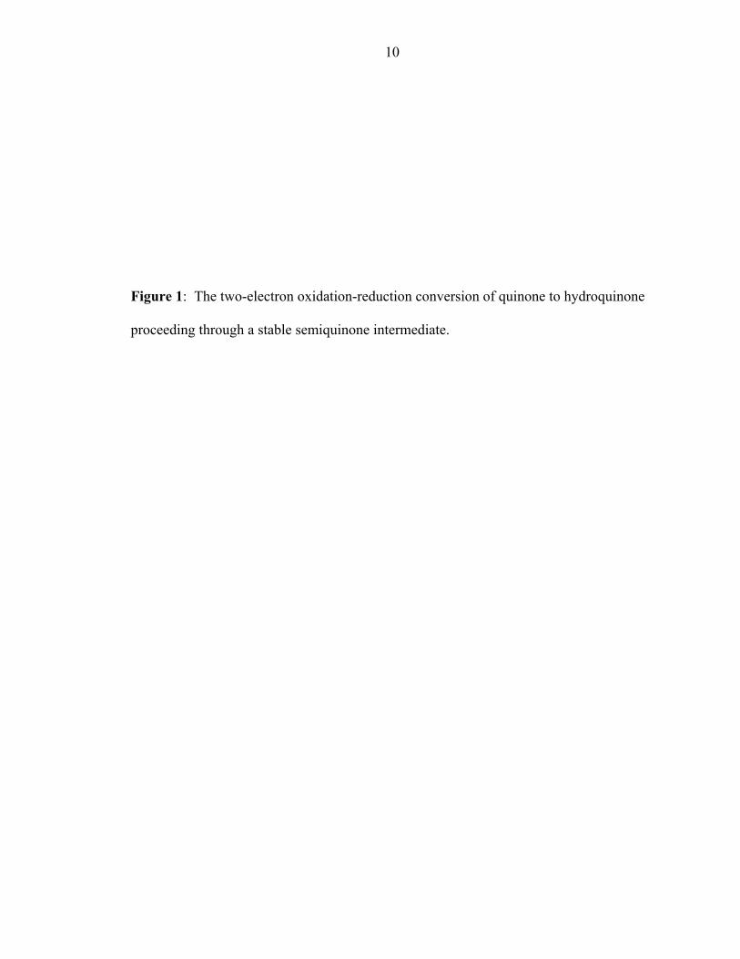
In the 1930s, Michaelis investigated the reversible oxidation-reduction properties
of quinols to quinones and determined that this two-electron reaction can be carried out in
two sequential one-electron steps through a stable semiquinone intermediate (Figure 1).8,9
Furthermore, Michaelis speculated on the nature and role of radicals (especially
semiquinones) in the oxidative catalytic processes of enzymes and also on the role of the
protein medium for stabilizing or destabilizing the resonance structures of the radicals.10-
12 The techniques available to Michaelis for studying radicals were potentiometric (by
redox titrations), magnetic (susceptibility studies), and colorimetric, all three of which
could only be applied to stable radicals. With the development of electron paramagnetic
resonance (EPR) spectroscopy in the 1940s and 1950s and its early applications to
biological samples,13 a new technique emerged for studying radicals. In addition, frozen
samples could also be irradiated with ionizing γ-rays and the stability and reactivity of the
generated radicals could be studied as a function of temperature (4-200 K). The first
experiments were performed on various biological tissues,14,15 which gave resonances
centered around the free electron g value (ge = 2.0023). Gordy extended these studies to
irradiated solutions, powders, and single crystals of the amino acids, peptides, and
proteins, thus beginning the study of the properties of amino acid radicals.16-20
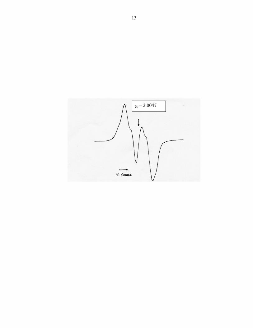
In 1973, Ehrenberg and Reichard21 reported the EPR spectrum (Figure 2) of a
stable organic-based radical in the resting state of the B2 protein ribonucleotide reductase
(RNR) from E. coli. The RNR system from E. coli consists of two separate proteins that
are responsible for the reduction of ribonucleotides to deoxyribonucleotides. The B2
4
(also called R2 in the current literature) protein is a di-iron enzyme, which activates
molecular oxygen to produce a stable organic radical. The reduction of ribonucleotides
takes place in the B1 (also called R1) subunit and its initiation (by a thiyl radical, vide
infra) is dependent on the presence of the B2 subunit and on this radical22 (the crystal
structures of both proteins have been solved separately, R2 in 1990,23 and R1 in 1994,24
and models have been proposed,25 but not confirmed, for complex formation).
Subsequent work confirmed that the stable radical in the B2 protein is tyrosyl.26,27 This
discovery, one of the many surprises in biochemistry,28 prompted the search for other
amino acid radicals in proteins, and led to extensive studies to elucidate the roles of these
intermediates in enzyme catalysis.29-36
In addition to the tyrosyl radical in ribonucleotide reductase, two tyrosyl radicals
have been found in photosystem II. A EPR signal assigned as D·+ was first reported in
1956 in irradiated chloroplasts.37 In the 1970s, a similar EPR signal, called Z·+, was
detected.38,39 This radical species was found to be required for catalytic activity in the
oxidation of water and is much less stable than the catalytically inactive D·+. In 1987,
Babcock presented a study in which the two radicals of PSII were assigned to tyrosine.40
Subsequent work on the kinetic and spectroscopic properties of these tyrosine residues
(now called TyrZ and TyrD for Z and D, respectively) led to a metalloradical mechanism
in PSII for the oxidation of water to molecular oxygen whereby the TyrZ reduces the
oxidized chlorophyll special pair (P680+) in a proton dependent process. In turn, the
radical on TyrZ proceeds to oxidize the manganese cluster in the process of water
oxidation. Babcock's monumental work41-47 on the tyrosyl radicals in PSII also looked
into the proton dependence of the electron transfer oxidation and reduction processes at
5
TyrZ during photosynthetic water oxidation as an example of proton coupled electron
transfer. In addition to PSII, prostaglandin H synthase has also been found to require a
tyrosyl radical intermediate in the oxygenation of arachidonic acid to prostaglandin G2.48-
50
Tyrosyl radicals were the first amino acid radicals to be discovered and have been
the most extensively studied.51,52 In addition, some enzymes such as the copper amine
oxidases can modify a particular tyrosine residue to topa quinone (Michaelis' quinone is
now protein based rather than exogenous), which is catalytically active.53-55 Galactose
oxidase has also been found to contain a tyrosyl radical intermediate in catalysis.56-58
This residue, like the topa quinone, is post-translationally modified via a cross link of
cysteine to tyrosine.59 The phenol end of the tyrosine residue is also coordinated to
copper. Thus, a two-electron redox reaction can occur, with one equivalent coming from
copper and the other from the non-innocent tyrosine ligand. Examples of other novel
cofactors and post-translational modified amino acid residue in proteins have been
reviewed recently.60,61
Subsequent to the discovery of tyrosyl radical intermediates in enzyme catalytic
processes, tryptophan radicals were identified in cytochrome c peroxidase (spin-coupled
to a ferric heme),62-65 DNA photolyase (free),66-70 as well as various engineered
tryptophan mutants of RNR.71-75 Except for a commentary76 following the initial
discovery of tryptophan radicals, this field has not been reviewed.
In 1989, Boussac and coworkers observed an organic radical intermediate EPR
signal in the S2 S3 step in the photocycle of Ca2+ depleted photosystem II.77 As the
S3 signal is normally undetectable by EPR, this curious result raised questions about the

6
nature of the radical. Oxidized amino acids that interact magnetically with the
manganese complex were considered as possible candidates for the radical. On the basis
of an optical spectrum of this modified S3 state, a histidinyl radical was proposed to
correspond to the signal occurring in the modified S3 state.78 Further investigation79
challenged this assignment with the suggestion that the radical is TyrZ, which prompted a
reply80 containing analyses reinforcing the original tentative assignment as more probably
a histidinyl radical than tyrosyl radical. Since these experiments, the histidinyl radical
has not since been revisited as an intermediate in PSII. However, a recent report suggests
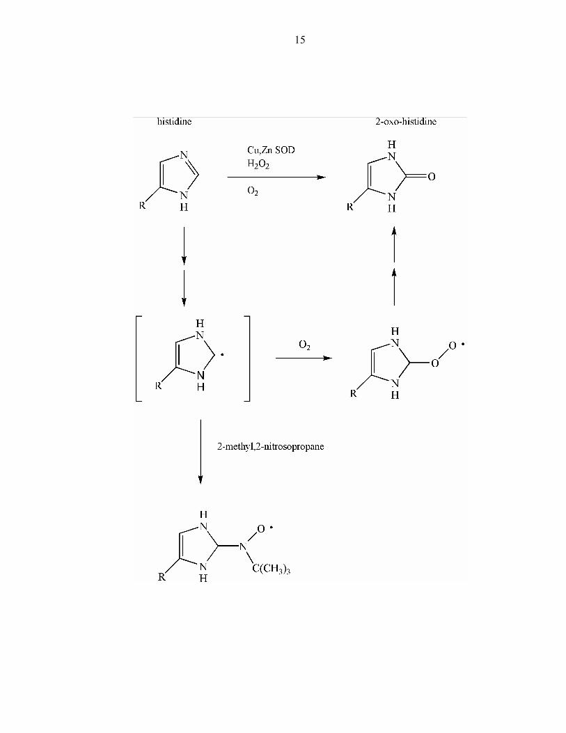
histidinyl radical formation in a copper-zinc superoxide dismutase (Cu/Zn SOD) as
detected by 2-methyl-2-nitrosopropane spin trapping.81 This study followed the
characterization of 2-oxo-histidine that was generated selectively at one histidine
(His118) in bovine Cu/Zn SOD upon addition of H2O2 to the enzyme.82 The spin-trapped
adduct on His118 (Figure 3) provided another validating data point for Michaelis'
original hypothesis that two-electron oxidation-reductions proceed in one-electron steps,
even though the overall mechanism of formation of 2-oxo-histidine has not yet been
established.
In the 1980s, while working on pyruvate formate lyase (PFL), an EPR doublet
with a 15 G separation was observed as a catalytic intermediate.83,84 This signal
collapsed into a singlet in deuterated buffer; the radical was assigned to glycine,
providing the first example of a Cα backbone radical,85 whose α-proton is exchangeable
with deuteron, explaining the conversion of the EPR doublet to a singlet. In addition,
PFL loses all activity in the presence of oxygen and subsequent cleavage occurs on the
polypeptide chain at the N—Cα bond of the glycine.85 While studying the mechanism of

7
oxygen inactivation of PFL, Kozarich and coworkers presented evidence for the glycyl
peroxyl radical, which is expected to be responsible for backbone cleavage, and also a
long-lived cysteine-based sulfinyl (RSO·) radical.86,87 Glycyl radicals have since been
assigned in the Class III ribonucleotide reductase88 and in benzylsuccinate synthase,89
which derive from anaerobic organisms. In all three known cases, the glycyl radicals are
generated by hydrogen atom abstraction by 5'-deoxyadenosyl free radical following
homolytic cleavage of S-adenosylmethionine.90 The glycyl radical, in turn, abstracts a
hydrogen atom from an adjacent cysteine residue, which is the catalytically active
species. A more detailed review of glycyl radical enzymes has recently appeared.91
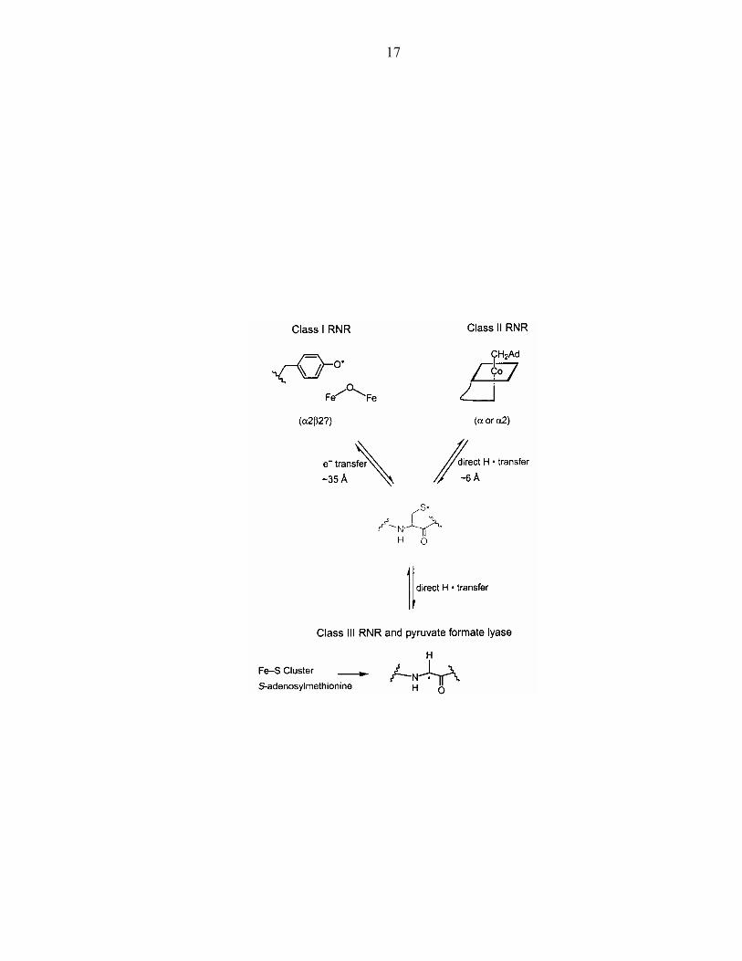
In the course of the study of the three classes of ribonucleotide reductase, Stubbe
found that all three enzymes required a cysteine residue for proper function.92 The
proposed mechanism has a cysteine thiyl radical abstracting H-atoms from
ribonucleotides in the initiating step and then providing H-atom reducing equivalents in
the final step, making the cysteinyl radical catalytic (the formal reducing equivalents are
provided by two other cysteines going to cystine). Here, Stubbe found an example of a
divergent evolutionary process (Figure 4) in which reactivity was centered around
cysteinyl radicals that were generated in different ways. In class I, a tyrosyl radical is
presumed to initiate a radical chain of oxidations to get to the catalytic cysteine; in class
II, adenosine cobalamin produces the thiyl radical; in class III, a glycyl radical oxidizes
cysteine. Stubbe has observed the thiyl radical in the class II RNR and found that even
though it is spin-coupled to the cob(II)alamin, deuteration of the β-protons of cysteine
lead to changes in lineshape, due to the hyperfine coupling to I = 1 nuclei compared to I =
½ nuclei and their different nuclear magnetic moments.93 To date, this is the only thiyl

8
radical that has been detected, although it is not the only thiyl radical catalytic
intermediate in an enzyme process. Lassmann recently reported the detection of thiyl and
sulfinyl radicals in γ-irradiated RNR94,95 and Graslund96 has demonstrated sulfinyl radical
by dioxygen activation in RNR by introducing a cysteine residue near the di-iron center
of the R2 protein.
With the demonstration of catalytically active amino acid radicals in enzyme
catalysis, it became necessary to assess the redox properties of these radicals and their
role in long-range electron transfer. Dutton and coworkers have inserted tyrosine and
tryptophan residues into protein maquettes and have studied their redox properties by
differential scanning calorimetry.97 With theoretical methods to treat the effective
dielectric around these radicals, they have established the dependence of the
electrochemical properties of these radicals on their protein microenvironment. High-
frequency EPR has been used by Un,98,99 Griffin,100 and Lendzian74 to determine accurate
values of the g-tensor components in tyrosyl and tryptophan radicals (a recent review101
of high-frequency EPR and its applications to bioinorganic chemistry provides more
extensive references to the wide range of applications of this technique to organic
radicals and to metal ions and clusters). The g1 component is sensitive to the strength of
the hydrogen bond to the tyrosyl radical and can provide a handle on the polarity of the
protein microenvironment; and the g2 and g3 components have been found to remain
essentially constant for all tyrosyl radicals measured to date.
In their work on electron transfer in proteins, Gray and coworkers have
established the distance and driving-force dependences on oxidations by Ru3+ in
ruthenium-modified proteins.102-104 From these studies, estimates of the reorganization
9
energy have been made. When the driving force equals the reorganization energy, the
electron transfer rate is activationless (and thus fastest). Further increases in the driving
force can lead to inverted rates, first proposed by Marcus,105,106 where the rate of the
electron transfer step decreases with increasing driving force. Gray and coworkers
reacted fac-[ReI(CO)3(phen)(OH2)]+ with azurin (substituting the aquo for a single
surface histidine) with the intention of studying electron transfer at higher driving forces
than those afforded by ruthenium complexes: E° Re2+/+ = 2 eV; E° Ru3+/2+ ~ 1 eV.107
The possible outcomes include: slower rates, due to the inverted effect; faster rates due
to multistep tunneling through amino acid radical intermediates; enhanced rates due to
the near resonance of the 2 eV rhenium acceptor with the 2.2 eV bridge energy (measured
for the oxidation of toluene and applied to phenylalanine).108 The last of these models is
the tunneling energy effect first proposed by McConnell109 and developed further by
Beratan.110 Oxidation rates were found to be enhanced and therefore a systematic study
was undertaken to determine whether or not amino acid radicals can be produced by Re2+
and whether they have a role in multistep electron tunneling.111 A preliminary
communication has appeared describing the formation of tryptophan and tyrosyl radicals
in rhenium modified copper proteins.112
This thesis explores the nature of photogenerated [Re]2+ and the properties of
tyrosyl and cysteinyl radicals in rhenium-modified azurins and their effects on the rate of
oxidation of Cu(I). Tryptophan radicals in rhenium-modified azurins and [Re]2+ model
complexes are treated in depth in the thesis of Jeremiah Miller.113
10
Figure 1: The two-electron oxidation-reduction conversion of quinone to hydroquinone
proceeding through a stable semiquinone intermediate.

11
12
Figure 2: EPR spectrum of the stable organic radical species in RNR (adapted from
Ehrenberg and Reichard).21
13
g = 2.0047
14
Figure 3: Possible scheme of 2-oxo-histidine formation through a histidinyl radical.
15
16
Figure 4: Divergent evolution illustrated by the three classes of RNR.92
17
18
References
(1) Gomberg, M. J. Am. Chem. Soc. 1900, 22, 757-771. (2) Gomberg, M. J. Am. Chem. Soc. 1914, 36, 1144-1170. (3) Gomberg, M. Chem. Rev. 1924, 1, 91-141. (4) Gomberg, M. Chem. Rev. 1925, 2, 301-314. (5) Gomberg, M. J. Chem. Ed. 1932, 9, 439-451. (6) McBride, J. M. Tetrahedron 1974, 30, 2009-2022. (7) Tidwell, T. T. Adv. Phys. Org. Chem. 2001, 36, 1-58. (8) Michaelis, L. Chem. Rev. 1935, 16, 243-286. (9) Michaelis, L.; Schubert, M. P. Chem. Rev. 1938, 22, 437-470. (10) Michaelis, L.; Smythe, C. V. Annu. Rev. Biochem. 1938, 71, 1-36. (11) Michaelis, L. Cold Springs Harbor Symp. Quant. Biol. 1939, 7, 33-49. (12) Michaelis, L. In The Enzymes; Sumner, J. B., Myrback, K., Eds.; Academic Press:
New York, 1951; Vol. 2. (13) Sogo, P. B.; Tolbert, B. M. In Advances in Biological and Medicinal Physics;
Lawrence, J. H., Tobias, C. A., Eds.; Academic Press: New York, 1957; Vol. 5, pp 1-35.
(14) Commoner, B.; Townsend, J.; Pake, G. E. Nature 1954, 174, 689-691. (15) Commoner, B.; Heise, J. J.; Lippincott, B. B.; Norberg, R. E.; Passonneau, J. V.;
Townsend, J. Science 1957, 126, 57-63. (16) Gordy, W.; Ard, W. B.; Shields, H. Proc. Nat. Acad. Sci. U.S.A. 1955, 41, 983-
996. (17) Gordy, W.; Ard, W. B.; Shields, H. Proc. Nat. Acad. Sci. U.S.A. 1955, 41, 996-
1004. (18) Shields, H.; Gordy, W. J. Phys. Chem. 1958, 62, 783-789. (19) Shields, H.; Gordy, W. J. Phys. Chem. 1958, 62, 789-798. (20) Gordy, W.; Shields, H. Radiation Research 1958, 9, 611-625. (21) Ehrenberg, A.; Reichard, P. J. Biol. Chem. 1972, 247, 3485-3488. (22) Atkin, C. L.; Thelander, L.; Reichard, P.; Lang, G. J. Biol. Chem. 1973, 248,
7464-7472. (23) Nordlund, P.; Sjöberg, B.-M.; Eklund, H. Nature 1990, 345, 593-598. (24) Uhlin, U.; Eklund, H. Nature 1994, 370, 533-539. (25) Eklund, H.; Uhlin, U.; Farnegardh, M.; Logan, D. T.; Nordlund, P. Prog. Biophys.
Mol. Biol. 2001, 77, 177-268. (26) Sjöberg, B.-M.; Reichard, P.; Gräslund, A.; Ehrenberg, A. J. Biol. Chem. 1977,
252, 536-541. (27) Reichard, P.; Ehrenberg, A. Science 1983, 221, 514-519. (28) Frey, P. A. Biochem. Mol. Biol. Ed. 2002, 30, 152-162. (29) Williams, R. J. P. Phil. Trans. Roy. Soc. London Ser. B 1985, 311, 593-603. (30) Stubbe, J. Biochemistry 1988, 27, 3893-3900. (31) Stubbe, J. Annu. Rev. Biochem. 1989, 58, 257-285. (32) Frey, P. A. Chem. Rev. 1990, 90, 1343-1357. (33) Pedersen, J. Z.; Finazzi-Agro, A. FEBS Lett. 1993, 325, 53-58.
19
(34) Marsh, E. N. G. BioEssays 1995, 17, 431-441. (35) Frey, P. A. Curr. Op. Chem. Biol. 1997, 1, 347-356. (36) Stubbe, J.; van der Donk, W. A. Chem. Rev. 1998, 98, 705-762. (37) Commoner, B.; Heise, J. J.; Townsend, J. Proc. Nat. Acad. Sci. U.S.A. 1956, 42,
710-718. (38) Blankenship, R. E.; Babcock, G. T.; Warden, J. T.; Sauer, K. Febs Letters 1975,
51, 287-293. (39) Babcock, G. T.; Sauer, K. Biochim. Biophys. Acta 1975, 376, 315-328. (40) Barry, B. A.; Babcock, G. T. Proc. Natl. Acad. Sci. U. S. A. 1987, 84, 7099-7103. (41) Babcock, G. T.; Barry, B. A.; Debus, R. J.; Hoganson, C. W.; Atamian, M.;
McIntosh, L.; Sithole, I.; Yocum, C. F. Biochemistry 1989, 28, 9557-9565. (42) Hoganson, C. W.; Babcock, G. T. In Metal Ions in Biological Systems, Vol 30,
1994; Vol. 30, pp 77-107. (43) Babcock, G. T.; Espe, M.; Hoganson, C.; Lydakis-Simantiris, N.; McCracken, J.;
Shi, W.; Styring, S.; Tommos, C.; Warncke, K. Acta Chem. Scand. 1997, 51, 533-540.
(44) Hoganson, C. W.; Babcock, G. T. Science 1997, 277, 1953-1956. (45) Tommos, C.; Hoganson, C. W.; Di Valentin, M.; Lydakis-Simantiris, N.; Dorlet,
P.; Westphal, K.; Chu, H. A.; McCracken, J.; Babcock, G. T. Curr. Op. Chem. Biol. 1998, 2, 244-252.
(46) Tommos, C.; Babcock, G. T. Acc. Chem. Res. 1998, 31, 18-25. (47) Tommos, C.; Babcock, G. T. Biochim. Biophys. Acta 2000, 1458, 199-219. (48) Karthein, R.; Nastainczyk, W.; Ruf, H. H. Eur. J. Biochem. 1987, 166, 173-180. (49) Karthein, R.; Dietz, R.; Nastainczyk, W.; Ruf, H. H. Eur. J. Biochem. 1988, 171,
313-320. (50) Dietz, R.; Nastainczyk, W.; Ruf, H. H. Eur. J. Biochem. 1988, 171, 321-328. (51) Prince, R. C. Trends. Biochem. Sci. 1988, 13, 286-288. (52) Pesavento, R. P.; van der Donk, W. A. Adv. Prot. Chem. 2001, 58, 317-385. (53) Klinman, J. P.; Mu, D. Annu. Rev. Biochem. 1994, 63, 299-344. (54) Klinman, J. P. Chem. Rev. 1996, 96, 2541-2561. (55) Klinman, J. P. J. Biol. Chem. 1996, 271, 27189-27192. (56) Whittaker, M. M.; Whittaker, J. W. J. Biol. Chem. 1988, 263, 6074-6080. (57) Whittaker, M. M.; Whittaker, J. W. J. Biol. Chem. 1990, 265, 9610-9613. (58) Whittaker, J. W. Adv. Prot. Chem. 2002, 60, 1-49. (59) Ito, N.; Phillips, S. E. V.; Stevens, C.; Ogel, Z. B.; McPherson, M. J.; Keen, J. N.;
Yadav, K. D. S.; Knowles, P. F. Nature 1991, 350, 87-90. (60) Okeley, N. M.; van der Donk, W. A. Chem. Biol. 2000, 7, R159-R171. (61) Klinman, J. P.; Dove, J. E., Eds. Adv. Prot. Chem.; Academic Press: San Diego,
2001; Vol. 58. (62) Erman, J. E.; Vitello, L. B.; Mauro, J. M.; Kraut, J. Biochemistry 1989, 28, 7992-
7995. (63) Sivaraja, M.; Goodin, D. B.; Smith, M.; Hoffman, B. M. Science 1989, 245, 738-
740. (64) Houseman, A. L. P.; Doan, P. E.; Goodin, D. B.; Hoffman, B. M. Biochemistry
1993, 32, 4430-4443.
20
(65) Huyett, J. E.; Doan, P. E.; Gurbiel, R.; Houseman, A. L. P.; Sivaraja, M.; Goodin, D. B.; Hoffman, B. M. J. Am. Chem. Soc. 1995, 117, 9033-9041.
(66) Heelis, P. F.; Okamura, T.; Sancar, A. Biochemistry 1990, 29, 5694-5698. (67) Li, Y. F.; Heelis, P. F.; Sancar, A. Biochemistry 1991, 30, 6322-6329. (68) Essenmacher, C.; Kim, S.-T.; Atamian, M.; Babcock, G. T.; Sancar, A. J. Am.
Chem. Soc. 1993, 115, 1602-1603. (69) Kim, S.-T.; Sancar, A.; Essenmacher, C.; Babcock, G. T. Proc. Nat. Acad. Sci.
U.S.A. 1993, 90, 8023-8027. (70) Aubert, C.; Vos, M. H.; Mathis, P.; Eker, A. P. M.; Brettel, K. Nature 2000, 405,
586-590. (71) Bollinger, J. M., Jr.; Tong, W. H.; Ravi, N.; Huynh, B. H.; Edmondson, D. E.;
Stubbe, J. J. Am. Chem. Soc. 1994, 116, 8024-8032. (72) Baldwin, J.; Krebs, C.; Ley, B. A.; Edmondson, D. E.; Huynh, B. H.; Bollinger, J.
M., Jr. J. Am. Chem. Soc. 2000, 122, 12195-12206. (73) Krebs, C.; Chen, S.; Baldwin, J.; Ley, B. A.; Patel, U.; Edmondson, D. E.; Huynh,
B. H.; Bollinger, J. M., Jr. J. Am. Chem. Soc. 2000, 122, 12207-12219. (74) Bleifuss, G.; Kolberg, M.; Potsch, S.; Hofbauer, W.; Bittl, R.; Lubitz, W.;
Graslund, A.; Lassmann, G.; Lendzian, F. Biochemistry 2001, 40, 15362-15368. (75) Ivancich, A.; Dorlet, P.; Goodin, D. B.; Un, S. J. Am. Chem. Soc. 2001, 123,
5050-5058. (76) Prince, R. C.; George, G. N. Trends. Biochem. Sci. 1990, 15, 170-172. (77) Boussac, A.; Zimmermann, J.-L.; Rutherford, A. W. Biochemistry 1989, 28,
8984-8989. (78) Boussac, A.; Zimmermann, J.-L.; Rutherford, A. W.; Lavergne, J. Nature 1990,
347, 303-306. (79) Hallahan, B. J.; Nugent, J. H. A.; Warden, J. T.; Evans, M. C. W. Biochemistry
1992, 31, 4562-4573. (80) Boussac, A.; Rutherford, A. W. Biochemistry 1992, 31, 7441-7445. (81) Gunther, M. R.; Peters, J. A.; Sivaneri, M. K. J. Biol. Chem. 2002, 277, 9160-
9166. (82) Uchida, K.; Kawakishi, S. J. Biol. Chem. 1994, 269, 2405-2410. (83) Knappe, J.; Neugebauer, F. A.; Blaschkowski, H. P.; Gänzler, M. Proc. Nat.
Acad. Sci. U.S.A. 1984, 81, 1332-1335. (84) Knappe, J.; Wagner, A. F. V. Methods Enzymol. 1995, 258, 343-362. (85) Wagner, A. F. V.; Frey, M.; Neugebauer, F. A.; Schäfer, W.; Knappe, J. Proc.
Nat. Acad. Sci. U.S.A. 1992, 89, 996-1000. (86) Reddy, S. G.; Wong, K. K.; Parast, C. V.; Peisach, J.; Magliozzo, R. S.; Kozarich,
J. W. Biochemistry 1998, 37, 558-563. (87) Zhang, W.; Wong, K. K.; Magliozzo, R. S.; Kozarich, J. W. Biochemistry 2001,
40, 4123-4130. (88) Sun, X.; Ollagnier, S.; Schmidt, P. P.; Atta, M.; Mulliez, E.; Lepape, L.; Eliasson,
R.; Gräslund, A.; Fontecave, M.; Reichard, P.; Sjöberg, B.-M. J. Biol. Chem. 1996, 271, 6827-6831.
(89) Krieger, C. J.; Roseboom, W.; Albracht, S. P. J.; Spormann, A. M. J. Biol. Chem. 2001, 276, 12924-12927.
(90) Frey, P. A. Annu. Rev. Biochem. 2001, 70, 121-148.
21
(91) Knappe, J.; Wagner, A. F. V. Adv. Prot. Chem. 2001, 58, 277-315. (92) Stubbe, J.; Ge, J.; Yee, C. S. Trends Biochem.Sci. 2001, 26, 93-99. (93) Licht, S.; Gerfen, G. J.; Stubbe, J. A. Science 1996, 271, 477-481. (94) Kolberg, M.; Bleifuss, G.; Sjoberg, B. M.; Graslund, A.; Lubitz, W.; Lendzian, F.;
Lassmann, G. Arch. Biochem. Biophys. 2002, 397, 57-68. (95) Kolberg, M.; Bleifuss, G.; Graslund, A.; Sjoberg, B. M.; Lubitz, W.; Lendzian, F.;
Lassmann, G. Arch. Biochem. Biophys. 2002, 403, 141-144. (96) Adrait, A.; Ohrstrom, M.; Barra, A. L.; Thelander, L.; Graslund, A. Biochemistry
2002, 41, 6510-6516. (97) Tommos, C.; Skalicky, J. J.; Pilloud, D. L.; Wand, A. J.; Dutton, P. L.
Biochemistry 1999, 38, 9495-9507. (98) Un, S.; Dorlet, P.; Rutherford, A. W. Appl. Mag. Reson. 2001, 21, 341-361. (99) Ivancich, A.; Dorlet, P.; Goodin, D. B.; Un, S. J. Am. Chem. Soc. 2001, 123,
5050-5058. (100) Bennati, M.; Stubbe, J.; Griffin, R. G. Appl. Mag. Reson. 2001, 21, 389-410. (101) Andersson, K. K.; Schmidt, P. P.; Katterle, B.; Strand, K. R.; Palmer, A. E.; Lee,
S.-K.; Solmon, E. I.; Gräslund, A.; Barra, A.-L. J. Biol. Inorg. Chem. 2003, 8, 235-247.
(102) Winkler, J. R.; Gray, H. B. Chem. Rev. 1992, 92, 369-379. (103) Bjerrum, M. J.; Casimiro, D. R.; Chang, I.-J.; Di Bilio, A. J.; Gray, H. B.; Hill, M.
G.; Langen, R.; Mines, G. A.; Skov, L. K.; Winkler, J. R.; Wuttke, D. S. J. Bioenerg. Biomemb. 1995, 27, 295-302.
(104) Gray, H. B.; Winkler, J. R. Annu. Rev. Biochem. 1996, 65, 537-561. (105) Marcus, R. A.; Sutin, N. Biochim. Biophys. Acta 1985, 811, 265-322. (106) Marcus, R. A. Angew. Chem. Int. Ed. Engl. 1993, 32, 1111-1121. (107) Connick, W. B.; DiBilio, A. J.; Hill, M. G.; Winkler, J. R.; Gray, H. B. Inorg.
Chim. Acta 1995, 240, 169-173. (108) Madec, C.; Courtot-Coupez, J. J. Electroanal. Chem. 1977, 84, 177-185. (109) McConnell, H. M. J. Chem. Phys. 1961, 35, 508-515. (110) Beratan, D. N.; Onuchic, J. N.; Hopfield, J. J. J. Chem. Phys. 1987, 86, 4488-
4498. (111) Winkler, J. R.; Di Bilio, A. J.; Farrow, N. A.; Richards, J. H.; Gray, H. B. Pure
Appl. Chem. 1999, 71, 1753-1764. (112) Di Bilio, A. J.; Crane, B. R.; Wehbi, W. A.; Kiser, C. N.; Abu-Omar, M. M.;
Carlos, R. M.; Richards, J. H.; Winkler, J. R.; Gray, H. B. J. Am. Chem. Soc. 2001, 123, 3181-3182.
(113) Miller, J. E. Ph.D. Thesis; California Institute of Technology: Pasadena, 2003.

22
Chapter 2
Histidine Radicals Coordinated to Rhenium

23
Introduction
Electron transfer reactions in metalloproteins have been studied extensively by
introducing ruthenium complexes to surface amino acid residues—in particular, single
surface histidines.1-3 In their ground states, the ruthenium complexes are kinetically and
thermodynamically unreactive to redox with the donor or acceptor of the metalloprotein.
However, following light excitation into their metal-to- ligand charge transfer (MLCT)
excited states, these ruthenium complexes become powerful oxidants and reductants
(Figure 1). In these photoinduced experiments, the rates of electron transfer that can be
measured are limited to the rates that are faster than those of the excited state emission
(kem = 1.7 x 10⁶ s-1 for Ru(bpy)32+*).4,5
To access a greater temporal window, the flash quench method is used.6 In this
approach, the excited states react with exogenous oxidative7,8 or reductive quenchers9-11
to yield ground state oxidants or reductants respectively (Figure 2). The fastest electron
transfer rate that can be measured is limited by the rate of excited state quenching (kq),
while the slowest depends on the stability of ground state oxidant or reductant in water.
Thus the range is defined by kq > kET > kgr, which spans from nanoseconds to seconds.
From these studies, complemented with site-directed mutagenesis to insert histidine
residues at different surface sites, a reliable picture can be obtained of the distance
dependence on the electron transfer and thus the role of the intervening medium, which in
turn have led to the proposed electron tunneling pathways model.12-14 In addition, with
the flash quench system, variation of the substituents of the ruthenium complex changes
the driving force of the electron transfer process. Thus, the dependence of the driving
24
force on the electron transfer has been monitored and experimental values for the
reorganization energy have also been obtained.3
The oxidative flash quench method for studying electron transfer in proteins has
been more thoroughly explored and applied in proteins (cytochrome c,6 azurin,15
plastocyanin,16 HiPip17 and DNA18) than the reductive flash quench method (which has
been applied in cytochrome c19 and cytochrome P45020 and protein triggered folding21-23).
In the oxidative flash quench experiment, Ru3+ is produced. For Ru(bpy)33+, the redox
potential is known (Eº Ru3+/2+ = 1.26 eV versus NHE in water)24-26 and its optical27,28 and
EPR spectra29,30 have been obtained (Ru3+ is stable in water on the order of minutes, but
can be reduced to Ru2+, presumably by the oxidation of water). The absorption and
luminescence properties27,31,32 of [Ru(bpy)3]2+ provide the complete spectroscopic handle
for the ruthenium system for time-dependent optical spectroscopic measurements.
The blue copper protein azurin, from Pseudomonas aeruginosa, is one of the
model protein systems in which oxidation have been studied by flash-quench of surface
attachment of ruthenium complexes. Azurin is a small protein (128 residues) that has a
beta sheet structure of eight antiparallel strands in which a copper atom is embedded.33-35
The single copper atom is coordina ted by two histidines (46, 117) and one cysteine (112)
in a trigonal plane; a methionine (121) sulfur and a backbone carbonyl oxygen of glycine
(45) are weakly coordinated axial ligands.36 This beta sheet structure has made azurin an
outstanding model for the study of electronic coupling of the beta sheet.15 These results
complemented the studies of electronic coupling in alpha helical structures.3 Along with
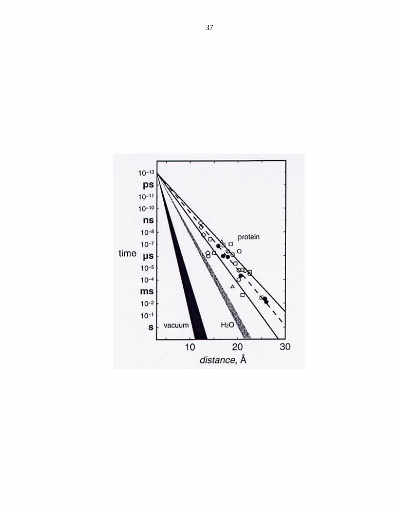
the studies of electronic coupling in water37 a master tunneling timetable was constructed
25
(Figure 3) clearly showing the exponential decay of the rate of electron transfer
depending on distance.
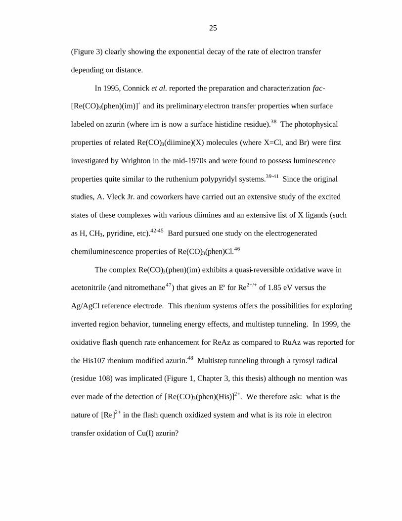
In 1995, Connick et al. reported the preparation and characterization fac-
[Re(CO)3(phen)(im)]+ and its preliminary electron transfer properties when surface
labeled on azurin (where im is now a surface histidine residue).38 The photophysical
properties of related Re(CO)3(diimine)(X) molecules (where X=Cl, and Br) were first
investigated by Wrighton in the mid-1970s and were found to possess luminescence
properties quite similar to the ruthenium polypyridyl systems.39-41 Since the original
studies, A. Vleck Jr. and coworkers have carried out an extensive study of the excited
states of these complexes with various diimines and an extensive list of X ligands (such
as H, CH3, pyridine, etc).42-45 Bard pursued one study on the electrogenerated
chemiluminescence properties of Re(CO)3(phen)Cl.46
The complex Re(CO)3(phen)(im) exhibits a quasi-reversible oxidative wave in
acetonitrile (and nitromethane47) that gives an Eº for Re2+/+ of 1.85 eV versus the
Ag/AgCl reference electrode. This rhenium systems offers the possibilities for exploring
inverted region behavior, tunneling energy effects, and multistep tunneling. In 1999, the
oxidative flash quench rate enhancement for ReAz as compared to RuAz was reported for
the His107 rhenium modified azurin.48 Multistep tunneling through a tyrosyl radical
(residue 108) was implicated (Figure 1, Chapter 3, this thesis) although no mention was
ever made of the detection of [Re(CO)3(phen)(His)]2+. We therefore ask: what is the
nature of [Re]2+ in the flash quench oxidized system and what is its role in electron
transfer oxidation of Cu(I) azurin?
26
Before addressing this question, we would like to note the rarity of mononuclear
complexes of Re2+ (by contrast, the Re2+Re2+ multiply bonded dimers are well known and
have been extensively studied.49 Ballhausen50 noted in 1962 that: "With the electronic
structure [Xe](5d)5(6s)2, rhenium should resemble Mn, and this expectation is found to be
justified. The greatest difference between the two elements is that Re++ is nearly
unknown." At that time, no Re2+ complex had been prepared and it was not until 1973
when Chatt and coworkers reported the first mononuclear complex of Re2+ as produced
by chemical oxidation of a Re+ dinitrogen complex.51 Very few Re2+ complexes have
since followed, although Harman and coworkers have recently demonstrated the
preparation of [Re(bpy)3]2+, TpRe(phen)Cl, and related complexes.52,53 It is interesting to
note the chemical similarities of Tc2+ and Re2+, which are without parallels to Mn2+
chemistry. One particular example is the absence of the fac-{Mn(CO)3} moiety, as
opposed to the extensive studies of the rhenium and technicium analogs.
In this chapter, we attempt to understand the properties of [Re(CO)3(phen)(His)]2+
in Pseudomonas aeruginosa azurin mutants as generated by the flash quench oxidation of
the Re+ excited state. The study of Re2+ model complexes is addressed by Jeremiah
Miller in his thesis.47
Materials and Methods
The two mutant azurins that are described in this chapter are (1)
W48F/Y72F/H83Q/Q107H/Y108F and (2) W48F/Y72F/Y108F. The preparation of
these mutants, the flash quench the rhenium-modified proteins, the EPR detection of flash

27
quench products, and the DFT methods of computation for [Re]2+ are described in the
Appendices. The results of the DFT calculations are applicable to the study of the [Re]2+
species in the rhenium-labeled azurins and in the [Re]2+ model systems generated by
oxidative flash quench.47 In this thesis, the results will be discussed in the context of the
[Re]2+ species in the protein. The complementary analysis of the model chemistry is
treated in the thesis of Jeremiah Miller.47
Electron Transfer. Electron transfer kinetics of Cu+ oxidation for both mutations
were measured on the Nanosecond 1 (NS-1) setup in the Beckman Institute Laser
Resource Center. Samples were prepared in 25 mM potassium phosphate, buffered to pH
7, and contained 50 µM rhenium-azurin and 5 mM Ru(NH3)63+. The azurins had been
reduced previously to Cu+ by 1 mM solution of sodium dithionite. Dithionite was
removed by gel- filtration (PD-10, Pharmacia).
Results
Electron Transfer. We pursued the flash quench oxidation of Cu(I) azurin in the
rhenium modified His107 mutant to compare with the published ruthenium rates. The
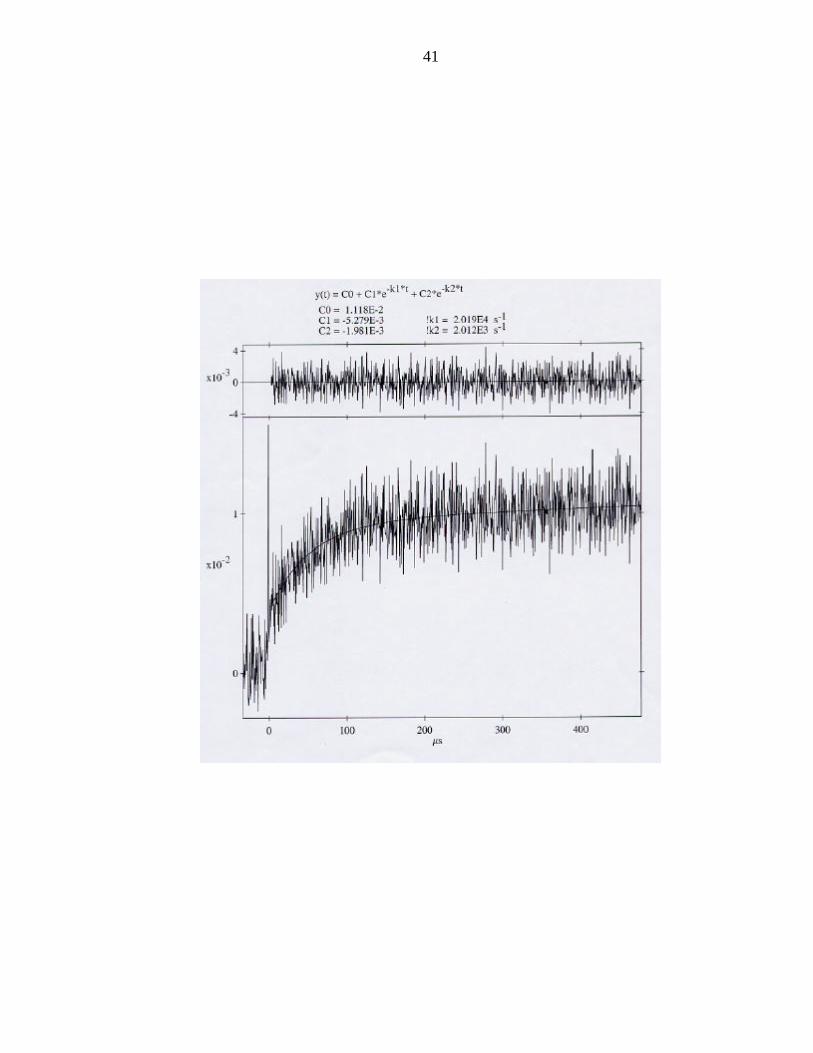
transient absorption spectrum monitored at 632.8 nm shows an enhanced rate for the
oxidation of Cu(I) by "Re2+" (Figure 5). Rates of Ru3+ oxidation of Cu(I) and the
analogous “Re2+” oxidations are plotted in Figure 6 as a function the rhenium to copper
distance.
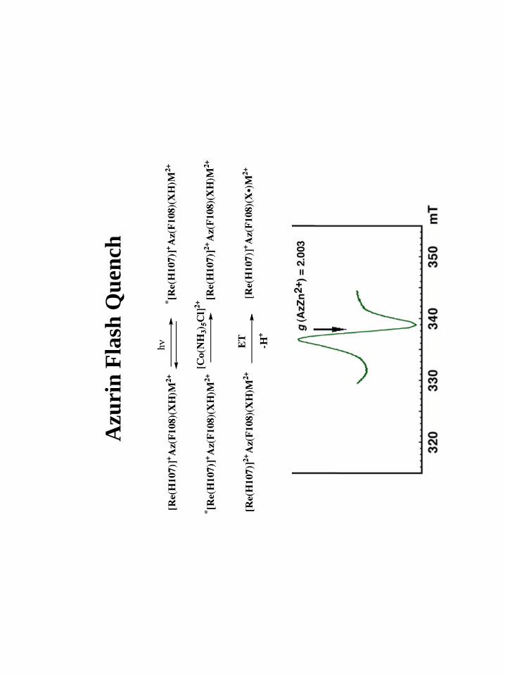
EPR. Oxidative flash quench of the Re His107 zinc azurins gave the X-band
spectra shown in Figures 7. The effective g-values were found to be 2.003. A high-
28
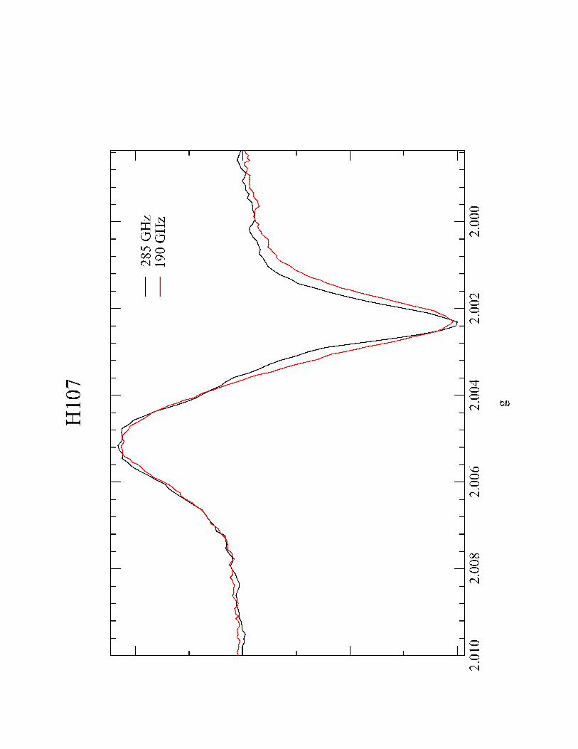
frequency, high-field EPR spectrum was obtained on the product of the His107 flash
quench oxidation (Figure 8) and confirms the effective g-value found by X-band. We
were unable to simulate these spectra into their g-tensor and hyperfine (A) tensor
components.
We would like to mention an ongoing collaboration with Professor A. Vlcek of
the University College in London on the nature of the excited states in the
Re(His107) azurin. The excited state dynamics of Re(His107) in the Cu(I) and Cu(II)
forms of azurin were studied along with the Re(Im) model complex (Dr. Angelo Di Bilio
and Professor Vlcek). These studies demonstrate that the charge transfer is rhenium to
phenanthroline in character; no imidazole oxidation in the charge transfer is observed (A.
Vleck, personal communication).
X-Ray Crystallography. The X-ray structure of the rhenium-labeled
W48F/Y72F/H83Q/Q107H/Y108F zinc azurin was solved by Mr. Cristian Gradinaru in
the laboratory of Professor Brain R. Crane at Cornell University using protein that was
sent from Caltech. The structure was found to be similar to the wild-type and other
ruthenium, rhenium, and osmium labeled azurins.35
Discussion
In the two mutant proteins studied in this chapter, all tyrosines and tryptophans
were removed from the protein, eliminating them as possible oxidation products.
Phenylalanines are abundant although our radicals bear no resemblance to the benzyl
radicals (in addition, the potential to oxidize toluene was reported to be 2.2 eV v. NHE,54
which is above the oxidizing power of "Re2+" as determined by cyclic voltammetry in
29
acetonitrile and nitromethane). A glycyl radical, which is based on the carbon backbone,
would be expected to give a narrow EPR signal, split into a ~15 G doublet by an
exchangeable alpha proton. The EPR spectrum of the Re(His107) oxidation product in
deuterated phosphate buffer was found to be identical to its protic counterpart. No post-
translational modifications of azurin prior to the photochemical experiment have ever
been observed as demonstrated by mass spectroscopy (this is shown for the cysteine
mutant proteins in Chapter 4 of this thesis).
We interpret our results to indicate the formation of rhenium coordinated
imidazole radials formed after flash quench oxidation of the MLCT excited state. The
width of the EPR signal (160 gauss in the X-band EPR) is comparable to those found in
irradiated imidazoles. We were unable to obtain optical spectra of these radicals because
their most intense peak (~30000 cm-1, with epsilon ~5000 M-1cm-1)55 would be found
under the intense rhenium to phenanthroline charge transfer band. Hyperfine structure is
not observed due line broadening induced by hyperfine coupling to rhenium, whose two
isotopes, 185Re and 187Re, which are 63% and 37% naturally abundant, respectively, have
nuclear spins of I=5/2. DFT calculations substantiate this proposal as the wavefunction
for [Re]2+ is found to have significant spin density on both the imidazole and the
rhenium. Remarkably, this shift in spin density is dependent on the protonation state of
the imidazole. In the deprotonated form, the spin density resides mainly on the imidazole
(80%), while in the protonated form, the spin density resides mainly on the rhenium
(80%). We are currently exploring the nature of the electronic structure in these rhenium
species based on the rotation of the imidazole. F.A. Walker and coworkers have pursued
such a model for the co- and counter-rotation of bis- imidazole ligands in hemes using a

30
theoretical approach that they have tested experimentally with ESEEM.56,57 We suggest
that the redox active species in Cu(I) oxidation is the rhenium-coordinated imidazole
radical, whose proton dependent redox potential would be less than the 2.0 eV of Re2+
and more in line with the potentials obtained from model imidazoles by pulse radiolysis
(~1.3 eV).58 This model can explain the rate enhancement as a “trivial hop” by oxidation
of the ligand, thus shortening the distance to the Cu atom. By correcting the rhenium to
copper distances in the tunneling timetable to the rhenium to histidine imidazole ?-
carbon, all of the rhenium rates fall on the ß=1.1 Å-1 line (Figure 9).
In pulse radiolysis studies, the pKa of the imidazole radical was found to be in the
range of 5-7.58 Our experiments were conducted at pH 7, which makes it likely that both
the Re2+ and the Re+(His•) species could be present and that they both could have
participated in the oxidation of Cu(I). The faster phase is assigned to the His radical
oxidation of Cu(I). We have not yet been able to assign the slower phase in the biphasic
exponential, although we suspect that the slower phase, which still has an enhanced rate,
may correspond to McConnell's tunneling energy effect, as the energy of the acceptor,
Re2+ (2 eV) is nearly in resonance with the bridge (2.2 eV, the oxidation potential of
toluene, a model for phenylalanine). Further studies of the oxidation of Cu(I) must be
conducted in order to test whether the proposed model is viable.
The idea of a radical ligand coordinated to a metal is not new. Gray and coworker
first proposed ligand centered oxidations in Ni dithiolene complexes in the 1960s.59
These ligands and others were termed non- innocent due to their involvement in redox and
are excellent examples of complexes that highly covalent. The interest in non- innocent
ligand chemistry did not advance until the 1990s, when Wieghardt and coworkers made a
31
series of complexes with the purpose of obtaining ligand centered oxidations. These
studies followed up a proposal that in galactose oxidase, the two reducing equivalents in
the oxidation of galactose came from Cu(I) and the phenol of tyrosine which is
coordinated to the same Cu. Wieghardt's group has made complexes with coordinated
phenoxylato, anilato, and phenylthiolato ligands whose oxidations can be assigned
unambiguously to those ligands.60 Wolfgang Kaim61 (copper semiquinone) and Dan
Stack62 (galactose oxidase models) and their respective coworkers have also advanced
various model systems containing non- innocent ligands. Representative studies of non-
innocent ligands in proteins include galactose oxidase63-65 and the amine oxidases. 66,67
The most extensive EPR studies have been on organic radicals68 and on transition
metal complexes.69,70 In the former case, the Huckel approach has served as an
appropriate starting point for understanding their electronic structures,71 while in the
latter, it is ligand field theory.72 However, there is no adequate theory to describe the
electronic structures of metal coordinated radicals, which has proved to be a limitation on
the study of non-innocent ligands

32
Figure 1: Scheme for the photoinduced oxidative and reductive electron transfer
reactions in proteins.
kf: rate constant for forward electron transfer
kb: rate constant for back electron transfer (recombination)
kem: excited state decay (τ = 1/kem = the excited state lifetime = 600 ns for
[Ru(bpy)3]2+*.
MOx: oxidized redox cofactor in protein
MRed: reduced redox cofactor in protein
In this scheme, [Ru]n+ denotes [Ru(bpy)3]n+ or more appropriately in the protein
[Ru(bpy)2(im)(HisX)]n+
bpy: 2,2'-bipyridine
im: imidazole
His: histidine
X: amino acid residue

33
34
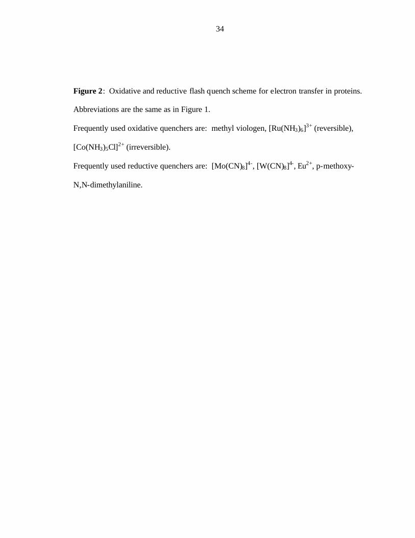
Figure 2: Oxidative and reductive flash quench scheme for electron transfer in proteins.
Abbreviations are the same as in Figure 1.
Frequently used oxidative quenchers are: methyl viologen, [Ru(NH3)6]3+ (reversible),
[Co(NH3)5Cl]2+ (irreversible).
Frequently used reductive quenchers are: [Mo(CN)8]4-, [W(CN)8]4-, Eu2+, p-methoxy-
N,N-dimethylaniline.
35

36
Figure 3: Master tunneling timetable for biological electron transfer reactions (adapted
from Ponce et al.).37
37

38
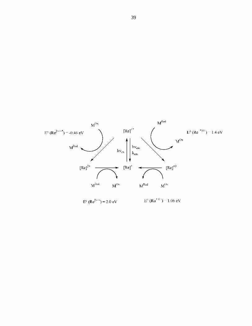
Figure 4: Modified Latimer diagram for Re+.
[Re]n+ is taken to be [Re(CO)3(phen)(im)]n+.
39
40
Figure 5: Transient absorption spectrum of the flash quench oxidation of Cu(I) in the
Re(His107) azurin.
41

42
Figure 6: Tunneling timetable for the oxidative flash quench rates: Ru v. Re.

Scie
nce
1995
, 268
, 17
33-1
735.
Mill
er, D
iBili
o,
Weh
bi, M
uset
h,
Gre
en, R
icha
rds,
W
inkl
er, a
nd G
ray
Bio
chim
. Bio
phys
. A
cta.
2003
(sub
mitt
ed)
Harry B Gray
43
Harry B Gray
Harry B Gray
Harry B Gray

44
Figure 7: X-band EPR spectrum of the oxidative flash quench product of the Re(His107)
azurin taken at 77 K.
Azu
rin
Fla
sh Q
uenc
h
Harry B Gray
45
Harry B Gray
Harry B Gray

46
Figure 8: High-frequency EPR of Re(His107) photoproduct taken at 77K.
Harry B Gray
47

48
Figure 9: Tunneling table of imidazole oxidations of Cu(I).

Harry B Gray
49
50
References
(1) Winkler, J. R.; Gray, H. B. Chem. Rev. 1992, 92, 369-379. (2) Bjerrum, M. J.; Casimiro, D. R.; Chang, I.-J.; Di Bilio, A. J.; Gray, H. B.; Hill, M.
G.; Langen, R.; Mines, G. A.; Skov, L. K.; Winkler, J. R.; Wuttke, D. S. J. Bioenerg. Biomemb. 1995, 27, 295-302.
(3) Gray, H. B.; Winkler, J. R. Annu. Rev. Biochem. 1996, 65, 537-561. (4) Sutin, N. J. Photochem. 1979, 10, 19-40. (5) Sutin, N.; Creutz, C. Pure Appl. Chem. 1980, 52, 2717-2738. (6) Chang, I.-J.; Gray, H. B.; Winkler, J. R. J. Am. Chem. Soc. 1991, 113, 7056-7057. (7) Gafney, H. D.; Adamson, A. W. J. Am. Chem. Soc. 1972, 94, 8238-8239. (8) Navon, G.; Sutin, N. Inorg. Chem. 1974, 13, 2159-2164. (9) Creutz, C.; Sutin, N. Inorg. Chem. 1976, 15, 496-499. (10) Creutz, C.; Sutin, N. J. Am. Chem. Soc. 1976, 98, 6384-6385. (11) Creutz, C.; Sutin, N.; Brunschwig, B. S. J. Am. Chem. Soc. 1979, 101, 1297-1298. (12) Beratan, D. N.; Onuchic, J. N.; Winkler, J. R.; Gray, H. B. Science 1992, 258,
1740-1741. (13) Langen, R.; Colón, J. L.; Casimiro, D. R.; Karpishan, T. B.; Winkler, J. R.; Gray,
H. B. J. Biol. Inorg. Chem. 1996, 1, 221-225. (14) Winkler, J. R. Curr. Op. Chem. Biol. 2000, 4, 192-198. (15) Langen, R.; Chang, I. J.; Germanas, J. P.; Richards, J. H.; Winkler, J. R.; Gray,
H. B. Science 1995, 268, 1733-1735. (16) Di Bilio, A. J.; Dennison, C.; Gray, H. B.; Ramirez, B. E.; Sykes, A. G.; Winkler,
J. R. J. Am. Chem. Soc. 1998, 120, 7551-7556. (17) Babini, E.; Bertini, I.; Borsari, M.; Capozzi, F.; Luchinat, C.; Zhang, X. Y.;
Moura, G. L. C.; Kurnikov, I. V.; Beratan, D. N.; Ponce, A.; Di Bilio, A. J.; Winkler, J. R.; Gray, H. B. J. Am. Chem. Soc. 2000, 122, 4532-4533.
(18) Stemp, E. D. A.; Barton, J. K. Inorg. Chem. 2000, 39, 3868-3874. (19) Mines, G. A.; Bjerrum, M. J.; Hill, M. G.; Casimiro, D. R.; Chang, I. J.; Winkler,
J. R.; Gray, H. B. J. Am. Chem. Soc. 1996, 118, 1961-1965. (20) Wilker, J. J.; Dmochowski, I. J.; Dawson, J. H.; Winkler, J. R.; Gray, H. B.
Angew. Chem. Int. Edit. 1999, 38, 90-92. (21) Pascher, T.; Chesick, J. P.; Winkler, J. R.; Gray, H. B. Science 1996, 271, 1558-
1560. (22) Wittung-Stafshede, P.; Lee, J. C.; Winkler, J. R.; Gray, H. B. Proc. Natl. Acad.
Sci. U. S. A. 1999, 96, 6587-6590. (23) Wittung-Stafshede, P.; Gray, H. B.; Winkler, J. R. J. Am. Chem. Soc. 1997, 119,
9562-9563. (24) Lin, C.-T.; Böttcher, W.; Chou, M.; Creutz, C.; Sutin, N. J. Am. Chem. Soc. 1976,
98, 6536-6544. (25) Gray, H. B.; Maverick, A. W. Science 1981, 214, 1201-1205. (26) Sutin, N.; Creutz, C. J. Chem. Ed. 1983, 60, 809-814. (27) McCaffery, A. J.; Mason, S. F.; Norman, B. J. J. Chem. Soc. (A) 1969, 1428-
1441.
51
(28) Gaudiello, J. G.; Sharp, P. R.; Bard, A. J. J. Am. Chem. Soc. 1982, 104, 6373-6377.
(29) DeSimone, R. E.; Drago, R. S. J. Am. Chem. Soc. 1970, 92, 2343-2352. (30) DeSimone, R. E. J. Am. Chem. Soc. 1973, 95, 6238-6244. (31) Paris, J. P.; Brandt, W. W. J. Am. Chem. Soc. 1959, 81, 5001-5002. (32) Crosby, G. A.; Perkins, W. G.; Klassen, D. M. J. Chem. Phys. 1965, 43, 1498-
1503. (33) Adman, E. T. Adv. Prot. Chem. 1991, 42, 145-197. (34) Sykes, A. G. Adv. Inorg. Chem. 1991, 36, 377-408. (35) Crane, B. R.; Di Bilio, A. J.; Winkler, J. R.; Gray, H. B. J. Am. Chem. Soc. 2001,
123, 11623-11631. (36) Gray, H. B.; Malmstrom, B. G.; Williams, R. J. P. J. Biol. Inorg. Chem. 2000, 5,
551-559. (37) Ponce, A.; Gray, H. B.; Winkler, J. R. J. Am. Chem. Soc. 2000, 122, 8187-8191. (38) Connick, W. B.; DiBilio, A. J.; Hill, M. G.; Winkler, J. R.; Gray, H. B. Inorg.
Chim. Acta 1995, 240, 169-173. (39) Wrighton, M. S.; Morse, D. L. J. Am. Chem. Soc. 1974, 96, 998-1003. (40) Luong, J. C.; Nadjo, L.; Wrighton, M. S. J. Am. Chem. Soc. 1978, 100, 5790-
5795. (41) Giordano, P. J.; Wrighton, M. S. J. Am. Chem. Soc. 1979, 101, 2888-2897. (42) Stufkens, D. J.; Vlcek, A. Coord. Chem. Rev. 1998, 177, 127-179. (43) Vlcek, A. Coord. Chem. Rev. 1998, 177, 219-256. (44) Farrell, I. R.; Vlcek, A. Coord. Chem. Rev. 2000, 208, 87-101. (45) Vlcek, A. Coord. Chem. Rev. 2000, 200, 933-977. (46) Richter, M. M.; Debad, J. D.; Striplin, D. R.; Crosby, G. A.; Bard, A. J. Anal.
Chem. 1996, 68, 4370-4376. (47) Miller, J. E. Ph.D. Thesis; California Institute of Technology: Pasadena, 2003. (48) Winkler, J. R.; Bilio, A. J. D.; Farrow, N. A.; Richards, J. H.; Gray, H. B. Pure
Appl. Chem. 1999, 71, 1753-1764. (49) Cotton, F. A.; Walton, R. A. Multiple Bonds Between Metal Atoms; Clarendon
Press: Oxford, 1993. (50) Ballhausen, C. J. Introduction to Ligand Field Theory; McGraw-Hill: New York,
1962. (51) Chatt, J.; Dilworth, J. R.; Leigh, G. J. J. Chem. Soc.-Dalton Trans. 1973, 612-618. (52) Helberg, L. E.; Orth, S. D.; Sabat, M.; Harman, W. D. Inorg. Chem. 1996, 35,
5584-5594. (53) Gunnoe, T. B.; Meiere, S. H.; Sabat, M.; Harman, W. D. Inorg. Chem. 2000, 39,
6127. (54) Madec, C.; Courtot-Coupez, J. J. Electroanal. Chem. 1977, 84, 177-185. (55) Rao, P. S.; Simic, M.; Hayon, E. J. Phys. Chem. 1975, 79, 1260-1263. (56) Shokhirev, N. V.; Walker, F. A. J. Am. Chem. Soc. 1998, 120, 981-990. (57) Raitsimring, A. M.; Walker, F. A. J. Am. Chem. Soc. 1998, 120, 991-1002. (58) Navaratnam, S.; Parsons, B. J. J. Chem. Soc., Faraday Trans. 1998, 94, 2577-
2581. (59) Stiefel, E. I.; Waters, J. H.; Billig, E.; Gray, H. B. J. Am. Chem. Soc. 1965, 87,
3016.
52
(60) Chaudhuri, P.; Wieghardt, K. Prog. Inorg. Chem. 2001, 50, 151-216. (61) Kaim, W. Dalton Trans. 2003, 761-768. (62) Wang, Y. D.; DuBois, J. L.; Hedman, B.; Hodgson, K. O.; Stack, T. D. P. Science
1998, 279, 537-540. (63) Whittaker, J. W.; Whittaker, M. M. Pure Appl. Chem. 1998, 70, 903-910. (64) Rogers, M. S.; Dooley, D. M. In Adv. Prot. Chem., Vol 58, 2001; Vol. 58, pp 387-
436. (65) Rogers, M. S.; Dooley, D. M. Curr. Op. Chem. Biol. 2003, 7, 189-196. (66) Klinman, J. P. Chem. Rev. 1996, 96, 2541-2561. (67) McGuirl, M. A.; Dooley, D. M. Curr. Op. Chem. Biol. 1999, 3, 138-144. (68) Weil, J. A.; Bolton, J. R.; Wertz, J. E. Electron Paramagnetic Resonance:
Elementary Theory and Applications; Wiley: New York, 1994. (69) Abragam, A.; Bleaney, B. Electron Paramagnetic Resonance of Transition Metal
Ions; Clarendon Press: Oxford, 1970. (70) Pilbrow, J. R. Transition Ion Electron Paramagnetic Resonance; Clarendon Press:
Oxford, 1990. (71) Ballhausen, C. J.; Gray, H. B. Molecular Orbital Theory; W.A. Benjamin: New
York, 1964. (72) Griffith, J. S. The Theory of Transition Metal Ions; Cambridge University Press:
Cambridge, 1964.
53
Chapter 3
Tyrosyl Radicals

54
Introduction
In the first experiments of electron transfer in rhenium modified azurins, the
Re(His107) labeled protein showed an enhanced rate of electron tunneling over the
analogous ruthenium derivative (2x10⁴ s-1 v. 200 s-1). As the following residue is
tyrosine 108, the initial model proposed that a tyrosyl radical intermediate is formed
rapidly by reducing the Re2+ species. This would then be followed by reduction of the
tyrosyl radical by Cu(I), whose rate could be monitored by transient absorption kinetics
of Cu(II) formation. The scheme for this proposal is shown in Figure 1.1
In this chapter we address the questions of whether the rhenium flash quench
experiment can generate a tyrosyl radical at residue 108 and what role that radical may
play in the multistep electron tunneling model. This system is well suited towards
detailed studies of the spectroscopic properties of tyrosyl radicals. Furthermore, we have
mutated the azurin protein in order to incorporate a tyrosine residue in a completely
hydrophobic pocket (residue 48) and have undertaken the flash quench experiment from
His83.
Materials and Methods
The two mutants of azurin described in this chapter are (1) W48F/Y72F/H83Q/Q107H
and (2) W48Y/Y72F/Y108F. The preparation of the rhenium labeled mutants, the
photogeneration of the tyrosyl radicals, and the EPR detection of these radicals are
described in the Appendices.

55
Results
Our initial experiments focused on the ability of the rhenium flash quench system
to make the tyrosyl radical at residue 108. This was demonstrated in a zinc(II) derivative
of the protein (negating any possibility of multi-step tunneling through oxidation of CuI).
As the g⊥ of blue copper2 overlaps with EPR signal of the tyrosyl radical, the preference
of Zn2+ to Cu2+ in the active site that cannot undergo oxidation was made necessary. The
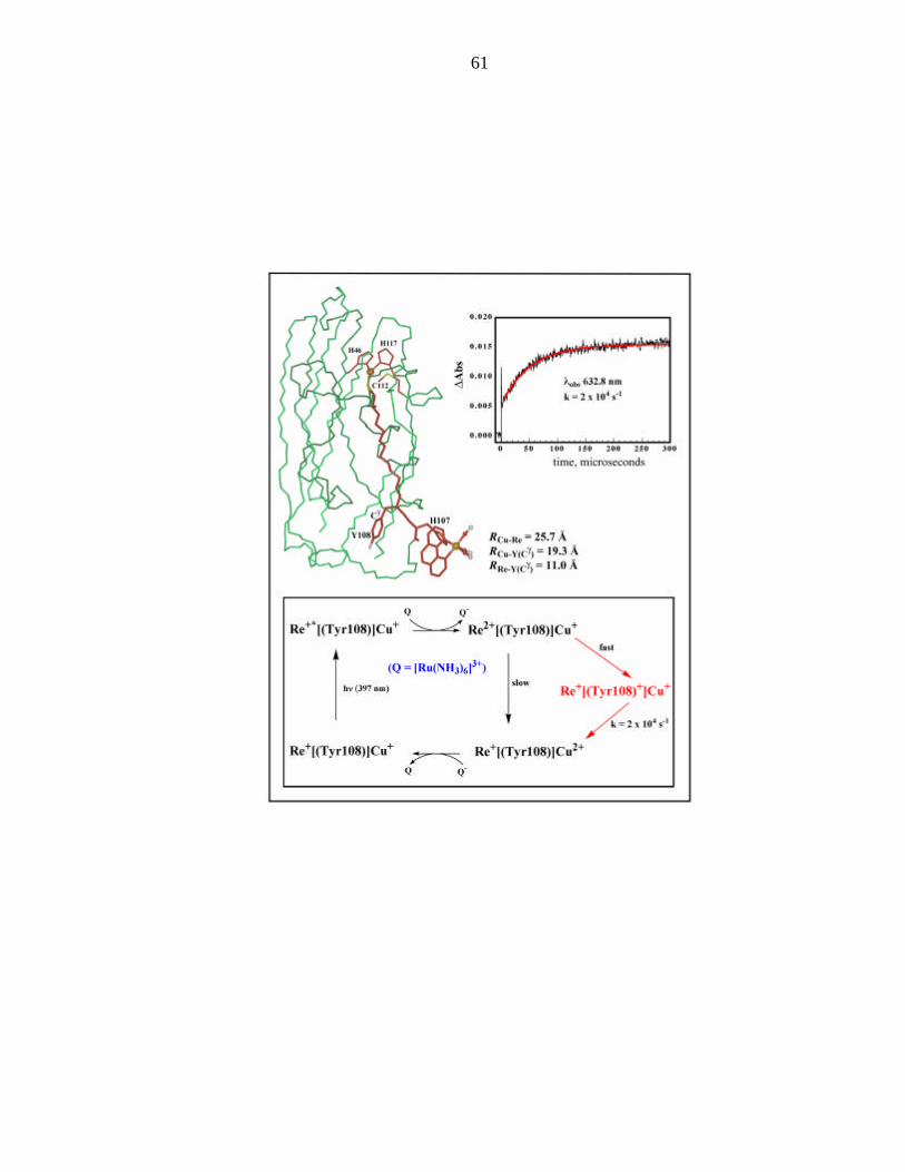
X-band EPR spectrum of the tyrosyl 108 radical is shown in Figure 2 and clearly
identifies the radical as tyrosyl in nature, with an effective g = 2.0042, and the
characteristic doublet spitting (due to the β-protons). The simulation is also shown in
Figure 2. The gy and gz components are standard for all tyrosyl radicals observed to date.
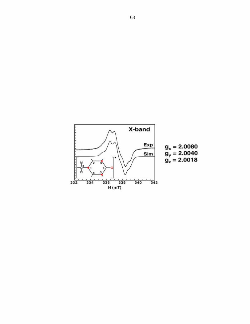
The gx component (gx = 2.008) is the parameter sensitive to hydrogen bonding. The
crystal structure3 indicates a hydrogen bond to the backbone amide of lysine 103;
glutamate 106 is in the vicinity, but is not directed towards the tyrosine residue (Figure
3). There is hyperfine coupling to the protons that are ortho to the phenolic carbon
(which is comparable to other tyrosyl radicals, indicating a similar spin distribution)4-6
and also to the two beta protons. These hyperfine values to the two beta protons are
inequivalent, and can be related to the dihedral angle that the phenol ring makes each of
these protons.7 Azurin also contains a tyrosine residue at position 72. While this residue
is found more than 20 Å from the rhenium unit, we mutated it to phenylalanine to avoid
amibiguity in our EPR experiment, even though we do not think that Tyr72 can be
oxidized preferentially over Tyr108 from Re(His107).

56
Having demonstrated the formation of tyrosyl radicals in rhenium-azurin, we
undertook the high-frequencey, high-field EPR of tyrosyl radical 108 in collaboration
with Professor Sun Un at the CEA-Saclay. High frequency EPR (at 190 and 285 GHz in
the case of our experiments) provides a very accurate measurement of g-tensors in
organic radicals (although the information about hyperfine interactions is lost, as they are
much smaller than the microwave quanta). The high-frequency EPR spectra for tyrosyl
radical 108, at 190 and 285 GHz are shown in Figure 4.
The wild-type azurin contains a Trp (48) residue in completely hydrophobic
pocket. We have mutated this residue to Tyr, labeled His83 with rhenium, and repeated
the flash quench experiment to see if we can make the tyrosyl radical (the formation of
the Trp radical and the difference in their EPR spectra, revealing their sensitivity to the
protein microenvironment are reported in Jeremiah Miller's thesis8). Figure 5 shows the
X-band EPR spectrum of this radical. The simulation of this spectrum (Figure 6) shows
the expected gy and gz components of g-tensor and the increased gx value of 2.0137. The
effective g of 2.0048 value appears coincident with that of the tyrosyl radical at position
108 (Figure 7). The higher value of gx tracks with the decreased amount of hydrogen
bonding character to the phenolic oxygen atom. We suggest that this radical is
deprotonated (as is the case for tyrosyl 108 radical) and we attribute protein dynamical
motions in the seconds it takes to freeze the EPR sample with allowing the proton to
escape from the hydrophobic pocket. The hyperfine parameters show similar coupling
for the ortho protons, although the values for the coupling to the beta protons suggest a
different dihedral angle as compared to the tyrosyl 108 radical. We are currently
57
undertaking the high-frequency, high-field EPR of tyrosyl 48 radical with Professor Sun
Un to obtain an accurate assessment of the g-tensor.
We would like to mention an on-going collaboration with Professor Stenbjorn
Styring and Dr. Ann Magnuson at Lund University (Lund, Sweden) on the time-resolved
EPR characteristics of the tyrosyl 108 radical. In the
Trp48Phe/Tyr72Phe/His83Gln/Gln107His mutant (Re is at His107 and Tyr108 is
present) the Tyr108 radical lives for 10 minutes (data are not shown). In the
Tyr72Phe/His83Gln/Gln107His mutant (containing Tyr108 and Trp48) the Tyr radical
lives for 15 seconds. We are currently pursuing the electron transfer between Tyr108 and
Trp48, starting with both radicals (the oxidation of Tyr108 by Trp48 has been
investigated by Jeremiah Miller in his thesis). The rhenium-azurin system is ideal for
studying electron transfer between tyrosine and tryptophan residues, for which there have
been studies using pulse radiolytically generated radicals.9,10
Discussion
Tyrosyl radicals have been studied more extensively than any other amino acid
radical in proteins and enzymes.6 In this regard, they become test cases for the rhenium
flash quench system. EPR studies of two tyrosyl radicals in different protein
environments confirmed the rhombic (gx ? gy ? gz) nature of the g-tensor. As the gx
component is sensitive to the strength of the hydrogen bond to the radical, it provides an
important experimental parameter for assessing the polarity and solvent accessibility of
the protein microenvironment.11-13 High-frequency EPR studies have been pursued on

58
tyrosyl radicals in RNR,14,15 PSII,16 and prostaglandin H synthase,17 and provide accurate
determinations of the g-tensor components. Our work on tyrosyl radicals complements
the existing studies, and, in the case of the radical at Y48, provides a reference point for a
tyrosyl radical in a completely hydrophobic environment. Simulations of the X-band
EPR spectrum give a gx value of 2.0137, the highest gx value for tyrosyl radicals
measured to date. The protein microenvironment around residue 48 when it is tyrosine
provides the reference point for the electronic structure of tyrosyl radicals in a
hydrophobic environment. High-frequency EPR experiments on the Tyr48 radical are
currently in progress to obtain an accurate measure of this important parameter
The question remains as to the role of tyrosyl radicals in electron transfer
processes in our rhenium-modified azurins. Electrochemical work has found the redox
potential to be ~ 1 eV at pH 7.18 This should provide sufficient driving force to oxidize
Cu(I), which has an E° Cu2+/+ ~ 300 meV.2 Our experiments do not support Cu(I)
oxidation by tyrosyl radicals as the rates of oxidation are the same in proteins containing
tyrosine residues as they are in Tyr à Phe mutations (Chapter 2, this thesis ). The current
model for the rhenium-azurin systems has a rhenium-coordinated histidine radical
forming after the quenching of the excited state. This is followed by oxidation of
tyrosines or Cu(I). In the Re(His107) protein, this can imply that the rate of tyrosyl
radical formation is less than the rate of Cu(I) oxidation (2 x 10⁴ s-1), which means that
tyrosyl radical formation is not kinetically favorable. However, pulse radiolytic studies
on His-Tyr dipeptides suggest rapid tyrosyl radical formation following selective
histidine oxidation (k > 108 s-1).19 With this in mind, tyrosyl radicals can still be formed
as competing processes to Cu(I) oxidation. We are currently investigating the transient

59
absorption spectroscopy of the tyrosine mutants to determine the role of tyrosyl radicals
during the Cu(I) oxidation event.

60
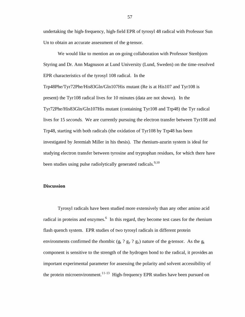
Figure 1: Proposed electron reaction scheme with a tyrosyl “hop.” (adapted from
Winkler et al.)1
61

62
Figure 2. Tyr108 radical EPR and EPR simulation
63

64
Figure 3: Crystal structure showing the environment around Tyr108.
65

66
Figure 4. Tyr108 radical high field EPR
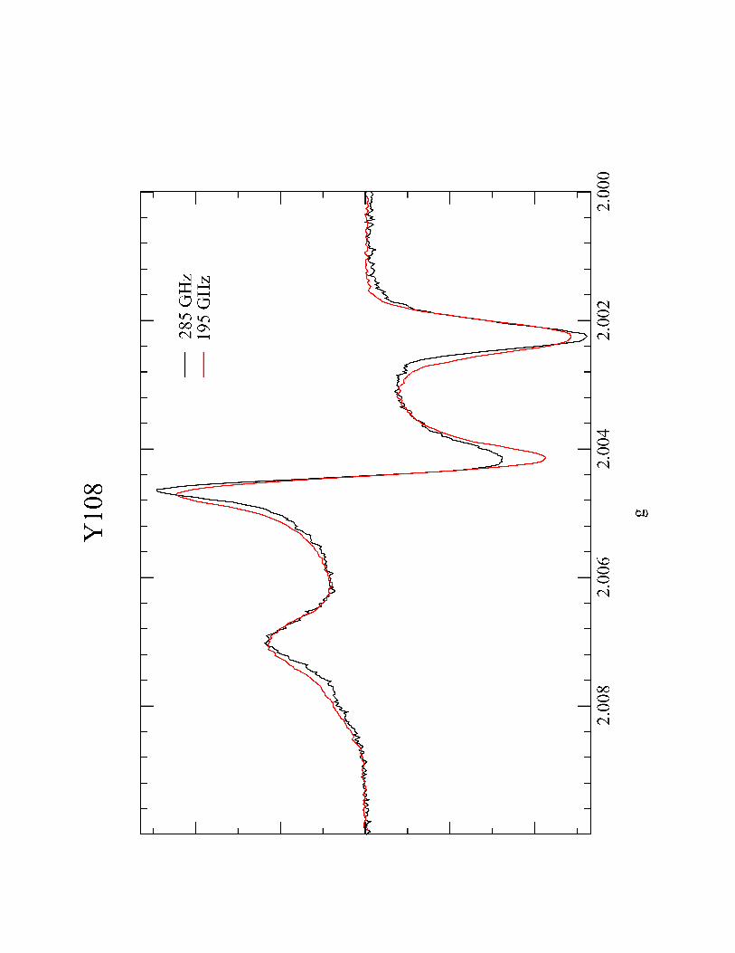
Harry B Gray
67

68
Figure 5. Tyr48 radical EPR

Tyr
osy
l 48
Rad
ical
3310
3330
3350
3370
3390
3410
3430
3450
Mag
net
ic F
ield
(G
auss
)
g av
g=2.
0048
g x=2
.013
7, g
y=2.
0047
, gz=
1.99
68
Harry B Gray
69
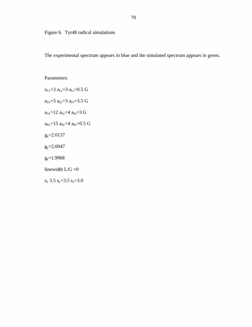
70
Figure 6. Tyr48 radical simulations
The experimental spectrum appears in blue and the simulated spectrum appears in green.
Parameters:
a1x=3 a1y=3 a1z=0.5 G
a2x=3 a2y=3 a2z=3.5 G
a3x=12 a3y=4 a3z=3 G
a4x=15 a4y=4 a4z=0.5 G
gx=2.0137
gy=2.0047
gz=1.9968
linewidth L/G =0
sx 3.5 sy=3.5 sz=3.0
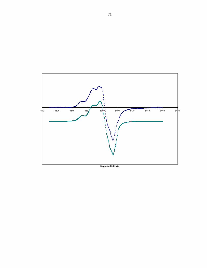
71
3300 3320 3340 3360 3380 3400 3420 3440 3460 3480
Magnetic Field (G)

72
Figure 7: Overlay of Tyr48 and Tyr108 radicals
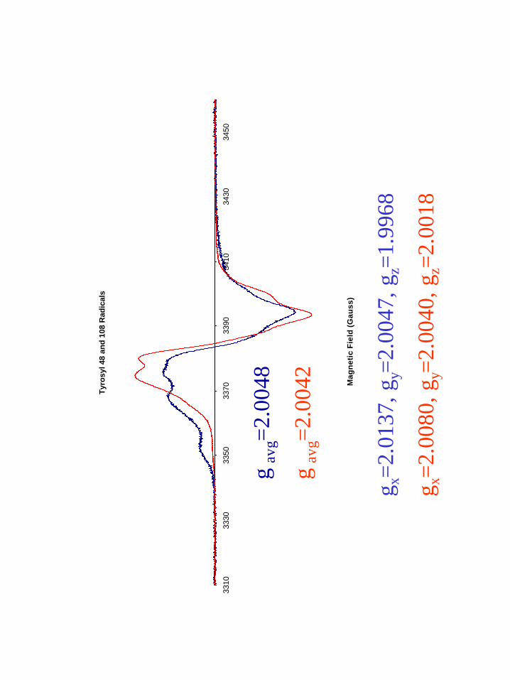
Tyr
osy
l 48
and
108
Rad
ical
s
3310
3330
3350
3370
3390
3410
3430
3450
Mag
net
ic F
ield
(G
auss
)
g av
g=2.
0048
g av
g=2.
0042
g x=2
.013
7, g
y=2.
0047
, gz=
1.99
68
g x=2
.008
0, g
y=2.
0040
, gz=
2.00
18
Harry B Gray
73

74
References
(1) Winkler, J. R.; Di Bilio, A. J.; Farrow, N. A.; Richards, J. H.; Gray, H. B. Pure Appl. Chem. 1999, 71, 1753-1764.
(2) Gray, H. B.; Malmstrom, B. G.; Williams, R. J. P. J. Biol. Inorg. Chem. 2000, 5, 551-559.
(3) Di Bilio, A. J.; Crane, B. R.; Wehbi, W. A.; Kiser, C. N.; Abu-Omar, M. M.; Carlos, R. M.; Richards, J. H.; Winkler, J. R.; Gray, H. B. J. Am. Chem. Soc. 2001, 123, 3181-3182.
(4) Atherton, N. M.; Land, E. J.; Porter, G. Trans. Faraday Soc. 1963, 59, 818-823. (5) Tommos, C.; Tang, X. S.; Warncke, K.; Hoganson, C. W.; Styring, S.;
McCracken, J.; Diner, B. A.; Babcock, G. T. J. Am. Chem. Soc. 1995, 117, 10325-10335.
(6) Pesavento, R. P.; van der Donk, W. A. Adv. Prot. Chem. 2001, 58, 317-385. (7) Babcock, G. T.; Espe, M.; Hoganson, C.; Lydakis-Simantiris, N.; McCracken, J.;
Shi, W.; Styring, S.; Tommos, C.; Warncke, K. Acta Chemica Scandinavica 1997, 51, 533-540.
(8) Miller, J. E. Ph.D. Thesisy; California Institute of Technology: Pasadena, 2003. (9) Butler, J.; Land, E. J.; Prutz, W. A.; Swallow, A. J. Biochim. Biophys. Acta 1982,
705, 150-162. (10) Hawkins, C. L.; Davies, M. J. Biochim. Biophys. Acta 2001, 1504, 196-219. (11) Un, S.; Atta, M.; Fontecave, M.; Rutherford, A. W. J. Am. Chem. Soc. 1995, 117,
10713-10719. (12) Ivancich, A.; Mattioli, T. A.; Un, S. J. Am. Chem. Soc. 1999, 121, 5743-5753. (13) Un, S.; Gerez, C.; Elleingand, E.; Fontecave, M. J. Am. Chem. Soc. 2001, 123,
3048-3054. (14) Gerfen, G. J.; Bellew, B. F.; Un, S.; Bollinger, J. M.; Stubbe, J.; Griffin, R. G.;
Singel, D. J. J. Am. Chem. Soc. 1993, 115, 6420-6421. (15) Bennati, M.; Stubbe, J.; Griffin, R. G. Appl. Mag. Reson. 2001, 21, 389-410. (16) Un, S.; Dorlet, P.; Rutherford, A. W. Appl. Mag. Reson. 2001, 21, 341-361. (17) Dorlet, P.; Seibold, S. A.; Babcock, G. T.; Gerfen, G. J.; Smith, W. L.; Tsai, A.
L.; Un, S. Biochemistry 2002, 41, 6107-6114. (18) DeFilippis, M. R.; Murthy, C. P.; Faraggi, M.; Klapper, M. H. Biochemistry 1989,
28, 4847-4853. (19) Tanner, C.; Navaratnam, S.; Parsons, B. J. Free Rad. Biol. Med. 1998, 24, 671-
678.
75
Chapter 4
Cysteine-Based Radicals
76
Introduction
The oxidative flash quench experiments of [Re(CO)3(phen)(His)]+ complexes in
various mutants of Pseudomonas aeruginosa azurin have demonstrated the formation of
tyrosyl and tryptophan radicals. Another major class of amino acid radicals that could be
studied by the rhenium flash quench method in azurin is based on cysteine. Considering
that cysteine thiyl radicals are postulated to initiate ribonucleotide reduction in RNR and
are the catalytically active component in all glycyl radical enzymes, the study of their
properties and reactivities in model proteins would provide great insight into their roles in
enzyme catalysis.
The study of radicals derived from thiols dates to the 1960s and 1970s, when
Symons1-3 and others4-6 investigated the EPR spectra of irradiated solutions and crystals
of sulfur compounds, including L-cysteine. Multiple products were detected, including
disulfide radical cations and anions, the perthiyl radical, and various oxygen derivatives
of the thiyl radical (Figure 1). Symons presented arguments on the difficulty (though not
impossibility) to observe a pure thiyl radical signal by EPR (its reactivity
notwithstanding) due to the near degeneracy of the px and py orbitals in which the
unpaired electron resides. With an orbitally degenerate ground state and the larger value
of the spin-orbit coupling constant for sulfur compared to carbon, the signal would be
expected to be inhomogenously broadened, which is the case compared to carbon-based
radicals. In addition, thiyl radical assignments could not be made based on g values
alone because of two complicating factors: hydrogen bonding to the thiyl radical breaks
the orbital degeneracy and leads to a shift in the g1; and in the pulse radiolysis
77
experiments, multiple sulfur radical products are produced (not selectively) that have
EPR parameters that are remarkably similar to one another. The g1 component of the
axial g-tensor is diagnostic of a thiyl radical as all other sulfur-centered radicals have
rhombic g-tensors.
In this chapter, we address the formation and properties of cysteine based radicals
in azurin. EPR studies on photogenerated species have been complimented with density
functional theory calculations on thiyl radicals to provide a quantitative handle on the
electronic structures of thiyl radicals and the effects of the electronic structures on EPR
spectra. In particular, the g1 value of the g-tensor is monitored as a function of the energy
separation between the px and py orbitals (and hence the strength of the hydrogen bond to
the radical) and the effective spin-orbit coupling parameter, a direct measure of
covalency to the β methylene group. Alkoxyl and selenyl radicals, which have smaller
and larger spin-orbital coupling values than thiyl radicals, have also been subjected to the
same computations and analyses. Our calculations are the first since 19667when sulfur
and selenium based radicals were treated by extended Huckel method of molecular
orbitals as described by Ballhausen and Gray.8 Although Engstrom has recently
calculated EPR parameters of sulfur centered radicals, no understanding in terms of
molecular orbitals is provided.
Materials and Methods
The two mutants of azur in described in this chapter are (1) W48F/Y72F/H83Q/Q107H/
Y108C and (2) W48C/Y72F/Y108F. The preparation of the rhenium labeled mutants, the
78
photogeneration of the cysteine-based radicals, and the EPR detection of these radicals
are described in the Appendices.
Results and Discussion
The EPR spectrum of the oxidative flash quench product of the His107 (Tyr108Cys)
rhenium labeled protein is shown in Figure 2. The isotropic g-value (2.0104) is found to
be greater than the free electron g-value and the g-values of the histidinyl and tyrosyl
radicals presented in Chapters 2 and 3 of this thesis, respectively. This is indicative of a
sulfur based radical species as the larger spin-orbit coupling value of sulfur versus
oxygen or carbon causes a shift towards greater g-values.
The spectrum does not have an axial g-tensor as would be expected for a thiyl
radical; the resolved hyperfine coupling cannot be explained in terms of a thiyl radical
either. Based on a similar EPR spectrum of a radical species generated in RNR by
Graslund (Figure 3),9 we assign this species to a sulfinyl radical (RSO·). The simulation
of the spectrum g-tensor components of g1 = 2.0202, g2 = 2.0118, g3 = 1.9992. These
parameters are almost identical to Graslund sulfinyl radical in RNR and Sevilla's sulfinyl
radical in pulse radiolytic studies.10-15 The resolvable hyperfine splitting is due to one
beta proton.
The formation of a sulfinyl radical on cysteine 108 was a surprising result. Mass
spectrometry on the protein (mutant W48F/Y72F/H83Q/Q107H/Y108C), labeled and
unlabled with rhenium, gave the expected masses of 13828 and 14278 (M + 450)
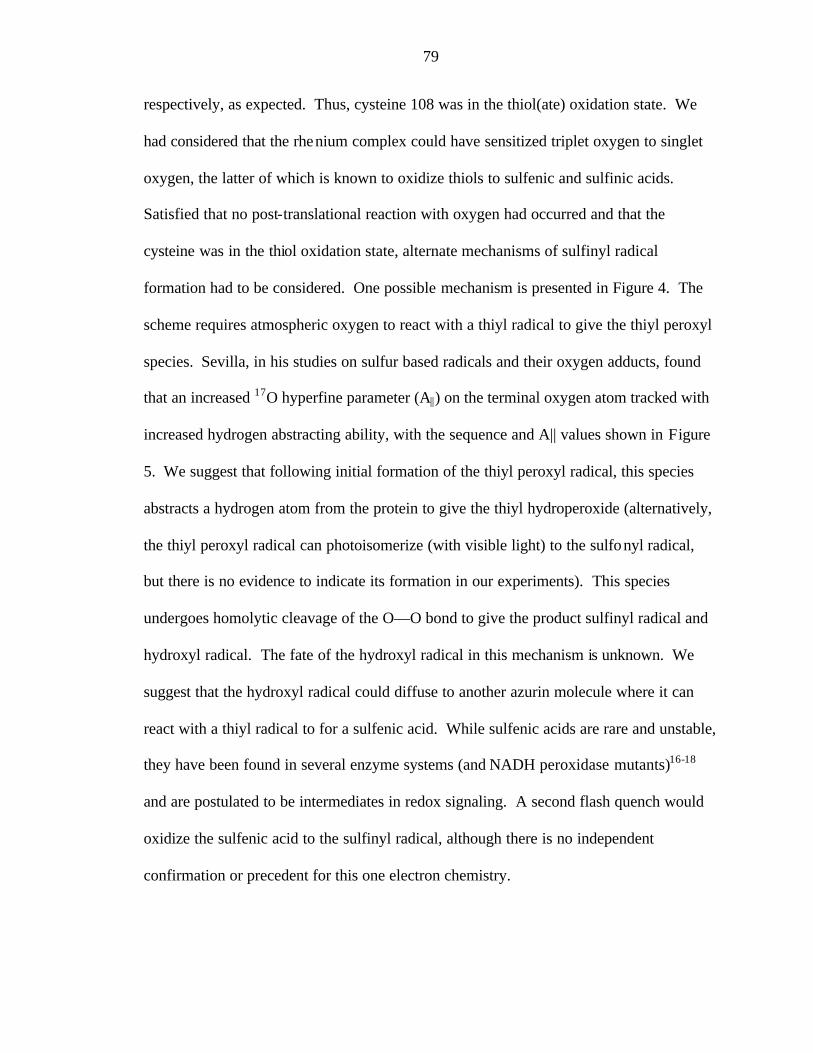
79
respectively, as expected. Thus, cysteine 108 was in the thiol(ate) oxidation state. We
had considered that the rhenium complex could have sensitized triplet oxygen to singlet
oxygen, the latter of which is known to oxidize thiols to sulfenic and sulfinic acids.
Satisfied that no post-translational reaction with oxygen had occurred and that the
cysteine was in the thiol oxidation state, alternate mechanisms of sulfinyl radical
formation had to be considered. One possible mechanism is presented in Figure 4. The
scheme requires atmospheric oxygen to react with a thiyl radical to give the thiyl peroxyl
species. Sevilla, in his studies on sulfur based radicals and their oxygen adducts, found
that an increased 17O hyperfine parameter (A||) on the terminal oxygen atom tracked with
increased hydrogen abstracting ability, with the sequence and A|| values shown in Figure
5. We suggest that following initial formation of the thiyl peroxyl radical, this species
abstracts a hydrogen atom from the protein to give the thiyl hydroperoxide (alternatively,
the thiyl peroxyl radical can photoisomerize (with visible light) to the sulfonyl radical,
but there is no evidence to indicate its formation in our experiments). This species
undergoes homolytic cleavage of the O—O bond to give the product sulfinyl radical and
hydroxyl radical. The fate of the hydroxyl radical in this mechanism is unknown. We
suggest that the hydroxyl radical could diffuse to another azurin molecule where it can
react with a thiyl radical to for a sulfenic acid. While sulfenic acids are rare and unstable,
they have been found in several enzyme systems (and NADH peroxidase mutants)16-18
and are postulated to be intermediates in redox signaling. A second flash quench would
oxidize the sulfenic acid to the sulfinyl radical, although there is no independent
confirmation or precedent for this one electron chemistry.

80
Whence oxygen? All samples were illuminated under an argon atmosphere
(supposedly). However, the sulfinyl radical was also formed in samples irradiated under
atmospheric oxygen and nitrogen pressures (and thus not degassed with argon). More
rigorous means of oxygen removal are needed in order to have the chance to trap the thiyl
radical at Cys108.
The sulfinyl radical was found to live for at least 10 minutes. In the freeze flash-
quench experiment, the sample is frozen immediately after illumination (sample freezing
time is estimated to be a few seconds). Upon thawing for 2 minutes, the sulfinyl
spectrum remained, although the signal intensity had decreased but had become sharper.
We are currently exploring whether there could have been an additional species (the thiyl
radical itself) overlapping with the sulfinyl radical). Further thawing of the sample for 10
minutes followed by freezing revealed a similar EPR spectrum.
The EPR spectrum for the irradiated His83 Cys48 protein is shown in Figure 5.
The resonating feature cannot be conclusively assigned. It appears where the g⊥
of thiyl radicals are found and where the g|| of blue copper is found. As samples had a
blue tint, there was a blue copper impurity whose g|| masked the diagnostic g|| of the thiyl
radical. We are still pursuing this thiyl radical by EPR in zinc and apo preparations of
rhenium-modified azurin.
A preliminary x-ray absorption (XAS) experiment by Dr. Pierre Kennepohl
(personal communication) has demonstrated the 1s à 3p transition in the oxidative flash
quench product of His83 Cys48, suggesting thiyl radical formation and necessitating
further investigation by EPR.
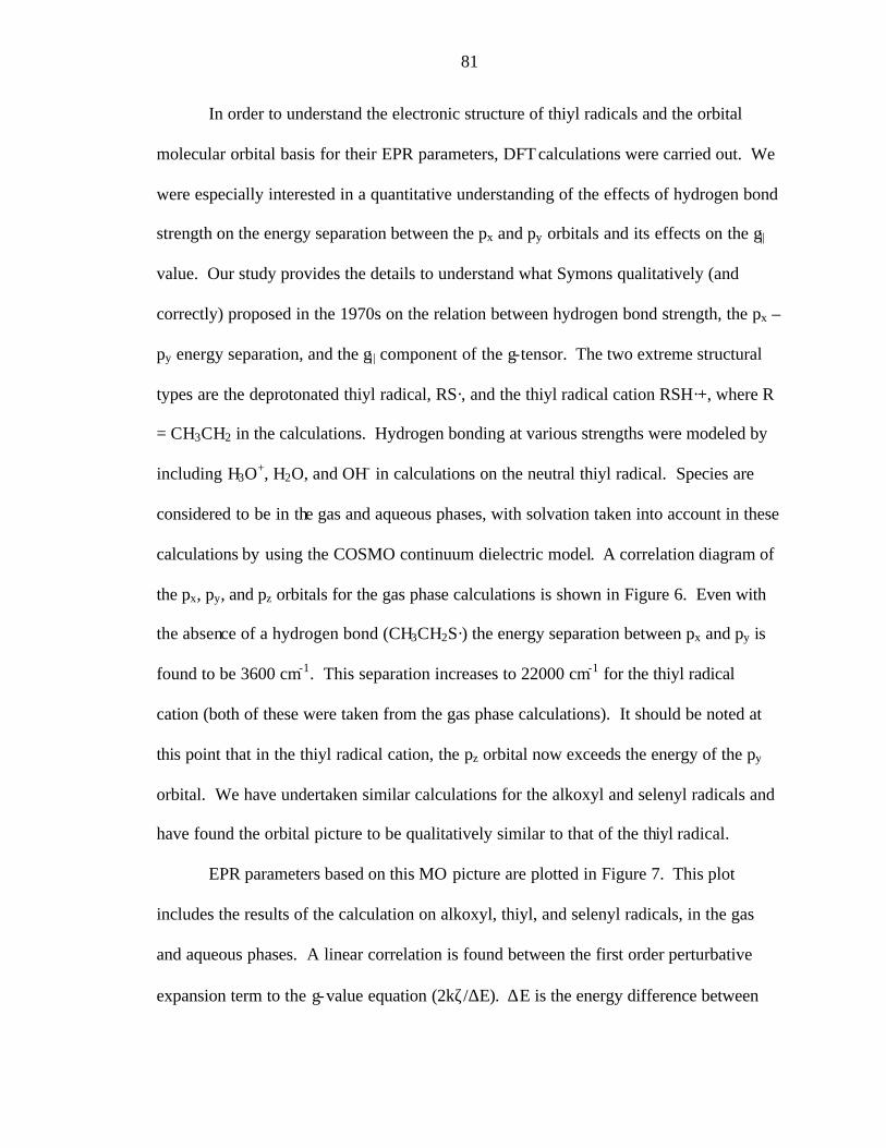
81
In order to understand the electronic structure of thiyl radicals and the orbital
molecular orbital basis for their EPR parameters, DFT calculations were carried out. We
were especially interested in a quantitative understanding of the effects of hydrogen bond
strength on the energy separation between the px and py orbitals and its effects on the g||
value. Our study provides the details to understand what Symons qualitatively (and
correctly) proposed in the 1970s on the relation between hydrogen bond strength, the px –
py energy separation, and the g| | component of the g-tensor. The two extreme structural
types are the deprotonated thiyl radical, RS·, and the thiyl radical cation RSH·+, where R
= CH3CH2 in the calculations. Hydrogen bonding at various strengths were modeled by
including H3O+, H2O, and OH- in calculations on the neutral thiyl radical. Species are
considered to be in the gas and aqueous phases, with solvation taken into account in these
calculations by using the COSMO continuum dielectric model. A correlation diagram of
the px, py, and pz orbitals for the gas phase calculations is shown in Figure 6. Even with
the absence of a hydrogen bond (CH3CH2S·) the energy separation between px and py is
found to be 3600 cm-1. This separation increases to 22000 cm-1 for the thiyl radical
cation (both of these were taken from the gas phase calculations). It should be noted at
this point that in the thiyl radical cation, the pz orbital now exceeds the energy of the py
orbital. We have undertaken similar calculations for the alkoxyl and selenyl radicals and
have found the orbital picture to be qualitatively similar to that of the thiyl radical.
EPR parameters based on this MO picture are plotted in Figure 7. This plot
includes the results of the calculation on alkoxyl, thiyl, and selenyl radicals, in the gas
and aqueous phases. A linear correlation is found between the first order perturbative
expansion term to the g-value equation (2kζ/∆E). ∆E is the energy difference between
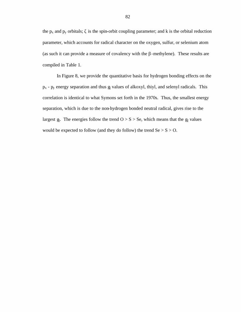
82
the px and py orbitals; ζ is the spin-orbit coupling parameter; and k is the orbital reduction
parameter, which accounts for radical character on the oxygen, sulfur, or selenium atom
(as such it can provide a measure of covalency with the β-methylene). These results are
compiled in Table 1.
In Figure 8, we provide the quantitative basis for hydrogen bonding effects on the
px - py energy separation and thus g|| values of alkoxyl, thiyl, and selenyl radicals. This
correlation is identical to what Symons set forth in the 1970s. Thus, the smallest energy
separation, which is due to the non-hydrogen bonded neutral radical, gives rise to the
largest g||. The energies follow the trend O > S > Se, which means that the g|| values
would be expected to follow (and they do follow) the trend Se > S > O.

83
Figure 1. Some thiol derived sulfur based radicals
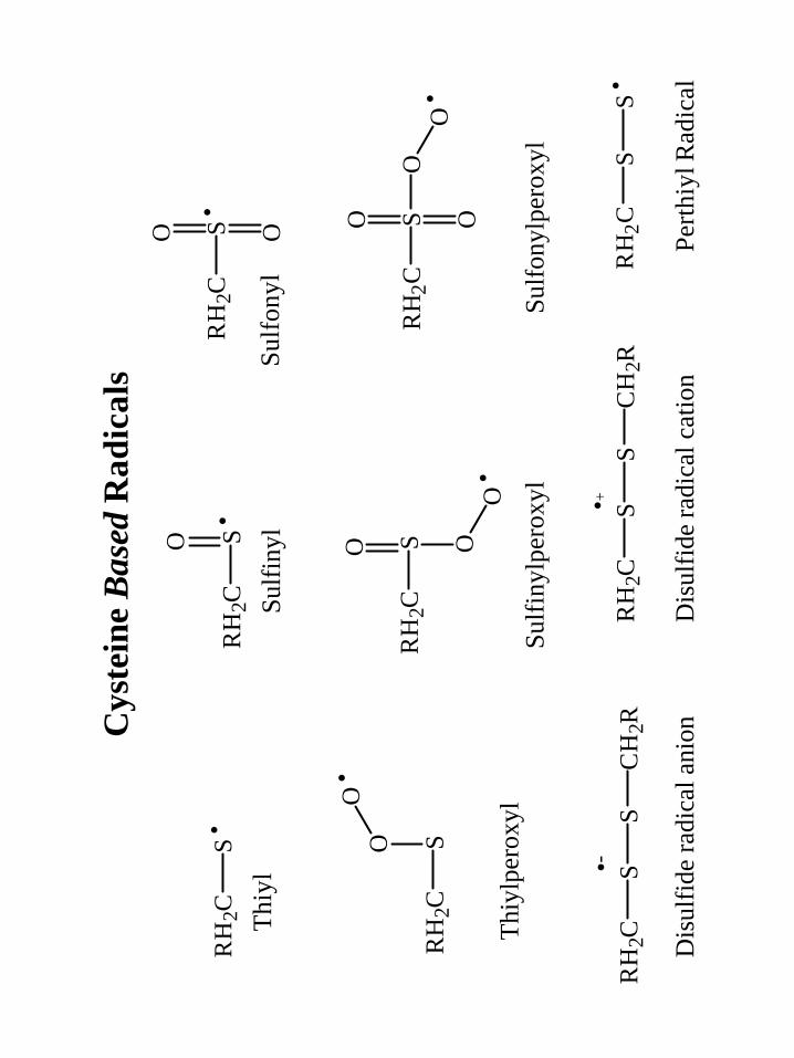
Cys
tein
e B
ased
Rad
ical
s
Thi
ylSu
lfiny
lSu
lfon
yl
Thi
ylpe
roxy
lSu
lfon
ylpe
roxy
lSu
lfin
ylpe
roxy
l
Pert
hiyl
Rad
ical
Dis
ulfid
e ra
dica
l ani
on
S·R
H2C
S·O
RH
2CS·
RH
2C
O O
SR
H2C
OO·
SR
H2C
O O
OO·
SR
H2C
OO·
O
SR
H2C
S·S
CH
2RS
RH
2C·-
SC
H2R
SR
H2C
·+
Dis
ulfid
e ra
dica
l cat
ion
Harry B Gray
84

85
Figure 2. Cys 108 EPR
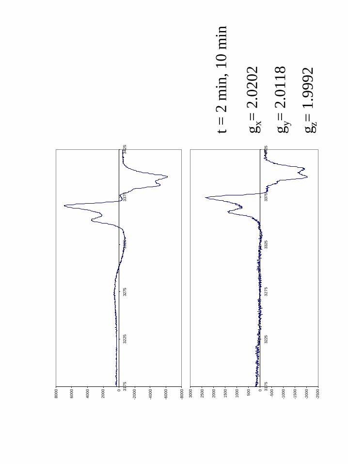
-250
0
-200
0
-150
0
-100
0
-5000
500
1000
1500
2000
2500
3000
3175
3225
3275
3325
3375
3425
-800
0
-600
0
-400
0
-200
00
2000
4000
6000
8000
3175
3225
3275
3325
3375
3425
t = 2
min
, 10
min
g x=
2.02
02
g y=
2.01
18
g z=
1.99
92
Harry B Gray
86

87
Figure 3. Sulfinyl radical in RNR by Graslund et al.9
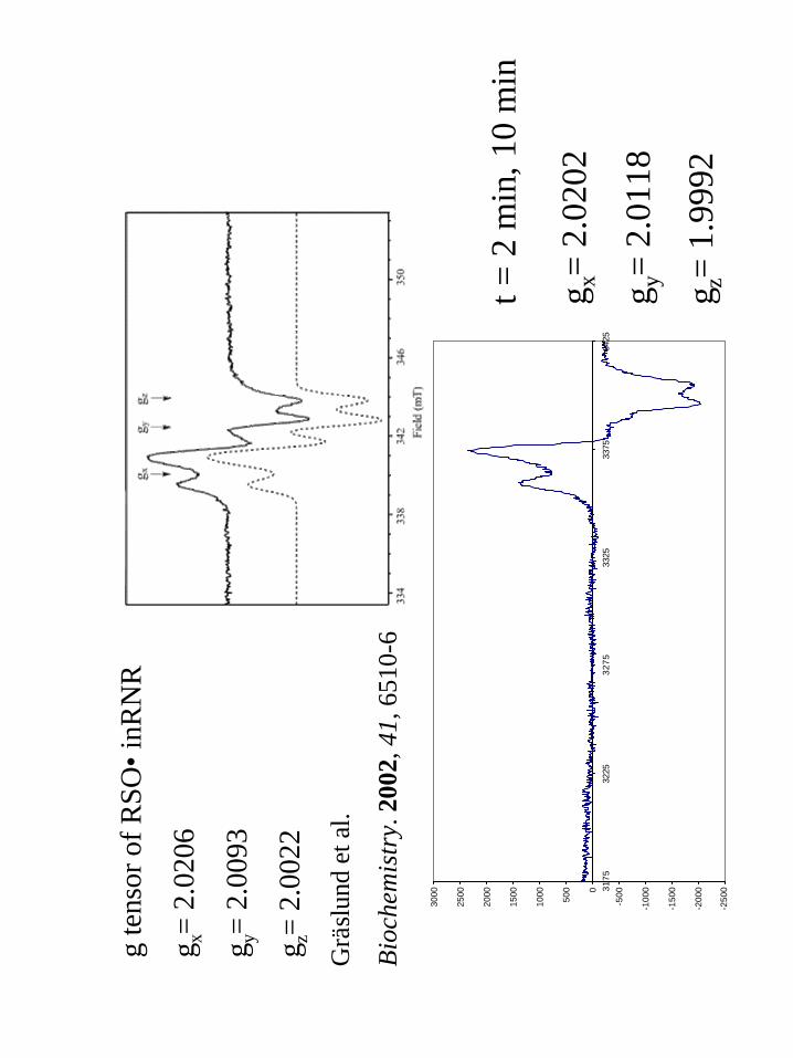
-250
0
-200
0
-150
0
-100
0
-5000
500
1000
1500
2000
2500
3000
3175
3225
3275
3325
3375
3425
t = 2
min
, 10
min
g x=
2.02
02
g y=
2.01
18
g z=
1.99
92
g te
nsor
of R
SO•i
nRN
R
g x=
2.02
06
g y=
2.00
93
g z=
2.00
22
Grä
slun
det
al.
Bio
chem
istr
y. 2
002,
41,
651
0-6
Harry B Gray
88

89
Figure 4. Possible mechanisms of formation of the sulfinyl radical
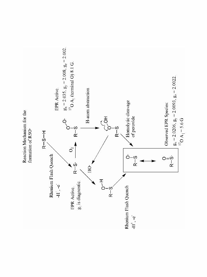
Harry B Gray
90

91
Figure 5. Cys48 EPR
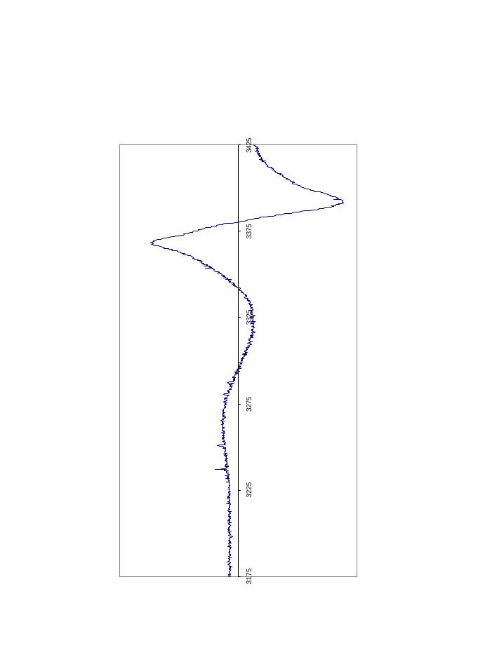
3175
3225
3275
3325
3375
3425
Harry B Gray
92

93
Figure 6. Theoretical calculations of the gx dependence on hydrogen bonding versus the
energy separation between px and py in thiyl radicals.
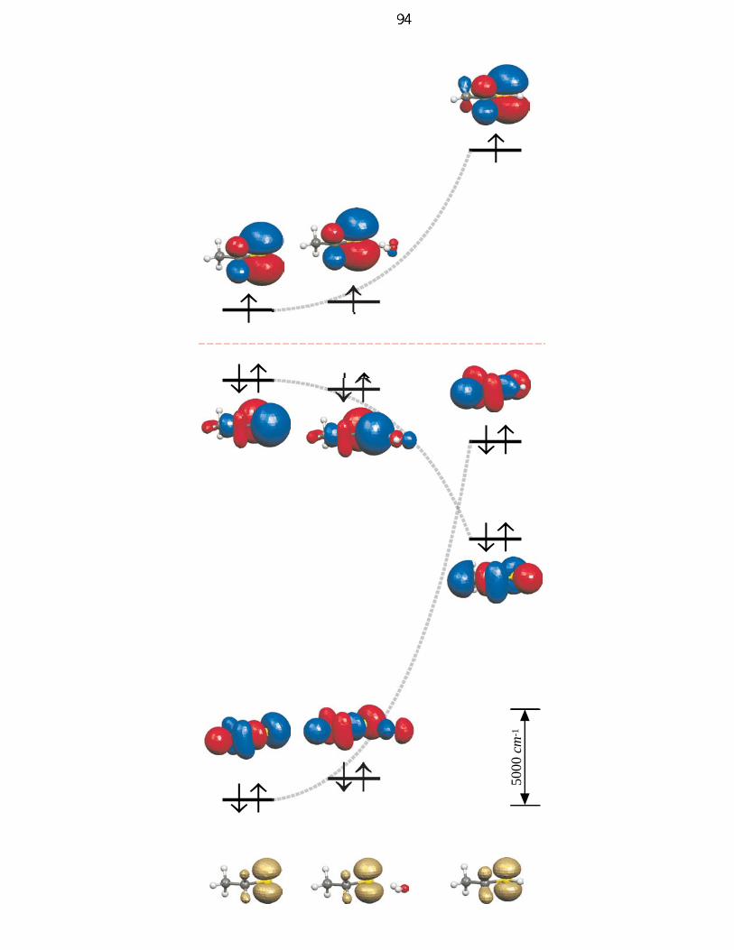
↓↑
↑
↓↑
↓↑
↑
↓↑
↑
↓↑↓↑
5000
cm
-1
Harry B Gray
94

95
Figure 7: Plot of the dependence between energy separation and the shift in g1 value.
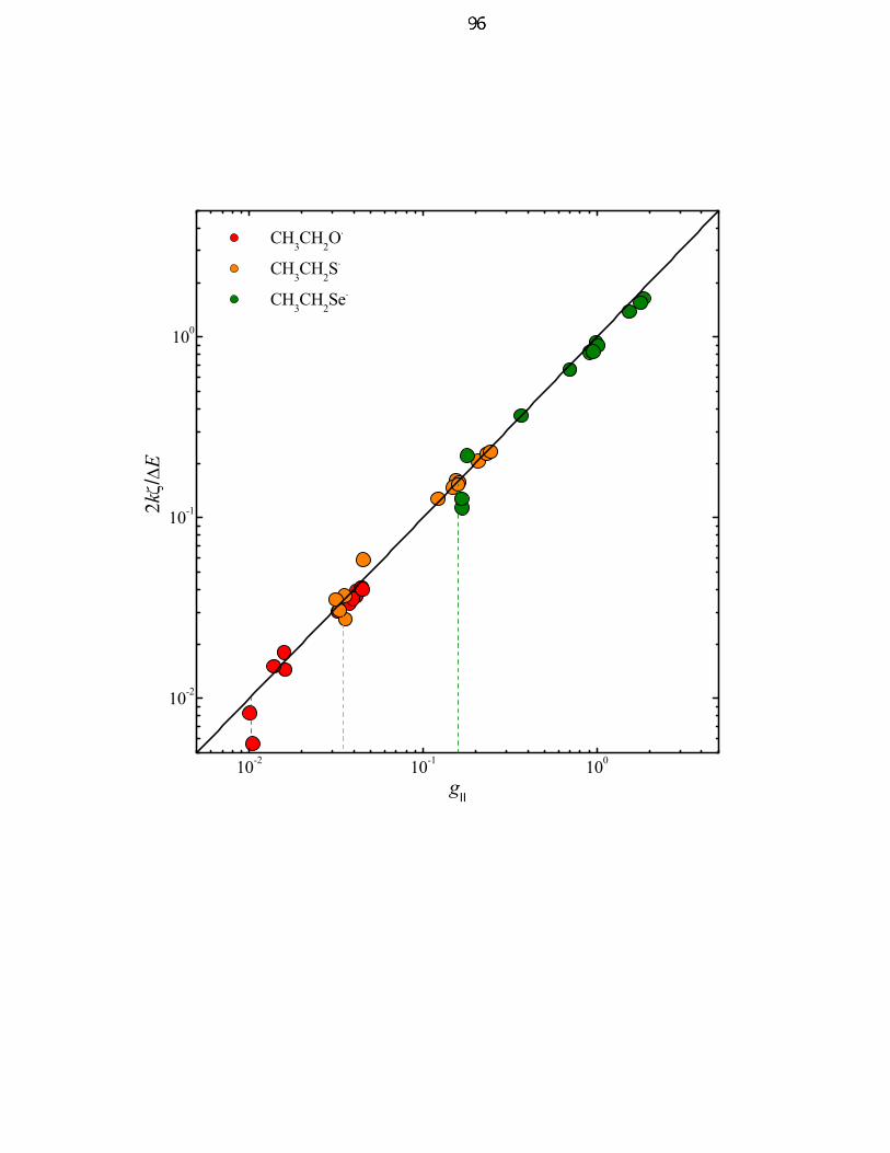
10-2 10-1 100
10-2
10-1
100
CH3CH2O.
CH3CH2S.
CH3CH2Se.
2kζ/∆E
gII
Harry B Gray
96

97
Table 1: Compilation of molecular orbital and EPR parameters of alkoxyl, thiyl, and
selenyl radicals.
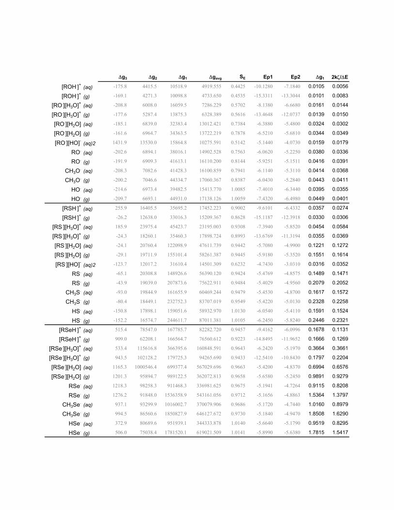
∆g3 ∆g2 ∆g1 ∆gavg SE Ep1 Ep2 ∆g1 2kζ/∆E
[ROH.]+ (aq) -175.8 4415.5 10518.9 4919.555 0.4425 -10.1280 -7.1840 0.0105 0.0056
[ROH.]+ (g) -169.1 4271.3 10098.8 4733.650 0.4535 -15.3311 -13.3044 0.0101 0.0083
[RO.][H3O]+ (aq) -208.8 6008.0 16059.5 7286.229 0.5702 -8.1380 -6.6680 0.0161 0.0144
[RO.][H3O]+ (g) -177.6 5287.4 13875.3 6328.389 0.5616 -13.4648 -12.0737 0.0139 0.0150
[RO.][H2O] (aq) -185.1 6839.0 32383.4 13012.421 0.7384 -6.3880 -5.4800 0.0324 0.0302
[RO.][H2O] (g) -161.6 6964.7 34363.5 13722.219 0.7878 -6.5210 -5.6810 0.0344 0.0349
[RO.][HO]- (aq)2 1431.9 13530.0 15864.8 10275.591 0.5142 -5.1440 -4.0730 0.0159 0.0179
RO. (aq) -202.6 6894.1 38016.1 14902.528 0.7563 -6.0620 -5.2250 0.0380 0.0336
RO. (g) -191.9 6909.3 41613.1 16110.200 0.8144 -5.9251 -5.1511 0.0416 0.0391
CH3O. (aq) -208.3 7082.6 41428.3 16100.859 0.7941 -6.1140 -5.3110 0.0414 0.0368
CH3O. (g) -200.2 7046.6 44334.7 17060.367 0.8387 -6.0430 -5.2840 0.0443 0.0411
HO. (aq) -214.6 6973.4 39482.5 15413.770 1.0085 -7.4010 -6.3440 0.0395 0.0355
HO. (g) -209.7 6693.1 44931.0 17138.126 1.0059 -7.4320 -6.4980 0.0449 0.0401
[RSH.]+ (aq) 255.9 16405.5 35695.2 17452.223 0.9002 -9.6101 -6.4332 0.0357 0.0274
[RSH.]+ (g) -26.2 12638.0 33016.3 15209.367 0.8628 -15.1187 -12.3918 0.0330 0.0306
[RS.][H3O]+ (aq) 185.9 23975.4 45423.7 23195.003 0.9308 -7.3940 -5.8520 0.0454 0.0584
[RS.][H3O]+ (g) -24.3 18260.1 35460.3 17898.724 0.8993 -13.6769 -11.3194 0.0355 0.0369
[RS.][H2O] (aq) -24.1 20760.4 122098.9 47611.739 0.9442 -5.7080 -4.9900 0.1221 0.1272
[RS.][H2O] (g) -29.1 19711.9 155101.4 58261.387 0.9445 -5.9180 -5.3520 0.1551 0.1614
[RS.][HO]- (aq)2 -123.7 12017.2 31610.4 14501.309 0.6232 -4.7430 -3.0310 0.0316 0.0352
RS. (aq) -65.1 20308.8 148926.6 56390.120 0.9424 -5.4769 -4.8575 0.1489 0.1471
RS. (g) -43.9 19039.0 207873.6 75622.911 0.9484 -5.4029 -4.9560 0.2079 0.2052
CH3S. (aq) -93.0 19844.9 161655.9 60469.244 0.9479 -5.4530 -4.8700 0.1617 0.1572
CH3S. (g) -80.4 18449.1 232752.3 83707.019 0.9549 -5.4220 -5.0130 0.2328 0.2258
HS. (aq) -150.8 17898.1 159051.6 58932.970 1.0130 -6.0540 -5.4110 0.1591 0.1524
HS. (g) -152.2 16574.7 244611.7 87011.381 1.0105 -6.2450 -5.8240 0.2446 0.2321
[RSeH.]+ (aq) 515.4 78547.0 167785.7 82282.720 0.9457 -9.4162 -6.0996 0.1678 0.1131
[RSeH.]+ (g) 909.0 62208.1 166564.7 76560.612 0.9223 -14.8495 -11.9652 0.1666 0.1269
[RSe.][H3O]+ (aq) 533.4 115616.8 366395.6 160848.591 0.9643 -6.2420 -5.1970 0.3664 0.3661
[RSe.][H3O]+ (g) 943.5 102128.2 179725.3 94265.690 0.9433 -12.5410 -10.8430 0.1797 0.2204
[RSe.][H2O] (aq) 1165.3 1000546.4 699377.4 567029.696 0.9663 -5.4200 -4.8370 0.6994 0.6576
[RSe.][H2O] (g) 1201.3 95894.7 989122.5 362072.813 0.9658 -5.6580 -5.2450 0.9891 0.9279
RSe. (aq) 1218.3 98258.3 911468.3 336981.625 0.9675 -5.1941 -4.7264 0.9115 0.8208
RSe. (g) 1276.2 91848.0 1536358.9 543161.056 0.9712 -5.1656 -4.8863 1.5364 1.3797
CH3Se. (aq) 937.1 93299.9 1016002.7 370079.906 0.9686 -5.1720 -4.7440 1.0160 0.8979
CH3Se. (g) 994.5 86560.6 1850827.9 646127.672 0.9730 -5.1840 -4.9470 1.8508 1.6290
HSe. (aq) 372.9 80689.6 951939.1 344333.878 1.0140 -5.6640 -5.1790 0.9519 0.8295
HSe. (g) 506.0 75038.4 1781520.1 619021.509 1.0141 -5.8990 -5.6380 1.7815 1.5417
99
References
(1) Nelson, D.; Symons, M. C. R. Chem. Phys. Lett. 1975, 36, 340-341. (2) Nelson, D. J.; Petersen, R. L.; Symons, M. C. R. J. Chem. Soc.-Perkin Trans. 2
1977, 2005-2015. (3) Symons, M. C. R. J. Chem. Soc.-Perkin Trans. 2 1974, 1618-1620. (4) Budzinsk, E. E.; Box, H. C. J. Phys. Chem. 1971, 75, 2564-&. (5) Gilbert, B. C.; Laue, H. A. H.; Norman, R. O. C.; Sealy, R. C. J. Chem. Soc.-
Perkin Trans. 2 1975, 892-900. (6) Kou, W. W. H.; Box, H. C. J. Chem. Phys. 1976, 64, 3060-3062. (7) Chen, I.; Das, T. P. J. Chem. Phys. 1966, 45, 3526-&. (8) Ballhausen, C. J.; Gray, H. B. Molecular Orbital Theory; W.A. Benjamin: New
York, 1964. (9) Adrait, A.; Ohrstrom, M.; Barra, A. L.; Thelander, L.; Graslund, A. Biochemistry
2002, 41, 6510-6516. (10) Sevilla, M. D.; Becker, D.; Swarts, S.; Herrington, J. Biochem. Biophys. Res.
Commun. 1987, 144, 1037-1042. (11) Sevilla, M. D.; Becker, D.; Yan, M. Y. J. Chem. Soc.-Faraday Trans. 1990, 86,
3279-3286. (12) Sevilla, M. D.; Becker, D.; Yan, M. Int. J. Radiat. Biol. 1990, 57, 65-81. (13) Swarts, S. G.; Becker, D.; Debolt, S.; Sevilla, M. D. J. Phys. Chem. 1989, 93,
155-161. (14) Razskazovskii, Y.; Colson, A. O.; Sevilla, M. D. J. Phys. Chem. 1995, 99, 7993-
8001. (15) Razskazovskii, Y.; Sevilla, M. D. J. Phys. Chem. 1996, 100, 4090-4096. (16) Allison, W. S. Accounts Chem. Res. 1976, 9, 293-299. (17) Claiborne, A.; Yeh, J. I.; Mallett, T. C.; Luba, J.; Crane, E. J.; Charrier, V.;
Parsonage, D. Biochemistry 1999, 38, 15407-15416. (18) Claiborne, A.; Mallett, T. C.; Yeh, J. I.; Luba, J.; Parsonage, D. In Advances in
Protein Chemistry, Vol 58, 2001; Vol. 58, pp 215-276.

100
Appendix A
Preparation of Rhenium-Modified Azurins

101
Introduction
This appendix describes the methods of preparation of rhenium-modified mutant
azurins that are common to the entire thesis. Mutant constructs for the H83Q/Q107H
azurin were obtained as gifts from Cynthia Kiser.
Materials and Methods
Cell Growth.
All cells are grown in LB media. Depending on the vector, the cells will have
resistance to the antibiotics ampicillin if the pET-3a vector is used or kanamycin if the
pET-9a vector is used. The most commonly used cell strains are XL1-Blue, for cyclizing
plasmid DNA and for DNA purification; BL21(DE3) and BL21*(DE3) for protein
expression. No protein will be produced in the XL1-Blue cells.
Cells can be stored at –80 °C by the addition of sterile glycerol to a final
concentration of 20 %. Thus, many of the cell strains (for example, for wild type azurin)
were prepared by an earlier generation of graduate students. To revive these cells, use a
sterile loop (available at the Biology Stockroom) to scrape the surface of the frozen cells
(DO NOT THAW THE CELLS. THEY WILL DIE). Then, gently streak the loop with
the cells on the surface of a LB plate with the appropriate antibiotic. The plates can be
prepared in advanced and stored at 4 °C, however they must be equilibrated to room
temperature before use. Place the plate in a 37 °C incubator; colonies should appear in
102
12-18 hours. After 24 hours, if no colonies are found, the plate should be discarded. At
this point, the antibiotic will have been completely consumed or degraded. The
information in the paragraph can be applied to any cell strain.
Using sterile forceps, pick up a sterile toothpick (from a dry autoclaved jar or
toothpicks). Pick up one colony from the plate of cells. This is done by touching the
toothpick on to the surface of a colony. Transfer the entire toothpick into a 10 mL Falcon
tube that contains 4 mL of LB media (dilute 0.8 mL of the 5X LB by adding 3.2 mL
sterile water) and 4 µL of 60 mg/mL ampicillin stock solution. The tube is then capped
before being taken to a 37 °C shaker (shaking at 200 rpm). The cells are grown until the
optical density (OD) at 600 nm of the cell containing media has reached 1.0. This usually
takes at least seven hours for the BL21(DE3) or BL21*(DE3) strains, although the typical
practice is to grow the 4 mL "starter" culture up to twelve hours, without adverse
consequences to the cells. 4 mL cultures of XL1-Blue are usually grown for 16 hours to
maximize the production of DNA.
Once the desired optical density has been reached, larger-scale preparations can
be innoculated with cells from the starter culture. To 1 L of already autoclaved LB
media, add the appropriate antibiotic (1 mL of the antibiotic stock solution gives a final
concentration, in the case of ampicillin, of 60 µg/mL ampicillin). Then add 1 mL of the
starter culture to the 1 L of LB media. This is known as a 1:1000 innoculation, which is
standard for E. coli. Some other organisms do not grow as well and thus require a larger
concentration of the starter culture for the innoculation. It is a good general practice
whenever growing cells in a shaking incubator never to fill the flask more than 25 % of
the total volume. Thus, it should be understood that 1 L of culture (as mentioned above)

103
has been prepared and autoclaved and will be used in a 4 L flask. Similarly, 1.5 L of
culture are prepared and used in a 6 L flask. The same applies to smaller flasks. Thus,
250 mL of media will be used in a 1 L flask. The reason for this is so that the cells are
properly aerated. With a 4 mL starter culture, there will be three 1 mL innoculations into
three flasks containing 1 L of LB media (or two flasks containing 1.5 L of LB media).
The flasks are then placed into a 37 °C incubator shaking at 200 rpm until the optical
density of the cell containing media at 600 nm reaches 1.0. This takes about 7-8 hours.
The optical density of 1.0 is the ideal, although in practice, high yields of protein are
obtained when the optical density of the cells is between 1.0 and 2.0. The remainder of
the starter culture is transferred to a 2 mL cryo vial (available at the Biology Stockroom).
Autoclaved 100 % glycerol is then added to the cyro vial to give final concentration of 20
% glycerol. The mixture should be stirred gently with the pipette tip to ensure complete
homogeneity of the 20 % glycerol containing cell stock. The vial is capped and then
transferred immediately to the –80 °C freezer. This process is not necessary if there is
already a glycerol containing frozen cell stock.
Once the larger scale cultures have achieved an OD600 in the range of 1.0 to 2.0,
protein expression is then induced by the addition of IPTG (isopropyl-β-D-
thiogalactoside) stock solution (1 mL of the stock solution for 1 L of cell containing
media). The cultures should remain in the shaking incubator for another four hours.
During this time, the cells spend most of their energy making protein. The cells will
continue to make protein beyond four hours, although azurin and many other overly
expressed proteins can leak out of the cells into the media over a long period of time. It
104
is recommended that following the addition of IPTG that the cells not grow for more than
five hours before harvesting.
Cells can be harvested by centrifuging the entire culture. 1 L centrifuge bottles
that fit into a Beckman JLA-8.1 centrifuge are ideal for 3-6 L cultures because only one
run is necessary. The balanced centrifuge bottles are centrifuged for 15 minutes at 6000
rpm at 4 °C. After centrifugation, the media is decanted into a large flask, leaving only
the cell pellet behind. Before discarding the media, add bleach to the flask containing the
decanted media. Swirl the mixture and let it sit for a few minutes. This is necessary to
kill any residual bacteria in the media. Then, the bleached media can be poured into the
sink.
The cell pellet is then resuspended in a one-tenth volume of a high osmolarity
sucrose solution. One-tenth volume means this: if 3 L of cell culture were grown, 300
mL of the high osmolarity sucrose solution will be required. This is not a stringent
requirement. 500 mL of this sucrose solution will work just as well. The suspension is
then transferred into 300 mL centrifuge bottles (this is why 500 mL of the sucrose
solution is also convenient, because the 500 mL of the resuspended solution can be
divided evenly between two centrifuge bottles. The balanced bottles are then placed on a
shaker for 15 minutes at room temperature. Afterwards, the centrifuge bottles are spun at
8000 rpm for 15 minutes. The supernatent is decanted; then, bleach is added to it before
it is poured into the sink. The noticeably swelled cell pellet is then resuspended in a cold
solution of 5mM MgSO4 in water. This solution should be prepared in advanced and
stored a 4 °C refrigerator or cold room until ready for use. The resuspended cells are
shaken for 15 minutes at 4 °C and then are spun at 10000 rpm for 15 minutes. At this
105
stage, the supernatent should be saved. An osmotic shock on the cells has been
performed, causing the cells to release the contents of the periplasm. Thus, the
supernatent is known as the periplasmic extract. The cell pellet can be discarded. At this
stage, the periplasmic extract contains: 5 mM MgSO4 and the contents of the periplasm.
To make the solution a buffer, 250 mM sodium acetate (buffered to pH 4.5 by acetic
acid) is added to the periplasmic extrudate to give a final concentration of 25 mM sodium
acetate. A white precipitant forms (attibutable to DNA and protein, including azurin
aggregates). The merits of the acetate precipitation step are not well founded, although
since the preparation yields enough azurin for subsequent experiments, no other
alternatives have been explored. Following the acetate precipitation, the periplasmic
extrudate is centrifuged at 8000 rpm for 15 minutes. The supernatent is saved; the white
pellet is discarded. To the recovered supernatent is added either a solution 100 mM
CuSO4 or ZnSO4 to give a final concentration of 10 mM CuSO4 or ZnSO4 (Ni or Co
azurin can be prepared in the same way using the respective divalent sulfate salt
solutions). The solution is left to sit overnight at 4 °C to allow for metal incorperation
(from past experience, the day of cell growth and then osmotic shock ends late in the day,
which is a convenient stopping point to go home for sleep). The solution containing Cu
is blue; the solution containing Zn is clear (colorless). Since azurin can incorperate Zn, a
metal present in the periplasm (while Cu is not present in the periplasm), the solution to
which Cu has been added may contain a mixture of Cu and Zn azurin. It is advisable to
determine the metal content of the azurin before any spectroscopic experiments are
performed.

106
Protein Purification.
Following incorporation of the metal into azurin, the periplasmic extrudate is
concentrated by Amicon filtration (using a YM10 cutoff membrane). Concentrated
fractions are rinsed exhaustively with 25 mM sodium acetate, buffered to pH 4.5 until the
effluent is colorless (excess metal has washed through) and until the absorption at 260 nm
is close to zero (small bits of DNA having washed through). Azurin may be collected
and stored in acetate buffer at 4 °C.
To purify azurin, the protein must be equilibrated with 20 mM diethanolamine
(DEA), pH 9. This can be done by Amicon filtration by washing the protein that is
originally in acetate buffer with the DEA buffer until the effluent reaches pH 9. At this
point, the amount of acetate in the protein solution is negligible. Azurin will then be
loaded on to a MONO Q column (Q stands for quaternary amine—this column is an
anion exchanger). The MONO Q column is hooked up to the FPLC setup (see Appendix
B for instructions on using the FPLC). For MONO Q, buffer A is 20 mM DEA, pH 9 and
buffer B is 20 mM DEA, 200 mM NaCl, pH 9. The column is washed with three column
volumes of buffer B followed by three column volumes of buffer A. After loading the
protein in the superloop (valve position 1.1), it is loaded onto the column (valve position
1.2) with buffer A. For a 10/10 column, about 10 mg can be loaded on to the column for
purification. Once the protein is loaded on to the column, the programmed gradient can
be started. From 0 % B, the gradient is increased to 10 % B in 2 mL. The gradient is
then held at 10 % B until the intensity of the eluted bands (at 255 nm) returns close to the
baseline (this usually takes about 6 mL). Azurin is still bound to the column at this point,

107
so anything eluted can be discarded. The gradient is then increased to 15 % in 2 mL and
held. Azurin elutes at 15 % B (30 mM NaCl), so the one band that appears at this
concentration of buffer B is the azurin of interest. After azurin has been eluted, the
gradient is continued to 100 % buffer B. Other peaks appear between 15 and 100 % B.
These are not azurins. They may be discarded. The eluted azurin is collected,
concentrated, equilibrated with HEPES buffer, and stored until ready for use.
Preparation of Rhenium Complexes.
Re(CO)5Cl and the diimine are dissolved in toluene in a 1:2 molar ratio. This
mixture is then heated to 60 °C for 5 hours. The solution will change color from white to
dark yellow. The solution is then concentated by rotary evapoation. The yellow solid is
then redissolved in CH2Cl2 (Re(CO)5Cl is insoluble in CH2Cl2) and filtered. The
Re(CO)3(diimine)Cl is then precipitated by addition of n-pentane.
The first step of the prep is described in a paper by Mark Wrighton
(J.Am.Chem.Soc. 1974, volume 96, pages 998-1003) who modified the original synthesis
of Geoffrey Wilkinson (J.Chem.Soc. 1959, pages 1501-1505). A slight excess of the
diimine is mixed with Re(CO)5Cl in benzene or toluene and heated at 60 °C for a couple
of hours. The starting rhenium compound is white; phenanthroline is also white, while
the 4,7-dimethylphenanthroline is dull and golden. I usually did this on the scale of 0.5 g
Re(CO)5Cl (1.4 mmol) and 0.4 g diimine (~2 mmol) in 30 mL of toluene. You can
increase or decrease the scale of the reaction without any problem, if you want; you do
not need to carry this reaction out under an inert atmosphere, but I would make sure that
108
you have a condenser above your flask so that you do not accidentally boil off your
solvent. After heating (while stirring) this mixture, it will become bright yellow. The
reaction is complete after three or four hours. The reason it takes this long is because
Re(CO)5Cl is only slightly soluble in toluene or benzene, so it takes a while for the
rhenium to react with the readily soluble diimine. I have set up the reaction before going
home in the evening and then worked up the product the next morning, so it is not a
problem to let the reaction go longer than just a couple of hours. The tricarbonylrhenium
diimmine is practically insoluble in benzene or toluene, so you will see a yellow
precipitant at the bottom of your flask. You can filter this solid off immediately and rinse
the solid with n-pentane (to dissolve off any remaining free diimine). Again, the
tricarbonyl rhenium diimine is sparing soluble in pentane and any hydrocarbon solvent,
so you don't have to worry about losing your product in the rinse. The yield on this
reaction is high >80%. Mark Wrighton's paper (mentioned earlier) and a follow up paper
(J.Am.Chem.Soc. 1978, volume 100, pages 5790-5795) describe the photophysical and
excited state properties of the tricarbonyl rhenium diimine halides.
The next step in the synthesis requires the removal of the halide. You can either
do that by abstracting the halide with a silver salt (silver triflate, for example) or by
protonating the chloride with a strong acid. I have experience with the latter, which
comes from a prep by Sullivan and Meyer (J.Chem.Soc. Chem.Commun. 1984, pages
1244-1245). The yellow solid, Re(CO)3(diimine)Cl (0.5 g) is added to 30 mL of
dichloromethane. When you stir this mixture, you will see it is a slurry. In other words,
the rhenium compound is mainly suspended in the solvent, but not very well dissolved.
Then, you will add dropwise pure triflic acid (trifluorosulfonic acid). I would buy this
109
from Fluka, because the bottle has a screw cap. If you buy triflic acid from Aldrich, it
comes in ampules, which you would have to score and break to get to the acid. Of
course, it is more difficult to close a open ampule to store the triflic
acid for later. Triflic acid is extremely hygroscopic, so you will notice that when you
open the bottle of the acid, it will begin to fume. Also, be EXTREMELY careful when
handling this acid. Triflic acid has a pKa of -13, so it can protonate chloride to make
hydrochloric acid. This is the point of using it in this reaction. Triflic acid will protonate
the coordinated chloride, which will then dissociate from the rhenium. Hydrochloric acid
will bubble away and the triflate anion will take its place. Well, that's enough about the
explanation. I should get back to the details of the experiment. Once you have made the
slurry of Re(CO)3(diimine)Cl in dichloromethane, you will add triflic acid dropwise
using a pasteur pipet until all of the solution becomes transparent (the rhenium is now in
solution). You will add about 1 mL total. Just remember to add the triflic acid dropwise,
and not in one shot. Now, you will leave the reaction to stir at room temperature for
about 3 or 4 hours. You will know when the reaction is finished when all of the HCl gas
has bubbled away. You can determine this by suspending a wet (by water) piece of pH
paper just over the surface of the reaction. Once the pH approaches 7, the reaction is
done. Then, add diethylether dropwise (you can add the ether faster than the acid, but I
would not add 30 mL of ether in one shot). At this point, a yellow solid will precipitate
out. This will be mainly the Re(CO)3(diimine)(triflate) with some of the starting material,
Re(CO)3(diimine)Cl as an impurity (both are yellow). Collect this yellow solid and rinse
it with ether. If you add the Re(CO)3(diimine)(triflate) to water, water itself will undergo
a ligand substitution reaction do displace the triflate to the outer sphere. Thus, add the
110
yellow solid from the previous reaction into a beaker of water; then, heat the beaker. The
triflate adduct will take a while to dissolve into the warm water (~60-70 C); the chloride
impurity left over from the previous step is not soluble in water, and that will just float at
the top of the beaker. You should filter off the undissolved yellow solid (let the solution
stir for about an hour) and you will be left with a tranparent and orange solution. This is
the Re(CO)3(diimine)(H2O)+ species. When the diimine is phenanthroline, the compound
is saturated at 5 mM in water (at room temperature); the
4,7-dimethylphenanthroline analog saturates at 2 mM. So what you want to do is to
slowly boil of most of water and then cool the mixture down to room temperature. The
Re(CO)3(diimine)(H2O)][triflate] will precipitate as orange crystals. Now, you can
collect the crystals and store them until you are ready to do the rhenium labeling
reactions on the protein.
Rhenium-Labeling Reactions.
The procedure is published in the supporting information of the Di Bilio et al.
paper (JACS, volume 123, 2001, pages 3181-3182).
Before labeling, the protein is equilibrated in 25 mM HEPES buffer, pH 7.2 (you
don't want to have a solution much more basic than this, because you would deprotonate
the coordinated water on the rhenium, giving you now the rhenium hydroxy species
which is insoluble in water). You should concentrate the protein as much as possible (the
most concentrated that I have been able to achieve is 6 mM = ~85 mg/ml, but for the
labeling, I use anywhere between 2-6 mM). You would then dilute your protein sample
111
in HEPES buffer 15 fold with your rhenium complex dissolved in water. Since the (4,7-
dimethylphenanthroline)- (tricarbonyl)rhenium aquo species saturates at 2 mM, you
would have up to 0.9 mM azurin in with 1.8 mM rhenium in your reaction mixture. You
can set up the reaction in the 15 or 50 mL conical vials (I'm sorry to go into so much
detail on this; the bottom line is that you can set you can set up the reaction in any
container and on any scale). I let the reaction sit for a week at 37 C. Some rhenium will
inevitably preciptate out, however, you added excess rhenium, so the labeling should still
go. I should point out that when you make your stock rhenium solution you will have to
heat the sample and stir gently until the solid dissolves. You will not be able to dissolve
the rhenium compound at room temperature in a reasonable amount of time. After the
one week, I concentrate the sample so that the unreacted rhenium washes through. I then
run a gel filtration column (containing Sephadex G-10) equilibrated in 25 mM sodium
acetate (buffered to pH 4.5 with acetic acid) to separate out the remaining rhenium. At
this point, I let the protein sit overnight at 4 C, at the very least, although longer cannot
hurt. Acetate can pull off some rhenium mislabeled at glutamates or aspartates. Now
you can equilibrate the protien in 20 mM NaPi, 750 mM NaCl, pH 7.2 in preparation for
the IMAC column. Here again, I would let this sit overnight at 4 °C in case there is still
rhenium that is going to precipitate.
112
Appendix B
Photochemical and Spectroscopic Methods
113
INTRODUCTION
MATERIALS AND METHODS
Freeze-Flash Quench Generation and Trapping of Radicals.
All photochemical methods and electron transfer experiments are described in
detail in the thesis of Jeremiah Miller.1
X-Band EPR.
The X-band EPR setup at Caltech is described in the paper by Di Bilio et al. 2
High-Field (HF) EPR. EPR spectra at high magnetic fields were recorded at the
Commissariat à l'Energie Atomique (CEA) Saclay in France in collaboration with
Professor Sun Un. The magnetic field was swept by a 10.5 T magnet at two static
microwave frequencies, 190 and 285 GHz. The experimental setup and the simulations
of the spectra have been described in the literature.3
114
References
(1) Miller, J. E. Ph.D. Thesis; California Institute of Technology: Pasadena, 2003. (2) Di Bilio, A. J.; Crane, B. R.; Wehbi, W. A.; Kiser, C. N.; Abu-Omar, M. M.;
Carlos, R. M.; Richards, J. H.; Winkler, J. R.; Gray, H. B. J. Am. Chem. Soc. 2001, 123, 3181-3182.
(3) Ivancich, A.; Mattioli, T. A.; Un, S. J. Am. Chem. Soc. 1999, 121, 5743-5753.

115
Epilogue
116
All scientific ideas are built upon the work of others. Moses Gomberg, who
discovered the triphenylmethyl radical, served as the PhD advisor for John Bailar. Bailar,
who is recognized as the father of coordination chemistry in the United States, studied the
stereochemistry of inorganic complexes and served as the PhD advisor to Fred Basolo.
Basolo became interested in the mechanisms of ligand substitution in transition metal
complexes. His book on Mechanisms of Inorganic Reactions (2nd Edition, 1971) co-
authored with Ralph Pearson is a classic work in the field.
In the late 1950s, Basolo and Pearson served as the PhD advisors to Harry Gray,
whose thesis was on the mechanism of ligand substitution of square-planar platinum
complexes. Since then, for more than forty years, Harry has advanced the frontiers of
chemistry, particularly in the study of the electronic structures of inorganic and
bioinorganic complexes and in the kinetics and mechanism of inorganic and bioinorganic
processes. In addition, along with Jay Winkler, Harry applied his interests in inorganic
photochemistry to the problems of long-range electron transfer in proteins and to the
study of the dynamics of protein folding. This is a small sampling of Harry's amazing
work.
The Century of chemistry by these four chemists—Gomberg, Bailar, Basolo, and
Gray—paved the way for the next generation of chemists, which includes me. It is
humbling to be a part of this lineage and to see where my contribution to science fits
within the Bigger Picture.