Amino acid homopolymers
173
-
Upload
kafosid -
Category
Health & Medicine
-
view
238 -
download
1
Transcript of Amino acid homopolymers


Microbiology MonographsVolume 15
Series Editor: Alexander SteinbuchelMunster, Germany

Microbiology Monographs
Volumes published in the series
Inclusions in Prokaryotes
Volume Editor: Jessup M. Shively
Vol. 1, 2006
Complex Intracellular Structures
in Prokaryotes
Volume Editor: Jessup M. Shively
Vol. 2, 2006
Magnetoreception and Magnetosomes
in Bacteria
Volume Editor: Dirk Schuler
Vol. 3, 2007
Predatory Prokaryotes – Biology, Ecology
and EvolutionVolume Editor: Edouard Jurkevitch
Vol. 4, 2007
Amino Acid Biosynthesis – Pathways,
Regulation and Metabolic Engineering
Volume Editor: Volker F. Wendisch
Vol. 5, 2007
Molecular Microbiology of Heavy Metals
Volume Editors: Dietrich H. Nies
and Simon Silver
Vol. 6, 2007
Microbial Linear Plasmids
Volume Editors: Friedhelm Meinhardt
and Roland Klassen
Vol. 7, 2007
Prokaryotic Symbionts in PlantsVolume Editor: Katharina Pawlowski
Vol. 8, 2009
Hydrogenosomes and Mitosomes:
Mitochondria of Anaerobic Eukaryotes
Volume Editor: Jan Tachezy
Vol. 9, 2008
Uncultivated Microorganisms
Volume Editor: Slava S. Epstein
Vol. 10, 2009
Microbial Megaplasmids
Volume Editor: Edward Schwartz
Vol. 11, 2009
Endosymbionts in Paramecium
Volume Editor: Masahiro Fujishima
Vol. 12, 2009
Alginates: Biology and Applications
Volume Editor: Bernd H. A. Rehm
Vol. 13, 2009
Plastics from Bacteria: Natural Functions
and Applications
Volume Editor: Guo-Qiang Chen
Vol. 14, 2010
Amino-Acid Homopolymers Occurring
in Nature
Volume Editor: Yoshimitsu Hamano
Vol. 15, 2010

Yoshimitsu HamanoEditor
Amino-Acid HomopolymersOccurring in Nature

EditorYoshimitsu Hamano, Ph.D.Department of BioscienceFukui Prefectural University4-1-1 Matsuoka-Kenjojima, Eiheiji-choFukui [email protected]
Series EditorProfessor Dr. Alexander SteinbuchelInstitut fur Molekulare Mikrobiologie und BiotechnologyWestfalische Wilhelms-UniversitatCorrensstr. 348149 [email protected]
ISSN 1862-5576 e-ISSN 1862-5584ISBN 978-3-642-12452-5 e-ISBN 978-3-642-12453-2DOI 10.1007/978-3-642-12453-2Springer Heidelberg Dordrecht London New York
Library of Congress Control Number: 2010930857
# Springer-Verlag Berlin Heidelberg 2010This work is subject to copyright. All rights are reserved, whether the whole or part of the material isconcerned, specifically the rights of translation, reprinting, reuse of illustrations, recitation, broadcasting,reproduction on microfilm or in any other way, and storage in data banks. Duplication of this publicationor parts thereof is permitted only under the provisions of the German Copyright Law of September 9,1965, in its current version, and permission for use must always be obtained from Springer. Violationsare liable to prosecution under the German Copyright Law.The use of general descriptive names, registered names, trademarks, etc. in this publication does notimply, even in the absence of a specific statement, that such names are exempt from the relevantprotective laws and regulations and therefore free for general use.
Cover design: SPi Publisher Services
Printed on acid-free paper
Springer is part of Springer Science+Business Media (www.springer.com)

Preface
Biopolymers are the most abundant molecules in living matter. Microorganisms are
capable of producing a wide variety of biopolymers, including polynucleotides,
polyamides (protein), polysaccharides, polyphosphate, polyesters, and polyketides.
However, homopolymers, which are made up of only a single type of amino acid,
are far less ubiquitous; in fact, only two amino-acid homopolymers are known to
occur in nature: poly-e-L-lysine (e-poly-L-lysine, e-PL) and g-poly-glutamic acid
(g-PGA).e-PL, consisting of 25–30 L-lysine residues with a linkage between the a-carboxyl
group and the e-amino group, is produced by actinomycetes. Because e-PL is a
polycationic peptide and thus exhibits antimicrobial activity against a wide spectrum
of microorganisms, including Gram-positive and Gram-negative bacteria, and
because it is both safe and biodegradable, e-PL is used as a food preservative in
several countries. In contrast, g-PGA is an unusual anionic polypeptide in which
D- and/or L-glutamate is polymerized via g-amide linkages. g-PGA is secreted into the
growth medium of Bacillus subtilis as a fermentation product with a variable molecu-
lar weight (typically, 10–1,000 kDa).
Over the past decade, the biological and chemical functions of these two homo-
poly amino acids have been reported, thereby being promising materials for medi-
cal and industrial applications. This Microbiology Monographs volume covers the
current knowledge and most recent advances in regard to the occurrence, biosyn-
thetic mechanisms, biodegradations, and industrial and medical applications of
these polymers.
Fukui, Japan Yoshimitsu Hamano
v

Contents
Occurrence and Production of Poly-Epsilon-L-Lysinein Microorganisms . . . . . . . . . . . . . . . . . . . . . . . . . . . . . . . . . . . . . . . . . . . . . . . . . . . . . . . . . . . . . 1
Munenori Takehara and Hideo Hirohara
Biochemistry and Enzymology of Poly-Epsilon-L-LysineBiosynthesis . . . . . . . . . . . . . . . . . . . . . . . . . . . . . . . . . . . . . . . . . . . . . . . . . . . . . . . . . . . . . . . . . . . . 23
Yoshimitsu Hamano
Biochemistry and Enzymology of Poly-Epsilon-L-LysineDegradation . . . . . . . . . . . . . . . . . . . . . . . . . . . . . . . . . . . . . . . . . . . . . . . . . . . . . . . . . . . . . . . . . . . . 45
Toyokazu Yoshida
Biotechnological Production of Poly-Epsilon-L-Lysine for Foodand Medical Applications . . . . . . . . . . . . . . . . . . . . . . . . . . . . . . . . . . . . . . . . . . . . . . . . . . . . . 61
Kazuya Yamanaka and Yoshimitsu Hamano
Occurrence and Biosynthetic Mechanismof Poly-Gamma-Glutamic Acid . . . . . . . . . . . . . . . . . . . . . . . . . . . . . . . . . . . . . . . . . . . . . . 77
Makoto Ashiuchi
Enzymatic Degradation of Poly-Gamma-Glutamic Acid . . . . . . . . . . . . . . . . . . 95
Keitarou Kimura and Zui Fujimoto
Pharmaceutical and Medical Applicationsof Poly-Gamma-Glutamic Acid . . . . . . . . . . . . . . . . . . . . . . . . . . . . . . . . . . . . . . . . . . . . . 119
Takami Akagi, Michiya Matsusaki, and Mitsuru Akashi
Food Applications of Poly-Gamma-Glutamic Acid . . . . . . . . . . . . . . . . . . . . . . . . 155
Hiroyuki Tanimoto
Index . . . . . . . . . . . . . . . . . . . . . . . . . . . . . . . . . . . . . . . . . . . . . . . . . . . . . . . . . . . . . . . . . . . . . . . . . . . 169
vii
Occurrence and Production ofPoly-Epsilon-L-Lysine in Microorganisms
Munenori Takehara and Hideo Hirohara
Contents
1 Introduction . . . . . . . . . . . . . . . . . . . . . . . . . . . . . . . . . . . . . . . . . . . . . . . . . . . . . . . . . . . . . . . . . . . . . . . . . . . . . . . . . . 2
2 Screening and Discovery of Poly-e-L-Lysine Polymers . . . . . . . . . . . . . . . . . . . . . . . . . . . . . . . . . . . . . . 3
2.1 First Discovery as Dragendorff-Positive Substance . . . . . . . . . . . . . . . . . . . . . . . . . . . . . . . . . . . . 4
2.2 Every Producer Strain Has e-PL-Degrading Activity . . . . . . . . . . . . . . . . . . . . . . . . . . . . . . . . . . 4
2.3 High Throughput Screening in Agar Plates . . . . . . . . . . . . . . . . . . . . . . . . . . . . . . . . . . . . . . . . . . . . 5
2.4 Frequent Occurrences Found by Two-Stage Culture Method . . . . . . . . . . . . . . . . . . . . . . . . . . 5
3 Production Behavior in Streptomyces Strains . . . . . . . . . . . . . . . . . . . . . . . . . . . . . . . . . . . . . . . . . . . . . . . . 6
3.1 Features Shown by the Two Strains . . . . . . . . . . . . . . . . . . . . . . . . . . . . . . . . . . . . . . . . . . . . . . . . . . . 7
3.2 Effects of the Culture Medium . . . . . . . . . . . . . . . . . . . . . . . . . . . . . . . . . . . . . . . . . . . . . . . . . . . . . . . . . 8
3.3 Release of Polymers into the Culture Broth . . . . . . . . . . . . . . . . . . . . . . . . . . . . . . . . . . . . . . . . . . . 9
3.4 Development of e-PL-Hydrolyzing Activity . . . . . . . . . . . . . . . . . . . . . . . . . . . . . . . . . . . . . . . . . 10
4 Polymer Structure of e-PL in Streptomyces Strains . . . . . . . . . . . . . . . . . . . . . . . . . . . . . . . . . . . . . . . . 12
5 Two Advantageous Producers . . . . . . . . . . . . . . . . . . . . . . . . . . . . . . . . . . . . . . . . . . . . . . . . . . . . . . . . . . . . . 14
5.1 Streptomyces lydicus USE-11 . . . . . . . . . . . . . . . . . . . . . . . . . . . . . . . . . . . . . . . . . . . . . . . . . . . . . . . . 14
5.2 Streptomyces aureofaciens USE-82 . . . . . . . . . . . . . . . . . . . . . . . . . . . . . . . . . . . . . . . . . . . . . . . . . . 14
6 Production Control and Chain Length Shortening . . . . . . . . . . . . . . . . . . . . . . . . . . . . . . . . . . . . . . . . . 15
6.1 Cell Density-Dependent Production . . . . . . . . . . . . . . . . . . . . . . . . . . . . . . . . . . . . . . . . . . . . . . . . . 15
6.2 Chain Length Shortening by Aliphatic Hydroxy-compounds . . . . . . . . . . . . . . . . . . . . . . . . 16
6.3 Chain Length Shortening Assisted by Sulfated b-Cyclodextrin . . . . . . . . . . . . . . . . . . . . . 17
7 Poly(Amino Acid) Coproduced with e-PL . . . . . . . . . . . . . . . . . . . . . . . . . . . . . . . . . . . . . . . . . . . . . . . . . 18
7.1 Poly-g-L-Diaminobutanoic Acid . . . . . . . . . . . . . . . . . . . . . . . . . . . . . . . . . . . . . . . . . . . . . . . . . . . . . 18
7.2 Lariat-Shaped Poly-g-L-Glutamic Acid . . . . . . . . . . . . . . . . . . . . . . . . . . . . . . . . . . . . . . . . . . . . . . 19
8 Concluding Remarks . . . . . . . . . . . . . . . . . . . . . . . . . . . . . . . . . . . . . . . . . . . . . . . . . . . . . . . . . . . . . . . . . . . . . . . 20
References . . . . . . . . . . . . . . . . . . . . . . . . . . . . . . . . . . . . . . . . . . . . . . . . . . . . . . . . . . . . . . . . . . . . . . . . . . . . . . . . . . . . . . 21
M. Takehara (*)
Department of Materials Science, University of Shiga Prefecture, 2500 Hassaka, Hikone
522-8533, Japan
e-mail: [email protected]
H. Hirohara (*)
Department of Materials Science, University of Shiga Prefecture, 2500 Hassaka, Hikone 522-
8533, Japan
MEA Laboratory, 4-31-18 Takakuradai, Minami-ku, Sakai 590-0117, Japan
e-mail: [email protected]
Y. Hamano (ed.), Amino-Acid Homopolymers Occurring in Nature,Microbiology Monographs 15, DOI 10.1007/978-3-642-12453-2_1,# Springer-Verlag Berlin Heidelberg 2010
1

Abstract This chapter addresses the occurrence and production of poly-e-L-lysine(e-PL) in filamentous bacteria from the family Streptomycetaceae and ergot fungi,
especially in the genus Streptomyces. The presence of e-PL, first discovered from a
strain among 2,000 actinomycetes, was found quite frequently in various strains of
Streptomyces by novel screening methods, including the two-stage culture of cell
growth and e-PL production cultures. Using the newly isolated producer strains of
Streptomyces, their production behaviors were studied not only in terms of the time
course of several production factors and effect of culture medium components, but
also other aspects of the release of synthesized e-PL into the culture broth and of the
simultaneous development of e-PL hydrolase activity with the e-PL-producingmachinery. The e-PLs obtained were evaluated structurally. The results revealed
that the polymers had a nearly monodispersed structure, and could be classified into
five groups based on their chain lengths. The cell density-dependent control of the
production of e-PL, the chain length shortening by aliphatic hydroxy-compounds, and
the coproduction of novel amino acid homopolymers with e-PL are also discussed.
1 Introduction
Poly-e-L-lysine (e-PL) (also called e-poly-L-lysine) is an L-lysine linear homopoly-
mer biosynthesized extracellularly, and has a unique structure linking e-amino
and a-carboxylic acid functional groups (Fig. 1). The polymer of 25–35 residues
was discovered as a secreted product from a strain of Streptomyces albulusNo. 346,now designated S. albulus NBRC 14147 (NBRC 14147), in culture filtrates
(Shima and Sakai 1977). The compound is biodegradable and water soluble, and
has various functions such as antimicrobial activity (Shima et al. 1984; Hiraki
2000), antiphage action (Shima et al. 1982), endotoxin-selective removal action
(Hirayama et al. 1999), and antiobesity action due to the inhibition of pancreatic
lipase (Tsujita et al. 2006). This polymer is practically nontoxic in acute, subchro-
nic and chronic feeding studies in rats, and nonmutagenic in bacterial reversion
assays (Hiraki et al. 2003). Since the discovery of NBRC 14147, the production of
e-PL has been enhanced nearly 100-fold through various optimization attempts in
fermentation techniques such as strict controls of the pH and glucose concentration
of culture media using a certain mutant of the first strain (Kahar et al. 2001). e-PLis manufactured at the commercial scale by a fermentation process using the
mutant of NBRC 14147, and is used as a food preservative in several countries
(Oppermann-Sanio and Steinb€uchel 2002; Yoshida and Nagasawa 2003).
Despite the fact that this polymer was scientifically so interesting and practically
so useful, studies on e-PL have been rather limited both in quantity, scope and the
HN
NH3+
O
n
abd
ge
Fig. 1 Chemical structure of
e-poly-L-lysine (e-PL)biosynthesized in
microorganisms
2 M. Takehara and H. Hirohara

level of detail examined as compared with poly-g-glutamic acid (g-PGA) (see
chapter “Occurrence and Biosynthetic Mechanism of Poly-Gamma-Glutamic
Acid” by Ashiuchi) or cyanophycin, the storage amino acid polymer which accu-
mulates inside producing cells (Oppermann-Sanio and Steinb€uchel 2002, 2003).This might be mainly attributed to the fact that ever since the first discovery of the
S. albulus strain, no microorganisms producing e-PL had been isolated until
recently when two novel screening methods succeeded in isolating several strains
of Streptomycetaceae and ergot fungi (Nishikawa and Ogawa 2002; Kito et al.
2002a). All of the specific properties mentioned above were studied using e-PLsamples from NBRC 14147 or its mutant. g-PGA was discovered 40 years before
e-PL (Ivanovics and Erdos 1937), and many experiments have been performed on it
over the years in various fields and levels. Cyanophycin, discovered in the nine-
teenth century, has also been well studied in terms of its biosynthesis at the
molecular and biological levels (Oppermann-Sanio and Steinb€uchel 2002, 2003).Under these circumstances, the presence of e-PL was found to be much more
frequent than had been anticipated, through the screening of various actinomycete
strains (Hirohara et al. 2007). Of the plus 200 strains found to produce cationic
polymers, ten strains and their e-PLs were studied in detail. All ten belonged to the
genus Streptomyces. The authors examined the effects of the components of the
culture medium on e-PL production as well as the production behaviors in these
strains (Hirohara et al. 2006). They reported the number of lysine residues (Rn),
number and weight average molecular weight (Mn, Mw), and polydispersity index
(Mw/Mn) of the polymers obtained from glycerol or glucose (Hirohara et al. 2007).
They also studied how the e-PL was released into the culture broth, and how the
development of e-PL-production and hydrolyzing activities were correlated in
certain producer strains (Saimura et al. 2008). All of these reports will further
facilitate the study of e-PL in both fundamental research and technical applications
by obtaining a variety of novel polymers with desirable polymeric structures.
This chapter gives an up-to-date overview on the occurrence and production of
e-PL in microorganisms. It includes the frequent occurrence of e-PLs with various
Rns, the nearly monodispersed structures of e-PLs irrespective of their Rns, the
control of production, shortening of the chain length through esterification, and the
coproduction of another amino-acid homopolymer (poly(amino acid)) with e-PL.The biosynthetic mechanism is not discussed here, since the genes and enzymes
involved in the biosynthesis are discussed fully in chapter “Biochemistry and
Enzymology of Poly-Epsilon-L-Lysine Biosynthesis” by Hamano.
2 Screening and Discovery of Poly-«-L-Lysine Polymers
A quarter of a century after the discovery of the first producer strain, a dozen
microorganisms have been found to produce the polymer using two novel and
independent screening methods. Thereafter, the much more frequent presence of
e-PL than had been previously anticipated was supported by screenings of various
Occurrence and Production of Poly-Epsilon-L-Lysine in Microorganisms 3

strains of Streptomyces employing two-stage culture methods. The structure of the
polymer was identified by 1H and 13C nuclear magnetic resonance (NMR) experi-
ments (Table 1). This section will reveal that it is not difficult to obtain a strain
producing a sufficient amount of e-PL with a desirable chemical structure.
2.1 First Discovery as Dragendorff-Positive Substance
An attractive biopolymer, e-PL, was discovered at first as a high molecular-weight
compound secreted from a strain of S. albulus in the course of screening for
Dragendorff-positive substances (i.e., alkaloids or quaternary nitrogen compounds)
from approximately 2,000 actinomycetes (Shima and Sakai 1977, 1981a). The
substance purified from the culture filtrates was identified as e-PL by infrared
spectra, paper chromatography, optical rotation, and chemical methods, and its
degree of polymerization and the molecular weight were determined (Shima and
Sakai 1981b). Since the discovery of the first producer and its sufficient
production of e-PL, it has been the sole material for the investigation of the polymer
ever since.
2.2 Every Producer Strain Has e-PL-Degrading Activity
Kito et al. (2002a) isolated an e-PL-degrading enzyme from NBRC 14147, and
suggested a correlation between high e-PL-degrading activity and e-PL-producingactivity. It is known that a certain biopolymer is digested with a polymer-degrading
enzyme(s) produced by its own host. Thus, it is not strange that every e-PL producer
strain also has high e-PL-degrading activity. This was exemplified by the e-PL-degrading activity in the membrane fraction of type culture strains of S. virginiae(NBRC 12827) and S. norsei (NBRC 15452). These two strains were demonstrated to
produce 1.7 and 0.3 g l�1 of e-PL in the culturemedia, respectively.We also observed
all of the e-PL producers examined also had e-PL-hydrolyzing activity (Sect. 3).
As a result, screenings for e-PL producer strains could be performed based on
their e-PL-degrading activity as a barometer of e-PL-producing capability. This
method deserves more attention, since it is applicable for type culture strains from
Table 1 Proton and 13C NMR chemical shifts of e-PL in D2O at pD 2.7 (d in ppm)
aCH aCCH bCH2 gCH2 dCH2 dNCH2 eCH2 eNCH2 C(=O)
d1H 3.95 3.85 1.88 1.41 1.58 1.71 3.25 3.01
d13C 56.0 33.2 24.5 30.7 41.9 172.4
Recorded on a JEOL JMN-LA400 FT NMR spectrometer at 400 MHz for 1H and 100 MHz for13C. The superscripted N or C denote the N- or C-terminal groups of the polymer. The e-PL sample
was obtained from strain USE-11
4 M. Takehara and H. Hirohara

publicly accessible culture collections as screening targets, using a substrate with a
specific chromophore such as L-lysyl-p-nitroanilide. This is convenient for chemists
or biochemists who are rather hesitant to carry out screenings using soil samples.
However, it should be noted that the e-PL-degrading activity does not always showthe presence of e-PL-producing activity. Protease A originating from Aspergillusoryzae, for instance, has good e-PL-degrading activity (Kito et al. 2002b), but this
fungus does not produce e-PL.
2.3 High Throughput Screening in Agar Plates
A simple screening method with an acidic polymeric dye, Poly R-478, succeeded in
obtaining several e-PL-producing microorganisms (Nishikawa and Ogawa 2002).
This method detected the basic polymers that interacted with the charged dye
embedded in the agar plate. Using a solid culture medium, it was possible to
examine up to 100–300 colonies simultaneously on a single culture plate. From
300 soil samples, more than ten e-PL-producing strains were found by this high
throughput screening method. The chemical structures of the polymers were con-
firmed by thin-layer chromatography, and the Rns of the polymer were determined
by matrix-assisted laser desorption/ionization time-of-flight mass spectrometry
(MALDI-TOF MS).
The e-PL-producers obtained were identified as from the genera of Streptomyces,Streptoverticillum,Kitasatospora, and Epichloe. The distribution of e-PL producers
was limited in the filamentous bacteria of the family Streptomycetaceae and ergot
fungi. It is noteworthy that e-PLs were produced by microorganisms separated by a
large evolutionary distance, among which the biosynthetic genes might transfer
horizontally. Despite the restricted distribution, there was structural diversity in the
isolated e-PLs with regard to the Rn. The molecular weights of the e-PLs were
reported to range between 800 and 2,000 from their MALDI-TOF mass spectra. A
strain of Claviceps purpurea, an ergot fungus, was already known to produce basic
proteins (clavicepamines) that contain e-PL polymers as the fundamental structural
units (Szokan et al. 1997).
2.4 Frequent Occurrences Found by Two-Stage Culture Method
The frequent presence of e-PL was found in various strains of Streptomyces(Hirohara et al. 2007). The two-stage culture screening method for cell growth
and e-PL production cultures was applied to soil actinomycetes to obtain strains that
secrete e-PL. At the first stage, a loopful of each colony was inoculated into a test
tube containing a growth culture medium, and was incubated for 20–48 h at pH 6.8
and 30�C (cell growth culture). At the second stage, the mycelia collected by
centrifugation were resuspended with production medium, and cultured for up to
Occurrence and Production of Poly-Epsilon-L-Lysine in Microorganisms 5

7 days at pH 4.5 and 30�C (e-PL production culture). Glycerol was used as the
carbon source in both screening cultures.
Of the 1,900 actinomycete colonies isolated on glycerol-Czapek plates, more than
200 colonies were found to give positive results on the Methyl Orange (MeO)
precipitation test. All of the secretions from the 200 isolates seemed to be e-PL,since their SDS–PAGE analysis gave broad bands within the range of molecular
weight estimates of e-PL (2,000–4,500). At the late period of the screening study,
nearly 30% of the producer strains were obtained from soil samples under decayed,
thick fallen leaves in the woods or forest. Among the 200 colonies, nearly 50 strains
secreted fairly large amounts (�0.3 g l�1) of e-PL in their culture broths. Surpris-
ingly, the occurrence of e-PL was much more frequent than had been anticipated
previously.
Among the 50 strains that produced large amounts of e-PL, 10 strains along withthe e-PLs they produced were studied in detail. All ten strains were identified as theStreptomyces genus, and were designated as shown in Table 2, together with each
production level. The ten strains were deposited in publicly accessible culture
collections. The two-stage culture screening method was effective, and the MeO
detection method was highly sensitive to e-PL. It may not be difficult to obtain a
desirable e-PL producer from such soil samples as mentioned above.
3 Production Behavior in Streptomyces Strains
The production behavior of e-PL is examined in this section using a few strains out
of the ten newly isolated Streptomyces strains described in the preceding section. Allexaminations were performed using glycerol as the carbon source by the two-stage
culture method. This method clearly differentiates between cell growth and e-PLproduction stages, and is suitable for studying the production stage exclusively
by separating it from the cell growth stage. In Sect. 3.4, however, a one-pot
Table 2 Novel e-PL producer
strains of Streptomyces andtheir production levels
(modified from Hirohara et al.
2007)
Producer strain Abbreviation Production
level (g l�1)
Streptomyces lydicus USE-11 USE-11 4.0
Streptomyces sp. USE-12 USE-12 2.0
S. albulus subsp. USE-13 USE-13 2.5
S. albulus NBRC 14147a NBRC 14147 2.8
S. celluloflavus USE-31 USE-31 0.8
S. celluloflavus subsp. USE-32 USE-32 0.5
Streptomyces sp. USE-33 USE-33 0.4
Streptomyces sp. USE-51 USE-51 0.8
S. herbaricolor USE-52 USE-52 0.5
S. lavendulae USE-81 USE-81 0.8
S. aureofaciens USE-82 USE-82 4.5aThe first strain discovered by Shima and Sakai (1977)
6 M. Takehara and H. Hirohara

fermentation method was employed to successively observe the cell growth and
e-PL production stages.
3.1 Features Shown by the Two Strains
The production features were studied in the two producer strains USE-11 and USE-51.
The former synthesizes e-PL with identical chain lengths with the NBRC 14147
polymer at high production levels, whereas the latter produces polymers with much
shorter chain lengths than the former at a low level. The time course of the e-PLproduced, together with the glycerol, citrate, and pH levels of the medium, was
illustrated using cells growth-cultured for 33 h in USE-11 and 25 h in USE-51
(Fig. 2a and b). The e-PL production by all strains tested exhibited the common
phenomenon of reduced polymer levels to zero after reaching the optimums, as
shown in Fig. 2. Such a disappearance corresponded well with the pH increase from
pH 4.5 to a neutral pH in the culture media. This pH increase accompanied by the
disappearance of e-PL was associated with the exhaustion of glycerol instead of
citrate having its buffer action. These phenomena were also commonplace in all
e-PL-producing strains examined. It was shown with USE-51 that constant feeding
with glycerol to compensate for the consumption maintained the production level,
as well as the pH value (Fig. 3). This may indicate that the disappearance of the
polymer was caused by digestion due to an e-PL-hydrolyzing enzyme(s) produced
by each e-PL producer strain.
Fig. 2 Time course of e-PL production (open circle), concentration of glycerol (filled circle) andcitrate (open triangle), and pH of the culture medium (filled triangle) in: (a) USE-11 and (b)USE-51. The culture medium consisted initially of 76 mM (NH4)2SO4 and 11 mM L-lysine�HClin addition to glycerol and citrate (updated from Hirohara et al. 2006)
Occurrence and Production of Poly-Epsilon-L-Lysine in Microorganisms 7

It is not easy to obtain a plausible answer as to explain why the optimum level
is maintained by the constant feeding of glycerol and continuous culture at
pH 4.5. The hypothesis that the production and digestion of the polymer are
balanced out in the culture medium may be easily ruled out by the fact that no
e-PL-hydrolyzing activity was detected at pH 4.5 as mentioned above.
3.2 Effects of the Culture Medium
As a nitrogen source, (NH4)2SO4 yielded the best results of various nitrogen
substances such as NH4Cl, NH4NO3, NaNO3, urea, casamino acid, polypeptone,
or yeast extract for USE-11. This effect of (NH4)2SO4 was also observed in many
other Streptomyces producer strains, including NBRC 14147. The NH4+ form was
the most effective nitrogen source, and the presence of SO42� in the production
culture medium was found to be critical for e-PL synthesis in all strains examined.
Among the organic acids in the citric acid cycle (TCA cycle), citrate facilitated
the production best and yielded the highest level of polymer, whereas succinate
completely inhibited the polymer production in all strains examined. Other organic
acids in the cycle such as a-ketoglutarate and malate were in-between in USE-11 or
USE-51. This may be due to the fact that citrate facilitates the conversion of
oxaloacetate to L-aspartate rather than the cycle-forming reaction to citrate. Thus,
it is desirable to add citrate to the production medium. In USE-82, however, the
addition of citrate, malate, or a-ketoglutarate to the production culture medium
equally enhanced the production of e-PL. This effect of a-ketoglutarate suggests
Fig. 3 e-PL production (opencircle), glycerol concentration(filled circle) and pH of the
culture medium (filledtriangle) in fed-batch cultureof USE-51 cells growth-
cultured for 25 h. Arrowsindicate the addition of
glycerol to maintain a
concentration of 220 mM
(Hirohara et al. 2006 (ESM-1))
8 M. Takehara and H. Hirohara

that the flux in the TCA cycle may diverge to L-glutamate to such an extent that it
may combine with L-aspartate-b-semialdehyde to generate meso-diaminopimelate
(Fig. 4). The effects of malate might indicate that this organic acid facilitates the
syntheses of both L-aspartate and L-glutamate.
In media consisting of citrate, glycerol, and (NH4)2SO4, the addition of 11 mM
L-lysine gave positive effects on the optimum production level of e-PL in USE-11.
However, no effects were observed on e-PL production in USE-51. An ample
supply of 110 mM L-lysine caused a slight decrease in the optimum e-PL production
in USE-11, whereas a great decrease to less than one-tenth of the original level was
observed in USE-51. It is known in various bacteria, including the Streptomycesgenus, that L-lysine, an end product of primary metabolism, effectively regulates
aspartokinase, the first enzyme in the diaminopimelic acid pathway from L-aspar-
tate to L-lysine, through feedback inhibition. The great production of e-PL indicates
that the enzyme in USE-11 might be resistant to feedback inhibition from L-lysine
to a considerable extent, as was recently demonstrated in NBRC 14147 (Hamano
et al. 2007). D-Lysine showed strong inhibitory effects in all the strains tested, and
no D-isomer was incorporated into e-PL.
3.3 Release of Polymers into the Culture Broth
One of the most interesting questions to be answered for an extracellular biopoly-
mer is how the synthesized molecules are released into culture broth. As an
attempt to answer this question with e-PL, Saimura et al. (2008) measured the
amount of e-PL that had accumulated in cells from the beginning of a production
culture using USE-11, and compared these amounts with those from polymers
secreted into the culture broth. The results are shown in Fig. 5 as time courses of
e-PL accumulation.
Acetyl-CoA
Citrate
TCA cycle
L-Lys
Glycerol
Malate
meso-Diamino-pimelate
Succinate
OxaloacetateL-Asp
-Ketoglutarate
-PL
NH4+
(NH4)2SO4
L-Asp- -semi-aldehyde
L-Glu
Fig. 4 Schematic
representation of the putative
pathway for e-PL synthesis in
Streptomyces strains
Occurrence and Production of Poly-Epsilon-L-Lysine in Microorganisms 9

The production of e-PL began with the production culture, but the release into
the culture broth had a threshold level (Fig. 5). The level was very low (only
2–2.5%) as compared with the observed optimum production level of 4.0 g l�1.
This implies that almost all of the e-PL molecules produced in the cell were
released into the broth immediately after production. In this context, an interesting
result was observed that almost 100% of the e-PL in the cells could be washed out
with 3 MNaCl. This suggests that the elongating polymer chains passed through the
pore of an integral membrane protein outside of the cells, and that the polymer
segments were already present outside of the cells when the elongating intermedi-
ates were terminated by a nucleophilic chain transfer agent. Since cellular surfaces
are negatively charged, the terminated e-PL molecules may first remain on the
cellular surfaces via electrostatic interactions, and then the continuously produced
e-PL molecules overflow into the culture broth.
3.4 Development of e-PL-Hydrolyzing Activity
Every e-PL producer strain had e-PL-degrading enzyme activity (sect. 2.2). The
e-PL secreted was digested in a neutral pH range by an e-PL-hydrolyzing enzyme
produced by its own producer strains (sect. 3.1). Thus, we examined the correla-
tions between the development of e-PL-hydrolyzing activity and the production of
e-PL with USE-11 (Fig. 6) (Saimura et al. 2008). The one pot fermentation method
was employed to facilitate the earlier development of both e-PL-producing and
e-PL hydrolase activities than the two-stage culture method, as well as to observe
successive cell growth and e-PL production stages. Both activities began to develop
at 24-h postfermentation, immediately after the medium pH spontaneously declined
0 1 2 4 6 8 12 18 24 300
0.05
0.10
0.15
0.20
0.25
Production culture time (h)ε-
PL
(g
l–1 )
Fig. 5 Time course of e-PLaccumulation in the cells
(filled box) and culture broth
(open circle) of USE-11(Saimura et al. 2008)
10 M. Takehara and H. Hirohara

to 4.5 to produce the polymer. The e-PL hydrolase activity increased simulta-
neously until 35–40-h postfermentation while the pH value was maintained at
4.5. On the other hand, while the pH was intentionally kept at 6–7, the USE-11
cells did not produce any e-PL, and the hydrolase activity detected was negligible.
Thus, it appears that the presence of the e-PL molecules causes the development of
the hydrolase activity. However, the addition of the e-PL polymer to the medium
did not activate the hydrolase activity over a neutral pH range. It is therefore
plausible that the operation of the e-PL-producing machinery induces the hydrolase
activity when the medium pH is maintained around 4.5. The activated hydrolase
might be an e-PL specific hydrolyzing enzyme directly associated with e-PLproduction in USE-11.
A decrease in the hydrolase activity was detected at 46-h postfermentation and
further declines were observed at 59 h (Fig. 6b). This decline might be due to the
fact that the e-PL specific hydrolase was digested by a protease(s), capable of acting
at pH 4.5, secreted into the culture broth independently of e-PL. It is known that theStreptomyces genus produces a variety of extracellular proteases. The e-PL hydro-
lase in USE-11 may be an anchored or a peripheral membrane protein on the outside
of the cells, since e-PL was digested only when the polymer solution was kept in
contact with the cultured cells.
0
0.2
0.4
0.6
0.8
20 30 40 50
4.0
6.0
8.0
0
2.0
4.0
6.0
8.0
60
Culture time (h)
ε-P
L (
g l
–1)
ε-P
L h
ydro
lase
act
ivit
y(1
03 U
l–1 )
a
bp
H
Fig. 6 Time courses of pH of the culture media, e-PL productions at pH 4.5 (open circle) andpH 6–7 (filled triangle), and e-PL hydrolase activity at pH 4.5 (open box) and pH 6–7 (filled box) inUSE-11 during a one pot fermentation process. (a) The pH value decreased spontaneously to
produce the polymer. By feeding citrate buffer (upward arrowhead), the value was maintained at
pH 4.5 (open circle) so as not to decrease further, whereas by adding NaOH (downward arrow-head), the pH value was kept at 6–7 (filled triangle). (b) The e-PL production and e-PL hydrolase
activities were determined from each culture medium (updated from Saimura et al. 2008)
Occurrence and Production of Poly-Epsilon-L-Lysine in Microorganisms 11

4 Polymer Structure of «-PL in Streptomyces Strains
We evaluated the polymer structures, i.e., the Rn, Mn, and Mw/Mn of the e-PLsproduced by the ten newly isolated Streptomyces strains, along with those from
NBRC 14147, using ion-pair high performance liquid chromatography (ion-pair
HPLC) (Hirohara et al. 2007). The Rn of the NBRC 14147 e-PL has been measured
by MALDI-TOF MS (Nishikawa and Ogawa 2006). Glycerol or other aliphatic
hydroxy-compounds were found to reduce the Rn by C-terminal esterification.
MALDI-TOF MS is a powerful technique for the structural characterization of
biomolecules and polymers. However, the spectral intensities for molecules with
high molecular weights greater than 103 were demonstrated to decrease with an
increase in molecular weight (Shimada et al. 2003). This problem is too important
to be neglected for molecules with a molecular weight distribution such as e-PL,and attempts to overcome this problem have been still continued (Nagahata et al.
2007; Schlosser et al. 2009). Thus, we employed the ion-pair HPLC method for
estimating the Mn and Mw of the e-PLs. The method is based on the number of
charged amino groups, and thus the determination of theMn andMw by this method
is reliable so long as baseline separation is maintained.
The Rn, Mn, and Mw/Mn values of the 11 e-PLs produced by the new strains and
NBRC 14147 using the two-stage culture method are summarized in Table 3. The
e-PLs could be classified into five groups according to their Rns. The groups were
designated as shown in the second column of the Table in order of Rn for the
convenience of discussion below. Figure 7 shows ion-pair chromatograms of e-PLsfrom both 2% glycerol and 2% glucose in the five groups. It should be noted that the
largest Rn from glucose was unchanged by the use of glycerol in all of the strains
examined, except for USE-33 (Fig. 7, Table 3). The average Rn from glucose in
each group was 32, 28, 25, 19, and 16 from the top, respectively. Thirty-six was the
longest chain length found so far. These numbers, apparently multiples of 4, might
Table 3 Characterization of e-PLs produced from glycerol or glucosea (updated from Hirohara
et al. 2007)
Producer strain Group Rnb Mn
b Mw/Mnb
Glycerol Glucose Glycerol Glucose Glycerol Glucose
USE-11 A 15–35 24–36 3,500 � 60 4,050 � 10 1.03 1.01
USE-12 14–35 22–35 3,500 � 80 3,920 � 10 1.03 1.01
USE-13 10–36 – 3,500 � 20 – 1.03 –
NBRC 14147 14–35 22–36 3,450 � 60 3,960 � 10 1.05 1.01
USE-33 B 8–32 12–35 2,920 � 50 3,600 � 30 1.06 1.04
USE-31 C 12–29 17–29 2,840 � 60 3,140 � 10 1.03 1.01
USE-32 10–29 17–28 2,720 � 70 3,110 � 10 1.03 1.01
USE-51 D 10–23 13–23 2,150 � 10 2,390 � 10 1.03 1.01
USE-52 10–23 13–23 2,150 � 30 2,390 � 10 1.03 1.01
USE-81 E 8–19 10–20 1,670 � 20 2,000 � 10 1.03 1.02
USE-82 5–20 10–21 1,680 � 60 2,080 � 20 1.06 1.02aThe initial concentration was 2% (w/v) for both carbon sourcesbDetermined from ion-pair chromatograms
12 M. Takehara and H. Hirohara

reflect different varieties in the e-PL synthetic mechanism or the subunit structure
of the e-PL-synthesizing enzyme in the cell membrane.
The carbon source had a remarkable effect on the molecular weights andMw/Mn
ratios of the e-PLs. Glucose yielded nearly monodispersed e-PLs in most of the
strains. e-PL is the first poly(amino acid) that showed monodispersity, which is one
of the most desirable characteristics in a polymeric compound, and is critical for
determining the relationship between the molecular weight and its function. All of
the e-PLs from 2% glycerol had 10–20% lower Mn values and a slightly broader
Mw/Mn ratio than those from 2% glucose, but still showed a fairly narrow molecular
weight distribution. The molecular weights of the polymers were neither changed
by the culture time nor the culture medium composition other than the carbon
Fig. 7 Ion-pair
chromatograms from the
HPLC analysis of e-PLhydrochlorides (e-PL�HCls)produced from glucose (red)or glycerol (black). Thepolymers were produced by
the strains: (a) USE-11, (b)USE-33, (c) USE-31, (d)USE-51, (e) USE-81, (f)partially hydrolyzed
e-PL�HCl secreted by USE-82and (g) chemically
synthesized e-L-lysineoligomers�HCl consisting of 5or 10 residues (asteriskindicates impurity peaks)
Occurrence and Production of Poly-Epsilon-L-Lysine in Microorganisms 13

source. These results indicate that the molecular weight and polydispersity index of
e-PL were primarily determined by each producer strain.
5 Two Advantageous Producers
The first strain NBRC 14147 and its product have been used exclusively thus far for
fundamental research and the application studies of e-PL. This may be partly
because of the high yield of e-PL in this strain, and partly because of the advanta-
geous polymer structure of the e-PL produced. This section describes the merits of
two high yield producers out of the ten Streptomyces strains discussed in the
previous sections. These two strains also produce e-PLs no less advantageous and
useful than the NBRC 14147 e-PL for the fundamental studies or the application
aspects of the polymers.
5.1 Streptomyces lydicus USE-11
USE-11 yields e-PL classified into group A (Table 3) at a high yet stable production
level, irrespective of the cell growth culture time. Experimental results suggested
that this strain had great metabolic fluxes in e-PL synthesis as well as L-lysine
supply in the cells (Hirohara et al. 2006). The strain not only showed high
productivity, but also produced an e-PL-hydrolyzing enzyme with great activity,
and further enabled us to simply purify and isolate the e-PL polymer, because e-PLwas the major compound among the excreted peptidyl compounds in the culture
medium. Taking these advantages into consideration, the reason why and how the
synthesized e-PL molecules were released into the culture broth and the develop-
ment of e-PL specific hydrolase activity were investigated (Saimura et al. 2008).
The results are described in Sect. 3. In addition, the lack of any control over the
production system in this strain, as is discussed in the following section, might help
with the isolation of both e-PL-synthesizing and e-PL-degrading enzymes, and
facilitate the cloning of the encoding genes. We emphasize that USE-11 is the
most useful and advantageous producer strain from the viewpoints of fundamental
research as well as technical applications.
5.2 Streptomyces aureofaciens USE-82
USE-82 also produced the highest level of e-PL of all producer strains examined
(Takehara et al. 2010). The average Rn of the polymer classified into group E was
one-half of that from NBRC 14147. Since e-PL with more than ten lysine residues
has optimal antimicrobial activity (Shima et al. 1984), the use of the USE-82 e-PL
14 M. Takehara and H. Hirohara

as a food preservative may be attractive. It also might reduce the bitter taste
characteristic of L-lysine residues, and improve the taste of e-PL for consumption.
It is interesting to understand how USE-82 produces e-PL at high production
levels, despite its short chain length, and how the strain produces short chain
lengths of e-PL. USE-82 might have great metabolic fluxes in both the L-lysine
supply from the citric acid cycle and e-PL synthesis from L-lysine just as USE-11
had. Quite recently, an e-PL-synthesizing enzyme was identified for the first time
from NBRC 14147 (Yamanaka et al. 2008). However, it is still unclear how the
chain termination occurs during the synthesis of e-PL. Thus, USE-82 might be a
valuable research target to elucidate the mechanism underlying the termination
mode.
6 Production Control and Chain Length Shortening
This section deals with the cell density-dependent production of e-PL with short
chain lengths, in group D (Table 3), and the chain length shortening of the polymer
by hydroxy-compounds via esterification at the C-terminus of e-PL. It may be
useful to discuss such phenomena for the further investigation of e-PL in both
fundamental research and technical applications.
6.1 Cell Density-Dependent Production
The production of e-PL in USE-51 was found to depend strongly upon the time used
for cell growth culture in the two-stage culture method for clearly differentiating
between cell growth and e-PL production (Hirohara et al. 2006). The 25-h growth-
cultured cells gave the highest production level among all of the cells growth-
cultured from 22 to 36 h. An extremely low level of e-PL was produced with the
22-h growth-cultured cells, and no polymer was detected with the 36-h cultured
cells. Cell growth culture time normally reflects the phase of cell proliferation, and
hence the cell density. After monitoring the time courses for the CFU number and
cell-density during cell growth culture, we examined the optimum production level
of e-PL against the cell density with USE-51 and USE-11 (Fig. 8). The former strain
yields e-PLwith short chain lengths on group D at a low production level, whereas in
the latter strain, the polymer had twofold longer chains than the former e-PL at a high
production level (Tables 2 and 3). The production level in USE-51 was strongly
dependent upon the cell density, whereas USE-11 produced e-PL belonging to group
A at high production levels, independent of the cell density. A very similar result to
the former was also observed in USE-52, another low yield producer of short chain
length e-PL belonging to group D.
Cell density-dependent phenomena have been reported in the production of
secondary metabolites such as antibiotics or morphological differentiation in
Occurrence and Production of Poly-Epsilon-L-Lysine in Microorganisms 15

Streptomyces (Kleerebezem and Quadri 2001; Nunez et al. 2003). These phenom-
ena are known as quorum sensing, which controls gene expression in response to
cell-density by cells communicating with each other by means of certain chemicals
(March and Bentley 2004). A similar system might operate for short chain e-PLproduction in the above mentioned producer strains. Further studies are needed on
this interesting phenomenon concerning the production of e-PL.
6.2 Chain Length Shortening by Aliphatic Hydroxy-compounds
Nishikawa and Ogawa (2006) reported that the chain length of e-PL was shortened by
the use of short-chain aliphatic hydroxy-compounds, including glycerol, as carbon
sources. They showed byMALDI-TOFMS and 13C NMR spectroscopy analyses that
a hydroxy group in the compound formed ester linkage with the terminal carboxyl
group of the elongating e-PL using e-PL polymers produced in NBRC 14147 or a
Streptomycetaceae bacterium, Kitasatospora kifunense MN-1. Glycerol had a weak
potential to terminate the elongation of e-PL, whereas 1,5-pentanediol showed the
strongest effect on the shortening of theRn among the compounds examined. TheRn of
the ester decreased with increasing hydroxy-compound concentration. MALDI-TOF
MS analysis revealed that the Rn of NBRC 14147 e-PL decreased to 13–28 residues in
the presence of 2.5% 1,5-pentanediol from 24 to 35 residues in the absence of the
compound in the culturemedia. Incidentally, theRn of the polymer thus shortenedwas
rather similar to the values of e-PL in group D (Table 3). Five percent 1,5-pentanediol
completely inhibited polymer production without inhibiting cell growth. The authors
also reported that almost 100% of the e-PL released into the culture media of both
NBRC 14147 andK. kifunenseMN-1 appeared to be esterified with glycerol when the
polymer was produced from 5% glycerol.
It is known that 13C NMR spectra are not susceptible to quantitative analysis.
Remarkable and nonquantitative effects have been reported in the MALDI-TOF
Fig. 8 Effects of cell-density
of production culture on the
optimum production level of
e-PL in USE-51 (open circle)and USE-11 (filled cirlce)
16 M. Takehara and H. Hirohara

mass spectra of a polymer with a molecular distribution greater than 103 (Shimada
et al. 2003). In addition to these findings, the observation that the greatest Rn of the
polymer from glycerol was identical to that from glucose in almost all of the strains
examined (Sect. 4) encouraged us to conduct a quantitative analysis of C-terminal
e-PL-glycerol ester using its 1H NMR spectrum. We assigned resonances
corresponding to the esterified C terminus a-proton (d in ppm ¼ 4.21) and carboxyl
terminus a-proton (d ¼ 3.85) in the expanded 600 MHz 1H NMR spectra of the
e-PL polymer. The percentages of the ester were evaluated to be 9, 15, and 15% in
USE-11, NBRC 14147, and USE-33, respectively, from the relative integrated area
of the peaks. Furthermore, it should be noted that no signals of the esterified C
terminus a-proton were observed for polymers with short chain lengths produced in
USE-51 or USE-82 (Hirohara et al. 2007), despite the fact that a shortening of the
Rn of e-PLs from glycerol was observed in these two strains (Table 3). Nishikawa
(2009) reported that oligomeric e-PL molecules produced in the presence of
1-octanol were not esterified by this alcohol, which had a strong ability to shorten
e-PL molecules to between 4-mers and 7-mers. These results clearly indicate that
the chain length shortening is not always caused by esterification.
6.3 Chain Length Shortening Assisted by Sulfated b-Cyclodextrin
The use of aliphatic hydroxy-compounds such as 1,5-pentanediol may be unsuitable
as a food-related product, despite their strong effects on the shortening of the e-PLchain length. However, glycerol, generally regarded as safe for food, had little
effect on the shortening of the chain length. An attempt was made to reduce the Rn
of e-PL by adding a b-cyclodextrin (b-CD) derivative to culture medium containing
glycerol as a carbon source (Nishikawa 2009). When less than 1% b-CD sulfated in
the high portions out of 21 hydroxy groups was added to the culture medium of
Streptomyces mashuense MN-6 (Nishikawa and Ogawa 2002), all of the e-PLmolecules were esterified by glycerol at the C-terminus. The average Rn of the
obtained polymer, evaluated by their MALDI-TOF mass spectra, was decreased
from 30.5 with 5% glucose to 12.9 with 0.6% sulfated b-CD and 5% glycerol.
Neither the sulfated b-CD alone nor unmodified b-CD along with glycerol showed
such an effect on chain shortening.
The above observations might be suitable for a food-related use of e-PL. Theauthor reported that the polyanionic b-CD derivative might reinforce the action
of amphiphilic glycerol, and interact with the nascent e-PL chains generated by an
e-PL synthesizing enzyme to form a polyion complex between the sulfated b-CDand e-PL. It may be pointed out that USE-81 or USE-82 gave nearly monodispersed
e-PLs with similar chain lengths (Table 3) to the polymer with the assistance of the
b-CD derivative, and that it is easy to obtain partially hydrolyzed e-PLs with short
chain lengths as shown in Fig. 7f. However, the author argued that the above
shortening method was superior in energy efficiency to enzymatic or chemical
methods degrading e-PLs.
Occurrence and Production of Poly-Epsilon-L-Lysine in Microorganisms 17
7 Poly(Amino Acid) Coproduced with «-PL
It is commonplace that Streptomyces species produce multiple secondary metabo-
lites such as antibiotics and other biologically active compounds. However, it is not
known whether such metabolites represent two distinct polymeric compounds. Two
research groups found particular Streptomyces strains known as e-PL producers
which secreted two kinds of novel poly(amino acid)s along with e-PL into the
culture broths. This section reviews each of these two novel biopolymers copro-
duced with e-PL.
7.1 Poly-g-L-Diaminobutanoic Acid
The two strains of USE-31 and USE-32 out of the ten e-PL producers described in
the previous sections were found to secrete a novel polymeric compound, together
with e-PL, into their culture broths. The compound was identified as poly-g-L-diaminobutanoic acid (g-PAB), an L-a,g-diaminobutanoic acid (L-DAB) linear
homopolymer linking g-amino and a-carboxylic acid functional groups (Fig. 9a)
by amino-acid and chiral HPLC analyses, as well as one- and two-dimensional 1H
and 13C NMR experiments (Takehara et al. 2008). Both strains coproduced high
yields of the two poly(amino acid)s in the presence of SO42� at pH 4.0 under
sufficient aeration in a mini-jar fermentor. g-PAB may be regarded as the third
amino acid homopolymer occurring in nature.1 The average Rn and Mn of g-PABsproduced by these two strains were estimated from the NMR signal area ratios of
Fig. 9 Chemical structure of:
(a) poly-g-L-diaminobutanoic
acid and (b) lariat-shapedpoly-g-L-glutamic acid
coproduced with e-PL in
Streptomyces strains. Thelatter is given as the most
probable structure
1We obtained a publication (Ohkuma et al. 1988) on an interesting biopolymer found as a novel
antiviral agent, produced by an actinomycete (ATCC 31158), which was identified as poly-g-D-diaminobutanoic acid. This polymer had aMn ¼ 5,700 and might actually be the third poly(amino
acid) discovered in nature.
18 M. Takehara and H. Hirohara

the internal to terminal a-protons, as well as by gel filtration HPLC. The results areshown in Table 4, along with the average Rn values of the e-PLs coproduced. Theg-PABs from these two strains had an almost identical polymer structure with each
other, and the Rn values of the g-PAB and e-PL coproduced happened to be similar
to each other within experimental error, although the significance of the similarity is
not clear at present.
L-DAB is known as a precursor of the siderophores and a primary metabolite in
some Gram-negative bacteria (Wang et al. 1996; Ikai and Yamamoto 1997) and is
formed from L-aspartate b-semialdehyde and L-glutamate (Vandenende et al.
2004). It may be reasonable to hypothesize that L-DAB monomer molecules in
both USE-31 and USE-32 are synthesized in a similar manner to this. No copoly-
mers composed of the two amino acids L-DAB and L-lysine were found in either of
the broths from the two producers. This might indicate that these two amino acids
are polymerized by different enzymes, even if they were both generated by similar
machinery.
From its similarity to e-PL in chemical structure, g-PAB can be regarded as a
potential candidate for specific, advanced materials for technical applications in
various fields just like e-PL. Both g-PAB and e-PL have antibiotic antimicrobial
activity, but show somewhat different spectra from each other in terms of activity.
g-PAB exhibited stronger inhibitory activities against various yeasts but slightly
weaker actions against bacteria than e-PL. The use of g-PAB along with e-PL might
be more advantageous in exploiting their specific functions than their separate
usage. It would be interesting to study whether they have synergistic actions, or
only broader spectra, against biological targets by using them together, since
antibiotics in general act synergistically against biological competitors (Challis
and Hopwood 2003).
7.2 Lariat-Shaped Poly-g-L-Glutamic Acid
A strain of Streptomyces roseoverticillatus previously isolated as an e-PL producer
via high throughput screening with Poly R-478 (Nishikawa and Ogawa 2002) was
Table 4 Rn and Mn values of
g-PAB coproduced with e-PLby two Streptomyces strains(modified from Takehara
et al. 2008)
Strain
USE-31 USE-32
Average Rna 21.2 � 1.9 20.7 � 0.8
Average Rn of e-PLb 22.0 � 0.5 21.1 � 0.5
Mna 2,140 � 190 2,090 � 80
Mnc 2,200 � 40 2,200 � 40
aFrom the NMR signal area ratios of the internal to terminal
a-protons in the range of pD 1.8–7.2bCoproduced e-PL (Hirohara et al. 2007)cEstimated by gel filtration HPLC
Occurrence and Production of Poly-Epsilon-L-Lysine in Microorganisms 19

observed to secrete an acidic substance into its culture filtrate (Nishikawa and
Kobayashi 2009). GC/MS andHPLC analyses revealed that the substance coproduced
with e-PL was a mixture of L-glutamic acid oligomers consisting of 10–13 residues
linking g-carboxylic acid and a-amino functional groups. MALDI-TOF mass spectra
indicated that the poly-g-L-glutamic acid dehydrated to form a circular structure in the
molecule (Fig. 9b), different from the known g-PGA produced by Bacillus species.This novel polymer, designated lariat-shaped g-PGA, is useful for controlling e-PLdispersion by forming a polyion complex between the polymers. However, no gene
for the biosynthesis of the lariat-shaped-g-PGA was found in the region of that for
e-PL biosynthesis, despite the apparent correlation between the two polymers. Fur-
thermore, the glutamic acid oligomer was produced by a disrupted mutant of the e-PLbiosynthesis gene. This strain might therefore be rather useful to study the initial stage
of g-PGA biosynthesis.
8 Concluding Remarks
Recent studies on e-PL have resulted in sufficient knowledge on the occurrence
and production of this polymer, especially its presence. Novel screening methods
have revealed that e-PLs are frequently present in Streptomyces strains to a great
extent. Therefore, we may obtain, without great difficulty, e-PL polymers with a
variety of chain lengths. Using these e-PLs and strains, valuable knowledge has
also accumulated on the polymer structure of e-PL, as well as various aspects ofits production behavior. This knowledge will certainly contribute to the future
exploration of e-PL in not only fundamental research, but also in technical
applications.
However, further studies are obviously needed to address unresolved problems.
One of the most desirable occurrences may be the discovery and isolation of e-PLswith Rns values of more than 36. A polymer with a great degree of polymerization
of over about 50 generally shows definite functions as polymer. It has been reported
that chemically synthesized e-PL with an Rn 40–200 showed antitumor activity
in vitro and in vivo (Szokan et al. 1997). Another interesting phenomenon to be
addressed would be the cell density-dependent production observed in the two short
chain length e-PL producers with low yields. It may be worthwhile for the purposes
of further analysis on production to clarify such a phenomenon. Another issue may
be chain length shortening. Further studies on this may elucidate the chain termi-
nation mechanism of the growing e-PL polymer, which might give a hint as to how
to produce e-PLs with Rn values greater than 36. The other would be studies on the
coproduction of novel poly(amino acid)s with e-PL. All of these would greatly
contribute to an analysis of the e-PL production process, including the elucidation
of the production machinery.
Acknowledgments We are indebted to all our coworkers at the Department of Materials Science
in the University of Shiga Prefecture who contributed and are contributing to e-PL research.
20 M. Takehara and H. Hirohara

References
Challis GL, Hopwood DA (2003) Synergy and contingency as driving forces for the evolution of
multiple secondary metabolite production by Streptomyces species. Proc Natl Acad Sci USA
100:14555–14561
Hamano Y, Nicchu I, Shimizu T, Onji Y, Hiraki J, Takagi H (2007) e-Poly-L-lysine producer,
Streptomyces albulus, has feedback-inhibition resistant aspartokinase. Appl Microbiol Bio-
technol 76:873–882
Hiraki J (2000) e-Polylysine: its development and utilization. Fine Chem 29:18–25
Hiraki J, Ichikawa T, Ninomiya S, Seki H, Uohama K, Seki H, Kimura S, Yanagimoto Y, Barnett
JW Jr (2003) Use of ADME studies to confirm the safety of e-polylysine as a preservative infood. Regul Toxicol Pharmacol 37:328–340
Hirayama C, Sakata M, Nakamura M, Ihara H, Kunitake M, Todokoro M (1999) Preparation
of poly(e-L-lysine) adsorbents and application to selective removal of lipopolysaccharides.
J Chromatogr B 721:187–195
HiroharaH, TakeharaM, SaimuraM, IkezakiA,MiyamotoM (2006)Biosynthesis of poly(e-L-lysine)sin two newly isolated strains of Streptomyces sp. Appl Microbiol Biotechnol 73:321–331
Hirohara H, Saimura M, Takehara M, Miyamoto M, Ikezaki A (2007) Substantially monodis-
persed poly(e-L-lysine)s frequently occurred in newly isolated strains of Streptomyces sp. ApplMicrobiol Biotechnol 76:1009–1016
Ikai H, Yamamoto S (1997) Identification and analysis of a gene encoding L-2,4-diamono-
butyrate:2-ketoglutarate 4-aminotransferase involved in the 1,3-diaminopropane production
pathway in Acinetobacter baumannii. J Bacteriol 179:5118–5125Ivanovics G, Erdos L (1937) Ein Beitrag zum Wesen der Kapselsubstanz des Milizbrandbazillus.
Z Immunit€atsforsch 90:5–19
Kahar P, Iwata T, Hiraki J, Park EY, Okabe M (2001) Enhancement of e-polylysine production byStreptomyces albulus strain 410 using pH control. J Biosci Bioeng 91:190–194
Kito M, Takimoto R, Yoshida T, Nagasawa T (2002a) Purification and characterization of an
e-poly-L-lysine-degrading enzyme from an e-poly-L-lysine producing strain of Streptomycesalbulus. Arch Microbiol 178:325–330
Kito M, Onji Y, Yoshida T, Nagasawa T (2002b) Occurrence of e-poly-L-lysine-degrading enzyme
in e-poly-L-lysine-tolerant Sphingobacterium multivorum OJ10: purification and characteriza-
tion. FEMS Microbiol Lett 207:147–151
Kleerebezem M, Quadri LE (2001) Peptide pheromone-dependent regulation of antimicrobial
peptide production in Gram-positive bacteria: a case of multicellular behavior. Peptides
22:1579–1596
March JC, Bentley WE (2004) Quorum sensing and bacterial cross-talk in biotechnology. Curr
Opin Biotechnol 15:495–502
Nagahata R, Shimada K, Kishine K, Sato H, Matsuyama S, Togashi H, Kinugasa S (2007)
Interlaboratory comparison of average molecular mass and molecular mass distribution of a
polystyrene reference material determined by MALDI-TOF mass spectrometry. Int J Mass
Spectrom 263:213–221
Nishikawa M (2009) Molecular mass control using polyanionic cyclodextrin derivatives for the
epsilon-poly-L-lysine biosynthesis by Streptomyces. Enzyme Microb Technol 45:295–298
Nishikawa M, Kobayashi K (2009) Streptomyces roseoverticillatus produces two different poly
(amino acid)s: lariat-shaped g-poly(L-glutamic acid) and e-poly(L-lysine). Microbiology
155:2988–2993
Nishikawa M, Ogawa K (2002) Distribution of microbes producing antimicrobial e-poly-L-lysinepolymers in soil microflora determined by a novel method. Appl Environ Microbiol
68:3575–3581
Nishikawa M, Ogawa K (2006) Inhibition of epsilon-poly-L-lysine biosynthesis in Streptomyce-taceae bacteria by short-chain polyols. Appl Environ Microbiol 72:2306–2312
Occurrence and Production of Poly-Epsilon-L-Lysine in Microorganisms 21

Nunez LE, Mendez C, Brana AF, Blanco G, Salas JA (2003) The biosynthetic gene cluster for the
b-lactam carbapenem thienamycin in Streptomyces cattleya. Chem Biol 10:301–311
Ohkuma H, Tenmyo O, Konishi M, Oki T, Kawaguchi H (1988) BMY-28190, a novel antiviral
antibiotic complex. J Antibiot 41:849–854
Oppermann-Sanio FB, Steinb€uchel A (2002) Occurrence, functions and biosynthesis of polya-
mides in microorganisms and biotechnological production. Naturwissenschaften 89:11–22
Oppermann-Sanio FB, Steinb€uchel A (2003) Cyanophycin. In: Fahnestock SR, Steinb€uchel A(eds) Biopolymers, vol 7. Wiley, Weinheim, pp 83–106
Saimura M, Takehara M, Mizukami S, Kataoka K, Hirohara H (2008) Biosynthesis of nearly
monodispersed poly(e-L-lysine) in Streptomyces species. Biotechnol Lett 30:377–385Schlosser G, Jakab A, Pocsfalvi G, Vekey K, Hudecz F, Mezo G (2009) Matrix/analyte ratio
influencing polymer molecular weight distribution in matrix-assisted laser desorption/ioniza-
tion time-of-flight mass spectrometry. Rapid Commun Mass Spectrom 23:1249–1254
Shima S, Sakai H (1977) Polylysine produced by Streptomyces. Agric Biol Chem 41:1807–1809
Shima S, Sakai H (1981a) Poly-L-lysine produced by Streptomyces. II. Taxonomy and fermenta-
tion studies. Agric Biol Chem 45:2497–2502
Shima S, Sakai H (1981b) Poly-L-lysine produced by Streptomyces. III. Chemical studies. Agric
Biol Chem 45:2503–2508
Shima S, Fukuhara Y, Sakai H (1982) Inactivation of bacteriophages by e-poly-L-lysine producedby Streptomyces. Agric Biol Chem 46:1917–1919
Shima S, Matsuoka H, Iwamoto T, Sakai H (1984) Antimicrobial action of e-poly-L-lysine.J Antibiot 37:1449–1455
Shimada K, Nagahata R, Kawabata S, Matsuyama S, Saito T, Kinugasa S (2003) Evaluation of the
quantitativeness of matrix-assisted laser desorption/ionization time-of-flight mass spectrome-
try using an equimolar mixture of uniform poly(ethylene glycol) oligomers. J Mass Spectrom
38:948–954
Szokan Gy, Almas M, Krizsan K, Khlafulla AR, Tyihak E, Szende B (1997) Structure determina-
tion and synthesis of lysine isopeptides influencing on cell proliferation. Biopolymers
42:305–318
Takehara M, Saimura M, Inaba H, Hirohara H (2008) Poly(g-L-diaminobutanoic acid), a novel
poly(amino acid), coproduced with poly(e-L-lysine) by two strains of Streptomyces cellulo-flavus. FEMS Microbiol Lett 286:110–117
Takehara M, Hibino A, Saimura M, Hirohara H (2010) High-yield production of short chain length
poly(e-L-lysine) consisting of 5–20 residues by Streptomyces aureofaciens, and its antimicro-
bial activity. Biotechnol Lett (In press), doi: 10.1007/s10529-010-0294-9
Tsujita T, Takaichi H, Takaku T, Aoyama S, Hiraki J (2006) Antiobesity action of e-polylysine,a potent inhibitor of pancreatic lipase. J Lipid Res 47:1852–1858
Vandenende CS, Vlasschaert M, Seah SYK (2004) Functional characterization of an aminotrans-
ferase required for pyoverdine siderophore biosynthesis in Pseudomonas aeruginosa PAO1.
J Bacteriol 186:5596–5602
Wang J, Mushegian A, Lory S, Jin S (1996) Large-scale isolation of candidate virulence genes of
Pseudomonas aeruginosa by in vivo selection. Proc Natl Acad Sci USA 93:10434–10439
Yamanaka K, Maruyama C, Takagi H, Hamano Y (2008) e-Poly-L-lysine dispersity is controlled
by a highly unusual nonribosomal peptide synthetase. Nature Chem Biol 4:766–772
Yoshida T, Nagasawa T (2003) e-Poly-L-lysine: microbial production, biodegradation and appli-
cation potential. Appl Microbiol Biotechnol 62:21–26
22 M. Takehara and H. Hirohara
Biochemistry and Enzymology ofPoly-Epsilon-L-Lysine Biosynthesis
Yoshimitsu Hamano
Contents
1 Introduction . . . . . . . . . . . . . . . . . . . . . . . . . . . . . . . . . . . . . . . . . . . . . . . . . . . . . . . . . . . . . . . . . . . . . . . . . . . . . . . . 24
2 Genetic System in an e-PL Producer, S. albulus NBRC14147 . . . . . . . . . . . . . . . . . . . . . . . . . . . . . 25
2.1 Identification of the Cryptic-Plasmid pNO33 Replicon . . . . . . . . . . . . . . . . . . . . . . . . . . . . . . 25
2.2 Construction of the pNO33-Based Shuttle Vectors
for E. coli and Streptomyces Strains . . . . . . . . . . . . . . . . . . . . . . . . . . . . . . . . . . . . . . . . . . . . . . . . . 26
2.3 PEG-Mediated Transformation of S. albulus CR1Protoplast with pLAE001 . . . . . . . . . . . . . . . . . . . . . . . . . . . . . . . . . . . . . . . . . . . . . . . . . . . . . . . . . . . . 26
2.4 Conjugal Transfer of the oriT-Vector, pLAE003,from E. coli to S. albulus CR1 . . . . . . . . . . . . . . . . . . . . . . . . . . . . . . . . . . . . . . . . . . . . . . . . . . . . . . . 27
2.5 Construction of a Genetically Engineered Strain of S. albulus CR1for e-PL Overproduction . . . . . . . . . . . . . . . . . . . . . . . . . . . . . . . . . . . . . . . . . . . . . . . . . . . . . . . . . . . . . 28
3 e-PL Synthetase . . . . . . . . . . . . . . . . . . . . . . . . . . . . . . . . . . . . . . . . . . . . . . . . . . . . . . . . . . . . . . . . . . . . . . . . . . . . 30
3.1 Purification of Pls from S. albulus NBRC14147 . . . . . . . . . . . . . . . . . . . . . . . . . . . . . . . . . . . . . 31
3.2 Enzymatic Characterization of the Purified Pls . . . . . . . . . . . . . . . . . . . . . . . . . . . . . . . . . . . . . . 31
3.3 Cloning of the Gene Encoding Pls . . . . . . . . . . . . . . . . . . . . . . . . . . . . . . . . . . . . . . . . . . . . . . . . . . . 32
3.4 Catalytic Mechanism of Pls . . . . . . . . . . . . . . . . . . . . . . . . . . . . . . . . . . . . . . . . . . . . . . . . . . . . . . . . . . 34
3.5 Substrate Specificity of Pls . . . . . . . . . . . . . . . . . . . . . . . . . . . . . . . . . . . . . . . . . . . . . . . . . . . . . . . . . . 35
4 Concluding Remarks and Future Perspectives . . . . . . . . . . . . . . . . . . . . . . . . . . . . . . . . . . . . . . . . . . . . . 38
References . . . . . . . . . . . . . . . . . . . . . . . . . . . . . . . . . . . . . . . . . . . . . . . . . . . . . . . . . . . . . . . . . . . . . . . . . . . . . . . . . . . . . . 42
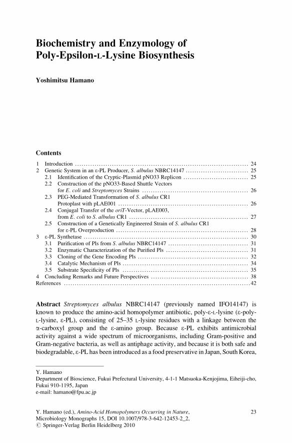
Abstract Streptomyces albulus NBRC14147 (previously named IFO14147) is
known to produce the amino-acid homopolymer antibiotic, poly-e-L-lysine (e-poly-L-lysine, e-PL), consisting of 25–35 L-lysine residues with a linkage between the
a-carboxyl group and the e-amino group. Because e-PL exhibits antimicrobial
activity against a wide spectrum of microorganisms, including Gram-positive and
Gram-negative bacteria, as well as antiphage activity, and because it is both safe and
biodegradable, e-PL has been introduced as a food preservative in Japan, SouthKorea,
Y. Hamano
Department of Bioscience, Fukui Prefectural University, 4-1-1 Matsuoka-Kenjojima, Eiheiji-cho,
Fukui 910-1195, Japan
e-mail: [email protected]
Y. Hamano (ed.), Amino-Acid Homopolymers Occurring in Nature,Microbiology Monographs 15, DOI 10.1007/978-3-642-12453-2_2,# Springer-Verlag Berlin Heidelberg 2010
23

the United States, and other countries. This chapter covers the current knowledge
and most recent advances in regard to the genetic system for S. albulus NBRC14147and e-PL synthetase.
1 Introduction
Streptomyces strains are known for their ability to synthesize commercially useful
secondary metabolites having a wide range of biological activities. Streptomycesalbulus NBRC14147 (previously named IFO14147) is known to produce the
amino-acid homopolymer antibiotic, e-PL, consisting of 25–35 L-lysine residues
with a linkage between the a-carboxyl group and the e-amino group (Fig. 1) (Shima
and Sakai 1977; Shima and Sakai 1981a; Shima and Sakai 1981b). Because e-PLexhibits antimicrobial activity against a wide spectrum of microorganisms, includ-
ing Gram-positive and Gram-negative bacteria (Shima et al. 1984), as well as
antiphage activity (Shima et al. 1982), and because it is both safe and biodegrad-
able, e-PL has been introduced as a food preservative in Japan, South Korea, the
United States, and other countries.
The biological activity of e-PL is known to be dependent on its molecular size.
Shima and coworkers investigated the relationship between the molecular size of
e-PL and its antimicrobial activity against Escherichia coli K-12 (Shima et al.
1984). e-PL with more than nine L-lysine residues severely inhibited microbial
growth; however, the L-lysine octamer demonstrated negligible antimicrobial activ-
ity. In contrast, chemically synthesized a-poly-L-lysine that contains a considerablylonger chain of L-lysine residues (50 residues), which show linkages between the
a-carboxyl and a-amino groups, demonstrates a lower activity than e-PL. Thus,
ε-PL (25-35 mer)
L-lysine
H2N
NH2
H2N
HN
NH
OH
OH
NH2
O
O
O
ONH2
NH2
n(23 - 33)
Fig. 1 Chemical structures of L-lysine and e-PL (25-mer to 35-mer)
24 Y. Hamano

polymerization of L-lysine via an isopeptide bond is required to exert its biological
activity, and the polymerization mechanisms involved in the chain-length diversity
of e-PL are of particular interest.
Investigation of an enzyme synthesizing e-PL should facilitate biosynthetic
engineering and help to create new classes of biopolymers. This review focuses
on characterization of an e-PL synthetase (Pls) and its biological machinery for
e-PL synthesis. In addition, an overview of effective genetic system for an e-PLproducer, S. albulus NBRC14147, which was used as a powerful tool for develop-
ing a deeper understanding of the Pls and for constructing an e-PL overproducer,
will be given.
2 Genetic System in an «-PL Producer, S. albulus NBRC14147
Among the various gene transfer methods, polyethylene glycol (PEG)-mediated
protoplast transformation is the standard for Streptomyces strains. In the initial
PGE-mediated method transformation experiments on S. albulus NBRC14147,
which were described for Streptomyces lividans (Kieser et al. 2000) and which
used the typical cloning vectors having the pIJ101, pSG5, or pRES replicon (Kieser
et al. 2000), no transformants were obtained. This unsuccessful outcome could be
attributed to either or both of these reasons: (1) the vectors employed do not work in
this strain; (2) the transformation methods are not suitable for direct application to
the present strain of S. albulus. This section provides a brief overview of effective
genetic system developed by Hamano et al.
2.1 Identification of the Cryptic-Plasmid pNO33 Replicon
A novel plasmid, pNO33, was detected in S. albulus NBRC14147 (Takagi et al.
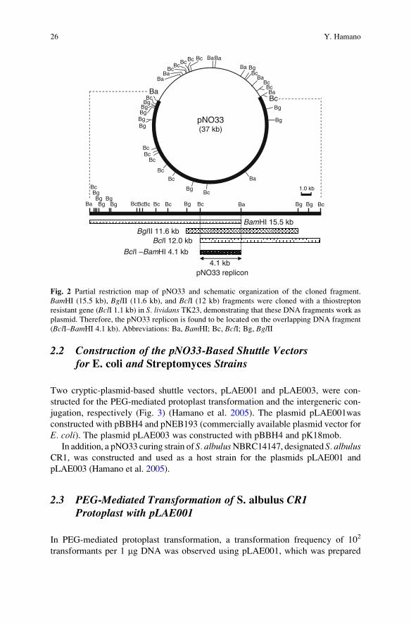
2000). This large plasmid (37 kb) is a cryptic plasmid (Fig. 2), as none of its functions
are yet known. To construct a cloning vector that can work in the S. albulus strain, thereplicon (BclI–BamHI 4.1 kb fragment, Fig. 2) of the cryptic plasmid pNO33 was
used (Hamano et al. 2005). A circular plasmid carrying the BclI–BamHI 4.1 kb
fragment with the antibiotic (thiostrepton) resistant gene worked as a plasmid vector
in S. lividans TK23, indicating that this plasmid (pBBH4) would also functionally
operate as a replicon in S. albulus.A database search with BLAST showed that the nucleotide sequence of this
replicon had no similarity with those of the known replicons for cloning vectors,
including pIJ702, which has the pIJ101 replicon and is frequently used in Strepto-myces strains. Hamano et al. also reported that pBBH4 and pIJ702 are compatible
for replication in the same cell of S. lividans (Hamano et al. 2005).
Biochemistry and Enzymology of Poly-Epsilon-L-Lysine Biosynthesis 25
2.2 Construction of the pNO33-Based Shuttle Vectorsfor E. coli and Streptomyces Strains
Two cryptic-plasmid-based shuttle vectors, pLAE001 and pLAE003, were con-
structed for the PEG-mediated protoplast transformation and the intergeneric con-
jugation, respectively (Fig. 3) (Hamano et al. 2005). The plasmid pLAE001was
constructed with pBBH4 and pNEB193 (commercially available plasmid vector for
E. coli). The plasmid pLAE003 was constructed with pBBH4 and pK18mob.
In addition, a pNO33 curing strain of S. albulusNBRC14147, designated S. albulusCR1, was constructed and used as a host strain for the plasmids pLAE001 and
pLAE003 (Hamano et al. 2005).
2.3 PEG-Mediated Transformation of S. albulus CR1Protoplast with pLAE001
In PEG-mediated protoplast transformation, a transformation frequency of 102
transformants per 1 mg DNA was observed using pLAE001, which was prepared
Ba
Bc Bg
Bc Bc Bg Ba Bg Bg Bg Bg Bg
Bg Bc Bc Bc Bc Bc
1.0 kb
BamHI 15.5 kbBgl II 11.6 kb
BclI 12.0 kb
4.1 kbpNO33 replicon
Bg Bg Bg
Bc Ba
Bg Bg
Bc Bc
Bc
Bc
Bc
Bg Bc
Ba
Bg
Bg
Bc Ba
Bc Bc
Ba Bc
Bg Ba
Ba Ba Bc Bc Bc
Ba Ba
Bc Bc
pNO33(37 kb)
Bcl I –BamHI 4.1 kb
Fig. 2 Partial restriction map of pNO33 and schematic organization of the cloned fragment.
BamHI (15.5 kb), BglII (11.6 kb), and BclI (12 kb) fragments were cloned with a thiostrepton
resistant gene (BclI 1.1 kb) in S. lividans TK23, demonstrating that these DNA fragments work as
plasmid. Therefore, the pNO33 replicon is found to be located on the overlapping DNA fragment
(BclI–BamHI 4.1 kb). Abbreviations: Ba, BamHI; Bc, BclI; Bg, BglII
26 Y. Hamano

from S. lividans TK23 (Hamano et al. 2005). When pLAE001 prepared from
S. albulus CR1 was used for the transformation, the efficiency increased 100-fold,
demonstrating that S. albulus CR1 shows a strong restriction barrier. In fact, the
authors reported that pLAE001 prepared from a methylation-proficient E. colistrain, such as XL1-blue MRF0, cannot transform S. albulus CR1.
2.4 Conjugal Transfer of the oriT-Vector, pLAE003,from E. coli to S. albulus CR1
Recently, there has been considerable interest in the use of intergeneric conjuga-
tion as a means of gene transfer to bypass protoplast formation and regeneration
(Mazodier et al. 1989; Matsushima et al. 1994; Flett et al. 1997; Voeykova et al.
1998; Paranthaman and Dharmalingam 2003; Choi et al. 2004; Stinchi et al.
2003). In S. albulus CR1, exconjugants were obtained employing a standard
procedure, although the conjugation efficiency was very low (exconjugants per
recipient spores: 9.0 � 10�8). Nihira et al. have reported that the optimal concen-
tration of MgCl2 for conjugation differs among the various strains (Choi et al.
2004). In fact, in S. albulus CR1, the solid medium containing an optimal
concentration of MgCl2 (40 mM) provided the highest frequency (4.0 � 10�7)
(Hamano et al. 2005).
PstI
SalISmaI
BssHII
BssHII
SmaI
SphIEcoRI
SalIKpnISmaI
BssHIIKpnI
KpnI
pNO33rep
lacZa
bla
pMB1 ori
tsr
pLAE001(7.9 kb)
EcoR
ISacI
KpnI
SmaI
Asc
IBs
sHII
Bam
HI
PacI
XbaI
Sal I
PmeI
Sbf I
PstI
SphI
Hind
III
(Bcl I/Bgl II)
(BamHI/BclI)
(BclI/BglII)
SmaI
SmaI
SphIEcoRI
Sal IKpnI
SmaI
KpnIKpnI
SphI SmaIKpnI
SacIEcoRI
SacI
pNO33reppMB1 ori
pLAE003(7.9 kb)
oriT
aph II
Bam
HI
XbaI
SalI
Pst I
SphI
Hind
III
(BclI/BamHI)
Fig. 3 pNO33-based shuttle vectors. pLAE001 and pLAE003 were used for the PEG-mediated
protoplast transformation and intergeneric conjugation from E. coli to S. albulus CR1, respec-
tively. Bold letters indicate cloning sites for restriction enzymes. Abbreviations: pNO33rep,
pNO33 replicon (BclI–BamHI 4.1 kb); bla, b-lactamase gene (Ampicillin resistance gene); aph II,aminoglycosides phosphotransferase II (neomycin/kanamycin resistance gene); tsr, thiostreptonresistance gene
Biochemistry and Enzymology of Poly-Epsilon-L-Lysine Biosynthesis 27

2.5 Construction of a Genetically Engineered Strainof S. albulus CR1 for e-PL Overproduction
Shima et al. have reported that the L-lysine molecule is directly utilized in e-PLbiosynthesis (Shima et al. 1983). In most bacteria, L-lysine is biosynthesized by the
amino-acid biosynthetic pathway from L-aspartic acid (aspartate pathway, Fig. 4).
This pathway is also involved in the formation of other amino acids (i.e., L-methionine,
L-isoleucine, and L-threonine). The first two enzymes in this pathway are asparto-
kinase (Ask) (EC.2.7.2.4), which catalyzes the phosphorylation of L-aspartic acid to
produce L-4-phospho aspartic acid, and aspartate semialdehyde dehydrogenase
(Asd) (EC.1.2.1.11), which reduces L-4-phospho aspartic acid into L-aspartate
4-semialdehyde. These two key enzymes are subject to complex regulation by the
end-product amino acids. Because of the complexity of this pathway, different
bacterial species have evolved diverse patterns of Ask regulation. For example,
E. coli and Bacillus subtilis have three separate Ask isozymes, each controlled by
one of the end products of the aspartate pathways, diaminopimelic acid, lysine,
threonine, and methionine (Hitchcock et al. 1980; Theze et al. 1974; Zhang et al.
1990; Zhang and Paulus 1990). In contrast, only one Ask has been described in
L-aspartic acid
aspartokinase (Ask)
L-4-phospho aspartic acid
aspartate semialdehydedehydrogenase (Asd)
L-aspartate 4-semialdehyde L-homoserine
L-methionine
L-threonine
L-isoleucineLL-2,6-diaminopimelic acid bacterialcell wall
L-lysine
e-PL
homoserinedehydrogenase
AVG
(in Streptomyces sp. NRRL5331)
(in S. albulus NBRC14147)
a-aminoadipic acid CPM-C
(in A. lactamdurans and S. clavuligerus)
L-cysteine L-valine
Fig. 4 The amino-acid biosynthetic pathway from L-aspartic acid (aspartate pathway). AVGaminoethoxyvinylglycine; CPM-C cephamycin C
28 Y. Hamano

antibiotic-producing actinomycetes. There are some reports of cloned ask genes
from the rifamycin SV producer Amycolatopsis mediterranei (Zhang et al. 1999),
from the cephamycin C (CPM-C, Fig. 4) producers Amycolatopsis lactamdurans(Hernando-Rico et al. 2001) and Streptomyces clavuligerus (Tunca et al. 2004),
and from the aminoethoxyvinylglycine (AVG, Fig. 4) producer Streptomycessp. NRRL5331 (Cuadrado et al. 2004). These studies have shown that regulatory
mechanisms can differ even among the actinomycetes. Zhang et al. have reported
inhibition of “A. mediterranei” Ask by L-lysine alone (Zhang et al. 2000), whereas
Asks of A. lactamdurans and S. clavuligerus are feedback-regulated by the con-
certed action of L-lysine and L-threonine (Tunca et al. 2004; Hernando-Rico et al.
2001). Interestingly, these experimental observations indicate that the Asks of
A. lactamdurans and S. clavuligerus (CPM-C producers) are slightly more resistant
to concerted feedback-inhibition than Ask IIIs of E. coli (Ogawa-Miyata et al. 2001)
and B. subtilis (Kobashi et al. 2001), although there is no discussion of this result inthe respective reports. As such, the L-lysine productivity of these strains should be
higher than those of E. coli and B. subtilis. This result could be due to the need for
that, L-lysine, which is one of biosynthetic precursors of CPM-C, must be fully
served to not only nascent protein biosynthesis but also the CPM-C biosynthesis
(Fig. 4).
The e-PL producer, S. albulus NBRC14147, can produce a large amount of e-PL(usually 1–3 g/l). Therefore, Ask(s) of S. albuluswas also expected to be potentiallyresistant to feedback-inhibition by L-lysine and/or L-threonine to provide sufficient
amounts of L-lysine for e-PL biosynthesis, similar to those of A. lactamdurans andS. clavuligerus. In fact, Hamano et al. demonstrated that the recombinant Ask of
S. albulus NBRC14147 was found to be partially resistant to feedback-inhibition
in vitro analysis (Hamano et al. 2007).
Hernando-Rico et al. have reported a construction of the feedback-resistant Ask of
A. lactamdurans by substitutions of the two amino-acid residues, Ser301 and Gly345
(Hernando-Rico et al. 2001). However, a homologous expression of the mutated Ask
has not been carried out due to the extreme difficulty in transforming A. lactamdurans.In S. albulus CR1, by random mutagenesis of the ask gene with error-prone PCR and
the subsequent site-directed mutagenesis, Hamano et al. successfully constructed the
mutated Ask, Ask (M68V), whose feedback-inhibition regulation was completely
removed. Ask of S. albulus CR1 also has Ser301 and Gly345, but no mutated Ask with
substitutions of these two amino-acid residues were obtained. Rather, rAsk (M68V)
was more appropriate for homologous expression in S. albulus CR1 to investigate
whether L-lysine accumulation in the cell leads to e-PL overproduction, as the
calculated VmaxAsp/Km
Asp values of rAsk (M68V) were ten-fold higher than that of
the rAsk (wild type).
Using the genetic system for S. albulus CR1 described earlier, Hamano et al.
constructed the genetically engineered strain of S. albulus CR1, which produced
rAsk (M68V). Compared with the productivity in the use of Ask (wild), the
homologous expression of Ask (M68V) predictably conferred a higher productivity
of e-PL (Fig. 5) (Hamano et al. 2007).
Biochemistry and Enzymology of Poly-Epsilon-L-Lysine Biosynthesis 29

3 «-PL Synthetase
Two amino-acid “homopolymers” comprising a single type of amino acid are
known in nature (Oppermann Sanio and Steinbuchel 2002): g-poly-glutamic acid
(g-PGA) and e-PL. The latter, which consists of 25–35 L-lysine residues with
linkages between a-carboxyl groups and e-amino groups (Fig. 1), exhibits anti-
microbial activity against a spectrum of microorganisms including bacteria and
fungi (Oppermann Sanio and Steinbuchel 2002; Shima et al. 1982; Shima et al.
1984). Due to its safety and biodegradability, it is used as a food preservative in
several countries. The biological activity of e-PL is known to be dependent on its
molecular size. Shima and coworkers investigated the relationship between the
molecular size of e-PL and its antimicrobial activity against E. coli K-12 (Shima
et al. 1984). e-PL with more than nine L-lysine residues severely inhibited microbial
growth; however, the L-lysine octamer demonstrated negligible antimicrobial activ-
ity. In contrast, chemically synthesized a-poly-L-lysine that contains a considerablylonger chain of L-lysine residues (50 residues), which show linkages between the
a-carboxyl and a-amino groups, demonstrates a lower activity than e-PL. Thus,polymerization of L-lysine via an isopeptide bond is required to exert its biological
activity, and the polymerization mechanisms involved in the chain-length diversity
of e-PL are of particular interest.
Nonribosomal peptide synthetases (NRPSs) are multifunctional enzymes con-
sisting of semiautonomous domains that synthesize numerous secondary metabo-
lites (Walsh 2003; Schwarzer et al. 2003; Mootz et al. 2002; Marahiel et al. 1997).
Using an assembly-line logic comprising multiple modules, they utilize a thiotem-
plated mechanism to activate, tether, and modify amino-acid monomers, sequen-
tially elongating the peptide chain before releasing the complete peptide. The order
and number of modules of a NRPS system determine the sequence and length of the
0
5
10
15
20
0 24 48 72 96 120 144 168
e-PL (g/l)
(hr)
pLAE006-ask (M68V) ( )
pLAE006-ask (wild) ( )
pLAE006 (no insert) ( )
Fig. 5 The e-PLproductivities in the
S. albulus CR1 strains
expressing Ask (wild) or Ask
(M68V). The S. albulus CR1strains harboring pLAE006
(pLAE003 derivative
harboring the ermE*promotor), pLAE006-ask
(wild), and pLAE006-ask
(M68V) were cultivated in
e-PL production medium
containing 100 mg/ml
neomycin by using a 5-l
capacity bench scale jar-
fermentor
30 Y. Hamano

peptide product. It has been reported that e-PL might be produced by NRPSs using a
thiotemplate mechanism (Kawai et al. 2003; Saimura et al. 2008). However, as they
used the crude extract of an e-PL-producing microorganism, it was not possible to
confirm the biosynthetic mechanism. Additionally, the chain-length diversity of
e-PL products is difficult to explain with this generic model. In addition, Hamano
et al. recently identified and characterized e-PL-degrading enzymes in an e-PLproducer, S. albulus NBRC14147 (Hamano et al. 2006). This raised the question
of whether the degrading enzymes, rather than the biosynthetic machinery, might be
responsible for generating products of diverse chain length. Actually, this section
will reveal that the chain-length diversity of e-PL is directly generated by the
synthetase.
3.1 Purification of Pls from S. albulus NBRC14147
S. albulus NBRC14147 was grown to the late logarithmic phase, in which e-PLproduction was observed. The mycelium collected from a 700ml-culture broth were
sonicated and centrifuged to obtain a cell-free extract. After ultracentrifugation of
the cell-free extract, e-PL-synthesis was observed in an insoluble fraction, suggest-ing that Pls is an insoluble protein such as a membrane protein. Yamanaka et al.
therefore solubilized this fraction with a nonionic detergent, Nonidet P-40 (NP-40)
(Yamanaka et al. 2008). By successive purification steps including column chroma-
tography, the solubilized Pls was finally purified 168-fold to apparent homogeneity.
The relative molecular mass of the native enzyme as estimated by gel-filtration
chromatography was 270 kDa. However, the molecular mass as estimated by
denaturing SDS–polyacrylamide gel electrophoresis (SDS–PAGE) was 130 kDa,
suggesting that Pls is a homodimer.
3.2 Enzymatic Characterization of the Purified Pls
The purified enzyme was incubated with L-lysine and ATP. A polydisperse group
of enzyme-dependent polymer products were detected, with their retention times
corresponding to those of reference standard polymers consisting of 3–17 residues
(Fig. 6). Incubation of Pls with L-lysine for different times did not change the
relative amounts of each polymer produced during the polymerization reaction.
ESI-tandem MS (ESI-MS/MS) analysis of the enzymatically synthesized polymer
with 14 residues was identical to that of the reference standard polymer with 14
residues. Identical mass spectra to the reference standards were also observed for
products of other chain lengths. Chemical modification of the synthesized poly-
mers using 2,4-dinitrophenol (DNP) followed by TLC analysis against reference
DNP-modified a- and e-lysine polymers demonstrated that the a-amino groups of
the L-lysine residues were labeled with DNP. This result shows that the polymer
Biochemistry and Enzymology of Poly-Epsilon-L-Lysine Biosynthesis 31

consists of L-lysine residues with linkages between the a-carboxyl and e-amino
groups and confirms that the polydisperse polymer products are indeed e-PL.Yamanaka et al. investigated other enzymatic properties (Yamanaka et al. 2008).
The enzyme required Mg2+, ATP, 20–30% glycerol, 2 mM dithiothreitol, and
0.2–0.4% NP-40 for full activity. ATP was converted to AMP during the Pls
reaction. No activity was detected with other nucleotides such as GTP, CTP, and
TTP in the Pls polymerization reaction. Maximum activity occurs at an optimum
pH of 8.5. The effect of temperature on the enzyme activity was investigated over
the range of 10–45�C, with the maximum activity being observed at 25–30�C.
3.3 Cloning of the Gene Encoding Pls
To clone the gene encoding Pls, some of the amino-acid sequences of Pls were
determined, and then PCR primers are designed. Using the PCR product as a probe,
a 33-kbp DNA fragment containing the pls gene from S. albulus NBRC14147 was
obtained. Interestingly, a BLAST database search showed that homologous genes
are widely distributed among microorganisms. The pls gene encoded a protein of
1,319 amino acids containing the internal amino-acid sequences previously deter-
mined. The calculated molecular mass (138,385 Da) was in good agreement with
the result from the SDS–PAGE analysis of the purified Pls. The authors also
constructed a knockout mutant of the putative pls gene using the genetic system
described earlier in Sect. 3. The mutant produced no e-PL, demonstrating that the
cloned gene encodes Pls (Yamanaka et al. 2008).
With the protein sequence in hand, they determined the predicted function
of the enzyme. In a traditional NRPS, the amino-acid substrate is activated as an
10 15 20 25 30 35
100
0
907
778898
843
0
100Intensity (%)
[M+2H]2+
MS/MS
100
0
Intensity (%) 14-mer
0
100
898778
843MS/MS
10 15 20 25 30 35 min.
e-PL25-35 mer
14-merIntensity (%)
Intensity (%)
m/z200 600 1,000 1,400 1,800
10-mer
5-mer
3-mer
17-mer
m/z200 600 1,000 1,400 1,800
14-mer
907[M+2H]2+14-mer
MS
MS
min.
Fig. 6 Identification of the Pls reaction products. The reaction mixture (lower chromatogram and
ESI mass spectrum) and a hydrolysate of e-PL (upper chromatogram and ESI mass spectrum) were
analyzed by HPLC/ESI-MS. The e-PL hydrolysates (0.1 mg/ml) were prepared by hydrolysis with
1N HCl
32 Y. Hamano

acyl-O-AMP by an adenylation domain (A-domain) and subsequently loaded onto
the 40-phosphopantetheine (40-PP) arm of the adjacent thiolation domain (T-domain)
(Walsh 2003; Schwarzer et al. 2003; Mootz et al. 2002; Marahiel et al. 1997),
resulting in the formation of an acylthioester and AMP release. A domain search
showed the presence of an A-domain and a T-domain in the N-terminal region of
the Pls. Their prior observation that AMP is released during the course of the
reaction, along with the similarity of the ten residue sequence that confers sub-
strate specificity in the putative Pls A-domain to that of the A-domain of the BacB
(Challis et al. 2000) protein that adenylates L-lysine and the presence of Ser553
from the putative T-domain in proper alignment to be a 40-PP-binding residue as
compared with the T-domains of traditional NRPSs provides strong support for
the classification of these regions of Pls as A- and T-domains. This distinguishes
the mechanism of e-PL biosynthesis from that of g-PGA (Oppermann Sanio
and Steinbuchel 2002), glutathione (Hibi et al. 2004), or cyanophycin (Oppermann
Sanio and Steinbuchel 2002), which require phosphorylation of the carboxyl
group.
Surprisingly, given the evidence for the presence of the A- and T-domains, Pls
had no domain with significant sequence similarity to the traditional condensation
domains (C-domains) that are crucial in peptide bond formation in NRPSs. Fur-
thermore, it had no traditional thioesterase domain (TE-domain), which catalyzes
release of the final product from NRPS enzymes by hydrolysis to the free acid or
cyclization to an amide or ester. Instead, a physicochemical analysis of the Pls
amino-acid sequence with SOSUI (Hirokawa et al. 1998) suggested the existence of
six transmembrane domains (TM-domains) surrounding three tandem soluble
domains that display significant sequence similarity (with pairwise identities of
27, 22, and 23%) (Fig. 7). Alignment of the tandem domains with traditional
C-domains demonstrated that the tandem domains did contain motifs showing
similarity to His-motifs, or the HHxxxDG sequences found in all traditional
C-domains, but the two histidine residues, known to be critical for catalysis, were
not conserved. However, both the primary sequence and predicted three-dimen-
sional structure of these domains showed similarity to acetyltransferases, which do
show structural similarity to C-domains (Bergendahl et al. 2002; Keating et al.
2002). For this reason, Yamanaka et al. named the tandem sequences the C1-, C2-,
and C3-domains, with the expectation that they would have a role in peptide bond
formation (Yamanaka et al. 2008).
HS
616
638
676
654
852
874
911
889
1087
1109
1148
1126
4’-PP
H2N (1) A-domain T C1 C2 C3
TM1 TM2 TM3 TM4 TM5 TM6
COOH (1319)
Fig. 7 Domain architecture of Pls. The A-, T-, six TM-domains, and three tandem domains (C1-,
C2-, and C3-domain) are shown schematically. The numbers on Pls are the amino-acid residue
numbers
Biochemistry and Enzymology of Poly-Epsilon-L-Lysine Biosynthesis 33

3.4 Catalytic Mechanism of Pls
Yamanaka et al. explored the catalytic mechanism of the Pls using an ATP–PPiexchange assay (Yamanaka et al. 2008). They observed Pls-mediated adenylation of
L-lysine, but not any other proteinogenic amino acid (Fig. 8a). To investigate the
function of the putative T-domain, they constructed a His-tagged recombinant Pls
containing only the A- and T-domain (rPls-AT) using S. lividans TK23 as a heterolo-gous host. Incubation of rPls-AT with L-[U-14C]lysine and ATP as substrates resulted
Relative activity (%)
L-Lysine (1)
L-Glysine
L-Alanine
L-Serine
L-Threonine
L-Cysteine
L-Valine
L-Leucine
L-Isoleucine
L-Methionine
L-Proline
L-Phenylalanine
L-Tyrosine
L-Tryptophan
L-Aspartic acid
L-Glutamic acid
L-Asparagine
L-Glutamine
L-Histidine
L-Arginine
none
0
0 2.5 5 10 20 40 80 160
plusD-lysine (mM)
L-[U-14C]lysine (20 mM)
(kDa)
rPls-AT(A+T-domain)
83
62
47.5
32.5
83
62
47.5
32.5
rPls-AT(A+T-domain)
D-lysine (mM)
a
CBB stain
Autoradiography
Densitometry
b
10050
1200
Intensity (pixel)
1000
800
600
400
200
0
0
2.5 5 10 20 40 80 160
Fig. 8 Functional analysis of Pls. (a) The relative adenylation activities were determined on
proteinogenic amino acids. Each value is represented as the mean of three experiments. (b)Aminoacylation of rPls-AT with L-[U-14C]lysine was investigated. rPls-AT (Fig. 9) was incubated
with L-[U-14C]lysine plus D-lysine at 30�C for 12 h. The reaction mixtures were then subjected
to SDS–PAGE (4 mg protein per lane). Proteins were stained with CBB R-250. The dried gel
was exposed on an imaging plate and visualized by BAS-2500. The data from densitometry analysis
of the autoradiography was shown. Data from this experiment is consistent with the fact that rPls-
AT was modified with the 40-PP cofactor in S. lividans TK23; the 40-PP was posttranslationally
transferred from Coenzyme A to the conserved serine residue (Ser553) of the T-domain by
endogenous 40-phosphopantetheinyl transferase(s), thus converting the inactive apo-form to its
active holo-form
34 Y. Hamano

in loading of the labeled lysine onto the enzyme (Fig. 8b). However, e-PL production
was not detected, suggesting that the three tandem domains (C1-, C2-, and C3-
domain) of Pls are essential for catalyzing the L-lysine polymerization reaction. To
gain a better understanding of the function of the tandem domains, two additional
recombinant Pls enzymes, rPls-ATC1C2 (lacking the C3-domain) and rPls-ATC1
(lacking both the C2- and C3-domain), were constructed. As observed for rPls-AT,
which lacks all three C domains, no polymer products were detected for rPls-ATC1C2
and rPls-ATC1, suggesting that either the C3-domain or the interconnected action of
all three domains is essential for peptide-bond formation.
As described earlier, Pls is predicted not to have a TE-domain, which is tradition-
ally required for release of the product from the NRPSmachinery. This suggested that
the growing polymer products are not covalently attached to Pls during the polymeri-
zation reaction. To investigate this hypothesis, Pls was incubated with L-[U-14C]
lysine and ATP. Analysis of the reaction mixture confirmed that Pls was radiolabeled
during the reaction (Fig. 9a); however, performic acid treatment of the labeled Pls,
which will release any small molecules attached via thioester bonds, only returned
L-[U-14C]lysine monomers instead of any polylysine chains (Fig. 9b). These results
strongly suggested that the growing polymer products are not covalently attached to
Pls during the polymerization reaction. Yamanaka et al. similarly observed that short-
chain e-PL oligomers (3-mer to 9-mer) are neither adenylated nor incorporated into
polymers primed with free, deuterated L-lysine (L-Lys-d8); instead, Pls produced
L-Lys-d8 homopolymers. To further test this mechanism, they employed L-lysine
ethyl and methyl esters as substrates. HPLC/ESI-MS analysis of the reaction revealed
that Pls produced the corresponding ethyl (Fig. 10) and methyl ester forms of e-PL.While retention of the final ester functionality confirmed that the growing polymers
or polymer products are not tethered to Pls via covalent bonds such as thioesters or
esters during the polymerization reaction (which would result in complete loss of the
ester group), it also raised the question of how the enzyme catalyzes polymerization
using ester substrates at all. The answer lay in a unique function of Pls: the A-domain
converts these L-lysine esters to L-lysine by its own esterase activity (Fig. 11), then
the resulting L-lysine can be adenylated and loaded to the T-domain.
3.5 Substrate Specificity of Pls
Given that Pls can accept lysine esters as substrates, the substrate specificity of
Pls was investigated further with seven L-lysine analogs (Fig. 12). L-Ornithine
(L-Orn), L-kynurenine (L-KNR), and 3-amino-L-tyrosine (L-ATY) were not accepted
as substrates. Pls was able to adenylate D-lysine (relative activity ¼ 19%), but AMP-
forming activity was not detected, suggesting that the adenylated D-lysine is not
loaded onto the T-domain. Indeed, D-lysine serves as an inhibitor of the aminoacyla-
tion of L-[U-14C]lysine (Fig. 8b), in agreement with a model where D-lysine is stalled
on the A-domain. Pls did successfully adenylate and pantetheinylate (as monitored
by AMP formation) the rest of the analogs, including (5R)-5-hydroxy-L-lysine
Biochemistry and Enzymology of Poly-Epsilon-L-Lysine Biosynthesis 35

(L-HLY; relative activity for adenylation, 43%; relative activity for AMP-formation,
6%), O-(2-aminoethyl)-L-serine (L-AES; corresponding values, 13 and 12%), and
S-(2-aminoethyl)-L-cysteine (L-AEC; corresponding values, 5 and 6%). However, no
homopolymers from these analogs were observed (see the lower chromatograms in
Fig. 13b–d). In contrast, when a small amount of L-lysine was added to the reaction
(kDa)25015010075
50
37
2520
1510
reaction time (s)0 5 10 20 40 80 0 5 10 20 40 80
reaction time (s)a
b
Pls
Enzymaticallysynthesized e - PL
1 21 2 3
L-Lysine
e - PL (3-mer to 9-mer)Origin
Solvent front
3
Unknown
Fig. 9 Performic-acid oxidation of the radiolabeled Pls formed in the Pls reaction with
L-[U-14C]lysine. (a) After incubation of Pls with L-[U-14C]lysine and ATP at 5�C for 0–80 s,
the reaction mixtures were subjected to SDS–PAGE (5–20% gradient gel, 16 mg protein per
lane). Proteins were stained with CBB R-250 (image on the left). The dried gel was exposed on
an imaging plate and visualized by BAS-2500 (Fuji film; image on the right). (b) The reactionmixture incubated for 10 s was further used for the oxidation experiments of Pls. L-[U-14C]
lysine (lane 1), and the radiolabeled Pls treated with performic (lane 3) and formic acid (lane 2)
were analyzed by silica gel thin-layer chromatography (TLC) (left image). The dried TLC was
exposed on an imaging plate and visualized by BAS-2500. Additionally, to confirm that these
acids do not hydrolyze the short-chain e-PL oligomers, the oligomers (3-mer to 9-mer) were
treated with formic (lane 2) and performic acids (lane3) and analyzed by silica gel TLC (image
on the right). L-lysine (lane 1) was also used as the reference standards to confirm the mobility
of the short-chain e-PL oligomers. The samples were detected with ninhydrin reagent. In this
oxidation experiments, an amino acid bound to enzyme as thioester is released from the
enzyme not by formic acid but by performic acid. In the left image of panel (b), we detected
the extra band (lane 3), which is known as the commonly observed unknown spots in performic
oxidation experiments
36 Y. Hamano

1 2 3 4 5 6
Origin
L-lysineL-lysine methyl ester
L-lysine ethyl ester
Solvent front
7 8 9
rPls-AT
ATP
L-lysine
L-lysine methyl ester
L-lysine ethyl ester
+
–
+
+ +
+
+
+– – –
–
–
–
–
–
– –+
+
+ +
+
+ + +
+ +
+
+
–
– – – –
–
– –
– –
– –
–
+
+
Enzyme reaction with;
Fig. 11 Esterase activity in the A-domain. The rPls-AT reaction with 2 mM L-lysine, 2 mM
L-lysine methyl ester, or 2 mM L-lysine ethyl ester was performed and analyzed by silica gel TLC.
TLC was developed in 1-butanol/pyridine/acetic acid/H2O, 2:1:1:2 (v/v). The products were
detected with ninhydrin reagent. These results demonstrated that the esterase activities were not
dependent on ATP. Addition, (+); No addition, (�)
Intensity (x108)
2
0
10 15 20 25 30 35 min
1
Intensity (%)
0
100
200 m/z
1,328.9
e -PL-O-Et (10-mer)[M+H]+
600 1,4001,000 1,800
MS
379.3 431.4
559.5596.5
641.5687.6
725.6769.6
815.6
852.7897.7
943.8
980.7
1,071.9
1,109.9
1,200.0
1,237.9
1,310.9
1,153.91,025.9513.4
400 500 600 700 800 900 1,000 1,100 1,200 1,300 m/z
1,200.0 1,071.9 943.8 815.6 687.6 559.5 431.4
1,153.91,025.9897.7769.6641.5513.4
0
100Intensity (%)
MS/MS
e -PL-O-Et (10mer) MW;1,327.8
e -PL-O-Et(10-mer) (14-mer)
H2NNH
HN
NH
O
NH2
NH2
O NH2
O HN
NH
HN
NH2
O NH2
O NH2
O
NH
HN
NH2
O NH2
O
NH
NH2
O NH2
O
O
Fig. 10 Pls reaction with L-lysine ethyl ester. The Pls polymerization reaction with 2 mM L-lysine
ethyl ester was performed and analyzed by HPLC/ESI-MS. The mass spectra and MS/MS spectra
of the selected polymers (10-mer) in the reactions are shown
Biochemistry and Enzymology of Poly-Epsilon-L-Lysine Biosynthesis 37

mixtures, heteropolymers consisting of L-lysine and the analogs were produced (see
the upper chromatograms in Fig. 13b–d). These results indicate that the A- and
T-domains are partially tolerant to substrate analogs, whereas the three tandem
acetyltransferase domains show high specificity for L-lysine.
4 Concluding Remarks and Future Perspectives
In the e-PL producing strain S. albulus NBRC14147, Hamano and coworkers
successfully developed the genetic system, which was a powerful tool to construct
a genetically engineered strain and to investigate the biosynthetic mechanism
L-lysine
L-HLY
D-lysine
L-AES
L-AEC
L-Orn
L-KNR
L-ATY
(%)
0 25 50 75 100
NH2
S
OH
OH
OH
OH
OH
OH
OH
OH
OH
H2N
H2N
H2N
H2N
H2N
H2N
H2N
HO
AdenylationAMP-forming
O
O
O
O
O
O
O
O
O
O
NH2
NH2
NH2
NH2
NH2
NH2NH2
NH2
Fig. 12 Substrate specificities in the A- and T-domain of Pls. The relative activities in the
adenylation and AMP-forming activities were determined on L-lysine and the L-lysine analogues.
Each value is represented as the mean of three experiments. Error bars are not shown, because
relative standard deviations of less than 5%were commonly calculated. L-HLY, (5R)-5-hydroxy-L-
lysine; L-AES, O-(2-aminoethyl)-L-serine; L-AEC, S-(2-aminoethyl)-L-cysteine; L-Orn, L-ornithine;
L-KNR, L-kynurenine; L-ATY, 3-amino-L-tyrosine
38 Y. Hamano

0
2
4
6
7
0
2
4
6
7
Intensity´107
0
2
4
6
7´106
0
2
0
2
4
6
7´106
0
2
4
0
2
4
6
7
´106
0
2
10 15 20 25 30 35
780 800 820 m/z
MSMS
(M+H)+788
18
806(M+H)+
16
(M+H)+804
16
(M+H)+820
(M+H)+790
2 2
(M+H)+792
(M+H)+836
16
(M+H)+794
(M+H)+796
22 2
18
824(M+H)+
18
842(M+H)+
840
Inte
nsity
(%
)
100
0
Inte
nsity
(%
)
100
0In
tens
ity (
%)
100
0
Inte
nsity
(%
)
100
0
MSMS
MSMS
MSMS
6-mer
Pls reaction with L-lysine (2 mM)a
Pls reaction with L-lysine (0.4 mM) plus L-HLY (1.6 mM)b
c
d
Pls reaction with L-HLY (2 mM)
Pls reaction with L-lysine (0.2 mM) plus L-AES (1.8 mM)
Pls reaction with L-AES (2 mM)
Pls reaction with L-lysine (0.2 mM) plus L-AEC (1.8 mM)
Pls reaction with L-AEC (2 mM)
min
Fig. 13 In vitro production of heteropolymers consisting of L-lysine and the L-lysine analogues.
The Pls reaction with 2 mM L-lysine (a), 1.6 mM L-HLY plus or minus 0.4 mM L-lysine (b),1.8 mM L-AES plus or minus 0.2 mM L-lysine (c), or 1.8 mM L-AEC plus or minus 0.2 mM
L-lysine (d) was performed and analyzed by HPLC/ESI-MS. The mass spectra of the selected
polymers (6-mer) in the reactions are shown in the right-hand panels
Biochemistry and Enzymology of Poly-Epsilon-L-Lysine Biosynthesis 39

of e-PL. They also purified a membrane protein with six TM-domains, from
the membrane fraction of S. albulus NBRC14147, and characterized it as Pls, the
biological machinery for e-PL synthesis. Their explorations of Pls resulted in the
development of a new model for the generation of chain-length diversity of e-PLproducts, which integrates traditional NRPS logic, in the form of A- and T-domains,
with amino-acid ligase functionality in three tandem domains that show similarity
to both acetyltransferases and, through these transferases, traditional C-domains.
The catalytic mechanism is initiated in N-terminus by the A- and T-domains with
the adenylation and transfer of an incoming L-lysine monomer (or “extending
unit”), with polymerization occurring as freely diffusible substrates (or “priming
units”) are added by the C-terminal tandem domains to the extending unit (Fig. 14).
Since this cycle has no predetermined endpoint, other than the loss of the noncova-
lently bound polymer chain to solution, Pls acts iteratively for e-PL chain growth to
obtain a multitude of chain lengths (observed in their studies as ranging from 3 to 17
residues). This mechanism, in which a single polymer is created and then released
(Fig. 14), is supported not only by the characterization of more basic Pls properties
such as ATP turnover and e-amine linkage formation but also by the observations
that there was no difference in the relative amounts of each polymer during the
polymerization reaction and that preexisting short chain polymers could not be
incorporated into new chains (i.e., that the short chains were not simply intermedi-
ates of the longer chains).
Though e-PL chain-length diversity can be explained by this mechanism, a
detailed description of how the growing polymer interacts with the protein remains
a subject for future work. Yamanaka et al. hypothesize that the protein contains a
long slender-shaped tunnel, or cavity, that is continuously occupied by a growing
polymer during the polymerization reaction. This would explain why the polymeri-
zation reaction is specific to the e-amino groups of the priming units, as the
similarly reactive a-amino groups could be buried or otherwise protected from
reaction by the Pls catalytic cavity (Fig.14).
In the analysis of the sequence of the three tandem domains, Yamanaka et al.
identified a region reminiscent of the known His-motif from traditional C-domains,
but lacking the histidine residues thought to be required for catalysis. It has been
reported recently that the two histidine residues in the His-motif of VibH are also
not critical for catalysis (Keating et al. 2002). As VibH, the functional C-domain of
a vibriobactin NRPS, utilizes a similar mechanism to Pls – catalyzing peptide-bond
formation between an NRPS-bound substrate, dihydroxybenzoate, and the freely
diffusible substrate, norspermidin – these combined results may suggest that the
absence or lack of necessity of the two histidine residues may define a secondary
motif that can be used to identify C-domains operating via characteristic amino-
acid ligase-like mechanisms.
Finally, the investigations demonstrated that the chain-length diversity of e-PL is
directly generated by the synthetase, rather than via the differential degradation of a
uniform polymer by e-PL degrading enzymes. With these findings in hand, it will be
interesting to determine whether the activity of these degrading enzymes serves to
create a shorter polymer with some defined function or simply regenerates the lysine
40 Y. Hamano

S
4’-PP
A-domain
T-domain
C1-, C2-, C3-domain
C1-, C2-, C3-domain
Priming unit;L-Lysine,
L-Lys-O-Me,and L-Lys-O-Et
Extending units; L-Lysine, L-HLY, L-AES, L-AEC, L-Lys-O-Me*, and L-Lys-O-Et*
S
4’-PP
A-domain
T-domain
C1-, C2-, C3-domain
C1-, C2-, C3-domain
Extending units
Polymerization reactions
L-Lysine
L-Lysine
L-Lysine
OO
AMP
O O
HO
OH
O
HO
O
O
NH2 NH2
NH2
NH2
NH2
NH2
OH2N
H2N
H2N
H2N
H2NH2N
H2N H2N
AMP
O
HN
NH
OH
O
O
On
* Converted to L-lysine by the esterase activity ofthe A-domain.
Polymerproducts
NH2NH2
NH2
NH2
Fig. 14 Pls polymerization mechanism and the proposed model of the Pls catalytic cavity with
substrate specificity. The clarified polymerization mechanism is shown schematically. L-Lys-O-Me,
L-lysine methyl ester; L-Lys-O-Et, L-lysine ethyl ester
Biochemistry and Enzymology of Poly-Epsilon-L-Lysine Biosynthesis 41

building blocks from the unusual e-PL chain architecture. Similarly, further explora-
tions of the homologous genes identified in our BLAST search should prove exciting,
as their encoded proteins may synthesize amino-acid homopolymers other than e-PLand g-PGA. Investigations of these proteins aswell as further exploration of Pls shouldfacilitate biosynthetic engineering and help to create new classes of biopolymers.
References
Bergendahl V, Linne U, Marahiel MA (2002) Mutational analysis of the C-domain in nonriboso-
mal peptide synthesis. Eur J Biochem 269:620–629
Challis GL, Ravel J, Townsend CA (2000) Predictive, structure-based model of amino acid
recognition by nonribosomal peptide synthetase adenylation domains. Chem Biol 7:211–224
Choi SU, Lee CK, Hwang YI, Kinoshita H, Nihira T (2004) Intergeneric conjugal transfer of
plasmid DNA from Escherichia coli to Kitasatospora setae, a bafilomycin B1 producer. Arch
Microbiol 181:294–298
Cuadrado Y, Fernandez M, Recio E, Aparicio JF, Martin JF (2004) Characterization of the ask-asdoperon in aminoethoxyvinylglycine-producing Streptomyces sp. NRRL 5331. Appl Microbiol
Biotechnol 64:228–236
Flett F, Mersinias V, Smith CP (1997) High efficiency intergeneric conjugal transfer of plasmid
DNA from Escherichia coli to methyl DNA-restricting streptomycetes. FEMS Microbiol Lett
155:223–229
Hamano Y, Nicchu I, Hoshino Y, Kawai T, Nakamori S, Takagi H (2005) Development of gene
delivery systems for the e-poly-L-lysine producer, Streptomyces albulus. J Biosci Bioeng
99:636–641
Hamano Y, Yoshida T, Kito M, Nakamori S, Nagasawa T, Takagi H (2006) Biological function of
the pld gene product that degrades e-poly-L-lysine in Streptomyces albulus. Appl Microbiol
Biotechnol 72:173–181
Hamano Y, Nicchu I, Shimizu T, Onji Y, Hiraki J, Takagi H (2007) epsilon-Poly-L-lysine
producer, Streptomyces albulus, has feedback-inhibition resistant aspartokinase. Appl Micro-
biol Biotechnol 76:873–82
Hernando-Rico V, Martin JF, Santamarta I, Liras P (2001) Structure of the ask-asd operon and
formation of aspartokinase subunits in the cephamycin producer ‘Amycolatopsis lactamdurans’.Microbiology 147:1547–1555
Hibi T, Nii H, Nakatsu T, Kimura A, Kato H, Hiratake J, Oda J (2004) Crystal structure of gamma-
glutamylcysteine synthetase: insights into the mechanism of catalysis by a key enzyme for
glutathione homeostasis. Proc Natl Acad Sci USA 101:15052–15057
Hirokawa T, Boon Chieng S, Mitaku S (1998) SOSUI: classification and secondary structure
prediction system for membrane proteins. Bioinformatics 14:378–379
Hitchcock MJ, Hodgson B, Linforth JL (1980) Regulation of lysine- and lysine-plus-threonine-
inhibitable aspartokinases in Bacillus brevis. J Bacteriol 142:424–432Kawai T, Kubota T, Hiraki J, Izumi Y (2003) Biosynthesis of e-poly-L-lysine in a cell-free system
of Streptomyces albulus. Biochem Biophys Res Commun 311:635–640
Keating TA, Marshall CG, Walsh CT, Keating AE (2002) The structure of VibH represents
nonribosomal peptide synthetase condensation, cyclization and epimerization domains. Nat
Struct Biol 9:522–526
Kieser T, Bibb MJ, Buttner MJ, Chater KF, Hopwood DA (2000) Practical Streptomyces Genetics.
The John Innes Foundation, Norwich, UK
Kobashi N, Nishiyama M, Yamane H (2001) Characterization of aspartate kinase III of Bacillussubtilis. Biosci Biotechnol Biochem 65:1391–1394
42 Y. Hamano

Marahiel MA, Stachelhaus T, Mootz HD (1997) Modular peptide synthases involved in nonribo-
somal peptide synthesis. Chem Rev 97:2651–2673
Matsushima P, Broughton MC, Turner JR, Baltz RH (1994) Conjugal transfer of cosmid DNA
from Escherichia coli to Saccharopolyspora spinosa: effects of chromosomal insertions on
macrolide A83543 production. Gene 146:39–45
Mazodier P, Petter R, Thompson C (1989) Intergeneric conjugation between Escherichia coli andStreptomyces species. J Bacteriol 171:3583–3585
Mootz HD, Schwarzer D, Marahiel MA (2002) Ways of assembling complex natural products on
modular nonribosomal peptide synthetases. Chembiochem 3:490–504
Ogawa-Miyata Y, Kojima H, Sano K (2001) Mutation analysis of the feedback inhibition site of
aspartokinase III of Escherichia coli K-12 and its use in L-threonine production. Biosci
Biotechnol Biochem 65:1149–1154
Oppermann Sanio FB, Steinbuchel A (2002) Occurrence, functions and biosynthesis of poly-
amides in microorganisms and biotechnological production. Naturwissenschaften 89:11–22
Paranthaman S, Dharmalingam K (2003) Intergeneric conjugation in Streptomyces peucetius andStreptomyces sp. strain C5: chromosomal integration and expression of recombinant plasmids
carrying the chiC gene. Appl Environ Microbiol 69:84–91
Saimura M, Takehara M, Mizukami S, Kataoka K, Hirohara H (2008) Biosynthesis of nearly
monodispersed poly(epsilon-L-lysine) in Streptomyces species. Biotechnol Lett 30:377–385Schwarzer D, Finking R, Marahiel MA (2003) Nonribosomal peptides: from genes to products.
Nat Prod Rep 20:275–287
Shima S, Sakai H (1977) Polylysine produced by Streptomyces. Agric Biol Chem 41:1807–1809
Shima S, Sakai H (1981a) Poly-L-lysine produced by Streptomyces. II. Taxonomy and fermenta-
tion studies. Agric Biol Chem 45:2497–2502
Shima S, Sakai H (1981b) Poly-L-lysine produced by Streptomyces. III. Chemical studies. Agric
Biol Chem 45:2503–2508
Shima S, Matsuoka H, Sakai H (1982) Inactivation of bacteriophages by e-poly-L-lysine producedby Streptomyces. Agric Biol Chem 46:1917–1919
Shima S, Oshima S, Sakai H (1983) Biosynthesis of e-poly-L-lysine by washed mycelium of
Streptomyces albulus No. 346. Nippon Nogeikagaku Kaishi 57:221–226
Shima S, Matsuoka H, Iwamoto T, Sakai H (1984) Antimicrobial action of e-poly-L-lysine.J Antibiot (Tokyo) 37:1449–1455
Stinchi S, Azimonti S, Donadio S, Sosio M (2003) A gene transfer system for the glycopeptide
producer Nonomuraea sp. ATCC39727. FEMS Microbiol Lett 225:53–57
Takagi H, Hoshino Y, Nakamori S, Inouye S (2000) Isolation and sequence analysis of plasmid
pNO33 in the e-poly-L-lysine-producing actinomycete Streptomyces albulus IFO14147.
J Biosci Bioeng 89:94–96
Theze J, Margarita D, Cohen GN, Borne F, Patte JC (1974) Mapping of the structural genes of the
three aspartokinases and of the two homoserine dehydrogenases of Escherichia coli K-12.J Bacteriol 117:133–143
Tunca S, Yilmaz EI, Piret J, Liras P, Ozcengiz G (2004) Cloning, characterization and heterolo-
gous expression of the aspartokinase and aspartate semialdehyde dehydrogenase genes of
cephamycin C-producer Streptomyces clavuligerus. Res Microbiol 155:525–534
Voeykova T, Emelyanova L, Tabakov V, Mkrtumyan N (1998) Transfer of plasmid pTO1 from
Escherichia coli to various representatives of the order Actinomycetales by intergeneric
conjugation. FEMS Microbiol Lett 162:47–52
Walsh C (2003) Antibiotics: Action, Origins, Resistance. ASM Press, Washington DC
Yamanaka K, Maruyama C, Takagi H, Hamano Y (2008) Epsilon-poly-L-lysine dispersity is
controlled by a highly unusual nonribosomal peptide synthetase. Nat Chem Biol 4:766–772
Zhang JJ, Paulus H (1990) Desensitization of Bacillus subtilis aspartokinase I to allosteric
inhibition by meso-diaminopimelate allows aspartokinase I to function in amino acid biosyn-
thesis during exponential growth. J Bacteriol 172:4690–4693
Biochemistry and Enzymology of Poly-Epsilon-L-Lysine Biosynthesis 43

Zhang JJ, Hu FM, Chen NY, Paulus H (1990) Comparison of the three aspartokinase isozymes in
Bacillus subtilis Marburg and 168. J Bacteriol 172:701–708
Zhang W, Jiang W, Zhao G, Yang Y, Chiao J (1999) Sequence analysis and expression of the
aspartokinase and aspartate semialdehyde dehydrogenase operon from rifamycin SV-producing
Amycolatopsis mediterranei. Gene 237:413–419Zhang WW, Jiang WH, Zhao GP, Yang YL, Chiao JS (2000) Expression in Escherichia coli,
purification and kinetic analysis of the aspartokinase and aspartate semialdehyde dehydroge-
nase from the rifamycin SV-producing Amycolatopsis mediterranei U32. Appl Microbiol
Biotechnol 54:52–58
44 Y. Hamano

Biochemistry and Enzymology ofPoly-Epsilon-L-Lysine Degradation
Toyokazu Yoshida
Contents
1 Introduction . . . . . . . . . . . . . . . . . . . . . . . . . . . . . . . . . . . . . . . . . . . . . . . . . . . . . . . . . . . . . . . . . . . . . . . . . . . . . . . . 46
2 Assay for e-PL-Degrading Activity . . . . . . . . . . . . . . . . . . . . . . . . . . . . . . . . . . . . . . . . . . . . . . . . . . . . . . . . 46
3 e-PL Degradation by Microorganisms . . . . . . . . . . . . . . . . . . . . . . . . . . . . . . . . . . . . . . . . . . . . . . . . . . . . . 48
3.1 e-PL Degradation by e-PL-Tolerant Microorganisms . . . . . . . . . . . . . . . . . . . . . . . . . . . . . . . . . 49
3.2 e-PL Degradation by e-PL-Producing Microorganisms . . . . . . . . . . . . . . . . . . . . . . . . . . . . . . . 52
4 Molecular Genetic Analysis of e-PL-Degrading Enzyme of S. albulus . . . . . . . . . . . . . . . . . . . . 55
4.1 Cloning and Analysis of the pld Gene for e-PL-Degrading Enzyme of S. albulus . . . 56
4.2 Biological Function of the pld Gene in S. albulus . . . . . . . . . . . . . . . . . . . . . . . . . . . . . . . . . . . . 56
5 Conclusion . . . . . . . . . . . . . . . . . . . . . . . . . . . . . . . . . . . . . . . . . . . . . . . . . . . . . . . . . . . . . . . . . . . . . . . . . . . . . . . . . 57
References . . . . . . . . . . . . . . . . . . . . . . . . . . . . . . . . . . . . . . . . . . . . . . . . . . . . . . . . . . . . . . . . . . . . . . . . . . . . . . . . . . . . . . 58
Abstract Poly-e-L-lysine (e-poly-L-lysine, e-PL) is used as a food additive on
the basis of its strong antimicrobial activity (Fine chem 29:18–25, 2000). e-PL is
industrially produced by fermentation process using Streptomyces albulus.Recently, the biosynthetic mechanism (Nat Chem Biol 4:766–772, 2008) and
microbial degradation of e-PL (FEMS Microbiol Lett 207:147–151, 2002; Arch
Microbiol 178:325–330, 2002; J Biosci Bioeng 96:92–94, 2003; Appl Microbiol
Biotechnol 72:173–181, 2006) have been reported. The e-PL-degrading activity isdetected in e-PL-tolerant bacteria, indicating the contribution of e-PL-degradingactivity to bacterial growth in the presence of e-PL. The purified e-PL-degradingenzymes catalyze the endo- and/or exo-type degradation of e-PL (FEMS Micro-
biol Lett 207:147–151, 2002; J Biosci Bioeng 96:92–94, 2003). Various peptides
and proteins serve as substrates for the e-PL-degrading enzymes. Probably, the
adventitious possession of proteases, exhibiting high e-PL-degrading activity,
enables e-PL-tolerant bacteria to grow well even in the presence of e-PL. e-PL-producing Streptomyces strains also possess high e-PL-degrading activity in their
membrane fraction (Arch Microbiol 178:325–330, 2002). e-PL-degrading enzyme
T. Yoshida
Department of Biomolecular Science, Gifu University, 1-1 Yanagido, Gifu 501-1193, Japan
e-mail: [email protected]
Y. Hamano (ed.), Amino-Acid Homopolymers Occurring in Nature,Microbiology Monographs 15, DOI 10.1007/978-3-642-12453-2_3,# Springer-Verlag Berlin Heidelberg 2010
45

purified from S. albulus is characterized as a zinc-containing aminopeptidase. In
Streptomyces strains, the correlative distribution of e-PL-degrading activity and
e-PL-producing activity is observed, suggesting that the e-PL-degrading activity is
localized in the cell membrane of e-PL producers for self-protection against e-PL.
1 Introduction
Since the discovery of poly-e-L-lysine (e-poly-L-lysine, e-PL) in the 1970s (Shima
and Sakai 1977), many investigators have noted its wide spectrum of antimicrobial
activity, and e-PL has been widely used as a food additive (Hiraki 2000). e-PL can
be produced industrially, and the studies on its further applications are continuing.
e-PL is a homo-poly amino acid characterized by the peptide bond between
a-carboxyl group and e-amino group. Peptide bonds of protein and peptide are
hydrolyzed by various enzymatic reactions. However, there have been few reports
on the biological degradation of e-PL in natural environment or in vivo. Although
the safety of e-PL as a food additive was demonstrated experimentally using rats
(Neda et al. 1999), the biological decomposition or utilization of e-PL in rats
was not studied. e-PL-degrading microorganisms have been isolated from natural
environment as e-PL-tolerant bacteria (Kito et al. 2002a, 2003), in spite of high
antimicrobial activity of e-PL for various microorganisms (Hiraki 2000). Enzymo-
logical characterization of the e-PL-degrading enzymes from e-PL-tolerant bacteriareveals that the degrading activity of e-PL-tolerant bacteria is not specialized for
e-PL degradation. e-PL-degrading activity is also detected in e-PL-producingStreptomyces albulus cells (Shima et al. 1983; Hiraki et al. 1998; Kahara et al.
2001). The presence of e-PL-degrading enzyme is disadvantageous for industrial
e-PL production by S. albulus. However, the enzyme catalyzing e-PL degradation
in S. albulus had not been verified, until Kito et al. (2002b) found the activity in themembrane fraction of the e-PL producer. The recent functional analysis of the pldgene encoding e-PL-degrading enzyme of S. albulus (Hamano et al. 2006) demon-
strated that S. albulus probably possesses, at least, two kinds of e-PL-degradingenzymes.
2 Assay for «-PL-Degrading Activity
In this chapter, “e-PL” indicates a mixture of polymers with 25–35 L-lysine
residues. Studies on e-PL have been mainly carried out using the preparation
produced by S. albulus, the polymerization degree (n) being 25–35.
e-PL consists of 25–35 L-lysine residues linked with the a-carboxyl and e-amino
groups, and the mode of e-PL degradation is divided into exo-type and endo-type.
Therefore, the degradation activity for e-PL is generally calculated from residual
e-PL concentration in culture medium and reaction mixture. The best simple and
46 T. Yoshida

rapid method to determine e-PL concentration is a colorimetric procedure using an
anionic dye, methyl orange (Itzhaki 1972). In this method, e-PL interacts with
methyl orange on the basis of its cationic property and forms a water-insoluble
complex. e-PL concentration can be estimated from the absorbance at 465 nm of the
methyl orange remaining in solution. Free L-lysine and short lengths of L-lysine
polymers do not associate with methyl orange, and so the colorimetric method can
estimate the total amount of long lengths of L-lysine polymers.
The polymerization degree of L-lysine polymers can be measured using high-
performance liquid chromatography (HPLC) (Kito et al. 2002a). Using a reverse-
phase column for HPLC analysis, various lengths of L-lysine polymers are individually
detected at 215 nm by the gradient elution of acetonitrile in phosphate buffer
(pH 2.6) containing NaClO4 and sodium octane sulfonate as an ion-pair reagent.
Although the HPLC analysis is time-consuming, the estimation of polymerization
degrees of L-lysine polymers is essential to the studies on e-PL degradation. Free
L-lysine concentration released from e-PL is generally determined using amino acid
analyzer or enzymatically using L-lysine-a-oxidase (Kusakabe et al. 1980).To verify the mode of e-PL degradation, the polymerization degree and concen-
tration of L-lysine polymers should be analyzed. In the representative time course of
exo-type e-PL degradation, various lengths of L-lysine polymers are constantly
detected by HPLC analysis until complete degradation of e-PL (Fig. 1a), and the
sizes of L-lysine polymers are gradually reduced in accordance with the formation
of free L-lysine. In the reaction mixture, L-lysine concentration enhances in propor-
tion to time. Therefore, as for the exo-type e-PL degradation, the degrading activity
for e-PL can be also evaluated as the formed L-lysine concentration. In endo-type
e-PL degradation (Fig. 1b), various lengths of L-lysine polymers are initially
Fig. 1 Analysis of e-PL degradation using HPLC system. (a) exo-type e-PL degradation; (b) endo-type e-PL degradation. LMW low molecular weight
Biochemistry and Enzymology of Poly-Epsilon-L-Lysine Degradation 47

detected as in the case of exo-type degradation. However, further incubation results
in the accumulation of shorter lengths of L-lysine polymers in the reaction mixture.
In the case of mixed-type degradation as observed using Chryseobacterium sp. OJ7
(Kito et al. 2003), various peptides containing L-lysine residue(s) should be tested
as the substrate to confirm the mode of e-PL degradation (Fig. 2).
For the assay of e-PL-degrading enzyme in the purification procedures, L-lysyl-
p-nitroanilide serves as a convenient substrate, irrespective of the mode of e-PLdegradation (Kito et al. 2002a, b, 2003). e-PL-degrading enzymes efficiently
catalyze the hydrolysis of L-lysyl-p-nitroanilide; the formed p-nitroaniline is easilymeasured spectrophotometrically with a high sensitivity.
3 «-PL Degradation by Microorganisms
The notable antimicrobial activity of e-PL against various microorganisms attracts
a great deal of attention for its use as a food preservative. When the amino groups of
e-PL are partially amidated using aromatic carboxylic acids such as 4-chlorobenzoic
Fig. 2 e-PL degradation by
the purified e-PL-degradingenzyme from
Chryseobacterium sp. OJ7.
e-PL was incubated with the
purified e-PL-degradingenzyme for the indicated
time, and the polymerization
degree of L-lysine was
analyzed by HPLC
48 T. Yoshida

acid, the antimicrobial activity of e-PL is remarkably diminished (Shima et al. 1984).
The proposed mechanism of the inhibitory effect of e-PL on microbial growth
is electrostatic adsorption of e-PL to the cell surface of microorganisms. This
is based on the molecule’s cationic properties, and leads to stripping of the outer
membrane and abnormal distribution of cytoplasm as observed by electron micros-
copy (Shima et al. 1984). However, the degradation activity for e-PL has been
found in several bacteria isolated from soils as e-PL-tolerant microorganisms (Kito
et al. 2002a, 2003). The tolerance of microorganisms against e-PL seems to be
derived from their ability to degrade e-PL efficiently. Similarly, e-PL-producingStreptomyces strains possess e-PL-degrading activity in their membrane fraction,
probably, for self-protection against e-PL.
3.1 e-PL Degradation by e-PL-Tolerant Microorganisms
The physiological role of e-PL for e-PL-producing S. albulus remains unclear.
However, e-PL exhibits antimicrobial activity with a wide spectrum of microorgan-
isms including Gram-positive and Gram-negative bacteria (Shima et al. 1984;
Hiraki 2000), and is applied under practical circumstances as a food additive on
the basis of its strong antimicrobial activity (Shima et al. 1984; Hiraki 2000); the
minimum inhibitory concentration (MIC) of e-PL for the growth of many bacteria is
indicated as below 100 mg ml�1 (Shima et al. 1984; Hiraki 2000). Although MICs
for fungi are high, the inhibitory effect of e-PL is superior to that of other
antimicrobial compounds used as food preservatives. Kito et al. (2002a, 2003)
suspected that e-PL-tolerant microorganisms might have e-PL-hydrolyzing pro-
teases. The isolated e-PL-tolerant bacteria, Sphigobacterium multivorum OJ10
(Kito et al. 2002a) and Chryseobacterium sp. OJ7 (Kito et al. 2003), exhibited
high e-PL-degrading activity. The enzymes catalyzing e-PL degradation by both
bacteria were purified to homogeneity and characterized.
3.1.1 «-PL-Degrading Enzyme of Sp. multivorum OJ10
e-PL-tolerant bacteria were isolated from soil samples by enrichment culture using
e-PL as a main carbon source (Kito et al. 2002a). The enrichment culture medium
contained 0.1–1 mg ml�1 e-PL, in the light of MICs for various bacteria (below
100 mg ml�1) (Shima et al. 1984; Hiraki 2000). The strain OJ10, which exhibited the
highest e-PL-degrading activity, was selected and identified to be Sp. multivorum. Sp.multivorum OJ10 can grow in the presence of 10 mg ml�1 e-PL without a prolonged
lag phase, and complete growth inhibition is not observed even at 100 mg ml�1.
Shima et al. (1984) reported that MICs for bacteria are generally 1–8 mg ml�1, and
those of fungi and yeast are about 120–250 mg ml�1. This clearly indicates the
contribution of e-PL-degrading activity to e-PL-tolerance of Sp. multivorum OJ10.
e-PL-degrading activity of Sp. multivorumOJ10 is detected in the cell-free extract
prepared from the bacterial cells. The degrading activity of Sp. multivorum OJ10 is
Biochemistry and Enzymology of Poly-Epsilon-L-Lysine Degradation 49

also found in the cells cultivated in nutrient medium without e-PL, suggesting that
e-PL-degrading enzyme is a constitutive enzyme. Using the buffer containing dithio-
threitol and CoCl2, the degrading enzyme was purified to homogeneity. The purified
enzyme is a monomeric structure of 80 kDa. The e-PL-degrading enzyme displays
a high sensitivity to p-chloromercurienzoate, N-ethylmaleimide, 5,50-dithiobis(2-nitrobenzoic acid), Ag+, Hg2+, EDTA, a,a0-dipyridyl, o-phenanthroline, Tiron,and N,N0-diethyldithiocarbamate; the activity is activated by Co2+ and Ca2+.
The purified enzyme catalyzes exo-type degradation of e-PL, with the release ofL-lysine. The apparent Km values for e-PL and L-lysyl-p-nitroanilide are 186 mM(assuming n ¼ 30) and 2.08 mM, respectively. The purified e-PL-degradingenzyme catalyzes the hydrolysis of various substrates (Table 1). However, a-PL(n ¼ 186–382) is inert as the substrate. When Lys-Phe-His-Gln-Lys-His-His-Ser-
His-Ser-His-Arg-Gly-Tyr is incubated with the purified enzyme, L-Lys, L-Phe,
L-His, and L-Gln are released in that order. The e-PL-degrading enzyme of
Sp. multivorum OJ10 is similar to aminopeptidases Y and Co from Saccharomycescerevisiae (Yasuhara et al. 1994; Achstetter et al. 1982), which catalyze the
efficient hydrolysis of Lys-, Arg-, and Leu-4-methylcoumaryl-7-amides and
Table 1 Substrate specificity
of e-PL-degrading enzyme
from Sp. multivorum OJ10
Substrate Concentration
(mg or mM)
Relative
activity (%)
e-PL 4 mg 2
a-PL 4 0
L-Lys-p-nitroanilide 5 mM 100
L-Arg-p-nitroanilide 5 55
L-Leu-p-nitroanilide 5 122
L-Ala-p-nitroanilide 5 53
L-Pro-p-nitroanilide 5 0
Lys-Lys 1 6
Lys-Lys-Lys 1 3
Lys-Lys-Lys-Lys 1 0.8
Lys-Ala 1 3
Lys-Asp 1 15
Lys-Gly 1 9
Lys-Leu 1 17
Lys-Met 1 5
Lys-OMe 1 18
Lys-amide 1 14
Ala-Lys 1 4
Gly-Lys 1 2
Met-Lys 1 7
Arg-Arg-Lys-Ala-Ser-Gly-Pro 1 1
Lys-Lys-Arg-Ala-Ala-Arg-
Ala-Thr-Ser-amide
1 4
Lys-Glu-Glu-Ala-Glu 1 0.4
His-Gly-Lys 1 1
Lys-Phe-His-Glu-Lys-His-His-
Ser-His-Arg-Gly-Tyr
0.5 4
50 T. Yoshida

Lys-, Arg-, and Leu-p-nitroanilides, respectively, and are activated by Co2+. Thus,
the e-PL-degrading enzyme of Sp. multivorum OJ10 probably belongs to a group of
cobalt-activated aminopeptidases. e-PL might not be a physiological substrate for
Sp. multivorum OJ10, and the enzyme activity is not inducibly formed by e-PL.Through the enrichment culture using e-PL, Sp. multivorum OJ10 possessing an
aminopeptidase catalyzing e-PL degradation could survive and be selected as an
e-PL-tolerant strain.
3.1.2 «-PL-Degrading Enzyme of Chryseobacterium sp. OJ7
Chryseobacterium sp. OJ7 was also isolated by enrichment culture using e-PL(0.1–1 mg ml�1) (Kito et al. 2003). The complete growth inhibition of the bacte-
rium is not observed even in the presence of 100 mg ml�1. Chryseobacterium sp.
OJ7 constitutively produces and secretes e-PL-degrading enzyme in the culture
medium. The enzyme activity is not contained in the cell-free extract and cell
membrane fraction prepared from the bacterial cells. From the culture filtrate, the
e-PL-degrading enzyme was purified to homogeneity and characterized. The
purified enzyme is a dimmer of 19.5 kDa-subunit. The enzyme activity is inhibited
by Hg2+, Ag+, Ni2+, Cd2+, Cu2+, o-phenanthroline, a,a0-dipyridyl, p-chloromercuri-
benzoate, EDTA, 8-hydroxyquinoline, and phenylmethylsulfonyl fluoride, sug-
gesting the importance of Cys residue(s) and metal ion(s) for the enzyme activity.
The purified enzyme from Chryseobacterium sp. OJ7 predominantly catalyzes
the endo-type degradation of e-PL (Fig. 2), in contrast to those of Sp. multivorumOJ10 (Kito et al. 2002a). The enzyme partially catalyzes the release of the
terminal lysine residue of e-PL. The exo-type degradation is probably derived
from the primary structure of e-PL, a homo-poly amino acids. When horseradish
peroxidase or casein is incubated with the purified enzyme, the fragmentation
of both proteins is observed. The lysyl endopeptidase of Achromobacter lyticus(Masaki et al. 1981) does not hydrolyze e-PL. The purified e-PL-degradingenzyme catalyzes the hydrolysis of the following compounds, and the release of
p-nitroaniline or L-lysine is observed: Lys-, Ala-, Leu-, Pro-, and Arg-p-nitroani-lides, Lys-Lys, Lys-Lys-Lys, Lys-Lys-Lys-Lys, Met-Lys, Lys-Ala, Lys-Asp,
Lys-Leu, Lye-Met, Lys-amide, Lys methylester, and Lys-4-methylcoumaryl-
7-amide. a-PL with molecular mass of 1,000–4,000 and 4,000–15,000 is not
hydrolyzed by the purified enzyme. Gly-His-Lys, Gly-Lys, Ala-Lys, Lys-Gly,
e-N-Boc-Lys are inert as substrates. Thus, the adventitious possession of pro-
teases, exhibiting high e-PL-degrading activity, enables Sp. multivorum OJ10 to
grow well in the presence of e-PL.Commercially available proteases such as Protease A, Protease P, and Peptidase
R from Aspergillus oryzae, A. melleus, and Rhizopus oryzae, respectively (all
supplied by Amano Enzyme Co., Japan), catalyze typical endo-type degradation
of e-PL as shown in Fig. 1b (Kito et al. 2002a). The e-PL-degrading activity of theseproteases might contribute to the high MIC values of e-PL for fungi.
Biochemistry and Enzymology of Poly-Epsilon-L-Lysine Degradation 51

3.2 e-PL Degradation by e-PL-Producing Microorganisms
e-PL is industrially produced by fermentation process using S. albulus and used as afood additive due to its high antimicrobial activity (Shima and Sakai 1977; Shima
et al. 1982, 1984). S. albulus produces and accumulated e-PL efficiently in the
culture medium at pH 4.2–4.5, while e-PL is immediately degraded by this strain at
pH 5.0–8.0 (Shima et al. 1983; Hiraki et al. 1998; Kahara et al. 2001). The presence
of e-PL-degrading enzyme is disadvantageous for industrial e-PL production by
S. albulus. Therefore, in the e-PL fermentation using S. albulus, the time course of
e-PL-degrading activity was investigated (Kito et al. 2002b).
The biological activity of e-PL is known to be dependent on its molecular size
(Shima et al. 1984). L-Lysine polymers with more than nine L-lysine residues show
severe inhibition of microbial growth, but the e-octamer of L-lysine shows negligi-
ble antimicrobial activity. The precursor of e-PL biosynthesis has been identified as
L-lysine by an incorporation experiments using [14C]-lysine in S. albulus (Shima
et al. 1983). Moreover, it has been reported that e-PL might be produced by
nonribosomal peptide synthetase (NRPS) (Kawai et al. 2003; Saimura et al.
2008). NRPSs are multifunctional enzymes consisting of semiautonomous domains
that synthesize numerous secondary metabolites (Schwarzer et al. 2003; Mootz
et al. 2002; Marahiel et al. 1997). Amino acids are first activated by ATP to produce
adenylate which, being unstable, is subsequently converted to a thioester. The
thioesterified amino acid is then incorporated into the peptide as another substrate.
The order and number of modules of an NRPS system determine the sequence and
length of the peptide product. However, it is difficult to explain the diversity of
polymerization degree (n ¼ 25–35) of e-PL produced by S. albulus, on the basis
of NRPS system. These raise a question of whether e-PL-degrading enzyme of
S. albulus, rather than NRPS system, might be involved in generating the diversity
of polymerization degree of e-PL. Therefore, the identification and characterizationof e-PL-degrading enzyme(s) of S. albulus had been important subjects for a long
time. Kito et al. (2002b) found e-PL-degrading activity in the membrane fraction of
S. albulus and characterized the purified enzyme.
3.2.1 Time Course of «-PL-Degrading Activity During «-PLProduction by S. albulus
The e-PL-degrading activity of S. albulus, cultivated in the medium containing
glucose, polypeptone, yeast extract, and mineral salts, was measured (Kito et al.
2002b). Under this culture conditions, in a pH range of 5.0–7.0, no accumulation
of e-PL is observed in the culture medium (Fig. 3a). Instead, high level of e-PL-degrading activity is detected in the membrane fraction of S. albulus cells. When
S. albulus was cultivated in the medium containing glucose, ammonium sulfate,
yeast extract, and mineral salts (Fig. 3b), the pH of culture broth was maintained in
a range of 3.0–4.0 after 48 h. e-PL-degrading activity of the membrane fraction
52 T. Yoshida

gradually decreases, and e-PL accumulates in the culture medium. Thus, e-PLproduction seems to be inversely related with the e-PL-degrading activity.
3.2.2 Characterization of «-PL-Degrading Enzyme from S. albulus
Most of e-PL-degrading activity (>99%) of S. albulus is localized in the membrane
faction (Kito et al. 2002b). Addition of 1.2 M NaSCN into the purification buffer
effectively solubilizes the enzyme from the membrane fraction. Triton X-100,
Tween 80, Tween 20, Brij 35, CHAPSO, and CHAPS are not effective at various
concentrations on solubilization of e-PL-degrading enzyme. The addition of 1 mM
Zn2+ to the solubilizing buffer enhances the recovery of enzyme activity. The
purified enzyme gives a single band on SDS–PAGE with a subunit molecular
mass of 54 kDa. The enzyme activity is severely inhibited by o-phenanthroline,and can be restored in the presence of Mg2+, Ca2+, Fe3+, and Zn2+. Thus, e-PL-degrading enzyme of S. albulus belongs to Zn-containing proteases.
The purified e-PL-degrading enzyme from S. albulus shows the highest activityaround pH 7.0, whereas the enzyme activities under acidic conditions are consider-
ably low. In the report by Kahara et al. (2001), the optimum pH of e-PL fermenta-
tion is around 4.0, while an increase in the pH of culture broth results in the
degradation of e-PL produced. Glucose feeding is inevitable because glucose was
the energy source of e-PL synthesis, and the pH of the culture broth is maintained at
around 4.0 by adding glucose. The need to strictly control the pH of the culture
broth around pH 4.0 during the fermentation process to avoid e-PL degradation is
consistent with the optimum pH profile of e-PL-degrading enzyme.
Fig. 3 e-PL production and e-PL-degrading activity in the course of cultivation of S. albulus.e-PL-degrading activity of the membrane fraction prepared from S. albulus is indicated as the
formed L-lysine from e-PL. Circles e-PL; triangles e-PL-degrading activity of membrane fraction;
squares pH of the medium
Biochemistry and Enzymology of Poly-Epsilon-L-Lysine Degradation 53

To investigate the mode of e-PL degradation, the purified enzyme was incubated
with e-PL, and the time course of L-lysine formation and the lengths of L-lysine
polymers were analyzed (Kito et al. 2002b). In the initial stage of the incubation,
various lengths of L-lysine polymer are detected, and the amount of L-lysine
increases proportionally with time. In the second stage, in which almost all e-PLis consumed, the lengths of L-lysine polymers gradually decrease, and the velocity
of L-lysine formation is slower than that during the initial stage. This indicates that
longer forms of L-lysine polymers serve as more suitable substrates than shorter
forms. Further incubation leads to the entire degradation of e-PL. Thus, during e-PLfermentation process by S. albulus, the e-PL-degrading enzyme is probably not
involved in controlling the length of L-lysine polymers.
L-Lysyl-p-nitroanilide, L-arginyl-p-nitroanilide, and L-leucyl-p-nitroanilide are
efficiently hydrolyzed by the degrading enzyme. Regarding oligopeptides in
which the N-terminal is L-lysine residue such as Lys-Lys, Lys-Lys-Lys, Lys-Lys-
Lys-Lys, Lys-Lys-Arg-Ala-Ala-Arg-Ala-Thr-Ser-amide, and Lys-Gln-Ala-Gly-Asp-
Val, incubation with the enzyme results in the release of L-lysine. Gly-His-Lys,
Arg-Arg-Lys-Ala-Ser-Gly-Pro, Leu-Met-Tyr-Pro-Thr-Leu-Lys, and a-poly-L-lysine(molecular weight of 1,000–4,000) are inert as substrates, and L-lysine is not released.
The enzyme exhibits no lysyl endopeptidase activity for peptides in which the
L-lysine residue is embedded within. Therefore, the purified e-PL-degrading enzyme
is a member of aminopeptidase requiring metal ion as in the case of e-PL-degradingenzyme from Sp. multivorum OJ10 (Kito et al. 2002a). Aminopeptidases Y and Co
from S. cerevisiae are Co2+-activated enzyme that hydrolyze L-lysyl-, L-arginyl-, and
L-leucyl-7-amide, and L-lysyl-, L-arginyl-, and L-leucyl-p-nitroanilide, respectively(Achstetter et al. 1982; Yasuhara et al. 1994). An aminopeptidase from Streptomycesgriseus, Ca2+-activated zinc aminopeptidase, which is similar to amiopeptidase Y in
primary structure, efficiently catalyzes the release of L-alanine, L-valine, L-proline,
and L-lysine (Ben-Meir et al. 1993; Maras et al. 1996). The lysine aminopeptidase of
Aspergillus niger acts preferably on substrates containing an N-terminal L-lysine and
is a Zn2+ enzyme (Basten et al. 2001). The cell wall-associated aminopeptidase of
Lactobacillus helveticus, which is activated by Co2+, Ca2+, and Mn2+, acts on L-lysyl-
p-nitroanilide as the most suitable substrate (Blanc et al. 1993). However, in all the
reports, e-PL was not tested as a substrate. Therefore, we cannot discuss the ability of
these aminopeptidases to degrade e-PL.
3.2.3 Correlative Distribution of «-PL-Degrading and «-PL-ProducingActivities
In the course of studies on e-PL-degrading enzymes of S. albulus, the distribution ofe-PL-degrading enzyme in various Streptomyces strains was examined (Kito et al.
2002b). Among the tested Streptomyces strains, the membrane fraction prepared
from S. virginiae NBRC12827 and S. norsei NBRC15452 exhibits high e-PL-degrading activity, the mode of e-PL degradation being exo-type (Fig. 4). Membrane
fraction of other strains such as S. melanogenesis NBRC12890 and S. purpurascens
54 T. Yoshida

NBRC13077 exhibits considerably low e-PL-degrading activity (<0001 mmol-L-
lysine min�1 mg protein–1). When e-PL-producing activity of these Streptomycesstrains was also investigated, an interesting correlative distribution between e-PL-degrading activity and e-PL-producing activity was found. Namely, S. virginiaeNBRC12827 and S. norsei NBRC15452 produced e-PL in the culture medium.
The molecular mass of the e-PL produced by both strains is almost the same as
that produced by S. albulus. In Streptomyces strains showing low e-PL-degradingactivity, e-PL-producing activity is not detected at all. This close correlation suggestsa physiological role of the e-PL-degrading enzyme. Excreted e-PL is adsorbed to the
cell surface by virtue of its ionic affinity (Shima et al. 1984), and consequently
microorganisms, sensitive to e-PL, cannot grow due to this adsorbent action. The
e-PL-degrading enzymes of e-PL producers might play a role in self-protection
against e-PL, and this is suggested by the fact that e-PL-degrading enzymes are
found in e-PL-tolerant microorganisms such as Sp. multivorum OJ10 (Kito et al.
2002a) and Chryseobacterium OJ7 (Kito et al. 2003). As for S. albulus, good growthis observed even in the presence of 100 mg ml�1 e-PL. Generally, the MICs of e-PLfor bacteria and for fungi and yeasts are 1–8 and�250 mg ml�1, respectively (Shima
et al. 1983; Hiraki 2000). The difference in MICs might derive from the substrate
specificity of proteases or cell surface conditions.
4 Molecular Genetic Analysis of «-PL-DegradingEnzyme of S. albulus
The e-PL-degrading enzyme of S. albulus probably plays a role in self-protection
against the e-PL produced by the microorganism itself as described above.
Recently, the pld gene encoding e-PL-degrading enzyme of S. albulus has been
Fig. 4 Distribution of
e-PL-degrading enzyme and
e-PL-producing activity in
Streptomyces strains.e-PL-degrading activity of
membrane fraction of cells is
indicated as the formed
L-lysine from e-PL. Thefollowing strains barely
exhibited e-PL-degradingactivity: S. purpurascensNBRC13077, S. griseusNBRC3237, S. griseolusNBRC3402,
S. phaeochromogenesJCM4070, S. griseoviridisNBRC12874, S. antibioticusNBRC12838, and S.melanogenes NBRC12890
Biochemistry and Enzymology of Poly-Epsilon-L-Lysine Degradation 55

cloned and analyzed (Hamano et al. 2006). Using a knockout mutant, in which the
pld gene is inactivated, the biological function of e-PL-degrading enzyme is
investigated (Hamano et al. 2006).
4.1 Cloning and Analysis of the pld Gene for e-PL-DegradingEnzyme of S. albulus
The N-terminal amino acid sequence of e-PL-degrading enzyme purified from
S. albulus, ADFTPGA, did not show significant similarity to the N-terminal
sequences obtained in available databases. Therefore, the pld gene was cloned
from S. albulus using an oligonucleotide probe designed based on the N-terminal
amino acid sequence (Hamano et al. 2006). The pld gene encodes a 495-amino-acid
protein with a calculated molecular mass of 53,931 kDa; this is in good agreement
with the value obtained from the purified e-PL-degrading enzyme (Kito et al.
2002b). In the primary structure, a Zn2+-binding motif sequence was found, indicat-
ing that e-PL is classified as a kind of Zn2+-containing aminopeptidase. The putative
signal peptide of secretory proteins, observed in both prokaryotic and eukaryotic
cells (Nilsson and con Heijne 1991), is also found in the deduced N-terminal
sequence of the e-PL-degrading enzyme of S. albulus. However, the e-PL-degradingactivity is detected in the membrane fraction of S. albulus cells but not in the culturefiltrate (Kito et al. 2002b). Probably, the degrading enzyme tightly binds to the cell
membrane surface. The database search reveals that S. avermitilis MA-4680 and
S. coelicolor A3(2) possess the genes SAV1675 and SCO6736, respectively, andboth genes are homologous to the pld gene and are annotated as putative metallo-
protease genes.
In the immediate upstream region of the pld gene, the genes encoding two
peptidases, X-Pro dipeptidyl-peptidase and carboxypeptidase, are located. The
pld gene organization of the peptidase-rich region of S. albulus genome is identical
to that of the S. avermitilis MA-4680 genome, and it differs from the gene organi-
zation observed in S. coelicolor A3(2). Using S. avermitilis NBRC14893, MIC of
e-PL for the strain was examined. S. avermitilis is more sensitive to e-PL (MIC of
6.25 mg ml�1) than S. albulus, and e-PL-producing activity is not observed using
the strain. Additional analysis of flanking regions surrounding the peptidase-rich
region is also performed. The gene organization adjacent to the peptidase-rich
region of S. albulus genome is different from those of S. avermitilis MA-4680
and S. coelicolor A3(2) genomes, suggesting that the peptidase gene-rich region of
S. albulus has been evolutionally developed for self-protection to e-PL.
4.2 Biological Function of the pld Gene in S. albulus
By sequencing analysis of the pld gene of S. albulus, the homologous genes,
SAV1675 and SCO6736, are found in S. avermitilis MA-4680 and S. coelicolor
56 T. Yoshida

A3(2), respectively. To confirm whether the pld gene plays a role for e-PL-toleranceof S. albulus, a mutant (S. albulus CRM001) in which the pld gene is inactivated, wasconstructed (Hamano et al. 2006), and its tolerance against e-PL and e-PL-degradingactivity was examined.
In the MIC study, the MIC value of e-PL for S. albulus CRM001 is significantly
lowered to 12.5 mg ml�1 in comparison with that of the parent strain, 50 mg ml�1.
This clearly indicates the involvement of the pld gene in e-PL-tolerance of the
parent S. albulus strain. However, by prolonging cultivation time, the mutant
CRM001 showed tolerance against high concentration of e-PL. Therefore, in
addition to the pld gene, other gene(s) probably confer tolerance against e-PL.After incubation of e-PL with S. albulus CRM001 cells, e-PL is completely
degraded on HPLC analysis, also demonstrating that the mutant CRM001 produces
other enzyme(s) degrading e-PL. The amount of L-lysine released from e-PL by
S. albulus CRM001 cells is reduced in comparison with the parent S. albulus; theaminopeptidase activities of S. albulus CRM001 and the parent strain are 2.3 and
5.9 units mg�1, respectively. These results are in good agreement with the fact that
the pld gene encoding the aminopeptidase for e-PL is inactivated in the strain
CRM1. The detailed HPLC analysis of e-PL degradation using the parent S. albuluscells reveals that, in addition to the exo-type aminopeptidase, S. albulus possessese-PL-degrading enzyme(s) catalyzing end-type degradation of e-PL. For a sufficiente-PL degradation leading to e-PL-tolerance phenotype, probably, S. albulusrequires not only the pld gene but also other unknown gene(s).
5 Conclusion
e-PL is applied as a food additive due to its high antimicrobial activity, and its
further applications as a biodegradable cationic polymer have been developed. e-PLis chemically stable; no degradation is observed after boiling at 100�C for 30 min or
autoclaving at 120�C for 20 min (Hiraki 2000). The safety of e-PL has been
confirmed as a food preservative. However, there had been no reports on biodegra-
dation of e-PL, until the discovery of e-PL-degrading enzymes in e-PL-tolerantbacteria by Kito et al. (2002a). The e-PL-degrading enzymes of e-PL-tolerantbacteria are characterized as exo- and endo-type peptidase, catalyzing the hydro-
lysis of various peptides and artificial substrates. The degrading activity of the
peptidases from e-PL-tolerant bacteria is not specific for e-PL. The adventitious
possession of peptidases catalyzing e-PL degradation enables e-PL-tolerantbacteria to grow in the presence of high concentration of e-PL.
e-PL-degrading activity is also detected in the membrane fraction of e-PL-pro-ducing S. albulus. Streptomyces strains exhibiting high e-PL-degrading activity can
produce e-PL in their culture medium. This correlative distribution of e-PL-degradingactivity and e-PL-producing activity suggests the contribution of e-PL-degradingactivity to self-protection against e-PL. The purified e-PL-degrading enzyme from
S. albulus, a representative e-PL-producing strain, is characterized as zinc-containing
Biochemistry and Enzymology of Poly-Epsilon-L-Lysine Degradation 57

aminopeptidase. Molecular genetic analysis of the pld gene encoding e-PL-degradingenzyme revealed that, at least, two enzymes are involved in the e-PL degradation for
self-protection against e-PL in S. albulus. Recently, Yamanaka et al. (2008) have
found e-PL synthetase (Pls) activity in the membrane fraction of S. albulus and
verified as a novel NRPS on the basis of the structural analysis. In the flanking region
of the pls gene, the gene encoding a putative aminopeptidase, probably catalyzing
e-PL degradation, has been found.
References
Achstetter T, Ehman C, Wolf DH (1982) Aminopeptidase Co, a new yeast peptidase. Biochem
Biophys Res Commun 109:341–347
Basten DE, Visser J, Schaap PJ (2001) Lysine aminopeptidase of Aspergillus niger. Microbiology
147:2045–2050
Ben-Meir D, Spungin A, Ashkenazi R, Blumberg S (1993) Specificity of Streptomyces griseusaminopeptidase and modulation of activity by divalent metal ion binding and substitution. Eur
J Biochem 212:107–112
Blanc B, Laloi P, Atlan D, Gilbert C, Portalier R (1993) Two cell-wall-associated aminopeptidases
from Lactobacillus helveticus and the purification and characterization of APII from strain
ITGL1. J Gen Microbiol 139:1441–1448
Hamano Y, Yoshida T, Kito M, Nakamori S, Nagasawa T, Takagi (2006) Biological function of
the pld gene product that degrades e-poly-L-lysine in Streptomyces albulus. Appl Microbiol
Biotechnol 72:173–181
Hiraki J (2000) e-Polylysine; its development and utilization. Fine chem 29:18–25
Hiraki J, Hatakeyama M, Morita H, Izumi Y (1998) Improved e-poly-L-lysine production of an
S-(2-aminoethyl)-L-cysteine resistant mutant of Streptomyces albulus. Seibutsu-kogaku Kaishi76:487–493
Itzhaki FR (1972) Colorimetric method for estimating polylysine and polyarginine. Anal Biochem
50:569–574
Kahara P, Iwata T, Hiraki J, Park YE, Okabe M (2001) Enhancement of e-polylysine productionby Streptomyces albulus strain 410 using pH control. J Biosci Bioeng 91:190–194
Kawai T, Kubota T, Hiraki J (2003) Biosynthesis of e-poly-L-lysine in a cell-free system of
Streptomyces albulus. Biochem Biophys Res Commun 311:635–640
Kito M, Onji Y, Yoshida T, Nagasawa T (2002a) Occurrence of e-poly-L-lysine-degradingenzyme in e-poly-L-lysine tolerant Sphingobacterium multivorum OJ10: purification and
characterization. FEMS Microbiol Lett 207:147–151
Kito M, Takimoto R, Yoshida T, Nagasawa T (2002b) Purification and characterization of an
e-poly-L-lysine-degrading enzyme from an e-poly-L-lysine-producing strain of Streptomycesalbulus. Arch Microbiol 178:325–330
Kito M, Takimoto R, Onji Y, Yoshida T, Nagasawa T (2003) Purification and characterization of
an e-poly-L-lysine-degrading enzyme from the e-poly-L-lysine-tolerant Chryseobacterium sp.
OJ7. J Biosci Bioeng 96:92–94
Kusakabe H, Kodama K, Kuninaka A, Yoshino H, Misono H, Soda K (1980) A new antitumor
enzyme. L-lysine alpha-oxidase from Trichoderma viride. Purification and enzymological
properties. J Biol Chem 255:976–981
Marahiel MA, Stachelhaus T, Mootz HD (1997) Modular peptide synthases involved in nonribo-
somal peptide synthesis. Chem Rev 97:2651–2673
Maras B, Greenblatt HM, Shoham G, Spungin-Bialik A, Blumberg S, Barra D (1996) Aminopep-
tidase from Streptomyces griseus: primary structure and comparison with other zinc-containing
aminopeptidases. Eur J Biochem 236:843–846
58 T. Yoshida

Masaki T, Tanabe M, Nakamura K, Soejima M (1981) Studies on a new proteolytic enzyme
from Achromobacter lyticus M497-1. I. Purification and some enzymatic properties. Biochim
Biophys Acta 24:44–50
Mootz HD, Schwarzer D, Marahiel MA (2002) Ways of assembling complex natural products on
modular nonribosomal peptide synthetases. Chembiochem 3:490–504
Neda K, Sakurai T, Takahashi M, Aiuchi M, Ohgushi M (1999) Two-generation reproduction
study with teratology test of e-poly-L-lysine by dietary administration in rats. Jpn Pharmacol
Ther 27:1139–1159
Nilsson I, con Heijne G (1991) A de novo designed signal peptide cleavage cassette function
in vivo. J Biol Chem 266:3408–3410
Saimura M, Takehara M, Mizukami S, Kataoka K, Hirohara H (2008) Biosynthesis of nearly
monodispersed poly(epsilon-L-lysine) in Streptomyces species. Biotechnol Lett 30:377–385Schwarzer D, Finking R, Marahiel MA (2003) Nonribosomal peptides: from genes to products.
Nat Prod Rep 20:275–287
Shima S, Sakai H (1977) Polylysine produced by Streptomyces. Agric Biol Chem 41:1807–1809
Shima S, Fukuhara Y, Sakai H (1982) Inactivation of bacteriophages by e-poly-L-lysine producedby Streptomyces. Agric Biol Chem 46:1917–1919
Shima S, Oshima S, Sakai (1983) Biosynthesis of e-poly-L-lysine by washed mycelium of
Streptomyces albulus No. 346. Nippon Nogeikagaku Kaishi 57:221–226
Shima S, Matsuoka H, Iwamoto T, Sakai H (1984) Antimicrobial action of e-poly-L-lysine.J Antibiot 37:1449–1455
Yamanaka K, Maruyama C, Takagi H, Hamano Y (2008) e-Poly-L-lysine dispersity is controlled
by a highly unusual nonribosomal peptide synthetase. Nat Chem Biol 4:766–772
Yasuhara T, Nakai T, Ohashi A (1994) Aminopeptidase Y, a new aminopeptidase from Saccha-romyces cerevisiae. J Biol Chem 269:13644–13650
Biochemistry and Enzymology of Poly-Epsilon-L-Lysine Degradation 59

Biotechnological Productionof Poly-Epsilon-L-Lysine for Foodand Medical Applications
Kazuya Yamanaka and Yoshimitsu Hamano
Contents
1 Introduction . . . . . . . . . . . . . . . . . . . . . . . . . . . . . . . . . . . . . . . . . . . . . . . . . . . . . . . . . . . . . . . . . . . . . . . . . . . . . . . . 62
2 Microbial Production of e-PL . . . . . . . . . . . . . . . . . . . . . . . . . . . . . . . . . . . . . . . . . . . . . . . . . . . . . . . . . . . . . . 63
2.1 e-PL Production in S. albulus No. 346 (Wild-Type) . . . . . . . . . . . . . . . . . . . . . . . . . . . . . . . . . . 63
2.2 Improvement of e-PL Production Using a Mutant Strain of S. albulus . . . . . . . . . . . . . . . 63
2.3 Optimization of e-PL Production for Commercial Supply . . . . . . . . . . . . . . . . . . . . . . . . . . . . 64
3 Biological Activities of e-PL . . . . . . . . . . . . . . . . . . . . . . . . . . . . . . . . . . . . . . . . . . . . . . . . . . . . . . . . . . . . . . 65
3.1 Antimicrobial Activities and Antiphage Activity of e-PL . . . . . . . . . . . . . . . . . . . . . . . . . . . . 65
3.2 Antimicrobial Mechanism of e-PL . . . . . . . . . . . . . . . . . . . . . . . . . . . . . . . . . . . . . . . . . . . . . . . . . . . . 65
3.3 Microbicidal Activities of e-PL . . . . . . . . . . . . . . . . . . . . . . . . . . . . . . . . . . . . . . . . . . . . . . . . . . . . . . . 68
3.4 Antimicrobial Profiles of e-PL . . . . . . . . . . . . . . . . . . . . . . . . . . . . . . . . . . . . . . . . . . . . . . . . . . . . . . . . 69
3.5 Safety of e-PL . . . . . . . . . . . . . . . . . . . . . . . . . . . . . . . . . . . . . . . . . . . . . . . . . . . . . . . . . . . . . . . . . . . . . . . . 69
4 Application of e-PL . . . . . . . . . . . . . . . . . . . . . . . . . . . . . . . . . . . . . . . . . . . . . . . . . . . . . . . . . . . . . . . . . . . . . . . . 70
4.1 Food Preservative . . . . . . . . . . . . . . . . . . . . . . . . . . . . . . . . . . . . . . . . . . . . . . . . . . . . . . . . . . . . . . . . . . . . . 70
4.2 Medical and Other Applications . . . . . . . . . . . . . . . . . . . . . . . . . . . . . . . . . . . . . . . . . . . . . . . . . . . . . . 72
5 Concluding Remarks and Future Perspectives . . . . . . . . . . . . . . . . . . . . . . . . . . . . . . . . . . . . . . . . . . . . . 73
References . . . . . . . . . . . . . . . . . . . . . . . . . . . . . . . . . . . . . . . . . . . . . . . . . . . . . . . . . . . . . . . . . . . . . . . . . . . . . . . . . . . . . . 73
Abstract Poly-e-L-lysine (e-poly-L-lysine, e-PL) consisting of 25–35 L-lysine resi-
dues in isopeptide linkages is one of only two amino-acid homopolymers known in
nature. e-PL was found as a Dragendorff-positive substance produced by Strepto-myces albulus isolated from soil more than 30 years ago. Because of its strong
K. Yamanaka
Yokohama Research Center, Chisso Corporation, 5-1 Ookawa, Kanazawa-Ku, Yokohama
236-8605, Japan
Y. Hamano (*)
Department of Bioscience, Fukui Prefectural University, 4-1-1 Matsuoka-Kenjojima, Eiheiji-cho,
Fukui 910-1195, Japan
e-mail: [email protected]
Y. Hamano (ed.), Amino-Acid Homopolymers Occurring in Nature,Microbiology Monographs 15, DOI 10.1007/978-3-642-12453-2_4,# Springer-Verlag Berlin Heidelberg 2010
61

antimicrobial activity and safety, e-PL has been industrially produced and widely
used as a natural food preservative. To comply with growing demand for e-PL in
food applications, numerous studies aimed at improving the productivity of e-PLhave been carried out. In addition, there has been great interest in medical and other
applications of e-PL and its derivatives. In this review, the microbial production of
e-PL, its antimicrobial activity and safety, and various applications are comprehen-
sively described.
1 Introduction
Two amino-acid homopolymers comprising a single amino acid building block are
known in nature: g-poly-glutamic acid (g-PGA) and e-poly-L-lysine (e-PL). Thelatter, e-PL, consists of 25–35 L-lysine residues with isopeptide linkage between its
e-amino and a-carboxyl group. This characteristic polymer was discovered as a
Dragendorff-positive substance produced by actinomycete Streptomyces albulusstrain 346 isolated from soil (Shima and Sakai 1977, 1981a, b). e-PL shows strong
antimicrobial activity against a wide spectrum of microorganisms including bacte-
ria, fungi, and also some kinds of viruses (Shima et al. 1984). Since the safety of
e-PL was demonstrated in experiments using rats (Hiraki 1995, 2000), it has been
widely used as a natural food preservative in a number of countries including Japan,
the United States, and Korea.
To comply with growing demand for e-PL in food applications, since the
discovery of e-PL in the culture filtrate of S. albulus, a large number of studies
aimed at improving the productivity of e-PL have been reported. For example,
Hiraki et al. (1998) derived a e-PL high-producer by nitrosoguanidine treatment.
The productivity of the resultant mutant was ten times higher than that of the wild
strain. At present, e-PL is industrially produced by using a second-generation
mutant of S. albulus.Diverse amino-acid polymers have been chemically synthesized to find attrac-
tive bioactive substances. Among them, it is well known that a-poly-L-lysine(a-PL) shows various applications in the life science field. However, because
a-PL shows some toxicity (Sela and Katchalski 1959), naturally occurring e-PL is
expected to be an ideal alternative substance to synthetic a-PL in medical use. In
fact, due to its safety and edible features, e-PL and its derivatives have attracted
great interest for use in medical and other applications. Gaining further understand-
ing of the unique physicochemical properties of e-PL, including its water soluble,
polycationic, nontoxic, and edible features, is necessary to develop more biomedi-
cal and industrial applications. Similarly, further improvement of the microbial
production system is needed. In this review, the microbial production of e-PL, itsantimicrobial activity and mechanism, safety, and applications in food are compre-
hensively described. Additionally, some of the notable research and practical uses
in medical and other applications are reviewed.
62 K. Yamanaka and Y. Hamano

2 Microbial Production of «-PL
As mentioned above, e-PL was discovered as an extracellular material produced by
S. albulus No. 346, now designated S. albulus NBRC 14147, isolated from soil
more than 30 years ago (Shima and Sakai 1977). To date, a number of e-PLproducers including the S. albulus strains have been isolated from soil. Surpris-
ingly, the distribution of e-PL producers was quite limited among bacteria in the
family Streptomycetaceae and ergot fungi (Nishikawa and Ogawa 2002). The
productivities of e-PL from most of these strains are much lower than that of
S. albulus 346. Therefore, most studies on the microbial production of e-PL have
been focused on S. albulus 346 or its derivatives.
2.1 e-PL Production in S. albulus No. 346 (Wild-Type)
In the first study with the wild-type strain, the shake flask cultures in basal medium
containing 5% glycerol, 1% (NH4)2SO4, 0.5% Yeast extract, 0.05% MgSO4�7H2O,
0.003% FeSO4�7H2O, 0.004% ZnSO4·7H2O, and 1/50M KH2PO4–Na2HPO4 buffer
(pH 6.8) typically yielded 0.2 g/l of e-PL after cultivation for 96 h at 30�C. In this
fermentation process, the accumulation of e-PL was observed only after cell growth
reached the stationary phase, and a low-pH environment is needed for e-PL produc-
tion. The substitution of a nitrogen source to organic nitrogen compounds such as
polypeptone and casamino acid did not induce the low-pH environment, and conse-
quently inhibited e-PL production, showing that the restricted nitrogen source was
required for e-PL production. Although nitrate compounds inhibited e-PL production,
sulfates were required. Based on these results, Shima and coworkers developed a
e-PL production system with the mycelium in the buffer containing glucose and
(NH4)2SO4 (Shima et al. 1983). By controlling the acidic pH condition (pH
4.0–4.5), high productivity of e-PL (4–5 g/l) was observed in this system.
2.2 Improvement of e-PL Production Using a MutantStrain of S. albulus
Shima et al. have reported that the L-lysine molecule is directly utilized in e-PLbiosynthesis (Shima et al. 1983). In most bacteria, L-lysine is biosynthesized by the
amino-acid biosynthetic pathway from L-aspartic acid (aspartate pathway, see chapter
“Biochemistry and Enzymology of Poly-Epsilon-L-Lysine Biosynthesis” byHamano).
The first two enzymes in this pathway are aspartokinase (Ask) (EC.2.7.2.4), which
catalyzes the phosphorylation of L-aspartic acid to produce L-4-phospho aspartic
acid, and aspartate semialdehyde dehydrogenase (Asd) (EC.1.2.1.11), which reduces
L-4-phospho aspartic acid into L-aspartate 4-semialdehyde. These two key enzymes
are subjected to complex regulation by the end-product amino acids. Mutants in
Biotechnological Production of Poly-Epsilon-L-Lysine 63
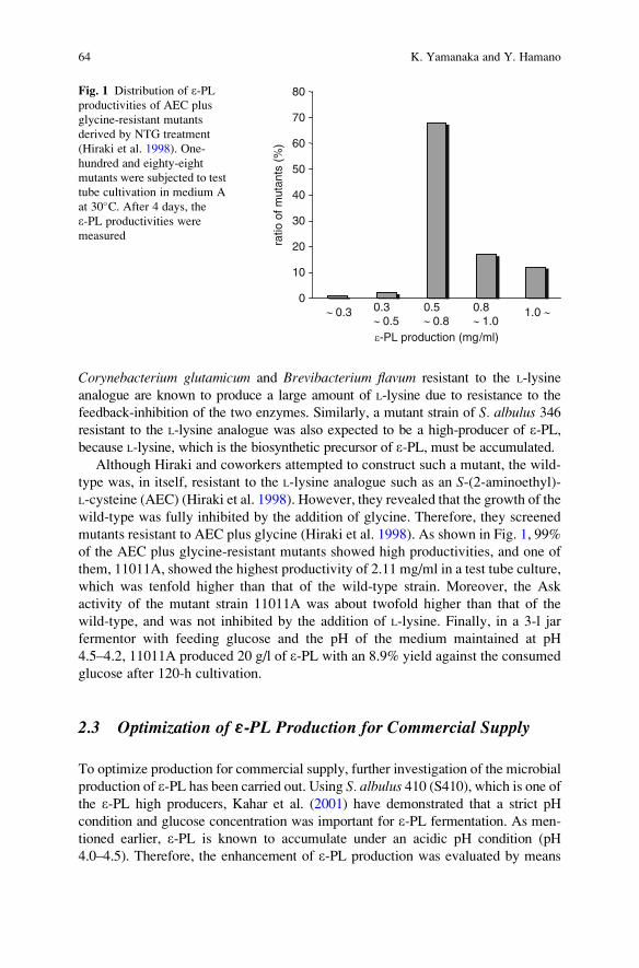
Corynebacterium glutamicum and Brevibacterium flavum resistant to the L-lysine
analogue are known to produce a large amount of L-lysine due to resistance to the
feedback-inhibition of the two enzymes. Similarly, a mutant strain of S. albulus 346resistant to the L-lysine analogue was also expected to be a high-producer of e-PL,because L-lysine, which is the biosynthetic precursor of e-PL, must be accumulated.
Although Hiraki and coworkers attempted to construct such a mutant, the wild-
type was, in itself, resistant to the L-lysine analogue such as an S-(2-aminoethyl)-
L-cysteine (AEC) (Hiraki et al. 1998). However, they revealed that the growth of the
wild-type was fully inhibited by the addition of glycine. Therefore, they screened
mutants resistant to AEC plus glycine (Hiraki et al. 1998). As shown in Fig. 1, 99%
of the AEC plus glycine-resistant mutants showed high productivities, and one of
them, 11011A, showed the highest productivity of 2.11 mg/ml in a test tube culture,
which was tenfold higher than that of the wild-type strain. Moreover, the Ask
activity of the mutant strain 11011A was about twofold higher than that of the
wild-type, and was not inhibited by the addition of L-lysine. Finally, in a 3-l jar
fermentor with feeding glucose and the pH of the medium maintained at pH
4.5–4.2, 11011A produced 20 g/l of e-PL with an 8.9% yield against the consumed
glucose after 120-h cultivation.
2.3 Optimization of e-PL Production for Commercial Supply
To optimize production for commercial supply, further investigation of the microbial
production of e-PL has been carried out. Using S. albulus 410 (S410), which is one ofthe e-PL high producers, Kahar et al. (2001) have demonstrated that a strict pH
condition and glucose concentration was important for e-PL fermentation. As men-
tioned earlier, e-PL is known to accumulate under an acidic pH condition (pH
4.0–4.5). Therefore, the enhancement of e-PL production was evaluated by means
0
10
20
30
40
50
60
70
80
~ 0.3 0.3~ 0.5
0.5~ 0.8
0.8~ 1.0
1.0 ~
e-PL production (mg/ml)
ratio
of m
utan
ts (
%)
Fig. 1 Distribution of e-PLproductivities of AEC plus
glycine-resistant mutants
derived by NTG treatment
(Hiraki et al. 1998). One-
hundred and eighty-eight
mutants were subjected to test
tube cultivation in medium A
at 30�C. After 4 days, the
e-PL productivities were
measured
64 K. Yamanaka and Y. Hamano

of a strict pH control strategy. The optimized fermentation condition was divided into
two phases. In the first phase, the cell growth was accelerated by maintaining the pH
higher than 5.0. In the second phase, the pH of the culture medium was maintained
around 4.0, at which the highest e-PL productivity was observed. In addition, because
the yield of e-PL against consumed glucose and its productivity were affected by the
residual glucose concentration of the culture medium, the glucose concentration was
strictly kept at about 10 g/l. In their strategy, e-PL productivity was enhanced to
48.3 g/l. Thus, since the first study, the e-PL productivity of S. albulus has been
significantly improved by using these biotechnological techniques (Table 1). At
present, e-PL has been industrially produced by a fermentation process using a
mutant strain derived from 11011A and the pH control strategy (Chisso Corp.).
3 Biological Activities of «-PL
e-PL shows strong antimicrobial activity against both Gram-positive and Gram-
negative bacteria, fungi, and some kinds of viruses. The most plausible antimicro-
bial mechanism of e-PL is physical disruption of the microbial cytoplasmic
membrane following electrostatic absorption. Microbicidal action then occurs,
and its activity is comparable to that of disinfectants widely used in medical and
industrial fields.
3.1 Antimicrobial Activities and Antiphage Activity of e-PL
e-PL shows strong antimicrobial activity against a wide spectrum of microorgan-
isms (Shima et al. 1984). The minimum inhibitory concentrations (MICs) for most
bacteria were 1–8 mg/ml, although those for yeast and fungi tended to be slightly
higher. For example, MIC for Staphylococcus aureus and Candida albicans was4 and 128 mg/ml, respectively (Shima et al. 1984). e-PL also shows high thermo-
stability; the antimicrobial activity of e-PL was not affected at all after the heat
treatment at 120�C for 20 min (Hiraki 1999). Although e-PL showed strong and
broad antimicrobial activity, it did not directly show inhibitory activity for bacterial
spore germination (Hiraki 1999). In the presence of 500 mg/ml e-PL, inactivation ofbacteriophages belonging to the long-tail and noncontractile morphological type
was observed (Shima et al. 1982). Although a large number of studies on the
antimicrobial activities of e-PL have been reported, there have been an insufficient
number of studies on its antiphage activities.
3.2 Antimicrobial Mechanism of e-PL
Shima et al. (1984) also investigated the relationship between the chain length
of e-PL and its antimicrobial activity for Escherichia coli K-12. e-PLs with more
Biotechnological Production of Poly-Epsilon-L-Lysine 65

Tab
le1Im
provem
entofmicrobialproductionofe-PL
Year
S.albulusstrains
Culture
method(initialnutrients)
Maxim
um
productivity
(culture
time)
Improvem
ent
(fold)
References
1977
346(w
ild)
Shakeflaskculture
inbasalmedium
(50g/lglycerol,
10g/l(N
H4) 2SO4,5g/lyeastextract)
0.2
g/l(4
days)
–Shim
aandSakai
(1977)
1983
346(w
ild)
Reactionwithcellsin
citratebuffer
(50g/lglycerol,
10g/l(N
H4) 2SO4,20g/lcitrate(pH
4–4.5))
5.0
g/l(8
days)
25
Shim
aet
al.(1983)
1998
11011A
(mutant)
Testtubeculture
inmedium
A(50g/lglucose,10g/l
(NH4) 2SO4,5g/lyeastextract)
2.1
g/l(4
days)
10.5
Hirakiet
al.(1998)
Jarferm
entationin
medium
Awithfeedingglucose
andpHmaintenance
(pH
4.2)
20g/l(5
days)
100
2001
S410(m
utant)
Jarferm
entationin
M3Gmedium
withglucose
concentrationandpHcontrolled
(10g/l,pH
5.0!4.0)(50g/lglucose,10g/l(N
H4) 2SO4,5g/l
yeastextract)
48.3
g/l(8
days)
241
Kahar
etal.(2001)
66 K. Yamanaka and Y. Hamano

than nine L-lysine residues strongly inhibited their growth, although the activity of
e-PLs with less than eight residues were reduced (>100 mg/ml). The importance of
free a-amino groups of e-PL for antimicrobial activity was demonstrated by a
reduction of the activity with chemical modification of their a-amino groups.
Based on a study using electron microscopy, the proposed mechanism of antimi-
crobial action of e-PL was concluded to be electrostatic interaction with the
microbial cell surface, followed by disorganization of the membrane and abnormal
distribution of the cytoplasm, ultimately leading to physiological damage of the
microbial cells (Shima et al. 1984). Muto (2003) reported that both Gram-positive
and Gram-negative bacteria exposed to e-PL were disinfected. After the exposure
of bacterial cells to a low concentration of e-PL (10 mg/ml) in 10 mM potassium
phosphate buffer (pH 7.0) for 15–60 min, the reduction of survival cells (>4–5
Log) was observed in all bacterial strains tested, including E. coli, B. subtilis, andS. aureus. In addition, e-PL induced leakage of cytoplasmic components from
E. coli cells. The leakage of NADH-dependent cytoplasmic malate dehydrogenase
(MDH), an enzyme of the tricarboxylic acid cycle, from e-PL treated cells was
observed, and the increase of leaked MDH activity showed good correlation with a
decrease in cell survival (Muto 2009a, Fig. 2). As reported previously, polycatio-
nic compounds such as protamine interact with the cell surface membrane and
induce leakage of cytoplasmic components such as ATP and b-galactosidase(Johansen et al. 1997). In the same way, e-PL should first absorb to the cell
surface and then disrupt the cytoplasmic membrane of microorganisms. After
fatal damage to the membrane, cytoplasmic components are thought to leak out.
Based on all of these results, it can be concluded that the antimicrobial mechanism
of e-PL is physical disruption of the cytoplasmic membrane following electrostatic
absorption.
viab
le c
ells
(C
FU
/ml)
0
2
4
6
8
10
1 10 100
40x 109
ε-PL concentration (μg/ml)
rele
ased
MD
H a
ctiv
ity (
U/m
l)
0
10
20
30Fig. 2 Leakage of the
cytoplasmic malate
dehydrogenase (MDH) from
E. coli cells exposed to e-PL(Muto 2009a). E. coli cells(>9 Log CFU/ml) were
exposed to e-PL in 10 mM
Tris–HCl (pH 7.0). After
exposure for 1 h, the number
of survival cells (open circle)and MDH activity leaked to
supernatant (closed circle)were determined
Biotechnological Production of Poly-Epsilon-L-Lysine 67

3.3 Microbicidal Activities of e-PL
Although many studies on the growth inhibitory activities of e-PL have been
reported, itsmicrobicidal activities are poorlyunderstood.Basedon theunderstand-
ing of its antimicrobial mechanism, the microbicidal activities of e-PL have been
investigated (Muto 2009a). The minimum bactericidal concentrations (MBCs) of
e-PL, which was defined as the lowest concentration of e-PL that disinfected over
99.99% of the inoculated cells within 2 h, were determined (Table 2). TheMBCs of
e-PL against all of the tested microorganisms including Gram-positive, Gram-
negative bacteria, and also yeasts were <50 mg/ml and were comparable to those
of disinfectants such as cetylpyridinium chloride (CPC) and 1,2-benzisothiazolin-
3-one (BIT),which arewidely used inmedical and industrial fields. Due to its safety
and edible feature, the availability of e-PL as a bacteriocide that can be used in
multiple fields including the medical one has been suggested (Muto 2009a). In
addition, a clear correlation between microbial susceptibility to e-PL and relative
cell size was observed (Muto 2009a, Fig. 3). Yeast strains were also included in
this consistent correlation. Thus, the microbicidal activities of e-PL depended not
on the microbial species, but directly on the microbial cell size. This phenomenon
also strongly supports the conclusion proposed in the section above that the
antimicrobial activity of e-PL is caused by a physical action on the cytoplasmic
membrane.
Table 2 Comparison of minimum bactericidal concentrations (MBCs) of e-PL and different
disinfectants for the selected microorganisms (Muto 2009a). MBCs were defined as the lowest
concentration that disinfected over 99.99% of the inoculated cells within 2 h and were determined
with 10 mM potassium phosphate buffer (pH 7.0)
Microorganisms MBC (mg/mL)
e-PL PHMB BAC CPC BIT
Gram-negative bacteria
Escherichia coli IFO 3972 6.3 <3.1 6.3 12.5 100
Pseudomonas aeruginosa IFO 13275 <3.1 <3.1 6.3 12.5 200
Salmonella choleraesuis IFO 3163 <3.1 n.d. n.d. n.d. n.d.
Klebsiella pneumoniae IFO 13277 6.3 n.d. n.d. n.d. n.d.
Gram-positive bacteria
Bacillus subtilis IFO 13719 13 n.d. n.d. n.d. n.d.
Staphylococcus aureus IFO 12732 6.3 6.3 <3.1 <3.1 100
S. aureus IFO 13276 6.3 <3.1 <3.1 6.3 100
Listeria monocytogenes JCM 2873 6.3 6.3 6.3 <3.1 200
Yeast
Saccharomyces cerevisiae IFO 10517 25 n.d. n.d. n.d. n.d.
Pichia anomala IFO 0141 25 n.d. n.d. n.d. n.d.
Candida albicans IFO 1594 13 6.3 12.5 12.5 200
PHMB polyhexamethylene biguanide, BAC benzalkonium chloride, CPC cetylpyridinium
chloride, BIT 1,2-benzisothiazolin-3-one, n.d. not determined
68 K. Yamanaka and Y. Hamano

3.4 Antimicrobial Profiles of e-PL
The antimicrobial activities of e-PL were observed in weak acidic or physiological
pH conditions. e-PL is a cationic polymer and its isoelectric point is around 9.0. The
antimicrobial and antiphage activities of e-PL are significantly weakened under the
alkaline conditions, because these activities are simply based on the electrostatic
interaction. Hiraki (2000) reported that the MICs for E. coli observed in physiologi-cal pH conditions are 25–50 mg/ml, whereas those at pH 8.0 are over 200 mg/ml. In
addition, the activity was weakened with increasing ionic strength and/or the
presence of anionic compounds such as metaphosphoric acid, due to loss of its
cationic charge. Similarly, under the extremely low pH condition, the activities
would be inhibited due to loss of the counter anionic charge for e-PL on the
microbial cytoplasmic membrane.
3.5 Safety of e-PL
The safety of e-PL was confirmed by experiments using rats. In a two-generation
reproduction study, the nontoxic dosage level of e-PL was concluded to be
10,000 ppm, and e-PL at 30,000 ppm did not cause any toxicities for reproduction,
neurological function, embryonic, and fetal development and growth (Neda et al.
43
1
72
65
89
0
1
2
3
4
5
6
7
–6.5 –5.5 –4.5 –3.5
relative microbial cell size(Log OD660/CFU)
susc
eptib
ility
to e
-PL
Fig. 3 Relationship between microbial susceptibility to e-PL and relative cell size (Muto 2009a).
The susceptibility to e-PL was defined as the logarithms of the number of disinfected microbial
cells by the treatment with 10 mg/ml e-PL in 10 mM potassium phosphate buffer (pH 7.0) within
2 h. Relative microbial cell sizes were estimated from the relationship between the turbidity
measured at 660 nm and the number of viable cells in microbial suspensions. 1, Escherichia coli;2, Salmonella choleraesuis; 3, Pseudomonas aeruginosa; 4, Klebsiella pneumoniae; 5, Bacillussubtilis; 6, Bacillus cereus; 7, Listeria monocytogenes; 8, Saccharomyces cerevisiae; 9, Pichiaanomala
Biotechnological Production of Poly-Epsilon-L-Lysine 69

1999). The pharmacokinetics of e-PL in vivo were also investigated (Hiraki et al.
2003). Absorption, distribution, metabolism and excretion (ADME) studies using14C-radiorabeled e-PL revealed that e-PL was poorly absorbed in the gastrointestinal
tract and most of the dosed radioactivity was eliminated by excretion within 168 h.
Furthermore, no accumulation of e-PL in any tissue or organ was observed by whole
body autoradiography. Based on these results showing its safety, e-PL was approved
by the Japanese Ministry of Health, Labour and Welfare as a preservative in food in
the late 1980s. Thereafter, e-PL was listed in the Korea Food Additives Code and has
been used in Korea. Recently, the US Food and Drug Administration indicated that
they had no questions about the manufacturer’s conclusions that e-PL is generally
recognized as safe (GRAS) for use as an antimicrobial agent in cooked or sushi rice at
levels up to 50 mg/kg of rice (USFDA 2004).
4 Application of «-PL
Since the safety of e-PL has been clarified, e-PL has been used for various applica-
tions, particularly as a food preservative. Recently, some interesting applications in
medical field have been reported.
4.1 Food Preservative
e-PL has a long history of safe use in Japan as a natural food preservative, and
many food products containing e-PL can be found in food shops and super-
markets. e-PL has been used in various food products in Japan, including tradi-
tional Japanese dishes, boiled rice, noodles, potato salad, and cooked vegetables
(Hiraki 2000). In addition, e-PL is also used in desserts, including steamed cake
and custard cream (Hiraki et al. 2003). The e-PL formulation recipes for some
commonly consumed foods are shown in Table 3 (Muto 2009b). These foods
Table 3 Recipes of the e-PL formulation for several food products. The table was modified from
that reported by Muto (2009b). e-PL was used as a formulation including 30% sodium acetate and
20% of the other organic acids. Lifetime, period that bacterial growth was inhibited (<5 Log
CFU/g)
Food products Recipe (as a formulation) Preservation
Temperature Lifetime
Hamburger steak Add 0.5% and mix with putty before frying 25�C 48 h
Hamburger patty Add 2–3% and mix with putty 4�C 20 days
Boiled vegetables Cook in the 0.8% solution 25�C 72 h
Croquette Add 0.6% and mix before frying 25�C 72 h
Cooked fish Cook in the 0.6% solution 25�C 48 h
Potato salad Add 0.6% and mix 10�C 72 h
Mashed potato Add 0.6% and mix after cooked 4�C 12 weeks
70 K. Yamanaka and Y. Hamano

typically contain 10–500 ppm of e-PL. For agglomerative foods such as sliced fish
and fish sushi, spraying and dipping treatment with 1,000–5,000 ppm of e-PL were
effective (Hiraki 2000).
4.1.1 Improvement of «-PL Preservative Activity by Combinationwith Other Additives
In general use, because the concentration of e-PL required for food preservation is
quite low due to its strong antimicrobial activity, the taste of the foods is not affected.
Although e-PL can be used alone for food preservation, the preservative activity of
e-PL is greatly improved by combining it with other food additives (Hiraki 2000).
The combination of e-PL and other food additives such as glycine, ethanol, organic
acids including vinegar and lactic acid, and emulsifying agent is effective for multiple
food preservation due to the synergistic effect. For preservation of raw chicken
containing a high level of microbial contaminants (4–5 Log CFU/g as aerobic
bacteria), dipping treatment using 450 ppm e-PL solution containing 0.9% organic
acid and 1.4% sodium acetate gave a 12-day preservative effect at 4�C, which was
three times longer than that without treatment (Muto 2009a). The synergistic effect
helps to reduce the total amount of preservatives and serves the additional positive
effect of enhancing the preservative activity without affecting the original food taste.
In fact, e-PL is used together with these additives in most cases.
4.1.2 Efficacy for Controlling Food-Borne Pathogens
Microbially contaminated foods are estimated to cause approximately 76 million
cases of illness every year in the United States (Mead et al. 1999). The availability
of e-PL as an antimicrobial agent against food-borne pathogens of E. coli O157:H7,Salmonella typhimurium, and Listeria monocytogenes has been reported (Geornaras
and Sofos 2005). When these three pathogens were incubated with e-PL in tryptic
soy broth supplemented with 0.6% yeast extract, 200–400 mg/ml of e-PL inhibited
their growth. Furthermore, for Gram-negative pathogens of E. coli O157:H7 and
S. typhimurium, the antimicrobial activity of e-PL was enhanced by combination
with 0.25% sodium diacetate or 0.1% acetic acid. For Gram-positive pathogens of
L. monocytogenes, Bacillus cereus cells and its spores, it has been reported that a
combination with e-PL and antimicrobial peptide nisin A, which is produced
by Lactococcus lactis ssp. lactis (Hurst 1967) and has been used as a food pre-
servative in the United States, shows synergistic antimicrobial activity (Badaoui
Najjar et al. 2007). The combination of e-PL with the above mentioned additives
can be considered an effective all-natural formulation for controlling a wide
spectrum of food-borne pathogens, spores, and spoilage organisms, although
more research is needed to determine the most effective combination or conditions
for each food application.
Biotechnological Production of Poly-Epsilon-L-Lysine 71

4.2 Medical and Other Applications
Diverse amino-acid polymers have been chemically synthesized to find attractive
bioactive substances. Among them, a-PL has shown various applications in the
life science field due to its polycationic property. In the medical field, a-PL has
been used to enhance the efficacy of some interferon inducers (Levy et al. 1975;
Champney et al. 1976). It has also been reported that a-PL enhances the drug
permeability of Chinese hamster ovary (CHO) cells (Ryser and Shen 1980), and the
polyionic complex of DNA with polycations such as a-PL is effective as a nonviral
gene delivery carrier (Chiou et al. 1994; Dorudi et al. 1993). However, for practical
use of these medical applications, the toxicity, biocompatibility, and biodegradabil-
ity of a-PL need to be considered. a-PL has been used in medical applications
mainly for its polycationic property. Because a-PL shows some toxicities (Sela and
Katchalski 1959), naturally occurring e-PL would be an ideal alternative substance
to synthetic a-PL for medical use. Recently, in fact, a gene delivery system, which
exhibits low cytotoxicity, has been developed using polyionic complex of DNA
with a e-PL derivative (Chisso Corp. and Nagasaki 2006).
4.2.1 Suppression of Dietary Fat Absorption
A novel application of e-PL in medical use has been reported. Obesity is a serious
disease that can lead to numerous health problems including diabetes (Hill et al.
2000). Because it is known that pancreatic lipase plays a crucial role in lipid
absorption from the intestine (Duan 2000), natural products that can inhibit pancre-
atic lipase activity could suppress dietary fat absorption. It has been reported that
highly basic proteins such as protamine, purothionin, and histone inhibit lipase
activity in vitro (Tsujita et al. 1996). Kido et al. (2003) have reported that
10–100 mg/l e-PL inhibited pancreatic lipase activity in substrate emulsions con-
taining bile salts and phosphatidylcholine as an emulsifying agent. In addition, e-PLwas found to retain its inhibitory activity after incubation with digestive enzymes
such as trypsin, a-chymotrypsin, and pepsin, due to its unusual isopeptide bonds,
whereas a-PL, showing the same level of inhibitory activity, did not. Therefore, it
was suggested that e-PL would inhibit lipase in the digestive tract, in which proteins
and a-PL that have the usual a-peptide bonds may be digested. Based on these
results, they concluded that e-PL would be able to suppress dietary fat absorption
from the small intestine by inhibiting pancreatic lipase activity.
4.2.2 Endotoxin Remover and Other Practical Uses
Lipopolysaccharide (LPS), which is a constituent of the outer membrane of Gram-
negative bacteria, is a potential contaminant of physiological fluids. Because LPS
causes pyrogenic and shock reactions, it must be removed from medicines that are
injected. It has been reported that endotoxins can be selectively removed by using
72 K. Yamanaka and Y. Hamano

particles prepared by cross-linking between e-PL and chloromethyloxirane
(Hirayama et al. 1999). By applying this principle, a chromatographic resin,
e-PL-immobilized cellulose beads (ET clean, Chisso Corp.), was developed and
has been widely used as an endotoxin remover in some countries, including Japan,
Korea, China, India, and the EU.
A cell-culture device with e-PL as a coating material for the surface of devices
has also been developed, and showed low cytotoxicity and improvement in cell
adhesion (Chisso Corp. and Nagasaki 2007). In some biomedical fields, e-PL is also
used to prevent periodontal disease. The combination with 0.02–2.0% (w/w) e-PLand 0.1–10% (w/w) bentonite, which is a kind of clay mineral, inhibited the
production of oral bacterial toxin (Lion Corp. 2006). In addition, e-PL has been
used as a component of the drug solution in disposable wipes, which are used for
sanitation (Asahikasei Corp. 1999). Many other e-PL applications have been
patented. In the future, further applications of e-PL will be developed.
5 Concluding Remarks and Future Perspectives
Since the discovery of e-PL, numerous studies on its microbial production, antimi-
crobial activity, and application have been reported. e-PL is now industrially pro-
duced and used in food and medical applications due to its polycationic property and
safety. Many applications of e-PL have been patented, and further studies on novel
applications are in progress. The water-solubility, polycationic, nontoxic, edible
feature, and other physicochemical properties of e-PL are applicable to other fields,
such as agriculture and environmental science. Recently, a e-PL synthetase and its
gene were identified (Yamanaka et al. 2008). The identification of this e-PL synthe-
tase and its gene will help to create new classes of biopolymers which will be useful
in various applications as well as help to improve microbial production systems.
References
Asahikasei Corp. (1999) Sterile wet tissue. JP Patent PH11-113779A
Badaoui Najjar M, Kashtanov D, Chikindas ML (2007) e-poly-L-lysine and nisin A act synergisti-
cally against Gram-positive food-borne pathogens Bacillus cereus and Listeria monocyto-
genes. Lett Appl Microbiol 45:13–18
Champney KJ, Levy HB, Lerner AM (1976) Sustained interferon in human serum following poly
(ICLC). Proceedings of the American Society of Clinical Investigation. Clin Res 24:451–452
Chiou HC, Tangco MV, Levine SM, Robertson D, Kormis K, Wu CH, Wu GY (1994) Enhanced
resistance to nuclease degradation of nucleic acids complexed to asialoglycoprotein-polylysine
carriers. Nucleic Acids Res 22:5439–5446
Chisso Corp., Nagasaki T (2006) Nucleic acid complex and method for transfection of nucleic acid
into cell by using the same. JP Patent P2006-304644A
Chisso Corp., Nagasaki T (2007) Cell culture device and method for producing the same. JP Patent
P2007-020444A
Biotechnological Production of Poly-Epsilon-L-Lysine 73

Chisso Corp. ET clean. http://www.chisso.co.jp/fine/en/cellufine/grade/grade-1-etclean.html, last
accessed on date 17 Nov 2009
Dorudi S, Northover JM, Vile RG (1993) Gene transfer therapy in cancer. Br J Surg 80:566–572
Duan RD (2000) Enzymatic aspects of fat digestion in the gastrointestinal tract. In: Christopher
AB, Vriese S (eds) Fat digestion and absorption. AOCS, Champaign, pp 25–46
Geornaras I, Sofos JN (2005) Activity of e-polylysine against Escherichia coli O157:H7, Salmo-nella Typhimurium, and Listeria monocytogenes. J Food Sci 70:M404–M408
Hill JO, Melanson EL, Wyatt HT (2000) Dietary fat intake and regulation of energy balance:
implications for obesity. J Nutr 130:284S–288S
Hiraki J, Hatakeyama M, Morita H, Izumi Y (1998) Improved e-poly-L-lysine production of an
S-(2-aminoethyl)-L-cysteine resistant mutant of Streptomyces albulus. Seibutu Kougaku
Kaishi 76:487–493
Hiraki J, Ichikawa T, Ninomiya S, Seki H, Uohama K, Seki H, Kimura S, Yanagimoto Y, Barnett
JW Jr (2003) Use of ADME studies to confirm the safety of e-polylysine as a preservative infood. Regul Toxicol Pharmacol 37:328–340
Hiraki J (1995) Basic and applied studies on e-polylysine. J Antibact Antifung Agents 23:349–354Hiraki J (1999) e-poly-L-lysine, its antimicrobial activity and utilization. In: Japan Food Additives
Association News, vol 18. Japan Food Additives Association, Tokyo, pp 223–233
Hiraki J (2000) e-poly-L-lysine, its development and utilization. In: Fine chemical, vol 29. CMC
Press, Tokyo, pp 18–25
Hirayama C, Sakata M, Nakamura M, Ihara H, Kunitake M, Todokoro M (1999) Preparation
of poly(e-lysine) adsorbents and application to selective removal of lipopolysaccharides.
J Chromatogr B Biomed Sci Appl 721:187–195
Hurst A (1967) Function of nisin and nisin-like basic proteins in the growth cycle of Streptococcus
lactis. Nature 214:1232–1234
Johansen C, Verheul A, Gram L, Gill T, Abee T (1997) Protamine-induced permeabilization of
cell envelopes of gram-positive and gram-negative bacteria. Appl Environ Microbiol
63:1155–1159
Kahar P, Iwata T, Hiraki J, Park EY, Okabe M (2001) Enhancement of e-polylysine production byStreptomyces albulus strain 410 using pH control. J Biosci Bioeng 91:190–194
Kido Y, Hiramoto S, Murao M, Horio Y, Miyazaki T, Kodama T, Nakabou Y (2003) e-polylysineinhibits pancreatic lipase activity and suppresses postprandial hypertriacylglyceridemia in rats.
J Nutr 133:1887–1891
Levy HB, Baer G, Baron S, Buckler CE, Gibbs CJ, Iadarola MJ, London WT, Rice J (1975) A
modified polyriboinosinic-polyribocytidylic acid complex that induces interferon in primates.
J Infect Dis 132:434–439
Lion Corp. (2006) Composition for preventing periodontal disease. JP Patent P2006-151877A
Mead PS, Slutsker L, Dietz V, McCaig LF, Bresee JS, Shapiro C, Griffin PM, Tauxe RV (1999)
Food-related illness and death in the United States. Emerg Infect Dis 5:607–625
Muto M (2003) Antimicrobial activity of e-poly-L-lysine and its utilization for preservation of
confectionery products. In: Japan Food Science, vol 42. Japan Food Hygiene Association,
Tokyo, pp 65–72
Muto M (2009a) Chemical technology for preservation of food products; food preservative,
e-poly-L-lysine. Boukin Boubai 37:765–772
Muto M (2009b) Utilization of e-poly-L-lysine for curtailing of waste food products. In: A
Technical Journal on Food Chemistry & Chemicals, Vol 6. Food Chemical News, Tokyo,
pp 43–46
Neda K, Sakurai T, Takahashi M, Aiuchi M, Ohgushi M (1999) Two-generation reproduction
study with teratology test of e-poly-L-lysine by dietary administration in rats. Jpn Pharmacol
Ther 27:1139–1159
Nishikawa M, Ogawa K (2002) Distribution of microbes producing antimicrobial e-poly-L-lysinepolymers in soil microflora determined by a novel method. Appl Environ Microbiol
68:3575–3581
74 K. Yamanaka and Y. Hamano

Ryser HJ, Shen WC (1980) Conjugation of methotrexate to poly (L-lysine) as a potential way to
overcome drug resistance. Cancer 45:1207–1211
Sela M, Katchalski E (1959) Biological properties of poly-a-amino acids. Adv Protein Chem
14:392–479
Shima S, Fukuhara Y, Sakai H (1982) Inactivation of bacteriophages by e-poly-L-lysine producedby Streptomyces. Agric Biol Chem 46:1917–1919
Shima S, Matsuoka H, Iwamoto T, Sakai H (1984) Antimicrobial action of e-poly-L-lysine.J Antibiot 37:1449–1455
Shima S, Oshima S, Sakai H (1983) Biosynthesis of e-poly-L-lysine by washed mycelium of
Streptomyces albulus no. 346. Nippon Nogeikagaku Kaishi 57:221–226
Shima S, Sakai H (1977) Polylysine produced by Streptomyces. Agric Biol Chem 41:1907–1909
Shima S, Sakai H (1981a) Poly-L-lysine produced by Streptomyces. Part II. Taxonomy and
fermentation studies. Agric Biol Chem 45:2497–2502
Shima S, Sakai H (1981b) Poly-L-lysine produced by Streptomyces. Part III. Chemical studies.
Agric Biol Chem 45:2503–2508
Tsujita T, Matsuura Y, Okuda H (1996) Studies on the inhibition of pancreatic and carboxylester
lipases by protamine. J Lipid Res 37:1481–1487
US Food and Drug Administration (USFDA) (2004) GRAS Notice 000135: e-Polylysine. Office ofFood Additive Safety. http://www.fda.gov. Cited 17 Nov 2009
Yamanaka K, Maruyama C, Takagi H, Hamano Y (2008) e-poly-L-lysine dispersity is controlled
by a highly unusual nonribosomal peptide synthetase. Nat Chem Biol 4:766–772
Biotechnological Production of Poly-Epsilon-L-Lysine 75
Occurrence and Biosynthetic Mechanismof Poly-Gamma-Glutamic Acid
Makoto Ashiuchi
Contents
1 Introduction . . . . . . . . . . . . . . . . . . . . . . . . . . . . . . . . . . . . . . . . . . . . . . . . . . . . . . . . . . . . . . . . . . . . . . . . . . . . . . . . 78
2 Occurrence and Producers . . . . . . . . . . . . . . . . . . . . . . . . . . . . . . . . . . . . . . . . . . . . . . . . . . . . . . . . . . . . . . . . . 79
2.1 Poly-g-DL-Glutamic Acid Producers . . . . . . . . . . . . . . . . . . . . . . . . . . . . . . . . . . . . . . . . . . . . . . . . . 80
2.2 Poly-g-D-Glutamic Acid Producers . . . . . . . . . . . . . . . . . . . . . . . . . . . . . . . . . . . . . . . . . . . . . . . . . . 80
2.3 Poly-g-L-Glutamic Acid Producers . . . . . . . . . . . . . . . . . . . . . . . . . . . . . . . . . . . . . . . . . . . . . . . . . . 81
3 Modes of Nonribosomal Amino Acid Polymerization . . . . . . . . . . . . . . . . . . . . . . . . . . . . . . . . . . . . . 81
3.1 Thiotemplate Mechanism for Poly-g-Glutamate Synthesis . . . . . . . . . . . . . . . . . . . . . . . . . . 82
3.2 Amide Ligation Mechanism for Poly-g-Glutamate Synthesis . . . . . . . . . . . . . . . . . . . . . . . 83
4 Biosynthesis of Poly-g-Glutamic Acid Precursors . . . . . . . . . . . . . . . . . . . . . . . . . . . . . . . . . . . . . . . . . 84
4.1 L-Glutamic Acid Biosynthesis . . . . . . . . . . . . . . . . . . . . . . . . . . . . . . . . . . . . . . . . . . . . . . . . . . . . . . . 85
4.2 D-Glutamic Acid Biosynthesis . . . . . . . . . . . . . . . . . . . . . . . . . . . . . . . . . . . . . . . . . . . . . . . . . . . . . . . 85
5 Biosynthesis of Poly-g-Glutamic Acid . . . . . . . . . . . . . . . . . . . . . . . . . . . . . . . . . . . . . . . . . . . . . . . . . . . . . 86
5.1 PgsB Component of Poly-g-Glutamic Acid Synthetase . . . . . . . . . . . . . . . . . . . . . . . . . . . . . . 87
5.2 PgsC Component of Poly-g-Glutamic Acid Synthetase . . . . . . . . . . . . . . . . . . . . . . . . . . . . . . 87
5.3 PgsA Component of Poly-g-Glutamic Acid Synthetase . . . . . . . . . . . . . . . . . . . . . . . . . . . . . 87
5.4 PgsE Component of Poly-g-Glutamic Acid Synthetase . . . . . . . . . . . . . . . . . . . . . . . . . . . . . . 88
6 Concluding Remarks and Future Perspectives . . . . . . . . . . . . . . . . . . . . . . . . . . . . . . . . . . . . . . . . . . . . . 88
References . . . . . . . . . . . . . . . . . . . . . . . . . . . . . . . . . . . . . . . . . . . . . . . . . . . . . . . . . . . . . . . . . . . . . . . . . . . . . . . . . . . . . . 89
Abstract Poly-g-glutamic acid (PGA) is an anionic polyamide biomaterial in
which usually over 10,000 molecules of glutamic acid are polymerized via g-amide
linkages. PGA is found in various organisms, and essentially synthesized in a
ribosome-independent manner. To date, two distinct modes for nonribosomal
amino acid polymerization have been proposed: the thiotemplate mechanism and
the amide ligation process. The former mode of polymerization often brings about a
disagreement of the stereochemistry between a polymer produced and the substrate(s),
differently from the latter mode. DL-PGA synthesis in Bacillus subtilis proceeds via
M. Ashiuchi
Agricultural Science, Graduate School of Integrated Arts and Sciences, Kochi University,
Nankoku, Kochi 783-8502, Japan
e-mail: [email protected]
Y. Hamano (ed.), Amino-Acid Homopolymers Occurring in Nature,Microbiology Monographs 15, DOI 10.1007/978-3-642-12453-2_5,# Springer-Verlag Berlin Heidelberg 2010
77

the amide ligation mechanism and thus both isomers of glutamic acid can serve as
substrates. The operon architecture responsible for DL-PGA synthesis occurs in the
chromosome of B. subtilis, and all the components of the PGA synthetase complex,
that is, PgsB, -C, -A, and -E, are membrane-associated. Here, we present the
structural features and predicted functions of each Pgs component.
1 Introduction
Poly-g-glutamic acid (PGA) is an anionic polyamide biomaterial, where more than
10,000 molecules of glutamic acid are polymerized via g-amide linkages (Ashiuchi
and Misono 2002b), possessing a chiral center in every glutamyl unit. Following the
discovery of PGA (Kramar 1921; Ivanovics and Erdos 1937), it has been the subject
of much interest in modern biopolymer study. Simple methods for detection,
purification, and determination of PGA were established (Ashiuchi and Misono
2002b; Do et al. 2001), the majority of which are useful in genetic and biochemical
studies (Ashiuchi and Misono 2002a). Stereochemically, three different types of
PGA have been identified: the homopolymer of D-glutamic acid (D-PGA), the
homopolymer of L-glutamic acid (L-PGA), and the random copolymer constituted
of D- and L-glutamic acid (DL-PGA).
Regardless of its stereochemistry, PGA is nontoxic to humans and the environ-
ment, has been found to be edible (Ashiuchi and Misono 2002b), and possesses
enormous potential as a new macromolecular material. As shown in Table 1,
various applications of PGA have been established. As an example, cross-linked
PGA exhibits an extremely high degree of water absorbency (Choi and Kunioka
1995) possibly making it a suitable substitute for nondegradable hydrogels. This
would be an enormous benefit in agricultural, environmental, and biomedical
applications, including biodegradable diapers, water reservoirs, and slow-release
systems for drugs or fertilizers. Much is known about PGA-based flocculants
(Yokoi et al. 1996; Shih et al. 2001; Taniguchi et al. 2005), with some available
for wastewater treatment, water purification, and downstream processing for food and
fermentation manufacturers. The industrial utilization of PGA is rapidly increasing
with applications, such as a metals/radionuclides-binding agent (Ashiuchi and
Misono 2002b), a protease inhibitor, a cryoprotectant (Birrer et al. 1994; Mitsuiki
et al. 1998; Yokoigawa et al. 2006), a bitterness-relieving agent (Shih and Van
2001), a thickener, an animal feed additive, an osteoporosis-preventing factor
(Tanimoto et al. 2001; Yang et al. 2008), humectants, a drug delivery agent
(Li et al. 1999; Kishida et al. 1998; Kim et al. 1999; Hashida et al. 1999; Ikumi
et al. 2008), a gene vector (Dekie et al. 2000), a curable biological adhesive
(Spotniz 1996; Otani et al. 1998; Sekine et al. 2000), a dispersant, and an anti-
body/enzyme-conjugating (immobilizing) material (Prodhomme et al. 2003; Cheng
et al. 2008).
Before applying PGA-based materials to practical use, some issues remain to be
solved. The first is cost factor, specifically that the cost estimated under existing
78 M. Ashiuchi
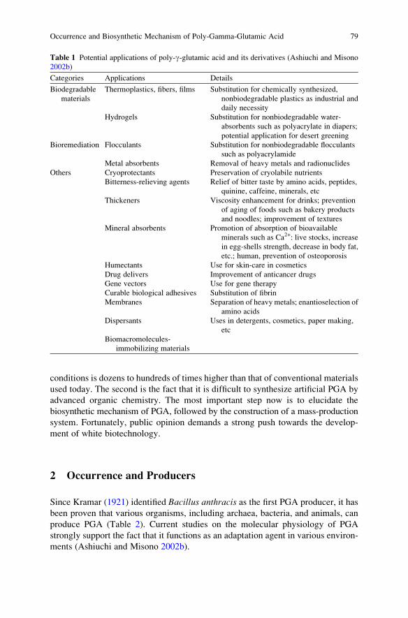
conditions is dozens to hundreds of times higher than that of conventional materials
used today. The second is the fact that it is difficult to synthesize artificial PGA by
advanced organic chemistry. The most important step now is to elucidate the
biosynthetic mechanism of PGA, followed by the construction of a mass-production
system. Fortunately, public opinion demands a strong push towards the develop-
ment of white biotechnology.
2 Occurrence and Producers
Since Kramar (1921) identified Bacillus anthracis as the first PGA producer, it has
been proven that various organisms, including archaea, bacteria, and animals, can
produce PGA (Table 2). Current studies on the molecular physiology of PGA
strongly support the fact that it functions as an adaptation agent in various environ-
ments (Ashiuchi and Misono 2002b).
Table 1 Potential applications of poly-g-glutamic acid and its derivatives (Ashiuchi and Misono
2002b)
Categories Applications Details
Biodegradable
materials
Thermoplastics, fibers, films Substitution for chemically synthesized,
nonbiodegradable plastics as industrial and
daily necessity
Hydrogels Substitution for nonbiodegradable water-
absorbents such as polyacrylate in diapers;
potential application for desert greening
Bioremediation Flocculants Substitution for nonbiodegradable flocculants
such as polyacrylamide
Metal absorbents Removal of heavy metals and radionuclides
Others Cryoprotectants Preservation of cryolabile nutrients
Bitterness-relieving agents Relief of bitter taste by amino acids, peptides,
quinine, caffeine, minerals, etc
Thickeners Viscosity enhancement for drinks; prevention
of aging of foods such as bakery products
and noodles; improvement of textures
Mineral absorbents Promotion of absorption of bioavailable
minerals such as Ca2+: live stocks, increase
in egg-shells strength, decrease in body fat,
etc.; human, prevention of osteoporosis
Humectants Use for skin-care in cosmetics
Drug delivers Improvement of anticancer drugs
Gene vectors Use for gene therapy
Curable biological adhesives Substitution of fibrin
Membranes Separation of heavy metals; enantioselection of
amino acids
Dispersants Uses in detergents, cosmetics, paper making,
etc
Biomacromolecules-
immobilizing materials
Occurrence and Biosynthetic Mechanism of Poly-Gamma-Glutamic Acid 79

2.1 Poly-g-DL-Glutamic Acid Producers
Sawamura (1913) isolated a B. subtilis strain from a Japanese fermented soybean
food known as natto. Further studies on its mucilage clarified the existence of
DL-PGA (Bovarnick 1942; Kubota et al. 1993; Ashiuchi et al. 2001a).
DL-PGA producers of Bacillus were classified into two groups: exogenous
glutamic acid-dependent and -independent groups. B. subtilis IFO 3335 (Kunioka
and Goto 1994; Kunioka 1997), B. licheniformis ATCC 9945A (Thorne et al. 1954;
Leonard et al. 1958), B. subtilis MR-141 (Ogawa et al. 1997), B. subtilis subsp.
chungkookjang (Ashiuchi et al. 2001a), and B. subtilis F-2-01 (Kubota et al. 1993)
are included in the former category. B. subtilis 5E (Shih and Van 2001), B. subtilisTAM-4 (Ito et al. 1996), B. licheniformis A35 (Cheng et al. 1989), and B. licheni-formis S173 (Kambourova et al. 2001) belong to the exogenous glutamic acid-
independent group. As shown in Table 2, B. licheniformis PGA is diverse in its
stereochemistry (Perez-Camero et al. 1999; Thorne and Leonard 1958; Leonard
et al. 1958; Cromwick and Gross 1995a). B. licheniformis might possess two or
more distinct PGA synthetic systems (Cromwick and Gross 1995b). Other than
Bacillus, some strains of Staphylococcus epidermidis also produce DL-PGA for
evading mammalian immune defense mechanisms (Kocianova et al. 2005).
2.2 Poly-g-D-Glutamic Acid Producers
To our knowledge, B. anthracis is only a sole D-PGA producer (Table 2). Although
D-PGA is avirulent in mammals, it nullifies the immunity of hosts and eventually
promotes the severity of anthrax symptoms (Keppie et al. 1963).
Table 2 Structural features of poly-g-glutamic acid and D-glutamic acid-supplying activity of the
producers
Producers Molecular
masses (kDa)
D-Glutamyl
unit content (%)
D-Glutamic acid-
supplying activitya
DAT GLR
Bacillus subtilis (natto) 10–1,000 50–80 þb þBacillus subtilis subsp. chungkookjang >1,000 35–75 3c 36c
Bacillus licheniformis ATCC9945 10–1,000 10–100 �b þBacillus anthracis NDd 100 � þBacillus megaterium WH320 >1,000 3–10 0c <1c
Natrialba aegyptiaca >1,000 0 0 0
Hydra 3–25 0 ND NDaGLR glutamic acid racemase; DAT D-amino acid aminotransferasebþ, activity is present; �, activity was absentcmU/mg (of protein); where one unit (U) was defined as the amount of enzyme that catalyzed the
formation of 1 mmol of D-glutamic acid from the corresponding substrate(s) per mindND, not determined
80 M. Ashiuchi

2.3 Poly-g-L-Glutamic Acid Producers
L-PGA has been isolated from various extremophilic organisms (Ashiuchi and
Misono 2002b). The alkaliphiles Bacillus halodurans (Aono 1987) and Natrono-coccus occultus (Niemetz et al. 1997) produce low-molecular-mass L-PGA for
neutralization of the near-cell surfaces, whereas the extremely halophilic archaeon
Natrialba aegyptiaca (Hezayen et al. 2001) produces high-molecular-mass L-PGA
(Ashiuchi and Misono 2002b) for the prevention of drastic dehydration under
extremely high-saline conditions. A recent study identified salt-inducible L-rich
PGA from Bacillus megaterium (Shimizu et al. 2007). L-PGA is widely distributed
in cnidarians as well, where the polymer is the major constituent of sticky sub-
stances in the nematocysts ofHydra (Weber 1989, 1990). In cooperation with major
bioactive cations like Ca2+, Mg2+, and K+, L-PGA generates and regulates the
internal osmotic pressure of such acid producers.
3 Modes of Nonribosomal Amino Acid Polymerization
Antibiotic peptides and polyamino acids often contain nonproteinaceous amino acids
(D-glutamyl residues of PGA), unusual linkage formula (g-amide linkages of PGA),
and other unique modifications. They can be synthesized in a ribosome-independent
manner. To date, two distinct modes for nonribosomal amino acid polymerization
have been proposed (Fig. 1): the thiotemplate mechanism (Kleinkauf and Von
Dohren 1996) and by the action of amide ligation (Ashiuchi and Misono 2002b).
The thiotemplate mechanism is catalyzed by nonribosomal peptide synthetases
(NRPSs). NRPSs are often called multienzyme systems because of the coexistence
of typical adenylation domains, peptidyl carrier domains, specific epimerization
Fig. 1 Nonribosomal amino acid-ligation mechanisms. The thiotemplate (a) and amide ligation
(b) mechanisms are illustrated, where AA1/AA2 and E represent the amino acid substrates and
enzyme, respectively. In the former, the essential sulfhydryl groups are derived from a moiety of
40-phosphopanthethein cofactor bound covalently to the seryl residue(s) in a highly conserved
region of NRPSs
Occurrence and Biosynthetic Mechanism of Poly-Gamma-Glutamic Acid 81

domains, N-methylation domains, and thiolase domains (Vater et al. 1985; Stein
et al 1995; Kleinkauf and Von Dohren 1996). They may be characterized by the
following observations: (1) the presence of NRPSs consensus sequences; (2)
coincident ATP hydrolysis to generate AMP and PPi; (3) production of compara-
tively small polyamides (3–22 amino acid residues); and (4) strict selectivity for
L-amino acid substrates, which sometimes results in a disagreement of the stereo-
chemistry between a polymer produced and the substrate(s). Yamanaka et al.
(2008) found that one known polyamino acid, poly-e-L-lysine, is synthesized via
a novel thiotemplate mechanism by a unique NRPS-like enzyme consisting of only
a single polypeptide, namely poly-e-L-lysine synthetase.The amide ligation mechanism is catalyzed by typical amide ligases, includ-
ing murein-biosynthetic enzymes (Eveland et al. 1997), or ATP-grasp peptide
synthetases, including g-glutamylcysteine synthetase and D-alanyl–D-alanyl ligase
(Galperin and Koonin 1997). These may be characterized via the following obser-
vations: (1) presence of consensus sequences typical for amide ligases or sequence
similarity to ATP-grasp peptide synthetases; (2) coincident ATP hydrolysis that
generates ADP and Pi; and (3) a lack of isomerization activity for amino acid
residues in a growing chain, resulting in an agreement of the stereochemistry
between a polymer produced and the substrate(s). To our knowledge, D-amino
acid residues in nonribosomal peptides generated via the amide ligation mechanism
are derived from free D-amino acids in cells. Among the known polyamino acids,
cyanophycin (water-insoluble multi-L-arginyl–poly-a-L-apspartic acid) is producedby a probable bifunctional amide ligase, namely cyanophycin synthetase (Aboulmagd
et al. 2000, 2001; Hai et al. 2002).
In contrast, due to the extreme instability of Bacillus PGA synthetases (Gardner
and Troy 1979; Ashiuchi et al. 2001b), attempts were made by means of molecular
microbiology to elucidate the synthetic mechanism of PGA (Makino et al. 1989;
Kubota et al. 1993; Kunioka and Goto 1994; Cromwick and Gross 1995b; Ashiuchi
et al. 2006; Kimura et al. 2009) with little success. The incorporation of D-glutamic
acid into bacterial cells in response to excess intracellular D-glutamic acid, and the
metabolism of increased D-glutamic acid pools are extremely complicated and
the mechanisms by which these occur remain obscure. It is because of these reasons
that two different theories regarding PGA biosynthesis have been postulated but
remain to be proven.
3.1 Thiotemplate Mechanism for Poly-g-Glutamate Synthesis
It is indeed known today that B. licheniformis produces DL-PGA with various
stereochemical compositions (Table 2), but B. licheniformis ATCC9945 was
thought to produce only D-PGA at that time (Troy 1973b). From the strain, Troy
and colleagues found a membrane-associated activity for the synthesis of D-PGA
from L-glutamic acid (Troy 1973a; Gardner and Troy 1979). In contrast, D-glutamic
acid was neither a substrate nor an inhibitor. The nucleotide thus formed by the
82 M. Ashiuchi

by-reaction, i.e. L-glutamic acid-dependent ATP hydrolysis, was AMP (Gardner
and Troy 1979).
These findings encouraged a proposed thiotemplate mechanism for the synthesis
of PGA (exactly for D-PGA). As shown in Fig. 2, two sulfhydryl groups of enzyme
will be involved in catalysis: the loading site S1H and the elongation site S2H. First,
L-glutamic acid is activated via ATP hydrolysis to generate L-glutamyl-g-adenylicacid (step a), which is covalently bound to an S1H group and then isomerized into
the D-form (step b). A thioester bond between a growing D-PGA chain and S2H
suffers nucleophilic attack by the amino group of a D-glutamyl moiety of the loaded
S1H, converting into a new amide bond of the growing chain (step c). Finally, theelongated PGA chain returns to S2H again (step d ) or may be released in the
presence of a reaction terminator. Although information regarding the genome of
B. licheniformis has already been published (Veith et al. 2004), motifs such as an
NRPS-like PGA synthetase remain unidentified. However, if identified, we may be
able to determine a novel principle regarding the hyperelongation of biopolyamide
molecules and the strict regulation of their stereochemistry.
3.2 Amide Ligation Mechanism for Poly-g-Glutamate Synthesis
Ashiuchi and colleagues identified a membrane-associated PGA synthetic activity
of B. subtilis subsp. chungkookjang (Ashiuchi et al. 2003a, 2004). As opposed to
the case involving B. licheniformis ATCC9945, both D- and L-glutamic acid served
Fig. 2 A proposed thiotemplate mechanism for poly-g-glutamic acid synthesis. Step a, theactivation of L-glutamic acid; Step b, its covalent binding to the S1H group of the enzyme and
subsequent isomerization into the D-form; Step c, the elongation process of a D-PGA chain;
and Step d, the return of the elongated chain on the S2H group
Occurrence and Biosynthetic Mechanism of Poly-Gamma-Glutamic Acid 83

to act as the substrates. The nucleotide formed via coincident ATP hydrolysis
was ADP and not AMP (Ashiuchi et al. 2001b; Urushibata et al. 2002a). Some
genetic studies indicated that DL-PGA synthetase of B. subtilis is not an NRPS-like
enzyme but a modular protein complex possessing an amide ligase-like component
(Ashiuchi et al. 1999a, 2001b; Urushibata et al. 2002a; Terui et al. 2005). Interest-
ingly, all the molecular machinery for B. anthracis D-PGA synthesis (Candela and
Fouet 2006), B. licheniformis DL-PGA synthesis (Veith et al. 2004), S. epidermidisDL-PGA synthesis (Kocianova et al. 2005), and B. megaterium L-rich PGA synthesis
(Minouchi and Ashiuchi 2009) are homologous to the PGA synthetase of B. subtilisin primary structure.
These findings led to the proposed amide ligation mechanism for the synthesis of
PGA (Fig. 3). In principle, PGA is not covalently bound to a membrane-associated
enzyme at any stage; consequently, an additional anchoring reaction to mureins will
be required for the encapsulation of PGA (Candela and Fouet 2005). First, the
phosphoryl group of ATP is transferred to the terminal carboxyl group of a growing
chain of PGA (Fig. 3, step a) and the resulting ADP is liberated from an active site
of enzyme. An amide linkage is newly formed via nucleophilic attack of the amino
group of either D- or L-glutamic acid monomer on the phosphorylated carboxyl
group of the growing chain (step b). The polyamide (main) chain of PGA will be
highly elongated if a series of the reaction (steps a and b) iteratively and successivelyoccurs at an active site of enzyme (step c). In discussing why such diversity occurs inthe molecular sizes and stereochemistry of Bacillus PGA (Table 2), the amide
ligation mechanism may be somewhat superior to the thiotemplate mechanism.
4 Biosynthesis of Poly-g-Glutamic Acid Precursors
Although glutamic acid is the precursor of PGA (Ashiuchi and Misono 2002a), it is
debatable whether D-glutamic acid serves as a substrate for the synthesis of D- and
DL-PGA (Kada et al. 2004; Ashiuchi et al. 2007; Kimura et al. 2009; Fouet 2009).
As described above, this is due to the coexistence of two entirely different proposals
regarding the PGA synthetic mechanism. Whilst, a recent NRPS study focused on
COOH COOH ATPPGAm
H2N (CH2)2
OPO32–
ADP
PGAm
COOH
H
COOH
H H
O
NH NH OHC
O
C
Oγ
C
Pi
PGAm+1
(c)
PGAn
(n >>10,000)
*C*C (CH2)2 (CH2)2m
(a)
(b)
*C
Fig. 3 A proposed amide ligation mechanism for poly-g-glutamic acid synthesis. Step a, theactivation of a growing PGA chain; Step b, the elongation process of a DL-PGA chain; and Step c,the iteration of the reaction steps a and b in the enzyme
84 M. Ashiuchi

the synthesis of the D-glutamic acid-containing peptide microcystin and suggested
that free D-glutamic acid, synthesized by glutamic acid racemase, is directly
incorporated at its corresponding amino acid residue (Sielaff et al. 2003). This
fact implies that a unidirectional isomerization of L- to D-glutamic acid residues in a
peptide chain cannot be arranged even in the thiotemplate mechanism.
4.1 L-Glutamic Acid Biosynthesis
Endogenous L-glutamic acid in B. subtilis is provided via both anabolic and
catabolic pathways. Generally, two molecules of L-glutamic acid are synthesized
from an L-glutamine molecule and an a-ketoglutaric acid molecule by a coupled
system involving L-glutamine synthetase and L-glutamic acid synthase (Belitsky
et al. 2000). In the absence of glutamine, L-glutamic acid is synthesized from
a-ketoglutaric acid and inorganic ammonia by L-glutamic acid dehydrogenase
(Belitsky and Sonenshein 1998). L-Aspartic acid aminotransferase can convert
L-aspartic acid and a-ketoglutaric acid into oxaloacetic acid and L-glutamic
acid, respectively. Moreover, exogenous amino acids belonging to the glutamic
acid family, such as L-arginine and L-proline, are imported and catabolized into
L-glutamic acid (Belitsky and Sonenshein 1998; Shih and Van 2001). It is likely that
genes responsible for PGA synthesis are induced with excess L-glutamic acid
in cells (Urushibata et al. 2002b). Insights into endogenous L-glutamic acid in
B. subtilis have assisted in characterizing exogenous glutamic acid-independent
PGA producers as described above.
4.2 D-Glutamic Acid Biosynthesis
As shown in Table 2, D-amino acid aminotransferase (DAT) and glutamic acid
racemase (GLR) are well studied as D-glutamic acid-supplying enzymes of Bacillus.More attention is now being paid to the relationship between such enzyme activities
and PGA stereochemistry. D- or D-rich PGA producers (B. anthracis and B. liche-niformis) frequently demonstrate a high DAT activity; DL-PGA producers
(B. subtilis) exhibit GLR activity; and L-rich or L-PGA producers (B. megateriumand N. aegyptiaca) possess little or no activity for D-glutamic acid supply.
4.2.1 D-Amino Acid Aminotransferase
Usually, DAT converts D-alanine and a-ketoglutaric acid into pyruvic acid and
D-glutamic acid with D-alanine provided from the L-isomer by alanine racemase
(Ashiuchi and Misono 2002b). Intracellular DAT activity was found in B. anthracis(Thorne and Molnar 1955) and B. licheniformis ATCC9945a (Thorne et al. 1955).
Occurrence and Biosynthetic Mechanism of Poly-Gamma-Glutamic Acid 85

According to that coupled system, exogenous L-alanine is one of the best substrates
for D- or D-rich PGA production. B. subtilis subsp. chungkookjang, however, did notproduce PGA in the presence of L-alanine (Ashiuchi et al. 2001a) despite the fact
that the bacterium contains high alanine racemase activity. It seems unlikely that
DAT is functionally significant in the synthesis of B. subtilis DL-PGA.
4.2.2 Glutamic Acid Racemase
GLR is ubiquitously conserved in bacteria for the purpose of D-glutamic acid
synthesis, an essential component of mureins (Doublet et al. 1992) and often called
MurI. The expression of MurI is usually downregulated in most bacteria, due to its
function as an endogenous DNA gyrase inhibitor (Ashiuchi et al. 2002; Sengupta
et al. 2008). Ashiuchi et al. (1998) found exceptionally high levels of GLR activity
in a natural strain of B. subtilis that can produce PGA in abundance. They purified
GLR and cloned the glr gene (corresponding to the racE gene of domestic
B. subtilis strains). Due to the absence of GLR function (Ashiuchi et al. 2003b),
it is easily overproduced even in Escherichia coli cells. In the enzymatic reaction,
the velocity of the conversion for L-glutamic acid into the D-isomer is higher than
that of the reverse direction (Ashiuchi et al. 1998). The Glr enzyme may have a
potential for supplying endogenous D-glutamic acid efficiently. Ashiuchi et al.
(1999b) further proved that the yrpC gene encodes another GLR of B. subtilis.The function of the YrpC isozyme appeared to result in growth inhibition (Ashiuchi
et al. 1999b, 2003b), suggesting that it is an MurI-type GLR of B. subtilis. In fact,
a single disruptant of the glr gene of B. subtilis required no D-glutamic acid for
apparent cell growth, but a double disruptant of the glr and yrpC genes of B. subtilisrevealed strict D-glutamic acid auxotrophy (Ashiuchi et al. 2007). It had been
suggested that the YrpC isozyme serves as an anaplerotic enzyme for GLR in
D-glutamic acid supply. Additionally, Ashiuchi et al. (2007) established a novel
phenotype of B. subtilis: a conditional D-glutamic acid auxotrophy that happens in
the glr-single disruptant if B. subtilis DL-PGA synthetase was induced enforcedly.
The implication of this is that PGA synthetase preferentially consumes endogenous
D-glutamic acid.
5 Biosynthesis of Poly-g-Glutamic Acid
Ashiuchi and colleagues identified the gene cluster responsible for DL-PGA synthe-
sis from the chromosomal DNA of a natural strain of B. subtilis (Ashiuchi et al.1999a, 2001b, 2004). This gene cluster encompassed four open reading frames
(ORFs) that are now known as pgsB, -C, -A (or -AA), and -E (Candela et al. 2005).
Although all the components of B. subtilis PGA synthetase (PgsB, -C, -A, and -E)
are membrane-associated, some solubilized forms of the Pgs components have
been characterized. PgsB was found to catalyze ATP hydrolysis only in the
presence of L-glutamic acid (Urushibata et al. 2002a), whereas water-soluble
86 M. Ashiuchi

forms of PgsBC and PgsBCA catalyzed ATP hydrolysis in the presence of either
D- or L-glutamic acid (Ashiuchi et al. 2001b). The ATPase-like reaction is required
for step a in Fig. 3, and the molecular interaction between the Pgs components
may be crucial for determining the substrate specificity for a glutamic acid
monomer. PGA with a high molecular mass, however, could not be synthesized
(Ashiuchi et al. 2003a). The most unique feature of PGA for it to qualify as a
macromolecule is that it possesses a molecular mass over 1,000 kDa. In this
respect, any water-soluble Pgs components identified to date cannot be defined as
PGA synthetases. They are no more than glutamic acid-dependent ATP hydro-
lases or typical amide ligases that may synthesize a very short g-glutamyl peptide.
For the time being, only a membrane-associated form of PGA synthetase can
execute all the steps in Fig. 3 and eventually produce high-molecular-mass PGAs
(Ashiuchi et al. 2004).
Further investigation on the structural biology of membranous PGA synthetases
should be carried out for further understanding the molecular mechanisms of PGA
biosynthesis. The structural features and predicted functions of each Pgs component
are summarized here.
5.1 PgsB Component of Poly-g-Glutamic Acid Synthetase
It is thought that PgsB is the main component in catalysis (Urushibata et al. 2002a;
Kimura et al. 2009), as its primary structure resembles those of water-soluble
Mg/ATP-dependent amide ligases that catalyze the addition of a short g-L-glutamyl
chain to a folic acid moiety (Eveland et al. 1997; Tomosho et al. 2008).
5.2 PgsC Component of Poly-g-Glutamic Acid Synthetase
PgsC is likely a membrane-embedded component of the enzyme (Ashiuchi and
Misono 2002b). A structural resemblance of PgsC to the N-acetyltransferase-domain of N-acetylglutamic acid synthetase (Vetting et al. 2005; Min et al. 2009)
has been determined. Interestingly, poly-e-L-lysine synthetase also possesses the
tandem repeat of three N-acetyltransferase-like domains at the C-terminus and all
these domains are important in catalysis (Yamanaka et al. 2008). The role of
N-acetyltransferase-like architecture(s) is possibly indispensable for the synthesis
of exo-polyamino acids.
5.3 PgsA Component of Poly-g-Glutamic Acid Synthetase
PgsA possesses membrane-anchoring regions (Ashiuchi and Misono 2002b) and
localizes near cell surfaces (Narita et al. 2006; Lee et al. 2006). Its orthologs can be
identified in various organisms, owing to the homology with cytosolic protein
Occurrence and Biosynthetic Mechanism of Poly-Gamma-Glutamic Acid 87

serine/threonine phosphatases where divalent cations, such as Zn2+, Mn2+, Fe2+,
and Ca2+, are arranged (Nordlund and Eklund 1995; Rusnak and Mertz 2000). PgsA
possibly functions in PGA export and/or a posttranslational modification of
D-glutamic acid-containing PGA synthetases of Bacillus.
5.4 PgsE Component of Poly-g-Glutamic Acid Synthetase
Compared with other Pgs components, the role of PgsE remains relatively
unknown, although a recent publication proposed that it might be functionally
identical to a membrane-associated CapE, an essential component of a plasmid-
borne PGA synthetic system in B. anthracis (Candela et al. 2005). Most recently,
Ashiuchi and Yamashiro (2009) accidentally discovered a novel function of PgsE,
which may be particularly significant in some plasmid-borne PGA synthetic sys-
tems (Ashiuchi et al. 1999a, 2006).
6 Concluding Remarks and Future Perspectives
It has been approximately 100 years since the first PGA producer was identified
(Sawamura 1913). Although recent genetic studies have contributed to the identifi-
cation of the molecular machinery for PGA production (Ashiuchi and Misono
2002a; Candela and Fouet 2006), enzymological analysis is still at an early stage.
Considering that relatively small compounds with g-L- or g-D-glutamyl residues
are formed via an amide ligation mechanism, it is not surprising that PGA can
be synthesized in the same manner; however, a chain-elongation process in the
synthesis of PGA is peculiar and remains to be elucidated to fully understand the
molecular mechanism of PGA biosynthesis. It will be interesting to determine
whether the membrane association of PGA synthetase is crucial for the chain-
elongation process.
Most plastics and hydrogels today are produced from petrochemicals and are
becoming a necessity for present-day industries and daily life due to their conve-
nience and economy. Abuse of these conveniences, however, often gives rise to
environmental and health problems (Dearfield and Abernathy 1988). The demands
of current society are urgent in developing earth friendly biodegradable macro-
molecules, whose manufacturing and application will contribute to savings in
energy and resources, curbing the greenhouse effect, developing eco-compatible
processes and products, and diversifying agriculture for food production. As
described in the introduction, PGA is one of these promising macromolecules,
and we can find many patents on the improvement of PGA production and its
utilization (Ashiuchi and Misono 2002b). The author hopes that the elucidation
of the PGA synthetic mechanism leads to the establishment of an efficient
manufacturing system of PGA, which is eventually followed by manufacturing of
PGA-based eco-materials suitable for future lifestyles.
88 M. Ashiuchi

References
Aboulmagd E, Oppermann-Sanio FB, Steinb€uchel A (2000) Molecular characterization of the
cyanophycin synthetase from Synechocystis sp. strain PCC6308. Arch Microbiol 174:297–306
Aboulmagd E, Oppermann-Sanio FB, Steinb€uchel A (2001) Purification of Synechocystis sp.
strain PCC6308 cyanophycin synthetase and its characterization with respect to substrate and
primer specificity. Appl Environ Microbiol 67:2176–2182
Aono R (1987) Characterization of structural component of cell walls of alkalophilic strain of
Bacillus sp. C-125. Biochem J 245:467–472
Ashiuchi M, Misono H (2002a) Biochemistry and molecular genetics of poly-g-glutamate synthe-
sis. Appl Microbiol Biotechnol 59:9–14
Ashiuchi M, Misono H (2002b) Poly-g-glutamic acid. In: Fahnestock SR, Steinb€uchel A (eds)
Biopolymers, vol 7. Wiley, Weinheim, pp 123–174
Ashiuchi M, Yamashiro D (2009) Moonlighting function of the pgsE-gene product, a novel
member of the membranous enzyme complex responsible for the synthesis of D-glutamate-
containing poly-g-glutamate. In: The first international conference of D-amino acid research,
1–4 July 2009, Awaji, Japan, Abstract no: O-35, pp 35
Ashiuchi M, Tani K, Soda K, Misono H (1998) Properties of glutamate racemase from Bacillussubtilis IFO 3336 producing poly-g-glutamate. J Biochem (Tokyo) 123:1156–1163
Ashiuchi M, Soda K, Misono H (1999a) A poly-g-glutamate synthetic system of Bacillus subtilisIFO 3336: Gene cloning and biochemical analysis of poly-g-glutamate produced by Escher-ichia coli clone cells. Biochem Biophys Res Commun 263:6–12
Ashiuchi M, Soda K, Misono H (1999b) Characterization of yrpC gene product of Bacillus subtilisIFO 3336 as glutamate racemase isozyme. Biosci Biotechnol Biochem 63:792–798
Ashiuchi M, Kamei T, Baek DH, Shin SY, Sung MH, Soda K, Yagi T, Misono H (2001a) Isolation
of Bacillus subtilis (chungkookjang), a poly-g-glutamate producer with high genetic compe-
tence. Appl Microbiol Biotechnol 57:764–769
Ashiuchi M, Nawa C, Kamei T, Song JJ, Hong SP, Sung MH, Soda K, Misono H (2001b)
Physiological and biochemical characteristics of poly-g-glutamate synthetase complex of
Bacillus subtilis. Eur J Biochem 268:5321–5328
Ashiuchi M, Kuwana E, Yamamoto T, Komatsu K, Soda K, Misono H (2002) Glutamate racemase
is an endogenous DNA gyrase inhibitor. J Biol Chem 277:39070–39073
Ashiuchi M, Kamei T, Misono H (2003a) Poly-g-glutamate synthetase of Bacillus subtilis. J Mol
Catal B Enzyme 23:101–106
Ashiuchi M, Kuwana E, Komatsu K, Soda K, Misono H (2003b) Differences in effects on DNA
gyrase activity between two glutamate racemases of Bacillus subtilis, the poly-g-glutamate
synthesis-linking Glr enzyme and the YrpC (MurI) isozyme. FEMS Microbiol Lett
223:221–225
Ashiuchi M, Shimanouchi K, Nakamura H, Kamei T, Soda K, Park C, Sung MH, Misono H (2004)
Enzymatic synthesis of high-molecular-mass poly-g-glutamate and regulation of its stereo-
chemistry. Appl Environ Microbiol 70:4249–4255
Ashiuchi M, Shimanouchi K, Horiuchi T, Kamei T, Misono H (2006) Genetically engineered
poly-g-glutamate producer from Bacillus subtilis ISW1214. Biosci Biotechnol Biochem
70:1794–1797
Ashiuchi M, Nishikawa Y, Matsunaga K, Yamamoto M, Shimanouchi K, Misono H (2007)
Genetic design of conditional D-glutamate auxotrophy for Bacillus subtilis: use of a vector-
borne poly-g- glutamate synthetic system. Biochem Biophys Res Commun 362:646–650
Belitsky BR, Sonenshein AL (1998) Role and regulation of Bacillus subtilis glutamate dehydro-
genase genes. J Bacteriol 180:6298–6305
Belitsky BR, Wray LV Jr, Fisher SH, Bohannon DE, Sonenshein AL (2000) Role of TnrA in
nitrogen source-dependent repression of Bacillus subtilis glutamate synthase gene expression.
J Bacteriol 182:5939–5947
Occurrence and Biosynthetic Mechanism of Poly-Gamma-Glutamic Acid 89

Birrer GA, Cromwick AM, Gross RA (1994) g-Poly(glutamic acid) formation by Bacillus liche-niformis 9945A: Physiological and biochemical studies. Int J Biol Macromol 16:265–275
Bovarnick M (1942) The formation of extracellular D-glutamic acid polypeptide by Bacillussubtilis. J Biol Chem 145:415–424
Candela T, Fouet A (2005) Bacillus anthracis CapD, belonging to the g-glutamyltranspeptidase
family, is required for the covalent anchoring of capsule to peptidoglycan. Mol Microbiol
57:717–726
Candela T, Fouet A (2006) Poly-gamma-glutamate in bacteria. Mol Microbiol 60:1091–1098
Candela T, Mock M, Fouet A (2005) CapE, a 47-amino-acid peptide, is necessary for Bacillusanthracis polyglutamate capsule synthesis. J Bacteriol 187:7765–7772
Cheng C, Asada Y, Aida T (1989) Production of g-polyglutamic acid by Bacillus subtilis A35under denitrifying conditions. Agric Biol Chem 53:2369–2375
Cheng SW, Shaw JF, Yang KH, Chang SF, Shieh CJ (2008) Studies of optimum conditions for
covalent immobilization of Candida rugosa lipase on poly(g-glutamic acid) by RSM. Bio-
resource Technol 99:2800–2805
Choi HJ, Kunioka M (1995) Preparation conditions and swelling equilibria of hydrogel prepared
by g-irradiation from microbial poly-g-glutamic acid. Radiat Phys Chem 46:175–179
Cromwick AM, Gross RA (1995a) Effect of manganese (II) on Bacillus licheniformis ATCC9945A:Physiology and g-poly(glutamic acid) formation. Int J Biol Macromol 17:259–267
Cromwick AM, Gross RA (1995b) Investigation by NMR of metabolic routes to bacterial g-poly(glutamic acid) using 13C-labeled citrate and glutamate as media carbon sources. Can J
Microbiol 41:902–909
Dearfield KL, Abernathy CO (1988) Acrylamide: Its metabolism, development and reproductive
effects, genotoxicity, and carcinogenicity. Mutant Res 195:45–77
Dekie L, Toncheve V, Dubruel P, Schacht EH, Baarrett L, Seymour LW (2000) Poly-L-glutamic
acid derivatives as vectors for gene therapy. J Control Release 65:187–202
Do JH, Chang HN, Lee SY (2001) Efficient recovery of g-poly (glutamic acid) from highly viscous
culture broth. Biotechnol Bioeng 76:219–223
Doublet P, Van Heijenoort J, Mengin-Lecreulx D (1992) Identification of the Escherichia colimurI gene, which is required for the biosynthesis of D-glutamic acid, a specific component of
bacterial peptidoglycan. J Bacteriol 174:5772–5779
Eveland SS, Pompliano DL, Anderson MS (1997) Conditionally lethal Escherichia coli murein
mutants contain point defects that map tp regions conserved among murein and folyl poly-
g-glutamate ligases: identification of a ligase superfamily. Biochemistry 36:6223–6229
Fouet A (2009) The surface ofBacillus anthracis. MolAspectsMed doi: 10.1016/j.mam.2009.07.001
Galperin MY, Koonin EV (1997) A diverse superfamily of enzymes with ATP-dependent carbox-
ylate-amine/thiol ligase activity. Protein Sci 6:2639–2643
Gardner JM, Troy FA (1979) Chemistry and biosynthesis of the poly(g-D-glutamyl) capsule in
Bacillus licheniformis. J Biol Chem 254:6262–6269
Hai T, Oppermann-Sanio FB, Steinb€uchel A (2002) Molecular characterization of a thermostable
cyanophycin synthetase from the thermophilic cyanobacterium Synechococcus sp. strain
MA19 and in vitro synthesis of cyanophycin and related polyamides. Appl Environ Microbiol
68:93–101
Hashida M, Akamatsu K, Nishikawa M, Yamashita F, Takakura Y (1999) Design of polymeric
prodrugs of prostaglandin E1 having galactose residues for hepatocyte targeting. J Control
Release 62:253–262
Hezayen FF, Rehm BHA, Tindall BJ, Steinb€uchel A (2001) Transfer of Natrialba asiatica B1T to
Natrialba taiwanensis sp. nov., a novel extremely halophilic, aerobic, non-pigmented member
of the Archaea from Egypt that produces extracellular poly(glutamic acid). Int J Syst Evol
Microbiol 51:1133–1142
Ikumi Y, Kida T, Sakuma S, Yamashita S, Akashi M (2008) Polymer � phloridzin conjugates as
an anti-diabetic drug that inhibits glucose absorption through the Na+/glucose cotransporter
(SGLT1) in the small intestine. J Control Release 125:42–49
90 M. Ashiuchi
Ito Y, Tanaka T, Ohmachi T, Asada Y (1996) Glutamic acid independent production of poly
(g-glutamic acid) by Bacillus subtilis TAM-4. Biosci Biotechnol Biochem 60:1239–1242
Ivanovics G, Erdos L (1937) Ein Beitrag zum Wesen der Kapselsubstanz der Milzbrandbazillus.
Z Immunitatsforsch 90:5–19
Kada S, Nanamiya H, Kawamura F, Horinouchi S (2004) Glr, a glutamate racemase, supplies
D-glutamate to both peptidoglycan synthesis and poly-g-glutamate production in g-PGA-producing Bacillus subtilis. FEMS Microbiol Lett 236:13–20
Kambourova M, Tangney M, Priest FG (2001) Regulation of polyglutamic acid synthesis by
glutamate in Bacillus licheniformis and Bacillus subtilis. Appl Environ Microbiol 67:1004–1007
Keppie J, Harris-Smith PW, Smith H (1963) The chemical basis of the virulence of Bacillusanthracis. IX. Its aggressins and their mode of action. Br J Exp Pathol 44:446–453
Kim KS, Kim TK, Graham NB (1999) Controlled release behavior of prodrugs based on the
biodegradable poly(L-glutamic acid) microspheres. Polym J 31:813–816
Kimura K, Tran LSP, Do TH, Itoh Y (2009) Expression of the pgsB encoding the poly-gamma-
DL-glutamate synthetase of Bacillus subtilis (natto). Biosci Biotechnol Biochem 73:1149–1155
Kishida A, Murakami K, Goto H, Akashi M (1998) Polymer drugs and polymeric drugs. X. Slow
release of 5-fluorouracil from biodegradable poly(g-glutamic acid) and its benzyl ester matri-
ces. J Bioact Comp Polym 13:270–278
Kleinkauf H, Von Dohren H (1996) A nonribosomal system of peptide biosynthesis. Eur J
Biochem 236:335–351
Kocianova S, Vuong C, Yao Y, Voyich JM, Fischer ER, DeLeo FR, Otto M (2005) Key role of
poly-g-DL-glutamic acid in immune evasion and virulence of Staphylococcus epidermidis.J Clin Invest 115:688–694
Kramar E (1921) Zentr Bakteriol Parastitenk, Abt 1, vol 87, pp 401
Kubota H, Matsunobu T, Uotani K, Takebe H, Satoh A, Tanaka T, Taniguchi M (1993) Production
of poly(g-glutamic acid) by Bacillus subtilis F-2-01. Biosci Biotechnol Biochem 57:
1212–1213
Kunioka M (1997) Biosynthesis and chemical reactions of poly(amino acid)s from microorgan-
isms. Appl Microbiol Biotechnol 47:469–475
KuniokaM, Goto A (1994) Biosynthesis of poly(g-glutamic acid) from L- glutamic acid, citric acid,
and ammonium sulfate in Bacillus subtilis IFO3335. Appl Microbiol Biotechnol 40:867–872
Lee JS, Poo H, Han DP, Hong SP, Kim K, Cho MW, Kim E, Sung MH, Kim CJ (2006) Mucosal
immunization with surface-displayed severe acute respiratory syndrome coronavirus spike
protein on Lactobacillus casei induces neutralizing antibodies in mice. J Virol 80:4079–4087
Leonard CG, Housewright RD, Thorne CB (1958) Effects of some metallic ions on glutamyl
polypeptide synthesis by Bacillus subtilis. J Bacteriol 76:499–503Li C, Price JE, Milas L, Hunter NR, Ke S, Tansey W, Charnsagavej C, Wallace S (1999)
Antitumor activity of poly(L-glutamic acid)- paclitaxel on syngeneic and xenografted tumors.
Clin Cancer Res 5:891–897
Makino S, Uchida I, Terakado N, Sasakawa C, Yoshikawa M (1989) Molecular characterization
and protein analysis of the cap region, which is essential for encapsulation in Bacillusanthracis. J Bacteriol 171:722–730
Min L, Jin Z, Caldovic L, Morizono H, Allewell NM, Tuchman M, Shi D (2009) Mechanism of
allosteric inhibition of N-acetyl-L-glutamate synthase by L-arginine. J Biol Chem 284:
4873–4880
Minouchi Y, Ashiuchi M (2009) Identification of L-rich poly-g-glutamate synthetase from Bacillusmegaterium (in preparation)
Mitsuiki M, Mizuo A, Tanimoto H, Motoki M (1998) Relationship between the antifreeze
activities and the chemical structures of oligo- and poly(glutamic acid)s. J Agric Food Chem
46:891–895
Narita J, Okano K, Kitao T, Ishida S, Sewaki T, Sung MH, Fukuda H, Kondo A (2006) Display of
a-amylase on the surface of Lactobacillus casei cells by use of the PgsA anchor protein, and
production of lactic acid from starch. Appl Environ Microbiol 72:269–275
Occurrence and Biosynthetic Mechanism of Poly-Gamma-Glutamic Acid 91

Niemetz R, K€archer U, Kandlera O, Tindall BJ, Konig H (1997) The cell wall polymer of the
extremely halophilic archaeon, Natronococcus occultus. Eur J Biochem 249:905–911
Nordlund P, Eklund H (1995) Di-iron-carboxylate proteins. Curr Opin Struct Biol 5:758–766
Ogawa Y, Yamaguchi F, Yuasa K, Tahara Y (1997) Efficient production of g-polyglutamic acid by
Bacillus subtilis (natto) in jar fermenters. Biosci Biotechnol Biochem 61:1684–1687
Otani Y, Tabata Y, Ikeda Y (1998) Hemostatic capability of rapidly curable from gelatin, poly
(L-glutamic acid), and carbodiimide. Biomaterials 19:2091–2098
Perez-Camero G, Congregado F, Bou JJ, Munoz-Guerra S (1999) Biosynthesis and ultlasonic
degradation of bacterial poly(g-glutamic acid). Biotechnol Bioeng 63:110–115
Prodhomme EJF, Tutt AL, Glennie MJ, Bugg TDH (2003) Multivalent conjugates of poly-g-D-glutamic acid from Bacillus licheniformis with antibody F(ab’) and glycopeptide ligands.
Bioconjugate Chem 14:1148–1155
Rusnak F, Mertz P (2000) Calcineurin: Form and function. Physiol Rev 80:1483–1521
Sawamura S (1913) On Bacillus natto. J Coll Agric (Tokyo) 5:189–191Sekine T, Nakamura T, Shimizu Y, Ueda H, Matsumoto K, Takimoto Y, Kiyotani T (2000) A new
type of surgical adhesive made from porcine collagen and polyglutamic acid. J Biomed Mater
Res 35:305–310
Sengupta S, Ghosh S, Nagaraja V (2008) Moonlighting function of glutamate racemase from
Mycobacterium tuberculosis: Racemization and DNA gyrase inhibition are two independent
activities of the enzyme. Microbiology 154:2796–2803
Shih IL, Van YT (2001) The production of poly-(g-glutamic acid) from microorganisms and its
various applications. Bioresource Technol 79:207–225
Shih IL, Van YT, Yeh LC, Lin HG, Chang YN (2001) Production of a biopolymer flocculant from
Bacillus licheniformis and its flocculation properties. Bioresource Technol 78:267–272
Shimizu K, Nakamura H, Ashiuchi M (2007) Salt-inducible bionylon polymer from Bacillusmegaterium. Appl Environ Microbiol 73:2378–2379
Sielaff H, Dittmann E, De Marsac NT, Bouchier C, Von Dohren H, Borner T, Schwecke T (2003)
The mcyF gene of the microcystin biosynthetic gene cluster from Microcystis aeruginosaencodes an aspartate racemase. Biochem J 373:909–916
Spotniz WD (1996) History of tissue adhesives. In: Sierra D, Saits R (eds) Surgical adhesives and
sealants: current technology and application. Technomic, USA, pp 3–11
Stein T, Kluge B, Vater J (1995) Gramicidin S synthetase 1 (phenylalanine racemase), a prototype
of amino acid racemases containing the cofactor 40-phosphopantetheine. Biochemistry
34:4633–4642
Taniguchi M, Kato K, Shimauchi A, Ping X, Nakayama H, Fujita K, Tanaka T, Terui Y, Hirasawa E
(2005) Proposals for wastewater treatment by applying flocculating activity of cross-linked poly-
g-glutamic acid. J Biosci Bioeng 99:245–251
Tanimoto H, Mori M, Motoki M, Torii K, Kadowaki M, Noguchi T (2001) Natto mucilage
containing poly-g-glutamic acid increases soluble calcium in the rat small intestine. Biosci
Biotechnol Biochem 65:516–521
Terui Y, Iida H, Ono E, Miki W, Hirasawa E, Fujita K, Tanaka T, Taniguchi M (2005)
Biosynthesis of poly-g-glutamic acid in plants: Transient expression of poly-g-glutamate
synthetase complex in tobacco leaves. J Biosci Bioeng 100:443–448
Thorne CB, Leonard CG (1958) Isolation of D- and L-glutamyl polypeptides from culture filtrate of
Bacillus subtilis. J Biol Chem 233:1109–1112
Thorne CB, Molnar DM (1955) D-Amino acid transamination in Bacillus anthracis. J Bacteriol70:420–426
Thorne CB, Gomez CG, Noyes HE, Housewright RD (1954) Production of glutamyl polypeptide
by Bacillus subtilis. J Bacteriol 68:307–315Thorne CB, Gomez CG, Housewright RD (1955) Transamination of D-amino acid by Bacillus
subtilis. J Bacteriol 69:357–362Tomosho JW, Moran RG, Coward JK (2008) Concentration-dependent processivity of multiple
glutamate ligations catalyzed by folylpoly-g-glutamate synthetase. Biochemistry 47:9040–9050
92 M. Ashiuchi

Troy FA (1973a) Chemistry and biosynthesis of the poly(g-D-glutamyl) capsule in Bacilluslicheniformis. I. Properties of the membrane-mediated biosynthesis reaction. J Biol Chem
248:305–316
Troy FA (1973b) Chemistry and biosynthesis of the poly(g-D-glutamyl) capsule in Bacilluslicheniformis. II. Characterization and structural properties of the enzymatically synthesized
polymer. J Biol Chem 248:316–324
Urushibata Y, Tokuyama S, Tahara Y (2002a) Characterization of the Bacillus subtilis ywsC gene,
involved in g-polyglutamic acid production. J Bacteriol 184:337–343
Urushibata Y, Tokuyama S, Tahara Y (2002b) Difference in transcription levels of cap genes for
g-polyglutamic acid production between Bacillus subtilis IFO16449 andMarburg 168. J Biosci
Bioeng 93:252–254
Vater J, Mallow N, Gerhardt S, Gadow A, Kleinkauf H (1985) Gramicidin S synthetase. Temper-
ature dependence and thermodynamic parameters of substrate amino acid activation reactions.
Biochemistry 24:2022–2027
Veith B, Herzberg C, Steckel S, Feesche J, Maurer KH, Ehrenreich P, B€aumer S, Henne A,
Liesegang H, Merkl R, Ehrenreich A, Gottschalk G (2004) The complete genome sequence of
Bacillus licheniformis DSM13, an organism with great industrial potential. J Mol Microbiol
Biotechnol 7:204–211
Vetting MW, De Carvalho LPS, Yu M, Hegde SS, Magnet S, Roderick SL, Blanchard JS (2005)
Structure and functions of the GNAT superfamily of acetyltransferases. Arch Biochem Bio-
phys 433:212–226
Weber J (1989) Nematocysts (stinging capsules of Cnidaria) as Donnan- potential-dominated
osmotic systems. Eur J Biochem 184:465–476
Weber J (1990) Poly(g-glutamic acid)s are the major constituents of Nematocysts in Hydra(Hydrozoa, Cnidaria). J Biol Chem 265:9664–9669
Yamanaka K, Maruyama C, Takagi H, Hamano Y (2008) e-Poly-L-lysine dispersity is controlled
by a highly unusual nonribosomal peptide synthetase. Nat Chem Biol 4:766–772
Yang LC, Wu JB, Ho GH, Yang SC, Huang YP, LinWC (2008) Effects of poly-g-glutamic acid on
calcium absorption in rat. Biosci Biotechnol Biochem 72:3084–3090
Yokoi H, Arima T, Hirose J, Hayashi S, Takasaki Y (1996) Flocculation properties of poly
(g-glutamic acid) produced by Bacillus subtilis. J Ferment Bioeng 82:84–87
Yokoigawa K, Sato M, Soda K (2006) Simple improvement in freeze-tolerance of baker’s yeast
with poly-g-glutamate. J Biosci Bioeng 102:215–219
Occurrence and Biosynthetic Mechanism of Poly-Gamma-Glutamic Acid 93

Enzymatic Degradation ofPoly-Gamma-Glutamic Acid
Keitarou Kimura and Zui Fujimoto
Contents
1 Introduction . . . . . . . . . . . . . . . . . . . . . . . . . . . . . . . . . . . . . . . . . . . . . . . . . . . . . . . . . . . . . . . . . . . . . . . . . . . . . . . . 96
2 Occurrence of g-PGA-Degrading Enzymes . . . . . . . . . . . . . . . . . . . . . . . . . . . . . . . . . . . . . . . . . . . . . . . . 97
2.1 Bacterial Degradation of g-PGA . . . . . . . . . . . . . . . . . . . . . . . . . . . . . . . . . . . . . . . . . . . . . . . . . . . . . 98
2.2 Bacteriophage-Related g-PGA Degradation . . . . . . . . . . . . . . . . . . . . . . . . . . . . . . . . . . . . . . . . 104
3 Stereochemistry of g-PGA and Substrate Specificity . . . . . . . . . . . . . . . . . . . . . . . . . . . . . . . . . . . . . 107
3.1 Stereochemistry of g-PGA . . . . . . . . . . . . . . . . . . . . . . . . . . . . . . . . . . . . . . . . . . . . . . . . . . . . . . . . . . 108
3.2 Mode of Action in g-PGA Hydrolysis . . . . . . . . . . . . . . . . . . . . . . . . . . . . . . . . . . . . . . . . . . . . . . 109
3.3 Modified g-PGA . . . . . . . . . . . . . . . . . . . . . . . . . . . . . . . . . . . . . . . . . . . . . . . . . . . . . . . . . . . . . . . . . . . . 109
4 Structural Analyses of g-PGA-Degrading Enzymes . . . . . . . . . . . . . . . . . . . . . . . . . . . . . . . . . . . . . . 110
4.1 CapD of B. anthracis and GGT of B. subtilis . . . . . . . . . . . . . . . . . . . . . . . . . . . . . . . . . . . . . . . 1104.2 PghP of Bacteriophage FNIT1 . . . . . . . . . . . . . . . . . . . . . . . . . . . . . . . . . . . . . . . . . . . . . . . . . . . . . 112
5 Conclusion and Future Perspectives . . . . . . . . . . . . . . . . . . . . . . . . . . . . . . . . . . . . . . . . . . . . . . . . . . . . . . 112
References . . . . . . . . . . . . . . . . . . . . . . . . . . . . . . . . . . . . . . . . . . . . . . . . . . . . . . . . . . . . . . . . . . . . . . . . . . . . . . . . . . . . 113
Abstract Enzymes that degrade poly-g-glutamate (g-PGA) are found mainly in
bacteria. g-PGA is a polymer of glutamic acid linked by g-peptide bond synthesizedby a membrane protein complex. It surrounds bacterial cells, and functions as a
physical barrier against bacteriophages or phagocytosis and as an extracellular
nutrient reservoir. g-PGA-degrading enzymes of Bacillus subtilis, B. anthracis,Flavobacterium polyglutamicum, Myrothecium sp., and bacteriophages act differ-
ently on g-PGA and they are essential to facilitate or to antagonize such physiological
functions of g-PGA. g-PGA contains D-isomer of glutamic acid. g-PGA-degradingenzymes recognize not only the g-peptide bond but also the stereochemistry
of the polymer. Among the g-PGA-degrading enzymes, B. anthracis CapD and
K. Kimura (*)
Applied Microbiology Division, National Food Research Institute, 2-1-12 Kannondai Tsukuba,
Ibaraki 305-8642, Japan
e-mail: [email protected]
Z. Fujimoto
National Institute of Agrobiological Sciences, Protein Research Unit, 2-1-2 Kannondai Tsukuba,
Ibaraki 305-8602, Japan
Y. Hamano (ed.), Amino-Acid Homopolymers Occurring in Nature,Microbiology Monographs 15, DOI 10.1007/978-3-642-12453-2_6,# Springer-Verlag Berlin Heidelberg 2010
95

bacteriophage PghP have been examined in their tertiary structure. Catalytic mecha-
nism and the recognition of stereochemistry of the substrate are discussed based on
their 3D structures. Other g-PGA-degrading enzymes are classified based on the
mode of action and substrate specificities. Their catalytic and physiological functions
are reviewed.
1 Introduction
Poly-g-glutamic acid (g-PGA)-degrading enzymes specifically act on the g-carboxyl-a-amide bond of a g-glutamyl polymer, which is resistant to conventional proteases or
peptidases. During the early studies on g-PGA biosynthesis, a g-PGA-degradingenzyme was used to identify substances synthesized in vitro (Troy 1973a, b). The
g-PGA-degrading enzyme used at that time was not further characterized. However,
based on the mode of hydrolytic action, it was presumably an endo-type enzyme
similar to CapD ofBacillus anthracis (Troy 1973b; Uchida et al. 1993). CapD belongs
to the g-glutamyltranspeptidase (GGT) family (Pfam01019) and is a virulence factor
of pathogenic B. anthracis that produces g-PGA; this g-PGA surrounds the cell as a
protective capsule against the host immune system (Candela and Fouet 2005; Uchida
et al. 1993). CapD functions both as a hydrolase and transpeptidase, and anchors
g-PGA to the cell wall by its transpeptidase activity.
Another g-PGA-degrading enzyme of the GGT family is an extracellular
enzyme of B. subtilis that produces g-PGA as a nutrient reservoir in the early
stationary phase (Kimura et al. 2004b). B. subtilis GGT hydrolyzes g-PGA in the
starved late stationary phase to utilize glutamate as a nutrient, which is the con-
comitant product of g-PGA hydrolysis. Amino acid sequences of B. subtilis GGTand B. anthracis CapD are 30% identical. However, these enzymes act differently
on g-PGA and show different cellular localization. B. subtilis GGT is an exo-type
enzyme, whereas B. anthracis CapD is an endo-type enzyme located in the cell
envelope (Candela and Fouet 2005; Kimura et al. 2004b). These two enzymes
appear to have evolved from a common ancestral protein to perform every physio-
logical function required to survive in the natural environment.
g-PGA-degrading enzymes can also be found in organisms that do not
produce g-PGA. Fungi and bacteriophages degrade g-PGA to use it as a nutrient
or facilitate infection in g-PGA-producing host cells (Tanaka et al. 1993a, b;
Hongo and Yoshimoto 1970; Kimura and Itoh 2003). Animal macrophage cells
appear to possess an enzyme that degrades g-PGA of B. anthracis to protect
themselves from the pathogen (Sutherland et al. 2008; Sutherland and Kozel
2009). These enzymes are distinct from the GGT family proteins. Here, we
review the enzymatic degradation of g-PGA with regards to the mode of action,
physiological significance, gene expression, substrate specificity, and enzyme
structure.
g-PGA produced by B. subtilis is a polymer with stereochemistry. D- and
L-glutamates are copolymerized in a single filament (g-DL-PGA) by the membrane
96 K. Kimura and Z. Fujimoto

g-PGA synthetase PgsBCA (Ashiuchi et al. 1999; Urushibata et al. 2002; Candela
and Fouet 2006). In contrast, B. anthracis produces g-PGA consisting solely of
D-glutamate (g-D-PGA) by CapBCAE, which is homologous to PgsBCA (Makino
et al. 1988, 1989; Candela et al. 2005). Several models have been proposed to explain
how the enantiomers are aligned in g-DL-PGA based on enzymological, chemical, and
physical analyses, but their alignment is still unclear.
As mentioned in other chapters (see chapters, “Pharmaceutical and Medical
Applications of Poly-Gamma-Glutamic Acid” and “Food Applications of Poly-
Gamma-Glutamic acid” by Akagi et al. and Tanimoto, respectively), g-DL-PGA of
B. subtilis is used in food and pharmaceutical applications (Akagi et al. 2005, 2006;
Tanimoto et al. 2001, 2007) and g-D-PGA is a target molecule in anthrax therapeu-
tics (Scorpio et al. 2007). g-PGA-degrading enzymes are used in some of these
application studies. In the last section, structural features of g-PGA-degradingenzymes are discussed.
2 Occurrence of g-PGA-Degrading Enzymes
Enzymatic degradation of g-PGA was first reported in Flavobacterium polygluta-micum (Volcani and Margalith 1957) and microorganisms including B. subtilis,B. licheniformis, B. anthracis, andMyrothecium sp. are the main source of g-PGA-degrading enzymes (Throne et al. 1954; Kimura et al. 2004b; King et al. 2000;
Uchida et al. 1993; Tanaka et al. 1993a, b) (Table 1). Other than those listed above,
there are taxonomically unidentified microbes that have g-PGA-degrading activity
and utilize it as a nutrient (Obst and Steinb€uchel 2004). Among them, g-PGA-degrading enzymes of B. subtilis (natto) and B. anthracis have been extensively
investigated because their g-PGA plays an important role in terms of industrial
production or medical treatment, respectively (Candela and Fouet 2005; Scorpio
et al. 2007; Richter et al. 2009; Kimura et al. 2004b; Tanimoto et al. 2007; Akagi
et al. 2006).
g-D-PGA injected intravenously into mice accumulates primarily in liver
and spleen cells followed by rapid clearance from serum and excretion in urine
(Sutherland et al. 2008). The molecular size of g-D-PGA found in the urine is
smaller compared with that of native serum g-D-PGA. This in vivo degradation
of g-D-PGA occurs at least in spleenic macrophages (Sutherland and Kozel 2009).
Macrophage-like J774.2 cells can internalize g-D-PGA by receptor-mediated endo-
cytosis and degrade it in lysosomes (Sutherland and Kozel 2009). However,
lysosomal g-D-PGA-degrading enzyme has not been identified.
g-PGA of hydra (the only example of a g-PGA-producing eukaryote) is rela-
tively small (3–25 kDa) and is an L-isomer (Weber 1990; Szczepanek et al. 2002).
Hydra explores nematocyst capsule by increasing the internal osmotic pressure
using the g-PGA. Degradation process or final fate of the g-PGA synthesized in the
nematocyst capsule is obscure.
Enzymatic Degradation of Poly-Gamma-Glutamic Acid 97
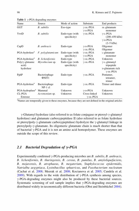
g-Glutamyl hydrolase (also referred to as folate conjugase or pteroyl-g-glutamyl
hydrolase) and glutamate carboxypeptidase II (also referred to as folate hydrolase
or pteroylpoly-g-glutamate carboxypeptidase) hydrolyze the g-glutamyl linkage in
pteroylpoly-g-glutamate. Its oligomeric glutamate chain is much shorter than that
of bacterial g-PGA and it is not an amino acid homopolymer. These enzymes are
outside the scope of this review.
2.1 Bacterial Degradation of g-PGA
Experimentally confirmed g-PGA-producing microbes are B. subtilis, B. anthracis,B. licheniformis, B. thuringensis, B. cereus, B. pumilus, B. amyloliquefaciens,B. mojavensis, B. atrophaeus, B. megaterium, Staphylococcus epidermidis,Natrialba aegyptiaca, Lysinibacillus sphaericus, and Fusobacterium nucleatum(Cachat et al. 2008; Meerak et al. 2008; Kocianova et al. 2005; Candela et al.
2009). With regards to the wide distribution of g-PGA synthesis among species,
g-PGA-degrading enzymes might also be produced by these bacterial sources.
Systematic screening of soil sample implies that g-PGA-degrading enzymes are
distributed widely in taxonomically different bacteria (Obst and Steinb€uchel 2004).
Table 1 g-PGA degrading enzymes
Name Source Mode of action Substrate End products
GGT B. subtilis Exo-type g-DL-PGA DL-glutamate
monomerg-D-PGAYwtD B. subtilis Endo-type (with
specificity)
g-DL-PGA g-L-PGA(200–450 kDa)
g-D-PGA(2–5 kDa)
CapD B. anthracis Endo-type g-D-PGA Oligomer
g-DL-PGA Oligomer
PGA-hydrolasea F. polyglutamicum Endo-type (with
specificity)
g-DL-PGA L-glutamate
monomerg-D-PGAPGA-hydrolasea B. licheniformis Endo-type g-DL-PGA Unknown
Poly(g-glutamic
acid)
hydrolase
Myrothecium sp. Endo-type (with
specificity)
g-DL-PGA g-L-glutamyl
tripeptide
D-glutamate rich
g-DL-PGAPghP Bacteriophage
FNIT1Endo-type g-DL-PGA Pentamer,
tetramer,
trimer
PGA-hydrolasea Bacteriophage
NP-1 cl
Endo-type g-DL-PGA Trimer and dimer
PGA-hydroplasea Mammal Unknown g-D-PGA Unknown
CL-PGA
Esterase
Acremonium sp. Unknown Cross-linked
g-DL-PGAUnknown
aNames are temporally given to these enzymes, because they are not defined in the original articles
98 K. Kimura and Z. Fujimoto

2.1.1 Bacillus subtilis
The laboratory lineage of B. subtilis (B. subtilis 168) is a g-PGA-negative strain.
Therefore, B. subtilis (natto), a starter strain used for “natto” fermentation (“natto”is a Japanese fermented soybean food) or undomesticated g-PGA-positive strains
have been employed in g-PGA studies (Stanley and Lazazzera 2005; Ashiuchi et al.
2001; Kimura et al. 2009). g-PGA-degrading enzymes have been studied mainly in
B. subtilis (natto).B. subtilis (natto) produces g-DL-PGA in the early stationary phase through
regulation by the quorum-sensing system ComQXPA, which governs stationary
phase gene expression (Tran et al. 2000; Dubnau 1999; Kimura et al. 2009;
Lazazzera 2000; Ogura et al. 2001) (Fig. 1). g-PGA-degrading GGT expression is
also regulated by ComQXPA. ComX, a small peptide secreted in the medium by the
ComQ function, binds to the membrane receptor kinase ComP when the ComX
concentration becomes high enough for binding. ComP and the cognate response
regulator ComA comprise a two-component system. The cell-density signal of
ComX is transmitted by phosphorelay from ComP to ComA. ComA is essential
for g-PGA synthesis, but it does not directly regulate the g-PGA synthetic operon
pgsBCA. DegQ, which is positively regulated by ComA, and the DegS–DegU two-
component system are required for pgsBCA expression (Stanley and Lazazzera
2005; Murray et al. 2009; Kimura et al. 2009).
Fig. 1 Schematic representation of the regulatory system of g-DL-PGA synthesis and degradation
in B. subtilis (natto)
Enzymatic Degradation of Poly-Gamma-Glutamic Acid 99

Stationary phase synthesis and degradation of g-DL-PGA are associated with its
physiological role. A high cell density in the stationary phase is a sign of over-
hanging starvation. It triggers the synthesis of a nutrient reservoir of which plant
starch and bacterial polyphosphate are well-known examples. Likewise, B. subtilis(natto) utilizes g-DL-PGA as an extracellular nutrient reservoir (Kimura et al.
2004a, b). B. subtilis (natto) degrades g-DL-PGA stored outside the cells in the
late stationary phase by specific enzymes to reuse glutamate. In the late stationary
phase, exoenzymes that enable the utilization of nutritionally unfavorable carbo-
hydrate polymers or proteins are also expressed (Ogura et al. 2003; Tsukahara and
Ogura 2008).
Incidentally, g-DL-PGA degradation in the stationary phase is a well-known
phenomenon, and an extracellular GGT was identified as the enzyme responsible
for it (Throne et al. 1954; Kunioka and Goto 1994; Ogawa et al. 1991; Abe et al.
1997; Kimura et al. 2004b). Generally, the GGT family of proteins transfer
g-glutamyl compounds to H2O (in hydrolysis) or amino acids (in transpeptida-
tion) (Tate and Meister 1985; Suzuki et al. 1986; Suzuki and Kumagai 2002;
Okada et al. 2006). B. subtilis GGT hydrolyzes g-DL-PGA (Vmax ¼ 10 mmol/mg
protein/min, Km ¼ 9 mM) and liberates glutamate from the N-terminal end of
g-DL-PGA irrespective of the type of glutamate isomers (Table 2; Kimura et al.
2004b). 6-Diazo-5-oxo-L-norleucine (DON) inhibits both B. subtilis and Escher-ichia coli GGT, implying that the catalytic center of both enzymes are structurally
redundant, but g-PGA-hydrolyzing activity is absent in E. coli GGT (Table 2;
Kimura et al. 2004b). Structural difference and substrate specificity of the GGT
family proteins of B. subtilis, E. coli, and B. anthracis are discussed in Sect. 4.1.
B. subtilis (natto) NAFM90 (ggt::Spcr), a GGT-deficient mutant, produces
g-DL-PGA as much as wild-type cells. Consequently, NAFM90 (ggt::Spcr) cannotreuse glutamate as the nutrient reservoir. Such an uneconomical overconsumption
of food resources does not allow sustainable vegetative growth of cells (Kimura
et al. 2004b).
In the late stationary phase, g-DL-PGA eventually disappears from the culture
medium containing wild-type cells. In contrast, the amount of g-DL-PGA in the
NAFM90 (ggt::Spcr) culture medium does not change (Kimura et al. 2004b).
However, g-DL-PGA accumulated in the NAFM90 culture medium has a smaller
molecular size (0.1 MDa) than that of the native g-DL-PGA (>2 MDa) (Kimura
Table 2 Comparison of GGT family proteins of B. subtilis, B. anthracis, and E. coli
B. subtilis B. anthracis E. coli
Km to g-Glu-pNAa 8 mM NTb 35 mMMode of hydrolysis Exo-type Endo-type Exo-type
Cleavage of g-PGA Yes Yes No
Inhibition by DONc Yes No Yes
Loop structure (Pro438d to Gly449d) No No Yesag-Glu-pNA, g-glutamyl p-nitroanilide, a synthetic chromogenic GGT substratebNT not tested, B. anthracis CapD does not hydrolyse g-Glu-pNAcDON 6-diazo-5-oxo-noeleucinedAmino acid residue numbers are those of E. coli GGT
100 K. Kimura and Z. Fujimoto

et al. 2004b). The reduction in size is a result of fragmentation of g-DL-PGA where
ywrD gene product is involved. YwrD belongs to the GGT family (amino acid
sequence identities to B. subtilis GGT and B. anthracis CapD are 29% and 26%,
respectively). g-DL-PGA degradation was not observed at all when both ggt andywrD were disrupted (Kimura and Itoh, unpublished results). g-DL-PGA fragmen-
tation by YwrD increases its molar concentration and drastically reduces the
viscosity of the medium containing native g-DL-PGA, which promotes the exo-
type hydrolysis by GGT (Kimura et al. 2004b). YwrD does not hydrolyze glutathi-
one or g-Glu-p-NA (Minami et al. 2004). YwrD might be an endo-type g-PGAhydrolase, but we do not have direct experimental evidence about it.
During the late stationary phase when g-DL-PGA is actively degraded to gluta-
mate monomers by the synergistic reaction of GGT and YwrD, free glutamate
concentration in the medium is kept low (<0.03 mM) (Kimura et al. 2004a).
B. subtilis (natto) produces 1–10 mg/ml of g-DL-PGA in various culture conditions,
and the concentration of g-DL-PGA is equivalent to 8–80 mM of glutamic acid. It
seems that glutamate monomers liberated from g-DL-PGA are rapidly incorporated
in the cells and g-DL-PGA digestion by GGT is the rate-limiting step in glutamic
acid consumption. L-Glutamate in the medium severely suppresses GGT expression
at the transcription level (Kimura et al. 2004b). This feedback regulation of GGT
prevents overconsumption of g-DL-PGA and enables B. subtilis cells to use it for a
long time. L-Aspartate, L-asparagine, L-glutamine, and L-proline also partially
suppressed GGT expression. Amino acids such as L-isoleucine and L-valine,
whose metabolic pathways are not closely related to that of L-glutamate, did not
suppress GGT expression at all (Kimura et al., unpublished results).
More than 50% of glutamate in g-DL-PGA are D-isomers (Nagai et al. 1997).
D-amino acids are generally toxic for cells (Soutourina et al. 2000; Yang et al.
2003). B. subtilis cells have relatively more abundant glutamate racemase than
other bacteria (Ashiuchi et al. 1998; Kada et al. 2004). Glutamate racemases are
anabolic enzymes that supply D-glutamate to peptidoglycan in bacteria. However,
the glutamate racemases of B. subtilis – RacE and YrpC – have both anabolic and
catabolic functions (Kimura et al. 2004a). Similar to the cases of catabolic enzymes,
RacE and YrpC expression is suppressed in the rich medium, when rapidly pro-
liferating cells need D-glutamate for cell wall construction (Kimura et al. 2004a).
Furthermore, D-glutamate clearly exerted a growth-inhibitory effect when racemase
genes were disrupted (Kimura et al. 2004a). These observations suggested that in
addition to the essential role of supplying D-glutamate for cell wall construction,
the glutamate racemases RacE and YrpC act as catabolic enzymes that convert
D-glutamate to L-glutamate and are a part of the g-PGA recycling pathway (Kimura
et al. 2004a) (Fig. 1).
B. subtilis and B. licheniformis are taxonomically very close to each other. The
amino acid sequences of the g-DL-PGA synthetic pgsB, C, and A gene products are
91%, 91%, and 68% identical when compared with each other; the neighboring
genes are also homologous between them (Fig. 2). These two bacteria have essen-
tially the same g-DL-PGA synthetic and degrading systems. The ywtD gene (also
referred to as pgsD or pgdS) is located immediately downstream of the pgs operon in
Enzymatic Degradation of Poly-Gamma-Glutamic Acid 101

B. subtilis and B. licheniformis. Recombinant YwtD hydrolyzes g-DL-PGA in vitro
into large (200–450 kDa) and small (2–5 kDa) fragments (Chunhachart et al. 2006;
Ashiuchi et al. 2006; Suzuki and Tahara 2003). The large fragments are L-glutamate-
rich polypeptides and the small fragments are D-glutamate rich. Analysis of terminal
glutamate of the fragments produced by YwtD implies that YwtD hydrolyzes the
g-glutamyl linkage between D-glutamates (Ashiuchi et al. 2006). The recombinant
YwtD hydrolyzes g-DL-PGA but not synthetic oligo-g-glutamate (Chunhachart et al.
2006). YwtD probably recognizes the long chain of g-DL-PGA. B. subtilis (natto)NAFM61 (ggt::Spcr, ywrD::Ermr) does not degrade g-DL-PGA at all (see above), but
the large and the small fragment corresponding to the in vitro hydrolysis products by
YwtD were not found in the culture medium. Therefore, the in vivo significance of
YwtD is obscure. The absence of ywtD in B. anthracis, whose g-D-PGA is tightly
anchored to the cell wall (Candela and Fouet 2005; Richter et al. 2009), prompts us
to hypothesize that YwtD is involved in releasing g-PGA to the culture media.
2.1.2 Bacillus anthracis
g-D-PGA of B. anthracis covers cells as a protective capsule. Strains lacking g-D-PGA are avirulent because they cannot escape the phagocytic attack of the host
immune system (Fouet and Mock 2006). Therefore, g-D-PGA is a target molecule
in anthrax therapeutics (Scorpio et al. 2007; Richter et al. 2009). The capD gene of
Fig. 2 Structure of the cap/pgs g-PGA synthetic operon of B. subtilis (natto), B. licheniformis, andB. anthracis. Numbers indicate amino acid sequence identities (%) of Cap/Pgs proteins to those
of Bacillus subtilis (natto). The capD gene of B. anthracis is similar to ggt and ywrD genes of
B. subtilis or B. licheniformis. ggt and ywrD genes reside in loci far from the pgs region in these
two bacteria. B. anthracis does not possess the ywtD gene
102 K. Kimura and Z. Fujimoto

B. anthracis is a part of the g-D-PGA synthetic operon (Fig. 2). CapD is not essential
for g-D-PGA synthesis, but is required for attaching the synthesized glutamate
filament to cell wall peptidoglycan (Candela and Fouet 2005; Richter et al. 2009).
g-D-PGA released in the medium as a free form in the absence of CapD has no such
barrier function (Candela and Fouet 2005). CapD is located on the cell envelope,
whereas the GGT of B. subtilis is secreted in the medium.
CapD belongs to the GGT family. GGT, as its name indicates, is an enzyme that
hydrolyzes or transfers a wide variety of g-glutamyl moiety-containing substrates
such as glutathione (Tate and Meister 1981). However, unlike the GGT of
B. subtilis, which is an exo-g-glutamyl hydrolase that liberates a glutamate mono-
mer from the N-terminal end of g-PGA, CapD performs an endo-type hydrolysis
(Uchida et al. 1993; Candela and Fouet 2005; Richter et al. 2009). CapD does not
hydrolyze g-glutamyl-p-nitroanilide (g-Glu-p-NA), a popular substrate of the GGTfamily proteins (Tate andMeister 1985). DON, which inhibits most GGTs including
those of B. subtilis or E. coli has no effect on CapD (Richter et al. 2009; Table 2).
Substances that inhibit CapD but not mammalian GGT are candidates for anti-
anthrax drugs. A compound that inhibits CapD was found recently (Richter et al.
2009). The inhibitory compound, capsidin, which mimics the backbone structure of
g-D-Glu–D-Glu, inhibits CapD with an IC50 value of 6.63 mM and prevents the
capsulation in vivo. Capsidin noncompetitively inhibits CapD by acetylation of the
active site threonine without affecting mammalian GGT (Richter et al. 2009).
B. anthracis lacks ywtD gene, which resides at the corresponding position of
CapD in the genomes of B. subtilis and B. licheniformis. In B. subtilis and
B. licheniformis, g-DL-PGA is secreted in the medium as a free form (Kunioka and
Goto 1994; Kimura et al. 2004b; Birrer et al. 1994). YwtD might cleave g-DL-PGAonce anchored to the cell wall during synthesis. The in vitro hydrolytic activity of
YwtD on g-DL-PGA supports this hypothesis. Experimental elucidation is awaited.
2.1.3 Other Microorganisms
F. polyglutamicum utilizes g-DL-PGA as a sole carbon and nitrogen source (Volcani
and Margalith 1957). g-DL-PGA-hydrolyzing activity was observed in the cell
extracts of F. polyglutamicum. L-Glutamic acid was the only enzymatic breakdown
product and its amount was equivalent to that in the substrate (Volcani and
Margalith 1957). The F. polyglutamicum enzyme appears to cleave the g-L-gluta-myl–L-glutamate linkage in g-DL-PGA because the g-DL-PGA filament is a copoly-
mer of L- and D-glutamates (see Sect. 3). This enzyme, which is insensitive to
iodoacetate and capable of hydrolyzing pteroyl-g-glutamic acid, is distinct from the
other g-PGA-degrading enzymes. Volcani and Margalith also reported that they
obtained several different bacteria utilizing g-DL-PGA as a sole source of nitrogen
and carbon in soil samples, but only F. polyglutamicum was characterized.
Birrer et al. reported cell-associated g-DL-PGA degradation in B. licheniformisand King et al. reported g-PGA-associated g-DL-PGA degradation in B. lichenifor-mis ATCC9945a (Birrer et al. 1994; King et al. 2000). The g-PGA-associated
Enzymatic Degradation of Poly-Gamma-Glutamic Acid 103

enzyme is an endo-type that does not hydrolyze the synthetic dipeptide thioester
substrate AcNH-D-Glu-g –D-Glu–g-SEt. In both cases, enzymes were not purified or
characterized in detail.
Tanaka et al. screened 683 fungal strains by monitoring the reduction in the
viscosity of g-DL-PGA to find endo-type g-DL-PGA-degrading enzymes (exo-type
hydrolysis does not drastically reduce viscosity). The 68 kDa g-PGA hydrolase
was homogeneously purified from Myrothecium sp. TM-4222 culture broth
(Tanaka et al. 1993a, b). It degrades g-DL-PGA to mixture of oligo-g-L-glutamate.
The major hydrolytic product was g-L-glutamyl tri-peptide, but no L-glutamate
monomer was observed. The digestion of g-DL-PGA yields a D-glutamate-rich
fragment (D-Glu:L-Glu ¼ 89:11) (Tanaka et al. 1993a, b). This D/L ratio is quite
high compared to the ratio in the substrate (D:L ¼ 55:45) (see Sect. 3.1).
2.2 Bacteriophage-Related g-PGA Degradation
Contamination by bacteriophages continues to be a problem in the fermentation
industry. Bacteriophages that spoil natto fermentation were collected to survey the
damage (Nagai and Yamasaki 2009). They were detected mainly from natto, butwere found also on the surface of drains, containers, and in pipes of natto factories.
For suppliers of natto, phage contamination that spoils the growth of host cells or
stops the fermentation process is easy to handle. They can detect contamination
visually and can discard spoiled products before shipping. However, if the number
of contaminating bacteriophages is small enough (usually below 10 phages/pack-
age) to allow apparently normal growth of B. subtilis (natto), the contaminated
product escapes monitoring. In such a case, suppliers receive an unfavorable
response from consumers who purchased them due to a g-PGA degrading enzyme
produced by the phage (see below).
2.2.1 PghP of B. subtilis (natto) Phage FNIT1
Bacteriophage-related g-PGA degradation has been known since the 1970s (Hongo
and Yoshimoto 1970). Strong g-DL-PGA-degrading activity was found in the
B. subtilis (natto) culture supernatant infected with FNIT1 isolated from spoiled
natto, and PghP (poly-g-glutamate hydrolase P) was homogeneously purified
(Kimura and Itoh 2003). This enzyme degrades g-DL-PGA to g-glutamyl oligomers
and finally to g-glutamyl penta-, tetra-, and tri-peptides. PghP is a monoiodoacetate-
sensitive 25 kDa metallopeptidase requiring Zn2+ or Mn2+ for hydrolysis (Kimura
and Itoh 2003).
PghP hydrolyzes g-DL-PGA and very quickly decreases polymer viscosity. PghP
(1 mg) can hydrolyze 1.5 g of g-DL-PGA to the final products within 1 min (Kimura
and Itoh 2003). Natto is mixed with a soy sauce or other seasonings and stirred just
104 K. Kimura and Z. Fujimoto

before serving to give the essential sticky texture to it, which allows contaminated
PghP to act and eliminate viscosity.
2.2.2 Effect of g-PGA Degradation on Bacteriophages
B. subtilis (natto) colonies are covered by a sticky material produced in large
amounts (Fig. 3). The sticky material is a mixture of g-DL-PGA and levan (contri-
bution of levan to the stickiness and viscosity is small because disruption of the
levan synthetic gene minimally affects stickiness and viscosity). Breakdown of the
sticky material by PghP allows phage progenies to easily spread and infect host
cells (Kimura and Itoh 2003). E. coli phage 29 has an endo-N-acetylneuraminidase
in its phage particle (spike) for hydrolyzing the K1 capsule (poly-a-2,8-linkedsialosyl unit) (Kwiatkowski et al. 1982). Some phages of Klebsiella and Strepto-coccus also have enzymes for hydrolyzing capsular polysaccharides on phage
particles (Cescutti and Paoletti 1994). PghP is not displayed on the phage particle.
Thus, the first phage cannot remove g-DL-PGA for infection (the FNIT1 receptor isnot identified, but phages have to establish contact with the host cell surface). PghP
synthesis during phage propagation in host cells appears to be antithetical to its
function. However, B. subtilis g-DL-PGA is produced in the stationary phase (Tran
et al. 2000; Kimura et al. 2009). Phages can infect young colonies without the
barrier of g-DL-PGA in a natural environment, and the PghP produced can spread
around to hydrolyze g-DL-PGA of neighboring colonies. PghP expression on the
phage particle might be a less effective strategy in this case. g-DL-PGA is produced
in a large amount and usually exists as a viscous water absorbent surrounding cells
(Fig. 3). Therefore, allowing the degrading enzyme to spread as much as possible
by the pioneer phage might be a good group strategy.
The host cell range for FNIT was examined using 49 B. subtilis test strains
(Ackermann et al. 1995). FNIT1 propagates and makes a clear halo on grasses of
B. subtilis cells including strains HER1395, HER1313, SIM46, HSY20, Q:1:1.3,
IP005, SIM218, and IP032 (Kimura et al., unpublished results). The host specificity
spectrumwas different from any other typing phages reported previously (Ackermann
et al. 1995).
The pghP gene (accession no. AB091475) in the phage genome has no signifi-
cant homology to known enzymes. It appears to be transcribed together with the
upstream gene encoding a hypothetical pectin lyase-like protein (Fig. 4). The pectin
Fig. 3 Morphology of
B. subtilis (natto) colonies.Wild-type and g-DL-PGA-deficient mutant (pgsBCA::SpcR) cells are grown on GSP
agar plates (Nagai et al. 1997)
overnight
Enzymatic Degradation of Poly-Gamma-Glutamic Acid 105

lyase-like protein might be involved in digesting cell wall components during
phage propagation.
2.2.3 PghP Distribution
Bacteriophages isolated from spoiled natto were classified into two groups (groups
I and II) based on genome structure and phage particle morphology (Nagai and
Yamasaki 2009) (Figs. 5, 6). The FNIT1 phage belongs to group II, having a
hexagonal head and sheathed tail. The pghP gene was found in both types of
phage (Kimura et al., unpublished results). A mobile insertion sequence of the
IS605 family was found in the 30-flanking region of pghP (Fig. 4). pghP was
possibly distributed by horizontal movement from one phage genome to another.
The g-DL-PGA-degrading enzyme of bacteriophage NP-1 cl produces trimers
and dimers of glutamic acid as the final products (Hongo and Yoshimoto 1970).
Furthermore, phages producing g-DL-PGA-degrading enzyme but not possessing
genes homologous to pghPwere found in soil (Kimura and Itoh 2003; Kimura et al.,
unpublished results). A variety of g-PGA-degrading enzymes is suggested in
bacteriophages.
Fig. 4 Schematic illustration of pghP and its neighboring genes in the bacteriophage FNIT1genome. PghP coding region (shaded) and possible ORFs are shown by arrows. A, PghP; B, pectinlyase-like protein; C, hypothetical protein; D, dihydrofolate reductase; E, adenylate kinase; F,
thimidylate synthase
Fig. 5 Electron micrographs
of B. subtilis (natto)bacteriophages JNDMP (a)and ONPA (b). Bar = 100 nm.
Adapted from Nagai and
Yamasaki (2009) with
publisher’s permission. Phage
particles are negatively
stained with 2%
phosphotungstic acid and
observed under an electron
microscope (JEOL-1200 EX,
Japan Electronic Optics
Laboratory, Tokyo, Japan)
106 K. Kimura and Z. Fujimoto
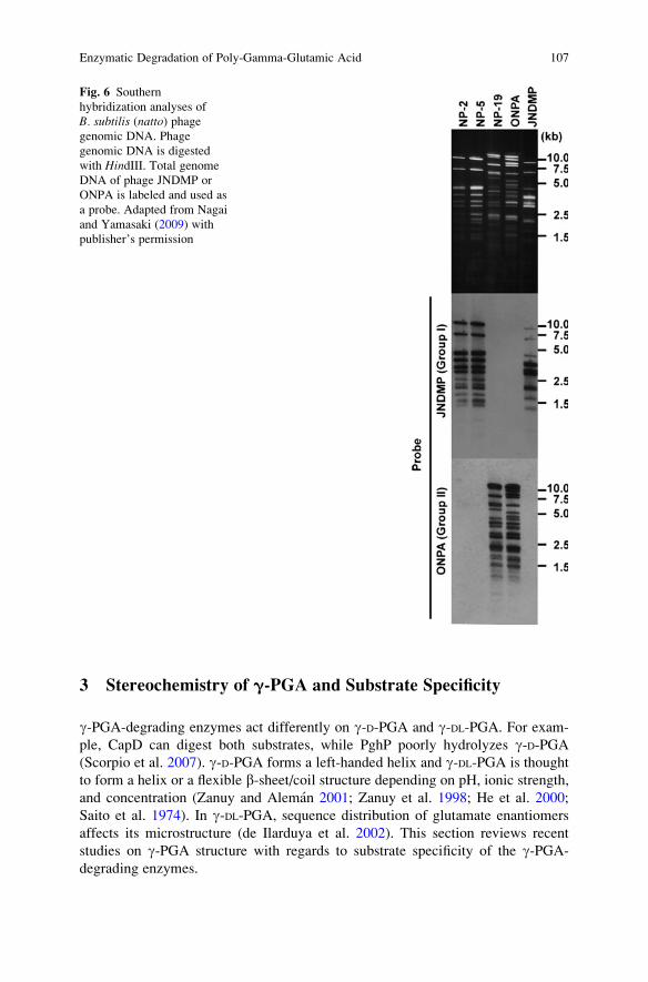
3 Stereochemistry of g-PGA and Substrate Specificity
g-PGA-degrading enzymes act differently on g-D-PGA and g-DL-PGA. For exam-
ple, CapD can digest both substrates, while PghP poorly hydrolyzes g-D-PGA(Scorpio et al. 2007). g-D-PGA forms a left-handed helix and g-DL-PGA is thought
to form a helix or a flexible b-sheet/coil structure depending on pH, ionic strength,
and concentration (Zanuy and Aleman 2001; Zanuy et al. 1998; He et al. 2000;
Saito et al. 1974). In g-DL-PGA, sequence distribution of glutamate enantiomers
affects its microstructure (de Ilarduya et al. 2002). This section reviews recent
studies on g-PGA structure with regards to substrate specificity of the g-PGA-degrading enzymes.
Fig. 6 Southern
hybridization analyses of
B. subtilis (natto) phagegenomic DNA. Phage
genomic DNA is digested
with HindIII. Total genome
DNA of phage JNDMP or
ONPA is labeled and used as
a probe. Adapted from Nagai
and Yamasaki (2009) with
publisher’s permission
Enzymatic Degradation of Poly-Gamma-Glutamic Acid 107

Chemical or physical treatment can alter the higher-order structure of g-PGA.For example, g-ray irradiation induces cross-linking between g-DL-PGA filaments
(Matsui et al. 2008). Grafting hydrophobic side chains such as phenylalanine causes
g-DL-PGA to self-assemble and form nanoparticles (Akagi et al. 2005, 2006). These
modified g-PGAs are used for various industrial purposes (Yoshikawa et al. 2008;
Buescher and Margaritis 2007). Enzymatic digestion of modified g-PGAs is men-
tioned below.
3.1 Stereochemistry of g-PGA
Chemical, enzymological, and physical analyses suggest that g-DL-PGA produced
by B. subtilis is a copolymer consisting of D- and L-glutamate incorporated into a
single filament, although we cannot exclude the possibility that small amounts of
D- or L- glutamate homopolymers are also produced.
Enantiomer composition and sequence distribution of g-DL-PGA was first
explored by Tanaka et al. (1993a) using PGA hydrolase isolated from the Myr-othecium sp. TM-4222 culture supernatant . In this study, the random distribution
of isomers in g-DL-PGA was not considered and heterogeneous isomer units were
suggested (Tanaka et al. 1993a, b). Likewise, g-DL-PGA microstructure in which
D- or L-glutamate repeating units are alternately linked in a single chain is proposed
by chemical hydrolysis of g-DL-PGA (Wang et al. 2008). Wang et al. chemically
hydrolyzed g-DL-PGA (50% D-Glu content) and obtained g-glutamyl dipeptides.
The g-glutamyl dipeptides D–D, L–L, D–L, and L–D were separated by chiral-specific
HPLC and these four dipeptides appeared in a ratio of 5.9:6.0:1.0:1.0. If the
enantiomers are randomly linked in a single chain, this ratio would be close to
1.0:1.0:1.0:1.0. Therefore, there seem to be blocks of homopolymers in the fila-
ments (Wang et al. 2008).
PghP digests g-DL-PGA and produces pentamers, tetramers, and trimers of
glutamic acid, but it cannot hydrolyze synthetic hexa-oligo-g-D-glutamate (Shiga
and Kimura, unpublished results). The stretch of D-glutamate in g-DL-PGA is
perhaps less than a hexamer. However, estimation of the stereochemical structure
of g-DL-PGA only from enzymatically digested products should be performed
carefully. Generally, peptide chains longer than a hexamer are required to form a
helical structure (Moretto et al. 2008). PghP might randomly cut the g-glutamyl
peptide bond regardless of the stereochemistry of the cutting site, and a structural
change in the substrate such as a shift from helix to random coil might prevent the
enzyme from recognizing the shortened substrate.13C NMR analyses of g-CH and b-CH2 in a-esterified g-DL-PGA (poly a-ethyl
g-DL-glutamic acid) and various synthetic g-PGA reference molecules can detect
the local sequence of enantiomers. de Ilarduya et al. showed that g-DL-PGAconsists of a block stereocopolymer of D and L units as well as a minor amount
(about 10%) of two enantiomerically pure homopolymers, which is consistent
with the microstructure proposed by enzymatic and chemical cleavage experiments
108 K. Kimura and Z. Fujimoto

(de Ilarduya et al. 2002; Wang et al. 2008; Tanaka et al. 1993b; Ashiuchi et al.
2006; Chunhachart et al. 2006).
3.2 Mode of Action in g-PGA Hydrolysis
There exist three types of hydrolysis: exo-type, nonspecific endo-type, and endo-
type. GGT liberates glutamate from the N-terminal end of the polypeptide and
demonstrates no isomer specificity (Kimura et al. 2004b). PghP hydrolyzes g-DL-PGA by the endo-type mechanism (Kimura and Itoh 2003; Scorpio et al. 2007). B.anthracis CapD hydrolyzes both g-D-PGA and g-DL-PGA by the endo-type mecha-
nism (Scorpio et al. 2007). g-D-PGA is covalently attached to the cell wall by the
transpeptidase activity of CapD. CapD hydrolyzes “native” g-D-PGA produced as a
free form in the culture medium of the capD mutant strain RTC40 (capD::Spcr), aswell as “denatured” g-D-PGA isolated from wild type cells by autoclave (Kimura
and Fouet, unpublished results). The final product of CapD hydrolysis has not been
examined in detail.
YwtD ofB. subtilis and g-PGA hydrolases ofF. polyglutamicum andMyrotheciumsp. are endo-type hydrolases with stereochemical specificity (Ashiuchi et al. 2006;
Volcani and Margalith 1957; Tanaka et al. 1993a). These enzymes produce g-L-PGAor g-D-PGA from g-DL-PGA. YwtD does not hydrolyze synthetic oligo-g-glutamate
(octamer). It probably recognizes the cleavage site of peptide chains longer than eight
residues (Chunhachart et al. 2006).
3.3 Modified g-PGA
Irradiating g-DL-PGA induces random cross-linking between the filaments of
the glutamate chain. The cross-linked g-DL-PGA (CL-PGA) irradiated by 20 kGy
g-ray absorbs 4,500% (w/w) of water and forms a hydrogel with various properties
(Hara 2001). CL-PGA degradation was examined in compost to assess its biode-
gradability by measuring amounts of CO2 released based on the ISO14855 standard
method. Eighty percent of the CL-PGA was degraded within 2 weeks (Hara 2001).
Matsui et al. found a fungal strain, FK-1, belonging to the genus Acremonium that
produces a CL-PGA liquefying enzyme (Matsui et al. 2008). It also hydrolyzes
g-DL-PGA chemically cross-linked by an ester bond (Matsui et al. 2008). The
liquefying enzyme has not been further characterized.
Amphiphilic poly(a-alkyl g-glutamate) such as g-DL-PGA derivatives conju-
gated with L-phenylalanine ethylester assembles into nanoparticles in water
(Akagi et al. 2005, 2006). GGT of B. subtilis is known to degrade the backbone
of the particle, which is a promising material as a drug delivery carrier (Yoshikawa
et al. 2008).
Enzymatic Degradation of Poly-Gamma-Glutamic Acid 109
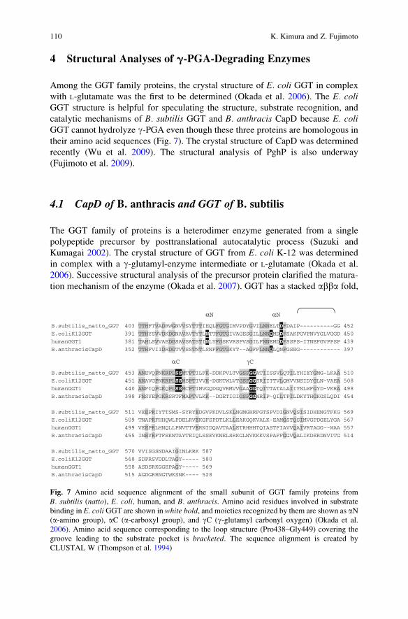
4 Structural Analyses of g-PGA-Degrading Enzymes
Among the GGT family proteins, the crystal structure of E. coli GGT in complex
with L-glutamate was the first to be determined (Okada et al. 2006). The E. coliGGT structure is helpful for speculating the structure, substrate recognition, and
catalytic mechanisms of B. subtilis GGT and B. anthracis CapD because E. coliGGT cannot hydrolyze g-PGA even though these three proteins are homologous in
their amino acid sequences (Fig. 7). The crystal structure of CapD was determined
recently (Wu et al. 2009). The structural analysis of PghP is also underway
(Fujimoto et al. 2009).
4.1 CapD of B. anthracis and GGT of B. subtilis
The GGT family of proteins is a heterodimer enzyme generated from a single
polypeptide precursor by posttranslational autocatalytic process (Suzuki and
Kumagai 2002). The crystal structure of GGT from E. coli K-12 was determined
in complex with a g-glutamyl-enzyme intermediate or L-glutamate (Okada et al.
2006). Successive structural analysis of the precursor protein clarified the matura-
tion mechanism of the enzyme (Okada et al. 2007). GGT has a stacked abba fold,
αN αN
B.subtilis_natto_GGT 403 TTHFTVADRWGNVVSYTTTIEQLFGTGIMVPDYGVILNNELTDFDAIP----------GG 452
E.coliK12GGT 391 TTHYSVVDKDGNAVAVTYTLNTTFGTGIVAGESGILLNNQMDDFSAKPGVPNVYGLVGGD 450
humanGGT1 381 TAHLSVVAEDGSAVSATSTINLYFGSKVRSPVSGILFNNEMDDFSSPS-ITNEFGVPPSP 439
B.anthracisCapD 352 TTHFVIIDRDGTVVSSTNTLSNFFGTGKYT--AGFFLNNQLQNFGSEG------------ 397
αC γC
B.subtilis_natto_GGT 453 ANEVQPNKRPLSSMTPTILFK-DDKPVLTVGSPGGATIISSVLQTILYHIEYGMG-LKAA 510
E.coliK12GGT 451 ANAVGPNKRPLSSMSPTIVVK-DGKTWLVTGSPGGSRIITTVLQMVVNSIDYGLN-VAEA 508
humanGGT1 440 ANFIQPGKQPLSSMCPTIMVGQDGQVRMVVGAAGGTQITTATALAIIYNLWFGYD-VKRA 498
B.anthracisCapD 398 FNSYEPGKRSRTFMAPTVLKK--DGETIGIGSPGGNRIP-QILTPILDKYTHGKGSLQDI 454
B.subtilis_natto_GGT 511 VEEPRIYTTSMS-SYRYEDGVPKDVLSKLNGMGHRFGTSPVDIGNVQSISIDHENGTFKG 569
E.coliK12GGT 509 TNAPRFHHQWLPDELRVEKGFSPDTLKLLEAKGQKVALK-EAMGSTQSIMVGPDGELYGA 567
humanGGT1 499 VEEPRLHNQLLPNVTTVERNIDQAVTAALETRHHHTQIASTFIAVVQAIVRTAGG--WAA 557
B.anthracisCapD 455 INEYRFTFEKNTAYTEIQLSSEVKNELSRKGLNVKKKVSPAFFGGVQALIKDERDNVITG 514
B.subtilis_natto_GGT 570 VVISGSNDAAIGINLKRK 587
E.coliK12GGT 568 SDPRSVDDLTAGY----- 580
humanGGT1 558 ASDSRKGGEPAGY----- 569
B.anthracisCapD 515 AGDGRRNGTWKSNK---- 528
Fig. 7 Amino acid sequence alignment of the small subunit of GGT family proteins from
B. subtilis (natto), E. coli, human, and B. anthracis. Amino acid residues involved in substrate
binding in E. coli GGT are shown in white bold, and moieties recognized by them are shown as aN(a-amino group), aC (a-carboxyl group), and gC (g-glutamyl carbonyl oxygen) (Okada et al.
2006). Amino acid sequence corresponding to the loop structure (Pro438–Gly449) covering the
groove leading to the substrate pocket is bracketed. The sequence alignment is created by
CLUSTAL W (Thompson et al. 1994)
110 K. Kimura and Z. Fujimoto

similar to the folds seen in members of the N-terminal nucleophile hydrolase
superfamily (Fig. 8). The small subunit is surrounded by the large subunit and
b-strands of the large and small subunits form the two central b-sheets. Theg-glutamyl moiety of the intermediate covalently binds to Thr391, which is the
N-terminal residue of the small subunit and the catalytic residue of the enzyme.
The bound L-glutamate in the enzyme pocket interacts with Arg114, Ser462,
Ser463, Gln430, Asp433, Gly483, and Gly484 by hydrogen bonds and salt bridges.
With the exception of Arg114, all residues involved in substrate binding are located
in the small subunit (Fig. 7). The a-carboxyl group of the g-glutamyl moiety is
bound to Arg114, Ser462, Ser463, and the a-amino group is bound to Asn411,
Gln430, and Asp433. The g-glutamyl carbonyl oxygen atom is hydrogen-bonded
with main-chain amino groups of Gly483 and Gly484.
Amino acid residues corresponding to Arg114, Gly483, and Gly484 of E. coliare conserved in B. subtilis GGT and B. anthracis CapD (Fig. 7). Ser462, Ser463,
and Asp433 are conserved in B. subtilis GGT but not in B. anthracis CapD.
Neither Gln430 nor Asn411 are conserved. It is noteworthy that the loop structure
from Pro438 to Gly449 of E. coli GGT that covers the groove leading to the
binding pocket is missing in B. subtilis GGT and B. anthracis CapD (Fig. 7).
The loop structure contains a Tyr444 residue forming a wall that shields the
pocket from the solvent and probably prevents large molecules such as g-PGAfrom accessing the catalytic site in E. coli GGT (Okada et al. 2006). Mammalian
GGT that cannot hydrolyze g-PGA has the loop structure similar to that of E. coliGGT. The substrate-binding pockets of bacilli GGTs are more open than those of
other GGTs.
Neither B. subtilis GGT nor B. anthracis CapD displays stereochemical speci-
ficity in the enzyme reaction. This might be explained by the absence of residues
corresponding to Asn411 and Gln430 that bind to the a-amino group of the
substrate. Ser463 and Ser464 bonded with the a-carboxy group in E. coli GGT
Fig. 8 Ribbon drawings of the crystal structures of E. coli GGT (protein bank code 2DBX) (a),B. anthracis CapD (3GA9) (b), and bovine CPA (3CPA) (c). The bound ligands (L-glutamate,
L-glutamyl-L-glutamate, and glycyl-L-tyrosine) are shown in green ball-and-stick drawings. Thelarge and small subunits of GGT and CapD are shown in blue and orange, respectively. The figureis prepared by Raster3D (Merritt and Bacon 1997)
Enzymatic Degradation of Poly-Gamma-Glutamic Acid 111

are conserved in B. subtilis GGT but not in B. anthracis CapD. B. subtilis GGTprobably recognizes the N-terminal end of the polypeptide by Gly483, Gly484,
Ser463, and Ser464 residues that bind the a-carboxyl group and g-glutamyl car-
bonyl oxygen. CapD seems to recognize neither a-amino nor a-carboxyl groups atthe N-terminal end of g-PGA. CapD probably interacts only with the g-glutamyl
carbonyl oxygen of g-PGA via the conserved Gly–Gly motif and degrades it by
an endo-type mechanism without stereochemical specificity. The crystal structure
of CapD was recently determined in complex with a nonhydrolyzable analog,
a-L-Glu–L-Glu (Wu et al. 2009). Its overall structure resembles that of E. coliGGT. Substrate recognition by residues Pro427–Gly428–Gly429 that appeared to
contribute to the CapD hydrolytic reaction was structurally confirmed.
4.2 PghP of Bacteriophage FNIT1
PghP and CapD are endo-type hydrolases. However, as discussed above, PghP – a
metalloenzyme – does not belong to the GGT family and is an orphan enzyme.
BLAST search of PghP retrieved genes from Bacillus phage SPP1, B. pumilus,B. licheniformis, B. subtilis, Staphylococcus haemolyticus, and B. cereus. These areall hypothetical proteins.
PghP of bacteriophage FNIT1 was crystallized and its structural analysis is in
progress (Fujimoto et al. 2009). Preliminary analyses revealed that PghP is a
globular protein having an open a/b mixed-core structure with a seven-stranded
parallel/antiparallel b-sheet. The overall core structure resembles that of bovine
carboxypeptidase A (CPA; Fig. 8) or related zinc peptidases, which are phylogen-
etically very far from PghP and belong to the M14 metallopeptidase family
(Kilshtain-Vardi et al. 2003; Cappalonga et al. 1992; MEROPS: http://merops.
sanger.ac.uk). The CPA crystal structure was first determined by Lipscomb et al.
(1970) and successive structural analyses revealed the catalytic residues and sub-
strate recognition mechanism in detail. CPA has one zinc ion at the catalytic center
coordinated by two histidine and one glutamate residues, and the bound zinc ion
and another glutamic acid, Glu270, are responsible for catalysis. The His–Glu–His
zinc coordinate motif of CPA was found also in PghP at a topologically equivalent
position (Fujimoto and Kimura, manuscript in preparation). Structural comparison
of PghP, M14 metallopeptidases, and the GGT family proteins can be helpful for
elucidating the molecular mechanisms of the g-PGA hydrolyzing enzymes, as well
as the recognition mechanism of g-PGA stereochemistry.
5 Conclusion and Future Perspectives
B. subtilis, B. anthracis, and other microbes produce g-PGA having different
physiological functions and utilize them to survive in natural environments. As
observed in the GGT family proteins of B. anthracis, B. subtilis, and E. coli,
112 K. Kimura and Z. Fujimoto

degradation enzymes appear to have evolved to perform such functions. Although
g-PGA production is not examined directly, genes homologous to the pgs/capg-PGA synthetic system can be found in genomes of Oceanobacillus iheyensisHTE831, Desulfitobacterium hafniense Y51, Francisella tularensis Schu 4, Lep-tospira interrogans serovar lai 56601, and several other microorganisms (in some
cases, pgsA/capA homologue is missing) (http://www.ncbi.nlm.nih.gov/). These
bacteria are potential sources of novel g-PGA-degrading enzymes.
Acknowledgments Parts of work presented were supported by grants from Ministry of Agricul-
ture, Forestry, and Fishery of Japan. K.K. acknowledges Drs. Fouet A and Mock M, and National
Agricultural Research Organization for a sabbatical year in France at the Institut Pasteur.
References
Abe K, Ito Y, Ohmachi T, Asada Y (1997) Purification and properties of two isozymes of
g-glutamyltranspeptidase from Bacillus subtilis TAM-4. Biosci Biotechnol Biochem 61:
1621–1625
Ackermann H-W, Azizbekyan RR, Bernier RL, de Barje H, Saindouk S, Valero J-R, Yu M-X
(1995) Phage typing of Bacillus subtilis and B. thuringensis. Res Microbiol 146:643–657
Akagi T, Higashi M, Kaneko T, Kida T, Akashi M (2005) In vitro enzymatic degradation of
nanoparticles prepared from hydrophobically-modified poly(gamma-glutamic acid). Macro-
mol Biosci 5:598–602
Akagi T, Higashi M, Kaneko T, Kida T, Akashi M (2006) Hydrolytic and enzymatic degradation
of nanoparticles based on amphiphilic poly(gamma-glutamic acid)-graft-L-phenylalanine
copolymers. Biomacromolecules 7:297–303
Ashiuchi M, Tani K, Soda K, Misono H (1998) Properties of glutamate racemase from Bacillussubtilis IFO3336 producing poly-gamma-glutamate. J Biochem 123:1156–1163
Ashiuchi M, Soda K, Misono H (1999) A poly-g-glutamate synthetic system of Bacillus subtilisIFO3336: gene cloning and biochemical analysis of poly-g-glutamate produced by Escherichiacoli clone cells. Biochem Biophys Res Commun 263:6–12
Ashiuchi M, Kamei T, Baek DH, Shin SY, Sung MH, Soda K, Yagi T, Misono H (2001) Isolation
of Bacillus subtilis (chungkookjang), a poly-gamma-glutamate producer with high genetic
competence. Appl Microbiol Biotechnol 57:764–769
Ashiuchi M, Nakamura H, Yamamoto M, Misono H (2006) Novel poly-gamma-glutamate-
processing enzyme catalyzing gamma-glutamyl DD-amidohydrolysis. J Biosci Bioeng
102:60–65
Birrer GA, Cromwick A-M, Gross RA (1994) g-Poly(glutamic acid) formation by Bacilluslicheniformis 9945a: physiological and biochemical studies. Int J Biol Macromol 16:265–275
Buescher JM, Margaritis A (2007) Microbial biosynthesis of polyglutamic acid biopolymer and
applications in the biopharmaceutical, biomedical and food industries. Crit Rev Biotechnol
27:1–19
Cachat E, Barker M, Read TD, Priest FG (2008) A Bacillus thuringiensis strain producing a
polyglutamate capsule resembling that of Bacillus anthracis. FEMS Microbiol Lett 285:
220–226
Candela T, Fouet A (2005) Bacillus anthracis CapD, belonging to the gamma-glutamyltranspep-
tidase family, is required for the covalent anchoring of capsule to peptidoglycan. Mol Micro-
biol 57:717–726
Candela T, Fouet A (2006) Poly-gamma-glutamate in bacteria. Mol Microbiol 60:1091–1098
Candela T, Mock M, Fouet A (2005) CapE, a 47-amino-acid peptide, is necessary for Bacillusanthracis polyglutamate capsule synthesis. J Bacteriol 187:7765–7772
Enzymatic Degradation of Poly-Gamma-Glutamic Acid 113

Candela T, Moya M, Haustant M, Fouet A (2009) Fusobacterium nucleatum, the first Gram-
negative bacterium demonstrated to produce polyglutamate. Can J Microbiol 55:627–632
Cappalonga AM, Alexander RS, Christianson DW (1992) Structural comparison of sulfoximine
and sulfonamide inhibitors in their complexes with zinc enzymes. J Biol Chem
267:19192–19197
Cescutti P, Paoletti S (1994) On the specificity of a bacteriophage-borne endoglycanase for
the native capsular polysaccharide produced by Klebsiella pneumoniae SK1 and its derived
polymers. Biochem Biophys Res Commun 198:1128–1134
Chunhachart O, Hanayama T, Hidesaki M, Tanimoto H, Tahara Y (2006) Structure of the
hydrolyzed product (F-2) released from gamma-polyglutamic acid by gamma-glutamyl hydro-
lase YwtD of Bacillus subtilis. Biosci Biotechnol Biochem 70:2289–2291
de Ilarduya AM, Ittobane N, Bermudez M, Alla A, El Idrissi M, Munoz-Guerra S (2002) Poly
(alpha-alkyl gamma-glutamate)s of microbial origin. 2. On the microstructure and crystal
structure poly(alpha-ethyl gamma-glutamate)s. Biomacromolecules 3:1078–1086
Dubnau D (1999) DNA uptake in bacteria. Annu Rev Microbiol 53:217–244
Fouet A, Mock M (2006) Regulatory networks for virulence and persistence of Bacillus anthracis.Curr Opin Microbiol 9:160–166
Fujimoto Z, Shiga I, Itoh Y, Kimura K (2009) Crystallization and preliminary crystallographic
analysis of poly-g-glutamate hydrolase from bacteriophage FNIT1. Acta Crystallogr
F65:913–916
Hara T (2001) Biodegradable food container made of natto resin. Shokuhin Kougyou 44:43–49
(In Japanese)
He LM, Neu MP, Vanderberg LA (2000) Bacillus licheniformis g-glutamyl exopolymer: physico-
chemical characterization and U(VI) interaction. Environ Sci Technol 34:1694–1701
Hongo M, Yoshimoto A (1970) Bacteriophage of Bacillus natto. part III. Action of phage-inducedg-polyglutamic acid depolymerase on g-polyglutamic acid and the enzymatic hydrolyzates.
Agric Biol Chem 34:1055–1063
Kada S, Nanamiya H, Kawamura F, Horinouchi S (2004) Glr, a glutamate racemase, supplies
D-glutamate to both peptidoglycan synthesis and poly-gamma-glutamate production in
gamma-PGA-producing Bacillus subtilis. FEMS Microbiol Lett 236:13–20
Kilshtain-Vardi A, Glick M, Greenblatt HM, Goldblum A, Shoham G (2003) Refined structure of
bovine carboxypeptidase A at 1.25 A resolution. Acta Crystallogr D59:323–333
Kimura K, Itoh Y (2003) Characterization of poly-g-glutamate hydrolase encoded by a bacterio-
phage genome: possible role in phage infection of Bacillus subtilis encapsulated with poly-
g-glutamate. Appl Environ Microbiol 69:2491–2497
Kimura K, Tran L-S P, Itoh Y (2004a) Roles and regulation of the glutamate racemase isogenes,
racE and yrpC, in Bacillus subtilis. Microbiology 150:2911–2920
Kimura K, Tran L-S P, Uchida I, Itoh Y (2004b) Characterization of Bacillus subtilis gamma-
glutamyltransferase and its involvement in the degradation of capsule poly-gamma-glutamate.
Microbiology 150:4115–4123
Kimura K, Tran L-S P, Do TH, Itoh Y (2009) Expression of the pgsB encoding the poly-gamma-DL-
glutamate synthetase of Bacillus subtilis (natto). Biosci Biotechnol Biochem 73:1149–1155
King EC, Blacker AJ, Bugg TDH (2000) Enzymatic breakdown of poly-gamma-D-glutamic acid in
Bacillus licheniformis: identification of a polyglutamyl gamma-hydrolase enzyme. Biomacro-
molecules 1:75–83
Kocianova S, Vuong C, Yao YF, Voyich JM, Fischer ER, DeLeo FR, Otto M (2005) Key role of
poly-gamma-DL-glutamic acid in immune evasion and virulence of Staphylococcus epidermi-dis. J Clin Invest 115:688–694
Kunioka M, Goto A (1994) Biosyntheis of poly(gamma-glutamic acid) from L-glutamic acid,
ciric-acid, and ammonium-sulfate in Bacillus subtilis IF03335. Appl Microbiol Biotechnol
40:867–872
Kwiatkowski B, Boschek B, Thiele H, Stirm S (1982) Endo-N-acetylneuraminidase associated
with bacteriophage particles. J Virol 43:697–704
114 K. Kimura and Z. Fujimoto

Lazazzera BA (2000) Quorum sensing and starvation: signals for entry into stationary phase. Curr
Opin Microbiol 3:177–182
Lipscomb WN, Reeke GN, Hartsuck JA, Quiocho FA, Bethge PH (1970) The structure of carboxy-
peptidase A. 8. Atomic interpretation at 0.2 nm resolution, a new study of the complex of glycyl-
L-tyrosine with CPA, and mechanistic deductions. Philos Trans Roy Soc Lond 257:177–214
Makino S, Sasakawa C, Uchida I, Terakado N, Yoshikawa M (1988) Cloning and CO2-dependent
expression of the genetic region for encapsulation from Bacillus anthracis. Mol Microbiol
2:371–376
Makino S, Uchida I, Terakado N, Sasakawa C, Yoshikawa M (1989) Molecular characterization
and protein analysis of the cap region, which is essential for encapsulation in Bacillusanthracis. J Bacteriol 171:722–730
Matsui O, Fujita KI, Nakayama H, Taniguchi M, Tarui Y, Hirasawa E, Usuki Y, Tanaka T (2008)
Isolation of an Acremonium sp. capable of liquefying cross-linked poly(gamma-glutamic acid)
hydrogels and the fungal enzyme involved in the disruption of gamma-ray irradiation-mediated
cross-linking. J Biosci Bioeng 105:422–424
Meerak J, Yukphan P, Miyashita M, Sato H, Nakagawa Y, Tahara Y (2008) Phylogeny of gamma-
polyglutamic acid-producing Bacillus strains isolated from a fermented locust bean product
manufactured in West Africa. J Gen Appl Microbiol 54:159–166
Merritt EA, Bacon DJ (1997) Raster3D photorealistic molecular graphics. Methods Enzymol
277:505–524
Minami H, Suzuki H, Kumagai H (2004) g-Glutamyltranspeptidase, but not YwrD, is important in
utilization of extracellular glutathione as a sulfur source in Bacillus subtilis. J Bacteriol
186:1213–1214
Moretto A, Formaggio F, Kaptein B, Broxterman QB, Wu L, Keiderling TA, Toniolo C (2008)
First homo-peptides undergoing a reversible 3(10)-helix to alpha-helix transition: critical
main-chain length. Biopolymers 90:567–574
Murray EJ, Kiley TB, Stanley-Wall NR (2009) A pivotal role for the response regulator DegU in
controlling multicellular behaviour. Microbiology 155:1–8
Nagai T, Yamasaki F (2009) Bacillus subtilis (natto) bacteriophages isolated in Japan. Food Sci
Technol Res 15:293–298
Nagai T, Koguchi K, Itoh Y (1997) Chemical analysis of poly-g-glutamic acid produced by
plasmid-free Bacillus subtilis (natto): evidence that plasmids are not involved in poly-
g-glutamic acid production. J Gen Appl Microbiol 43:139–143
Obst M, Steinb€uchel A (2004) Microbial degradation of poly(amino acid)s. Biomacromolecules
5:1166–1176
Ogawa Y, Hosoyama H, Hamano M, Motai H (1991) Purification and properties of g-glutamyl-
transpeptidase from Bacillus subtilis (natto). Agric Biol Chem 55:2971–2977
Ogura M, Yamaguchi H, Yoshida K, Fujita Y, Tanaka T (2001) DNA microarray analysis of
Bacillus subtilis DegU, ComA and PhoP regulons: an approach to comprehensive analysis of
B. subtilis two-component regulatory systems. Nucleic Acids Res 29:3804–3813
Ogura M, Shimane K, Asai K, Ogasawara N, Tanaka T (2003) Binding of response regulator DegU
to the aprE promoter is inhibited by RapG, which is counteracted by extracellular PhrG in
Bacillus subtilis. Mol Microbiol 49:1685–1697
Okada T, Suzuki H, Wada K, Kumagai H, Fukuyama K (2006) Crystal structures of gamma-
glutamyltranspeptidase from Escherichia coli, a key enzyme in glutathione metabolism, and its
reaction intermediate. Proc Natl Acad Sci USA 103:6471–6476
Okada T, Suzuki H, Wada K, Kumagai H, Fukuyama K (2007) Crystal structure of the g-glutamyltranspeptidase precursor protein from Escherichia coli. J Biol Chem 282:2433–2439
Richter S, Anderson VJ, Garufi G, Lu LH, Budzik JM, Joachimiak A, He C, Schneewind O,
Missiakas D (2009) Capsule anchoring in Bacillus anthracis occurs by a transpeptidation
reaction that is inhibited by capsidin. Mol Microbiol 71:404–420
Saito T, Iso N, Mizuno H, Kaneda H, Suyama Y, Kawamura S, Osawa S (1974) Conformational
change of a natto mucin in solution. Agric Biol Chem 38:1941–1946
Enzymatic Degradation of Poly-Gamma-Glutamic Acid 115
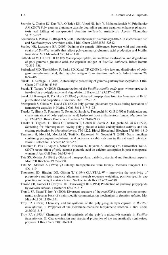
Scorpio A, Chabot DJ, Day WA, O’Brien DK, Vietri NJ, Itoh Y, Mohamadzadeh M, Friedlander
AM (2007) Poly-gamma-glutamate capsule-degrading enzyme treatment enhances phagocy-
tosis and killing of encapsulated Bacillus anthracis. Antimicrob Agents Chemother
51:215–222
Soutourina J, Plateau P, Blaquet S (2000) Metabolism of D-aminoacyl-tRNA in Escherichia coliand Saccharomyces cerevisiae cells. J Biol Chem 275:32535–32542
Stanley NR, Lazazzera BA (2005) Defining the genetic differences between wild and domestic
strains of Bacillus subtilis that affect poly-gamma-DL-glutamic acid production and biofilm
formation. Mol Microbiol 57:1143–1158
Sutherland MD, Kozel TR (2009) Macrophage uptake, intracellular localization, and degradation
of poly-gamma-D-glutamic acid, the capsular antigen of Bacillus anthracis. Infect Immun
77:532–538
Sutherland MD, Thorkildson P, Parks SD, Kozel TR (2008) In vivo fate and distribution of poly-
gamma-D-glutamic acid, the capsular antigen from Bacillus anthracis. Infect Immun 76:
899–906
Suzuki H, Kumagai H (2002) Autocatalytic processing of gamma-glutamyltranspeptidase. J Biol
Chem 277:43536–43543
Suzuki T, Tahara Y (2003) Characterization of the Bacillus subtilis ywtD gene, whose product is
involved in g-polyglutamic acid degradation. J Bacteriol 185:2379–2382
Suzuki H, Kumagai H, Tochikura T (1986) g-Glutamyltranspeptidase from Escherichia coli K-12:purification and properties. J Bacteriol 168:1325–1331
Szczepanek S, Cikala M, David CN (2002) Poly-gamma-glutamate synthesis during formation of
nematocyst capsules in Hydra. J Cell Sci 115:745–751
Tanaka T, Hiruta O, Futamura T, Uotani K, Satoh A, Taniguchi M, Oi S (1993a) Purification and
characterization of poly(g-glutamic acid) hydrolase from a filamentous fungus, Myrotheciumsp. TM-4222. Biosci Biotechnol Biochem 57:2148–2153
Tanaka T, Yaguchi T, Hiruta O, Futamura T, Uotani K, Satoh A, Taniguchi M, Oi S (1993b)
Screening for microorganism having poly(g-glutamic acid) endohydrolase activity and the
enzyme production by Myrothecium sp. TM-4222. Biosci Biotechnol Biochem 57:1809–1810
Tanimoto H, Mori M, Motoki M, Torii K, Kadowaki M, Noguchi T (2001) Natto mucilage
containing poly-gamma-glutamic acid increases soluble calcium in the rat small intestine.
Biosci Biotechnol Biochem 65:516–521
Tanimoto H, Fox T, Eagles J, Satoh H, Nozawa H, Okiyama A, Morinaga Y, Fairweather-Tait SJ
(2007) Acute effect of poly-gamma-glutamic acid on calcium absorption in post-menopausal
women. J Am Coll Nutr 26:645–649
Tate SS, Meister A (1981) g-Glutamyl transpeptidase: catalytic, structural and functional aspects.
Mol Cell Biochem 39:357–368
Tate SS, Meister A (1985) g-Glutamyl transpeptidase from kidney. Methods Enzymol 113:
400–419
Thompson JD, Higgins DG, Gibson TJ (1994) CLUSTAL-W – improving the sensitivity of
progressive multiple sequence alignment through sequence weighting, position-specific gap
penalties and weight matrix choice. Nucleic Acids Res 22:4673–4680
Throne CB, Gomez CG, Noyes HE, Housewright RD (1954) Production of glutamyl polypeptide
by Bacillus subtilis. J Bacteriol 68:307–315Tran L-SP, Nagai T, Itoh Y (2000) Divergent structure of the comQXPA quorum-sensing compo-
nents: molecular basis of strain-specific communication mechanism in Bacillus subtilis. Mol
Microbiol 37:1159–1171
Troy FA (1973a) Chemistry and biosynthesis of the poly(g-D-glutamyl) capsule in Bacilluslicheniformis. I. Properties of the membrane-mediated biosynthetic reaction. J Biol Chem
248:305–315
Troy FA (1973b) Chemistry and biosynthesis of the poly(g-D-glutamyl) capsule in Bacilluslicheniformis. II. Characterization and structural properties of the enzymatically synthesized
polymer. J Biol Chem 248:316–324
116 K. Kimura and Z. Fujimoto

Tsukahara K, Ogura M (2008) Characterization of DegU-dependent expression of bpr in Bacillussubtilis. FEMS Microbiol Lett 280:8–13
Uchida I, Makino S, Sasakawa C, Yoshikawa M, Sugimoto C, Terakado N (1993) Identification of
a novel gene, dep, associated with depolymerization of the capsule polymer in Bacillusanthracis. Mol Microbiol 9:487–496
Urushibata Y, Tokuyama S, Tahara Y (2002) Characterization of the Bacillus subtilis ywsC gene,
involved in g-polyglutamic acid production. J Bacteriol 184:337–343
Volcani BE, Margalith P (1957) A new species (Flavobacterium polyglutamicum) which hydro-
lyzes the gamma-L-glutamyl bond in polypeptides. J Bacteriol 74:646–655
Wang F, Ishiguro M, Mutsukado M, Fujita KI, Tanaka T (2008) Microstructure of poly (gamma-
glutamic acid) produced by Bacillus subtilis consisting of clusters of D- and L-glutamic acid
repeating units. J Agric Food Chem 56:4225–4228
Weber J (1990) Poly(g-glutamic acid)s are the major constituents of Nematocysts in Hydra(Hydrazoa, Cnidaria). J Biol Chem 265:9664–9669
Wu R, Richter S, Zhang R-G, Anderson VJ, Missiakas D, Joachimiak A (2009) Crystal structure of
Bacillus anthracis transpeptidase enzyme CapD. J Biol Chem doi/10.10784/jbc.M109.019034
Yang H, Zheng G, Peng X, Qiang B, Yuan J (2003) D-amino acids and D-Tyr-tRNATyr deacylase:
stereospecificity of the translation machine revisited. FEBS Lett 552:95–98
Yoshikawa T, Okada N, Oda A, Matsuo K, Mukai Y, Yoshioka Y, Akagi T, Akashi M, Nakagawa
S (2008) Development of amphiphilic gamma-PGA-nanoparticle based tumor vaccine: poten-
tial of the nanoparticulate cytosolic protein delivery carrier. Biochem Biophys Res Commun
366:408–413
Zanuy D, Aleman C (2001) Poly(gamma-glutamic acid) in aqueous solution: molecular dynamics
simulations of 10-and 20-residue chains at different temperatures. Biomacromolecules
2:651–657
Zanuy D, Aleman C, Munoz-Guerra S (1998) On the helical conformation of un-ionized poly
(gamma-D-glutamic acid). Int J Biol Macromol 23:175–184
Enzymatic Degradation of Poly-Gamma-Glutamic Acid 117

Pharmaceutical and Medical Applicationsof Poly-Gamma-Glutamic Acid
Takami Akagi, Michiya Matsusaki, and Mitsuru Akashi
Contents
1 Introduction . . . . . . . . . . . . . . . . . . . . . . . . . . . . . . . . . . . . . . . . . . . . . . . . . . . . . . . . . . . . . . . . . . . . . . . . . . . . . . . 120
2 Poly(g-Glutamic Acid)–Drug Conjugates . . . . . . . . . . . . . . . . . . . . . . . . . . . . . . . . . . . . . . . . . . . . . . . . . 120
2.1 g-PGA–Anticancer Drug Conjugates . . . . . . . . . . . . . . . . . . . . . . . . . . . . . . . . . . . . . . . . . . . . . . . 121
2.2 g-PGA–Antidiabetic Drug (Phloridzin) Conjugates . . . . . . . . . . . . . . . . . . . . . . . . . . . . . . . . 123
2.3 g-PGA–F(ab0) Antibody Conjugates . . . . . . . . . . . . . . . . . . . . . . . . . . . . . . . . . . . . . . . . . . . . . . . . 124
3 Self-Assembled Nanoparticles for Drug Delivery System . . . . . . . . . . . . . . . . . . . . . . . . . . . . . . . . 125
3.1 g-PGA-Graft–Phenylalanine Copolymer . . . . . . . . . . . . . . . . . . . . . . . . . . . . . . . . . . . . . . . . . . . . 126
3.2 g-PGA–Poly(lactide) Block Copolymer . . . . . . . . . . . . . . . . . . . . . . . . . . . . . . . . . . . . . . . . . . . . 132
3.3 g-PGA–Chitosan Complex . . . . . . . . . . . . . . . . . . . . . . . . . . . . . . . . . . . . . . . . . . . . . . . . . . . . . . . . . . 135
4 Hydrogels for Tissue Engineering . . . . . . . . . . . . . . . . . . . . . . . . . . . . . . . . . . . . . . . . . . . . . . . . . . . . . . . . 138
4.1 g-PGA–Sulfonate as a Heparionid Polymer . . . . . . . . . . . . . . . . . . . . . . . . . . . . . . . . . . . . . . . . 139
4.2 g-PGA–Sulfonate Hydrogels with FGF-2 Activity . . . . . . . . . . . . . . . . . . . . . . . . . . . . . . . . . 139
4.3 Hydrogel Template Approach for Tissue Engineering . . . . . . . . . . . . . . . . . . . . . . . . . . . . . . 141
5 Concluding Remarks and Future Perspectives . . . . . . . . . . . . . . . . . . . . . . . . . . . . . . . . . . . . . . . . . . . . 143
References . . . . . . . . . . . . . . . . . . . . . . . . . . . . . . . . . . . . . . . . . . . . . . . . . . . . . . . . . . . . . . . . . . . . . . . . . . . . . . . . . . . . 146
Abstract Poly(amino acid)s have received considerable attention for biomedical
applications. Poly(g-glutamic acid) (g-PGA), a natural polymer, is synthesized by
several gram-positive bacteria. g-PGA is anionic, water soluble, biodegradable,
edible, nontoxic, and nonimmunogenic for humans and the environment, and its
a-carboxylate side chains can be chemically modified to introduce various drugs, or
to modulate the amphiphilicity of the polymer. These features of g-PGA are very
useful for pharmaceutical and biomedical applications. This paper reviews the prepa-
ration of polymeric drugs, nanoparticles, and hydrogels composed of g-PGA and their
medical applications as drug carriers and tissue-engineering materials. g-PGA–drugconjugates, nanoparticles, and hydrogels fabricated from g-PGA or its derivatives
have wide application for drug delivery system and regenerative medical technique.
T. Akagi, M. Matsusaki, and M. Akashi (*)
Department of Applied Chemistry, Graduate School of Engineering, Osaka University, 2-1
Yamadaoka, Suita, Osaka 565-0871, Japan
e-mail: [email protected]
Y. Hamano (ed.), Amino-Acid Homopolymers Occurring in Nature,Microbiology Monographs 15, DOI 10.1007/978-3-642-12453-2_7,# Springer-Verlag Berlin Heidelberg 2010
119
1 Introduction
g-PGA is a naturally occurring poly(amino acid) that is synthesized by certain
strains of Bacillus (Kubota et al. 1993). The polymer is made of D- and L-glutamic
acid units linked through the a-amino and the g-carboxylic acid groups, respec-
tively, and its a-carboxylate side chains can be chemically modified to introduce
various bioactive ligands, or to modulate the overall function of the polymer (King
et al. 1998; Morillo et al. 2001; Prodhomme et al. 2003; Tachibana et al. 2003;
Shimokuri et al. 2004). With the exception of biologically synthesized g-PGA,chemically synthesized g-PGA has been reported. g-PGA cannot be synthesized by
polycondensation of glutamic acid, because intramolecular cyclization predominantly
proceeds to form a stable five-membered lactam, pyroglutamic acid. Moreover, it
is difficult to synthesize g-PGA from seven-membered glutamic acid g-N-carboxyanhydride (NCA), because the compound is so unstable that it easily releases CO2
to afford pyroglutamate without forming g-PGA (Sanda et al. 2001). To overcome
these problems, Endo et al. synthesized g-glutamic acid 16-mer a-ethyl ester in
stepwise transesterification of the ethyl ester group into benzyl ester group,
followed by hydrogenation afforded g-glutamic acid 16-mer with free carboxyl
groups (Sanda et al. 2002). It is expected that chemically synthesized nonrace-
mized g-PGA would show highly controlled properties and functions.
g-PGA is water soluble, biodegradable, edible, and nontoxic for humans and the
environment. Unlike general poly(amino acid)s, g-PGA has unique characteristics
on enzymatic degradation and immunogenicity. It has been reported that g-PGA is
resistance against many proteases because g-linked glutamic acids are not easily
recognized by common proteases (Oppermann et al. 1998; Obst and Steinbuchel
2004). Moreover, several studies have shown that g-PGA by itself is a poor
immunogen and does not induce booster responses, probably because of its simple
homopolymeric structure, similar to those of polysaccharides (Schneerson et al.
2003; Rhie et al. 2003; Wang et al. 2004; Joyce et al. 2006; Kubler-Kielb et al.
2006). Therefore, the potential applications of g-PGA and its derivatives have been
of interest in a broad range of fields, including the medicine, food, cosmetic, and
water treatment (Shih and Van 2001). In this review, we summarize the preparation
of multifunctional g-PGA and their pharmaceutical and biomedical applications as
drug carriers and tissue-engineering materials.
2 Poly(g-Glutamic Acid)–Drug Conjugates
Chemotherapy for cancers is usually limited by the toxicity of drugs to normal
tissues. The design of polymer–drug conjugates provides a synthetic approach that
can overcome some of the problems. Several synthetic polymer-based anticancer
drug conjugates have entered clinical studies (Maeda 2001; Li 2002; Li andWallace
2008). Unlike other synthetic polymers that have been tested in clinical studies,
g-PGA is unique in that it is composed of naturally occurring glutamic acid linked
120 T. Akagi et al.

together through amide bonds rather than a nondegradable C–C backbone. The free
carboxyl group in each repeating unit of glutamic acid provides functionality for
drug attachment. These features make g-PGA a promising candidate as a carrier of
polymer–drug conjugates for selective delivery of chemotherapeutic agents.
2.1 g-PGA–Anticancer Drug Conjugates
In the mid-1970s, Ringsdorf propounded a polymer–drug conjugate model that could
enhance the delivery of an anticancer drug to a tumor (Ringsdorf 1975). In this model,
it was predicted that when an anticancer drug is conjugated to a polymeric carrier, its
pharmacological properties could be manipulated by changing the physicochemical
properties of the polymer. For example, an insoluble drug can be made water-soluble
by introducing solubilizing moieties into the polymer. Likewise, active targeting is
possible if a targeting moiety is introduced into the polymer (Fig. 1). To date, several
synthetic polymers conjugated with anticancer drug have been successfully intro-
duced into clinical practice, including polystyrene– maleic anhydride copolymer
(SMA) (Maeda et al. 2001), N-(2-hydroxypropyl)-methacrylamide copolymer
(HPMA) (Minko et al. 2000), and poly(a-L-glutamic acid) (a-PGA) (Li et al. 1998)(Fig. 2). A related, but structurally different, a-PGA comprised of glutamic acid is
g-PGA. In this polymer, L-glutamic acid monomers are linked via amide bonds
between g-carboxyl and a-amino groups of adjacent monomers. The polymer is not
degraded by various proteases, but is cleaved to glutamic acid monomers under mild
acid, where a-PGA remains mostly intact (Weber 1990). Therefore, it is expected that
g-PGA has a unique function as polymer–drug conjugates.
Kishida et al. prepared a conjugate of g-PGA (1,660 kDa) with 5-fluorouracil
(5-FU) [g-poly(a-hydroxymethyl-5-fluorouracil-glutamate)] (g-PFUG) as a poly-
meric prodrug matrix (Fig. 3). 5-FU is a chemotherapeutic drug that is used widely
for the treatment of malignant cancers, and is usually the first choice of drug for the
treatment of hepatic cellular cancer. But its use has been limited by its systemic
toxicities, which have severe gastrointestinal toxicities, hematologic side effects,
Polymer chain
Drug
Spacer
Ligand of specificmolecular recognitionSolubilizer
Fig. 1 Basic concept of polymeric drug (Ringsdorf’s model). Model for targetable polymer–drug
conjugates according to Ringsdorf, in which a solubility enhancer, a pharmacokinetic modifier, a
homing device, and specific drugs could be attached to the same polymeric chain
Pharmaceutical and Medical Applications of Poly-Gamma-Glutamic Acid 121

and severe disturbance in bone marrow (Di Paolo et al. 2001). Moreover, 5-FU has
a serum half-life of only 15 min, further limiting its usefulness (Jin et al. 2005). In
order to maximize the therapeutic effect of 5-FU and minimize its adverse effects,
g-PFUG that can achieve controlled and sustained release and targeting of 5-FU
were made. A mixture of 1-, 3-, and 1,3-dihydroxymethyl-5-fluorouracil was intro-
duced to carboxyl group of g-PGA via ester bond. g-PFUG films were prepared by
casting from DMF. The g-PFUG matrix degraded in 7 days and incorporated 5-FU
was released in 3 days. 5-FU release was improved by mixing free 5-FU into
g-PFUG (Kishida et al. 1998a). Moreover, a slow release of 5-FU was achieved
using a g-PGA benzyl ester (g-PBG) prepared by esterification of g-PGA and
benzyl bromide as the matrix, which is more hydrophobic in nature. By selecting
adequate amount of g-PFUG or g-PBG, one can control the 5-FU release in vitrofrom a range in a few hours to over 20 days (Kishida et al. 1998b). g-PGA conjugated
( )( )C = O
nNH-CH-CH2-CH2-C
O
O
CH2
N
N
O
O
mNH-CH-CH2-CH2-C
O
COOH
F
Fig. 3 Chemical structure
of g-PGA-5-FU [g-poly(a-hydroxymethyl-5-
fluorouracil-glutamate)]
(g-PFUG) conjugate
( )( ) ( ) ( )
( ) ( ) ( )C O
NH
CH2 C m
C=O
NH
CH2
HC
CH3
OH
CH2 C n
C=O
CH3 CH3
GlyPheLeuGly
CH2
NH CH2 C m
O
CH2
COONa
CH2
NH CH2 C n
O
CH2
C=O
O
DrugDrug
c
a
b
CH2 CH CH CH CH CHx y z
C C
OO O
CO
HO
Drug
Fig. 2 Polymer–drug conjugates. Chemical structures of drug conjugated (a) polystyrene-maleic
anhydride copolymer (SMA), (b) N-(2-hydroxypropyl) methacrylamide copolymer (HPMA), and
(c) poly(a-L-glutamic acid) (a-PGA)
122 T. Akagi et al.

with a drug is useful as a biodegradable polymeric prodrug, and may be the basis for a
oral drug delivery system (DDS).
Cisplatin (cis-dichlorodiamineplatinum(II), CDDP) is one of the extensively
used chemotherapeutic agents for the treatment of different cancers such as testicu-
lar cancer, ovarian cancer, bladder cancer, lymphoma, and glioma. However, severe
side effects including acute nephrotoxicity and chronic neurotoxicity have limited
the clinical use of CDDP (Pinzani et al. 1994). To reduce these side effects and
enhance the anticancer activity, g-PGA–CDDP conjugate was prepared (Ye et al.
2006). CDDP was chemically conjugated to g-PGA (45–60 kDa) through the
displacement of CDDP chlorine atoms by hydrogen of carboxyl groups on g-PGA(Fig. 4). CDDP could be easily conjugated into g-PGA through a covalent bond.
The yield of CDDP incorporation into the g-PGA was 12.3%. The CDDP was
released in the initial 6 h in a burst manner, and thereafter in a sustained manner.
Accompanied by the CDDP release, g-PGA itself was dissociated into monomers,
allowing excretion from the body. The g-PGA–CDDP not only reduced the toxicity
of the CDDP but also produced desirable pharmacokinetics and enhanced antitumor
activity. a-PGA has been previously used as a carrier to conjugate camptothecin
(CPT) against resistant human lung cancer; this previous preparation involved
several chemical steps with a chemical component as the initial synthetic material
(Zou et al. 2001). g-PGA–CDDP has advantage that g-PGA can be obtained in large
quantities with high quality and high purity by biosynthesis methods without any
chemical modification steps. The PGA–CDDP conjugate was a promising com-
pound for avoiding the toxicity of platinous drugs, while retaining potent antitumor
activity.
2.2 g-PGA–Antidiabetic Drug (Phloridzin) Conjugates
Phloridzin, which is found in the bark and stems of apple trees, is known to inhibit
glucose transport competitively through the binding of the glucose moiety to the
Na+/glucose cotransporter (SGLT). Therefore, phloridzin is expected to be used for
antidiabetic drug. However, phloridzin has not been used as an oral antidiabetic
drug because toxic phloretin is released through the hydrolysis of a glucoside bond
( )( )C = O
nNH-CH-CH2-CH2-C
O
O
Pt
mNH-CH-CH2-CH2-C
O
COOH
NH3H3N
Cl
Pt
NH3H3N
ClCl
CDDP
Fig. 4 Chemical structure
of g-PGA–cisplatin (CDDP:
cis-dichlorodiamineplatinum
(II)) conjugate
Pharmaceutical and Medical Applications of Poly-Gamma-Glutamic Acid 123

of phloridzin by intestinal b-glucosidase (Tsujihara et al. 1996). This hydrolysis
also results in a reduction of phloridzin activity because of the lack of intramolecu-
lar glucose residues. Although low-molecular weight-phloridzin analogs and phlor-
etin derivatives have been examined, promising results have not been obtained yet
(Asano et al. 2004). To overcome this problem, Sakuma et al. designed and
prepared a novel g-PGA (58 kDa)–phloridzin conjugate, via a o-amino triethylene
glycol linker (Fig. 5) (Ikumi et al. 2008). The potential of g-PGA–phloridzinconjugate (PGA–PRZ) obtained as a novel oral antidiabetic drug was examined
by in vitro and in vivo experiments. A PGA–PRZ with a 15% phloridzin content
inhibited glucose transport from mucosal to serosal sides of the everted rat’s small
intestine, and its inhibitory effect was as strong as that of intact phloridzin. When
the PGA–PRZ was given orally to rats before glucose administration, the glucose-
induced hyperglycemic effect was significantly suppressed. On the other hand,
reduction of an increase in the blood glucose concentration was scarcely observed
when the PGA–PRZwas substituted with a double amount of intact phloridzin. This
difference in the biological activity between PGA–PRZ and intact phloridzin
resulted from the improved stability of a glucoside bond of phloridzin through the
conjugation with g-PGA (Sakuma et al. 2009). D-glucose, which is essential for the
inhibition of SGLT1, was not released from PGA–PRZ with a phloridzin content of
greater than 15%, even though an immediate release of D-glucose from intact
phloridzin was observed, and the toxic phloretin was not released from the conju-
gate. These results suggest that the g-PGA–phloridzin conjugate has potential as
oral antidiabetic drugs with high safety.
2.3 g-PGA–F(ab0) Antibody Conjugates
Several systems have been developed to restrict the delivery of the chemotherapeutic
agent to the tumor site. With the identification of cell-specific receptor/antigens on
tumor cells and tumor endothelial cells, it has been possible to actively target
chemotherapeutic or antiangiogenic agents using ligand- or antibody-bearing delivery
systems (Satchi-Fainaro et al. 2000). Targeting of cytotoxic drugs via comodification
of the drug to a water-soluble polymer with a cell-specific antibody is an attractive
( )( )C = O
nNH-CH-CH2-CH2-C
O
mNH-CH-CH2-CH2-C
O
COOH
OHOHO
OH
OHO
O OH
O
OH
OO
NH
Fig. 5 Chemical structure of
g-PGA–phloridzin(PGA–PRZ) conjugate.
Phloridzin was introduced via
a o-amino triethylene glycol
linker to g-PGA
124 T. Akagi et al.

approach for anticancer chemotherapy. Tumor-specific antibody F(ab0) fragments
have been successfully conjugated to HPMA with cytotoxic agents doxorubicin or
adriamycin to give cytotoxic conjugates (Jelinkova et al. 1998; Kunath et al. 2000).
Prodhomme et al. have established the feasibility of attaching bioactive mole-
cules and macromolecules such as cell-specific antibody F(ab0) fragments to g-PGA(Prodhomme et al. 2003). Using synthetic linkers, the a-carboxylate side chains ofg-PGA were conjugated to an exposed thiol side chain of an antibody F(ab0)fragment, a monoclonal rat IgG2a antibody (mAb) with activity against idiotypic
determinants carried by the IgM molecule of the BCL1 lymphoma. The most
successful linker was found to be maleimide linker, containing an ethylene glycol
spacer capable of hydrogen bonding with the aqueous solvent. The g-PGA–antibodyconjugate could retain its biological recognition properties, but showed reduced
binding efficacy and cell growth inhibition, compared with the free antibody. g-PGAwas also conjugated with the free amino group of glycopeptide antibiotic vancomy-
cin. The g-PGA–vancomycin conjugate showed slightly lower antibacterial activity
than free vancomycin. In this case, however, there is some indication of enhanced
biological activity against vancomycin-resistant strains, against which the free
ligand shows low affinity. The observation that the immobilized ligands retain
their biological activity suggests that g-PGA–antibody conjugates could in principlebe used to target cytotoxic drugs in the human body. g-PGA provide interesting
alternatives to synthesis polymers for antibody F(ab0) immobilization, which may
prove to have certain advantages for biocompatibility.
3 Self-Assembled Nanoparticles for Drug Delivery System
Nanoparticles prepared from synthetic or natural polymers have applications in
various technological and biomedical fields, because their chemical structures,
surface functionalities, and particle size can be easily controlled. Nanoparticles
with entrapped therapeutic agents, such as small molecules, peptides, proteins, and
DNA, have recently been shown to possess significant potential as DDS (Hans and
Lowman 2002; Allen and Cullis 2004; Torchilin 2006; Vasir and Labhasetwar
2007; Mundargi et al. 2008). Particulate delivery systems are concerned with the
systematic release of a pharmaceutical agent to maintain a therapeutic level of the
drug in the body for a sustained period of time. This may be achieved by incorpor-
ating the therapeutic agent into a degradable polymer particle, releasing the agent
continuously as the matrix degradation (Edlund and Albertsson 2000). DDS with
nanoparticles is one of the most promising because it may reduce unwanted toxic
side effects and improve the therapeutic effect.
Self-assembling polymer or block/graft copolymers that can form nanostructure
have been extensively investigated in the field of biotechnology and pharmaceu-
ticals. In general, hydrophobic interactions, electrostatic forces, hydrogen bonds,
van der Waal forces, or combinations of these interactions are available as the
driving forces for the formation of the polymer complexes (Kakizawa and Kataoka
Pharmaceutical and Medical Applications of Poly-Gamma-Glutamic Acid 125

2002; Zhang and Eisenberg 1995; Dou et al. 2003; Reihs et al. 2004; Kang et al.
2005). Numerous investigators have shown that the biological distribution of drugs,
proteins, and DNA can be modified, both at the cellular and organ levels, using
micro/nanoparticles delivery systems. Recently, many studies have focused on self-
assembled biodegradable nanoparticles for biomedical and pharmaceutical applica-
tions. In particular, poly(amino acid)s have received considerable attention for their
medical applications as potential polymeric drug carriers. Several amphiphilic
block and graft copolymers based on poly(amino acid) have been employed such
as poly(L-glutamic acid) (Holowka et al. 2005), poly(L-aspartic acid) (Arimura et al.
2005), poly(L-lysine) (Akiyoshi et al. 2000), poly(L-arginine) (Holowka et al.
2007), and poly(L-asparagine) (Jeong et al. 2003) as hydrophilic segments, and
poly(b-benzyl-L-aspartate) (Kataoka et al. 2000), poly(g-benzyl-L-glutamate) (Lin
et al. 2007a), and poly(L-histidine) (Lee et al. 2003) as hydrophobic segments.
Amphiphilic copolymers based on poly(amino acid) form micelles through self-
association in water. g-PGA or its derivatives have also been found to form self-
assembled, nano-sized particles, and are applied for biomedical fields.
3.1 g-PGA-Graft–Phenylalanine Copolymer
Nanoparticles fabricated by the self-assembly of amphiphilic block copolymers or
hydrophobically modified polymers have been explored as drug carrier systems
(Akagi et al. 2007a; Wang et al. 2007a).
In general, these amphiphilic copolymers consisting of hydrophilic and hydro-
phobic segments are capable of forming polymeric structures in aqueous solutions
via hydrophobic interactions. These self-assembled nanoparticles are composed of
an inner core of hydrophobic moieties and an outer shell of hydrophilic groups
(Gaucher et al. 2005; Letchford and Burt 2007).
Akashi et al. prepared nanoparticles composed of hydrophobically modified poly
(g-glutamic acid) (g-PGA) (Matsusaki et al. 2004; Kaneko et al. 2005). g-PGA as
the hydrophilic backbone and L-phenylalanine (Phe) as the hydrophobic segment
were synthesized by grafting Phe to g-PGA using water-soluble carbodiimide
(WSC) (Fig. 6). The g-PGA-graft–Phe copolymer (g-PGA–Phe) with more than
50% grafting degree formed monodispersed nanoparticles in water due to their
amphiphilic properties. To prepare nanoparticles, g-PGA–Phe dissolved in DMSO
was added to various concentration of NaCl solution, and then the resulting solu-
tions were dialyzed and freeze-dried. The g-PGA–Phe formed monodispersed
nanoparticles, and the particle size of the g-PGA–Phe nanoparticles could be easilycontrolled (30–200 nm) by changing NaCl concentration (Kim et al. 2009) (Fig. 7).
The nanoparticles showed a highly negative zeta potential (�25 mV) due to the
ionization of the carboxyl groups of g-PGA located near the surfaces. The specific
self-assembly behavior of g-PGA–Phe in aqueous solution was due to multiple
phenyl groups stacking. Beside the particle formation of g-PGA by using hydro-
phobic interaction, nanoparticles formed by complexation of g-PGA with bivalent
126 T. Akagi et al.

metal ion complex (Bodnar et al. 2008) or chemical cross-linking of carboxyl group
of g-PGA (Radu et al. 2008) have been reported.
The hydrolytic and enzymatic degradation of the g-PGA–Phe nanoparticles
in vitro was studied by gel permeation chromatography (GPC), scanning electron
microscopy (SEM), dynamic light scattering (DLS), and 1H-NMR measurements
(Akagi et al. 2005a, 2006a). The hydrolysis ratio of g-PGA and these hydrophobic
derivatives was found to decrease upon increasing the hydrophobicity of the g-PGAderivates. The degradation of the g-PGA backbone by g-glutamyl transpeptidase
(g-GTP) resulted in a dramatic change in nanoparticle morphology. With increasing
time, the g-PGA–Phe nanoparticles began to decrease in size and finally disap-
peared completely. The enzymatic degradation (pronase E, protease, cathepsin B,
and lipase) of the nanoparticles occurred via the hydrolysis of g-PGA as the main
chain, and Phe as the side chain. Sutherland et al. reported that g-PGA was
degraded in macrophages starting 4 h after uptake with continued degradation
occurring for at least 24 h (Sutherland et al. 2008; Sutherland and Kozel 2009).
Nanoparticles composed of biodegradable amphiphilic g-PGA with reactive func-
tion groups can undergo further modification, and are expected to have a variety of
potential pharmaceutical and biomedical applications, such as drug or vaccine
carriers.
The capture of water-soluble drugs such as proteins in the nanoparticle carrier
system can be carried out through various approaches. Double-emulsion solvent
evaporation/extraction is a common and convenient method for the encapsulation
of proteins into a polymer matrix (Quellec et al. 1998). However, the possible
denaturation of the proteins at the oil–water interface limits the usage of this
method. It has been reported that this interface causes conformational changes in
bovine serum albumin (BSA) (Sah 1999; Panyam et al. 2003). The prevention of
NH-CH-CH2-CH2-C( ) n
O
H2N-CH-C-O-CH2-CH3
OO
CH2L-phenylalanineethylester (Phe)
COOH
Poly(g-glutamic acid) (g-PGA)
NH-CH-CH2-CH2-C( ) l
O
NH-CH-CH2-CH2-C( ) m
O
C OCOOH
NHCH-COOCH2CH3
CH2
CH3CH3-CH2-N=C=N-(CH2)3-N g-PGA-graft-PheCH3
1-ethyl-3-(3-dimethylaminopropyl) carbodiimide(water-soluble carbodiimide, WSC)
Fig. 6 Synthesis of g-PGA-graft-Phe (g-PGA–Phe). g-PGA was hydrophobically modified by Phe
in the presence of WSC
Pharmaceutical and Medical Applications of Poly-Gamma-Glutamic Acid 127

protein denaturation and degradation, as well as high entrapment efficiency, would
be of particular importance in the preparation of nanoparticles containing water-
soluble drugs such as a protein. Therefore, this novel type of nanoparticle needs to
be developed.
Protein-loaded g-PGA–Phe nanoparticles were prepared by encapsulation, cova-lent immobilization, or physical adsorption methods (Fig. 8) to study their potential
applications as protein carriers (Akagi et al. 2005b, 2006b). To prepare the protein-
encapsulated g-Phe–Phe nanoparticles, 0.25–4 mg of protein [thyroglobulin, cata-
lase, concanavalin A (Con A), bovine serum albumin (BSA), ovalbumin (OVA),
peroxidase, b-lactoglobulin, myoglobin, lysozyme, a-lactalbumin, and cytochrome
c] was dissolved in 1 ml of saline, and 1 ml of the g-PGA–Phe (10 mg/ml in DMSO)
was added to the protein solutions. The resulting solutions were centrifuged and
repeatedly rinsed. The encapsulation of proteins of various molecular weights and
isoelectric points into the nanoparticles was successfully achieved. All proteins
used in this experiment were successfully encapsulated into the nanoparticles. The
34 92 139 192 nm
500 nm
0
5
10
15
20
1 10 100 1000
Particle size(nm)
Inte
nsity
(%
)
0M
0.05M
0.10M0.15M
34
92
139192 nm
c
a
b
Fig. 7 (a) Size changes of g-PGA–Phe nanoparticles prepared at various NaCl concentrations.
The size of nanoparticles was measured by dynamic light scattering (DLS). (b) Photographs ofg-PGA–Phe nanoparticles (2.5 mg/ml) dispersed in water. (c) Scanning electron microscope
(SEM) image of g-PGA–Phe nanoparticles prepared by 0.15 M NaCl solution
128 T. Akagi et al.

encapsulation efficiency was found to be in the range of 30–60% for most samples.
For all samples tested, it was observed that the encapsulation efficiency for a given
protein was not markedly influenced by the physical properties of that protein. The
size of the nanoparticles was increased when various proteins were encapsulated.
When OVA was encapsulated into the nanoparticles (90 mg/mg NP), the size of
nanoparticle increased from 180 to 256 nm. These results might be due to an
increase in the swelling capacity of the nanoparticles due to the hydrophilic proper-
ties of the protein. The OVA encapsulated into the nanoparticles was not released
(less than 10%) over the pH range of 4–8, even after 10 days. Moreover, it was
found that the g-PGA–Phe nanoparticles have some excellent properties, as fol-
lows. The enzyme-encapsulated nanoparticles showed high enzymatic activity.
In the case of protein-encapsulated nanoparticles prepared by the self-assembly of
g-PGA–Phe, the encapsulated protein may be more stable than via the emulsion
method. Proteins encapsulated into the nanoparticles appear to be adequate in terms
of the preservation of the protein structure. The g-PGA–Phe nanoparticles and
protein-encapsulated nanoparticles could be preserved by freeze-drying process.
The results of cytotoxicity tests showed that the nanoparticles did not cause any
( )NH-CH-CH2-CH2-C
COOH
n
O
Introductionof Phe
Self-assembly by hydrophobic interactions
: Phe
Preparation of protein-loaded nanoparticles
Encapsulation Surface immobilization Adsorption
Protein
g-PGA-Phenanoparticles
g-PGA-Phe
Poly(g-glutamic acid) (g-PGA)
Fig. 8 Preparation of protein-encapsulated, -immobilized, or -adsorbed g-PGA–Phe nanoparticles
Pharmaceutical and Medical Applications of Poly-Gamma-Glutamic Acid 129

relevant cell damage. Therefore, it is expected that the g-PGA–Phe nanoparticle
will have great potential as multifunctional carriers in pharmaceutical and biomed-
ical applications, such as drug and vaccine delivery systems. Also, Portilla-Arias
et al. reported the preparation of nanoparticles made of alkyl esters of g-PGA and
their potential application as drug and protein carrier (Portilla-Arias et al. 2009).
Polymeric nanoparticles with entrapped antigens represent an exciting approach
to control the release of vaccine antigens and to optimize the desired immune
response via selective targeting of the antigen to antigen-presenting cells (APCs)
(Panyam and Labhasetwar 2003; Jilek et al. 2007). The submicron size-range of
these delivery systems offers a number of distinct advantages over microparticles,
including relatively higher intracellular uptake compared with microparticles
(Harding and Song 1994; Nixon et al. 1996). Dendritic cells (DCs) are considered
to be initiators and modulators of immune responses and are capable of processing
antigens through both major histocompatibility complex (MHC) class I and II
pathways (Banchereau and Steinman 1998). Therefore, the delivery of therapeutic
macromolecules, such as proteins, peptides, and DNA, to DCs and the activation of
the antigen-presenting pathway are some of the most important issues in the
development of effective vaccines (Gamvrellis et al. 2004). Moreover, adjuvants
that possess the ability to induce of DC maturation are useful for DC-based
immunotherapy (Elamanchili et al. 2004).
Akagi et al. demonstrated the use of nanoparticles composed of amphiphilic poly
(amino acid) derivatives as vaccine delivery and adjuvants (Akagi et al. 2007b; Uto
et al. 2007, 2009a). To evaluate the uptake of OVA-encapsulated g-PGA–Phenanoparticles (OVA-NPs) by DCs, murine bone marrow-derived DCs were incu-
bated with 250-nm-sized OVA-NPs for 30 min at 37�C. The cells were then
analyzed by flow cytometry (FCM). OVA-NPs were efficiently taken up into DCs,
whereas the uptake of OVA alone was barely detectable at the same concentration of
OVA (Fig. 9). OVA-NPs were more efficiently taken up than OVA alone by DCs,
OVA OVA-NPs
Fluorescence intensity100
010
20
Cel
l Cou
nts
3040
50
010
2030
4050
101 102 103 104 100 101 102 103 104
Fig. 9 Uptake of OVA-encapsulated g-PGA–Phe nanoparticles (OVA-NPs) by dendritic cells
(DCs). Mouse bone marrow-derived DCs were incubated in the absence (gray histogram) or
presence (line histogram) of 250-nm-sized OVA-NPs or OVA alone (25 mg/ml) for 30 min at
37�C. The cells were then analyzed by flow cytometry (FCM)
130 T. Akagi et al.

and the uptake of OVA-NPs was inhibited at 4�C. These results suggest that OVA-NPs were phagocytosed mainly via endocytosis by the DCs. In the case of OVA
alone, an approximately 30-fold higher concentration was required to elicit a similar
amount of intracellular OVA as compared with OVA-NPs.
The maturation of DCs is associated with increased expression of several cell
surface markers, including the costimulatory molecules CD40, CD80, CD83, CD86,
MHC class I, and class II (Banchereau and Steinman 1998). The maturation of DCs
is deeply involved in their ability to process and present antigens to T cells. To
determine whether the uptake of g-PGA–Phe nanoparticles mediates the phenotypic
maturation of DCs, DCs were incubated with g-PGA–Phe nanoparticles for 48 h, andthe expression of surface molecules was measured by FCM. Upon exposure of these
DCs to the nanoparticles, the expression of CD86 andMHC class I was increased in a
dose-dependent manner. The expression levels of costimulatory molecules in NPs-
pulsed DCs were similar to those of lipopolysaccharide (LPS)-pulsed DCs. These
results suggest that g-PGA–Phe nanoparticles have great potential as adjuvant for DCmaturation. The mechanisms responsible for DC maturation by g-PGA–Phe nano-
particles are still unclear. However, it is hypothesized that not only the uptake
process of nanoparticles but also the characteristics of polymers consisting of
nanoparticles are important for the induction of DC maturation. In addition, soluble
g-PGA-induced innate immune responses in Toll-like receptor 4 (TLR4)-dependent
manner in DCs have been reported (Kim et al. 2007; Lee et al. 2009). Treatment with
high molecular weight g-PGA induced a significant upregulation of CD40, CD80,
and CD86 expression in wild-type DCs. In contrast, DCs from TLR4-defective mice
did not show an enhanced expression of maturation makers in response to the
2,000 kDa g-PGA treatment. It is suggested that the g-PGA–Phe nanoparticles alsoinduce DC maturation in a TLR4-dependent manner using the same 2,000 kDa
g-PGA, because g-PGA is located near the nanoparticle surfaces.
Induction and regulation of an adaptive immune response by vaccination is
possible for a broad range of infectious diseases or cancers. Vaccine delivery or
adjuvant that can induce antigen-specific humoral and cellular immunity is useful
for development of effective vaccine systems. Cellular immunity is required to
remove intracellular pathogens, while humoral immunity plays a central role in
neutralizing extracellular microorganisms. The efficacy of antigen (Ag)-encapsulated
g-PGA–Phe nanoparticles on the induction of Ag-specific cellular and humoral
immune responses was examined using OVA as a model Ag (Uto et al. 2007,
2009b). The immune responses were investigated in mice after subcutaneous
immunization with OVA-encapsulated g-PGA–Phe nanoparticles (OVA-NPs).
Ag-specific cytotoxic T lymphocyte (CTL) response was not observed in the spleen
cells obtained from the control (PBS) and OVA-immunized mice. In contrast, the
spleen cells obtained from the mice immunized with OVA-NPs showed a more
potent Ag-specific CTL response than those obtained from mice immunized with
OVA plus complete Freund’s adjuvant (CFA). When Ag-specific antibody
responses were examined and compared among the groups after immunization,
both OVA-NP- and OVA mixed with CFA (OVA + CFA)-immunized mice
showed significantly higher levels of OVA-specific total IgG, IgG1, and IgG2a
Pharmaceutical and Medical Applications of Poly-Gamma-Glutamic Acid 131

antibodies than OVA-immunized mice. These results indicate that the g-PGA–Phenanoparticles have the ability to prime cellular and humoral immunity by vaccina-
tion. It has been demonstrated that the g-PGA–Phe nanoparticles are also effective
for vaccines against human immunodeficiency virus (HIV) (Wang et al. 2007b,
2008), influenza virus (Okamoto et al. 2007, 2009), Japanese encephalitis virus
(Okamoto et al. 2008), human T-cell leukemia virus Type-I (HTLV-I) (Matsuo
et al. 2007), or cancers (Yoshikawa et al. 2008a, b). The antigen-loaded g-PGA–Phenanoparticles would provide an efficient antigen delivery and adjuvant system for
vaccination against viral infections or tumors (Fig. 10).
3.2 g-PGA–Poly(lactide) Block Copolymer
The amphiphilic self-assembled nanoparticles are composed of a hydrophobic inner
core and a hydrophilic outer shell. Core-forming hydrophobic block may affect the
drug loading capacity and its release kinetics as a result of hydrophobic interactions
between drugs and polymers. In contrast, the hydrophilic shell-forming block
determines surface properties of the nanoparticles and influences interactions
between the surrounding environments and the nanoparticles (Nam et al. 2003).
Biodegradable polymer, such as poly(lactide) (PLA), poly(e-caprolactone) (PCL),poly(b-benzyl L-aspartate) (PLBA), and poly(g-benzyl L-glutamate) (PLBG), has
been used mostly for the core-forming hydrophobic segment of nanoparticles. On
the other hand, poly(ethylene oxide) (PEO), a nontoxic and highly hydrated poly-
mer, has been used as the outer shell segment of nanoparticles because of its
superior biocompatibility (Lee et al. 2004).
Sung et al. synthesized amphiphilic block copolymers composed of g-PGA as
the hydrophilic segment and PLA as the hydrophobic segment to prepare a novel
type of self-assembled nanoparticles. Block copolymers composed of g-PGA(4 kDa) and PLA (10 kDa) (g-PGA–PLA) were synthesized using N,N-carbonyl-diimidazole (CDI) to activate the terminal hydroxyl group of PLA. g-PGA was
successfully conjugated to PLA by CDI (Fig. 11) (Liang et al. 2005). g-PGA–PLA
Induction of immune responsesAntigen-presenting cells
Uptake ofNPs
Degradation of NPs
Intracellular antigen release
Antigen-loadedg-PGA-Phe nanoparticles
CTL
Humoral Cellular
Antibody
Cytotoxic Tlymphocytes
(CTL)
Virus
Prevention Therapy
20 ~ 300 nm
Protein, Peptide
Activation
Fig. 10 Induction of immune responses by nanoparticle-based vaccine
132 T. Akagi et al.

nanoparticles were prepared using the emulsion/solvent evaporation technique.
The block copolymers (10 mg) were dissolved in 1 ml methylene chloride, then
vortexed and emulsified in 10 ml of a 0.1 wt.% sodium cholate solution using
a sonicator (cycles of 1 s sonication followed by 1 s of pauses, total time 5 min).
Afterward, the solvent was evaporated in a vacuum oven at 37�C for 1 h. The
nanoparticles were then recovered by centrifugation for 20 min at 20,000 � g at
4�C. Subsequently, the nanoparticles were resuspended by PBS (pH 7.4). The
prepared nanoparticles had a mean particle size of about 140 nm, and negative
surface charge with a zeta potential of about�20 mV due to the carboxyl groups on
the hydrophilic g-PGA shell. In the stability study, no aggregation or precipitation
of nanoparticles was observed during storage for up to 1 month, as a result of the
electrostatic repulsion between the negatively charged nanoparticles. For the poten-
tial of targeting liver cancer cells, the prepared g-PGA–PLA nanoparticles were
further conjugated with galactosamine. Galactosamine was conjugated to the
surface of nanoparticles via an amide linkage by N-(3-dimethylaminopropyl)-N0-ethylcarbodiimide (EDC) in the presence of N-hydroxysuccinimide (NHS).
Hepatocytes are known to recognize galactose and N-acetylgalactosamine-
terminated glycoproteins via the asialoglycoprotein (ASGP) receptors located on
their surfaces. It was reported that the ASGP receptors are also abundantly
expressed on the surfaces of various hepatoma cell lines (Fallon and Schwartz
1989). To assess the extents of internalization of the prepared galactosamine-
immobilized g-PGA–PLA nanoparticles (Gal-NPs) into HepG2 cells (a liver cancer
cell line), the Gal-NPs were labeled with rhodamine-123. With increasing the
galactosamine content conjugated on the nanoparticles, the uptake of Gal-NPs by
HepG2 cells increased significantly. In contrast, the Gal-NPs were not taken up by
Hs68 cells (without ASGP receptors). These results indicated that the galactosy-
lated nanoparticles had a specific interaction with HepG2 cells via ligand–receptor
recognition. The g-PGA–PLA nanoparticles conjugated with galactosamine may be
a potential DDS for targeted delivery to liver cancers.
Paclitaxel (Taxol) is one of the most active anticancer drugs used in cancer
chemotherapy. However, the use of paclitaxel is limited by its toxicity and limited
mNH-CH-CH2-CH2-C
O
CH3
( )( )n
O
C-CH-O
O
CC6H5-CH2-O OH
COOH
PLA g-PGA
Nanoparticles
PLAg-PGA
Fig. 11 Chemical structure of g-PGA–PLA block copolymer and the formation of self-assembled
g-PGA–PLA nanoparticles
Pharmaceutical and Medical Applications of Poly-Gamma-Glutamic Acid 133

aqueous solubility. Paclitaxel is highly hydrophobic and has a solubility of approx-
imately 1 mg/ml in aqueous solution at pH 7.4. It is currently formulated as a
concentrated solution paclitaxel/Cremophor EL/ethyl alcohol, but several toxic
effects have been attributed to Cremophor EL, including serious hypersensitivity
reactions. Therefore, numerous attempts have been made to develop drug formula-
tions with reduced systemic toxicity and an enhanced therapeutic index by using
water or other vehicles (Li et al. 2000). Paclitaxel-loaded g-PGA–PLA nanoparti-
cles were prepared to study the potential of targeting liver cancer cells (Liang et al.
2006a). The paclitaxel-loaded nanoparticles with various feed weight ratios of
paclitaxel to block copolymer (P/BC ratio) were produced using an emulsion/
solvent evaporation technique. Increasing the P/BC ratio significantly increased
the drug loading content of the prepared nanoparticles. In the case of P/BC ratio of
1/10, the particle size, zeta potential, drug loading content, and loading efficiency
was 129 nm, �20 mV, 5%, and 54%, respectively, and showed efficiently loading
efficiency. Moreover, the size of nanoparticles increased with increasing loading
content of drug. When the P/BC ratio was increased from 0/10 to 3/10, the size of
nanoparticles increased from 115 to 263 nm. The loaded paclitaxel was continu-
ously released from nanoparticles prepared with distinct P/BC ratios. All samples
released a burst of paclitaxel in the initial stage. An amount of 10–25% of the
loaded drug was released in the first hour. This may be due to some of the drugs that
were deposited near the g-PGA shell of the prepared nanoparticles. As the P/BC
ratio increased, the rate of release of paclitaxel from the prepared nanoparticles
decreased significantly. It was reported that a hydrophobic drug encapsulated
within nanoparticles partially crystallized at a higher drug loading content, while
it formed a molecular dispersion at a lower drug loading content (Gref et al. 1994).
For the potential of targeting liver cancer cells, galactosamine was further conju-
gated on the prepared paclitaxel-loaded g-PGA–PLA nanoparticles (Gal–P-NPs) as
a targeting moiety. It was found that the activity in inhibiting the growth of HepG2
cells (a liver cancer cell line) by the Gal–P-NPs was comparable to that of a
clinically available paclitaxel formulation (Phyxol). These results suggested that
the Gal–P-NPs are internalized into HepG2 cells via receptor-mediated endocyto-
sis, inhibiting the growth of cells. Moreover, the biodistribution of the prepared
Gal–P-NPs was studied in vivo in normal mice and hepatoma-tumor-bearing nude
mice (Liang et al. 2006b). The antitumor efficacy of the prepared nanoparticles in
hepatoma-tumor-bearing nude mice was also examined. It was found that the
groups injected with Phyxols, the paclitaxel-loaded nanoparticles or the Gal–P-NPs
significantly delayed the tumor growth as compared with the nontreated group.
Among all studied groups, the group injected with the Gal–P-NPs appeared to have
the most significant efficacy in the reduction of the size of the tumor. This is
because a large number of the Gal–P-NPs were observed at the tumor site, and
subsequently released their encapsulated paclitaxel to inhibit the growth of the
tumor. These results indicated that the Gal–P-NPs had a specific interaction with
the hepatoma tumor induced in nude mice via ligand–receptor recognition. There-
fore, the active targeting nature of the prepared Gal–P-NPs is considered to be used
as a potential DDS for the targeted delivery to liver cancers.
134 T. Akagi et al.

3.3 g-PGA–Chitosan Complex
Polymer complexes associated with two or more complementary polymers are
widely used in potential applications in the form of particles, hydrogels, films, and
membranes. In particular, a polyion complex (PIC) can be easily formed when
oppositely charged polyelectrolytes are mixed in aqueous solution and interact via
electrostatic (coulombic) interactions (Fig. 12). Nanoscaled structural materials (e.g.,
nanoparticles, micelles, nanogels, and hollow nanospheres) composed of PIC are
prepared by tuning the preparation conditions, such as the charge ratio of the anionic-
to-cationic polymers, temperature, concentration, and type of polyelectrolyte
(Kakizawa and Kataoka 2002; Muller et al. 2005; Hartig et al. 2007). PIC containing
g-PGA and chitosan (CT) as a cationic polymer has been applied for preparation of
nanoparticles, hydrogels, and films for biomedical applications. CT is a polysac-
charide constituted by N-glucosamine and N-acetyl-glucosamine units (Fig. 13), in
which the number ofN-glucosamine units exceeds 50%. CT is positively charged and
solubilized by protonation of its amino groups when the solution pH is below 6.
CT can be degraded into nontoxic products in vivo, and thus it has been widely usedin various biomedical applications (Hsieh et al. 2005; Kang et al. 2007).
Polyion complex(PIC) nanoparticles
NH3+
NH3+
NH3+
NH3+
NH3+
NH3+
NH3+
NH3+ NH3
+COO–
COO–
COO– COO–
COO–
COO–
COO– COO–
COO–
Anionicpolymer Electrostatic
interaction
Cationicpolymer
Fig. 12 Formation of polyion complex (PIC) nanoparticles composed of oppositely charged
polyelectrolytes via electrostatic (coulombic) interactions
O
OH
CH2OH
NHCOCH3
O
O
OH
NH2
O
CH2OH
n
Chitosan (b-1, 4-poly-D-gucosamine)
Fig. 13 Chemical structure
of chitosan (b-1, 4-poly-D-glucosamine)
Pharmaceutical and Medical Applications of Poly-Gamma-Glutamic Acid 135

Sung et al. investigated the PIC particle formation of g-PGA and CT by self-
assembly in aqueous media (Lin et al. 2005). Nanoparticles were obtained upon
addition of a g-PGA (160 kDa) aqueous solution (pH 7.4) into a low-molecular
weight CT (50 kDa) aqueous solution (pH 6.0) at varying concentrations under
magnetic stirring. Nanoparticles were collected by ultracentrifugation. The particle
sizes and the zeta potential of g-PGA and CT (g-PGA–CT) nanoparticles, preparedat varying concentrations of g-PGA and CT, were determined. It was found that the
particle size and the zeta potential of the prepared nanoparticles were mainly
determined by the relative amount of the local concentration of g-PGA in the
added solution to the surrounding concentration of CT. At a fixed concentration
of CT, an increase in the g-PGA concentration allowed g-PGA molecules interact-
ing with more CT molecules and thus formed a larger size of nanoparticles. When
the amount of CT molecules exceeded that of local g-PGA molecules, some of the
excessive CT molecules were located onto the g-PGA–CT nanoparticle surfaces.
Thus, the resulting nanoparticles may display a structure of a neutral PIC core
surrounded by a positively charged CT shell ensuring the colloidal stabilization.
These results clearly indicated that the size (from 80 to 400 nm) and surface charge
(from �35 to +25 mV) of g-PGA–CT nanoparticles could be easily controlled by
changing the mixing ratio of two polymers. Hajdu et al. also prepared g-PGA(1,200 kDa)–CT (320 kDa) nanoparticles (Hajdu et al. 2009). The size and size
distribution of the nanoparticles depend on the concentrations of g-PGA and CT
solutions and their ratio as well as on the pH of the mixture and the order of
addition. The particle size was in the range of 20–285 nmmeasured by transmission
electron microscopy (TEM), and the average hydrodynamic diameters were
between 150 and 330 nm.
However, the stability and characteristics of prepared PIC are influenced by
various factors involving their chemical compositions and their surrounding envi-
ronment. In particular, for PIC micelles or nanoparticles, the ionic strength and pH
of the solution is a key parameter for stability because of the shielding effect of the
ionic species on the electrostatic interactions (Jaturanpinyo et al. 2004). Therefore,
destabilization of PIC under physiological conditions limits their applications as a
drug carrier. In fact, in the case of g-PGA–CT nanoparticles, at pH 7.4, CT was
deprotonated and thus led to the collapse or aggregation of nanoparticles. The
prepared nanoparticles remained intact in the pH range of 2.5–6.6. To overcome
this problem, the stabilization of multi-ion-crosslinked nanoparticles composed
of g-PGA and CT blended with tripolyphosphate (TPP) and MgSO4. By adding
MgSO4 and TPP, the stability of nanoparticles at distinct pH values increased
significantly (Lin et al. 2008). The multi-ion-crosslinked nanoparticles had a
superior stability over a broader pH range than uncrosslinked nanoparticles.
An Oral route is the most convenient and comfortable means of administering
protein/peptide drugs such as insulin, because the pain caused by injection could be
avoided, leading to a higher patient compliance. However, it is well known that the
bioavailability of peptide and protein drugs after oral administration is very low
because of their instability in the gastrointestinal (GI) tract and low permeability
through the intestinal mucosa (Sakuma et al. 2001). Nanoparticles have been
136 T. Akagi et al.

studied extensively as carriers for oral drug delivery, whose purpose is to improve
the bioavailability of drugs with poor absorption characteristics (Damg et al. 1990;
Sakuma et al. 1997). To this end, a pH-responsive particulate delivery system that is
stable in the gastrointestinal (GI) tract and can swell significantly or degrade and
subsequently release the loaded drug when adhering and infiltrating into the mucus
of the intestinal tract is desired. Insulin-loaded PIC nanoparticles composed of
g-PGA and N-trimethyl chitosan (TMC) (g-PGA–TMC) were prepared for oral
insulin delivery via the paracellular pathway (Mi et al. 2008). The insulin solution
was premixed with an aqueous g-PGA. The mixed solution was then added into an
aqueous TMC under magnetic stirring at room temperature. The loading efficiency
and loading content of insulin in g-PGA–TMC nanoparticles were only 20.8%. To
increase the amount of insulin loaded, Mg2+ was blended in the premixed aqueous
insulin/g-PGA in the preparation of nanoparticles. In the presence of Mg2+, the
insulin loading efficiency (73.8%) in g-PGA–TMC nanoparticles increased signifi-
cantly due to cross-linkage between insulin and g-PGA via divalent metal ions. The
release profiles of insulin from the insulin-loaded g-PGA–TMC nanoparticles were
investigated at pH 2.5, 6.4, and 7.4, simulating the pH environments of the fasting
stomach, the duodenum, and the bloodstream, respectively. At pH 2.5, the amount
of insulin released from the nanoparticles was about 20% within the first hour. At
pH 6.4, there was only a minimal amount of insulin released from the nanoparticles.
At pH 7.4, the nanoparticles were significantly swelled and a sustained release
profile of insulin was observed. The release of insulin from the prepared nanopar-
ticles was mainly influenced by their pH-dependent swelling property.
The g-PGA–TMC, g-PGA–CT, and g-PGA–CT blended with TPP and MgSO4
nanoparticles with a positive surface charge (or shelled with TMC or CT) could
transiently open the tight junctions between Caco-2 cells in vitro and thus increasedthe paracellular permeability (Lin et al. 2005, 2007b, 2008; Mi et al. 2008). It has
been suggested that interactions of the positively charged amino groups of CT with
the negatively charged sites on cell surfaces and tight junctions induce a redistribu-
tion of F-actin and the tight junction’s protein. Moreover, the in vivo results clearlyindicated that the insulin-loaded nanoparticles could effectively reduce the blood
glucose level in a diabetic rat model (Sonaje et al. 2009). The results suggest that
g-PGA–CT nanoparticles may be a suitable carrier for transmucosal delivery of
insulin within the entire intestinal tract.
Gene delivery has great potential for the treatment of many different diseases. The
basic idea of gene therapy involves delivery of an exogenous gene into the cells to
express the encoded protein, which may be insufficiently or aberrantly expressed
naturally (Li and Huang 2006). DNA delivery is, however, a difficult process and a
suitable vector is required for efficient protection as well as release. Both viral and
nonviral vectors have been used for gene delivery. Nonviral gene delivery is used in
DNA condensation by cationic agents. Cationic polymers have been widely chosen to
condense DNA through electrostatic interactions between negatively charged DNA
and the positively charged cationic sites (Mann et al. 2008). g-PGA–CT nanoparticles
have been applied for DNA delivery system. CT/DNA complex nanoparticles have
been considered as a vector for gene delivery. Although advantageous for DNA
Pharmaceutical and Medical Applications of Poly-Gamma-Glutamic Acid 137

packing and protection from enzymatic degradation, CT-based complexes may lead
to difficulties in DNA release at the site of action. To improve the transfection
efficiency of CT/DNA complexes, g-PGA/CT/DNA conjugated nanoparticles were
prepared by an ionic-gelation method for transdermal DNA delivery using a low-
pressure gene gun (Lee et al. 2008). PlasmidDNAwasmixed with an aqueous g-PGA(20 kDa). Nanoparticles were obtained upon addition of the mixed solution into an
aqueous CT (80 kDa). The solutions were thoroughly mixed for 10–15 s, and then
nanoparticles were collected by centrifugation. The prepared g-PGA/CT/DNA nano-
particles were pH-sensitive and had a more compact internal structure with a greater
density than the conventional CT/DNA. The analysis of small angle X-ray scattering
(SAXS) results indicated that incorporating g-PGA would cause the formation of
compounded nanoparticles whose internal structure might facilitate the dissociation
of CT and DNA. As compared with CT/DNA, g-PGA/CT/DNA nanoparticles
improved their penetration depth into the mouse skin and enhanced gene expression.
Moreover, in addition to improving the release of DNA intracellularly, the incorpora-
tion of g-PGA in nanoparticles markedly increased their cellular internalization (Peng
et al. 2009). Taken together, g-PGA significantly enhanced the transfection efficiency
of this developed gene delivery system. The results indicated that g-PGA played
multiple important roles in enhancing the cellular uptake and transfection efficiency
of g-PGA/CT/DNA nanoparticles.
Kurosaki et al. also discovered a vector coated by g-PGA for effective and safe
gene delivery (Kurosaki et al. 2009). To develop a useful nonviral vector, PIC
constructed with pDNA, polyethylenimine (PEI), and various polyanions, such
as polyadenylic acid, polyinosinic–polycytidylic acid, a-polyaspartic acid, and
g-PGA, was prepared. The pDNA/PEI complex had a strong cationic surface charge
and showed extremely high transgene efficiency although it agglutinated with
erythrocytes and had extremely high cytotoxicity. The g-PGA could electrostati-
cally coat the pDNA/PEI complex to form stable anionic particles. The coating
of g-PGA dramatically decreased the toxicities of pDNA/PEI complex. Moreover,
the pDNA/PEI/g-PGA complex was highly taken by the cells via g-PGA-specificreceptor-mediated pathway and showed extremely high transgene efficiencies.
Further studies would be necessary to examine the detailed uptake mechanism
and clinical safety as gene delivery vector.
4 Hydrogels for Tissue Engineering
Since tissue-engineering concept was proposed in 1993 (Laner and Vacanti 1993),
many trials have been performed to develop biodegradable and biocompatible
scaffolds for regenerating tissues and delivery platform of incorporated drugs. A
polymeric scaffold is significant not only for keeping space and stiffness for cell
growth, differentiation, and organization, but also for supplying nutrients. In gen-
eral, poly(lactic acid) and poly(glycolic acid) are used for preparing the scaffold, but
they have a limitation such as low functionality, induction of inflammation during
138 T. Akagi et al.

hydrolysis, and hardness mismatch to the surrounding tissues. Recently, application
of g-PGA as a scaffold has attracted much attention in tissue-engineering field.
4.1 g-PGA–Sulfonate as a Heparionid Polymer
Heparin, highly sulfated glycosaminoglycan, is known to have many biological
activities, such as anticoagulant activity, antiviral activity, and plasma cleaning
activity. However, heparins sometimes give rise to side effects such as hemorrhage
(Leonard and Scribner 1969), thrombocytopenia (Kelton 1986), allergies, and lipid
metabolism (Deuber and Schultz 1991). Accordingly, sulfated naturally occurring
polysaccharides have been studied as a heparin-like polymer (heparinoid), such as
dextran (Mauzac and Jozefonvictz 1984), chitin (Nishimura and Tokura 1987), and
chitosan (Muzzarelli et al. 1984). However, conventional heparinoids derived from
naturally occurring polymers have a limitation to control sulfate content due to
the low chemical reactivity of the saccharic structure. Matsusaki et al. recently
synthesized sulfonated g-PGA (g-PGA-S) by condensation of amine group of
2-aminoethane-1-sulfonic acid (taurine) to carboxyl group of g-PGA (Matsusaki
et al. 2002). Sulfonate contents of g-PGA-S were easily controlled from 0 to 80%
by changing concentration of condensation reagents in feed, and the anticoagulant
activity increased with increasing the sulfonate content (Fig. 14).
Heparin is also known to have a stabilizing activity of growth factors as an
important biological activity (Fahama et al. 1996). Basic fibroblast growth factor
(FGF-2) activities of g-PGA-S were investigated in relation to the sulfonate con-
tent by chlorate-treated fibroblast cell culture test (Matsusaki et al. 2005a). The
chlorate-treated L929 fibroblasts that had suppressed biosynthesis of heparin sulfate
proteoglycans (Keller et al. 1989) were used to analyze the FGF-2 activity of g-PGA-S. When 72% of carboxyl groups in g-PGA were sulfonated (g-PGA-S72), cellnumber reached a maximum, suggesting higher FGF-2 activity. The activity of
g-PGA-S72 was higher than that of other heparinoids (Fig. 15). The g-PGA-S72also revealed higher FGF-2 protective effects in comparison to original g-PGA.Molecular modeling experiment demonstrated that electrostatic interactions can
take place between sulfonic and carboxylic acids of g-PGA-S and basic amino acid
residues of FGF-2. Therefore, the fairly high biological activities of g-PGA-S would
be useful to prepare biologically active scaffold for tissue engineering.
4.2 g-PGA–Sulfonate Hydrogels with FGF-2 Activity
Tissue engineering and regenerative medicine have been directed in the develop-
ment of next-generation medical technology, and three key factors – cells, growth
factors, and scaffolds – are important for tissue engineering. The injection of
growth factor solutions has been used to heal and regenerate tissues and organs.
Pharmaceutical and Medical Applications of Poly-Gamma-Glutamic Acid 139

However, it is difficult to directly apply bioactive proteins to tissue-engineering
processes because of their sensitivity to denaturation and degradation by external
environmental factors such as pH and enzyme (Cohen et al. 1991). The incorpora-
tion of various growth factors into scaffolds has been actively studied, but the
localized and stimulus-sensitive controlled release of growth factors from conven-
tional hydrogels has not been achieved yet due to the primary burst and deactivation
of the released growth factors. It is therefore desirable to develop hydrogels with
growth factor activity that can stably interact with the hydrogel over a long time,
and for the activity of the released growth factors to be maintained because the
hydrogel can protect them from environmental denaturation like proteoglycans
such as heparansulfate proteoglycan. Furthermore, a strategy for the stimulus-
sensitive delivery of growth factors to a target area is required for localized and
controlled release. Therefore, g-PGA–S hydrogels with FGF-2 activity as a plat-
form for stable incorporation of FGF-2 were prepared (Matsusaki et al. 2005b) and
pH-sensitive release of the incorporated FGF-2 was investigated (Matsusaki and
Akashi 2005). It was designed as a semi-interpenetrating polymer network (semi-
IPN)-like hetero gel (S72-netgel), consisting of g-PGA crosslinked network and
H NH CH
COOH
CH2 CH2 C
O
OHn
a
+ NH2 CH2 CH2 SO3H
taurineg-PGA
NHCH2CH2SO3H
H NH CH
COOH
CH2 CH2 C
O
nNH CH CH2 CH2 C
O
OHm
C O
g-PGA-S
b
0
2
4
6
8
10
12
14
Sulfonate content (%)
Clo
tting
tim
e (m
in)
glass 0 13 25 51 81
Fig. 14 Synthetic scheme of sulfonated g-PGA (g-PGA-S) (a) and effect of sulfonate content on
anticoagulant activity at 1 mg/ml (b) (Updated from Matsusaki et al. 2002)
140 T. Akagi et al.

incorporated g-PGA-S72 polymer chain, to maintain mobility and FGF-2 activity of
g-PGA-S72 (Fig. 16a). The swelling ratio and cell adhesion property of the S72-
netgels increased with increasing incorporation amount of g-PGA-S72. S72-netgelsincluding 36 mol% sulfonic acid (S72-netgel-36) could encapsulate 280 ng of FGF-
2 and retained about 60% of the FGF-2 even after 15 days of incubation in buffer,
probably due to the stable interaction of FGF-2 with g-PGA-S72 in the netgels. Therelease of FGF-2 from the S72-netgel-36 was successfully controlled by alternating
immersion in pH = 7.4 and acidic pH buffers such as pH = 6.0 and 4.0 because of
shrinkage of the gels at acidic pH condition corresponding to protonation of
carboxyl anion (Fig. 16b). Furthermore, the FGF-2 released from the hydrogels
retained its activity without denaturation. It has been reported that the ischemic,
inflamed, and tumor areas in the body have an acidic pH (<6.5) compared to the
surrounding tissues and blood (Na et al. 2004). These g-PGA-S scaffolds might be
useful as a pH-sensitive controlled delivery matrix for tissue engineering, such as
the neovascular treatment of ischemia and inflammation.
4.3 Hydrogel Template Approach for Tissue Engineering
The in vitro construction of engineered tissues composed of cells and three-
dimensional (3D) polymeric scaffolds that act as analogs to the natural extracellular
matrix (ECM) has been studied as a typical tissue-engineering approach (Lee and
Mooney 2001). However, polymeric scaffolds still have a risk of immunogenicity,
disease transmission, or inflammation during the hydrolysis process in the
0
2
4
6
8
10
Without polymer PVS
DSHeparin
PSS
Without polymer and FGF-2
g-PGA-S72
Cel
l num
ber
(×10
4 )
Serum (–) + FGF-2
Sample name
NS
Fig. 15 Comparison of
FGF-2 activity of g-PGA-S72to that of various heparinoids
(Updated from Matsusaki
et al. 2005a)
Pharmaceutical and Medical Applications of Poly-Gamma-Glutamic Acid 141

transplanted parts (Hubbell 2003). Since a scaffold is significant not only for
keeping space and stiffness for cell growth, differentiation, and organization, but
also for supplying nutrients, substitution of synthetic components of scaffold to
ECM components produced from cultured cells will be useful to avoid these risks.
Therefore, Matsusaki et al. designed hydrogel template approach for the in vitro
NH
H NH CH
COOH
CH2 CH2 C
O
nNH CH CH2 CH2 C
O
OHm
C O
CH2 CH2 SO3–
g-PGA-S72 interpenetrating polymer
S72-netgelNH CH CH2 CH2 C
COOH
OH
O
H NH CH
C
CH2 CH2 C
O
NH CH CH2 CH2 C
COOH
OH
O
mO
O
CH2CHHO
CH2O
CH2CH2OCH2CHHOCH2OC OCHNHH CH2 CH2 C
O
n m
n
g-PGA main network
a
b
FG
F-2
rel
ease
per
cent
age
(%)
Time (hour)
pH=4.0 pH=4.0 pH=4.0
0
20
40
60
80
100
0 10 20 30 40 50 60 70 80
S72-netgel-36
g-PGA hydrogel
pH=7.4
pH=4.0
Fig. 16 Schematic illustration of a semi-IPN-like hetero gel (S72-netgel) composed of g-PGA and
g-PGA-S72 (a) and pH-sensitive stepwise release of FGF-2 from S72-netgel-36 (b) (Updated fromMatsusaki and Akashi 2005)
142 T. Akagi et al.

construction of a large-sized 3D-engineered tissue composed of cultured cells and
ECM produced by the cells (Matsusaki et al. 2007). The disulfide-crosslinked
g-PGA hydrogels (g-PGA-SS gels) were prepared as a template hydrogel. The
fibroblast cells were 3D-cultured on/in the freeze-dried template gels for 10 days,
and the 3D-engineered tissues consisted of the cultured cells and ECM was suc-
cessfully obtained by the decomposition of the template with cysteine, because it is
well known that the disulfide-crosslinked hydrogels can be decomposed by cleav-
ing the disulfide linkage (–S–S–) to thiol groups (–HS SH–) with biocompatible
reductants such as glutathione and L-cysteine (Fig. 17). The obtained engineered
tissue was self-standing and highly dense composite of the cultured cells and
collagen produced by the cells. Furthermore, the usage of FGF-2 and ascorbic
acid can produce thicker and highly cell-incorporated engineered tissues (Yoshida
et al. 2010).
Since the disulfide-crosslinked template scaffold does not be limited to a hydro-
gel, a nanofiber matrix is also good candidate as the template scaffold. A water-
insoluble and reduction-responsive nonwoven scaffold was developed from g-PGA(Yoshida et al. 2009a). g-PGA nonwovens were crosslinked by cystamine with
disulfide bond, and these crosslinked fibers (g-PGA-SS fiber) were easily decom-
posed under physiological condition using reductant, L-cysteine. Furthermore, the
g-PGA-SS fiber revealed excellent cell adhesion and proliferation (Fig. 18). The
g-PGA-SS nonwovens can be applicable as a disulfide template matrix to this
template approach.
In tissues and organs, cells and ECMs are regularly arranged to form hierarchical
architectures such as the liver, skin, and blood vessels, and thus oriented 3D-
engineered tissues have attracted much attention in the material science field
(Hubbell 2003). To obtain 3D-engineered tissues consisting of oriented cells and
ECM, pore structure of g-PGA-SS gels by using silica cloth constructed by weaving
silica fiber of 9-mm-diameter was controlled (Yoshida et al. 2009b). Since silica
easily dissolves in hydrofluoric acid (HF) solution, porous structures derived from
silica templates can be formed in hydrogels by removing silica with HF (Suzuki
et al. 2000). The g-PGA-SS gels with aligned 2D porous structures (OP gels) were
successfully fabricated, and cell culture experiment revealed well alignment of the
adhered cells on/in the OP gels. Moreover, 3D-engineered tissues composed of
oriented cells and ECM produced by the cells could be obtained by decomposition
of the template gels (Fig. 19). This template approach with OP gels will be powerful
technique for in vitro construction of implantable tissue with oriented structures of
cells and ECM.
5 Concluding Remarks and Future Perspectives
g-PGA is a very promising biodegradable polymer that produces various strains of
Bacillus. Potential applications of g-PGA as thickener, cytoprotectant, humectant,
biological adhesive, flocculant, or heavy metal absorbent, etc. have been reported.
Pharmaceutical and Medical Applications of Poly-Gamma-Glutamic Acid 143

a
b
1 cm 1 cm
2 mm
3D-cell cultureDecomposition of
template gel
Cysteine
g-PGA-SS gel Cell-hydrogel hybrid 3D-engineered tissue
100 mm
SEM
50 mm 50 mm
Decompositionof template
g-PGA-SSgel(Template hydrogel)
Disulfide bond
Cell
3D-cell culture
Cell-hydrogel hybrid
Reductants
Cleaving ofdisulfide crosslinkage
3D-engineered tissue
ECM(Produced bycultured cells)
S-S S-S
S-S
S-S
S-S
S-S
SH HS
SH H
S
SH
HS
HS
SH
S-S
S-S
Fig. 17 Schematic representation of the 3D-engineered tissues fabricated by hydrogel template
approach (a). Photographs and SEM images of the template gel, the cell–gel hybrid, and the
3D-engineered tissues (b). The template gel was decomposed in medium containing 5 mM cysteine
at 37oC for 12 h (Updated from Matsusaki et al. 2007)
144 T. Akagi et al.
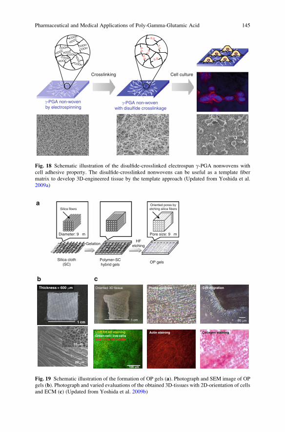
g-PGA non-wovenby electrospinning
Crosslinking
g-PGA non-wovenwith disulfide crosslinkage
Cell culture
S-S S-S
S-S
S-S
S-S
S-S
COOH
HOOC
COOH
COOH
COOH
COOH
COOH
COOHHOOC
HOOCHOOC
HO
OC
Fig. 18 Schematic illustration of the disulfide-crosslinked electrospun g-PGA nonwovens with
cell adhesive property. The disulfide-crosslinked nonwovens can be useful as a template fiber
matrix to develop 3D-engineered tissue by the template approach (Updated from Yoshida et al.
2009a)
a
cb
50 mm1 cm
Oriented 3D tissue Phase contrast Cell migration
LIVE/DEAD stainingGreen cell: live cellsRed cells: died cells
Collagen stainingActin staining
1 cm
20 mm
10 mm
Thickness = 600 mm
100 um
Oriented pores byetching silica fibersSilica fibers
Pore size: 9 µmDiameter: 9 µm
OP gelsPolymer-SChybrid gels
HFetchingGelation
Silica cloth(SC)
Fig. 19 Schematic illustration of the formation of OP gels (a). Photograph and SEM image of OP
gels (b). Photograph and varied evaluations of the obtained 3D-tissues with 2D-orientation of cellsand ECM (c) (Updated from Yoshida et al. 2009b)
Pharmaceutical and Medical Applications of Poly-Gamma-Glutamic Acid 145

This review describes the preparation of polymeric drugs, nanoparticles, and
hydrogels composed of g-PGA and their pharmaceutical and biomedical applica-
tions. The production of g-PGA has already been established on the industrial scale
because it can be produced easily and extracellularly in high yield by culturing of
bacteria in a fermenter. Moreover, the various molecular weight of g-PGA can be
obtained commercially. g-PGA by itself is shown to be weakly or nonimmunogenic
and safety. Biomaterials consisting of g-PGA described in this review are expected
to be introduced into clinical studies in the near future.
References
Akagi T, Higashi M, Kaneko T, Kida T, Akashi M (2005a) In vitro enzymatic degradation of
nanoparticles prepared from hydrophobically-modified poly(g-glutamic acid). Macromol
Biosci 5:598–602
Akagi T, Kaneko T, Kida T, Akashi M (2005b) Preparation and characterization of biodegradable
nanoparticles based on poly(g-glutamic acid) with L-phenylalanine as a protein carrier.
J Control Release 108:226–236
Akagi T, Higashi M, Kaneko T, Kida T, Akashi M (2006a) Hydrolytic and enzymatic degradation
of nanoparticles based on amphiphilic poly(g-glutamic acid)-graft-L-phenylalanine copolymer.
Biomacromolecules 7:297–303
Akagi T, Kaneko T, Kida T, Akashi M (2006b) Multifunctional conjugation of proteins on/into
core-shell type nanoparticles prepared by amphiphilic poly(g-glutamic acid). J Biomater Sci
Polym Ed 17:875–892
Akagi T, Baba M, Akashi M (2007a) Preparation of nanoparticles by the self-organization of
polymers consisting of hydrophobic and hydrophilic segments: potential applications. Polymer
48:6729–6747
Akagi T, Wang X, Uto T, Baba M, Akashi M (2007b) Protein direct delivery to dendritic cells using
nanoparticles based on amphiphilic poly(amino acid) derivatives. Biomaterials 28:3427–3436
Akiyoshi K, Ueminami A, Kurumada S, Nomura Y (2000) Self-association of cholesteryl-bearing
poly(L-lysine) in water and control of its secondary structure by host�guest interaction with
cyclodextrin. Macromolecules 33:6752–6756
Allen TM, Cullis PR (2004) Drug delivery systems: entering the mainstream. Science
303:1818–1822
Arimura H, Ohya Y, Ouchi T (2005) Formation of core-shell type biodegradable polymeric
micelles from amphiphilic poly(aspartic acid)-block-polylactide diblock copolymer. Bio-
macromolecules 6:720–725
Asano T, Anai M, Sakoda H, Fujishiro M, Ono H, Kurihara H, Uchijima Y (2004) SGLT as a
therapeutic target. Drugs Future 29:461–466
Banchereau J, Steinman RM (1998) Dendritic cells and the control of immunity. Nature
392:245–252
Bodnar M, Kjoniksen AL, Molnar RM, Hartmann JF, Daroczi L, Nystrom B, Borbely J (2008)
Nanoparticles formed by complexation of poly-g-glutamic acid with lead ions. J Hazard Mater
153:1185–1192
Cohen S, Yoshioka T, Lucarelli M, Hwang LH, Langer R (1991) Controlled delivery systems for
proteins based on poly(lactic/glycolic acid) microspheres. Pharm Res 8:713–720
Damg C, Michel C, Aprahamian M, Couvreur P, Devissagnet JP (1990) Nanocapsules as carriers
for oral peptide delivery. J Cantrol Release 13:233–239
Deuber HJ, Schultz W (1991) Reduced lipid concentration during four years of dialysis with low
molecular weight heparin. Kidney Int 40:496–500
146 T. Akagi et al.

Di Paolo A, Danesi R, Falcone A, Cionini L, Vannozzi F, Masi G, Allegrini G, Mini E, Bocci G,
Conte PF, Del Tacca M (2001) Relationship between 5-fluorouracil disposition, toxicity and
dihydropyrimidine dehydrogenase activity in cancer patients. Ann Oncol 12:1301–1306
Dou H, Jiang M, Peng H, Chen D, Hong Y (2003) pH-dependent self-assembly: micellization and
micelle-hollow-sphere transition of cellulose-based copolymers. Angew Chem Int Ed 42:
1516–1519
Edlund U, Albertsson AC (2000) Degradable polymer microspheres for controlled drug delivery.
Adv Polym Sci 157:67–112
Elamanchili P, Diwan M, CaoM, Samuel J (2004) Characterization of poly(D, L-lactic-co-glycolicacid) based nanoparticulate system for enhanced delivery of antigens to dendritic cells.
Vaccine 22:2406–2412
Fahama S, Hileman RE, Fromm JR, Linhardt RJ, Rees DC (1996) Heparin structure and interac-
tions with basic fibroblast growth factor. Science 271:1116–1120
Fallon RJ, Schwartz AL (1989) Receptor-mediated delivery of drugs to hepatocytes. Adv Drug
Deliv Rev 4:49–63
Gamvrellis A, Leong D, Hanley JC, Xiang SD, Mottram P, Plebanski M (2004) Vaccines that
facilitate antigen entry into dendritic cells. Immunol Cell Biol 82:506–516
Gaucher G, Dufresne MH, Sant VP, Kang N, Maysinger D, Leroux JC (2005) Block copolymer
micelles: preparation, characterization and application in drug delivery. J Control Release
109:169–188
Gref R, Minamitake Y, Peracchia MT, Trubetskoy V, Torchilin V, Langer R (1994) Biodegradable
long-circulating polymeric nanospheres. Science 263:1600–1603
Hajdu I, Bodnar M, Filipcsei G, Hartmann JF, Daroczi L, Zrinyi M, Borbely J (2009) Nanopar-
ticles prepared by self-assembly of chitosan and poly-g-glutamic acid. Colloid Polym Sci
286:343–350
Hans ML, Lowman AM (2002) Biodegradable nanoparticles for drug delivery and targeting. Curr
Opin Solid State Mater Sci 6:319–327
Harding CV, Song R (1994) Phagocytic processing of exogenous particulate antigens by macro-
phages for presentation by class I MHC molecules. J Immunol 153:4925–4933
Hartig SM, Greene RR, DasGupta J, Carlesso G, Dikov MM, Prokop A, Davidson JM (2007)
Multifunctional nanoparticulate polyelectrolyte complexes. Pharm Res 24:2353–2369
Holowka EP, Pochan DJ, Deming TJ (2005) Charged polypeptide vesicles with controllable
diameter. J Am Chem Soc 127:12423–12428
Holowka EP, Sun VZ, Kamei DT, Deming TJ (2007) Polyarginine segments in block copolypep-
tides drive both vesicular assembly and intracellular delivery. Nat Mater 6:52–57
Hsieh CY, Tsai SP, Wang DM, Chang YN, Hsieh HJ (2005) Preparation of g-PGA/chitosancomposite tissue engineering matrices. Biomaterials 26:5617–5623
Hubbell JA (2003) Materials as morphogenetic guides in tissue engineering. Curr Opin Biotechnol
14:551–558
Ikumi Y, Kida T, Sakuma S, Yamashita S, Akashi M (2008) Polymer-phloridzin conjugates as an
anti-diabetic drug that inhibits glucose absorption through the Na+/glucose cotransporter
(SGLT1) in the small intestine. J Control Release 125:42–49
Jaturanpinyo M, Harada A, Yuan X, Kataoka K (2004) Preparation of bionanoreactor based on
core-shell structured polyion complex micelles entrapping trypsin in the core cross-linked with
glutaraldehyde. Bioconjugate Chem 15:344–348
Jelinkova M, Strohalm J, Plocova D, Subr V, Stcastny M, Ulbrich K, Rihova B (1998) Targeting
of human and mouse T-lymphoctes by monoclonal antibody-HPMA copolymer-doxoru-
bicin conjugates directed against different T-cell surface antigens. J Control Release 52:
253–270
Jeong JH, Kang HS, Yang SR, Kim JD (2003) Polymer micelle-like aggregates of novel amphi-
philic biodegradable poly(asparagine) grafted with poly(caprolactone). Polymer 44:583–591
Jilek S, Merkle HP, Walter E (2007) DNA-loaded biodegradable microparticles as vaccine
delivery systems and their interaction with dendritic cells. Adv Drug Deliv Rev 57:377–390
Pharmaceutical and Medical Applications of Poly-Gamma-Glutamic Acid 147

Jin Y, Li J, Rong LF, Lu XW, Huang Y, Xu SY (2005) Pharmacokinetics and tissue distribution of
5-fluorouracil encapsulated by galactosylceramide liposomes in mice. Acta Pharmacol Sin
26:250–256
Joyce J, Cook J, Chabot D, Hepler R, Shoop W, Xu Q, Stambaugh T, Aste-Amezaga M, Wang S,
Indrawati L, Bruner M, Friedlander A, Keller P, Caulfield M (2006) Immunogenicity
and protective efficacy of Bacillus anthracis poly-g-D-glutamic acid capsule covalently cou-
pled to a protein carrier using a novel triazine-based conjugation strategy. J Biol Chem
281:4831–4843
Kakizawa Y, Kataoka K (2002) Block copolymer micelles for delivery of gene and related
compounds. Adv Drug Deliv Rev 54:203–222
Kaneko T, Higashi M, Matsusaki M, Akagi T, Akashi M (2005) Self-assembled soft nanofibrils of
amphipathic polypeptides and their morphological transformation. ChemMater 17:2484–2486
Kang N, Perron ME, Prudhomme RE, Zhang Y, Gaucher G, Leroux JC (2005) Stereocomplex
block copolymer micelles: core-shell nanostructures with enhanced stability. Nano Lett
5:315–319
Kang HS, Park SH, Lee YG, Son I (2007) Polyelectrolyte complex hydrogel composed of chitosan
and poly(g-glutamic acid) for biological application: preparation, physical properties, and
cytocompatibility. J Appl Polym Sci 103:386–394
Kataoka K, Matsumoto T, Yokoyama M, Okano T, Sakurai Y, Fukushima S, Okamoto K, Kwon
GS (2000) Doxorubicin-loaded poly(ethylene glycol)-poly(b-benzyl-L-aspartate) copolymer
micelles: their pharmaceutical characteristics and biological significance. J Control Release
64:143–153
Keller KM, Brauer PR, Keller JM (1989) Modulation of cell surface heparan sulfate structure by
growth of cells in the presence of chlorate. Biochemistry 28:8100–8107
Kelton JC (1986) Heparin-induced thrombocyopenia. Haemostasis 16:173–186
Kim TW, Lee TY, Bae HC, Hahm JH, Kim YH, Park C, Kang TH, Kim CJ, Sung MH, Poo H
(2007) Oral administration of high molecular mass poly-g-glutamate induces NK cell-
mediated antitumor immunity. J Immunol 179:775–780
Kim H, Akagi T, Akashi M (2009) Preparation of size tunable amphiphilic poly(amino acid)
nanoparticles. Macromol Biosci 9:842–848
King EC, Watkins WJ, Blacker AJ, Bugg TDH (1998) Covalent modification in aqueous solution
of poly-g-D-glutamic acid from Bacillus licheniformis. J Polym Sci A Polym Chem 36:
1995–1999
Kishida A, Goto H, Murakami K, Kakinoki K, Endo T, Akashi M (1998a) Polymer drugs and
polymeric drugs IX: Synthesis and 5-fluorouracil release profiles of biodegradable polymeric
prodrugs g-poly(a-hydroxymethyl-5-fluorouracil-glutamate). J Bioact Compat Polym 13:
222–233
Kishida A, Murakami K, Goto H, Kubota H, Endo T, Akashi M (1998b) Polymer drugs and
polymeric drugs X: Slow release of 5-fluorouracil from biodegradable poly(g-glutamic acid)
and its benzyl ester matrixes. J Bioact Compat Polym 13:270–278
Kubler-Kielb J, Liu TY, Mocca C, Majadly F, Robbins JB, Schneerson R (2006) Additional
conjugation methods and immunogenicity of Bacillus anthracis poly-g-D-glutamic acid-
protein conjugates. Infect Immun 74:4744–4749
Kubota H, Matsunobu T, Uotani K, Takebe H, Satoh A, Tanaka T, Taniguchi M (1993) Production
of poly(g-glutamic acid) by Bacillus subtilis F-2-01. Biosci Biotech Biochem 57:1212–1213
Kunath K, Kopeckova P, Minko T, Kopecek J (2000) HPMA copolymer-anticancer drug-
OV-TL16 antibody conjugates. 3. The effect of free and polymer-bound adriamycin on the
expression of some genes in the OVCAR-3 human ovarian carcinoma cell line. Eur J Pharm
Biopharm 49:11–15
Kurosaki T, Kitahara T, Fumoto S, Nishida K, Nakamura J, Niidome T, Kodama Y, Nakagawa H,
To H, Sasaki H (2009) Ternary complexes of pDNA, polyethylenimine, and g-polyglutamic
acid for gene delivery systems. Biomaterials 30:2846–2853
Laner R, Vacanti JP (1993) Tissue engineering. Science 260:920–926
148 T. Akagi et al.

Lee KY, Mooney DJ (2001) Hydrogel for tissue engineering. Chem Rev 101:1869–1879
Lee ES, Shin HJ, Na K, Bae YH (2003) Poly(L-histidine)-PEG block copolymer micelles and
pH-induced destabilization. J Control Release 90:363–374
Lee J, Cho EC, Cho K (2004) Incorporation and release behavior of hydrophobic drug in functiona-
lized poly(D, L-lactide)-block-poly(ethylene oxide) micelles. J Control Release 94:323–335
Lee PW, Peng SF, Su CJ, Mi FL, Chen HL, Wei MC, Lin HJ, Sung HW (2008) The use of
biodegradable polymeric nanoparticles in combination with a low-pressure gene gun for
transdermal DNA delivery. Biomaterials 29:742–751
Lee TY, Kim YH, Yoon SW, Choi JC, Yang JM, Kim CJ, Schiller JT, Sung MH, Poo HR (2009)
Oral administration of poly-g-glutamate induces TLR4- and dendritic cell-dependent antitu-
mor effect. Cancer Immunol Immunother 58:1781–1794
Leonard CD, Scribner BH (1969) Subdural hematoma in patients undergoing hemodialysis.
Lancet 2:239–249
Letchford K, Burt H (2007) A review of the formation and classification of amphiphilic block
copolymer nanoparticulate structures: micelles, nanospheres, nanocapsules and polymer-
somes. Eur J Pharm Biopharm 65:259–269
Li C (2002) Poly(L-glutamic acid)–anticancer drug conjugates. Adv Drug Deliv Rev 54:695–713
Li SD, Huang L (2006) Gene therapy progress and prospects: non-viral gene therapy by systemic
delivery. Gene Ther 13:1313–1319
Li C, Wallace S (2008) Polymer-drug conjugates: recent development in clinical oncology. Adv
Drug Delivery Rev 60:886–898
Li C, Yu DF, Newman RA, Cabral F, Stephens LC, Hunter N, Milas L, Wallace S (1998) Complete
regression of well-established tumors using a novel water-soluble poly(L-glutamic acid)-
paclitaxel conjugate. Cancer Res 58:2404–2409
Li C, Newman RA, Wu QP, Ke S, Chen W, Hutto T, Kan Z, Brannan MD, Charnsangavej C,
Wallace S (2000) Biodistribution of paclitaxel and poly(L-glutamic acid)-paclitaxel conjugate
in mice with ovarian OCa-1 tumor. Cancer Chemother Pharmacol 46:416–422
Liang HF, Yang TF, Huang CT, Chen MC, Sung HW (2005) Preparation of nanoparticles
composed of poly(g-glutamic acid)-poly(lactide) block copolymers and evaluation of their
uptake by HepG2 cells. J Control Release 105:213–225
Liang HF, Chen SC, Chen MC, Lee PW, Chen CT, Sung HW (2006a) Paclitaxel-loaded poly
(g-glutamic acid)-poly(lactide) nanoparticles as a targeted drug delivery system against
cultured HepG2 cells. Bioconjugate Chem 17:291–299
Liang HF, Chen CT, Chen SC, Kulkarni AR, Chiu YL, Chen MC, Sung HW (2006b) Paclitaxel-
loaded poly(g-glutamic acid)-poly(lactide) nanoparticles as a targeted drug delivery system for
the treatment of liver cancer. Biomaterials 27:2051–2059
Lin YH, Chung CK, Chen CT, Liang HF, Chen SC, Sung HW (2005) Preparation of nanoparticles
composed of chitosan/poly-g-glutamic acid and evaluation of their permeability through Caco-
2 cells. Biomacromolecules 6:1104–1112
Lin J, Zhang S, Chen T, Lin S, Jin H (2007a) Micelle formation and drug release behavior of
polypeptide graft copolymer and its mixture with polypeptide block copolymer. Int J Pharm
336:49–57
Lin YH, Mi FL, Chen CT, Chang WC, Peng SF, Liang HF, Sung HW (2007b) Preparation and
characterization of nanoparticles shelled with chitosan for oral insulin delivery. Biomacro-
molecules 8:146–152
Lin YH, Sonaje K, Lin KM, Juang JH, Mi FL, Yang HW, Sung HW (2008) Multi-ion-crosslinked
nanoparticles with pH-responsive characteristics for oral delivery of protein drugs. J Control
Release 132:141–149
Maeda H (2001) SMANCS and polymer-conjugated macromolecular drugs: advantages in cancer
chemotherapy. Adv Drug Delivery Rev 46:169–185
Maeda H, Sawa T, Konno T (2001) Mechanism of tumor-targeted delivery of macromolecular
drugs, including the EPR effect in solid tumor and clinical overview of the prototype polymeric
drug SMANCS. J Control Release 74:47–61
Pharmaceutical and Medical Applications of Poly-Gamma-Glutamic Acid 149
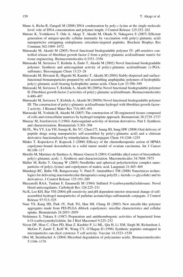
Mann A, Richa R, Ganguli M (2008) DNA condensation by poly-L-lysine at the single molecule
level: role of DNA concentration and polymer length. J Control Release 125:252–262
Matsuo K, Yoshikawa T, Oda A, Akagi T, Akashi M, Okada N, Nakagawa S (2007) Efficient
generation of antigen-specific cellular immunity by vaccination with poly(g-glutamic acid)
nanoparticles entrapping endoplasmic reticulum-targeted peptides. Biochem Biophys Res
Commun 362:1069–1072
Matsusaki M, Akashi M (2005) Novel functional biodegradable polymer IV: pH-sensitive con-
trolled release of fibroblast growth factor-2 from a poly(g-glutamic acid)sulfonate matrix for
tissue engineering. Biomacromolecules 6:3351–3356
Matsusaki M, Serizawa T, Kishida A, Endo T, Akashi M (2002) Novel functional biodegradable
polymer: Synthesis and anticoagulant activity of poly(g-glutamic acid)sulfonate (g-PGA-sulfonate). Bioconjugate Chem 13:23–28
Matsusaki M, Hiwatari K, Higashi M, Kaneko T, Akashi M (2004) Stably-dispersed and surface-
functional bionanoparticles prepared by self-assembling amphipathic polymers of hydrophilic
poly(g-glutamic acid) bearing hydrophobic amino acids. Chem Lett 33:398–399
Matsusaki M, Serizawa T, Kishida A, Akashi M (2005a) Novel functional biodegradable polymer
II: Fibroblast growth factor-2 activities of poly(g-glutamic acid)sulfonate. Biomacromolecules
6:400–407
Matsusaki M, Serizawa T, Kishida A, Akashi M (2005b) Novel functional biodegradable polymer
III: The construction of poly(g-glutamic acid)sulfonate hydrogel with fibroblast growth factor-
2 activity. J Biomed Mater Res 73A:485–491
Matsusaki M, Yoshida H, Akashi M (2007) The construction of 3D-engineered tissues composed
of cells and extracellular matrices by hydrogel template approach. Biomaterials 28:2729–2737
Mauzac M, Jozefonvictz J (1984) Anticoagulant activitiy of dextran derivatives. Part I: Synthesis
and characterization. Biomaterials 5:301–304
Mi FL, Wu YY, Lin YH, Sonaje K, Ho YC, Chen CT, Juang JH, Sung HW (2008) Oral delivery of
peptide drugs using nanoparticles self-assembled by poly(g-glutamic acid) and a chitosan
derivative functionalized by trimethylation. Bioconjugate Chem 19:1248–1255
Minko T, Kopeckova P, Kopecek J (2000) Efficacy of the chemotherapeutic action of HPMA
copolymer-bound doxorubicin in a solid tumor model of ovarian carcinoma. Int J Cancer
86:108–117
Morillo M, Martinez de Ilarduya A, Munoz-Guerra S (2001) Comblike alkyl esters of biosynthetic
poly(g-glutamic acid). 1. Synthesis and characterization. Macromolecules 34:7868–7875
Muller M, Reihs T, Ouyang W (2005) Needlelike and spherical polyelectrolyte complex nano-
particles of poly(L-lysine) and copolymers of maleic acid. Langmuir 21:465–469
Mundargi RC, Babu VR, Rangaswamy V, Patel P, Aminabhavi TM (2008) Nano/micro techno-
logies for delivering macromolecular therapeutics using poly(D, L-lactide-co-glycolide) and itsderivatives. J Control Release 125:193–209
Muzzarelli RAA, Tanfani F, Emanuelli M (1984) Sulfated N-(carboxymethyl)chitosans: Novel
blood anticoagulants. Carbohydr Res 126:225–231
Na K, Lee KH, Bae YH (2004) pH-sensitivity and pH-dependent interior structural change of self-
assembled hydrogel nanoparticles of pullulan acetate/oligo-sulfonamide conjugate. J Control
Release 97:513–525
Nam YS, Kang HS, Park JY, Park TG, Han SH, Chang IS (2003) New micelle-like polymer
aggregates made from PEI-PLGA diblock copolymers: micellar characteristics and cellular
uptake. Biomaterials 24:2053–2059
Nishimura S, Tokura S (1987) Preparation and antithrombogenic activities of heparinoid from
6-O-(carboxymethyl)chitin. Int J Biol Macromol 9:225–232
Nixon DF, Hioe C, Chen PD, Bian Z, Kuebler P, Li ML, Qiu H, Li XM, Singh M, Richardson J,
McGee P, Zamb T, Koff W, Wang CY, O’Hagan D (1996) Synthetic peptides entrapped in
microparticles can elicit cytotoxic T cell activity. Vaccine 14:1523–1530
Obst M, Steinbuchel A (2004) Microbial degradation of poly(amino acid)s. Biomacromolecules
5:1166–1176
150 T. Akagi et al.

Okamoto S, Yoshii H, Akagi T, Akashi M, Ishikawa T, Okuno Y, Takahashi M, Yamanishi K,
Mori Y (2007) Influenza hemagglutinin vaccine with poly(g-glutamic acid) nanoparticles
enhances the protection against influenza virus infection through both humoral and cell-
mediated immunity. Vaccine 25:8270–8278
Okamoto S, Yoshii H, Ishikawa T, Akagi T, Akashi M, Takahashi M, Yamanishi K, Mori Y (2008)
Single dose of inactivated Japanese encephalitis vaccine with poly(g-glutamic acid) nanopar-
ticles provides effective protection from Japanese encephalitis virus. Vaccine 26:589–594
Okamoto S, Matsuura M, Akagi T, Akashi M, Tanimoto T, Ishikawa T, Takahashi M, Yamanishi K,
Mori Y (2009) Poly(g-glutamic acid) nano-particles combined with mucosal influenza
virus hemagglutinin vaccine protects against influenza virus infection in mice. Vaccine 27:
5896–5905
Oppermann FB, Fickaitz S, Steinbiichel A (1998) Biodegradation of polyamides. Polym Degrad
Stab 59:337–344
Panyam J, Labhasetwar V (2003) Biodegradable nanoparticles for drug and gene delivery to cells
and tissue. Adv Drug Deliv Rev 55:329–347
Panyam J, Dali MM, Sahoo SK, Ma W, Chakravarthi SS, Amidon GL, Levy RJ, Labhasetwar
V (2003) Polymer degradation and in vitro release of a model protein from poly(D, L-lactide-
co-glycolide) nano- and microparticles. J Control Release 92:173–187
Peng SF, Yang MJ, Su CJ, Chen HL, Lee PW, Wei MC, Sung HW (2009) Effects of incorporation
of poly(g-glutamic acid) in chitosan/DNA complex nanoparticles on cellular uptake and
transfection efficiency. Biomaterials 30:1797–1808
Pinzani V, Bressolle F, Haug IJ, Galtier M, Blayac JP, Balmes P (1994) Cisplatin-induced
renal toxicity and toxicity-modulating strategies: a review. Cancer Chemother Pharmacol
35:1–9
Portilla-Arias JA, Camargo B, Garcia-Alvarez M, Martinez de Ilarduya A, Munoz-Guerra S
(2009) Nanoparticles made of microbial poly(g-glutamate)s for encapsulation and delivery
of drugs and proteins. J Biomater Sci Polym Ed 20:1065–1079
Prodhomme EJ, Tutt AL, Glennie MJ, Bugg TD (2003) Multivalent conjugates of poly-g-D-glutamic acid from Bacillus licheniformis with antibody F(ab0) and glycopeptide ligands.
Bioconjugate Chem 14:1148–1155
Quellec P, Gref R, Perrin L, Dellacherie E, Sommer F, Verbavatz JM, Alonso MJ (1998) Protein
encapsulation within polyethylene glycol-coated nanospheres. I. Physicochemical characteri-
zation. J Biomed Mater Res 42:45–54
Radu JEF, Novak L, Hartmann JF, Beheshti N, Kjoniksen AL, Nystrom B, Borbely J (2008)
Structural and dynamical characterization of poly-g-glutamic acid-based cross-linked nano-
particles. Colloid Polym Sci 286:365–376
Reihs T, Muller M, Lunkwitz K (2004) Preparation and adsorption of refined polyelectrolyte
complex nanoparticles. J Colloid Interface Sci 271:69–79
Rhie GE, Roehrl MH, Mourez M, Collier RJ, Mekalanos JJ, Wang JY (2003) A dually active
anthrax vaccine that confers protection against both bacilli and toxins. Proc Natl Acad Sci USA
100:10925–10930
Ringsdorf H (1975) Structure and properties of pharmacologically active polymers. J Polym Sci
Symp 51:135–153
Sah H (1999) Stabilization of proteins against methylene chloride / water interface induced
denaturation and aggregation. J Control Release 58:143–151
Sakuma S, Suzuki N, Kikuchi H, Hiwatari K, Arikawa K, Kishida A, Akashi M (1997) Oral
peptide delivery using nanoparticles composed of novel graft copolymers having hydrophobic
backbone and hydrophilic branches. Int J Pharm 149:93–106
Sakuma S, Hayashi M, Akashi M (2001) Design of nanoparticles composed of graft copolymers
for oral peptide delivery. Adv Drug Deliv Rev 47:21–37
Sakuma S, Sagawa T,Masaoka Y, KataokaM, Yamashita S, Shirasaka Y, Tamai I, Ikumi Y, Kida T,
Akashi M (2009) Stabilization of enzyme-susceptible glucoside bonds of phloridzin through
conjugation with poly(g-glutamic acid). J Control Release 133:125–131
Pharmaceutical and Medical Applications of Poly-Gamma-Glutamic Acid 151

Sanda F, Fujiyama T, Endo T (2001) Chemical synthesis of poly-g-glutamic acid by polyconden-
sation of g-glutamic acid dimer: synthesis and reaction of poly-g-glutamic acid methyl ester.
J Polym Sci A Polym Chem 39:732–741
Sanda F, Fujiyama T, Endo T (2002) Stepwise synthesis of g-glutamic acid 16-mer. Macromol
Chem Phys 203:727–734
Satchi-Fainaro R, Duncan R, Barnes CM (2000) Polymer therapeutics for cancer: current status
and future challenges. Adv Polym Sci 193:1–65
Schneerson R, Kubler-Kielb J, Liu TY, Dai ZD, Leppla SH, Yergey A, Backlund P, Shiloach J,
Majadly F, Robbins JB (2003) Poly(g-D-glutamic acid) protein conjugates induce IgG anti-
bodies in mice to the capsule of Bacillus anthracis: a potential addition to the anthrax vaccine.Proc Natl Acad Sci USA 100:8945–8950
Shih IL, Van YT (2001) The production of poly(g-glutamic acid) from microorganisms and its
various application. Bioresource Technol 79:207–225
Shimokuri T, Kaneko T, Serizawa T, Akashi M (2004) Preparation and thermosensitivity of
naturally occurring polypeptide poly(g-glutamic acid) derivatives modified by alkyl groups.
Macromol Biosci 4:407–411
Sonaje K, Lin YH, Juang JH, Wey SP, Chen CT, Sung HW (2009) In vivo evaluation of safety
and efficacy of self-assembled nanoparticles for oral insulin delivery. Biomaterials 30:
2329–2339
Sutherland MD, Kozel TR (2009) Macrophage uptake, intracellular localization, and degradation
of poly-g-D-glutamic acid, the capsular antigen of Bacillus anthracis. Infect Immun 77:
532–538
Sutherland MD, Thorkildson P, Parks SD, Kozel TR (2008) In vivo fate and distribution of poly-
g-D-glutamic acid, the capsular antigen from bacillus anthracis. Infect Immun 76:899–906
Suzuki K, Yumura T, Tanaka Y, Serizawa T, Akashi M (2000) Interpenetrating inorganic-organic
hybrid gels: Preparation of hybrid and replica gels. Chem Lett 29:1380–1381
Tachibana Y, Kurisawa M, Uyama H, Kobayashi S (2003) Thermo- and pH-responsive biode-
gradable poly(a-N-substituted g-glutamine)s. Biomacromolecules 4:1132–1134
Torchilin VP (2006) Multifunctional nanocarriers. Adv Drug Delivery Rev 58:1532–1555
Tsujihara K, Hongu M, Saito K, Inamasu M, Arakawa K, Oku A, Matsumoto M (1996) Na+-
glucose cotransporter inhibitors as antidiabetics. I. synthesis and pharmacological properties of
4’-dehydroxyphlorizin derivatives based on a new concept. Chem Pharm Bull 44:1174–1180
Uto T, Wang X, Sato K, Haraguchi M, Akagi T, Akashi M, Baba M (2007) Targeting of antigen to
dendritic cells with poly(g-glutamic acid) nanoparticles induce antigen-specific humoral and
cellular immunity. J Immunol 178:2979–2986
Uto T, Akagi T, Hamasaki T, Akashi M, Baba M (2009a) Modulation of innate and adaptive
immunity by biodegradable nanoparticles. Immunol Lett 125:46–52
Uto T, Wang X, Akagi T, Zenkyu R, Akashi M, Baba M (2009b) Improvement of adaptive
immunity by antigen-carrying biodegradable nanoparticles. Biochem Biophys Res Commun
379:600–604
Vasir JK, Labhasetwar V (2007) Biodegradable nanoparticles for cytosolic delivery of therapeu-
tics. Adv Drug Delivry Rev 59:718–728
Wang TT, Fellows PF, Leighton TJ, Lucas AH (2004) Induction of opsonic antibodies to the g-D-glutamic acid capsule of Bacillus anthracis by immunization with a synthetic peptide-carrier
protein conjugate. FEMS Immunol Med Microbiol 40:231–237
Wang X, Akagi T, Akashi M, Baba M (2007a) Development of core-corona type polymeric
nanoparticles as an anti-HIV-1 vaccine. Mini-Rev Org Chem 4:281–290
Wang X, Uto T, Akagi T, Akashi M, Baba M (2007b) Induction of potent CD8+ T-cell responses
by novel biodegradable nanoparticles carrying human immunodeficiency virus type 1 gp120.
J Virol 81:10009–10016
Wang X, Uto T, Akagi T, Akashi M, Baba M (2008) Poly(g-glutamic Acid) nanoparticles as an
efficient antigen delivery and adjuvant system: potential for an anti-AIDS vaccine. J Med Virol
80:11–19
152 T. Akagi et al.

Weber J (1990) Poly(g-glutamic acid)s are the major constituents of nematocysts in Hydra
(Hydrozoa, Cnidaria). J Biol Chem 265:9664–9669
Ye H, Jin L, Hu R, Yi Z, Li J, Wu Y, Xi X, Wu Z (2006) Poly(g, L-glutamic acid)-cisplatin
conjugate effectively inhibits human breast tumor xenografted in nude mice. Biomaterials
27:5958–5965
Yoshida H, Klinkhammer K, Matsusaki M, Moller M, Klee D, Akashi M (2009a) Disulfide-
crosslinked electrospun poly(g-glutamic acid) nonwovens as reduction-responsive scaffolds.
Macromol Biosci 9:568–574
Yoshida H, Matsusaki M, Akashi M (2009b) Scaffold-mediated 2D cellular orientations for
construction of three dimensionally engineered tissues composed of oriented cells and extra-
cellular matrices. Adv Funct Mater 19:1001–1007
Yoshida H, Matsusaki M, Akashi M (2010) Development of thick and highly cell-incorporated
engineered tissues by hydrogel template approach with basic fibroblast growth factor or
ascorbic acid. J Biomater Sci Polymer Ed 21:415– 428
Yoshikawa T, Okada N, Oda A, Matsuo K, Matsuo MY, Yoshioka Y, Akagi T, Akashi M,
Nakagawa S (2008a) Development of amphiphilic g-PGA-nanoparticle based tumor vaccine:
Potential of the nanoparticulate cytosolic protein delivery carrier. Biochem Biophys Res
Commun 366:408–413
Yoshikawa T, Okada N, Oda A, Matsuo K, Matsuo K, Kayamuro H, Ishii Y, Yoshinaga T, Akagi
T, Akashi M, Nakagawa S (2008b) Nanoparticles built by self-assembly of am phiphilic poly
(g-glutamic acid) can deliver antigens to antigen-presenting cells with high efficiency: A new
tumor-vaccine carrier for eliciting effector T cells. Vaccine 26:1303–1313
Zhang L, Eisenberg A (1995) Multiple morphologies of crew-cut aggregates of polystyrene-b-poly(acrylic acid) block copolymers. Science 1268:1728–1731
Zou Y, Wu QP, Tansey W, Chow D, Hung MC, Charnsangavej C, Wallace S, Li C (2001)
Effectiveness of water soluble poly(L-glutamic acid)–camptothecin conjugate against resistant
human lung cancer xenografted in nude mice. Int J Oncol 18:331–336
Pharmaceutical and Medical Applications of Poly-Gamma-Glutamic Acid 153

Food Applications of Poly-Gamma-GlutamicAcid
Hiroyuki Tanimoto
Contents
1 Introduction . . . . . . . . . . . . . . . . . . . . . . . . . . . . . . . . . . . . . . . . . . . . . . . . . . . . . . . . . . . . . . . . . . . . . . . . . . . . . . . 156
2 Physiological Function . . . . . . . . . . . . . . . . . . . . . . . . . . . . . . . . . . . . . . . . . . . . . . . . . . . . . . . . . . . . . . . . . . . . 156
2.1 Enhancing Intestinal Calcium Absorption . . . . . . . . . . . . . . . . . . . . . . . . . . . . . . . . . . . . . . . . . . 156
2.2 Dental/Oral Care . . . . . . . . . . . . . . . . . . . . . . . . . . . . . . . . . . . . . . . . . . . . . . . . . . . . . . . . . . . . . . . . . . . . 159
2.3 Antidiabetic Activity . . . . . . . . . . . . . . . . . . . . . . . . . . . . . . . . . . . . . . . . . . . . . . . . . . . . . . . . . . . . . . . . 159
2.4 Prevention of Increase of Blood Pressure . . . . . . . . . . . . . . . . . . . . . . . . . . . . . . . . . . . . . . . . . . . 160
2.5 Skin Care . . . . . . . . . . . . . . . . . . . . . . . . . . . . . . . . . . . . . . . . . . . . . . . . . . . . . . . . . . . . . . . . . . . . . . . . . . . 161
2.6 Other Functions . . . . . . . . . . . . . . . . . . . . . . . . . . . . . . . . . . . . . . . . . . . . . . . . . . . . . . . . . . . . . . . . . . . . . 161
3 Physical Properties Modifying Function . . . . . . . . . . . . . . . . . . . . . . . . . . . . . . . . . . . . . . . . . . . . . . . . . . 162
3.1 Antifreeze Activity . . . . . . . . . . . . . . . . . . . . . . . . . . . . . . . . . . . . . . . . . . . . . . . . . . . . . . . . . . . . . . . . . . 162
3.2 Edible Hydrogel/Capsule . . . . . . . . . . . . . . . . . . . . . . . . . . . . . . . . . . . . . . . . . . . . . . . . . . . . . . . . . . . 163
3.3 Other Functions . . . . . . . . . . . . . . . . . . . . . . . . . . . . . . . . . . . . . . . . . . . . . . . . . . . . . . . . . . . . . . . . . . . . . 164
4 Taste Perception Modifying Function . . . . . . . . . . . . . . . . . . . . . . . . . . . . . . . . . . . . . . . . . . . . . . . . . . . . 164
4.1 Masking Taste . . . . . . . . . . . . . . . . . . . . . . . . . . . . . . . . . . . . . . . . . . . . . . . . . . . . . . . . . . . . . . . . . . . . . . 164
4.2 Enhancing Taste/Flavor . . . . . . . . . . . . . . . . . . . . . . . . . . . . . . . . . . . . . . . . . . . . . . . . . . . . . . . . . . . . . 165
5 Conclusion and Outlook . . . . . . . . . . . . . . . . . . . . . . . . . . . . . . . . . . . . . . . . . . . . . . . . . . . . . . . . . . . . . . . . . . 166
References . . . . . . . . . . . . . . . . . . . . . . . . . . . . . . . . . . . . . . . . . . . . . . . . . . . . . . . . . . . . . . . . . . . . . . . . . . . . . . . . . . . . 166
Abstract Poly-g-glutamic acid (g-PGA) is a unique polymer in which both D- and
L-glutamic acids are combined with g-linkages. g-PGA is safe, water soluble,
anionic, and edible substance, which has many features such as high water absorb-
ability, viscosity, metal-binding property, less susceptible for intestinal enzymes,
and tasteless, etc. and the production of which has been already industrially
established. Therefore, g-PGA is highly attractive, promising food ingredient in
the food industry. Several food applications using its physiological and physical
properties modifying and taste perception modifying function have been demon-
strated at present. However, the mechanism which g-PGA demonstrates its
H. Tanimoto
Quality Assurance Group, Quality Assurance & External Scientific Affairs Department, Ajino-
moto Co., Inc., 15-1, Kyobashi 1-Chome, Chuo-Ku, Tokyo 104-8315, Japan
e-mail: [email protected]
Y. Hamano (ed.), Amino-Acid Homopolymers Occurring in Nature,Microbiology Monographs 15, DOI 10.1007/978-3-642-12453-2_8,# Springer-Verlag Berlin Heidelberg 2010
155

functions and the relationships between the functions and its structural properties
(molecular weight, D/L ratio, etc.) has not been understood. Detailed research in this
area will be needed and clarity in this area will lead to develop more attractive
applications of g-PGA.
1 Introduction
Poly-g-glutamic acid (g-PGA) is well-known as a component of traditional Japanese
food, natto. Natto is made from soybeans fermented by Bacillus subtilis. g-PGA is a
main component of natto mucilage (viscous substances) produced by B. subtilis.Similar products made from fermented soybeans have been also eaten in several
areas in Asia: Kinema in Nepal, Thua Nao in Thailand, and Chungkookjang in
Korea, etc. These traditional foods also contain g-PGA made with B. subtilis.Therefore, g-PGA has a long history of safe use in Japan as well as in the other
countries in Asia as a food substance. In Japan, g-PGA is registered as a food
additive under the name of B. subtilis gum. Several toxicological studies including
90-day subchronic oral toxicity studies (Nakamura et al. 1999) were conducted to
confirm safety of industrial g-PGA.g-PGA is an unusual anionic polymer/polypeptide in which both D- and
L-glutamic acids are combined with g-linkages. In relation to the structural char-
acteristics, g-PGA has several features which make g-PGA promising in the food
industry. g-PGA is biodegradable. However, g-PGA containing D-glutamic acids
and g-linkages within its sequence is less susceptible to intestinal digestive enzymes
than usual polypeptide containing L-amino acids and a-linkage. g-PGA is water
soluble and has a highly water-holding capacity. g-PGA has a highly metal-binding
ability. g-PGA solution with high concentration has viscosity. g-PGA containing
L-glutamic acids within its sequence is tasteless compared with L-glutamic acid
(especially sodium salt) having a strong taste. g-PGA has several functions attrac-
tive for food application: e.g., physiological function, physical properties modifying
function, and taste perception modifying function. The production of g-PGA has
already been established on the industrial scale because it can be produced easily in
high yield. Several food applications of g-PGA using these functions in food
industries have been reported and some products have already been launched into
the food market.
2 Physiological Function
2.1 Enhancing Intestinal Calcium Absorption
Calcium (Ca) absorption from the intestine is relatively low. Generally, Ca needs to
be soluble in the intestine to be absorbed, but solubility of Ca in the intestine is
affected by several factors. Among dietary factors, phytate and oxalate are well
156 H. Tanimoto

known to form insoluble and unabsorbable complexes with Ca in the intestinal
tract and to reduce intestinal Ca absorption of calcium. On the other hand, casein
phosphopeptide (CPP) is well known to increase intestinal Ca absorption by
enhancing Ca solubility owing to negative charges within its molecule. It is
reported that CPP increases Ca solubility in vitro and in vivo and accelerates Ca
absorption and retention in animals, and Ca absorption in humans.
Since g-PGA has many carboxyl groups within its molecule, we expected that
g-PGA also has Ca solubilizing ability. Moreover, it was expected that g-PGAmaintains its Ca solubilizing ability even in the intestinal tract since g-linkages areless susceptible to intestinal enzymes. Thus, g-PGA was expected to increase Ca
solubility in the intestinal tract and then intestinal Ca absorption. Actually, g-PGAis resistant to intestinal digestive enzymes such as pepsin, trypsin, and chymotryp-
sin and also resistant to other proteases such as elastase, thermolysin, and pronase,
etc. (Tanimoto et al. 2001b). Furthermore, we confirmed g-PGA is only partially
degraded and excreted into feces in rat feeding studies (unpublished data).
Ca solubilizing ability of g-PGA in vitro as well as in vivo was reported by using
natto mucilage, the main component of which is g-PGA (Tanimoto et al. 2001a). In
the in vivo study, both the amount and the ratio of soluble Ca in the lower part of the
small intestine in rats fed with natto mucilage were significantly increased. The
effect of g-PGA on Ca absorption and retention was investigated by a metabolic
balance study using rats (Tanimoto et al. 1998). In a result, Ca retention of rats was
significantly increased by g-PGA, which suggested that g-PGA accelerates Ca
absorption by solubilizing Ca in the intestinal tract. Bone Ca content of tibia in
rats was also increased by g-PGA in the study. Finally, the effect of g-PGA on bone
formation was confirmed by chick feeding study (Tanimoto et al. 1998). Bone Ca
content of tibia in chick was significantly increased by g-PGA.Finally, the effect of g-PGA on Ca absorption in humans was investigated.
A single-blind, randomized, crossover study was performed and true fractional Ca
absorption (TFCA) in 24 healthy nonsmoking postmenopausal women was exam-
ined by double isotope method using stable isotopes (44Ca and 42Ca) to determine
the effect of g-PGA (Tanimoto et al. 2007). In a result, g-PGAwas shown to enhance
TFCA significantly and found to increase Ca absorption in humans (Fig. 1). The
effect of g-PGA was remarkable in the subgroup of the subjects whose TFCA
without g-PGA was less than the mean value (Fig. 2). It was suggested that g-PGAhad greater benefit for individuals with low Ca absorption. A Ca supplement contain-
ing g-PGA has been developed and the stimulatory effect of the supplement on Ca
absorption in humans was investigated by measuring urinary Ca excretion (Tanimoto
et al. 2003). In 16 men, urinary DCa from the initial value of the test group (Ca
supplement containing g-PGA was given) was significantly higher than that of the
control group (Ca supplement without g-PGA was given). On the other hand, there
were no differences in urinary bone resorption markers between the groups. In
conclusion, the Ca supplement containing g-PGA was suggested to increase Ca
absorption in humans.
Other groups also researched the effect of g-PGA on Ca solubility, absorption
and bone formation. Ca solubilizing ability of g-PGA in vitro as well as in vivo (in
the upper and lower intestine) was reported (Park et al. 2005). They reported Ca
Food Applications of Poly-Gamma-Glutamic Acid 157

solubilizing ability in the mouse small intestine of high-molecular-weight g-PGA(MW 1,000 K) was higher than that of g-PGA (MW 500 K) and suggested that
molecular size of g-PGA was correlated with Ca solubility. Moreover, Ca solubi-
lizing ability of g-PGA in vitro was also reported (Yang et al. 2008). They
conducted single oral dose study with rats and investigated intestinal Ca solubility
and Ca absorption assessed by plasma Ca kinetics. In a result, g-PGA was shown to
increase Ca solubility and absorption. They also conducted a metabolic balance
study using rats and confirmed that g-PGA accelerated apparent Ca absorption,
Ca retention, bone Ca content, and finally bone mineral density. However, they
reported g-PGA decreased Ca solubility in the intestinal tract in this chronic Ca-
balance experiment.
It is generally accepted that intestinal Ca absorption involves two processes:
a saturable (active and transcellular) transport that is regulated by vitamin D and
0.6
1.0
1.4
1.8
15 25 35 45 55
TFCA(%) without g-PGA
MEAN
n = 13 n = 11
(%)
with γ-PGA/without g-PGAratio
MEAN
Fig. 2 Relationship between TFCA (%) without g-PGA and ratio of �g-PGA
25
30
35
40
45
with g-PGA without g-PGA
( %)
MEAN±SEM
P< 0.01
Fig. 1 True fractional Ca
absorption
158 H. Tanimoto
takes place in the upper small intestine, and a nonsaturable (passive and paracellular)
transport that is dependent on soluble Ca concentration and takes place in the whole
intestine but predominates in the lower small intestine. In addition, it is also generally
accepted that saturable transport is dominant only at low Ca concentrations, and
nonsaturable transport becomes dominant at higher luminal Ca concentrations,
though there has been an argument. Therefore, g-PGA is considered to increase
nonsaturable transport, especially in the lower intestine, which is consistent with
the data that g-PGA increases Ca solubility in the lower small intestine (Tanimoto
et al. 2001a; Park et al. 2005). However, it is reported that g-PGA decreased Ca
solubility when apparent Ca absorption and retention was accelerated by g-PGA(Yang et al. 2008). Furthermore, expression of calbinding-D9k, a vitamin D-induced
Ca-binding protein, in the upper small intestine was also increased in the same
experiment, which suggested that g-PGA increased saturable Ca transport in the
upper small intestine. They suggested that the retardation of intestinal transit time
occurred by g-PGA in this chronic Ca-balance experiment was responsible for the
increase of Ca absorption. The mechanism which g-PGA increases Ca absorption is
still unclear. Further studies will be needed to clear the issue.
2.2 Dental/Oral Care
The application on the use of g-PGA as a salivation promoter (sialagogue) is
demonstrated (Uotani et al. 2005a). The food composition containing g-PGA as a
salivation promoter available for chewing gum, candy, and drink is reported.
Accordingly, the composition is useful in preventing dry mouth resulting in unpleas-
antness, halitosis, and dental caries without impairing the taste of the composition.
The salivation promoting activity of g-PGA was investigated in humans compared
with physiological saline. They reported 30–50% higher salivation promoting abil-
ity of g-PGA (average Mw of g-PGA is 1,200 K and 300 K) than physiological
saline. On the other hand, the effect of hyaluronic acid is only 10% higher than
physiological saline.
The effect of g-PGA on dental recalcification is also demonstrated by them
(Uotani et al. 2005b). The food applications (e.g., chewing gum and candy) of
g-PGA effective in promoting dental recalcification and preventing dental caries are
introduced. Solubilization of oral Ca is essential for dental recalcification, and
dental recalcification occurs in the result of the pH adjustment of saliva. Therefore,
the food applications promote dental recalcification directly through its Ca solubi-
lizing ability and indirectly through its salivation promoting ability.
2.3 Antidiabetic Activity
The application on the use of g-PGA as a hypoglycemic agent for food is reported
(Uotani and Takebe 2005). They found that g-PGA prevented increase of blood
Food Applications of Poly-Gamma-Glutamic Acid 159

glucose by making intestinal absorption of sugar slower. They suggested viscosity
of g-PGA made intestinal transit time slower, in a result, absorption of sugar was
delayed. They confirmed this effect by investigating time courses of blood glucose
level in rats after single oral ingestion of sucrose with/without g-PGA.The insulin-mimetic potential of vanadium is confirmed in vitro as well as in vivo
in both animal and human studies. Among vanadium salts, vanadyl salts (+4 oxidation
state of vanadium), e.g., vanadyl sulfate (VOSO4), get attention. However, it is a
problem that the absorption and incorporation of this inorganic salt is generally
considered to be very low. New vanadyl compound, vanadyl-g-PGA complex (VO-
g-PGA), has been introduced to have an antidiabetic activity (Karmaker et al. 2006). It
was found that VO-g-PGA had significantly higher in vitro insulin-mimetic activity
than VOSO4, which was examined by determining both inhibition of free fatty acid
release and glucose uptake in isolated rat adipocytes, and that VO-g-PGA had higher
bioavailability than VOSO4, which was examined by metallokinetic study in which
time courses of vanadyl concentration in the blood of rats treated intravenously with
VO-g-PGA or VOSO4. Moreover, VO-g-PGA showed a significant higher hypogly-
cemic effect than saline in both single oral administration study and daily oral
administration study for 16 days using streptozotocin(STZ)-induced Type 1 diabetic
mice. In addition, the effect is higher than VOSO4. Finally, antidiabetic effect of VO-
g-PGAwas also supported by the results of oral glucose tolerance test using STZ-mice
and HbA1c level in the serum of STZ-mice after 16 days administration.
Further studies to confirm the antidiabetic activity of VO-g-PGA were con-
ducted by them (Karmaker et al. 2008). A longer period time (28 days) of daily
oral administration study using STZ-mice showed that VOSO4 (10 mg/kg BW as
vanadium) had significant antidiabetic ability, blood glucose in mice treated with
VOSO4 improved up to the blood glucose level in nondiabetic normal standard ddY
mice, and that the improvement was strongly correlated by the improvement in oral
glucose tolerance ability and HbA1c level. The food composition containing VO-g-PGA as an antidiabetic agent is demonstrated (Sakurai et al. 2007).
Antidiabetic effect of Zn-g-PGA complex (Sakurai et al. 2007) and Cu-g-PGAcomplex (Karmaker et al. 2007) was also reported.
2.4 Prevention of Increase of Blood Pressure
It is reported that potassium salt of g-PGA (K-g-PGA) adsorbs sodium in replace-
ment of releasing potassium (Kishimoto et al. 2008). The food composition con-
taining K-g-PGA which is effective for preventing and improving hypertension
is demonstrated. They conducted animal study using stroke-prone spontaneously
hypertensive (SHRSP) rats and investigated the effect of K-g-PGA on an increase
of blood pressure. It is found that the diet containing K-g-PGA significantly
prevented increase of blood pressure of SHR rats compared with control. Also, in
K-g-PGA group fecal sodium and urine potassium were significantly higher than
those in control group. Renin–angiotensin system evaluated by analyzing serum
160 H. Tanimoto
factors in vitro in K-g-PGA group was significantly depressed. Moreover, plasma
NOx, which was known to increase in SHRSP rats, was lower in K-g-PGA group
than in control. Accordingly, it was concluded that K-g-PGA prevented the increase
of blood pressure by reducing sodium absorption and retention, increasing potas-
sium absorption, depressing rennin–angiotensin system, and lowering plasma NOx.
The effect of KHCO3 was also evaluated. KHCO3 was still effective but less
effective than K-g-PGA.
2.5 Skin Care
Vitamin C is essential for collagen formation, so that is needed for repair and
growth for skin tissues. Furthermore, vitamin C has antioxidation function effective
in protecting ultraviolet rays and preventing pigmentation of skins. However,
vitamin C is very unstable physicochemically and easily broke down by heat,
light, etc. It is reported that g-PGA–vitamin C complex in which carboxyl group
of g-PGA links with hydroxyl group of vitamin C by ester bond improves the
stability of vitamin C which plays an important role for skins (Sung et al. 2008).
They also reported increased intestinal absorption of vitamin C when the complex
was orally administered with mice.
Moreover, they found that g-PGA–vitamin C complex has an inhibitory effect
on collagenase, an enzyme which degrades collagen present in skins and reduces
skin elasticity and form wrinkles. (Sung et al. 2007). The food composition
containing g-PGA–vitamin C complex as an inhibitor of collagenase is demon-
strated. As skin aging progresses, the expression of collagenase is promoted.
Also, collagenase is activated by ultraviolet irradiation. By inhibiting skin colla-
genase activity the composition is effective for improving skin elasticity and
reducing wrinkles. Therefore, the composition is effective for skin not only by
inhibiting collagenase activity, but also by having antioxidant effect leading to
antiaging of skin. In the result of collagenase inhibition assay in vitro they
conducted, g-PGA–vitamin C complex had an inhibitory effect on collagenase.
On the other hand, neither just a mixture of g-PGA and vitamin C nor vitamin C
alone had any effect on collagenase activity. In addition, antioxidant effect of
g-PGA–vitamin C complex assessed by free radical removing ability in vitro was
as high as that of vitamin C.
The functional food containing g-PGA for preventing or improving atopic
dermatitis by normalizing immune system imbalances is also introduced by them
(Sung et al. 2009b).
2.6 Other Functions
The food composition for preventing thrombus formation which contains g-PGA as
an anticoagulant is reported (Sung et al. 2009c). The anticoagulant effect of g-PGA
Food Applications of Poly-Gamma-Glutamic Acid 161

was investigated in vitro using blood of mouse. As a result, g-PGA had an
anticoagulant ability at a concentration of 0.5% in an MW range of 50–10,000 K
regardless of MW. When the anticoagulant ability of g-PGA was compared with
that of heparin, g-PGA had similar ability to heparin at a concentration of 5 mg/ml.
The food composition for treating or preventing hypertriglyceridemia which
contains g-PGA as an inhibitor of lipid absorption is demonstrated (Nogusa et al.
2009). It is found that g-PGA inhibit lipase activity in vitro. In animal studies using
both rats and mice, the inhibitory effect of g-PGA on lipid absorption was con-
firmed by time courses in serum triglyceride level after a single oral ingestion of
lipid with/without g-PGA.The application on the use of g-PGA as a blood ammonia lowering agent
(Hidesaki and Tanimoto 2007a) or as a bowel moving improver (Hidesaki and
Tanimoto 2007b) is also demonstrated. Finally, the functional food containing
g-PGA as an effective ingredient for inhibiting viral infection to prevent diseases
is also introduced (Sung et al. 2009a).
3 Physical Properties Modifying Function
3.1 Antifreeze Activity
Freezing and thawing frequently cause undesirable deterioration in foods. To
protect them from deterioration, the addition of cryoprotectants has been widely
used. It is reported that sodium salts of peptides mainly consisting of acidic amino
acids (e.g., enzymic digests of bovine hemoglobin) or sodium salts of acidic amino
acids (e.g., monosodium glutamate) have high antifreeze activity. According to
these results, antifreeze activities of various types of polyglutamic acids (g-PGAsand poly-a-glutamic acids) were investigated by differential scanning calorimetry
(DSC) to characterize their antifreeze activities and clarify the relationships
between their antifreeze activities and chemical structures (Mitsuiki et al. 1998).
As a result, the antifreeze activities of PGA tended to decrease as their molecular
weights increased; however, PGA with molecular weight range below 20 K had
antifreeze activity higher than that of glucose, which is known as a highly antifreeze
substance. In addition, the antifreeze activities were only slightly affected by the
optical isomerism (D- or L-Glu) and the peptide linkage type (a- or g-Iinkage) anddecreased in the order Na salt ¼ K salt >> Ca salt >> acidic form. It was sug-
gested that the high antifreeze activities of PGA are mainly caused by Coulomb’s
force of the dissociating counter ions. It is believed that g-PGA is an effective
cryoprotectant for frozen foods, because it has weaker taste than the commonly
used lower molecular weight cryoprotectants such as saccharides, inorganic salts,
and amino acids, and therefore, it can be added to foods in larger quantities without
a serious change in the taste.
Similar study in which the antifreeze activities of various types of PGAs were
investigated by DSC is reported (Shih et al. 2003). They reported similar results in
162 H. Tanimoto

which the antifreeze activities of g-PGA increased as its molecular weight
decreased but was indifferent to its D/L-glutamate composition. However, the
antifreeze activities decreased in the order Mg salt >> Ca salt ¼ Na salt >> K
salt, which was different from the previous report. The mechanism by which the
antifreeze activities of g-PGA can be explained is still yet to be determined.
The application of g-PGA in the baking industry, the use of g-PGA as a
cryoprotectant for frozen dough, is demonstrated (Yokoigawa et al. 2006). They
examined the effect of g-PGA on the freeze-tolerance of several types of commer-
cial baker’s yeast and as a result, confirmed improvement of the freeze-tolerance.
The survival ratio of ordinary baker’s yeast cells frozen at �30�C for 3 days was
improved (10% to more than 70%) by adding more than 1% g-PGA to the medium,
irrespective of the difference of molecular weight of g-PGA (50–10,000 K). Similar
results were also obtained with other types of baker’s yeast (e.g., low-temperature-
sensitive yeast). When the baker’s yeast cell was frozen at �30�C for 3 days in
dough supplemented with more than 1% g-PGA, the cell after freezing and thawingshowed higher leavening ability than that frozen in dough without g-PGA, irre-spective of molecular weight of g-PGA (50–10,000 K). It is concluded that g-PGAprotects baker’s yeast from lethal freeze injury, as a result, the yeast cell has a high
leavening ability after freezing and thawing. The application of g-PGA for frozen
foods is also demonstrated (Mitsuiki et al. 1996). By addition of g-PGA to frozen
foods, both freezing and thawing time are shortened so that unfreezable water
content of frozen foods is increased.
3.2 Edible Hydrogel/Capsule
It is reported that biodegradable crosslinked g-PGA hydrogel with a very high water
absorption capacity can be prepared by g-irradiation of aqueous solution of g-PGA(Kunioka 1993). The specific water content of this hydrogel is as high as 3,500
(weight of water/dry weight of polymer). However, this hydrogel cannot be used in
food industry since it is exposed to g-irradiation. Edible water-absorbable polymer,
prepared by metal crosslinking between g-PGA and aluminum potassium sulfate
and available in food industry, is introduced, the specific water content of which is
as high as 250 (Hara 2007).
The use for edible capsule shell sheet is reported. The edible capsule prepared
from nonanimal materials including g-PGA is demonstrated (Furuta et al. 2005).
The edible gelatin capsule, formed of a sheet composition containing gelatin and
g-PGA, is also reported. This gelatin capsule inhibits an insolubilization of the
capsule sheet which occurs during storage of the ordinary gelatin capsule (Furuta
2005). Finally, the edible capsule prepared from a composition containing vegeta-
ble polysaccharides and g-PGA is reported (Satokawa et al. 2008). The capsule has
sufficient strength and elongation, superior adhesiveness and transparency, and a
low burden on the drying step in the production process.
Food Applications of Poly-Gamma-Glutamic Acid 163

3.3 Other Functions
Several other applications of g-PGA useful for food industry are reported in the
patents. The use of g-PGA as a thickener in foods/beverages is introduced. For
example, the use in beverages to enhance viscosity and to improve drinkability is
demonstrated (Yamanaka and Kikuchi 1990). The addition of g-PGA in the
production of starch foods, such as bakery products and noodles, to prevent
aging and to improve textures and shape-retaining capacity is reported (Konno
et al. 1989a). The use in cereals results in improving textures (Konno et al.
1989c).
Also, there are several reports on the use of g-PGA as a stabilizer. For example,
the uses for ice cream stabilizer (Wada et al. 1972) and foam stabilizer of egg white
(Kasubuchi et al. 2008) are reported. Used in polyphenols-containing beverages
(Konno et al. 1989b), g-PGA is reported to prevent precipitation of polyphenols.
Used in acidic milk beverages (Yurikusa et al. 2007) and protein-containing acidic
residue foods/beverages (Yurikusa et al. 2008), g-PGA is reported to improve
protein dispersibility, thus, to prevent protein precipitation. Also, it is demonstrated
that g-PGA improves dispersibility or solubility of powdered foods and drinks into
liquid (Ono et al. 2007). Dispersibility or solubility of thickener into liquid, such as
xanthan gum, guar gum, pectin, and carrageenan, is also effectively improved by
addition of g-PGA (Satokawa 2009). Finally, oxidative stability of oil in water type
emulsified foods, such as mayonnaise and dressings, is improved by addition of
g-PGA (Nakanishi and Tanimoto 2007).
4 Taste Perception Modifying Function
4.1 Masking Taste
Salt, a substance consisting of sodium and chloride, is essential for the maintenance
of health, and appropriate amounts of salt enhance the taste of many dishes as well.
But too much sodium can be harmful, like many other dietary components. Exces-
sive intake of sodium is not only a primary cause of hypertension, which may lead
to cardiovascular diseases such as stroke, heart attack, heart failure, etc., but also is
associated with increased renal disease and risk of bone demineralization. The
World Health Organization has set a worldwide target of reducing salt to 5 g/day
or less for all adults, while actual salt intake in most developed countries remain
9–12 g/day. Efforts to reduce salt intake are made by individuals as well as food
manufacturers, by using smaller amounts of salt and/or by using salt substitutes. In
using either approach, taste is usually an issue and it is difficult to continue a low
sodium diet for a long time.
164 H. Tanimoto

Potassium chloride is a common salt replacer. It has a salty taste in itself, but its
metallic bitter aftertaste is a problem and deterrent to its use. Therefore, a common
type of salt substitute with potassium chloride is a mixture of sodium chloride with
a ratio of up to half and half. As this common type of salt substitute has very similar
saltiness to sodium chloride, the mixture may be used just as regular salt. However,
undesirable bitter taste of potassium chloride is still a problem for using this
common type of salt substitute as regular salt.
It is found that g-PGA has the effect of masking the bitter taste of potassium
chloride and by using g-PGA a palatable low(half)-sodium salt with potassium
chloride can be made (Sato et al. 2008). The effect of g-PGA on masking the bitter
taste of potassium chloride was evaluated by sensory test. After preparing 1.0%
mixture solution with sodium chloride and potassium chloride in the ratio of 1:1 and
then adding g-PGA at levels from 0.05 to 0.40% to the mixture, the bitter metallic
taste is sensory evaluated. In result, at the rate of more than 0.15% the bitter
metallic taste masked effectively (Table 1).
The use of g-PGA as a bitterness-relieving agent is shown (Sonoda et al. 2000).
The bitter taste of amino acids, peptides, minerals, vitamins, and caffeine is greatly
relieved by addition of g-PGA. It is also demonstrated that the endurance of sweet
aftertaste of high-intensity sweetener such as aspartame and sucralose is inhibited
and taste balance of high-intensity sweetener is improved by addition of g-PGA(Sato and Koyama 2009). In addition, bitter aftertaste high-intensity sweetener is
also inhibited by g-PGA.
4.2 Enhancing Taste/Flavor
The use of g-PGA as a flavor improver for spice-containing foods and beverages is
demonstrated (Ishii et al. 2008). It is shown that g-PGA improves the spiciness,
prevents the deterioration in flavor, and keeps a well-balanced flavor in the food and
beverages such as curry, potage soup, barbeque sauce, cola, and ginger ale. It is also
shown that g-PGA enhances body and thickness of beverages containing milk
ingredients and polyphenols, such as cocoa, milk coffee, and milk tea (Ishii and
Tomiyama 2007). Finally, g-PGA strengthens the saltiness of foods and drinks such
as Chinese noodle soup, in case pH of the foods and drinks is between 4.5 and 8 in
taking (Kobayashi et al. 2007).
Table 1 The effect of g-PGA on masking the bitter taste of potassium chloride
g-PGA 0 0.0005 0.001 0.0015 0.002 0.0025 0.003 0.0035 0.004 –
NaCl 0.5 0.5 0.5 0.5 0.5 0.5 0.5 0.5 0.5 1.0
KCl 0.5 0.5 0.5 0.5 0.5 0.5 0.5 0.5 0.5 0
Saltiness ++ ++ ++ ++ ++ ++ ++ ++ ++ +++
Bitterness +++ +++ ++ ++ ++ ++ ++ ++ ++ –
Food Applications of Poly-Gamma-Glutamic Acid 165
5 Conclusion and Outlook
g-PGA is highly attractive, promising food ingredient in the food industry. Several
food applications using its physiological and physical properties modifying and
taste perception modifying function have been demonstrated at present. Ca supple-
ment containing g-PGA which increases Ca absorption and low-sodium salt with
potassium chloride containing g-PGA which masks the bitterness of potassium
chloride have already been in the market. However, the mechanism which g-PGAdemonstrates its functions and the relationships between the functions and its
structural properties (molecular weight, D/L ratio, etc.) has not been understood.
Detailed research in this area will be needed and clarity in this area will lead to
develop more attractive applications of g-PGA.
References
Furuta K (2005) Capsule film composition and gelatin capsule. United States Patent Application:
Publication No. 2005/0100593
Furuta K, Maruyama K, Takakura Y, Sato H (2005) Edible capsules. United States Patent
Application: Publication No. 2005/0019388
Hara T (2007) Edible water-absorbable polymer and production method of it. Japanese Patent
Application: Patent No. 3945950
Hidesaki M, Tanimoto H (2007a) Blood ammonia lowering agent and foods and drinks for
lowering blood ammonia. Japanese Patent Application: Publication No. 2007-22981
Hidesaki M, Tanimoto H (2007b) Bowel movement improving agent and foods for improving
bowel movement. Japanese Patent Application: Publication No. 2007-22982
Ishii K, Tomiyama Y (2007) Method to enhance body and/or thickness of beverages containing
milk ingredients and polyphenols. Japanese Patent Application: Publication No. 2007-189946
Ishii K, Kobayashi S, Tomiyama Y, Nagata K (2008) Flavor improver. International Patent
Application: Publication No. WO2008/029504
Karmaker S, Saha TK, Yoshikawa Y, Yasui H, Sakurai H (2006) A novel drug delivery system for
Type 1 diabetes: Insulin-mimetic vanadyl-poly(g-glutamic acid) complex. J Inorg Biochem
100:1535–1546
Karmaker S, Saha TK, Sakurai H (2007) Investigation of a CuII-poly(g-glutamic acid) complex in
aqueous solution and its insulin-mimetic activity. Macromol Biosci 7:456–466
Karmaker S, Saha TK, Sakurai H (2008) Antidiabetic activity of the orally effective vanadyl-poly
(g-glutamic acid) complex in streptozotocin(STZ)-induced Type 1 diabetic mice. J Biomater
Appl 22:449–464
Kasubuchi Y, Yurikusa A, Mabe K, Nishikawa S (2008) Foam stabilizers of egg white. Japanese
Patent Application: Publication No. 2008-61
Kishimoto N, Morishima H, Uotani K (2008) Compositions for prevention of increase of blood
pressure. Japanese Patent Application: Publication No. 2008-255063
Kobayashi S, Tomiyama Y, Amino Y, Ono K (2007) Method of strengthening saltiness of food
containing g-polyglutamic acid. International Patent Application: Publication No. WO2007/
108558
Konno A, Taguchi T, Yamaguchi T (1989a) Bakery products and noodles containing polyglutamic
acid. United States Patent: Patent No. 4888193
166 H. Tanimoto

Konno A, Taguchi T, Yamaguchi T (1989b) Beverages and method to improve their quality.
Japanese Patent Application: Publication No. S64-37251
Konno A, Taguchi T, Yamaguchi T (1989c) Cereals and method to improve their quality. Japanese
Patent Application: Publication No. S64-37260
Kunioka M (1993) Properties of hydrogels prepared by g-irradiation in microbial poly(g-glutamic
acid) aqueous solutions. Kobunshi Ronbunshu 50:755–760
Mitsuiki M, Mizuno A, Toba S, Motoki M (1996) Frozen foods and production method of them.
Japanese Patent Application: Publication No. H08-112084
Mitsuiki M,Mizuno A, Tanimoto H, Motoki M (1998) Relationship between the antifreeze activity
and the chemical structures of oligo- and poly(glutamic acid)s. J Agric Food Chem 46:891–895
Nakamura H, Imazawa T, Nishikawa A, Furukawa F, Ikeda T, Miyauchi M, Hirose M (1999) A
90-day subchronic oral toxicity study of Bacillus subtilis Gum in F344 rats. Bull Natl Health
Sci 117:119–122
Nakanishi Y, Tanimoto H (2007) Antioxidant for oil in water type emulsified foods. Japanese
Patent: Patent No. 3993792
Nogusa Y, Tanimoto H, Adachi Y, Suzuki K, Furuta C (2009) Lipid absorption inhibitor contain-
ing poly-g-glutamic acid. Japanese Patent Application: Publication No. 2009-173634
Ono K, Koyama M, Tomiyama Y, Ishii K (2007) Dispersing agent for powdered foods and drinks.
Japanese Patent Application: Publication No. 2007-104920
Park C, Choi YH, Shin HJ, Poo H, Song JJ, Kim CJ, Sung MH (2005) Effect of high-molecular-
weight poly-g-glutamic acid form Bacillus subtilis (chungkookjang) on Ca solubility and
intestinal absorption. J Microbial Biotechnol 15:855–858
Sakurai H, Yoshikawa Y, Karmaker S, Saha TK, Sewaki T, Osanai Y, Iwamoto Mie, Choi JC,
Park C, Sung MH (2007) Antidiabetic agent comprising anionic polyamino acid-metal com-
plex. International Patent Application: Publication No. WO2007/043606
Sato S, Koyama M (2009) Inhibitor for sweet aftertaste of high-intensity sweetener. Japanese
Patent Application: Publication No. 2009-118741
Sato S, Satokawa H, Iwasaki T, Tomiyama Y, Ishii K, Seguro K (2008) Taste improver. Inter-
national Patent Application: Publication No. WO2008/146491
Satokawa H (2009) Inhibitor for unmixed-in lump of thickening polysaccharides. Japanese Patent
Application: Publication No. 2009-95341
Satokawa H, Furuta K, Iwasaki K (2008) Composition for capsule coating. United States Patent
Application: Publication No. 2008/0113019
Shih IL, Van YT, Sau YY (2003) Antifreeze activities of poly(g-glutamic acid) produced by
Bacillus licheniformis. Biotechnol Lett 25:1709–1712Sonoda C, Sakai K, Murase K (2000) Bitterness relieving agent. International Patent Application:
Publication No. WO2000/021390
Sung MH, Park C, Choi JC, Uyama H, Poo H (2007) Collagenase inhibitor containing poly-
gamma-glutamic acid-vitamin c complex and use thereof. International Patent Application:
Publication No. WO2007/075016
Sung MH, Park C, Kim C, Park GS, Uyama H, Poo HR, Song JJ (2008) Poly-gamma-glutamic
acid-vitamin complex and use thereof. United States Patent Application: Publication No.
2008/0132440
Sung MH, Kim CJ, Poo H, Choi YK, Lee IH, Yoo DW (2009a) Composition for preventing virus
infection comprising poly-gamma-glutamic acid. International Patent Application: Publication
No. WO2009/035173
Sung MH, Kim CJ, Poo H, Jeong DJ, Kim YS (2009b) Composition for treating or preventing
atopic dermatitis comprising poly-gamma-glutamic acid. International Patent Application:
Publication No. WO2009/035174
Sung MH, Park C, Hong SP, Poo H, Kim TW (2009c) Anticoagulant and composition for
preventing thrombus containing poly-gamma-glutamic acid. United States Patent Application:
Publication No. 2009/0186804
Food Applications of Poly-Gamma-Glutamic Acid 167
Tanimoto H, Sato H, Karasawa M, Iwasaki K, Oshima A, Adachi S (1998) Feed composition
containing poly-g-glutamic acid. European Patent Application: Publication No. EP0826310
Tanimoto H, Mori M, Motoki M, Torii K, Kadowaki M, Noguchi T (2001a) Natto mucilage
containing poly-g-glutamic acid increases soluble calcium in the rat small intestine. Biosci
Biotechnol Biochem 65:516–521
Tanimoto H, Sato H, Kuraishi C, Kido K, Seguro K (2001b) Compositions and goods containing
minerals and poly-gamma-glutamic acid. European Patent: Patent No. EP0605757
Tanimoto H, Nozawa H, Okada K, Miyano R, Hidesaki M, Tsunematsu M, Ito K (2003) A calcium
supplement containing poly-g-glutamic acid increases human calcium absorption. Nippon
Nougeikagaku Kaishi 77:504–507
Tanimoto H, Fox T, Eagles J, Satoh H, Nozawa H, Okiyama A, Morinaga Y, Fairweather-Tait S
(2007) Acute effect of poly-g-glutamic acid on calcium absorption in post-menopausal women.
J Am Coll Nutr 26:645–649
Uotani K, Takebe H (2005) Hypoglycemic agent. Japanese Patent Application: Publication No.
2005-200330
Uotani K, Kubota H, Endou H, Tokita F (2005a) Sialagogue and containing the same, oral composi-
tion and food composition. International Patent Application: Publication No. WO2005/049050
Uotani K, Takebe H, Kubota H, Endou H (2005b) Pharmaceutical compositions, foods and drinks.
Japanese Patent Application: Publication No. 2005-255645
Wada K, Hata J, Omata S, Murao S (1972) Production method of frozen desserts. Japanese Patent
Application: Publication No. S47-19735
Yamanaka S, Kikuchi R (1990) Drinking agent. Japanese Patent: Patent No. 2844645
Yang LC, Wu JB, Ho GH, Yang SC, Huang YP, LinWC (2008) Effects of poly-g-glutamic acid on
calcium absorption in rats. Biosci Biotechnol Biochem 72:3084–3090
Yokoigawa K, Sato M, Soda K (2006) Simple improvement in freeze-tolerance of bakers’ yeast
with poly-g-glutamate. J Biosci Bioeng 102:215–219
Yurikusa A, Kasubuchi Y, Mabe K, Nushikawa S (2007) Acidic milk beverages. Japanese Patent
Application: Publication No. 2007-259806
Yurikusa A, Kasubuchi Y, Mabe K, Nushikawa S (2008) Agent for preventing protein
re-aggregation. Japanese Patent Application: Publication No. 2008-11712
168 H. Tanimoto

Index
AAbsorption, distribution, metabolism and
excretion (ADME) studies, 70
Accumulation, 9, 10, 29, 48, 52, 63, 70
Acremonium sp., 98, 109
Adenylation domain, 33, 81
Adjuvant, 130–132
ADME studies. See Absorption, distribution,metabolism and excretion studies
Aliphatic hydroxy-compound, 2, 12, 16–17
S-(2-Aminoethyl)-L-cysteine (AEC), 36,
38, 64
Aminopeptidase, 46, 50, 51, 54, 56–58
Amphiphilic block copolymers, 125,
126, 132
Amphiphilic graft copolymers, 125, 126
Antibody, 78, 124–125, 131, 132
Antidiabetic activity, 159–160
Antifreeze activity, 162–163
Antigen-presenting cells (APCs), 130
Antimicrobial activity, 2, 14, 19, 24, 30, 46,
48, 49, 52, 57, 62, 65, 67–69, 71, 73
Antiphage activity, 24, 65, 69
Aspartate pathway, 28, 63
Aspartokinase (Ask), 9, 28–30, 63
BBacilli, 111
Bacillus anthracis, 79, 80, 84, 85, 88, 95–98,100–103, 109–112
Bacillus licheniformis, 80, 82–85, 97, 98,101–103, 112
Bacillus subtilis, 28, 29, 67–69, 80, 83–86,96–112, 156
Bacillus subtilis (natto), 80, 97, 99–102,104–107, 110
Bacteriophage, 65, 96, 98, 104–107, 112
Basic fibroblast growth factor, 139
1,2-Benzisothiazolin-3-one (BIT), 68
Biocompatibility, 72, 125, 132, 138, 143
Biodegradability, 2, 24, 30, 57, 72, 78, 79,
88, 109, 120, 123, 126, 127, 132,
138, 143, 156, 163
Biopolyamide, 83
BIT. See 1,2-Benzisothiazolin-3-oneBlood pressure, 160–161
CCa absorption, 156–159, 166
CapD, 96, 98, 101–103, 107, 109–112
Cell adhesion, 73, 141, 143
Cell density-dependent production,
15–16, 20
Cetylpyridinium chloride (CPC), 68
Chemical shift, 4
Chiral polymer, 18, 78
Chitosan (CT), 135–139
Chryseobacterium sp. OJ7, 48, 49, 51, 55
Cisplatin, 123
Citric acid cycle, 8, 15
Clavicepamine, 5
Conjugation, 26, 27, 124
CPC. See Cetylpyridinium chloride
Cross-linked g-DL-PGA (CL-PGA), 98, 109
CT. See ChitosanC-terminal esterification, 12, 15
b-Cyclodextrin derivative, 17
Cytotoxicity, 72, 73, 129, 138
169

DD-amino acid aminotransferase (DAT), 80,
85–86
DDS. See Drug delivery systems
Dendritic cells (DCs), 130, 131
Dental/Oral care, 159
D-glutamic acid (D-PGA), 78, 80, 82–86, 88,
102
6-Diazo-5-oxo-L-norleucine (DON), 100,
103
Disinfectant, 65, 68
Disulfide-crosslinked hydrogel, 143
Drug delivery system (DDS), 123, 125–138
EECM. See Extracellular matrix
Edible hydrogel/capsule, 163
Electrostatic interaction, 10, 67, 69, 136,
137, 139
Encapsulation, 84, 127–129
Endotoxin, 2, 72–73
Endo-type hydrolase, 109, 112
Enhancing taste/flavor, 165
Ergot fungus, 2, 3, 5, 63
Exo-type hydrolase, 100, 101, 104, 109
Extending unit, 40
Extracellular matrix (ECM), 141–143, 145
FFat absorption, 72
Feedback-inhibition, 9, 29, 64
Flavobacterium polyglutamicum, 95, 97,98, 103, 109
5-Fluorouracil (5-FU), 121, 122
Food applications, 61–73, 97, 155–166
Food-borne pathogens, 71
Food industry, 66, 156, 163, 164
Food preservative, 2, 15, 24, 30, 48, 49,
57, 62, 70–71
5-FU. See Fluorouracil
GGalactosamine, 133, 134
Gene delivery, 72, 137, 138
Gene delivery carrier, 72
Generally recognized as safe (GRAS), 70
GGT. See g–Glutamyltranspeptidase
g-Glu-pNA, 100Glutamic acid racemase (GLR), 80, 85, 86
g–Glutamyltranspeptidase (GGT), 96, 98–101,
103, 109–112
GRAS. See Generally recognized as safe
HHeparinoid, 139, 141
HPLC analysis, 13, 47, 57
Hydrogel, 78, 79, 88, 109, 135, 138–144,
146, 163
Hydrophobically modified polymers, 126
Hydrophobic interaction, 125, 126, 132
IImmune response, 130–132
Ion-pair chromatogram, 12, 13
Ion-pair HPLC method, 12
Isopeptide linkage, 62
LLariat-shaped poly-g-L-glutamic acid, 18–20
Lipopolysaccharide (LPS), 72, 131
Loop-structure, 100, 110, 111
L-phenylalanine, 109, 126
MMALDI-TOF mass spectrometry, 5, 12, 16,
17, 20
Masking taste, 164–165
MBCs. See Minimum bactericidal
concentrations
Membrane association, 88
Metabolic flux, 14, 15
Microbicidal activity, 65, 68–69
Minimum bactericidal concentrations
(MBCs), 68
Minimum inhibitory concentrations
(MICs), 49, 51, 55–57, 65, 69
Myrothecium sp., 97, 98, 104, 108, 109
NN-acetyltransferase-like architecture, 87Nanoparticles, 108, 109, 125–138, 146
Natto, 80, 97, 99–102, 104–107, 110, 156, 157
Nitrosoguanidine treatment, 62
Nonribosomal peptide synthetase (NRPS),
30–33, 35, 40, 52, 58, 81–84
Nuclear magnetic resonance (NMR), 4,
16–18, 108, 127
Number average molecular weight, 3
Number of lysine residue, 3
OObesity, 72
Pg-PAB. See Poly-g-L-diaminobutanoic acid
Paclitaxel, 133, 134
170 Index

Pancreatic lipase, 2, 72
pBBH4, 25, 26
PEG-mediated transformation, 26–27
PGA. See Poly-g-glutamic acid
PghP, 96, 98, 104–110, 112
Pgs, 78, 86–88, 101, 102, 113
Pharmacokinetics, 70, 121, 123
Phloridzin, 123–124
Physical properties modifying function,
156, 162–164
Physiological function, 96, 112, 156–162
PIC. See Polyion complex
PLA. See Poly(lactide)pLAE001, 26–27
pLAE003, 26, 27, 30
e-PL-degrading enzyme, 4, 10, 14, 31, 40,
46, 48–58
pld gene, 46, 55–58
e-PL hydrolase activity, 2, 11
Pls, 25, 31–42, 58
e-PL-tolerant bacterium, 46, 49, 57
pNO33, 25–27
Poly(lactide) (PLA), 132–134
Polydispersity index, 3, 14
Poly-e-L-lysine (e-PL), 1–20, 23–42, 45–58,61–73, 82, 87
Poly-g-D-glutamic acid, 80
Poly-g-DL-glutamic acid (g-DL-PGA),80, 96–109
Poly-g-glutamic acid (g-PGA), 3, 20, 30,33, 42, 62, 77–88, 95–113, 119–146,
155–166
Poly-g-L-diaminobutanoic acid (g-PAB),18–19
Poly-g-L-glutamic acid, 18–20, 81, 156
Polyion complex (PIC), 17, 20, 135–138
Polymer–drug conjugates, 120–122
Priming unit, 40
Protamine, 67, 72
Protein complex, 84
Protoplast, 25–27
RRegenerative medicine, 139
SScaffold, 138–143
Screening, 2–6, 19, 20, 98
Self-assembly, 126, 129
Self-protection, 46, 49, 55–58
Skin care, 79, 161
Sphingobacterium multivorum OJ10, 49–51,
54, 55
Stereochemistry, 78, 80, 82–85, 96, 107–109,
112
Streptomyces albulus, 2–4, 25, 29, 46, 49,52–58, 62–66
Streptomyces albulus 410 (S410, mutant), 64, 66
Streptomyces albulus 11011A (mutant), 66
Streptomyces albulus NBRC14147, 2, 6,24–32, 38, 40, 63
Streptomyces aureofaciens USE-82, 6, 14–15Streptomyces lydicus USE-11, 6, 14Streptomyces sp. USE-51, 6–9, 12, 13
TTaste perception modifying function, 156,
164–166
Thiolation domain, 33
Tissue-engineering, 120, 138–143
Toll-like receptor, 131
Transmembrane domain, 33, 40
Two-generation reproduction study, 69
Two-stage culture method, 4–6, 10, 12, 15
UThe US Food and Drug Administration, 70
VVaccine, 127, 130–132
WWeight average molecular weight, 3
Index 171