American Journal of Pathology - ULiege :SLUG:Snail AJP.pdf · American Journal of Pathology Copy of...
19
American Journal of Pathology Copy of e-mail Notification zjh7597 Your article (# 7597), MS# AJP07-1004 to be published in American Journal of Pathology ===== American Journal of Pathology Dear Author, The proofs for the above-referenced article are ready for your review. This email contains instructions on retrieving and returning your proofs and reprint order form. Please read the following information thoroughly before proceeding. In order to retrieve your proofs, please refer to this URL address: http://rapidproof.cadmus.com/RapidProof/retrieval/index.jsp Login: your e-mail address (the account where this message was delivered) Password: ---- The site contains 1 file, consisting of: Page Proof instructions Reprint Order form A copy of your page proofs for your article (This proof contains 12 pages.) Please note you will need to have Adobe Acrobat Reader software to read these files. This is free software and is available for user downloading at http://www.adobe.com/products/acrobat/readstep.html. After printing the PDF file, please read the page proofs carefully and: 1) indicate changes or corrections in the margin of the page proofs; 2) answer all queries (footnotes A,B,C, etc.) on the last page of the PDF proof; 3) proofread any tables and equations carefully; 4) check that any Greek characters, such as gamma, delta, mu, etc., have translated correctly. WITHIN 24 HOURS OF RECEIPT, please return the following to the address given below: 1) hardcopy original PDF set of page proofs, 2) for figures that require corrections, good quality hard copy figures (we CANNOT accept figures on disk at this stage), Special Notes: 1. Author, please note which figures are NOT acceptable for publication (fill in figure numbers)_________. Please supply good quality hard copy (NOT electronic files) to be used for scanning when you return your page proofs. 2. For articles with color, please review your images as they appear on the PDF. If you have concerns about the quality of the figures, please contact me before returning your page proofs. 3. Per publisher policy, excessive delays by the authors in returning proofs may result in a publication delay of your article.
Transcript of American Journal of Pathology - ULiege :SLUG:Snail AJP.pdf · American Journal of Pathology Copy of...

American Journal of PathologyCopy of e-mail Notification zjh7597
Your article (# 7597), MS# AJP07-1004 to be published in American Journal of Pathology=====American Journal of Pathology
Dear Author,
The proofs for the above-referenced article are ready for your review. This email contains instructions on retrieving and returning your proofs and reprint order form. Please read the following information thoroughly before proceeding.
In order to retrieve your proofs, please refer to this URL address:http://rapidproof.cadmus.com/RapidProof/retrieval/index.jspLogin: your e-mail address (the account where this message was delivered)Password: ----
The site contains 1 file, consisting of:Page Proof instructionsReprint Order formA copy of your page proofs for your article (This proof contains 12 pages.)
Please note you will need to have Adobe Acrobat Reader software to read these files. This is free software and is available for user downloading at http://www.adobe.com/products/acrobat/readstep.html.
After printing the PDF file, please read the page proofs carefully and:1) indicate changes or corrections in the margin of the page proofs;2) answer all queries (footnotes A,B,C, etc.) on the last page of the PDF proof;3) proofread any tables and equations carefully;4) check that any Greek characters, such as gamma, delta, mu, etc., have translated correctly.
WITHIN 24 HOURS OF RECEIPT, please return the following to the address given below:1) hardcopy original PDF set of page proofs,2) for figures that require corrections, good quality hard copy figures (we CANNOT accept figures on disk at this stage),
Special Notes:
1. Author, please note which figures are NOT acceptable for publication (fill in figure numbers)_________. Please supply good quality hard copy (NOT electronic files) to be used for scanning when you return your page proofs.2. For articles with color, please review your images as they appear on the PDF. If you have concerns about the quality of the figures, please contact me before returning your page proofs.3. Per publisher policy, excessive delays by the authors in returning proofs may result in a publication delay of your article.

American Journal of PathologyCopy of e-mail Notification zjh7597
4. For questions concerning reprint and publication charges, please refer to the enclosed reprint order form or contact Jane Wolf at 800-407-9190, 410-819-3993, 410-820-9765(fax), or email at [email protected].
If you have any problems or questions, please contact me. PLEASE ALWAYS INCLUDE YOUR ARTICLE NO. (7597) WITH ALL CORRESPONDENCE.
SincerelyJacquelyn OldhamJournal Production ManagerCadmus Professional Communications8621 Robert Fulton DriveColumbia, MD 21046Tel: 410-691-6449Fax: 410-684-2794 E-mail: [email protected]

THE AMERICAN JOURNAL OF PATHOLOGY
About Your Manuscript or Disk
Attached please find the copyedited page proofs for your article, including author queries (requests for clarification or additional information). Please print out the attached pages and hand-mark any corrections, being sure to fully address any queries (either in the margins of the proof or on the query list).
How to Make Corrections to Proof
Please check proof for proper translation of Greek characters, math terms, and equations. Please read and, if necessary, answer all queries. Write corrections directly on the page proof, in the margins. Please do not make any other mark in the text except for a small caret or deletion mark. Do not draw lines from the correction in the margin to the text. Make all marks and notations directly on the page proof. Please use standard proofreading marks.
Corrections to Figures (Color and Black and White)
Check all figures for correct numbering, positioning, and cropping. Note questions about figure quality in the margin of the relevant page. Mark correction on proof and return new, corrected figures with page proofs. Please return all black and white and color figures, including new (corrected) figures, if necessary.
Whom to Contact
Copy editing: Jacquelyn Oldham, Phone: 410 691-6449; E-mail: [email protected].
Reprints and page charges: Reprint Department, Phone: 410 819 3993; Fax: 410 820 9765.
Returning Your Material
Please return printed and corrected page proofs and all figures within 48 hours (by overnight express mail, if possible) to Jacquelyn Oldham, Journal Production Manager, Cadmus Professional Communications, 8621 Robert FultonDr., Suite 100, Columbia, MD 21046. After marking all corrections to page proof, please copy and keep one set of proof for your records.
Please return the reprint/publication charge form whether or not you order reprints. Mail the form to: Cadmus Professional Communications Reprints, P.O. Box 751903, Charlotte, NC 28275-1903. The form must be returned by the 10th day of month preceding the month of issue.


American Journal of Pathology 2007This is your reprint order form or pro forma invoice
(Please keep a copy of this document for your records.)
Author Name _______________________________________________________________________________________________Title of Article _______________________________________________________________________________________________Issue of Journal_______________________________ Reprint # _____________Number of Pages_______________________________ Manuscript # _____________Color in Article? Yes / No (Please Circle)Please include the journal name and reprint number or manuscript number on your purchase order or other correspondence.
Reprint Costs (Please see page 2 of 2 for reprint costs.)
Number of reprints ordered ______ $__________Taxes $__________(Add appropriate sales tax for Virginia, Maryland, Pennsylvania, and the District of Columbia or Canadian GST to the reprints if your order is to be shipped to these locations.)
Add $32 for each additional ship location $__________
Publication Fees (Please see page 2 of 2 for publication fees.)
Page charges: $65 per page $_________Color in journal* (enter amount quoted previously): $_________Supplemental Data* (minimum $50): $_________*For exact cost, please refer to the Publication Charge Approval form sent to you previously by the Editorial Office.
Total Amount Due $_________
Ordering Details
Invoice AddressName _________________________________________Institution _________________________________________Department _________________________________________Street _________________________________________City _________________ State _____ Zip _________Country _________________________________________Phone __________________ Fax _________________E-mail Address _____________________________________Purchase Order No.__________________________________
Enclosed:Personal Check ___________Institutional Purchase Order _________Credit Card Payment Details _______Checks must be paid in U.S. dollars and drawn on a U.S. Bank.
Shipping Address (cannot ship to a P.O. Box.)Name _________________________________________Institution______________________________________Street _________________________________________City ______________ State ______ Zip ___________Country________________________________________Quantity___________________ Fax ________________Phone: Day ________________ Evening ____________
Additional Shipping Address* (cannot ship to a P.O. Box)Name _________________________________________Institution______________________________________Street _________________________________________City ______________ State ______ Zip ___________Country________________________________________Quantity___________________ Fax ________________Phone: Day ________________ Evening _____________* Add $32 for each additional shipping address
Credit Card Payment DetailsCredit Card: ___ VISA ___ Am. Exp. ___ MasterCardCard Number __________________________________Expiration Date__________________________________Please complete Invoice address as it appears on credit card statementName: __________________________________________Address: ________________________________________________________________________________________Signature __________________________________Cadmus will process credit cards and Cadmus Journal Services will appear on the credit card statement.
Please send your order form and purchase order or prepayment made payable to:
Cadmus ReprintsP.O. Box 751903Charlotte, NC 28275-1903
Note: Do not send express packages to this location.
FEIN #:541274108
Signature __________________________________________ Date _______________________________________Signature is required. By signing this form, the author agrees to accept the responsibility for the payment of reprints and/or all charges described in this document.
This reprint and publication charge order form must be returned to Cadmus Reprints with payment or a signed institutional purchase order by the 10th day of the month preceding the month of issue. For reprint questions, please contact Jane Wolf at 800-407-9190, 410-819-3993, 410-820-9765(fax), or email at wolf [email protected]. It is the policy of Cadmus Reprints to issue one invoice per order. Please print clearly.
Page 1 of 2 JW-12/21/06, revised 1-12-07
AJP07-10043332111

American Journal of Pathology2007 Black and White Reprint Prices
Domestic (USA only)# of
Pages 100 200 300 400 500
1-8 $383 $458 $533 $608 $683
9-16 $458 $533 $608 $683 $758
17-32 $558 $633 $708 $783 $858
International (includes Canada and Mexico)# of
Pages 100 200 300 400 500
1-8 $418 $517 $615 $714 $813
9-16 $517 $639 $761 $885 $1,007
17-32 $630 $763 $900 $1,032 $1,165
Minimum order is 100 copies.
Color in ReprintsThere is no extra charge for color in reprints.
Publication FeesPage ChargesEvery unsolicited article will be charged a rate of $65 per page for all pages in the article. Corresponding authors of published manuscripts who are current, due-paying members of ASIP at the time of acceptance will receive a full rebate of page charges, which will be applied to the invoice at the time of billing. Articles arising from research in developing countries are eligible for waiver of publication charges if all authors are located in a qualifying country; however this waiver must have been applied for and approved at time of submission (for details, see http://ajp.amjpathol.org/misc/waivernations.pdf).Otherwise, no page charges will be waived.
Articles Published with ColorIf your article contains color illustrations, you were informed of the cost for this service by the Editorial Office of The American Journal of Pathology. Please state exact color charge on the reverse side and add to your payment or purchase order accordingly.
Supplemental Data FeeIf your article contains supplemental material to be published on the Journal website, you were informed of the cost for this service by the Editorial Office (minimum is $50).
ShippingShipping costs are included in the reprint prices. Domestic orders are shipped via UPS Ground service. Foreign orders are shipped via an expedited air service. The shipping address printed on an institutional purchase order always supercedes.
Multiple ShipmentsOrders can be shipped to more than one location. Please be aware that it will cost $32 for each additional location.
DeliveryYour order will be shipped within 2 weeks of the journal print date. Allow extra time for delivery.
Tax DueResidents of Virginia, Maryland, Pennsylvania, and the District of Columbia are required to add the appropriate sales tax to each reprint order. For orders shipped to Canada, please add 6% Canadian GST unless exemption is claimed.
OrderingPrepayment or a signed institutional purchase order is required to process your order. Please reference journal name and reprint number or manuscript number on your purchase order or other correspondence. You may use the reverse side of this form as a proforma invoice. Please return your order form and purchase order or prepayment to:
Cadmus ReprintsP.O. Box 751903Charlotte, NC 28275-1903
Note: Do not send express packages to this location.FEIN #:541274108
Please direct all inquiries to:Jane Wolf800-407-9190 (toll free number)410-819-3993 (direct number)410-820-9765 (FAX number)[email protected]
This reprint and publication charge order form must be returned to Cadmus Reprints with payment or a signed institutional purchase order by the 10th day of the month preceding the month of issue.
Please return this form even if no reprints are ordered.
Page 2 of 2

Transforming Growth Factor-�1-Mediated Slug andSnail Transcription Factor Up-Regulation Reducesthe Density of Langerhans Cells in EpithelialMetaplasia by Affecting E-Cadherin Expression
Michael Herfs,* Pascale Hubert,* Natalia Kholod,*Jean Hubert Caberg,* Christine Gilles,†
Geert Berx,‡ Pierre Savagner,§ Jacques Boniver,*and Philippe Delvenne*From the Department of Pathology * and the Laboratory of
Tumour and Development Biology,† CRCE, University of Liege,
Liege, Belgium; the Department for Molecular Biomedical
Research,‡ Unit of Molecular and Cellular Oncology, Ghent
University, Ghent, Belgium; and the Centre de Recherche en
Cancerologie,§ CRLC Val d’Aurelle-Paul Lamarque, Montpellier,
France
Epithelial metaplasia (EpM) is an acquired tissue ab-normality resulting from the transformation of epi-thelium into another tissue with a different structureand function. This adaptative process is associatedwith an increased frequency of (pre)cancerous le-sions. We propose that EpM is involved in cancerdevelopment by altering the expression of adhesionmolecules important for cell-mediated antitumor im-munity. Langerhans cells (LCs) are intraepithelialdendritic cells that initiate immune responses againstviral or tumor antigens on both skin and mucosalsurfaces. In the present study, we showed by immu-nohistology that the density of CD1a� LCs is reducedin EpM of the uterine cervix compared with nativesquamous epithelium and that the low number of LCsobserved in EpM correlates with the down-regulationof cell-surface E-cadherin. We also demonstrated thattransforming growth factor-�1 is not only overex-pressed in metaplastic tissues but also reduces E-cad-herin expression in keratinocytes in vitro by inducingthe promoter activity of Slug and Snail transcriptionfactors. Finally, we showed that in vitro-generated LCsadhere poorly to keratinocytes transfected with ei-ther Slug or Snail DNA. These data suggest that trans-forming growth factor-�1 indirectly reduces antigen-presenting cell density in EpM by affecting E-cadherinexpression, which might explain the increased suscep-tibility of abnormal tissue differentiation to the devel-opment of cancer by the establishment of local immu-
nodeficiency responsible for EpM tumorigenesis. (Am
J Pathol 2008, 172:000–000; DOI: 10.2353/ajpath.2008.071004)
Epithelial metaplasia (EpM) is initially an adaptative pro-cess that can occur in various organs in response topersistent injury. This epithelial tissue remodeling can beincomplete (immature) or complete (mature) dependingon the persistence or not of the native epithelium. Al-though EpM is frequently observed in many organs suchas the uterine cervix, bronchial tract, lower esophagus,and stomach, the phenomenon remains poorly under-stood. The process is usually associated with inflamma-tion or proliferation of cells, such as that observed duringtissue regeneration, and is likely connected to a modifiedexpression of one or several genes at the level of multi-potent stem cell cells.1,2
The metaplasia to dysplasia to cancer progression isfrequently encountered,3,4 particularly in the uterine cer-vix where the substantial majority (87%) of (pre)cancer-ous lesions develop within a specific microenvironment,the transformation zone where a metaplastic process isobserved.5 This implies that exogenous or endogenousfactors specific to the anatomical milieu of the transfor-mation zone may be conducive to oncogenic humanpapillomavirus (HPV) infection and cancer development.In a previous report, we proposed that a side effect ofEpM is a deregulated production of immune factors im-portant for the antitumor/antiviral immune response.6 Ac-cordingly, epithelial cells can influence immune reactionsin mucosal surfaces through the production of cytokinesand/or chemokines or via cell-cell contact.7 For example,
Supported by the Marshall Program of the Walloon Region (Neoangio no.616476), the Belgian Fund for Medical Scientific Research, and the Cen-tre Anti-Cancereux pres l’Universite de Liege.
M.H. and P.H. contributed equally to this study.
Accepted for publication January 28, 2008.
P.D. is a senior research associate and M.H. is a research follow of theBelgian National Fund for Scientific Research.
Address reprint requests to Michael Herfs, Department of PathologyB35, CHU Sart Tilman, 4000 Liege, Belgium. E-mail: [email protected].
The American Journal of Pathology, Vol. 172, No. 5, May 2008
Copyright © American Society for Investigative Pathology
DOI: 10.2353/ajpath.2008.071004
1
balt2/zjh-jpath/zjh-jpath/zjh00508/zjh7597-08z xppws S�1 3/14/08 9:24 4/Color Figure(s): F1,3–6 Art: AJP07-1004 Input-krb
AQ:A-B
AQ:C
Fn1

the intraepithelial expression of adhesion molecules, nec-essary to maintain a balanced turnover of immunocom-petent cells, is probably influenced by the epithelial dif-ferentiation, which is altered in metaplastic areas.
The squamous epithelium is composed not only ofkeratinocytes but also of a type of immature dendriticcells (DCs), the Langerhans cells (LCs), which are impor-tant for the immunosurveillance of cutaneous or mucosalsurfaces. Indeed, within the last several years, the impor-tance of DC/LC intratumor infiltration has been empha-sized. In particular, in the uterine cervix, the regression ofHPV-related lesions has been shown to be associatedwith an increased intraepithelial infiltration of DCs/LCs.8
Several studies indicate that alterations in antigen pre-sentation might occur in metaplasia. For example, previ-ous works have demonstrated that metaplastic areas areassociated with a lower density of LCs.9,10 Besides thecommunication of epithelial and immune cells by solublefactors, there also exist important cell-cell interactions.For example, the protein E-cadherin, which is critical forintercellular adhesiveness and maintenance of normalepithelial tissue architecture, is also involved in the ho-mophilic and heterotypic interactions between epithelialcells and diverse cell types of the immune response,including LCs11 and intraepithelial T lymphocytes (via�E�7 integrin).12 Furthermore, it is well known that theloss of E-cadherin expression is associated with the ma-lignant transformation of metaplastic areas,13–15 meta-static dissemination, and unfavorable tumor prognosis.The mechanisms involved in the regulation of E-cadherinexpression are either the hypermethylation of the CDH1(E-cadherin) gene16,17 or the activity of transcription fac-tors, such as Snail or Slug, previously described as strongrepressors of E-cadherin expression.18–21 These zinc-fin-ger transcription factors bind to E-boxes in the CDH1 pro-moter and their expression has been correlated to invasive-ness of several human tumors derived from epithelialtissues.22–25 The expression of Snail and Slug factors canbe particularly induced by transforming growth factor-�1(TGF-�1),26–28 which is highly expressed in several humanpathogenic processes such as fibrosis, inflammation, andcancer development.29,30
In this study, we postulated that inflammatory or immu-nomodulatory mediators, such as TGF-�1, could contrib-ute to the malignant transformation of EpM by negativelyinterfering with E-cadherin expression and intraepithelialadhesion of antigen-presenting cells. We showed that thereduced density of intraepithelial LCs in areas of matureand immature EpM is correlated with a lower expressionof E-cadherin and that TGF-�1 inhibits E-cadherin ex-pression in keratinocytes by inducing the activity of Slugand Snail promoters.
Materials and Methods
Cervical Biopsy Specimens
Fifty-one paraffin-embedded cervical specimens fromwomen who underwent total hysterectomy for noncervicalbenign uterine disease were retrieved from the Tumor
Bank of the University of Liege. The mean age was 41.2years (range, 35 to 47 years) and only four patients weremenopausal. These tissue samples included both normalexocervical epithelium and areas of EpM (mature and/orimmature). Forty frozen cervical biopsies taken from non-menopausal women were also selected and containedeither exocervical tissues,12 mature metaplasia,14 or im-mature metaplasia.14 DNA was prepared from all cervicalspecimens with the QIAamp DNA mini kit (Qiagen,Valencia, CA) and tested, by PCR, for the presence ofHPV DNA sequences using previously described proto-cols31 and consensus primers (GP5�/GP6).32,33 The pro-tocol was approved by the Ethics Committee of the Uni-versity Hospital of Liege.
Cell Culture
Human skin keratinocytes (HaCaTs) were grown in a 3:1mixture of Dulbecco’s modified Eagle’s medium andHam’s F12 medium containing 10% fetal calf serum(Gibco, Invitrogen Corp., Carlsbad, CA) and suppliedwith 1% nonessential amino acid (Gibco) and 1% sodiumpyruvate (Gibco). The cells were incubated at 37°C in ahumidified CO2 atmosphere until a 50 to 60% confluencewas reached. For some experiments, HaCaTs were cul-tivated in a serum deprivation medium (0.2% fetal calfserum) for 24 hours before stimulation with 10 ng/ml ofTGF-�1 (Prepro Tech, Rocky Hill, NJ).34
Immunostaining
Serial sections of cervical biopsy specimens underwentimmunostaining using antibodies directed against E-cad-herin (clone HECD-1; Zymed, San Francisco, CA), CD1a(clone MTB1; Novocastra, Newcastle, UK), involucrin(clone SY5; Novocastra), TGF-�1 (clone TB21; SantaCruz Biotechnology, Santa Cruz, CA), Snail (clone E-18;Santa Cruz Biotechnology), and Slug (clone D-19; SantaCruz Biotechnology). Anti-E-cadherin, CD1a, involucrin,and TGF-�1 immunostaining was performed on paraffinsections whereas anti-Snail and Slug antibodies wereused on frozen tissues. Sections were incubated with theprimary antibodies for 1 hour at room temperature (E-cadherin, CD1a, involucrin, TGF-�1) or overnight at 4°C(Snail, Slug). The revelation was performed with the En-vision kit (DAKO, Glostrup, Denmark) (TGF-�1), with theperoxidase LSAB2 system (DAKO) (E-cadherin, CD1a,involucrin) or with a conjugated anti-goat secondary an-tibody (Alexa Fluor 488, Invitrogen) (Slug, Snail), accord-ing to the supplier’s recommendations.
For the immunostaining of cell monolayers, the cellswere grown on glass slides and treated as previouslyindicated. The slides were quickly washed with phos-phate-buffered saline (PBS) followed by a fixation in 4%paraformaldehyde for 20 minutes. After incubation in100% cold methanol for 5 minutes, the slides were incu-bated with a fluorescein isothiocyanate-conjugated anti-E-cadherin primary antibody (clone 36; BD TransductionLaboratories, Franklin Lakes, NJ). The nuclei were stainedwith 4,6-diamidino-2-phenylindole (DAPI). Cells were ob-
2 Herfs et alAJP May 2008, Vol. 172, No. 5
balt2/zjh-jpath/zjh-jpath/zjh00508/zjh7597-08z xppws S�1 3/14/08 9:24 4/Color Figure(s): F1,3–6 Art: AJP07-1004 Input-krb

served at timely intervals for 72 hours and pictures wereobtained using an epifluorescence microscope (Carl ZeissInc., Oberkochen, Germany).
Immunostaining Assessment
The E-cadherin immunostaining was evaluated, in hyster-ectomy specimens, by using a semiquantitative score ofthe intensity and extent of the staining according to anarbitrary scale. For staining intensity, 0 represented sam-ples in which membrane E-cadherin expression was un-detectable, whereas 1, 2, and 3 denoted samples with,respectively, a low, moderate, and strong staining. Forstaining extent, 0, 1, 2, and 3 represented samples inwhich E-cadherin expression was detectable, respec-tively, in �5%, 6 to 25%, 26 to 75%, and �75% of theepithelium. The same staining extent evaluation was usedto assess Slug and Snail immunoreactivity. To provide aglobal score for each case, the results obtained with thetwo scales were multiplied, yielding a single scale withsteps of 0 to 9.35,36 This scoring system was also used toevaluate the TGF-�1 immunostaining. A similar scoringsystem was used for CD1a evaluation, with the modifica-tion that the staining intensity was replaced by the densityof CD1a� LCs [low (1), moderate (2), and high (3)]. Thismethod was validated using a computerized image anal-ysis system (CAS; Becton Dickinson, Erembodegen,Belgium) following a method described previously.37
Western Blotting Analysis
Cells were lysed in a buffer containing 50 mmol/L Tris, pH7.5, 300 mmol/L NaCl, 1 mmol/L ethylenediaminetet-raacetic acid, 1% Igepal CA-630 (Sigma, St. Louis, MO),1 mmol/L phenylmethyl sulfonyl fluoride (Sigma), andprotease inhibitors (Roche, Bale, Switzerland). Afterquantification (BCA protein assay; Pierce, Rockford, IL),25 �g of proteins were separated by electrophoresis on 4to 12% NuPAGE polyacrylamide gels (Invitrogen) andtransferred onto polyvinylidene difluoride membranes.The membranes were subsequently blocked with 5%skim milk for 30 minutes and incubated overnight at 4°Cwith anti-�-actin (Sigma-Aldrich, St. Louis, MO), anti-E-cadherin (BD Transduction Laboratories), anti-Snail (SantaCruz Biotechnology), and anti-Slug (Santa Cruz Biotech-nology) antibodies. The membranes were then washedwith TBS-T and incubated with appropriated secondaryantibodies. After washings, the protein bands were de-tected using an enhanced chemiluminescence system(ECL Plus; Amersham Biosciences, Piscataway, NJ).
Semiquantitative Reverse Transcriptase(RT)-PCR Analysis
One �g of total RNA extracted from cell cultures (RNeasymini kit, Qiagen) and quantified with a ND-1000 spectro-photometer (NanoDrop, Wilmington, DE) was reverse-transcribed using Superscript II reverse transcriptase (In-vitrogen) according to the manufacturer’s instructions.
The reactions were performed at 42°C for 50 minutes,followed by inactivation of the enzyme at 75°C for 15minutes. The cDNA was stored at �20°C. RT-PCR reac-tions were performed using the following primer se-quences: E-cadherin: forward: 5�-TATTCCTCCCATCAGCT-GCCC-3�; reverse: 5�-CAATGCGTTCTCTATCCAGAGG-3�;Snail: forward: 5�-AATCGGAAGCCTAACTACAGCGAG-3�;reverse: 5�-CCTTCCCACTGTCCTCATCTGACA-3�; Slug:forward: 5�-CCTTCCTGGTCAAGAAGCATTTCA-3�; reverse:5�-AGGCTCACATATTCCTTGTCACAG-3�; HPRT: forward: 5�-TTGGATATAAGCCAGACTTTGTTG-3�; reverse: 5�-AGATGT-TTCCAAACTCAACTTGAA-3�. Samples were run on 1.8%agarose gels containing ethidium bromide and visualizedwith an UV transilluminator. mRNA levels were deter-mined by densitometric analysis (Quantity One Software;Bio-Rad, Hercules, CA). HPRT was used as an internalcontrol and the mRNA levels were normalized to thishousekeeping gene.
Methylation-Specific PCR
DNA methylation status of CpG island at the 5� end ofCDH1 (E-cadherin) gene was determined by methylation-specific PCR. This method is based on cleavage by themethylation-sensitive endonuclease HpaII and subse-quently amplification of a gene fragment by PCR usingprimers specific to sequences flanking the restrictive en-zyme cut sites. One hundred ng of DNA extracted fromfrozen tissues sections were cleaved overnight at 37°Cusing the restriction enzyme HpaII (10 U) (Fermentas,Burlington, Canada). After a thermal treatment to distortthe enzyme, PCR was then performed using the followingprimer pairs: forward: 5�-GGGGGGCGGTGCTCCGG-3�;reverse: 5�-ATGGCTGGCCGGGGACGC-3�. These prim-ers allowed amplification of a part of CDH1 promotercontaining six potentially methylated CpG islands. Positiveand negative controls were performed using genomic DNAlacking enzymatic digestion and DNA treated with methyl-ation-unsensitive endonuclease MspI, respectively.
siRNA Transfection and Gene Silencing
The day before transfection, 1.5 � 105 HaCaT cells perwell of a six-well plate were seeded in 3 ml of appropriategrowth medium. For each transfection, 50 ng of smallinterfering RNA (siRNA) and 3 �l of Transfectin (Bio-Rad)were diluted in 1 ml of Optimem (Invitrogen). The mixturewas then incubated at room temperature for 20 minutesto allow the formation of siRNA-liposome complexes.Growth medium was aspirated from the cells and thetransfecting solution was added drop by drop. The cellswere incubated with the complexes for 4 hours at 37°C ina CO2 incubator. After incubation, 1 ml of growth medium(containing 20% of serum) was added without removingthe transfection mixture. Twenty-four hours after transfec-tion, the medium was replaced with normal growth me-dium. High-efficiency siRNA transfections were obtainedin growing HaCaT cells using this protocol (�90% cells).The efficiency of transfection in RNA interfering experi-ments was monitored by using labeled 3�ATTO 647N-
E-Cadherin-Mediated Interactions in EpM 3AJP May 2008, Vol. 172, No. 5
balt2/zjh-jpath/zjh-jpath/zjh00508/zjh7597-08z xppws S�1 3/14/08 9:24 4/Color Figure(s): F1,3–6 Art: AJP07-1004 Input-krb

negative control siRNA (Eurogentec, Seraing, Belgium).The anti-human Snail and Slug siRNAs used were, re-spectively, designed by Jorda and colleagues38 and byTripathi and colleagues.39
Plasmid Transfection and Heterotypic CellAdhesion Assay
pcDNA3.1 Zeo expression vector (Invitrogen) containingfull-length human Slug sequence and pEF6/Myc-His ver-sion A expression vector (Invitrogen) containing humanSnail sequence were transfected into HaCaT cells platedin two-well Lab-Tek chamber slide (Nunc, Roskilde, Den-mark) with 1.5 �l of Transfectin (Bio-Rad). The transfec-tion protocol used was similar to that previously de-scribed for siRNA. Forty-eight hours after the start oftransfection, 10 � 105 LCs generated as previously de-scribed40 and stained with the lipophilic fluorescentmarker CM-DIL (Molecular Probes, Invitrogen) were platedin each well. After 1 day of co-culture, the slides werewashed with PBS and fixed in 4% paraformaldehyde for 20minutes. The nuclei were revealed with DAPI and finally, thenumber of LCs observed by field (magnification, �200) wasdetermined by using a epifluorescence microscope (CarlZeiss Inc.) and by counting cells in 10 microscopic fields.
Statistical Analysis
Statistical analysis was performed with the Instat 3 soft-ware (Graph-Pad Software, San Diego, CA). The Spear-man correlation test was used to establish the correlationbetween E-cadherin expression and LC density in thedifferent histological tissues. The Kruskal-Wallis test wasused to assess the difference between E-cadherin ex-pression and LC density in the cervical specimens and toestimate the difference of LC number by microscopicalfield in the heterotypic cell adhesion assay. Correlationsand differences were considered as statistically signifi-cant when P values were less than 0.05.
Results
The Density of CD1a-Positive LCs Is Correlatedin Vivo with the Expression of Cell-SurfaceE-Cadherin
E-cadherin expression and CD1a� cells were studied in51 hysterectomy specimens. The exocervical squamousepithelium was tested as controls. Twenty-three samplesshow mature EpM and 39 immature EpM. Among the 23mature EpM cases, 11 were adjacent to immature EpM.All these tissue specimens were negative for HPV. Theimmunostaining results are shown in Figure 1. Squamousexocervical epithelium (Figure 1A) as well as mature EpM(Figure 1B) showed strongly positive involucrin immuno-reactivity whereas no or a low expression of this proteinwas found in immature EpM (Figure 1C). This marker forepithelial differentiation was therefore used to easily dis-tinguish between mature and immature EpM. In the nor-
mal exocervical epithelium, CD1a� cells were intermin-gled with keratinocytes in the (para)basal and intermediate celllayers (Figure 1D). In contrast, the density of these cellswas significantly lower in areas of mature (Figure 1E) andimmature (Figure 1F) EpM than in the squamous exocer-vical epithelium. This latter one was strongly positive forE-cadherin (Figure 1G). Positive cells were observedmostly in (para)basal and intermediate cell layers. Incontrast to exocervical epithelium, all of the mature (Fig-ure 1H) and immature (Figure 1I) EpM showed a loweranti-E-cadherin cell-surface immunoreactivity. However,in immature EpM, the glandular epithelium that persists atthe surface of metaplastic keratinocytes showed a posi-tive E-cadherin staining as the normal epithelium of theendocervix (data not shown). The density of CD1a� cellsand the E-cadherin immunoreactivity were statisticallyhigher in the normal exocervical epithelium than in ma-ture and immature EpM (P � 0001), as demonstrated bythe semiquantitative evaluation of E-cadherin (Figure 1J)and CD1a (Figure 1K) intraepithelial expression. More-over, a Spearman correlation between CD1a and E-cad-herin scores was also observed in normal exocervical epi-thelium (Spearman, r � 0.3973; P value � 0.005), in mature(Spearman, r � 0.4538; P value � 0.0296), and in immature(Spearman, r � 0.4448; P value � 0.0046) EpM.
The Majority of Metaplastic and ExocervicalBiopsies Are Unmethylated for CDH1 Gene
To determine the mechanism(s) responsible for the de-creased expression of E-cadherin in the EpM, we firstanalyzed the methylation status of a panel of CpG islandsassociated with the promoter of E-cadherin (CDH1) geneby a restriction enzyme PCR. After a treatment with themethylation-sensitive endonuclease HpaII, the methyl-ation level of CDH1 gene was established by PCR. DNAis not cleaved when enzymatic recognition sites aremethylated and can be amplified by PCR. In contrast,when the target DNA sequence is unmethylated, itscleavage by HpaII prevents the DNA amplification byPCR. Untreated DNA and DNA treated with methylation-unsensitive endonuclease MspI were the positive andnegative controls, respectively (Figure 2A). With thistechnique, methylated CDH1 gene was found in 0% (0 of12) of exocervical biopsies and in 14.3% (2 of 14) ofspecimens containing mature or immature EpM (Figure2B). No statistical difference in the methylation level ofCDH1 gene was observed between exocervical andmetaplastic specimens.
TGF-�1, Slug, and Snail Transcription FactorsAre Overexpressed in Areas of Mature andImmature EpM
Next, we investigated the possible role of TGF-�1 andSlug/Snail transcription factors in the down-regulation ofE-cadherin in EpM. By using immunohistochemistry incervical biopsies, we showed that, compared to the exo-cervical epithelium (Figure 3A), EpM expressed signifi-
4 Herfs et alAJP May 2008, Vol. 172, No. 5
balt2/zjh-jpath/zjh-jpath/zjh00508/zjh7597-08z xppws S�1 3/14/08 9:24 4/Color Figure(s): F1,3–6 Art: AJP07-1004 Input-krb
F1
F2
F3

Figure 1. Correlation between the density of CD1a� LCs and the E-cadherin expression in squamous exocervical epithelium and in EpM (mature and immature).A–C: Involucrin expression in normal squamous epithelium and in areas of mature and immature EpM. Compared with exocervical epithelium (A) and matureEpM (B), the involucrin immunoreactivity is strongly decreased in immature EpM (C). D–F: Density of CD1a� cells in normal squamous epithelium and in EpM.D: The normal squamous epithelium shows a high density of CD1a� cells in the basal and suprabasal cell layers. Mature (E) and immature (F) EpM are infiltratedby a low density of CD1a� cells. G–I: E-cadherin expression in normal squamous epithelium and in EpM. G: The normal squamous epithelium shows typicalcell-surface E-cadherin staining of basal and intermediate keratinocytes. The E-cadherin immunoreactivity was intermediate and low in mature (H) and immature(I) EpM, respectively. J and K: Semiquantitative evaluation of E-cadherin and CD1a expression, respectively, in normal exocervix (n � 51), mature (n � 23), andimmature (n � 39) areas of EpM. Asterisks indicate statistically significant differences (***P � 0.001). Original magnifications: �100 (A, D–G); �200 (B,C, H, I).
E-Cadherin-Mediated Interactions in EpM 5AJP May 2008, Vol. 172, No. 5
balt2/zjh-jpath/zjh-jpath/zjh00508/zjh7597-08z xppws S�1 3/14/08 9:24 4/Color Figure(s): F1,3–6 Art: AJP07-1004 Input-krb

cantly more TGF-�1 (P � 0.001; Figure 3, B and C).Furthermore, TGF-�1 staining was higher in immature(Figure 3C) than in mature (Figure 3B) EpM. Slug (Figure3, E and F) and Snail (Figure 3, H and I) transcriptionfactors were found to be highly expressed in mature andimmature EpM. The staining was observed both in thecytoplasm and the nucleus of (para)basal and apicalmetaplastic cells. In contrast, the immunoreactivity wasonly detected in the upper epithelial cell layers (Figure 3,D and G), and the expression of these proteins wassignificantly lower in the exocervical epithelium than inEpM (P � 0.001; Figure 3, J–L).
TGF-�1 Induces Slug and Snail Expression andRepresses E-Cadherin in Keratinocytes
To determine in vitro the potential implication of TGF-�1 inthe E-cadherin down-regulation in metaplastic keratino-cytes, HaCaTs were treated with TGF-�1 (10 ng/ml) for24, 48, and 72 hours. Immunostaining demonstrated thatE-cadherin was weakly detected in the cell junctions after48 and 72 hours of incubation with TGF-�1 (Figure 4A).This decreased expression of E-cadherin in the presenceof TGF-�1 was confirmed by Western blot (Figure 4B)and RT-PCR (Figure 4C). In addition, we found thatTGF-�1 strongly increased the Slug transcription factorexpression (Figure 4C). The maximum RNA level wasobtained 12 hours after exposure to TGF-�1 and wasfollowed by a steady decline in expression. However, theexpression of Slug remained greater than the basal levelat least up to 72 hours of TGF-�1 treatment. In contrast,the expression of Snail transcription factor was weaklyinduced by TGF-�1. Similar results were obtained at theprotein level (Figure 4B).
Slug and Snail Silencing Attenuates theDown-Regulation of E-Cadherin Caused by TGF-�1in Keratinocytes
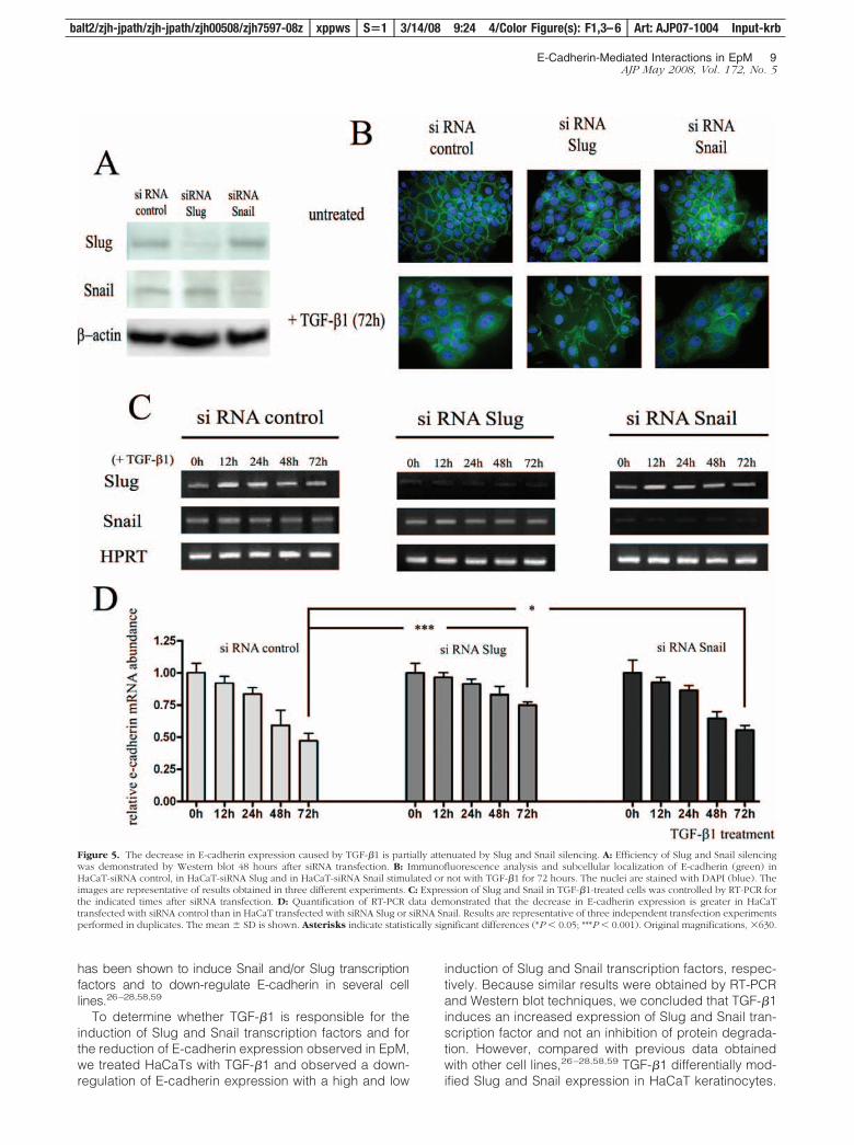
We next determined whether Slug and/or Snail transcrip-tion factors are required for the decreased expression ofE-cadherin induced by TGF-�1 by examining the effectsof Slug and Snail silencing on TGF-�1-induced down-regulation of E-cadherin in keratinocytes. Gene silencing
efficiency was analyzed by Western blot (Figure 5A) andRT-PCR (Figure 5C). Immunocytology (Figure 5B) andRT-PCR (Figure 5D) showed that the down-regulation ofE-cadherin induced by TGF-�1 was partially suppressedin Slug- and Snail-silenced HaCaTs. RT-PCR analysisshowed that after 72 hours of TGF-�1 treatment, E-cad-herin mRNA was present in HaCaT-control siRNA at alevel corresponding to 49% of that detected in untreatedcells, compared to 77% (P � 0.001) and 56% (P � 0.05)in HaCaTs transfected, respectively, with Slug and SnailsiRNA. No synergistic effect was observed when keratin-ocytes were transfected with siRNAs against both Slugand Snail (data not shown). These data suggest that Slugand, at a lower extent, Snail transcription factors are, atleast in part, necessary for the down-regulation of E-cadherin induced by TGF-�1 in keratinocytes.
The Interactions between LCs andKeratinocytes Are Affected by theDown-Regulation of E-Cadherin Caused by Slugand Snail Transcription FactorsWe finally investigated whether the reduction of E-cad-herin expression caused by Slug and Snail transcriptionfactors affects the adhesion of LCs to keratinocytes. Wetransiently transfected keratinocytes with Snail and Slugexpression vectors and showed that Slug and Snail pro-teins levels were higher in transfected cells (Figure 6A).RT-PCR analysis showed similar results (data not shown).In addition, we confirmed by immunofluorescence (Fig-ure 6B) and Western blot (Figure 6A) the down-regulationof E-cadherin induced by these two transcription factors.Forty-eight hours after transfection, a heterotypic cell ad-hesion assay was performed by using fluorescent la-beled LCs that were incubated with keratinocytes trans-fected with the different vectors and we demonstrated thatLCs poorly adhere to Slug- and Snail-transfected keratino-cytes compared to cells transfected with empty vectors(Figure 6C). Similar results were obtained when the expres-sion of E-cadherin in keratinocytes was reduced by a pre-treatment with TGF-�1 for 72 hours (data not shown).
Discussion
Although the precise mechanisms underlying inductionof EpM are still obscure, it has been known for many
Figure 2. The methylation status of CDH1 (E-cadherin) gene in exocervical and metaplastictissues. A: Representative examples of PCR re-sults for methylated and unmethylated CDH1gene. After a treatment with the restriction en-zyme HpaII, a PCR product is visualized underUV illumination for methylated tissue samples.Genomic DNA without enzyme digestion andDNA treated with methylation-unsensitive endo-nuclease MspI represent positive and negativecontrol, respectively. B: Methylation analysis ofCDH1 gene in cervical tissues with exocervicalepithelium (n � 12), mature (n � 14), or im-mature EpM (n � 14).
6 Herfs et alAJP May 2008, Vol. 172, No. 5
balt2/zjh-jpath/zjh-jpath/zjh00508/zjh7597-08z xppws S�1 3/14/08 9:24 4/Color Figure(s): F1,3–6 Art: AJP07-1004 Input-krb
F4
F5
F6

years that specific metaplastic sites are at higher risk ofdeveloping cancer compared with the adjacent nativeepithelium.5,41,42 The increased risk of malignant trans-
formation within the metaplastic epithelium could resultfrom the accumulation of somatic gene mutations directlycaused by factors responsible for EpM.4,43 However,
Figure 3. TGF-�1 (A–C), Slug (D–F), and Snail (G–I) immunostaining in cervical biopsy specimens. The exocervical epithelium shows a low TGF-�1 staining(A) whereas mature (B) and immature (C) EpM demonstrate, respectively, an intermediate and high expression of TGF-�1. The normal squamous exocervicalepithelium shows a medium expression of Slug (D) and a low expression of Snail (G) transcription factors only in upper cell layers. The dashed line delineatesthe epithelium from the stroma. In contrast, mature (E and H) and immature (F and I) EpM demonstrates a strong Slug (E and F) and Snail (H and I)immunoreactivity. J: Semiquantitative evaluation of TGF-�1 expression, respectively, in normal exocervix (n � 51), mature (n � 23), and immature (n � 39) areasof EpM. Semiquantitative evaluation of Slug (K) and Snail (L) expression, respectively, in normal exocervix (n � 12), mature (n � 14), and immature (n � 14)areas of EpM. Asterisks indicate statistically significant differences (***P � 0.001). Original magnifications: �100 (A–C, D, G); �200 (E, F, H, I).
E-Cadherin-Mediated Interactions in EpM 7AJP May 2008, Vol. 172, No. 5
balt2/zjh-jpath/zjh-jpath/zjh00508/zjh7597-08z xppws S�1 3/14/08 9:24 4/Color Figure(s): F1,3–6 Art: AJP07-1004 Input-krb

intrinsic immune features altered in the metaplastic epi-thelium could also contribute to cancer development bypreventing an efficient antitumor immune response.6 Thiseffect could be mediated by a differential expression ofsoluble and/or membrane-associated factors. Consistentwith this hypothesis, an altered expression of severalsoluble factors such as TGF-�, tumor necrosis factor(TNF)-�, and interleukin (IL)-10 has been observed inesophageal and cervical EpM.10,44,45 Similarly, the ex-pression of E-cadherin, which is necessary for the reten-tion of antigen-presenting cells in epithelial tissues,36 isdecreased in gastric and esophageal areas of intestinalmetaplasia compared to normal epithelium.14,46
LCs are antigen-presenting cells that play a key role inthe immunosurveillance of epidermis and mucosal sur-faces. LCs are required for the initiation of cellular im-mune responses to pathogens and the infiltration of tu-mors by antigen-presenting cells has been correlatedwith a better prognosis.47–49 In the present study, wedemonstrated that the immunosurveillance represented bythe density of CD1a� LCs is decreased both in mature andimmature cervical EpM compared with the normal squa-mous epithelium and is correlated with a lower intraepithe-lial expression of E-cadherin. These findings are in agree-ment with previous works reporting a reduced LC density inthe cervical transformation zone.9,10 The observed correla-tion between E-cadherin expression on keratinocytes andLC density in normal and metaplastic epithelium suggestsan important role of the heterotypic E-cadherin-mediatedinteraction between keratinocytes and LCs for the LC reten-tion in the squamous epithelium. A similar correlation has
been previously performed in HPV16-infected skin in whichE6 viral oncoprotein inhibits E-cadherin expression.50 How-ever, in contrast to cervical (pre)neoplasic lesions that areassociated with HPV infection,51–53 the biopsies selected inthis study were HPV-negative, suggesting that E6 viral on-coprotein is not involved in the down-regulation of E-cad-herin in HPV-negative EpM.
To determine the mechanism by which E-cadherin ex-pression is modulated in EpM, the methylation level ofCDH1 (E-cadherin) promoter was studied by using amethylation-specific PCR. We found that the majority ofanalyzed cervical metaplastic samples are unmethyl-ated. As already shown in intestinal metaplasia areas inthe lower esophagus54 and the stomach,55 the frequencyof hypermethylation was less than 40%, suggesting thatother mechanisms are responsible for the down-regula-tion of E-cadherin observed in every EpM.
Slug and Snail are transcription factors that negativelyregulate the expression of E-cadherin.18–21 In the presentstudy, we found, by immunostaining, that these proteinsare strongly expressed in the entire thickness of EpM. Incontrast, these two proteins were only weakly detected inthe upper epithelial cell layers of the normal exocervicalepithelium and their expression was inversely correlatedwith that of E-cadherin that was mainly present in the(para)basal and intermediate cells of the squamous epi-thelium. As previously observed in pancreatic and esoph-ageal cancer cells,56,57 Slug and Snail were observed in thecytoplasm as well as in the nucleus of keratinocytes. Inter-estingly, TGF-�1 was also expressed, at higher levels, inEpM compared with the adjacent native epithelium. TGF-�1
Figure 4. TGF-�1 inhibits E-cadherin and induces Slug and Snail expression in keratinocytes. A: The immunofluorescence demonstrates a diminished expressionof E-cadherin (green) at the cell membrane after incubation with TGF-�1 during 48 hours. The nuclei were stained with DAPI (blue). Western blot (B) and RT-PCR(C) confirm the down-regulation of E-cadherin and show the increased expression of Slug and, at a lower level, of Snail transcription factors after TGF-�1treatment. Results are representative of three independent experiments performed in duplicates. The mean � SD is shown. Asterisks indicate statisticallysignificant differences (*P � 0.05; **P � 0.01; ***P � 0.001). Original magnifications, �630.
8 Herfs et alAJP May 2008, Vol. 172, No. 5
balt2/zjh-jpath/zjh-jpath/zjh00508/zjh7597-08z xppws S�1 3/14/08 9:24 4/Color Figure(s): F1,3–6 Art: AJP07-1004 Input-krb
has been shown to induce Snail and/or Slug transcriptionfactors and to down-regulate E-cadherin in several celllines.26–28,58,59
To determine whether TGF-�1 is responsible for theinduction of Slug and Snail transcription factors and forthe reduction of E-cadherin expression observed in EpM,we treated HaCaTs with TGF-�1 and observed a down-regulation of E-cadherin expression with a high and low
induction of Slug and Snail transcription factors, respec-tively. Because similar results were obtained by RT-PCRand Western blot techniques, we concluded that TGF-�1induces an increased expression of Slug and Snail tran-scription factor and not an inhibition of protein degrada-tion. However, compared with previous data obtainedwith other cell lines,26–28,58,59 TGF-�1 differentially mod-ified Slug and Snail expression in HaCaT keratinocytes.
Figure 5. The decrease in E-cadherin expression caused by TGF-�1 is partially attenuated by Slug and Snail silencing. A: Efficiency of Slug and Snail silencingwas demonstrated by Western blot 48 hours after siRNA transfection. B: Immunofluorescence analysis and subcellular localization of E-cadherin (green) inHaCaT-siRNA control, in HaCaT-siRNA Slug and in HaCaT-siRNA Snail stimulated or not with TGF-�1 for 72 hours. The nuclei are stained with DAPI (blue). Theimages are representative of results obtained in three different experiments. C: Expression of Slug and Snail in TGF-�1-treated cells was controlled by RT-PCR forthe indicated times after siRNA transfection. D: Quantification of RT-PCR data demonstrated that the decrease in E-cadherin expression is greater in HaCaTtransfected with siRNA control than in HaCaT transfected with siRNA Slug or siRNA Snail. Results are representative of three independent transfection experimentsperformed in duplicates. The mean � SD is shown. Asterisks indicate statistically significant differences (*P � 0.05; ***P � 0.001). Original magnifications, �630.
E-Cadherin-Mediated Interactions in EpM 9AJP May 2008, Vol. 172, No. 5
balt2/zjh-jpath/zjh-jpath/zjh00508/zjh7597-08z xppws S�1 3/14/08 9:24 4/Color Figure(s): F1,3–6 Art: AJP07-1004 Input-krb

Zavadil and colleagues34 have previously shown that thepatterns of activation of Slug and Snail by TGF-�1 weremutually exclusive and cell-type-dependent. Moreover,the difference in Snail expression observed in vivo(strong) and in vitro (low) is probably related to the factthat TGF-�1 is not the only soluble factor that can induceSnail expression. For example, the prostaglandin E2
(PGE2) can also up-regulate Snail60 and then could ex-plain the high expression of Snail observed in vivo. Ac-cordingly, PGE2 synthase was found to be highly ex-pressed in mature and immature EpM compared toexocervical epithelium and a significantly increased Snailexpression was observed in vitro in keratinocytes treatedwith PGE2 (data not shown).
In addition, we showed that Slug or Snail silencingpartially abrogated the down-regulation of E-cadherin in-duced by TGF-�1 in keratinocytes. These results are inagreement with those reported by Takano and col-leagues58 and demonstrate that these transcription fac-tors are implicated in the reduction of E-cadherin expres-sion caused by TGF-�1. However, the restitution ofE-cadherin expression was stronger for Slug as com-pared to Snail silencing, suggesting that TGF-�1 reduces
the E-cadherin expression in keratinocytes, mainly via theSlug transcription factor.
Finally, to determine the significance of this down-regulation of E-cadherin expression observed in EpM interms of LC adhesion to keratinocytes, the impact ofE-cadherin under-expression was studied by using arelevant heterotypic cell adhesion assay. We showed thatthe adhesion between LCs and epithelial cells is alteredwhen E-cadherin expression by keratinocytes is inhib-ited. The importance of homophilic E-cadherin-mediatedinteractions between LCs and epithelial cells has beenpreviously reported by several studies.11,36 However, thereduction of E-cadherin was unable to completely abro-gate the LCs/keratinocytes interactions. Although there isevidence that E-cadherin stimulates the adhesion of LCs/DCs directly, we cannot exclude, in our study, the role ofother adhesion molecules such as �6 integrins, CD44, orCD47.61–63
In conclusion, we demonstrated that TGF-�1 can indi-rectly induce decreased antigen presentation functionsin EpM by affecting E-cadherin expression. The inabilityof the local immune system to mount a cell-mediatedimmune response against pathogens or cells in transfor-
Figure 6. The decrease in E-cadherin expression induced by Slug and Snail transcription factors affects the interactions between LCs and keratinocytes. A:Seventy-two hours after transfection with Slug and Snail transcription factors, the expression of E-cadherin, Slug and Snail was assessed by Western blot. B:Immunofluorescence analysis and subcellular localization of E-cadherin (green) in human Slug or Snail transfected keratinocytes. Transfections with correspond-ing empty expression vectors were used as controls. For each condition of transfection, a representative example of LC (red) density observed by field in theheterotypic cell adhesion assay is shown. The nuclei are stained with DAPI (blue). C: Graphic representation of the mean number � SD of LCs observed by fieldin the co-culture experiments. For each condition, three independent experiments were performed. The adhesion of LCs to human Slug- or Snail-transfectedkeratinocytes was significantly lower compared to cells transfected with empty vectors. Asterisks indicate statistically significant differences (**P � 0.01; ***P �0.001). Original magnifications: �630 (B, left); �200 [B (right), C].
10 Herfs et alAJP May 2008, Vol. 172, No. 5
balt2/zjh-jpath/zjh-jpath/zjh00508/zjh7597-08z xppws S�1 3/14/08 9:24 4/Color Figure(s): F1,3–6 Art: AJP07-1004 Input-krb
mation, because of a deficit of adhesion molecules nec-essary for cell-to-cell interactions, might play an impor-tant role in the susceptibility of EpM for developingcancer. The progressive alteration of E-cadherin expres-sion that has been demonstrated in bronchial,15 esoph-ageal,13 and gastric14 metaplasia-dysplasia-carcinomasequences could not only be an early indication signalingthe malignant transformation of metaplastic cells butmight also constitute one of the major determinants forestablishing local immunodeficiency responsible for EpMtumorigenesis.
References
1. Slack JM: Metaplasia and transdifferentiation: from pure biology tothe clinic. Nat Rev Mol Cell Biol 2007, 8:369–378
2. Tosh D, Slack JM: How cells change their phenotype. Nat Rev MolCell Biol 2002, 3:187–194
3. Elson DA, Riley RR, Lacey A, Thordarson G, Talamantes FJ, ArbeitJM: Sensitivity of the cervical transformation zone to estrogen-in-duced squamous carcinogenesis. Cancer Res 2000, 60:1267–1275
4. Quinlan JM, Colleypriest BJ, Farrant M, Tosh D: Epithelial metaplasiaand the development of cancer. Biochim Biophys Acta 2007,1776:10–21
5. Burghardt E, Ostor AG: Site and origin of squamous cervical cancer:a histomorphologic study. Obstet Gynecol 1983, 62:117–127
6. Delvenne P, Hubert P, Jacobs N: Epithelial metaplasia: an inadequateenvironment for antitumour immunity? Trends Immunol 2004,25:169–173
7. Uchi H, Terao H, Koga T, Furue M: Cytokines and chemokines in theepidermis. J Dermatol Sci 2000, 24(Suppl 1):S29–S38
8. Sikorski M, Bieda T, Bobek M, Zrubek H: Dynamics of cervical Lang-erhans cell counts in the course of HPV-positive CIN treatment withthe use of human recombinant interferon gamma. Eur J GynaecolOncol 2005, 26:294–298
9. Al-Saleh W, Giannini SL, Jacobs N, Moutschen M, Doyen J, Boniver J,Delvenne P: Correlation of T-helper secretory differentiation and typesof antigen-presenting cells in squamous intraepithelial lesions of theuterine cervix. J Pathol 1998, 184:283–290
10. Giannini SL, Hubert P, Doyen J, Boniver J, Delvenne P: Influence ofthe mucosal epithelium microenvironment on Langerhans cells: im-plications for the development of squamous intraepithelial lesions ofthe cervix. Int J Cancer 2002, 97:654–659
11. Tang A, Amagai M, Granger LG, Stanley JR, Udey MC: Adhesion ofepidermal Langerhans cells to keratinocytes mediated by E-cad-herin. Nature 1993, 361:82–85
12. Cepek KL, Shaw SK, Parker CM, Russell GJ, Morrow JS, Rimm DL,Brenner MB: Adhesion between epithelial cells and T lymphocytesmediated by E-cadherin and the alpha E beta 7 integrin. Nature 1994,372:190–193
13. Bailey T, Biddlestone L, Shepherd N, Barr H, Warner P, Jankowski J:Altered cadherin and catenin complexes in the Barrett’s esophagus-dysplasia-adenocarcinoma sequence: correlation with disease pro-gression and dedifferentiation. Am J Pathol 1998, 152:135–144
14. Chan AO, Wong BC, Lan HY, Loke SL, Chan WK, Hui WM, Yuen YH,Ng I, Hou L, Wong WM, Yuen MF, Luk JM, Lam SK: Deregulation ofE-cadherin-catenin complex in precancerous lesions of gastric ade-nocarcinoma. J Gastroenterol Hepatol 2003, 18:534–539
15. Kato Y, Hirano T, Yoshida K, Yashima K, Akimoto S, Tsuji K, Ohira T,Tsuboi M, Ikeda N, Ebihara Y, Kato H: Frequent loss of E-cadherinand/or catenins in intrabronchial lesions during carcinogenesis of thebronchial epithelium. Lung Cancer 2005, 48:323–330
16. Graff JR, Herman JG, Lapidus RG, Chopra H, Xu R, Jarrard DF,Isaacs WB, Pitha PM, Davidson NE, Baylin SB: E-cadherin expressionis silenced by DNA hypermethylation in human breast and prostatecarcinomas. Cancer Res 1995, 55:5195–5199
17. Yoshiura K, Kanai Y, Ochiai A, Shimoyama Y, Sugimura T, HirohashiS: Silencing of the E-cadherin invasion-suppressor gene by CpGmethylation in human carcinomas. Proc Natl Acad Sci USA 1995,92:7416–7419
18. Batlle E, Sancho E, Franci C, Dominguez D, Monfar M, Baulida J,Garcia De HA: The transcription factor snail is a repressor of E-cadherin gene expression in epithelial tumour cells. Nat Cell Biol2000, 2:84–89
19. Bolos V, Peinado H, Perez-Moreno MA, Fraga MF, Esteller M, Cano A:The transcription factor Slug represses E-cadherin expression andinduces epithelial to mesenchymal transitions: a comparison withSnail and E47 repressors. J Cell Sci 2003, 116:499–511
20. Cano A, Perez-Moreno MA, Rodrigo I, Locascio A, Blanco MJ, delBarrio MG, Portillo F, Nieto MA: The transcription factor snail controlsepithelial-mesenchymal transitions by repressing E-cadherin expres-sion. Nat Cell Biol 2000, 2:76–83
21. Hajra KM, Chen DY, Fearon ER: The SLUG zinc-finger protein re-presses E-cadherin in breast cancer. Cancer Res 2002, 62:1613-1618
22. Miyoshi A, Kitajima Y, Sumi K, Sato K, Hagiwara A, Koga Y, MiyazakiK: Snail and SIP1 increase cancer invasion by upregulating MMPfamily in hepatocellular carcinoma cells. Br J Cancer 2004, 90:1265-1273
23. Miyoshi A, Kitajima Y, Kido S, Shimonishi T, Matsuyama S, Kitahara K,Miyazaki K: Snail accelerates cancer invasion by upregulating MMPexpression and is associated with poor prognosis of hepatocellularcarcinoma. Br J Cancer 2005, 92:252–258
24. Sugimachi K, Tanaka S, Kameyama T, Taguchi K, Aishima S,Shimada M, Sugimachi K, Tsuneyoshi M: Transcriptional repressorsnail and progression of human hepatocellular carcinoma. Clin Can-cer Res 2003, 9:2657–2664
25. Hardy RG, Vicente-Duenas C, Gonzalez-Herrero I, Anderson C,Flores T, Hughes S, Tselepis C, Ross JA, Sanchez-Garcia I: Snailfamily transcription factors are implicated in thyroid carcinogenesis.Am J Pathol 2007, 171:1037–1046
26. Choi J, Park SY, Joo CK: Transforming growth factor-beta1 repressesE-cadherin production via slug expression in lens epithelial cells.Invest Ophthalmol Vis Sci 2007, 48:2708–2718
27. Medici D, Hay ED, Goodenough DA: Cooperation between snail andLEF-1 transcription factors is essential for TGF-beta1-induced epithe-lial-mesenchymal transition. Mol Biol Cell 2006, 17:1871–1879
28. Peinado H, Quintanilla M, Cano A: Transforming growth factor beta-1induces snail transcription factor in epithelial cell lines: mechanismsfor epithelial mesenchymal transitions. J Biol Chem 2003, 278:21113-21123
29. Monteleone G, Pallone F, MacDonald TT: Smad7 in TGF-beta-medi-ated negative regulation of gut inflammation. Trends Immunol 2004,25:513–517
30. Siegel PM, Massague J: Cytostatic and apoptotic actions of TGF-betain homeostasis and cancer. Nat Rev Cancer 2003, 3:807–821
31. Delvenne P, Fontaine MA, Delvenne C, Nikkels A, Boniver J: Detec-tion of human papillomaviruses in paraffin-embedded biopsies ofcervical intraepithelial lesions: analysis by immunohistochemistry, insitu hybridization, and the polymerase chain reaction. Mod Pathol1994, 7:113–119
32. Qu W, Jiang G, Cruz Y, Chang CJ, Ho GY, Klein RS, Burk RD: PCRdetection of human papillomavirus: comparison between MY09/MY11and GP5�/GP6� primer systems. J Clin Microbiol 1997, 35:1304-1310
33. Snijders PJ, van den Brule AJ, Schrijnemakers HF, Snow G, MeijerCJ, Walboomers JM: The use of general primers in the polymerasechain reaction permits the detection of a broad spectrum of humanpapillomavirus genotypes. J Gen Virol 1990, 71:173–181
34. Zavadil J, Cermak L, Soto-Nieves N, Bottinger EP: Integration ofTGF-beta/Smad and Jagged1/Notch signalling in epithelial-to-mes-enchymal transition. EMBO J 2004, 23:1155–1165
35. Detry C, Waltregny D, Quatresooz P, Chaplet M, Kedzia W,Castronovo V, Delvenne P, Bellahcene A: Detection of bone sialopro-tein in human (pre)neoplastic lesions of the uterine cervix. CalcifTissue Int 2003, 73:9–14
36. Hubert P, Caberg JH, Gilles C, Bousarghin L, Franzen-Detrooz E,Boniver J, Delvenne P: E-cadherin-dependent adhesion of dendriticand Langerhans cells to keratinocytes is defective in cervical humanpapillomavirus-associated (pre)neoplastic lesions. J Pathol 2005,206:346–355
37. Delvenne P, Al-Saleh W, Gilles C, Thiry A, Boniver J: Inhibition ofgrowth of normal and human papillomavirus-transformed keratino-
E-Cadherin-Mediated Interactions in EpM 11AJP May 2008, Vol. 172, No. 5
balt2/zjh-jpath/zjh-jpath/zjh00508/zjh7597-08z xppws S�1 3/14/08 9:24 4/Color Figure(s): F1,3–6 Art: AJP07-1004 Input-krb

cytes in monolayer and organotypic cultures by interferon-gammaand tumor necrosis factor-alpha. Am J Pathol 1995, 146:589–598
38. Jorda M, Olmeda D, Vinyals A, Valero E, Cubillo E, Llorens A, Cano A,Fabra A: Upregulation of MMP-9 in MDCK epithelial cell line in re-sponse to expression of the Snail transcription factor. J Cell Sci 2005,118:3371–3385
39. Tripathi MK, Misra S, Khedkar SV, Hamilton N, Irvin-Wilson C, SharanC, Sealy L, Chaudhuri G: Regulation of BRCA2 gene expression bythe SLUG repressor protein in human breast cells. J Biol Chem 2005,280:17163–17171
40. Hubert P, Bousarghin L, Greimers R, Franzen-Detrooz E, Boniver J,Delvenne P: Production of large numbers of Langerhans’ cells withintraepithelial migration ability in vitro. Exp Dermatol 2005,14:469–477
41. Cossentino MJ, Wong RK: Barrett’s esophagus and risk of esopha-geal adenocarcinoma. Semin Gastrointest Dis 2003, 14:128–135
42. Whiting JL, Sigurdsson A, Rowlands DC, Hallissey MT, Fielding JW:The long term results of endoscopic surveillance of premalignantgastric lesions. Gut 2002, 50:378–381
43. Wild CP, Hardie LJ: Reflux. Barrett’s oesophagus and adenocarcinoma:burning questions. Nat Rev Cancer 2003, 3:676–684
44. Giannini SL, Al-Saleh W, Piron H, Jacobs N, Doyen J, Boniver J,Delvenne P: Cytokine expression in squamous intraepithelial lesionsof the uterine cervix: implications for the generation of local immuno-suppression. Clin Exp Immunol 1998, 113:183–189
45. Tselepis C, Perry I, Dawson C, Hardy R, Darnton SJ, McConkey C,Stuart RC, Wright N, Harrison R, Jankowski JA: Tumour necrosisfactor-alpha in Barrett’s oesophagus: a potential novel mechanism ofaction. Oncogene 2002, 21:6071–6081
46. Swami S, Kumble S, Triadafilopoulos G: E-cadherin expression ingastroesophageal reflux disease. Barrett’s esophagus, and esopha-geal adenocarcinoma: an immunohistochemical and immunoblotstudy. Am J Gastroenterol 1995, 90:1808–1813
47. Barnetson RS, Satchell A, Zhuang L, Slade HB, Halliday GM: Imi-quimod induced regression of clinically diagnosed superficial basalcell carcinoma is associated with early infiltration by CD4 T cells anddendritic cells. Clin Exp Dermatol 2004, 29:639–643
48. Inoshima N, Nakanishi Y, Minami T, Izumi M, Takayama K, Yoshino I,Hara N: The influence of dendritic cell infiltration and vascular endo-thelial growth factor expression on the prognosis of non-small celllung cancer. Clin Cancer Res 2002, 8:3480–3486
49. Reichert TE, Scheuer C, Day R, Wagner W, Whiteside TL: The numberof intratumoral dendritic cells and zeta-chain expression in T cells asprognostic and survival biomarkers in patients with oral carcinoma.Cancer 2001, 91:2136–2147
50. Matthews K, Leong CM, Baxter L, Inglis E, Yun K, Backstrom BT,Doorbar J, Hibma M: Depletion of Langerhans cells in human papil-lomavirus type 16-infected skin is associated with E6-mediated downregulation of E-cadherin. J Virol 2003, 77:8378–8385
51. Bohmer G, van den Brule AJ, Brummer O, Meijer CL, Petry KU: No
confirmed case of human papillomavirus DNA-negative cervical in-traepithelial neoplasia grade 3 or invasive primary cancer of theuterine cervix among 511 patients. Am J Obstet Gynecol 2003,189:118–120
52. Bosch FX, Munoz N: The viral etiology of cervical cancer. Virus Res2002, 89:183–190
53. Walboomers JM, Jacobs MV, Manos MM, Bosch FX, Kummer JA,Shah KV, Snijders PJ, Peto J, Meijer CJ, Munoz N: Human papillo-mavirus is a necessary cause of invasive cervical cancer worldwide.J Pathol 1999, 189:12–19
54. Eads CA, Lord RV, Wickramasinghe K, Long TI, Kurumboor SK,Bernstein L, Peters JH, DeMeester SR, Demeester TR, Skinner KA,Laird PW: Epigenetic patterns in the progression of esophageal ad-enocarcinoma. Cancer Res 2001, 61:3410–3418
55. To KF, Leung WK, Lee TL, Yu J, Tong JH, Chan MW, Ng EK, ChungSC, Sung JJ: Promoter hypermethylation of tumor-related genes ingastric intestinal metaplasia of patients with and without gastric can-cer. Int J Cancer 2002, 102:623–628
56. Hotz B, Arndt M, Dullat S, Bhargava S, Buhr HJ, Hotz HG: Epithelialto mesenchymal transition: expression of the regulators snail, slug,and twist in pancreatic cancer. Clin Cancer Res 2007, 13:4769–4776
57. Uchikado Y, Natsugoe S, Okumura H, Setoyama T, Matsumoto M,Ishigami S, Aikou T: Slug expression in the E-cadherin preservedtumors is related to prognosis in patients with esophageal squamouscell carcinoma. Clin Cancer Res 2005, 11:1174–1180
58. Takano S, Kanai F, Jazag A, Ijichi H, Yao J, Ogawa H, Enomoto N,Omata M, Nakao A: Smad4 is essential for down-regulation of E-cadherin induced by TGF-beta in pancreatic cancer cell line PANC-1.J Biochem (Tokyo) 2007, 141:345–351
59. Thuault S, Valcourt U, Petersen M, Manfioletti G, Heldin CH, Moustakas A:Transforming growth factor-beta employs HMGA2 to elicit epithelial-mesenchymal transition. J Cell Biol 2006, 174:175–183
60. Dohadwala M, Yang SC, Luo J, Sharma S, Batra RK, Huang M, Lin Y,Goodglick L, Krysan K, Fishbein MC, Hong L, Lai C, Cameron RB,Gemmill RM, Drabkin HA, Dubinett SM: Cyclooxygenase-2-depen-dent regulation of E-cadherin: prostaglandin E(2) induces transcrip-tional repressors ZEB1 and snail in non-small cell lung cancer. Can-cer Res 2006, 66:5338–5345
61. Fukunaga A, Nagai H, Noguchi T, Okazawa H, Matozaki T, Yu X,Lagenaur CF, Honma N, Ichihashi M, Kasuga M, Nishigori C,Horikawa T: Src homology 2 domain-containing protein tyrosinephosphatase substrate 1 regulates the migration of Langerhans cellsfrom the epidermis to draining lymph nodes. J Immunol 2004,172:4091–4099
62. Price AA, Cumberbatch M, Kimber I, Ager A: Alpha 6 integrins arerequired for Langerhans cell migration from the epidermis. J Exp Med1997, 186:1725–1735
63. Staquet MJ: Adhesion and migration of epidermal dendritic cells.Pathol Biol (Paris) 1995, 43:858–862
12 Herfs et alAJP May 2008, Vol. 172, No. 5
balt2/zjh-jpath/zjh-jpath/zjh00508/zjh7597-08z xppws S�1 3/14/08 9:24 4/Color Figure(s): F1,3–6 Art: AJP07-1004 Input-krb

JOBNAME: AUTHOR QUERIES PAGE: 1 SESS: 3 OUTPUT: Fri Mar 14 10:29:19 2008/balt2/zjh�jpath/zjh�jpath/zjh00508/zjh7597�08z
A—Short running head okay? If not, please provide a new one of 40 characters or less.
B—Au/ Genus, loci, alleles, and genotypes are styled as italic; proteins, gene products,phenotypes, mRNAs, and cDNAs are not. Please confirm or correct throughout. Please makesure that all products/supplies have their manufacturers, cities, and states or countries cited atthe first mention. Per journal style, most nonstandard abbreviations must be used at leastthree times in the abstract and five times in the text to be retained; nonstandard abbreviationsused fewer times have thus been written out.
C—Please write out CRCE and CRLC in the affiliation line. Also, is the line okay as set?
AUTHOR QUERIES
AUTHOR PLEASE ANSWER ALL QUERIES 1