American Journal of Microbiology and Biotechnology - …article.aascit.org/file/pdf/9190753.pdf ·...
6
American Journal of Microbiology and Biotechnology 2015; 2(3): 38-43 Published online May 20, 2015 (http://www.aascit.org/journal/ajmb) ISSN: 2375-3005 Keywords Candida Spp, Catheters, Fungal Infectivity, Biofilms, SEM Received: April 3, 2015 Revised: April 18, 2015 Accepted: April 19, 2015 Nosocomial Fungal Infectivities: In Vivo Formation of Candida Biofilms on Catheters Surfaces Sidi Mohammed Lahbib Seddiki 1, 2, * , Zahia Boucherit-Otmani 1 , Reda Ali Bettioui 3 , Kebir Boucherit 2 , Mourad Taleb 4 , Dennis Kunkel 5 1 Laboratory Antifungal Antibiotic, Physico-chemical synthesis and biological activity, University of Tlemcen, Imama, Algeria 2 University Center of Naâma, Institute of Sciences, Dept. of nature and life sciences, Naâma, Algeria 3 Laboratory: Actions of the human for the protection of the environment and application in public health, department of ecology and environment, University of Tlemcen, Imama, Algeria 4 Department of Epidemiology and preventive medicine, University Hospital of Sidi Bel Abbes, Algeria 5 Dennis Kunkel Microscopy Inc, Kailua, USA Email address [email protected] (S. M. L. Seddiki) Citation Sidi Mohammed Lahbib Seddiki, Zahia Boucherit-Otmani, Reda Ali Bettioui, Kebir Boucherit, Mourad Taleb, Dennis Kunkel. Nosocomial Fungal Infectivities: In Vivo Formation of Candida Biofilms on Catheters Surfaces. American Journal of Microbiology and Biotechnology. Vol. 2, No. 3, 2015, pp. 38-43. Abstract The invasive nosocomial infections due to Candida species are responsible for increasing the length of stay, cost of hospitalization and morbidity in immunocompromised patients. Their severity and rapid progressivity are owed to the difficulty of diagnosis. Various catheters, which are often used to train a body fluid (blood, urine, infusion, parenteral nutrition, medication ...) inside the body of the patient or vice versa, are susceptible to be altered by Candida spp. and promote the formation of biofilms which consolidates the risk of invasive nosocomial infections; i.e., these structures are considered as a nest for disease because it is not easily eradicate by conventional antifungal therapy. Such as the diagnosis of candidiasis related to catheter is difficult, the differentiation between catheter infection and a simple contamination is essential to establish an antifungal treatment. This study aimed adapts to yeasts the Brun-Buisson (1987) method which only concerned by bacteria, it’s why we conducted our study between February 2011 and January 2012 at the Hospital University Center of Sidi Bel Abbès-Algeria that aims to evaluate the various types of fungal catheters infectivities (contaminations, colonization and infections) and their corresponding rates, as well as the responsible yeast species. At the end, the ability to form biofilms was checked. The results showed that three types of fungal infectivities of catheters were identified. On the other hand, SEM images showed clearly Candida biofilms on the surfaces of catheters. 1. Introduction Nosocomial invasive fungal infections, including those caused by Candida species, are a major cause of morbidity and mortality in immunocompromised patients 1,2 . These infections are usually severe, rapidly scalable and difficult to diagnosis and to treat 3 . In conjunction with the medical advances, the extensive use of different types of
Transcript of American Journal of Microbiology and Biotechnology - …article.aascit.org/file/pdf/9190753.pdf ·...

American Journal of Microbiology and Biotechnology 2015; 2(3): 38-43
Published online May 20, 2015 (http://www.aascit.org/journal/ajmb)
ISSN: 2375-3005
Keywords Candida Spp,
Catheters,
Fungal Infectivity,
Biofilms,
SEM
Received: April 3, 2015
Revised: April 18, 2015
Accepted: April 19, 2015
Nosocomial Fungal Infectivities: In Vivo Formation of Candida Biofilms on Catheters Surfaces
Sidi Mohammed Lahbib Seddiki1, 2, *
, Zahia Boucherit-Otmani1,
Reda Ali Bettioui3, Kebir Boucherit
2, Mourad Taleb
4, Dennis Kunkel
5
1Laboratory Antifungal Antibiotic, Physico-chemical synthesis and biological activity, University
of Tlemcen, Imama, Algeria 2University Center of Naâma, Institute of Sciences, Dept. of nature and life sciences, Naâma,
Algeria 3Laboratory: Actions of the human for the protection of the environment and application in public
health, department of ecology and environment, University of Tlemcen, Imama, Algeria 4Department of Epidemiology and preventive medicine, University Hospital of Sidi Bel Abbes,
Algeria 5Dennis Kunkel Microscopy Inc, Kailua, USA
Email address [email protected] (S. M. L. Seddiki)
Citation Sidi Mohammed Lahbib Seddiki, Zahia Boucherit-Otmani, Reda Ali Bettioui, Kebir Boucherit,
Mourad Taleb, Dennis Kunkel. Nosocomial Fungal Infectivities: In Vivo Formation of Candida
Biofilms on Catheters Surfaces. American Journal of Microbiology and Biotechnology.
Vol. 2, No. 3, 2015, pp. 38-43.
Abstract The invasive nosocomial infections due to Candida species are responsible for
increasing the length of stay, cost of hospitalization and morbidity in
immunocompromised patients. Their severity and rapid progressivity are owed to the
difficulty of diagnosis. Various catheters, which are often used to train a body fluid
(blood, urine, infusion, parenteral nutrition, medication ...) inside the body of the
patient or vice versa, are susceptible to be altered by Candida spp. and promote the
formation of biofilms which consolidates the risk of invasive nosocomial infections;
i.e., these structures are considered as a nest for disease because it is not easily
eradicate by conventional antifungal therapy. Such as the diagnosis of candidiasis
related to catheter is difficult, the differentiation between catheter infection and a
simple contamination is essential to establish an antifungal treatment. This study
aimed adapts to yeasts the Brun-Buisson (1987) method which only concerned by
bacteria, it’s why we conducted our study between February 2011 and January 2012 at
the Hospital University Center of Sidi Bel Abbès-Algeria that aims to evaluate the
various types of fungal catheters infectivities (contaminations, colonization and
infections) and their corresponding rates, as well as the responsible yeast species. At
the end, the ability to form biofilms was checked. The results showed that three types
of fungal infectivities of catheters were identified. On the other hand, SEM images
showed clearly Candida biofilms on the surfaces of catheters.
1. Introduction
Nosocomial invasive fungal infections, including those caused by Candida species,
are a major cause of morbidity and mortality in immunocompromised patients1,2
. These
infections are usually severe, rapidly scalable and difficult to diagnosis and to treat3.
In conjunction with the medical advances, the extensive use of different types of

American Journal of Microbiology and Biotechnology 2015; 2(3): 38-43 39
catheters in hospitals has grown significantly in recent
decades4. However, nosocomial infections due to Candida
spp. increase in parallel with the use of these devices5,6
.
Indeed, more than half of these infections are related to the
presence of catheters7, which are responsible for an increase
in the length of stay, cost of hospitalization and morbidity8.
On the other hand, the emergence of non-albicans species
of Candida in hospital, like C. glabrata, C. parapsilosis and
C. tropicalis, is regularly observed in the last two decades9,10
.
Without a doubt, epidemiological changes are linked to the
frequency of fungal nosocomial infections, the involved
species and the development of resistance to conventional
antifungal agents, i.e., amphotericin B and triazole11
.
Furthermore, Candida spp. may colonize the catheter tip and
form biofilms12-14
, which is a real risk to patients7. So, the
presence of the catheter is a major risk factor for the
development and persistence of candidal nosocomial
infection15
.
In addition, the differentiation between the catheter
infection and its contamination is essential to establish an
antimicrobial treatment16,17
. According to16,18
, it is important
to differentiate between colonization, contamination and
infection of catheters by culturing its distal end.
Usually, two methods are adopted for the evaluation of
catheter-related infections, the semi-quantitative method19
and the quantitative method20
. However, these methods
process only catheters altered by bacteria.
Seddiki et al (2013) distinguish three types of alterations
catheters caused by Candida spp. They termed these
alterations “types of catheter infectivity” which refer to the
degree of infectiveness of catheters.
The objective of this study is to isolate yeasts from various
catheters after their ablations from admitted patients to
different hospital units in Sidi Bel Abbes (Algeria), to
respond to the request of biologists concerning the
appropriate method to make the difference between infection,
colonization and contamination of catheters and to evaluate
the ability of isolated strains to form biofilms on their
surfaces.
2. Materials and Methods
2.1. Sampling and Identification
Samples were taken between February 2011 and January
2012 from ten services at the University Hospital of Sidi
Bel Abbes (ICU, pediatric surgery, general surgery,
neurosurgery, traumatology, urology, gastroenterology,
pneumophtisiology, clinical hematology and endocrinology).
The concerned patients are those with significant risk
factors for candidiasis (implanted medical devices, broad
spectrum of antibiotic therapy, long stays in service,
invasive procedures ...). For each patient, data were
collected using a datasheet (Table 1).
Table 1. Technical data sheet for the information collection during each
sampling.
Hospital: Service:
Name First Name
Gender Male � Female �
Age Adress
Phone Number
Number of patients in the same room : Number of the beds :
Admission date in hospital :
Date of catheter insertion : Sampling date :
Date of discharge from hospital : Origin of the sample Catheter � Type :
O ther � Specify :
Immunosuppression Yes � Specify: No �
Antibiotic therapy Yes � Specify:
No � Evolution of disease Healing �
Death �
Unknown � Isolated strains:
According to22,23
, only implanted catheters for 48 hours or
more were taken. All types of catheters were include in this
study, sampling was carried out from different catheters
(PVC: Peripheral Venous Catheter, CVC: Central Venous
Catheter, UC: Urinary Catheter, TC: Tracheotomic Catheter,
OC: Orobronchique Catheter and DC: Drainage Catheter).
By taking measures of asepsis, each catheter was removed
separately, its distal end was cut (3-5 cm long) and then
placed in a sterile tube containing 1 mL of physiological
saline. Purification of yeast was performed by successive
subcultures on Sabouraud medium.
Identification of purified strains was based on their
morphological and biochemical characteristics. Microculture
testing (germ-tube and chlamydospore formation) and the
yeast identification system (Api Candida® System;
bioMériux, Marcy L’Etoile, France) were used.
2.2. Types of Fungal Catheters Infectivities
Since 2003, the department of epidemiology and
preventive medicine of the University Hospital of Sidi Bel
Abbes has adopted a strategy for the surveillance of
nosocomial infections by repeated prevalence surveys in
order to measure their rates and track their evolutionary
trends. The prevalence survey conducted in 2009 showed that
56.8% of patients with nosocomial infection had a peripheral
venous catheter, 11.4% had central venous catheter and 13.6%
with other types of catheters. In addition, broad-spectrum
antibiotics were often used in different services, while
antifungals were rarely prescribed (unpublished data).
The distinction between infection, colonization and
contamination of catheters was assessed by referring to the
significance threshold (103 cells/mL) and the collected data
taken during the sampling (systemic and/or local clinical
symptoms)17,20,24
.

40 Sidi Mohammed Lahbib Seddiki et al.: Nosocomial Fungal Infectivities: In Vivo Formation of Candida Biofilms on
Catheters Surfaces
The segments of catheters which exhibit a negative result
were transferred to new tubes containing liquid of Sabouraud
medium supplemented of 50 mg/L of chloramphenicol and
then incubated at 30 °C for 24 to 48 hours or even to 72
hours.
The results of different types of fungal infectivities were
made subject to a statistical study, the values of variances (P)
less than 0.05 were considered significant.
2.3. SEM Observation of Biofilms
To demonstrate the ability of isolates to form biofilms and
to observe their structures, three segments of different
catheters were taken to examine them in scanning electron
microscope (SEM).
The choice of segments was performed following the light
microscopic observations of suspensions in which the
segments catheters were submerged. Observation of pseudo-
mycelial and mycelial structures or substantial clumps of
cells allowed predicting the existence of biofilms on the
surfaces of the catheters.
To carry SEM photographs, samples were fixed with
glutaraldehyde and then sent to the laboratory Dennis Kunkel
Microscopy. Inc., USA. The samples were examined using a
Hitachi S-4800 (Hitachi Ltd, Tokyo, Japan) field-emission
SEM.
The fixation consisted of cutting segments catheters of 7 to
8 mm long in Eppendorf tubes containing 1 mL of PBS
buffer saline, 10 mM (2.7mM KCl, 137 mM NaCl, pH 7.4)
and glutaraldehyde at a final concentration of 2.5%. The
tubes were placed in a shock chamber and left at 4 °C before
shipment.
3. Results and Discussion
3.1. Types of Fungal Catheters Infectivities
Among 457 taken samples, 37 strains of Candida spp.
were isolated, i.e., a rate of 7.79%. Contaminations of
catheters were dominant with a rate of 55.56%. They were
related essentially to Candida glabrata at a rate of 33.33%.
The second concerned species in this type of infectivity was
C. albicans (16.66%) whereas C. parapsilosis was
responsible only for 5.55% of contaminations. Colonizations
of catheters were less important than contaminations
(30.56%). C. glabrata was responsible for 22.23% of
colonized catheters; however, 5.56% were caused by C.
albicans and only 2.78% by C. parapsilosis. On the other
hand, the rate of catheters infections was 13.88% where C.
glabrata and C. albicans were responsible respectively for
8.33% and 5.56% of this type of infectivity (Figure 1).
Figure 1. Types and rates of fungal catheters infectivities at the university hospital of Sidi Bel Abbes - Algeria.
In this study, the ICU ranks first with regard to infections
of catheters with a rate of 4.76% followed by the
pneumophtisiology service (2.38%) and then, the general
surgery department which was ranked third (2.13%). Yet, this
service was relatively the most responsible for catheters
colonizations with a rate of 8.51%, followed by the pediatric
surgery service with 4.44%. However, the ICU ranks third for
this type of infectivity (3.18%), although, it ranks first with
9.52% of contaminations catheters. In addition, it was found
that two to three types of fungal infectivities were observed
in different services, except for the pediatric surgery and
clinical hematology services where only the catheters
colonizations were distinguished. Statistical analysis reveals
a significant difference between the studied services (P =
0.01), and a clear difference between the types of fungal
infectivities (P = 0.03).
The World Health Organization25
estimated that 5% to 12%
of patients in the world develop a nosocomial infection,
while more than 60% are associated with the implantation
of a medical or surgical device especially the catheters.
Indeed, nosocomial infections can occur at any time during
the use of catheters24
. Thus, our results are consistent with
others14,26-29
which showed that the use of catheters is the
main source of invasive infections in hospitalized patients,
American Journal of Microbiology and Biotechnology 2015; 2(3): 38-43 41
particularly in ICU. In addition, the frequency of fungal
microorganisms causing nosocomial infections varies from
one country to another according to the institutions, the
intensive care, the antibiotic protocols and the type of
catheters used30-32
.
In fact, our results demonstrated that the types of fungal
infectivities vary depending on the type of catheters used.
The Central catheters were the most implicated in infections
with a rate of 18.18%; this could be explained by several
factors including the duration of implantation greater than 8
days and up to 21 days16,33
. However, the drainage and
orobronchial catheters were responsible respectively for
5.88% and 5.4% of colonizations. For infections, the
orobronchial catheters were essentially responsible (2.7%).
Additionally, a significant difference between the types of
catheters used was noted (P = 0.04). This result is not in
agreement with those of34,35
that showed catheters
infections were related to the presence of CVC.
3.2. SEM Observation of Biofilms
Figure 2. SEM morphology of Candida albicans "HFKT1" biofilm
developed on the inner surface of a peripheral vascular catheter. The arrow
indicates the extracellular matrix (Magnification × 2000).
Figure 2 shows a biofilm formed by the isolated strain C.
albicans "HFKT1" from a peripheral venous catheter from
the clinical hematology department after an implanted time
of 5 days. The extensions of the hyphae of the biofilm are
stretched on the inside of the catheter; the yeast cells and the
extracellular matrix are clearly identifiable (Figure 2).
For C. glabrata "RFKT1" isolated from ICU, the biofilm
structure is clearly visible. It is grown on the inner face of a
vascular catheter device after an implanted time of two days,
it is formed of blastospores were encapsulated in
extracellular matrix (Figure 3.)
Figure 3. SEM morphology of Candida glabrata "RFKT1" biofilm
developed on the inner surface of a peripheral vascular catheter. Arrows
indicate the extracellular matrix (Magnifications × 4500).
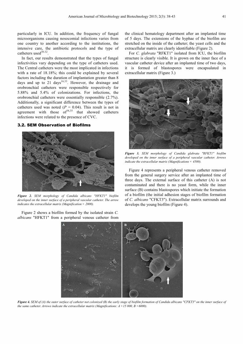
Figure 4 represents a peripheral venous catheter removed
from the general surgery service after an implanted time of
three days. The external surface of this catheter (A) is not
contaminated and there is no yeast form, while the inner
surface (B) contains blastospores which initiate the formation
of a biofilm (the initial adhesion stages of biofilm formation
of C. albicans "CFKT3"). Extracellular matrix surrounds and
develops the young biofilm (Figure 4).
Figure 4. SEM of (A) the outer surface of catheter not colonized (B) the early stage of biofilm formation of Candida albicans "CFKT3" on the inner surface of
the same catheter. Arrows indicate the extracellular matrix (Magnifications: A ×15 000, B ×6000).

42 Sidi Mohammed Lahbib Seddiki et al.: Nosocomial Fungal Infectivities: In Vivo Formation of Candida Biofilms on
Catheters Surfaces
It is well established that many Candida species produce
biofilms, most fungal clinical manifestations that are
associated with the use of catheters are associated with
biofilms formation36,37
. The diagnosis of the infected catheter
and the research of implicated yeasts are improved by
performing a review SEM38
. This review, indeed, provides
detailed topography of biofilms at high magnification
image39
. In addition, the three-dimensional structure of the
biofilm is important for understanding the physiology and
ecology of their microbial system40
. Biofilms of Candida spp.
are considered as a functional consortium in which fungal
cells enveloped by an extracellular matrix41,42
.
On the other side, mature biofilms of Candida albicans
consist of yeast, hyphae and pseudohyphae43,44
. In contrast,
the biofilm of C. glabrata is monomorphic; it is formed only
of blastospores enclosed in an extracellular matrix45
.
4. Conclusion
Three types of fungal infectivities of catheters were
identified in Sidi Bel Abbes University Hospital.
Contamination ranks first with a rate of 55.56%, followed by
colonization (30.56%) and then by infections together with a
rate of 13, 88%.
475 samples were taken, 37 strains (7.79%) of Candida
spp. were isolated, along with the dominance of Candida
glabrata. The distribution of strains varied from one service
to another. The ICU ranks first with a rate of 19.05%
followed by general surgery (14.89%). The neurosurgery and
clinical hematology rank last with 2.27% and 2.32%
respectively. The adapted Brun-buisson (1987) method to
yeasts seems to be appropriate to assess the types of fungal
catheter infectivity. It offers to clinicians the opportunity to
make a treatment decision in a very short time, especially if
there is infection of the catheter. The use of Thoma cells for
counting yeasts, without waiting the microbial culture which
needs a minimum period of 24 hours, allows save time. On
the other hand, 31/37 isolated strains had the potential to
form biofilms, those structures presented mainly significant
candidiasis risk factors. The images of the SEM show the
biofilms formed by Candida spp. on the internal surfaces of
the catheters. The three-dimensional biofilm structure of this
yeast was formed of aggregate of blastospores, pseudohyphae
and hyphae encapsuled in an extracellular matrix.
To conclude, it is highly recommended to be so vigilant
when using catheters to prevent fungal infections; the
essential recommendation is the limitation of the indications
and the duration of catheterization. Thus, manipulations on
intravenous lines should be minimized.
Acknowledgments
The authors would like to thank Abdelali AGOUNI,
Lecturer in Cardiovascular Biology at University of Surrey,
Faculty of Health and Medical Sciences, England, UK, for
his help and support.
The thanks of the authors are also addressed to Asma
KEBIRI and Sabeur BENKRALED, teachers of English at
the University center of Naâma, for reviewing the article
language.
References
[1] Ascioglu S, Rex JH, de Pauw B, et al. Defining Opportunistic Invasive Fungal Infections in Immunocompromised Patients with Cancer and Hematopoietic Stem Cell Transplants: An International Consensus. Clin Infect Dis 2002; 34 : 7-14.
[2] Alangaden GJ. Nosocomial Fungal Infections: Epidemiology, Infection Control, and Prevention. Infect Dis Clin North Am 2011; 25: 201–225.
[3] Falagas ME, Roussos N, Vardakas KZ. Relative frequency of albicans and the various non-albicans Candida spp among candidemia isolates from inpatients in various parts of the world: a systematic review. Int J Infect Dis 2010; 14 : e954-66.
[4] Boo TW, O’Reilly B, O’Leary J, Cryan B. Candidaemia in an Irish tertiary referral hospital: Epidemiology and prognostic factors. Mycoses 2005; 48 : 251 –259.
[5] Ramage G, Mowat E, Jones B, Williams C, Lopez-Ribot J. Our Current Understanding of Fungal Biofilms. Crit Rev Microbiol 2009; 35 : 340-55.
[6] Alem MA, Oteef MD, Flowers TH, Douglas LJ. Production of tyrosol by Candida albicans biofilms and its role in quorum sensing and biofilm development. Eukaryot Cell 2006; 5 : 1770-1779.
[7] Rodney MD. Biofilm Elimination on Intravascular Catheters: Important Considerations for the Infectious Disease Practitioner. Clin Infect Dis 2011; 52 : 1038-1045.
[8] Hasan F, Xess I, Wang X, Jain N, Fries BC. Biofilm formation in clinical Candida isolates and its association with virulence. Microbes Infect 2009; 11 : 753–761.
[9] Silva S, Henriques M, Martins A, Oliveira R, Williams D, Azeredo J. Biofilms of non-Candida albicans Candida species: quantification, structure and matrix composition. Med Mycol 2009; 47: 681-689.
[10] Boucherit-Atmani Z, Seddiki SML, Boucherit K, Sari-Belkharoubi L, Kunkel D. Candida albicans biofilms formed into catheters and probes and their resistance to amphotericin B. J Mycol Med 2011; 21 : 182-7.
[11] Gouin F, Vaggiano M. Candidoses systémiques : épidémiologie et diagnostic. Réan. Urg 1996; 5: 7-11.
[12] Saiman L, Ludington E, Dawson JD, et al. Risk factors for Candida species colonization of neonatal intensive care unit patients. Pediatr Infect Dis J 2001; 20 : 1119-1124.
[13] Douglas LJ. Candida biofilms and their role in infection. Trends Microbiol 2003; 11: 30-36.
[14] Hot A, Mittaine B, Dupont B. Infections fongiques invasives du grand prématuré. J Mycol Med 2007; 17: 33-41.
[15] Rex JH, Walsh TJ, Sobel JD, et al. Practice guidelines for the treatment of candidiasis. Infectious Diseases Society of America. Clin Infect Dis 2000; 30 : 662–78.

American Journal of Microbiology and Biotechnology 2015; 2(3): 38-43 43
[16] Brun-buisson C. analyse critique des méthodes diagnostiques d'infection liée au cathéter sur matériel enlèvent. Réan. Urg 1994; 3 : 343-46.
[17] Carrière C, Marchandin H. Infections liées aux cathéters veineux centraux : diagnostic et définitions. Néphrologie 2001; 22 : 433-37.
[18] Veyssier P, Domart Y, Liebbe AM (Collection Abrégés de Médecine). Infections Nosocomiales, 2nded, Paris, 1998.
[19] Maki DG, jarretf F, sarafin HW. A semi-quantitative culture method for identification of catheter-related infection in the burn patient. J. Surg. Res 1977; 22: 513-520.
[20] Brun-Buisson C, Abrouk F, Legrand P, Huet Y, Larabi S, Rapin M. Diagnosis of central venous catheter-related sepsis: critical level of quantitative tip cultures. Arch Intern Med 1987; 147 : 873-877.
[21] Seddiki SML, Boucherit-OtmaniZ, Boucherit K, Badsi-Amir S, Taleb M, Kunkel D. Assessment of the types of catheter infectivity caused by Candida species and their biofilm formation. First study in an intensive care unit in Algeria. Intrnational Journal of General Medecine 2013; 6: 1-7.
[22] Mermel LA, Allon M, Bouza E, et al. Clinical Practice Guidelines for the Diagnosis and Management of Intravascular Catheter-Related Infection: 2009 Update by the Infectious Diseases Society of America. Clin Infect Dis 2009; 49 : 1-45.
[23] Gürcüoglu E, Akalın H, Ener B, et al. Nosocomial candidemia in adults: Risk and prognostic factors. J Mycol Med 2010; 20 : 269-278.
[24] Mermel LA, Farr BM, Sherertz RJ, et al. Guidelines for the management of intravascular catheter related infections. Clin Infect Dis 2001; 32 : 1249-1272.
[25] World Health Organization: http://www.who.int/gpsc/background/fr/ (06-09-2014 / 16:02 PM).
[26] Eggimann P, Sax H, Pittet D. Catheter-related infections, Microbes Infect 2004; 6: 1033–1042.
[27] Seabra R, Bhogal N. Hospital infections, animal models and alternatives. Eur J Clin Microbiol Infect Dis 2009; 28: 561–568.
[28] Chow JK, Golan Y, Ruthazer R, et al. Factors Associated with Candidemia Caused by Non-albicans Candida Species Versus Candida albicans in the Intensive Care Unit. Clin Infect Dis 2008; 46 : 1206-1213.
[29] Timsit JF, Dubois Y, Minet C, et al. New materials and devices for preventing catheter-related infections. Ann Intensive Care 2011; 1: 34.
[30] Fridkin SK, Jarvis WR. Epidemiology of nosocomial fungal infections. Clin Microbiol Rev 1996; 9: 499-511.
[31] Vincent JL. Nosocomial infections in adult intensive-care units. Lancet 2003; 361: 2068–2077.
[32] Pfaller MA, Diekema DJ. Epidemiology of Invasive Candidiasis: a Persistent Public Health Problem. Clinical Microbiology Reviews 2007; 20 5: 133–163.
[33] Al Mohajer M, Darouiche RO. Prevention and Treatment of Urinary Catheter-Associated Infections. Curr Infect Dis Rep 2013; 15: 116-123.
[34] Donlan RM, Costerton JW. Biofilms: survival mechanisms of clinically relevant microorganisms. Clinical Microbiology Reviews 2002; 15: 167 – 13.
[35] Shoham S. Invasive Candidiasis in Patients with Implants. Curr Fungal Infect Rep 2011; 5: 12-17.
[36] Martinez LR, Fries BC. Fungal Biofilms: Relevance in the Setting of Human Disease. Curr Fungal Infect Rep 2010; 4: 266–75.
[37] Bonhomme J, d’Enfert C. Candida albicans biofilms: building a heterogeneous, drug-tolerant environment. Curr Op Microbiol 2013; 16: 398-403.
[38] Paulitsch AH, Willinger B, Zsalatz B, Stabentheiner E, Marth E, Buzina W. In-vivo Candida biofilms in scanning electron microscopy. Med Mycol 2009 ; 47 : 690-696.
[39] Chandra J, Mukherjee PK, Ghannoum MA. In vitro growth and analysis of Candida biofilms. Nat Protoc 2008; 3: 1909-1924.
[40] Harrison JJ, Ceri H, Yerly J, et al. The use of microscopy and three-dimensional visualization to evaluate the structure of microbial biofilms cultivated in the Calgary Biofilm Device. Biol Proced Online 2006; 8: 194-215.
[41] Bachmann SP, VandeWalle K, Ramage G, et al. In vitro activity of caspofungin against Candida albicans biofilms. Antimicrob Agents Chemother 2002; 46 : 3591-3596.
[42] Mukherjee PK, Zhou G, Munyon R, Ghannoum MA. Candida biofilm: a well-designed protected environment. Med Mycol 2005; 43 : 191-208.
[43] Baillie GS, Douglas LJ. Iron-limited biofilms of Candida albicans and their susceptibility to amphotericin B. Antimicrob Agents Chemother 1998; 42 : 2146 –2149.
[44] Alem MA, Oteef MD, Flowers TH, Douglas LJ. Production of tyrosol by Candida albicans biofilms and its role in quorum sensing and biofilm development. Eukaryot Cell 2006; 5: 1770 - 79.
[45] Nett J, Lincoln L, Marchillo K, Andes D. ß-1,3 Glucan as a Test for Central Venous Catheter Biofilm Infection. The Journal of Infectious Diseases 2007; 195 : 1705 - 1712.