Amarantus Bioscience Holdings,...
39
LymPro Test ® White Paper Amarantus BioScience Holdings, Inc. 1 v1.0 Amarantus Bioscience Holdings, Inc. The Lymphocyte Proliferation (LymPro) test for Alzheimer’s disease by Adam J Simon, PhD, Corporate Advisor 1. Executive Summary Alzheimer’s disease is currently a very large unmet medical need, which is projected to explode in the developed world with the demographics of the aging population. Accurate and affordable diagnostic information is critical to the management of patients and to the success of therapeutic product sponsor companies as they try to develop therapies for what some estimate to be a $20 billion market. Among technologies, blood analysis is preferred among all other options. Only non-invasive tests are potentially more attractive. The Lymphocyte Proliferation (LymPro) Test ® measures the proliferative response of white blood cells to mitogenic stimulation. Based on differences observed in the response of cells from patients with Alzheimer’s disease as compared with age matched controls and patients with other dementias, it appears that the test has strong potential as a diagnostic for Alzheimer’s. We believe that the LymPro test has the potential to address these needs and is poised for clinical and commercial development. Differential cell cycle and proliferative response of peripheral white blood cells from Alzheimer’s patients has been corroborated by several groups creating a corpus of data which shows it is real. While this is consistent with the Cell Cycle Hypothesis, how central it is to the core pathophysiology of Alzheimer’s disease is still to be determined. Regardless, the diagnostic accuracy and clinical utility of the LymPro test can only be determined after investment in well designed and carefully executed clinical studies. It will be essential that the Company does an excellent job of establishing strong analytical performance characteristics before moving forward with clinical performance assessments. One mistake many inexperienced In Vitro Diagnostic (IVD) developers make is not spending enough time and resources in the beginning months to lock down the finer analytical details to insure assay and calibrator stability, in order to be able to deliver stable performance over years in the future. One only has to look at present Research Use Only (RUO) CSF A and tau assays to understand that IVD level device development takes a serious commitment from diagnostic professionals. The LymPro Test offers a large upside potential that the hard work ahead appears well worth the effort and risk. Although there is no guarantee of success, done well, the LymPro test could one day either stand alone or become part of a multi-variate biomarker panel to aid in the diagnosis of individuals in the early stages of Alzheimer’s disease. Investors should be cognizant that the incidence of Alzheimer’s disease and dementia appear consistently increasing, not only in the U.S. but also around the world. Although the downside risks are clearly present, the upside potential is truly disproportionate. Carefully controlled and well executed studies are required; the future for the LymPro Test ® looks very promising at this time.
Transcript of Amarantus Bioscience Holdings,...

LymPro Test ® White Paper
Amarantus BioScience Holdings, Inc. 1 v1.0
Amarantus Bioscience Holdings, Inc.
The Lymphocyte Proliferation (LymPro) test for Alzheimer’s disease
by
Adam J Simon, PhD, Corporate Advisor
1. Executive Summary
Alzheimer’s disease is currently a very large unmet medical need, which is projected to explode in the developed world with the demographics of the aging population. Accurate and affordable diagnostic information is critical to the management of patients and to the success of therapeutic product sponsor companies as they try to develop therapies for what some estimate to be a $20 billion market. Among technologies, blood analysis is preferred among all other options. Only non-invasive tests are potentially more attractive. The Lymphocyte Proliferation (LymPro) Test ® measures the proliferative response of white blood cells to mitogenic stimulation. Based on differences observed in the response of cells from patients with Alzheimer’s disease as compared with age matched controls and patients with other dementias, it appears that the test has strong potential as a diagnostic for Alzheimer’s. We believe that the LymPro test has the potential to address these needs and is poised for clinical and commercial development. Differential cell cycle and proliferative response of peripheral white blood cells from Alzheimer’s patients has been corroborated by several groups creating a corpus of data which shows it is real. While this is consistent with the Cell Cycle Hypothesis, how central it is to the core pathophysiology of Alzheimer’s disease is still to be determined. Regardless, the diagnostic accuracy and clinical utility of the LymPro test can only be determined after investment in well designed and carefully executed clinical studies. It will be essential that the Company does an excellent job of establishing strong analytical performance characteristics before moving forward with clinical performance assessments. One mistake many inexperienced In Vitro Diagnostic (IVD) developers make is not spending enough time and resources in the beginning months to lock down the finer analytical details to insure assay and calibrator stability, in order to be able to deliver stable performance over years in the future. One only has to look at present
Research Use Only (RUO) CSF A and tau assays to understand that IVD level device development takes a serious commitment from diagnostic professionals.
The LymPro Test offers a large upside potential that the hard work ahead appears well worth the effort and risk. Although there is no guarantee of success, done well, the LymPro test could one day either stand alone or become part of a multi-variate biomarker panel to aid in the diagnosis of individuals in the early stages of Alzheimer’s disease. Investors should be cognizant that the incidence of Alzheimer’s disease and dementia appear consistently increasing, not only in the U.S. but also around the world. Although the downside risks are clearly present, the upside potential is truly disproportionate. Carefully controlled and well executed studies are required; the future for the LymPro Test ® looks very promising at this time.

LymPro Test ® White Paper
Amarantus BioScience Holdings, Inc. 2 v1.0
Table of Contents 1. Executive Summary ............................................................................................................................... 1
2. Introduction .......................................................................................................................................... 4
2.1. A big problem ................................................................................................................................ 4
2.2. Theories around the cause of Alzheimer’s disease ....................................................................... 7
2.3. Genetic evidence and risk factors ................................................................................................. 8
2.4. Value creation through Clinical Utility and other applications ..................................................... 8
3. Technology .......................................................................................................................................... 10
3.1. Cerebrospinal Fluid (CSF) ............................................................................................................ 10
3.2. Positron Emission Tomography (PET) ......................................................................................... 10
3.3. Magneto encephalography (MEG) .............................................................................................. 10
3.4. Magnetic Resonance Imaging (MRI) ........................................................................................... 10
3.5. Electro encephalography (EEG) .................................................................................................. 11
3.6. Cognition ..................................................................................................................................... 11
3.7. Blood ........................................................................................................................................... 11
4. Peripheral Lymphocytes and the Cell Cycle in AD .............................................................................. 13
4.1. Review of the Cell Cycle .............................................................................................................. 13
4.2. The Cell Cycle Dysfunction theory of AD .................................................................................... 13
4.3. The 1990’s research leading up to the LymPro test ................................................................... 13
4.4. Drs. Arendt & Stieler create the Lymphocyte Proliferation test ................................................. 13
4.5. Others groups corroborate lymphocyte dysregulation in AD ..................................................... 16
4.6. Amarantus’ Investment Hypothesis ............................................................................................ 19
5. The LymPro Test ® History (as recounted by others) .............................................................................. 20
6. Company Next Steps ............................................................................................................................... 21
6.1 Ongoing ............................................................................................................................................. 21
6.2 Short term ......................................................................................................................................... 21
6.3 Near term .......................................................................................................................................... 21
6.4 Medium Term ................................................................................................................................... 21
6.5 Long Term ......................................................................................................................................... 21
6.6 Timelines and Financial Projections .................................................................................................. 22
7. Regulatory Authority Considerations ...................................................................................................... 23
7.1 Regulatory Overview ......................................................................................................................... 23

LymPro Test ® White Paper
Amarantus BioScience Holdings, Inc. 3 v1.0
7.2 Intended Use ..................................................................................................................................... 23
8. Reimbursement Strategy ....................................................................................................................... 25
8.1 United States ..................................................................................................................................... 25
8.2 Rest of World (ROW) ......................................................................................................................... 27
9. Summary ................................................................................................................................................. 28
10. Acknowledgements ............................................................................................................................... 29
11. References and Published Literature .................................................................................................... 30
11.1 PubMed search: “cell cycle lymphocyte Alzheimer” (22-Apr-2013) ............................................... 30
11.2 Related Literature not Cited in this White Paper ............................................................................ 33
11.3 Literature References Cited in this White Paper ............................................................................ 36
LymPro Test ® White Paper
Amarantus BioScience Holdings, Inc. 4 v1.0
2. Introduction
2.1. A big problem Alzheimer’s disease (AD) is a chronic neurodegenerative disorder affecting millions of people worldwide. It is the number one form of dementia in the world. Dementia, a loss of mental function affecting memory, cognition, speech etc., has a variety of causes and is believed to reflect underlying dysfunction of brain cells. The risk of being afflicted with AD increases with age, with one in nine people over the age of 65 having the disease. Globally, dementia has been estimated to affect 36 million people in 2010, increasing to an estimated 66 million in 2030, and 115 million by 2050 as the population continues to live longer [1]. Alzheimer’s Disease International estimates the global cost of dementia at $604 billion in 2010 [1]. The World Health Organization (WHO) Dementia report estimates “there were 7.7 million new cases of dementia in 2010, or one new case every four seconds. [2]” “It is believed that over 682 million people will live with dementia in the next 40 years. [1]” In the United States (US), approximately 5.2 million individuals suffer from AD in 2013, although only half have a physician’s diagnosis [3]. The incidence (or rate at which new cases of disease develop) is age dependent, with approximately 53 new cases per 1,000 people age 65 to 74, increasing to 170 new cases per 1,000 people age 75 to 84, to 231 new cases per 1,000 people age 85 and older [4] with a total of 454,000 new cases occurring in 2010. AD is the sixth leading cause of death across all ages in the United States [5], and its prevalence is expected to quadruple by 2050. Unfortunately compared to cardiovascular disease, stroke, prostate and breast cancers, AD is the only cause of death increasing, and it is increasing rapidly, with an estimated 68% growth in death rate from 2000 to 2010. AD also represents approximately 10% of overall healthcare spend in the US, a penetration that is expected to deepen as an aging population and improved medicine leads to increased diagnoses. Healthcare spending in the US ranks higher than that of any other country, spending roughly $2 trillion, or 18% of GDP, on healthcare. In 2012, 15.4 million caregivers provided an estimated 17.5 billion hours of unpaid care, valued at more than $216 billion, beyond the actual healthcare expenses [6]. Alzheimer’s disease represents a large portion of this capital expenditure. It is estimated that the cost of caring for people with AD and other dementia’s will rocket northwards from an estimated $203 billion in 2013 to a projected $1.2 trillion per year by 2050, with Medicare covering approximately 70% of costs [7]. The key driver of this growing shift in Alzheimer’s is a rapidly expanding elderly population. In 2010 there were roughly 40 million Americans over the age of 65, or 13% of the population. By 2020, the number is expected to rise to roughly 56 million Americans, or 16% of the population. Because Alzheimer’s disease primarily affects those 65 and over, this demographic shift is playing a major role in this disease category. Other contributing factors to the increasing diagnoses of Alzheimer’s include advanced medicine, a more educated consumer, and a greater awareness of the disease among the general public. The clinical practice of AD diagnosis and treatment monitoring are currently handled through clinician and care-giver assessment of cognitive ability and performance of activities of daily living, including executive function. The best clinical assessment tools available today still suffer from significant inter- and intra-patient variability. What’s worse, they are highly dependent on the level of clinician experience. The ability to accurately diagnose AD has been estimated to range from 80% to 85% (with 15% to 20% mis-diagnosed) in clinical trials [8] and up to 25% mis-diagnosed in clinical practice [9,10,11,12].

LymPro Test ® White Paper
Amarantus BioScience Holdings, Inc. 5 v1.0
Figure 1. Alzheimer’s Association Fact and Figures 2013 Figure 4. “Projected Number of People Age 65 and
Older (Total and by Age Group) in the U.S. Population with Alzheimer’s disease, 2010 to 2050.“
Figure 2. Alzheimer’s Association Fact and Figures 2013 Figure 5. “Percentage change in selected causes of death
(all ages) between 2000 and 2010.“

LymPro Test ® White Paper
Amarantus BioScience Holdings, Inc. 6 v1.0
Figure 3. Percentage of GDP spend on healthcare by country. Data adapted from
http://www.kff.org/insurance/snapshot/OECD042111.cfm.
Figure 4. Alzheimer’s Association Fact and Figures 2013 data graphed to show how U.S. healthcare spend is
expected to change in Alzheimer’s disease, from $203 billion in 2013 to over $1.2 trillion in 2050.

LymPro Test ® White Paper
Amarantus BioScience Holdings, Inc. 7 v1.0
Figure 5. The aging of the U.S. population aged 65 years old or older, as projected from the US Census Bureau.
It is estimated that the private sector spends $3 billion to $5 billion annually on AD research, and the National Institutes of Health invests about $500 million annually [13]. Despite significant research, advances in the development of robust diagnostic tests and effective treatment for AD remain elusive. Historically, the vast majority of AD clinical research required the enrollment of patients with mild AD or mild-to-moderate AD, although the community is moving towards earlier stages including “MCI due to AD,” prodromal AD, or pre-dementia AD, as well as asymptomatic AD where at risk populations have not yet shown symptoms. Clinical trials for AD tend to be lengthy and expensive due to the slow neurodegenerative process and the difficulty in accurate diagnosis and monitoring of treatment response. Section 2 Technology reviews various technology approaches being applied to the challenge of diagnosing AD and its earlier form MCI due to AD. Demand for earlier and accurate diagnosis of cognitive impairment will increase as a significant portion of MCI patients develop AD. Amarantus seeks to provide a robust, minimally invasive and affordable means of improving the diagnosis of mild AD and eventually for MCI or cognitive impairment, the point at which the first symptoms of AD begin emerging, and at which disease modifying therapies are believed to be most effective. In addition, the LymPro test may be able to address the need of identifying and enriching clinical trial populations beyond the at-risk populations currently being studied within drug development trials. The original LymPro test was conceived to assess the regulatory machinery of the cell-cycle in the brain (the so-called “Cell Cycle Theory of AD”), by functionally measuring properties of cells which are purified from a simple tube of whole blood.
2.2. Theories around the cause of Alzheimer’s disease It is important to understand that AD is a diagnosis of exclusion while one is alive and can only be confirmed today post-mortem at autopsy. As such, there are many theories to explain the origins of Alzheimer’s disease. Most theories start with the hallmark pathological lesions of the brain observed at autopsy: extracellular amyloid senile plaques and intracellular neurofibrillary tau tangles. In addition, wonderful genetic studies have identified several genes involved in an early onset or Familial form of AD (FAD) which affects people decades younger than the typical late onset or sporadic form of the disease. From post-mortem pathology and genetic studies of FAD, the prominent “Amyloid Cascade Hypothesis” of AD was conceived [14,15]. Within the Amyloid Cascade Hypothesis, aberrant cellular processing, by beta and gamma secretase, of a long protein polymer called Amyloid Precursor Protein (APP) occurs in
the brain leading to a neuro-toxic form of a small peptide called A or beta-amyloid. This protein peptide has been measured to be either 40 or 42 amino acids long in its most abundant forms. Within the
Amyloid Cascade Hypothesis, something goes wrong and more A42 is produced leading to either direct
neurotoxic effects on the brain or the A42 aggregates into oligomeric assemblies of A42 which some
believe are neurotoxic. In any case according to the hypothesis, too much A42 is bad and leads to impaired cognition and neuro degeneration. Many excellent reviews have been written; it is highly recommended to read further as interest warrants. Most of the large pharmaceutical trials that have been

LymPro Test ® White Paper
Amarantus BioScience Holdings, Inc. 8 v1.0
in the news over the past years have been either small molecule inhibitors of the enzymes that cleave
APP (so called secretase inhibitors) or are antibodies directed at A42. Unfortunately, in all these cases to date, there has not been clear and convincing evidence to support the hypothesis. Alternate theories of Alzheimer’s disease abound. In particular, the tau tangles have given rise (or spawned) a number of theories linking tau hyper phosphorylation and subsequent accelerated tangle formation within the brain to the neuropathology of AD. As a result many kinases and other pathways have been and are currently being investigated to either reduce the abundance of tau overall, or reduce the amount of phosphorylation on the tau molecule [16,17,18,19]. Some excellent research scientists believe that AD is caused by dysfunction in the processing of cellular proteins or an energy imbalance in the form of mitochondrial dysfunction. Recall that mitochondria are the power sources of the cell. If the mitochondria are unable to provide the energy necessary to run the cellular machinery, so the theory goes, then so goes a neurodegenerative process. Other excellent scientists believe that neuro inflammatory responses of the body by the immune system are responsible for the neurodegenerative cascade [20]. Some believe that anti-oxidants and free radicals play a large role in the neurodegenerative process [21]. The truth is: no one has clinical evidence to suggest that they understand the cause of AD and cognitive impairment. Today, many trials are being conducted but until there is clear and convincing evidence of disease modification, it will remain a hypothesis for everyone. This then is both a huge challenge and a huge therapeutic opportunity, which in turn creates a huge need for better mechanism of action independent diagnostic information which the LymPro test is ideally positioned to provide.
2.3. Genetic evidence and risk factors
Analysis of familial Alzheimer’s disease (FAD) or early-onset form of the disease has clearly established that for this form of the disease, the majority of risk is derived from mutations in Amyloid Precursor Protein (APP) as well as Presenilin 1 (PS1) and Presenilin 2 (PS2), one of the four components of the gamma
secretase complex (along with Nicastrin, APH-1 and Pen-2) which cleaves APP at the c-terminus of A. With so many mutations discovered in APP, and the predominant FAD mutations in the PS1 and PS2 genes, this corpus of genetic evidence has buttressed the Amyloid Cascade Hypothesis, even though these genetic markers are not implicated in the sporadic or late onset form of the disease. In fact, for the sporadic or late-onset form of Alzheimer’s, which is estimated to account for over 97% of all
AD cases, the only well documented risk factor beyond age is ApoE isoform, in which the 4 form in either one or two copies (heterozygote and homozygote, respectively) is a susceptibility risk factor for the age of onset of AD [22,23].
2.4. Value creation through Clinical Utility and other applications
A three step development plan for the LymPro Test ® will create value by generating high quality peer-reviewed evidence to support a number of applications in both clinical practice and drug development. The first step, common to all applications, involves establishing the reliable and robust analytical performance of the assay (also known as analytical validity). This typically involves reproducibility, linearity, stability, sensitivity and limits of quantitation as well as characterization of calibrators to be used to establish comparable levels. Once the analytical performance of the assay is established, typically within a GLP or Quality Management System, then the second step of testing clinical samples from relevant populations of interest is conducted and the clinical performance of the test established for its previously designated intended use(s). This step typically involves estimates of clinical sensitivity and specificity, positive and negative predictive values, and positive and negative likelihood ratios. The essence of establishing clinical performance is demonstrating that the test is diagnostically accurate relative to a gold standard or reference method.

LymPro Test ® White Paper
Amarantus BioScience Holdings, Inc. 9 v1.0
The last step to creating value is proving that a test can positively affect clinical outcomes for patients, providers, and payers, and is typically called “clinical utility.” This step involves generating evidence that outcomes are better when taking the test than not. Today, this is typically shown by reduced cost and improved health economic outcome; perhaps patient satisfaction will become an additional quality measure sometime in the future. For applications in drug development, clinical utility evidence for the diagnostic will often be generated in parallel with that of the therapeutic product. The potential for multiple clinical applications of the LymPro test provides significant opportunity for value creation. One of the first that will be addressed is to help a physician correctly diagnose someone who has Alzheimer’s disease or one of the earlier stages, such as Mild Cognitive Impairment, pro-dromal AD [24,25] or predementia AD. Another important use is to help pharmaceutical and biotechnology companies develop their investigational products by recruiting the right patients into the clinical trials, sometimes called an enrichment strategy. Clinical value of the diagnostic will increase further once a disease modifying therapeutic product is available, as the diagnostic would enable an actionable medical decision by the physician which can positively change the clinical outcome. The more effective the therapy, the more clinically valuable is the diagnostic that gets the right people on the right drug at the right dose at the right time. This is the promise of personalized medicine.

LymPro Test ® White Paper
Amarantus BioScience Holdings, Inc. 10 v1.0
3. Technology Today, most neuroscience diagnosis is done clinically by trained professionals using their best, but still human, subjective clinical impressions and judgment. For Alzheimer’s disease, there is no objective diagnostic tool available to aid in the diagnosis of the disease except for the recent approval of Avid Radiopharmaceuticals Amyvid (now Lilly). Nonetheless, there are several other modalities of neuroscience investigative procedures available today that may provide diagnostic information. All but MEG, fMRI and EEG are static snapshots of a dynamic organ; neither fMRI nor MEG is accessible or affordable. Would you want to diagnose what is wrong with a car engine only by taking a series of photographs? We posit that one shouldn’t take that approach for a dynamic and always changing organ like the brain either. The competitive landscape:
3.1. Cerebrospinal Fluid (CSF) CSF samples and protein assays of particular analytes remain today among the best tools in the diagnosis of Alzheimer’s disease [26] and encephalitis. Unfortunately, the procedure involves a lumbar puncture - the insertion of a hallow cannula or needle into the lower spinal column in order to collect 5-10 ml of blood free CSF. Many patients find the thought of a lumbar puncture procedure troubling. Additionally, many who undergo lumbar puncture procedures find the procedure painful, and unfortunately there aren’t any commercially available in vitro diagnostic quality assays to complement the lumbar puncture diagnostic procedure (that is until either Saladax Biomedical / Ortho Clinical Diagnostics or Roche Diagnostics release their publically
reported CSF A42 and CSF Tau assays) [27].
3.2. Positron Emission Tomography (PET) This approach requires the use of large multi-million dollar cameras to detect the decay of minute quantities of radioactive tracers that are injected into the blood stream and which give off correlated photon pairs indicating where the tracer is bound to tissue in vivo. FDG-PET is an FDA approved tracer which measures glucose metabolism and has been successfully used to image brain energy consumption. More recently Amyvid from Avid Radiopharmaceuticals, now Lilly Diagnostics received FDA approval as a radiotracer to in vivo label the amyloid plaques of the brain. These studies cost thousands of dollars per imaging session per patient and aren’t accessible to mobile and portable sites of use. Expensive detectors with costly reagents that aren’t widely available would appear to be a challenging path forward given government’s pending healthcare cost reduction initiatives.
3.3. Magneto encephalography (MEG) These huge and costly instruments employ advanced superconducting magnets near absolute zero temperatures to measure minute currents of the brain. They are fantastic instruments of technology but are scarcely available in the US, let alone other countries in the world. They are great research tools and Amarantus may try to collaborate with researchers using them in investigations - but they will likely not become common place in clinical practice in their present form.
3.4. Magnetic Resonance Imaging (MRI) These common place instruments are able to measure the gross anatomy of the brain within the skull with resolution approaching 100 microns in a standard 1.5 Tesla clinical MRI. Although they are costly and accessible only at an imaging center (in patient or outpatient), they are standard of care to insure that there is no gross brain tumor or evidence of white matter infarct, typical

LymPro Test ® White Paper
Amarantus BioScience Holdings, Inc. 11 v1.0
after sub-clinical or mini strokes have occurred. In one modality, functional MRI is conducted whereby a patient is given tasks to complete while they are lying in a MRI brain scanner and asked to participate in task based maneuvers to understand which anatomical structures are active during which dynamic task. These thousands of dollar studies are difficult to implement well as motion artifacts and noise are a challenge. They are not commonly conducted in routine clinical practice.
3.5. Electro encephalography (EEG) EEG is well known now for nearly a century since Hans Burger in 1928 discovered the surface potentials on the scalp. In contrast to most other neuro imaging techniques, EEG is trying to make movies of the brain to capture dynamics, not take static snapshots with long periodicity between them. Unfortunately, over 80%-90% of the peer reviewed EEG literature is constrained by the request to record the human subject in a “resting state eyes open” or “resting state eyes closed” condition. Recordings consist of typically, 20, 32, 64 or 128 electrodes and typically span at least a twenty minute sample of time. Unfortunately, it is difficult for the brain to rest for 20 minutes let alone even a few minutes as it wanders off and thinks about other activities. For these reasons, we believe the attempts to use EEG diagnostically have failed and will continue to fail until one activates the brain in the attempt to find and measure characteristic EEG biomarker features of one brain state versus another.
3.6. Cognition There are many companies creating cognitive assessments of a human subject from a neuropsychological perspective. Many of these are quite good, including the CogState battery of tasks, the CNS Vital Signs, and the CANTAB battery. This said, cognitive function relies on the integrated activity of many neuronal structures and processes and this may obscure early detection of underlying neuronal pathology. In addition, computer cognition assessment tools have limitations on their ability to accurately and objectively measure brain function. Equally importantly, they can be prone to subject bias as they require cooperation from the participant and can be fooled by human subjects interested to cheat the test / system.
3.7. Blood Blood is an ideal biological specimen for minimally invasive diagnostic procedures. The entire AD community would appreciate discovery of blood based biomarkers and thus diagnostics of the brain; yet there is one major hurdle to that solution, the Blood Brain Barrier which provides a protective barrier from internal insult within a host. Rarely has it been compellingly shown that a peripheral measure in the blood is truly diagnostic of what is going on within the privileged compartment of the brain and the central nervous system (CNS). For this reason, discovery of blood based analytes as biomarkers for Alzheimer’s is probable at best. The necessary verification and validation of any of those markers by several groups at arm’s length has not yet occurred and substantial research will be required to demonstrate that the peripheral measure in the blood is meaningfully reflective of the brain and CNS. Today, there are few choices among blood tests for Alzheimer’s disease. Many researchers and clinicians assay blood samples through the Myriad RBM multi-plexed platform where several pools of analytes are measured on a multiplexed Luminex analyzer providing information on hundreds of different analytes from a single drop of blood [28]. Although the original panel was 89 analytes, it then grew to over 120, and is now approaching 250 such analytes. DiaGenic ASA (Oslo, Norway) has a novel blood test for the early detection of Alzheimer’s disease based on a 96-gene expression array using an extracted RNA from blood based approach [29]. Opko (Miami, FL) is using an antibody based method to fish out antigens and antibodies that are specific for a given condition from humans. They have published on their discovery of three

LymPro Test ® White Paper
Amarantus BioScience Holdings, Inc. 12 v1.0
peptoids that bind two different AD-specific antibodies and concluded a licensing deal with LabCorp in 2012 [30]. There appears to be a direct relationship between Opko’s measured biomarkers and the publically available data. Cytox Ltd, a cell cycle dysfunction company, is focusing on measuring the G0 to G1 transition or G1/S cell cycle checkpoint. The scientific founder is a pioneer in the cell cycle literature [31].

LymPro Test ® White Paper
Amarantus BioScience Holdings, Inc. 13 v1.0
4. Peripheral Lymphocytes and the Cell Cycle in AD .
4.1. Review of the Cell Cycle Recall how normal cells divide from an initial resting state in the quiescent or resting G0 phase [32]. Upon response to triggers of various sorts, the cell would enter the cell cycle and advance to the Gap 1 or G1 phase. In G1, the cell increases in size and prepares for DNA synthesis. From G1, the cell undergoes a host of activities before passing through the G1/S regulatory checkpoint which ensures that everything is set for DNA synthesis. Once in the Synthesis or S phase, the DNA within the cell is replicated. As the cell leaves S phase to Gap 2 or G2 phase, the cell continues to grow and an enormous cascade of cyclin-dependent kinases (CDKs) phosphorylate many proteins to prepare for the signaling necessary later in the mitotic process. At the end of G2, the G2/M regulatory checkpoint assures that the cell is ready for cell division within the Mitosis or M phase. Normal cells continue through the G2/M check point to M phase where mitosis leads to the creation of the mitotic spindle and the creation of two daughter cells from a given parent cell.
4.2. The Cell Cycle Dysfunction theory of AD
While the amyloid cascade hypothesis goes back to Glenner and Wong’s discovery of amyloid beta protein in 1984 [33,34] and Tau was discovered in 1986 [35,36,37,38], the Cell Cycle Hypothesis of AD found its origins in the review of the immune systems data through the 1990s and the neuroanatomical observations that neurons in the brains of AD patients had evidence of abnormally entering the cell cycle in higher frequency than neurons in healthy control subjects. This led several research teams to conclude that AD may be the aberrant result of normally quiescent neurons (that are not supposed to divide via the cell cycle), which were cajoled somehow to abnormally re-enter the cell cycle. As it turns out, the genetic makeup of neurons is such that they lack genetic coding for several essential proteins required in the later phases of the cell cycle, making neurons within the brain generally unable to divide. Thus, if a resting neuron in G0 is triggered into re-entering the cell cycle to G1 phase, the Cell Cycle hypothesis of AD suggests that human subjects who have dysfunctional regulatory machinery are unable to arrest the cell cycle at the G1/S check point and instead, the neurons continue through S phase where nucleic acid is synthesized and move into the G2 phase before getting stuck indefinitely in G2 phase. Recall that the G2 phase is full of CDK activity, leading to both hyper-phosphorylated tau and
increased levels of A peptide as the G2 neuron is unable to stop producing both of these hall mark
Alzheimer’s proteins. In the end, the Hypothesis suggests that G2 neurons create A plaques and neurofibrillary tau tangles. Please refer to the following literature for more expansive and contemporary discussion [39,40,41,42,43].
4.3. The 1990’s research leading up to the LymPro test Dr. Thomas Arendt and colleagues at Leipzig University were making seminal observations beginning in 1994 that neurons in post-mortem analysis of Alzheimer’s disease brains express cell division related proteins [44,45,46,47,48,49,50]. Other groups at Albert Einstein College of Medicine [51], Oxford University [52,53,54,55], and Case Western Reserve University [56] were making similar observations from 1997 to 2001. This body of data began to shed significant clues into some of the surprising biology that was being observed within the post-mortem brains of Alzheimer’s disease patients that was not known at the time or expected based on the beliefs at the time.
4.4. Drs. Arendt & Stieler create the Lymphocyte Proliferation test
Based on the research at the time, Drs. Thomas Arendt and Jens Stieler in Leipzig Germany wrote in their 2001 Neuroreport paper:

LymPro Test ® White Paper
Amarantus BioScience Holdings, Inc. 14 v1.0
Recent studies propose a failure of neuronal differentiation control as a pathogenetic event of primary importance in Alzheimer’s disease. Disturbances of intracellular mitogenic signaling in differentiated neurons in AD are a likely cause of the aberrant activation and progression of the cell-cycle. As the dynamic regulation of cell-cycle control, however, is difficult to study in the human brain, we thought to test a potentially systemic disease-related dysfunction of proliferation control in AD on cellular systems which are easily accessible in patients. Peripheral blood lymphocytes (PBL) express a variety of cell surface receptors comparable to neurons. Control of proliferation and differentiation of lymphocytes, moreover, involves signal transduction systems similar to neurons [57].
If we analyze these opening sentences of their 2001 paper, we see several critical elements formulated in the authors’ minds. 1) First, they rely on the research of the previous eight years (1993-2001) which suggests that a failure
of neuronal differentiation control, or said alternatively that the inability for neurons to stop themselves within the cell-cycle, could be viewed as an event of primary importance in the development of Alzheimer’s disease.
2) Second, they hypothesize that dysfunctional intracellular signaling in neurons in AD are the likely cause of neurons inappropriately re-entering and progressing thru the cell-cycle.
3) Third, and in a stroke of genius, the authors were the first to publish that if proliferation control was aberrant in the brain, they could assess proliferation control on cellular systems which are easily accessible; in this case they define that to be peripheral blood lymphocytes.
4) Lastly, they draw a direct link between the regulatory control of proliferation (i.e. cell division) of peripheral lymphocytes as a surrogate for the signal transduction which is happening in the brain in the neurons, but in a much more easily accessed tissue. However, the author is not aware that the direct correlation within a given mammalian host has been well documented or established.
In essence, the Leipzig authors created an approach to test properties of the brain by looking at cells in blood. Furthermore, they realized that just looking at the cells from blood was insufficient; they believed that it was necessary to look at the functional or dynamic response of the cells from the blood to a type of insult or trigger that would be reflective of what may be going on in a neuro degenerating brain of an Alzheimer’s patient. To this end, they drew blood from many AD patients and healthy control subjects and to each subject’s blood, they purified the cells of interest (called lymphocytes), exposed them to various stimulants which would trigger the cells to begin the cell division or proliferation process. By looking a half-day later at the cells, they measured the expression of a cell surface marker, CD69, known to be reflective of cell cycle initiation and proliferation. They created an index for each human subject in the study as the ratio of the CD69 expression level when stimulated by mitogen divided by the endogenous CD69 level, thus creating a so-called “stimulation index” (SI). Then, they measured the SI on various sub-populations of blood cells and made an astonishingly simple observation. Those with Alzheimer’s disease in their study population were not able to enter the cell cycle as readily as the healthy control subjects. They hypothesized that the dysfunction of the cellular regulatory machinery observed in the lymphocytes of the blood as measured by the “stimulation index,” was reflective of the cellular machinery in the neurons of the brain responsible for the neuro degeneration in the Alzheimer’s disease patients. The first figure and most important scientific result of their 2001 Neuroreport paper is presented below as Figure 6. One sees two (2) pairs of bars in the bar graph, each pair is a comparison of the Stimulation Index (SI), plotted on the y-axis of the graph, comparing the average of the control subjects (in hash on the left) to the average SI from the AD subjects (an open bar on the right of the pair). At the top of each bar, the standard deviation (SD) is plotted as an error bar to give a sense of how broad the distribution of SI values was for each of the groups. Along the x-axis, one can see the labels for the two sub-populations of lymphocytes presented. In this case, the CD4+ sub-population is shown as the left pair, where CD4+ indicates that the lymphocyte is a helper/inducer lymphocyte whereas the pair of bars on the right reflect

LymPro Test ® White Paper
Amarantus BioScience Holdings, Inc. 15 v1.0
CD19+ lymphocytes, or those from B-cells which are involved in the humoral or antibody immune response. The asterisks on top of the AD bar indicates that the difference between the Control bar and the AD bar is statistically meaningful, or said otherwise, the probability that the two results come from the same distribution is less than 0.1%, hence they appear to be meaningfully different.
Figure 6. Stieler et al 2001 Figure 1(a). “Impairment of mitogenic activation of CD4+ and CD19+ lymphocytes
after PHA stimulation (12 mg/ml) in AD. Values are mean+-SD, * p<0.001; Student’s t-Test.” N=27 AD vs. N= 45
CTLs.
If one reviews the original 2001 Neuroreport, published Fig. 2 of the paper shows a comparison of a representative healthy subject on the left and a representative AD-patient on the right when analyzed by a FACscan flow cytometer. One can clearly see many more dots in the graph to the left (CTL) above the 10
2 mark on the CD69-PE y-axis compared to the number of dots in the right hand graph for an AD
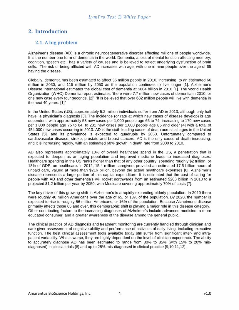
subject. Each dot represents one cell that was purified from the patients’ blood, stimulated and analyzed for the presence of cell-surface markers in the flow cytometer. It is extremely important to appreciate that this pioneering work was replicated and extended several years later when a follow-up study was conducted by the Leipzig team in collaboration with Provista Life Sciences LLC and published in the Neurobiology of Aging in 2012 [58]. The first figure from the Neurobiology of Aging 2012 paper is shown below as Figure 7. Here one can see the “Stimulation Index 2” plotted on the y-axis for 3 groups of patients: (1) Alzheimer’s disease; (2) Parkinson’s disease [dementia or PDD]; and (3) Healthy Control (respectively from left to right). The box represents the values of the SI from the 25 to 75 percentile (or from the lower quartile to the upper quartile). 50% of the data lies within the box. The median value of the distribution is shown as the cross bar within the box, representing the value for which there is one value below this line for every value above this line. The “whiskers” extending out of the box show the range of values observed from the lowest value to the highest value (excluding outliers). The caption reports that this is in CD19 positive lymphocytes, thus equivalent to the right hand pair of bars in the original 2001 Neuroreport shown above as Figure 6. Upon careful review, one will note that the first paper used PHA and the second report used PWM as mitogenic stimulant. Regardless, results appear consistent independent of mitogenic stimulant. In later figures of the 2012 Neurobiology of Aging paper, the authors show various multi-variate scoring models and their Receiver Operator Characteristic curve analysis of those models to predict who is AD from PDD. Although of interest and important, new models will need to be generated from the Amarantus LymPro assay data generated in their GLP contract lab and thus will not be addressed further at this time. Additional questions can be addressed in similar fashion with new data to be generated by Amarantus.
LymPro Test ® White Paper
Amarantus BioScience Holdings, Inc. 16 v1.0
Figure 7. Stieler et al 2012 Figure 1. “Box-and-Whiskers Plot of self-normalized CD69 expression of B cells
(CD19) in response to PWM mitogenic stimulation. Response to mitogenic stimulation was suppressed in AD
subjects (N=32) compared with either [Parkinson’s disease dementia] PDD (N=26) or nondemented subjects
(N=30).”
4.5. Others groups corroborate lymphocyte dysregulation in AD
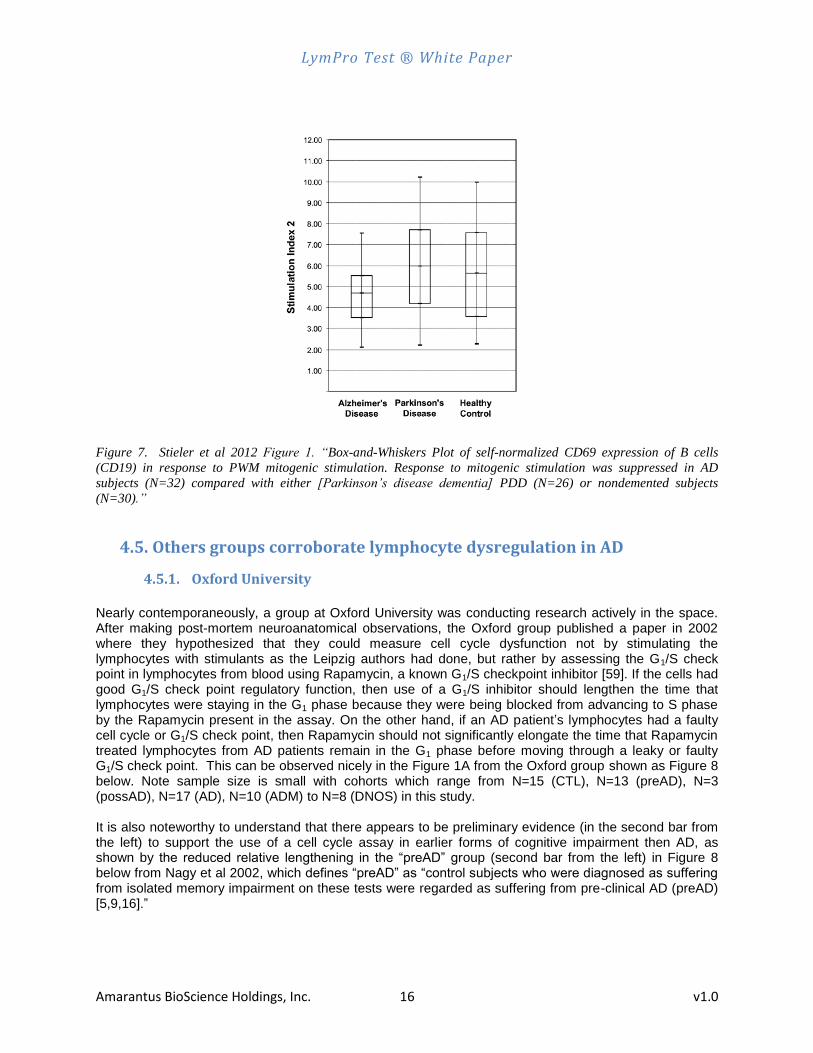
4.5.1. Oxford University Nearly contemporaneously, a group at Oxford University was conducting research actively in the space. After making post-mortem neuroanatomical observations, the Oxford group published a paper in 2002 where they hypothesized that they could measure cell cycle dysfunction not by stimulating the lymphocytes with stimulants as the Leipzig authors had done, but rather by assessing the G1/S check point in lymphocytes from blood using Rapamycin, a known G1/S checkpoint inhibitor [59]. If the cells had good G1/S check point regulatory function, then use of a G1/S inhibitor should lengthen the time that lymphocytes were staying in the G1 phase because they were being blocked from advancing to S phase by the Rapamycin present in the assay. On the other hand, if an AD patient’s lymphocytes had a faulty cell cycle or G1/S check point, then Rapamycin should not significantly elongate the time that Rapamycin treated lymphocytes from AD patients remain in the G1 phase before moving through a leaky or faulty G1/S check point. This can be observed nicely in the Figure 1A from the Oxford group shown as Figure 8 below. Note sample size is small with cohorts which range from N=15 (CTL), N=13 (preAD), N=3 (possAD), N=17 (AD), N=10 (ADM) to N=8 (DNOS) in this study. It is also noteworthy to understand that there appears to be preliminary evidence (in the second bar from the left) to support the use of a cell cycle assay in earlier forms of cognitive impairment then AD, as shown by the reduced relative lengthening in the “preAD” group (second bar from the left) in Figure 8 below from Nagy et al 2002, which defines “preAD” as “control subjects who were diagnosed as suffering from isolated memory impairment on these tests were regarded as suffering from pre-clinical AD (preAD) [5,9,16].”

LymPro Test ® White Paper
Amarantus BioScience Holdings, Inc. 17 v1.0
Figure 8. Nagy et al 2002 Neuroscience Letters, Fig. 1A. (above). “Control, healthy elderly individuals with normal
cognitive and neuropsychological test results; Pre-AD, healthy elderly individuals with neuropsychological test
results suggestive of pre-clinical AD; PossAD, possible AD as diagnosed by the NINCDS criteria; AD, probable AD
as diagnosed by the NINCDS criteria; aDM, AD (NINCDS) and evidence of other types of dementia; DNOS,
patients with dementia who do not meet the requirements of the NINCDS criteria for probable AD. The lines
interconnect patient categories that are significantly different from one another.”
4.5.2. University of South Florida
In 2002, Tan et al published a paper on CD45 isoform alteration in CD4+ T cells as a potential diagnostic marker of Alzheimer’s disease [60]. In their paper, they assessed the CD45RA, CD45RO, CD45RB isoforms on CD4+ T cells sub-populations via Flow Cytometry. Interestingly, they found that AD patients have lower numbers of naïve CD4+ T cells as measured by CD45RA expression level. This result can be seen in Figure 9 below, where both the Cognitive Abnormal and probable AD subjects show reduced CD45RA expression compared with Normal and Other dementias. Although this result is not a functional cell cycle result like that from the Oxford University group, it does indicate a weakened immune response capability in AD patients in the exact CD4+ lymphocyte sub-population as originally presented by Stieler et al 2001 (see Figure 6 above).
Figure 9. Tan J, et al, 2002 J Neuroimmunology, Fig 2B (above). “CD4+ T cells from probable AD
patients and cognitively abnormal participants have altered CD45RA isoform expression profiles compared to
controls. * * * (B) Scatterplot showing percentage of CD45RA-positive CD4+ T cells. The means are
represented as a solid bar. * * * “

LymPro Test ® White Paper
Amarantus BioScience Holdings, Inc. 18 v1.0
4.5.3. Capital Medical University & Ministry of Education, People’s Republic of China
In 2010, Zhou and Jia published a paper on the functional effect of Rapamycin on peripheral blood lymphocytes in both mild and severe Alzheimer’s disease [61], effectively corroborating the Oxford groups work from 2002. In fact, Zhou and Jia cite Nagy et al 2002 as their reference 14. This is an important paper as it corroborates the Rapamycin based assessment of G1/S checkpoint integrity, further buttressing the human clinical evidence that there are peripheral regulatory dysfunctions measurable in the blood lymphocytes of AD patients compared to healthy controls. As can be seen in Figure 10 below (Figure 1 from the 2010 Zhou paper), the percentage of cells in G1 phase is much less in the mild or severe AD cohorts when lymphocytes were treated with Rapamycin, thus indicating that the G1/S check point is leaky and not well regulated in mild and severe AD. As a supplemental corollary, the middle distributions show that there is a disproportionately large fraction of mild and severe AD lymphocytes that have passed the G1/S check point and are in the S phase or the G2 phase.
Figure 10. Zhou X and Jia J, Neuroscience Letters 2010, Fig. 1 (above). “Statistical analysis of the cell cycle
distributions of lymphocytes from mild-AD, severe-AD and non-AD subjects. ** p< 0.01 vs non-AD cells.”
4.5.4. Samsung Medical Center, Seoul, Korea
In 2012, Song et al published a paper on the functional effect of Rapamycin on peripheral blood lymphocytes in mild Alzheimer’s disease (N=26) versus Controls subjects (N=28) [62], effectively corroborating both the Oxford group’s work from 2002 and Zhou & Jia 2010. This work shows that the G1/S checkpoint is faulty in the Seoul Korea sample of AD and Control subjects. As can be seen in Figure 11 below (Figure 1 from the 2012 Song et al paper), the percentage of cells in G1 phase goes down in the AD patients at T48 whereas the S phase percentage goes up, nicely demonstrating the leaky nature of the G1/S check point in AD patients relative to healthy controls. In conclusion, there are now six peer-reviewed papers from five independent research teams that either directly demonstrate cell cycle dysfunction in Alzheimer’s disease as measured in peripheral blood lymphocytes or are consistent with the hypothesis (as in the case of Tan et al 2002 above). Additional corroborative evidence can be found in the list of publications cited in this White Paper found in Section 11.1.
Conceptually, it is important to distinguish the replicated lymphocytic dysfunctional evidence (as summarized above by the six peer-reviewed papers from five independent groups) and the Cell Cycle Hypothesis from which it was originally conceived.

LymPro Test ® White Paper
Amarantus BioScience Holdings, Inc. 19 v1.0
Figure 11. Song et al, Psychiatry Investig 2012, Fig. 1 (above).
4.6. Amarantus’ Investment Hypothesis
“Where there is smoke, there usually is fire.” Today, there are multiple lines of evidence that cell cycle regulation is dysfunctional in AD subjects as measured peripherally via blood derived lymphocytes, yet it is still an open question as to “how relevant or central is Cell Cycle Dysfunction to the core pathophysiology of Alzheimer’s disease and the earlier phases of the disease?”
To answer or de-risk this issue, there is no other path than to invest in conducting well designed clinical studies to answer the question. Thus it is important to reflect for a moment on what is being proposed. Biological markers that can be observed on the surface of cells purified from blood can tell us something profound about properties of neurons in the brain. Moreover, they hypothesize that challenge of cells purified from the blood of an individual to certain stimulants, one can measure the response of those cells in a test tube and say something about how the neurons of the brain are responding to stimulants within the central nervous system. It is a truly ingenious proposition which experimentally has not only replicated in their own hands (Stieler et al, Neurobiology of Aging, 2012), but is also buttressed by the published works of Nagy et al 2002, Tan et al 2002, Zhou and Jia 2010, and Song et al 2012.
Regardless, Amarantus should focus on determining the diagnostic accuracy of the LymPro
Test to identify subjects with Alzheimer’s disease and the earlier phases of the disease.

LymPro Test ® White Paper
Amarantus BioScience Holdings, Inc. 20 v1.0
5. The LymPro Test ® History (as recounted by others) The LymPro assay was original conceived by Thomas Arendt and Jens Stieler. Their host institution, University of Leipzig, filed patent applications to protect the intellectual property. GW Medical licensed the associated intellectual property from Leipzig University in 2004 and went on to begin commercial development, primarily through NIH funding. In 2007, GW Medical was acquired by Provista Life Sciences as part of a strategic alignment of capabilities and assets. Provista Life Sciences was a privately-held diagnostics company focused on developing diagnostic tests and commercializing them in its CLIA-certified laboratory as Laboratory-developed Tests (“LDT”). In 2008, Provista was working to complete a pivotal Clinical Performance Study under an NIH SBIR Phase 2 grant, when the instrumentation they were using became defective, ultimately making the results from that study unreliable and causing them to miss their milestone with the NIH. At the same time Provista Life Sciences had another promising diagnostic test in its pipeline, the dtectDx Breast, a breast cancer diagnostic targeting pre-symptomatic breast cancer. At that time in 2008, the Provista investors decided they wanted to focus their resources on the breast cancer asset and monetize the Alzheimer’s asset. Coupled with the inability to achieve the milestones set out in the Phase 2 NIH grant due to the instrumentation failure and a general lack of funding from private investors in neuroscience research from 2008-2011, the LymPro Test ® seemed to be a much less attractive asset than the dtectDx Breast test. As such, the LymPro Test ® was spun out into MemoryDx to allow the founders of GW Medical to continue their pursuit of bringing the LymPro test to market. The team at GW Medical/Provista/Memory Dx established the methods, got quality results and published their follow-up 2012 Neurobiology of Aging study. Memory Dx entered into an exclusive licensing agreement with Amarantus in December 2012, giving Amarantus rights to develop LymPro commercially for AD. It is important to understand that it was a strategic business decision that Provista was faced with between advancing their breast cancer asset or their AD asset. Recalling that their breast cancer assay was already validated as an assay under CLIA, was beginning to generate revenue, and in a market with many drugs approved and being approved, it was probably a wise business decision. However, in today’s climate where the FDA is pressing for earlier drug development, AD appears to be a ripe opportunity for the LymPro test.

LymPro Test ® White Paper
Amarantus BioScience Holdings, Inc. 21 v1.0
6. Company Next Steps
6.1 Ongoing Amarantus is actively engaged in executing an agreement with an outside GLP laboratory to conduct contract research to support Amarantus R & D. This would include the development and standardization of the assay in the external contract GLP facility. This laboratory has extensive experience running flow cytometer assays and appears an excellent fit. Once the SOPs have been established at the GLP laboratory, an assessment of the analytical performance characteristics and reproducibility of the assay will be established using Peripheral Blood Lymphocytes (PBLs) from a significant number of healthy human subjects. Any deficiencies that are identified will be addressed and resolved prior to initiation of clinical studies.
6.2 Short term Once the analytical performance is established and stabilized over time, the Company expects to run samples on healthy control subjects and panels of human quality control samples. Using a small pilot set of Alzheimer’s disease derived blood samples, analytic performance parameters will be evaluated, including estimates of Stimulation Index variance and SI effect size between AD and control subjects in the newly established assay. This information will be vital to enable meaningful sample size N and power analysis to properly design clinical studies to support the Intended Use objectives that the Company anticipates to set. Additionally, other risks identified while running these pilot disease samples will be mitigated to insure smooth operations and reliable data. The Company intends to reach out to pharmaceutical and biotechnology companies developing therapeutic products in 2013 to enable potential strategic partners to become aware of Amarantus’ development of the LymPro test.
6.3 Near term
The Company intends to verify the clinical performance of the re-established assay in their contract GLP laboratory in a clinical performance Verification Study to reproduce the published LymPro literature Stimulation Index differences (Stieler et al 2001 & 2012) between mild to moderate AD patients and healthy control subjects. As a result of a recent mandate from the FDA, pharmaceutical drug R&D for Alzheimer’s likely will be focused on the earlier stages of the disease. Therefore preliminary assessment of the clinical performance of the assay in early stage patients will be included in the initial clinical performance Verification Study. Lastly, during this period of time, the Company expects to assemble a seasoned IVD executive management team to develop and commercialize the intellectual property as they execute on the present plan and expand the portfolio of activities.
6.4 Medium Term In the medium term, the Company expects to execute multiple and larger clinical performance validation studies, preferably in collaboration with pharmaceutical and biotechnology companies operating in the space, but alone if necessary, where differential diagnosis groups may be included for assessment of clinical specificity. It will be incumbent on the Company’s executive team to identify those pharma and biotech companies with the best synergies moving forward in clinical development before pivotal phase 3 studies. Also, with the clinical performance Verification Study data in hand, the Company will likely file a pre-submission document with the FDA and initiate dialogue with the agency around Intended Use, clinical study design, human specimens and statistical analysis plans. Demonstration of assay technology transfer to other laboratories throughout the world is essential to support multi-national worldwide assay results.
6.5 Long Term

LymPro Test ® White Paper
Amarantus BioScience Holdings, Inc. 22 v1.0
Lastly, if there is a pharma partner that makes sense, then the Company will partner. Otherwise, the Company intends to move forward with FDA and other global regulatory authorities’ clearance/approval as an in vitro diagnostic device with the most relevant Intended Uses, some of which have not yet been investigated but may include MCI, pre-dementia AD or pro-dromal AD.
6.6 Timelines and Financial Projections The Company is currently finalizing agreements with its projected GLP development partner, and is preparing a detailed timeline with an associated financial model which it intends to make available in the near future.

LymPro Test ® White Paper
Amarantus BioScience Holdings, Inc. 23 v1.0
7. Regulatory Authority Considerations
7.1 Regulatory Overview The US (and many other geography’s) regulatory climate for In Vitro Diagnostic (IVD) devices is guided by an assessment of the risk to the patient or subject. Globally, the FDA bar is considered one of the highest for IVD products. In the US, diagnostic IVD products are labeled as Research Use Only (RUO), Investigational Use Only (IUO), 510(k) cleared or PMA approved IVD products. The labeling depends on the level of risk that the FDA perceives a false test reading would cause to the patient or subject, and the degree of evidence that presently exists [63]. The bar for a pre-market notification or 510(k) clearance is lower than that for a Pre-Market Approval or PMA IVD medical device. FDA risk assessment is driven by the Intended Use statement which describes what will be measured, in whom it will be measured and why it will be measured. In addition the FDA expects IVD manufacturers to provide both analytical performance data and clinical performance data to support the Intended Use for their IVD diagnostic product as submitted. The regulatory authority evaluates these data to determine if the device is both “safe and effective” for the use in the population specified in the Intend Use. Although the FDA believes it has regulatory authority over diagnostic tests developed by and for use in a single laboratory (referred to as Laboratory Developed Tests or LDTs), to date they have exercised enforcement discretion and have not actively regulated these assays. Rather the laboratories developing and offering these tests are regulated by CMS (the Centers for Medicare and Medicaid Services) under CLIA (Clinical Laboratory Improvement Amendments). They certify that the lab operates with the appropriate lab personnel and Standard Operating Procedures necessary to achieve reliable results. To meet the definition of Lab Developed Test under CLIA, the LymPro assay must be developed (with established analytical performance and validity) in a single laboratory. If Amarantus wished to offer the test from multiple locations, each laboratory will need to re-establish the analytical validity of the test. In any event if the test is to be made available from multiple laboratories either nationally or internationally, it is recommended that Amarantus and their partners conduct proficiency testing across the laboratory sites to ensure equivalency. While it may be possible to make the LymPro test available as an LDT, in the short term, it is likely to require substantial time and resource to establish the evidentiary dossier necessary for a positive coverage determination and reimbursement. Thus the Company may want to consider its strategic focus positioning the LymPro test to support the clinical development of drugs for AD, including companion diagnostic applications, as well as a FDA approved / cleared standalone IVD diagnostic product. Assuming there is a high likelihood that the LymPro assay is used to select patients for Phase 3 pivotal therapeutic product clinical trials (as an inclusion/exclusion criteria upon enrollment), development of the LymPro test within Amarantus will need to be conducted within a quality management system (subject to 21 CFR Part 820 QSR (Good Manf. Practice for Med. Dev.)).
7.2 Intended Use Based upon the presently published data from Stieler et al (2001 and 2012), candidate language to support an FDA submission may look similar to the following. INTENDED USE
The Amarantus LymPro test is intended for the in vitro qualitative determination of a stimulation index in human blood derived lymphocytes in patients with evidence of cognitive impairment to aid in the diagnosis of dementia of the Alzheimer's type. The test result is not to be used as a standalone diagnostic, but is to be used as an adjunct to clinical assessments and other diagnostic evaluations (e.g. MRI results).
INDICATION FOR USE
A positive test is clinically useful as an aid to help identify those patients with cognitive impairment who have dementia of the Alzheimer’s type.

LymPro Test ® White Paper
Amarantus BioScience Holdings, Inc. 24 v1.0
It is important to understand that when this sort of language is submitted to the FDA in the form of a “pre-submission” package (formerly a Pre-IDE package), there are often changes warranted to the Intended Use language or mandated to be changed by the FDA. With a professional and collaborative spirit, Amarantus should approach the FDA to collaborate to achieve the clearance and/or approval of the LymPro Test. Appropriate regulatory experts should lead Amarantus’ interaction with the FDA in the not too distant future, at least for a pre-submission dialogue around Intended Use, clinical study design, patient specimen selection and statistical analysis plan issues. For companion diagnostic use, a collaborative regulatory interaction including the therapeutic product sponsor is essential to a successful joint co-development and approval. Finally, it is important to appreciate that new research and review of existing research can create new diagnostic opportunities to utilize the LymPro assay for different Intended Uses. As a non-limiting example, the “preAD” group of Nagy et al 2002 (Figure 8 above) motivates further evaluation of the LymPro test in early forms of cognitive impairment, including MCI, prodromal AD or pre-dementia AD where there is minimal evidence of cognitive impairment.

LymPro Test ® White Paper
Amarantus BioScience Holdings, Inc. 25 v1.0
8. Reimbursement Strategy
8.1 United States
There are three main components to reimbursement for diagnostics in the US: (i) coverage, (ii) coding, and (iii) pricing and payment.
8.1.1 Coverage
For payers, the decision whether or not to pay for (cover) a particular diagnostic product is generally driven by evaluation of the clinical evidence supporting the clinical utility of the test. That is the degree to which patient health outcomes are improved with the use of the test, and more importantly for the payers, evidence to support the net economic benefits provided by the improved outcomes. A recent exception is in the case of a companion diagnostic, one that is required for the safe and effective use of a particular therapeutic product; private payers have accepted inclusion of language to that effect in the drug label in lieu of the standard clinical utility data package. (It should be noted however that most of the patients likely to be tested with the LymPro test will be under the jurisdiction of Medicare (the older than 65 years population). Medicare’s coverage policies in particular tend to be more population based rather than individual benefit in their evidentiary review. It is likely that the LymPro test will be ready for launch in a CLIA lab setting prior to the approval as a companion diagnostic of a companion therapeutic product. This means that Amarantus will likely need to undertake the studies necessary to provide the “reasonable and necessary” clinical evidence to support positive decisions by the payers, in particular Medicare. These studies need to:
Validate the test in terms of its analytical performance (sensitivity, specificity and reproducibility, limits of detection etc) across a range of patients who would meet the inclusion criteria for the test;
Confirm the diagnostic accuracy (or clinical validity) of the LymPro test with Alzheimer’s disease or other pre-specified Intended Use populations;
Document the clinical utility (i.e. reduction in costs or improvements in medical outcomes as a result of receiving additional information from the test);
Provide evidence of the health economic impact of those clinical outcomes;
In addition many payers are now requesting evidence that physicians are changing their clinical interventions on the basis of the test results.
The good news about launching the test via a CLIA lab model is that despite the individual origins of the patient samples, all the billing will be conducted through a single Medicare Administrative Contractor or MAC. The medical director at the MAC wields significant power in the decision about whether or not to “cover” a test. Finding a knowledgeable medical director that is willing and able to review the evidence dossier is key, and consequently locating the CLIA lab in a jurisdiction with a diagnostically savvy medical director is prudent. The good news is that by virtue of a “local coverage decision” by the regional MAC, a de facto “national coverage decision” for Medicare patients is obtained. Key influences on payers are peer reviewed papers that cover analytical performance (analytical validity), clinical performance (clinical validity) and clinical utility. Positive technology assessment reports provided by various groups such as the Blue Cross Blue Shield Association Technology Evaluation Center (TEC) and ICER at the Massachusetts General Hospital’s Institute for Technology Assessment are also exceedingly helpful. These reports provide a rigorous assessment of the clinical evidence and assess whether or not the technology improves outcomes such as, quality of life and ability to function. These Technology

LymPro Test ® White Paper
Amarantus BioScience Holdings, Inc. 26 v1.0
Assessment groups do not evaluate costs. The criteria that these groups use to review and assess published trial results are well documented and will be a key input to designing our clinical trial plans. It will also be important to generate and publish data from the clinical utility studies that assess the health economic impact of the changes in outcomes resulting from use of the LymPro test. Modeling the data to reflect the characteristics of the individual health plan will be an important component for approaching and educating the medical directors of the payer plans that influence or directly write policies for coverage. Prior to the availability of these data, Amarantus can generate theoretical models based upon current testing paradigms and previously published literature reflecting patient/physician behavior. These models in combination with published performance data can be used to educate and contract with select private payers (especially single payer systems like Kaiser, Geisinger), hopefully generating favorable coverage policies on a plan-by-plan basis. It should be noted that in the absence of a disease modifying therapy, generating positive health economic outcomes, clinical outcomes evidence for the LymPro test as “an aid to diagnosis of dementia of the Alzheimer’s type” will be challenging. However, the Company should also evaluate cost reduction scenarios which may include the potential of a positive LymPro test result to shorten the “diagnostic odyssey.”
8.1.2 Coding Amarantus intends to pursue the issuance of a Current Procedural Terminology (or CPT) procedure code specifically for the LymPro Test. The strategy will be to initially use a miscellaneous code and negotiate directly with payers for payment of submitted claims until a specific code is issued. This process generally takes 12-18 months through the AMA and would likely result in a temporary (assuming Amarantus could show reasonable utilization data) CPT code. In this case there is no obvious “under the radar strategy” since none of the existing flow cytometry codes will match the ICD9/10 Diagnostic codes for dementia of the Alzheimer type (331.0). In any case the LymPro Test is sufficiently different from existing CPT flow codes that matching the vignette would also be a stretch. Eventually with more clinical evidence, a national or permanent Tier 1 code could be established.
Figure 12. Process for healthcare reimbursement.
8.1.3 Pricing & Payment Pricing levels will initially be negotiated directly with the payers based upon a dossier of information, that may include the following: i) cost analysis of resources required to perform the test (both fixed and variable); ii) amortization of the R&D investment to develop the test; iii) payment amounts determined by other payers; iv) a health economic model reflecting the performance of the LymPro test; v) charges, payment amounts and resources required for tests that may be comparable (crosswalk to other flow cytometry type test) or otherwise relevant (i.e. gap fill to an imaging tests such as Avid Radiopharmaceutical’s (now Lilly’s) Amyvid with an estimated $3,000-$5,000 per procedure in the US); and vi) projected test volume and utilization data. Because private payers often use Medicare as a
e
r ider
r ed re de
ia sis de
laim
rm di s
li
a
e

LymPro Test ® White Paper
Amarantus BioScience Holdings, Inc. 27 v1.0
benchmark when developing their own payment policies the outcome of the Medicare rate setting process could influence the payment rate set by other payers, consequently it is important to have a comprehensive strategy in place for commercialization of the test prior to engaging with the payers. Looking forward, it is anticipated that in the U.S. the emergence of a greater number of Managed Care & Accountable Care organizations will drive Rx/Dx combination pricing discussions.
8.2 Rest of World (ROW) Health economic outcome evidence is typically required throughout the world. Successfully satisfying the very high US standards for economic outcomes likely will position Amarantus for similar success in other countries. However, health economic outcome data may be required to be generated in those countries, and Amarantus will need to evaluate this issue in due course with the clinical performance data leading up to clinical utility evidence.

LymPro Test ® White Paper
Amarantus BioScience Holdings, Inc. 28 v1.0
9. Summary Alzheimer’s disease is currently a very large unmet medical need, which is projected to explode in the developed world with the demographics of the aging population. Accurate and affordable diagnostic information is critical to the management of patients and to the success of therapeutic product sponsor companies as they try to develop therapies for what some estimate to be a $20 billion market. Among technologies, blood analysis is preferred among all other options. Only non-invasive tests are potentially more attractive. The Lymphocyte Proliferation (LymPro) Test ® measures the proliferative response of white blood cells to mitogenic stimulation. Based on differences observed in the response of cells from patients with Alzheimer’s disease as compared with age matched controls and patients with other dementias, it appears that the test has strong potential as a diagnostic for Alzheimer’s. We believe that the LymPro test has the potential to address these needs and is poised for clinical and commercial development. Differential cell cycle and proliferative response of peripheral white blood cells from Alzheimer’s patients has been corroborated by several groups creating a corpus of data which shows it is real. While this is consistent with the Cell Cycle Hypothesis, how central it is to the core pathophysiology of Alzheimer’s disease is still to be determined. Regardless, the diagnostic accuracy and clinical utility of the LymPro test can only be determined after investment in well designed and carefully executed clinical studies. It will be essential that the Company does an excellent job of establishing strong analytical performance characteristics before moving forward with clinical performance assessments. One mistake many inexperienced In Vitro Diagnostic (IVD) developers make is not spending enough time and resources in the beginning months to lock down the finer analytical details to insure assay and calibrator stability, in order to be able to deliver stable performance over years in the future. One only has to look at present
Research Use Only (RUO) CSF A and tau assays to understand that IVD level device development takes a serious commitment from diagnostic professionals.
The LymPro Test offers a large upside potential that the hard work ahead appears well worth the effort and risk. Although there is no guarantee of success, done well, the LymPro test could one day either stand alone or become part of a multi-variate biomarker panel to aid in the diagnosis of individuals in the early stages of Alzheimer’s disease. Investors should be cognizant that the incidence of Alzheimer’s disease and dementia appear consistently increasing, not only in the U.S. but also around the world. Although the downside risks are clearly present, the upside potential is truly disproportionate. Carefully controlled and well executed studies are required; the future for the LymPro Test ® looks very promising at this time.

LymPro Test ® White Paper
Amarantus BioScience Holdings, Inc. 29 v1.0
10. Acknowledgements
I’d like to acknowledge the essential contributions of the following individuals contained within this LymPro Test White Paper. First, I’d like to thank Paul Cannon, PhD for very critical and excellent reviews of the entire manuscript to help clarify many of the concepts discussed; to Katherine Tynan, PhD for writing the core of Section 8 on Reimbursement and her extensive edits throughout the Regulatory section 7; to Jamaal Brown, MBA for writing a huge chunk of section 2.1 and keeping the unmet medical need well documented and described; to Gerald Commissiong and William Gartner for writing the core of Section 5 regarding the history of the LymPro assay.

LymPro Test ® White Paper
Amarantus BioScience Holdings, Inc. 30 v1.0
11. References and Published Literature
11.1 PubMed search: “cell cycle lymphocyte Alzheimer” (22-Apr-2013) 1: Song J, Wang S, Tan M, Jia J. G1/S checkpoint proteins in peripheral blood lymphocytes are potentially
diagnostic biomarkers for Alzheimer's disease. Neurosci Lett. 2012 Sep 27;526(2):144-9. doi:
10.1016/j.neulet.2012.08.020. Epub 2012 Aug 17. PubMed PMID: 22922216.
2: Tan M, Wang S, Song J, Jia J. Combination of p53(ser15) and p21/p21(thr145) in peripheral blood lymphocytes
as potential Alzheimer's disease biomarkers. Neurosci Lett. 2012 May 16;516(2):226-31. doi:
10.1016/j.neulet.2012.03.093. Epub 2012 Apr 6. PubMed PMID: 22503900.
3: Jóźwik A, Landowski J, Bidzan L, Fülop T, Bryl E, Witkowski JM. Beta-amyloid peptides enhance the
proliferative response of activated CD4CD28 lymphocytes from Alzheimer disease patients and from healthy
elderly. PLoS One. 2012;7(3):e33276. doi: 10.1371/journal.pone.0033276. Epub 2012 Mar 12. PubMed PMID:
22428008; PubMed Central PMCID: PMC3299766.
4: Zivković L, Spremo-Potparević B, Plecas-Solarović B, Djelić N, Ocić G, Smiljković P, Siedlak SL, Smith MA,
Bajić V. Premature centromere division of metaphase chromosomes in peripheral blood lymphocytes of Alzheimer's
disease patients: relation to gender and age. J Gerontol A Biol Sci Med Sci. 2010 Dec;65(12):1269-74. doi:
10.1093/gerona/glq148. Epub 2010 Aug 30. PubMed PMID: 20805239; PubMed Central PMCID: PMC2990265.
5: Bartolomé F, Muñoz U, Esteras N, Alquezar C, Collado A, Bermejo-Pareja F, Martín-Requero A. Simvastatin
overcomes the resistance to serum withdrawal-induced apoptosis of lymphocytes from Alzheimer's disease patients.
Cell Mol Life Sci. 2010 Dec;67(24):4257-68. doi: 10.1007/s00018-010-0443-2. Epub 2010 Jul 8. PubMed PMID:
20614159.
6: Bialopiotrowicz E, Kuzniewska B, Kachamakova-Trojanowska N, Barcikowska M, Kuznicki J, Wojda U. Cell
cycle regulation distinguishes lymphocytes from sporadic and familial Alzheimer's disease patients. Neurobiol
Aging. 2011 Dec;32(12):2319.e13-26. doi: 10.1016/j.neurobiolaging.2010.04.017. Epub 2010 Jun 11. PubMed
PMID: 20541838.
7: Stieler J, Grimes R, Weber D, Gartner W, Sabbagh M, Arendt T. Multivariate analysis of differential lymphocyte
cell cycle activity in Alzheimer's disease. Neurobiol Aging. 2012 Feb;33(2):234-41. doi:
10.1016/j.neurobiolaging.2010.03.001. Epub 2010 Apr 14. PubMed PMID: 20395014.
8: Zhou X, Jia J. P53-mediated G(1)/S checkpoint dysfunction in lymphocytes from Alzheimer's disease patients.
Neurosci Lett. 2010 Jan 14;468(3):320-5. doi: 10.1016/j.neulet.2009.11.024. Epub 2009 Nov 13. PubMed PMID:
19914341.
9: Bajić VP, Spremo-Potparević B, Zivković L, Bonda DJ, Siedlak SL, Casadesus G, Lee HG, Smith MA. The X-
chromosome instability phenotype in Alzheimer's disease: a clinical sign of accelerating aging? Med Hypotheses.
2009 Dec;73(6):917-20. doi: 10.1016/j.mehy.2009.06.046. Epub 2009 Jul 31. PubMed PMID: 19647374; PubMed
Central PMCID: PMC2787990.
10: Bajić VP, Spremo-Potparević B, Zivković L, Djelić N, Smith MA. Is the time dimension of the cell cycle re-
entry in AD regulated by centromere cohesion dynamics? Biosci Hypotheses. 2008;1(3):156-161. PubMed PMID:
19122823; PubMed Central PMCID: PMC2612585.
11: Muñoz U, Bartolomé F, Esteras N, Bermejo-Pareja F, Martín-Requero A. On the mechanism of inhibition of p27
degradation by 15-deoxy-Delta12,14-prostaglandin J2 in lymphoblasts of Alzheimer's disease patients. Cell Mol
Life Sci. 2008 Nov;65(21):3507-19. doi: 10.1007/s00018-008-8411-9. PubMed PMID: 18818877.
12: Spremo-Potparević B, Zivković L, Djelić N, Plećas-Solarović B, Smith MA, Bajić V. Premature centromere
division of the X chromosome in neurons in Alzheimer's disease. J Neurochem. 2008 Sep;106(5):2218-23. doi:

LymPro Test ® White Paper
Amarantus BioScience Holdings, Inc. 31 v1.0
10.1111/j.1471-4159.2008.05555.x. Epub 2008 Jul 9. PubMed PMID: 18624923; PubMed Central PMCID:
PMC2746937.
13: Sala SG, Muñoz U, Bartolomé F, Bermejo F, Martín-Requero A. HMG-CoA reductase inhibitor simvastatin
inhibits cell cycle progression at the G1/S checkpoint in immortalized lymphocytes from Alzheimer's disease
patients independently of cholesterol-lowering effects. J Pharmacol Exp Ther. 2008 Jan;324(1):352-9. Epub 2007
Oct 10. PubMed PMID: 17928568.
14: Muñoz U, Bartolomé F, Bermejo F, Martín-Requero A. Enhanced proteasome-dependent degradation of the
CDK inhibitor p27(kip1) in immortalized lymphocytes from Alzheimer's dementia patients. Neurobiol Aging. 2008
Oct;29(10):1474-84. Epub 2007 Apr 19. PubMed PMID: 17448572.
15: Lima PD, Leite DS, Vasconcellos MC, Cavalcanti BC, Santos RA, Costa-Lotufo LV, Pessoa C, Moraes MO,
Burbano RR. Genotoxic effects of aluminum chloride in cultured human lymphocytes treated in different phases of
cell cycle. Food Chem Toxicol. 2007 Jul;45(7):1154-9. Epub 2007 Jan 11. PubMed PMID: 17321660.
16: Boeras DI, Granic A, Padmanabhan J, Crespo NC, Rojiani AM, Potter H. Alzheimer's presenilin 1 causes
chromosome missegregation and aneuploidy. Neurobiol Aging. 2008 Mar;29(3):319-28. Epub 2006 Dec 13.
PubMed PMID: 17169464; PubMed Central PMCID: PMC2692942.
17: Zivković L, Spremo-Potparević B, Djelić N, Bajić V. Analysis of premature centromere division (PCD) of the
chromosome 18 in peripheral blood lymphocytes in Alzheimer disease patients. Mech Ageing Dev. 2006
Dec;127(12):892-6. Epub 2006 Oct 27. PubMed PMID: 17069875.
18: Dönmez-Altuntaş H, Akalin H, Karaman Y, Demirtaş H, Imamoğlu N, Ozkul Y. Evaluation of the nucleolar
organizer regions in Alzheimer's disease. Gerontology. 2005 Sep-Oct;51(5):297-301. PubMed PMID: 16110230.
19: Muñoz U, de Las Cuevas N, Bartolomé F, Hermida OG, Bermejo F, Martín-Requero A. The cyclopentenone
15-deoxy-delta(12,14)-prostaglandin J2 inhibits G1/S transition and retinoblastoma protein phosphorylation in
immortalized lymphocytes from Alzheimer's disease patients. Exp Neurol. 2005 Oct;195(2):508-17. PubMed PMID:
16061222.
20: Targos B, Barańska J, Pomorski P. Store-operated calcium entry in physiology and pathology of mammalian
cells. Acta Biochim Pol. 2005;52(2):397-409. Epub 2005 May 31. Review. PubMed PMID: 15933763.
21: de las Cuevas N, Muñoz U, Hermida OG, Martín-Requero A. Altered transcriptional regulators in response to
serum in immortalized lymphocytes from Alzheimer's disease patients. Neurobiol Aging. 2005 May;26(5):615-24.
PubMed PMID: 15708436.
22: Spremo-Potparevic B, Zivkovic L, Djelic N, Bajic V. Analysis of premature centromere division (PCD) of the X
chromosome in Alzheimer patients through the cell cycle. Exp Gerontol. 2004 May;39(5):849-54. PubMed PMID:
15130680.
23: Wong GT, Manfra D, Poulet FM, Zhang Q, Josien H, Bara T, Engstrom L, Pinzon-Ortiz M, Fine JS, Lee HJ,
Zhang L, Higgins GA, Parker EM. Chronic treatment with the gamma-secretase inhibitor LY-411,575 inhibits beta-
amyloid peptide production and alters lymphopoiesis and intestinal cell differentiation. J Biol Chem. 2004 Mar
26;279(13):12876-82. Epub 2004 Jan 6. PubMed PMID: 14709552.
24: Karussis D, Michaelson DM, Grigoriadis N, Korezyn AD, Mizrachi-Koll R, Chapman S, Abramsky O,
Chapman J. Lack of apolipoprotein-E exacerbates experimental allergic encephalomyelitis. Mult Scler. 2003
Oct;9(5):476-80. PubMed PMID: 14582773.
25: Zhang J, Kong Q, Zhang Z, Ge P, Ba D, He W. Telomere dysfunction of lymphocytes in patients with
Alzheimer disease. Cogn Behav Neurol. 2003 Sep;16(3):170-6. PubMed PMID: 14501538.

LymPro Test ® White Paper
Amarantus BioScience Holdings, Inc. 32 v1.0
26: de las Cuevas N, Urcelay E, Hermida OG, Saíz-Diaz RA, Bermejo F, Ayuso MS, Martín-Requero A.
Ca2+/calmodulin-dependent modulation of cell cycle elements pRb and p27kip1 involved in the enhanced
proliferation of lymphoblasts from patients with Alzheimer dementia. Neurobiol Dis. 2003 Aug;13(3):254-63.
PubMed PMID: 12901840.
27: Scanlan JM, Vitaliano PP, Zhang J, Savage M, Ochs HD. Lymphocyte proliferation is associated with gender,
caregiving, and psychosocial variables in older adults. J Behav Med. 2001 Dec;24(6):537-59. PubMed PMID:
11778349.
28: Nagy Z, Combrinck M, Budge M, McShane R. Cell cycle kinesis in lymphocytes in the diagnosis of Alzheimer's
disease. Neurosci Lett. 2002 Jan 11;317(2):81-4. PubMed PMID: 11755245.
29: Stieler JT, Lederer C, Brückner MK, Wolf H, Holzer M, Gertz HJ, Arendt T. Impairment of mitogenic activation
of peripheral blood lymphocytes in Alzheimer's disease. Neuroreport. 2001 Dec 21;12(18):3969-72. PubMed PMID:
11742221.
30: Yu PH. Involvement of cerebrovascular semicarbazide-sensitive amine oxidase in the pathogenesis of
Alzheimer's disease and vascular dementia. Med Hypotheses. 2001 Aug;57(2):175-9. PubMed PMID: 11461168.
31: Urcelay E, Ibarreta D, Parrilla R, Ayuso MS, Martín-Requero A. Enhanced proliferation of lymphoblasts from
patients with Alzheimer dementia associated with calmodulin-dependent activation of the na+/H+ exchanger.
Neurobiol Dis. 2001 Apr;8(2):289-98. PubMed PMID: 11300724.
32: Park E, Alberti J, Mehta P, Dalton A, Sersen E, Schuller-Levis G. Partial impairment of immune functions in
peripheral blood leukocytes from aged men with Down's syndrome. Clin Immunol. 2000 Apr;95(1 Pt 1):62-9.
PubMed PMID: 10794433.
33: Migliore L, Botto N, Scarpato R, Petrozzi L, Cipriani G, Bonuccelli U. Preferential occurrence of chromosome
21 malsegregation in peripheral blood lymphocytes of Alzheimer disease patients. Cytogenet Cell Genet. 1999;87(1-
2):41-6. PubMed PMID: 10640809.
34: Marx F, Blasko I, Zisterer K, Grubeck-Loebenstein B. Transfected human B cells: a new model to study the
functional and immunostimulatory consequences of APP production. Exp Gerontol. 1999 Sep;34(6):783-95.
PubMed PMID: 10579638.
35: Lombardi VR, García M, Rey L, Cacabelos R. Characterization of cytokine production, screening of lymphocyte
subset patterns and in vitro apoptosis in healthy and Alzheimer's Disease (AD) individuals. J Neuroimmunol. 1999
Jun 1;97(1-2):163-71. PubMed PMID: 10408971.
36: Trieb K, Ransmayr G, Sgonc R, Lassmann H, Grubeck-Loebenstein B. APP peptides stimulate lymphocyte
proliferation in normals, but not in patients with Alzheimer's disease. Neurobiol Aging. 1996 Jul-Aug;17(4):541-7.
PubMed PMID: 8832628.
37: Parshad RP, Sanford KK, Price FM, Melnick LK, Nee LE, Schapiro MB, Tarone RE, Robbins JH. Fluorescent
light-induced chromatid breaks distinguish Alzheimer disease cells from normal cells in tissue culture. Proc Natl
Acad Sci U S A. 1996 May 14;93(10):5146-50. PubMed PMID: 8643543; PubMed Central PMCID: PMC39422.
38: Androsova LV, Sekirina TP, Selezneva ND, Koliaskina GI, Gavrilova SI. [Changes in the immunological
parameters in Alzheimer's disease: their relation to disease severity]. Zh Nevrol Psikhiatr Im S S Korsakova.
1995;95(1):24-7. Russian. PubMed PMID: 7778389.
39: Yao XL, Jenkins EC, Wisniewski HM. Effect of aluminum chloride on mitogenesis, mitosis, and cell cycle in
human short-term whole blood cultures: lower concentrations enhance mitosis. J Cell Biochem. 1994
Apr;54(4):473-7. PubMed PMID: 8014196.

LymPro Test ® White Paper
Amarantus BioScience Holdings, Inc. 33 v1.0
40: Sanford KK, Parshad R, Price FM, Tarone RE, Schapiro MB. X-ray-induced chromatid damage in cells from
Down syndrome and Alzheimer disease patients in relation to DNA repair and cancer proneness. Cancer Genet
Cytogenet. 1993 Oct 1;70(1):25-30. PubMed PMID: 8221609.
41: Ledoux S, Rebai N, Dagenais A, Shaw IT, Nalbantoglu J, Sekaly RP, Cashman NR. Amyloid precursor protein
in peripheral mononuclear cells is up-regulated with cell activation. J Immunol. 1993 Jun 15;150(12):5566-75.
PubMed PMID: 8515076.
42: Nakamura K, Takeda M, Tanaka T, Tanaka J, Kato Y, Nishinuma K, Nishimura T. Glial fibrillary acidic protein
stimulates proliferation and immunoglobulin synthesis of lymphocytes from Alzheimer's disease patients. Methods
Find Exp Clin Pharmacol. 1992 Mar;14(2):141-9. PubMed PMID: 1598026.
43: Melaragno MI, Smith Mde A, Kormann-Bortolotto MH, Toniolo Neto JT. Lymphocyte proliferation and sister
chromatid exchange in Alzheimer's disease. Gerontology. 1991;37(6):293-8. PubMed PMID: 1765278.
44: Tobi SE, Moquet JE, Edwards AA, Lloyd DC, Itzhaki RF. Chromosomal radiosensitivity of lymphocytes from
Alzheimer's disease patients. J Med Genet. 1990 Jul;27(7):437-40. PubMed PMID: 2395162; PubMed Central
PMCID: PMC1017181.
45: Fischman HK, Reisberg B, Albu P, Ferris SH, Rainer JD. Sister chromatid exchanges and cell cycle kinetics in
Alzheimer's disease. Biol Psychiatry. 1984 Mar;19(3):319-27. PubMed PMID: 6426530.
46: Morimoto K, Miura K, Kaneko T, Iijima K, Sato M, Koizumi A. Human health situation and chromosome
alterations: sister chromatid exchange frequency in lymphocytes from passive smokers and patients with hereditary
diseases. Basic Life Sci. 1984;29 Pt B:801-11. PubMed PMID: 6442146.
47: Sulkava R, Rossi L, Knuutila S. No elevated sister chromatid exchange in Alzheimer's disease. Acta Neurol
Scand. 1979 Mar;59(2-3):156-9. PubMed PMID: 452839.
11.2 Related Literature not Cited in this White Paper
1) Miller AE, Neighbour PA, Katzman R, Aronson M, Lipkowitz R, 1981. Immunological studies in senile
dementia of the Alzheimer type: evidence for enhanced suppressor cell activity. Ann Neurol. 10(6): 506-510.
2) Arendt T, Zvegintseva HG, Leontovich TA, 1986. Dendritic changes in the basal nucleus of Meynert and in the
diagonal band nucleus in Alzheimer's disease--a quantitative Golgi investigation. Neuroscience. 19(4): 1265-
1278.
3) Torack RM, 1986. T-lymphocyte function in Alzheimer's disease. Neurosci Lett. 71(3): 365-369.
4) Ikeda T et al, 1991. Immune system-associated antigens on the surface of peripheral blood lymphocytes in
patients with Alzheimer’s disease. Acta Psychiatr. Scand 83: 444-448.
5) Pirttila T, Mattinen S, Frey H, 1992. The decrease of CD8-positive lymphocytes in Alzheimer's disease. J
Neurol Sci. 107(2): 160-165.
6) Lassmann H, Weiler R, Fischer P, Bancher C, Jellinger K, Floor E, Danielczyk W, Seitelberger F, Winkler H,
1992. Synaptic pathology in Alzheimer's disease: immunological data for markers of synaptic and large dense-
core vesicles. Neuroscience 46(1): 1-8.
7) Baumann K, Mandelkow EM, Biernat J, Piwnica-Worms H, Mandelkow E, 1993. Abnormal Alzheimer-like
phosphorylation of tau-protein by cyclin dependent kinases cdk2 and cdk5. FEBS Lett. 336: 417-424.
8) Suzuki T, Oishi M, Marshak DR, Czernik AJ, Nairn AC, Greengard P, 1994. Cell cycle-dependent regulation of
the phosphorylation and metabolism of the Alzheimer amyloid precursor protein. EMBO J. 13: 1114-1122.

LymPro Test ® White Paper
Amarantus BioScience Holdings, Inc. 34 v1.0
9) Burke WJ, McLaughlin JR, Chung HD, Gillespie KN, Grossberg GT, Luque FA, Zimmerman J, 1994.
Occurrence of cancer in Alzheimer and elderly control patients: an epidemiologic necropsy study. Alzheimer
Dis. Assoc. Disord. 8: 22-28.
10) Shalit F, Sredni B, Brodie C, Kott E, Huberman M. 1995. T lymphocyte subpopulations and activation markers
correlate with severity of Alzheimer's disease. Clin Immunol Immunopathol. 75(3): 246-50.
11) Gartner U, Holzer M, Heumann R, Arendt T, 1995. Induction of p21ras in Alzheimer pathology. Neuroreport 6:
1441-1444.
12) Tatebayashi Y, Takeda M, Kashiwagi Y, Okochi M, Kurumadani T, Sekiyama A, Kanayama G, Hariguchi S,
Nishimura T, 1995. Cell-cycle dependent abnormal calcium response in fibroblasts from patients with familial
Alzheimer's disease. Dementia 6: 9-16.
13) Trieb K, Ransmayr G, Sgonc R, Lassmann H, Grubeck-Loebenstein B, 1996. APP peptides stimulate
lymphocyte proliferation in normals, but not in patients with Alzheimer's disease. Neurobiol Aging 17(4): 541-
547.
14) Nagy Z, Esiri MM, Smith AD, 1997. Expression of cell division markers in the hippocampus in Alzheimer's
disease and other neurodegenerative conditions. Acta Neuropathol. (Berl) 93: 294-300.
15) Nagy Z, Esiri MM, Cato AM, Smith AD, 1997. Cell cycle markers in the hippocampus in Alzheimer's disease.
Acta Neuropathol. (Berl) 94: 6-15.
16) Vincent I, Jicha G, Rosado M, Dickson DW, 1997. Aberrant expression of mitotic cdc2/cyclin B1 kinase in
degenerating neurons of Alzheimer's disease brain. J. Neurosci. 17: 3588-3598.
17) Mecocci P, Polidori MC, Ingegni T, Cherubini A, Chionne F, Cecchetti R, Senin U, 1998. Oxidative damage to
DNA in lymphocytes from AD patients. Neurology 51(4): 1014-1017.
18) Arendt T, Holzer M, Gartner U, 1998. Neuronal expression of cycline dependent kinase inhibitors of the INK4
family in Alzheimer's disease. J. Neural Transm. 105: 949-960.
19) Nagy Z, Esiri MM, Smith AD, 1998. The cell division cycle and the pathophysiology of Alzheimer's disease.
Neuroscience 87: 731-739.
20) Lombardi VR, Garcia M, Rey L, Cacabelos R. 1999. Characterization of cytokine production, screening of
lymphocyte subset patterns and in vitro apoptosis in healthy and Alzheimer's Disease (AD) individuals. J
Neuroimmunol. 97(1-2): 163-171.
21) Gartner U, Holzer M, Arendt T, 1999. Elevated expression of p21ras is an early event in Alzheimer's disease
and precedes neurofibrillary degeneration. Neuroscience 91: 1-5.
22) Chang BD, Watanabe K, Broude EV, Fang J, Poole JC, Kalinichenko TV, Roninson IB, 2000. Effects of
p21Waf1/Cip1/Sdi1 on cellular gene expression: implications for carcinogenesis, senescence, and age-related
diseases. Proc. Natl. Acad. Sci. U. S. A 97: 4291-4296.
23) Nagy ZS, Smith MZ, Esiri MM, Barnetson L, Smith AD, 2000. Hyperhomocysteinaemia in Alzheimer's disease
and expression of cell cycle markers in the brain. J. Neurol. Neurosurg. Psychiatry 69: 565-566.
24) Kusdra L et al, 2000. Elevation of CD69+ monocytes/macrophage in patients with Alzheimer’s disease.
Immunobiology 202: 26-33.
25) Nagy Z, 2000. Cell cycle regulatory failure in neurons: causes and consequences. Neurobiol Aging 21: 761-769.

LymPro Test ® White Paper
Amarantus BioScience Holdings, Inc. 35 v1.0
26) Arendt T. 2001. Alzheimer's disease as a disorder of mechanisms underlying structural brain self-organization.
Neuroscience 102(4): 723-765.
27) Yang Y, Geldmacher DS, Herrup K, 2001. DNA replication precedes neuronal cell death in Alzheimer's
disease. J. Neurosci. 21: 2661-2668.
28) Urcelay E, Ibarreta D, Parrilla R, Ayuso MS, Martin-Requero A, 2001. Enhanced proliferation of lymphoblasts
from patients with Alzheimer dementia associated with calmodulin-dependent activation of the na+/H+
exchanger. Neurobiol. Dis. 8: 289-298.
29) Morocz M, Kalman J, Juhasz A, Sinko I, McGlynn AP, Downes CS, Janka Z, Rasko I, 2002. Elevated levels of
oxidative DNA damage in lymphocytes from patients with Alzheimer's disease. Neurobiol Aging. 23(1): 47-53.
30) Nagy Z, Combrinck M, Budge M, McShane R, 2002. Cell cycle kinesis in lymphocytes in the diagnosis of
Alzheimer's disease. Neurosci. Lett. 317: 81- 84.
31) Herrup K, Arendt T, 2002. Re-expression of cell cycle proteins induces neuronal cell death during Alzheimer's
disease. J Alzheimers Dis. 4(3): 243-247.
32) de las Cuevas N, Urcelay E, Hermida OG, Saiz-Diaz RA, Bermejo F, Ayuso MS, Martin-Requero A. 2003 .
Ca2+/calmodulin-dependent modulation of cell cycle elements pRb and p27kip1 involved in the enhanced
proliferation of lymphoblasts from patients with Alzheimer dementia. Neurobiol Dis. 13(3): 254-63.
33) Zhang J, Kong Q, Zhang Z, Ge P, Ba D, He W. 2003. Telomere dysfunction of lymphocytes in patients with
Alzheimer disease. Cogn Behav Neurol. 16(3): 170-6.
34) Jiang S, Zhang M, Ren D, Tang G, Lin S, Qian Y, Zhang Y, Jiang K, Li F, Wang D, 2003. Enhanced
production of amyloid precursor protein mRNA by peripheral mononuclear blood cell in Alzheimer's disease.
Am J Med Genet. 118B(1): 99-102.
35) Ogawa O, Lee HG, Zhu X, Raina A, Harris PL, Castellani RJ, Perry G, Smith MA, 2003. Increased p27, an
essential component of cell cycle control, in Alzheimer's disease. Aging Cell 2: 105-110.
36) Zhu X, McShea A, Harris PL, Raina AK, Castellani RJ, Funk JO, Shah S, Atwood C, Bowen R, Bowser R,
Morelli L, Perry G, Smith MA, 2004. Elevated expression of a regulator of the G2/M phase of the cell cycle,
neuronal CIP-1-associated regulator of cyclin B, in Alzheimer's disease. J. Neurosci. Res. 75: 698-703.
37) Langley B, Ratan RR, 2004. Oxidative stress-induced death in the nervous system: cell cycle dependent or
independent? J. Neurosci. Res. 77: 621-629.
38) Nagy Z, 2005. The last neuronal division: a unifying hypothesis for the pathogenesis of Alzheimer's disease. J.
Cell Mol. Med. 9: 531-541.
39) Casadesus G, Garrett MR, Webber KM, Hartzler AW, Atwood CS, Perry G, Bowen RL, Smith MA, 2006. The
estrogen myth: potential use of gonadotropin-releasing hormone agonists for the treatment of Alzheimer's
disease. Drugs R. D. 7: 187-193.
40) Khan TK, Alkon DL, 2006. An internally controlled peripheral biomarker for Alzheimer's disease: Erk1 and
Erk2 responses to the inflammatory signal bradykinin. Proc. Natl. Acad. Sci. U. S. A 103: 13203-13207.
41) Nagy Z, 2007. The dysregulation of the cell cycle and the diagnosis of Alzheimer's disease. Biochim Biophys
Acta. 1772: 402-408.
42) Uberti D, Lanni C, Racchi M, Govoni S, Memo M, 2008. Conformationally altered p53: a putative peripheral
marker for Alzheimer's disease. Neurodegener Dis 5(3-4): 209-11.

LymPro Test ® White Paper
Amarantus BioScience Holdings, Inc. 36 v1.0
43) Magaki S et al, 2008. Immuophenotypes in the circulation of patients with mild cognitive impairment. J
Psychiatr. Res. 42, 240-246.
44) Bonotis K, Krikki E, Holeva V, Aggouridaki C, Costa V, Baloyannis S.2008. Systemic immune aberations in
Alzheimer’s disease patients. J Neuroimmunol. 193, 183-187.
45) Larbi A et al, 2009. Dramatic shifts in circulating CD4 but not CD8 T cell subsets in mild Alzheimer’s disease.
J Alzheimer’s disease 17: 91-103.
46) Uberti D, Cenini G, Bonini SA, Barcikowska M, Styczynska M, Szybinska A, Memo M, 2010. Increased CD44
gene expression in lymphocytes derived from Alzheimer disease patients. Neurodegener Dis. 7(1-3): 143-147.
11.3 Literature References Cited in this White Paper 1. World Alzheimer’s Report 2012, published by Alzheimer’s Disease International, can be found at: http://www.alz.co.uk/research/world-report-2012 .
2. World Health Organization. Dementia: a public health priority. Geneva : World Health Organization, 2012. ISBN 978-92-4-156445-8.
3. Alzheimer’s Association, 2013. 2013 Alzheimer’s Disease Facts and Figures, Alzheimer’s & Dementia 9(2). To download the report, see: www.alz.org/downloads/facts_figures_2013.pdf .
4. Hebert LE, Beckett LA, Scherr PA, Evans, DA, 2001. Annual incidence of Alzheimer’s disease in the United States projected to the years 2000 through 2050. Alzheimer Dis Assoc Disord 15(4): 169-173.
5. Detailed Tables for the National Vital Statistics Report. Deaths: Final Data for 2010. National Center for Health Statistics. Available at http://www.cdc.gov/nchs/data/dvs/deaths_2010_release.pdf. Accessed on Jan. 30, 2013 {reference from [3] above}.
6. See Ref [3], AA Facts and Figures 2013, at p 29.
7. See Ref [3], AA Facts and Figures 2013, at p 42-43.
8. Rinne JO and Någren K, 2010. Positron emission tomography in at risk patients and in the progression of mild cognitive impairment to Alzheimer's disease. J Alzheimers Dis 19(1): 291-300.
9. Klatka LA, Schiffer RB, Powers JM, et al, 1996. Incorrect diagnosis of Alzheimer's disease. A clinicopathologic study. Arch Neurol 53(1): 35-42.
10. Rasmusson DX, Brandt J, Steele C, et al, 1996. Accuracy of clinical diagnosis of Alzheimer disease and clinical features of patients with non-Alzheimer disease neuropathology. Alzheimer Dis Assoc Disord 10(4): 180-188.
11. Pearl GS, 1997. Diagnosis of Alzheimer's disease in a community hospital-based brain bank program. South Med J 90(7): 720-722.
12. Schneider LS, 2010. Fixing Alzheimer disease trials by improving process and methods. Curr Alzheimer Res. 7: 564-565.
13. See: http://report.nih.gov/categorical_spending.aspx, accessed May 3, 2013.

LymPro Test ® White Paper
Amarantus BioScience Holdings, Inc. 37 v1.0
14. Hardy JA, Higgins GA, 1992. Alzheimer’s disease: the Amyloid Cascade Hypothesis. Science 286: 184-185.
15. Selkoe DJ, 1991. The molecular pathology of Alzheimer’s disease. Neuron 6(4): 487-498.
16. Mandelkow EM, Mandelkow E, 1994. Tau protein and Alzheimer's disease. Neurobiol Aging. 15 Suppl 2: S85-86.
17. Mandelkow EM, Mandelkow E, 2012. Biochemistry and cell biology of tau protein in neurofibrillary degeneration. Cold Spring Harb Perspect Med. 2(7): a006247.
18. Wang Y, Mandelkow E, 2012. Degradation of tau protein by autophagy and proteasomal pathways. Biochem Soc Trans. 40(4): 644-652.
19. Morris M, Maeda S, Vossel K, Mucke L, 2011. The many faces of tau. Neuron 70(3): 410-426.
20. Shi S, Wang Z, Qiao Z, 2013. The Multifunctional Anti-Inflammatory Drugs Used In The Therapy Of Alzheimer's Disease. Curr Med Chem. 2013 Apr 1. [Epub ahead of print].
21. Teixeira J, Silva T, Andrade PB, Borges F, 2013. Alzheimer Disease and Antioxidant Therapy: How long how far? Curr Med Chem. 2013 Jan 28. [Epub ahead of print].
22. Roses AD, 1996. Apolipoprotein E alleles as risk factors in Alzheimer’s disease. Annu Rev Med 47: 387-400.
23. Liu CC, Kanekiyo T, Xu H, Bu G, 2013. Apolipoprotein E and Alzheimer disease: risk, mechanisms and therapy. Nat Rev Neurol. 9(2): 106-118.
24. Dubois B, Feldman HH, Jacova C, Dekosky ST, Barberger-Gateau P, Cummings J, Delacourte A, Galasko D, Gauthier S, Jicha G, Meguro K, O'brien J, Pasquier F, Robert P, Rossor M, Salloway S, Stern Y, Visser PJ, Scheltens P, 2007. Research criteria for the diagnosis of Alzheimer's disease: revising the NINCDS-ADRDA criteria. Lancet Neurol. 6(8): 734-746.
25. Dubois B, Feldman HH, Jacova C, Cummings JL, Dekosky ST, Barberger-Gateau P, Delacourte A, Frisoni G, Fox NC, Galasko D, Gauthier S, Hampel H, Jicha GA, Meguro K, O'Brien J, Pasquier F, Robert P, Rossor M, Salloway S, Sarazin M, de Souza LC, Stern Y, Visser PJ, Scheltens P, 2010. Revising the definition of Alzheimer's disease: a new lexicon. Lancet Neurol. 9(11): 1118-1127.
26. For a nice review of the literature, please see the EMA website at www.ema.europa.eu/docs/en_GB/document_library/Regulatory_and_procedural_guideline/2012/04/WC500125019.pdf .
27. AAIC Vancouver, Vancouver, Canada, July 2012.
28. See http://www.myriadrbm.com/products-services/humanmap-services/human-discoverymap250/ .
29. See http://www.diagenic.com/?module=Articles&action=Article.publicOpen&id=200 .
30. See: http://www.opko.com/diagnostics/molecular-diagnostics-platform/disease-areas/ .
31. See: http://www.cytoxgroup.com/ .
32. For a primer on the cell cycle, please find web tutorials at http://en.wikipedia.org/wiki/Cell_cycle or http://www.biology.arizona.edu/cell_bio/tutorials/cell_cycle/main.html .

LymPro Test ® White Paper
Amarantus BioScience Holdings, Inc. 38 v1.0
33. Glenner GG, Wong CW, 1984. Alzheimer's disease: initial report of the purification and characterization of a novel cerebrovascular amyloid protein. Biochem Biophys Res Commun. 120(3): 885-890.
34. Glenner GG, Wong CW, Quaranta V, Eanes ED, 1984. The amyloid deposits in Alzheimer's disease: their nature and pathogenesis. Appl Pathol. 2(6): 357-369.
35. Grundke-Iqbal I, Iqbal K, Quinlan M, Tung YC, Zaidi MS, Wisniewski HM, 1986. Microtubule-associated protein tau. A component of Alzheimer paired helical filaments. J Biol Chem. 261(13): 6084-6089.
36. Grundke-Iqbal I, Iqbal K, Tung YC, Quinlan M, Wisniewski HM, Binder LI, 1986. Abnormal phosphorylation of the microtubule-associated protein tau (tau) in Alzheimer cytoskeletal pathology. Proc Natl Acad Sci U S A. 83(13): 4913-4917.
37. Nukina N, Ihara Y, 1986. One of the antigenic determinants of paired helical filaments is related to tau protein. J Biochem. 99(5): 1541-1544.
38. Ihara Y, Nukina N, Miura R, Ogawara M, 1986. Phosphorylated tau protein is integrated into paired helical filaments in Alzheimer's disease. J Biochem. 99(6): 1807-1810.
39. Webber KM, Raina AK, Marlatt MW, Zhu X, Prat MI, Morelli L, Casadesus G, Perry G, Smith MA, 2005. The cell cycle in Alzheimer disease: a unique target for neuropharmacology. Mech Ageing Dev. 126(10): 1019-1025.
40. Lopes JP, Oliveira CR, Agostinho P, 2009. Cell cycle re-entry in Alzheimer's disease: a major neuropathological characteristic? Curr Alzheimer Res. 6(3): 205-212.
41. Arendt T, 2012. Cell cycle activation and aneuploid neurons in Alzheimer's disease. Mol Neurobiol. 46(1): 125-135.
42. Herrup K, 2012. The contributions of unscheduled neuronal cell cycle events to the death of neurons in Alzheimer's disease. Front Biosci (Elite Ed). 4: 2101-2109.
43. Keeney JT, Swomley AM, Harris JL, Fiorini A, Mitov MI, Perluigi M, Sultana R, Butterfield DA, 2012. Cell cycle proteins in brain in mild cognitive impairment: insights into progression to Alzheimer disease. Neurotox Res. 22(3): 220-230.
44. Holzer M, Holzapfel HP, Zedlick D, Brückner MK, Arendt T, 1994. Abnormally phosphorylated tau protein in Alzheimer's disease: heterogeneity of individual regional distribution and relationship to clinical severity. Neuroscience 63(2): 499-516.
45. Gartner U, Holzer R, Heumannand R, Arendt T, 1995. Induction of p21ras in Alzheimer pathology. Neuroreport 6: 1441-1444.
46. Arendt T, Rödel L, Gärtner U, Holzer M, 1996. Expression of the cyclin-dependent kinase inhibitor p16 in Alzheimer's disease. Neuroreport 7(18): 3047-3049.
47. Bigl M, Bleyl AD, Zedlick D, Arendt T, Bigl V, Eschrich K, 1996. Changes of activity and isozyme pattern of phosphofructokinase in the brains of patients with Alzheimer's disease. J Neurochem. 67(3): 1164-1171.
48. Arendt T, Holzer M, Gartner U, 1998. Neuronal expression of cycline dependent kinase inhibitors of the INK4 family in Alzheimer’s disease. J. Neural Transm. 105: 949-960.

LymPro Test ® White Paper
Amarantus BioScience Holdings, Inc. 39 v1.0
49. Arendt T, Holzer M, Gärtner U, Brückner MK, 1998. Aberrancies in signal transduction and cell cycle related events in Alzheimer's disease. J Neural Transm Suppl. 54: 147-58.
50. Gartner U, Holzer M, Arendt T, 1999. Elevated expression of p21ras is an early event in Alzheimer's disease and precedes neurofibrillary degeneration. Neuroscience 91: 1-5.
51. Vincent I, Jicha G, Rosado M, Dickson DW, 1997. Aberrant expression of mitotic cdc2/cyclin B1 kinase in degenerating neurons of Alzheimer's disease brain. J. Neurosci. 17: 3588-3598.
52. Nagy Z, Esiri MM, Smith AD, 1997. Expression of cell division markers in the hippocampus in Alzheimer's disease and other neurodegenerative conditions. Acta Neuropathol. (Berl) 93: 294-300.
53. Nagy Z, Esiri MM, Cato AM, Smith AD, 1997. Cell cycle markers in the hippocampus in Alzheimer's disease. Acta Neuropathol. (Berl) 94: 6-15.
54. Nagy Z, Esiri MM, Smith AD, 1998. The cell division cycle and the pathophysiology of Alzheimer's disease. Neuroscience 87: 731-739.
55. Nagy ZS, Smith MZ, Esiri MM, Barnetson L, Smith AD, 2000. Hyperhomocysteinaemia in Alzheimer's disease and expression of cell cycle markers in the brain. J. Neurol. Neurosurg. Psychiatry 69: 565-566.
56. Yang Y, Geldmacher DS, Herrup K, 2001. DNA replication precedes neuronal cell death in Alzheimer's disease. J. Neurosci. 21: 2661-2668.
57. Stieler JT, Lederer C, Bruckner MK, Wolf H, Holzer M, Gertz HJ, Arendt T, 2001. Impairment of mitogenic activation of peripheral blood lymphocytes in Alzheimer's disease. NeuroReport 12: 3969-3972.
58. Steiler J, Grimes R, Weber D, Gartner W, Sabbagh, Arendt T, 2012. Multivariate analysis of differential lymphocyte cel cycle activity in Alzheimer’s disease. Neurobiology of Aging 33: 234-341.
59. Nagy Z, Combrinck M, Budge M, McShane R, 2002. Cell cycle kinesis in lymphocytes in the diagnosis of Alzheimer's disease. Neurosci. Lett. 317: 81- 84.
60. Tan J, Town T, Abdullah L, Wu Y, Placzek A, Small B, Kroeger J, Crawford F, Richards D, Mullan M. 2002. CD45 isoform alteration in CD4+ T cells as a potential diagnostic marker of Alzheimer's disease. J Neuroimmunol 132: 164-72.
61. Zhou X and Jia J, 2010. P53-mediated G1/S checkpoint dysfunction in lymphocytes from Alzheimer’s disease patients. Neuroscience Letters 468: 320-325.
62. Song M, Kwon Y-A, Lee Y, Kim H, Yn J H, Kim S, Kim D K, 2012. G1/S Cell Cycle Checkpoint Defect in Lymphocytes from Patients with Alzheimer’s. Psychiatry Investig 9: 413-417.
63. For further clarification, see http://www.fda.gov/medicaldevices/deviceregulationandguidance/guidancedocuments/ucm253307.htm . 9may2013