Alveolar Type II Epithelial Cells Contribute to the …Alveolar Type II Epithelial Cells Contribute...
11
Alveolar Type II Epithelial Cells Contribute to the Anti-Influenza A Virus Response in the Lung by Integrating Pathogen- and Microenvironment-Derived Signals S. Stegemann-Koniszewski, a,b Andreas Jeron, b Marcus Gereke, a,b Robert Geffers, c Andrea Kröger, d,e Matthias Gunzer, f Dunja Bruder a,b Immune Regulation, Helmholtz Centre for Infection Research, Braunschweig, Germany a ; Infection Immunology, Institute of Medical Microbiology, Infection Control and Prevention, Otto-von-Guericke University, Magdeburg, Germany b ; Genome Analytics, Helmholtz Centre for Infection Research, Braunschweig, Germany c ; Innate Immunity and Infection, Helmholtz Centre for Infection Research, Braunschweig, Germany d ; Molecular Microbiology, Institute of Medical Microbiology, Infection Control and Prevention, Otto-von-Guericke University, Magdeburg, Germany e ; Institute of Experimental Immunology and Imaging, University of Duisburg-Essen, Essen, Germany f S.S.-K. and A.J. contributed equally to this work. ABSTRACT Influenza A virus (IAV) periodically causes substantial morbidity and mortality in the human population. In the lower lung, the primary targets for IAV replication are type II alveolar epithelial cells (AECII), which are increasingly recognized for their immunological potential. So far, little is known about their reaction to IAV and their contribution to respiratory antivi- ral immunity in vivo. Therefore, we characterized the AECII response during early IAV infection by analyzing transcriptional regulation in cells sorted from the lungs of infected mice. We detected rapid and extensive regulation of gene expression in AECII following in vivo IAV infection. The comparison to transcriptional regulation in lung tissue revealed a strong contribu- tion of AECII to the respiratory response. IAV infection triggered the expression of a plethora of antiviral factors and immune mediators in AECII with a high prevalence for interferon-stimulated genes. Functional pathway analyses revealed high activity in pathogen recognition, immune cell recruitment, and antigen presentation. Ultimately, our analyses of transcriptional regula- tion in AECII and lung tissue as well as interferon I/III levels and cell recruitment indicated AECII to integrate signals provided by direct pathogen recognition and surrounding cells. Ex vivo analysis of AECII proved a powerful tool to increase our under- standing of their role in respiratory immune responses, and our results clearly show that AECII need to be considered a part of the surveillance and effector system of the lower respiratory tract. IMPORTANCE In order to confront the health hazard posed by IAV, we need to complete our understanding of its pathogenesis. AECII are primary targets for IAV replication in the lung, and while we are beginning to understand their importance for respi- ratory immunity, the in vivo AECII response during IAV infection has not been analyzed. In contrast to studies addressing the response of AECII infected with IAV ex vivo, we have performed detailed gene transcriptional profiling of AECII isolated from the lungs of infected mice. Thereby, we have identified an exceptionally rapid and versatile response to IAV infection that is shaped by pathogen-derived as well as microenvironment-derived signals and aims at the induction of antiviral measures and the recruitment and activation of immune cells. In conclusion, our study presents AECII as active players in antiviral defense in vivo that need to be considered part of the sentinel and effector immune system of the lung. Received 15 March 2016 Accepted 30 March 2016 Published 3 May 2016 Citation Stegemann-Koniszewski S, Jeron A, Gereke M, Geffers R, Kröger A, Gunzer M, Bruder D. 2016. Alveolar type II epithelial cells contribute to the anti-influenza A virus response in the lung by integrating pathogen- and microenvironment-derived signals. mBio 7(3):e00276-16. doi:10.1128/mBio.00276-16. Editor Michael G. Katze, University of Washington Copyright © 2016 Stegemann-Koniszewski et al. This is an open-access article distributed under the terms of the Creative Commons Attribution 4.0 International license. Address correspondence to S. Stegemann-Koniszewski, [email protected]. I nfluenza A virus (IAV) still poses a serious threat to human health, and a detailed understanding of IAV pathogenesis is es- sential to adequately confront this hazard. IAV infections are pri- marily restricted to the respiratory tract, where epithelial cells, alveolar macrophages (AM), and dendritic cells (DC) trigger the first innate responses (1). IAV bears ligands for several pathogen recognition receptors (PRR), and the main triggered PRR are Toll-like receptor 3 (TLR3) and TLR7 as well as RIG-I, MDA5, and the NLRP3 inflammasome (1). These are engaged in the an- tiviral response in a cell-type-specific manner (2, 3). Via partly redundant signaling pathways, PRR ligation leads to the activation of effector mechanisms comprised of type I/III interferons (IFNs), inflammatory mediators, antimicrobial effectors, and signals in- ducing adaptive immunity. In general, viral infections are marked by the strong release of type I interferons. These trigger the expres- sion of a multitude of interferon-stimulated genes (ISG) through the ubiquitously expressed IFN-/ receptor (IFNAR) (2). ISG expression is also induced through IFN- (IFN III), which is re- leased during IAV infection and is sensed through the interleukin-28 (IL-28) receptor (IL-28R) primarily expressed by epithelial cells of the respiratory tract and gut (4). In the lower respiratory tract, the lining epithelium is com- prised of alveolar type I and type II epithelial cells (AECI and AECII, respectively), and at this site, AECII are the main target RESEARCH ARTICLE crossmark May/June 2016 Volume 7 Issue 3 e00276-16 ® mbio.asm.org 1 on May 12, 2020 by guest http://mbio.asm.org/ Downloaded from
Transcript of Alveolar Type II Epithelial Cells Contribute to the …Alveolar Type II Epithelial Cells Contribute...
Alveolar Type II Epithelial Cells Contribute to the Anti-Influenza AVirus Response in the Lung by Integrating Pathogen- andMicroenvironment-Derived Signals
S. Stegemann-Koniszewski,a,b Andreas Jeron,b Marcus Gereke,a,b Robert Geffers,c Andrea Kröger,d,e Matthias Gunzer,f
Dunja Brudera,b
Immune Regulation, Helmholtz Centre for Infection Research, Braunschweig, Germanya; Infection Immunology, Institute of Medical Microbiology, Infection Control andPrevention, Otto-von-Guericke University, Magdeburg, Germanyb; Genome Analytics, Helmholtz Centre for Infection Research, Braunschweig, Germanyc; Innate Immunityand Infection, Helmholtz Centre for Infection Research, Braunschweig, Germanyd; Molecular Microbiology, Institute of Medical Microbiology, Infection Control andPrevention, Otto-von-Guericke University, Magdeburg, Germanye; Institute of Experimental Immunology and Imaging, University of Duisburg-Essen, Essen, Germanyf
S.S.-K. and A.J. contributed equally to this work.
ABSTRACT Influenza A virus (IAV) periodically causes substantial morbidity and mortality in the human population. In thelower lung, the primary targets for IAV replication are type II alveolar epithelial cells (AECII), which are increasingly recognizedfor their immunological potential. So far, little is known about their reaction to IAV and their contribution to respiratory antivi-ral immunity in vivo. Therefore, we characterized the AECII response during early IAV infection by analyzing transcriptionalregulation in cells sorted from the lungs of infected mice. We detected rapid and extensive regulation of gene expression inAECII following in vivo IAV infection. The comparison to transcriptional regulation in lung tissue revealed a strong contribu-tion of AECII to the respiratory response. IAV infection triggered the expression of a plethora of antiviral factors and immunemediators in AECII with a high prevalence for interferon-stimulated genes. Functional pathway analyses revealed high activityin pathogen recognition, immune cell recruitment, and antigen presentation. Ultimately, our analyses of transcriptional regula-tion in AECII and lung tissue as well as interferon I/III levels and cell recruitment indicated AECII to integrate signals providedby direct pathogen recognition and surrounding cells. Ex vivo analysis of AECII proved a powerful tool to increase our under-standing of their role in respiratory immune responses, and our results clearly show that AECII need to be considered a part ofthe surveillance and effector system of the lower respiratory tract.
IMPORTANCE In order to confront the health hazard posed by IAV, we need to complete our understanding of its pathogenesis.AECII are primary targets for IAV replication in the lung, and while we are beginning to understand their importance for respi-ratory immunity, the in vivo AECII response during IAV infection has not been analyzed. In contrast to studies addressing theresponse of AECII infected with IAV ex vivo, we have performed detailed gene transcriptional profiling of AECII isolated fromthe lungs of infected mice. Thereby, we have identified an exceptionally rapid and versatile response to IAV infection that isshaped by pathogen-derived as well as microenvironment-derived signals and aims at the induction of antiviral measures andthe recruitment and activation of immune cells. In conclusion, our study presents AECII as active players in antiviral defense invivo that need to be considered part of the sentinel and effector immune system of the lung.
Received 15 March 2016 Accepted 30 March 2016 Published 3 May 2016
Citation Stegemann-Koniszewski S, Jeron A, Gereke M, Geffers R, Kröger A, Gunzer M, Bruder D. 2016. Alveolar type II epithelial cells contribute to the anti-influenza A virusresponse in the lung by integrating pathogen- and microenvironment-derived signals. mBio 7(3):e00276-16. doi:10.1128/mBio.00276-16.
Editor Michael G. Katze, University of Washington
Copyright © 2016 Stegemann-Koniszewski et al. This is an open-access article distributed under the terms of the Creative Commons Attribution 4.0 International license.
Address correspondence to S. Stegemann-Koniszewski, [email protected].
Influenza A virus (IAV) still poses a serious threat to humanhealth, and a detailed understanding of IAV pathogenesis is es-
sential to adequately confront this hazard. IAV infections are pri-marily restricted to the respiratory tract, where epithelial cells,alveolar macrophages (AM), and dendritic cells (DC) trigger thefirst innate responses (1). IAV bears ligands for several pathogenrecognition receptors (PRR), and the main triggered PRR areToll-like receptor 3 (TLR3) and TLR7 as well as RIG-I, MDA5,and the NLRP3 inflammasome (1). These are engaged in the an-tiviral response in a cell-type-specific manner (2, 3). Via partlyredundant signaling pathways, PRR ligation leads to the activationof effector mechanisms comprised of type I/III interferons (IFNs),
inflammatory mediators, antimicrobial effectors, and signals in-ducing adaptive immunity. In general, viral infections are markedby the strong release of type I interferons. These trigger the expres-sion of a multitude of interferon-stimulated genes (ISG) throughthe ubiquitously expressed IFN-�/� receptor (IFNAR) (2). ISGexpression is also induced through IFN-� (IFN III), which is re-leased during IAV infection and is sensed through theinterleukin-28 (IL-28) receptor � (IL-28R�) primarily expressedby epithelial cells of the respiratory tract and gut (4).
In the lower respiratory tract, the lining epithelium is com-prised of alveolar type I and type II epithelial cells (AECI andAECII, respectively), and at this site, AECII are the main target
RESEARCH ARTICLE
crossmark
May/June 2016 Volume 7 Issue 3 e00276-16 ® mbio.asm.org 1
on May 12, 2020 by guest
http://mbio.asm
.org/D
ownloaded from

cells for IAV replication (5, 6). AECII cover about 5% of the alve-olar surface, while they comprise about 60% of the alveolar liningcells and 15% of the parenchymal cells (7). Until recently, thesecretion of surfactant, the maintenance of the mechanical bar-rier, and the provision of constitutive antimicrobial defense wereconceived as their main functions (7, 8). Beyond these, we are onlybeginning to understand the potential of AECII to regulate respi-ratory immune responses in autoimmunity and infection (9–12).
Since AECII are primary targets for viral replication in thelower lung and actively contribute to pulmonary immunity, it islikely that they influence efficient host responses directed at IAV.A number of studies have addressed the response of AECII to IAVin vitro and showed them to express functional PRR and to pro-duce cytokines and chemokines (13–17). The nature and the rel-evance of the AECII response to IAV, however, lack ultimate clar-ification, as these studies were performed using cell lines orprimary cells infected in culture. Little is known about the AECIIresponse to IAV infection in vivo and how this contributes to therespiratory immune reaction. To overcome these limitations, wecharacterized the in vivo response of AECII to IAV infection byanalyzing primary AECII from the lungs of infected mice.
RESULTSIAV infection triggers AECII-specific transcriptional regulationin vivo. We have previously optimized our IAV infection modelfor the isolation of pure and viable AECII that express increasingamounts of viral protein over the first days of infection (9, 18). Inthis model of lethal IAV infection, mice rapidly lose weight, thevirus replicates efficiently in the lungs, and high copy numbers of
the viral genome are detected in the isolated AECII (see Fig. S1 inthe supplemental material). Typically, inflammatory mediatorsand immune cell recruitment to the respiratory tract are inducedby day 3 postinfection (19), and we analyzed transcriptional reg-ulation in AECII and lung tissue at this time point. For AECII, twoindependent microarray experiments were conducted for eachcondition and the material for each independent experiment waspooled from five infected animals. For lung tissue, three indepen-dent microarrays were performed for each condition and eacharray represents an independent animal. Fold change (FC) valuesof 2 or more over controls were considered indicative of up- ordownregulation. In lung tissue, 878 transcripts were upregulatedand 123 were downregulated following IAV infection. Extensivetranscriptional regulation was also detected in AECII, with 546upregulated and 42 downregulated transcripts (Fig. 1A). In thelung, the 5 most intensely upregulated transcripts wereinterleukin-6 (IL-6) (FC � 90), CXCL10, and the IFN-inducedproteins Mx1 and Rsad2 as well as Slfn4, which has been suggestedto be IFN I inducible (20). There was an overlap with AECII,where Rsad2 and Slfn4, along with the IFN-induced proteins Ifit1,Ifit3, and Iigp1, were among the top 5 most regulated transcripts,indicating a prominent role for IFNs in the AECII response(Fig. 1B). The intense upregulation of CXCL5, CXCL9, andCXCL10 (Fig. 1B) in AECII as well as the large overlap of thetranscripts found regulated in either lungs or AECII (Fig. 1C)pointed at a strong potential of AECII to contribute to respiratoryimmunity. In order to confirm the AECII transcriptional regula-tion following in vivo IAV infection detected by the microarrayanalyses, the expression of a selection of these transcripts was an-
FIG 1 Respiratory IAV infection triggers AECII-specific transcriptional regulation. Mice were infected with IAV or treated with PBS and sacrificed 3 days later.Total RNA from whole lungs (n � 3 mice for each condition) and sorted AECII (n � 2 individual samples for each condition; 5 mice per sample) was subjectedto microarray analysis. Data were analyzed by comparing IAV-infected with uninfected control samples. (A) Scatter plots of regulated transcripts with a foldchange of ��2 (threshold represented by the diagonal lines). Data represent normalized log2 signal intensities (averaged over replicates). The number ofup-/downregulated transcripts and the gene symbols of the top 5 upregulated transcripts are indicated. (B) List of the 20 most intensely upregulated transcriptsin AECII with fold change values for AECII and lung. (C) Comparison of the transcripts identified in panel A with respect to regulation in lung and/or AECII.(D) Scatter plot showing absolute log2 fold changes over the respective PBS controls of the transcripts differentially expressed in AECII and/or lung tissue. Thedashed bisecting lines indicate equal fold changes. Gray lines indicate the fold change threshold of �2.
Stegemann-Koniszewski et al.
2 ® mbio.asm.org May/June 2016 Volume 7 Issue 3 e00276-16
on May 12, 2020 by guest
http://mbio.asm
.org/D
ownloaded from
alyzed by quantitative real-time PCR. Indeed, this approach con-firmed the significant upregulation of CXCL5, CXCL10, IFIT2,IRF7, MX2, and USP18 (see Fig. S2). Overall, the detection ofAECII-specific transcripts showed that AECII most likely servecell-type-specific functions. These transcripts were associatedwith Gene Ontology (GO) terms such as defense response (Bon-ferroni corrected P value, 2.8 � 10�12), innate immune response(corrected P value, 0.00013), and cytokine production (correctedP value, 0.00077). The quality, i.e., whether up- or downregula-tion occurred, of transcriptional regulation in AECII and lungtissue was not changed between the two sample sets (Fig. 1D).Overall, these results clearly demonstrated that AECII stronglyreact to IAV infection in vivo and hold strong potential to contrib-ute to the respiratory immune response. To characterize this con-tribution in greater detail, we performed AECII transcriptionalanalyses over the early course of infection.
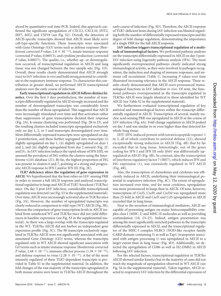
Early transcriptional regulation in AECII follows distinct ki-netics. Over the first 3 days postinfection, the number of tran-scripts differentially regulated in AECII strongly increased and thenumber of downregulated transcripts was considerably lowerthan the number of those upregulated. This suggested that AECIIwere increasingly stimulated over time and that activation ratherthan suppression of gene transcription dictated their response(Fig. 2A). k-means clustering was performed (Fig. 2B to D), andthe resulting clusters included transcripts upregulated exclusivelyonly on day 1, 2, or 3 and transcripts downregulated over time.Most differentially expressed transcripts were upregulated on day3 postinfection, and these further segregated into transcripts (i)slightly upregulated on day 1, (ii) slightly upregulated on days 1and 2, and (iii) slightly upregulated from day 2 onward (Fig. 2Cand D). As IAV infection induces the rapid release of IFN I/III, weassessed the prevalence of ISG within the clusters using the Inter-ferome v2.01 database (21). By far, the highest proportion of ISGwas present in clusters 6 and 7, pointing at a strong and progres-sive AECII response to IFN I and/or IFN III (Fig. 2E).
TLR7 deficiency alters the regulation of gene expression inAECII. We hypothesized that the host relies on IAV-sensing PRRin order to mount a full AECII response and analyzed transcrip-tional regulation in lungs and AECII of TLR7-knockout (TLR7ko)mice. On day 3 post-IAV infection, considerable transcriptionalregulation was detected (see Fig. S3 in the supplemental material).Over time, AECII were increasingly activated also in TLR7ko mice(Fig. 3A). However, the number of upregulated transcripts wasclearly reduced in comparison to wild-type (WT) AECII (Fig. 3B),whereas the comparison of gene transcription levels in AECII iso-lated from uninfected WT and TLR7ko mice did not yield differ-ences in baseline expression (see Fig. S3 in the supplemental ma-terial). As there was a large overlap with the transcripts regulatedin the WT, TLR7ko AECII did not harbor an independent geneexpression profile (Fig. 3C). The 90 transcripts exclusively regu-lated in TLR7ko AECII were not significantly associated with anyannotated Gene Ontology (GO) terms, whereas the transcriptsregulated only in WT AECII showed significant association withGO terms such as innate immune response (Bonferroni correctedP value, 2.68 � 10�21), immune effector process (1.38 � 10�18),and defense response to virus (2.28 � 10�15). A list of the mostintensely regulated of these TLR7-dependent transcripts is pro-vided in Table S1 in the supplemental material. In addition, thefold changes of the vast majority of the transcripts upregulated inboth mouse strains were lower in TLR7ko AECII throughout the
early course of infection (Fig. 3D). Therefore, the AECII responseof TLR7-deficient hosts during IAV infection was blunted regard-ing both the number of differentially expressed transcripts and thedegree of fold change regulation, demonstrating the importanceof a single PRR for the full AECII response.
IAV infection triggers transcriptional regulation of a multi-tude of immunological factors. We performed pathway analyseson the transcripts differentially expressed in AECII on day 3 post-IAV infection using Ingenuity pathway analysis (IPA). The mostsignificantly overrepresented pathways clearly indicated strongimmunological activity, as they were involved in pathogen recog-nition, the induction and shaping of immune responses, and im-mune cell recruitment (Table 1). Increasing P values over timeillustrated increasing relevance in the AECII response. These re-sults clearly demonstrated that AECII exert pronounced immu-nological functions in IAV infection in vivo. Of note, the func-tional pathways overrepresented in the transcripts regulated inTLR7ko AECII were largely identical to those identified for WTAECII (see Table S2 in the supplemental material).
We furthermore evaluated transcriptional regulation of keyantiviral and immunological factors within the transcript differ-entially regulated in AECII. Transcription of several, mainly nu-cleic acid-sensing PRR was upregulated in AECII in the course ofIAV infection (Fig. 4A). Fold change regulation in AECII peakedon day 3 and was similar to or even higher than that detected forwhole-lung tissue.
IFIT (IFN-induced protein with tetratricopeptide repeats) 1to 3, all of which contribute to antiviral defense (22), showedexceptionally strong induction in AECII (Fig. 4B) that by farexceeded that in lung tissue. Interestingly, out of the genescoding for IFN I and IFN III only Ifnb1 was differentially up-regulated in AECII (Fig. 4B). In contrast, also the transcriptionof interferon regulatory factor 7 (IRF7), which induces IFN andISG expression (1), was extensively regulated in WT AECII(Fig. 4B).
Also, the transcription of chemokines and cytokines was effi-ciently induced in AECII, underlining their immunological po-tential in respiratory infection (Fig. 4C). The extent of upregula-tion increased over time, and for most cytokines, upregulationwas more pronounced in lungs than in AECII. Of note, however,transcription of Cxcl5, Cxcl9, and Cxcl10 was upregulated morethan 25-fold in AECII and Cxcl5 and Ccl5 upregulation in AECIIexceeded that in lung tissue.
Next to the secretion of immunological mediators, AECII arecapable of presenting antigen on major histocompatibility com-plex class I (MHC-I) and MHC-II molecules as well as providingcostimulation (10, 23–25). Indeed, antigen presentation wasamong the most significantly enriched pathways in the transcriptsdifferentially expressed in AECII, and the transcriptional regula-tor of the MHC-I complex NLRC5 (NOD-like receptor familyCARD domain-containing 5) as well as Tap1 (transporter associ-ated with antigen processing 1) was upregulated in AECII to alarger extent than in lung tissue (Fig. 4D). Additionally, we de-tected the upregulation of CD86 as well as H2-DMb2 in AECIIfollowing IAV infection.
For the selected factors, transcriptional regulation in TLR7koAECII showed similar kinetics but in the majority of cases did notreach the magnitude of upregulation observed in WT AECII (seeFig. S4 in the supplemental material). Taken together, AECII re-acted to respiratory IAV infection by the differential expression of
AECII and Influenza A Virus In Vivo
May/June 2016 Volume 7 Issue 3 e00276-16 ® mbio.asm.org 3
on May 12, 2020 by guest
http://mbio.asm
.org/D
ownloaded from
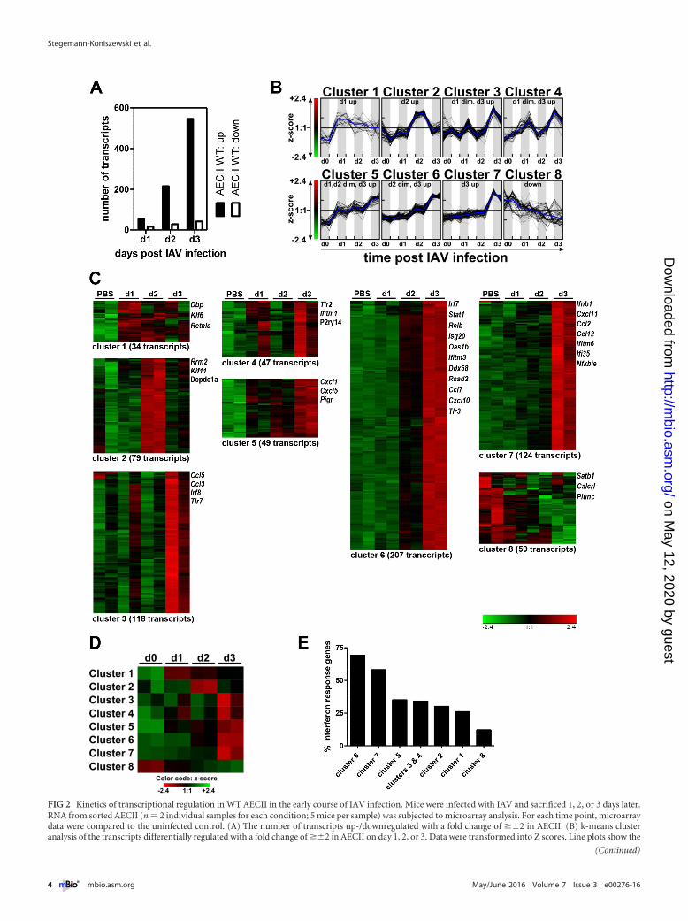
FIG 2 Kinetics of transcriptional regulation in WT AECII in the early course of IAV infection. Mice were infected with IAV and sacrificed 1, 2, or 3 days later.RNA from sorted AECII (n � 2 individual samples for each condition; 5 mice per sample) was subjected to microarray analysis. For each time point, microarraydata were compared to the uninfected control. (A) The number of transcripts up-/downregulated with a fold change of ��2 in AECII. (B) k-means clusteranalysis of the transcripts differentially regulated with a fold change of ��2 in AECII on day 1, 2, or 3. Data were transformed into Z scores. Line plots show the
(Continued)
Stegemann-Koniszewski et al.
4 ® mbio.asm.org May/June 2016 Volume 7 Issue 3 e00276-16
on May 12, 2020 by guest
http://mbio.asm
.org/D
ownloaded from
a multitude of molecules involved in the induction and shaping ofimmune responses, demonstrating their exceptionally high po-tential to contribute to respiratory immunity in vivo.
AECII transcriptional regulation correlates with PMN re-cruitment and IFN I/III levels. AECII most likely take part in therecruitment of immune effector cells to the respiratory tract.We determined the overall cellularity of bronchoalveolar la-vage (BAL) fluid, and a strong and significant increase in cellnumbers was detected on day 3 postinfection (Fig. 5A). Sincethe majority of the chemokines differentially expressed in AE-CII act as chemoattractants for macrophages and other my-eloid cells, mainly polymorphonuclear neutrophils (PMN), wedetermined the macrophage and PMN populations. In unin-fected mice, macrophages were the main cell type present andtheir relative contribution was significantly reduced by day 3(Fig. 5B). At the same time, there was a significant increase in
the PMN population size and the absolute PMN number(Fig. 5B and C). The kinetics of PMN recruitment and AECIItranscriptional regulation of chemokines suggest that AECIIcontribute to the attraction of innate effectors to the lung. Ofnote, the blunted transcriptional regulation of chemokines inTLR7-deficient hosts was in line with a delayed recruitment ofPMN (see Fig. S5 in the supplemental material).
In order to link ISG induction to interferon levels in the lung,we assessed the levels of bioactive IFN I/III in BAL fluid. A sub-stantial increase in IFN I/III activity was detected on day 2 post-IAV infection, with a significant increase by day 3 (Fig. 5C) whichdirectly correlated with the massive induction of ISG in AECII byday 3 postinfection. Of note, IFN I/III levels were significantlyreduced in TLR7-deficient mice (see Fig. S5 in the supplementalmaterial). Taken together, these findings imply a critical role forIFN I/III for the AECII response following IAV infection.
Figure Legend Continued
Z scores of the transcripts of the individual k-means clusters over time (for both replicates per time point). The blue lines indicate the average Z score. (C) Heatmap visualization of the Z scores in the individual k-means clusters. The number of transcripts and selected members are indicated for each cluster. (D) Heat mapvisualization of the average Z score value per column of the k-means clusters. (E) Percent interferon response genes within the individual k-means clusters asdetermined using the Interferome v2.01 database.
FIG 3 Transcriptional regulation in response to IAV infection is blunted in AECII isolated from TLR7-deficient hosts. Wild-type (WT) and TLR7ko mice wereinfected with IAV and sacrificed 1, 2, or 3 days postinfection. RNA from sorted AECII (n � 2 individual samples per time point; 5 mice per sample) was subjectedto microarray analysis. For each time point, microarray data were compared to the respective uninfected control. (A) Number of transcripts up- or downregu-lated with a fold change of ��2 in TLR7ko AECII in the course of IAV infection. (B) Comparison between the number of up- and downregulated transcripts inWT and TLR7ko AECII. (C) Venn diagram comparing the transcripts regulated in AECII on day 1, 2, and/or 3 post-IAV infection with regard to their regulationin WT AECII, TLR7ko AECII, or both. (D) Scatter plots displaying the absolute log2 fold changes of the transcripts regulated more than 2-fold (up or down) inWT and/or TLR7ko AECII on days 1, 2, and/or 3 post-IAV infection. The dashed bisecting lines indicate equal fold changes. Gray lines indicate the fold changethreshold of �2.
AECII and Influenza A Virus In Vivo
May/June 2016 Volume 7 Issue 3 e00276-16 ® mbio.asm.org 5
on May 12, 2020 by guest
http://mbio.asm
.org/D
ownloaded from

DISCUSSION
Nearly all of the research that has addressed the response of AECIIto IAV was performed in cells infected with the virus ex vivo (13–17), whereas in vivo studies mostly did not differentiate betweendifferent cell types of the epithelium (26).
Our analyses revealed that AECII strongly react to respiratoryIAV infection in vivo. The response was exceptionally versatile andincluded differential expression of a plethora of factors associatedwith antiviral activity and the induction of immune responses.Several antiviral proteins were among the most intensely upregu-lated transcripts, and the importance of their epithelial expressionwas highlighted by the finding that fold change induction inAECII often exceeded that in lung tissue. Deficiency in IFITM3,which was upregulated 12-fold in AECII and only 2-fold in lungtissue on day 3 (data not shown), leads to enhanced viral titers andincreased mortality in mice, and patients with IFITM3 mutationsexhibit compromised IAV restriction (27). Also, ISG15 and Gbp3(guanylate-binding protein 3) contribute specifically to the anti-IAV host defense (28, 29). Out of the genes encoding IFN I/III,only Ifnb1 was upregulated in AECII following in vivo IAV infec-tion. This was surprising at first, especially as AECII are describedto produce IFN I/III in response to IAV both in vivo and in vitro(13, 30). However, it has been shown that during in vivo IAVinfection IFN-� is predominantly produced not by the epitheliumbut by CD11c-expressing cell populations (31). Furthermore, thedepletion of plasmacytoid dendritic cells (pDC) was reported tolead to a significantly diminished IFN I production in the lungs ofIAV-infected mice (32). Therefore, we believe that also in ourmodel resident and newly recruited pDC are most likely the mainproducers of IFN I. This is well in line with the significantly de-creased IFN I/III production in TLR7ko mice, as pDC are knownto depend on TLR7 (33).
Extensive transcriptional regulation of CXCL5, CXCL9, andCXCL10 demonstrated how broadly AECII act on pulmonary hostdefense. Even though upregulation of most cytokines in lung tis-sue exceeded that in AECII, upregulation of CXCL5 and CCL5 inAECII was stronger than in lung tissue. Blocking of CCL5 in re-spiratory viral infection reduces the recruitment of CD4� andCD8� T cells (34), and AECII are the predominant source ofCXCL5 in the lungs of mice treated with lipopolysaccharide (35).Regarding the role of AECII in the recruitment of effector cells, thesignificantly attenuated PMN recruitment in TLR7ko mice waswell in line with the alleviated induction of CXCL5 expression intheir AECII. Of note, however, this reduction in PMN recruitment
is not necessarily a consequence of the reduced production ofchemotactic cytokines only by AECII, as their upregulation wasalso strongly reduced in whole-lung tissue of TLR7ko mice. Nev-ertheless, the extensive induction of cytokine and chemokinetranscription in AECII pointed at a strong contribution of AECIIto the overall lung response to infection in vivo.
Next to MHC-I antigen presentation in AECII, there was alsoevidence for MHC-II presentation and costimulation. In fact, le-thal IAV infection induces MHC-II expression on lung epithelialcells (23), and AECII have been observed to present antigen toCD4� T cells in the context of mycobacterial infection (25). Fur-thermore, we have previously shown that AECII efficiently pres-ent antigen to and activate CD4� T cells and that they are able topromote the induction of regulatory T cells (10). Therefore,MHC-I and also MHC-II antigen presentation displays strategiesfor AECII to shape the lower respiratory tract immune responseduring IAV infection in vivo.
A clear and distinct role for TLR7 in survival following IAVinfection has so far been demonstrated only in mice expressingfunctional Mx1, unlike the commonly used laboratory mousestrains (36). We and others have rather found TLR7 to be involvedin the fine-tuning of innate and adaptive anti-IAV responses (19,37, 38), and in our model of respiratory IAV infection, there wasno difference in morbidity and mortality between WT and TLR7-deficient hosts (see Fig. S6 in the supplemental material). Never-theless, we found the AECII response to IAV to be substantiallyblunted in the absence of TLR7. As both unchanged and clearlydiminished IFN I production have been described in TLR7komice (19, 39), it was surprising that TLR7 played such a centralrole in ensuring a robust early IFN response in our model. Mostlikely, the reduced levels of IFN I/III in the respiratory tract ofTLR7ko mice to a large extent accounted for the blunted AECIIresponse in these mice, especially since many of the regulatedtranscripts were ISG. Of note, the numbers of AECII isolated fromWT and TLR7ko animals were similar (see Fig. S6 in the supple-mental material). However, TLR7ko mice displayed a reduction inviral load in lung tissue and the isolated AECII (see Fig. S6 in thesupplemental material). IAV has previously been described to de-pend on TLR7 for efficient replication (39), and this reduction inviral load, even though not significant by day 3, was a likely reasonfor the blunted IFN I/III and in turn the blunted AECII responsein TLR7ko mice. We are not able to dissect the interdependence ofviral replication, IFN I/III production, and transcriptional regula-tion in AECII from our data. Silencing of TLR7 and other IAV
TABLE 1 Functional pathways most significantly overrepresented in the transcripts differentially expressed in AECIIa
Ranking by P value Canonical pathway AECII day 1 AECII day 2 AECII day 3
1 Communication between innate and adaptive immune cells 1.53 2.05 20.492 Role of pattern recognition receptors in recognition of bacteria and viruses 3.03 19.043 Granulocyte adhesion and diapedesis 2.53 3.31 15.444 Cross talk between dendritic cells and natural killer cells 14.795 Dendritic cell maturation 14.336 TREM1 signaling 1.73 2.42 13.797 Agranulocyte adhesion and diapedesis 2.44 3.14 12.918 Altered T cell and B cell signaling in rheumatoid arthritis 12.799 Interferon signaling 4.56 12.6410 Antigen presentation pathway 2.34 11.66a The 10 pathways most significantly overrepresented in the transcripts differentially regulated in WT AECII on day 3 postinfection are listed. Pathways were ranked by Fisher exacttest P value. The data columns indicate the �log(P value) for overrepresentation of the respective pathways for all microarray data sets (WT AECII; days 1, 2, and 3 postinfection),listing only those values indicating statistical significance (P � 0.05).
Stegemann-Koniszewski et al.
6 ® mbio.asm.org May/June 2016 Volume 7 Issue 3 e00276-16
on May 12, 2020 by guest
http://mbio.asm
.org/D
ownloaded from

FIG 4 IAV infection triggers the differential expression of a multitude of molecules involved in antimicrobial defense. The graphs depict the fold changeregulation of selected transcripts as determined by microarray analysis of WT AECII and lungs isolated at the indicated time points post-IAV infection. Data areshown as the mean and the individual results from two independent replicate microarray experiments (2 independent samples; 5 mice per sample) for AECII andthree independent replicate microarray experiments for lung tissue (three independent samples). The transcripts listed are grouped into those encoding pathogenrecognition receptors (A), factors associated with the IFN I/III response (B), cytokines and chemokines (C), and factors associated with antigen presentation (D).For each bar graph, the dashed horizontal line indicates a fold change of 2.
AECII and Influenza A Virus In Vivo
May/June 2016 Volume 7 Issue 3 e00276-16 ® mbio.asm.org 7
on May 12, 2020 by guest
http://mbio.asm
.org/D
ownloaded from

sensors such as RIG-I specifically in AECII will be essential to shedlight on this issue. Nevertheless, the high number of ISG triggeredand the correlation between the AECII response and IFN I/IIIlevels in the lung clearly showed that AECII react to their specificmicroenvironment in vivo. In addition to our findings, cell culturestudies have shown that pretreatment of AECII with tumor necro-sis factor alpha (TNF-�) and IFN-� greatly enhanced their cyto-kine and chemokine response to IAV (16). Also, IL-17A andTNF-� synergistically act on CXCL5 expression by AECII in vitroand in vivo (40).
Ultimately, the detailed contributions and interplay of patho-gen recognition and the signals provided by the microenviron-ment remain a central question for our understanding of AECII
activation in vivo. Dynamic PRR expression patterns in responseto IAV have been observed for AECII (41), and moreover, theyrespond directly to the virus through various TLR (30, 42). In ourstudy, the exceptionally rapid induction of gene expressionalchanges and the transcriptional upregulation of nucleic acid-sensing PRR in the course of the infection suggested a direct sens-ing of the virus by AECII also in vivo. Many of the transcriptsdifferentially expressed in AECII are typically triggered by PRRligation and activation of the NF-�B pathway, and the functionalpathway Role of Pattern Recognition Receptors in Recognition ofBacteria and Viruses was the second most significantly overrepre-sented pathway. Therefore, we propose a model in which the rapidand versatile in vivo AECII response following IAV infection is
FIG 5 The kinetics of cell recruitment and type I/III interferon levels correlate with AECII transcriptional profiles. WT mice were sacrificed at the indicated timepoints post-IAV infection. Bronchoalveolar lavage (BAL) fluid cells were counted (A), and the macrophage and polymorphonuclear cell (PMN) populations (B)were assessed by flow cytometry. Cell populations were analyzed by gating on macrophages (F4/80� cells) within all acquired cells and gating on PMN(Gr-1�/CD11b�) within the F4/80� cell fraction. (C) Cell numbers were calculated from the absolute cell count and percent population for all analyzedindividual mice. Data from individual mice and the mean per group are shown. (D) The concentration of bioactive type I IFN in BAL fluid was assessed using IFNI/III-sensitive reporter cells and an IFN-� standard. Data are shown as means � standard errors of the means or as individual mice and mean per group. All dataare compiled from n � 5 mice out of at least two independent experiments. Groups were compared by unpaired, two-sided t test (P values: *, �0.05; **, �0.005;***, �0.001).
Stegemann-Koniszewski et al.
8 ® mbio.asm.org May/June 2016 Volume 7 Issue 3 e00276-16
on May 12, 2020 by guest
http://mbio.asm
.org/D
ownloaded from
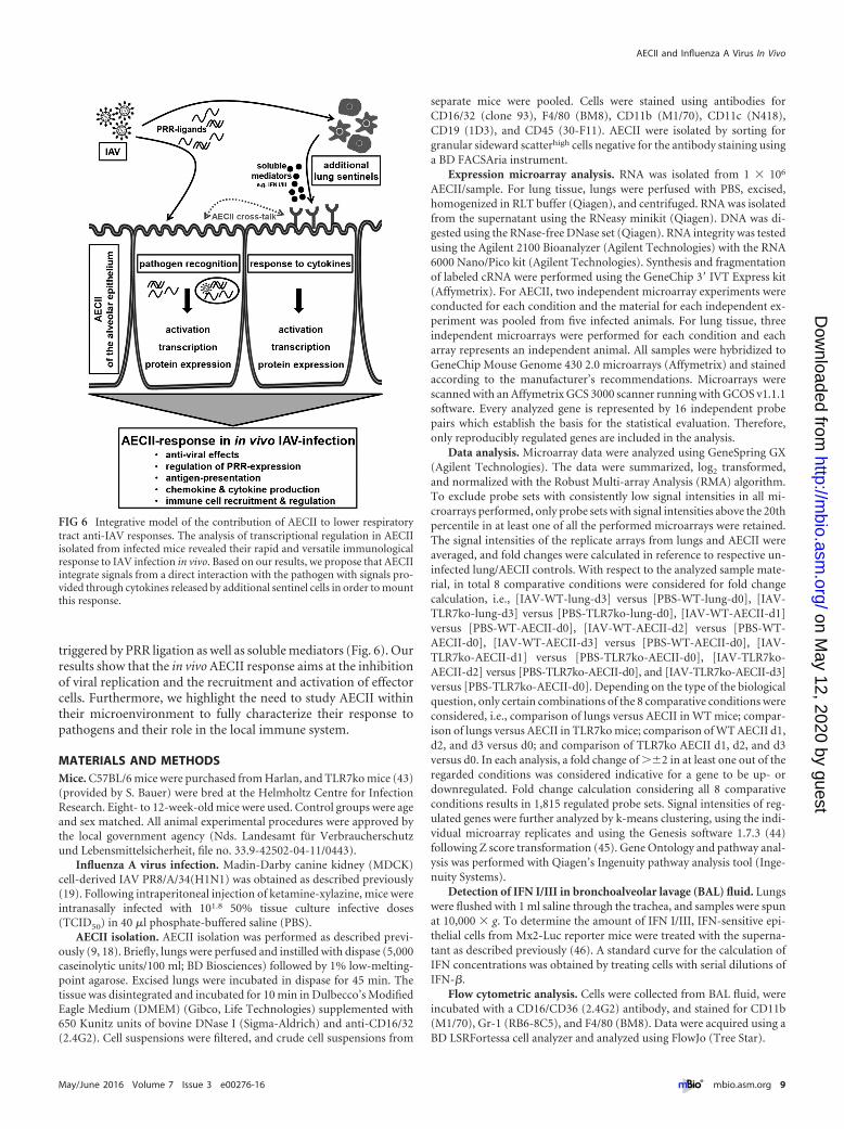
triggered by PRR ligation as well as soluble mediators (Fig. 6). Ourresults show that the in vivo AECII response aims at the inhibitionof viral replication and the recruitment and activation of effectorcells. Furthermore, we highlight the need to study AECII withintheir microenvironment to fully characterize their response topathogens and their role in the local immune system.
MATERIALS AND METHODSMice. C57BL/6 mice were purchased from Harlan, and TLR7ko mice (43)(provided by S. Bauer) were bred at the Helmholtz Centre for InfectionResearch. Eight- to 12-week-old mice were used. Control groups were ageand sex matched. All animal experimental procedures were approved bythe local government agency (Nds. Landesamt für Verbraucherschutzund Lebensmittelsicherheit, file no. 33.9-42502-04-11/0443).
Influenza A virus infection. Madin-Darby canine kidney (MDCK)cell-derived IAV PR8/A/34(H1N1) was obtained as described previously(19). Following intraperitoneal injection of ketamine-xylazine, mice wereintranasally infected with 101.8 50% tissue culture infective doses(TCID50) in 40 �l phosphate-buffered saline (PBS).
AECII isolation. AECII isolation was performed as described previ-ously (9, 18). Briefly, lungs were perfused and instilled with dispase (5,000caseinolytic units/100 ml; BD Biosciences) followed by 1% low-melting-point agarose. Excised lungs were incubated in dispase for 45 min. Thetissue was disintegrated and incubated for 10 min in Dulbecco’s ModifiedEagle Medium (DMEM) (Gibco, Life Technologies) supplemented with650 Kunitz units of bovine DNase I (Sigma-Aldrich) and anti-CD16/32(2.4G2). Cell suspensions were filtered, and crude cell suspensions from
separate mice were pooled. Cells were stained using antibodies forCD16/32 (clone 93), F4/80 (BM8), CD11b (M1/70), CD11c (N418),CD19 (1D3), and CD45 (30-F11). AECII were isolated by sorting forgranular sideward scatterhigh cells negative for the antibody staining usinga BD FACSAria instrument.
Expression microarray analysis. RNA was isolated from 1 � 106
AECII/sample. For lung tissue, lungs were perfused with PBS, excised,homogenized in RLT buffer (Qiagen), and centrifuged. RNA was isolatedfrom the supernatant using the RNeasy minikit (Qiagen). DNA was di-gested using the RNase-free DNase set (Qiagen). RNA integrity was testedusing the Agilent 2100 Bioanalyzer (Agilent Technologies) with the RNA6000 Nano/Pico kit (Agilent Technologies). Synthesis and fragmentationof labeled cRNA were performed using the GeneChip 3= IVT Express kit(Affymetrix). For AECII, two independent microarray experiments wereconducted for each condition and the material for each independent ex-periment was pooled from five infected animals. For lung tissue, threeindependent microarrays were performed for each condition and eacharray represents an independent animal. All samples were hybridized toGeneChip Mouse Genome 430 2.0 microarrays (Affymetrix) and stainedaccording to the manufacturer’s recommendations. Microarrays werescanned with an Affymetrix GCS 3000 scanner running with GCOS v1.1.1software. Every analyzed gene is represented by 16 independent probepairs which establish the basis for the statistical evaluation. Therefore,only reproducibly regulated genes are included in the analysis.
Data analysis. Microarray data were analyzed using GeneSpring GX(Agilent Technologies). The data were summarized, log2 transformed,and normalized with the Robust Multi-array Analysis (RMA) algorithm.To exclude probe sets with consistently low signal intensities in all mi-croarrays performed, only probe sets with signal intensities above the 20thpercentile in at least one of all the performed microarrays were retained.The signal intensities of the replicate arrays from lungs and AECII wereaveraged, and fold changes were calculated in reference to respective un-infected lung/AECII controls. With respect to the analyzed sample mate-rial, in total 8 comparative conditions were considered for fold changecalculation, i.e., [IAV-WT-lung-d3] versus [PBS-WT-lung-d0], [IAV-TLR7ko-lung-d3] versus [PBS-TLR7ko-lung-d0], [IAV-WT-AECII-d1]versus [PBS-WT-AECII-d0], [IAV-WT-AECII-d2] versus [PBS-WT-AECII-d0], [IAV-WT-AECII-d3] versus [PBS-WT-AECII-d0], [IAV-TLR7ko-AECII-d1] versus [PBS-TLR7ko-AECII-d0], [IAV-TLR7ko-AECII-d2] versus [PBS-TLR7ko-AECII-d0], and [IAV-TLR7ko-AECII-d3]versus [PBS-TLR7ko-AECII-d0]. Depending on the type of the biologicalquestion, only certain combinations of the 8 comparative conditions wereconsidered, i.e., comparison of lungs versus AECII in WT mice; compar-ison of lungs versus AECII in TLR7ko mice; comparison of WT AECII d1,d2, and d3 versus d0; and comparison of TLR7ko AECII d1, d2, and d3versus d0. In each analysis, a fold change of ��2 in at least one out of theregarded conditions was considered indicative for a gene to be up- ordownregulated. Fold change calculation considering all 8 comparativeconditions results in 1,815 regulated probe sets. Signal intensities of reg-ulated genes were further analyzed by k-means clustering, using the indi-vidual microarray replicates and using the Genesis software 1.7.3 (44)following Z score transformation (45). Gene Ontology and pathway anal-ysis was performed with Qiagen’s Ingenuity pathway analysis tool (Inge-nuity Systems).
Detection of IFN I/III in bronchoalveolar lavage (BAL) fluid. Lungswere flushed with 1 ml saline through the trachea, and samples were spunat 10,000 � g. To determine the amount of IFN I/III, IFN-sensitive epi-thelial cells from Mx2-Luc reporter mice were treated with the superna-tant as described previously (46). A standard curve for the calculation ofIFN concentrations was obtained by treating cells with serial dilutions ofIFN-�.
Flow cytometric analysis. Cells were collected from BAL fluid, wereincubated with a CD16/CD36 (2.4G2) antibody, and stained for CD11b(M1/70), Gr-1 (RB6-8C5), and F4/80 (BM8). Data were acquired using aBD LSRFortessa cell analyzer and analyzed using FlowJo (Tree Star).
FIG 6 Integrative model of the contribution of AECII to lower respiratorytract anti-IAV responses. The analysis of transcriptional regulation in AECIIisolated from infected mice revealed their rapid and versatile immunologicalresponse to IAV infection in vivo. Based on our results, we propose that AECIIintegrate signals from a direct interaction with the pathogen with signals pro-vided through cytokines released by additional sentinel cells in order to mountthis response.
AECII and Influenza A Virus In Vivo
May/June 2016 Volume 7 Issue 3 e00276-16 ® mbio.asm.org 9
on May 12, 2020 by guest
http://mbio.asm
.org/D
ownloaded from
Microarray data accession number. Microarray data were depositedin NCBI’s Gene Expression Omnibus and are accessible through the GEOseries accession number GSE57008.
SUPPLEMENTAL MATERIALSupplemental material for this article may be found at http://mbio.asm.org/lookup/suppl/doi:10.1128/mBio.00276-16/-/DCSupplemental.
Figure S1, PDF file, 0.4 MB.Figure S2, PDF file, 0.3 MB.Figure S3, PDF file, 0.4 MB.Figure S4, PDF file, 0.2 MB.Figure S5, PDF file, 0.2 MB.Figure S6, PDF file, 0.2 MB.Table S1, PDF file, 0.02 MB.Table S2, PDF file, 0.02 MB.
ACKNOWLEDGMENTS
This work was supported by the German Research Foundation (BR2221/1-1 to D. Bruder and GU769/5-1 to M. Gunzer) and the Helmholtz Asso-ciation (HGF) (President’s Initiative and Networking Fund W2/W3-029to D. Bruder).
We thank S. Akira (Osaka University) for permission to obtainTLR7ko mice; L. Gröbe and M. Höxter (Helmholtz Centre for InfectionResearch) for cell sorting; and P. Hagendorff, S. Kaser, S. Prettin, T.Hirsch, and M. Grashoff (Helmholtz Centre for Infection Research) andF. Ewert (Otto-von-Guericke University Magdeburg) for technical assis-tance.
FUNDING INFORMATIONThis work, including the efforts of Dunja Bruder and Matthias Gunzer,was funded by Deutsche Forschungsgemeinschaft (DFG) (BR2221/1-1and GU769/5-1). This work, including the efforts of Dunja Bruder, wasfunded by Helmholtz-Gemeinschaft (Helmholtz Association) (W2/W3-029).
REFERENCES1. Iwasaki A, Pillai PS. 2014. Innate immunity to influenza virus infection.
Nat Rev Immunol 14:315–328. http://dx.doi.org/10.1038/nri3665.2. Kato H, Sato S, Yoneyama M, Yamamoto M, Uematsu S, Matsui K,
Tsujimura T, Takeda K, Fujita T, Takeuchi O, Akira S. 2005. Celltype-specific involvement of RIG-I in antiviral response. Immunity 23:19 –28. http://dx.doi.org/10.1016/j.immuni.2005.04.010.
3. Kato H, Takeuchi O, Sato S, Yoneyama M, Yamamoto M, Matsui K,Uematsu S, Jung A, Kawai T, Ishii KJ, Yamaguchi O, Otsu K, TsujimuraT, Koh CS, Reis e Sousa C, Matsuura Y, Fujita T, Akira S. 2006.Differential roles of MDA5 and RIG-I helicases in the recognition of RNAviruses. Nature 441:101–105. http://dx.doi.org/10.1038/nature04734.
4. Mordstein M, Kochs G, Dumoutier L, Renauld JC, Paludan SR, KlucherK, Staeheli P. 2008. Interferon-lambda contributes to innate immunity ofmice against influenza A virus but not against hepatotropic viruses. PLoSPathog 4:e1000151. http://dx.doi.org/10.1371/journal.ppat.1000151.
5. Koerner I, Matrosovich MN, Haller O, Staeheli P, Kochs G. 2012.Altered receptor specificity and fusion activity of the haemagglutinin con-tribute to high virulence of a mouse-adapted influenza A virus. J Gen Virol93:970 –979. http://dx.doi.org/10.1099/vir.0.035782-0.
6. Weinheimer VK, Becher A, Tönnies M, Holland G, Knepper J, BauerTT, Schneider P, Neudecker J, Rückert JC, Szymanski K, Temmesfeld-Wollbrueck B, Gruber AD, Bannert N, Suttorp N, Hippenstiel S, WolffT, Hocke AC. 2012. Influenza A viruses target type II pneumocytes in thehuman lung. J Infect Dis 206:1685–1694. http://dx.doi.org/10.1093/infdis/jis455.
7. Fehrenbach H. 2001. Alveolar epithelial type II cell: defender of the alve-olus revisited. Respir Res 2:33– 46. http://dx.doi.org/10.1186/rr36.
8. Mason RJ. 2006. Biology of alveolar type II cells. Respirology 11(Suppl):S12–S15. http://dx.doi.org/10.1111/j.1440-1843.2006.00800.x.
9. Gereke M, Gröbe L, Prettin S, Kasper M, Deppenmeier S, Gruber AD,Enelow RI, Buer J, Bruder D. 2007. Phenotypic alterations in type IIalveolar epithelial cells in CD4� T cell mediated lung inflammation. Re-spir Res 8:47. http://dx.doi.org/10.1186/1465-9921-8-47.
10. Gereke M, Jung S, Buer J, Bruder D. 2009. Alveolar type II epithelial cellspresent antigen to CD4(�) T cells and induce Foxp3(�) regulatory Tcells. Am J Respir Crit Care Med 179:344 –355. http://dx.doi.org/10.1164/rccm.200804-592OC.
11. Sato K, Tomioka H, Shimizu T, Gonda T, Ota F, Sano C. 2002. Type IIalveolar cells play roles in macrophage-mediated host innate resistance topulmonary mycobacterial infections by producing proinflammatory cy-tokines. J Infect Dis 185:1139 –1147. http://dx.doi.org/10.1086/340040.
12. Unkel B, Hoegner K, Clausen BE, Lewe-Schlosser P, Bodner J, Gatten-loehner S, Janssen H, Seeger W, Lohmeyer J, Herold S. 2012. Alveolarepithelial cells orchestrate DC function in murine viral pneumonia. J ClinInvest 122:3652–3664. http://dx.doi.org/10.1172/JCI62139.
13. Crotta S, Davidson S, Mahlakoiv T, Desmet CJ, Buckwalter MR, AlbertML, Staeheli P, Wack A. 2013. Type I and type III interferons driveredundant amplification loops to induce a transcriptional signature ininfluenza-infected airway epithelia. PLoS Pathog 9:e1003773. http://dx.doi.org/10.1371/journal.ppat.1003773.
14. Gan H, Hao Q, Idell S, Tang H. 2015. Transcription factor Runx3 isinduced by influenza A virus and double-strand RNA and mediates airwayepithelial cell apoptosis. Sci Rep 5:17916. http://dx.doi.org/10.1038/srep17916.
15. Ito Y, Correll K, Zemans RL, Leslie CC, Murphy RC, Mason RJ. 2015.Influenza induces IL-8 and GM-CSF secretion by human alveolar epithe-lial cells through HGF/c-Met and TGF-alpha/EGFR signaling. Am JPhysiol Lung Cell Mol Physiol 308:L1178 –L1188. http://dx.doi.org/10.1152/ajplung.00290.2014.
16. Veckman V, Osterlund P, Fagerlund R, Melén K, Matikainen S,Julkunen I. 2006. TNF-alpha and IFN-alpha enhance influenza-A-virus-induced chemokine gene expression in human A549 lung epithelial cells.Virology 345:96 –104. http://dx.doi.org/10.1016/j.virol.2005.09.043.
17. Yu WC, Chan RW, Wang J, Travanty EA, Nicholls JM, Peiris JS, MasonRJ, Chan MC. 2011. Viral replication and innate host responses in pri-mary human alveolar epithelial cells and alveolar macrophages infectedwith influenza H5N1 and H1N1 viruses. J Virol 85:6844 – 6855. http://dx.doi.org/10.1128/JVI.02200-10.
18. Gereke M, Autengruber A, Gröbe L, Jeron A, Bruder D, Stegemann-Koniszewski S. 2012. Flow cytometric isolation of primary murine type IIalveolar epithelial cells for functional and molecular studies. J Vis Exp70:4322. http://dx.doi.org/10.3791/4322.
19. Stegemann-Koniszewski S, Gereke M, Orrskog S, Lienenklaus S, PascheB, Bader SR, Gruber AD, Akira S, Weiss S, Henriques-Normark B,Bruder D, Gunzer M. 2013. TLR7 contributes to the rapid progressionbut not to the overall fatal outcome of secondary pneumococcal diseasefollowing influenza A virus infection. J Innate Immun 5:84 –96. http://dx.doi.org/10.1159/000345112.
20. Katsoulidis E, Carayol N, Woodard J, Konieczna I, Majchrzak-Kita B,Jordan A, Sassano A, Eklund EA, Fish EN, Platanias LC. 2009. Role ofSchlafen 2 (SLFN2) in the generation of interferon alpha-induced growthinhibitory responses. J Biol Chem 284:25051–25064. http://dx.doi.org/10.1074/jbc.M109.030445.
21. Rusinova I, Forster S, Yu S, Kannan A, Masse M, Cumming H,Chapman R, Hertzog PJ. 2013. Interferome v2.0: an updated database ofannotated interferon-regulated genes. Nucleic Acids Res 41:D1040 –D1046. http://dx.doi.org/10.1093/nar/gks1215.
22. Diamond MS, Farzan M. 2013. The broad-spectrum antiviral functionsof IFIT and IFITM proteins. Nat Rev Immunol 13:46 –57. http://dx.doi.org/10.1038/nri3344.
23. Brown DM, Lee S, Garcia-Hernandez MDLL, Swain SL. 2012. Multi-functional CD4 cells expressing gamma interferon and perforin mediateprotection against lethal influenza virus infection. J Virol 86:6792– 6803.http://dx.doi.org/10.1128/JVI.07172-11.
24. Corbière V, Dirix V, Norrenberg S, Cappello M, Remmelink M, Mas-cart F. 2011. Phenotypic characteristics of human type II alveolar epithe-lial cells suitable for antigen presentation to T lymphocytes. Respir Res12:15. http://dx.doi.org/10.1186/1465-9921-12-15.
25. Debbabi H, Ghosh S, Kamath AB, Alt J, Demello DE, Dunsmore S,Behar SM. 2005. Primary type II alveolar epithelial cells present microbialantigens to antigen-specific CD4� T cells. Am J Physiol Lung Cell MolPhysiol 289:L274 –L279. http://dx.doi.org/10.1152/ajplung.00004.2005.
26. Brandes M, Klauschen F, Kuchen S, Germain RN. 2013. A systemsanalysis identifies a feedforward inflammatory circuit leading to lethalinfluenza infection. Cell 154:197–212. http://dx.doi.org/10.1016/j.cell.2013.06.013.
Stegemann-Koniszewski et al.
10 ® mbio.asm.org May/June 2016 Volume 7 Issue 3 e00276-16
on May 12, 2020 by guest
http://mbio.asm
.org/D
ownloaded from
27. Everitt AR, Clare S, Pertel T, John SP, Wash RS, Smith SE, Chin CR,Feeley EM, Sims JS, Adams DJ, Wise HM, Kane L, Goulding D, DigardP, Anttila V, Baillie JK, Walsh TS, Hume DA, Palotie A, Xue Y. 2012.IFITM3 restricts the morbidity and mortality associated with influenza.Nature 484:519 –523. http://dx.doi.org/10.1038/nature10921.
28. Lenschow DJ, Lai C, Frias-Staheli N, Giannakopoulos NV, Lutz A,Wolff T, Osiak A, Levine B, Schmidt RE, García-Sastre A, Leib DA,Pekosz A, Knobeloch KP, Horak I, Virgin HW. 2007. IFN-stimulatedgene 15 functions as a critical antiviral molecule against influenza, herpes,and Sindbis viruses. Proc Natl Acad Sci U S A 104:1371–1376. http://dx.doi.org/10.1073/pnas.0607038104.
29. Nordmann A, Wixler L, Boergeling Y, Wixler V, Ludwig S. 2012. A newsplice variant of the human guanylate-binding protein 3 mediates anti-influenza activity through inhibition of viral transcription and replication.FASEB J 26:1290 –1300. http://dx.doi.org/10.1096/fj.11-189886.
30. Ioannidis I, Ye F, McNally B, Willette M, Flaño E. 2013. Toll-likereceptor expression and induction of type I and type III interferons inprimary airway epithelial cells. J Virol 87:3261–3270. http://dx.doi.org/10.1128/JVI.01956-12.
31. Kallfass C, Lienenklaus S, Weiss S, Staeheli P. 2013. Visualizing the betainterferon response in mice during infection with influenza A viruses ex-pressing or lacking nonstructural protein 1. J Virol 87:6925– 6930. http://dx.doi.org/10.1128/JVI.00283-13.
32. Jewell NA, Vaghefi N, Mertz SE, Akter P, Peebles RS, Jr, Bakaletz LO,Durbin RK, Flaño E, Durbin JE. 2007. Differential type I interferoninduction by respiratory syncytial virus and influenza A virus in vivo. JVirol 81:9790 –9800. http://dx.doi.org/10.1128/JVI.00530-07.
33. Diebold SS, Kaisho T, Hemmi H, Akira S, Reis e Sousa C. 2004. Innateantiviral responses by means of TLR7-mediated recognition of single-stranded RNA. Science 303:1529 –1531. http://dx.doi.org/10.1126/science.1093616.
34. Culley FJ, Pennycook AM, Tregoning JS, Dodd JS, Walzl G, Wells TN,Hussell T, Openshaw PJ. 2006. Role of CCL5 (RANTES) in viral lungdisease. J Virol 80:8151– 8157. http://dx.doi.org/10.1128/JVI.00496-06.
35. Jeyaseelan S, Manzer R, Young SK, Yamamoto M, Akira S, Mason RJ,Worthen GS. 2005. Induction of CXCL5 during inflammation in the ro-dent lung involves activation of alveolar epithelium. Am J Respir Cell MolBiol 32:531–539. http://dx.doi.org/10.1165/rcmb.2005-0063OC.
36. Kaminski MM, Ohnemus A, Cornitescu M, Staeheli P. 2012. Plasmacy-toid dendritic cells and Toll-like receptor 7-dependent signalling promoteefficient protection of mice against highly virulent influenza A virus. J GenVirol 93:555–559. http://dx.doi.org/10.1099/vir.0.039065-0.
37. Jeisy-Scott V, Davis WG, Patel JR, Bowzard JB, Shieh WJ, Zaki SR, KatzJM, Sambhara S. 2011. Increased MDSC accumulation and Th2 biasedresponse to influenza A virus infection in the absence of TLR7 in mice.PLoS One 6:e25242. http://dx.doi.org/10.1371/journal.pone.0025242.
38. Koyama S, Ishii KJ, Kumar H, Tanimoto T, Coban C, Uematsu S,Kawai T, Akira S. 2007. Differential role of TLR- and RLR-signaling in theimmune responses to influenza A virus infection and vaccination. J Im-munol 179:4711– 4720. http://dx.doi.org/10.4049/jimmunol.179.7.4711.
39. Pang IK, Pillai PS, Iwasaki A. 2013. Efficient influenza A virus replicationin the respiratory tract requires signals from TLR7 and RIG-I. Proc NatlAcad Sci U S A 110:13910 –13915. http://dx.doi.org/10.1073/pnas.1303275110.
40. Liu Y, Mei J, Gonzales L, Yang G, Dai N, Wang P, Zhang P, Favara M,Malcolm KC, Guttentag S, Worthen GS. 2011. IL-17A and TNF-alphaexert synergistic effects on expression of CXCL5 by alveolar type II cells invivo and in vitro. J Immunol 186:3197–3205. http://dx.doi.org/10.4049/jimmunol.1002016.
41. Hui KP, Lee SM, Cheung CY, Mao H, Lai AK, Chan RW, Chan MC, TuW, Guan Y, Lau YL, Peiris JS. 2011. H5N1 influenza virus-inducedmediators upregulate RIG-I in uninfected cells by paracrine effects con-tributing to amplified cytokine cascades. J Infect Dis 204:1866 –1878.http://dx.doi.org/10.1093/infdis/jir665.
42. Guillot L, Le Goffic R, Bloch S, Escriou N, Akira S, Chignard M,Si-Tahar M. 2005. Involvement of Toll-like receptor 3 in the immuneresponse of lung epithelial cells to double-stranded RNA and influenza Avirus. J Biol Chem 280:5571–5580. http://dx.doi.org/10.1074/jbc.M410592200.
43. Hemmi H, Kaisho T, Takeuchi O, Sato S, Sanjo H, Hoshino K, Horiu-chi T, Tomizawa H, Takeda K, Akira S. 2002. Small anti-viral com-pounds activate immune cells via the TLR7 MyD88-dependent signalingpathway. Nat Immunol 3:196 –200. http://dx.doi.org/10.1038/ni758.
44. Sturn A, Quackenbush J, Trajanoski Z. 2002. Genesis: cluster analysis ofmicroarray data. Bioinformatics 18:207–208. http://dx.doi.org/10.1093/bioinformatics/18.1.207.
45. Cheadle C, Vawter MP, Freed WJ, Becker KG. 2003. Analysis of microar-ray data using Z score transformation. J Mol Diagn 5:73– 81. http://dx.doi.org/10.1016/S1525-1578(10)60455-2.
46. Nandakumar R, Finsterbusch K, Lipps C, Neumann B, Grashoff M, NairS, Hochnadel I, Lienenklaus S, Wappler I, Steinmann E, Hauser H, Piet-schmann T, Kröger A. 2013. Hepatitis C virus replication in mouse cells isrestricted by IFN-dependent and -independent mechanisms. Gastroenterol-ogy 145:1414–1423. http://dx.doi.org/10.1053/j.gastro.2013.08.037.
AECII and Influenza A Virus In Vivo
May/June 2016 Volume 7 Issue 3 e00276-16 ® mbio.asm.org 11
on May 12, 2020 by guest
http://mbio.asm
.org/D
ownloaded from