Alternative splicing and its regulation under normal and abnormal
24
Institutionen för fysik, kemi och biologi Examensarbete Alternative splicing and its regulation under normal and abnormal conditions Jenny Ackelman Examensarbetet utfört vid IFM Biologi 2010-05-31 LITH-IFM-G-EX--10/ 2350—SE Linköpings universitet Institutionen för fysik, kemi och biologi 581 83 Linköping
Transcript of Alternative splicing and its regulation under normal and abnormal

Institutionen för fysik, kemi och biologi
Examensarbete
Alternative splicing and its regulation under normal and abnormal conditions
Jenny Ackelman
Examensarbetet utfört vid IFM Biologi 2010-05-31
LITH-IFM-G-EX--10/ 2350—SE
Linköpings universitet Institutionen för fysik, kemi och biologi 581 83 Linköping

2
Rapporttyp Report category
Licentiatavhandling
x Examensarbete C-uppsats
D-uppsats
Övrig rapport
_______________
Språk Language
Svenska/Swedish
X Engelska/English
________________
Titel
Title
Alternative splicing and its regulation under normal and abnormal conditions Författare
Author
Jenny Ackelman
Sammanfattning Abstract
During the maturation of pre-mRNA introns are removed and exons are spliced together, to form a primary transcript,
a reaction that is catalyzed by the spliceosome. Alternative splicing is a complex reaction that mainly utilizes one of
four mechanisms; exon skipping, 5’ splice site choice, 3’ splice site choice and intron retention. To achieve accurate
splicing four sequence elements are essential, two of which are located in the splice sites themselves; 5’ splice sites
and 3’ splice sites, but also the polypyrimidine tract and the branch point sequence. Alternative splicing can be
regulated by histone or chromatin modulations, siRNA, transcription efficiency and various proteins, many of which
belong to either the SR protein family or the hnRNP family of proteins. SR proteins usually promote exon inclusion,
while hnRNP proteins usually promote exon skipping. There are also regulatory elements that are called exonic
splicing enhancers or silencers depending on if they promote or inhibit the inclusion of the exon they reside in. These
elements also exist in introns and are then called intronic splicing enhancers or silencers. The enhancer elements are
most commonly targeted by SR proteins and the silencer elements are usually targeted by hnRNP proteins. This
paper will mainly focus on the regulation of alternative splicing and the role of alternative splicing under abnormal
conditions, such as when mutations cause disease.
ISBN
__________________________________________________
ISRN
__________________________________________________
Serietitel och serienummer ISSN Title of series, numbering
LITH-IFM-G-Ex—10/2350--SE
Nyckelord Keyword
Alternative splicing, ESE, ESS, hnRNP proteins, ISE, ISS, Regulation, Spliceosome, SR proteins.
Datum
Date
2010-05-31
URL för elektronisk version
Avdelning, Institution
Biologi, IFM
Division, Department
Biology, IFM

3
Contents Abstract ................................................................................................................................................... 4
List of abbreviations ................................................................................................................................ 5
Introduction ............................................................................................................................................. 6
Regulation of alternative splicing ............................................................................................................ 8
Exonic/intronic splicing enhancer/silencer ......................................................................................... 8
Intronic regulatory elements/pseudo exons ................................................................................... 9
Transcription efficiency ..................................................................................................................... 11
Histone modifications and Chromatin modifications ....................................................................... 11
siRNA and gene silencing................................................................................................................... 11
SR proteins ........................................................................................................................................ 11
SC35 ............................................................................................................................................... 13
SRrp37 ........................................................................................................................................... 14
ASF/SF2 .......................................................................................................................................... 14
hnRNP proteins ................................................................................................................................. 15
hnRNP A1/A2 ................................................................................................................................. 15
hnRNP A3 ....................................................................................................................................... 17
hnRNP H......................................................................................................................................... 17
hnRNP L ......................................................................................................................................... 17
TDP-43 ........................................................................................................................................... 18
PTB ................................................................................................................................................. 19
Conclusions ............................................................................................................................................ 20
References ............................................................................................................................................. 21
4
Abstract During the maturation of pre-mRNA introns are removed and exons are spliced together, to
form a primary transcript, a reaction that is catalyzed by the spliceosome. Alternative splicing
is a complex reaction that mainly utilizes one of four mechanisms; exon skipping, 5’ splice
site choice, 3’ splice site choice and intron retention. To achieve accurate splicing four
sequence elements are essential, two of which are located in the splice sites themselves; 5’
splice sites and 3’ splice sites, but also the polypyrimidine tract and the branch point
sequence. Alternative splicing can be regulated by histone or chromatin modulations, siRNA,
transcription efficiency and various proteins, many of which belong to either the SR protein
family or the hnRNP family of proteins. SR proteins usually promote exon inclusion, while
hnRNP proteins usually promote exon skipping. There are also regulatory elements that are
called exonic splicing enhancers or silencers depending on if they promote or inhibit the
inclusion of the exon they reside in. These elements also exist in introns and are then called
intronic splicing enhancers or silencers. The enhancer elements are most commonly targeted
by SR proteins and the silencer elements are usually targeted by hnRNP proteins. This paper
will mainly focus on the regulation of alternative splicing and the role of alternative splicing
under abnormal conditions, such as when mutations cause disease.
Keywords
Alternative splicing, ESE, ESS, hnRNP proteins, ISE, ISS, Regulation, Spliceosome, SR
proteins.

5
List of abbreviations
ASF/SF2 – Alternative splicing factor/splicing factor 2
ATM - Ataxia telangiectasia gene
BR genes – Balbiani ring genes
CFTR gene – Cystic fibrosis transmembrane conductance regulator
Clk/Sty – Family of protein kinases
ESE – Exonic splicing enhancers
ESR - Exonic splicing regulatory sequences
ESS – Exonic splicing silencers
dsDNA – Double stranded DNA
GRD – Glycine rich domain
GST-MS2 protein – Coat protein
hnRNP – Heterogeneous nuclear RNA
Htra2-β1 – A serine-protease
IRS – Intronic splicing regulatory sequences
ISPE – Intronic splicing processing element
ISE – Intronic splicing enhancers
ISS – Intronic splicing silencers
nPTB – Neuronal PTB
nSR100 – neural-specific SR related protein, 100 kDa
PDH – Pyruvate dehydrogenase
Pol II – Polymerase II
PSF – PTB-associated splicing factor
PTB – Polypyrimidine tract binding protein
RRM – RNA recognition motifs
RS domains – Arginine/serine rich domain
siRNA – Small interfering RNA
SMA – Spinal muscular atrophy
SMN – Survival of motor neuron gene
sp1 – Protein that promotes initiation of transcription
snRNP - Small nuclear ribonucleoproteins
SR proteins – Serine/arginine-rich proteins
SRrp37 – SR related protein 37
SRPK – Protein kinase family
ssDNA-protein – Single stranded DNA
SW6 – VP16 mutant
TGS - Transcriptional gene silencing
U2AF – Non-snRNP protein required for U2snRNP binding
VP16 –Protein that promotes transcription

6
Introduction
During the maturation of pre-mRNA the non coding introns are removed and the coding
exons are spliced together, to form a primary transcript. This process is essential for the
construction of proteins in higher eukaryotes (Xiang et al., 2005). The size of the human
genome, 25,000 genes, can not singlehandedly be accounted for the human complexity. In
alternative splicing several isoforms of mRNA are constructed from a single gene or locus, in
some cases thousands of transcripts are created, which gives rise to numerous different
functional proteins (Zhou et al., 2009; Koren et al., 2007). Extensive alterations of transcripts
lead to different products which affect the function of genes in biology, disease and evolution
(Zhou et al., 2009). A quantitative study performed by Mironov et al., 1999, has revealed that
at least one third of human genes are alternatively spliced and more recent studies predict that
up to 90 % of human genes are alternatively spliced (Koren et al., 2007).
The splicing reactions are catalyzed by the spliceosome. The spliceosome serves as the site
for the two transesterification reactions that remove an intron and connect the two nearby
exons (Xiang et al., 2005). The spliceosome consists of numerous, at least a hundred, proteins
and five small nuclear ribonucleoproteins (snRNP), U1, U2, U5 and U4/U6. The U4/U6 and
U5 snRNPs are also referred to as tri-snRNP. The formation of the spliceosome is initiated by
the ATP-independent interaction of the U1snRNP with the 5’ splice site which forms the E
(early) complex. This complex also contains the U2snRNP that is loosely associated with the
pre-mRNA. The following step is an ATP-dependent step where the U2snRNP base-pairs
with the branch site which stabilizes the complex, now called the A complex or the
prespliceosome. These steps in the assembly are important for the recognition of splice sites
and the regulation of alternative splicing is often accomplished by modifying the organization
of the E and A complex formation. To form the spliceosome from the prespliceosome the tri-
snRNP binds to the pre-spliceosome to form the spliceosomal B complex. After the first
transesterification reaction of splicing, complex C is formed. After the second
transesterification the mRNA and the excised intron are released and the spliceosome
dissociates (Figure 1) (Hartmuth et al., 2002).
Figure 1. The stepwise assembly of the spliceosome. From Stark and Lührmann (2006).

7
There are two models for the mechanism of exon and intron recognition and selection by the
splicing machinery, intron definition and exon definition. In the intron definition the splicing
machinery recognizes an intronic unit and is placed across the intron. This mechanism is
thought to be old in an evolutionary perspective and the introns are selected to remain short.
Exon definition takes place when the machinery instead is placed over exons, which limits the
length of the exon. It is believed that the higher amount of GC in exons allows the exon to be
defined. Exon definition has been evolved later in evolution and is the main mechanism in
higher eukaryotes (Keren et al., 2010).
There are mainly four pathways of alternative splicing; exon skipping (or cassette exons), 5’
splice site choice, 3’ splice site choice and intron retention (Figure 2). In cassette exons a
whole exon is either skipped or included in the mature mRNA transcript. In the 5’ – and 3’
splice site choices the exons are flanked by a constitutive splice site on one side and by two or
more competing alternative splice sites on the other side. This results in an alternative region
that is either included or excluded in the mRNA transcript (Koren et al., 2007). The
alternative splicing is frequent in the 5´UTR region and is linked to different starting points of
transcription which enables the cell to use different starting points for the translation. This
gives rise to proteins with additional domains, instead of variations of the same domain
(Mironov et al., 1999). Intron retention is the least studied pathway for alternative splicing,
mainly due to the fact that these variants are thought to be largely derived from un-spliced or
partially spliced pre-mRNAs (Galante et al., 2004). This is believed to be the result of failure
of the splicing machinery to recognize weak splice signals flanking the introns or other
sequences. In intron retention a sequence, which might be a real intron, can be spliced out as
an intron or an intron can be retained which generates different mRNA transcripts. The
retained introns exhibit weaker splice signals and are associated with genes that have shorter
introns. They also exhibit higher levels of expression and have a lower density of ESSs and
ISSs than traditionally spliced genes (Ding et al., 2009).
Figure 2. The main mechanisms of alternative splicing. The red dotted lines and the red
arrows depict the alternative splicing and the green lines and arrows depict constitutional
splicing. In the first three illustrations the yellow boxes show the exons, or part of exons that
is either included or excluded and the intron retention shows a part of an exon that is excised
as an intron. From Wang and Burge (2008).
To achieve accurate splicing four splice sequences, located in the introns are essential, two
of which are located in the splice sites themselves; 5’ splice sites (GT), 3’ splice sites (AG).
The other two splicing signals are the polypyrimidine tract and the branch point sequence

8
(Tsai et al., 2007). These four signals are called the core splicing signals (Wang and Burge,
2008). The 5’ splice site contains a 9-base consensus sequence that includes a universally
conserved GU. This is surrounded by additional nucleotides that are less conserved. The 3’
splice site includes a conserved AG which is surrounded by less conserved C and G.
Upstream of the AG region there is a 10-base sequence that is rich in pyrimidines and this
region is known as the polypyrimidine tract. The fourth element that is required for correct
splicing is the branch point. In the first step of splicing the 5’ exon-intron boundary is
attacked by a nucleotide located near the 3’ end of the intron. This result in cleavage and the
formation of a lariat structure where a new 5’-2’ phosphodiester bond is formed. This takes
place at the so called branch point, upstream of the 3’ end of the intron. The branch point
usually consists of an A with a loose consensus of YNYURAY surrounding it. This region is
not very conserved and the position varies which can result in so called cryptic branch points
which can be found in most pseudo exons. There is evidence that the branch point is
recognized early in splicing by splicing factors which indicates that the location of the branch
point is important in the recognition of the 3’ splice site (Zhang et al., 2005).
There is a certain type of exons called composite exons. There are two classes of composite
exons. The exons of the first class are followed by a 3’splice site and a polyA site. The
competition for inclusion between two such exons results in the production of a mature
mRNA that contains two possible 3’-end termini. This class is termed as skipped exons
because the nearest exon in the pair is used as a terminal exon, skipping the last one, or they
are both skipped entirely. The second class consists of exons with 3’ and 5’ splice sites which
are immediately followed by a polyA site. They are characterized by a suboptimal 5’ splice
site and a weak polyA site. This second class of competing composite exons can be used as
terminal or internal exons depending on the cell, which means that the first one could
terminate the processing of the mRNA, or be included which gives the second exon the role
of terminal exon. The alternative splicing of IgM is an example of composite exons (Anquetil
et al., 2009).
Regulation of alternative splicing
The regulation of alternative splicing depends on the interaction of a large number of splicing
factors and regulatory elements in the pre-mRNA. One group of such factors consists of
widely expressed proteins that seem to have wide-ranging roles in mRNA biogenesis. This
group can be divided into two sub groups; the SR proteins and the hnRNP proteins, where the
former tends to promote exon inclusion and the latter usually has the opposite effect (Ouyang,
2009). The regulation also depends on regulatory elements that are called exonic splicing
regulatory sequences, ESRs, which are divided into exonic splicing enhancers (ESE) or
exonic splicing silencers (ESS). There are also intronic splicing regulatory sequences (IRS)
that are divided into intronic splicing enhancers (ISE) or intronic splicing silencer (ISS)
(Goren et al., 2006). In addition to these ways of regulation the splicing can also be affected
by the rate and amount of pausing of transcriptional elongation (Alló et al., 2009).
Exonic/intronic splicing enhancer/silencer There are cis acting regulatory elements that are called exonic splicing regulatory sequences,
or ESRs which are usually very short motifs, 4-18 nucleotides. These can either be ESEs or
ESSs depending on if they promote or inhibit the inclusion of the exon they reside in. The
alternative spliced exons that are either short in length or contain weak 5’ splice sites are
those who show the highest conservation of the ESRs, which suggest that, the splicing of

9
these exons depend on the presence of ESRs (Goren et al., 2006). There are also intronic
splicing regulatory elements, ISRs, which are either ISEs or ISSs depending on if they
enhance or inhibit the usage of adjacent splice sites from an intronic location. Both the exonic
and intronic splicing regulatory elements often function by recruiting trans-acting splicing
factors that modulate exon selection and regulate alternative splicing by activating or
suppressing splice site recognition or the spliceosome assembly. ESEs are the most
understood element and it often bind to the positive regulators of the SR family, so called
because of it contains arginine-serine repeats (Xiang et al., 2005; Wang and Burge, 2008).
ESEs often contain repeats of the motif GARGAR where R is a purine, so called purine-rich
domains (Gallego et al., 1997).
SR proteins participate in splicing by enhancing the recruitment of the spliceosome
components U1 and U2. In the absence of the ESE the binding of the spliceosome
components to the 5’ splice site and branch point is less stable and the rate of the spliceosome
formation is slowed down. The splicing event can instead be inhibited by splicing silencers,
located either in exons or introns. They act by binding to a group of proteins that include
members of the heterogeneous nuclear ribonucleoprotein, hnRNP, family. If a silencer
element is present in an exon (ESS) it suppresses the exon definition, a mechanism that
involves binding of a splicing co-activator. When a silencer element is located in the intron
(ISS) it suppresses the recognition of the splice site, which is important for alternative
splicing (Szeszel-Fedorowicz et al., 2006).
However a recent study (Goren et al., 2006) suggests that the same ESR sequence can
function as an enhancer in one exon and as a silencer in another. This was demonstrated by
mutating four ESRs located in three different exons and observing their effect on splicing, but
also by sliding two known binding sites for two SR proteins, ASF/SF2 and SRp40, along an
alternatively spliced exon. The different positions of these two SR binding sites along the
exon had different effects of the alternative splicing pattern. SRp40 is known to be a strong
silencer but in several of the cases it was found to be a strong or moderate enhancer.
Similarly, ASF/SF2 is known to be an enhancer it exerted silencing effects in several
positions. There were also significant variations in the levels of enhancing or silencing effects
depending on the locations along the exon. This suggests that the distinction between the ESE
and ESS elements is not only dependent on the sequence, but also on the location (Goren et
al., 2006).
Intronic regulatory elements/pseudo exons Human introns generally consist of thousands of bases and contain both cryptic, or pseudo,
splice sites and cis-acting regulatory elements. The potentially cryptic sequences must be
distinguished from real exons and skipped in the mature mRNA. These sequences can be the
cause of non-functional pre-mRNA isoforms and normally, inclusion of pseudo exons is
actively inhibited by the formation of RNA secondary structures that prevent recognition of
cryptic 5’ and 3’ splice sites (Pastor et al., 2009; Dhir et al., 2010). Genomic variants that
change the splicing regulatory elements can affect the normal splicing, which in turn can
cause a range of diseases. These mutations can be located either in near proximity to the exon
or in distant regions of the intron. The mutations that lead to intronic alterations far away
from the exon are often considered as neutral or non-important when it comes to pre-mRNA
processing. However, there is increasing evidence that these intronic mutations play an
important role and are associated with human disease. They act by creating new splice sites or
strengthen the pre-existing cryptic splice sites, thus creating alternative splice sites (Pastor et
al., 2009).
10
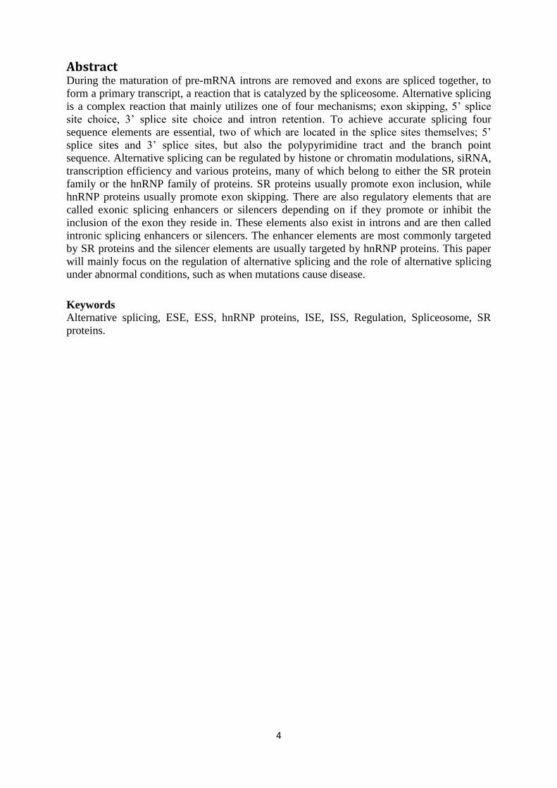
One example of this is the ATM gene and mutations in this gene can cause ataxia
telangiectasia. Ataxia telangiectasia is a rare autosomal recessive genetic disorder that is
characterized by progressive neurodegeneration, immunodeficiency and a high risk of cancer.
There is also telangiectasia, dilation of small blood vessels, in eyes and skin (Lavin et al.,
2007). The ATM gene contains a pseudo exon which is included in the mature mRNA through
a complex mechanism. The inclusion of the pseudo exon is inhibited by the binding of the
U1snRNP at an intronic splicing processing element, or ISPE. When this ISPE is intact the
U1snRNp binds to it, but the spliceosomal assembly and the formation of the A complex is
ineffective. This suggests that the U1snRNP mediated recruitment of U2snRNP is
unsuccessful, which would make the U1snRNP a splicing silencer in this situation. This leads
to the repression of the ATM upstream cryptic 3’ splice site and repression of the pseudo
exon. The binding of U1snRNP to the ISPE element also reduces the binding affinity for
ASF/SF2 to an enhancer sequence that is located in the pseudo exon, which also leads to
repression of the pseudo exon. (Dhir et al., 2010). A mutation in the ISPE element in ATM
leads to activation of two nearby pre-existing cryptic splice sites, which leads to intron
removal around the pseudo exon. The ISPE deletion removes the static interference of
U1snRNP on the cryptic splice site and results in alternative splicing in the upstream section
in the intron and activates excision of the upstream portion of the intron. This also generates a
splicing precursor. Further processing of the precursor to a cryptic exon depends on the
presence of a downstream ISE located in an antisense Alu. Alu repeats are located in the
human genome in gene-rich regions and are specially imbedded within introns and Alu
sequences containing splicing regulatory elements and can change regular splicing into
alternative splicing. The ISE embedded in the antisense Alu facilitates recognition of the
weak 5’ splice site. This leads to an incorporation of the cryptic exon into the mature mRNA
and it is responsible for ataxia telangiectasia (Figure 3 and 4) (Pastor et al., 2009).
Figure 3. The cryptic exon inclusion when the cryptic splice site is activated due to the
mutation. From Pastor et al. (2009).
Figure 4. In the WT the binding of U1snRNP to the ISPE interferes with the recognition of the
nearby cryptic 3’ splice site and no precursor is constructed, which leads to normal
processing. The mutant ISPE (triangle) removes the interference of the U1snRNP, which
activates the cryptic 3’ splice site and the intermediate precursor is formed. In the presence of
the ISE element (yellow circle) this intermediate is processed to mature mRNA that contains
the cryptic exon. From Pastor et al. (2009).

11
Transcription efficiency In mammalian cells the methylation of intragenic DNA initiates the formation of a closed
chromatin structure which reduces the efficiency of RNA polymerase II (Pol II). The reduced
efficiency of Pol II induces an alternative exon inclusion. The rate of transcription also affects
the outcome when two splicing reactions occur simultaneously during the transcription. Fast
and highly efficient transcription favors exon skipping whilst slow and not as efficient
transcription favors the inclusion of an exon (Alló et al., 2009).
Histone modifications and Chromatin modifications Nucleosomes are depleted in introns that are neighboring an acceptor splice site and at the
polyA site and abundant in exons. The position of the nucleosomes seems to be independent
of transcription but highly expressed genes have reduced levels of nucleosomes on the exons,
compared with genes with low expression rates. This suggests that there might be
modifications during transcription and the different levels of nucleosomes in exons and
introns might facilitate exon definition. The higher levels of nucleosomes increase the
strength of splice sites. The nucleosomes will slow Pol II down which increases the
possibility that the splicing factors recognize a weak splice site. The histone molecules may
contribute to the recognition of regulatory elements on the mRNA by splicing regulators and
hence affect the splice site selection (Fox-Walsh and Fu, 2010).
siRNA and gene silencing Small interfering RNAs, siRNAs trigger transcriptional gene silencing (TGS) in human cells.
This is achieved through heterochromatin formation at target DNA sequences and the process
includes recruitment of chromatin-modifying enzymes which results in methylation of
histones and DNA. siRNAs can also regulate gene expression in mammalian cells through the
histone modification and DNA methylation. Recent findings by Alló and colleagues, 2009,
indicate that siRNA targeting gene sequences surrounding an alternative exon can regulate its
alternative pre-mRNA splicing. It is the siRNA that target the intron downstream of the
alternative exon that promotes inclusion of the alternative exon. The siRNA surrounding the
alternative exon forms a closed chromatin structure that prevents efficient Pol II elongation.
This delay in elongation gives more time to the splicing machinery for the recognition and
inclusion of an alternative exon. The interactions between siRNA and Pol II have been
experimentally validated by Alló and colleagues (2009), with the use of a promoter which
could be activated by Sp1 (promotes initiation) and VP16 (promotes initiation and
elongation). When the transcription of the genes was transactivated by Sp1 the siRNA
increased the exon inclusion but when the VP16 transactivator was used this effect vanished.
However when a VP16 mutant, SW6 which does not support elongation, was used the effect
of the siRNA was not removed (Alló et al., 2009).
SR proteins Members of the serine/arginine-rich (SR) protein family are important components in
constitutive and alternative splicing, as well as in many other aspects of mRNA metabolism
such as mRNA transport, translation and stability in eukaryotic cells (Saiprasad and Anireddy,
2010). It is not yet concluded whether all individual SR proteins can perform all of the
different SR protein functions or if several independent SR proteins take part in excision of
each intron. It is possible that alternative splice site choices are decided by which combination
of splicing factors, including SR proteins, which associate with a pre-mRNA. SR proteins
typically contain one or two N-terminal RNA recognition motifs (RRM) RNA binding

12
domains (RBD) and also a C-terminal arginine/serine rich (RS) domain that is variable in
length (Gravely and Maniatis, 1998; Björk et al., 2009). The SR protein specificity for
substrates is defined by their RNA recognition motif domain. There are also some RNA
sequences that have specificity for SR proteins. Natural pre-mRNA probably contains a
combination of these motifs and it is likely that these sequences on the pre-mRNAs determine
the gene-specific SR protein association patterns. However, it is also possible that other
proteins, such as hnRNP proteins, influence the binding of SR proteins and one such protein,
hrp59, is believed to bind to a specific pre-mRNA at an exon site that overlaps with the SR
protein exon site, thus inhibiting the binding of the SR protein (Björk et al. 2009). The SR
proteins are phosphorylated at the serine residues in the RS domain and activated by two
families of protein kinases, SRPK and Clk/Sty. SRPK1 binds to a motif in ASF/SF2 that
restricts phosphorylation to the N-terminal of the RS domain whilst Clk/Sty can
phosphorylate the whole of the RS domain of other SR proteins, which results in a
hyperphosphorylated state (Long and Caceres, 2009). This posttranslational modification is
required for the SR protein to be able to interact with a carrier protein, SR-transportin, that
translocates the SR protein from the cytoplasm to the nucleus and the amount of
phosphorylation is essential for the SR protein activity in alternative splicing (Aubol et al.
2003; Ma et al., 2009; Ma et al., 2009).
SR proteins have been shown to interact with each other and RNA molecules, but also with
proteins in the splicing machinery, such as the U1snRNP (Gravely and Maniatis, 1998; Björk
et al., 2009). It is also possible that the SR proteins facilitate the recruitment of the tri-snRNP
(Long and Caceres, 2009). All of these protein-protein interactions require the RS domain
which, when phosphorylated is thought to function in the formation of protein-protein and
protein-RNA interactions required for spliceosome assembly (Calarco et al., 2009). The
phosphorylated RS domains bind splicing signals that are partially base paired with UsnRNPs
and promote this base pairing (Björk et al., 2009). The RS domains can also contact the
mRNA directly via the branch point and 5’ splice site, which suggest an alternative
mechanism for spliceosome assembly (Long and Caceres, 2009). About 40 RS domains have
been related to splicing in mammalian cells and it is suggested that the levels of these proteins
may contribute to cell- and tissue-dependent alternative splicing (Calarco et al., 2009). One
function of the SR proteins, such as ASF/SF2, is to promote the binding of U1snRNP to 5’
splice sites (Li and Manley, 2005). SR proteins may also function by ensuring correct pairing
of 5’ and 3’ splice sites by simultaneous interaction with U1snRNP and U2AF, which
interacts with the 5’ and 3’ splice site respectively. U2AF is a non-snRNP protein which
consists of two subunits that is required for the binding of U2snRNP to the pre-mRNA branch
site and it binds specifically to the polypyrimidine tract at 3’ splice sites (Figure 5).
Figure 5. Simultaneous binding of SR proteins to U1snRNP and U2AF. From Long and
Caceres (2009).

13
In addition to these constitutional functions the SR proteins also play an important role in
regulating alternative splicing (Graveley and Maniatis, 1998). Binding of SR proteins to
ESEs prevents exon skipping, which ensures correct 5’ to 3’ linear order of exons in spliced
mRNA. There are two models to which this could be explained. The first is relying on the
ability of the ESE-bound SR proteins to recruit and stabilize interactions between the U1
snRNP at the 5’ splice site and U2AF at the 3’ splice site. This process is called exon
definition. The other model is based on the capability of ESE bound proteins to antagonize the
effects of hnRNP proteins, which recognize ESSs. The SR proteins may also form protein-
protein interactions across introns to juxtapose the 5’ and 3’ splice sites early in the
spliceosomal assembly in an RS dependent manner. The SR protein ASF/SF2 is also able to
interact directly with the RNA at the branch point to promote spliceosomal assembly (Long
and Caceres, 2009). The SR proteins that are attached to a splicing enhancer can also recruit
components of the splicing machinery to nearby enhancer-dependent introns with weak splice
sites. Introns often contain non-consensus pyrimidine tracts to which U2AF binds weakly. It
has been shown that the small subunit of the U2AF protein can simultaneously interact with
the large subunit as well as the SR proteins within the enhancer complex. This stabilizes the
U2AF protein binding to the weak 3’ splice site (Graveley and Maniatis, 1998).
Not all SR proteins contain an RS domain and those who lack that domain are mostly the ones
that have been shown to have tissue-specific expression patterns. Several of these proteins
play an important role in cell- and tissue-specific alternative splicing. They include the
neuronal-specific Nova-1/2 and HuB proteins, the neuronal- and muscle-expressed proteins
Fox-1/2 and MBNL family proteins and the neuronal-, myoblast- and testis-expressed nPTB
protein. nPTB is a paralog of the widely expressed PTB splicing repressor protein. The switch
from expression of PTB to nPTB has been connected to regulation of neuronal alternative
splicing and results in the formation of less repressive nPTB-bound splicing complexes that
are responsive to positive-acting factors in neurons. The mechanism of action is however not
well understood (Calarco et al., 2009). One tissue- and vertebrate-restricted RS domain
protein, neural-specific SR related protein of 100 kDa (nSR100) has been identified. The lack
of this protein results in exclusion of a large number of alternative exons in the nervous
system. These exons are mainly located in genes with important roles in neural cell
differentiation. nSR100 creates neuronal specificity in alternative splicing by activating nPTB
expression and by binding directly to its regulated target transcripts (Calarco et al., 2009).
SC35 Experiments with the SR protein SC35 in the Balbiani ring (BR) genes performed by Björk
and colleagues, 2009, have shown that individual SR proteins have several different functions
in the cell nucleus during processing and transport of a single pre-mRNP, so called when a
eukaryotic mRNA particle is transported from the nucleus to the cell cytoplasm. On the BR
genes the interference with SC35 does not block initiation or elongation but the distribution of
nearby BR pre-mRNPs changed. Is has been shown that SC35, in mammalian cells, recruits
the kinase positive transcription elongation factor b to the elongation complex. This kinase is
important to RNA polymerase II and elongation. The interference with SC35 leads to an
increased number of transcripts in the distal region, which is consistent with the fact that the
elongation process will be slowed down with the impairment of SC35 function (Björk et al.,
2009).
Pyruvate dehydrogenase (PDH) complex deficiency is a common genetic disorder of
mitochondrial energy metabolism. It is a major cause of lactic acidosis and most of the cases
are sporadic and result from a mutation arising within the germ cells of one of the parents. At
least 75 mutations have been identified in the E1α subunit-encoding gene (E1α is a subunit of

14
the PDH complex) and two exonic mutations have been associated with a skipping of exon 6.
A shift from G to A in the intron downstream of exon 7 5’ splice site causes alternative
splicing which involves the use of a cryptic 5’ splice site instead of the normal 5’ splice site.
This mutation creates, or strengthens a SC35 binding enhancer element and also strengthens a
pre-existing ASF/SF2 binding motif. SC35 activates the cryptic 5’ splice site which leads to
the retention of intronic sequences. Increased levels of siRNA have been shown to reduce the
levels of SC35 and thereby reversing the positive regulation of SC35 and increasing the
amount of normal mRNA (Gabut et al., 2005).
SRrp37 SRrp37, SR related protein 37, is a recently found splicing regulator protein and its expression
shows tissue specific expression patterns and it has also been shown that SRrp37 is involved
in alternative splicing initiation. SRrp37 has been found to co-localize with SC35 in nuclear
specks, but it is also found in nucleoli. This might be due to maturation or a traffic pathway
for this protein. Newly synthesized endogenous SRrp37 is localized in speckles, whereas the
mature protein is present in nucleoli. This subcellular localization of SRrp37 in both nucleoli
and nuclear speckles suggest a role in both rRNA and mRNA processing. A large amount of a
protein in the nucleoli is common with proteins that are either ribosomal proteins or proteins
involved in the maturation of rRNA, whereas location in the speckles suggests that the protein
is involved in pre-mRNA splicing. Experiments performed by Ouyang, 2009, have indicated
that SRrp37 prefers not to use 5’ splice sites but instead promotes the use of a 3’ splice site.
This stands in contrast to other SR proteins, such as ASF/SF2, which tends to promote both 3’
and 5’ splice site choices (Ouyang, 2009).
ASF/SF2 ASF/SF2, or Alternative splicing factor/splicing factor 2, is a sequence specific RNA binding
protein with two RNA binding domains and a C-terminal RS domain for protein-protein
interaction (Aubol et al., 2003). It promotes the binding of U1snRNP and recognition of 5’
splice sites. This increase in affinity for the 5’ splice site is the result of ASF/SF2 promoting
of the annealing of complementary RNA (Eperon, et al., 1993). Furthermore, it interacts with
the c-terminal domain (CTD) of RNA polymerase II which is necessary for its recruitment to
the transcription sites (Li and Manley, 2005). It is foremost a nuclear protein, but it is known
to shuttle between the cytoplasm and the nucleus. Phosphorylation and dephosphorylation are
crucial in the splicing function of ASF/SF2 and affect its cellular localization (Moulton and
Tsokos, 2010). The phosphorylation of the RS domain in ASF/SF2 acts to enhance protein–
protein interactions with other RS domain-containing splicing factors, such as one subunit of
the U1snRNP. The phosphorylation that is necessary for the activation of ASF/SF2 only takes
place at one or more of the 20 serine residues in the RS domain (Aubol et al., 2003). ASF/SF2
tends to favor the downstream 5’ splice site; however, at higher concentrations additional 5’
splice sites could be recognized and thus activated. This would probably be those 5’ splice
sites that are closest to the 3’ splice site due to proximal localization, or even cryptic 5’ splice
sites (Zuo and Manley, 1994; Wang and Manley, 1995). ASF/SF2 can also promote a
proximal choice between two competing 3’ splice sites. In this case, the 5’ splice site is fixed
and ASF/SF2 promotes the use of a weak 3’ splice site. ASF/SF2 can interact with U2AF
which binds to the branch point near the 3’ splice site. To evaluate the ability of ASF/SF2 to
sense the proximity or distance, the effect of hnRNP A1 must be taken into consideration.
hnRNP A1 promotes distal splicing and antagonizes the proximal splicing that is favored by
ASF/SF2, which indicates that both ASF/SF2 and hnRNP A1 play important roles in splice
site choices (Bai et al., 1999).

15
The β-tropomyosin gene generates a number of different protein products due to tissue-
specific alternative splicing. It contains three pairs of exons that are mutually exclusive and in
two of these the exon choice is related to the use of different promoters or polyadenylation
signals. The third pair of exons, 6A and 6B, are selected in a tissue-specific manner and are
independent of promoter or polyadenylation site choice. Exon 6A is included and expressed in
fibroblasts and smooth muscle cells and exon 6B is exclusively included in skeletal muscle
cells. Sequences at the 3’ end of the intron between the two exons and within exon 6B have
been shown to repress the use of the 3’ splice site choice in exon 6B in non-muscle cells. A
pyrimidine-rich region in the intron downstream of exon 6A is a splicing enhancer essential
for recognition of the 5’-splice site of exon 6A. The SR protein ASF/SF2 is able to activate
the use of exon 6A through specific recognition of the S4 enhancer sequence. That is
somewhat unexpected, since ASF/SF2 often is involved in the recognition of exonic enhancer
sequences rich in purines. The splicing factor SC35 on the other hand behaves as an inhibitor
of the 6A exon and directly antagonizes ASF/SF2. It has been shown that abundance of these
proteins differs between skeletal muscle cells and non-skeletal muscle cells. The shift towards
SC35 in skeletal muscle cells contributes to the exclusion of exon 6A in these types of cells
(Gallego et al., 1997).
hnRNP proteins hnRNPs, or heterogeneous nuclear ribonucleoproteins, are a family of proteins that have
many functions. They are involved in telomere biogenesis, mRNA stability and turnover,
cytoplasmic trafficking of mRNA and in splicing control (Moran-Jones et al., 2009). In
splicing, they are trans-acting factors that bind to either ESSs or ISSs. This functions to
inhibit the use of certain splice sites during splicing events. However, there are some cases
where hnRNPs promote splicing instead of inhibit it. The usual structure of hnRNP proteins
consist of two or more RNA binding domains and a secondary domain. The secondary
domain is thought to facilitate protein-protein interactions and ssDNA-protein interactions.
But it is also involved in RNA-protein interactions, just as the RNA binding domains.
Furthermore, most hnRNPs can interact with other hnRNPs (Clower et al., 2010). There are
about 20 classes of hnRNP proteins and the hnRNP A/B family includes the paralogues A0-
A3 and the last three proteins have multiple isoforms that is the result of alternative splicing
(Zhu et al., 2002; Landsberg et al., 2006). When they are bound to ESSs or ISSs, they can
bind along the RNA through cooperative binding, which can interfere with the binding of
spliceosomal components or activating proteins, such as the SR proteins (Clower et al., 2010).
It has also been suggested that several proteins of the hnRNP family can multimerize, thus
creating regions of silencing across exons or modulate the conformation of the pre-mRNA
and thereby influence exon recognition (D’Ambrogio et al., 2009).
hnRNP A1/A2 hnRNP A1 is one of the most common hnRNP proteins and is involved in many alternative
splicing events. In humans it is a 320 amino acid protein with two N-terminal RNA
recognition domains and the secondary domain is rich in glycine (GRD, glycine rich domain),
a structure that it shares with hnRNP A2 (Clower et al., 2010). hnRNP A2 is a nuclear protein
that is involved in RNA packaging and splicing, but also telomere biogenesis, cytoplasmic
RNA trafficking, and cap-dependent translation. It has high affinity for, amongst others, a
RNA sequence with homology to a region found in 3’ splice site selection elements
(Landsberg et al., 2006). hnRNP A2 is believed to be involved, via up-regulation or
mislocalization, in may human cancers and is found in many tumor-derived cell lines (Moran-
Jones et al,. 2009).
16
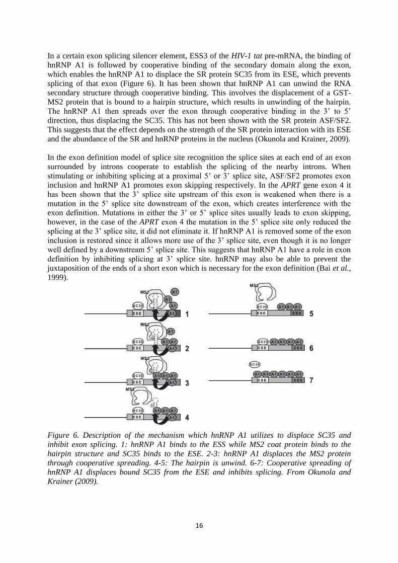
In a certain exon splicing silencer element, ESS3 of the HIV-1 tat pre-mRNA, the binding of
hnRNP A1 is followed by cooperative binding of the secondary domain along the exon,
which enables the hnRNP A1 to displace the SR protein SC35 from its ESE, which prevents
splicing of that exon (Figure 6). It has been shown that hnRNP A1 can unwind the RNA
secondary structure through cooperative binding. This involves the displacement of a GST-
MS2 protein that is bound to a hairpin structure, which results in unwinding of the hairpin.
The hnRNP A1 then spreads over the exon through cooperative binding in the 3’ to 5’
direction, thus displacing the SC35. This has not been shown with the SR protein ASF/SF2.
This suggests that the effect depends on the strength of the SR protein interaction with its ESE
and the abundance of the SR and hnRNP proteins in the nucleus (Okunola and Krainer, 2009).
In the exon definition model of splice site recognition the splice sites at each end of an exon
surrounded by introns cooperate to establish the splicing of the nearby introns. When
stimulating or inhibiting splicing at a proximal 5’ or 3’ splice site, ASF/SF2 promotes exon
inclusion and hnRNP A1 promotes exon skipping respectively. In the APRT gene exon 4 it
has been shown that the 3’ splice site upstream of this exon is weakened when there is a
mutation in the 5’ splice site downstream of the exon, which creates interference with the
exon definition. Mutations in either the 3’ or 5’ splice sites usually leads to exon skipping,
however, in the case of the APRT exon 4 the mutation in the 5’ splice site only reduced the
splicing at the 3’ splice site, it did not eliminate it. If hnRNP A1 is removed some of the exon
inclusion is restored since it allows more use of the 3’ splice site, even though it is no longer
well defined by a downstream 5’ splice site. This suggests that hnRNP A1 have a role in exon
definition by inhibiting splicing at 3’ splice site. hnRNP may also be able to prevent the
juxtaposition of the ends of a short exon which is necessary for the exon definition (Bai et al.,
1999).
Figure 6. Description of the mechanism which hnRNP A1 utilizes to displace SC35 and
inhibit exon splicing. 1: hnRNP A1 binds to the ESS while MS2 coat protein binds to the
hairpin structure and SC35 binds to the ESE. 2-3: hnRNP A1 displaces the MS2 protein
through cooperative spreading. 4-5: The hairpin is unwind. 6-7: Cooperative spreading of
hnRNP A1 displaces bound SC35 from the ESE and inhibits splicing. From Okunola and
Krainer (2009).

17
hnRNP A3 hnRNP A3 is a poorly understood protein of the hnRNP family. It has a similar structure to
the hnRNP A1 and A2 and certain isoforms are known to co-localize with hnRNP A1 in
neuronal transport granules and interact with nuclear actin, together with hnRNP A2
(Makeyev et al., 2005). It has recently been discovered that hnRNP A3 also is a telomere-
binding protein. It has been shown that an hnRNP A3 mutant, hnRNP dA3, has a DNA-
binding activity that is specific to the G-rich strand (Huang et al., 2008). Even though it is not
involved in the regulation of alternative splicing, it undergoes alternative splicing itself. The
hnRNPA3 gene has homologous sequences, pseudo exons, on many chromosomes, but only
the sequence on chromosome 2 has introns and shows the features of an active gene locus. A
sequence that was thought to be the real gene has been proved to be a processed pseudo exon
called HNRPA3P1. This pseudo exon contains a frame shift mutation that results in premature
termination of translation and a non-functional protein. The real hnRNP A3 gene has four
isoforms that are created through alternative splicing where three of them lack nucleotides
either in the beginning or in the middle. This suggests that there are two optional ways of the
alternative splicing. However, the mechanisms behind the alternative splicing remain largely
unknown (Makeyev et al., 2005).
hnRNP H hnRNP H can, in contrast to many other hnRNP proteins, act both as an activator and as a
repressor of splicing. An activation effect has been seen in, amongst others, the regulation of
exon 6D of HIV-1 and the apoptotic mediator Bcl-x. In HIV-1, hnRNP H binds to a glycine
rich domain and is required for the association of U1snRNP to exon 6D, which enhances exon
inclusion. hnRNP is involved in the Bcl-x production by directing the 5’ splice site choice.
Repression on the other hand has been detected at the 5’ splice sites in the NF-1 and THSβ
genes. It can bind to G-rich 5’ splice sites and block U1snRNP binding, which leads to exon
skipping. This especially takes place when there are mutations that impair U1snRNP pairing.
The fact that hnRNP H can both activate and repress splicing by binding to GRDs suggests
that in addition to the binding sites, the sequence and the position of the sites are important for
the splicing activity (Crawford and Patton, 2006).
hnRNP L hnRNP L is a nuclear RNA binding protein with many functions and is involved in mRNA
export, mRNA stability and splicing and it has four RNA-recognition motifs. When it is
interacting with introns, hnRNP L can act either as an activator or a repressor in alternative
splicing, depending on the splice site proximity. hnRNP L is able to regulate its own
expression through alternative splicing. Excess hnRNP L can bind to CA rich motifs located
in exon 6 with high affinity. This leads to inclusion of exon 6A and in this case, the CA rich
domain acts as an intronic enhancer of exon 6A inclusion. The inclusion of exon 6A brings
with it a premature stop codon, which leads to a reduction of functional hnRNP L mRNA. It
is believed that the CA rich domain contains different clusters and that several hnRNP L
molecules binds to each cluster parts which may induce conformational changes which
generates an hnRNP L concentration-dependent splice-regulatory signal. This role of
activation contrasts to the usual repression activity of hnRNP proteins (Rossbach et al., 2009).
The CD45 gene encodes a hematopoiesis-specific transmembrane protein tyrosine
phosphatase. Three variable exons of the CD45 gene are skipped during T-cell activation
which leads to a decreased phosphatase activity and maintenance of T-cell homeostasis. An
exonic splicing silencer, ESS1, has been identified in the CD45 exon 4. This element
mediates both partial exon repression in resting cells and increased exon skipping during T-
cell stimulation. The partial exon repression is the result of the binding of at least five hnRNP

18
proteins to the ESS1 element with hnRNP L acting as the main functional regulator. This
stalls the assembly of the U1 and U2snRNP components which inhibits the formation of the
spliceosome. The activation of the T cells leads to two changes in the composition of the
ESS1 and the hnRNP L complex. A posttranslational modification of hnRNP L increases the
silencing activity slightly while addition of a PTB-associated splicing factor, PSF, to the
complex have a more prominent effect on exon skipping. The posttranslational modifications
of hnRNP L is not associated with increased nuclear concentrations or changed affinity for
ESS1 but may be related to phosphorylation or decreased acetylation. The levels of PSF are
the same in both resting and in activated T cells, which suggests that the binding of PSF to the
ESS1 element is not regulated by protein expression or localization. One suggestion for the
mechanism of PSF regulation involves signal-induced changes in PSF association with other
nuclear proteins and PSF is known to be a binding partner of PTB (discussed below), other
nuclear proteins, DNA and RNA. This indicates that PSF may exist in a protein complex in
the nucleus of resting T cells and upon activation of the cell, PSF is released and then
recruited to the ESS1 complex. The functions of hnRNP L and PSF suggests that hnRNP L
plays a major role in the basal level of exon repression under resting conditions and the
combined effects of hnRNP L and PSF regulates the ESS1-dependent activation-induced
silencing (Melton et al., 2007).
TDP-43 Nuclear factor TDP-43 is a multifunctional RNA binding protein that is a member of the
hnRNP family. It is involved in transcription, pre-mRNA splicing, alternative splicing,
mRNA stability and mRNA transport. It is a 414 amino acid protein and it has four distinct
regions; an N-terminal sequence which contains a nuclear localization signal and two RNA
recognition motifs (RRM). The RRM1 of TDP-43 mediates its RNA binding ability and
RRM2 is needed for correct assembly. The fourth region is a glycine rich C-terminal domain.
The GRD has been shown to be essential for the protein to function as a splicing silencer of
exon 9 of the CFTR gene. This region is also responsible for the capability of forming
complexes with other hnRNPs (Bose et al., 2009; D’Ambrogio et al., 2009). It has also been
reported that this region is required for the ability of TDP-43 to act as a transcriptional
insulator for a mouse gene. It has been shown that TDP-43 is able to bind to hnRNP A2.
Since the very same region is necessary for the TDP-43 splicing inhibitory activity it is likely
that TDP-43 is required to form an hnRNP complex through its C-terminus to inhibit exon
splicing (D’Ambrogio et al., 2009). TDP-43 does not only interact with hnRNP proteins, but
also with RNA helicases, splicing factors, translation regulatory proteins and proteins
involved in mRNA transport and stability. TDP-43 has recently been identified as an
important protein in diseases, such as ALS (Amyotrophic Lateral Sclerosis), FTLD-TDP
(Frontal Temporal Dementia-TDP), and IBMPFD (Inclusion Body Myopathy, Paget's disease
and Frontotemporal Dementia). The pathology is typically characterized by clearance of TDP-
43 from the nucleus and accumulation in the cytoplasm of affected cells, which indicates that
the diseases involve either loss of TDP-43 nuclear function or toxic TDP-43 accumulation in
the cytoplasm (Freibaum et al., 2009).
Spinal muscular atrophy, SMA, is a common autosomal recessive disorder that is caused by
degeneration of motor neurons. The survival of motor neuron gene (SMN) is spliced into two,
almost identical transcripts, SMN1 and SMN2. The difference is a shift from C to T at
position 6 in exon 7 in SMN2. Homozygous loss of SMN1 cannot be compensated by SMN2
since the change in nucleotides at position 6 causes exon 7 skipping through alternative
splicing. There has been two different suggestions to this splice pattern; the first is loss of an
ESE and the second is gain of an ESS. Several trans-acting factors are involved in the exon 7
splicing, such as SR proteins and hnRNP proteins. The SR proteins act as positive regulators

19
through the AG-rich ESE element in SMN exon 7 and hnRNP A1 acts as a negative regulator
by binding to the ESS element that is created by the change from C to T in the SNM2 exon 7.
However, it is now believed that over expressed TDP-43 can promote exon inclusion of exon
7, which gives it a dual role in alternative splicing (Figure 7). Both RRMs are needed for this
regulation as well as interactions with an hnRNP protein and Htra2-β1 (a serine-protease). It
seems that TDP-43 interacts with the RNA in a sequence-independent manner, however
evidence suggest that the exon inclusion is mediated through complex formation at an ESE
element in exon 7, thus inhibiting the negative effect by hnRNP A1 (Bose et al. 2008).
Figure 7. The difference between SMN1 and SMN2. In this model the exon gains an ESS for
hnRNP A1 in SMN2. The hnRNP A 1 blocks inclusion of exon 7. When TDP-43 is over
expressed it inhibits the effects of hnRNP A1, possibly by forming a new complex with the
other proteins involved in this reaction that is resistant to the negative effect of hnRNP A1.
From Bose et al. (2008).
PTB The polypyrimidine tract binding protein, PTB or hnRNP I, is an RNA-binding protein that
binds to polypyrimidine tracts, such as those located at or upstream of 3’ splice sites. This can
regulate alternative splicing by creating a region of silencing (Clower et al., 2010). PTB has
the ability to interfere with exon definition but it seems that the PTB binding sites are not
sufficient for exon silencing. Some exons require other silencer sequences or weak splice sites
to achieve inhibition of exon inclusion. In the case of IIIb exon of FGF-R2 a weak
polypyrimidine tract and an ESS that interact with hnRNP A1 is also needed to promote exon
skipping. This suggests that PTB acts together with co-repressors to mediate exon silencing.
PTB binding sites sometimes overlap binding sites for U2AF and this competition could be
one reason for the inhibitory action of PTB. The majority of exons that are silenced by PTB
are flanked by PTB binding sites on both nearby introns and it is suggested that PTB bind
across the exon. In neuronal cells PTB is responsible for exon silencing, but there are still
exons that are only expressed in neuronal cells. Lower levels of PTB in these cells and also
the presence of a neuronal type of PTB, nPTB is thought to be responsible for this. nPTB can
compete with PTB for binding but has a weaker repressive effect (Wagner and Garcia-Blanco,
2001).

20
Conclusions
Alternative splicing is a very complex mechanism that is responsible for the human diversity,
large proteome and phenotypic complexity. This has become clearer during the last decade
and it is now believed that up to 90% of the human genes undergo alternative splicing. Both
constitutional and alternative splicing is catalyzed by the spliceosome. The spliceosome
consists of five UsnRNPs and many, up to hundreds, of proteins. The recruitment and affinity
of the spliceosome components are often dependent on regulatory proteins and sequences. In
alternative splicing there are mainly four ways to create the alternative transcripts. There is
the possibility of choosing an alternative 5’ or 3’ splice site, skipping of whole exons and
intron retention. However, there is some debate about the intron retention mechanism, if it
should fall under alternative splicing or not.
The regulatory system consists of, amongst others, regulatory SR proteins and hnRNP
proteins. The SR proteins usually promote exon inclusion, such as ASF/SF2, SC35 and
SRrp37, however there are exceptions. In the case of the β-tropomyosin gene SC35
antagonizes ASF/SF2 which leads to exclusion of exon 6A, which would give SC35 an
inhibitory role. hnRNP proteins tends to inhibit exon inclusion such as hnRNP A1, hnRNP
A2, hnRNP H, hnRNP L, TDP-43 and PTB. But there are exceptions among the hnRNPs as
well and one such exception is hnRNP H which can act both as an activator and as a repressor
of splicing. This regulatory activation can be seen in exon 6D of HIV-1 and in Bcl-x. The
regulation of alternative splicing is also dependent on exonic splicing regulatory sequences
and intronic regulatory sequences. These can be either enhancer or silencer, depending on
where and how they act. The enhancer sequences are targets for SR proteins and the silencer
sequences are targets for hnRNP proteins.
Alternative splicing relies on a complex mechanism that involves many components and
interactions. It is also quite sensitive to mutations. If a mutation changes the affinity for
regulatory proteins, creates or removes regulatory sequences or creates or changes the affinity
for splice sites it changes the dynamic of the alternative splicing. Exons can be excluded,
pseudo exons can be included and the length of the exons can be altered. This can lead both to
nonfunctional proteins and proteins that cause disease. In the case of ataxia telangiectasia
there is a mutation on an ISPE element on the ATM gene. This leads in several steps to the
inclusion of a pseudo exon, which causes the disease. Pyruvate dehydrogenase complex
deficiency is a genetic disorder of mitochondrial metabolism. A mutation in exon 7 of the E1α
subunit leads to the use of a cryptic 5’ splice site instead of the constitutive splice site. The
mutation creates or enhances an enhancer element for SC35. SC35 activates this cryptic splice
site via the enhancer element, which leads to retention of intronic sequences. SMA, or spinal
muscular atrophy, is dependent on the SMN gene. This gene has two very similar transcripts
and the difference is a nucleotide shift in the second variant that creates an ESS element.
Binding of TDP-43 to this element causes exon exclusion and disease due to nonfunctional
SMN.
Due to all this further understanding of alternative splicing and its regulation is a very
important step in managing and preventing disease. If the regulation of alternative splicing
could be altered, diseases that are now incurable could get successful treatment. Alternative
splicing is involved in different cancers and if the splicing could be inhibited or altered in the
tumors, more efficient tumor control could be managed. Alternative splicing is an area where
more research is needed, both for the understanding of human complexity and for disease
management.

21
References
Alló, M., Buggiano, V., Fededa, J. P., Petrillo, E., Schor, I., de la Mata, M, Agirre, E., Plass,
M., Eyras, E., Elela, S. A., Klinck, R., Chabot, B. and Kornblihtt, A. R. Control of alternative
splicing through siRNA-mediated transcriptional gene silencing. Nature structural and
molecular biology. 2009, 16:7 p. 717-725
Anquetil, V., Sommer, C. L., Mereau, A., Hamon, S., Lerivray, H., and Hardy, S.
Polypyrimidine Tract Binding Protein Prevents Activity of an Intronic Regulatory Element
That Promotes Usage of a Composite 3’-Terminal Exon. The Journal of Biological
Chemistry. 2009, 284:47 p. 32370–32383
Aubol, B. E., Chakrabarti, S., Ngo, J., Shaffer, J., Nolen, B., Fu, X.-D., Ghosh, G. and
Adams, J. A. Processive phosphorylation of alternative splicing factor/splicing factor 2.
Proceedings of the National Academy of Science. 2003, 100:22 p. 12601–12606
Bai, Y., Lee, D., Yu, T. and Chasin, L.A. Control of 3’ splice site choice in vivo by ASF/SF2
and hnRNP A1. Nucleic Acids Research. 1999, 27:4 p. 1126-1134
Björk, P., Jin, S., Zhao, J., Singh, O. P., Persson, J.-O., Hellman, U., and Wieslander, L.
Specific combinations of SR proteins associate with single pre-messenger RNAs in vivo and
contribute different functions. Journal of Cellular Biology. 2009, 184:4 p. 555–568
Bose, J. K., Wang, I.-F., Hung, L., Tarn, W.-Y. and Shen, C.-K. J. TDP-43 Overexpression
Enhances Exon 7 Inclusion during the Survival of Motor Neuron Pre-mRNA Splicing. The
Journal of Biological Chemistry. 2008, 283:43 p. 28852–28859
Calarco, J. A., Superina, S., O’Hanlon, D., Gabut, M., Raj, B., Pan, Q., Skalska, U., Clarke,
L., Gelinas, D., van der Kooy, D., Zhen, M., Ciruna, B. and Blencowe, B. J. Regulation of
Vertebrate Nervous System Alternative Splicing and Development by an SR-Related Protein.
Cell. 2009, 138 p. 898–910
Clower, C. V., Chatterjee, D., Wang, Z., Cantley, L. C., Vander Heiden, M. G. and Krainer,
A. R. The alternative splicing repressors hnRNP A1/A2 and PTB influence pyruvate kinase
isoform expression and cell metabolism. Proceedings of the National Academy of Science.
2010, 107:5 p. 1894-1899
Crawford, J. B. and Patton, J. G. Activation of α-Tropomyosin Exon 2 Is Regulated by the SR
Protein 9G8 and Heterogeneous Nuclear Ribonucleoproteins H and F. Molecular and
Cellular Biology. 2006, 26:23 p. 8791–8802
D’Ambrogio, A., Buratti, E., Stuani, C., Guarnaccia, C., Romano, M., Ayala, Y. M. and
Baralle, F. E. Functional mapping of the interaction between TDP-43 and hnRNP A2 in vivo.
Nucleic Acids Research. 2009, 37:12 p. 4116–4126
Dhir, A., Buratti, E., van Santen, M. A., Lührmann, R. and Baralle, F. E. The intronic splicing
code: multiple factors involved in ATM pseudoexon definition. The EMBO Journal. 2010, 29
p. 749–760

22
Ding, W., Lin, L., Ren, F., Zou, H., Duan, Z. and Dai, J. Effects of splice sites on the intron
retention in histamine H3 receptors from rats and mice. Journal of Genetics and Genomics.
2009, 36 p. 475-482
Eperon, I. C., Ireland, D. C., Smith, R. A., Mayeda, A. and Krainer, A. R. Pathways for
selection of 5' splice sites by U 1 snRNPs and SF2/ASF. The EMBO Journal. 1993, 12:9 p.
3607-3617
Fox-Walsh, K. and Fu, X.-D. Chromatin: The Final Frontier in Splicing Regulation?
Developmental Cell. 2010, 18 p. 336-338
Freibaum, B. D., Chitta, R. K., High, A. A. and Taylor, J. P. Global Analysis of TDP-43
Interacting Proteins Reveals Strong Association with RNA Splicing and Translation
Machinery. Journal of Proteome Research. 2010, 9 p. 1104–1120
Gabut, M., Miné, M., Marsac, C., Brivet, M., Tazi, J. and Soret, J. The SR Protein SC35 Is
Responsible for Aberrant Splicing of the E1α Pyruvate Dehydrogenase mRNA in a Case of
Mental Retardation with Lactic Acidosis. Molecular and Cellular Biology. 2005, 25:8 p.
3286–3294
Galante, P., Sakabe, N., Kirschbaum-Slager, N., Souza, S. Detection and evaluation of intron
retention events in the human transcriptome. RNA. 2004, 10 p. 757–765
Gallego, M. E., Gattoni, R., Stévenin, J., Marie, J., Expert-Bezancon, A. The SR splicing
factors ASF/SF2 and SC35 have antagonistic effects on intronic enhancer-dependent splicing
of the b-tropomyosin alternative exon 6A. The EMBO Journal. 1997, 16:7 p. 1772–1784
Goren, A., Ram, O., Amit, M., Keren, H., Lev-Maor, G., Vig, I., Pupko, T. and Ast, G.
Comparative Analysis IdentifiesExonic Splicing Regulatory Sequences—The Complex
Definition of Enhancers and Silencers. Molecular Cell. 2006, 22 p. 769–781
Graveley, B. R. and Maniatis, T. Arginine/Serine-Rich Domains of SR Proteins Can Function
as Activators of Pre-mRNA Splicing. Molecular Cell. 1998, 1 p. 765–771
Hartmuth, K., Urlaub, H., Vornlocher, H.-P, Will, C. L., Gentzel, M., Wilm, M. and
Luhrmann, R. Protein composition of human prespliceosomes isolated by a tobramycin
affinity-selection method. Proceedings of the National Academy of Science. 2002, 99:26 p.
16719–16724
Huang, R., Tsai, S.-T., Hsieh, K.-H. and Wang, T.-C. V. Heterogeneous nuclear
ribonucleoprotein A3 binds single-stranded telomeric DNA and inhibits telomerase extension
in vitro. Biochimica et Biophysica Acta. 2008, 1783 p. 193–202
Keren, H., Lev-Maor, G. and Ast, G. Alternative splicing and evolution: diversification, exon
definition and function. Nature Review Genetics. 2010, 11 p. 345-355
Koren, E., Lev-Maor, G., Ast, G. The Emergence of Alternative 3’ and 5’ Splice Site Exons
from Constitutive Exons. PLoS Computinal Biology. 2007 3(5)

23
Landsberg, M. J., Moran-Jones, K., and Smith, R.. Molecular Recognition of an RNA
Trafficking Element by Heterogeneous Nuclear Ribonucleoprotein A2. Biochemistry. 2006,
45:12 p. 3943-3951
Lavin, M. F., Gueven, N., Bottle, S. and Gatti, R. A. Current and potential therapeutic
strategies for the treatment of ataxia-telangiectasia. British Medical Bulletin. 2007, p. 1–19
Li, X. and Manley, J. L. Inactivation of the SR Protein Splicing Factor ASF/SF2 Results in
Genomic Instability. Cell. 2005, 122 p. 365–378
Long, J. C. and Caceres, J. F. The SR protein family of splicing factors: master regulators of
gene expression. Biochemical Journal. 2009, 417 p. 15–27
Ma, C.-T., Hagopian, J. C., Ghosh, G., Fu, X.-D. and Adams, J. A. Regiospecific
Phosphorylation Control of the SR Protein ASF/SF2 by SRPK1. Journal of Molecular
Biology. 2009, 390 p. 618–634
Makeyev, A. V., Kim, C. B., Ruddle, F. H., Enkhmanakh, B., Erdenechimeg, L. and
Bayarsaihan, D. HnRNP A3 Genes and Pseudogenes in the Vertebrate Genomes. Journal of
Experimental Zoology. 2005, 303A p. 259-271
Melton, A. A., Jackson, J., Wang, J. and Lynch, K. W. Combinatorial Control of Signal-
Induced Exon Repression by hnRNP L and PSF. Molecular and Cellular Biology. 2007,
27:19 p. 6972–6984
Mironov, A. A., Fickett, J. W. and Gelfand, M. S. Frequent Alternative Splicing of Human
Genes. Genome Research. 1999 9: 1288-1293.
Moran-Jones, K., Grindlay, J., Jones, M., Smith, R. and Norman, J. C. hnRNP A2 Regulates
Alternative mRNA Splicing of TP53INP2 to Control Invasive Cell Migration. Cancer
Research. 2009, 69:24 p. 9219-9227
Moulton, V. R. and Tsokos, G. C. Alternative Splicing Factor/Splicing Factor 2 Regulates the
Expression of the ξ Subunit of the Human T Cell Receptor-associated CD3 Complex. The
Journal Of Biological Chemistry. 2010, 285:17 p. 12490–12496
Okunola, H. L. and Krainer, A. R. Cooperative-Binding and Splicing-Repressive Properties
of hnRNP A1. Molecular and Cellular Biology. 2009, 29:20 p. 5620-5631
Ouyang, P. SRrp37, a Novel Splicing Regulator Located in the Nuclear Speckles and
Nucleoli, Interacts With SC35 and Modulates Alternative Pre-mRNA Splicing In Vivo. Journal
of Cellular Biochemistry. 2009, 108 p. 304–314
Palusa, S. G. and Reddy, A. S. N. Extensive coupling of alternative splicing of pre-mRNAs of
serine⁄arginine (SR) genes with nonsense-mediated decay. New Phytologist. 2010, 185 p. 83–
89
Pastor, T., Talotti, G., Lewandowska, M. A. and Pagani, F. An Alu-derived intronic splicing
enhancer facilitates intronic processing and modulates aberrant splicing in ATM. Nucleic
Acids Research. 2009, 37:21 p. 258–7267

24
Rossbach, O., Hung, L.-H., Schreiner, S., Grishina, I., Heiner, M., Hui, J. and Bindereif, A.
Auto- and Cross-Regulation of the hnRNP L Proteins by Alternative Splicing. Molecular and
Cellular Biology. 2009, 29:6 p. 1442–1451
Stark, H. and Lührmann, R. Cryo-Electron Microscopy of Spliceosomal Components. Annual
Review of Biophysics and Biomolecular Structure. 2006, 35 p. 435–57
Stark, H. and Lührmann, R. Cryo-Electron Microscopy of Spliceosomal Components. Annual
Review of Biophysics and Biomolecular Structure. 2006, 35 p. 435–57
Szeszel-Fedorowicz, W., Talukdar, I., Griffith, B. N., Walsh, C. M., and Salati, L. M. An
Exonic Splicing Silencer Is Involved in the Regulated Splicing of Glucose 6-Phosphate
Dehydrogenase mRNA. The Journal of Biological Chemistry. 2006, 281:45 p. 34146-34158
Tsai, K.-W., Tarn, W.-Y. and Lin, W.-C. Wobble Splicing Reveals the Role of the Branch
Point Sequence-to-NAGNAG Region in 3’ Tandem Splice Site Selection. Molecular and
cellular biology. 2007, 27:16 p. 5835–5848
Wagner, E. and Garcia-Blanco, M. Polypyrimidine Tract Binding Protein Antagonizes Exon
Definition. Molecular and Cellular Biology. 2001, 21:10 p. 3281-3288
Wang, J. and Manley, J. L. Over expression of the SR proteins ASF/SF2 and SC35 influences
alternative splicing in vivo in diverse ways. RNA. 1995, 1 p. 335-346
Wang, Z. and Burge, C. B. Splicing regulation: From a parts list of regulatory elements to an
integrated splicing code. RNA. 2008, 14 p. 802-813
Zhang, X. H.-F., Kangsamaksin, T., Chao, M. S. P., Banerjee, J. K., and Chasin, L. A. Exon
Inclusion Is Dependent on Predictable Exonic Splicing Enhancers. Molecular and Cellular
Biology. 2005, 25:16 p. 7323-7332
Zhou, W., Calciano, M. A., Jordan, H., Brenner, M., Johnson, S., Wu, D., Lei, L., Pallares,
D., Beurdeley, P., Rouet, F., Gill, P. S., Bracco, L., Soucaille, C. and Einstein, R. High
resolution analysis of the human transcriptome: detection of extensive alternative splicing
independent of transcriptional activity. BMC Genetics. 2009, 10:63
Zhu, D., Xu, G., Ghandhi, S. and Hubbard, K. Modulation of the Expression of p16INK4a
and
p14ARF
by hnRNP A1 and A2 RNA Binding Proteins: Implications for Cellular Senescence.
Journal of Cellular Physiology. 2002, 193 p. 19-25
Zuo, P. and Manley, J. L. The human splicing factor ASF/SF2 can specifically recognize pre-
mRNA 5’ splice sites. Proceedings of the National Academy of Science. 1994, 91 p. 3363-
3367