Alternative Protein Crystallization Technique: Cross...
29
11 Alternative Protein Crystallization Technique: Cross-Influence Procedure (CIP) Ivana Nemčovičová 1,2 and Ivana Kutá Smatanová 3 1 La Jolla Institute for Allergy and Immunology, La Jolla, 2 Slovak Academy of Sciences, Institute of Chemistry, Bratislava, 3 University of South Bohemia, IPB, Nové Hrady, 3 Academy of Sciences of the Czech Republic, INSB, Nové Hrady, 1 USA, 2 Slovakia, 3 Czech Republic 1. Introduction In general, the crystallization of proteins is a very complex process. Experiences of many scientists point out that majority of proteins is difficult to crystallize and even if a protein tends to crystallize relatively easily there are many parameters that must be taken into account. There are multiple reasons that point out the difficulty of protein crystal growth. Apparently, protein molecules are very complex (large, flexible molecules often composed of several subunits), relatively chemically and physically unstable (unfolding, hydration requirements, temperature sensitivity) and they have dynamic properties. If the solution changes, the molecule properties (e.g. conformation, charge and size) will change too. Furthermore, every macromolecule is unique in its physical and chemical properties since every amino acid sequence produces a unique three-dimensional structure having distinctive surface characteristics. Thus, conditions applied for one protein can only marginally apply to others (Giacovazzo, 2002; Lattman, 2008). Therefore, finding of successful crystallization conditions for a particular protein remains a highly empirical process. During optimization a variable set of parameters is screened to determine appropriate conditions for nucleation and growth of single crystals suitable for X- ray diffraction analysis. In parallel to modern high-throughput approaches used in the protein crystallization, in recent years we performed basic research on physico-chemical properties and molecular interactions influencing crystal growth. Empirically, we have explored another tool useful for optimization strategy that was first described by Tomčová and Kutá Smatanová (2007). A new crystallization procedure modifying protein crystal morphology, internal packing and influencing crystal growth was tested particularly. For the first time the metal ion salts were added simultaneously to the protein drop and even to neighboring drops to allow a cross-influence effect of additives during crystallization experiment. The presence of metal ions significantly influences the crystal growth, as the modification of crystal morphology and internal packing were observed. This newly discovered cross-crystallization method (Tomčová & Kutá Smatanová, 2007; Tomčová et al., 2006) was called Cross-Influence Procedure (CIP). www.intechopen.com
Transcript of Alternative Protein Crystallization Technique: Cross...

11
Alternative Protein Crystallization Technique: Cross-Influence Procedure (CIP)
Ivana Nemčovičová1,2 and Ivana Kutá Smatanová3 1La Jolla Institute for Allergy and Immunology, La Jolla,
2Slovak Academy of Sciences, Institute of Chemistry, Bratislava, 3University of South Bohemia, IPB, Nové Hrady,
3Academy of Sciences of the Czech Republic, INSB, Nové Hrady, 1USA,
2Slovakia, 3Czech Republic
1. Introduction
In general, the crystallization of proteins is a very complex process. Experiences of many scientists point out that majority of proteins is difficult to crystallize and even if a protein tends to crystallize relatively easily there are many parameters that must be taken into account. There are multiple reasons that point out the difficulty of protein crystal growth. Apparently, protein molecules are very complex (large, flexible molecules often composed of several subunits), relatively chemically and physically unstable (unfolding, hydration requirements, temperature sensitivity) and they have dynamic properties. If the solution changes, the molecule properties (e.g. conformation, charge and size) will change too. Furthermore, every macromolecule is unique in its physical and chemical properties since every amino acid sequence produces a unique three-dimensional structure having distinctive surface characteristics. Thus, conditions applied for one protein can only marginally apply to others (Giacovazzo, 2002; Lattman, 2008). Therefore, finding of successful crystallization conditions for a particular protein remains a highly empirical process. During optimization a variable set of parameters is screened to determine appropriate conditions for nucleation and growth of single crystals suitable for X-ray diffraction analysis. In parallel to modern high-throughput approaches used in the protein crystallization, in recent years we performed basic research on physico-chemical properties and molecular interactions influencing crystal growth. Empirically, we have explored another tool useful for optimization strategy that was first described by Tomčová and Kutá Smatanová (2007). A new crystallization procedure modifying protein crystal morphology, internal packing and influencing crystal growth was tested particularly. For the first time the metal ion salts were added simultaneously to the protein drop and even to neighboring drops to allow a cross-influence effect of additives during crystallization experiment. The presence of metal ions significantly influences the crystal growth, as the modification of crystal morphology and internal packing were observed. This newly discovered cross-crystallization method (Tomčová & Kutá Smatanová, 2007; Tomčová et al., 2006) was called Cross-Influence Procedure (CIP).
www.intechopen.com

Crystallization and Materials Science of Modern Artificial and Natural Crystals 250
This book chapter contains the brief introduction to protein crystallization that is given within the first paragraph. The second paragraph anticipates general principles of macromolecular crystallization by uniting the nucleation kinetics, crystal growth, and physical methods with the phase diagram. Advantages and disadvantages of each described crystallization technique are exclusively introduced. The main chapter describing the alternative crystallization techniques is taking advantage of discussing the importance of additives in protein crystallization, as well as, the novel approach to macromolecular crystallization and reporting the effects of CIP on crystallization of two different proteins. In addition, for the first time the detailed protocol of CIP is given within this chapter to help readers to perform their own cross-crystallization experiment by using selective additives. Until now there has been no monograph devoted exclusively to the role of cross-influence of additives and their general usage guideline in protein crystallization. The present text is an attempt to fill this gap. We have learned heavily on the work of numerous other authors during the writing and this text is primarily addressed both to specialists and to graduate students who are interested in looking for a mini-review of the modern alternative techniques used in macromolecular crystallization. Thus, this book chapter stands as a valuable guide to the alternative protein crystallization.
2. The basic principles of macromolecular crystallization
2.1 Introduction to crystal growth
Crystallographers try to grow the protein crystals by slow, controlled precipitation from aqueous solution under conditions, which do not evoke the protein denaturation. A number of substances cause proteins to precipitate. Ionic compounds, usually salts, precipitate proteins by a process called salting out (Weber, 1991; Stura 1991). Organic solvents also provoke precipitation, but they often interact with hydrophobic parts of proteins and thereby denature them. The water-soluble polymer polyethylene glycol (PEG) is widely used because it is a powerful precipitant and a weak denaturant (McRee, 1999).
Fig. 1. Example of various protein crystals from the author’s personal crystal gallery.
www.intechopen.com

Alternative Protein Crystallization Technique: Cross-Influence Procedure (CIP) 251
One simple way of causing slow precipitation is adding denaturant to an aqueous solution of protein until the denaturant concentration is slightly below than concentration of required precipitating the protein. In this case the water is allowed to evaporate slowly that gently raises the concentration of both protein and denaturant until precipitation occurs. Whether the protein forms crystals of specific shapes, a useless amorphous solid, depends on many properties of the solution, such as a protein concentration, temperature, pH, ionic strength, etc. Finding the exact conditions to produce good crystals of specific protein often requires many exhaustive trails, and in some cases, it is more art than science. However, under certain circumstances, many molecular substances, including proteins, solidify to form crystals. In entering the crystalline state from solution, individual molecules of the substance adopt one or a few identical orientations. The resulting crystal is an orderly three-dimensional array of molecules, held together by non-covalent interactions or proteins in the crystal stick to each other primarily by hydrogen bonds through inverting water molecules (D. Voet & J. G. Voet, 1995). In terms of X-ray crystallography, the sample being examined is in the crystalline state, called crystal. A few macromolecular crystals are shown on Fig. 1.
2.2 The principle of crystallization in phase diagram
Crystallization of a protein is a multiparametric process in which the parameters are varied in the search for optimal crystallization conditions. The most common parameters are protein concentration, the nature and the concentration of the precipitant, pH, and temperature. Specific additives that affect the crystallization by manipulating of sample-sample and sample-solvent interactions to enhance or alter sample solubility can also be added in low concentration (see next paragraph).
Fig. 2. A typical schematic solubility curve for a protein, as a function of the salt concentration or another parameter.
Two-dimensional solubility diagram (Fig. 2) is classically used to explain formation of crystal nuclei and their growth. The solubility curve (B) divides the concentration space into two areas - the undersaturated (A) and supersaturated zones (C). Each point on this curve
www.intechopen.com

Crystallization and Materials Science of Modern Artificial and Natural Crystals 252
corresponds to a concentration, at which the solution is in equilibrium with the precipitating agent. These correspond to the situation either at the end of the crystal growth process coming from a supersaturated solution or to a situation when crystal dissolution occurs in an undersaturated solution. In the area under the solubility curve, the solution is undersaturated (A) and the crystallization will never take place. Above the solubility curve lays the supersaturation zone (C); here, for a given concentration of precipitating agent, the protein concentration is higher than that at equilibrium. Depending on the kinetics to reach equilibrium and the level of supersaturation, this region may itself be subdivided into three zones (Table 1; Chirgadze, 1998; Stura, 1991; McPherson, 1990).
1. The precipitation zone (D) is the zone, where the excess of protein molecules immediately separates from the solution to form amorphous aggregates.
2. The nucleation zone (C) is the zone, where the excess of protein molecules aggregates in a crystalline form. Near the precipitation zone, crystallization may occur as a shower of microcrystals, which can be confused with a precipitate.
3.
A metastable zone (Z); a supersaturated solution may not nucleate for a long period, unless the solution is mechanically shocked or a seed crystal introduced. To grow well-ordered crystals of large size, the optimal conditions would have to begin with the formation of a preferably single nucleus in the nucleation zone just beyond the metastable zone. As the crystals grow, the solution would return to the metastable region and no more nuclei could occur. The remaining nuclei would grow, at a decreasing rate that would help to avoid defect formation, until equilibrium is reached.
Table 1. Schematic localization (see in Fig. 2) and a short description of the three zones of the phase diagram.
To obtain the best results, crystals should be grown at a lower level of supersaturation than is required for nuclei formation. To achieve crystal growth, supersaturation must be reduced to a lower level; maintaining a high supersaturation would result in the formation of many nuclei and therefore many small crystals (Y). The basic strategy is to bring the system into a state of limited degree of supersaturation by modifying the properties of the solvent through equilibration with a precipitating agent, addition of additives, or by altering some physical properties such as temperature, etc. The most important condition is that the crystals should grow slowly to reach a maximum degree of order in their structure. In practice this fundamental rule is not always obeyed. The supersaturation can be also maintained by introducing the suitable excipients to the crystallization system. However, the easiest and most common way to change the degree of supersaturation is by changing the temperature to lower degree and/or adding the polymers (as Glycerol; 10% should be good starting concentration) to slow down the crystallization process.
2.3 The nucleation kinetics
As was described in previous paragraph, crystallization takes place in supersaturated solutions, which are far from equilibrium. To attain equilibrium a solid state must be first created, a nucleus must be formed (see also next paragraph, “Formation of self-assembly”). The system proceeds to equilibrium through crystal growth. The formation of a nucleus is the rate-limiting step in the crystallization process, the kinetic determinant. It requires more
www.intechopen.com

Alternative Protein Crystallization Technique: Cross-Influence Procedure (CIP) 253
energy than does any prior or subsequent step. If no adequate amount of energy ever becomes available, the system will remain in a metastable, nonequilibrium state of supersaturation (McPherson, 1990; Drenth, 2006). No nuclei will form, and no crystals will grow.
Fig. 3. The activation energy and nucleation barrier: Analogous to phase diagram for conventional chemical reactions, the formation of crystals from solution can be similarly represented. As shown here, molecules free in solution under conditions of supersaturation (left) are at a higher energy state with respect to those in the crystalline state (right). Figure originaly adapted from Drenth (2006) and modified by authors.
There are two approaches helping the system over the activation barrier: putting energy into system by some means, or effectively lowering the energy barrier (Fig. 3). Chemists do it by heating of their reactants and putting force energy into the system; biochemists do it by adding of biological catalysts (enzymes) to their reactants. Crystal growers utilize both approaches; they increase the supersaturation of their solution and later they seed the solution with preexisting crystals or with heterogeneous nuclei or introducing additive into crystallization system. The important point is that nucleation is difficult and improbable; growth of preexisting crystals is by contrast easier and more likely. Nucleation is characterized by a distinct series of events, and exactly these events do not need pertain to growth, which is the ordered addition of molecules to a growing lattice (McPherson & Weickmann, 1990; McPherson, 1991).
2.4 Crystal formation requires the self-assembling
Many self-assembly processes rely on the self-assembling nature of anorganic molecules, including complex species such as DNA and proteins; these methods are termed chemical or
www.intechopen.com

Crystallization and Materials Science of Modern Artificial and Natural Crystals 254
molecular self-assembly (Fink, 2005). Generally, the molecular self-assembly is the spontaneous organization of relatively rigid molecules into structurally, well defined aggregates, through weak, reversible interactions such as hydrogen bonds, ionic bonds and van der Waals bonds. The aggregated structure represents a minimum energy structure or equilibrium phase. Other simpler methods rely on geometric self-organization, in which hard spheres or hard rods will arrange themselves into two- and three-dimensional structures based on packing considerations. Self-assembly is scientifically interesting and technologically important for various reasons. Firstly the self-assembly is centrally important in life. The cell contains an astonishing range of complex structures such as lipid membranes, folded proteins, structured nucleic acids, protein aggregates, molecular machines, and many others that are formed by self-assembly. The second reason is that self-assembly provides routes to a range of materials with regular structures: molecular crystals, liquid crystals, and semicrystalline and phase-separated polymers are examples. In crystallization of proteins, the nuclei can be created by simple addition of the selective excipient that might form the self-assembly. The concepts of self-assembly historically have come from studying of molecular processes (Fink, 2005). A self-assembling system consists of a group of molecules or segments of a macromolecule that interact with one another (Fig. 4). These molecules or molecular segments may be the same or different. Their interaction leads from some less ordered state (a solution, disordered aggregate, or random coil) to a final high ordered state (a crystal or folded macromolecule). Self-assembly occurs when molecules interact with one another through a balance of attractive and repulsive interactions. These interactions are generally weak and non-covalent (van der Waals and Coulomb interactions, hydrophobic interactions, and hydrogen bonds) but relatively weak covalent bonds (coordination bonds) are recognized increasingly as appropriate for self-assembly. Complementarity in shapes among the self-assembling components is also crucial.
Fig. 4. Schematic illustration of the essential differences between irreversible aggregation (A) and ordered self-assembly (B). The aggregation occurs when there is a net attraction and an equilibrium separation between the components. The equilibrium separation normally represents a balance between attraction and repulsion. (A and B). (A) Components (shown in blue) that interact with one another irreversibly form disordered glasses (shown in green). (B) Components that can equilibrate, or adjust their positions once in contact, can form ordered crystals if the ordered form is the lowest-energy form (shown in red).
www.intechopen.com

Alternative Protein Crystallization Technique: Cross-Influence Procedure (CIP) 255
For self-assembly to generate ordered structures, the association must be either reversible or must allow the components to adjust their positions within an aggregate once it has formed (Fig. 4). The strength of the bonds between the components, therefore, must be comparable to the forces tending to disrupt them. For molecules, the forces are generated by thermal motion. Processes, in which collision between molecules leads to irreversible sticking, generate glasses, not crystals. The self-assembly of molecules normally is carried out in solution or at an interface to allow the required motion of the components. The interaction of the components with their environment can strongly influence the course of the process. The molecules must be also mobile for self-assembly to occur. In solution, thermal motion provides the major part of the motion required to bring the molecules into the contact. In nanoscale, mesoscopic, and macroscopic self-assembly systems, the components interact in ways that are analogous to those involving molecules. To design such systems, the first challenge often is assuring the mobility of the components, as they become larger than molecules (Fig. 4), Brownian motion rapidly becomes irrelevant, and gravity and friction become important (Fink, 2005).
2.5 Physical methods of protein crystallization
The most of the classical protein crystallization methods have been already well described in scientific literature. However, this paragraph is briefly summarizing the four of them, which are commonly employed to affect supersaturation in the crystallization of macromolecules: vapor diffusion, free interface diffusion, batch, and dialysis. Although each of these techniques achieves supersaturation of the particular macromolecule to be crystallized, the means by which supersaturation is achieved in each case varies to a great extent. A discussion on each of these techniques is presented below; moreover, we exclusively introduced their advantages and disadvantages to help readers choose the right technique.
2.5.1 Vapor diffusion
Vapor diffusion technique utilizes evaporation and diffusion of water between solutions of different concentration as a way to approach and achieve supersaturation of macromolecules. Typically, a solution containing a macromolecule is mixed in a ratio 1:1 with a solution containing the precipitant at the final concentration, which is to be achieved after vapor equilibration. The drop containing the 1:1 mixture of protein and precipitant (both of which have been diluted to 1/2 the original concentration by mixing with the other) is then suspended and sealed over the well solution, which contains the precipitant at the target concentration, as either a hanging or a sitting drop. Glass capillaries containing protein and precipitant at concentrations below that required for crystallization can also be vapor equilibrated against a well solution in a sealed test tube. DeMattei and coworkers have shown capillary based vapor equilibration to occur at rates up 102 times slower than drop based methods, resulting in improved crystals (DeMattei et al., 1992). The difference in precipitant concentration between the drop and the well solution is the driving force that causes water to evaporate from the drop until the concentration of the precipitant in the drop equals that of the well solution. Since the volume of the well solution is much larger than the volume of the drop (1-3 mL as compared to 1-20 μL) its dilution by the water vapor leaving the drop is negligible. Fowlis has demonstrated that the rate of vapor equilibration in normal gravity is dependent strictly on the rate of vapor diffusion of water in the space separating the drop and the well (Fowlis et al., 1988). Due to convection effects (caused by
www.intechopen.com

Crystallization and Materials Science of Modern Artificial and Natural Crystals 256
the increased concentration of the precipitant at the edge of the drop as water evaporates), the rate of diffusion of a water molecule in the suspended drop (in solution) is actually higher than that of the vaporized water molecule. In microgravity experiments, the rate of equilibration is based solely on the rate of diffusion of water in the drop until crystal nucleation increases the rate of equilibration (Provost & Robert, 1991). Many scientists report that the use of simple agarose gels can offset convection currents under normal gravity producing improved results with hanging drops (McPherson, 1999; Otalora et al., 2009; Van Driessche et al., 2008). Vapor diffusion is the optimal technique to use either when screening a large number of conditions (by varying the composition of each well solution) or when the lack of protein prevents the use of other methods. Furthermore, this method can be used to increase or decrease the concentration of protein in the equilibrated state relatively to its initial concentration. This is done, by varying the volume of protein mixed with the well solution when the drop is initially setup. Since the drop equilibrates so that the precipitant concentration matches that of the well solution, the final volume of the drop will always be equal to that of the initial amount of well solution mixed with the protein. One of the drawbacks to vapor equilibration is higher tendency to form smaller crystals than in other methods. This may be due to small drop volumes limiting the quantity of crystallizable solute present or creating a higher level of impurities as compared to other techniques, which utilize larger volumes. As crystals grow, the concentration of defective molecules increases relatively to perfect molecules (which are being selected for the crystal). When this factor is combined with the higher probability of impurities diffusing to the face of the crystal (due to the smaller volumes), the likelihood of inclusion of defects into the growing crystal increases (Giacovazzo et al., 2002). Thus, the production of X-ray quality crystals may be better suited to the use of batch, free interface diffusion, or dialysis techniques, which utilize larger solution volumes at equilibrium.
2.5.2 Free interface diffusion
Free interface diffusion is the method when layering of a low-density solution onto one of higher density, usually in the form of concentrated protein onto concentrated salt, can be used as the means of growing large crystals. Nucleation and crystal growth generally occur at the interface between the two layers (García-Ruiz et al., 2003; García-Ruiz & Morena, 1994), at which both the concentration of salt and the concentration of protein are at their highest values. The two solutions slowly intermix over time, and should be made up so that at equilibrium, (at which point in time both solutions are diluted to some fraction of their initial values), the concentration of the precipitant is still high enough to promote crystal growth. Since the solute to be crystallized must be concentrated, this method tends to consume fairly large amounts of protein.
2.5.3 Batch
In the batch method (Rayment, 2002; D'Arcy et al., 1996; Blow et al., 1994) concentrated protein is mixed with concentrated precipitant to produce a final concentration, which is supersaturated in terms of the solute macromolecule and therefore leads to crystallization. This can be done with up to milliliters amounts of solution and typically results in larger crystals due to the larger volumes of solute present and the lower chance of impurities diffusing to the face of the crystal. This technique is much expensive in terms of
www.intechopen.com

Alternative Protein Crystallization Technique: Cross-Influence Procedure (CIP) 257
consumption of the soluble macromolecule, and thus should not generally be used to screen initial conditions for crystallization.
2.5.4 Dialysis
Dialysis utilizes diffusion and equilibration of small precipitant molecules through a semipermeable membrane as a means of slowly approaching the concentration, at which the macromolecule solute crystallizes. Initially, the solute is contained within the dialysis membrane that is than equilibrated against a precipitant solution. Equilibration against the precipitant in the surrounding solvent slowly achieves supersaturation for the solute within the dialysis membrane, eventually resulting in crystallization. Dialysis tubes can be used by itself, in the case of large amounts of protein being available, or tubes can be used to cover the opening of a dialysis button, allowing diffusion of the surrounding solvent in to the solute through the dialysis membrane. Dialysis buttons themselves come in a variety of sizes from 7-200 μL. The advantage of dialysis in comparison with other methods is in the simplicity with which the precipitating solution can be varied, simply by moving the entire dialysis button or sack from one condition to another. Protein can thus be continuously recycled until the correct conditions for crystallization are found (Carter et al., 1988). One drawback of this method is that it does not work at all with concentrated PEG solutions, as they tend to draw all the water out of the button or sack faster than PEG dialyzes across the membrane, thus resulting in precipitated protein.
3. Alternative crystallization techniques
3.1 Importance of additives in protein crystallization
Any foreign substance other than the crystallizing compound is considered as an additive. Thus, a solvent used for growth and any other compound deliberately added to the growth medium or inherently present in it, is an additive. Different terms, such as impurity, admixture, excipient, inhibitor and poison, are used in the literature for foreign substances other than the solvent used for obtaining supersaturated solutions (Stura et al., 1991; Sangwal, 1998; Cox & Weber, 1988). Irrespective of its concentration, a deliberately added impurity is called an additive, but by the term admixture we mean an impurity added in relatively large amounts (up to several percent). A surfactant may be any chemical compound active on the surface in changing its growth behavior. An impurity can accelerate or decelerate the growth process. The impurity that decelerates growth is called a poison or an inhibitor, while one that accelerates growth is named as a growth promoter (Sangwal, 1998). Foreign substances (called additives) present in the aqueous solution used for the crystallization of substances can be as diverse as simple ions of common bivalent metal salts, various proteinaceous compounds such as aspartic and glutamic acids, as well as natural compounds as protein cofactors or ligands. During the different phases of crystallization, the same additive can modify the crystallization behavior of both highly and sparingly soluble proteins. Thus, additives affect different processes involved during crystallization. Therefore, the understanding of interactions between additives and crystallizing phase is important.
3.1.1 Additive screening
In general, the additive screen commonly used in protein crystallography consists of a number of different small molecules that can affect the solubility and crystallizability of
www.intechopen.com
Crystallization and Materials Science of Modern Artificial and Natural Crystals 258
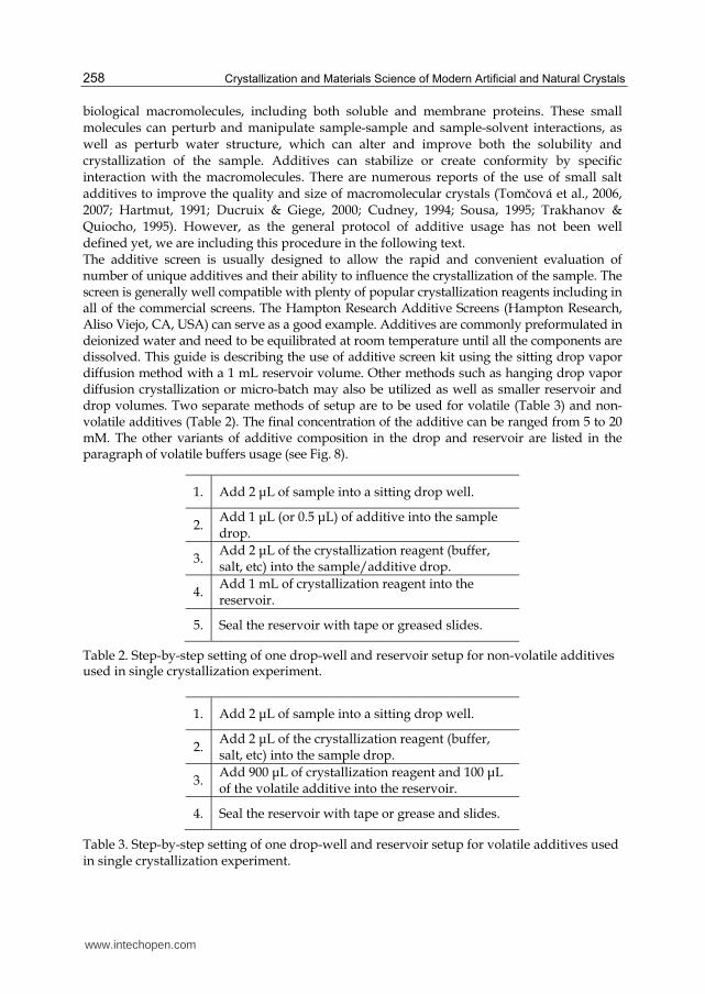
biological macromolecules, including both soluble and membrane proteins. These small molecules can perturb and manipulate sample-sample and sample-solvent interactions, as well as perturb water structure, which can alter and improve both the solubility and crystallization of the sample. Additives can stabilize or create conformity by specific interaction with the macromolecules. There are numerous reports of the use of small salt additives to improve the quality and size of macromolecular crystals (Tomčová et al., 2006, 2007; Hartmut, 1991; Ducruix & Giege, 2000; Cudney, 1994; Sousa, 1995; Trakhanov & Quiocho, 1995). However, as the general protocol of additive usage has not been well defined yet, we are including this procedure in the following text. The additive screen is usually designed to allow the rapid and convenient evaluation of number of unique additives and their ability to influence the crystallization of the sample. The screen is generally well compatible with plenty of popular crystallization reagents including in all of the commercial screens. The Hampton Research Additive Screens (Hampton Research, Aliso Viejo, CA, USA) can serve as a good example. Additives are commonly preformulated in deionized water and need to be equilibrated at room temperature until all the components are dissolved. This guide is describing the use of additive screen kit using the sitting drop vapor diffusion method with a 1 mL reservoir volume. Other methods such as hanging drop vapor diffusion crystallization or micro-batch may also be utilized as well as smaller reservoir and drop volumes. Two separate methods of setup are to be used for volatile (Table 3) and non-volatile additives (Table 2). The final concentration of the additive can be ranged from 5 to 20 mM. The other variants of additive composition in the drop and reservoir are listed in the paragraph of volatile buffers usage (see Fig. 8).
1. Add 2 μL of sample into a sitting drop well.
2. Add 1 μL (or 0.5 μL) of additive into the sample drop.
3. Add 2 μL of the crystallization reagent (buffer, salt, etc) into the sample/additive drop.
4. Add 1 mL of crystallization reagent into the reservoir.
5. Seal the reservoir with tape or greased slides.
Table 2. Step-by-step setting of one drop-well and reservoir setup for non-volatile additives used in single crystallization experiment.
1. Add 2 μL of sample into a sitting drop well.
2. Add 2 μL of the crystallization reagent (buffer, salt, etc) into the sample drop.
3. Add 900 μL of crystallization reagent and 100 μL of the volatile additive into the reservoir.
4. Seal the reservoir with tape or grease and slides.
Table 3. Step-by-step setting of one drop-well and reservoir setup for volatile additives used in single crystallization experiment.
www.intechopen.com

Alternative Protein Crystallization Technique: Cross-Influence Procedure (CIP) 259
3.1.2 Natural additives – Cofactors and ligands
A cofactor is a non-protein chemical compound that is bound to a protein and it is required for its biological activity. These proteins are commonly enzymes, and cofactors can be considered as auxiliary molecules assisting in biochemical transformations. Cofactors are either organic or inorganic. They can also be classified depending on how tightly they bind to an enzyme (called also as coenzymes or prosthetic groups). For example, DNA and RNA binding proteins can be co-crystallized with oligonucleotides. Crystallization of certain macromolecule often requires presence of natural ligands in crystallizing solution. Some of these compounds can be added directly to the crystallization drop (co-crystallization), while other need to be co-incubated in advance to form a complex. Recent studies on ligand-protein crystallization show several advantages. The ligands, in general, stabilizing macromolecule in mother liquor and thus crystallization is less affected by protein aggregation (see also next paragraph). The stabilizing effect was also observed in crystallization of high affinity protein complexes (Brooijmans et al., 2002). However, this approach is somewhat specialized and clearly not universally applicable. The natural ligand may be unknown or not available in sufficient amounts. Crystallization with the natural ligand is not always useful in drug development, as the natural ligand occupies what is likely to be the most potent site for a drug.
3.1.3 Inhibitors and heavy-atoms ligands
It is the solvent content that makes the difference between a classical molecular crystal and a protein crystal: in the former, all the atoms can be described in terms of a regular lattice, whilst in the latter a crystalline array coexists with high portion of material in the liquid state (Brooijmans et al., 2002). The mother liquid, whose content can range approximately from 30 to 75 % (or more), has a strong influence on the behavior of this kind of crystal, making them very peculiar and creating some advantages along with some obvious disadvantages. The major disadvantage is that protein crystals are much less ordered than classical crystals, not only for the large amount of disordered material presents in the crystal itself, but also because surface groups of the macromolecule in contact with the solvent can show a great mobility. On the other hand, among the advantages is that the environment of the macromolecule in the crystal is not such different from that of the solution from which the crystal was obtained (the influence of the solvent on the conformation of the protein cannot be underestimated) and we can profit from the solvent in the preparation of heavy-atoms derivatives, as well as inhibitor soaking (Garman & Murray, 2003). Thus, another group of additives are considered inhibitors and heavy-atoms. A general guideline, how to co-crystallize or soak the existing crystal with the heavy atom, or an inhibitor, cannot exist for obvious reasons. However, there are some empirical rules, which should be followed (see The Hampton Research Heavy Atom Screens - Hampton Research, Aliso Viejo, CA, USA). The concentration may range from 0.05–50 mM, but should not be more than 100 mM, it depends on molar ratio between a molecule to be crystallized and the heavy atom or inhibitor to be introduced into the molecule. Therefore, a small stock solution of 10 mM is an appropriate working stock solution. Please note, the heavy atoms and inhibitors are only sparingly soluble in water. Addition of these reagents to macromolecular crystals (so-called soaking) can be accomphisehed by solubilizing the heavy atom or inhibitor in a carrier solvent that is less polar than water. Acetonitrile is a frequently used carrier solvent. Disolve these reagents in the carrier solvent and then pipet the solution with heavy atom or inhibitor into the crystal mother liquor so that the final carrier solvent concentration in the crystal
www.intechopen.com

Crystallization and Materials Science of Modern Artificial and Natural Crystals 260
mother liquor is 3 to 5% v/v. Important note here, the volatile reagents containing heavy atoms should be handeled in a fume hood. In some cases one can first derivate the protein with heavy atom then attempt crystallization. This procedure is less frequently used since the procedure may not produce derivated crystals, which are isomorphous with the native crystals because the positioning of the heavy atom or inhibitor molecules may disrupt intermolecular contact. Sometimes the presence of these additives with the native macromolecule in solution can change the solubility of the macromolecule which in turn can change the crystallization conditions. The native and derivated crystals now grow under different conditions and thus one must screen for new crystallization condition. Also, with the macromolecule free in solution it is possible that additional heavy atom sites are now introduced (which can complicate the phasing) since sites previously hidden by crystal contacts are now exposed. However, the method can be useful when one is trying to derivate the macromolecule with a heavy atom, ligand or inhibitor that is large enough to diffuse into and through the crystal’s solvent channels.
3.1.4 Antibody and affibody fragments
While antibody-based therapeutics have become firmly established as frontline drugs, the use of antibodies as research tools in small molecule drug discovery is still in its infancy. This paragraph is focused on the use of antibody fragments as crystallization chaperones to aid the structural determination of otherwise uncrystallizable target proteins. Genetic engineering of proteins, specifically enhancing crystallization, is now common practice to troubleshoot proteins that crystallize with difficulty. For example, engineering an increase in the hydrophilic surface area of the molecule through the generation of a fusion protein has been used to enhance crystallization of several integral membrane proteins. Easy-crystallizing T4 lysozyme has been utilized successfully as a fusion partner by replacing the cytoplasmic loop in the target protein. However, enzymatic or genetic modification carries with it risk that the conformation adopted by the recombinant target may not be achievable with the native protein. Co-complexing a target protein with an auxiliary protein (antibody or affibody fragment), which acts as a chaperone, represents a particularly attractive option with wide applicability (Griffin & Lawson, 2011). Inclusion of the chaperone increases the probability of high-quality crystal formation by minimizing the target conformational heterogeneity through ‘locking’ or ‘clamping’ the target in a particular conformation (possibly previously unknown), masking inhibitory surfaces and extending facilitating surfaces. Formation of the crystal lattice based on contacts between antibody molecules is also very likely, in most circumstances, to be an advantage, so that sites of biological interest on target proteins are not occluded by crystal contacts. In addition, an antibody fragment with a previously characterized structure can facilitate molecular replacement phasing. While antibody fragments such as the Fab, Fv, single-chain Fv and single-domain camelid-derived VHH have proven ability to enhance solubility and stabilize target proteins, alternative scaffolds such as affibodies and the helical, but disulphide-free, designed ankyrin repeat proteins with randomized surface residue positions (DARPins) have also shown promisingly. DARPins and antibody fragments may be considered complementary in assisting the crystallography of proteins, as largely independent epitopes are defined by the two scaffolds. Another promising approach, aimed at generalizing antibody mediated crystallization, is to engineer a tag binding epitope into a known loop region of a protein, thus facilitating the
www.intechopen.com

Alternative Protein Crystallization Technique: Cross-Influence Procedure (CIP) 261
generic application of Fab fragments from well-known, and easily sourced, antibodies. If this approach prove to be successful with a wide range of targets, it will eliminate the need to generate or select new antibody-binding sites for each new protein, and thus has considerable attraction.
3.2 Influence of additives promoting crystal formation 3.2.1 Addition and effect of salts in protein crystallization
A protein’s multiple acid-base groups make its solubility properties during crystallization dependent on the concentration and type of dissolved salts, the polarity of the solvent, the pH, and the temperature. Different proteins vary greatly in their solubility under given set of conditions. Certain proteins precipitate from solution under conditions, in which others remain quite soluble. This effect is routinely used as the basis for protein crystallization and greatly affected by addition of various excipients (additives described above). Thus, the solubility of a protein in aqueous solution is a sensitive function of the concentration of dissolved salts. The salt concentration is usually expressed in terms of the ionic strength, which is defined by molar concentration and ionic charge. The use of this parameter to account for the effects of ionic charges results from theoretical consideration of ionic solution. However, protein solubility at a given ionic strength varies with the types of ions in solution. The order of effectiveness of these various ions on influencing protein solubility is quite similar for different proteins and it is apparently mainly due to the ions size and hydration. Thus, the solubility of a protein at low ionic strength generally increases with the salt concentration. The explanation of this salting in phenomenon is that as salt concentration of protein solution increases, the additional counter-ions more effectively shield the protein molecules multiple ionic charges and thereby increase the protein solubility. At high ionic strengths, the solubility of proteins, as well as those of most other substances, decreases. This effect, known as salting out, is primarily a result of the competition between the added salt ions and the other dissolved solutes for molecules of solvation. At high salt concentration many of the added ions are solvated that the amount of bulk solvent available becomes insufficient to dissolve other solutes. In thermodynamic terms, the solvents activity is decreased. Hence, solute–solute interactions become stronger than solute–solvent interactions and the solute precipitates. This is the basis for the most commonly used protein crystallization procedures. Ammonium sulfate is the most commonly used reagent for salting out proteins during the crystallization because its high solubility (3.9 M in water at 0 °C) permits the achievement of solutions with high ionic strengths (D. Voet & J. G. Voet, 1995). Certain ions, notably I-, ClO4-
, SCN-, Li+, Mg2+, Ca2+, and Ba2+, increase the protein solubility rather than salting them out. These ions also tend to denaturate proteins. Conversely, ions that decrease the solubility of proteins stabilize their native structures so that proteins having been salted out are not denaturated.
3.2.2 Additives influencing the pH
Proteins generally bear numerous ionisable groups, which have a variety of pK’s. At a pH characteristic for each protein, the positive charges on the molecule exactly balance molecule’s negative charges. At this pH, the protein’s isoelectric point, pI, the protein molecule does not carry the net charge and is therefore immobile in an electric field. Thus, the solubility behavior, which is shared by most proteins, is easily explained.
www.intechopen.com

Crystallization and Materials Science of Modern Artificial and Natural Crystals 262
Physicochemical considerations suggest that the solubility properties of uncharged molecules during the crystallization are insensitive to salt concentration. Therefore, a protein at its isoelectric point should not be subjected to salting in (D. Voet & J. G. Voet, 1995). Oppositely, as the pH is varied from a protein’s pI, that is, as the protein’s net charge increases, it should be increasingly subjected to salting in because the electrostatic interactions between neighboring molecules that promote aggregation and precipitation likewise increase. Hence, in crystallization solution of moderate salt concentration, the solubility of a protein as a function of pH is expected to be at a minimum at the protein pI and to increase about this point with respect to pH. Proteins vary in their amino acid compositions and therefore in their pI’s. The solubility of the protein may be changed by adding salt as additive or enhancing the concentration of acid or base (changing pH-value of the solution). Consequently, the pH of the solution may affect not only the growth and dissolution rate (Langer, 1985; Kuznetsov & Hodorowicz, 1987), but also the different physical properties of the saturated solution like osmotic pressure, the density, the surface tension and the metastable region. Only a limited number of studies deal with the effect of pH level on the crystallization kinetics. Langer studied the effect of pH levels on the growth rate of various salts. His results show a maximum crystal growth in the neutral solution, with lower crystal growth rates in both acidic and alkaline solutions. There are several more or less satisfactory explanations of the effect of pH on crystallization from general point of view. A plausible explanation says that the presence of free acids or bases modifies the nature and the concentration of ions in solution. Mohameed and Ulrich (1996) explain the effect of pH on crystal growth in terms of a structure in a solution, namely of a hydration of ions. Most cations and OH- ions are hydrated, the largest hydration enthalpy has the H+ ion so that its presence in solution has stronger tendency of interaction with water molecules than, for example, the K+ ion so that a competition of ions to acquire water molecules takes place. The K+ ions have smaller chances to be fully hydrated and therefore they tend to drift towards the crystal surface rather than to remain in the solution.
3.2.3 Effect of organic solvents in protein crystallization
Water-miscible organic solvents, such as acetone and ethanol, are generally good protein precipitants because their low dielectric constants, reduce the solvating power of their aqueous solutions for dissolved ions such as proteins. The decreasing of the dielectric constant by organic solvents also magnifies the differences in the salting out behavior of proteins thus this effect can be helpful in crystallization. Some water-miscible organic solvents, such as dimethyl sulfoxide (DMSO) or N,N-dimethylformamine (DMF), are rather good protein solvents, although they have relatively high dielectric constant (D. Voet & J. G. Voet, 1995).
3.2.4 Crystal-additive interactions
During the nucleation and growth of crystals, different types of interactions are involved between growth species (molecule or ions) and the growing surface. These interactions include van der Waals, ionic and hydrogen bonding. Van der Waals interactions are important during the growth of simple organics compounds such as n-alkanes and ionic interactions during the crystallization of simple ionic crystals, while hydrogen bonding is crucial for both organic and inorganic crystals. During the growth in the presence of additives, similar chemical interactions occur at the interface between the additive species
www.intechopen.com

Alternative Protein Crystallization Technique: Cross-Influence Procedure (CIP) 263
and the solid phase. Recent results on the associations between protein molecules in crystal lattices, crystal–solution surface energy, elastic properties, strength, and spontaneous crystal cracking are reviewed and discussed in Chernov (2003). Unlike that of colloids, which are crystallized at increasing particle density because of mutual particle repulsion, protein crystallization is driven by specific chemical attraction between macromolecules that result in the onset of intermolecular macrobonds within the crystal (see self-assembly paragraph, Fig. 4). Each of these macrobonds includes at least several interatomic (ionic) contacts and forms a patch on the macromolecular surface. An area occupied by each patch on a molecule is defined as the surface area accessible to a spherical water molecule probe rolling over the surface atoms and overlapping with a similar area on a neighboring molecule (Richards, 1977). For the same contact, the areas on the two molecules may be different due to pockets in each of the contact areas. This roughness of surface determinates the strength of intermolecular binding (Matsuura & Chernov, 2003). The total area from all contact patches on a macromolecular surface usually varies widely. However, the patches do not occupy specific areas on macromolecular surfaces that are capable only of making contacts. It follows that different pH, temperature, and solution composition, resulting in different polymorphs, activate different mutually compatible areas on molecular surfaces to produce contacts, thus enabling different packing and crystal structures. From the formal, purely geometrical standpoint, a crystal lattice may be built out of any species of arbitrary shape. Choice between the endless varieties of possibilities is made by the strongest attractions between the patches mutually compatible under various conditions. We may speculate that the larger the difference between various possible contact systems, the easier to crystallize a perfect crystal. However, during crystal dissolution or growth affected by an additive, detachment or attachment of a molecule occurs in solution in order to create initial nuclei. Therefore, part of the energy spent for virtual separations returns from hydration (Chernov, 2003; see self-assembly paragraph).
3.3 Cross-influence procedure (CIP)
In parallel to modern high-throughput approaches, basic research on physico-chemical properties of proteins and their molecular interactions has increasingly gained on importance again in recent years. Physico-chemical properties of the crystallization of biological macromolecules are of particular interest for an efficient way to get high-quality crystals. To improve the success of any crystallization attempts significantly as well as to find new methods of predicting it we have explored another tool useful for optimization strategy that was firstly described in Tomčová and Kutá Smatanová (2006). A crystallization procedure was tested to modify crystal morphology, internal packing and also to influence a crystal growth. For the first time the metal ion salts were added simultaneously to the protein drop and even to neighboring drops to allow a cross-influence effect during crystallization experiment. This new crystallization procedure was verified and the effects of selected additives on crystallization of two different proteins; one well-known “model” protein thaumatin and one crystallographically unexplored di-heme cytochrome c4 from an anaerobic organism (Tomčová et al., 2006) using this procedure were tested. Thaumatin has been chosen for our study because its crystals are often obtained in many different crystallization conditions as well-shaped tetragonal bipyramids, thus it makes sense to study any changes in their internal packing and morphology by cross-crystallization method (Tomčová & Kutá Smatanová, 2007). The final results are summarized and briefly discussed in followed paragraph.
www.intechopen.com

Crystallization and Materials Science of Modern Artificial and Natural Crystals 264
3.3.1 The principles of cross-crystallization method
The CIP is a procedure applied to standard vapor diffusion sitting and/or hanging drop method. This procedure is based on using a set of additives that influence the quality of crystal growth. In principle, the inclusion of other droplets (containing metallic compounds) against the same reservoir slightly changes the vapor pressure of water over the neighboring drop including protein. Products of reaction of metallic salts and precipitant in acidic buffer are often unstable, light sensitive complex compounds (Greenwood & Earnshaw, 2002; Hennig & Theopold, 1951; Nemčovič et al., 2008) that decompose into volatiles, which influence pH slightly. Whereas, used salts of metals (Ba, Co and Cd) have different chemical reactivity, volatile is released in sequence and thus a quality of crystal is improved. As was describe previously (Tomčová et al., 2006), the Emerald BioStructures CombiClover Crystallization Plate (EBS plate, Emerald BioStructures, Bainbridge Island, WA, USA) with one central reservoir connected to four satellite drop-chambers (A, B, C, D) via dedicated vapor diffusion channels, was used in this procedure (Fig. 5). Each of drop-chambers A, B, C, D was filled with 0.5 μl of different additive (in this case; 5 mM chloride salts of copper, cadmium, cobalt and barium) and equal volume of the precipitating agent. A protein was added only into the drop-chamber B containing 5 mM cupric chloride. Additives and reservoir solution, and not protein, were placed to the three remaining drop-chambers to promote crystallization in the fourth drop-chamber. The range of protein concentration 5-20 mg/ml, a reservoir solution volume of 1.0 ml and drops consisting of 1 μl protein solution plus 1 μl of reservoir solution were used in each crystallization trial (Fig.5; Fig.7).
Fig. 5. Schematic side and top view of Emerald BioStructures Combi Clover Crystallization Plate (EBS plate) for sitting drop experiments. Blue color presents reservoir solution, red areas indicate each additives and green color represents protein-containing solution.
3.3.2 Hanging drop variant of CIP
The Hampton Research Linbro plate (Hampton Research, Aliso Viejo, CA, USA) with one central reservoir covered with 15 mm square cover slide was used as a hanging drop alternative in this method. All hanging drops with the same volumes and contents as sitting drops were placed on a siliconized cover slide (Fig. 6). This variant of cross-crystallization shows several disadvantages for protein crystal growth. Compared to the sitting drop method the drops are closer to each other. During crystallization mostly clustered twinned crystals growing on inside perimeter of a drop were observed (see next paragraph). Another variant of hanging drop application consists of the cover slip with drops above the EBS plate reservoir, which allows a hanging drop crystallization experiment to be conducted simultaneously with four sitting-drop experiments.
www.intechopen.com

Alternative Protein Crystallization Technique: Cross-Influence Procedure (CIP) 265
Fig. 6. Schematic side and top view of Hampton Research Linbro plate for hanging drop crystallization experiments. Blue color presents reservoir solution, red areas indicate each additives and green color represents protein-containing solution.
Fig. 7. Original picture of the EBS crystallization plate (with cytochrome crystals – in right d) used for performing cross-crystallization experiment.
3.3.3 The observed effect of CIP: Cytochrome crystallization
Initial crystallization conditions yielding crystals of Cyt c4 in not good quality (Table 4. A-E) were further optimized to improve diffraction quality and especially stability of the crystals (Table 4. F-J). Standard vapor diffusion method with combinations of additives was applied. Screening of pH values (from 6 to 5), NaCl concentration (from 0.1 to 0 M) and (NH4)2SO4
concentration (from 1.7 M to 3.2 M) was used for optimization. In this optimization step, using of CIP improved quality of crystals by addition of additives (Table 4., Fig. 7). Deep red well-shaped cytochrome crystals grew within 3–4 days at 20 °C in the presence of 5 mM cupric chloride and ammonium sulphate in citric acid buffer at pH 5. Those crystals were not reproducible unless the other metal salts (CdCl2, BaCl2, CoCl2) were present in the remaining drop chambers. Only metal salts and reservoir solution, and not protein, were required in the three remaining wells to promote crystallization in the fourth well. The CIP effect has been tested several times. The cytochrome crystals grew only in hexagonal prism form in all these cases. The same outer shape of crystals was observed even when a cytochrome was cross-crystallized by hanging drop (Table 4). Results described in Table 4 show that sodium chloride in combination with ammonium sulphate produce quasi crystals only and these are not good enough for diffraction experiments (drops A-C); presence of any of three used metal salts (CdCl2, CoCl2 and even BaCl2) in protein solution makes heavy protein precipitation (drop D). Comparing drops E and F, it is clear that CIP causes a change in crystal formation what allow us to see the effect of CIP. Drops F-J show diffractable crystals in each successive optimization step.
www.intechopen.com

Crystallization and Materials Science of Modern Artificial and Natural Crystals 266
Table 4. Overview of cytochrome crystallization experiments: A, C, D – standard crystallization in hanging drop; B, E – standard crystallization in sitting drop; F, J – cross-crystallization in sitting drop; G-I – cross-crystallization in hanging drop.
3.3.4 The observed effect of CIP: Thaumatin crystallization
Previously thaumatin has been crystallized in four different forms: orthorhombic (1.75 Å), monoclinic (2.60 Å), tetragonal (1.75 Å) and hexagonal (1.60 Å) (McPherson, 1999; Lee et al., 1988; McPherson & Weickmann, 1990; Charron et al., 2004; Van der Wel et al., 1975). Orthorhombic and monoclinic crystal forms were obtained by using hanging drop vapor-diffusion method from polyethylene glycol as a precipitating agent (Lee et al., 1988; McPherson et al., 1990). The third, tetragonal crystal form was found in crystals grown by vapor diffusion method in the presence of 1 M sodium potassium tartrate containing 0.1 M ADA (sodium-N-2-acetamido-iminodiacetic acid) at pH 6.5 (McPherson, 1999). These crystals were similar to one grown from ammonium sulphate solution (Van der Wel et al., 1975). Thaumatin has also been crystallized in hexagonal crystal form from a tartrate and glycerol containing solution after shifting temperature from 293 K to 277 K (Charron et al., 2004; Ng et al., 1997). All these crystals grew as tetragonal bipyramids with dimensions commonly exceeding 0.5-1.0 mm (see Table 5, Fig. A1-2). In our experiments, thaumatin was crystallized using the standard sitting drop method from the polyethylene glycol (PEG) as a precipitating agent (Tomčová et al., 2007). Well-constructed tetragonal bipyramids were obtained from described crystallization conditions.
www.intechopen.com

Alternative Protein Crystallization Technique: Cross-Influence Procedure (CIP) 267
Table 5. Overview of thaumatin crystallization experiments: A1-B1 – standard crystallization in sitting drop; A2-B2 – standard crystallization in hanging drop; C1-F1 – cross-crystallization in sitting drop; C2-F2 – cross-crystallization in hanging drop.
www.intechopen.com

Crystallization and Materials Science of Modern Artificial and Natural Crystals 268
Table 6. Summary of crystallization and crystallographic statistics for both forms of cytochrome (plates and hexagonal prisms) and thaumatin (tetragonal bipyramids and hexagonal prisms). Crystal morphology and internal packing influenced by CIP are presented on the top of the table and X-ray diffraction statistics is listed below.
www.intechopen.com

Alternative Protein Crystallization Technique: Cross-Influence Procedure (CIP) 269
The effect of metal salt ions on cross-crystallization was tested. The most dramatic change in thaumatin crystal morphology and internal packing was observed when thaumatin was crystallized in hexagonal prisms shape (see Table 5, Fig. D1-2 and Table 6). From comparison of drops A1-2 with drops F1-2 and drops C1-2 with drops E1-2 illustrated in Table 5, it is clear there is no difference in crystal morphology, thus the cross-influence of single Cu2+ is minimal or probably was not evincible. This distinction in CIP composition allows us to conclude that several different metallic compounds (for example Co, Cd, Ba) added into satellite drops are necessary for effective CIP. The inclusion of CIP in drops D1-2 against drops B1-2 seems to reduce propensity of crystals to grow in rods and improve the diffraction quality while the inclusion of CIP realized simultaneously with addition of Cu2+ to protein (drops A1-2 against drops D1-2) shows more significant effect on crystal growth and morphology. In this case, cupric chloride caused the greatest change in crystal outer shape as it is present in Table 5 (drops A1-2 and drops D1-2).
3.3.5 Volatile buffers as crystallization inducers
Recently, the volatile buffers were found to be used in cross-crystallization experiments to induce crystallization of entire macromolecule (Tomčová et al., 2007). They are powerful in changing the pH and vapor pressure over the crystallization drop.
Reservoir volume
5.2 M Volatile buffer
Final drop concentration of volatile buffer
Drop pH
1000 μL 20 μL 0.1 M 3 500 μL 10 μL 0.1 M 3 100 μL 2 μL 0.1 M 3 75 μL 1.5 μL 0.1 M 3 50 μL 1 μL 0.1 M 3
Table 7. Using 5.2 M Acetic acid, the approximate final drop concentration will be 0.1 M Acetic acid. The pH of 0.1 M Acetic acid is approximately 3 but the actual final drop pH after addition of Acetic acid will depend upon the sample buffer and crystallization reagents in the drop.
Reservoir volume
5.2 M Volatile buffer
Final drop concentration of volatile buffer
Drop pH
1000 μL 20 μL 0.1 M 9 500 μL 10 μL 0.1 M 9 100 μL 2 μL 0.1 M 9 75 μL 1.5 μL 0.1 M 9 50 μL 1 μL 0.1 M 9
Table 8. Using 5.2 M Ammonium hydroxide, the approximate final drop concentration will be 0.1 M Ammonium hydroxide. The pH of 0.1 M Ammonium hydroxide is approximately 9 but the actual final drop pH after addition of Ammonium hydroxide will depend upon the sample buffer and crystallization reagents in the drop.
www.intechopen.com

Crystallization and Materials Science of Modern Artificial and Natural Crystals 270
The volatile buffers, when added only to the reagent reservoir of a vapor diffusion experiment, can alter the pH of the crystallization drop by vapor diffusion of the volatile acid or base component from reservoir into the drop. This may be particularly useful when the sample is known to have pH dependent solubility and may be used to induce crystallization. For example, Acetic acid can be added to the reservoir to lower the pH of the drop. On the other hand, Ammonium hydroxide component can be added to the reservoir to raise the pH of the drop. Obviously, the final pH, the actual final volatile buffer concentration in the drop, rate and overall time of equilibration, will vary with drop and reservoir volume, geometry and temperature (Mikol, 1989; McPherson, 1990). The following table (Table 7-8) offers a general guideline when using a volatile buffer manipulates drop pH to induce crystallization (see also Hampton Research Volatile Buffers Usage; Hampton Research, Aliso Viejo, CA, USA). The volatile buffer may be added at the time of initial drop/reservoir set up (see Fig. 8). In this method, the initial drop pH will be that of the sample and crystallization reagent but it changes over time as the volatile buffer vapor diffuses from the reservoir to the drop. Alternatively, as a salvage method, to induce crystallization or improve the crystal, the volatile buffer can be added after the drop has fully equilibrated with the reagent reservoir.
Fig. 8. The schematic view of the volatile buffer composition added at the same time to the hanging drop and reservoir. Each of the A-H crystallization experiment consists of reservoir solution and/or protein solution (shown in blue) and the placement of additive (shown in red). Additive can be volatile compound and can be placed in neighboring drops (C, F, G, H) or added to the reservoir (B, C, E, H).
www.intechopen.com

Alternative Protein Crystallization Technique: Cross-Influence Procedure (CIP) 271
4. Conclusion: New optimization tools in protein crystallization
In this chapter, we described cross-crystallization alias cross-influence procedure (CIP) in details and summarized several factors about CIP and selective additives that facilitate protein crystallization; through promotion of intermolecular contacts and gradually changing pH by the reaction of precipitating solution with divalent metallic compounds, stabilization of the protein with salts or changing the aggregation state with precipitating agents. In fact, any addition of a new substance into a crystallization mixture resulting in crystallization is usually classified as a new crystallization procedure and handled as a hot tip. From previous studies it was found that cupric ions in phosphate buffers have a tendency to produce heavy precipitate and even salt crystals (Lee et al., 1988; Jancarik & Kim, 1991; Jancarik et al., 2004). Another example of an additive effect, which is explained on a molecular basis, is a formation of intermolecular contacts by intercalated divalent transition metal cations. Cadmium (in sulphate solutions) was long known as an inducing agent in crystallization of horse spleen ferritin (Trakhanov & Quiocho, 1995) and has been rediscovered as a useful agent to promote crystallization and increase diffraction quality in several cases. However, the specific morphology of thaumatin and cytochrome crystals may depend on factors such as a source of material used during crystal growth, chemicals presented in a crystallizing buffer in the mother liquor or on the mother liquor itself. Products of reaction MCl2 (where M is metal) and ammonium sulphate with citric acid are unstable, light sensitive complex compounds like C6H8O7.nM.nNH3 (Greenwood & Ernshaw, 2002; Hennig & Theopold, 1951) decomposing into volatile ammonium that slightly influence pH. As Ba, Co and Cd have different reactivity, ammonium is released in sequence and thus quality of crystals is improved (Table 6). In a single crystal form the angles between the faces are constant (Drenth & Haas, 1992), but this is not true if the crystals belong to the different crystal forms such as tetragonal bipyramids and hexagonal prisms in the case of thaumatin. Their appearance depends on the use of metal salt cations (for example cupric chloride) with different chemical reactivity and partially on the buffer and the precipitating agent. We assume that added metal cations influence a character of evaporation in a protein drop and gradually change the pH even if they are only in the proximity of this protein drop. The influence of Cu2+ ions on cytochrome crystal growth appears to be specific, because no other successful combination of ion salts with cytochrome was found among these four salts singly or in pairs. A similar effect was observed even in thaumatin crystallization when conditions with cupric chloride produced thaumatin crystals with a different morphology. The combination of particular salts promoting crystallization by CIP can be reproduced with other metallic compounds as well, or even other volumes of the same drop in the remaining drop’s chambers. Due to this fact, CIP can be appropriately used in any crystallization step to find or/and improve crystallization conditions. The presence of copper ions significantly influences the crystal growth, as the modification of crystal morphology and internal packing were observed. On the basis of our results and analyses of copper influence and CIP effect, we propose that the copper addition realized simultaneously with CIP provides a useful technique to modify crystal morphology and improve diffraction quality in protein crystallization and serves as a powerfull crystallization technique. In addition, for the first time the detailed
www.intechopen.com
Crystallization and Materials Science of Modern Artificial and Natural Crystals 272
protocol of CIP and general guideline of additive usage was given within this chapter to help readers to perform their own cross-crystallization experiment. Thus, this book chapter stands as a valuable guide to modern alternative protein crystallization. Perhaps we have stimulated your interest in crystallography itself, and have made you wonder if you might jump in and crystallize and determine the structure of that interesting protein you are studying. We are happy that we can encourage you by reiterating that crystallography, though still one of structural biology’s more challenging callings, is faster and easier than ever before. Screening for crystal growth conditions does not require expensive equipment or chemicals and these chapter is giving you new ideas how to crystallize your protein of interest with no additional cost.
5. Acknowledgment
This research was supported by the Ministry of Education of the Czech Republic (LC06010, ME09016, COST LD11011, CZ.1.05/2.1.00/01.0024 and MSM6007665808) and by the Academy of Sciences of the Czech Republic (AV0Z60870520). We thank the EMBL for access to the X13 beamline at the DORIS storage ring of DESY in Hamburg. I. N. acknowledges the continuous support of the Slovak Academy of Sciences (Institute of Chemistry, Bratislava, Slovakia) and the La Jolla Institute for Allergy and Immunology (Department of Cellular Biology, La Jolla, California, USA).
6. References
Blow, D.M.; Chayen, N.E.; Lloyd, L.F.; & Saridakis, E. (1994). Control of nucleation of protein crystals. Protein Science, Vol. 3, Issue 10, pp. 1638-1643, DOI:10.1002/pro.5560031003
Brooijmans, N.; Sharp, K.A.; & Kuntz, I.D. (September 2002). Stability of Macromolecular Complexes. Proteins: Structure, Function, and Genetics, Vol. 48, Issue 4, pp. 645–653, DOI:10.1002/prot.10139
Carter, C.W.Jr.; Baldwin, E.T.; & Frick, L. (July 1988). Statistical design of experiments for protein crystal growth and the use of a precrystallization assay. Journal of Crystal
Growth, Vol. 90, Issue 1-3, pp. 60-73, DOI:10.1016/0022-0248(88)90299-0 Charron, C.; Giege, R.; & Lorber, B. (January 2004). Structure of thaumatin in a hexagonal
space group: comparison of packing contacts in four crystal lattices. Acta
Crystallographica Section D Biological Crystallography, Vol. 60, Issue 1, pp. 83-89, DOI:10.1107/S0907444903022613
Chernov, A. (April 2003). Protein crystals and their growth. Journal of Structural Biology, Vol. 142, Issue 1, pp. 3-21, DOI:10.1016/S1047-8477(03)00034-0
Chirgadze, D. (1998). Available from http://www.xray.bioc.cam.ac.uk/xray_resources/ whitepapers/xtal-in-
action/node1.html Cox, M.J.; & Weber, P.C. (1988). An investigation of protein crystallization parameters using
successive automated grid searches (SAGS). Journal of Crystal Growth, Vol. 90, Issue 1-3, pp. 318-324, DOI:10.1016/0022-0248(88)90327-2
www.intechopen.com

Alternative Protein Crystallization Technique: Cross-Influence Procedure (CIP) 273
Cudney, R.; Patel, S.; Weisgraber, K.; Newhouse, Y.; & McPherson, A. (July 1994). Screening and optimization strategies for macromolecular crystal growth. Acta
Crystallographica Section D Biological Crystallography, Vol. 50, Issue 4, pp. 414-423, DOI:10.1107/S0907444994002660
D'Arcy, A.; Elmore, C.; Stihle, M.; & Johnston, J.E. (1996). A novel approach to crystallising proteins under oil. Journal of Crystal Growth, Vol. 168, issue 1-4, pp. 175-180, DOI:10.1016/0022-0248(96)00351-X
DeMattei, R.C.; Feigelson, R.S.; & Weber, P.C. (1992). Factors affecting the morphology of isocitrate lyase crystals. Journal of Crystal Growth, Vol. 122, Issue 1-4, pp. 152-160, DOI:10.1016/0022-0248(92)90238-E
Drenth, J. & Haas, C. (1992). Protein crystals and their stability. Journal of Crystal Growth, Vol. 122, Issue 1-4, pp. 107-109, DOI: 10.1016/0022-0248(92)90233-9
Drenth, J. (November 2006). Principles of Protein X-Ray Crystallography. (3rd Ed.), Springer Science and Business Media, ISBN 9780387337463, England
Ducruix, A.; & Giege, R. (January 2000). Crystallization of Nucleic Acids and Proteins: A
Practical Approach, (2nd Ed.), Oxford University Press, ISBN 978-0199636785, USA Fink, J.M. (2005). Diploma Thesis: Self-organizing nanostructures, Department of Applied
Physics, University of Vienna, Austria Fowlis, W.W.; DeLucas, L.J.; Twigg, P.J.; Howard, S.B.; Meehan, E.J.Jr.; & Baird, J.K. (1988).
Experimental and theoretical analysis of the rate of solvent equilibration in the hanging drop method of protein crystal growth. Journal of Crystal Growth, Vol. 90, Issue 1-3, pp. 117-129, DOI:10.1016/0022-0248(88)90306-5
García-Ruiz, J.M; & Morena, A. (Jul 1994). Investigations on protein crystal growth by the gel acupuncture method. Acta Crystallographica Section D Biological Crystallography, Vol. 50, Part 4, pp. 484-90, DOI:10.1107/S0907444993014350
García-Ruiz, J.M. (2003) Counterdiffusion methods for macromolecular crystallization. Methods in Enzymology, Vol. 368, pp. 130-154, DOI:10.1016/S0076-6879(03) 68008-0
Garman, E.; & Murray, J.W. (November 2003). Heavy-atom derivatization, Acta
Crystallographica Section D Biological Crystallography, Vol. 59, Issue 11, pp. 1903-1913, DOI:10.1107/S0907444903012794
Giacovazzo, C.; Monsaco, H.L.; Artioli, G.; Viterbo, D.; Ferraris, G.; Gilli, G.; Zanotti, G.; & Catti, M. (2002). Fundamentals of Crystallography, (2nd Ed.), Oxford University Press, ISBN 978-0198509585, Oxford, UK
Greenwood, N.N.; & Earnshaw, A. (2002). Chemistry of the Elements. (2nd Ed.), Pergamon Press, ISBN 9780080220567, The University of Michigan, USA
Griffin, L.; & Lawson, A. (September 2011). Antibody fragments as tool in crystallography. Clinical and Experimental Immunology, Vol. 165, Issue 3, pp. 285-291, DOI:10.1111/j.1365-2249.2011.04427.x
Hartmut, Michel (1991). Crystallization of membrane proteins, CRC Press, ISBN 978-0849348167, The University of Michigan, USA
Hennig, W.; & Theopold, W. (1951). Complex compounds of citric acid and calcium, Zeitschrift fur Kinderheilkunde, Vol. 69, Issue 1, pp. 55-61
www.intechopen.com
Crystallization and Materials Science of Modern Artificial and Natural Crystals 274
Jancarik, J.; & Kim, S.-H. (1991). Sparse matrix sampling: a screening method for crystallization of proteins. Journal of Applied Crystallography, Vol. 24, pp. 409-411, DOI:10.1107/S0021889891004430
Jancarik, J.; Pufan, R.; Hong, C.; Kim S.H.; & Kim, R. (September 2004). Optimum solubility (OS) screening: an efficient method to optimize buffer conditions for homogeneity and crystallization of proteins. Acta Crystallographica Section D Biological
Crystallography, Vol. 60, Issue 9, pp. 1670-1673, DOI:10.1107/S0907444904010972 Kuznetsov, D.A.; & Hodorowicz, S. (1978). On the effect of pH of solution on the kinetics of
crystallization. Kristall und Technik, Vol. 13, Issue 12, pp. 1413-1416, DOI:10.1002/crat.19780131206
Langer, H. (1985). Zum Stofftransport beim Kristallwachstum aus Lösungen, Ph.D. Thesis, RWTH Aachen, Aachen 1985
Lattman, E.; Loll P.J.; & Loll P. (2008). Protein crystallography: a concise guide, Johns Hopkins University Press, ISBN 978-0801888069, Baltimore, USA
Lee, J.H.; Weickmann, J.L.; Koduri, R.K.; Ghoshdastidar, K.; Saito, K.; Blair, L.C.; Date, T.; Lai, J.S.; Hollenberg, S.M.; & Kendall, R.L. (July 1988). Expression of synthetic thaumatin genes in yeast. Biochemistry, Vol. 27, Issue 14, pp. 5101-5107, DOI: 10.1021/bi00414a023
Matsuura, Y.; & Chernov, A.A. (August 2003). Morphology and the strength of intermolecular contacts in protein crystals. Acta Crystallographica Section D Biological
Crystallography, Vol. 59, Issue 8, pp. 1347-1356, DOI:10.1107/S0907444903011107 McPherson, A. (1990). Current approaches to macromolecular crystallization. European
Journal of Biochemistry, Vol. 189, Issue 1, pp. 1-23, DOI:10.1111/j.1432-1033.1990.tb15454.x
McPherson, A. (1991). A brief history of protein crystal growth. Journal of Crystal Growth,
Vol. 110, issue 1-2, pp. 1-10, DOI:10.1016/0022-0248(91)90859-4 McPherson, A. (August 1999). Crystallization of Biological Macromolecules, Cold Spring Harbor
Laboratory Press, ISBN 978-0879696177, New York, USA McPherson, A.; & Weickmann, J. (April 1990). X-ray analysis of new crystal forms of the
sweet protein thaumatin. Journal of Biomolecular Structure and Dynamics, Vol. 7, Issue 5, pp. 1053-1060
McRee, D.E. (1999). Practical Protein Crystallography, (Hardcover), Academic Press Title, Elsevier, ISBN 978-0-12-486052-0
Mikol, V.; Rodeau, J.-L.; & Giegé, R. (1989). Changes of pH during biomacromolecule crystallization by vapor diffusion using ammonium sulfate as the precipitant. Journal of Applied Crystallography, Vol. 22, pp. 155-161, DOI:10.1107/ S0021889888013433
Mohameed, H.A.; & Ulrich, J. (1996). Influence of the pH-value on the growth and dissolution rate of potassium chloride. Crystal Research and Technology, Vol. 31, pp. 27-31
Nemčovič, M.; Jakubíková, L.; Vídeň, I., & Farkaš, V. (2008). Induction of conidiation by endogenous volatile compounds in Trichoderma spp. FEMS Microbiology Letters, Vol. 284, Issue 2, pp. 231-236, DOI:10.1111/j.1574-6968.2008.01202.x
www.intechopen.com

Alternative Protein Crystallization Technique: Cross-Influence Procedure (CIP) 275
Ng, J.D.; Lorber, B.; Giege, R.; Koszelak, S.; Day, J.; Greenwood, A.; & McPherson, A. (November 1997). Comparative analysis of thaumatin crystals grown on earth and in microgravity. Acta Crystallographica Section D Biological Crystallography, Vol. 53, Issue 6, pp. 724-733, DOI:10.1107/S090744499700694X
Otalora, F.; Gavira, J.A.; Ng, J.D.; & García-Ruiz, J.M. (November 2009). Counterdiffusion methods applied to protein crystallization. Progress in Biophysics and Molecular
Biology, Vol. 101, Issue 1-2, pp. 26-37, DOI:10.1016/j.pbiomolbio.2009.12.004 Provost, K. & Robert, M-C. (1991). Application of gel growth to hanging drop technique.
Journal of Crystal Growth, Vol. 110, pp. 258-264, DOI:10.1016/0022-0248(91) 90894-B
Rayment, I. (February 2002). Small-scale batch crystallization of proteins revisited: an underutilized way to grow large protein crystals. Structure, Vol. 10, Issue 2, pp. 147-51, DOI:10.1016/S0969-2126(02)00711-6
Richards, F.M. (June 1977). Areas, volumes, packing and protein structure. Annual
Reviews of Biophysics and Bioengeneering, Vol. 6, pp. 151-176, DOI: 10.1146/annurev.bb.06.060177.001055
Sangwal, K. (1998). Growth kinetics and surface morphology of crystals grown from solutions: Recent observations and their interpretations. Progress in crystal growth
and characterization of materials, Vol. 36, Issue 3, Pages 163-248, DOI:10.1016/S0960-8974(98)00009-6
Sousa, R. (May 1995). Use of glycerol, polyols and other protein structure stabilizing agents in protein crystallization. Acta Crystallographica Section D Biological Crystallography, Vol. 51, Issue 3, pp. 271-277, DOI:10.1107/S0907444994014009
Stura, E.A.; Nemerow, G.R.; & Wilson, I.A. (1991). Strategies in protein crystallization. Journal of Crystal Growth, Vol. 110, pp. 1-12
Tomčová, I. & Kutá Smatanová, I. (August 2007). Copper co-crystallization and divalent metal salts cross-influence effect: A new optimization tool improving crystal morphology and diffraction quality. Journal of Crystal Growth, Vol. 306, Issue 2, pp. 383-389, DOI:10.1016/j.jcrysgro.2007.05.054
Tomčová, I., Branca, R.M.M., Bodó, G., Bagyinka, Cs.; & Kutá Smatanová, I. (August 2006). Cross-crystallization method used for the crystallization and preliminary diffraction analysis of a novel di-haem cytochrome c4. Acta Crystallographica Section
F, Vol. 62, Part 8, pp. 820-824, DOI:10.1107/S1744309106027710 Trakhanov, S.; & Quiocho, F.A. (September 1995). Influence of divalent cations on protein
crystallization. Protein Science, Vol. 4, Issue 9, pp. 1914-1919, DOI: 10.1002/pro.5560040925
Van der Wel, H.; Van Soest, T.; & Royers, E.C. (1975). Crystallization and crystal data of thaumatin I, a sweet-tasting protein from Thaumatococcus daniellii benth. FEBS
Letters, Vol. 56, Issue 2, pp. 316-317 Van Driessche, A.E.S.; Otalora, F.; Gavira, J.A.; & Sazaki, G. (2008). Is agarose an impurity or
an impurity filter? In situ observation of the joint gel/impurity effect on protein crystal growth kinetics. Crystal Growth and Design,Vol. 8, Issue 10, pp. 3623–3629, DOI:10.1021/cg800157t
www.intechopen.com

Crystallization and Materials Science of Modern Artificial and Natural Crystals 276
Voet, D. & Voet, J.G. (1995). Biochemistry, (Hardcover), John Wiley & Sons, ISBN 0-471-58651-X, New York, USA
Weber, P.C. (1991). Physical principles of protein crystallization. Advances in Protein
Chemistry, Vol. 41, pp. 1-36, 1991, DOI:10.1016/S0065-3233(08)60196-5
www.intechopen.com

Crystallization and Materials Science of Modern Artificial andNatural CrystalsEdited by Dr. Elena Borisenko
ISBN 978-953-307-608-9Hard cover, 328 pagesPublisher InTechPublished online 20, January, 2012Published in print edition January, 2012
InTech EuropeUniversity Campus STeP Ri Slavka Krautzeka 83/A 51000 Rijeka, Croatia Phone: +385 (51) 770 447 Fax: +385 (51) 686 166www.intechopen.com
InTech ChinaUnit 405, Office Block, Hotel Equatorial Shanghai No.65, Yan An Road (West), Shanghai, 200040, China
Phone: +86-21-62489820 Fax: +86-21-62489821
Crystal growth is an important process, which forms the basis for a wide variety of natural phenomena andengineering developments. This book provides a unique opportunity for a reader to gain knowledge aboutvarious aspects of crystal growth from advanced inorganic materials to inorganic/organic composites, itunravels some problems of molecular crystallizations and shows advances in growth of pharmaceuticalcrystals, it tells about biomineralization of mollusks and cryoprotection of living cells, it gives a chance to learnabout statistics of chiral asymmetry in crystal structure.
How to referenceIn order to correctly reference this scholarly work, feel free to copy and paste the following:
Ivana Nemčovičová and Ivana Kutá Smatanová (2012). Alternative Protein Crystallization Technique: Cross-Influence Procedure (CIP), Crystallization and Materials Science of Modern Artificial and Natural Crystals, Dr.Elena Borisenko (Ed.), ISBN: 978-953-307-608-9, InTech, Available from:http://www.intechopen.com/books/crystallization-and-materials-science-of-modern-artificial-and-natural-crystals/alternative-crystallization-technique-cross-crystallization-or-cross-influence-procedure-cip-