Alterations in the hypothalamic melanocortin pathway in ...
17
Alterations in the hypothalamic melanocortin pathway in amyotrophic lateral sclerosis Pauline Vercruysse, 1,2,3 Je ´ ro ˆme Sinniger, 1,2 Hajer El Oussini, 1,2 Jelena Scekic-Zahirovic, 1,2 Ste ´ phane Dieterle ´, 1,2 Reinhard Dengler, 4 Thomas Meyer, 5 Stephan Zierz, 6 Jan Kassubek, 3 Wilhelm Fischer, 3 Jens Dreyhaupt, 7 Torsten Grehl, 8 Andreas Hermann, 9 Julian Grosskreutz, 10 Anke Witting, 3 Ludo Van Den Bosch, 11 Odile Spreux-Varoquaux, 12,13,14 the GERP ALS Study Group, † Albert C. Ludolph 3 and Luc Dupuis 1,2 † See Appendix 1 Amyotrophic lateral sclerosis, the most common adult-onset motor neuron disease, leads to death within 3 to 5 years after onset. Beyond progressive motor impairment, patients with amyotrophic lateral sclerosis suffer from major defects in energy metabolism, such as weight loss, which are well correlated with survival. Indeed, nutritional intervention targeting weight loss might improve survival of patients. However, the neural mechanisms underlying metabolic impairment in patients with amyotrophic lateral sclerosis remain elusive, in particular due to the lack of longitudinal studies. Here we took advantage of samples collected during the clinical trial of pioglitazone (GERP-ALS), and characterized longitudinally energy metabolism of patients with amyo- trophic lateral sclerosis in response to pioglitazone, a drug with well-characterized metabolic effects. As expected, pioglitazone decreased glycaemia, decreased liver enzymes and increased circulating adiponectin in patients with amyotrophic lateral sclerosis, showing its efficacy in the periphery. However, pioglitazone did not increase body weight of patients with amyotrophic lateral sclerosis independently of bulbar involvement. As pioglitazone increases body weight through a direct inhibition of the hypothal- amic melanocortin system, we studied hypothalamic neurons producing proopiomelanocortin (POMC) and the endogenous mel- anocortin inhibitor agouti-related peptide (AGRP), in mice expressing amyotrophic lateral sclerosis-linked mutant SOD1(G86R). We observed lower Pomc but higher Agrp mRNA levels in the hypothalamus of presymptomatic SOD1(G86R) mice. Consistently, numbers of POMC-positive neurons were decreased, whereas AGRP fibre density was elevated in the hypothalamic arcuate nucleus of SOD1(G86R) mice. Consistent with a defect in the hypothalamic melanocortin system, food intake after short term fasting was increased in SOD1(G86R) mice. Importantly, these findings were replicated in two other amyotrophic lateral sclerosis mouse models based on TDP-43 (Tardbp) and FUS mutations. Finally, we demonstrate that the melanocortin defect is primarily caused by serotonin loss in mutant SOD1(G86R) mice. Altogether, the current study combined clinical evidence and experimental studies in rodents to provide a mechanistic explanation for abnormalities in food intake and weight control observed in patients with amyotrophic lateral sclerosis. Importantly, these results also show that amyotrophic lateral sclerosis progression impairs responsiveness to classical drugs leading to weight gain. This has important implications for pharmacological management of weight loss in amyotrophic lateral sclerosis. 1 Inserm U1118, Me ´canismes centraux et pe ´riphe ´tiques de la neurode ´ge ´ne ´rescence, Strasbourg, F-67085 France 2 Universite ´ de Strasbourg, Faculte ´ de Me ´decine, UMRS1118, Strasbourg, F-67085 France 3 Department of Neurology, University of Ulm, Germany 4 Department of Neurology, Hannover Medical School, Hannover, Germany 5 Department of Neurology, Charite ´ University Hospital, Berlin, Germany 6 Department of Neurology, University of Halle-Wittenberg, Germany 7 Institute of Epidemiology and Medical Biometry, University of Ulm, Germany doi:10.1093/brain/aww004 BRAIN 2016: 139; 1106–1122 | 1106 Received July 27, 2015. Revised November 13, 2015. Accepted December 04, 2015. Advance Access publication March 16, 2016 ß The Author (2016). Published by Oxford University Press on behalf of the Guarantors of Brain. All rights reserved. For Permissions, please email: [email protected] Downloaded from https://academic.oup.com/brain/article-abstract/139/4/1106/2464236 by guest on 25 March 2018
Transcript of Alterations in the hypothalamic melanocortin pathway in ...
Alterations in the hypothalamic melanocortinpathway in amyotrophic lateral sclerosis
Pauline Vercruysse,1,2,3 Jerome Sinniger,1,2 Hajer El Oussini,1,2 Jelena Scekic-Zahirovic,1,2
Stephane Dieterle,1,2 Reinhard Dengler,4 Thomas Meyer,5 Stephan Zierz,6 Jan Kassubek,3
Wilhelm Fischer,3 Jens Dreyhaupt,7 Torsten Grehl,8 Andreas Hermann,9
Julian Grosskreutz,10 Anke Witting,3 Ludo Van Den Bosch,11 Odile Spreux-Varoquaux,12,13,14
the GERP ALS Study Group,† Albert C. Ludolph3 and Luc Dupuis1,2
†See Appendix 1
Amyotrophic lateral sclerosis, the most common adult-onset motor neuron disease, leads to death within 3 to 5 years after onset.
Beyond progressive motor impairment, patients with amyotrophic lateral sclerosis suffer from major defects in energy metabolism,
such as weight loss, which are well correlated with survival. Indeed, nutritional intervention targeting weight loss might improve
survival of patients. However, the neural mechanisms underlying metabolic impairment in patients with amyotrophic lateral
sclerosis remain elusive, in particular due to the lack of longitudinal studies. Here we took advantage of samples collected
during the clinical trial of pioglitazone (GERP-ALS), and characterized longitudinally energy metabolism of patients with amyo-
trophic lateral sclerosis in response to pioglitazone, a drug with well-characterized metabolic effects. As expected, pioglitazone
decreased glycaemia, decreased liver enzymes and increased circulating adiponectin in patients with amyotrophic lateral sclerosis,
showing its efficacy in the periphery. However, pioglitazone did not increase body weight of patients with amyotrophic lateral
sclerosis independently of bulbar involvement. As pioglitazone increases body weight through a direct inhibition of the hypothal-
amic melanocortin system, we studied hypothalamic neurons producing proopiomelanocortin (POMC) and the endogenous mel-
anocortin inhibitor agouti-related peptide (AGRP), in mice expressing amyotrophic lateral sclerosis-linked mutant SOD1(G86R).
We observed lower Pomc but higher Agrp mRNA levels in the hypothalamus of presymptomatic SOD1(G86R) mice. Consistently,
numbers of POMC-positive neurons were decreased, whereas AGRP fibre density was elevated in the hypothalamic arcuate nucleus
of SOD1(G86R) mice. Consistent with a defect in the hypothalamic melanocortin system, food intake after short term fasting was
increased in SOD1(G86R) mice. Importantly, these findings were replicated in two other amyotrophic lateral sclerosis mouse
models based on TDP-43 (Tardbp) and FUS mutations. Finally, we demonstrate that the melanocortin defect is primarily
caused by serotonin loss in mutant SOD1(G86R) mice. Altogether, the current study combined clinical evidence and experimental
studies in rodents to provide a mechanistic explanation for abnormalities in food intake and weight control observed in patients
with amyotrophic lateral sclerosis. Importantly, these results also show that amyotrophic lateral sclerosis progression impairs
responsiveness to classical drugs leading to weight gain. This has important implications for pharmacological management of
weight loss in amyotrophic lateral sclerosis.
1 Inserm U1118, Mecanismes centraux et periphetiques de la neurodegenerescence, Strasbourg, F-67085 France2 Universite de Strasbourg, Faculte de Medecine, UMRS1118, Strasbourg, F-67085 France3 Department of Neurology, University of Ulm, Germany4 Department of Neurology, Hannover Medical School, Hannover, Germany5 Department of Neurology, Charite University Hospital, Berlin, Germany6 Department of Neurology, University of Halle-Wittenberg, Germany7 Institute of Epidemiology and Medical Biometry, University of Ulm, Germany
doi:10.1093/brain/aww004 BRAIN 2016: 139; 1106–1122 | 1106
Received July 27, 2015. Revised November 13, 2015. Accepted December 04, 2015. Advance Access publication March 16, 2016
� The Author (2016). Published by Oxford University Press on behalf of the Guarantors of Brain.
All rights reserved. For Permissions, please email: [email protected]
Downloaded from https://academic.oup.com/brain/article-abstract/139/4/1106/2464236by gueston 25 March 2018

8 Department of Neurology, BG University Hospital Bergmannsheil, Bochum, Germany9 Department of Neurology, Technische Universitat Dresden, and German Center for Neurodegenerative Disease (DZNE),
Dresden, Germany10 Department of Neurology, University Hospital, Jena, Germany11 Laboratory of Neurobiology, KU Leuven and Vesalius Research Center, VIB, Leuven, Belgium12 Faculte de Medecine Paris-Ile de France-Ouest, France13 Universite de Versailles Saint-Quentin-en-Yvelines, France14 Centre Hospitalier Versailles, Le Chesnay, France
Correspondence to: Luc Dupuis,
INSERM U1118, Faculte de medecine, bat 3, 8e etage, 11 rue Humann, 67085 Strasbourg,
Cedex, France
E-mail: [email protected]
Correspondence may also be addressed to: Albert C. Ludolph,
RKU, Universitatsklinik Ulm, Oberer Eselsberg 45, 89081 Ulm, Germany
E-mail: [email protected]
Keywords: amyotrophic lateral sclerosis; calorie intake; hypothalamus; thiazolinediones; weight loss
Abbreviations: ALS = amyotrophic lateral sclerosis; TZD = thiazolinedione
IntroductionAmyotrophic lateral sclerosis (ALS), the most common
adult-onset motor neuron disease, is characterized by the
simultaneous degeneration of upper and lower motor neu-
rons, leading to muscle atrophy and paralysis, and death
within 3 to 5 years after onset. A subset of ALS cases are of
familial origin and five major genes are currently associated
with familial ALS (C9orf72, SOD1, FUS, TARDBP and
TBK1) (Leblond et al., 2014; Cirulli et al., 2015;
Freischmidt et al., 2015; Lattante et al., 2015). The
SOD1 gene was the first associated with ALS and most
ALS mouse models currently used are based upon overex-
pression of mutant forms of SOD1 (Gurney et al., 1994;
Ripps et al., 1995).
Beyond progressive motor impairment, patients with ALS
suffer from major, yet incompletely characterized, defects in
energy metabolism (Dupuis et al., 2011). First, ALS is more
likely to occur with lower premorbid body fat (Gallo et al.,
2013; O’Reilly et al., 2013) or better cardiovascular or physical
fitness (Turner et al., 2012; Huisman et al., 2013). Second,
weight loss is negatively correlated with survival (Desport et
al., 1999; Marin et al., 2011; Paganoni et al., 2011). This
weight loss is associated with, and likely caused by, intrinsic
hypermetabolism (Desport et al., 2001; Bouteloup et al., 2009),
and is exacerbated by dysphagia occurring with bulbar involve-
ment. Third, ALS patients develop abnormalities in lipid
(Dupuis et al., 2008; Dorst et al., 2011; Lindauer et al.,
2013) and glucose (Pradat et al., 2010) metabolisms.
Interestingly, these metabolic alterations are largely replicated
in transgenic mice expressing mutant SOD1 (Dupuis et al.,
2004; Fergani et al., 2007; Palamiuc et al., 2015). Not much
is known on the underlying mechanisms of energy metabolism
impairment despite the fact that elucidation of such mechan-
isms would offer therapeutic strategies to treat weight loss
pharmacologically. Furthermore, deciphering the mechanisms
of energy metabolism impairment could identify disease-mod-
ifying interventions as a hypercaloric diet was recently found to
increase survival of patients with ALS under gastrostomy (Wills
et al., 2014; Dorst et al., 2015).
Most of the studies to date have characterized energy
metabolism in patients with ALS in steady state, at one
single time point. The dynamic nature of energy metabol-
ism and its homeostatic regulation thus severely limit the
interpretation of these studies. Interventions performed
during randomized clinical trials often have metabolic ef-
fects, and such studies include long term follow-up of pa-
tients for many months. These clinical studies thus provide
high-quality information useful to understand the metabolic
defects of patients with ALS.
Here, we performed a post hoc analysis of samples ob-
tained during the clinical trial of pioglitazone (Dupuis et
al., 2012) to characterize energy metabolism of ALS pa-
tients on a metabolic challenge. Indeed, pioglitazone, like
other thiazolinediones (TZDs), has pleiotropic effects on
energy metabolism that have been extremely well charac-
terized in both mouse models and human patients. In the
periphery, pioglitazone sensitizes to insulin, leading to
decreased glycaemia, and decreases circulating levels of
liver enzymes (Promrat et al., 2004; Belfort et al., 2006;
Sanyal et al., 2010; DeFronzo et al., 2011). In the CNS,
pioglitazone inhibits the hypothalamic melanocortin system
to increase food intake (Diano et al., 2011; Lu et al., 2011;
Ryan et al., 2011; Long et al., 2014). Specifically, pioglita-
zone decreases activity of the hypothalamic neurons produ-
cing proopiomelanocortin (POMC), the precursor of a
number of anorexigenic peptides such as �-MSH (melano-
cyte stimulating hormone) (Diano et al., 2011; Lu et al.,
2011; Ryan et al., 2011; Long et al., 2014). In humans, this
leads to a robust (3–5 kg) weight gain that was repeatedly
Hypothalamic dysfunction in ALS BRAIN 2016: 139; 1106–1122 | 1107
Downloaded from https://academic.oup.com/brain/article-abstract/139/4/1106/2464236by gueston 25 March 2018

observed in multiple clinical trials (Promrat et al., 2004;
Belfort et al., 2006; Sanyal et al., 2010; DeFronzo et al.,
2011). Here, we show that patients with ALS display
normal peripheral action of pioglitazone, while they lack
weight gain. In transgenic mice expressing mutant SOD1,
pioglitazone failed to increase food intake. This was asso-
ciated with prominent involvement of the hypothalamic
melanocortin system, also observed in other mouse
models of ALS, independent of mutant SOD1 overexpres-
sion. Last, we show that the melanocortin defect occurs
downstream of the previously documented serotonin loss
(Dentel et al., 2013). Altogether, our analysis of the data
from the pioglitazone trial disclosed a previously unantici-
pated defect in patients with ALS that could account for a
subset of ALS-related metabolic defects.
Materials and methods
Patients and treatments
All the biological materials from human ALS patients weresampled as part of the GERP-ALS trial (clinicaltrials.gov ref-erence: NCT00690118) (Dupuis et al., 2012). Briefly, patientswith possible, probable (clinically or laboratory-supported) ordefinite ALS according to the revised version of the El Escorialcriteria were considered for enrolment into the study. Includedpatients displayed onset of progressive weakness within 36months prior to study and had disease duration of46months and53 years (inclusive) with disease onset definedas date of first muscle weakness, excluding fasciculation andcramps. They reached a best-sitting slow vital capacity be-tween 50% and 95% of predicted normal. They were capableof thoroughly understanding the information provided andgave written informed consent. All included patients hadbeen treated with 100 mg riluzole daily for at least 3 monthsprior to inclusion. Detailed exclusion and inclusion criteriahave been described earlier (Dupuis et al., 2012). The studyprotocol was approved by the ethics committee of theUniversity of Ulm and all other participating centres.
The two treatment groups were 100 mg riluzole plus 45 mgpioglitazone (pioglitazone group) and 100 mg riluzole plus pla-cebo (placebo group). Patients were randomly assigned to oneof the two treatment groups and both groups were matchedfor age, gender and site of onset (Dupuis et al., 2012).
Procedures and biochemical analysisof human samples
After inclusion, patients underwent a screening phase and atreatment phase (18 months), with stepwise increase indosage (Dupuis et al., 2012). Clinical and physical examin-ations, blood sampling, and drug compliance were recordedat on-site visits (1, 2, 6, 12 and 18 months after baselinevisit). Body weight was recorded at on-site visits, except for3-, 9- and 15-month time points (telephone contacts). Therewere no differences in results when excluding these three timepoints. Routine clinical laboratory tests were performed ateach on-site visit (baseline and 1, 2, 6, 12 and 18 monthsafter baseline). All tests were carried out according to standard
laboratory procedures at each study centres’ locally accreditedlaboratory, which defined the normal reference range for eachanalyte. The following laboratory tests were performed usingstandard methods: alanine aminotransferase (ALAT), aspartateaminotransferase (ASAT), fasting blood glucose. Adiponectinmeasurements were done in the neurochemical laboratory inUlm (MSD assay).
Animals
Transgenic mice were housed in the animal facility of themedicine faculty of Strasbourg University, with 12 h/12 hlight/dark and unrestricted access to food and water. In allexperiments, littermates were used for comparison.Transgenic SOD1(G86R) were maintained in their initialFVB/N genetic background according to previous studies(Dentel et al., 2013). Transgenic mice expressingTDP43(A315T) were previously described and were main-tained as heterozygous in their initial C57Bl6/J background(Wegorzewska et al., 2009). Heterozygous Fus�NLS/ + areknock-in mice expressing FUS protein deleted from itsC-terminal nuclear localization signal (NLS) from one copyof the endogenous Fus gene. These mice were generated andmaintained in C57Bl6/J background. The motor phenotype ofthese mice will be described elsewhere (Scekic-Zahirovic, sub-mitted for publication). Tph2-YFP mice were purchased fromJackson laboratories (Bar Harbor; strain 014555) and main-tained in their initial genetic background. Female Tph2-YFPmice were crossed with male SOD1(G86R) to generate com-pound transgenic mice.
For biochemical analysis, animals were sacrificed at the agesindicated at 2 pm, and tissues were quickly dissected, frozen inliquid nitrogen, and stored at� 80�C until use. For histologicalanalysis, animals were anaesthetized by intraperitoneal injec-tion of ketamine (Imalgene 1000�, Merial; 90 mg/kg bodyweight) and xylazine (Rompun 2%�, Bayer; 10 mg/kg bodyweight) at the ages indicated at 2 pm. After perfusion of 4%paraformaldehyde (v/v PFA, Sigma), brains were removed,stored in the same fixative overnight at 4�C and stored inphosphate-buffered saline (PBS) until used. These experimentswere authorized by the local ethical committee of StrasbourgUniversity (CREMEAS).
Drugs and treatments
Pioglitazone (Actos�, Takeda) was dissolved in 10% (v/v) di-methyl sulphoxide (DMSO, Fisher Scientific) and a single oraladministration was given by gavage at a dose of 40 mg/kgbody weight. Fluoxetine (Sigma) was dissolved in 0.9% (w/v)NaCl (Sigma) and administrated intraperitoneally at a dose of20 mg/kg body weight.
Measurements of food intake
For the pioglitazone experiment, mice were fasted from 9 amto 3 pm, and pioglitazone was administrated at 3 pm. Foodwas reintroduced 1 h after gavage and food intake was re-corded for 24 h.
For short-term fasting experiments, mice were fasted from 8am to 3 pm (7 h fasting conditions) or from 2 pm to 3 pm (1 hfasting conditions) and food was reintroduced after 7 h or 1 h
1108 | BRAIN 2016: 139; 1106–1122 P. Vercruysse et al.
Downloaded from https://academic.oup.com/brain/article-abstract/139/4/1106/2464236by gueston 25 March 2018

of fasting. Food intake was measured 1 h and 24 h afterrefeeding.
For fluoxetine experiment, mice were fasted from 1 pm to2 pm, and fluoxetine was injected at 2 pm. Food was reintro-duced 30 min after fluoxetine injection and food intake wasmeasured for 1 h. For biochemical analysis, a second injectionof fluoxetine was done after 1 h of feeding and mice weresacrificed 30 min later.
Histology
Fixed brains were included in 6% (w/v) agar (Sigma) and sec-tioned from Bregma 0.02mm to Bregma �2.90 mm into 40 mmcoronal sections on a vibratome. Arcuate nucleus was identi-fied according to Paxinos Brain Atlas. POMC immunohisto-chemistry was performed on half of the selected brain sections.AGRP and green fluorescent protein (GFP) immunohistochem-istries were performed each on anatomically matched sections.Immunohistochemistry was performed on floating sectionsusing standard histological techniques. Endogenous peroxid-ases were inactivated using 3% (v/v) H2O2. For POMC immu-nohistochemistry, permeabilization and saturation of non-specific sites were done with 0.25% (v/v) TritonTM (Sigma)and 50 mg/ml bovine serum albumin (BSA, Sigma). For GFPimmunohistochemistry, permeabilization and saturation ofnon-specific sites were done with 0.5% (v/v) TritonTM
(Sigma) and 5% (v/v) horse serum (Gibco, LifeTechnologies). For AGRP immunohistochemistry, antigen re-trieval with citrate buffer was done before permeabilizationand saturation of non-specific sites with 0.5% (v/v) TritonTM
(Sigma) and 5%(v/v) horse serum (Gibco, Life Technologies).Rabbit anti-POMC primary antibody (Phoenix Peptide;1:2000), rabbit anti-AgRP primary antibody (PhoenixPeptide; 1:2000) or rabbit anti-GFP primary antibody(Invitrogen, Life Technologies, 1:1000) were incubated over-night at room temperature. Biotinylated donkey anti-Rabbitsecondary antibody (Jackson; 1:500) was incubated for90 min at room temperature. Staining was performed usingVectastain Elite ABC kit (Vector). After revelation with 3,3’-diaminobenzidine (DAB, Sigma; 0.5 mg/ml), sections weremounted and images of all sections were taken.
For quantification, bright-field images of lower brain part forPOMC and AgRP stainings or bright-field images of right arc-uate nucleus part for GFP staining were acquired with a NikonDS –Ri1 camera attached to a Nikon microscope (NikonEclipse E800) fitted with a Plan Apo 4� lens (N.A. = 0.20,Nikon) and a plan Apo 10� lens (N.A. = 0.45, Nikon),respectively. White balance, gain, exposure, and light settingswere kept the same when acquiring all images of a givenstaining.
Image analysis
An operator blinded to the genotype quantified all experi-ments. Total numbers of POMC-positive cell bodies in thearcuate nucleus were determined for each animal and normal-ized per section. Quantification of GFP staining and AGRPstaining was processed to binary images using NIH ImageJas described previously (Grider et al., 2006). Briefly, afterchanging tiff images to 8-bit images, images were invertedand processed with Feature J plugin for ImageJ (version1.50d) to select the smallest Hessian values and « smoothing
scale » of 1.0. The resulting images were transformed intobinary images by thresholding. Threshold was determined onone cut and same threshold was applied to every image.Occupied area for AGRP or GFP staining was measured.
RNA extraction and quantitativereverse transcription-polymerasechain reaction
RNA was extracted from mouse hypothalami using TRIzol�
reagent (Life Technologies). After reverse transcription withiScriptTM reverse transcription Supermix for RT-qPCR (Bio-Rad), cDNA was obtained from 1 mg of RNA. MessengerRNA levels were obtained by quantitative PCR using SsoAdvanced Universal SYBR Green Supermix (Bio-Rad) withcorresponding sense and antisense primers. Standard curveswere constructed by amplifying serial dilutions of cDNA.Starting quantities of samples were calculated with Bio-Radsoftware. Messenger RNA levels of genes of interest were nor-malized to expression levels of the 18S ribosomal, Tbp andPola2 RNA housekeeping genes using GeNorm(Vandesompele et al., 2002). Primer sequences are shown inSupplementary Table 1.
Serotonin levels
Hypothalamic serotonin levels were measured using high per-formance liquid chromatography, as previously described(Dentel et al., 2013).
Statistical analysis
For statistical analyses in ALS patients, group comparisonswere performed using mixed effects regression model analysisby the Institute of Epidemiology and Medical Biometrics at theUniversity of Ulm using the statistical software package SASVersion 9.2 under Windows.
For animal experiments, comparison of two groups was per-formed using unpaired Student’s t-test, except for experimentswith pioglitazone in which paired t-test was used. Comparisonof three or four groups was performed using one-way ANOVAand Tukey post hoc test. Statistics in animal experiments wereperformed using Prism version 6.0.
All results from analysis were regarded as hypothesis gener-ating only. All statistical tests were carried out two-sided at asignificance level of 5%.
Results
Normal peripheral response to pio-glitazone in patients with ALS
We took advantage of data collected during the pioglita-
zone GERP-ALS trial to investigate energy metabolism in
ALS patients in response to pioglitazone.
In this clinical trial, 219 patients with ALS were en-
rolled and randomly allocated to either placebo (n = 110;
bulbar: n = 33, spinal: n = 77), or pioglitazone (n = 109;
Hypothalamic dysfunction in ALS BRAIN 2016: 139; 1106–1122 | 1109
Downloaded from https://academic.oup.com/brain/article-abstract/139/4/1106/2464236by gueston 25 March 2018

bulbar: n = 32, spinal: n = 77) treatment after stratifica-
tion based on site of onset (bulbar or spinal) (Dupuis et
al., 2012).
The metabolic effects of TZDs, including pioglitazone
are well understood in models and their effects are largely
described in patients (Fig. 1A and Supplementary Table
2). Pioglitazone treatment is known to increase levels of
adiponectin, an adipose-derived hormone through direct
transcriptional activation of the adiponectin gene in adi-
pocytes (Maeda et al., 2001). Consistently, pioglitazone
treatment increased the levels of circulating adiponectin
4-fold in patients with ALS after 6 months of treatment,
and this was maintained after 12 months of treatment
(Fig. 1B). Pioglitazone decreases glycaemia through hep-
atic and skeletal muscle PPARg (encoded by PPARG). In
patients with ALS, pioglitazone decreased glycaemia (Fig.
1C), although this effect was milder than observed in
other populations, including non-diabetic patients
(Belfort et al., 2006; Sanyal et al., 2010; DeFronzo et
al., 2011). Consistent with a direct action on liver, pio-
glitazone decreased levels of ASAT and more robustly
levels of ALAT (Fig. 1D and E). In all, pioglitazone dis-
played the expected metabolic effects on adipocytic, mus-
cular, and hepatic biomarkers, and was likely able to
activate PPARg in these tissues.
Pioglitazone does not lead to weightgain in patients with ALS
TZDs are also known to act in hypothalamic melanocortin
neurons to promote feeding (Diano et al., 2011; Long et
al., 2014), and this activation of PPARg in melanocortin
neurons is responsible for the robust weight gain associated
with TZD treatment (Lu et al., 2011; Ryan et al., 2011;
Long et al., 2014). Thus, the evolution of body weight
upon pioglitazone treatment represents a reliable proximal
marker of PPARg action in hypothalamic melanocortin
neurons. In ALS patients, pioglitazone had no effect on
weight loss (Fig. 2A), or body mass index (BMI) (Fig. 2B
and Supplementary Table 2). Increased weight in response
to pioglitazone is due to increased food intake, and a subset
of ALS patients experience dysphagia. However, pioglita-
zone had also no effect on BMI and weight loss when con-
sidering spinal onset patients (Fig. 2C and D), or patients
with preserved everyday life during at least 6 months (re-
sults from EuroQoL EQ-5D questionnaire, Fig. 2E and F).
Lastly, patients with preserved bulbar function during at
least 6 months after inclusion [as assessed using ALS func-
tional rating scale-revised (FRS-R) bulbar subscale] did not
lose weight, yet pioglitazone had no effect on their BMI
(Fig. 2G and H). Importantly, there were no differences
among groups for intake of drugs affecting body weight
and food intake, in particular anti-epileptics, anti-diabetics,
anti-psychotics or selective serotonin reuptake inhibitors
(Supplementary Table 4). Thus, pioglitazone did not
increase weight in ALS patients, and this was not related
with dysphagia nor confounded by intake of other drugs.
Pioglitazone does not increase foodintake in mutant SOD1 mice
We hypothesized that the lack of weight gain for patients
with ALS under pioglitazone was due to defects in stimu-
lating food intake. To test this hypothesis, we used trans-
genic mice expressing mutant SOD1(G86R) (SOD1m mice)
as a model of ALS and examined food intake in response to
pioglitazone. In rodents, pioglitazone has various effects on
food intake depending on genetic background, dose, route
and associated diet (Diano et al., 2011; Ryan et al., 2011;
Long et al., 2014). Using a protocol (Fig. 3A) adapted from
Ryan et al. (2011), we observed that a single oral dose of
pioglitazone (40 mg/kg) increased food intake by 10–15%
in wild-type FVB/N mice (Fig. 3B and C). However, food
intake was not increased in littermate SOD1m mice either 1
month before motor symptoms (Fig. 3 B) or at disease
onset (Fig. 3C). Thus, pioglitazone was not able to increase
food intake in SOD1m mice.
Defects in melanocortin neurons inmutant SOD1 mice
Hypothalamic melanocortin neurons constitute the primary
target of TZDs to promote food intake (Diano et al., 2011;
Long et al., 2014). The melanocortin system mainly com-
prises two antagonistic neuronal types located in the arcu-
ate nucleus: POMC neurons, which secrete the
anorexigenic peptide alpha melanocyte-stimulating hor-
mone (�MSH), and AGRP neurons, which promote food
intake, mostly through production of AGRP, an endogen-
ous �MSH antagonist. Pomc mRNA levels were 2-fold
lower in SOD1m mice at 75 days of age (Fig. 4A) or at
onset (Fig. 4B), whereas Agrp mRNA levels were higher in
non-symptomatic mice, but not at onset (Fig. 4A and B).
This involvement of the melanocortin system was relatively
selective, as we did not observe expression changes in mul-
tiple neuropeptides involved in energy homeostasis, in par-
ticular CART (Cartpt), NPY, CRF (Crh), AVP, TRH,
galanin (GAL), somatostatin (SST) and BDNF.
Importantly however, we observed decreased expression
of MCH (Pmch) at both ages, and orexin, at onset (Fig.
4C). Consistent with decreased POMC expression, we
observed �30% fewer POMC-positive neurons in the ar-
cuate nucleus of SOD1m mice as compared with their wild-
type littermates at 75 days of age and almost 50% fewer at
onset (Fig. 5). Furthermore, we observed increased density
of AGRP-positive fibres in arcuate nucleus as well as in
projection regions present on the same sections such as
dorso-medial hypothalamus and lateral hypothalamus
(Fig. 6). In all, the melanocortin system appears shifted
towards decreased melanocortin tone in SOD1m mice.
1110 | BRAIN 2016: 139; 1106–1122 P. Vercruysse et al.
Downloaded from https://academic.oup.com/brain/article-abstract/139/4/1106/2464236by gueston 25 March 2018

Multiple ALS mouse models displayfunctional and molecular alterationsin hypothalamic melanocortin system
We then asked whether the observed melanocortin defects
translated into functional abnormalities. Indeed, the combin-
ation of decreased POMC with increased AgRP is usually
found in situations of promotion of food intake, such as
during fasting or in the case of leptin deficiency (Mizuno
et al., 1998, 1999; Ziotopoulou et al., 2000), and compro-
mised melanocortin system is likely to affect food intake
behaviour, in particular during refeeding (Perez-Tilve et al.,
2010). Thus, we measured food intake after short-term fast-
ing in SOD1m mice. Consistent with decreased POMC
levels, 1 h food intake of SOD1m mice was 2-fold higher
than wild-type littermates after 7 h (Fig. 7A) or 1 h (Fig. 7B)
Figure 1 Effects of pioglitazone on peripheral biomarkers in ALS patients. (A) Summary of metabolic effects of pioglitazone (and
TZDs) in humans. (B–E) Changes in plasma adiponectin (% from the baseline, B), glycaemia (% from the baseline, C), circulating aspartate amino-
transferase (ASAT, changes in U/l from the baseline, D), and alanine amino-transferase (ALAT, changes in U/l from the baseline, E). Pioglitazone
treated patients are significantly different from placebo treated patients for these items as assessed using a mixed effects regression model
analysis. Data are presented as mean and standard error (SE).
Hypothalamic dysfunction in ALS BRAIN 2016: 139; 1106–1122 | 1111
Downloaded from https://academic.oup.com/brain/article-abstract/139/4/1106/2464236by gueston 25 March 2018

Figure 2 Effects of pioglitazone on weight loss in ALS patients. Weight loss (kg loss per month, A, C, E and G) and changes in BMI from
the baseline (B, D, F and H) in the whole ALS population (A and B), spinal onset patients (C and D), in patients with relatively preserved quality
of life (E and F) and in patients with preserved bulbar function (G and H). To select the patients with preserved quality of life, we used the results
from the EuroQoL questionnaire to identify patients that had no or only few problems with their everyday life. Selected patients answered that
they had either no or few problems in their everyday life during at least 6 months after their allocation to a group. To select the patients with
preserved bulbar function, we used the results from ALS-FRS-R bulbar subscale and selected patients with a score equal or superior to 10
(maximum: 12) 6 months after inclusion. No significant difference is noted for these items. Data are presented as mean and SE.
1112 | BRAIN 2016: 139; 1106–1122 P. Vercruysse et al.
Downloaded from https://academic.oup.com/brain/article-abstract/139/4/1106/2464236by gueston 25 March 2018
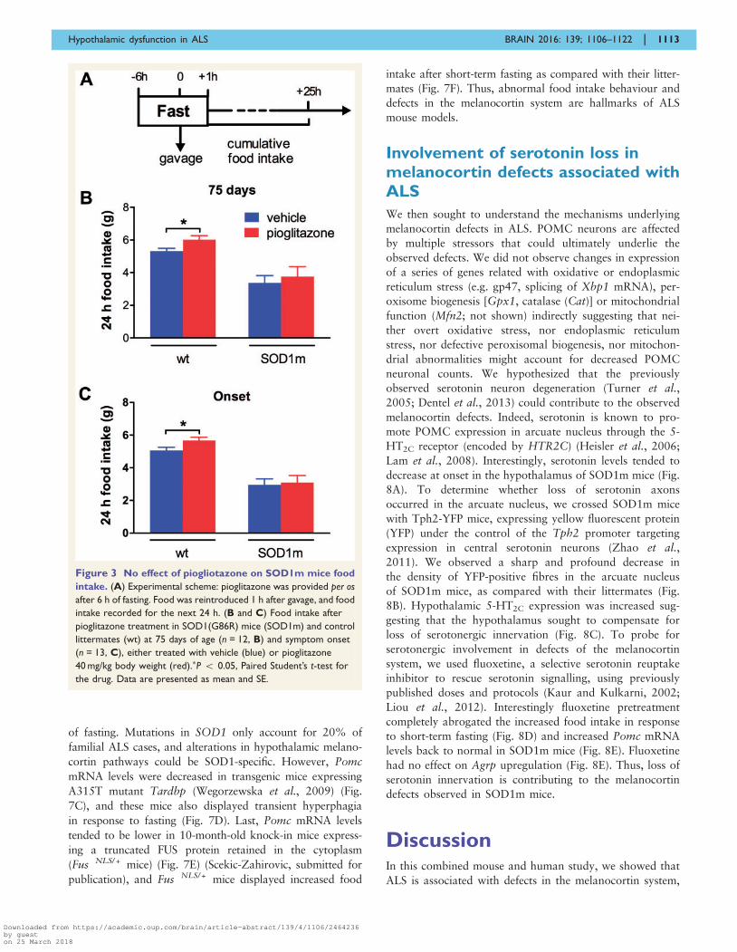
of fasting. Mutations in SOD1 only account for 20% of
familial ALS cases, and alterations in hypothalamic melano-
cortin pathways could be SOD1-specific. However, Pomc
mRNA levels were decreased in transgenic mice expressing
A315T mutant Tardbp (Wegorzewska et al., 2009) (Fig.
7C), and these mice also displayed transient hyperphagia
in response to fasting (Fig. 7D). Last, Pomc mRNA levels
tended to be lower in 10-month-old knock-in mice express-
ing a truncated FUS protein retained in the cytoplasm
(Fus�NLS/ + mice) (Fig. 7E) (Scekic-Zahirovic, submitted for
publication), and Fus�NLS/ + mice displayed increased food
intake after short-term fasting as compared with their litter-
mates (Fig. 7F). Thus, abnormal food intake behaviour and
defects in the melanocortin system are hallmarks of ALS
mouse models.
Involvement of serotonin loss inmelanocortin defects associated withALS
We then sought to understand the mechanisms underlying
melanocortin defects in ALS. POMC neurons are affected
by multiple stressors that could ultimately underlie the
observed defects. We did not observe changes in expression
of a series of genes related with oxidative or endoplasmic
reticulum stress (e.g. gp47, splicing of Xbp1 mRNA), per-
oxisome biogenesis [Gpx1, catalase (Cat)] or mitochondrial
function (Mfn2; not shown) indirectly suggesting that nei-
ther overt oxidative stress, nor endoplasmic reticulum
stress, nor defective peroxisomal biogenesis, nor mitochon-
drial abnormalities might account for decreased POMC
neuronal counts. We hypothesized that the previously
observed serotonin neuron degeneration (Turner et al.,
2005; Dentel et al., 2013) could contribute to the observed
melanocortin defects. Indeed, serotonin is known to pro-
mote POMC expression in arcuate nucleus through the 5-
HT2C receptor (encoded by HTR2C) (Heisler et al., 2006;
Lam et al., 2008). Interestingly, serotonin levels tended to
decrease at onset in the hypothalamus of SOD1m mice (Fig.
8A). To determine whether loss of serotonin axons
occurred in the arcuate nucleus, we crossed SOD1m mice
with Tph2-YFP mice, expressing yellow fluorescent protein
(YFP) under the control of the Tph2 promoter targeting
expression in central serotonin neurons (Zhao et al.,
2011). We observed a sharp and profound decrease in
the density of YFP-positive fibres in the arcuate nucleus
of SOD1m mice, as compared with their littermates (Fig.
8B). Hypothalamic 5-HT2C expression was increased sug-
gesting that the hypothalamus sought to compensate for
loss of serotonergic innervation (Fig. 8C). To probe for
serotonergic involvement in defects of the melanocortin
system, we used fluoxetine, a selective serotonin reuptake
inhibitor to rescue serotonin signalling, using previously
published doses and protocols (Kaur and Kulkarni, 2002;
Liou et al., 2012). Interestingly fluoxetine pretreatment
completely abrogated the increased food intake in response
to short-term fasting (Fig. 8D) and increased Pomc mRNA
levels back to normal in SOD1m mice (Fig. 8E). Fluoxetine
had no effect on Agrp upregulation (Fig. 8E). Thus, loss of
serotonin innervation is contributing to the melanocortin
defects observed in SOD1m mice.
DiscussionIn this combined mouse and human study, we showed that
ALS is associated with defects in the melanocortin system,
Figure 3 No effect of piogliotazone on SOD1m mice food
intake. (A) Experimental scheme: pioglitazone was provided per os
after 6 h of fasting. Food was reintroduced 1 h after gavage, and food
intake recorded for the next 24 h. (B and C) Food intake after
pioglitazone treatment in SOD1(G86R) mice (SOD1m) and control
littermates (wt) at 75 days of age (n = 12, B) and symptom onset
(n = 13, C), either treated with vehicle (blue) or pioglitazone
40 mg/kg body weight (red).�P 5 0.05, Paired Student’s t-test for
the drug. Data are presented as mean and SE.
Hypothalamic dysfunction in ALS BRAIN 2016: 139; 1106–1122 | 1113
Downloaded from https://academic.oup.com/brain/article-abstract/139/4/1106/2464236by gueston 25 March 2018

Figure 4 Altered melanocortin-related gene expression in SOD1m mice. (A and B) Messenger RNA levels of Pomc and Agrp in the
hypothalamus of SOD1(G86R) mice (black, SOD1m) and control littermates (white, wt) at 75 days of age (n = 15, A) and symptom onset (n = 11,
B). �P 5 0.05. ��P 5 0.005. ���P 5 0.0005, Student’s t-test. Data are presented as mean and SE. (C and D) Messenger RNA levels of hypothalamic
neuropeptides in the hypothalamus of SOD1(G86R) mice (black columns, SOD1m) and control littermates (white columns, wt) at 75 days of age
(n = 15, C) and symptom onset (n = 11, D). �P 5 0.05; ��P 5 0.005, Student’s t-test. Data are presented as mean and SE.
1114 | BRAIN 2016: 139; 1106–1122 P. Vercruysse et al.
Downloaded from https://academic.oup.com/brain/article-abstract/139/4/1106/2464236by gueston 25 March 2018
the major hypothalamic circuit controlling food intake and
energy expenditure. Pioglitazone did not increase weight in
ALS patients, thus providing indirect evidence of altered
hypothalamic melanocortin pathway. Pathological and
functional deficits of melanocortin system were found in
ALS mouse models directly demonstrating these defects.
These findings further extend the spectrum of defects in
ALS and provide a mechanistic explanation for a subset
of metabolic signs observed in these patients. Our study
also has important implications for the design of therapies
to target weight loss in this disease.
We first observed that pioglitazone did not increase body
weight or slow down weight loss in patients with ALS. This
was a surprising observation as progressive weight gain has
been repeatedly observed in all clinical trials of pioglitazone
in multiple non-neurological diseases (Promrat et al., 2004;
Belfort et al., 2006; Sanyal et al., 2010; DeFronzo et al.,
2011). Despite this lack of weight gain, ALS patients under
pioglitazone displayed all other biomarkers of efficacy,
including decreased glycaemia, decreased circulating liver
enzymes or increased adiponectin, thus ruling out that pa-
tients with ALS simply did not respond to the drug.
Interestingly, a series of recent studies dissected out how
TZDs lead to weight gain through activation of PPARgin hypothalamic POMC neurons leading to increased
food intake (Diano et al., 2011; Lu et al., 2011; Ryan et
al., 2011; Long et al., 2014). Consistent with the notion
that pioglitazone hypothalamic response was blunted in
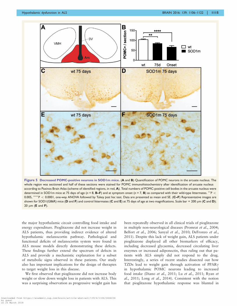
Figure 5 Decreased POMC-positive neurons in SOD1m mice. (A and B) Quantification of POMC neurons in the arcuate nucleus. The
whole region was sectioned and half of these sections were stained for POMC immunohistochemistry after identification of arcuate nucleus
according to Paxinos Brain Atlas (scheme of identified regions, in red, A). Total numbers of POMC-positive cell bodies in the arcuate nucleus were
determined in SOD1m mice at 75 days of age (n = 8, B–F) and at symptom onset (n = 7, B) as compared with their wild-type littermates. ��P 50.005, ����P 5 0.0001, one-way ANOVA followed by Tukey post hoc test. Data are presented as mean and SE. (C–F) Representative images are
shown for SOD1(G86R) mice (D and F) and control littermates (C and E) at 75 days of age at two magnifications. Scale bar = 200 mm (C and D);
20 mm (E and F).
Hypothalamic dysfunction in ALS BRAIN 2016: 139; 1106–1122 | 1115
Downloaded from https://academic.oup.com/brain/article-abstract/139/4/1106/2464236by gueston 25 March 2018
ALS patients, pioglitazone was not able to promote food
intake in SOD1m mice. Indeed, the melanocortin system is
dramatically affected in these mice, with decreased POMC
expression and loss of POMC positive neurons; similar al-
terations were observed in TDP-43- and FUS-based mouse
models pointing out that such defects are a general feature
in ALS. As pioglitazone decreases the activity of POMC
neurons thus increasing food intake (Diano et al., 2011),
we propose that the already decreased melanocortin tone in
ALS prevents the silencing of POMC neurons by pioglita-
zone. Consistent with this, SOD1 mice displayed hyperpha-
gia in response to short-term fasting, an orexigenic stimulus
that also leads to decreased POMC neuronal activity
(Perez-Tilve et al., 2010; Diano et al., 2011). Thus, our
results point to a general decrease in melanocortin tone
in ALS, leading to both a lack of response to TZDs, and
abnormal food intake behaviour in response to fasting.
What is the contribution of impaired melanocortin
system to the metabolic phenotypes associated with ALS?
The melanocortin system exerts multiple actions on energy
metabolism, either dependent on or independent of food
intake. First, an expected consequence of decreased mela-
nocortin tone is increased energy intake, especially in re-
sponse to an orexigenic stimulus such as food deprivation.
This is indeed what has been observed in multiple trans-
genic mouse models of ALS, suggesting that the defect in
the melanocortin system translates into a functional deficit.
Furthermore, consistent with the observed melanocortin
defect, we previously observed slightly increased cumulative
food intake in SOD1m mice (Dupuis et al., 2004).
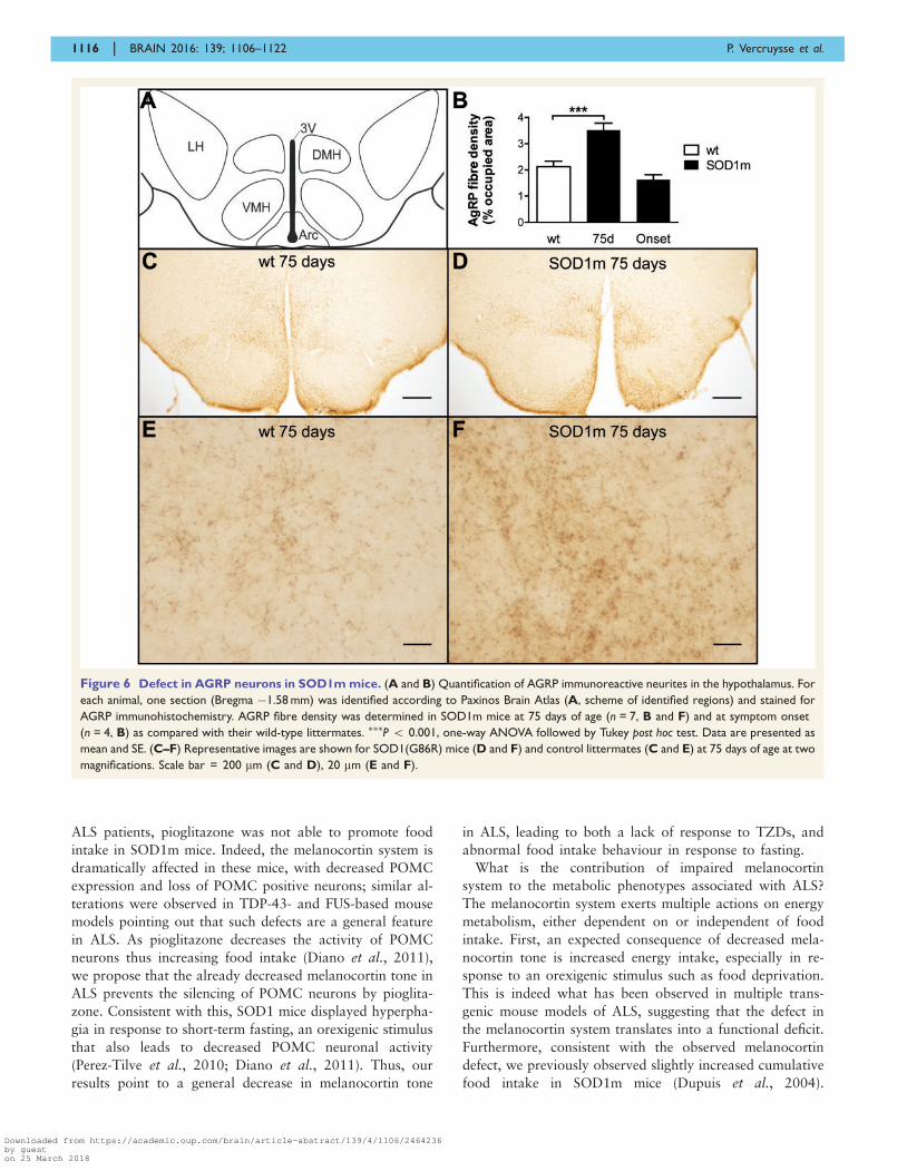
Figure 6 Defect in AGRP neurons in SOD1m mice. (A and B) Quantification of AGRP immunoreactive neurites in the hypothalamus. For
each animal, one section (Bregma �1.58 mm) was identified according to Paxinos Brain Atlas (A, scheme of identified regions) and stained for
AGRP immunohistochemistry. AGRP fibre density was determined in SOD1m mice at 75 days of age (n = 7, B and F) and at symptom onset
(n = 4, B) as compared with their wild-type littermates. ���P 5 0.001, one-way ANOVA followed by Tukey post hoc test. Data are presented as
mean and SE. (C–F) Representative images are shown for SOD1(G86R) mice (D and F) and control littermates (C and E) at 75 days of age at two
magnifications. Scale bar = 200 mm (C and D), 20 mm (E and F).
1116 | BRAIN 2016: 139; 1106–1122 P. Vercruysse et al.
Downloaded from https://academic.oup.com/brain/article-abstract/139/4/1106/2464236by gueston 25 March 2018

Figure 7 Multiple ALS mouse models display functional and molecular alterations in hypothalamic melanocortin system. (A
and B) Food intake was measured for 1 h, after either 7 h (A) or 1 h (B) of fasting in SOD1(G86R) mice (black columns, SOD1m) and control
littermates (white columns, wt) at 75 days of age (n = 10 and n = 14, respectively, for A and B). �P50.05, Student’s t-test. Data are presented as
mean and SE. (C) Messenger RNA levels of Pomc and Agrp in the hypothalamus of transgenic mice expressing A315T TDP-43 mutation (black
columns, TDP43m) and control littermates (white columns, wt) at non-symptomatic stage (n = 6). Unpaired t-test. �P 5 0.05. (D) Food intake
was measured 1 h after refeeding in TDP43m mice (n = 8). �P 5 0.05, ��P 5 0.01, Multiple t-test. Data are presented as mean and SE. (E)
Messenger RNA levels of Pomc and Agrp in the hypothalamus of transgenic mice Fus �NLS/ + knock-in mice (black columns, �NLS/ + ) and
control littermates (white columns, + / + ) at 10 months of age (n = 4). Data are presented as mean and SE. (F) Food intake was measured 1 h
after refeeding in �NLS/ + mice (n = 10) at 10 months of age. �P 5 0.05, Student’s t-test. Data are presented as mean and SE.
Hypothalamic dysfunction in ALS BRAIN 2016: 139; 1106–1122 | 1117
Downloaded from https://academic.oup.com/brain/article-abstract/139/4/1106/2464236by gueston 25 March 2018

The situation in ALS patients is less clear with respect to
energy intake due to a relative lack of studies reporting
dietary intake accurately and to confounding effects of dys-
phagia in advanced ALS patients. However, two case-con-
trol studies reported that increased dietary fat intake was
associated with ALS (Nelson et al., 2000; Huisman et al.,
2015). Moreover, Huisman et al. (2015) and collaborators
observed that presymptomatic daily energy intake was
increased in ALS patients as compared with controls, and
this would be consistent with both melanocortin
Figure 8 Involvement of serotonin loss in melanocortin defects of mutant SOD1 mice. (A) Serotonin levels in the hypothalamus of
SOD1(G86R) mice (black columns, SOD1m) and control littermates (white columns, wt) (n = 5 at 75 days and n = 5 at onset). One-way ANOVA
followed by Tukey post hoc test. Data are presented as mean and SE. (B) Serotonergic innervation in the arcuate nucleus. GFP immunohisto-
chemistry was performed on Tph2-YFP mice on arcuate nucleus cuts after identification on Paxinos Brain Atlas (i, scheme of identified regions,
in red, in the upper left column). Representative images are shown for SOD1(G86R) Tph2-YFP mice and control littermates at onset at
two magnifications. Scale bar = 200 mm (iii, iv), 20 mm (v and vi). Serotonergic fibre densities in the arcuate nucleus were quantified by a
blind observer as described previously (Grider et al., 2006) in SOD1m mice at symptom onset (ii–vi, n = 4) as compared with their wild-type
littermates. �P 5 0.05, Student’s t-test. Data are presented as mean and SE. (C) Messenger RNA levels of Htr2c (5-HT2C receptor) in the
hypothalamus of SOD1(G86R) mice (black, SOD1m) at 75 days of age and onset and in control littermates (white, wt) (n = 8). ��P 5 0.01, one-
way ANOVA. Data are presented as mean and SE. (D) Food intake was measured 1 h after refeeding in SOD1(G86R) mice (SOD1m) at 80 days of
age and control littermates (wt) after fluoxetine 20 mg/kg body weight (red) or vehicle (blue) injection (n = 38). ��P 5 0.005, Multiple t-test. Data
are presented as mean and SE. (E) Messenger RNA levels of Pomc and Agrp in the hypothalamus of SOD1(G86R) mice (SOD1m) and control
littermates (wt) at 85 days of age after fluoxetine 20 mg/kg body weight (red) or vehicle (blue) injection (n = 20). �P 5 0.05, one-way ANOVA.
Data are presented as mean and SE.
1118 | BRAIN 2016: 139; 1106–1122 P. Vercruysse et al.
Downloaded from https://academic.oup.com/brain/article-abstract/139/4/1106/2464236by gueston 25 March 2018

impairment and lack of weight gain under pioglitazone.
Second, the melanocortin defect could be responsible for
alterations in peripheral metabolic pathways. Indeed, the
melanocortin system regulates peripheral lipid metabolism
in rodents, by activating cholesterol reuptake by the liver
(Perez-Tilve et al., 2010) and reduces hepatic lipogenesis
(Leckstrom et al., 2011) independently of food intake. In
the same line, the melanocortin system is controlling glu-
cose metabolism and insulin response (Obici et al., 2001).
Interestingly, ALS patients have been reported to display
increased circulating cholesterol levels (Dupuis et al.,
2008), and glucose intolerance (Pradat et al., 2010).
Third, the melanocortin defect could impair regulation of
the autonomic nervous system (Sohn et al., 2013), and
autonomic abnormalities have sometimes been found in
ALS patients (Baltadzhieva et al., 2005). The relationships
between these different phenotypes and melanocortin de-
fects are unclear and will have to be clarified in further
studies. Importantly, the observed melanocortin defect is
unable to explain the weight loss associated with ALS, as,
on the contrary, the increased orexigenic drive triggered by
POMC deficiency likely compensates for weight loss by
increasing food intake. Other mechanisms, either peripheral
or central, have still to be identified to explain weight loss.
What causes the melanocortin defect in ALS? A first ob-
vious potential mechanism is energy deficit. Indeed, SOD1m
mice display weight loss due to hypermetabolism, leading to
decreased fat mass. These mice also display decreased circu-
lating insulin and leptin levels (Dupuis et al., 2004).
Ablation of leptin in ob/ob mice or fasting is indeed suffi-
cient to cause decreased Pomc mRNA (Mizuno et al., 1998,
1999; Ziotopoulou et al., 2000). However, leptin levels are
only decreased by 30% in 75-day-old mice (Dupuis et al.,
2004), an age at which we already observe strong Pomc
mRNA decreases. We and others had previously observed
serotonin loss in ALS (Turner et al., 2005; Dentel et al.,2013), and we hypothesized that this contributed to mela-
nocortin impairment in SOD1m mice. Indeed, serotonin is a
major activator of POMC neurons through the 5-HT2C re-
ceptor, and this occurs through direct electrical stimulation
(Heisler et al., 2002) but also trough transcriptional activa-
tion (Zhou et al., 2007; Lam et al., 2008; Xu et al., 2008).
Consistently, loss of 5-HT2C receptor leads to decreased
Pomc mRNA in the hypothalamus (Wang and Chehab,
2006). Our observation of restoration of Pomc mRNA
levels, as well as reversal of transient hyperphagia in
SOD1m mice by fluoxetine argues for serotonin loss being
a primary cause of melanocortin defect. This, however, does
not exclude that direct modulation of electrical activity by
either decreased leptin or other cues, either extrinsic or in-
trinsic, further exacerbate the observed defect. Brain sero-
tonin system is itself affected by organismal energy status
(Dwarkasing et al., 2015; Zemdegs et al., 2015) and our
data do not exclude that defects in serotonin levels found
in ALS patients and models is caused, or contributed by,
weight loss and hypermetabolism. Consistent with this
notion, decreasing leptin, whose major action is on the
melanocortin system, was able to revert partial weight loss
and increased energy expenditure in SOD1m mice (Lim et
al., 2014), suggesting that the melanocortin system can be
further inhibited by leptin ablation.
What are the consequences of our current findings in
ALS? There are at least three consequences of our finding
for ALS research. First, melanocortin impairment seems to
be a general event occuring in sporadic ALS patients, as
well as in animal models caused by disparate mutations
leading to ALS. Interestingly, a series of recent studies
demonstrated the occurence of similar hypothalamic
abnormalities in FTD (Piguet et al., 2011; Ahmed et al.,2014a, b, 2015), and in particular, increased AGRP
(Ahmed et al., 2015). Thus, melanocortin impairment ap-
pears associated with overall ALS/FTD continuum. It re-
mains to be determined how melanocortin impairment
relates with motor neuron degeneration in ALS. Second,
this study reinforces the notion of systemic involvement in
ALS. That melanocortin impairment appears downstream of
serotonin loss, also brings about the notion that circuitry
dysfunction might contribute to aspects of ALS phenotype.
How these serotonin and melanocortin defects might be
related to the spreading of TDP-43 aggregates
(Brettschneider et al., 2013) remains to be resolved.
Second, these results have consequences for the design of
pharmacological strategies to combat weight loss in ALS.
Weight loss in ALS is likely to be multi-factorial, with pri-
mary causes such as hypermetabolism, and bulbar involve-
ment, and could be exacerbated secondary to other
symptoms such as deficit in upper limbs or depression.
Treating weight loss could identify disease-modifying inter-
ventions as a hypercaloric diet was recently found to in-
crease survival of ALS patients under gastrostomy (Wills
et al., 2014; Dorst et al., 2015). Many drugs that could
be used to prevent weight loss, including atypical anti-
psychotics (e.g. olanzapine) inhibit POMC neurons (Kirk
et al., 2009; Weston-Green et al., 2012; Lian et al.,
2014). Drugs targeting the cannabinoid system increase
body weight by increasing beta-endorphin release from
POMC neurons (Koch et al., 2015). As beta-endorphin is
derived from POMC, which is decreased in ALS mice, it
appears likely that cannabinoids might not be able to pro-
mote food intake through this mechanism in ALS. Thus, our
current study provides a note of caution for the use of these
drugs to counteract weight loss in the specific context of
ALS patients, and suggest that disease progression might
impair responsiveness of ALS patients to classical drugs
leading to weight gain. A number of neural pathways con-
trolling energy homeostasis have not yet been studied in the
context of ALS and could be potential targets for treating
weight loss (Morton et al., 2014). First, pathways involved
in the emergency response to glucose deprivation (gluco-
paenia) such as NPY might be useful, although the precise
neurochemical pathways still need to be elucidated. Second,
drugs affecting food reward might be of interest to improve
the attractability of food during the disease. Last, the exist-
ence of emergency neuronal circuits involved in stress-
Hypothalamic dysfunction in ALS BRAIN 2016: 139; 1106–1122 | 1119
Downloaded from https://academic.oup.com/brain/article-abstract/139/4/1106/2464236by gueston 25 March 2018

induced anorexia was recently elucidated. Inhibiting these
pathways in ALS might also be a possible target for treating
weight loss (Morton et al., 2014). Alternatively, and besides
pharmacological treatments, increasing caloric density of the
diet is likely an efficient strategy to counteract weight loss
(Wills et al., 2014; Dorst et al., 2015), although current
results do not allow us to determine whether lipid enriched
or carbohydrate enriched would be more efficient.
In all, our post hoc analysis of the pioglitazone trial re-
vealed that the melanocortin system is profoundly altered in
ALS, and that this might be important for understanding and
preventing impairment of energy metabolism in ALS patients.
AcknowledgementsWe acknowledge the technical help of Marie Jo Ruivo,
Annie Picchinenna, Coraline Kostal, Marc Antoine Goy
and Paul Rochet.
FundingThis work was supported by Association de recherche sur la
SLA (ARSLA) and Fondation Thierry Latran (SpastALS, to
LD). Work in our laboratories is supported by ALS
Association Investigator Initiated Award (grants 2235,
3209 and 8075; to L.D.); the Frick Foundation (award
2013 to L.D.); Association Francaise contre les Myopathies
(grant #18280; to L.D.); Virtual Helmholtz Institute “RNA
dysmetabolism in ALS and FTD VI-510” (WP2, to L.D.,
A.W., A.C.L. and A.H.); Fondation « recherche sur le cer-
veau » (call 2015, to L.D.). Research leading to these results
has received funding from the European Community’s
Health Seventh Framework Programme (FP7/2007-2013;
EuroMOTOR grant agreement n� 259867).
Supplementary materialSupplementary material is available at Brain online.
ReferencesAhmed RM, Irish M, Kam J, van Keizerswaard J, Bartley L, Samaras
K, et al. Quantifying the eating abnormalities in frontotemporal
dementia. JAMA Neurol 2014a; 71: 1540–6.
Ahmed RM, Latheef S, Bartley L, Irish M, Halliday GM, Kiernan MC,
et al. Eating behavior in frontotemporal dementia: peripheral hor-
mones vs hypothalamic pathology. Neurology 2015; 85: 1310–7.
Ahmed RM, MacMillan M, Bartley L, Halliday GM, Kiernan MC,
Hodges JR, et al. Systemic metabolism in frontotemporal dementia.
Neurology 2014b; 83: 1812–8.Baltadzhieva R, Gurevich T, Korczyn AD. Autonomic impairment in
amyotrophic lateral sclerosis. Curr Opin Neurol 2005; 18: 487–93.Belfort R, Harrison SA, Brown K, Darland C, Finch J, Hardies J, et al.
A placebo-controlled trial of pioglitazone in subjects with nonalco-
holic steatohepatitis. N Engl J Med 2006; 355: 2297–307.
Bouteloup C, Desport JC, Clavelou P, Guy N, Derumeaux-Burel H,
Ferrier A, et al. Hypermetabolism in ALS patients: an early and
persistent phenomenon. J Neurol 2009; 256: 1236–42.
Brettschneider J, Del Tredici K, Toledo JB, Robinson JL, Irwin DJ,
Grossman M, et al. Stages of pTDP-43 pathology in amyotrophic
lateral sclerosis. Ann Neurol 2013; 74: 20–38.
Cirulli ET, Lasseigne BN, Petrovski S, Sapp PC, Dion PA, Leblond CS,
et al. Exome sequencing in amyotrophic lateral sclerosis identifies
risk genes and pathways. Science 2015; 347: 1436–41.
DeFronzo RA, Tripathy D, Schwenke DC, Banerji M, Bray GA,
Buchanan TA, et al. Pioglitazone for diabetes prevention in impaired
glucose tolerance. N Engl J Med 2011; 364: 1104–15.
Dentel C, Palamiuc L, Henriques A, Lannes B, Spreux-Varoquaux O,
Gutknecht L, et al. Degeneration of serotonergic neurons in amyo-
trophic lateral sclerosis: a link to spasticity. Brain 2013; 136(Pt 2):
483–93.
Desport JC, Preux PM, Magy L, Boirie Y, Vallat JM, Beaufrere B,
et al. Factors correlated with hypermetabolism in patients with
amyotrophic lateral sclerosis. Am J Clin Nutr 2001; 74: 328–34.
Desport JC, Preux PM, Truong TC, Vallat JM, Sautereau D, Couratier
P. Nutritional status is a prognostic factor for survival in ALS pa-
tients. Neurology 1999; 53: 1059–63.
Diano S, Liu ZW, Jeong JK, Dietrich MO, Ruan HB, Kim E, et al.
Peroxisome proliferation-associated control of reactive oxygen spe-
cies sets melanocortin tone and feeding in diet-induced obesity. Nat
Med 2011; 17: 1121–7.
Dorst J, Dupuis L, Petri S, Kollewe K, Abdulla S, Wolf J, et al.
Percutaneous endoscopic gastrostomy in amyotrophic lateral scler-
osis: a prospective observational study. J Neurol 2015; 262: 849–58.
Dorst J, Kuhnlein P, Hendrich C, Kassubek J, Sperfeld AD, Ludolph
AC. Patients with elevated triglyceride and cholesterol serum levels
have a prolonged survival in Amyotrophic Lateral Sclerosis.
J Neurol 2011; 258: 613–7.
Dupuis L, Corcia P, Fergani A, Gonzalez De Aguilar JL, Bonnefont-
Rousselot D, Bittar R, et al. Dyslipidemia is a protective factor in
amyotrophic lateral sclerosis. Neurology 2008; 70: 1004–9.
Dupuis L, Dengler R, Heneka MT, Meyer T, Zierz S, Kassubek J,
et al. A randomized, double blind, placebo-controlled trial of piogli-
tazone in combination with riluzole in amyotrophic lateral sclerosis.
PLoS One 2012; 7: e37885
Dupuis L, Oudart H, Rene F, Gonzalez de Aguilar JL, Loeffler JP.
Evidence for defective energy homeostasis in amyotrophic lateral
sclerosis: benefit of a high-energy diet in a transgenic mouse
model. Proc Natl Acad Sci USA 2004; 101: 11159–64.
Dupuis L, Pradat PF, Ludolph AC, Loeffler JP. Energy metabolism in
amyotrophic lateral sclerosis. Lancet Neurol 2011; 10: 75–82.
Dwarkasing JT, Boekschoten MV, Argiles JM, van Dijk M, Busquets
S, Penna F, et al. Differences in food intake of tumour-bearing cach-
ectic mice are associated with hypothalamic serotonin signalling.
J Cachexia Sarcopenia Muscle 2015; 6: 84–94.
Fergani A, Oudart H, Gonzalez De Aguilar JL, Fricker B, Rene F,
Hocquette JF, et al. Increased peripheral lipid clearance in an
animal model of amyotrophic lateral sclerosis. J Lipid Res 2007;
48: 1571–80.
Freischmidt A, Wieland T, Richter B, Ruf W, Schaeffer V, Muller K,
et al. Haploinsufficiency of TBK1 causes familial ALS and fronto-
temporal dementia. Nat Neurosci 2015; 18: 631–6.
Gallo V, Wark PA, Jenab M, Pearce N, Brayne C, Vermeulen R, et al.
Prediagnostic body fat and risk of death from amyotrophic lateral
sclerosis: the EPIC cohort. Neurology 2013; 80: 829–38.
Grider MH, Chen Q, Shine HD. Semi-automated quantification of
axonal densities in labeled CNS tissue. J Neurosci Methods 2006;
155: 172–9.
Gurney ME, Pu H, Chiu AY, Dal Canto MC, Polchow CY, Alexander
DD, et al. Motor neuron degeneration in mice that express a
human Cu,Zn superoxide dismutase mutation. Science 1994; 264:
1772–5.
1120 | BRAIN 2016: 139; 1106–1122 P. Vercruysse et al.
Downloaded from https://academic.oup.com/brain/article-abstract/139/4/1106/2464236by gueston 25 March 2018
Heisler LK, Cowley MA, Tecott LH, Fan W, Low MJ, Smart JL, et al.
Activation of central melanocortin pathways by fenfluramine.
Science 2002; 297: 609–11.
Heisler LK, Jobst EE, Sutton GM, Zhou L, Borok E, Thornton-Jones
Z, et al. Serotonin reciprocally regulates melanocortin neurons to
modulate food intake. Neuron 2006; 51: 239–49.
Huisman MH, Seelen M, de Jong SW, Dorresteijn KR, van Doormaal
PT, van der Kooi AJ, et al. Lifetime physical activity and the risk of
amyotrophic lateral sclerosis. J Neurol Neurosurg Psychiatry 2013;
84: 976–81.
Huisman MH, Seelen M, van Doormaal PT, de Jong SW, de Vries JH,
van der Kooi AJ, et al. Independent associations of presymptomatic
body mass index, and consumption of fat and alcohol with amyo-
trophic lateral sclerosis. JAMA Neurol 2015; 72: 1155–62.
Kaur G, Kulkarni SK. Evidence for serotonergic modulation of pro-
gesterone-induced hyperphagia, depression and algesia in female
mice. Brain Res 2002; 943: 206–15.
Kirk SL, Glazebrook J, Grayson B, Neill JC, Reynolds GP.
Olanzapine-induced weight gain in the rat: role of 5-HT2C and
histamine H1 receptors. Psychopharmacology 2009; 207: 119–25.
Koch M, Varela L, Kim JG, Kim JD, Hernandez-Nuno F, Simonds SE,
et al. Hypothalamic POMC neurons promote cannabinoid-induced
feeding. Nature 2015; 519: 45–50.
Lam DD, Przydzial MJ, Ridley SH, Yeo GS, Rochford JJ, O’Rahilly S,
et al. Serotonin 5-HT2C receptor agonist promotes hypophagia via
downstream activation of melanocortin 4 receptors. Endocrinology
2008; 149: 1323–8.
Lattante S, Ciura S, Rouleau GA, Kabashi E. Defining the genetic
connection linking amyotrophic lateral sclerosis (ALS) with fronto-
temporal dementia (FTD). Trends Genet 2015; 31: 263–73.
Leblond CS, Kaneb HM, Dion PA, Rouleau GA. Dissection of genetic
factors associated with amyotrophic lateral sclerosis. Exp Neurol
2014; 262 (Pt B): 91–101.
Leckstrom A, Lew PS, Poritsanos NJ, Mizuno TM. Central melano-
cortin receptor agonist reduces hepatic lipogenic gene expression in
streptozotocin-induced diabetic mice. Life Sciences 2011; 88: 664–9.
Lian J, Huang XF, Pai N, Deng C. Betahistine ameliorates olanzapine-
induced weight gain through modulation of histaminergic, NPY and
AMPK pathways. Psychoneuroendocrinology 2014; 48: 77–86.
Lim MA, Bence KK, Sandesara I, Andreux P, Auwerx J, Ishibashi J,
et al. Genetically altering organismal metabolism by leptin-deficiency
benefits a mouse model of amyotrophic lateral sclerosis. Hum Mol
Genet 2014; 23: 4995–5008.
Lindauer E, Dupuis L, Muller HP, Neumann H, Ludolph AC,
Kassubek J. Adipose Tissue Distribution Predicts Survival in
Amyotrophic Lateral Sclerosis. PLoS One 2013; 8: e67783.
Liou YJ, Chen CH, Cheng CY, Chen SY, Chen TJ, Yu YW, et al.
Convergent evidence from mouse and human studies suggests the
involvement of zinc finger protein 326 gene in antidepressant treat-
ment response. PLoS One 2012; 7: e32984
Long L, Toda C, Jeong JK, Horvath TL, Diano S. PPARgamma ab-
lation sensitizes proopiomelanocortin neurons to leptin during high-
fat feeding. J Clin Invest 2014; 124: 4017–27.
Lu M, Sarruf DA, Talukdar S, Sharma S, Li P, Bandyopadhyay G,
et al. Brain PPAR-gamma promotes obesity and is required for the
insulin-sensitizing effect of thiazolidinediones. Nat Med 2011; 17:
618–22.
Maeda N, Takahashi M, Funahashi T, Kihara S, Nishizawa H,
Kishida K, et al. PPARgamma ligands increase expression and
plasma concentrations of adiponectin, an adipose-derived protein.
Diabetes 2001; 50: 2094–9.Marin B, Desport JC, Kajeu P, Jesus P, Nicolaud B, Nicol M, et al.
Alteration of nutritional status at diagnosis is a prognostic factor for
survival of amyotrophic lateral sclerosis patients. J Neurol
Neurosurg Psychiatry 2011; 82: 628–34.Mizuno TM, Kleopoulos SP, Bergen HT, Roberts JL, Priest CA,
Mobbs CV. Hypothalamic pro-opiomelanocortin mRNA is reduced
by fasting and [corrected] in ob/ob and db/db mice, but is stimulated
by leptin. Diabetes 1998; 47: 294–7.
Mizuno TM, Makimura H, Silverstein J, Roberts JL, Lopingco T,
Mobbs CV. Fasting regulates hypothalamic neuropeptide Y,
agouti-related peptide, and proopiomelanocortin in diabetic mice
independent of changes in leptin or insulin. Endocrinology 1999;
140: 4551–7.
Morton GJ, Meek TH, Schwartz MW. Neurobiology of food intake in
health and disease. Nat Rev Neurosci 2014; 15: 367–78.
Nelson LM, Matkin C, Longstreth WT Jr, McGuire V. Population-
based case-control study of amyotrophic lateral sclerosis in western
Washington State. II. Diet Am J Epidemiol 2000; 151: 164–73.
O’Reilly EJ, Wang H, Weisskopf MG, Fitzgerald KC, Falcone G,
McCullough ML, et al. Premorbid body mass index and risk of
amyotrophic lateral sclerosis. Amyotroph Lateral Scler
Frontotemporal Degener 2013; 14: 205–11.
Obici S, Feng Z, Tan J, Liu L, Karkanias G, Rossetti L. Central
melanocortin receptors regulate insulin action. J Clin Invest 2001;
108: 1079–85.
Paganoni S, Deng J, Jaffa M, Cudkowicz ME, Wills AM. Body mass
index, not dyslipidemia, is an independent predictor of survival in
amyotrophic lateral sclerosis. Muscle Nerve 2011; 44: 20–4.
Palamiuc L, Schlagowski A, Ngo ST, Vernay A, Dirrig-Grosch S,
Henriques A, et al. A metabolic switch toward lipid use in glycolytic
muscle is an early pathologic event in a mouse model of amyo-
trophic lateral sclerosis. EMBO Mol Med 2015; 7: 526–46.
Perez-Tilve D, Hofmann SM, Basford J, Nogueiras R, Pfluger PT,
Patterson JT, et al. Melanocortin signaling in the CNS directly regu-
lates circulating cholesterol. Nat Neurosci 2010; 13: 877–82.
Piguet O, Petersen A, Yin Ka Lam B, Gabery S, Murphy K, Hodges
JR, et al. Eating and hypothalamus changes in behavioral-variant
frontotemporal dementia. Ann Neurol 2011; 69: 312–9.
Pradat PF, Bruneteau G, Gordon PH, Dupuis L, Bonnefont-Rousselot
D, Simon D, et al. Impaired glucose tolerance in patients with amyo-
trophic lateral sclerosis. Amyotroph Lateral Scler 2010; 11: 166–71.
Promrat K, Lutchman G, Uwaifo GI, Freedman RJ, Soza A, Heller T,
et al. A pilot study of pioglitazone treatment for nonalcoholic stea-
tohepatitis. Hepatology 2004; 39: 188–96.
Ripps ME, Huntley GW, Hof PR, Morrison JH, Gordon JW.
Transgenic mice expressing an altered murine superoxide dismutase
gene provide an animal model of amyotrophic lateral sclerosis. Proc
Natl Acad Sci USA 1995; 92: 689–93.
Ryan KK, Li B, Grayson BE, Matter EK, Woods SC, Seeley RJ. A role
for central nervous system PPAR-gamma in the regulation of energy
balance. Nat Med 2011; 17: 623–6.
Sanyal AJ, Chalasani N, Kowdley KV, McCullough A, Diehl AM,
Bass NM, et al. Pioglitazone, vitamin E, or placebo for nonalcoholic
steatohepatitis. N Engl J Med 2010; 362: 1675–85.
Sohn JW, Harris LE, Berglund ED, Liu T, Vong L, Lowell BB, et al.
Melanocortin 4 receptors reciprocally regulate sympathetic and
parasympathetic preganglionic neurons. Cell 2013; 152: 612–9.
Turner MR, Rabiner EA, Hammers A, Al-Chalabi A, Grasby PM,
Shaw CE, et al. [11C]-WAY100635 PET demonstrates marked 5-
HT1A receptor changes in sporadic ALS. Brain 2005; 128(Pt 4):
896–905.
Turner MR, Wotton C, Talbot K, Goldacre MJ. Cardiovascular fitness
as a risk factor for amyotrophic lateral sclerosis: indirect evidence
from record linkage study. J Neurol Neurosurg Psychiatry 2012; 83:
395–8.
Vandesompele J, De Preter K, Pattyn F, Poppe B, Van Roy N, De
Paepe A, et al. Accurate normalization of real-time quantitative
RT-PCR data by geometric averaging of multiple internal control
genes. Genome Biol 2002; 3: research0034.
Wang B, Chehab FF. Deletion of the serotonin 2c receptor from trans-
genic mice overexpressing leptin does not affect their lipodystrophy
but exacerbates their diet-induced obesity. Biochem Biophys Res
Commun 2006; 351: 418–23.
Hypothalamic dysfunction in ALS BRAIN 2016: 139; 1106–1122 | 1121
Downloaded from https://academic.oup.com/brain/article-abstract/139/4/1106/2464236by gueston 25 March 2018
Wegorzewska I, Bell S, Cairns NJ, Miller TM, Baloh RH. TDP-43mutant transgenic mice develop features of ALS and
frontotemporal lobar degeneration. Proc Natl Acad Sci USA 2009;
106: 18809–14.
Weston-Green K, Huang XF, Deng C. Alterations to melanocortiner-gic, GABAergic and cannabinoid neurotransmission associated with
olanzapine-induced weight gain. PLoS One 2012; 7: e33548.
Wills AM, Hubbard J, Macklin EA, Glass J, Tandan R, Simpson EP,
et al. Hypercaloric enteral nutrition in patients with amyotrophiclateral sclerosis: a randomised, double-blind, placebo-controlled
phase 2 trial. Lancet 2014; 383: 2065–72.
Xu Y, Jones JE, Kohno D, Williams KW, Lee CE, Choi MJ, et al. 5-HT2CRs expressed by pro-opiomelanocortin neurons regulate
energy homeostasis. Neuron 2008; 60: 582–9.
Zemdegs J, Quesseveur G, Jarriault D, Penicaud L, Fioramonti X,
Guiard BP. High fat diet-induced metabolic disorders impairs sero-tonergic function and anxiety-like behaviours in mice. Br J
Pharmacol 2015. Advance Access published on October 16, 2015,
doi: 10.1111/bph.13343.
Zhao S, Ting JT, Atallah HE, Qiu L, Tan J, Gloss B, et al. Cell type-specific channelrhodopsin-2 transgenic mice for optogenetic dissection
of neural circuitry function. Nat Methods 2011; 8: 745–52.
Zhou L, Sutton GM, Rochford JJ, Semple RK, Lam DD, Oksanen LJ,
et al. Serotonin 2C receptor agonists improve type 2 diabetes via mel-anocortin-4 receptor signaling pathways. Cell Metab 2007; 6: 398–405.
Ziotopoulou M, Erani DM, Hileman SM, Bjorbaek C, Mantzoros CS.
Unlike leptin, ciliary neurotrophic factor does not reverse the star-vation-induced changes of serum corticosterone and hypothalamic
neuropeptide levels but induces expression of hypothalamic inhibi-
tors of leptin signaling. Diabetes 2000; 49: 1890–6.
Appendix 1
Collaborators: GERP ALS StudyGroup
All centres were located in Germany. Investigators are
listed by alphabetical order of centre and investigator:
Berlin (Department of Neurology, Charite University
Hospital): Nadja Borisow; Theresa Holm; Andre Maier;
Thomas Meyer; Bochum (Department of Neurology,
University Hospital Bergmannsheil): Paula Budde; Torsten
Grehl; Kai Gruhn; Bonn (Department of Neurology,
University Hospital of Bonn): Malte Bewersdorff; Michael
Heneka; Dresden (Department of Neurology, University
Hospital Carl Gustav Carus, Technische Universitat
Dresden): Andreas Hermann; Alexander Storch; Gottingen
(Department of Neurology, University Hospital of
Gottingen): Tobias Frank; Bettina Goricke; Jochen
Weishaupt; Halle (Department of Neurology, University
Hospital of Halle/Saale): Katharina Eger; Frank Hanisch;
Stephan Zierz; Hannover (Department of Neurology and
Clinical Neurophysiology, Hannover Medical School
(MHH), University Clinic): Anna-Lena Boeck; Reinhard
Dengler; Sonja Koerner; Katja Kollewe; Susanne Petri;
Jena (Department of Neurology, University Hospital
Jena): Julian Grosskreutz; Tino Prell; Thomas Ringer; Jan
Zinke; Munich (Department of Neurology, University of
Munich): Johanna Anneser; Gian Domenico Borasio;
Christine Chahli; Andrea S. Winkler; Muenster
(Department of Neurology, University of Muenster):
Matthias Boentert; Bianca Stubbe-Draeger; Peter Young;
Regensburg (Department of Neurology, University of
Regensburg): Ulrich Bogdahn; Steffen Franz; Verena
Haringer; Norbert Weidner; Rostock (Department of
Neurology, University of Rostock): Reiner Benecke;
Stefanie Meister; Johannes Prudlo; Matthias Wittstock;
Ulm (Department of Neurology, University of Ulm):
Johannes Dorst; Corinna Hendrich; Albert C. Ludolph;
Anne-Dorte Sperfeld; Ulrike Weiland; Wiesbaden
(Department of Neurology, Neurological clinic, DKD):
Sabine Neidhardt; Berthold Schrank; Wurzburg
(Department of Neurology, University of Wurzburg):
Marcus Beck; Peter Kraft; Klaus Toyka; Jochen
Ulzheimer; Carsten Wessig.
1122 | BRAIN 2016: 139; 1106–1122 P. Vercruysse et al.
Downloaded from https://academic.oup.com/brain/article-abstract/139/4/1106/2464236by gueston 25 March 2018