Allergic hypersensitivity to topical and systemic ... · Review article Allergic hypersensitivity...
17
Review article Allergic hypersensitivity to topical and systemic corticosteroids: a review The therapeutic properties of corticosteroids (CSs) were first demonstrated by Edward Kendall and Philip Hench in 1948 (1). During the 1950s it was discovered that hydrocortisone, a natural glucocorticoid hormone, could reduce inflammation and proliferation in some skin disorders (2). Chemical modifications of this basic hor- mone soon gave rise to multiple CSs of varying strength, each with its own specific properties. In addition to their basic role in the treatment for inflammatory skin pathol- ogies, CSs are also used in emergency medicine and in several internal diseases. Corticosteroids have an anti- inflammatory effect, which is mainly related to the inhibition of transcription of several pro-inflammatory cytokines/mediators. Other factors contributing to their anti-inflammatory and immunosuppressant properties include the apoptosis of basophils and the inhibition of adhesion molecule expression on the surface of endothe- lial cells (3). It may therefore appear to be paradoxical that CSs, potent anti-inflammatory agents and immuno- modulators used in the treatment of allergic manifesta- tions (in the broad sense), are capable of producing immediate or delayed allergic hypersensitivity (AHS) reactions. The first documented allergic reactions to CSs, following local application and injections of hydrocorti- sone, were described towards the end of the 1950s (4–6). Diagnosing an allergic reaction to CSs remains a challenge for clinicians. Its clinical presentation is frequently atypical and tests may be difficult to interpret. Moreover, its frequency is undoubtedly under-estimated. While knowledge of delayed hypersensitivity as a secondary effect of topical CSs (allergic contact eczema) is improving, little is known about immediate and delayed reactions to systemic CSs. It is critical to address such reactions, as appropriate diagnostic work-up should determine potential replacement agent(s) that can still be tolerated by the patient. Allergic and nonallergic hypersensitivity (AHS-NAHS) Drugs are xenobiotics that interact with many cellular activation systems when carrying out their therapeutic Corticosteroids, which are potent anti-inflammatory and immunomodulator agents used in the treatment of various inflammatory diseases including allergic diseases, can in some cases produce immediate or delayed hypersensitivity reactions. This review summarizes the epidemiological and clinical characteristics of such reactions, including related diagnostic issues. It also presents a detailed analysis of the proposed immunological mechanisms including underlying cross-reactions. M. Baeck 1 , L. Marot 1 , J.-F. Nicolas 2 , C. Pilette 3 , D. Tennstedt 1 , A. Goossens 4 1 Department of Dermatology, Cliniques Universitaires Saint-Luc, UniversitȖ Catholique de Louvain, Brussels, Belgium; 2 Department of Immuno-Allergology, University Hospital Centre Lyon Sud, Hospices Civiles de Lyon, Lyon, France; 3 Department of Pneumology, Cliniques Universitaires Saint-Luc, UniversitȖ Catholique de Louvain, Brussels; 4 Department of Dermatology, University Hospital, Katholieke Universiteit Leuven, Leuven, Belgium Key words: allergic contact dermatitis; corticosteroid; cross-reactions; delayed hypersensitivity; immediate hypersensitivity. M. Baeck Department of Dermatology Cliniques Universitaires Saint-Luc, U.C.L. Avenue Hippocrate, 10 B-1200 Brussels Belgium Accepted for publication 3 January 2009 Allergy 2009: 64: 978–994 Ó 2009 John Wiley & Sons A/S DOI: 10.1111/j.1398-9995.2009.02038.x 978
Transcript of Allergic hypersensitivity to topical and systemic ... · Review article Allergic hypersensitivity...
Review article
Allergic hypersensitivity to topical and systemic corticosteroids:
a review
The therapeutic properties of corticosteroids (CSs) werefirst demonstrated by Edward Kendall and Philip Henchin 1948 (1). During the 1950s it was discovered thathydrocortisone, a natural glucocorticoid hormone, couldreduce inflammation and proliferation in some skindisorders (2). Chemical modifications of this basic hor-mone soon gave rise to multiple CSs of varying strength,each with its own specific properties. In addition to theirbasic role in the treatment for inflammatory skin pathol-ogies, CSs are also used in emergency medicine and inseveral internal diseases. Corticosteroids have an anti-inflammatory effect, which is mainly related to theinhibition of transcription of several pro-inflammatorycytokines/mediators. Other factors contributing to theiranti-inflammatory and immunosuppressant propertiesinclude the apoptosis of basophils and the inhibition ofadhesion molecule expression on the surface of endothe-lial cells (3). It may therefore appear to be paradoxicalthat CSs, potent anti-inflammatory agents and immuno-modulators used in the treatment of allergic manifesta-tions (in the broad sense), are capable of producing
immediate or delayed allergic hypersensitivity (AHS)reactions. The first documented allergic reactions to CSs,following local application and injections of hydrocorti-sone, were described towards the end of the 1950s (4–6).
Diagnosing an allergic reaction to CSs remains achallenge for clinicians. Its clinical presentation isfrequently atypical and tests may be difficult to interpret.Moreover, its frequency is undoubtedly under-estimated.While knowledge of delayed hypersensitivity as asecondary effect of topical CSs (allergic contact eczema)is improving, little is known about immediate and delayedreactions to systemic CSs. It is critical to address suchreactions, as appropriate diagnostic work-up shoulddetermine potential replacement agent(s) that can stillbe tolerated by the patient.
Allergic and nonallergic hypersensitivity (AHS-NAHS)
Drugs are xenobiotics that interact with many cellularactivation systems when carrying out their therapeutic
Corticosteroids, which are potent anti-inflammatory and immunomodulatoragents used in the treatment of various inflammatory diseases including allergicdiseases, can in some cases produce immediate or delayed hypersensitivityreactions. This review summarizes the epidemiological and clinical characteristicsof such reactions, including related diagnostic issues. It also presents a detailedanalysis of the proposed immunological mechanisms including underlyingcross-reactions.
M. Baeck1, L. Marot1, J.-F. Nicolas2,C. Pilette3, D. Tennstedt1,A. Goossens4
1Department of Dermatology, CliniquesUniversitaires Saint-Luc, Universit� Catholique deLouvain, Brussels, Belgium; 2Department ofImmuno-Allergology, University Hospital Centre LyonSud, Hospices Civiles de Lyon, Lyon, France;3Department of Pneumology, CliniquesUniversitaires Saint-Luc, Universit� Catholique deLouvain, Brussels; 4Department of Dermatology,University Hospital, Katholieke Universiteit Leuven,Leuven, Belgium
Key words: allergic contact dermatitis; corticosteroid;cross-reactions; delayed hypersensitivity; immediatehypersensitivity.
M. BaeckDepartment of DermatologyCliniques Universitaires Saint-Luc, U.C.L.Avenue Hippocrate, 10B-1200 BrusselsBelgium
Accepted for publication 3 January 2009
Allergy 2009: 64: 978–994 � 2009 John Wiley & Sons A/S
DOI: 10.1111/j.1398-9995.2009.02038.x
978
effects. Adverse drug reactions are grouped into twocategories: those that are predictable, common, andrelated to the pharmacologic actions of the drug (TypeA reactions); and those that are unpredictable, uncom-mon and usually not related to the pharmacologicactions of the drug (Type B reactions) (7). The latterinclude AHS and NAHS. These reactions are some-times difficult to differentiate and involve differentmechanisms. However, differentiation of these twokinds of drug intolerance is important as AHS is rarebut can be life-threatening while NAHS is frequent, cansometimes be severe and in exceptional cases, can befatal (8, 9).
Allergic hypersensitivity
These reactions are because of specific immune effectors,antibodies and/or T lymphocytes. Allergic hypersensitiv-ity reactions require a prior sensitization to the drug, inorder to develop an adaptive immune response withproduction of drug-specific antibodies (IgE or IgG) orT lymphocyte (CD4+ or CD8+ ). Allergic reactionscan be classified, according to Gell and Coombs, into fourtypes of immune reactions: Type I, immediate-typehypersensitivity reactions (immediate AHS – IAHS)because of IgEs, Type II and Type III, cytotoxic andimmune complex reactions because of IgG or IgMantibodies, and Type IV, delayed-type hypersensitivity(delayed AHS – DAHS) because of CD4+ or CD8+T-cells (7, 8). The Type IV reactions were furthersubclassified by Posadas and Pichler (7) into foursubtypes (IVa–IVd) based on the clinical, immunohisto-chemical and functional heterogeneity of certain drugallergies. Indeed, T-cells have been found to differ in thecytokines they produce, which results in distinct types ofimmune defence.A suspected AHS is diagnosed by demonstrating the
presence of specific IgE or T-cells through in vivo orin vitro tests. The in vivo tests are mainly �skin prick�test and/or an early-reading intradermal reaction (IDR)test in cases of IAHS and �patch test� and/or a late-reading IDR test in cases of DAHS. In vitro diagnosismay include the identification of basophil activation/degranulation and IgEs specific to the drug for IAHS(10, 11) and lymphocyte transformation test (LTT)and IFN-gamma enzyme-linked immunospot assay(ELISPOT) for DAHS (12–14). Biological diagnosis ofa DAHS reaction has made significant progress inrecent years thanks to the work of Rozieres et al. (12)and Pichler et al. (14). These researchers have studiedvarious means of detecting specific T-cells to a givendrug (particularly beta-lactam antibiotics). However,because of technical issues in particular (e.g. standard-ization of the reagents used), validation of these testsfor routine clinical use remains difficult and at present,their use remains restricted to specialized clinicalcentres.
Nonallergic hypersensitivity
These reactions are much more frequent than AHS andare referred to as pseudo-allergies, false allergies, or drugintolerance. Unlike AHS reactions, they are not as aresult of specific immune responses. Such reactionsinvolve direct activation of the innate immune system(mast cells/basophils, complement) and do not involvespecific antibodies or T-cells. Thus, NAHS may occurafter the first application or administration of the drugand does not require prior sensitization. In vivo andin vitro tests are negative in this setting.
Sensitization routes
There is a huge variety of CS molecules and many ways ofadministering them. Both local and systemic treatmentsare possible and either mode can sensitize the patient andlead to a specific immune reaction. It is important tomake a distinction between the original sensitizationroute and the observed secondary reactions to topical orsystemic administration of a CS; these are not necessarilylinked. A patient who reacts to a systemically adminis-tered CS, for instance, may have already been sensitizedto this agent (or another chemically related agent) byprevious topical use – or vice versa.
The most common sensitization route is throughcutaneous use (15). Locoregional routes includingrespiratory (nasal or mouth inhalation), digestive andintra-articular administration have been less frequentlyimplicated. Sensitization related to systemic administra-tion of a CS, of which the intravenous route is the mostcommon, occurs even less frequently (15, 16).
Factors influencing sensitization to CSs
Topical route
Patients who suffer from a long-term dermatologicaldisease are at higher risk of sensitization when treated bytopical CSs: these include chronic eczema, stasis derma-titis, chronic ulceration, chronic actinic dermatitis, facial,anogenital, and hand and foot dermatitis (17). Changes tothe skin barrier and/or a local pro-inflammatory envi-ronment (favouring and priming antigen-presenting cellssuch as Langerhans cells) are the most likely determiningfactors.
It is not clear whether atopy represents a risk factor forthe development of a CS allergy, or whether observationof such allergies might be due to a selection bias becausethis group of patients is more likely to receive CSs. Thosepatients who are allergic to a CS often exhibit co-sensi-tization to multiple allergens (e.g. preservatives, excipi-ents and antibiotics). Degreef and Dooms-Goossens (18)demonstrated that 82% of their CS-allergic patients were
Allergic hypersensitivity to topical and systemic corticosteroids
� 2009 John Wiley & Sons A/S Allergy 2009: 64: 978–994 979
also sensitized to at least one other allergen. Notunexpectedly, contact allergy to CSs is only rarelyoccupationally induced (19).
Systemic route
The risk factors for developing sensitivity to systemic CSshave not been studied. It seems, however, that suchreactions occur more frequently in asthmatic subjects (20,21) and in patients regularly treated with systemic CSs(e.g. in the case of a missing or transplanted kidney) (22)than in other subjects. However, in these cases, it isdifficult to determine whether the higher incidence ofallergies observed is due to increased susceptibility, or togreater exposure to CSs.Other factors such as hypersensitivity to salicylic acid
might also constitute risk factors (23); neither can geneticpredisposition to CS allergies be excluded (24, 25).
Prevalence of allergy to CSs
According to the literature, the prevalence of allergicreactions to CSs is extremely variable. Several factorssuch as regional differences in prescribing habits (i.e. thetypes of CSs commonly prescribed and the number ofprescriptions given out), awareness of topical CS allergyamong medical professionals, patient selection and refer-ral, and diagnostic procedures, may have an influence.
Topical CSs
The literature reports that the frequency of CS allergyfollowing topical application ranges from 0.2% to 5% ormore (3, 17, 26, 27). Despite the wide use of inhaled CSs,very few cases of asthmatic patients experiencing allergicreactions have been reported (27, 28). Isaksson et al. (29),who examined patients with asthma or allergic rhinitiswho were treated with tixocortol pivalate, observed a CSsensitization rate of 1.4%. Among nonasthmatic patients,however, the rate of sensitization was 0.9%, and thedifference was not statistically significant. Malik et al.(30) were the first to evaluate the incidence of CS allergiesin patients with digestive tract inflammation who weretreated with steroid enemas. Of 44 patients, 9% weretested positive for one or more CS.
Systemic CSs
Allergic reactions following systemic administration ofCSs have rarely been reported in the literature and mostoften concern isolated cases. In 1998, Klein-Gitelmanet al. (31), in a cohort study of 213 children treated withintravenous CS for various rheumatological conditions,estimated the rate of allergic reactions to be 0.1% (forapproximately 10 000 doses of glucocorticosteroid), mostbeing of the immediate type. Other authors have
estimated the prevalence of such reactions at 0.3% (32).About a hundred publications have reported immediatereactions after oral or parenteral administration of CSs,the allergic nature of which was not always proved, whilethe prevalence of systemic contact dermatitis or systemicallergic dermatitis (33) to CSs has not been examined.
Clinical presentation
Recognizing CS allergy can be difficult, as its clinicalpresentation tends to be neither specific nor spectacular:the clinical signs are usually minor, or display acompletely atypical chronology, which is due to theanti-inflammatory properties of the CS. As mentioned byLe Coz (34), the clinical manifestation of such reactiondepends on two competing effects which are of variableintensity and offset in time: the immunological allergicresponse and the pharmacological �anti-allergic� effect.
Type I and Type IV are the most frequent allergicreactions observed with CSs. IgGs (Type II); immunecomplex (Type III) associated reactions, although possi-ble, have never been reported.
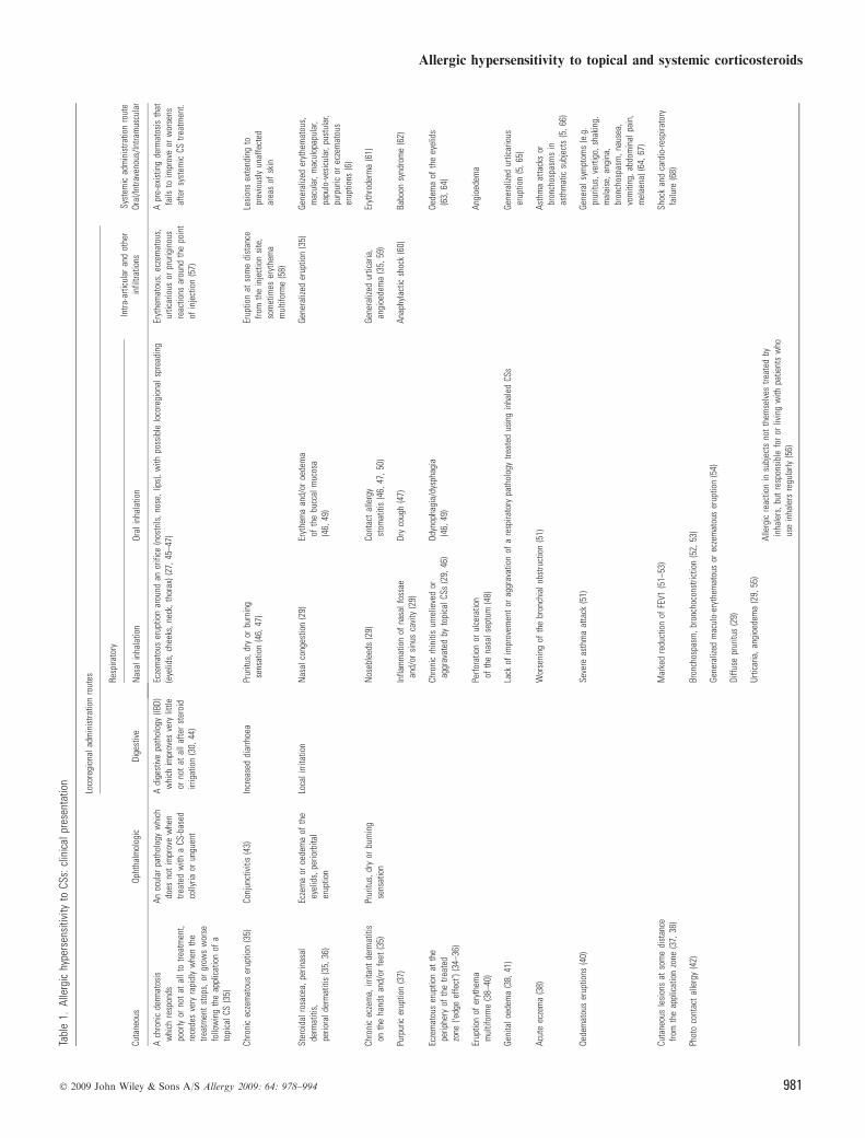
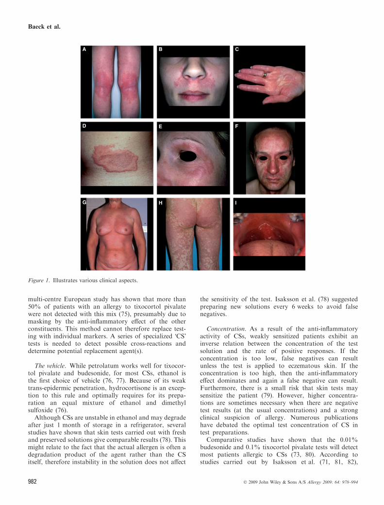
Allergic contact eczema following topical applicationof CSs is by far the most common form of delayedhypersensitivity. It may also be important to distinguishclinical symptoms according to the type of reaction(immediate or delayed) and the CS administration route(local or systemic). Table 1 summarizes the clinicalaspects associated with each administration route andFig. 1 illustrates various clinical aspects.
Diagnostic testing
In vivo tests
Delayed AHS. Type IV hypersensitivity can be identifiedby skin tests, mainly �patch� tests.
Agents tested. The principal markers for CS contactallergy are tixocortol pivalate (although not for skin use)(69, 70), budesonide (17, 71), and hydrocortisone17-butyrate (72), the first two having been introduced tothe European baseline series in the early 2000s (73). Aslight difference between the sensitization rate to tixocor-tol pivalate (2–5%) and to budesonide (1–2%) is reported(3, 17, 26), but taken together, these two allergens are saidto detect nearly 90% of CS-allergic patients (74), althoughthis percentage depends on the number of patients testedand the CSs used to screen for contact allergy. The otherCS preparations used by the patient should, however, alsobe tested, along with all other ingredients.
Certain authors have proposed using a mixture oftixocortol pivalate, budesonide, and hydrocortisone17-butyrate to detect CS allergy (75). However, a
Baeck et al.
980 � 2009 John Wiley & Sons A/S Allergy 2009: 64: 978–994
Tabl
e1.
Alle
rgic
hype
rsen
sitiv
ityto
CSs:
clin
ical
pres
enta
tion
Cuta
neou
sOp
htha
lmol
ogic
Loco
regi
onal
adm
inis
tratio
nro
utes
Dige
stiv
e
Resp
irato
ryIn
tra-a
rticu
lar
and
othe
rin
filtra
tions
Syst
emic
adm
inis
tratio
nro
ute
Oral
/Intra
veno
us/In
tram
uscu
lar
Nas
alin
hala
tion
Oral
inha
latio
n
Ach
roni
cde
rmat
osis
whi
chre
spon
dspo
orly
orno
tat
allt
otre
atm
ent,
rece
des
very
rapi
dly
whe
nth
etre
atm
ent
stop
s,or
grow
sw
orse
follo
win
gth
eap
plic
atio
nof
ato
pica
lCS
(35)
Anoc
ular
path
olog
yw
hich
does
not
impr
ove
whe
ntre
ated
with
aCS
-bas
edco
llyria
orun
guen
t
Adi
gest
ive
path
olog
y(IB
D)w
hich
impr
oves
very
little
orno
tat
alla
fter
ster
oid
irrig
atio
n(3
0,44
)
Ecze
mat
ous
erup
tion
arou
ndan
orifi
ce(n
ostri
ls,n
ose,
lips)
,with
poss
ible
loco
regi
onal
spre
adin
g(e
yelid
s,ch
eeks
,nec
k,th
orax
)(27
,45–
47)
Eryt
hem
atou
s,ec
zem
atou
s,ur
ticar
ious
orpr
urig
inou
sre
actio
nsar
ound
the
poin
tof
inje
ctio
n(5
7)
Apr
e-ex
istin
gde
rmat
osis
that
fails
toim
prov
eor
wor
sens
afte
rsy
stem
icCS
treat
men
t.
Chro
nic
ecze
mat
ous
erup
tion
(35)
Conj
unct
iviti
s(4
3)In
crea
sed
diar
rhoe
aPr
uritu
s,dr
yor
burn
ing
sens
atio
n(4
6,47
)Er
uptio
nat
som
edi
stan
cefro
mth
ein
ject
ion
site
,so
met
imes
eryt
hem
am
ultif
orm
e(5
8)
Lesi
ons
exte
ndin
gto
prev
ious
lyun
affe
cted
area
sof
skin
Ster
oida
lros
acea
,per
inas
alde
rmat
itis,
perio
rald
erm
atiti
s(3
5,36
)
Ecze
ma
oroe
dem
aof
the
eyel
ids,
perio
rbita
ler
uptio
n
Loca
lirri
tatio
nN
asal
cong
estio
n(2
9)Er
ythe
ma
and/
oroe
dem
aof
the
bucc
alm
ucos
a(4
6,49
)
Gene
raliz
eder
uptio
n(3
5)Ge
nera
lized
eryt
hem
atou
s,m
acul
ar,m
acul
opap
ular
,pa
pulo
-ves
icul
ar,p
ustu
lar,
purp
uric
orec
zem
atou
ser
uptio
ns(6
)
Chro
nic
ecze
ma,
irrita
ntde
rmat
itis
onth
eha
nds
and/
orfe
et(3
5)Pr
uritu
s,dr
yor
burn
ing
sens
atio
nN
oseb
leed
s(2
9)Co
ntac
tal
lerg
yst
omat
itis
(46,
47,5
0)Ge
nera
lized
urtic
aria
,an
gioe
dem
a(3
5,59
)Er
ythr
oder
ma
(61)
Purp
uric
erup
tion
(37)
Infla
mm
atio
nof
nasa
lfos
sae
and/
orsi
nus
cavi
ty(2
9)Dr
yco
ugh
(47)
Anap
hyla
ctic
shoc
k(6
0)Ba
boon
synd
rom
e(6
2)
Ecze
mat
ous
erup
tion
atth
epe
riphe
ryof
the
treat
edzo
ne(�e
dge
effe
ct�)
(34–
36)
Chro
nic
rhin
itis
unre
lieve
dor
aggr
avat
edby
topi
calC
Ss(2
9,46
)Od
ynop
hagi
a/dy
spha
gia
(46,
49)
Oede
ma
ofth
eey
elid
s(6
3,64
)
Erup
tion
ofer
ythe
ma
mul
tifor
me
(38–
40)
Perfo
ratio
nor
ulce
ratio
nof
the
nasa
lsep
tum
(48)
Angi
oede
ma
Geni
talo
edem
a(3
8,41
)La
ckof
impr
ovem
ent
orag
grav
atio
nof
are
spira
tory
path
olog
ytre
ated
usin
gin
hale
dCS
sGe
nera
lized
urtic
ario
user
uptio
n(5
,65)
Acut
eec
zem
a(3
8)W
orse
ning
ofth
ebr
onch
ialo
bstru
ctio
n(5
1)As
thm
aat
tack
sor
bron
chos
pasm
sin
asth
mat
icsu
bjec
ts(5
,66)
Oede
mat
ous
erup
tions
(40)
Seve
reas
thm
aat
tack
(51)
Gene
rals
ympt
oms
(e.g
.pr
uritu
s,ve
rtigo
,sha
king
,m
alai
se,a
ngin
a,br
onch
ospa
sm,n
ause
a,vo
miti
ng,a
bdom
inal
pain
,m
elae
na)(
64,6
7)
Cuta
neou
sle
sion
sat
som
edi
stan
cefro
mth
eap
plic
atio
nzo
ne(3
7,38
)M
arke
dre
duct
ion
ofFE
V1(5
1–53
)Sh
ock
and
card
io-re
spira
tory
failu
re(6
8)
Phot
oco
ntac
tal
lerg
y(4
2)Br
onch
ospa
sm,b
ronc
hoco
nstri
ctio
n(5
2,53
)
Gene
raliz
edm
acul
o-er
ythe
mat
ous
orec
zem
atou
ser
uptio
n(5
4)
Diffu
sepr
uritu
s(2
9)
Urtic
aria
,ang
ioed
ema
(29,
55)
Alle
rgic
reac
tion
insu
bjec
tsno
tth
emse
lves
treat
edby
inha
lers
,but
resp
onsi
ble
for
orliv
ing
with
patie
nts
who
use
inha
lers
regu
larly
(56)
Allergic hypersensitivity to topical and systemic corticosteroids
� 2009 John Wiley & Sons A/S Allergy 2009: 64: 978–994 981
multi-centre European study has shown that more than50% of patients with an allergy to tixocortol pivalatewere not detected with this mix (75), presumably due tomasking by the anti-inflammatory effect of the otherconstituents. This method cannot therefore replace test-ing with individual markers. A series of specialized �CS�tests is needed to detect possible cross-reactions anddetermine potential replacement agent(s).
The vehicle. While petrolatum works well for tixocor-tol pivalate and budesonide, for most CSs, ethanol isthe first choice of vehicle (76, 77). Because of its weaktrans-epidermic penetration, hydrocortisone is an excep-tion to this rule and optimally requires for its prepa-ration an equal mixture of ethanol and dimethylsulfoxide (76).Although CSs are unstable in ethanol and may degrade
after just 1 month of storage in a refrigerator, severalstudies have shown that skin tests carried out with freshand preserved solutions give comparable results (78). Thismight relate to the fact that the actual allergen is often adegradation product of the agent rather than the CSitself, therefore instability in the solution does not affect
the sensitivity of the test. Isaksson et al. (78) suggestedpreparing new solutions every 6 weeks to avoid falsenegatives.
Concentration. As a result of the anti-inflammatoryactivity of CSs, weakly sensitized patients exhibit aninverse relation between the concentration of the testsolution and the rate of positive responses. If theconcentration is too low, false negatives can resultunless the test is applied to eczematous skin. If theconcentration is too high, then the anti-inflammatoryeffect dominates and again a false negative can result.Furthermore, there is a small risk that skin tests maysensitize the patient (79). However, higher concentra-tions are sometimes necessary when there are negativetest results (at the usual concentrations) and a strongclinical suspicion of allergy. Numerous publicationshave debated the optimal test concentration of CS intest preparations.
Comparative studies have shown that the 0.01%budesonide and 0.1% tixocortol pivalate tests will detectmost patients allergic to CSs (73, 80). According tostudies carried out by Isaksson et al. (71, 81, 82),
A B C
D E F
G H I
Figure 1. Illustrates various clinical aspects.
Baeck et al.
982 � 2009 John Wiley & Sons A/S Allergy 2009: 64: 978–994
however, in some cases, still weaker solutions (between0.02% and 0.002%) are necessary to diagnose patientswith budesonide allergy. Most other CSs are tested at aconcentration of 1%, as for hydrocortisone 17-butyrate(72).
The reading. The anti-inflammatory effect of a CS alsoinfluences the reading time. Especially when the concen-tration is high, the reaction may be falsely negative atearly readings, because the anti-inflammatory effect canmask the allergic reaction. For this reason, numerousauthors have proposed readings on day 6 or day 7, oreven later (17, 83).Particular effects related to CSs are:
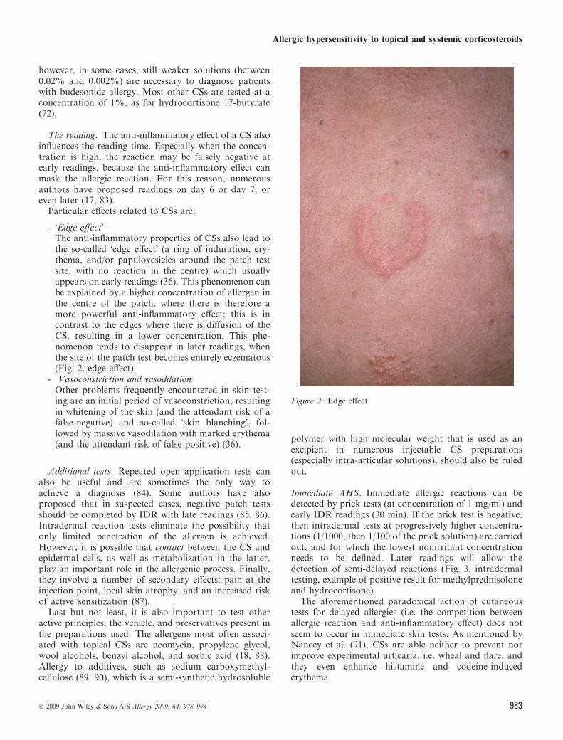
- �Edge effect�The anti-inflammatory properties of CSs also lead tothe so-called �edge effect� (a ring of induration, ery-thema, and/or papulovesicles around the patch testsite, with no reaction in the centre) which usuallyappears on early readings (36). This phenomenon canbe explained by a higher concentration of allergen inthe centre of the patch, where there is therefore amore powerful anti-inflammatory effect; this is incontrast to the edges where there is diffusion of theCS, resulting in a lower concentration. This phe-nomenon tends to disappear in later readings, whenthe site of the patch test becomes entirely eczematous(Fig. 2, edge effect).
- Vasoconstriction and vasodilationOther problems frequently encountered in skin test-ing are an initial period of vasoconstriction, resultingin whitening of the skin (and the attendant risk of afalse-negative) and so-called �skin blanching�, fol-lowed by massive vasodilation with marked erythema(and the attendant risk of false positive) (36).
Additional tests. Repeated open application tests canalso be useful and are sometimes the only way toachieve a diagnosis (84). Some authors have alsoproposed that in suspected cases, negative patch testsshould be completed by IDR with late readings (85, 86).Intradermal reaction tests eliminate the possibility thatonly limited penetration of the allergen is achieved.However, it is possible that contact between the CS andepidermal cells, as well as metabolization in the latter,play an important role in the allergenic process. Finally,they involve a number of secondary effects: pain at theinjection point, local skin atrophy, and an increased riskof active sensitization (87).Last but not least, it is also important to test other
active principles, the vehicle, and preservatives present inthe preparations used. The allergens most often associ-ated with topical CSs are neomycin, propylene glycol,wool alcohols, benzyl alcohol, and sorbic acid (18, 88).Allergy to additives, such as sodium carboxymethyl-cellulose (89, 90), which is a semi-synthetic hydrosoluble
polymer with high molecular weight that is used as anexcipient in numerous injectable CS preparations(especially intra-articular solutions), should also be ruledout.
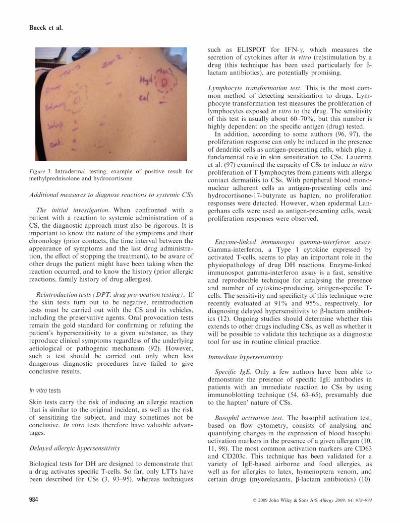
Immediate AHS. Immediate allergic reactions can bedetected by prick tests (at concentration of 1 mg/ml) andearly IDR readings (30 min). If the prick test is negative,then intradermal tests at progressively higher concentra-tions (1/1000, then 1/100 of the prick solution) are carriedout, and for which the lowest nonirritant concentrationneeds to be defined. Later readings will allow thedetection of semi-delayed reactions (Fig. 3, intradermaltesting, example of positive result for methylprednisoloneand hydrocortisone).
The aforementioned paradoxical action of cutaneoustests for delayed allergies (i.e. the competition betweenallergic reaction and anti-inflammatory effect) does notseem to occur in immediate skin tests. As mentioned byNancey et al. (91), CSs are able neither to prevent norimprove experimental urticaria, i.e. wheal and flare, andthey even enhance histamine and codeine-inducederythema.
Figure 2. Edge effect.
Allergic hypersensitivity to topical and systemic corticosteroids
� 2009 John Wiley & Sons A/S Allergy 2009: 64: 978–994 983
Additional measures to diagnose reactions to systemic CSs
The initial investigation. When confronted with apatient with a reaction to systemic administration of aCS, the diagnostic approach must also be rigorous. It isimportant to know the nature of the symptoms and theirchronology (prior contacts, the time interval between theappearance of symptoms and the last drug administra-tion, the effect of stopping the treatment), to be aware ofother drugs the patient might have been taking when thereaction occurred, and to know the history (prior allergicreactions, family history of drug allergies).
Reintroduction tests (DPT: drug provocation testing). Ifthe skin tests turn out to be negative, reintroductiontests must be carried out with the CS and its vehicles,including the preservative agents. Oral provocation testsremain the gold standard for confirming or refuting thepatient�s hypersensitivity to a given substance, as theyreproduce clinical symptoms regardless of the underlyingaetiological or pathogenic mechanism (92). However,such a test should be carried out only when lessdangerous diagnostic procedures have failed to giveconclusive results.
In vitro tests
Skin tests carry the risk of inducing an allergic reactionthat is similar to the original incident, as well as the riskof sensitizing the subject, and may sometimes not beconclusive. In vitro tests therefore have valuable advan-tages.
Delayed allergic hypersensitivity
Biological tests for DH are designed to demonstrate thata drug activates specific T-cells. So far, only LTTs havebeen described for CSs (3, 93–95), whereas techniques
such as ELISPOT for IFN-c, which measures thesecretion of cytokines after in vitro (re)stimulation by adrug (this technique has been used particularly for b-lactam antibiotics), are potentially promising.
Lymphocyte transformation test. This is the most com-mon method of detecting sensitization to drugs. Lym-phocyte transformation test measures the proliferation oflymphocytes exposed in vitro to the drug. The sensitivityof this test is usually about 60–70%, but this number ishighly dependent on the specific antigen (drug) tested.
In addition, according to some authors (96, 97), theproliferation response can only be induced in the presenceof dendritic cells as antigen-presenting cells, which play afundamental role in skin sensitization to CSs. Lauermaet al. (97) examined the capacity of CSs to induce in vitroproliferation of T lymphocytes from patients with allergiccontact dermatitis to CSs. With peripheral blood mono-nuclear adherent cells as antigen-presenting cells andhydrocortisone-17-butyrate as hapten, no proliferationresponses were detected. However, when epidermal Lan-gerhans cells were used as antigen-presenting cells, weakproliferation responses were observed.
Enzyme-linked immunospot gamma-interferon assay.Gamma-interferon, a Type 1 cytokine expressed byactivated T-cells, seems to play an important role in thephysiopathology of drug DH reactions. Enzyme-linkedimmunospot gamma-interferon assay is a fast, sensitiveand reproducible technique for analysing the presenceand number of cytokine-producing, antigen-specific T-cells. The sensitivity and specificity of this technique wererecently evaluated at 91% and 95%, respectively, fordiagnosing delayed hypersensitivity to b-lactam antibiot-ics (12). Ongoing studies should determine whether thisextends to other drugs including CSs, as well as whether itwill be possible to validate this technique as a diagnostictool for use in routine clinical practice.
Immediate hypersensitivity
Specific IgE. Only a few authors have been able todemonstrate the presence of specific IgE antibodies inpatients with an immediate reaction to CSs by usingimmunoblotting technique (54, 63–65), presumably dueto the hapten� nature of CSs.
Basophil activation test. The basophil activation test,based on flow cytometry, consists of analysing andquantifying changes in the expression of blood basophilactivation markers in the presence of a given allergen (10,11, 98). The most common activation markers are CD63and CD203c. This technique has been validated for avariety of IgE-based airborne and food allergies, aswell as for allergies to latex, hymenoptera venom, andcertain drugs (myorelaxants, b-lactam antibiotics) (10).
Figure 3. Intradermal testing, example of positive result formethylprednisolone and hydrocortisone.
Baeck et al.
984 � 2009 John Wiley & Sons A/S Allergy 2009: 64: 978–994
Encouraging results have also been obtained for CSs(D. Ebo and A. Rozieres, unpublished data).
Classification, allergenicity and cross-reactivity
Most of the studies on the allergenic nature and cross-reactions among CS that will be discussed here, concerntopical CS. Whether similar reactive profiles apply to CSadministered via the systemic or oral routes remains to bedemonstrated.
Chemical structure
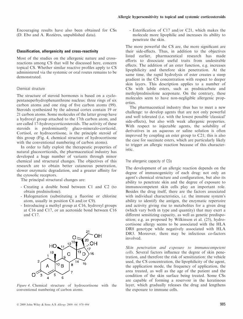
The structure of steroid hormones is based on a cyclo-pentaneperhydrophenanthrene nucleus: three rings of sixcarbon atoms and one ring of five carbon atoms (99).Steroids synthesized by the adrenal cortex contain 19 or21 carbon atoms. Some molecules of the latter group havea hydroxyl group attached to the 17th carbon atom, andare called 17-hydroxycorticosteroids. The activity of thesesteroids is predominantly gluco-mineralo-corticoid.Cortisol, or hydrocortisone, is the principle steroid ofthis group (Fig. 4, chemical structure of hydrocortisonewith the conventional numbering of carbon atoms).In order to fully exploit the therapeutic properties of
natural glucocorticoids, the pharmaceutical industry hasdeveloped a huge number of variants through minorchemical and structural changes. The objectives of thisresearch are to obtain better cutaneous penetration,slower enzymatic degradation, and a greater affinity forthe cytosolic receptors.The principal structural changes are:
- Creating a double bond between C1 and C2 (toobtain prednisolone).
- Halogenation (substituting a fluorine or chlorineatom, usually in position C6 and/or C9).
- Introducing a methyl group at C16, hydroxyl groupsat C16 and C17, or an acetonide bond between C16and C17.
- Esterification of C17 and/or C21, which makes themolecule more lipophilic and increases its ability topenetrate the skin.
The more powerful the CS are, the more significant aretheir side-effects. Thus, in addition to the objectiveslisted earlier, pharmaceutical research has madeefforts to dissociate useful traits from undesirableeffects. The addition of an ester function, e.g. increaseslipophilicity and therefore skin penetration. At thesame time, the rapid hydrolysis of ester creates a steepgradient in the CS concentration with respect to deeperskin layers. This description applies to a number ofCSs with labile esters, such as prednicarbate andmethylprednisolone aceponate. On the contrary, thesemolecules seem to have non-negligible allergenic prop-erties.
The pharmaceutical industry thus has to meet a newchallenge: to develop agents that are not only powerfuland well tolerated (i.e. with the lowest possible �classical�side-effects), but also with weak allergenic properties.With respect to injectable agents, the solubility ofderivatives in an aqueous or saline solution is oftenimproved by coupling an ester group to C21; this is alsothe case for succinate esters, which are particularly likelyto trigger an allergic reaction because of this character-istic.
The allergenic capacity of CSs
The development of an allergic reaction depends on thedegree of immunogenicity of each drug: not only anagent�s chemical structure and configuration, but also itsability to penetrate skin and the degree of exposure toimmunocompetent skin cells play an important role.Besides the drug itself, there are the factors associatedwith individual characteristics, i.e. the immune system�sability to identify the antigen, the enzymatic repertoireand activity giving rise to metabolites for a given drug(which vary both in type and quantity) that may exert adifferent sensitizing capacity, as well as genetic predispo-sition; e.g. as proposed by Wilkinson et al. (25), hydro-cortisone allergy seems to be associated with the HLADR8 genotype while negatively associated with HLADR3. Moreover, there may be infectious co-factorsinvolved.
Skin penetration and exposure to immunocompetentcells. Several factors influence the degree of skin pene-tration, and therefore the risk of sensitization: the vehicleused, the CS concentration, the lipophilicity of the agent,the application mode, the frequency of application, thearea treated, as well as the age of the patient and thecondition of the skin surface being treated. Some CSsare capable of forming a reservoir in the keratinouslayer, which gradually releases the drug and lengthensthe exposure to immune cells.
Figure 4. Chemical structure of hydrocortisone with theconventional numbering of carbon atoms.
Allergic hypersensitivity to topical and systemic corticosteroids
� 2009 John Wiley & Sons A/S Allergy 2009: 64: 978–994 985
Skin occlusion, which increases the pH as well as theskin penetration, therefore also increases the risk ofsensitization. This explains the high frequency of CShypersensitivity in patients with stasis dermatitis, venousulcers, or anogenital dermatitis conditions that arecharacterized by maceration.
Skin metabolism routes. The process of skin metabolismplays an important role in the allergenic potential of CSsand also influences their cross-reaction profiles (24). Thecutaneous metabolic reactions involved in the biotrans-formation of CSs have not yet been fully determined.The epidermis is a metabolically-active structure contain-ing a variety of enzymes, in this respect acting muchlike other organs (the liver, for instance). One of themost important steps in metabolizing CSs is hydrolysis.Indeed, for some agents, chemical or enzymatic hydro-lysis of the ester function is often necessary to obtainsufficient biological activity (100). The role of hydrolysisdepends on the stability and size of the ester, theconditions under which the product was preserved, andthe patient�s enzymes. C21 esters are less stable (labile)and thus easily hydrolyzed, whereas C17 esters are muchmore resistant. Agents with both a C17 mono-esterfunction and a C21 hydroxyl group, however, are alsounstable in an acidic or alkaline environment, and afterhydrolysis of the C21 ester, the C17 ester can rapidlyundergo nonenzymatic conversion into the C21 mono-ester. Enzymatic hydrolysis then quickly follows, con-verting it into free alcohol. In general, CSs whichmetabolize rapidly in the skin (the D2 group of labileesters) produce allergic reactions more often than thosethat metabolize slowly or not at all.
Sensitization capacity of metabolites. It has become clearthat the allergen that triggers an immune reaction maynot be the CS itself but may be a by-product of itsmetabolism. The principal metabolites of hydrocortisonein an aqueous or alcoholic solution are dehydro-21 ofsteroid glyoxals (101). Not all agents have the samesensitization capacity, which explains the large differencesobserved between various CSs.
Protein binding
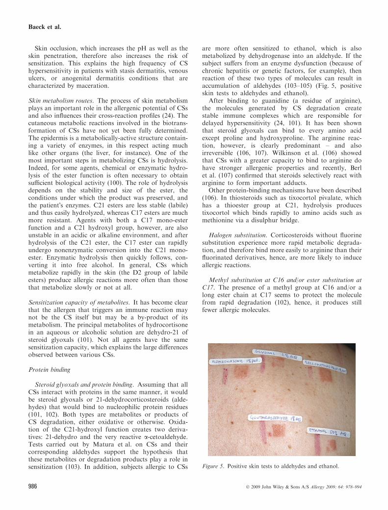
Steroid glyoxals and protein binding. Assuming that allCSs interact with proteins in the same manner, it wouldbe steroid glyoxals or 21-dehydrocorticosteroids (alde-hydes) that would bind to nucleophilic protein residues(101, 102). Both types are metabolites or products ofCS degradation, either oxidative or otherwise. Oxida-tion of the C21-hydroxyl function creates two deriva-tives: 21-dehydro and the very reactive a-cetoaldehyde.Tests carried out by Matura et al. on CSs and theircorresponding aldehydes support the hypothesis thatthese metabolites or degradation products play a role insensitization (103). In addition, subjects allergic to CSs
are more often sensitized to ethanol, which is alsometabolized by dehydrogenase into an aldehyde. If thesubject suffers from an enzyme dysfunction (because ofchronic hepatitis or genetic factors, for example), thenreaction of these two types of molecules can result inaccumulation of aldehydes (103–105) (Fig. 5, positiveskin tests to aldehydes and ethanol).
After binding to guanidine (a residue of arginine),the molecules generated by CS degradation createstable immune complexes which are responsible fordelayed hypersensitivity (24, 101). It has been shownthat steroid glyoxals can bind to every amino acidexcept proline and hydroxyproline. The arginine reac-tion, however, is clearly predominant – and alsoirreversible (106, 107). Wilkinson et al. (106) showedthat CSs with a greater capacity to bind to arginine dohave stronger allergenic properties and recently, Berlet al. (107) confirmed that steroids selectively react witharginine to form important adducts.
Other protein-binding mechanisms have been described(106). In thiosteroids such as tixocortol pivalate, whichhas a thioester group at C21, hydrolysis producestixocortol which binds rapidly to amino acids such asmethionine via a disulphur bridge.
Halogen substitution. Corticosteroids without fluorinesubstitution experience more rapid metabolic degrada-tion, and therefore bind more easily to arginine than theirfluorinated derivatives, hence, are more likely to induceallergic reactions.
Methyl substitution at C16 and/or ester substitution atC17. The presence of a methyl group at C16 and/or along ester chain at C17 seems to protect the moleculefrom rapid degradation (102), hence, it produces stillfewer allergic molecules.
Figure 5. Positive skin tests to aldehydes and ethanol.
Baeck et al.
986 � 2009 John Wiley & Sons A/S Allergy 2009: 64: 978–994
Cross vs concomitant reactions
Sensitized patients often test positive to several CSs.Although the existence of cross-reaction phenomena hasbeen proved, e.g. obtaining positive skin tests forsynthetic CSs to which a patient could never have been
exposed (because they are not yet on the market),simultaneous or subsequent sensitizations can never beentirely ruled out. It is often extremely difficult todetermine as to which CS a patient has been exposed toin the past. As Issakson emphasized, it is therefore moreappropriate to mention concomitant sensitization (which
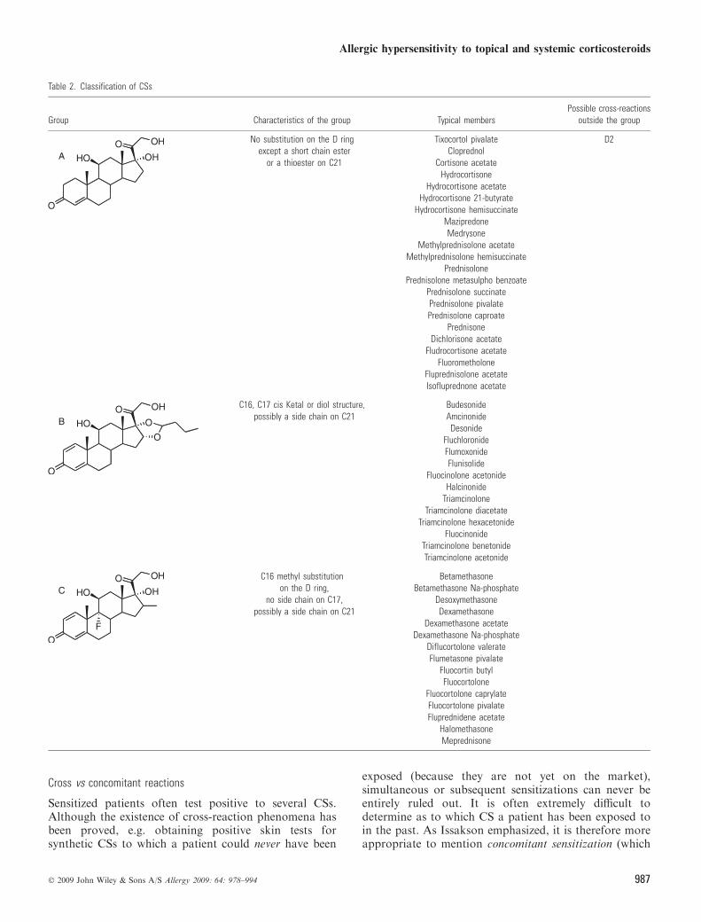
Table 2. Classification of CSs
Group Characteristics of the group Typical membersPossible cross-reactions
outside the group
No substitution on the D ringexcept a short chain ester
or a thioester on C21
Tixocortol pivalate D2Cloprednol
Cortisone acetateHydrocortisone
Hydrocortisone acetateHydrocortisone 21-butyrate
Hydrocortisone hemisuccinateMazipredoneMedrysone
Methylprednisolone acetateMethylprednisolone hemisuccinate
PrednisolonePrednisolone metasulpho benzoate
Prednisolone succinatePrednisolone pivalatePrednisolone caproate
PrednisoneDichlorisone acetate
Fludrocortisone acetateFluorometholone
Fluprednisolone acetateIsofluprednone acetate
C16, C17 cis Ketal or diol structure,possibly a side chain on C21
BudesonideAmcinonide
DesonideFluchloronideFlumoxonideFlunisolide
Fluocinolone acetonideHalcinonide
TriamcinoloneTriamcinolone diacetate
Triamcinolone hexacetonideFluocinonide
Triamcinolone benetonideTriamcinolone acetonide
C16 methyl substitutionon the D ring,
no side chain on C17,possibly a side chain on C21
BetamethasoneBetamethasone Na-phosphate
DesoxymethasoneDexamethasone
Dexamethasone acetateDexamethasone Na-phosphate
Diflucortolone valerateFlumetasone pivalate
Fluocortin butylFluocortolone
Fluocortolone caprylateFluocortolone pivalateFluprednidene acetate
HalomethasoneMeprednisone
Allergic hypersensitivity to topical and systemic corticosteroids
� 2009 John Wiley & Sons A/S Allergy 2009: 64: 978–994 987
may include cross-reactions) (108). For this reason, thepossibility of using animal models has been studied (109,110). Beside patch test data, statistical and three-dimen-sional structural analysis can also address the cross-reactivity issue.
Cross-reactions: ABCD classification. In 1989, based onstructural and clinical characteristics, Coopman et al.(111) classified CSs into four reactivity groups, namelyGroups A, B, C and D, the volume occupied bysubstituents on the D ring of the CS molecule being acritical element of CS binding to skin proteins.In 1995, Lepoittevin et al. (112) carried out confor-
mational analysis of the observed cross-reactivitiesbetween CSs, which supported this classification.Groups A, B and D are indeed very homogenous interms of molecular structure and significant differencesare only evident between CSs of different groups. Thebehaviour of budesonide, which cross-reacts not onlywith other acetonides but also with CS esters, can beattributed to its unique molecular structure: an acetalfunction gives it a resemblance to molecules from bothGroups B and D, respectively, according to theirstereospecificity. Group C, despite its structural simi-larity to Group A, has no unifying characteristics interms of structure or volume.Further clinical data ledMatura and Goossens (24, 100)
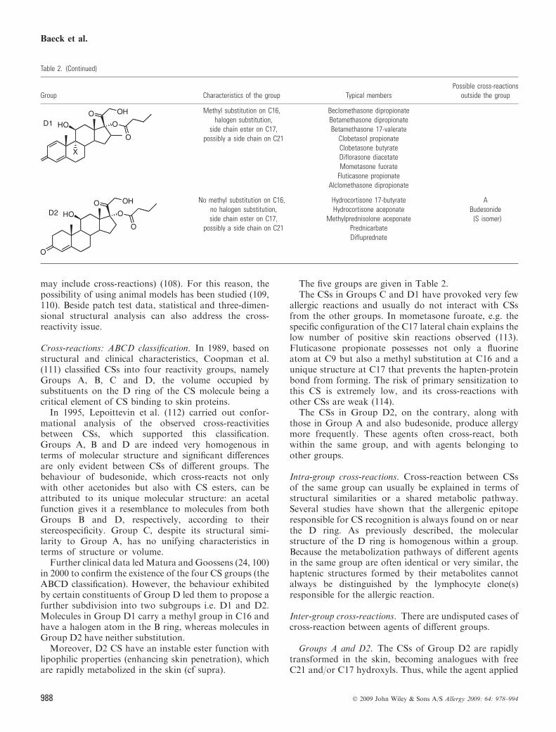
in 2000 to confirm the existence of the four CS groups (theABCD classification). However, the behaviour exhibitedby certain constituents of Group D led them to propose afurther subdivision into two subgroups i.e. D1 and D2.Molecules in Group D1 carry a methyl group in C16 andhave a halogen atom in the B ring, whereas molecules inGroup D2 have neither substitution.Moreover, D2 CS have an instable ester function with
lipophilic properties (enhancing skin penetration), whichare rapidly metabolized in the skin (cf supra).
The five groups are given in Table 2.The CSs in Groups C and D1 have provoked very few
allergic reactions and usually do not interact with CSsfrom the other groups. In mometasone furoate, e.g. thespecific configuration of the C17 lateral chain explains thelow number of positive skin reactions observed (113).Fluticasone propionate possesses not only a fluorineatom at C9 but also a methyl substitution at C16 and aunique structure at C17 that prevents the hapten-proteinbond from forming. The risk of primary sensitization tothis CS is extremely low, and its cross-reactions withother CSs are weak (114).
The CSs in Group D2, on the contrary, along withthose in Group A and also budesonide, produce allergymore frequently. These agents often cross-react, bothwithin the same group, and with agents belonging toother groups.
Intra-group cross-reactions. Cross-reaction between CSsof the same group can usually be explained in terms ofstructural similarities or a shared metabolic pathway.Several studies have shown that the allergenic epitoperesponsible for CS recognition is always found on or nearthe D ring. As previously described, the molecularstructure of the D ring is homogenous within a group.Because the metabolization pathways of different agentsin the same group are often identical or very similar, thehaptenic structures formed by their metabolites cannotalways be distinguished by the lymphocyte clone(s)responsible for the allergic reaction.
Inter-group cross-reactions. There are undisputed cases ofcross-reaction between agents of different groups.
Groups A and D2. The CSs of Group D2 are rapidlytransformed in the skin, becoming analogues with freeC21 and/or C17 hydroxyls. Thus, while the agent applied
Table 2. (Continued)
Group Characteristics of the group Typical membersPossible cross-reactions
outside the group
Methyl substitution on C16,halogen substitution,
side chain ester on C17,possibly a side chain on C21
Beclomethasone dipropionateBetamethasone dipropionateBetamethasone 17-valerate
Clobetasol propionateClobetasone butyrateDiflorasone diacetateMometasone fuorate
Fluticasone propionateAlclomethasone dipropionate
No methyl substitution on C16,no halogen substitution,side chain ester on C17,
possibly a side chain on C21
Hydrocortisone 17-butyrate ABudesonide
(S isomer)Hydrocortisone aceponate
Methylprednisolone aceponatePrednicarbateDifluprednate
Baeck et al.
988 � 2009 John Wiley & Sons A/S Allergy 2009: 64: 978–994
to the skin is one of the �labile ester� CSs (D2), itsmetabolite corresponds more closely to Group A. Oneexample of intracutaneous biometabolization is theconversion of hydrocortisone 17-butyrate (Group D2)to hydrocortisone 21-butyrate, followed by an enzymatictransformation into hydrocortisone (Group A). Thisillustrates the typical cross-reaction observed betweenGroups A and D2.
Budesonide and Group D2. Budesonide can be consid-ered as a unique case in the following sense: its acetalfunction is actually an equal mixture of the R and Sdiastereoisomers (112). Both R and S diastereoisomerscan develop cross-reactions with Group B CSs, but onlythe S diastereoisomer can cross-react with Group D2CSs (115). An analysis of their configuration shows thatwhile the R isomer exhibits symmetry that is character-istic of the B group, the S isomer can take on anasymmetric aspect. The result is a large hydrophobic�cavity� at the C17 ester function, similar to Group Dagents.
Sex-hormonal steroids. It has been observed that somepatients sensitized to CSs respond positively to skin testswith other steroids, such as the sex-hormonal steroids(116, 117). Wilkinson and Beck (118) estimatedthe prevalence of 17-hydroxyprogesterone allergies inpatients reacting positively to hydrocortisone to be 19–26% and considered these to be the expression of cross-senstitivity. They also considered that in most cases thereare no clinical consequences. However, CS allergy caninduce an allergy to, e.g. endogenous progesterone, forwhich the clinical presentation is a generalized eczema-tous eruption that gets worse during the premenstrualperiod: so-called auto-immune progesterone dermatitis(AIPD) (119). On the contrary, any patient presentingsymptoms compatible with AIPD should also be testedfor CS allergy.
Other hypotheses regarding cross-reactivity. The ABCDclassification cannot explain all observed cross-reactions.Furthermore, a purely structural analysis of CSs does notjustify the existence of Group C.Wilkinson et al. suggested that each CS has several
immunological sites (120) and consider that there aretwo principal sites involved in immune recognition, i.e.the C16/C17 and C6/C9 substitutions. They emphasizedthat modifications to the C21 lateral chain do notinfluence the allergenic nature of the substance, probablydue to skin metabolism and degradation of these esters.They also (120, 121) claimed that B-ring halogen substi-tutions (C6 or C9) are the major determinants of cross-reactions between CSs. Although the fluorine atom issimilar in shape and size to the hydrogen atom, theyexplain the different reactivity of halogenated and non-halogenated molecules as being caused by charge differ-ences (122). Hydrogen is relatively electro-positive and
fluorine is electro-negative. It is also possible for ahalogen atom in position C9 to exert an electricalattraction on the carbon atom at C11, conferring ahigher stability to the CS and therefore limiting thecreation of allergenic by-products. Several authors havebased their work on Wilkinson� hypothesis, proposinghalogenated replacement agents for their allergic patients.However, while this rule (positive allergic responses areless frequent for fluorinated derivatives) seems to hold inGroups A, C and D1, there seems to be no differencebetween fluorinated and nonfluorinated derivatives inGroup B.
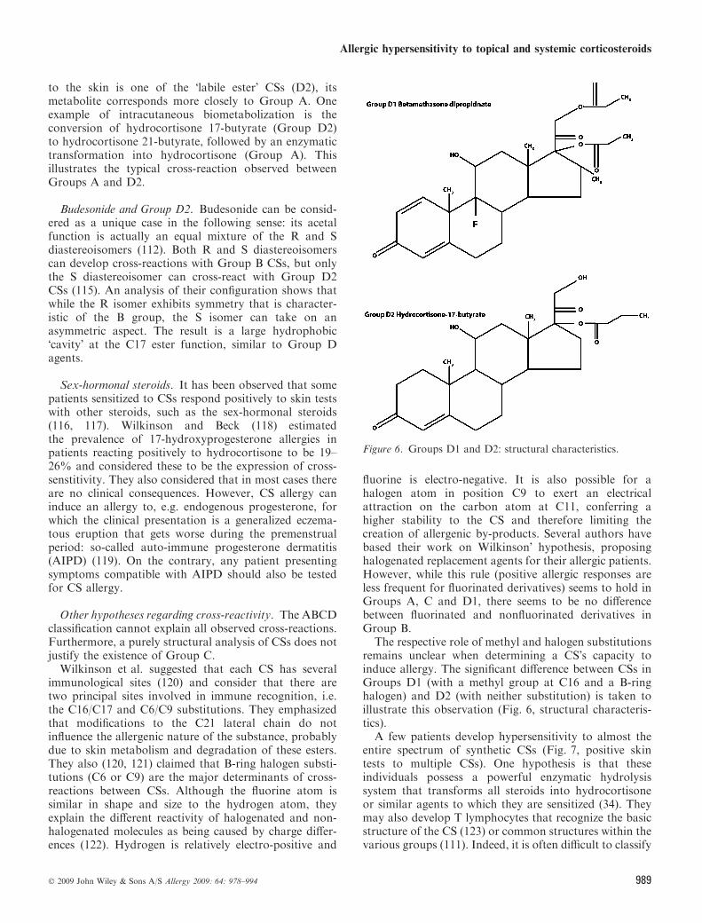
The respective role of methyl and halogen substitutionsremains unclear when determining a CS�s capacity toinduce allergy. The significant difference between CSs inGroups D1 (with a methyl group at C16 and a B-ringhalogen) and D2 (with neither substitution) is taken toillustrate this observation (Fig. 6, structural characteris-tics).
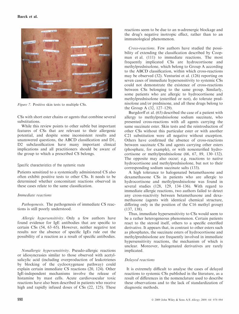
A few patients develop hypersensitivity to almost theentire spectrum of synthetic CSs (Fig. 7, positive skintests to multiple CSs). One hypothesis is that theseindividuals possess a powerful enzymatic hydrolysissystem that transforms all steroids into hydrocortisoneor similar agents to which they are sensitized (34). Theymay also develop T lymphocytes that recognize the basicstructure of the CS (123) or common structures within thevarious groups (111). Indeed, it is often difficult to classify
Figure 6. Groups D1 and D2: structural characteristics.
Allergic hypersensitivity to topical and systemic corticosteroids
� 2009 John Wiley & Sons A/S Allergy 2009: 64: 978–994 989
CSs with short ester chains or agents that combine severalsubstitutions.While this review points to other subtle but important
features of CSs that are relevant to their allergenicpotential, and despite some inconsistent results andunanswered questions, the ABCD classification and D1,D2 subclassification have many important clinicalimplications and all practitioners should be aware ofthe group to which a prescribed CS belongs.
Specific characteristics of the systemic route
Patients sensitized to a systemically administered CS alsooften exhibit positive tests to other CSs. It needs to bedetermined whether concomitant reactions observed inthese cases relate to the same classification.
Immediate reactions
Pathogenesis. The pathogenesis of immediate CS reac-tions is still poorly understood.
Allergic hypersensitivity. Only a few authors havefound evidence for IgE antibodies that are specific tocertain CSs (54, 63–65). However, neither negative testresults nor the absence of specific IgEs rule out thepossibility of a reaction as a result of specific antibodies.
Nonallergic hypersensitivity. Pseudo-allergic reactionsor idiosyncrasies similar to those observed with acetyl-salicylic acid (including overproduction of leukotrienesby blocking of the cyclooxygenase pathway) couldexplain certain immediate CS reactions (20, 124). OtherIgE-independent mechanisms involve the release ofhistamine by mast cells. Acute cardiovascular toxicreactions have also been described in patients who receivehigh and rapidly infused doses of CSs (22, 125). These
reactions seem to be due to an a-adrenergic blockage andthe drug�s negative inotropic effect, rather than to animmunological phenomenon.
Cross-reactions. Few authors have studied the possi-bility of extending the classification described by Coop-man et al. (111) to immediate reactions. The mostfrequently implicated CSs are hydrocortisone andmethylprednisolone, which belong to Group A accordingto the ABCD classification, within which cross-reactionsmay be observed (32). Venturini et al. (126) reporting onseven cases of immediate hypersensitivity to systemic CSscould not demonstrate the existence of cross-reactionsbetween CSs belonging to the same group. Similarly,some patients who are allergic to hydrocortisone andmethylprednisolone (esterified or not), do tolerate pred-nisolone and/or prednisone, and all these drugs belong tothe Group A (32, 127–129).
Burgdorff et al. (63) described the case of a patient withallergy to methylprednisolone sodium succinate, whopresented cross-reactions with all agents carrying thesame succinate ester. Skin tests and the reintroduction ofother CSs without this particular ester or with anotherC21 substitution were all negative without exception.Others have confirmed the absence of cross-reactionsbetween succinate CSs and agents carrying other esters(phosphate, for example), or with nonesterified hydro-cortisone or methylprednisolone (66, 67, 89, 130–132).The opposite may also occur; e.g. reactions to nativehydrocortisone and methylprednisolone, but not to theircorresponding sodium succinate salts (133).
A high tolerance to halogenated betamethasone anddexamethasone CSs in patients who are allergic tohydrocortisone and methylprednisolone was found inseveral studies (128, 129, 134–136). With regard toimmediate allergic reactions, two authors failed to detectany cross-reactivity between betamethasone and dexa-methasone (agents with identical chemical structure,differing only in the position of the C16 methyl group)(137, 138).
Thus, immediate hypersensitivity to CSs would seem tobe a rather heterogeneous phenomenon. Certain patientsreact to the steroid itself, others to a specific esterifiedderivative. It appears that, in contrast to other esters suchas phosphates, the succinate esters of hydrocortisone andmethylprednisolone are frequently involved in immediatehypersensitivity reactions, the mechanism of which isunclear. Moreover, halogenated derivatives are rarelyimplicated.
Delayed reactions
It is extremely difficult to analyse the cases of delayedreactions to systemic CSs published in the literature, as aresult of differences in the nomenclature used to describethese observations and to the lack of standardization ofdiagnostic methods.
Figure 7. Positive skin tests to multiple CSs.
Baeck et al.
990 � 2009 John Wiley & Sons A/S Allergy 2009: 64: 978–994
Conclusion
Allergic hypersensitivity to CSs is a common finding in adermatological practice, the skin being the main sensiti-zation route, and delayed-type allergic reactions are muchmore frequently encountered than immediate-type reac-tions. As a result of the anti-inflammatory properties ofCSs, however, the clinical signs of allergy may be maskedand the results of skin tests difficult to interpret. Hence,CS allergy still remains unfamiliar to many clinicians whooften prescribe these drugs, both for topical or systemicadministration.Moreover, because of the frequent occurrence of cross-
reactions between these agents, CS-allergic patients, evenwhen recognized as such, are sometimes still prescribed aCS that they cannot tolerate.With regard to the cross-sensitivity patterns, the
ABCD classification and D1, D2 subclassification seemvery useful in directing the search for a replacement
agent, but cannot be a substitute for a systematic,individualized evaluation of each patient�s sensitization/tolerance profile. Further studies are still needed tovalidate this classification of allergic contact dermati-tis and to determine whether this classification is alsouseful for immediate and delayed reactions to systemicCSs.
Acknowledgments
The author would like to warmly thank the members of the Der-matology Department at the University Hospital of Leuven, whohosted him. He would also like to thank collaborators from theImmuno-Allergology Department at the University Hospital CentreLyon Sud, for their hospitality and collaboration. His gratitudegoes also to Hugues Depasse and Michele Lemaire from the audio-visual centre at the University Hospital UCL Brussels, for theirvaluable assistance in illustrating this review.
References
1. Hench PS, Kendall EC, Slocumb CH,Polley HF. The effect of a hormoneof the adrenal cortex (17-hydroxy-11-dehydrocorticosterone:compound E)and of pituitary adrenocorticotropichormone on rheumatic arthritis. ProcStaff Meet Mayo Clin 1949;24:181.
2. Sulzberger MB, Witten VH. Pre-liminary and short reports. The effectof topically applied compound F inselected dermatoses. J Invest Dermatol1952;19:101–102.
3. Lauerma A. Contact hypersensitivity toglucocorticosteroids. Am J ContactDermat 1992;31:112–132.
4. Burckhardt W. Kontaktekzem durchhydrocortisone. Hautarzt1959;10:42–43.
5. Kooij R. Hypersensitivity to hydrocor-tisone. Br J Dermatol 1959;71:392–394.
6. Kendall PH. Untowards effects follow-ing local hydrocortisone injection. AnnPhys Med 1958;4:170–175.
7. Posadas SJ, Pichler WJ. Delayed drughypersensitivity reactions – new con-cepts. Clin Exp Allergy 2007;37:989–999.
8. Allergie et hypersensibilite. In: JanewayCA, Travers P, Walport M, ShlomchiMT editors. Immunobiologie, 2nd edn.De Boeck, 2003:471–500.
9. Johansson SG, Hourihane JO,Bousquet J, Bruijnzeel-koomen C,Dreborg S, Haahtela T et al. A revisednomenclature for allergy. An EAACIposition statement from the EAACInomenclature task force. Allergy2001;56:813–824.
10. Ebo D, Sainte-Laudy J, Bridts C,Mertens C, Hagendorens M,Schuerwegh A et al. Flow-assisteddiagnosis: current applications andfuture perspectives. Allergy2006;61:1028–1039.
11. Torres MJ, Blanca M, Fernandez J,Romano A, de Weck A, Aberer Wet al. Diagnosis of immediate allergicreactions to beta-lactam antibiotics.Allergy 2003;58:961–972.
12. Rozieres A Hypersensibilite retardeeaux medicaments. De laphysiopathologie au diagnosticimmunobiologique. Doctoral Thesis.Lyon: University Press, 2007.
13. Romano A, Blanca M, Torres MJ,Bircher A, Aberer W, Brockow K.Diagnosis of nonimmediate reactions tobeta-lactam antibiotics. Allergy2004;59:1153–1160.
14. Beeler A, Zaccaria L, Kawabata T,Gerber BO, Pichler WJ. CD69 upregu-lation on T cells as an in vitro markerfor delayed-type drug hypersensitivity.Allergy 2008;63:181–188.
15. Rasanen L, Hasan T. Allergy to sys-temic and intralesional corticosteroids.Br J Dermatol 1993;128:407–411.
16. Lauerma AI, Reitamo S. Allergicreactions to topical and systemiccorticosteroids. Eur J Dermatol1994;5:354–358.
17. Dooms-Goossens A, Andersen KE,Brandao FM. Corticosteroid contactallergy: an EECDRG multicentrestudy. Contact Dermatitis1996;35:40–44.
18. Degreef H, Dooms-Goossens A. Thenew corticosteroids: are they effectiveand safe? Dermatol Ther 1993;11:155–160.
19. Lauerma AI. Occupational contactsensitization to corticosteroids. ContactDermatitis 1998;39:328–329.
20. Karsh J, Yang WH. An anaphylacticreaction to intra-articular triamcino-lone: a case report and review of theliterature. Ann Allergy Asthma Immu-nol 2003;90:254–258.
21. Nakamura H, Matsuse H, Obase Y,Mitsuta K., Tomari S, Saeki S et al.Clinical evaluation of anaphylacticreactions to intravenous corticosteroidsin adult asthmatics. Respiration2002;69:309–313.
22. Kamm GL, Hagmeyer KO. Allergictype reactions to corticosteroids. AnnPharmacother 1999;33:451–460.
23. Just N, Nyunga M, Lelong J, WallaertB. Immediate allergy to oralcorticosteroids. Rev Med Int2005;26:331–334.
24. Matura M, Goossens A. Contactallergy to corticosteroids. Allergy2000;55:698–704.
25. Wilkinson SM, Morrey K, HollowoodK, Heagerty AH, English JSC.HLA-A,B and DR antigens inhydrocortisone contact hypersensitiv-ity. Contact Dermatitis 1993;28:295–297.
26. Isaksson M, Bruze M. Corticosteroids.Dermatitis 2005;16:3–5.
Allergic hypersensitivity to topical and systemic corticosteroids
� 2009 John Wiley & Sons A/S Allergy 2009: 64: 978–994 991
27. Bennett ML, Fountain JM, McCartyMA, Sherertz EF. Contact allergy tocorticosteroids in patients using inhaledor intranasal corticosteroids for allergicrhinitis or asthma. Am J Contact Der-mat 2001;12:193–196.
28. Isaksson M. Skin reactions to inhaledcorticosteroids. Drug Saf 2001;24:369–373.
29. Isaksson M, Bruze M, Hornblad Y,Svenonius E, Wihl JA. Contact allergyto corticosteroids in asthma/rhinitispatients. Contact Dermatitis1999;40:327–328.
30. Malik M, Tobin AM, Shanahan F,O�Morain C, Kirby B, Bourke J.Steroid allergy in patients with inflam-matory bowel disease. Br J Dermatol2007;157:967–969.
31. Klein-Gitelman AS, Pachman LM.Intravenous corticosteroids: adversereactions variable than expected inchildren. J Rheumatol 1998;25:1995–2002.
32. Freymond N, Catelain A, Queille E,Augey F, Nicolas JF. Allergic reactionto methylprednisolone. Rev Med Int2003;24:698–700.
33. Thyssen JP, Maibach HI. Drug-elicitedsystemic allergic (contact) dermatitis –update and possible pathomechanisms.Contact Dermatitis 2008;59:195–202.
34. Le Coz CJ. Hypersensitivity tocorticosteroids. Ann Dermatol Vene-reol 2002;129:346–347.
35. Dooms-Goossens A, Degreef H. Clini-cal aspects of contact allergy to corti-costeroids. Dermatology 1994;189:54–55.
36. Dooms-Goossens A. Corticosteroidcontact allergy: a challenge to patchtesting. Am J Contact Dermat1993;4:120–122.
37. Goh CL. Cross sensitivity to multipletopical corticosteroids. Contact Der-matitis 1989;20:65–67.
38. Rodriguez-Serna M, Silvestre JF,Quecedo E, Martinez A, Miguel FJ,Gauchia R. Corticosteroid allergy: re-port of 3 unusually acute cases. ContactDermatitis 1996;35:361–362.
39. Stingeni L, Caraffini S, Assalve D,Lapomarda V, Lisi P. Erythema-mul-tiforme-like contact dermatitis frombudesonide. Contact Dermatitis1996;34:154–155.
40. Stingeni L, Hansel K, Lisi P. Morbilli-form erythema-multiforme-like erup-tion from desoxymethasone. ContactDermatitis 1996;35:363–364.
41. Miranda-Romero A, Sanchez-Sambucety P, Bajo C, Martinez M,Garcia-Munoz M. Genital oedemafrom contact allergy to prednicarbate.Contact Dermatitis 1998;38:228–229.
42. Rietschel RL. Photocontact dermatitisto hydrocortisone. Contact Dermatitis1978;4:334–337.
43. Mathias CGT, Robertson DB. Delayedhypersensitivity to a corticosteroidsuspension containing methylpredniso-lone. Two cases of conjunctivalinflammation after retrobulbar injec-tion. Arch Dermatol 1985;121:258–261.
44. Monk BE, Skipper D. Allergy to topi-cal corticosteroids in inflammatorybowel disease. Gut 2003;52:597.
45. Dooms-Goossens A. Allergy to inhaledcorticosteroids: a review. Am J ContactDermat 1995;6:1–3.
46. Gonzalo Garijo MA, BobabillaGonzalez P. Cutaneous-mucosal aller-gic contact reaction due to topical cor-ticosteroids. Allergy 1995;50:833–836.
47. Bircher AJ, Pelloni F, LangauerMessmer S, Muller D. Delayed hyper-sensitivity reactions to corticosteroidsapplied to mucous membranes. Br JDermatol 1996;135:310–313.
48. Isaksson M, Bruze M, Wihl JA. Con-tact allergy to budesonide and perfo-ration of the nasal septum. ContactDermatitis 1997;37:133.
49. Pirker C, Misic, Frosch PJ. Angioe-dema and dysphagia caused by contactallergy to inhaled budesonide. ContactDermatitis 2003;49:77–79.
50. Callens A, Vaillant L, Machet L,Pelucio-Lopez C, de Calan S, LoretteG. Contact stomatitis from tixocortolpivalate. Contact Dermatitis1993;29:161.
51. Clark RJ. Exacerbation of asthma afternebulised beclomethasone dipropio-nate. Lancet 1986;6:574–575.
52. Mc Divern DV, Macfarlane JT.Severe bronchoconstriction after inha-lation of budesonide. Br Med J1984;288:447.
53. Miranda A, Garcia JJ, Torres MJ, DelCano A, Rondon C, Aguilar MJ et al.Bronchospasm reaction induced byinhaled budesonide. Allergy1993;48:148.
54. Lauerma A, Kiistala R, Makinen-Kiljunen S, Haahtela T. Allergic skinreaction after inhalation of budesonide.Clin Exp Allergy 1993;23:232–233.
55. Fuchs T, Uter W, Sprotte U. Urticariadue to budesonide – late phase IgEdependent reaction? Allergologie,Jahrgang 1991;14:234–238.
56. Ponten A. Airborne occupational con-tact dermatitis caused by extremely lowconcentrations of budesonide. ContactDermatitis 2006;55:121–124.
57. Amin N, Brancaccio R, Cohen D.Cutaneous reactions to injectablecorticosteroids. Dermatitis2006;17:143–146.
58. Valsecchi R, Reseghetti A, Leghissa P,Cologni L, Cortinovis R. Erythema-multiforme-like lesions fromtriamcinolone acetonide. ContactDermatitis 1998;38:362–363.
59. Lopez-Serrano MC, Moreno-AncilloA, Contreras J, Ortega N, Cabanas R,Barranco P et al. Two cases of specificadverse reactions to systemic corticos-teroids. Investig Allergol Clin Immunol1998;6:324–327.
60. Mace S, Vadas P, Pruzanski W. Ana-phylactic shock induced by intraartic-ular injection of methylprednisoloneacetate. J Rheumatol 1997;24:1191–1194.
61. Fernandez de Corres L, Urrutia I,Audicana M, Echechipia S,Gastaminza G. Erythroderma afterintravenous injection of methylpred-nisolone. Contact Dermatitis1991;25:68–70.
62. Armingaud P, Martin L, Wierzbicka E,Esteve E. Baboon syndrome due topolysensitization with corticosteroids.Ann Dermatol Venereol 2005;132:675–677.
63. Burgdorff T, Venelmalm L, Vogt T,Landthaler M, Stolz W. IgE mediatedanaphylactic reaction induced by suc-cinate ester of methylprednisolone. AnnAllergy Asthma Immunol 2002;89:425–428.
64. Pryse-Phillips WEM, Chandra RK,Rose B. Anaphylactoid reaction tomethylprednisolone pulsed therapy formultiple sclerosis. Neurology1984;34:1119–1121.
65. Rasanen L, Tarvainen K, Makinen-Kijunen S. Urticaria to hydrocortisone.Allergy 2001;56:352–353.
66. Fernandez S, Reano M, Vives R.6-Methylprednisolone-inducedbronchospasm. Allergy 1997;52:780–782.
67. Freedman MD, Schocker AL, ChapelN, Gerber JG. Anaphylaxis afterintravenous methylprednisoloneadministration. JAMA1981;245:607–608.
68. King RA. A severe anaphylactoidreaction to hydrocortisone. Lancet1960;2:1093–1094.
69. Wilkinson SM, English JSC. Hydro-cortisone sensitivity: a prospectivestudy of the value of tixocortol pivalateand hydrocortisone acetate as patchtest markers. Contact Dermatitis1991;25:132–133.
70. Isaksson M, Bruze M, Goossens A,Lepoittevin JP. Patch-testing withserial dilutions of tixocortol pivalateand potential cross-reactivesubstances. Acta Derm Venereol2000;80:33–38.
Baeck et al.
992 � 2009 John Wiley & Sons A/S Allergy 2009: 64: 978–994
71. Isaksson M, Bruze M, Goossens A,Lepoittevin JP. Patch testing withbudesonide in serial dilutions: thesignificance of dose, occlusion time andreading time. Contact Dermatitis1999;40:24–31.
72. Lauerma A. Screening for corticoste-roid contact sensitivity: comparison oftixocortol pivalate, hydrocortisone17-butyrate and hydrocortisone.Contact Dermatitis 1991;24:123–130.
73. Isaksson M, Brandao FM, Bruze M,Goossens A. Recommendation toinclude budesonide and tixocortolpivalate in the European standard ser-ies. Contact Dermatitis 2000;43:41–63.
74. Boffa MJ, Wilkinson SM, Beck MH.Screening for corticosteroid contacthypersensitivity. Contact Dermatitis1995;33:149–151.
75. Isaksson M, Andersen KE, BrandaoFM, Bruynzeel DP, Bruze M, CamarsaJ et al. Patch testing with corticosteroidmixes in Europe: a Multicentre Studyof the EECDRG. Contact Dermatitis2000;42:27–35.
76. Wilkinson SM, Beck MH. Corticoste-roid contact hypersensitivity: whatvehicle and concentration? ContactDermatitis 1996;34:305–308.
77. Isaksson M, Beck MH, Wilkinson SM.Comparative testing with budesonide inpetrolatum and ethanol in a standardseries. Contact Dermatitis 2002;47:123–124.
78. Isaksson M, Gruvberger B, Persson L,Bruze M. Stability of corticosteroidpatch test preparations. ContactDermatitis 2000;42:144–148.
79. Isaksson M, Bruze M. Late patch-testreactions to budesonide need not be asign of sensitization induced by the testprocedure. Am J Contact Dermat2003;14:154–156.
80. Isaksson M, Bruze M, Bjorkner B,Hindsen M, Svensson L. The benefit ofpatch testing with a corticosteroid at alow concentration. Am J Contact Der-mat 1999;10:31–33.
81. Isaksson M, Andersen KE, BrandaoFM, Bruynzeel DP, Bruze M, DiepgenT et al. Patch testing with budesonidein serial dilutions. A multicentre studyof the EECDRG. Contact Dermatitis2000;42:352–354.
82. Isaksson M, Bruze M, Matura M,Goossens A. Patch testing with lowconcentrations of budesonide detectscontact allergy. Contact Dermatitis1997;37:241–242.
83. Isaksson M. Corticosteroid contactallergy – the importance of late read-ings and testing with corticosteroidsused by the patients. Contact Derma-titis 2007;56:56–57.
84. Weber F, Barbaud A, Reichert-Penetrat S, Danchin A, Schmutz JL.Unusual clinical presentation in a caseof contact dermatitis due to corticos-teroids diagnosed by ROAT. ContactDermatitis 2001;44:105–106.
85. Seukeran DC, Wilkinson SM, BeckMH. Patch testing to detect corticoste-roid allergy: is it adequate? ContactDermatitis 1997;36:127–130.
86. Wilkinson SM, English JS. Patch testsare poor detectors of corticosteroidallergy. Contact Dermatitis 1992;26:67–68.
87. Mimesh S, Pratt M. Allergic contactdermatitis from corticosteroids: repro-ductibility of patch testing and corre-lation with intradermal testing.Dermatitis 2006;17:137–142.
88. Coloe J, Zirwas M. Allergens incorticosteroid vehicles. Dermatitis2008;19:38–42.
89. Murieta-Aguttes M, Michelen V,Leynadier F, Duarte-Risselin C,Halpern GM, Dry J. Systemic allergicreactions to corticosteroids. J Asthma1991;28:329–339.
90. Patterson D, Yunginger J, Dunn W,Jones R, Hunt L. Anaphylaxis inducedby the carboxymethylcellulose compo-nent of injectable triamcinoloneacetonide suspension (Kenalog). AnnAllergy Asthma Immunol1995;74:163–166.
91. Nancey S, Freymond N, Catelain A,Cousin F, Rozieres A, Nicolas JF.Effects of local corticosteroids on acuteexperimental urticaria. Eur J Dermatol2004;14:323–326.
92. Aberer W, Bircher A, Romano A,Blanca M, Campi P, Fernandez J et al.Drug provocation testing in the diag-nosis of drug hypersensitivity reactions:general considerations. Allergy2003;58:854–863.
93. Buettiker U, Keller M, Picheler WJ,Braathen LR, Yamalkar N. Oralprednisolone induced acute generalizedexanthematous pustulosis due to corti-costeroids of group A confirmed byepicutaneous testing and lymphocytetransformation tests. Dermatology2006;213:40–43.
94. Comaish S. A case of hypersensitivityto corticosteroids. Br J Dermatol1969;81:919–925.
95. Wilkinson SM, English JSC, MatteyDL. In vitro evidence of delayed-typehypersensitivity to hydrocortisone.Contact Dermatitis 1993;29:241–245.
96. Braathen SR, Thorsby E. Human epi-dermal Langerhans cells are more po-tent than blood monocytes in inducingsome antigen-specific T cell responses.Br J Dermatol 1983;108:139–146.
97. Lauerma A, Rasanen L, Reunala T,Reitamo S. Langerhans cells but notmonocytes are capable of antigen pre-sentation in vitro in corticosteroidcontact hypersensitivity. Br J Dermatol1990;123:699–705.
98. Ebo D, Dombrecht E, Bridts C, AertsN, De Clerck L, Stevens W. Combinedanalysis of intracellular signalling andimmunophenotype of human periph-eral blood basophils by flow cytometry:a proof of concept. Clin Exp Allergy2007;37:1668–1675.
99. Reynolds J. The extra Pharmacopeia.In: Martindale, editor. Corticosteroids,29th edn. London: The PharmaceuticalPress, 1989:872–902.
100. Matura MContact allergy to locallyapplied corticosteroids. DoctoralThesis. Leuven: Leuven UniversityPress, 1998.
101. Bundgaard H. The possible implicationof steroid-glyoxal degradation productsin allergic reactions to corticosteroids.Arch Pharm Chem 1980;8:83–90.
102. Fleischer D. Chemical stability ofpharmaceuticals: a handbook forpharmacists. In: Connors KA, AmidonGL, Stella VJ, editors. Hydrocortisone.New York: John Wiley and Sons,1986:483–490.
103. Matura M., Lepoittevin JP, Arbez-Gindre C, Goossens A. Testing withcorticosteroid aldehydes in corticosen-sitive subjects (preliminary results).Contact Dermatitis 1998;38:106–108.
104. Dooms-Goossens A. Is there a rela-tionship between contact allergy toalcohols and contact allergy to corti-costeroids? Eur J Dermatol 1993;3:713.
105. Kuligowski ME, Chang A, Van BaarH. Contact allergy to alcohols withcoexisting hypersensitivity to corticos-teroids. Eur J Dermatol 1993;3:183–186.
106. Wilkinson SM, Jones MF. Corticoste-roid usage and binding to arginine.Determinants of corticosteroid hyper-sensitivity. Br J Dermatol1996;135:225–230.
107. Berl V, Claudel E, Gerberick G,Lepoittevin JP. Allergic contact der-matitis to corticosteroids: a mechanisticstudy. Contact Dermatitis 2008;58:10.
108. Isaksson M, Bruze M. Corticosteroidcross reactivity. Contact Dermatitis2003;49:53–54.
109. Hausen BM, Foussereau J. The sensi-tizing capacity of tixocortol pivalate.Contact Dermatitis 1988;18:63–64.
110. Bruze M, Bjorkner B, Dooms-Goossens A. Sensitization studies withmometasone fuorate, tixocortol piva-late and budesonide in the guinea pig.Contact Dermatitis 1996;34:161–164.
Allergic hypersensitivity to topical and systemic corticosteroids
� 2009 John Wiley & Sons A/S Allergy 2009: 64: 978–994 993
111. Coopman S, Degreef H, Dooms-Goossens A. Identification ofcross-reaction patterns in allergiccontact dermatitis from topicalcorticosteroids. Br J Dermatol1989;121:27–34.
112. Lepoittevin JP, Drieghe J, Dooms-Goossens A. Studies in patients withcorticosteroid contact allergy: under-standing cross-reactivity among differ-ent steroids. Arch Dermatol1995;131:31–37.
113. Dooms-Goossens A, Lepoittevin JP.Studies on the contact allergenicpotential of mometasone furoate: aclinical and molecular study. Eur JDermatol 1996;6:339–340.
114. Goossens A, Huygens S, Matura M,Degreef H. Fluticasone propionate: arare contact sensitizer. Eur J Dermatol2001;11:29–34.
115. Isaksson M, Bruze M, Lepoittevin JP,Goossens A. Patch testing with serialdilutions of budesonide, its R and Sdiastereomers, and potentiallycross-reacting substances. Am JContact Dermat 2001;12:170–176.
116. Schoenmakers A, Vermorken A,Degreef H, Dooms-Goossens A. Cor-ticosteroid or steroid allergy? Dermati-tis 1992;26:159–162.
117. Lamb SR, Wilkinson SM. Contact al-lergy to progesterone and estradiol in apatient with multiple corticosteroidallergies. Dermatitis 2004;15:78–81.
118. Wilkinson SM, Beck MH. The signifi-cance of positive patch tests to 17-hy-droxyprogesterone. Contact Dermatitis1994;30:302–303.
119. Ingber A, Trattner A, David M.Hypersensitivity to an estrogen –progesterone preparation and possiblerelationship to autoimmune progester-one dermatitis and corticosteroidhypersensitivity. J Dermatolog Treat1999;10:139.
120. Wilkinson SM. Corticosteroid cross-reactions: an alternative view. ContactDermatitis 2000;42:59–63.
121. Wilkinson SM, Hollis S, Beck MH.Reactions to other corticosteroids inpatients with allergic contact dermatitisto hydrocortisone. Br J Dermatol1995;132:766–777.
122. Block H, Holliday AK. Modernphysical chemistry. London:Butterworths, 1973.
123. Isaksson M. Systemic contact allergy tocorticosteroids revisited. Contact Der-matitis 2007;57:386–388.
124. Dajani BM, Sliman NA, Shubair KS,Hamzeh YS. Bronchospasm caused byintravenous hydrocortisone sodiumsuccinate (solu-cortef) in aspirine-sensitive asthmatics. J Allergy ClinImmunol 1981;68:201–204.
125. Erstad BL. Severe cardiovascular ad-verse effect in association with acute,high dose corticosteroid administra-tion. Ann Pharmacother 1989;23:1019–1022.
126. Venturini M, Lobera T, Del Pozo MD,Gonzales I, Blasco A. Immediatehypersensitivity to corticosteroids.J Investig Allergol Clin Immunol2006;16:51–56.
127. Fulcher D, Katelaris C. Anaphylactoidreaction to intravenous hydrocortisonesodium succinate: a case report andliterature review. Med J Aust1991;154:210–214.
128. Sieck JO, Al-Ohaly Y, Saour J, KhanM, Henriquez H. An allergic reactionto intravenous methylprednisoloneadministration. Br J Clin Pract1990;44:723–725.
129. Rao KV, Andersen RC, O�Brien T.Successful renal transplantation in apatient with anaphylactic reaction tosolu-medrol (methylprednisolone so-dium succinate). Am J Med1982;72:161–163.
130. Calogiuri GF, Muratore L, Nettis E,Ventura MT, Ferrannini A, Tursi A.Anaphylaxis to hydrocortisone hemi-succinate with cross sensitivity to re-lated compounds in a pediatric patient.Br J Dermatol 2004;151:707–708.
131. Laine-Cessac P, Moshinaly H, GouelloJP, Geslin P, Allain P. Severe anaphy-lactoid reaction to intravenous corti-costeroid therapy. A case report.Therapie 1990;45:505–508.
132. Peces R, Gorostidi M, Azofra J,Sanchez L, Alvares J. Anaphylaxisfollowing intravenous methylpred-nisolone sodium succinate in a renaltransplant recipient. Nephron 1991;59:497–498.
133. Mendelson LM, Meltzer EO,Hamburger RN. Anaphylaxis-likereactions to corticosteroid therapy. JAllergy Clin Immunol 1974;54:125–131.
134. Mansfield LE, Ting S, Haverly RW.Anaphylaxis caused by the sodiumsuccinate ester of hydrocortisone andmethylprednisolone. J Asthma1986;23:81–83.
135. Ventura MT, Calogiuri GF, MatinoMG, Dagnello M, Buquicchio R,Foti C et al. Alternative glucocorticoidsfor use in case of adverse reaction tosystemic glucocorticoids: a study on10 patients. Br J Dermatol 2003;148:139–141.
136. Escribano-Rodriguez M, Gonzalez-PolJ, Munoz-Bellido F, De la Calle ToralA, Velazquez-Amor E, Conde-Hernandez J. Immediate reaction tomethylprednisolone with tolerance ofother corticoteroids. Allergy1997;52:677–678.
137. Erdmann SM, Abuzahra F, Merck HF,Schroeder A, Baron JM. Anaphylaxisinduced by glucocorticoids. J AmBoard Fam Pract 2005;18:143–146.
138. Ventura MT, Sanapo F, Calogiuri GF,Satriano F. Anaphylaxis induced byintramuscular betamethasone disodiumphosphate: reflections on a clinical case.Int J Immunopathol Pharmacol2007;20:387–391.
Baeck et al.
994 � 2009 John Wiley & Sons A/S Allergy 2009: 64: 978–994


![HIGHLIGHTS OF PRESCRIBING INFORMATION ...pi.lilly.com/us/baqsimi-uspi.pdf · • Hypersensitivity and Allergic Reactions [see Warnings and Precautions (5.3)]. 6.1 Clinical Trial Data](https://static.fdocuments.in/doc/165x107/5e85aab16701912d9a3af01b/highlights-of-prescribing-information-pilillycomusbaqsimi-uspipdf-a.jpg)